DHEA action is mediated by multiple receptors and metabolites.
115
University of Louisville University of Louisville ThinkIR: The University of Louisville's Institutional Repository ThinkIR: The University of Louisville's Institutional Repository Electronic Theses and Dissertations 5-2004 DHEA action is mediated by multiple receptors and metabolites. DHEA action is mediated by multiple receptors and metabolites. Kristy K. Michael Miller University of Louisville Follow this and additional works at: https://ir.library.louisville.edu/etd Recommended Citation Recommended Citation Miller, Kristy K. Michael, "DHEA action is mediated by multiple receptors and metabolites." (2004). Electronic Theses and Dissertations. Paper 975. https://doi.org/10.18297/etd/975 This Doctoral Dissertation is brought to you for free and open access by ThinkIR: The University of Louisville's Institutional Repository. It has been accepted for inclusion in Electronic Theses and Dissertations by an authorized administrator of ThinkIR: The University of Louisville's Institutional Repository. This title appears here courtesy of the author, who has retained all other copyrights. For more information, please contact [email protected].
-
Upload
khangminh22 -
Category
Documents
-
view
4 -
download
0
Transcript of DHEA action is mediated by multiple receptors and metabolites.

University of Louisville University of Louisville
ThinkIR: The University of Louisville's Institutional Repository ThinkIR: The University of Louisville's Institutional Repository
Electronic Theses and Dissertations
5-2004
DHEA action is mediated by multiple receptors and metabolites. DHEA action is mediated by multiple receptors and metabolites.
Kristy K. Michael Miller University of Louisville
Follow this and additional works at: https://ir.library.louisville.edu/etd
Recommended Citation Recommended Citation Miller, Kristy K. Michael, "DHEA action is mediated by multiple receptors and metabolites." (2004). Electronic Theses and Dissertations. Paper 975. https://doi.org/10.18297/etd/975
This Doctoral Dissertation is brought to you for free and open access by ThinkIR: The University of Louisville's Institutional Repository. It has been accepted for inclusion in Electronic Theses and Dissertations by an authorized administrator of ThinkIR: The University of Louisville's Institutional Repository. This title appears here courtesy of the author, who has retained all other copyrights. For more information, please contact [email protected].

DHEA ACTION IS MEDIATED BY MULTIPLE RECEPTORS AND METABOLITES
By
Kristy K. Michael Miller, B.S. Indiana University - Bloomington
A Dissertation Submitted to the Faculty of the
Graduate School of the University of Louisville in Partial Fulfillment of the Requirements
for the Degree of
Doctor of Philosophy
Department of Biochemistry and Molecular Biology University of Louisville
Louisville, Kentucky
May 2004

DHEA ACTION IS MEDIATED BY MULTIPLE RECEPTORS AND METABOLITES
By
Kristy K. Michael Miller, B.S. Indiana University - Bloomington, 1999
A Dissertation Approved on
April 30, 2004 (Date)
By the Following Dissertation Committee:
Dissertation Director
ii

DEDICATION
This dissertation is dedicated to my
"Papaw" Paul Jones whose prayers and example
helped me become the person I am today.
111

ACKNOWLEDGEMENTS
I have many people to thank for helping me attain my goal of finishing graduate
school with a Ph.D. in biochemistry. Foremost, is my research advisor, Dr. Russell A.
Prough. He invested a great deal of his time and energy teaching me, guiding me,
encouraging and supporting me. Therefore, I would like to express my sincerest
gratitude to Dr. Prough who not only has been a wonderful mentor but also a good friend.
I will forever be indebted to him for his support and guidance in helping me attain my
goals as a scientist and making me a better person.
Additionally, I would like to thank my committee members, Drs. Tom
Geoghegan, Robert Gray, Carolyn Klinge, and William Pierce Jr. who have also provided
me with guidance throughout my graduate training. There was never a time when they
did not stop what they were doing to answer my questions. They have been a wonderful
foundation of encouragement and support.
Special thanks also goes to Sharon L. Ripp who helped mentor me in my early
years of training. She helped me develop valuable scientific writing skills as well as
influenced my scientific thought process. I still seek her scientific skill and advice even
today. Beyond science, Dr. Ripp also listened to me, encouraged and advised me, and
without her, I would not have made it through the first few years of graduate school.
Many thanks go to the students, faculty and staff of the Department of
Biochemistry and Molecular Biology at the University of Louisville for helping make my
graduate experience enjoyable. Additionally, I would like to especially thank the people
IV

in the lab that I worked with side by side on daily basis. Their camaraderie always made
it fun to come to work everyday. I would especially like to thank Mary, who was like a
mom to me and always looked out and cared for me, Viola who was always there to
laugh with me, Immaculate who also enjoyed listening to country music, and many others
(Mike, Stephanie, Cam, Laura, and Boaz) who made everyday interesting and enjoyable.
Additionally, I would like to thank Dr. Jian Cai and Dr. Harrell Hurst in the
Department of Pharmacology for their technical help with the GC/MS. Also, many
thanks to Kat Mattingly who helped with the competitive binding assays.
I am also forever indebted to my parents and family who have always been there
to love, encourage and support me throughout the years of my life. Also, many thanks
and appreciation goes to my husband, Eric who has also endured this experience with me.
He has provided me with tremendous understanding and love even when I did not
deserve it. Without him, I would not have had the sanity to endure and reach my goals.
I would also like to thank Jill Lyles Ph.D. who inspired me to go to graduate
school. As an undergraduate, she gave me advice regarding a career and a future in
science. She provided me with inspiration and encouragement and still does so today.
Additionally, I would also like to thank my friends and those in my prayer group,
who have always encouraged me during this period in my life. They have helped me
endure the hardships and have shared with me in the joys of life. Special thanks to
Cheryl Jones, who I have always looked up to and who has never ceased to pray for me.
Finally, lowe everything to my Lord Jesus Christ who has given me the talents of
which I can use. It is only by His grace and love that I am strong and enabled to live the
life to which I am called. To Christ be all the glory.
v

ABSTRACT
DHEA ACTION IS MEDIATED BY MULTIPLE RECEPTORS AND
METABOLITES
ADVISOR: RUSSELL A. PROUGH Ph.D.
BY KRISTY K. MICHAEL MILLER
MAY 2004
Dehydroepiandrosterone (DHEA) is a C-19 adrenal steroid and the most abundant
circulating hormone in humans. Since circulating levels decline in late adulthood,
treatment of humans with DHEA has been suggested to have beneficial health effects.
Although the mechanism of action is unknown, DHEA may be metabolized to active
metabolites that exert their physiological effects by receptor-mediated processes and cell
signaling pathways. The purpose of this study was to investigate the mechanistic
processes ofDHEA action.
Since DHEA may exert its pleotropic effects by being metabolized to biologically
active species, a GC/MS method was developed to quantify the liver microsomal
metabolism ofDHEA of various species and identify the P450 enzymes responsible for
metabolism. 16a-hydroxy-DHEA and 7a-hydroxy-DHEA were formed in rat, hamster,
pig and human. CYP3A4 and CYP3A5 formed 7a-hydroxy-DHEA, 16a-hydroxy
DHEA, and the unique human metabolite, 7j3-hydroxy-DHEA, while the fetal enzyme
CYP3A 7 fonned only 16a-hydroxy and 7p-hydroxy-DHEA. By using this method to
examine the metabolite profiles of various P450s, the developmental expression patterns
VI

of the human cytochrome P4503A forms could be classified and therefore have
significant clinical relevance.
Nuclear receptors transduce the effects of hormones into transcriptional
responses. DHEA and metabolites were screened in a cell-based assay to determine the
interaction with estrogen receptors alpha and beta (ERa and ERP). DHEA, DHEA-S,
and androstendiol activated ERa, while DHEA, 7-oxo-DHEA, androstenedione and
androstenediol activated ERP demonstrating ER is activated directly by DHEA and some
metabolites.
These and other studies from our laboratory demonstrate that DHEA is
metabolized into various monohydroxylated metabolites. DHEA and metabolites directly
activate ER as well as the pregnane X receptor (PXR). Additionally, DHEA has been
shown to activate another nuclear receptor, peroxisome proliferator activated receptor
alpha (PP ARa) in vivo. This research suggests that DHEA action is mediated by multiple
receptors and metabolites with various biological activities, comprising of a complex
mode of action ofDHEA.
VB

TABLE OF CONTENTS Page
ACKNOWLEDGEMENTS ..................................................................... .iv ABSTRACT .......... ................................................................................ vi LIST OF TABLES ....... .......................................................................... ix LIST OF FIGURES ................................................................................. x
CHAPTER
I. INTRODUCTION .................................................................. 1
II. STEREO- AND REGIOSELECTIVITY ACCOUNT FOR THE
DIVERSITY OF DHEA METABOLITES PRODUCED BY
LIVER MICROSOMAL CYTOCHROMES P450 ........................... 23
III. DHEA AND METABOLITES ACTIVATE ESTROGEN
RECEPTOR ........................................................................ 50
IV. DISCUSSION ............................................................. '" ...... 76
REFERENCES ................................................................................... .. 82
APPENDICES ................................................................................... ... 97
CURRICULUM VITA ..... " ............. '" ....................... '" ....... " ................. 1 00
Vlll

LIST OF TABLES
Page
TABLE I. Physiological Concentrations of DHEA-S ........ " ............................ 5
T ABLE II. Substrates and functions of human and rat CYP genes ...................... 14
TABLE III. Content ofP450 and NADPH: Cytochrome P450 Oxidoreductase in
various baculovirus preparations ............................................... 27
TABLE IV. GC-SIM-MS data for several standards ....................................... 32
TABLE V. GC/MS analysis ofDHEA metabolites formed in various species ......... 38
TABLE VI. Rates ofDHEA metabolites formed from baculovirus expressed P450 .. .44
IX
Figure 1.
Figure 2.
Figure 3.
Figure 4.
Figure 5.
Figure 6.
Figure 7.
Figure 8.
Figure 9.
Figure 10.
Figure 11.
Figure 12.
Figure 13.
Figure 14.
Figure 15.
LIST OF FIGURES
Page
Biosynthesis ofDHEA and other steroids in humans ........................ 2
The /),,4 and /),,5 Steroidogenic Pathway .......................................... 3
Variation of circulating DHEA-S levels throughout human life ............ 7
Generalized catalytic cycle for P450 reactions ............................... .12
Structure/function organization of nuclear receptors ........................ 16
Localization of ER isoforms ..................................................... 19
Domain structure representation of human ERa and ER~ isoforms. '" ... 21
Separation of DHEA and metabolites by GCIMS ..... " ........ , . '" ........ 31
Rat liver microsomal metabolism ofDHEA at 0 minutes ................... 34
Rat liver microsomal metabolism ofDHEA at 10 minutes .................. 35
Time dependent formation ofDHEA metabolites by rat, hamster and pig
after GC/MS analysis ............................................................ .37
Time dependent formation ofDHEA metabolites by human liver
microsomal fractions .............................................................. 39
Characteristic mass spectrum of7~-OH-DHEA in human liver
microsomal fraction ...................................... '" ................ '" .. .40
Characteristic mass spectrum of7~-OH-DHEA standard .................. .41
Time dependent formation ofDHEA metabolites by human liver
microsomal fractions ............................................................. .43
x

Figure 16.
Figure 17.
Figure 18.
Figure 19.
Figure 20.
Figure 21.
Figure 22.
Figure 23.
Figure 24.
Figure 25.
Figure 26.
Structure of estradiol and common anti-estrogenic compounds ........... 52
17p-Estradiol increases expression of 3EREc38-dependent reporter
gene activity ....................................................................... 58
Increased ERELUC reporter activity in response to DHEA and
metabolites ......................................................................... 59
Nonnalized ERELUC reporter activity in response to DHEA and
metabolites ......................................................................... 62
Concentration dependent activation of ERa by DHEA, DHEA-S and
ADIOL in HEK293 cells ......................................................... 63
Concentration depending activation ofERP by DHEA, 7-oxo-DHEA,
ADIOL and AD lONE in HepG2 cells ......................................... 64
Inhibition ofERELUC reporter activity in presence of cotransfected ERa
in HEK293 cells .................................................................. 66
Inhibition of ERELUC reporter activity in presence of cotransfected ERP
in HepG2 cells .................................................................... 67
Effect ofP450 inhibitors (non-specific inhibitor, miconazole
or aromatase inhibitor, exemestane) on ERELUC reporter activity in
response to DHEA and metabolites in presence of cotransfected
ERa in HEK293 cells ............................................................ 69
Lack of effect ofP450 inhibitors on ERELUC reporter activity in
response to DHEA and metabolites in presence of
cotransfected Epa in HepG2 cells ............................................... 70
Competition curves for DHEA metabolite binding to ERa ................ 71
Xl

Figure 27.
Figure 28.
Competition curves for DHEA metabolite binding to ER~ ................. 72
DHEA action is mediated by multiple receptors and metabolites ......... 81
XlI

CHAPTER I
INTRODUCTION
DEHYDROEPIANDROSTERONE
Dehydroepiandrosterone (5-androsten-3~-01-17-one) is a naturally occurring C-19
adrenal steroid derived from cholesterol by a series of cytochrome P450 mono-oxygenase
and hydroxysteroid dehydrogenase catalyzed reactions (Figure 1).
Dehydroepiandrosterone (DHEA) is secreted primarily by the zona reticularis of the
adrenal cortex of humans and other primates. DHEA secretion is controlled by
adrenocorticotrophin (ACTH) and other pituitary factors (Nieschlag et al., 1973). The
adrenal cortex secretes 75-90% ofthe body's DHEA, with the remainder being produced
by the testes and ovaries (Vermeulen, 1980 and de Peretti and Forest, 1978 and Nieschlag
et aI., 1973).
Primates produce DHEA by the ~5-steroidogenic pathway in which the double
bond at the C-5 and C-6 position is maintained. In this process, the P450 side-chain
cleavage (P450scc or P45011Al) converts cholesterol to pregnenolone. Pregnenolone is
then hydroxylated at the C-17 position followed by a two carbon side chain cleavage by
P450C17 to form DHEA (Figure 2).
1

N
HSS DHEA-S • • DHEA
CYP17,20
Cholesterol
1 CYPIIAI (P450scc)
(desmolase) Pregnenolone ~ ~
3{3-HSD .... Progesterone ....
/~ 17{3-HSD
Glucocorticoids Mineralocorticoids
17{fHSD Androstenedione ... ~ Testosterone
5a-R 3a-HSD
Androsterone
17{3-KSR
CYP19 faromatase)
Estradiol
Figure 1. Biosynthesis of DHEA and other steroids in humans. The enzymes responsible for conversions are italicized, the listing
of more than one enzyme indicates a multisystem process. HSD, hydroxysteroid dehydrogenase; HSS, hydroxysteroid sulfatase; KSR,
ketosteroid reductase; R, reductase; sec, side chain cleavage enzyme; SH, sulfohydrolase (Figure adapted from Kroboth et al. J Clin
Pharmacal. (1999) 39: 327-348).

11 4-Steroidogenic
Pathway
CH3 I c=o
O~ Progesterone
I CH3 P450 C17 ~ 6=0
Estrogens
Cholesterol
P450 see ~ r~ 0
OOa)P Pregnenolone
a 5-Steroidogenic
Pathway
170H-Pregnenolone
J 0
(11)0 313HSD "0650 (11)0 ~
Alldro8telledione (.A.nckostenedi 0 l)
De hydr oep ia nd roste ro ne
(17 BHSD)
Testosterone
Figure 2. The 114 and 115 Steroidogenic Pathways. (Figure adapted from Conley and
Bird. Biol.Reprod. (1997) 56:789-799).
3

Little or no DHEA is produced by the adrenal of nonprimate species, such as mice
and rats. Instead, nonprimates produce sex steroids via the d 4-steroidogenic pathway in
which cholesterol is converted to pregnenolone by P450scc . Pregnenolone is then
converted to progesterone by 3p-hydroxysteroid dehydrogenase (3P-HSD) and it is taken
up by peripheral steroidogenic tissues and converted to androstenedione by P450C17
(Figure 2).
Although DHEA is the primary sterol in these biosynthetic pathways, DHEA is
largely found in circulation in its sulfated fonn, DHEA 3p-sulfate (DHEA-S), which can
be interconverted with DHEA by DHEA sulfotransferases and hydroxysteroid sulfatases
(Regalson et ai., 1994). Although DHEA-S is the hydrophilic storage fonn that circulates
in the blood, as stated previously, DHEA is the principle fonn used in steroid hormone
synthesis. Therefore, the differences in tissue-specific expression ofDHEA
sulfotransferase and steroid sulfatase determine the balance between DHEA
interconversions with DHEA inactivation (Allolio and Arlt, 2002).
In humans, plasma DHEA concentrations are found in the range of 1-4 ng/mL
(0.003 - 0.015J.lM) (Table I), but circulating DHEA-S concentrations are much greater
(Barrett-Connor E et al., 1986 and Hopper and Yen, 1975). Bird et ai., (1984) reported
that 64% and 74% of the daily production ofDHEA is converted to DHEA-S in women
and men, respectively, but only about 13% ofDHEA-S is hydrolyzed back to DHEA.
On a molar basis, circulating DHEA-S concentrations are 250 and 500 times higher (~1 -
10 J.lM) than those ofDHEA in women and men, respectively (Labrie F et ai., 1995).
The abundant circulating concentrations ofDHEA-S are due in part because DHEA is
4

Concentration Serum Level (J.lM) (J-lg/dL) 0.01 ",0.3 0.1 ",3
1 ",30 5 ",150
10 ",300 25 ",750 50 ",1500
100 ",3000 TABLE I. Physiological concentrations of DHEA-S.
5

cleared from the blood at a rate of approximately 2000 Llday, whereas DHEA-S
clearance is about 13 Llday (Lephart et al., 1987). Additionally, DHEA has a half-life in
blood of about 1 to 3 hours, while DHEA-S has a half-life of 10 to 20 hours (Rosenfeld et
at., 1975). Clearance rate is defined as the volume of plasma that would contain the
amount of drug excreted per unit volume. Therefore, clearance expresses the rate of drug
removal from the plasma, but not the amount of drug eliminated. The clearance rates of
DHEA and its sulfate are also influenced by their protein-binding characteristics. For
example, DHEA is weakly bound to albumin, while DHEA-S is strongly bound to
albumin.
PHYSIOLOGICAL AND PHARMACOLOGICAL CONCENTRATIONS OF
DHEA
In its sulfated form, DHEA is the most abundant circulating sterol in humans,
followed by androstenedione. During fetal development, plasma DHEA-S levels are
around 100-200 llg/dL (3-7 IlM), but fall rapidly after birth and remain low for the first
five years of life. Blood DHEA levels then rise and peak around 300 Ilg/dL (10 IlM)
during the second decade of postnatal life, followed by an age-dependent decline.
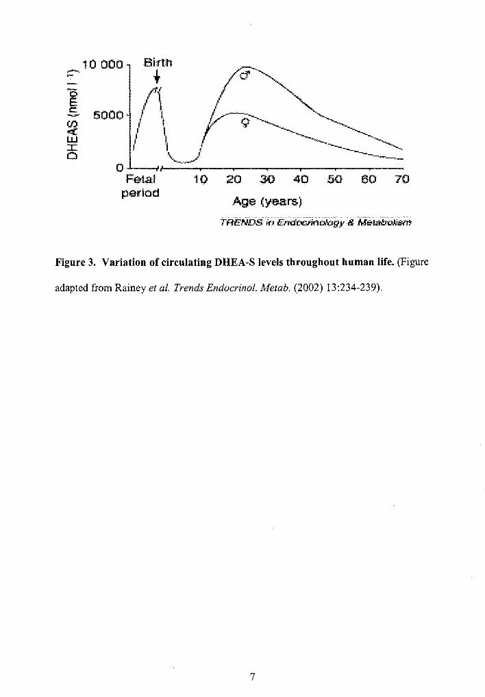
Additionally, there are clear gender differences in circulating levels of DHEA-S with
higher levels found in men than women (Figure 3).
Labrie and coworkers (1987) suggest that a decrease in 17,20-desmolase (see
Figure 1) activity may be responsible for the dramatic age-related reduction in DHEA
and DHEA-S secretion. Regardless, the drastic developmental changes in DHEA
6
10000 -. .-
5000
eo 70
Age~years)
Figure 3. Variation of circulating DHEA-S levels throughout human life. (Figure
adapted from Rainey et al. Trends Endocrinol. Metab. (2002) 13:234-239).
7

secretion are not observed by other steroid hormones, suggesting that the mechanisms
regulating DHEA formation are unique (Rainey et al., 2002). In contrast, serum
cholesterol levels tend to increase with age, while other steroid hormones, decline more
slowly relative to DHEA with age. The decline in circulating levels ofDHEA and its
sulfate derivative appear to be inversely correlated to the rise in cholesterol and the
pathophysiological effects of aging (Barret-Conner et aI., 1999).
Because the decline in DHEA is associated with some of the pathophysiological
effects of aging, many people supplement their own DHEA levels with exogenous DHEA
and even refer to DHEA as the "fountain of youth hormone." When administered,
DHEA is usually in an encapsulated powder in two or three divided doses. Although
appropriate physiological doses are not well defined and differ in men and women, many
clinical studies have been conducted using 50 mg/day for women and 100 mg/day for
men.
Currently, DHEA is available over-the-counter as a dietary supplement and is
therefore not regulated by the Food and Drug Administration. However, this has not
always been the case. DHEA was once marketed for weight loss and in 1985, the FDA
banned over-the-counter sales ofDHEA. DHEA is still outlawed by the International
Olympic Committee and the National Collegiate Athletic Association, but since the
passage of the Dietary Supplement Health and Education Act of 1994, DHEA has again
been widely available in health food stores in the US (and elsewhere) where is it
marketed as a dietary supplement. There are fewer regulations over the rule of nutritional
products than with nonprescription or prescription drugs. For example, expiration dates
8

are not required and there are no chemical standards for the product, and nutritional
supplements, such as DHEA, can be sold unless the FDA proves that they are unsafe.
DHEAACTION
Since DHEA and DHEA-S have higher serum concentrations than other
hormones, DHEA has been viewed as a potential androgen, as a storage repository for
androgens and precursor to sex hormones (Ebeling and Koivisto, 1994). However, other
than being a precursor to sex hormones and playing a role as such in the development of
pubic and axillary hair and the development and maintenance of immunocompetence, a
physiological role for DHEA has not been defined to date. DHEA is produced by the
adrenal gland in humans and is taken up by several tissues, including brain, liver, kidney,
and gonads, and is metabolized to androstenediol, testosterone, estrogen and other
biologically active steroids, depending on the tissue. The work of Labrie et al. (1987)
suggest that more than 30% of total androgen in men and over 90% of estrogen in
postmenopausal women are derived from peripheral conversion ofDHEA-S to DHEA.
Treatment with high doses of exogenous DHEA has been shown to have
beneficial effects on lowering body fat and in modulating the effects of diabetes,
atherosclerosis, and obesity in rodent models (Y oneyama et aI., 1997). Additionally,
DHEA has chemopreventative affects when administered to rodents in low doses (Rao et
ai., 1992 and Lubet et ai., 1998). It is purported that in humans, DHEA may also modify
the immune response, alter chemical carcinogenesis, reverse the deleterious effects of
glucocorticoids, as well as display neuroprotective and memory-enhancing effects
9

(Robinzon et at. 2003, Ben-Nathan et al. 1992, and Lapchak et at. 2001). However, the
mechanism of these processes is not known.
Since DHEA is marketed as a nutritional supplement in the US, allowing
companies to bypass the rigorous clinical trials required for FDA approval for medicinal
use, DHEA has not been subject to the strict quality control measures applied to other
drugs. Although DHEA is purported to have many beneficial effects, there is little
evidence to support the use ofDHEA and there has been no clinical trial that clearly
substantiated the evidence and safety for DHEA supplements. Therefore, with the current
utilization ofDHEA as a dietary supplement purported to protect against diabetes,
atherosclerosis, obesity, lupus and arthritis, the mechanism of action of this sterol and its
metabolites is important to study.
DHEA METABOLITES
As stated previously, treatment with exogenous DHEA has been shown to have
many beneficial effects. The mechanism by which DHEA exerts its beneficial effects
may involve the metabolism ofDHEA to multiple biologically active metabolites
(Fitzpatrick et ai., 2001 and Marwah et al. 2002). Miller et al. (2004) showed that human
liver microsomal metabolism ofDHEA produced 7a-OH-DHEA, 16a-OH-DHEA as
well as 7~-OH-DHEA.
The hydroxylated metabolites ofDHEA have been shown to exhibit biological
activity. For instance, Morfin and Starka (2001) showed that 7a- and 7~ -OH-DHEA
were efficient in preventing the nuclear uptake of eH]dexamethasone-activated
glucocorticoid receptor in brain cells demonstrating a key event for the neuroprotection
10

conferred by neurosteroids. Additionally, 16a-OH-DHEA is known to be the precursor
of fetal 16a-hydroxylated estrogens which are the main phenolic steroids produced
during pregnancy (Hampl and Starka, 2000)
CYTOCHROME P450
Cytochrome P450s (CYPs) are a family of he mop rote ins that were named as such
because a strong absorption band at 450 nm is observed when CO binds tightly to the
ferrous heme of the protein. P450s catalyze the NADPH and 02-dependent
monooxygenation of a wide variety of compounds by incorporating one atom of
molecular oxygen into the substrate and one atom into water. P450s are capable of
catalyzing an extraordinary range of biochemical reactions, from the synthesis of
cholesterol, bile acids, and steroid hormones to the oxidative metabolism of drugs and
xenobiotics at carbon, nitrogen, sulfur and phosphorous centers.
There are two different kinds of electron transfer chains observed for mammalian
P450s. Some P450s are found in the mitochondrial inner membrane and some are found
in the endoplasmic reticulum (ER). Both types of P450s are membrane-bound proteins.
In the catalytic cycle for P450 reactions (see Figure 4), NADPH-cytochrome P450
reductase separately donates electrons to the P450. Two electrons are acquired from
NADPH and transferred singly from FAD to FMN of the reductase, and then to the P450
heme iron (Nelson, online).
CYP genes are arranged into families and subfamilies based on the percentage of
amino acid sequence identity. Currently, there are more than 270 different CYP gene
11

Figure 4. Generalized catalytic cycle for P450 reactions. (Figure adapted from
Guengerich J Bioi. Chern. (1991) 266:10019-10022).
12

families and 18 recorded in mammals. The human CYP superfamily is composed of 57
genes (Nebert and Nelson, online). These genes code for enzymes that have been known
to have toxicological and pharmacological roles involved in metabolizing drugs,
xenobiotics, vitamins, steroids, and fatty acids (Table II).
Human P450s that are responsible for the metabolism of toxicological and
pharmacological compounds are almost exclusively in the CYPl, CYP2, CYP3, and
CYP4 families. However, members of the CYP3A subfamily are the most abundantly
expressed P450 enzymes in the human liver and gastrointestinal tract and are known to
metabolize more than 120 frequently prescribed drugs, as well as steroids and bile acids
(Nebert and Jorge-Nebert, 2002). Four human CYP3A enzymes have been identified;
CYP3A4, CYP3A5, CYP3A7, and CYP3A43. CYP3A4 and CYP3A5 are the most
abundantly expressed P450 enzymes and are expressed in adult human liver, while
CYP3A 7 is most prominently expressed in fetal liver. CYP3A43 is expressed at much
lower levels in the human liver and its function is not known (Komori M et al., 1989).
Many P450s function as steroid hydroxylases. For instance, members of the
CYP7, CYP8, CYP27, CYP39, and CYP46 family of enzymes playa role in bile acid
synthesis by hydroxylating cholesterol and subsequently oxidizing the resulting eight
carbon side chain to generate water soluble bile acids. Additionally, members of the
CYP 11, CYP 17, CYP 19 and CYP21 families participate in steroidogenesis, generating
androgens and estrogens from cholesterol. There are other P450s such as the CYP4
family that playa role in the metabolism of fatty acids, arachidonic acid, leukotrienes,
13

Human P450 Rat Analog Substrate/Function
lAl lAl Foreign chemicals, arachidonic acid, eicosanoids
lA2 lA2 Foreign chemicals, arachidonic acid, eicosanoids
lBl 1B1 Foreign chemicals, arachidonic acid, eicosanoids
2A6 2A2 Foreign chemicals, arachidonic acid, eicosanoids
2B6 2Bl Foreign chemicals, arachidonic acid, eicosanoids
2C8 2Cll Foreign chemicals, arachidonic acid, eicosanoids
2C9 2Cl2 Foreign chemicals, arachidonic acid, eicosanoids
2C19 2C13 Foreign chemicals, arachidonic acid, eicosanoids
2D6 2D2 Foreign chemicals, arachidonic acid, eicosanoids
3A4 3A23 Foreign chemicals, arachidonic acid, eicosanoids
3A5 3A9 Foreign chemicals, arachidonic acid, eicosanoids
3A7 3Al8 Foreign chemicals, arachidonic acid, eicosanoids
4AlI 4Al Fatty acids, arachidonic acid, eicosanoids
7Al 7Al Cholesterol, bile acid synthesis
8Bl 8Bl Prostacyc1in synthase, bile acid synthesis
lIBI lIBI Steroidogenesis
17Al 17Al Steroid 17a-hydroxylase, 17120 lyase
19A1 19A1 Aromatase to form estrogen
21A2 21A2 Steroid 2l-hydroxylase
27Al 27Al Bile acid biosynthesis, vitamin D3 hydroxylations
39Al 39Al 24-hydroxycholesterol, 7a-hydroxylase
46Al 46Al Cholesterol 24-hyroxylase
TABLE II. Substrates and functions of human and rat CYP genes. (Table adapted
from Nebert and Russell (2002) The Lancet. 360:1155-1162).
14

prostaglandin, epoxyeicosatrienoic acids (EETs), hydroxyeicosatetraenoic acids
(HETEs), and hydroperoxyeicosatetraenoic acids (HPETEs) (Nebert and Russell, 2002).
The products of many P450 reactions function as ligands for nuclear receptors.
For example, P450s catalyze both the formation and degradation of many nuclear
receptor ligands. Therefore, nuclear receptors playa key role in the regulation ofP450
gene transcription by serving as receptors for a diversity of ligands.
NUCLEAR RECEPTORS
The nuclear receptor superfamily consists of an array of transcription factors that
transform extracellular and intracellular signals into cellular response by inducing the
transcription of nuclear receptor target genes. Unlike hormones for cell surface receptors,
nuclear receptors transduce the effects of small, lipophilic hormones, such as
glucocorticoids, mineralocorticoids, sex steroids, and thyroid hormones into
transcriptional responses (Mangelsdorf and Evans, 1995).
The nuclear receptor superfamily is comprised of steroid nuclear receptors,
orphan nuclear receptors and (retinoic X receptor) RXR heterodimers (Figure 5). Steroid
nuclear receptors are receptors for which the hormonal ligand has been identified,
whereas the term orphan nuclear receptor was coined to describe gene products that
appeared to belong to the nuclear receptor family on the basis of gene sequence
similarity, or which the ligand(s), if required are unknown. In addition to steroid
receptors and orphan receptors, there are the RXR heterodimers which are nuclear
receptors that form heterodimers with the retinoid X receptor. The nuclear receptors that
are known to heterodimerize with RXR require RXR for DNA-binding. The activation
15

R)I(R ~t(troolm(trS; ; .,' .
T$\ MFI WR
,:.ppt.R",
,~ ~!1!IOOt!
aI/·ll\\l1s M 1,2lHOtl}2'VO
.f1I~_rsJ ·1i;diA:!~1".PGJ
~e , b!\OrJd.\l
· ~r.arOslOOe LXR OIl)isIerOl ~, . l!Il!'!Obiotl~
litorlomerJo l :relher.Clrphart ,A~!015
NGFI-B
SI"-'I R~
ROR
EM
Figure 5. Structure/function organization of nuclear receptors. (Figure adapted from
Olefsky (2001) Journal Bioi. Chern. 276:36863·36864)
16

state ofRXR varies among heterodimers. For instance, RXR can be completely inactive
in nonpermissive heterodimers, such as thyroid hormone receptor (TR) and the vitamin D
receptor (VDR) (Kurokawa et aI., 1994), or be freely active in permissive heterodimers
with PPAR (Kliewer et al., 1992).
Nuclear receptors share similarity with classical steroid hormone receptors in their
DNA binding domain (DBD), and ligand binding domain (LBD). Nuclear receptors are
comprised of certain regions of conserved function and sequence. There are common
structural features for all nuclear receptors (Figure 5), such as the DNA binding domain
(region C) which is the most highly conserved domain. A variable length hinge domain
is located between the DBD and LBD (region D). The N-terminal region contains the
Activation Function-l domain (region NB) which is a ligand-independent transactivation
domain. About 250 C-terminal residues constitute the LBD (region E) that also includes
the site for hormone-inducible transcription activating function is present in the LBD
(AF-2). Additionally, many receptors contain a variable length C-terminal region (region
F) whose function is poorly understood.
A number of molecules that were once thought of as metabolic intermediates are
in fact ligands for nuclear receptors, thereby providing a mechanism for coupling
metabolic pathways with changes in gene expression. For example, ligands which
activate pregnane X receptor (PXR), constitutive androstane receptor (CAR), liver X
receptor (LXR), farnesoid X receptor (FXR), and RXR (steroids and xenobiotics,
androstanes, hydroxycholesterols, bile acids, and 9-cis retinoic acid, respectively) have
been used to identify the biological roles of the receptors and provided insight into the
regulation of glucose, lipid and drug metabolism. Additionally, the role of nuclear
17

receptors in human diseases and their importance as therapeutic targets have implications
in human biology, as well as, understanding and development of new drug treatments
(Kliewer et at, 1999).
ESTROGEN RECEPTOR
The estrogen receptor (ER) is a member of the nuclear receptor superfamily that
mediates the biological responses of estrogens and is perhaps one of the most well
defined nuclear receptors. Estrogens influence a wide range of physiological processes
including growth, differentiation, and the development of reproductive tissues, bone
density maintenance, liver, fat and bone cell metabolism, cardiovascular and neuronal
activity as well as embryonic and fetal development. Estrogens also influence several
pathological processes such as breast, endometrium and ovarian cancers, osteoporosis,
atherosclerosis and Alzheimer's disease (Nonnan and Litwack, 1987). Estrogens have
both desirable and hannful effects on certain pathological processes, but the mechanisms
of these processes are poorly understood.
The biological actions of estrogens are mediated by estrogen binding as a ligand
to one of two specific estrogen receptors (ERs), ERa (NR3Al) and ERP (NR3A2).
Although they both mediate the effects of estrogen, the two receptors have unique and
distinctly different patterns of expression within the human (Figure 6). 17p-estradiol (E2)
is the typical ER ligand. The classic E2 target tissues have a high ERa content and
respond to E2 challenge with increases in transcription of certain genes containing well
documented estrogen responsive elements (EREs) 5'-GGTCAnnnTGACC-3' (n is any
nucleotide) within the promoter region or 5'- flanking region of the target gene (Klinge,
18

Cardiovascular syst.eOl;- ERa, ERJl
Ga.strw.ntetinal tract:ERp
UrogenitaJ lraO: ERa, ERj}
Figure 6. Localization of ER isoforms. ERa and ER~ have distinctly different
locations and concentrations within the human (Figure adapted from Gustafsson. (1999)
1. Endocrinol. 163:379-383).
19

2001). The classic E2 target tissues defined in the past are the uterus, mammary gland,
placenta, liver, central nervous system (eNS), cardiovascular system, and bone. In other
target tissues, the expression of ERa is either very low or non-detectable, while ERP is
highly expressed. ERP target tissues include prostate, testis, ovary, pineal gland, thyroid
gland, parathyroids, adrenals, pancreas, gallbladder, skin, urinary tract, lymphoid and
erythroid tissues (Gustafsson, 1999). Since ERa and ERP are differentially expressed
among tissues, both subtypes of the receptor are regulated in a tissue- and/or cell-specific
manner (Zhou et. al., 2001).
ERa was cloned in 1986 and ten years later, ERP was discovered in rat prostate
(Figure 7). There is a 97% amino acid identity between the two receptors in the DBD,
suggesting that ERP can recognize and bind to similar EREs as ERa. However, because
the LBD homology is only 47% between ERa and ERP, each receptor may have a
distinct spectrum of ligands by which they are activated (Kong et. aI., 2003 and Paech et
al., 1997). Indeed, ERP shows higher affinity for a number of phytoestrogens compared
to ERa.
In absence of ligand, ERa is localized within an inhibitory heat shock protein
complex. Upon ligand binding to an estrogenic compound, ER changes its conformation,
causing displacement of heat shock proteins, recruitment of coregulator proteins and
other transcription factors (Rachez and Freedman, 2001). The formation of this
preinitiation complex promotes the binding of ER as a homodimer or heterodimer to
EREs. Once bound to DNA, transcription is initiated, thereby regulating the activation or
repression of ER target genes. In addition to direct binding of ER to DNA, ER can also
regulate transcription via a "tethering" mechanism in which ER interacts with other DNA
20

180 Z60 JOI 5$,1 $Yf,
hERo: 0',,-.:-.• - --'1- -«.._:1":.::1=:1:.-. =-i~ I}O~' r\INS MIl C I)
hER!)
HO~101.0GY
E F
Figure 7. Domain structure representation of human ERa and ER~ isoforms.
(Figure adapted from Kong et al. (2003) Biochemical Society Transactions 31 :56-59)
21

bound transcription factors, i.e. AP-1 (Kushner, 2000), NF-KB (McKay and Cidlowski,
1998), and SP-1 (Safe, 2001) that stabilize the DNA and recruit other coactivators to the
transactivation complex (Webb et aI., 1999).
The ligand-dependent transcriptional activity of ER is mediated by various
domains within the receptor sequence. Although ERa and ER~ share only 59%
homology within the LBD, the DBD is highly conserved in both ERa and ER~, and
contains two distinct zinc fingers that playa critical role in DNA sequence specific
receptor binding and receptor dimerization. The AF-1 domain ofER has been found to
be stimulated through phosphorylation by mitogen-activated protein kinase (MAPK)
(Kato et aI., 1995). However, there is little or no sequence homology between the two
receptors within the N-terminal region due to the truncated N-terminal region of ER~
receptor (Figure 7) resulting in a lack ofER~ AF-1 activity.
Due to the lack of sequence homology in the N-terminal AF-1 and C-terminal
AF-2 regions, the two receptors not only exhibit distinctive response to estrogenic
compounds, but ER~ can function as a dominant inhibitor of ERa transcriptional activity
(Hall and McDonnell, 1999). Because their AF domains exhibit distinct properties, AFs
regulate ERs in a cell and promoter specific manner (Matthews and Gustafsson, 2003).
Therefore, although ER mediates the cellular responses of an estrogenic stimulus, the
functional response is dependent on tissue, pathway of regulation, and protein in which
the receptor action is mediated.
22

CHAPTER II
STEREO- AND REGIOSELECTIVITY ACCOUNT FOR THE DIVERSITY OF
DHEA METABOLITES PRODUCED BY LIVER MICROSOMAL
CYTOCHROMES P450
(This chapter was published in Drug Metabolism and Disposition 32:305-313)
INTRODUCTION
Dehydroepiandrosterone (DHEA) is a 19-carbon steroid derived from cholesterol
by a series of cytochrome P450 mono-oxgenase and hydroxysteroid dehydrogenase
dependent reactions (Conley and Bird, 1997). In its sulfated form, DHEA is the most
abundant circulating steroid in humans and is a precursor to the sex steroids, estrogen and
testosterone. Levels ofDHEA-S in the circulation are high during fetal development (1-5
~M), but fall rapidly after birth and remain low for the first five years of life. DHEA and
DHEA-S levels in blood then rise and peak during the second decade (~10 ~M), followed
by an age-dependent decline for individuals age 30 or above (Herbert, 1995). The
developmental changes in circulating levels DHEA and DHEA-S in the blood are not
paralleled by other steroid hormones, suggesting the mechanisms regulating DHEA
formation in adrenal are unique (Rainey et ai., 2002). In contrast, serum cholesterol
levels tend to increase with age, while DHEA levels decline with age. The decline in
circulating levels ofDHEA and its sulfate derivative appear to be inversely correlated to
23

the rise in cholesterol and the pathophysiological effects of aging (Barrett-Connor et al.,
1999).
Treatment with exogenous DHEA has been shown to have beneficial effects in
lowering body fat and modulating the effects of diabetes, atherosclerosis, and obesity in
rodent models (Y oneyama et al., 1997). Additionally, DHEA has cancer
chemopreventative actions when administered to rodents in low doses (Lubet et al., 1998;
Rao et ai., 1992). However, at higher doses, DHEA can cause peroxisome proliferation
resulting in hepatomegaly (Frenkel et ai., 1990) and subsequent development of
hepatocarcinomas (Rao et ai., 1992). With the current utilization ofDHEA as a dietary
supplement proposed to protect against diabetes, atherosclerosis, obesity and arthritis, the
mechanism of biological action of this sterol and its metabolites have become important
to study.
Since the rat adrenal does not express CYP17, the rat does not produce DHEA in
the adrenal (Kalimi and Regelson, 1990;Voutilainen et aI., 1986). However, DHEA is
formed in the human adrenal and is a precursor to sex steroids (Figure 1). In humans,
DHEA circulates as the 3p-sulfate conjugate DHEA-S until taken up by target tissues
where it is then converted to DHEA by sulfatases (Burstein and Dorfman, 1963). In
steroidogenic tissues, DHEA is metabolized to androgens and estrogens by
hydroxysteroid dehydrogenase reactions. However, other oxidative pathways of DHEA
metabolism have not been extensively studied.
The beneficial effects resulting from exogenous administration ofDHEA may
involve the metabolism ofDHEA to multiple biologically active species (Fitzpatrick et
al., 2001; Marwah et ai., 2002). Fitzpatrick et ai. (2001) used LC/MS to identify 7u- and
24

16a-OH-DHEA as the major metabolites produced by the human along with another
mono""hydroxylated DHEA species whose position of hydroxylation was unknown. The
purpose ofthis study was to quantify the liver microsomal metabolism ofDHEA by
various species and elucidate the P450s responsible for the metabolism ofDHEA. A
sensitive GC/MS method was developed to identify and quantify all the metabolites
produced by the metabolism ofDHEA. The results of this study provide a method for
quantifying the microsomal metabolism ofDHEA and demonstrate the regio- and
stereoselectivity of specific CYPs that accounts for the unique DHEA metabolite profiles
formed by various species.
MATERIALS AND METHODS
Chemicals. DHEA, 7a-hydroxy-DHEA, 7p-hydroxy-DHEA, 16a-hydroxy-DHEA,
androstenedione and etiocholanolone were purchased from Steraloids, Inc. (Wilton, NH).
Human liver samples were kindly provided by F. Peter Guengerich (Center for Molecular
Toxicology, Vanderbilt University School of Medicine, Nashville, TN). The use of these
human tissue samples were approved by the Institutional Review Boards of the
University of Louisville and Vanderbilt University. The human P450 baculovirus system
used to provide functional CYP preparations was designed to express both CYPs and
P450 oxidoreductase using a suspension culture ofbaculovirus-infected insect cells
(Rushmore et al., 2000). Fresh membrane fractions were prepared at Merck Research
Laboratories and the metabolic assays were performed at the University of Louisville.
25

Animals. Male Sprague-Dawley rats (225 g, HSD:SD) from Harlan, Indianapolis were
maintained on control diet (AIN-76A ICN Biomedicals, Cleveland, OH) for 5 days.
Animals were anesthetized with CO2 and the livers perfused with 0.9% sodium chloride
prior to dissection from the body. Livers were cut into small pieces and then
homogenized in a Potter-Elvehjem homogenizer containing 4 volumes of 50 mM
potassium phosphate buffer, pH 7.4, containing 0.25 M sucrose per gram liver.
Microsomal fractions was isolated by differential centrifugation as described by Remmer
et al. (1966). Microsomal fractions were resuspended in 0.1 M Tris-HCl buffer (pH 7.4),
containing 0.25 M sucrose and sedimented a second time. The final preparation was
resuspended in Tris-HCl buffer containing sucrose and 10% glycerol and stored at -70°C
for up to 3 months without loss of activity. Protein concentrations were determined by
measuring formation ofbicinchoninic acid Cu1+ complex at 562 nm.
NADPH: cytochrome c oxidoreductase assay. The baculovirus expression system
allows coexpression of both P450 and its flavoprotein oxidoreductase (Rushmore et al.,
2000) and NADPH: cytochrome c oxidoreductase activity was measured to characterize
the enzymatic efficiency in this baculovirus-expression system. The reactions were
carried out at 25°C in 0.05 M potassium phosphate buffer, pH 7.4 containing 100 ~M
NADPH, 40 ~M cytochrome c, and aliquots of the P450 sample being characterized. The
absorbance change at 550 nm was monitored at 25° C with a Cary 50 Bio UV-Visible
spectrophotometer assuming a molar absorptivity of21,100 M-1 cm-1 (Masters et al.,
1967). The P450/P450 oxidoreductase ratios for CYP3A4, CYP3A5, CYP3A7,
CYP2B6, and CYP2Bl preparations are shown in Table III. The ratios for all CYPs
26

TABLE III
Content of P450 and NADPH:Cytochrome P450 Oxidoreductase in various
baculovirus preparations.
Sample P4S0 Concentration P4S0 Oxidoreductase P4S0/POR Ratio (nmol/mL) (nmollmL)
-..... --.-.-----~-------.-,-----.. ---,-------......... -.--,-----.---."--------~~, ... --,.-..... ------------.. ,-.-.... - ...... _._ .... - .............. _-. CYP3A4 2.0 1.9 1.0
CYP3AS 1.0 O.S 2.0
CYP3A7 1.5 0.8 1.8
CYP2B6 1.5 1.4 1.1
CYP2Bl 1.0 1.3 0.8
The baculovirus expression system allows co-expression of both P450 and its
flavoprotein oxidoreductase. The P450 oxidoreductase activity was used to calculate the
concentration of flavoprotein using the factor 1,360 /lmol cytochrome c reduced per
minute per /lM of oxidoreductase protein (Yasukochi and Masters, 1976). The ratios for
all P450s are approximately equal or more than one, indicating that the content ofP4S0
oxidoreductase is most likely not rate limiting.
27

prepared were near 1, indicating that the content ofP450 oxidoreductase in the
preparations was likely not rate-limiting in the reaction.
DHEA metabolism. Hepatic microsomal protein fractions or recombinant CYPs were
incubated in 2 mL reaction mixtures containing 0.1 M Tris-HCI buffer, pH 7.5, 1 mM
EDTA, 10 mM MgS04 and an NADPH-regenerating system consisting of 1 mM p
NADPH, 0.8 mM isocitrate, and 0.1 D/ml ofisocitrate dehydrogenase. The samples were
oxygenated by blowing pure O2 into each tube for 15 seconds. The microsomal fractions
and regenerating system were preincubated 4 min. at 37°C prior to addition of 50 IlM
DHEA. After incubation for specified times at 37°C in a shaking water bath, the
reactions were terminated at various times by adding equal volumes of chilled ethyl
acetate. The rates of product formation were measured in the linear portion of the time
course. The metabolites were extracted from the aqueous phase three times with ethyl
acetate and dried under a stream ofN2 gas at room temperature.
Derivatization of samples. DHEA and its metabolites were prepared for GCIMS
analysis by adding 50 III ofMOX to the dried metabolites overnight at room temperature
to derivatize any oxo-functional groups. The sample was dried under a stream ofN2 gas
at room temperature, 50 III ofBSTFA-TMS was added, and the solution incubated at
70°C to derivatize hydroxyl groups. An internal standard, etiocholanolone, was added to
each sample prior to extraction with ethyl acetate and analysis by GC/MS.
28

Gas chromatography/mass spectrometric analysis. Single quadrapole GC/MS was
utilized to resolve and quantify the DHEA metabolites, using etiocholanolone as an
internal standard. Initial experiments assessed linearity of the reaction with time and
protein concentration. Reactions were carried out with microsomes from rat, pig and
hamster, as well as five different human samples to assess potential inter-individual
variability in product formation. Derivatized DHEA and metabolites were analyzed with
an HP5890/HP5973 GC/MS system (Hewlett-Packard, Palo Alto, CA). Separation was
achieved by using a bonded-phase capillary column (DB-17MS, 15 m x 0.25 mm LD. x
0.25 /-lm film thickness) from J&W Scientific (Folsom, CA). The GC injection port and
interface temperature was set to 280°C, with helium carrier gas maintained at 14 psig.
Injections were made in the splitless mode with the inlet port purged for 1 min following
injection. The GC oven temperature was held initially at 100°C for 0.5 minute, increased
at a rate of 30°C min-1 to 325°C, increased at a rate of 2°C min-1 to 325°C, and then held
for 5 min. Eluate from GC was analyzed under 70 eV electron ionization (EI) with full
mass scan. The mass scan range measured was m/Z 50-550. The peak area of each
metabolite standard relative to that of the added internal standard, etiocholanolone, was
determined for selected ion retrieval chromatograms to establish a standard curve for
quantitating DHEA metabolite formation. An internal standard curve was prepared for
each compound of interest spanning the concentrations above and below those observed
in the biological samples measured.
29

Statistical analysis. Experiments were conducted in triplicate and mean ± standard
deviation (SD) was determined. Statistical significance was determined using a two
tailed Student's t test withp ~ 0.05 as the criterion for significance.
RESULTS
Analysis of DHEA and its metabolites using GC/MS. Fitzpatrick et al. (2001) utilized
LC/MS to separate and quantify DHEA and its resulting oxidative metabolites. DHEA
was found to be converted by human liver microsomal fractions to 7a-OH-DHEA, 16a
OH-DHEA and an unknown mono-hydroxylated compound. 7-oxo-DHEA was also
observed iflonger incubation times were utilized (Fitzpatrick et at., 2001; Robinzon et
at., 2003). This method was hindered by poor ionization efficiencies ofDHEA and its
metabolites under conditions of chemical ionization at atmospheric pressure. For the
current studies, the possibility of attaining better sensitivity and resolution ofDHEA and
metabolites using GC/MS was examined. Therefore, a GC/MS method, utilizing
derivatization, was developed to separate and quantitate known DHEA metabolites.
DHEA and its metabolite standards contain keto and hydroxyl functional groups
that can be derivatized to form stable and more ionizable molecules. In order to stabilize
the compounds and improve their separation by GC, MOX was added to the commercial
standards or samples to derivatize oxo functional groups (i.e. prevent keto-enol
tautomerization) followed by the addition of BSTF A-TMS to derivatize hydroxyl groups
(Figure 8A). The standards were then separated by GCIMS after conditions for baseline
separation of all metabolites was achieved (Figure 8B). The identity of the compounds
produced was determined by co-migration with authentic standards and identical electron
30

\.;.)
A
HO - '-./ '-'./ ~ MOX (keto groups)
BSTFA-TMS . ~ (hydroxyl groups)
MW=389
B
o I
CH-Si-CH 3 I 3
CH 3
A
M=389 M-A=358 M-(A+B) = 268
B
C
C .. ... ... = U c .:: .. 0 !-
.. '" = '" "Q c = J:>
< .. ;.-;: .. .. 0::
5500000
4000000
2500000
1000000 j
i
320000
260000
200000 73
140000
80000 I Ql
8.91 Adione
Etio
7a-OH 9.68
9.JO 10~80 713-0H lOp DHEA
l~L 6a-OH
10.00
129
20000 1 II tb~'I~.l"l.~~~ I •• , ...... ,.I ILl! 'A, , mlz
7-oxo 13.92
~ Retention Time
® -(CHJ)JSiO
(TMS)
268
II l h
18.00
G -OCHJ (MOX)
@]rM"+
1 ~
Figure 8. Separation of DHEA and metabolites by GCIMS. A GCIMS method was developed for quantification ofDHEA
metabolites in various species. (A) Schematic representation ofthe structure and derivatization ofDHEA. (B) Chromatogram of the
separation ofDHEA and metabolites. (C) Electron ionization mass spectrum ofDHEA.

TABLE IV
GC-SIM-MS data for seven steroids.
Selected Ion (m/Z)b Characteristic Ions (m/z)
Steroid RT3 Ions quantified [M·t -CH3 -CH3O -(CH3)3SiO aC bd Linearitye (-MOX) (-TMS)
ADIONE 8.91 344,329 344 329 0.225 0.000461 0.912
Etio 9.60 270,360 360 270 IS IS IS
w 7a-OH-DHEA 9.68 387,356 356 387 0.722 0.0225 0.991 N
DHEA 10.40 268,358 358 268 0.172 0.00965 0.985
16a-OH-DHEA 10.54 446,356 446 356 0.112 0.0059 0.943
7/3-0H-DHEA 10.79 387,477 477 387 0.468 0.00984 0.976
7-oxo-DHEA 13.92 432,401 432 401 0.0124 0.0000345 0.935
IS (internal standard) a Retention time in minutes. Dronsused for quantitative analysis are underlined. C a = Slope = relative mass
response = mean peak area ratio of steroid X mass of IS/mass of steroid; b = y-intercept. d Linearity is represented by the linear
correlation coefficients of the calibration curves for each standard.

ionization mass spectra for each compound as shown for DHEA (Figure 8C). The
retention times and MS data are shown in Table IV. Etiocholanolone, which has been
previously shown not to be a direct metabolite ofDHEA under these conditions, was
used as an internal standard. The peak areas of each standard relative to etiocholanolone
were used to prepare a standard curve to quantify metabolite production.
Quantification of DHEA and metabolites by GC/MS. In order to study the liver
microsomal hydroxylation ofDHEA in various species, microsomal protein fractions (0.5
mg/mL) from rat, hamster or pig were incubated with 50 ~MDHEA and an NADPH
regenerating system consisting of sodium isocitrate, isocitrat¢ dehydrogenase and MgS04
for up to 20 minutes. Extracts ofthe microsomal incubation mixtures were derivatized
and then analyzed using GC/MS. In order to quantify and confirm metabolite identities,
two or three characteristic ions for each steroid were selected on their basis of their mass
fragmentation. The peak areas of the selected ions of each metabolite were obtained and
compared to that of the internal standard, and the absolute values were calculated using
calibration curves from the standards.
Figure 9 shows a representative chromatogram of the total ion current for rat liver
microsomal metabolism ofDHEA at 0 minutes. DHEA was metabolized by rat liver
microsomes to 7a-OH-DHEA and 16a-OH-DHEA in 10 minutes as indicated by the
presence of two metabolite peaks corresponding in retention times to the authentic
compounds (Figure 10). Moreover, NADPH was required for microsomal metabolism of
DHEA, since no metabolite peaks were formed in the absence of an NADPH
regenerating system (data not shown).
33

Figure 9. Rat liver microsomal metabolism of DHEA at 0 minutes. Rats were fed
control diet for 5 days and then liver microsomal fractions were isolated. Metabolic
assays were performed in triplicate with 2 mL reaction mixtures containing microsomal
protein (lmg/mL), NADPH regenerating system, and 50 /-lM DHEA incubated at 37°C
for 10 minutes in a shaking water bath. Reactions were terminated at 0 minutes. Ethyl
acetate extracts were examined by GC/MS.
34

::l.l ~+01
1.8 H01
-; U~+01
QI ... ~ 1.2 e+01
I;.)
~
" 9000000 ... ';J ... " 6000000 ~
3000000
Etio 9.68
a.-OH 9.12 DH EA
16a.-OH 10.59
R eien.ue>n Tim e (JIlin.)
15 .00
Figure 10. Rat liver microsomal metabolism ofDHEA at 10 minutes. Rats were fed
control diet for 5 days and then liver microsomal fractions were isolated. Metabolic
assays were performed in triplicate with 2 mL reaction mixtures containing microsomal
protein (lmg/mL), NADPH regenerating system, and 50 flM DHEA incubated at 37°C
for 10 minutes in a shaking water bath. Reactions were terminated at 10 minutes. Ethyl
acetate extracts were examined by GC/MS.
35

Rat, hamster, pig and human liver microsomal fractions all metabolized DHEA.
DHEA was rapidly metabolized in rat (7.2 nmollminlmg) and hamster (18.9
nmollminlmg). Rat liver micro somes produced two major monohydroxylated
metabolites, 7a-OH-DHEA (4.6 nmollminlmg) and 16a-OH-DHEA (2.6 nmol/minlmg).
In the hamster, DHEA was converted to 7a-OH-DHEA (7.4 nmol/minlmg) and 16a-OH
DHEA (0.26 nmollminlmg), as well as 11 unidentified metabolites that accounted for a
rate ofDHEA conversion of 11.2 nmol/min./mg. Pig microsomal metabolism ofDHEA
displayed lower rates of conversion than rat and hamster metabolism and produced three
metabolites, 7a-OH-DHEA (0.70 nmollmin./mg), 16a-OH-DHEA (0.16 nmollmin.lmg)
and ADIONE (0.26 nmol/min.lmg). Although ADIONE has been shown to be formed in
the cytosolic fractions of other species with NAD+, the formation of ADIONE by pig
liver microsomal fractions required NADPH, but not NAD+ or NADP+ (data not shown),
indicating the presence of a 3p-hydroxysteroid dehydrogenase enzyme activity in not
only cytosolic fractions, but also anabolic liver microsomal fractions of the pig (Figure 11
& Table V). Future studies will evaluate the role of CYPs in this reaction.
Upon incubation with 50 !J.M DHEA, one human liver microsomal fraction
(HL110) hydroxylated DHEA at a rate of7.8 nmollminlmg. Like rat, hamster, and pig,
7a-OH-DHEA (0.66 nmollminlmg) and 16a-OH-DHEA (3.6 nmol/minlmg) were
produced (Figure 12 & Table V). Unlike the other species, the human also converted
DHEA to 7P-OH-DHEA at a significant rate (3.5 nmollminlmg). The identity of the
unique metabolite, 7p-OH-DHEA, was established based on its GC retention time and a
mass spectrum identical (Figure 13) to 7P-OH-DHEA standard (Figure 14), but distinct
from other DHEA metabolite standards including 11 P-OH-DHEA (data not shown). Not
36

A
B
c
RAT
w ,-----------------------------~
=§. 50 ~HEA c: 40 o
~.10 .~ ~7a-OH C 16a-oH g ~O 0 >=:::::::::----8 10 /0 .~.. -----======
o~/~ --~~~~-=~-~---~~~====~====~ o 4 J1 16 ~o
Time (min.)
HAMSTER
60
:i' 50
-= c: 40 0
! .10 C II> 7a:<2,H () 20 c:
~--o-0 (.)
10 16a-OH --.. 0 0 12 16
Tlme(mln.)
PIG
70
60
:i" -= 50 C 0 40
, DHEA
-\
~O
~ C )0 II> () C 10 0
--. "" ~~o ______ ~< 16a::-o~H~*A==--=y (.)
10 ~-- .. -- ADIONE
0 0 48 96 120
Time (min.)
Figure 11. Time dependent formation of DHEA metabolites by rat, hamster and pig
after GC/MS analysis. (A) DHEA metabolite fonnation from rat liver microsomes. (B)
DHEA metabolite fonnation from hamster liver microsomes. (C) DHEA metabolite
fonnation from pig liver microsomes. (e: DHEA; 0: 7a-OH-DHEA; .... : 16a-OH-
DHEA; .: ADIONE). The results are expressed as the average of triplicate experiments
of at least two reactions in which the SD varied by ::: 5%.
37

\j.l 00
DHEA metabolized
(nmol/min/mg)
----_ .. _----_ ...... _--._---_._---Rat 7.2
Hamster 18.9
Pig 1.1
HL 103 0.45
HL 110 7.8
HL111 0.90
HL112 0.76
HL113 0.71
TABLE V GCIMS analysis of DHEA metabolites formed in varions species.
7a-OH-DHEA formed
(nmol/min/mg)
UNIDENTIFIED metabolites
formed ____________________________________________________________ ~(n~~~min/mg) _
7~-OH-DHEA formed
(nmol/min/mg)
16a-OH-DHEA formed
(nmol/min/mg)
ADIONE formed
(nmol/min/mg)
4.6*
7.4*
0.70**
0.07
0.66**
0.08
0.06
0.09
0.18
3.5*
0.40
0.34
0.30
2.6*
0.26
0.16**
0.20
3.6*
0.42
0.36
0.32
11.2*
0.26*
Total metabolite fonnation was based on amount ofDHEA (50 J-lM) converted to products during the linear phase of reaction. Known
metabolites were quantified by measuring the peak area and comparing to known standards nonnalized to the internal standard
etiocholanolone. The results are expressed as the average of triplicate experiments of at least two reactions in which the SD varied by
:::: 5%. The rates of metabolism during the linear portion of the reaction are statistically different from the zero time value (*p<0.05 or
**p<O.OI).

90
80 t~
~ 70 ~HEA c: 60 o ~ 50 ."
HUMAN LIVER 110
'E 40 "'"
8 30·............... 1 6a-OH
g 20 .---- ~~==~~~~~~::~r o IAI---.. 1(j-OA -10 .. :::==:~~ 7a-OH -~~ 6~~~---O---O---O-----------------m
0 ....... o 4 8 12 16 20
Tlme(mln.)
Figure 12. Time dependent formation of DHEA metabolites by human liver
microsomal fractions. DHEA metabolite formation from liver microsomes of human
subject 110. (e: DHEA; 0: 7a-OH-DHEA;.: 16a-OH-DHEA; 0: 7~-OH-DHEA).
The results are expressed as the average of triplicate experiments of at least two reactions
in which the SD varied by s: 5%.
39

120000 7f,-OH-DHEA (fro In HLIIO) G,'I
~ 1
80000 [M"t ~
~ G,'I
. .!I ~ ~ 40000 -(CH;»)SiOH
(fMS)
mJz
Figure 13. Characteristic mass spectrum of 7~-OH-DHEA from human microsomal
fractions. Electron ionization mass spectra of7~-OH-DHEA from human (110) liver
microsomal metabolism ofDHEA (inset: 20X 477 mass spectrum).
40

-(CH~);SiOH (1M:))
400000 7 7Jl-OH-DHEA mmdard [JIll"
~
c:J = 1 250000
~ ~ ~
~ -CH.~O
~ lOOOOO (MOX) [Ml
~ ~ mil
Figure 14. Characteristic mass spectrum of 7~-OH-DHEA standard. Electron
ionization mass spectra of7~-OH DHEA standard (inset: 20X 477 mass spectrum).
41

all human microsomal fractions tested oxidized DHEA as well as sample HLII0. In fact,
although fO).lr other human liver microsomal fractions displayed the same metabolite
profile as HLII0, the other human fractions metabolized less than 2 nmol/minlmg of
DHEA in 10 minutes (Figure 15), indicating inter-individual variability ofDHEA
metabolism of the human samples that were measured.
Cytochrome P450 metabolism of DHEA. To establish which cytochrome P450 was
responsible for DHEA metabolite production, 50 IlM DHEA was incubated with
membrane fractions from baculovirus-infected insect cells that express both a specific
P450 and its flavoprotein oxidoreductase, NADPH:cytochrome P450 oxidoreductase.
CYP3A4 and CYP3A5 apparently are responsible for the production of7a-OH-DHEA,
16a-OH-DHEA and 7~-OH-DHEA, with CYP3A4 exhibiting the highest rate of product
formation. CYP3A 7 is not expressed in adult liver, but is expressed in fetal liver
(Hakkola et at., 1994); it also formed 7~-OH-DHEA, but no detectable 7a- or 16a-OH
DHEA (Table IV). CYP2Dl was the rat P450 that most extensively converts DHEA to
16a-OH-DHEA. CYP2Bl and CYP2Cli also contributed to 16a-OH-DHEA metabolite
production, while CYP3A23 was the rat P450 apparently responsible for 7a-OH-DHEA
formation.
DISCUSSION
Many animal studies have suggested beneficial effects of DHEA administration in
pharmacological dosages. Exogenous DHEA administration to humans has also been
suggested to likely also have beneficial effects in cancer prevention, immune function,
42

HUMAN LIVER 103
11kK>H
" l' 20
nme(mln.)
H U MA N LIVER 112
11kK>H
Time (m In.)
H U MA N LIVER 111
ED DHEA ~ .-e-e_e ~ EO -e _______
i ()
~ S 'I
i ()
16a-OH
o __ ~~~F---=4-·~---~·~~----~--~ o
10
ED DHEA
--.,
'"
12
nma(mln.)
16
H U MA N LIVER 11 3
16a-OH
llme (min.)
20
Figure 15. Time dependent formation ofDHEA metabolites by human liver
microsomal fractions. Liver microsomal metabolism from 4 human samples. Ce :
DHEA; 0: 7a-OH-DHEA;~: 16a-OH-DHEA; 0: 7~-OH-DHEA). The results are
expressed as the average of triplicate experiments of at least two reactions in which the
SD varied by ::: 5%.
43

TABLE VI Rates of DHEA metabolites formed from baculovirus expressed P450
Rate of Fonnation (nmol/minlnmol P450)
7a-OHDHEA 7P-OHDHEA 16a-OHDHEA
10 min. 10 min. 10 min.
Human CYPs 3A4 0.50** 1.4** 1.0** 3A5 0.50** 0.75* 0.25* 3A7 ND 0.75* ND 2A6 ND ND ND 2B6 ND ND ND 2C8 ND ND ND 2C9 ND ND ND 2C19 ND ND ND 2D6 ND ND ND Rat CYPs 3A23 1.0* ND ND 2Bl ND ND 0.63* 2Cll ND ND 1.9** 2C12 ND ND ND 2C13 ND ND ND 2D1 ND ND 2.9*
Metabolic assays were perfonned in triplicate in 2 mL reactions mixtures containing CYP
baculovirus (~0.4 nmol/mL), NADPH regenerating system, and 50 flM DHEA and
incubated at 37°C for 10 minutes in a shaking water bath. Reactions were tenninated at 5
minutes and 10 minutes. Ethyl acetate extracts were examined by GC/MS. The results
are expressed as the average of triplicate experiments of at least two reactions in which
the SD varied by:s 5%. The rates of metabolism during the linear portion of the reaction
are statistically different from a reaction in the absence ofbaculovirus preparation
(*p<0.05 or **p<O.Ol). ND, not detected since the rate of product conversion was less
than 0.05 nmol/mininM P450.
44

diabetes, obesity and cardiovascular disease (Kroboth et at., 1999). Since DHEA is
considered a natural product/dietary supplement and is available as an over the counter
supplement, the mechanism of action of this sterol and its metabolites become important
to study.
DHEA is metabolized to androgens and estrogens in steroidogenic tissues;
however, the metabolism ofDHEA in other tissues has not been extensively studied.
Fitzpatrick et at. (2001) utilized LC/MS to identify the metabolites formed by the
transformation ofDHEA by rodent and human liver microsomal fractions. 16a-OH
DHEA, 7a-OH-DHEA and 7-oxo-DHEA were identified in both species. However, the
major metabolite produced in humans was a mono-hydroxylated DHEA metabolite
whose position of hydroxylation was unknown. Additionally, Fitzpatrick et at. (2001)
demonstrated that formation of these products was inhibited by miconazole indicating the
role of cytochrome P450s in the metabolism ofDHEA. With human liver microsomal
fractions, the high levels of DHEA hydroxylation was shown to be due to CYP3A, since
its metabolism to several products was strikingly inhibited by troleandomycin (approx.
80% inhibition), while the inhibitor was less effective in inhibiting DHEA hydroxylation
in rat liver microsomal fractions (approx. 20% inhibition). Our results demonstrate that
human liver microsomal hydroxylation ofDHEA is predominantly due to the role of
CYP3A, while in rat other CYPs account for significant conversion to 16a-OH-DHEA
(CYP2Bl, 2Cll, 2Dl, and others). In addition, a-napthoflavone (inhibitor ofCYP1) and
quinidine (inhibitor of CYP2D) also slightly inhibited DHEA hydroxylation by rat liver
microsomes (Fitzpatrick et al., 2001) demonstrating that several rat CYPs are involved in
DHEA hydroxylation. We have also shown that DHEA and its cytosolic metabolites
45

induce CYP3A23 (native gene in rat hepatocytes and reporter gene constructs in HepG2
cells) demonstrating that DHEA can induce its own metabolism to the 7a-OH-DHEA by
induction of CYP3A through action of the pregnane X receptor in rats (Ripp et aI., 2002).
This increase in 7a-hydroxylase over 16a-hydroxylase activity is also due to the negative
regulation ofCYP2Cll, a 16a-hydroxylase, by DHEA (Ripp et al., 2003), demonstrating
a complex metabolic scheme when contrasting metabolism across species. The purpose
of the current study was to further identify the unknown metabolite formed by the human
liver microsomal metabolism ofDHEA and identify the specific P450s responsible for
production of various DHEA metabolites.
Although LC/MS allowed for the identification of most of the DHEA metabolites,
quantification ofDHEA metabolism was difficult to attain due to low ionization
efficiency of metabolites under conditions of chemical ionization at atmospheric pressure
(Fitzpatrick et al. 2001). The current study, a GC/MS method was developed to provide a
more sensitive method for identification and quantification of the liver microsomal
metabolism of DHEA.
The current study examined the oxidative metabolism ofDHEA by rodent,
hamster, pig and human microsomal fractions. Each species extensively converted
DHEA into mono-hydroxylated metabolites. ADIONE was also produced in pig liver
microsomal fractions in the presence ofNADPH and oxygen. AD lONE is an anabolic
steroid that mimics the effects of testosterone to increase growth and development of
muscle tissue. Since it has been reported to promote lean muscle growth, AD lONE is
used frequently by athletes interested in increasing muscle mass (Ziegenfuss et al., 2002).
3~-hydroxysteroid dehydrogenases convert DHEA to ADIONE in the presence ofNAD.
46

Pigs are primarily raised for lean muscle production, suggesting a possible role for
enhanced levels of an NADPH-dependent microsomal 3~-hydroxysteroid-dehydrogenase
activity in pig liver. Hamster liver microsomal fractions also converted DHEA into 70.
OH-DHEA and 16a-OH-DHEA, as well as 11 unidentified hydroxylated DHEA species
that are possibly secondary metabolites. These results suggest that several cytochrome
P450 enzymes may playa role in the DHEA metabolism in the hamster and demonstrate
the significant species differences in the metabolism ofDHEA.
Metabolism ofDHEA by human microsomal fractions yielded both 7a-OH
DHEA and 16a-OH-DHEA; however, the human was the only species to produce 7~
hydroxy-DHEA. Fitzpatrick et al. (2001) previously reported that human liver
microsomal metabolism ofDHEA resulted in the production of7a-OH-DHEA, 16a-OH
DHEA, 7-oxo-DHEA and an unknown monohydroxylated DHEA accounting for nearly
half of total metabolite production. The current study identified 7a-OH-DHEA and 160.
OH-DHEA production, as well as 7~-OH-DHEA which accounts for approximately 44%
of total metabolite production. This mono-hydroxylated species, namely 7~-OH-DHEA,
is likely the unknown compound previously reported by Fitzpatrick et al. (2001) and was
recently shown to be formed by Stevens et al. (2003) to be formed by CYP3A4 and 3A5.
Not all human microsomal fractions exhibited extensive oxidative metabolism ofDHEA.
Although one human microsomal fraction (HLll 0), previously noted by Guengerich and
coworkers to contain high levels ofCYP3A (Guengerich et al., 1991), metabolized
DHEA at a high rate (7.8 nmol/minlmg), fractions from four other humans hydroxylated
DHEA at much lower rates (::: 2 nmol/minlmg ofDHEA). Although not all human
microsomal fractions formed hydroxylated metabolites at the same rate, all human
47

microsomal fractions exhibited similar metabolite profiles. The various rates in DHEA
metabolism among humans could be attributed to differences in CYP expression or
various CYP polymorphisms.
Although 7a-, 16a- and 7P-OH-DHEA were produced in human liver
microsomal fractions, 7-oxo-DHEA was also formed, albeit at later time points
(Fitzpatrick et al., 2001). Additionally, we have found that upon treatment with 50 f.lM
of7-oxo-DHEA, human liver fractions can convert 7-oxo-DHEA into 7a- and 7P-OH
DHEA indicating a complex metabolic pathway for DHEA in the liver that includes 11 p
hydroxysteroid dehydrogenase activity (Robinzon et al., 2003).
The human CYP3A family plays a dominant role in the metabolic elimination of
more drugs than any other biotransformation enzyme (Lamb a et al., 2002). Fitzpatrick et
al. (2001) reported that selective P4503A inhibitors were able to inhibit DHEA
metabolite production in the human. The current study utilized insect cells infected with
baculovirus expression vectors to examine the CYPs responsible for the liver microsomal
metabolism ofDHEA. Recombinant CYP3A4 was responsible for the majority of the
conversion ofDHEA into 7a-OH-DHEA, 16a-OH-DHEA and 7P-OH-DHEA. CYP3A5
also converted DHEA into the same metabolites; however, the hepatic fetal enzyme,
CYP3A7 was found to only hydroxylated DHEA to 7P-OH-DHEA. The rat CYP2Dl
converted DHEA to 16a-OH-DHEA as did CYP2Cll and CYP2Bl. Additionally,
CYP3A23, a major constitutive P450 in rat liver, was the CYP responsible for 7a-OH
DHEA production in the rat. This pattern of hydroxylation is strikingly different from
the human CYP3A4 or 3A5.
48

I The current study utilized recombinant P450 expressed in insect cells to examine !
DHEA metabolism. The assay of purified P450s requires that they be reconstituted with
NADPH:cytochrome P450 reductase in a complex mixture which includes detergent,
phospholipids and reduced glutathione (Gillam et al., 1995). Some in vitro reconstitution
experiments have shown that for a number ofP450s, the inclusion of cytochrome b5 can
significantly increase substrate turnover by monooxyenase system by improving the
coupling between the P450 and NADPH cytochrome P450 reductase (Holmans et al.,
1994; Gorsky and Coon., 1986; Bell and Guengerich, 1997). Cytochrome bs is a heme
protein whose mechanism of action in reconstituted systems is not clear. It has been
suggeSted that cytochrome bs plays a role in donating electrons from
NADPH:cytochrome P450 oxidoreductase to CYP (Bell and Guengerich., 1997;
Yamazaki et al., 1996; Morgan and Coon, 1984). Although the authors' acknowledge
there is evidence that the inclusion of cytochrome bs in bacterial membranes may
enhance CYP3A4 activity, inclusion of this cytochrome b5 did not enhance DHEA
metabolism by recombinant CYP3A4 under the conditions of our assay (K.K. Michael
Miller and R.A. Prough, unpublished data).
In conclusion, this study demonstrates that different species exhibit unique DHEA
metabolite profiles due to the stereospecificity of hydroxylation by the various CYPs that
metabolize DHEA. The unknown major metabolite produced by the human previously
reported by Fitzpatrick et al. (2001) was shown to be 7~-OH-DHEA. DHEA and some
of its metabolites are known to interact with certain nuclear receptors and activate CYP
transcription. This could explain the mechanism of some beneficial effects that have
been reported with the administration ofDHEA.
49

CHAPTER III
DHEA AND ITS METABOLITES ACTIVATE ESTROGEN RECEPTOR
INTRODUCTION
The estrogen receptor (ER) is a ligand-activated transcription factor and a
member of the steroid hormone nuclear receptor superfamily. The two subtypes ofER,
ERa and ER~, mediate the physiological effects of its primary ligand, 17~-estradiol (E2)
within various tissues (Nillson and Gustafsson, 2002). Binding of ER to an estrogenic
ligand induces a conformational change that results in activation ofER and binding of the
receptor to specific DNA sequences known as estrogen responsive elements (EREs)
(Klinge, 2001). Association with DNA initiates transcription, thereby regulating the
activation or repression of ER target genes (Parker et aI., 1993).
Estrogens are predominantly synthesized in the ovary and are responsible for
cellular growth and differentiation, required for puberty and reproductive processes, as
well as maintaining bone density and cholesterol levels. Additionally, estrogen is
essential for growth and development of the mammary gland, and therefore has been
associated with the promotion and growth of breast cancer (Clark et al., 1992).
Since estrogens are mitogens in approximately one-third of breast tumors, specific
estrogen antagonists have been developed for the treatment of hormone-dependent breast
50

cancer. (Jordan and Murphy, 1990). Tamoxifen is one of the most widely used
antiestrogens that inhibits transcriptional activation by the receptor (Berry et al., 1990).
Although tamoxifen serves to stop tumors from proliferating, it also exhibits partial
agonist activity (Jordan, 1984). Therefore, the antiestrogen compound ICI 182,780 was
developed which does not exhibit agonist activity. The ICI compound is a derivative of
E2, but contains an alklamide functional group in the 7a position of the sterol nucleus
(Bowler et aI., 1989) (Figure 16). ICI 182,780 binding to ERa results in a conformation
of the receptor which is different than that formed with known agonists of the receptor
(Pike ot aI., 2001). Further ICI 182,780 reduces steady-state levels of ERa by increasing
the turnover of the protein in the nucleus by targeting ER to the 26S proteosome (Reese
and Katzenellenbogen, 1992 and Wijayaratne et aI., 1999). In addition to tamoxifen and
ICI 182,780, aromatase inhibitors are a family of hormonal treatments that have shown
significant activity against breast cancer in post-menopausal woman with estrogen
sensitive tumors (Lonning, 1998). Aromatase, CYP 19 (Figure 1), is expressed in breast
cancer tissue and catalyzes the conversion of C 19 steroids to estrogens. Therefore,
aromatase inhibitors reduce the amount of circulating estrogen and thereby inhibit the
growth of estrogen sensitive tumors (Geisler et ai., 1996).
DHEA and its sulfate, DHEA-S, are estrogen precursors whose role in the
progression of breast cancer has yet to be clearly defined. Plasma levels ofDHEA-S are
higher than that of other sterols secreted by the adrenal gland (Ebeling and Koivisto,
1994). The blood plasma levels ofDHEA-S are maximal in the middle of the second
decade oflife and decline thereafter (Figure 3). Treatment of humans with DHEA has
51

lei 182,780
HO
Tamoxifen·
Figure 16. Structure of estradiol and common anti-estrogenic compounds.
52

been suggested to have many beneficial effects during this state of decline in DHEA
fonnation. In fact, DHEA has been reported to have anti-carcinogenic effects in the
mammary gland of rodents after chemical induction (Feo et al., 2000). However, a role
ofDHEA in human breast cancer has been debated for years. For instance, DHEA has
been reported to be present in nonnal tissue as well as breast tumors (Brignardell et aI.,
1995 and Massobrio et al., 1994). Although DHEA has been suggested to have a
protective effect in pre-menopausal women, a positive correlation was observed between
DHEA plasma levels and breast cancer risk in postmenopausal women (Adams, 1998
and Gordon et aI., 1990). Also, it has been reported that DHEA-S levels >90 ~g/dL pose
a potential risk factor for breast cancer progression in patients treated with tamoxifen
(Calhoun et al., 2003). Maggiolini et al. (1999) reported that DHEA and ADIOL directly
activated transfected ERa reporter genes and stimulated proliferation ofMCF-7 cells, an
ERa-dependent human breast cancer cell line, as well as, MCF-7SH cells, an estrogen
independent MCF-7 variant. Moreover, Mizokami et al. (2004) reported that ADIOL is a
major DHEA metabolite fonned in human prostate tissue and that ADIOL levels are
appreciable in prostate cancer tissue after honnone therapy.
Therefore, the possibility that DHEA and its metabolites activate ER is critical for
understanding the biological events of estrogen-mediated gene regulation in nonnal and
diseased tissues. In this study, DHEA and its metabolites were tested to for their ability
to activate human ERa and ERP in in vitro assays.
MATERIALS AND METHODS
53

Chemicals. Androstenediol, androstenedione, etiocholanolone, DHEA, DHEA-sulfate,
7a-hydroxy-DHEA, 7p-hydroxy-DHEA, 7-oxo-DHEA 11P-hydroxy-DHEA, 16a
hydroxy-DHEA, and estradiol were purchased from Steraloids, Inc. (Wilton, NH). ICI
182,780 and 4-hydroxytamoxifen (4-0HT) were purchased from Tocris, Inc. (Ellisville,
MO). Miconazole was purchased from Sigma (St. Louis, MO) and exemestane was a
generous gift from Pharmacia Upjohn Corp., Kalamazoo, MI. RRTHC, a selective ERP
antagonist/ERa agonist was a generous gift from Dr. John A Katzenellenbogen of the
University of Illinois (Sun et aI., 1999)
Plasmi4s. The pCMV expression plasmid containing the cDNA for human ERa (Reece
and Katzenellenbogen, 1991) was a gift from Dr. Benita Katzenellenbogen (University of
Illinois at Urbana). The pSG5 expression plasmid containing the cDNA for human ERP
(hERP 1, 530 aa) was a gift from Dr. Eva Enmark (Karolinska Hospital, Stockholm,
Sweden). The reporter plasmid ERELUC was constructed by inserting three copies of a
consensus oligonucleotide containing the ERE into the KpniiSacI site of a pGL3
promoter linked to the firefly luciferase reporter gene (Klinge, 1999). The expression
plasmid for p-galactosidase (pCMVP) was purchased from CLONTECH (Palo Alto,
CA). All plasmids were transformed into DH5a Escherichia coli bacteria, isolated, and
prepared for use in transient transfections using QIAGEN plasmid prep kits (QIAGEN,
Chatsworth, CA).
Transient Transfections. HEK293, HepG2, CHO-Kl and MDA-MB-231 cells (ATCC)
were grown at 37°C in 5% carbon dioxide atmosphere. Cells were plated at 1.5 X 10⁵
54

cells/well in 12-well plates containing minimal essential medium supplemented with 5%
charcoal-stripped fetal bovine serum. Twenty-four hours after plating, cells were
transfected using 4 ~g/ml LipofectAMINE (Invitrogen, Carlsbad, CA) with hERa or
hER~ expression plasmid (150 nglml), ERELUC reporter plasmid (250 nglml), and ~
galactosidease expression plasmid in serum free medium. Each well was overlaid with 1
ml oftransfection mixture and incubated overnight. After removal of the transfection
mixture, cells were supplemented with 5% charcoal-stripped serum. Transfected cells
were treated with 500X concentrated stocks ofDHEA and metabolites in ethanol, and
harvested 24 h later with 1 00 ~l of cell lysis buffer (Prom ega, Madison, WI). ~
Galactosidase and luciferase activities were detennined as described by Falkner et al.
(1998). The data are expressed as luciferase activity relative to ~-galactosidase activity
to correct for transfection efficiency. All transient transfection experiments were
perfonned in triplicate or quadruplicate, and experiments were repeated at least twice to
confinn results.
Estradiol Ligand Binding Assay. Purified recombinant human ERa or ER~ were
purchased from Panvera (Madison, WI) were incubated in a final volume of 54 ~L in
TDPK111 buffer (40 nM Tris-HCI (pH 7.5), 1 mM DTT, 0.5 mM PMSF, 111 mM KCI)
containing 30 nM eH]-E2 (2,3,4,7,-eH](N)17~-estradiol, 74 Ci/mmol, NET-317, NEN)
for one hour at 37°C prior addition of a 10% hydroxyapatite (HAP) solution in TDPK111
(Pavlik and Coulson, 1976). HAP was added and incubated for 30 minutes. The
supernatant was discarded and the pellet was resuspended in TDPK11 buffer and washed
two times. After the final wash, the pellet was resuspended in scintillation fluid for
55

determination of eH]E2. Eight reactions were performed for each concentration, four
"cold" reactions and four "hot" reactions. "Cold" reactions contained increasing molar
amounts ofE2, DHEA, DHEA-S, ADIOL, ADIONE, and 7-oxo-DHEA as a competitor
for ER binding with eH]E2• Ethanol was added in an equal volume to the "hot" reactions
containing ER and [3H]E2. The percent of binding of the test compounds to ER was
calculated by first subtracting the nonspecific binding to provide the specifically bound
ligand. The value on the Y axes is expressed as the percentage of eH]E2 bound.
Statistical Analysis. Experiments were conducted in triplicate or quadruplicate and
means ± standard deviations were determined. Statistical comparisons among treatment
groups were determined using a two-tailed Student's t test, with p < 0.05 as the criterion
for significance.
RESULTS
The ability of DHEA and metabolites to activate gene transcription through ERa and
ERP was tested in cell-based reporter gene assays. HEK293 (human embryonic kidney)
and HepG2 (human hepatoma) cell lines were transiently transfected with luciferase
reporter constructs containing a luciferase reporter plasmid constructed by using three
copies of a consensus ERE containing oligomer. Cells were also cotransfected with
expression plasmids for either human ERa or ERP and treated with 17p-estradiol, DHEA
or its metabolites. Because the expression ofERELUC is induced through ligand
mediated activation of the nuclear receptors ERa and ERP, the ability of 17p-estradiol to
activate gene transcription through ERa and ERP was tested in a number of cell lines
56

including HEK293, HepG2, CHO-Kl (Chinese hamster ovary), and MDA-MB-231 cells.
In our hands, ERa displayed a high basal activity in HepG2 cells, but had a limited
response to 17~-estradiol. In contrast, ER~ was not active in regulating ERELUC
expression in HEK293 cells and most other cells, but was ligand-activated in HepG2
cells. As shown in Figure 17, both cell lines tested supported activation of the ERELUC
reporter in response to the known ERa and ER~ agonist, 17~-estradiol, but ERa was
maximally responsive in HEK293 cells, while ER~ was maximally responsive in HepG2
cells. It was noted that the response ofER~ in HepG2 cells was increased nearly 8-fold
by 17~-estradiol, while ERa was maximally increased 3-4 fold in response to 17~
estradiol in HEK293 cells.
Subsequently, 5 ).lM ofDHEA and many of its known metabolites (Miller et ai.,
2004) were tested for their ability to induce expression ofERELUC in the presence of
ERa in HEK293 cells or in the presence ofER~ in HepG2 cells, respectively (Figure 18).
As a control, an empty expression vector was cotransfected with ERELUC and after
treatment with DHEA or its metabolites, there was no additional induction of ERELUC
over vehicle in either HEK293 cells or HepG2 cells, indicating that both cell types were a
viable null cell-based assay to test ER activation. Although the induction of the
expression of ERELUC via ER~ was more robust in HepG2 cells than the induction of
ERELUC via ERa in HEK293 cells, DHEA, DHEA-S, and ADIOL significantly induced
the expression ERELUC via human ERa in HEK293 cells. While 11 ~-hydroxy- DHEA,
7~-hydroxy-DHEA, 7a-hydroxy-DHEA, 16a-hydroxy-DHEA, and ETIO exhibited
modest activation of the ER~, the cytosolic metabolites AD lONE and ADIOL as well as
DHEA and 7-oxo-DHEA significantly induced ERELUC luciferase activity in HepG2
57

a-') 10 .. ;:I
ERa U ~ ~ * ERB i 8 * ~ - 6 G> en cu .. ~ 4 U ::::I
..J G> > 2 ',p
! G> a: 0
0 0.1 1 5 10 25 50 100 200
17B-E 2 concentr ation (nM)
Figure 17. 17~-Estradiol increases expression of 3EREc38-dependent reporter gene
activity. HEK 293 cells were transfected with ERELUC reporter plasmid and expression
vector for human ERa and HepG2 cells were transfected with ERELUC reporter plasmid
and an expression vector for human ER~. Both cells types were treated for 24 h with
17~-estradiol. The cells were harvested and the lysates were assayed for ~-galactosidase
and luciferase activities. Data represent the mean ± S.D. of three wells. Experiments
were repeated three times with similar results. Statistical significance was determined
using analysis of variance followed by Student's t tests. * significantly different from
cells treated with vehicle, p<O.05, ** p<O.Ol.
58

• ERa ~ ERB
II> (J
.c: II> >
N W <C
W
2S
UJ
< W J: C
<C w 2S ~
I
co. ,... ,..
<C w J: C
~ I
co. .....
<C w J: C
~ I
o .....
<C w J: C
~ I
o co ,..
<C w 2S o >< 9 .....
**
**
Figure 18. Increased ERELUC reporter activity in response to DHEA and
o IW
metabolites. HEK293 and HepG2 cells were transfected with ERELUC reporter plasmid
and expression vector for human ERa or human ER~ respectively. Both sets of cells
were treated for 24h with vehicle or 5 ~M ofDHEA or its metabolites. Cells were then
harvested and lysates assayed for ~-galactosidase and luciferase activities. Data represent
the mean ± S.D. of three wells. Experiments were repeated three times with similar
results. Statistical significance was determined using analysis of variance followed by
Student's t tests. *, significantly different from cells treated with vehicle, p < 0.05, **,
p<O.OI. Metabolites tested: 17~-estradiol (E2) DHEA, DHEA-sulfate (DHEA-S) 11~-
hydroxy-DHEA (11~-OH-DHEA), 7~-hydroxy-DHEA (7~-OH-DHEA), 7a-hydroxy-
59

DHEA (7a-OH-DHEA), 16a-hydroxy- expression ofERELUC via ER~ was more robust
in HepG2 cells than the induction ofERELUC via ERa in HEK293 cells, DHEA,
DHEA-S, and ADIOL significantly induced DHEA (16a-OH-DHEA), 7-oxo-DHEA,
androstenediol, androstenedione and etiocholanolone.
60

cells. Due to the difference in induction ofERELUC expression in the different cell
lines, Figure 19 shows the induction of luciferase expression through ERELUC by
DHEA metabolites normalized to 17p-estradiol. Concentration-response studies were
conducted to evaluate the potency of ERa and ERP mediated induction of ERELUC by
DHEA and metabolites (Figure 20 and Figure 21). ADIOL was the most potent inducer
ofERELUC, inducing expression by nearly 3-fold with ERa, while DHEA and DHEA-S
induced ERELUC expression approximately 2-fold with ERa. With ERP, ADIONE was
the most potent inducer, inducing expression of ERELUC by approximately 10-to 12-
fold. 7-oxo-DHEA, DHEA, and ADIOL also induced expression ofERELUC by
approximately 6- to 8-fold with ERp. This response of different sterols in maximal
activation is reminiscent of the differences seen between the rodent vs. the human
pregnane X receptor (PXR), where pregnenolone 16a-carbonitrile activates the rodent
receptor, but not the human receptor (Jones et aI., 2000). In contrast, rifampacin
activates the human pregnane X receptor, but not the murine receptor.
The ER is a member of the nuclear receptor superfamily that acts as a ligand
dependent transcription factor. Upon ligand binding, the receptor binds to the response
elements of the target genes to activate transcription. Since estrogens are mitogens in
approximately one-third of breast tumors (McGuire, 1976), specific estrogen antagonists
have been developed for the treatment of hormone-dependent breast cancer. The non
steroidal compound, tamoxifen is one of the most widely used antiestrogens (Jordan,
1984). Tamoxifen binds with high affinity to ER (Katzenellenbogen et al." 1983), but
inhibits transcriptional reporter activity by DHEA and metabolites when the cells were
61

~ .s: 2 .. +=I ERa (.)
<t ~ m ERB
~ -Q) 0
1 m ... oS! ·u :::l .J Q)
> +=I m 'ii 0 a:
~ ::t: ::t: 8 0 0 ~ .8 ,.:. ... ...
Figure 19. Normalized ERELUC reporter activity in response to DHEA and
metabolites. HEK293 and HepG2 cells were transfected with ERELUC reporter plasmid
and expression vector for human ERa or human ER~ respectively. Both sets of cells
were treated for 24 h with vehicle or 5flM DHEA metabolites. Cells were then harvested
and lysates assayed for ~-galactosidase and luciferase activities. Data are normalized to
the optimal E2 concentration which was set to 1. Data represent the mean ± S.D. of three
wells. Experiments were repeated three times with similar results. Metabolites tested:
DHEA, DHEA-sulfate (DHEA-S) 11 ~-hydroxy-DHEA (11 ~-OH-DHEA), 7~-hydroxy-
DHEA (7~-OH-DHEA), 7a-hydroxy-DHEA (7a-OH-DHEA), 16a-hydroxy-DHEA
(16a-OH-DHEA), 7-oxo-DHEA, androstendiol, androstenedione and etiocholanolone.
Statistical significance was determined using analysis of variance followed by Student's t
tests. *, significantly different from cells treated with vehicle, p < 0.05.
62

8
---- DHEA - -.- - DHEA-S - ... - ADIOL
6 -+-- E2
2
o 0.1
** T ** 'Ie, T
**/ ~ 1/
** I' T// ./ ~ ./
/ /./ ----+----¥' ~'---....... ---
10 100 1000
Log [Metabolite Concentration] (nM)
10000 100000
Figure 20. Concentration-dependent activation of ERa. by I>HEA, I>HEA-S, and
ADIOL in HEK293 cells. HEK293 cells were transfected with ERELUC reporter
plasmid and expression vector for human ERa.. Cells were treated for 24h with varying
concentrations ofE2, DHEA, DHEA-S, and ADIOL. Cells were harvested and lysates
were assayed for ~-galactosidase and luciferase activities. The data represent the mean ±
S.D. of three wells. Experiments were repeated at least twice with similar results.
Statistical significance was determined using analysis of variance followed by Student's t
tests. *, significantly different from vehicle-treated cells,p < 0.05, **,p<O.Ol. +, E2; .,
DHEA; +, DHEA-S; ., ADIOL.
63

15 ~ DHEA --A-- 7-oxo-DHEA
12 I:
---.- ADIOL -e-- ADIONE
0 -+-- E2 ** ;::; CJ
9 :::l ~ I: -~
6 ~
3
o 0.1 10 100 1000 10000 100000
Log [Metbolite C oncentr ation] (nM)
Figure 21. Concentration-dependent activation of ER~ by DHEA, 7-oxo-DHEA,
ADIOL, and ADIONE in HepG2 cells. HepG2 cells were transfected with ERELUC
reporter plasmid and expression vector for human ER~. The cells were treated for 24h
with varying concentrations of E2, DHEA, 7-oxo-DHEA, ADIOL, and ADIONE. Cells
were harvested and lysates were assayed for ~-galactosidase and luciferase activities.
Data represent the mean ± S.D. of three wells. Experiments were repeated at least twice
with similar results. Statistical significance was determined using analysis of variance
followed by Student's t tests. *, significantly different from vehicle-treated cells, p <
0.05, **,p<O.Ol. +, E2; ., DHEA; £., 7-oxo-DHEA; ., ADIOL; 0, ADIONE.
64

treated with 5 !J.M DHEA metabolites in combination with 100 nM: 4- hydroxytamoxifen
(4-0HT), 1 !J.M ICI 182,780 (ICI), 1 !J.M R,R,-THC and 50 nM 17~-estradiol (E2) in the
presence of cotransfected ERa or ER~ (Figure 22 and Figure 23)"The ER inhibitor, ICI
182,780 as well as the ER antagonist, 4-hydroxytamoxifen inhibited the ERa-mediated
induction of ERE by 17~-estradiol, DHEA, DHEA-S, and ADIOL and also the ER~
mediated induction of ERE driven reporter activity by 17~-estradiol, DHEA, 7-oxo
DHEA, ADIOL, and ADIONE. The ERa agonist/ER~ antagonist R,R,-THC
significantly inhibited the ER~ mediated induction ofluciferase activity by 17~-estradiol,
DHEA, 7-oxo-DHEA, ADIOL, and ADIONE, but did not inhibit ERE activation
mediated by ERa.In addition, 17~-estradiol did not act synergistically with DHEA
metabolites in ERa- or ER~-mediated induction ofERELUC.
Androgens are converted to estrogens by the aromatase enzyme complex, which
consists of the Ubiquitous non-specific flavoprotein, NADPH-cytochrome P450
reductase, and a specific microsomal form of cytochrome P450. A majority of breast
cancers are estrogen sensitive, because they require the presence of estrogen in order to
proliferate (Brodie et al., 1990). Aromatase inhibitors have recently been shown to have
significantly greater activity against breast cancer in post-menopausal women with
estrogen-sensitive tumors compared to tamoxifen (Smith, 2003). Their mode of action
is in preventing the conversion of androgen precursors into active estrogens. In order to
examine whether the ERa and ER~ ligand-mediated activation of ERELUC is mediated
by direct ligand activation ofDHEA metabolites, cells were pretreated with 5 !J.M
miconazole, a general P450 inhibitor and 100 nM exemestane, an aromatase inhibitor.
65

4
3
2
1
o
!I t;;)llff~1
Treatment 1 ~M ICI 100 nM 4-OHT 1 ~M RRTHC Treatment + 50 nM E2
Figure 22. Inhibition of ERELUC reporter activity in the pf(~sence of cotransfected
ERa in HEK293 cells. HEK293 cells were transfected with an ERELUC reporter
plasmid and an expression vector for human ERa. Cells were treated for 24 h with 5 ~M
DHEA metabolite, 1 ~M 182,780 ICI, 100 nM 4-hydroxytamoxifen (4-0HT), 1 ~M
R,R,-THC, or 50 nM 17~-estradiol (E2). Cells were then harvested and lysates assayed
for ~-galactosidase and luciferase activities. Data represent the mean ± S.D. of three
wells. Experiments were repeated three times with similar results. Statistical
significance was determined using analysis of variance followed by Student's t tests. *,
significantly different from treated cells, p < 0.05 *, or **p , 0.01.
66

15 Treatment 1 ~M ICI 100 nM 4-OHT 1 JJM RRTHC Treatment + 50 nM E2
N W <C w
5 <C w ::I: Q
I
o >< o I ,....
w ~ C <C
Figure 23. Inhibition of EREL UC reporter activity in the presence of cotransfected
ERP in HepG2 cells. HepG2 cells were transfected with ERELUC reporter plasmid and
expression vector for human ERp. Cells were treated for 24 h with 5 IlM DHEA
metabolite, 1 IlM 182,780 ICI, 100 nM 4-hydroxytamoxifen (4-0HT), 1 IlM R,R,-THC,
or 50 nM 17p-estradiol (E2). Cells were harvested and lysates assayed for p-
galactosidase and luciferase activities. Data represent the mean ± S.D. of three wells.
Experiments were repeated three times with similar results. Statistical significance was
determined using analysis of variance followed by Student's t tests. *, significantly
different from treated cells,p < 0.05 or **p , 0.01.
67

As shown by Figure 24, cells treated with miconazole and exemestane exhibited a
slight although insignificant decrease in ERELUC reporter activity in response to DHEA
and metabolites in the presence of ERa. However, the same decrease was seen with E2.
Figure 25 shows that neither miconazole or exemestane had a significant effect of
ERELUC reporter activity in response to DHEA or its metabolites in the presence of
cotransfected ER~. These results strongly suggest that DHEA and the metabolites we
have added to the cells bind directly to ERa and ER~ to activation of ERELUC
expression and that transcriptional activation seen is not caused by metabolism of the
added DHEA or DHEA metabolites to estrogen by aromatase.
In addition to the transient transfection assays, the metabolites that exhibited
significant induction of ERELUC were used in a HAP ligand binding assay (Pavlik and
Coulson, 1976) with recombinant human ERa or ER~ to further examine ligand binding
to ERa or ER~. Figure 26 shows that in our hands, the IC50 value of E2 for ERa is ~ 10
nM which is similar to the value reported in various literature (Branham et ai., 2002).
ADIOL bound to ERa with an IC50 of ~ 1 /-lM. DHEA and DHEA-S bound ERa with
IC50s of>500 /-lM and 100-500 /-lM respectively. Figure 27 shows that the IC50 of 17~-E2
for ER~ is ~50 nM. Again, this value is in agreement with previous studies (Branham et
ai.,2002). Like ERa, ADIOL exhibited significant binding to ER~ with an IC50 of ~50
nM followed by AD lONE with an IC50 of 50 /-lM. DHEA and 7 .. oxo-DHEA exhibited
IC50 values for ER~ of 500 /-lM and did not exhibit significant binding.
68

a') .., u 2.50 <t (ij ~ 2.00
Q)
~ 1.50 ... oS! 'g 1.00 .J Q)
> .., 0.50 m Q)
a:: 0.00
N W
< W I: C
Treatment 5 !-1M miconazole 100 nM Exemestam
Figure 24, Effect of P450 inhibitors (non-specific inhibitor, miconazole or aromatase
inhibitor, exemestane) on ERELUC reporter activity in response to DHEA and
metabolites in the presence of cotransfected ERa in HEK293 cells, HEK293 cells
were transfected with ERELUC reporter plasmid and expression vector for human ERa.
Cells were treated for 24 h with 5 f.lM DHEA metabolite and either 5 f.lM P450 inhibitor,
miconazole (non-specific P450 inhibitor) or 100 nM aromatase inhibitor, exemestane.
Cells were harvested and lysates assayed for p-galactosidase and luciferase activities.
Data represent the mean ± S.D. of three wells. Experiments were repeated three times
with similar results. None of the results were statistically different.
69

~10 .. . s; Treatment :=
~ (.) 5 ~ M m iconaz ole « 8 ~ 'i6 100 nM Exemestane
~ 6 ~
tn as ~
~ 4 'g .J CI)
> 2 := as Q) a:
0 C'II < ~ w w w ~ J: is c < is
<
Figure 25. Lack of effect of P450 inhibitors on ERELUC reporter activity in
response to DHEA and metabolites in the presence of cotralllsfected ER~ in HepG2
cells. HepG2 cells were transfected with ERELUC reporter plasmid and expression
vector for human ER~. Cells were treated for 24 h with 5 ~M DHEA metabolite and
either 5 ~M P450 inhibitor, miconazole (non-specific inhibitor) or 100 nM exemestane
(aromatase inhibitor). Cells were harvested and lysates assayed for ~-galactosidase and
luciferase activities. Data represent the mean ± S.D. of three wells. Experiments were
repeated three times with similar results.
70

~ 5 al 15 =s ell 10. ~
CII W
I -i5. ~ Q
100
80
60
40
20
o
•. _._._.-. -....-.., ................... +------+ ............................
"" .-.-...... ~ " .. +----~ \. , ,\ ~ - - - - - - - - - - - - - - - - - - - - - - - - - - - - - - - - - -\: - - - - --~: - - - - - - - - i - - - -
, lIL- \
-----+--- •. --+-
DHEA DHEA-S ADIOL E2
'--" ' ~ .• \ , ., ~' -.\ , ','
+ --0.00001 D. 0001 D.001 D.01 D, 1 10 1 DO "ODO 1 0000 1 OaODO 1DOODDO
log [com petitor concentr ation] (nM)
Figure 26. Competition curves for DHEA metabolite binding to ERa.. [3H]-17~-
estradiol (E2) is competing for binding to the ER with increasing concentrations of either
nonradiolabeled E2 or DHEA and metabolites (DHEA-S and ADIOL). Each data point
represents the mean of two independent binding assays. The competitor concentration
causing 50% reduction in eH]-E2 binding (ICso) is found at the intersection of the
binding curves with the 50% binding line (---------). +,E2; ., DHEA; +, DHEA-S; .,
ADIOL.
71

~ a m (5 :e as .b II)
UrI .... ~ '::f!. 0
100
80
60
40
20
o
•. ,.,.,. '-"" " '. " '. ~" --"- -~-+.: --..._.o=_ . .:::r-_._.~.
- - - - - - - - - - - - - - - - - - - - - - - - - - - - - - - - - -~ ..... - - - - - - - - - - - - - - - -
'~ ---- •. --0---.--+-
DHEA ADIOL ADIONE 7-oxo DHEA E2
'\ \.. \ \.
~.,.
, , - -'; ~~
\ , \
<s>
D.OOOOl 0.0001 0.001 0,01 O. 1 10 100 1000 10000 '00000 1000000
log [com petitor concentr ation] (nM)
Figure 27. Competition curves for DHEA metabolite binding to ER~. eH]-17~-
estradiol (E2) is competing for binding to the ER with increasing concentrations of either
nonradiolabeled E2 or DHEA and metabolites (ADIOL, ADIONE, and 7-oxo-DHEA).
Each data represents the mean of two independent binding assays. The competitor
concentration causing 50% reduction in eH]-E2 binding (IC50) is found at the intersection
of the binding curves with the 50% binding line (---------).+, E2; ., DHEA; A, 7-oxo-
DHEA; ., ADIOL; 0, ADIONE
72

DISCUSSION
ER plays an important role in the physiology of many tissues (Couse and Korach,
2001). Upon binding estrogen or estrogen-like ligands, ER regulates the expression of
certain genes by binding estrogen responsive elements (EREs), promoters within the 5'
flanking region of these genes. Estrogens are known to have profound effects on both
female and male reproductive systems, as well as, important roles in cardiovascular
system and maintenance of bone tissue (Nilsson and Gustafsson, 2002). Although
estrogens are purported to playa protective role in certain diseasles such as
atherosclerosis, osteoporosis, and Alzheimer's disease, estrogens can also promote
carcinomas in other tissues (Hoskins and Weber, 1994). DHEA has been purported to
share some of the same beneficial properties as estrogens without the carcinogenic effects
of estrogen.
The activation of an ERE-driven 1uciferase reporter by hERa and hER~ was
examined in response to E2, DHEA and DHEA metabolites. DHEA, DHEA-S and
ADIOL activated ERa-mediated ERE reporter expression in HEK293 cells, whereas
DHEA, 7-oxo-DHEA and the cytosolic metabolites, ADIOL and AD lONE activated
ER~-mediated ERE reporter expression in HepG2 cells. The ER antagonists ICI 182,780
and 4-OHT blocked the agonist activity ofDHEA and metabolites, whereas the P450
inhibitor, miconazole, and the aromatase inhibitor, exemestane, failed to inhibit the
induction ofERELUC. Taken together, these results indicate that agonist activity on
ERELUC is mediated by a direct interaction ofDHEA and selected metabolites with the
ligand-binding domain of ERa and ER~. The direct binding of ADIOL to both ERa and
ER~ was confirmed by ligand binding assay (Figure 26 and Figure 27). However,
73

according to the ligand binding assay, DHEA-S binds ERa weakly and DHEA bound
even more weakly. DHEA, ADIONE, and 7-oxo-DHEA also bind weakly to ER~. This
suggests that ADIOL binds to both receptors with relatively high affinity whereas the
other metabolites activate the ERa and ER~ mediated ERELUC activity by weakly
binding the receptors. However, it should be noted that since DHEA-S is present in
physiological concentrations of a log higher than DHEA, exogenous DHEA
supplementation could possibly increase physiological DHEA-S levels to a point that
activates ERa. As a result, the possibility for DHEA-S to be a potent ligand for ER (Le
Bail et at., 1998) as well as other receptors should be considered.
Interestingly, in addition to E2, the most potent inducer of ERa-mediated
ERELUC transcriptional activation is ADIOL, while the potent inducer ofER~-mediated
ERELUC transcriptional activation is ADIONE. Since the ligand-mediated activation of
ERELUC is more robust with ER~ in HepG2 cells then ERa in HEK293 cells, CHO-K1
(Chinese hamster ovary) and MDA-MB-231 (breast cancer) cells were transfected with
both ERs. None of the cell lines examined exhibited both ERa and ER~ mediated
activation of ERELUC transcriptional activity by E2 and the DHEA metabolites. In
HepG2 cells, the action of the AF-l domain is greater than that of the AF-2 domain,
while in HEK293 cells, the activity of the AF-l domain is similar to that of the AF-2
domain (Metivier et ai., 2001). Since AF-l is known has been found to be stimulated by
phosphorylation by mitogen-activated protein kinase (MAPK) (Kato et ai., 1995), the
ER~ mediated activation of ERELUC transcriptional activity by DHEA and metabolites
were examined in the presence of the MAPK inhibitor, PD98059. There was no
significant difference in the ERELUC transcriptional activity in the presence or absence
74

of the inhibitor, suggesting that DHEA and metabolite activation of ERa and ER~
transcription is not dependent on phosphorylation (data not shown).
In summary, these studies demonstrate that DHEA and metabolites are able to
directly activate ERa and ER~ in cell-based assays suggesting that DHEA and
metabolites mediate the activation of the classic estrogen receptor. These results provide
insight into the mechanism of action ofDHEA as well as its role in the progression of
breast cancer.
75

CHAPTER IV
DISCUSSION
Dehydroepiandrosterone (DHEA) is C-19 steroid produced by the adrenal gland of
humans. DHEA is synthesized from cholesterol by a series of cytochrome P450 mono
oxygenase and hydroxysteroid dehydrogenase catalyzed reactions. DHEA can also be
metabolized to androgens and estrogens in steroidogenic tissues where it is then taken up by
target tissues, such as testes and ovaries, and converted to sex steroids.
In its sulfated form, DHEA-S, DHEA is the most abundant circulating steroid in
humans. Plasma DHEA levels are highest in the second or third decade of life, but decline
significantly in late adulthood. The decline in DHEA levels has be(~n purported to be
associated with some of the deleterious affects of aging such as memory loss,
arteriosclerosis, obesity and cancer (Barret-Conner et al., 1999). As a result, DHEA is
marketed as a "fountain of youth hormone" and sold over-the-counter as a dietary
supplement.
Treatment of humans with exogenous DHEA has been suggested to have beneficial
effects including anti-atherosclerotic properties, enhancement of irrtmune function and
memory as well as amelioration of diabetes, systemic lupus erythernatosis and obesity
(Robinzon et al. 1999, Ben-Nathan et al., 1992, and Lapchak et al.,. 2001). In addition,
76

DHEA has been reported to have anti-carcinogenic effects in various organs in rodents
especially the mammary gland after chemical cancer induction (Mayer, 1998 and Feo et al.,
2000). In contrast, Maggiolini et al. (1999) reported that DHEA and androstendiol (ADIOL)
directly activate transfected ERa reporter genes as well as stimulate proliferation of breast
cancer cell lines. Additionally, Calhoun and coworkers (2003) suggest that high DHEA-S
levels pose a risk factor for breast cancer patients treated with tamoxifen.
Although DHEA is sold as an over-the-counter supplement and therefore is not
regulated by the Food and Drug Administration, a physiological role and the mechanism of
action of DHEA have not been defined to date. Therefore, the mechanism of action of this
sterol becomes important to study.
It has been suggested that the mechanism by which DHEA exerts its pleotropic
effects in humans may involve the metabolism ofDHEA in target tissues to biologically
active species that are thereby responsible for the various physiological and pharmacological
effects ofDHEA. In fact, 7-hydroxylated and 7-oxygenated metabolites ofDHEA have been
reported to have effects in the brain and immune system (Lathe, 2002). Additionally, 16-
hydroxylated metabolite of DHEA has been reported to be the main phenolic steroid during
pregnancy (Hampl and Starka, 2000)
In addition to being metabolized to active metabolites, DHEA has also been reported
to mediate gene expression by serving as a ligand for nuclear receptors. For instance, Ripp et
at. (2002) demonstrated that in in vitro cell-based assays DHEA and its metabolites ADIOL
and ADIONE were able to activate the human pregnane X receptor (PXR) which is a nuclear
receptor involved in the regulation of the CYP3A subfamily of enzymes that catalyze the
oxidation of endogenous steroids as well as the metabolism of a wider array of drugs. DHEA
77

and ADIOL are known to activate another nuclear receptor, peroxisome proliferator activated
receptor alpha (PP ARa) in vivo (Peters et al., 1996). Recent studies suggest that in addition
to nuclear receptors, DHEA can activate membrane receptors followed by activation of
intracellular cell signaling cascades (Liu and Dillon, 2002).
The overall goal of this project was to investigate the mechartism of action ofDHEA
as it pertains gene regulation mediated by selected nuclear receptors.. Our hypothesis was
that DHEA is metabolized to biologically active metabolites that exert their action through
various receptors and pathways. To begin to address our hypothesis and to investigate the
metabolism ofDHEA, the liver microsomal metabolism ofDHEA by various species was
quantified and the P450s responsible for DHEA metabolism were elucidated. A gas
chromatography-mass spectrometry method was developed for identification and
quantification ofDHEA and metabolites. The DHEA metabolites produced by liver
microsomal cytochrome P450s exhibited stereo- and regio-selectivity.
7a-OH-DHEA was the major metabolite formed by rat, hamster and pig followed by
16a-OH-DHEA. Several unidentified metabolites were formed by hamster liver
microsomes, and androstenedione was produced only by pig microsomes. Liver microsomal
fractions from one human demonstrated that DHEA was oxidatively metabolized to 7a-OH
DHEA, 16a-OH-DHEA, and a previously unidentified metabolite, 7~-OH-DHEA. Other
human microsomal fractions exhibited much lower rates of metabolism but with similar
metabolic profiles.
Using expressed cytochrome P450 preparations, CYP3A4 and CYP3A5 were shown
to be the cytochromes P450s responsible for production of7a-OH-DHEA, 7~-OH-DHEA
and 16a-OH-DHEA in adult liver microsomes, whereas the fetal form CYP3A 7 produced
78

16a-OH and 7~-OH-DHEA. CYP3A23 uniquely formed 7a-OH-DHEA, whereas other
P450s, CYP2Bl, CYP2Cl1 and CYP2D1 were responsible for 16a-OH-DHEA metabolite
production in rat liver microsomal fractions. These metabolites could potentially serve as
activators of nuclear receptors or be utilized in following the developmental pattern of
CYP3A isoforms.
In order to examine the DHEA-mediated activation of ERa and ER~, DHEA and its
metabolites were tested for their ability to activate ERa and ER~ in transient transfection
assays of HepG2 and HEK293 cells. Two cell types were used because ERa displayed high
basal rate in HepG2 cells, whereas ER~ was not well activated by 17~-estradiol in cells other
than HepG2 cells. DHEA, DHEA-S and ADIOL activated ERa-mediated ERE reporter
expression in HEK293 cells and DHEA, 7-oxo-DHEA, ADIOL, and ADIONE activated
ER~-mediated ERE reporter expression in HepG2 cells. The anti estrogens ICI 182,780 and
4-hydroxytamoxifen blocked the agonist activity ofDHEA and metabolites suggesting that
the agonist activity of DHEA and metabolites is mediated by a direct interaction between
ligand and ER. The general P450 inhibitor, miconazole and exemestane, an aromatase
inhibitor, decreased E2 and DHEA-mediated ERELUC activities mimicked by ERa to the
same extent. Neither miconazole nor exemestane inhibited ER~ activated ERELUC
transcription with E2, DHEA or DHEA metabolites. Taken together, these results suggest
that DHEA and metabolites do not exert their activity by being metabolized to estrogens in
this cell-based assay, but serve as direct ligands for ERa and ER~. This was demonstrated
directly in competitive binding assays with ERa and ER~.
DHEA and ADIOL were inducers of both ERa- and ERp-mediated ERELUC
transactivation. However, DHEA-S was an inducer of ERa, while ADIONE and 7-oxo-
79

DHEA were inducers ofERp. A ligand-binding assay confirmed the direct binding of
ADIOL to both ERa and ERp. However, the other metabolites had a higher ICso values for
their respective receptors, suggesting a weaker binding of the other metabolites to ERa and
ERp. Since DHEA-S has relatively higher circulating concentrations in the human, its
potential role as a potent ligand for nuclear receptors should be considered.
In summary, this study demonstrates that DHEA is extensively metabolized by liver
microsomal fractions and the human produces a unique metabolite, 7P-OH-DHEA. These
data support the hypothesis that the metabolism ofDHEA to various metabolites may playa
role in the biological action ofDHEA. Additionally, DHEA and metabolites were also
shown to directly activate ERa and ERP in in vitro cell-based assays, suggesting DHEA
mediates the activation of the classic estrogen receptor. Ultimately, the results of these
studies provide new insights into the many mechanisms of action described for DHEA and
demonstrate that DHEA exerts its biological effects through metabolism to mUltiple active
metabolites that mediate the action of nuclear receptors via several mechanisms (Figure 28).
80

DHEA
MET ABOLITES (70,- 7~-OH-DHEA
16a-OH-DHEA)
NUCLEAR RECEPTORS
ERa/ER~ (also PXR)
PLETHORA OF BIOLOGICAL RESPONSES
PLASMA MEMBRANE RECEPTOR
PPARa
Figure 28. DHEA action is mediated by multiple receptors and metabolites.
81

REFERENCES
Adams JB. (1998) Adrenal androgens and human breast cancer: a new appraisal. Breast
Cancer Res. Treat 51: 183-188.
Allolio Band Arlt W. (2002) DHEA treatment: myth or reality? Trends. En do. and
Metab. 13:288-294.
Barkhem T, Carlsson B, Nilsson Y, Enmark E Gustafsson J-A and Nilsson S. (1998)
Differential response of estrogen receptor a and estrogen receptor ~ to partial
estrogen agonists/antagonists. Mol. Pharmacal. 54: 1 05-112.
Barrett-Connor E, Khaw K-T, and Yen SSe. (1986) A prospective study of
dehydroepiandrosterone sulfate, mortality, and cardiovascular disease. N. Engl. J
Med. 315:1519-24.
Barrett-Connor E, von Muhlen D, Laughlin GA, and Kripke A. (1999) Endogenous
levels of dehydroepiandrosterone sulfate, but not other sex hormones, are
associated with depressed mood in older women: the Rancho Bernardo Study. J
Am. Geriatr. Soc. 47:685-691.
Bell LC and Guengerich FP. (1997) Oxidation kinetics of ethanol by human cytochrome
P450 2E1. Rate-limiting product release accounts for effects of isotopic hydrogen
substitution and cytochrome bs on steady-state kinetics. J Bioi. Chern.
272:29643-29651.
Ben-Nathan D, Lustig S, Kobiler D, Danenberg HD, Lupu E, Feuerstein G. (1992)
82

Dehydroepiandrosterone protects mice inoculated with V{est Nile virus and
exposed to cold stress. J Med Virol. 38: 159-66
Berry M, Metzger D, and Chambon P. (1990) Role of the two activating domains of the
oestergen receptor in the cell-type and promoter-context dependent agonistic
activity of the anti-oesterogen 4-hydroxytamoxfin. EMBO J. 9:2811-2818.
Bird CH, Masters Y, and Clark AF. (1984) Dehydroepiandrosterone suflate:kinetics of
metabolism in normal young men and women. Clin. Invest. Med. 7:119-122.
Bowler J, Lilley J, Pittam JD and Wakeling AE. (1989) Major nucleolar proteins shuttle
between nucleus and cytoplasm. Cell 56:379-390.
Branham WS, Dial SL, Moland CL, Hass BS, Blair RM, Fang H, Shi L, Tong W, Perkins
RG, Sheehan DM. (2002) Phytoestrogens and mycoestrogens bind to the rat
uterine estrogen receptor. J Nutr. 132:658-64.
Brignardell E, Cassoni P, Migliardi M, Pizzini A, Di Monaco M, Boccuzzi G, Massobrio
M. (1995) Dehydroepiandrosterone concentration in breast cancer tissue is
related to its plasma gradient across the mammary gland. Breast Cancer Res.
Treat 33:171-177.
Brodie AM, Banks PK, Inkster SE, Dowsett M, Coombes RC. (1990) Aromatase
inhibitors and hormone-dependent cancers. J Steroid Biochern Mol Bioi. 37:327-
333.
Burstein S, and Dorfman RI. (1963) Determination of mammalian steroid sulfatase with 7
alpha-3H-3 beta-hydroxyandrost-5-en-17-one sulfate. J BioI. Chern. 238:1656-
1660.
Calhoun K, Pommier R, Cheek J, Fletcher W, Toth-Fejel S. (2003) The effect of high
83

dehydroepiandrosterone sulfate levels on tamoxifen blockade and breast cancer
progression. Amer. J Surg. 185:411-415.
Clark JH, Shrader WT, O'Malley BW. (1992) Mechanisms ofaetion of steroid hormones
in: J.D Wilson, D.W. Foster (Eds.), Textbook of Endocrlnology, Saunders, New
York, pp. 35-90.
Conley AJ, and Bird IM. (1997) The role of cytochrome P450 17-alpha-hydroxylase and
3-beta-hydroxysteroid dehydrogenase in the integration of gonadal and adrenal
steroidogenesis via the delta 5 and delta 4 pathways of steroidogenesis in
mammals. BioI. Reprod. 56:789-799.
Couse JF, Korach KS. (2001) Contrasting phenotypes in reproductive tissues of female
estrogen receptor null mice. Ann N Y Acad Sci. 948: 1-8.
De Peretti E and Forest M. (1978) Pattern of plasma dehydroepiandrosterone sulfate
levels in humans from birth to adulthood: evidence for t(~sticular production. J
Clin Endocr. 47: 572-577.
Ebeling P and Koivisot VA. (1994) Physiological importance of dehydroepiandrosterone.
Lancet 343:1479-1481.
Falkner KC, Rushmore TH, Linder MW, Prough RA. (1998) Negative regulation of the
rat glutathione S-transferase A2 gene by glucocorticoids involves canonical
glucocorticoid consensus sequence. Mol Pharmacal. 531(6):1016-1026.
Feo F, Pascale RM, Simile MM, De Miglio MR. (2000) role of dehydroepiandrosterone
in experimental and human carcinogenesis. IN: Kalimi M, Regelson W (Eds.),
Dehydroepiandrosterone (DHEA). Biochemical, Physiological and Clinical
Aspects. Gryter, Berlin, pp 215-236.
84

Fitzpatrick JL, Ripp SL, Smith NB, Pierce WM, and Prough RA. (2001) Metabolism of
DHEA by cytochromes P450 in rat and human liver microsomal fractions. Arch.
Biochem. Biophys. 389:278-287.
Freedman LP. (1999) Increasing the complexity of co activation in nuclear receptor
signaling. Cell 97:5-8. .
Geisler J, King N, Dowsett M, Ottestad L, Lundgren S, Walton P, Kormeset PO, Lonning
PE. (1996) Influence of anastrozole (Arimidex), a selective, non-steroidal
aromatase inhibitor, on in vivo aromatisation and plasma oestrogen levels in
postmenopausal women with breast cancer. Br J Cancer,,74:1286-1291.
Gillam EM, Guo Z, Ueng YF, Yamazaki H, Cock I, Reilly PEB, Hooper WED and
Guengerich FP. (1995) Expression of cytochrome P450 3A5 in Escherichia coli:
effects of 5' modification, purification, spectral characterization, reconstitution
conditions, and catalytic activities. Arch. Biochem. Biophys. 317:374-384.
Gordon GB, Bush TL, Helzlsouer KJ, Miller SR, Comstock, GW. (1990) Relationship of
serum levels of dehydroepiandrosterone and dehydroepiandrosterone sulfate to
the risk of developing breast cancer. Cancer Res. 50:3859-3862.
Gorsky LD and Coon MJ. (1986) Effects of conditions for reconstitution with
cytochrome bj on the formation of products in cytochrome P-450-catalyzed
reactions. Drug Metab. Dispos. 14:89-96.
Guengerich FP, Brian WR, Iwasaki M, Sari MA, Baarnhielm c" and Bemtsson P. (1991)
Oxidation of dihydropyridine calcium channel blockers and analogues by human
liver cytochrome P-450 IIIA4. J. Med. Chem. 34:1838-1844.
Guengerich FP. (1991) Reactions and significance of cytochrome P-450 enzymes. 1. BioI
85

Chern. 266:10019-10022.
Gustafsson JA. (1999) Estrogen receptor beta--a new dimension in estrogen mechanism
of action. J Endocrinol.163(3):379-83.
Hakkola J, Pasanen M, Purkunen R, Saarikoski S, Pelkonen 0, Maenpaa J, Rane A,
Raunio H. (1994) Expression ofxenobiotic-metabolizing cytochrome P450 forms
in human adult and fetal liver. Biochem Pharmacol. 48:59-64.
Hall JM and McDonnell DP. (1999) The estrogen receptor beta-isoform (ERbeta) of the
human estrogen receptor modulates ERalpha transcriptional activity and is a key
regulator of the cellular response to estrogens and anties1trogens.
Endocrinology. 140:5566-5578.
Hampl Rand Starka L. (2000) Minireview: 16alpha-hydroxylated metabolites of
dehydroepiandrosterone and their biological significance. En doer. Regul. 34: 161-
163.
Herbert 1. (1995) The age of dehydroepiandrosterone. Lancet 345: 1193-1194
Holmans PL, Shet MS, Martin-Wixtrom CA, Fisher CW, and Estabrook RW. (1994) The
high-level expression in Escherichia coli of the membrane-bound form of human
and rat cytochrome b5 and studies on their mechanism of function. Arch. Biochem.
Biophys. 312:554-565.
Hopper BR and Yen SS. (1975) Circulating concentrations ofDehydroepiandrosteroene
and dehydroepiandrosterone sulfate during puberty. J Clin. Endocrinol. Metab.
40:458-61.
Hoskins K, Weber BL. (1994) The biology of breast cancer. Curr Opin Oncol. 6:554-
559.
86

Jones SA, Moore LB, Shenk JL, Wisely GB, Hamilton GA, McKee DD, Tomkinson NC,
LeCluyse EL, Lambert MH, Willson TM, Kliewer SA, Moore JT. (2000) The
pregnane X receptor: a promiscuous xenobiotic receptor that has diverged during
evolution. Mol Endocrinol. 14:27-39.
Jordan Be. (1984). Biochemical pharmacology of antiestrogen action. Pharmacal. Rev.
36:245-276.
Jordan BC and Murphy CS. (1990) Endocrine pharmacology of anti estrogens as
antitumor agents. Endocrine Rev. 11: 578-61 O.
Kalimi M and Regelson W. (1990) In: The biologic role of dehydroepiandrosterone,
Walter de Gruyter, New York, NY, pp. 1-445.
Kato S, Endoh H, Masuhiro Y, Kitamoto T, Uchiyama S, Sadaki H, Masushige S, Gotoh
Y, Nishida E, and Kawshima H. (1995) Activation of the estrogen receptor
through phosphorylation by mitogen-activated protein kinase. Science. 270: 1491-
1494.
Katzenellenbogen BS, Miller MA, Eckert RL, Sudo K. (1983) .Antiestrogen
pharmacology and mechanism of action. J Steroid Biochem. 19:59-68.
Kliewer SA Lehmann JM, and Willson TM. (1999) Orphan Nuclear Receptors: Shifting
Endocrinology into Reverse. Science 284:757-760.
Kliewer SA, Umesono K, Noonam DJ, Heyman RA, and Evans: RM. (1992)
Convergence of 9-cis retinoic acid and peroxisome proliferator signaling
pathways through heterodimer formation of their receptors. Nature 358:771-774.
Klinge CM. (1999) Estrogen receptor binding to estrogen response elements slows ligand
87

dissociation and synergistically activates reporter gene expression. Mol Cell
Endocrinol. 150:99-111.
Klinge CA. (2001) Estrogen receptor interaction with estrogen response elements.
Nucleic Acids Res. 29:2905-2919.
Komori M, Nisio K, Ohi H, Kitada M and Kamataki T. (1989) Molecular cloning and
sequence analysis of cDNA containing the entire coding region for human fetal
liver cytochrome P-450. J Biochem. 105:161-163.
Kong EH, Pick ACW, and Hubbard RE. (2003) Structure and mechanism of the
oestrogen receptor. Biochemical Society Transactions. 31 :56-59
Kurokawa R, DiRenzo J, Boehm M, Sugarman J, Gloss B, Rosenfeld MG, Heyman RA
and Glass CK. (1994) Regulation of retinoid signaling by receptor polarity and
allosteric control ofligand binding. Nature 371 :528-53l.
Kushner PJ, Agard DA, Greene GL, Scanlan TS, Shiau AK, Uht RM, Webb P. (2000)
Estrogen receptor pathways to AP-l. J Steroid Biochem Mol Bioi. 74:311-327.
Labrie F, Belangeer A, Cusan L, Gomez J-L, and Candas B. (1987) Marked decline in
serum concentrations of adrenal C 19 sex steroid precursors and conjugated
androgen metabolites during aging. J Clin. Endocrinol" Metab. 82: 2396-2402.
Labrie F, Belanger A, Simard J, Luu The V and Labrie C. (1995) in
Dehydroepiandrosterone (DHEA) and Aging) New York Academy of Sciences
New York.
Lamba JK, Lin YS, Schuetz EG, and Thummel KE. (2002) Genetic contribution to
variable human CYP3A-mediated metabolism. Adv. Drug Deliv. Rev. 54:1271-
1294.
88

Lapchak PA, Araujo DM. (2001) Preclinical development ofneurosteroids as
neuroprotective agents for the treatment of neurodegenerative diseases. Int Rev
Neurobiol.46:379-97.
Lathe R. (2002) Steroid and sterol 7-hydroxylation: ancient pathways. Steroids 67:967-
977.
Le Bail JC, Allen K, Nicolas JC, Habrioux G. (1998) Dehydroepiandrosterone sulfate
estrogenic action at its physiological plasma concentration in human breast cancer
cell lines. Anticancer Res.18:1683-1688.
Lephart ED, Baxter CR, and Parker CR Jr. (1987) Effect of burn trauma on adrenal and
testicular steroid hormone production. J Clin. Endocrinol. Metabol. 30:997-1001.
Liu D and Dillon JS (2002) Dehydroepiandrosterone activates endothelial cell nitric
oxide synthase by specific plasma membrane receptor coupled to GUi2,3 J Bioi.
Chern. 277:21379-21388.
Lonning PE. (1998) Aromatase inhibitors and their future role in post-menopausal
women with early breast cancer. Br J Cancer. 8 Suppl4: 12-5.
Lubet RA, Gordon GB, Prough RA, Lei XD, You M, Wang Y, Grubbs CJ, Steele VE,
KelloffGJ, Thomas CF, Moon RD. (1998) Modulation of methyl nitro sou rea
induced breast cancer in Sprague Dawley rats by dehydroepiandrosterone: dose
dependent inhibition, effects of limited exposure, effects on peroxisomal
enzymes, and lack of effects on levels of Ha-Ras mutations. Cancer Res. 58:921-
926.
McGuire WL (1976) Selecting endocrine therapy for metastatic breast cancer. Postgrad
Med.59:77-80.
89

transactivation function 1 (AF-1) and AF-2 mediated by steroid receptor
coactivator protein-I: requirement for the AF -1 alpha-helical core and for a direct
interaction between the N- and C-terminal domains. Mol Endocrino1.l5:1953-
1970.
Michael Miller KK, Cai J, Ripp SL, Pierce Jr. WM, Rushmore TH and Prough RA.
(2004) Stereo- and Regioselectivity Account for the diversity of
Dehydroepiandrosterone (DHEA) Metabolites Produced by Liver Microsomal
Cytochromes P450 Drug Metab. Disp. 32:3025-313.
Mizokami A, Koh E, Fujita H, Maeda Y, Egawa M, Koshida K, Honma S, Keller ET, and
Namiki M. (2004) The adrenal androgen androstenediol is present in prostate
cancer tissue after androgen deprivation therapy and activates mutated androgen
receptor. Cancer Res. 64:765-771.
Morgan ET and Coon MJ. (1984) Effects of cytochrome bs on cytochrome P-450-
catalyzed reactions. Studies with manganese-substituted cytochrome b .. Drug
Metab. Dispos. 12:358-364.
Morfin Rand Starka L. (2001) Neurosteroid 7 -hydroxylation products in the brain. Int.
Rev. Neurobiol. 46:79-95.
Nebert DW and Jorge-Nebert LF. (2002) Pharmacognetics and pharmacogenomics. IN:
Rimoin DL, Connor JM, Peritz RE, KorfBR, eds. Emery and Rimoin Principles
and Practice of Medical Genetics, 4th ed. Edinburgh: Harcourt Brace 591-63l.
Nebert DW and Russell DW. (2002) Clinical importance of the cytochromes P450.
Lancet 360: 1155-1161.
Nebert DW and Nelson DR. Cytochrome P450 (CYP) superfamily. IN: cooper DN ed.
91

Encyclopedia of the Human Genome [online]. London: Nature Publishing Group:
article 667.
Nelson DR. cytochrome P450 superfamily. Available at
http:// dmelson. utmem. edu/ cytochome P 450 .html.
Nieschlag E, Loriauz DL, Ruder HJ, Zucker IR, Kierchner MA, Lipsett MB. (1973) The
secretion of dehydroepiandrosterone and dehydroepiandrosterone sulfate in man.
1. Clin Endocr. 1973:57: 123-134.
Nilsson Sand Gustafsson J-A. (2002) Biological role of estrogen and estrogen receptors.
Crit Rev Biochem Mol Bioi. 37:1-28.
Nilsson S, Makela S, and Treuter E. (2001) Mechanisms of estrogen action. Physiol.
Rev 81 :1535-1565.
Nonnan AW and Litwack G (1987) Estrogens and progestins. In Litwack G (ed.)
Hormones Academic Press, London, UK PP 550-560.
Olefsky JM. (2001) Nuclear Receptor Minireview Series. 1. Biol. Chem. 276:36863-
36864.
Paech K, Webb P, Kuiper GG Nilsson S, Gustafsson J-A, Kushner PJ and Scanlan TS.
(1990) Differential ligand activation of estrogen receptors ERalpha and ERbeta at
API sites. Science 277: 1508-1510.
Parker MG, Arbuckle N, Dauvois S, Danielian P, White R. (1993) Structure and function
of the estrogen receptor. Ann NY Acad. Sci. 684: 119-126.
Pavlik EJ and Coulson PB. (1976) Hydroxylapatite "batch" assay for estrogen receptors:
increased sensitivity over present receptor assays. J Steroid Biochem. 7:357-68.
Peters JM, Zhou Y-C, Ram PA, Lee SST, Gonzalez FJ, and Waxman DJ. (1996)
92

Peroxisome proliferator-activated receptor a required for gene induction by
dehycripadosteron-3 p-sulfate. Mol. Pharmacal. 50:67-74.
Pike AC, Brzozowski AM, Walton J, Hubbard RE, Thorsell AG, Li YL, Gustafsson JA,
Carlquist M. (2001) Structural insights into the mode of action of a pure
anti estrogen. Structure (Camb). 9:145-53.
Rachez C and Freedman LP. (2001) Mediator complexes and transcription. Curro Opin.
Cell Bioi. 13: 274-280.
Rainey WE, Carr BR, Sasano H, Suzuki T, and Mason J1. (2002) Dissecting human
adrenal androgen production. Trends Endocrinol. Metab. 13:234-239.
Rao MS, Subbarao V, Yeldandi AV, and Reddy JK. (1992) Hepatocarcinogencity of
dehydroepiandrosterone in the rat. Cancer Res. 52:2977-2979.
Reese JC, and Katzenellenbogen BS. (1991) Mutagenesis of cysteines in the hormone
binding domain of the human estrogen receptor. Alterations in binding and
transcriptional activation by covalently and reversibly attaching ligands. J Bioi
Chem.266:10880-10887.
Reese JC, and Katzenellenbogen BS. (1992) Examination of the DNA-binding ability of
estrogen receptor in whole cells:implications for hormone-dependent
transactivation and the actions of antiestrogens. Mol. Cell. Bioi. 12:4531-4538.
Regalson W, Loria R, and Kalimi M (1994) Dehydroepiandrosterone (DHEA) - "the
mother steroid": 1. Immunological action. Ann NY Acad Sci 719:553-563
Remmer H, Schenkman J, Estabrook RW, Sasame H, Gillette J, Narasimhula S, Cooper
DY and Rosenthal O. (1966) Drug interaction with hepatic microsomal
cytochrome. Mol. Pharmacal. 2:187-190.
93

Ripp SL, Fitzpatrick JL, Peterson JM and Prough RA. (2002) Induction of CYP3A
expression by DHEA: Involvement of the pregnane X recl~ptor. Drug Metab.
Disp. 30,570-575.
Ripp SL, Falkner KC, Pendleton ML, Tamasi V and Prough RA. (2003) Negative
regulation of CYP2Cli by DHEA and peroxisome proliferators: Identification of
the negative regulatory region of the gene. Mol. Pharmacol. 64: 113-122.
Robinzon B, Michael KK, Ripp SL, Winters SJ, and Prough RA. (2003) Glucocorticoids
inhibit interconversion of7-hydroxy and 7-oxo metabolites of
dehydroepiandrosterone: a role for 11 p-hydroxysteroid dehydrogenases? Arch.
Biochem. Biophys 412:251-258.
Rosenfeld RS, Rosenberg BJ, Fukushima DK, and Hellman L. (1975) 24-Hour secretory
pattern of dehydroepiandrosterone and dehydroepiandrosterone sulfate. J Clin.
Endocrinol. Metab. 40:850-855.
Rushmore TH, Reider PJ, Slaughter D, Assang C, and Shou M. (2000) Bioreactor
Systems in Drug Metabolism: Synthesis of Cytochrome P450-Generated
Metabolites. Metab. Engin. 2:115-125.
Safe S. (2001) Transcriptional activation of genes by 17 beta-estradiol through estrogen
receptor-Sp 1 interactions. Vitam Horm. 62:231-52.
Smith IE (2003) Letrozole versus tamoxifen in the treatment of advanced breast cancer
and as neoadjuvant therapy. J Steroid Biochem Mol BioI. 86:289-93.
Stevens JC, Hines RN, Gu C, Koukouritaki SB, Manro JR, Tandler PJ, Zaya MJ. (2003)
Developmental expression of the major human hepatic CYP3A enzymes. J
Pharmacol Exp Ther. 307: 573-582.
94

Sun J, Meyers MJ, Fink BE, Rajendran R, Katzenellenbogen JA, Katzenellenbogen BS.
(1999) Novel ligands that function as selective estrogens or anti estrogens for
estrogen receptor-alpha or estrogen receptor-beta. Endocrinology. 40:800-804.
Venneulen A. (1980) Adrenal androgens and aging. In: Genazzani AR (ed.) Adrenal
androgens. New York: Raven Press 207-17.
Voutilainen R, Tapanainen J, Chung BC, Matteson KJ and Miller WL. (1986). Honnonal
regulation of P450scc (20,22-desmolase) and P450c 17 (17 -hydroxylase/17 ,20-
lyase) in cultured human granulosa cells. J Clin. Endocrinol. Metab. 63:202-207
Webb P, Nguyen P, Valentine C, Lopez GN, Kwok GR, Mclnemey E, Katzenellenbogen
BS, Enmark E, Gustafsson JA, Nilsson S, and Kushner Pl (1999) The estrogen
receptor enhances Ap-l activity by two distinct mechanisms with different
requirements for receptor transactivation functions. Mol. Endocrinol. 13: 1672-
1685.
Wijayaratne AL, Nagel SC, Paige LA, Christensen DJ, Norris JD, Fowlkes DM,
McDonnell DP. (1999) Comparative analyses of mechanistic differences among
anti estrogens. Endocrinology. 140:5828-40.
Yamazaki H, Nakano M, Imai Y, Ueng Y, Guengerich FP, and Shimada T. (1996) Roles
of cytochrome b5 in the oxidation of testosterone and nifedipine by recombinant
cytochrome P450 3A4 and by human liver microsomes. Arch. Biochern. Biophys.
325:174-182.
Yasukochi, Y and Masters, BSS. (1976) Some properties of a detergent-solublized
NADPH-cytochrome (cytochrome P450) reductase purified by biospecific affinity
chromatography. J Bioi. Chern. 251: 5337-5344.
95

Yoneyama A, Kamiya Y, Kawaguchi M, and Fujiinami T. (1997) Effects of
Dehydroepiandrosterone on proliferation of human aortic smooth muscle cells.
Life Sci. 60:833-838.
Zhou S, Zilberman Y, Wassermann K, Bain AD, Sadovsky Y, and Gazit D. (2001)
Estrogen modulates estrogen receptor alpha and beta expression, osteogenic
activity, and apoptosis in mesenchymal stem cells (MSCs) of osteoporotic mice.
J Cell Biochem. 81:144-155.
Ziegenfuss TN, Beradi JM, and Lowery LM. (2002) Effects of pro hormone
supplementation in humans: a review. Canad. J Appl. Physiol. 27:628-646.
96

APPENDICES
LIST OF ABBREVIATIONS
3~-HSD: 3~-hydroxy steroid dehydrogenase
4-0HT: 4-hydroxytamoxifen
7a-OH-DHEA: 7a-hydroxy-DHEA
7~-OH-DHEA: 7~-hydroxy-DHEA
16a-OH-DHEA: 16a-hydroxy-DHEA
7-oxo-DHEA: 3~-hydroxy-androst-5ene-7,17-dione
ACTH: adrenocorticotrophin
ADIOL: androstendiol (androst-5-ene-3, 17-diol)
ADIONE: androstenedione (androst-5-ene-3,17-dione)
AF -1 : activation function-l
AF-2: activation function-2
BSTF A-TMS: N,O-bis(trimethylsi1yl)trifluoroacetamide
CAR: constitutive androstane receptor
CYP: cytochrome P450
DBD: DNA binding domain
D HEA: deh ydroepiandrosterone (3 ~ -hydroxy-androst -5 -ene-l 7 ·-one)
DHEA-S: DHEA 3~-sulfate
DMSO: dimethyl sulfoxide
97

DTT: dithiothreitol
E2: 17~-estradiol
EET: epoxyeicosatrienoid acid
EI: electron ionization
ER: endoplasmic reticulum
ER: estrogen receptor
ERE: estrogen response element
ETIO: etiocholanolone
FAD: flavin adenine dinucleotide
FDA: Food and Drug Administration
FMN: flavin mononucleotide
FXR: farnesoid X receptor
GCIMS: gas chromatography-mass spectrometry
HAP: hydroxyapatite
HETE: hydroxyeicosatetraenoic acid
HPETE: hydroeperoxyeicosateraenoic acid
LC-MS: liquid chromatography-mass spectrometry
LBD: ligand binding domain
LXR: liver X receptor
MAPK: mitogen activated protein kinase
MOX: methoxyamine· HCl
NADPH: nicotinamide adenine dinucleotide phosphate
NAD: nicotinamide adenine dinucleotide
98

P450: cytochrome P450
P450 oxidoreductase: NADPH/cytochrome P450 oxidoreductase
PKA: protein kinase A
PKC: protein kinase C
PMSF: phenyl methyl sulfonyl fluoride
PP AR: peroxisome proliferator activated receptor
PPRE: PP ARa response element
RXR: retinoic acid receptor
SD: standard deviation
TR: thyroid hormone receptor
VDR: vitamin D receptor
99

CURRICULUM VITA
KRISTY K. MICHAEL MILLER Business Address: Department of Chemistry University of Evansville 1800 Lincoln Avenue Evansville, IN 47722 Phone: (812) 479-2035 Fax: (812) 475-6429
Home Address: 713 Northeast First Street Washington, IN 47501 Phone: (812) 254-5735 Email: [email protected]
EDUCATION University of Louisville Department of Biochemistry and Molecular Biology • Ph.D. - April 2004 Indiana University - Bloomington • B.S. December 1999 - Chemistry • Secondary Teacher Certification December, 1999 - Chemistry and Mathematics
TEACHING EXPERIENCE Assistant Professor of Chemistry • University of Evansville Department of Chemistry - August 2004
• Biochemistry • Biochemistry Laboratory • General Chemistry • Honors General Chemistry Laboratory
Adjunct Instructor at Vincennes University • Anatomy and Physiology - June 1,2004 - July 7, 2004 Adjunct Instructor at Oakland City University • Introduction to Physical Science - May 31, 2002 - June 28, 2:002 Graduate Teaching Assistant • Graduate Biochemistry II - University of Louisville - 2002
• Graded homework • Conducted weekly review sessions
Guest Lecturer • "Steroid Hormones" - Transylvania University - November 28, 2001 Future Professors Program • University of Louisville - 2001-2002 Secondary Teaching Certification • Chemistry • Mathematics
100

RESEARCH EXPERIENCE University of Louisville Department of Biochemistry and Molecular Biology • Dissertation Title: "DHEA Action is Mediated By Multiple Receptors and
M etabo lites" • Under the supervision of Professor Russell A. Prough Ph.D. - January 2000-April
2004 • Research involves elucidation of mechanism of gene regulation by
dehydroepiandrosterone (DHEA) • Cell culture • • • • • • •
Transient transfection of plasmid DNA ~-galactosidase and luciferase assays Cell proliferation assays Enzyme assays and kinetic determinations Gas chromatography/Mass Spectrometry Animal dosing and tissue isolation
ACTIVITIES/AFFILIATIONS Graduate Executive Committee (GEC) • University of Louisville Department of Biochemistry - 2002-2003
• Student representative to faculty within the department at the University of Louisville
• Responsible for welcoming prospective students International Society for the Study of Xenobiotics (ISSX) - 2002 - present • Attend and present at annual national meetings American Society for Pharmacology and Experimental Thenllpeutics (AS PET) -2002 - present
PUBLICATIONS Peer Reviewed Journal Articles:
1. Robinzon B, Michael KK, Ripp SL, Winters SJ, and Prough RA (2003). Glucocorticoids inhibit interconversion of7- hydroxy- and 7- oxo metabolites of dehydroepiandrosterone (DHEA): A role for 11 ~-hydroxysteroid dehydrogenases? Arch. Biochem. Biophys. 412:251-258.
2. Michael Miller KK, Cai J, Ripp SL, Pierce WM, Rushmore TH and Prough RA (2004). Regioselectivity accounts for the diversity of dehydroepiandrosterone metabolites produced by liver microsomal cytochrome P450s. Drug Metab. Dispos. 32:305-313
3. Robinzon B, Michael Miller KK and Prough RA (2003). Synthesis of eH]-7ahydroxy-, 7~-hydroxy-and 7-oxo-dehydroepiandrosterone using pig liver microsomal fractions. Analytical Biochem. SUBMITTED.
101

Manuscripts in preparation:
1. Michael Miller KK, Klinge CM, Ripp SL and Prough RA (2004). DHEA and Metabolites Activate Estrogen Receptor ~. in preparation.
Abstracts:
1. Michael KK, Cai J, and Prough RA. "Species Differences in the Metabolism of DHEA."Midwest Regional Molecular Endocrinology Conference - Bloomington, IN - oral presentation.
2. Michael KK, Cai J, and Prough RA. "Species Differences in the Metabolism of DHEA." Drug Metabolism Reviews 34:107 (2002). International Society for the Study ofXenobiotics - Orlando, FL - poster.
3. Ripp SL, Michael Miller KK, and Prough RA (2003). "DHEA Suppresses CYP2Cll Expression through a PPAR-Independent Mechanism." American Society of Pharmacology & Expermental Therapeutics (2003). Expermental Biology - San Diego, CA.
4. Michael Miller KK, Klinge CM, Ripp SL and Prough RA (2003). "DHEA and Metabolites Activate Estrogen Receptor ~." Drug Metabolism Reviews (2003). International Society for the Study of Xenobiotics - Providence, RI - poster, FINALIST.
5. Prough RA, Geoghegan TE, Gu S, and Michael Miller KK (2004). "Novel Receptors for Dehydroepiandrosterone and Metabolites." Drug Metabolism Reviews (2004). International Society for the Study of Xenobiotics -Vancouver, BC - presentation.
6. Michael Miller KK, Tamasi V, Ripp SL, and Prough RA (2004). "DHEA Regulates PPARu by Phosphorylation." Drug Metabolism Reviews (20104). International Society for the Study ofXenobiotics - Vancouver, BC - poster.
A W ARDS/HONORS • Who's Who Among Students in American Universities and Colleges - 2004 • American Heart Association Predoctoral Fellowship - July, 2001- July, 2003 • Reynolds Scholarship - Indiana University - 1995-1999 • Indiana Higher Education Award - Indiana University - 199:5-1999 • Honors Program - Indiana University - 1995-1997 • Honors Division Scholarship - Indiana University - 1995-1997
102

REFERENCES Russell A. Prough Ph.D. - Professor Department of Biochemistry and Molecular Biology University of Louisville School of Medicine Health Sciences Center, Bldg. A Louisville, KY 40202 Phone: (502) 852-5217 Fax: (502) 852-6222 e-mail: [email protected]
Kenneth Coy - Chemistry Teacher Bedford North Lawrence High School R.R. 13 Box 410 Bedford, Indiana 47421 Phone: (812) 279-9756 Fax: (812) 279-9304 e-mail: [email protected]
Sharon L. Ripp Ph.D. - Senior Research Scientist Pfizer Global Research and Development Groton Laboratories Eastern Point Road 4009 Groton, CT 06340 Phone: (860) 715-6492 Fax: (860) 441-4109 e-mail: [email protected]
Jill V. Lyles Ph.D. - Indiana University 3313 Rolling Oak Drive Bloomington, IN 47401 Phone: (812)330-9119 e-mail: [email protected]
103