Painted in blue; signed by hue celtic and Illyrian painting tattoo
ORIGINAL PAPER
Development of a nondestructive method for underglazepainted tiles—demonstrated by the analysis of Persianobjects from the nineteenth century
Ina Reiche & Stefan Röhrs & Joseph Salomon &
Birgit Kanngießer & Yvonne Höhn & Wolfgang Malzer &
Friederike Voigt
Received: 23 July 2008 /Revised: 14 October 2008 /Accepted: 23 October 2008 / Published online: 23 November 2008# Springer-Verlag 2008
Abstract The paper presents an analytical method devel-oped for the nondestructive study of nineteenth-centuryPersian polychrome underglaze painted tiles. As an exam-ple, 9 tiles from French and German museum collectionswere investigated. Before this work was undertaken littlewas known about the materials used in pottery at that time,although the broad range of colors and shades, togetherwith their brilliant glazes, made these objects stand outwhen compared with Iranian ceramics of the preceding
periods and suggested the use of new pigments, colorants,and glaze compositions. These materials are thought to berelated to provenance and as such appropriate criteria forart-historical attribution. The analytical method is based onthe combination of different nondestructive spectroscopictechniques using microfocused beams such as proton-induced X-ray emission/proton-induced γ-ray emission,X-ray fluorescence, 3D X-ray absorption near edge struc-ture, and confocal Raman spectroscopy and also visiblespectroscopy. It was established to address the specificdifficulties these objects and the technique of underglazepainting raise. The exact definition of the colors observedon the tiles using the Natural Color System®© helped toattribute them to different colorants. It was possible toestablish the presence of Cr- and U-based colorants as newmaterials in nineteenth-century Persian tilemaking. Thedifference in glaze composition (Pb, Sn, Na, and Kcontents) as well as the use of B and Sn were identifiedas a potential marker for different workshops.
Keywords Archaeometry/fine arts . X-ray spectroscopy .
Raman spectroscopy . UV–VIS . Colored glaze . Uranium
Introduction
Tiles from nineteenth-century Iran are still rather over-looked objects in museum collections. One distinctivegroup among them is polychrome underglaze painted tilesshowing figural representations of garden and huntingscenes, depictions of everyday life, portraits, or Europeansubjects. These tiles were used in public bathrooms,bazaars, and mosques, as well as in private houses andpalaces, and the broad repertory of motifs pictured on themreflects intellectual changes within Iranian society under the
Anal Bioanal Chem (2009) 393:1025–1041DOI 10.1007/s00216-008-2497-7
I. Reiche (*) : S. Röhrs : J. SalomonUMR 171 CNRS—Centre for Research andRestoration of the French Museums (C2RMF), Palais du Louvre,14 quai F. Mitterrand,75001 Paris, Francee-mail: [email protected]
B. Kanngießer :Y. Höhn :W. MalzerInstitute for Optic and Atomic Physics,Technical University Berlin (TUB),Hardenbergstr. 36,10623 Berlin, Germany
F. VoigtHumboldt University Berlin (HUB),Priv. G.-Scholl-Str. 84,15566 Schöneiche bei Berlin, Germany
Present address:S. RöhrsThe British Museum—Conservation, Documentation and Science,Great Russell Street,London WC 1 B3DG, UK
Present address:F. VoigtNational Museums Scotland—Department of World Cultures,Chambers Street,Edinburgh EH1 1JF, UK
Qajar dynasty (1796–1925). This “long nineteenth century”is characterized by an attempt to refashion the country, acontinuing process which confrontation with Europe andEuropean society intensified. The cultural and historicalstudy of these tiles can therefore contribute to a betterunderstanding of Iran’s social transformation both in thenineteenth century and later [1].
An interdisciplinary research project was started in anattempt to differentiate between workshops of tilemakers.Unfortunately the efficacy of an art-historical approach islimited in this case because of the tradition in Persianpainting of repeating a repertory of standardized formsrather than developing an individual style. Unlike otherIslamic ceramics from the centuries before, Qajar poly-chrome underglaze painted tiles are characterized by abroad range of colors and shades highlighted by brilliantglazes, evidently a characteristic of the potters of that time[2]. It was hoped that the physicochemical analysis of theirpigments and glazes would lead to the definition of newcriteria for attribution of these objects to particular origins.
However, the nature of the objects presents two principalanalytical difficulties which had to be resolved before anysuch work could be undertaken. The analytical solutionsdevised for this purpose are the subject of this article. Thefirst is that the tiles are mostly in very good condition, asthey were purchased directly from their makers and oftencame into museum collections immediately after theiracquisition. This fact made it impossible to take even themicro samples commonly used in studies of culturalheritage objects. Instead the investigation of glazes andcolorants buried under the glaze was only possible usingnondestructive analytical techniques comparable to theapproach presented by Padeletti et al. [3] in the study ofintact Italian majolica. In contrast to the Qajar tiles, themajolica objects are decorated on top of the glaze, makingnondestructive study using conventional techniques far less
challenging. Secondly, the method developed had to be ableto cope with an underglaze painting technique andsubstantially thick Persian translucent glazes (up to1 mm), often containing air bubbles.
The Qajar polychrome tiles studied find their nearestanalogue in Iznik pottery. Iznik ceramic sherds wereintensively studied by Tite [4], who obtained data on thebody, slip, and glaze technologies employed in theirproduction and established the use of glazes of 50–200-μm thickness. He also investigated the colorants used in theunderglaze decoration and considered possible antecedentsfor the pieces. However, to achieve this required theexamination of cross sections in a scanning electronmicroscope (SEM) and hence sampling of the objects.
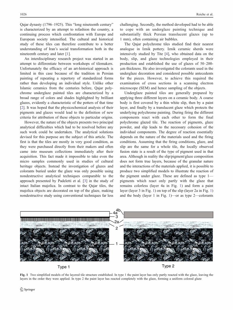
Underglaze painted tiles are generally prepared byapplying three different layers on a ceramic body. A coarsebody is first covered by a thin white slip, then by a paintlayer, and finally by a translucent glaze which protects theunderlying polychrome painting. During firing the differentcomponents react with each other to form the finalpolychrome glazed tile. The reaction of pigments, glazepowder, and slip leads to the necessary cohesion of theindividual components. The degree of reaction essentiallydepends on the nature of the materials used and the firingconditions. Assuming that the firing conditions, glaze, andslip are the same for a whole tile, the locally observedfusion state is a result of the type of pigment used in thatarea. Although in reality the slip/pigment/glaze compositiondoes not form true layers, because of the granular natureand the interactions of the materials applied, it is possible toproduce two simplified models to illustrate the reaction ofthe pigment under glaze. These are defined as type 1—pigments which react only partly with the glaze thatremains colorless (layer 4a in Fig. 1) and form a paintlayer (layer 3 in Fig. 1) on top of the slip (layer 2a in Fig. 1)and the body (layer 1 in Fig. 1)—or as type 2—colorants
Type 1 Type 2
4a3
2
1
2a
4b
Fig. 1 Two simplified models of the layered tile structure established. In type 1 the paint layer has only partly reacted with the glaze, leaving thelayers in the order they were applied. In type 2 the paint layer has reacted completely with the glaze, forming a uniform colored glaze
1026 Reiche et al.
which diffuse completely into the glaze that becomescolored (layer 4b). In all cases the glaze penetrates partlyacross the colored layer, allowing the cohesion of thedifferent materials (layer 2 in Fig. 1).
It is first necessary to clearly define the terms “color,”“colorants,” and “pigments” as used in this paper. A colorcorresponds to the final hue observed on the tiles. A coloringagent, or colorant, is the chemical species, together with itscoordination and oxidation state, responsible for the color. Apigment is the raw material used to create the color, generallycomposed of the coloring agent or its precursor. It alsocontains other secondary chemical species which can modifythe color or which may be unrelated to the coloring itself.The pigments can undergo a complete change of structure(type 2) and oxidation state during this heating process,making them no longer identical with the final coloring agent,or can sometimes survive firing nearly unchanged (type 1).
In the following sections, we report on how theanalytical difficulties were tackled in the study of a seriesof nineteenth-century Persian underglaze polychromepainted tiles held in French and German museum collec-tions. It is shown that different analytical routes areadvisable depending on the type of colorant. If the pigmentsor colorants are deeply buried under a thick glaze layer,they cannot be detected using nondestructive X-ray-basedmethods such as X-ray fluorescence (XRF) and proton-induced X-ray emission (PIXE). It was therefore necessaryto produce an analytical strategy which combined othercomplementary nondestructive analytical techniques forchemical, structural, and spectroscopic investigations. Forthis, microfocused-beam PIXE (micro-PIXE)/microfo-cused-beam proton-induced γ-ray emission (micro-PIGE),microfocused-beam XRF (micro-XRF), confocal micro-focused-beam Raman (micro-Raman) spectroscopy, visiblespectroscopy, and 3D microfocused-beam X-ray absorptionnear edge structure (3D micro-XANES) were used.
A clear definition of each of the various gradations ofcolors observed on the tiles was established to showwhether they were produced by change of concentrationor different colorants. To ensure identification was lessinfluenced by individual perception, the internationallyrecognized color system NCS—Natural Color System®©
of Scandinavian Colour Institute1 was chosen to classifyeach of the established color terms (Table 1).
The results obtained by the different analytical methodsare discussed with respect to the success of the analyticalmethods. The materials identified are compared with those
mentioned in general ceramic literature and particularly thaton Persian workshops of the Qajar period in order toidentify potential criteria for the differentiation of work-shops, production areas, or time periods.
Material studied
The following objects were chosen for this study:
1. One tile from the Ethnological Museum, Berlin (EM-B;inventory no. IB 9389) [5]; one bought in 1898 from theworkshop of the tilemaker Ali Muhammad in Tehran,who is thought to be Ali Muhammad Isfahani, the bestknown Iranian potter of his time
2. Four tiles from the Musée du Louvre, Paris (ML-P;inventory nos. MAO 901, MAO 902, MAO 1190, andMAO 1193), bought from the art market
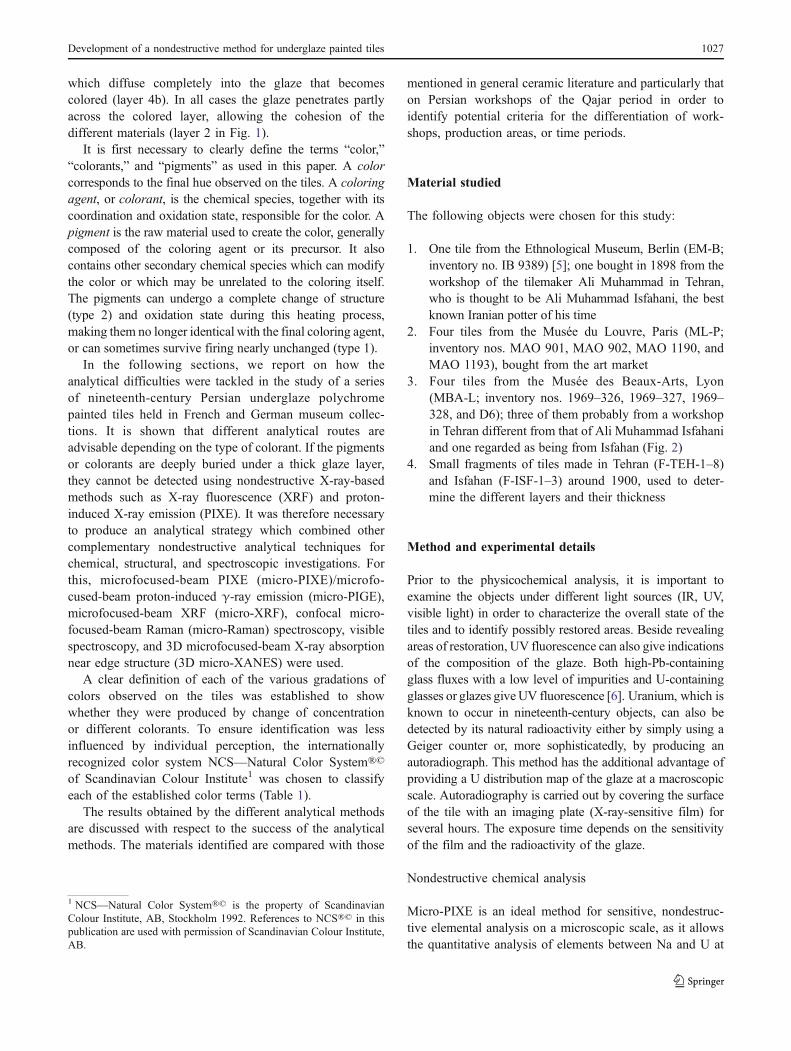
3. Four tiles from the Musée des Beaux-Arts, Lyon(MBA-L; inventory nos. 1969–326, 1969–327, 1969–328, and D6); three of them probably from a workshopin Tehran different from that of Ali Muhammad Isfahaniand one regarded as being from Isfahan (Fig. 2)
4. Small fragments of tiles made in Tehran (F-TEH-1–8)and Isfahan (F-ISF-1–3) around 1900, used to deter-mine the different layers and their thickness
Method and experimental details
Prior to the physicochemical analysis, it is important toexamine the objects under different light sources (IR, UV,visible light) in order to characterize the overall state of thetiles and to identify possibly restored areas. Beside revealingareas of restoration, UV fluorescence can also give indicationsof the composition of the glaze. Both high-Pb-containingglass fluxes with a low level of impurities and U-containingglasses or glazes giveUV fluorescence [6]. Uranium, which isknown to occur in nineteenth-century objects, can also bedetected by its natural radioactivity either by simply using aGeiger counter or, more sophisticatedly, by producing anautoradiograph. This method has the additional advantage ofproviding a U distribution map of the glaze at a macroscopicscale. Autoradiography is carried out by covering the surfaceof the tile with an imaging plate (X-ray-sensitive film) forseveral hours. The exposure time depends on the sensitivityof the film and the radioactivity of the glaze.
Nondestructive chemical analysis
Micro-PIXE is an ideal method for sensitive, nondestruc-tive elemental analysis on a microscopic scale, as it allowsthe quantitative analysis of elements between Na and U at
1 NCS—Natural Color System®© is the property of ScandinavianColour Institute, AB, Stockholm 1992. References to NCS®© in thispublication are used with permission of Scandinavian Colour Institute,AB.
Development of a nondestructive method for underglaze painted tiles 1027
major, minor, and trace levels. This technique can easily becombined with micro-PIGE for the analysis of lightelements between B and Na, although this requires theuse of a particle accelerator, which is seldom available inmuseum laboratories.
The glazes and the colors of the tiles were investigatedusing micro-PIXE and micro-PIGE with 3-MeV protonsfocused to a beam with a diameter of about 50 µm at theexternal microbeam facility of the 2-MV tandem acceleratorAGLAE dedicated to the analysis of art and archaeologicalobjects [7]. Protons enter the sample perpendicular to itssurface in a localized He atmosphere. Two Si(Li) detectorswere used for the detection of the low- and high-energy X-rays. The low-energy Si(Li) detector is equipped with anultrathin AP3.3 polymer window and a magnet to deflect thebackscattered protons. The high-energy Si(Li) detector isequipped with a 50-µm Al filter. Spectrum evaluation andquantification were performed using the GUPIX program [8].
Micro-PIGE was used to identify B and F contents andto provide a second and more reliable Na value (using the717, 110 or 197, and 440 keV lines, respectively). PIGEhas the advantage that the probed volume extends deeperunder the sample surface than with XRF because theoutgoing γ-rays are absorbed less than X-rays; therefore,PIGE results are less affected by surface alteration [9].
Each colored zone was measured at least once or, if pos-sible, several times at different points on the tiles. An area of0.04–1 mm2 was scanned because of the inhomogeneousdistribution of the colorant in the glaze. The acquisition timefor each area was about 5 min.
Micro-XRF analysis can be used as an alternative tomicro-PIXE/micro-PIGE for the characterization of the
chemical composition of the glaze and the coloring speciesin the glazes, but if it is used under atmospheric pressure itis less sensitive for light elements. It does, however, havethe advantage of portability, making the transport of objectsunnecessary and allowing in situ investigations (e.g. ofwall-mounted tiles) [10]. Micro-XRF was performed usingtwo different spectrometers. Quantitative analysis of theglaze was carried out using an energy-dispersive micro-XRF spectrometer with a fine-focus 30-W Mo tube and apolycapillary lens for focusing the primary beam down toapproximately 80–100 µm (ArtTAX spectrometer [10]).The open arrangement, with the measurement head mount-ed on a tripod support and no sample chamber, allows theinvestigation of objects of almost any size or shape. Heliumpurging between the measuring head and the sampleallowed better detection of light elements (down to Na)with a Si drift detector. An elemental sensitivity methodcalibrated with glass standards of a soda lime glass wasused to obtain quantitative results through the QXASsoftware program [11]. Additionally, micro-XRF measure-ments were made with the laboratory setup at the TechnicalUniversity of Berlin using a Mo X-ray tube and a micro-focused beam of 80–100 µm obtained with a polycapillaryfull lens. A Si(Li) detector was used for detection of thefluorescence.
The limiting factors affecting the quantitative results forboth techniques are the analytical depth and the homoge-neity of the sample with depth. For underglaze painted tileswith their layered sequence inhomogeneity is inherent. Ifthe exciting radiation passes through the glaze and reachesinto the pigment layer, the analytical values for the glazecomposition may be erroneous. However, this seems unlikely
Fig. 2 Nineteenth-centuryIranian polychrome underglazepainted tiles: a MBA-L 1969–328 and b MBA-L D6 from theMusée des Beaux-Arts, Lyon(Copyright D. Vigears, Centrefor Research and Restoration ofthe French Museums, repro-duced with the permission of theMusée des Beaux-Arts, Lyon)
1028 Reiche et al.
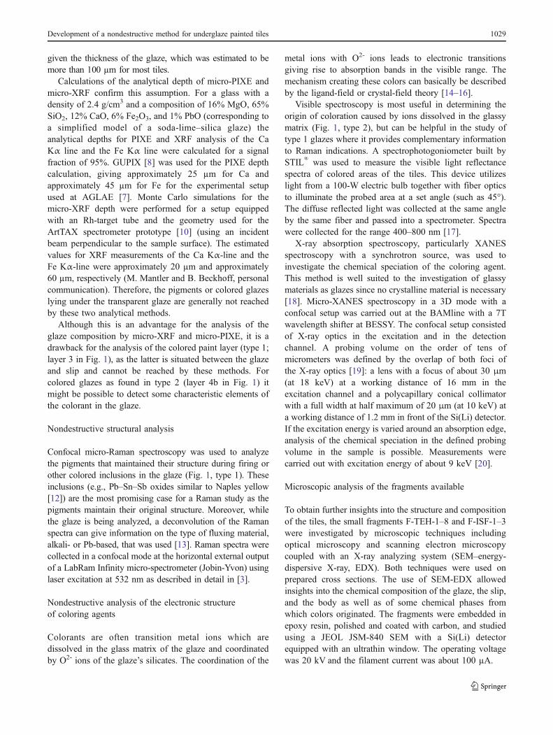
given the thickness of the glaze, which was estimated to bemore than 100 µm for most tiles.
Calculations of the analytical depth of micro-PIXE andmicro-XRF confirm this assumption. For a glass with adensity of 2.4 g/cm3 and a composition of 16% MgO, 65%SiO2, 12% CaO, 6% Fe2O3, and 1% PbO (corresponding toa simplified model of a soda-lime–silica glaze) theanalytical depths for PIXE and XRF analysis of the CaKα line and the Fe Kα line were calculated for a signalfraction of 95%. GUPIX [8] was used for the PIXE depthcalculation, giving approximately 25 µm for Ca andapproximately 45 µm for Fe for the experimental setupused at AGLAE [7]. Monte Carlo simulations for themicro-XRF depth were performed for a setup equippedwith an Rh-target tube and the geometry used for theArtTAX spectrometer prototype [10] (using an incidentbeam perpendicular to the sample surface). The estimatedvalues for XRF measurements of the Ca Kα-line and theFe Kα-line were approximately 20 µm and approximately60 µm, respectively (M. Mantler and B. Beckhoff, personalcommunication). Therefore, the pigments or colored glazeslying under the transparent glaze are generally not reachedby these two analytical methods.
Although this is an advantage for the analysis of theglaze composition by micro-XRF and micro-PIXE, it is adrawback for the analysis of the colored paint layer (type 1;layer 3 in Fig. 1), as the latter is situated between the glazeand slip and cannot be reached by these methods. Forcolored glazes as found in type 2 (layer 4b in Fig. 1) itmight be possible to detect some characteristic elements ofthe colorant in the glaze.
Nondestructive structural analysis
Confocal micro-Raman spectroscopy was used to analyzethe pigments that maintained their structure during firing orother colored inclusions in the glaze (Fig. 1, type 1). Theseinclusions (e.g., Pb–Sn–Sb oxides similar to Naples yellow[12]) are the most promising case for a Raman study as thepigments maintain their original structure. Moreover, whilethe glaze is being analyzed, a deconvolution of the Ramanspectra can give information on the type of fluxing material,alkali- or Pb-based, that was used [13]. Raman spectra werecollected in a confocal mode at the horizontal external outputof a LabRam Infinity micro-spectrometer (Jobin-Yvon) usinglaser excitation at 532 nm as described in detail in [3].
Nondestructive analysis of the electronic structureof coloring agents
Colorants are often transition metal ions which aredissolved in the glass matrix of the glaze and coordinatedby O2- ions of the glaze’s silicates. The coordination of the
metal ions with O2- ions leads to electronic transitionsgiving rise to absorption bands in the visible range. Themechanism creating these colors can basically be describedby the ligand-field or crystal-field theory [14–16].
Visible spectroscopy is most useful in determining theorigin of coloration caused by ions dissolved in the glassymatrix (Fig. 1, type 2), but can be helpful in the study oftype 1 glazes where it provides complementary informationto Raman indications. A spectrophotogoniometer built bySTIL® was used to measure the visible light reflectancespectra of colored areas of the tiles. This device utilizeslight from a 100-W electric bulb together with fiber opticsto illuminate the probed area at a set angle (such as 45°).The diffuse reflected light was collected at the same angleby the same fiber and passed into a spectrometer. Spectrawere collected for the range 400–800 nm [17].
X-ray absorption spectroscopy, particularly XANESspectroscopy with a synchrotron source, was used toinvestigate the chemical speciation of the coloring agent.This method is well suited to the investigation of glassymaterials as glazes since no crystalline material is necessary[18]. Micro-XANES spectroscopy in a 3D mode with aconfocal setup was carried out at the BAMline with a 7Twavelength shifter at BESSY. The confocal setup consistedof X-ray optics in the excitation and in the detectionchannel. A probing volume on the order of tens ofmicrometers was defined by the overlap of both foci ofthe X-ray optics [19]: a lens with a focus of about 30 μm(at 18 keV) at a working distance of 16 mm in theexcitation channel and a polycapillary conical collimatorwith a full width at half maximum of 20 μm (at 10 keV) ata working distance of 1.2 mm in front of the Si(Li) detector.If the excitation energy is varied around an absorption edge,analysis of the chemical speciation in the defined probingvolume in the sample is possible. Measurements werecarried out with excitation energy of about 9 keV [20].
Microscopic analysis of the fragments available
To obtain further insights into the structure and compositionof the tiles, the small fragments F-TEH-1–8 and F-ISF-1–3were investigated by microscopic techniques includingoptical microscopy and scanning electron microscopycoupled with an X-ray analyzing system (SEM–energy-dispersive X-ray, EDX). Both techniques were used onprepared cross sections. The use of SEM-EDX allowedinsights into the chemical composition of the glaze, the slip,and the body as well as of some chemical phases fromwhich colors originated. The fragments were embedded inepoxy resin, polished and coated with carbon, and studiedusing a JEOL JSM-840 SEM with a Si(Li) detectorequipped with an ultrathin window. The operating voltagewas 20 kV and the filament current was about 100 µA.
Development of a nondestructive method for underglaze painted tiles 1029
Results and discussion
The method established enabled us to identify the chemicalcomposition of the glaze and to determine the color toneand the corresponding coloring species or the pigment ofthe 9 tiles and 11 fragments selected.
Glaze analysis
The glaze ofML-P andMBA-L tiles as well as that of fragmentsF-TEH-1–8 and F-ISF-1–3 were analyzed by micro-PIXE/
micro-PIGE to obtain the chemical composition. The results ofthe glaze analysis from MBA-L D6 are shown in Table 2. It isevident how little the values vary, even for different coloredzones. To obtain the best estimation of the glaze compositionof a tile, raised values (over 1σ of the average) of potentialcoloring elements were omitted from the calculation.
The results for the glazes of tiles from MBA-L and ML-P(Table 3) show the same order of dispersion of elementalcomposition as that found for MBA-L D6.
The glaze composition of the objects from MBA-L andML-P show a similar silicate composition with high Na2O
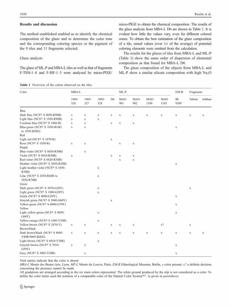
Table 1 Overview of the colors observed on the tiles
Color MBA-L ML-P EM-B Fragments
1969–326
1969–327
1969–328
D6 MAO901
MAO902
MAO1190
MAO1193
IB9389
Tehran Isfahan
BlueDark blue (NCS® S 4050-R90B) x x x x x x x x x x xLight blue (NCS® S 1050-R90B) x x xCerulean blue (NCS® S 1060-B) x x x x xBlue-green (NCS® S 1050-B10Gto 1050-B20G)
x x x
RedLight red (NCS® S 1070-R) xRose (NCS® S 1030-R) x x x xPurpleBlue-violet (NCS® S 6030-R50B) xViolet (NCS® S 6010-R50B) x x x xRed-violet (NCS® S 6020-R30B) x xHeather violet (NCS® S 2050-R20B) xLight heather-violet (NCS® S 1050-R30B)
x
Lilac (NCS® S 2050-R60B to2050-R70B)
x
GreenDark green (NCS® S 3070-G20Y) x xLight green (NCS® S 1080-G20Y) xGreen (NCS® S 4060-G20Y) xGrayish green (NCS® S 3040-G60Y) xYellow-green (NCS® S 0080-G70Y) xYellowLight yellow-green (NCS® S 0050-G60Y)
x x
Yellow-orange (NCS® S 1080-Y20R) xYellow-brown (NCS® S 2070-Y) x x x x x x? xBrown/blackDark brown/black (NCS® S 8005-Y80R/9005-B20G)
x x x x x x x x x x x
Light brown (NCS® S 6010-Y30R) xGrayish brown (NCS® S 7010-G50Y)
x x x
Gray (NCS® S 3005-Y20R) x x
Void entries indicate that the color is absentMBA-L Musée des Beaux-Arts, Lyon, MP-L Musée du Louvre, Paris, EM-B Ethnological Museum, Berlin, x color present; x? a definite decisionconcerning the presence cannot be madeAll gradations are arranged according to the six main colors represented. The white ground produced by the slip is not considered as a color. Todefine the color terms used the notation of a comparable color of the Natural Color System®© is given in parentheses
1030 Reiche et al.
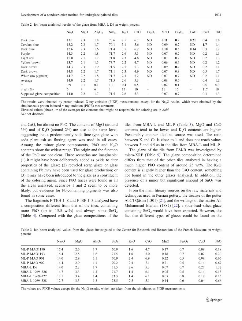
and CaO, but almost no PbO. The contents of MgO (around3%) and of K2O (around 2%) are also at the same level,suggesting that a predominantly soda lime type glaze withsoda plant ash as fluxing agent was used in all cases.Among the minor glaze components, PbO and K2Ocontents show the widest range. The origin and the functionof the PbO are not clear. Three scenarios are imaginable:(1) it might have been deliberately added as oxide to alterproperties of the glaze; (2) recycled scrap glaze or glasscontaining Pb may have been used for glaze production; or(3) it may have been introduced to the glaze as a constituentof the coloring agent. Since PbO traces were found in allthe areas analyzed, scenarios 1 and 2 seem to be morelikely, but evidence for Pb-containing pigments was alsofound in some cases.
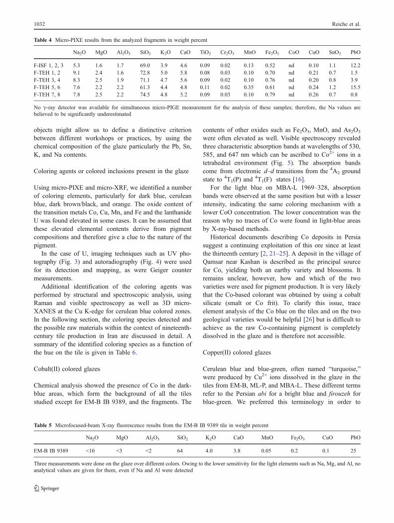
The fragments F-TEH-1–8 and F-ISF-1–3 analyzed havea composition different from that of the tiles, containingmore PbO (up to 15.5 wt%) and always some SnO2
(Table 4). Compared with the glaze compositions of the
tiles from MBA-L and ML-P (Table 3), MgO and CaOcontents tend to be lower and K2O contents are higher.Presumably another alkaline source was used. The ratiobetween K and Ca is close to 1 and does not reach valuesbetween 3 and 4.5 as in the tiles from MBA-L and ML-P.
The glaze of the tile from EM-B was investigated bymicro-XRF (Table 5). The glaze composition determineddiffers from that of the other tiles analyzed in having amuch higher PbO content of around 25 wt%. The K2Ocontent is slightly higher than the CaO content, somethingnot found in the other glazes analyzed. In addition, thepresence of a minor but significant amount of SnO2 wasdetected.
From the main literary sources on the raw materials andtechniques used in Persian pottery, the treatise of the potterAbū’l-Qāsim (1301) [21], and the writings of the master AliMuhammad Isfahani (1887) [22], a soda–lead–silica glazecontaining SnO2 would have been expected. However, thefact that different types of glazes could be found on the
Table 2 Ion beam analytical results of the glaze from MBA-L D6 in weight percent
Na2O MgO Al2O3 SiO2 K2O CaO Cr2O3 MnO Fe2O3 CoO CuO PbO
Dark blue 13.1 2.3 1.8 70.6 2.5 6.1 ND 0.11 0.9 0.21 0.4 1.8Cerulan blue 13.2 2.3 1.7 70.1 3.1 5.6 ND 0.09 0.7 ND 1.7 1.4Dark blue 12.6 2.3 1.6 71.4 3.5 6.2 ND 0.10 0.6 0.14 0.3 1.2Purple 14.3 2.2 1.8 71.7 2.6 5.3 ND 0.07 0.7 ND 0.2 1.1Light red 15.0 2.1 1.7 71.8 2.3 4.8 ND 0.07 0.7 ND 0.2 1.3Yellow-brown 13.7 2.1 1.5 73.7 2.2 4.7 ND 0.06 0.6 ND 0.2 1.2Dark brown 14.3 2.3 1.9 71.5 2.5 5.3 ND 0.09 0.9 ND 0.2 1.1Dark brown 14.8 2.2 1.7 71.1 2.3 4.9 ND 0.07 0.8 ND 0.5 1.7White (no pigment) 14.7 2.2 1.8 71.7 2.3 5.2 ND 0.07 0.7 ND 0.2 1.1Average 14.0 2.2 1.7 71.5 2.6 5.3 – 0.08 0.7 – 0.4 1.3σ 0.9 0.1 0.1 1.0 0.4 0.5 – 0.02 0.1 – 0.5 0.3σ rel (%) 6 4 6 1 17 10 – 21 15 – 117 19Supposed glaze composition 14.0 2.2 1.7 71.5 2.6 5.3 – 0.07 0.7 – 0.3 1.3
The results were obtained by proton-induced X-ray emission (PIXE) measurements except for the Na2O results, which were obtained by thesimultaneous proton-induced γ-ray emission (PIGE) measurementsElevated values (above 1σ of the average) for elements that might be responsible for coloring are in boldND not detected
Table 3 Ion beam analytical values from the glazes investigated at the Centre for Research and Restoration of the French Museums in weightpercent
Na2O MgO Al2O3 SiO2 K2O CaO MnO Fe2O3 CuO PbO
ML-P MAO1190 17.4 2.6 1.7 70.9 1.6 4.7 0.17 0.7 0.08 0.18ML-P MAO1193 16.4 2.8 1.6 71.5 1.6 5.0 0.18 0.7 0.07 0.20ML-P MAO 901 14.0 2.9 1.1 70.9 2.4 6.9 0.22 0.5 0.09 0.66ML-P MAO 902 14.4 2.9 1.1 70.2 2.4 7.1 0.21 0.5 0.14 0.67MBA-L D6 14.0 2.2 1.7 71.5 2.6 5.3 0.07 0.7 0.27 1.32MBA-L 1969–326 14.7 3.3 1.2 71.7 1.4 6.1 0.05 0.5 0.14 0.13MBA-L 1969–327 13.1 3.4 1.4 73.3 1.4 6.1 0.05 0.8 0.19 0.15MBA-L 1969–328 12.7 3.3 1.3 73.5 2.5 5.1 0.14 0.6 0.04 0.66
The values are PIXE values except for the Na2O results, which are taken from the simultaneous PIGE measurements
Development of a nondestructive method for underglaze painted tiles 1031
objects might allow us to define a distinctive criterionbetween different workshops or practices, by using thechemical composition of the glaze particularly the Pb, Sn,K, and Na contents.
Coloring agents or colored inclusions present in the glaze
Using micro-PIXE and micro-XRF, we identified a numberof coloring elements, particularly for dark blue, ceruleanblue, dark brown/black, and orange. The oxide content ofthe transition metals Co, Cu, Mn, and Fe and the lanthanideU was found elevated in some cases. It can be assumed thatthese elevated elemental contents derive from pigmentcompositions and therefore give a clue to the nature of thepigment.
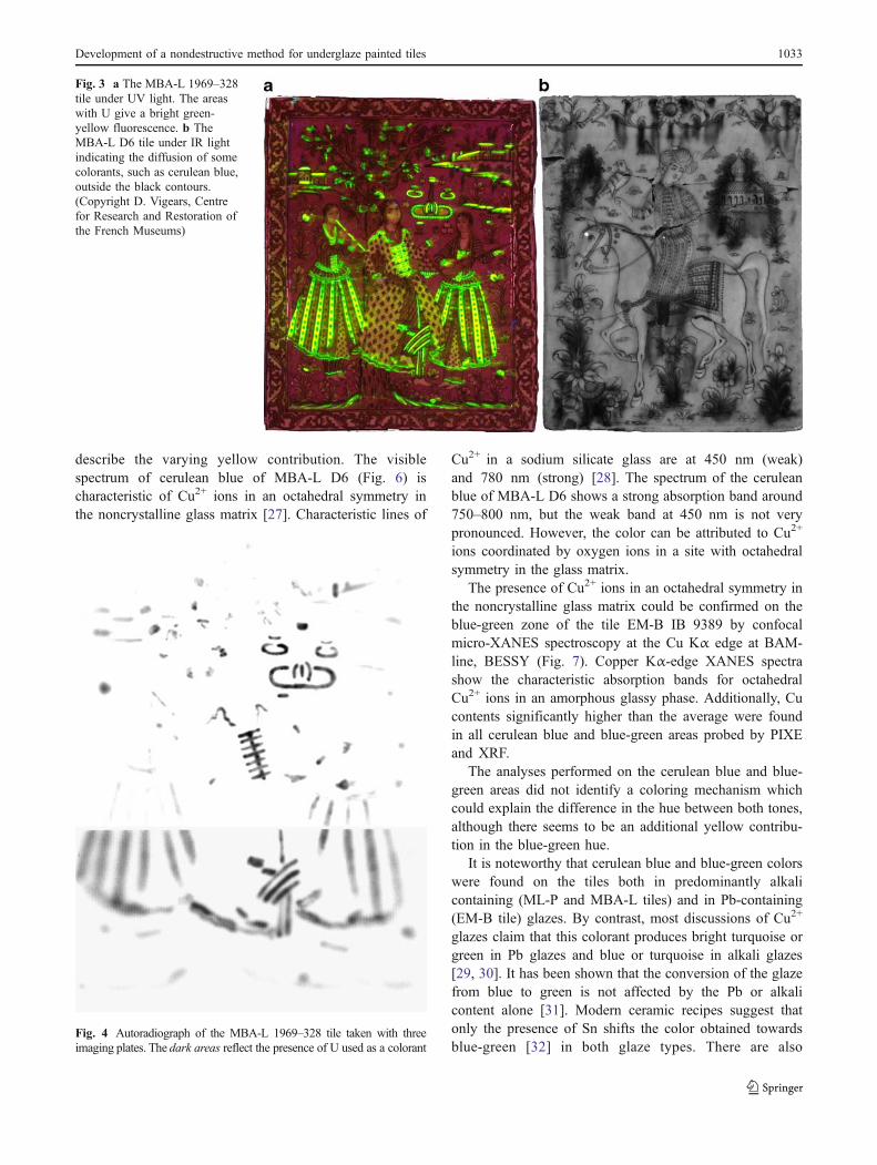
In the case of U, imaging techniques such as UV pho-tography (Fig. 3) and autoradiography (Fig. 4) were usedfor its detection and mapping, as were Geiger countermeasurements.
Additional identification of the coloring agents wasperformed by structural and spectroscopic analysis, usingRaman and visible spectroscopy as well as 3D micro-XANES at the Cu K-edge for cerulean blue colored zones.In the following section, the coloring species detected andthe possible raw materials within the context of nineteenth-century tile production in Iran are discussed in detail. Asummary of the identified coloring species as a function ofthe hue on the tile is given in Table 6.
Cobalt(II) colored glazes
Chemical analysis showed the presence of Co in the dark-blue areas, which form the background of all the tilesstudied except for EM-B IB 9389, and the fragments. The
contents of other oxides such as Fe2O3, MnO, and As2O3
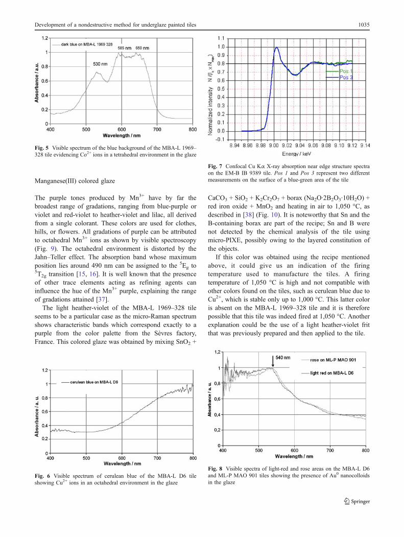
were often elevated as well. Visible spectroscopy revealedthree characteristic absorption bands at wavelengths of 530,585, and 647 nm which can be ascribed to Co2+ ions in atetrahedral environment (Fig. 5). The absorption bandscome from electronic d–d transitions from the 4A2 groundstate to 4T1(P) and
4T1(F) states [16].For the light blue on MBA-L 1969–328, absorption
bands were observed at the same position but with a lesserintensity, indicating the same coloring mechanism with alower CoO concentration. The lower concentration was thereason why no traces of Co were found in light-blue areasby X-ray-based methods.
Historical documents describing Co deposits in Persiasuggest a continuing exploitation of this ore since at leastthe thirteenth century [2, 21–25]. A deposit in the village ofQamsar near Kashan is described as the principal sourcefor Co, yielding both an earthy variety and blossoms. Itremains unclear, however, how and which of the twovarieties were used for pigment production. It is very likelythat the Co-based colorant was obtained by using a cobaltsilicate (smalt or Co frit). To clarify this issue, traceelement analysis of the Co blue on the tiles and on the twogeological varieties would be helpful [26] but is difficult toachieve as the raw Co-containing pigment is completelydissolved in the glaze and is therefore not accessible.
Copper(II) colored glazes
Cerulean blue and blue-green, often named “turquoise,”were produced by Cu2+ ions dissolved in the glaze in thetiles from EM-B, ML-P, and MBA-L. These different termsrefer to the Persian abi for a bright blue and firouzeh forblue-green. We preferred this terminology in order to
Table 4 Micro-PIXE results from the analyzed fragments in weight percent
Na2O MgO Al2O3 SiO2 K2O CaO TiO2 Cr2O3 MnO Fe2O3 CoO CuO SnO2 PbO
F-ISF 1, 2, 3 5.3 1.6 1.7 69.0 3.9 4.6 0.09 0.02 0.13 0.52 nd 0.10 1.1 12.2F-TEH 1, 2 9.1 2.4 1.6 72.8 5.0 5.8 0.08 0.03 0.10 0.70 nd 0.21 0.7 1.5F-TEH 3, 4 8.3 2.5 1.9 71.1 4.7 5.6 0.09 0.02 0.10 0.76 nd 0.20 0.8 3.9F-TEH 5, 6 7.6 2.2 2.2 61.3 4.4 4.8 0.11 0.02 0.35 0.61 nd 0.24 1.2 15.5F-TEH 7, 8 7.8 2.5 2.2 74.5 4.8 5.2 0.09 0.03 0.10 0.79 nd 0.26 0.7 0.8
No γ-ray detector was available for simultaneous micro-PIGE measurement for the analysis of these samples; therefore, the Na values arebelieved to be significantly underestimated
Table 5 Microfocused-beam X-ray fluorescence results from the EM-B IB 9389 tile in weight percent
Na2O MgO Al2O3 SiO2 K2O CaO MnO Fe2O3 CuO PbO
EM-B IB 9389 <10 <3 <2 64 4.0 3.8 0.05 0.2 0.1 25
Three measurements were done on the glaze over different colors. Owing to the lower sensitivity for the light elements such as Na, Mg, and Al, noanalytical values are given for them, even if Na and Al were detected
1032 Reiche et al.
describe the varying yellow contribution. The visiblespectrum of cerulean blue of MBA-L D6 (Fig. 6) ischaracteristic of Cu2+ ions in an octahedral symmetry inthe noncrystalline glass matrix [27]. Characteristic lines of
Cu2+ in a sodium silicate glass are at 450 nm (weak)and 780 nm (strong) [28]. The spectrum of the ceruleanblue of MBA-L D6 shows a strong absorption band around750–800 nm, but the weak band at 450 nm is not verypronounced. However, the color can be attributed to Cu2+
ions coordinated by oxygen ions in a site with octahedralsymmetry in the glass matrix.
The presence of Cu2+ ions in an octahedral symmetry inthe noncrystalline glass matrix could be confirmed on theblue-green zone of the tile EM-B IB 9389 by confocalmicro-XANES spectroscopy at the Cu Kα edge at BAM-line, BESSY (Fig. 7). Copper Kα-edge XANES spectrashow the characteristic absorption bands for octahedralCu2+ ions in an amorphous glassy phase. Additionally, Cucontents significantly higher than the average were foundin all cerulean blue and blue-green areas probed by PIXEand XRF.
The analyses performed on the cerulean blue and blue-green areas did not identify a coloring mechanism whichcould explain the difference in the hue between both tones,although there seems to be an additional yellow contribu-tion in the blue-green hue.
It is noteworthy that cerulean blue and blue-green colorswere found on the tiles both in predominantly alkalicontaining (ML-P and MBA-L tiles) and in Pb-containing(EM-B tile) glazes. By contrast, most discussions of Cu2+
glazes claim that this colorant produces bright turquoise orgreen in Pb glazes and blue or turquoise in alkali glazes[29, 30]. It has been shown that the conversion of the glazefrom blue to green is not affected by the Pb or alkalicontent alone [31]. Modern ceramic recipes suggest thatonly the presence of Sn shifts the color obtained towardsblue-green [32] in both glaze types. There are also
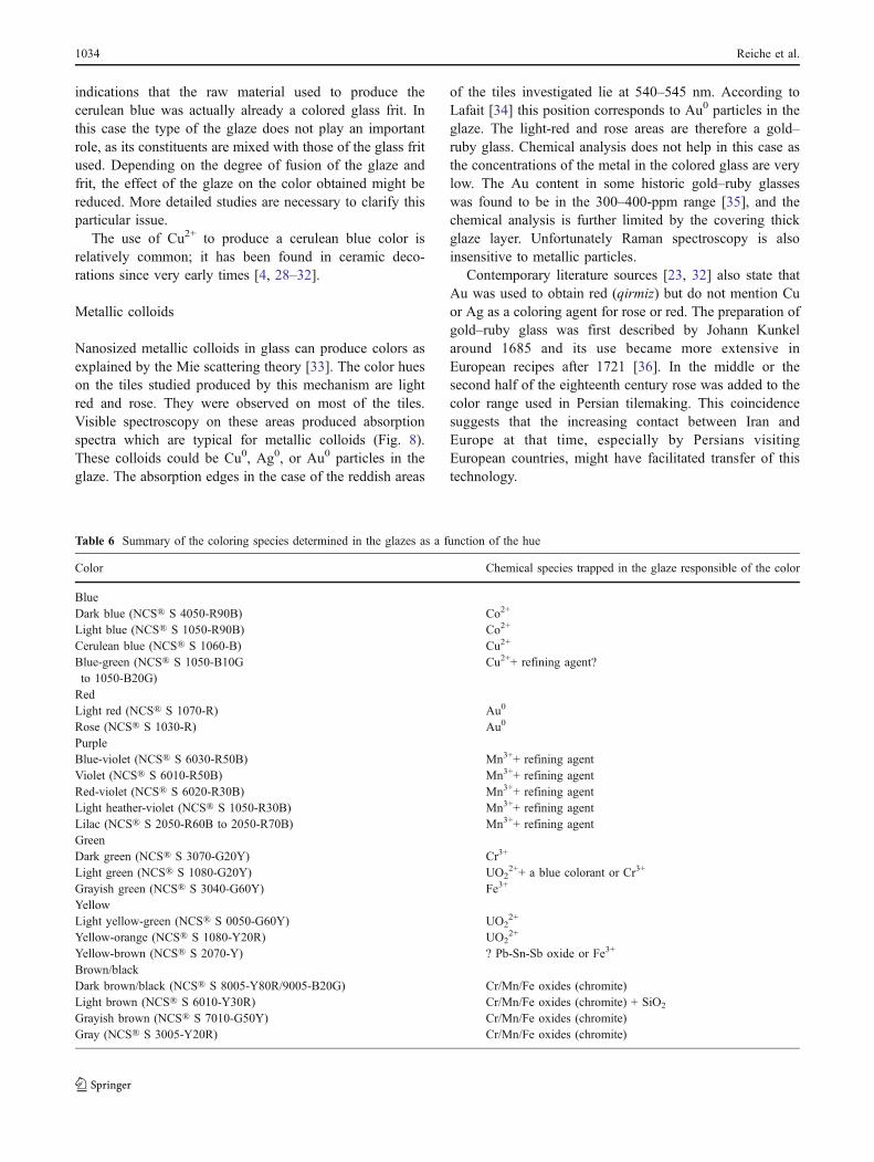
Fig. 4 Autoradiograph of the MBA-L 1969–328 tile taken with threeimaging plates. The dark areas reflect the presence of U used as a colorant
Fig. 3 a The MBA-L 1969–328tile under UV light. The areaswith U give a bright green-yellow fluorescence. b TheMBA-L D6 tile under IR lightindicating the diffusion of somecolorants, such as cerulean blue,outside the black contours.(Copyright D. Vigears, Centrefor Research and Restoration ofthe French Museums)
Development of a nondestructive method for underglaze painted tiles 1033
indications that the raw material used to produce thecerulean blue was actually already a colored glass frit. Inthis case the type of the glaze does not play an importantrole, as its constituents are mixed with those of the glass fritused. Depending on the degree of fusion of the glaze andfrit, the effect of the glaze on the color obtained might bereduced. More detailed studies are necessary to clarify thisparticular issue.
The use of Cu2+ to produce a cerulean blue color isrelatively common; it has been found in ceramic deco-rations since very early times [4, 28–32].
Metallic colloids
Nanosized metallic colloids in glass can produce colors asexplained by the Mie scattering theory [33]. The color hueson the tiles studied produced by this mechanism are lightred and rose. They were observed on most of the tiles.Visible spectroscopy on these areas produced absorptionspectra which are typical for metallic colloids (Fig. 8).These colloids could be Cu0, Ag0, or Au0 particles in theglaze. The absorption edges in the case of the reddish areas
of the tiles investigated lie at 540–545 nm. According toLafait [34] this position corresponds to Au0 particles in theglaze. The light-red and rose areas are therefore a gold–ruby glass. Chemical analysis does not help in this case asthe concentrations of the metal in the colored glass are verylow. The Au content in some historic gold–ruby glasseswas found to be in the 300–400-ppm range [35], and thechemical analysis is further limited by the covering thickglaze layer. Unfortunately Raman spectroscopy is alsoinsensitive to metallic particles.
Contemporary literature sources [23, 32] also state thatAu was used to obtain red (qirmiz) but do not mention Cuor Ag as a coloring agent for rose or red. The preparation ofgold–ruby glass was first described by Johann Kunkelaround 1685 and its use became more extensive inEuropean recipes after 1721 [36]. In the middle or thesecond half of the eighteenth century rose was added to thecolor range used in Persian tilemaking. This coincidencesuggests that the increasing contact between Iran andEurope at that time, especially by Persians visitingEuropean countries, might have facilitated transfer of thistechnology.
Table 6 Summary of the coloring species determined in the glazes as a function of the hue
Color Chemical species trapped in the glaze responsible of the color
BlueDark blue (NCS® S 4050-R90B) Co2+
Light blue (NCS® S 1050-R90B) Co2+
Cerulean blue (NCS® S 1060-B) Cu2+
Blue-green (NCS® S 1050-B10Gto 1050-B20G)
Cu2++ refining agent?
RedLight red (NCS® S 1070-R) Au0
Rose (NCS® S 1030-R) Au0
PurpleBlue-violet (NCS® S 6030-R50B) Mn3++ refining agentViolet (NCS® S 6010-R50B) Mn3++ refining agentRed-violet (NCS® S 6020-R30B) Mn3++ refining agentLight heather-violet (NCS® S 1050-R30B) Mn3++ refining agentLilac (NCS® S 2050-R60B to 2050-R70B) Mn3++ refining agentGreenDark green (NCS® S 3070-G20Y) Cr3+
Light green (NCS® S 1080-G20Y) UO22++ a blue colorant or Cr3+
Grayish green (NCS® S 3040-G60Y) Fe3+
YellowLight yellow-green (NCS® S 0050-G60Y) UO2
2+
Yellow-orange (NCS® S 1080-Y20R) UO22+
Yellow-brown (NCS® S 2070-Y) ? Pb-Sn-Sb oxide or Fe3+
Brown/blackDark brown/black (NCS® S 8005-Y80R/9005-B20G) Cr/Mn/Fe oxides (chromite)Light brown (NCS® S 6010-Y30R) Cr/Mn/Fe oxides (chromite) + SiO2
Grayish brown (NCS® S 7010-G50Y) Cr/Mn/Fe oxides (chromite)Gray (NCS® S 3005-Y20R) Cr/Mn/Fe oxides (chromite)
1034 Reiche et al.
Manganese(III) colored glaze
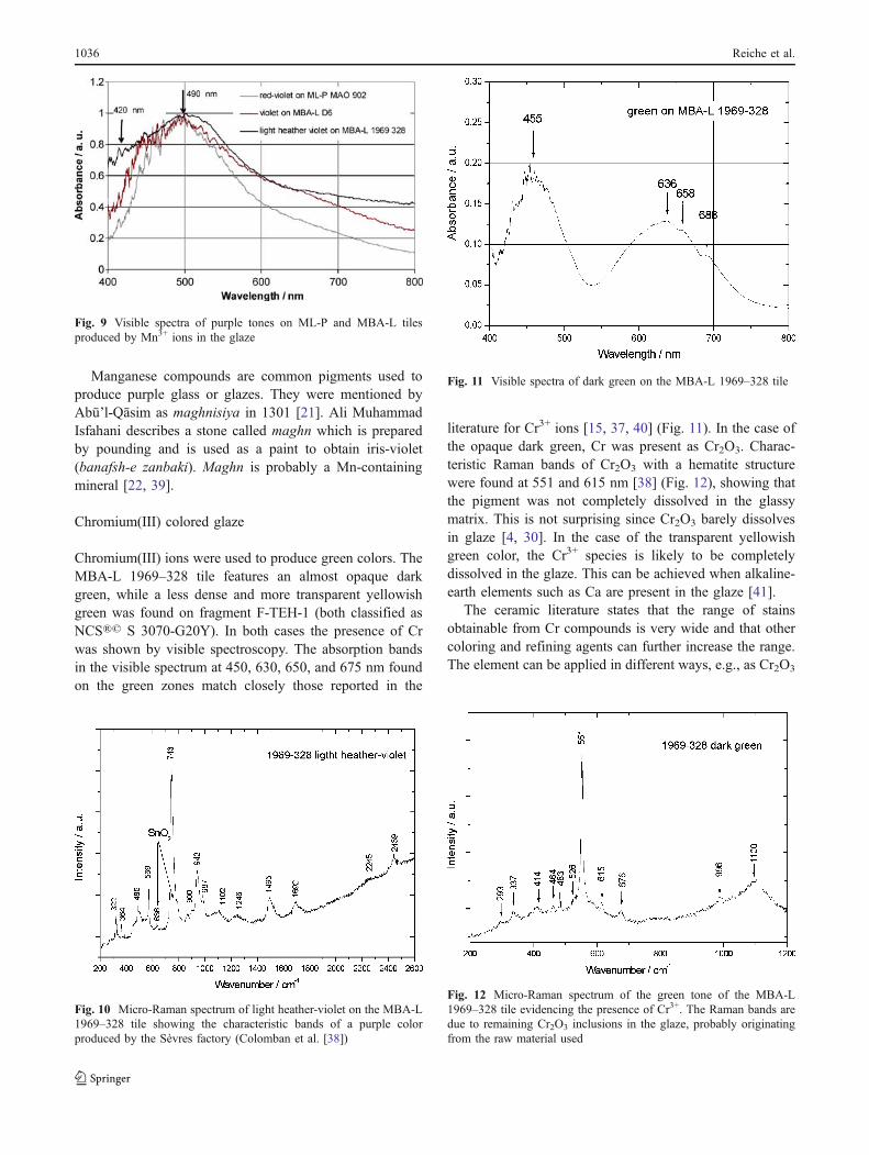
The purple tones produced by Mn3+ have by far thebroadest range of gradations, ranging from blue-purple orviolet and red-violet to heather-violet and lilac, all derivedfrom a single colorant. These colors are used for clothes,hills, or flowers. All gradations of purple can be attributedto octahedral Mn3+ ions as shown by visible spectroscopy(Fig. 9). The octahedral environment is distorted by theJahn–Teller effect. The absorption band whose maximumposition lies around 490 nm can be assigned to the 5Eg to5T2g transition [15, 16]. It is well known that the presenceof other trace elements acting as refining agents caninfluence the hue of the Mn3+ purple, explaining the rangeof gradations attained [37].
The light heather-violet of the MBA-L 1969–328 tileseems to be a particular case as the micro-Raman spectrumshows characteristic bands which correspond exactly to apurple from the color palette from the Sèvres factory,France. This colored glaze was obtained by mixing SnO2 +
CaCO3 + SiO2 + K2Cr2O7 + borax (Na2O·2B2O3·10H2O) +red iron oxide + MnO2 and heating in air to 1,050 °C, asdescribed in [38] (Fig. 10). It is noteworthy that Sn and theB-containing borax are part of the recipe; Sn and B werenot detected by the chemical analysis of the tile usingmicro-PIXE, possibly owing to the layered constitution ofthe objects.
If this color was obtained using the recipe mentionedabove, it could give us an indication of the firingtemperature used to manufacture the tiles. A firingtemperature of 1,050 °C is high and not compatible withother colors found on the tiles, such as cerulean blue due toCu2+, which is stable only up to 1,000 °C. This latter coloris absent on the MBA-L 1969–328 tile and it is thereforepossible that this tile was indeed fired at 1,050 °C. Anotherexplanation could be the use of a light heather-violet fritthat was previously prepared and then applied to the tile.
Fig. 6 Visible spectrum of cerulean blue of the MBA-L D6 tileshowing Cu2+ ions in an octahedral environment in the glaze
Fig. 5 Visible spectrum of the blue background of the MBA-L 1969–328 tile evidencing Co2+ ions in a tetrahedral environment in the glaze
Fig. 7 Confocal Cu Kα X-ray absorption near edge structure spectraon the EM-B IB 9389 tile. Pos 1 and Pos 3 represent two differentmeasurements on the surface of a blue-green area of the tile
Fig. 8 Visible spectra of light-red and rose areas on the MBA-L D6and ML-P MAO 901 tiles showing the presence of Au0 nanocolloidsin the glaze
Development of a nondestructive method for underglaze painted tiles 1035
Manganese compounds are common pigments used toproduce purple glass or glazes. They were mentioned byAbū’l-Qāsim as maghnisiya in 1301 [21]. Ali MuhammadIsfahani describes a stone called maghn which is preparedby pounding and is used as a paint to obtain iris-violet(banafsh-e zanbaki). Maghn is probably a Mn-containingmineral [22, 39].
Chromium(III) colored glaze
Chromium(III) ions were used to produce green colors. TheMBA-L 1969–328 tile features an almost opaque darkgreen, while a less dense and more transparent yellowishgreen was found on fragment F-TEH-1 (both classified asNCS®© S 3070-G20Y). In both cases the presence of Crwas shown by visible spectroscopy. The absorption bandsin the visible spectrum at 450, 630, 650, and 675 nm foundon the green zones match closely those reported in the
literature for Cr3+ ions [15, 37, 40] (Fig. 11). In the case ofthe opaque dark green, Cr was present as Cr2O3. Charac-teristic Raman bands of Cr2O3 with a hematite structurewere found at 551 and 615 nm [38] (Fig. 12), showing thatthe pigment was not completely dissolved in the glassymatrix. This is not surprising since Cr2O3 barely dissolvesin glaze [4, 30]. In the case of the transparent yellowishgreen color, the Cr3+ species is likely to be completelydissolved in the glaze. This can be achieved when alkaline-earth elements such as Ca are present in the glaze [41].
The ceramic literature states that the range of stainsobtainable from Cr compounds is very wide and that othercoloring and refining agents can further increase the range.The element can be applied in different ways, e.g., as Cr2O3
Fig. 9 Visible spectra of purple tones on ML-P and MBA-L tilesproduced by Mn3+ ions in the glaze
Fig. 10 Micro-Raman spectrum of light heather-violet on the MBA-L1969–328 tile showing the characteristic bands of a purple colorproduced by the Sèvres factory (Colomban et al. [38])
Fig. 11 Visible spectra of dark green on the MBA-L 1969–328 tile
Fig. 12 Micro-Raman spectrum of the green tone of the MBA-L1969–328 tile evidencing the presence of Cr3+. The Raman bands aredue to remaining Cr2O3 inclusions in the glaze, probably originatingfrom the raw material used
1036 Reiche et al.
(also as spinels NiCr2O4 and CdCr2O4) or as K2Cr2O7,which will decompose on heating [41]. However, no Ni orCd was found on these pieces by chemical analysis.
There is no evidence for the use of a green Cr3+ glaze inPersian underglaze painted ceramics before the second halfof the nineteenth century. One analysis of a green pigmentobtained from the Tehran master potter Ali MuhammadIsfahani in 1887 exists, carried out in Edinburgh in 1888.This gave a composition of “... oxide of chromium, togetherwith oxides of sodium and potassium, and magnesium andsilicic acid.” [39]. This substance was procured fromalchemists and might have been imported from Europe, asreported for other ceramic ingredients in that time [25].However, we cannot exclude the possibility that thepigment was produced in Iran as deposits of Cr wereknown [22, 42].
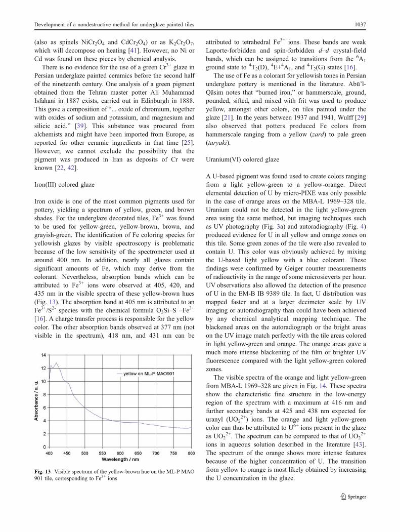
Iron(III) colored glaze
Iron oxide is one of the most common pigments used forpottery, yielding a spectrum of yellow, green, and brownshades. For the underglaze decorated tiles, Fe3+ was foundto be used for yellow-green, yellow-brown, brown, andgrayish-green. The identification of Fe coloring species foryellowish glazes by visible spectroscopy is problematicbecause of the low sensitivity of the spectrometer used ataround 400 nm. In addition, nearly all glazes containsignificant amounts of Fe, which may derive from thecolorant. Nevertheless, absorption bands which can beattributed to Fe3+ ions were observed at 405, 420, and435 nm in the visible spectra of these yellow-brown hues(Fig. 13). The absorption band at 405 nm is attributed to anFe3+/S2- species with the chemical formula O3Si–S
−–Fe3+
[16]. A charge transfer process is responsible for the yellowcolor. The other absorption bands observed at 377 nm (notvisible in the spectrum), 418 nm, and 431 nm can be
attributed to tetrahedral Fe3+ ions. These bands are weakLaporte-forbidden and spin-forbidden d–d crystal-fieldbands, which can be assigned to transitions from the 6A1
ground state to 4T2(D),4E+4A1, and
4T2(G) states [16].The use of Fe as a colorant for yellowish tones in Persian
underglaze pottery is mentioned in the literature. Abū’l-Qāsim notes that “burned iron,” or hammerscale, ground,pounded, sifted, and mixed with frit was used to produceyellow, amongst other colors, on tiles painted under theglaze [21]. In the years between 1937 and 1941, Wulff [29]also observed that potters produced Fe colors fromhammerscale ranging from a yellow (zard) to pale green(taryaki).
Uranium(VI) colored glaze
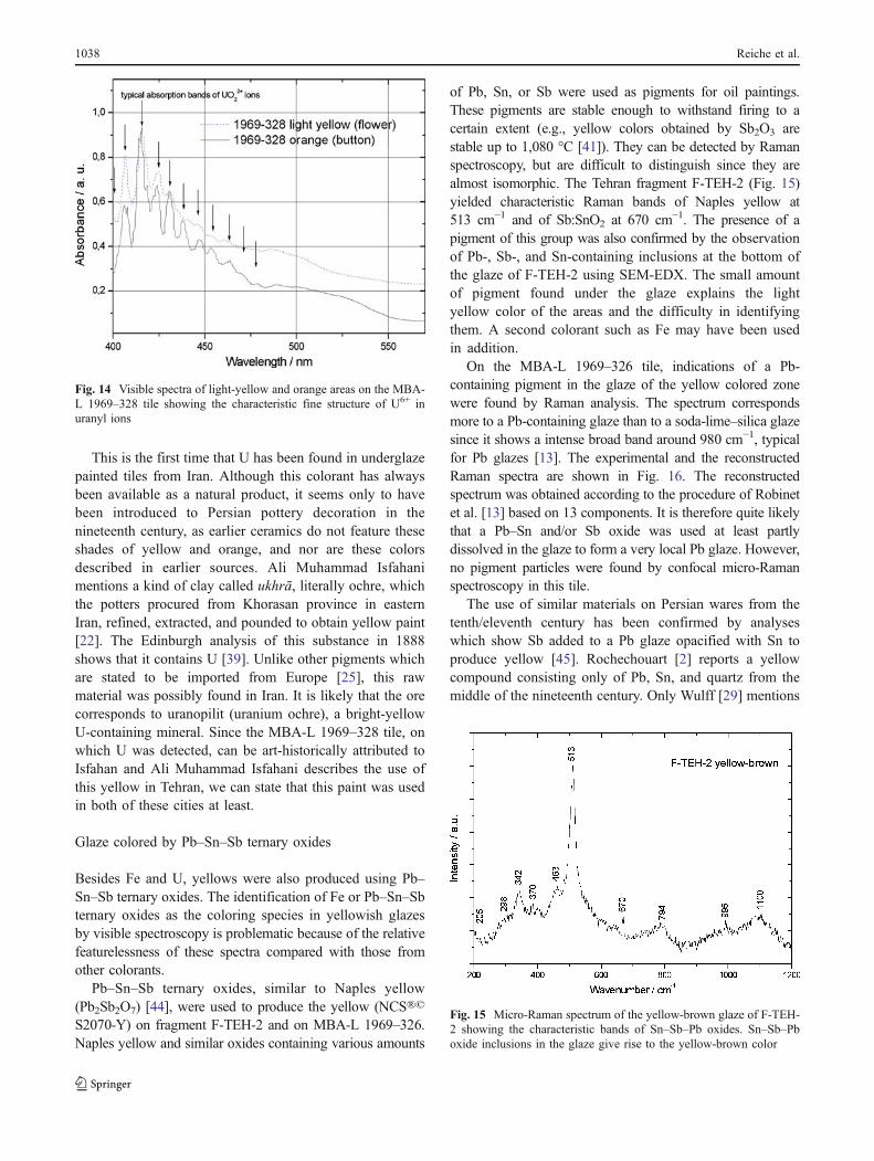
A U-based pigment was found used to create colors rangingfrom a light yellow-green to a yellow-orange. Directelemental detection of U by micro-PIXE was only possiblein the case of orange areas on the MBA-L 1969–328 tile.Uranium could not be detected in the light yellow-greenarea using the same method, but imaging techniques suchas UV photography (Fig. 3a) and autoradiography (Fig. 4)produced evidence for U in all yellow and orange zones onthis tile. Some green zones of the tile were also revealed tocontain U. This color was obviously achieved by mixingthe U-based light yellow with a blue colorant. Thesefindings were confirmed by Geiger counter measurementsof radioactivity in the range of some microsieverts per hour.UV observations also allowed the detection of the presenceof U in the EM-B IB 9389 tile. In fact, U distribution wasmapped faster and at a larger decimeter scale by UVimaging or autoradiography than could have been achievedby any chemical analytical mapping technique. Theblackened areas on the autoradiograph or the bright areason the UV image match perfectly with the tile areas coloredin light yellow-green and orange. The orange areas gave amuch more intense blackening of the film or brighter UVfluorescence compared with the light yellow-green coloredzones.
The visible spectra of the orange and light yellow-greenfrom MBA-L 1969–328 are given in Fig. 14. These spectrashow the characteristic fine structure in the low-energyregion of the spectrum with a maximum at 416 nm andfurther secondary bands at 425 and 438 nm expected foruranyl (UO2
2+) ions. The orange and light yellow-greencolor can thus be attributed to U6+ ions present in the glazeas UO2
2+. The spectrum can be compared to that of UO22+
ions in aqueous solution described in the literature [43].The spectrum of the orange shows more intense featuresbecause of the higher concentration of U. The transitionfrom yellow to orange is most likely obtained by increasingthe U concentration in the glaze.
Fig. 13 Visible spectrum of the yellow-brown hue on the ML-P MAO901 tile, corresponding to Fe3+ ions
Development of a nondestructive method for underglaze painted tiles 1037
This is the first time that U has been found in underglazepainted tiles from Iran. Although this colorant has alwaysbeen available as a natural product, it seems only to havebeen introduced to Persian pottery decoration in thenineteenth century, as earlier ceramics do not feature theseshades of yellow and orange, and nor are these colorsdescribed in earlier sources. Ali Muhammad Isfahanimentions a kind of clay called ukhrā, literally ochre, whichthe potters procured from Khorasan province in easternIran, refined, extracted, and pounded to obtain yellow paint[22]. The Edinburgh analysis of this substance in 1888shows that it contains U [39]. Unlike other pigments whichare stated to be imported from Europe [25], this rawmaterial was possibly found in Iran. It is likely that the orecorresponds to uranopilit (uranium ochre), a bright-yellowU-containing mineral. Since the MBA-L 1969–328 tile, onwhich U was detected, can be art-historically attributed toIsfahan and Ali Muhammad Isfahani describes the use ofthis yellow in Tehran, we can state that this paint was usedin both of these cities at least.
Glaze colored by Pb–Sn–Sb ternary oxides
Besides Fe and U, yellows were also produced using Pb–Sn–Sb ternary oxides. The identification of Fe or Pb–Sn–Sbternary oxides as the coloring species in yellowish glazesby visible spectroscopy is problematic because of the relativefeaturelessness of these spectra compared with those fromother colorants.
Pb–Sn–Sb ternary oxides, similar to Naples yellow(Pb2Sb2O7) [44], were used to produce the yellow (NCS®©
S2070-Y) on fragment F-TEH-2 and on MBA-L 1969–326.Naples yellow and similar oxides containing various amounts
of Pb, Sn, or Sb were used as pigments for oil paintings.These pigments are stable enough to withstand firing to acertain extent (e.g., yellow colors obtained by Sb2O3 arestable up to 1,080 °C [41]). They can be detected by Ramanspectroscopy, but are difficult to distinguish since they arealmost isomorphic. The Tehran fragment F-TEH-2 (Fig. 15)yielded characteristic Raman bands of Naples yellow at513 cm−1 and of Sb:SnO2 at 670 cm−1. The presence of apigment of this group was also confirmed by the observationof Pb-, Sb-, and Sn-containing inclusions at the bottom ofthe glaze of F-TEH-2 using SEM-EDX. The small amountof pigment found under the glaze explains the lightyellow color of the areas and the difficulty in identifyingthem. A second colorant such as Fe may have been usedin addition.
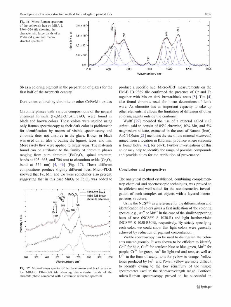
On the MBA-L 1969–326 tile, indications of a Pb-containing pigment in the glaze of the yellow colored zonewere found by Raman analysis. The spectrum correspondsmore to a Pb-containing glaze than to a soda-lime–silica glazesince it shows a intense broad band around 980 cm−1, typicalfor Pb glazes [13]. The experimental and the reconstructedRaman spectra are shown in Fig. 16. The reconstructedspectrum was obtained according to the procedure of Robinetet al. [13] based on 13 components. It is therefore quite likelythat a Pb–Sn and/or Sb oxide was used at least partlydissolved in the glaze to form a very local Pb glaze. However,no pigment particles were found by confocal micro-Ramanspectroscopy in this tile.
The use of similar materials on Persian wares from thetenth/eleventh century has been confirmed by analyseswhich show Sb added to a Pb glaze opacified with Sn toproduce yellow [45]. Rochechouart [2] reports a yellowcompound consisting only of Pb, Sn, and quartz from themiddle of the nineteenth century. Only Wulff [29] mentions
Fig. 15 Micro-Raman spectrum of the yellow-brown glaze of F-TEH-2 showing the characteristic bands of Sn–Sb–Pb oxides. Sn–Sb–Pboxide inclusions in the glaze give rise to the yellow-brown color
Fig. 14 Visible spectra of light-yellow and orange areas on the MBA-L 1969–328 tile showing the characteristic fine structure of U6+ inuranyl ions
1038 Reiche et al.
Sb as a coloring pigment in the preparation of glazes for thefirst half of the twentieth century.
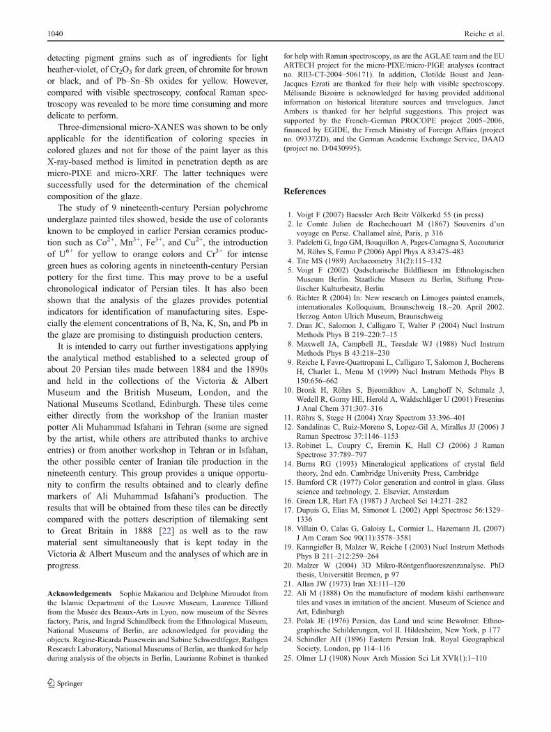
Dark zones colored by chromite or other Cr/Fe/Mn oxides
Chromite phases with various compositions of the generalchemical formula (Fe,Mg)(Cr,Al,Fe)2O4 were found inblack and brown colors. These colors were studied usingonly Raman spectroscopy as their dark color is problematicfor identification by means of visible spectroscopy andchromite does not dissolve in the glaze. Brown or blackwas used on all tiles to outline the figures, faces, and hair.More rarely they were applied to larger areas. The materialsfound can be attributed to the family of chromite phasesranging from pure chromite (FeCr2O4, spinel structure,bands at 605, 665, and 706 nm) to chromium oxide (Cr2O3,band at 554 nm) [4, 46] (Fig. 17). These differentcompositions produce slightly different hues. Micro-PIXEshowed that Fe, Mn, and Co were sometimes also present,suggesting that in this case MnO2 or FexOy was added to
produce a specific hue. Micro-XRF measurements on theEM-B IB 9389 tile confirmed the presence of Cr and Fetogether with Mn on dark brown/black areas [5]. Tite [4]also found chromite used for linear decorations of Iznikware. As chromite has an important capacity to take upother elements, it allows the limitation of diffusion of othercoloring agents outside the contours.
Wulff [29] recorded the use of a mineral called siahqalam, said to consist of 85% chromite, 10% Mn, and 5%magnesium silicate, extracted in the area of Natanz (Iran).Abū’l-Qāsim [21] mentions the use of the mineral muzarrad,mined from a location in Khorasan province where chromiteis found today [42], for black. Further investigations of thiscolor may help to identify the range of possible compoundsand provide clues for the attribution of provenance.
Conclusion and perspectives
The analytical method established, combining complemen-tary chemical and spectroscopic techniques, was proved tobe efficient and well suited for the nondestructive investi-gation of such complex art objects with a layered hetero-geneous structure.
Using the NCS®© as a reference for the differentiation andidentification of colors gives a first indication of the coloringspecies, e.g., Au0 or Mn3+ in the case of the similar-appearinghues of rose (NCS®© S 1030-R) and light heather-violet(NCS®© S 1050-R30B), respectively. By strictly specifyingeach color, we could show that light colors were generallyachieved by reduction of pigment concentration.
Visible spectroscopy can be used to distinguish the color-ants unambiguously. It was shown to be efficient to identifyCo2+ for blue, Cu2+ for cerulean blue or blue-green, Mn3+ forpurple, Cr3+ for green, Au0 for light red and rose, as well asU6+ in the form of uranyl ions for yellow to orange. Yellowtones produced by Fe3+ and Pb–Sn yellow are more difficultto identify owing to the low sensitivity of the visiblespectrometer used in the short-wavelength range. Confocalmicro-Raman spectroscopy proved to be successful in
Fig. 17 Micro-Raman spectra of the dark-brown and black areas onthe MBA-L 1969–328 tile showing characteristic bands of thechromite phase compared with a chromite reference spectrum
Fig. 16 Micro-Raman spectrumof the yellowish hue on MBA-L1969–326 tile showing thecharacteristic large bands of aPb-based glaze and recon-structed spectrum
Development of a nondestructive method for underglaze painted tiles 1039
detecting pigment grains such as of ingredients for lightheather-violet, of Cr2O3 for dark green, of chromite for brownor black, and of Pb–Sn–Sb oxides for yellow. However,compared with visible spectroscopy, confocal Raman spec-troscopy was revealed to be more time consuming and moredelicate to perform.
Three-dimensional micro-XANES was shown to be onlyapplicable for the identification of coloring species incolored glazes and not for those of the paint layer as thisX-ray-based method is limited in penetration depth as aremicro-PIXE and micro-XRF. The latter techniques weresuccessfully used for the determination of the chemicalcomposition of the glaze.
The study of 9 nineteenth-century Persian polychromeunderglaze painted tiles showed, beside the use of colorantsknown to be employed in earlier Persian ceramics produc-tion such as Co2+, Mn3+, Fe3+, and Cu2+, the introductionof U6+ for yellow to orange colors and Cr3+ for intensegreen hues as coloring agents in nineteenth-century Persianpottery for the first time. This may prove to be a usefulchronological indicator of Persian tiles. It has also beenshown that the analysis of the glazes provides potentialindicators for identification of manufacturing sites. Espe-cially the element concentrations of B, Na, K, Sn, and Pb inthe glaze are promising to distinguish production centers.
It is intended to carry out further investigations applyingthe analytical method established to a selected group ofabout 20 Persian tiles made between 1884 and the 1890sand held in the collections of the Victoria & AlbertMuseum and the British Museum, London, and theNational Museums Scotland, Edinburgh. These tiles comeeither directly from the workshop of the Iranian masterpotter Ali Muhammad Isfahani in Tehran (some are signedby the artist, while others are attributed thanks to archiveentries) or from another workshop in Tehran or in Isfahan,the other possible center of Iranian tile production in thenineteenth century. This group provides a unique opportu-nity to confirm the results obtained and to clearly definemarkers of Ali Muhammad Isfahani’s production. Theresults that will be obtained from these tiles can be directlycompared with the potters description of tilemaking sentto Great Britain in 1888 [22] as well as to the rawmaterial sent simultaneously that is kept today in theVictoria & Albert Museum and the analyses of which are inprogress.
Acknowledgements Sophie Makariou and Delphine Miroudot fromthe Islamic Department of the Louvre Museum, Laurence Tilliardfrom the Musée des Beaux-Arts in Lyon, now museum of the Sèvresfactory, Paris, and Ingrid Schindlbeck from the Ethnological Museum,National Museums of Berlin, are acknowledged for providing theobjects. Regine-Ricarda Pausewein and Sabine Schwerdtfeger, RathgenResearch Laboratory, National Museums of Berlin, are thanked for helpduring analysis of the objects in Berlin, Laurianne Robinet is thanked
for help with Raman spectroscopy, as are the AGLAE team and the EUARTECH project for the micro-PIXE/micro-PIGE analyses (contractno. RII3-CT-2004–506171). In addition, Clotilde Boust and Jean-Jacques Ezrati are thanked for their help with visible spectroscopy.Mélisande Bizoirre is acknowledged for having provided additionalinformation on historical literature sources and travelogues. JanetAmbers is thanked for her helpful suggestions. This project wassupported by the French–German PROCOPE project 2005–2006,financed by EGIDE, the French Ministry of Foreign Affairs (projectno. 09337ZD), and the German Academic Exchange Service, DAAD(project no. D/0430995).
References
1. Voigt F (2007) Baessler Arch Beitr Völkerkd 55 (in press)2. le Comte Julien de Rochechouart M (1867) Souvenirs d’un
voyage en Perse. Challamel aîné, Paris, p 3163. Padeletti G, Ingo GM, Bouquillon A, Pages-Camagna S, Aucouturier
M, Röhrs S, Fermo P (2006) Appl Phys A 83:475–4834. Tite MS (1989) Archaeometry 31(2):115–1325. Voigt F (2002) Qadscharische Bildfliesen im Ethnologischen
Museum Berlin. Staatliche Museen zu Berlin, Stiftung Preu-ßischer Kulturbesitz, Berlin
6. Richter R (2004) In: New research on Limoges painted enamels,internationales Kolloquium, Braunschweig 18.–20. April 2002.Herzog Anton Ulrich Museum, Braunschweig
7. Dran JC, Salomon J, Calligaro T, Walter P (2004) Nucl InstrumMethods Phys B 219–220:7–15
8. Maxwell JA, Campbell JL, Teesdale WJ (1988) Nucl InstrumMethods Phys B 43:218–230
9. Reiche I, Favre-Quattropani L, Calligaro T, Salomon J, BocherensH, Charlet L, Menu M (1999) Nucl Instrum Methods Phys B150:656–662
10. Bronk H, Röhrs S, Bjeomikhov A, Langhoff N, Schmalz J,Wedell R, Gorny HE, Herold A, Waldschläger U (2001) FreseniusJ Anal Chem 371:307–316
11. Röhrs S, Stege H (2004) Xray Spectrom 33:396–40112. Sandalinas C, Ruiz-Moreno S, Lopez-Gil A, Miralles JJ (2006) J
Raman Spectrosc 37:1146–115313. Robinet L, Coupry C, Eremin K, Hall CJ (2006) J Raman
Spectrosc 37:789–79714. Burns RG (1993) Mineralogical applications of crystal field
theory, 2nd edn. Cambridge University Press, Cambridge15. Bamford CR (1977) Color generation and control in glass. Glass
science and technology, 2. Elsevier, Amsterdam16. Green LR, Hart FA (1987) J Archeol Sci 14:271–28217. Dupuis G, Elias M, Simonot L (2002) Appl Spectrosc 56:1329–
133618. Villain O, Calas G, Galoisy L, Cormier L, Hazemann JL (2007)
J Am Ceram Soc 90(11):3578–358119. Kanngießer B, Malzer W, Reiche I (2003) Nucl Instrum Methods
Phys B 211–212:259–26420. Malzer W (2004) 3D Mikro-Röntgenfluoreszenzanalyse. PhD
thesis, Universität Bremen, p 9721. Allan JW (1973) Iran XI:111–12022. Ali M (1888) On the manufacture of modern kāshi earthenware
tiles and vases in imitation of the ancient. Museum of Science andArt, Edinburgh
23. Polak JE (1976) Persien, das Land und seine Bewohner. Ethno-graphische Schilderungen, vol II. Hildesheim, New York, p 177
24. Schindler AH (1896) Eastern Persian Irak. Royal GeographicalSociety, London, pp 114–116
25. Olmer LJ (1908) Nouv Arch Mission Sci Lit XVI(1):1–110
1040 Reiche et al.
26. Gratuze B, Soulier I, Blet M, Vallauri L (1996) Rev Archeom20:77–94
27. Pagès-Camagna S, Colinart S (2003) Archaeometry 45(4):637–65828. Bamford CR (1962) Phys Chem Glasses 3(6):189–20229. Wulff HE (1966) The traditional crafts of Persia; their develop-
ment, technology, and influence on Western and Eastern civiliza-tions. MIT Press, Cambridge, pp 161–165
30. Schultze-Frentzel U (1973) In: Islamische Keramik. Ausstellungs-katalog. Hetjens-Museum, Düsseldorf, p 329
31. Weyl WA (1999) Colored glasses. Society of Glass Technology,Sheffield
32. Lehnhäuser W, Lehnhäuser K (2000) Keramische Glasuren undihre Farben—für Studium, Handwerk und Industrie, 4th edn.Ritterbach, Frechen
33. Mie G (1908) Ann Phys 25:377–44534. Lafait J (2006) Verre 12(4):11–2135. Horn I (1998) Entwicklung einer quasizerstörungsfreien Proben-
nahme- und Analysetechnik für Glas und ihre Anwendung aufRubingläser des 17. und 18. Jahrhunderts. PhD thesis, TechnischeUniversität Berlin, Berlin
36. Speel E, Bronk H (2001) Berl Beitr Archaom 18:43–10037. Stalhandske C (2000) Glastek Tidskr 55(3):65–7138. Colomban P, Sagon G, Faurel X (2001) J Raman Spectrosc
32:351–36039. Floor W (2003) Traditional crafts in Qajar Iran [1800–1925].
Mazda, Costa Mesa, p 43340. Calas G, Majérus O, Galoisy L, Cormier L (2006) Chem Geol
229:218–22641. Shaw K (1962) Ceramic colours and pottery decoration. Macla-
ren, London42. Ostovār R-A (1992) In: Encyclopaedia Iranica, vol V. Encyclo-
paedia Iranica, New York, p 54843. Meinrath G (1998) Freiberg Online Geosci 1. http://www.geo.
tu-freiberg.de/fog/issues.html44. Roy A, Berrie BH (1998) In: IIC contributions to the Dublin
congress, 7–11 September 1998, pp 160–16545. Morgan P, Leatherby J (1987) In: Allan J, Roberts C (eds) Syria
and Iran: three studies in medieval ceramics. Oxford UniversityPress, Oxford, p 82
46. RRUFF database (2008) RRUFF Project, Tuscon. http://rruff.info/
Development of a nondestructive method for underglaze painted tiles 1041
Copyright © 2022 FDOKUMEN