Detoxification of Toxic Heavy Metals by Marine Bacteria Highly Resistant to Mercury
19
Detoxification of toxic heavy metals by marine bacteria highly resistant to mercury. De Jaysankar, N. Ramaiah and L. Vardanyan. Marine Biotechnology. Vol. 10, No. 4, pp. 471-477, 2008. Detoxification of toxic heavy metals by marine bacteria highly resistant to mercury De Jaysankar 1, 2 *, N. Ramaiah 1 and L. Vardanyan 3 1 National Institute of Oceanography, Dona Paula, Goa 403 004, India 2 Present address: Graduate School of Kuroshio Science (GRAKUS), Kochi University, Nankoku, Kochi 783 8502, Japan 3 Insitute of Hydroecology and Ichthyology, NAS, Yerevan 375 033, Armenia Running title: Detoxification of toxic heavy metals by marine BHRM ____________ *Corresponding author: Graduate School of Kuroshio Science (GRAKUS), Kochi University, Nankoku, Kochi 783-8502, NIPPON (Japan), Tel & Fax: +81-88-864-5152, Email: [email protected]
-
Upload
independent -
Category
Documents
-
view
1 -
download
0
Transcript of Detoxification of Toxic Heavy Metals by Marine Bacteria Highly Resistant to Mercury
Detoxification of toxic heavy metals by marine bacteria highly resistant to mercury. De Jaysankar, N. Ramaiah and L. Vardanyan. Marine Biotechnology. Vol. 10, No. 4, pp. 471-477, 2008.
Detoxification of toxic heavy metals by marine bacteria highly resistant to mercury
De Jaysankar1, 2*, N. Ramaiah1 and L. Vardanyan3
1National Institute of Oceanography, Dona Paula, Goa 403 004, India
2 Present address: Graduate School of Kuroshio Science (GRAKUS), Kochi University,
Nankoku, Kochi 783 8502, Japan
3Insitute of Hydroecology and Ichthyology, NAS, Yerevan 375 033, Armenia
Running title: Detoxification of toxic heavy metals by marine BHRM
____________
*Corresponding author: Graduate School of Kuroshio Science (GRAKUS), Kochi University,
Nankoku, Kochi 783-8502, NIPPON (Japan), Tel & Fax: +81-88-864-5152, Email:
2
Abstract
Pollution in industrial areas is of serious environmental concern and interest in bacterial
resistance to heavy metals is of practical significance. Mercury (Hg), Cadmium (Cd) and lead
(Pb) are known to cause damage to living organisms including human beings. Several marine
bacteria highly resistant to mercury (BHRM) capable of growing at 25 ppm (mg L-1) or higher
concentrations of mercury were tested during this study to evaluate their potential to detoxify Cd
and Pb. Results indicate their potential of detoxification not only of Hg, but also Cd and Pb.
Through biochemical and 16S rRNA gene sequence analyses, these bacteria were identified to
belong to Alcaligenes faecalis (seven isolates), Bacillus pumilus (three isolates), Bacillus sp.
(one isolate) Pseudomonas aeruginosa (one isolate) and Brevibacterium iodinium (one isolate).
The mechanisms of heavy metal detoxification were through volatilization (for Hg), putative
entrapment in the extracellular polymeric substance (for Hg, Cd and Pb) as revealed by the
scanning electron microscopy (SEM) and energy dispersive X-ray spectroscopy (EDS), and/or
precipitation as sulfide (for Pb). These bacteria removed over 70% of Cd and 98% of Pb within
72 and 96 h respectively from growth medium that had initial metal concentrations of 100 ppm.
Their detoxification efficiency for Hg, Cd and Pb indicates good potential for application in
bioremediation of toxic heavy metals.
Keywords: Heavy metals, bacteria highly resistant to mercury, detoxification, volatilization,
merA, energy dispersive X-ray spectroscopy, scanning electron microscopy, bioremediation

3
INTRODUCTION
Metal-resistant microorganisms have been isolated from polluted environments and studies on
the interactions between heavy metals and microorganisms have focused on bacterial
transformation and conversion of metallic ions by reduction (Chang et al., 1993). Metal-resistant
microorganisms may be useful as indicators of potential toxicity to other forms of life (Doelman
et al., 1984) and are important in studies of mechanisms, determinants and genetic transfer of
microbial metal-resistance (De Rore et al., 1994). Hg2+, Pb2+ and Cd2+ are of serious concern as
they are non-biodegradable, highly toxic and are present in a variety of waste streams that
contaminate the environment. These three metals are included on the US Environmental
Protection Agency’s list of priority pollutants (Cameron, 1992).
Bioremediation processes are very attractive in comparison with physicochemical methods for
heavy metal removal because they can have lower cost and higher efficiency at low metal
concentrations (Gadd and White, 1993). Mechanisms of metal resistance in microbes include
precipitation of metals as phosphates, carbonates and/or sulfides; volatilization via methylation
or ethylation; physical exclusion of electronegative components in membranes and extracellular
polymeric substances (EPS); energy-dependent metal efflux systems; and intracellular
sequestration with low molecular weight, cysteine-rich proteins (Gadd, 1990; Silver, 1996).
The mer operon that confers mercury resistance to bacteria is widely distributed in mercury-
resistant bacterial populations (Osborn et al., 1997; Barkay et al., 2003) and is fairly highly
conserved. In contrast, other metal resistance systems have evolved several times (Silver and
Phung, 1996). Single bacterial strains can be resistant to many metals. The genome sequence of
P. putida KT2440 contains 61 open reading frames involved in tolerance and resistance to a
range of metals (Cànovas et al., 2003). Resistance to multiple metals including Hg, Cd, Zn, Sn,
Cu, and Pb was found in a bacterium isolated on the basis of tributyltin resistance (Pain and
Cooney, 1998).
The present study focuses on 13 marine bacterial strains that are highly resistant to mercury (De
et al., 2003) and investigates their resistance and ability to detoxify Cd and Pb in addition to Hg.
4
Since Hg, Cd and Pb are present in many environments, the remediation of these metals is of
great ecological interest.
MATERIALS AND METHODS
Bacterial isolates. Mercury-resistant bacteria were isolated from various locations along the
Indian Coast. Seawater nutrient agar (SWNA, Himedia Laboratories Pvt. Ltd., India; formula per
liter: peptone 5.0 g, beef extract 1.5 g, yeast extract 1.5 g, sodium chloride 15 g, aged seawater
500 ml, deionized water 500 ml and agar 15 g), amended with 10 ppm HgCl2 (ca. 50 mM
Hg)(Merck, Germany) was used for their isolation and enumeration. Bacterial colonies capable
of growth when streaked onto SWNA plates with 25 ppm mercury were termed bacteria highly
resistant to mercury (BHRM). Purified cultures of 13 BHRM isolates were previously grown in
seawater nutrient broth (SWNB: SWNA without agar) amended with 25 ppm Hg, characterized
and identified using biochemical tests and 16S rDNA sequencing (De et al., 2007).
Volatilization of Hg. Details of this experiment is already published (De et al., 2006). In brief,
11 BHRM, six isolates of Alcaligenes faecalis (GP15, GO02, GP16, GP06, GP14, GP17), three
isolates of Bacillus pumilus (CM10, CH13, GP08), one P. aeruginosa isolate (CH07), one
Brevibacterium iodinium isolate (GP13), one mercury-sensitive bacterium (unidentified; as
negative control) and Pseudomonas putida KT2442::mer73 (as positive control) were tested.
Strains were grown in marine broth 2216 (Difco). Cells were pelletted by centrifugation (at
10,000 X g for 10 minutes), washed with phosphate buffer (pH 7) and transferred to microtitre
plates. Mercurated phosphate buffer (10 ppm Hg final concentration) was added to the cells. The
plate was covered with Kodak XAR film and incubated at 30ºC in the dark for 4 hours, prior to
removal and development of the film.
Detection of merA. Genomic DNA was extracted from cultures grown for 18 h using the
NucleoSpin Extract Kit (Macherey Nagel, Germany). The merA region was amplified using
primer pairs (A1 forward: 5′ACC ATC GGC GGC ACC TGC GT3′; A5 reverse: 5′ACC ATC
GTC AGG TAG GGG ACC AA3′) synthesized by Invitrogen (Invitrogen GmbH, Germany).
The PCR was performed (Mastercycler gradient, Eppendorf, Hamburg, Germany) using
predenaturation at 95°C for 5 min followed by 30 cycles of denaturation at 94°C for 15 sec,

5
annealing at 58°C for 30 sec, extension at 68°C for 1 min and a final extension at 68°C for 4
min. The PCR products were visualized by agarose gel electrophoresis.
Detoxification of Cd. Two isolates (CH07, GP06) were used in the experiment with Cd. A Cd
sensitive Proteus sp. isolate CH05 and heat killed bacterial cells were included as negative
controls. Bacteria were tested in SWNB amended with different concentrations of Cd (as CdCl2)
at room temperature (28±2ºC). Sub-samples (1 ml) were taken every 24 h, centrifuged at 10,000
X g for 15 minutes at 24ºC and supernatants filtered through sterilized pre-weighed 0.22 µm
membrane filters. Filtrates were diluted 10-fold with 10% HNO3 for estimation of the heavy
metals from the test media. The pellets were digested overnight in 1 M HCl, sonicated twice for
45 seconds, and centrifuged at 8,000 X g for 5 min. The supernatants were collected and diluted
with 10% HNO3 for estimation of heavy metals accumulated by the cells. The cell pellets were
dried for at least 48 hour at 70ºC and weighed to determine bacterial biomass. The Cd
concentrations were determined by inductively coupled plasma-atomic emission spectrometry
(Varian, Liberty Series II AX, Sequential ICP-AES) under the following conditions (pump rate:
15 rpm, plasma gas: 15.0 L min-1, fast pump: on, PMT voltage: 650V, auxiliary gas: 1.50 L min-
1, rinse time: 10 sec, sample uptake: 30 sec, wavelength: 226.502 nm, replicate no. 3). The metal
concentrations were calculated using appropriate blanks (growth medium diluted with 10%
HNO3) and standards.
Detoxification of Pb. Three BHRM isolates (CH07, GP13, and S3) were tested in SWNB
amended with different concentrations of Pb [as (CH3COO)2Pb] at room temperature (28±2ºC).
Mercury-sensitive isolate CH05 and heat-killed bacterial cells were included as negative
controls. The experimental procedure was similar to that for Cd. Atomic absorption
spectrophotometry (Perkin Elmer, Analyst 200 for flame analysis) under the following
conditions (slit width: 1.0 nm, lamp current 8.0.mA, scale expansion: 1.000, wavelength: 217
nm, atomization: air-acetylene, integration time: 3.0 sec, replicate no. 3) was carried out for
determination of Pb concentrations, using appropriate blanks and standards for calibration.
Scanning Electron Microscopy (SEM) and Energy Dispersive X-ray Spectroscopy (EDS).
Samples (10 ml) of broth cultures from test flasks with Cd and Pb were centrifuged at 8,000 X g
at 4ºC for 5 minutes. The cells were washed twice with 0.1 M phosphate buffered saline (PBS;
6
15 mM phosphate buffer, 138 mM NaCl, 2.7 mM KCl, pH 7.4 at 25ºC) and fixed overnight in
2% glutaraldehyde (prepared in 0.1 M PBS). The cells were washed with PBS and distilled water
prior to dehydration through an ethanol series (10% to absolute), held at each concentration for
30 minutes. Samples were placed on a brass stub, sputter-coated with gold and examined by
SEM (SEM-JEOL-JSM5800LV). EDS (OXFORD ISIS 300 EDS) was carried out to detect Cd
and Pb and their compounds that were either adsorbed to the cell surface or entrapped in the
exopolymeric substance (EPS).
RESULTS
Bacterial isolates. 16S rRNA gene sequence analysis showed that the BHRM isolates are closely
related to A. faecalis (GO01, GO02, GP06, GP14, GP15, GP16 and GP17), B. pumilus (GP08,
CH13 and S3), P. aeruginosa (CH07), B. iodinium (GP13) and Bacillus sp. (CM10). The partial
16S rRNA gene sequences have been deposited in the Gene Bank database under accession
numbers DQ37744-DQ377468 (De et al., 2007).
Volatilization of Hg. All of the 11 BHRM were capable of volatilizing mercury indicated by
fogging of the XAR film covering their respective wells due to the reduction of AgCl2 by
gaseous Hg. The negative control did not volatilize mercury indicated by clear film over this
isolate.
Presence of merA genes. PCR analyses revealed the presence of putative merA gene fragments
in nine isolates: GO02, GP06, GP08, GP14, GP15, GP16, GP17, CH07 and GO01. All these
isolates volatilized mercury. Putative merA genes were not detected in isolates CH13 and CM10
that were shown to volatilize Hg. The mechanism of this apparently non-merA mediated
volatilization is not known.
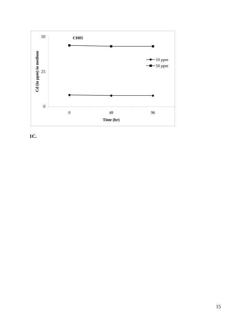
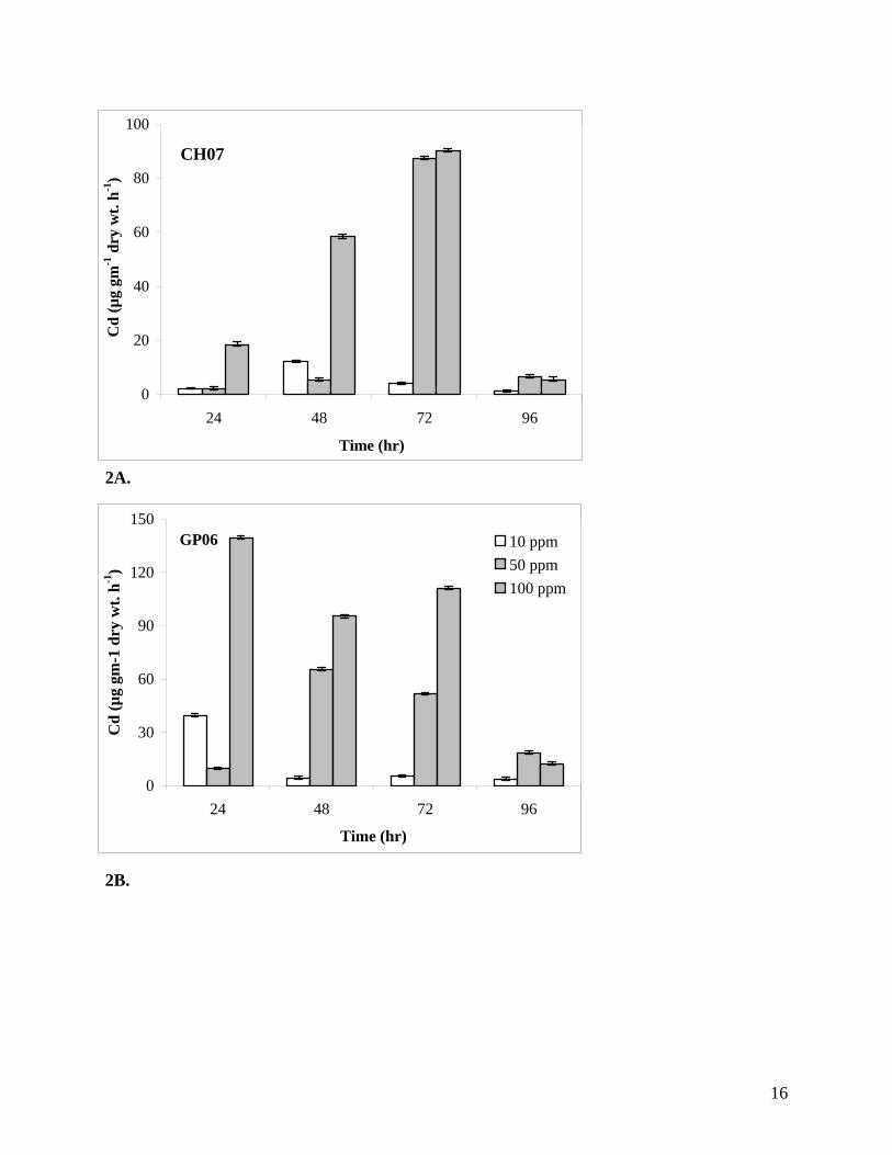
Detoxification of Cd. Isolate CH07 removed Cd from the medium at a faster rate than GP06
during the first 48 hours (Figures 1A-B). The Cd concentration was reduced from an initial
concentration of 100 ppm to 17.4 ppm by CH07 and to 19.2 ppm by GP06 in about 72 h. This
was consistent with an increase in Cd concentration in the cell pellets (Figures 2A-B), Isolates
CH07 and GP06 removed over 75% and 70% of Cd respectively from the growth medium. In
7
controls containing killed cells, removal of metals was <5%. The metal-sensitive control isolate
CH05 showed no sign of growth or Cd accumulation (Figure 1C).
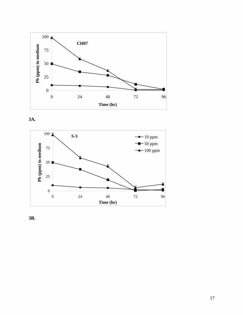
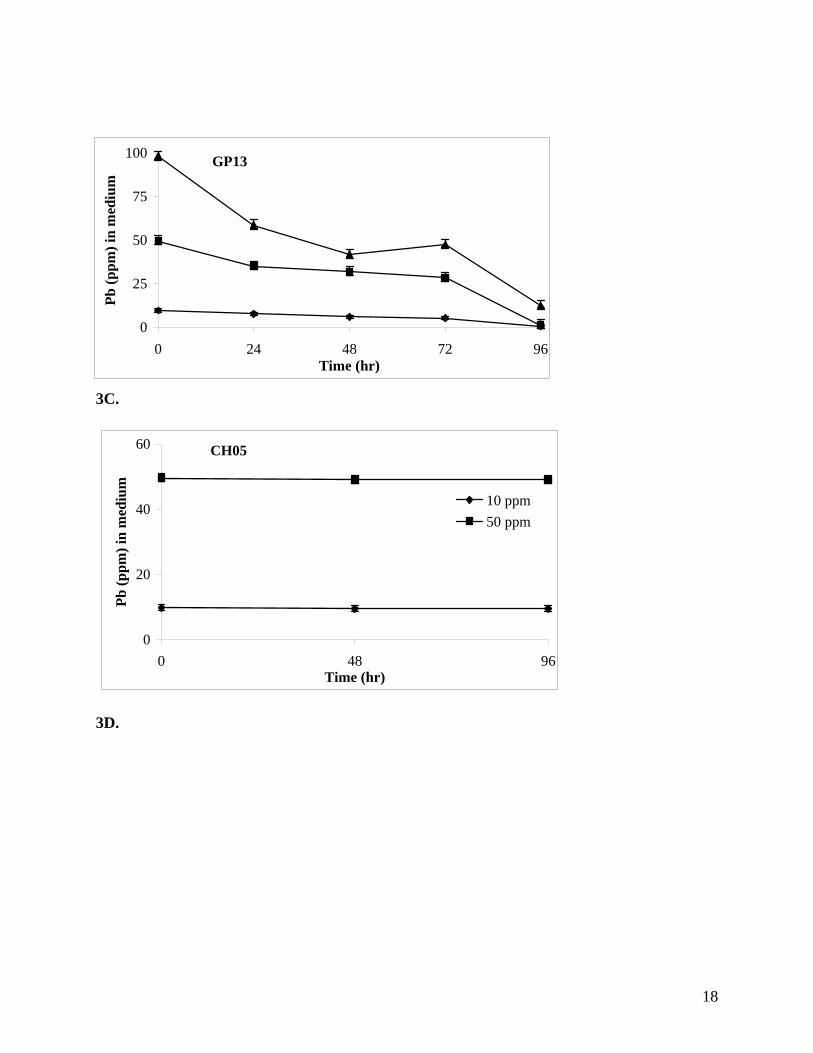
Detoxification of Pb. All three BHRM isolates tested were able to remove Pb from the growth
medium (Figures 3A-C). In case of CH07, the concentration of Pb was reduced to 1.8 ppm
(>98% removal) from an initial concentration of 100 ppm in 96 h. SEM and EDS analysis
(below) indicated that Pb was likely present in the EPS. Isolates GP13 and S3 removed more
than 87 and 88% of Pb respectively from the growth medium. The fact that these isolates were
capable of hydrogen sulfide production and indications from preliminary chemical tests and
EDS, suggest that Pb was precipitated as sulfides by these two strains. The metal-sensitive
negative control isolate CH05 showed no sign of growth (Figure 3D).
SEM and EDS. The morphology of isolate GP13 was unchanged after growth in medium
without added Pb ((Figure 4a) and with 50 ppm Pb (Figure 4b). This suggests that Pb is not toxic
to the isolate under the conditions tested. Preliminary studies showed that the Pb-resistant
bacteria produced large quantities of EPS. EDS analysis indicated that Pb was most likely
entrapped in the EPS in case of CH07. Preliminary chemical tests showed that Pb was most
likely precipitated as sulfide by isolates GP13 and S3. EDS analysis of the cells showed the
concentrations of Pb and Cd to be as high as 21% and 19%, respectively. Bacteria grown in
medium without these heavy metals showed no EDS signals for either Cd or Pb.
DISCUSSION
Bacterial mobile genetic elements, such as plasmids or transposons, can carry multiple genes
encoding metal and antibiotic resistance. Thus, exposure to one agent may select for
microorganisms resistant to several toxicants. Such organisms may be important in performing
biological processes in contaminated habitats. Metal-resistant strains may also have application
in remediation of metal-contaminated environments. A genetically engineered Escherichia coli
strain with Hg2+ transport system and metallothionein has been used to bioaccumulate mercury
from wastewater (Deng and Wilson, 2001). Deinococcus radiodurans has been engineered to
remediate radioactive mixed waste as well as vaporize mercury (Brim et al., 2000).
8
Previous studies in terrestrial and freshwater environments have shown that as many as 100%
culturable mercury-resistant environmental isolates contain genes that have homology to either
merTn21 (Barkay et al., 1989) or merTn501 (Bruce et al., 1995). However, there are conflicting
findings on the frequency of prototypic mer genes in culturable mercury-resistant isolates
obtained from marine samples (Rasmussen and Sørensen, 1998; Reyes et al., 1999). In this
study, only 9 of the 11 BHRM isolates were positive for merA, whereas all of them volatilized
Hg2+ to Hg0. This may suggest the presence of putative non-mer mediated mercury volatilization
in two isolates but this interesting possibility remains to be confirmed.
The reduction of Cd in culture supernatants and accumulation in cell pellets of bacteria found in
this study is consistent with microbial bioabsorption of the metal (Nies, 1999). Several
mechanisms of Cd resistance have been described (Lee et al., 2001) and may be responsible for
Cd resistance in these marine BHRM. Previously, P. aeruginosa CW-96-1 was reported to grow
aerobically at 5 mM Cd (Wang et al., 1997) and P. aeruginosa PU21 (Rip64) accumulated 58
mg Cd g-1 dry wt (Chang et al., 1997). Klebsiealla planticola (Cd-1) grew anaerobically at a Cd
concentration of 15 mM and precipitated CdS (Sharma et al., 2000). Bang et al. (2000) achieved
91% removal of Cd from an initial concentration of 200 μM Cd by using a strain of E. coli.
Highly resistant strains of Bacillus H9 and Pseudomonas H1 tolerated up to 275 and 225 μg ml-1
of soluble Cd, respectively (Roane and Pepper, 2000).
P. aeruginosa PU21 (Rip64) resting cells were reported to take up as much as 110 mg Pb g-1 dry
cell mass whereas inactivated cells absorbed 70 mg Pb g-1 dry cell mass (Chang et al., 1997).
This bacterium could adsorb Hg2+ up to 400 mg Hg g-1 dry cell mass. Cysteine-rich transport
proteins associated with the cell membrane were postulated to be important in metal adsorption
in this bacterium.
SEM and EDS analyses suggest that Pb was entrapped in EPS in the case of isolate CH07. It is
likely that Pb was precipitated as sulfide by isolates GP13 and S3. In all cases in our study, dead
cells accumulated only very small amount of heavy metals, suggesting that the BHRM possess
resistance mechanisms for toxic heavy metals and are capable of active removal of metals from
the surrounding medium. Negatively charged EPS could bind lead and prevent its entry into the
9
cell in case of CH07 and a similar resistance mechanism has been reported for mercury (Dong et
al., 2000).
Resistance to multiple metals has been found in several other bacterial systems and characterized
at the molecular level. Liesegang et al. (1993) reported that A. eutrophus CH34 harbors
numerous heavy metal resistance determinants including three for mercury resistance, one for
chromate resistance and two for divalent cations, called czc (for Cd2+, Zn2+ and Co2+) and cnr
(for Co2+ and Ni2+). Czc is an efflux pump that functions as a chemio-osmotic divalent
cation/proton antiporter (Nies and Silver, 1995). Homologues of the czc genes, called czr,
which confer resistance to Cd and Zn, have also been identified in P. aeruginosa (Hassan et al.,
1999). Mutational analysis indicated that cadA and cadR are fully responsible for cadmium
resistance and partially for zinc resistance (Lee et al., 2001). It is quite likely that the multi-metal
resistant BHRM possess the genetic components for dealing with many toxic metal ions. These
isolates are of interest for molecular characterization of mechanisms for resistance to multiple
metals and and hold promise for bioremediation of toxic heavy metals, including in
environments that are contaminated by several metals.
ACKNOWLEDGEMENTS
We thank the Director of the NIO for facilities and encouragement. We thank Drs. P. V.
Narvekar and M. S. Prasad, Mrs. A. Mesquita, Mr. B. Fernandez and Mr. Khedekar for their
suggestion and technical assistance. We thank all the anonymous reviewers and Dr. Russell Hill
at the Center of Marine Biotechnology, University of Maryland Biotechnology Institute,
Baltimore, Md. for their help in improving the ms. CSIR-SRF grant 31/26/75/2002 EMR-I and
UGC-DAAD short-term scholarship to De J are gratefully acknowledged. This is NIO
contribution number xxx.

10
REFERENCES
Bang, S.W., Clark, D.S., and Keasling, J.D. (2000). Engineering hydrogen sulfide production and cadmium removal by expression of the thiosulfate reductase gene (phsABC) from Salmonella enterica serovar typhimurium in Escherichia coli. Appl Environ Microbiol 66:3939-3944.
Barkay, T., Liebert, C., and Gillman, M. (1989). Environmental significance of the potential for mer(Tn21)-mediated reduction of Hg2+ to Hg0 in natural waters. Appl Environ Microbiol 55:1196-1202.
Barkay, T., Miller, S.M., and Summers, A.O. (2003). Bacterial mercury resistance from atoms to ecosystems. FEMS Microbiol Rev 27:355-384.
Brim, H., McFarlan, S.C., Fredrickson, J.K., Minton, K. Zhai, M., Wackett, L.P., and Daly, M.J. (2000). Engineering Deinococcus radiodurans for metal remediation in radioactive mixed waste environments. Nat Biotechnol 18:85-90.
Bruce, K.D., Osborn, A.M., Pearson, A.J., Strike, P., and Ritchie, D.A. (1995). Genetic diversity within mer genes directly amplified from communities in noncultivated soil and sediment bacteria. Mol Ecol 4:605-612.
Cameron, R.E. (1992). Guide to site and soil description for hazardous waste characterization. Vol. I: Metals. Environmental Protection Agency EPA/600/4-91/029. Pp. 250.
Cànovas, D., Cases, I., and Lorenzo, V.de. (2003). Heavy metal tolerance and metal homeostasis in Pseudomonas putida as revealed by complete genome analysis. Env Microb 5:1242-1256.
Chang, J.S., Hong, J., Ogunseitan, O.A., and Olson, H.B. (1993). Interaction of mercuric ions with the bacterial growth medium and its effects in enzymatic reduction of mercury. Biotechnol Prog 9:526-532.
Chang, J.S., Law, R., and Chang, C.C. (1997). Biosorption of lead, copper and mercury by biomass of Pseudomonas aeruginosa PU21. Wat Res 31:1651-1658.
De, J., Ramaiah, N. Mesquita, A., and Verlekar, X.N. (2003). Tolerance to various toxicants by marine bacteria highly resistant to mercury. Mar Biotechnol 5:185-193.
De, J., Sarkar, A., and Ramaiah, N. (2006). Bioremediation of toxic substances by mercury resistant marine bacteria. Ecotoxicology DOI 10.1007/s10646-006-0066-4.
De, J., and Ramaiah, N. (2007). Characterization of marine bacteria highly resistant to mercury exhibiting multiple resistances to toxic chemicals. Ecol Ind 7(3):511-520.
11
De Rore, H., Top, E., Houwen, F., Mergcay, M., and Verstraete, W. (1994). Evolution of heavy metal-resistant transconjugants in a soil environment with a concomitant selective pressure. FEMS Microbiol Ecol 14:263-273.
Deng, X., and Wilson, D.B. (2001). Bioaccumulation of mercury from wastewater by genetically engineered Escherichia coli. Appl Microbiol Biotechnol 56:276-279.
Doelman, P., Jansen, E., Michels, M., and Til, M.V. (1994). Effects of heavy metals in soil on microbial diversity and activity as shown by the sensitivity-resistance index, an ecologically relevant parameter. Biol Fertil Soil 17:177-184.
Dong, D., Nelson, Y.M., Lion, L.W., Shuler, M.L., and Ghiorse, W.M. (2000). Adsorption of Pb and Cd onto metal oxides and organic material in natural surface coatings as determined by selective extractions: new evidence for the importance of Mn and Fe oxides. Wat Res 34:427-436.
Gadd, G.M. (1990). Heavy metal accumulation by bacteria and other microorganisms. Experientia 46:834-840.
Gadd, G.M., and White, C. (1993). Microbial treatment of metal pollution- a working biotechnology? Trends Biotechnol 11:353-359.
Hassan, M.E.T., der Lelie, D.V., Springael, D., Römling, N., Ahmed N., and Mergeay, M. (1999). Identification of a gene cluster, czr, involved in cadmium and zinc resistance in Pseudomonas aeruginosa. Gene 238:417-425.
Lee, S.W., Glickmann, E., and Cooksey, A. (2001). Chromosomal locus for cadmium resistance in Pseudomonas putida consisting of cadmium-transporting ATPase and a MerR family response regulator. Appl Environ Microbiol 67:1437-1444.
Liesegang, H., Lemke, K., Siddiqui, R.A., and Schlegel, H.G. (1993). Characterization of the inducible nickel and cobalt resistance determinant cnr from pMOL28 of Alcaligenes eutrophus CH34. J Bacteriol 175:767-778.
Nies, D.H., and Silver, S. (1995). Ion efflux systems involved in bacterial metal resistances. J Ind Microbiol 14:186-199.
Nies, D.H. (1999). Microbial heavy-metal resistance. Appl Microbiol Biotechnol 51:730-750.
Osborn, A.M., Bruce, K.D., Strike, P., and Ritchie, D. A. (1997). Distribution, diversity and evolution of the bacterial mercury resistance (mer) operon. FEMS Microbiol Rev 19:239-262.

12
Pain, A., and Cooney, J. J. (1998). Characterization of organotin resistant bacteria from Boston Harbor sediment. Arch Environ Contam Toxicol 35:412-416.
Rasmussen, L.D., and Sørensen, S. J. (1998). The effect of longterm exposure of mercury in the bacterial community in marine sediment. Curr Microbiol 36:291-297.
Reyes, N.S., Frischer, M.E., and Sobecky, P.A. (1999). Characterization of mercury resistance mechanisms in marine sediment microbial communities. FEMS Microbiol Ecol 30:273-284.
Roane, T.M., and Pepper, I.L. (2000) Microbial Responses to Environmentally Toxic Cadmium. Microb Ecol 38:358-64.
Sharma, P.K., Blackwill, D.L., Frenkel, A., and Vairavamurthy, M.A. (2000). A new Klebsiella planticola strain (Cd-1) grows anaerobically at high cadmium concentrations and precipitates cadmium sulfide. Appl Environ Microbiol 66:3083-3087.
Silver, S. (1996). Bacterial resistances to toxic metals- a review. Gene 179:9-19.
Silver, S., and Phung, T.L. (1996). Bacterial heavy metal resistance: New Surprise. Annu Rev Microbiol 50:753-789.
Wang, C.L., Michels, P.C., Dawson, S.C., Kitisakkul, S., Bross, J.A., Keasling, J.D., and Clark, D.S. (1997). Cadmium removal by a new strain of Pseudomonas aeruginosa in aerobic culture. Appl Environ Microbiol 63:4075-4078.
13
LEGENDS TO FIGURES
Figures 1A-C. Cd removal by BHRM isolates CH07 (A) and GP06 (B) bacteria from the
supernatant of medium SWNB amended with different concentrations of Cd, compared with
metal-sensitive control isolate CH05 (C).
Figures 2A-B. Cd concentrations (µg g-1 dry wt. h-1) in cell pellets of BHRM isolates CH07 (A)
and GP06 (B) grown in SWNB amended with different concentrations of Cd.
Figures 3A-D. Removal of lead from medium by BHRM isolates when grown in seawater
nutrient broth amended with different concentrations of Pb. A., CH07; B., S3; C., GP13; and D.,
negative control CH05
Figures 4A-C. SEM and EDS of cells of isolate GP13 grown in (A) SWNB and (B) SWNB
amended with 50 ppm Pb. Panel C confirms a Pb signal measured by EDS from cells in (B).

14
CH07
0
25
50
75
100
0 24 48 72 96
Time (hr)
Cd
(ppm
) in
med
ium
1A.
GP06
0
25
50
75
100
0 24 48 72 96Time (hr)
Cd
(ppm
) in
med
ium
10 ppm50 ppm100 ppm
1B.
15
CH05
0
25
50
0 48 96
Time (hr)
Cd
(in p
pm) i
n m
ediu
m
10 ppm50 ppm
1C.
16
2A.
2B.
CH07
0
20
40
60
80
100
24 48 72 96
Time (hr)
Cd
(µg
gm-1
dry
wt.
h-1)
GP06
0
30
60
90
120
150
24 48 72 96
Time (hr)
Cd
(µg
gm-1
dry
wt.
h-1)
10 ppm50 ppm100 ppm
17
3A.
3B.
CH07
0
25
50
75
100
0 24 48 72 96
Time (hr)
Pb (p
pm) i
n m
ediu
m
S-3
0
25
50
75
100
0 24 48 72 96Time (hr)
Pb (p
pm) i
n m
ediu
m
10 ppm50 ppm100 ppm
18
3C.
CH05
0
20
40
60
0 48 96Time (hr)
Pb (p
pm) i
n m
ediu
m
10 ppm50 ppm
3D.
GP13
0
25
50
75
100
0 24 48 72 96Time (hr)
Pb (p
pm) i
n m
ediu
m

19
Figure 4A
Figure 4B Figure 4C