Determination of the Optimum Conditions for Prodigiosin ...
103
Republic of Iraq Ministry of Higher Education and Scientific Research Al-Nahrain University College of Science Department of Biotechnology Determination of the Optimum Conditions for Prodigiosin Production by the Locally Isolated Serratia marcescens A Thesis Submitted to the College of Science Al-Nahrain University as a Partial Fulfillment of the Requirements for the Degree of Master of Science in Biotechnology. By Israa Hasan Mahmood Dahee B.Sc. Biotechnology/ College Science/ Al-Nahrain University 2006 Supervised by Dr. Hameed Majeed and Dr. Abdul Kareem Jasim (Assit. Prof.) November 2009 Thi′ lhiga 1430
-
Upload
khangminh22 -
Category
Documents
-
view
2 -
download
0
Transcript of Determination of the Optimum Conditions for Prodigiosin ...

Republic of Iraq Ministry of Higher Education and Scientific Research Al-Nahrain University College of Science Department of Biotechnology
Determination of the Optimum Conditions for Prodigiosin Production
by the Locally Isolated Serratia marcescens
A Thesis Submitted to the College of Science Al-Nahrain University as a
Partial Fulfillment of the Requirements for the Degree of Master of Science in Biotechnology.
By Israa Hasan Mahmood Dahee
B.Sc. Biotechnology/ College Science/ Al-Nahrain University 2006
Supervised by
Dr. Hameed Majeed and Dr. Abdul Kareem Jasim (Assit. Prof.)
November 2009 Thi′lhiga 1430

Acknowledgements
First of all Praise to Allah the lord of the universe, peace be upon Mohammed the messenger of Allah and upon his Relatives.
First and foremost my thanks must go to my supervisor Dr. Hameed M. Al- Dulaimi and Dr. Abdul Kareem Jasim for their invaluable guidance and great support throughout my study and preparation. I specially appreciate their warm and gentle approach to my supervision, as well as their immense patience in all their dealings with me during my time at the laboratory. The knowledge I gained from their and both academic and non-academic matters have been invaluable and will definitely be beneficial to my future career.
I would like to thank my family, I want them to know that I am very grateful for their unreserved love, to which have been a source of inspiration and moral support.
A word of thanks is due to the staff of Biotechnology Department for their help.
I am grateful to Miss. Tania, Miss. Arroba, Miss. Asmaa, Mrs.
Ramina, for their help and encouragement throughout the study.
With pleasure I would like to thank my friends for their kind support: Heba, Sara Ahmed, Raghad, Farah, Enass, Hamsa, Sara Salih, and to whom that I didn’t mention.

i
Summary A total of 57 samples were collected from different environments
(soil, water, and sewage samples) from different locations in Baghdad
governorate. The total isolates obtained from these samples were 30
isolates, 15 of them were identified as Serratia spp according to cultural
and morphological characteristics. Biochemical tests were carried out on
these 15 isolates. Results showed that 5 of these isolates were identified as
Serratia marcescens.
The ability of these isolates in prodigiosin production was examined.
Results showed that all these isolates are prodigiosin producers, among
them S.marcescens S11 was the efficient one in prodigiosin production, the
prodigiosin activity in culture medium of this isolate was 200 U/cell.
Optimum conditions for prodigiosin production by S. marcescens S11
were studied. Results showed that the optimum conditions for prodigiosin
production were achieved when the production medium supplemented
with olive oil as a carbon source, and casein hydrolysate as a nitrogen
source in a concentration of 1.5% for broth, KH2PO4 as a phosphate
source, initial medium pH 8, and incubation at 28 C. Under these
conditions, prodigiosin activity in culture medium was increased to 3000
U/cell.
S. marcescens S11 was subjected to mutagenesis to increase its ability
in prodigiosin production. Random mutagenesis was achieved using
physical mutagen by UV irradiation, and chemical mutagen using
Mitomycin C. Results showed that subjection of S. marcescens S11 to UV
irradiation and Mitomycin C caused to obtain several mutants characterized
with its high ability in prodigiosin production.

ii
Prodigiosin activity culture medium of the most efficient over-producer
mutant (S11H7) raised after physical mutagenesis was 350 U/cell, while the
prodigiosin activity in culture filtrate of the most efficient over-producer
mutant (S11H54) raised after chemical mutagenesis was 400U/cell in
comparison with 200 U/cell for the wild-type.

Supervisors certification
We, certify that this thesis" Determination of the Optimum Conditions for Prodigiosin Production by the Locally Isolated Serratia marcescens" was prepared by "Dr. Hameed Majeed and Dr. Abdul Kareem Jasim" under our supervision at the College of Science / Al-Nahrain University as a partial fulfillment of the requirements for the Degree of Master of Science in Biotechnology.
Signature: Signature:
Supervisor: Dr. Hameed Majeed. Supervisor: Dr. Abdul Kareem Jasim
Scientific Degree: Assistant Prof. Scientific Degree: Assistant Prof.
Date: Date:
In view of the available recommendations, I forward this thesis for debate by the
examining committee.
Signature:
Name: Kadhim Mohammad Ibrahim
Scientific Degree: Professor
Title: Head of Biotechnology Department
Date:
iii List of Contents
Page No. Subject
Summary …………………………………………………………………….i
List of contents …………………………………………………………….iii
List of tables ………………………………………………………………vii
List of figures ………………………………………………………….....viii
List of abbreviations..………………………………………………………x
Chapter One: Introduction and Literature Review
1.1 Introduction …………………………………………………………......1
1.2 Literature review ………………………………………………………..3
1.2.1 Serratia genus …………………………………………………………3
1.2.2 Serratia marcescens ………………………………………………….4
1.2.3 Ecology of Serratia marcesecns ………………………………............6
1.2.4 Quorum sensing in Serratia …………………………………………...7
1.2.5Prodigiosin …………………………………………………………......8
1.2.5.1 Prodigiosin production …………………………………………….10
1.2.5.2 Prodigiosin biosynthesis …………………………………………...11
1.2.6 Prodigiosin gene expression …………………………………………14
1.2.7 Naturally occurring prodigiosins and related compounds …………...15
1.2.8 Application of prodigiosin in Biotechnology ………………………..17
1.2.9 Factors affecting the productivity of the prodigiosin pigment …….... 19
1.2.9.1 Effect of pH in prodigiosin productivity …………………………..19
1.2.9.2 Effect of temperature in prodigiosin productivity.………………...19
1.2.9.3 Effect of Nutritional factors on prodigiosin production.…………..19
1.2.9.3.1 Effect of carbon source …………………………………………. 19
1.2.9.3.2 Effect of nitrogen source………………………………………... 20
1.2.9.3.3 Effect of phosphate source……………………………………… 20

iv List of Contents
1.2.10 Extraction and purification of prodigiosin………………………….21
1.2.11 Mutagenesis ………………………………………………………..21
1.2.11.1 Physical mutagens………………………………………………..23
1.2.11.1.1 Mutagenic properties of Ultraviolate radiation………………...23
1.2.11.2 Chemical mutagens ………………………………………………24
1.2.11.2.1 Mitomycin C ………………………………………………......24
Chapter Two: Materials and Methods
2.1 Materials ……………………………………………………………....26
2.1.1 Equipment and apparatus …………………………………………...26
2.1.2 Chemicals …………………………………………………………..27
2.1.3 Media …………………………………………………………….....28
2.1.3.1 Ready to use media ……………………………………………......28
2.1.3.2 Synthetic media …………………………………………………...28
2.1.4 Reagents ……………………………………………………………..30
2.1.5 Buffer and Solution …………………………………………………31
2.2 Methods ………………………………………………………………31
2.2.1 Sterilization methods ……………………………………………......31
2.2.2 Isolation of S. marcescens …………………………………………..31
2.2.2.1 Samples collection …………………………………………….......31
2.2.2.2 Samples preparation ……………………………………………..32
2.2.3 Identification of Serratia spp ……………………………………......32
2.2.3.1 Cultural and morphological characteristics ………………….........33
2.2.4 Maintenance of bacterial isolates ……………………………….….36
2.2.5 Ability of bacterial isolates in prodigiosin production ………...…...36
2.2.6 Optimum conditions for prodigiosin production …………………...38
2.2.6.1 Effect of carbon source ……………………………………………38
2.2.6.2 Concentration of carbon source …………………………………..38

v List of Contents
2.2.6.3 Effect of nitrogen source …………………………………………39
2.2.6.4 Concentration of nitrogen source …………………………………39
2.2.6.5 Effect of phosphate source ………………………………………..39
2.2.6.6 Effect of temperature …………………………………………….40
2.2.6.7 Effect of pH ……………………………………………………….40
2.2.7 Mutagenesis of S.marcescens ……………………………………..40
2.2.7.1 Physical mutagenesis ……………………………………………...40
2.2.7.2 Chemical mutagenesis …………………………………………….41
Chapter Three: Results and Discussion
3.1 Isolation of Serratia species ………………………………………….42
3.2 Identification of bacterial isolates …………………………………….43
3.3 Screening of S.marcescens isolates for prodigiosin production ……..45
3.4 Optimum conditions for prodigiosin production ……………………...46
3.4.1 Effect of carbon source ……………………………………………...46
3.4.2 Effect of carbon source concentration…………………………….....47
3.4.3 Effect of nitrogen source.. .………………………………………….49
3.4.4 Effect of nitrogen source concentration ..…………………………..51
3.4.5 Effect of phosphate source…………………………………………..52
3.4.6 Effect of Temperature ………………………………………………53
3.4.7 Effect of pH ………………………………………………………....55
3.5 Mutagenesis of S.marcescens S11……………………………………56
3.5.1 Physical mutagenesis by UV radiation ……………………………..57
3.5.2 Chemical mutagenesis by Mitomycin C…………………………….60

vi List of Contents
Conclusions and Recommendations
Conclusions ……………………………………………………………….65
Recommendations ………………………………………………………...66 References ………………………………………………………………...67


Introduction and Literature Review 1
1. Introduction and Literature Review
1.1 Introduction
Microbes are single cell organisms that are the oldest form of life on
earth. Microbes are makers or destroyers. They can promote health or cause
disease. Microbes inhabit almost every niche of the world, from 20 miles
beneath the earth's surface to 20 miles overhead. Numerous microorganisms
synthesize small-molecular-weight compounds that have no demonstrable
function in the cells. Some microbes produce pigments as secondary
metabolism of cellular growth and multiplication, many of these pigments
are neglected for long time and recent studies shows many of these pigments
can be life saving (Srijith, 2006).
Prodigiosin, a secondary metabolite (red pigment) produced by Serratia
marcescens, S.rubidaea, Vibriopsychreorythrous, Alteromonas rubra,
Rugamonas rubra and Gram positive actinomycetes, such as
Streptoverticillium rubrireticuli and Streptomyces longisporus (Rowan and
Fisher, 1997). Prodigiosin is of great interest due to its antifungal,
antibacterial, antiprotozoal, antimalarial, immunosuppressive, and anticancer
activities (D’Alessio et al., 2000; Montaner et al., 2000).
Serratia spp are gram negative bacteria, classified in the large family
of Enterobacteriaceae. Serratia spp can be distinguished from other genera
by its production of three special enzymes DNAase, lipase and gelatinase
(Giri et al., 2004). Serratia appears to be ubiquitous genus in nature, and ten
species are recognized, Serratia spp occur in water and soil, on plant, in
insects and in man and animal (Singlton et al., 2001).

Introduction and Literature Review 2
Serratia marcescens has a historical background that may be described
as literally colorful because of their red pigment (Sekiguchi et al., 2004).
Because of the limited studies and importance of both Serratia
marcescens and prodigiosin in genetic engineering, biological control and
medicine, this study work was aimed to:
Isolation and identification of S.marcescens from different
environmental sources.
Screening the ability of the local isolates in prodigiosin production
and determine the efficient one in prodigiosin production.
Determination the optimum conditions for prodigiosin production
by the selected isolate.
Enhance prodigiosin production by mutagenesis using physical
mutagenesis by UV irradiation, and chemical mutagenesis by
Mitomycin C.

Introduction and Literature Review 3
1.2 Literature Review
1.2.1 Serratia genus
The genus Serratia comprises gram-negative rods, 0.5–0.8µm in
diameter and 0.9–2.0 µm in length and is part of the family
Enterobacteriaceae and at now the genus Serratia consists of 12 recognized
species: S. entomophila, S. ficaria, S.fonticola, S. grimesii, S. liquefaciens, S.
marcescens, S. odorifera, S. plymuthica, S. proteamaculans, S. quinivorans,
S. rubidaea, and S. ureilytica (Houdt et al., 2007).
Generally motile, by means of peritrichous flagella and are facultative
anaerobic. Colonies are white, pink, red in color. Almost all strain grow at
temperature between 10 and 36ºC, at pH 5–9. Serratia can be distinguished
from other genera by its production of three special enzymes DNAase,
Lipase and Gelatinase (Giri and Anandkumar, 2004).
The most common sites for Serratia infection include the urinary tract,
respiratory tract, bloodstream, gastrointestinal tract and central nervous
system (CNS). In adults, CNS infection mostly occurs following
neurosurgery. Serratia is a virulent organism, when it enters the
bloodstream, endotoxins are released and can cause fever, septic shock,
thrombocytopenia and Disseminated intravascular Coagulation (DIC). The
mortality from Serratia bacteraemia is high (Law, 2001).
Serratia genus responsible for 1.4% of nosocomial septicemia and can
causes infection in several sites, wounds and the eye, where it may cause
conjunctivitis, keratitis, endophthalmitis and tear duct infections. It's also a
rare cause of pneumonia and meningitis (Khanafari et al., 2006).
Introduction and Literature Review 4
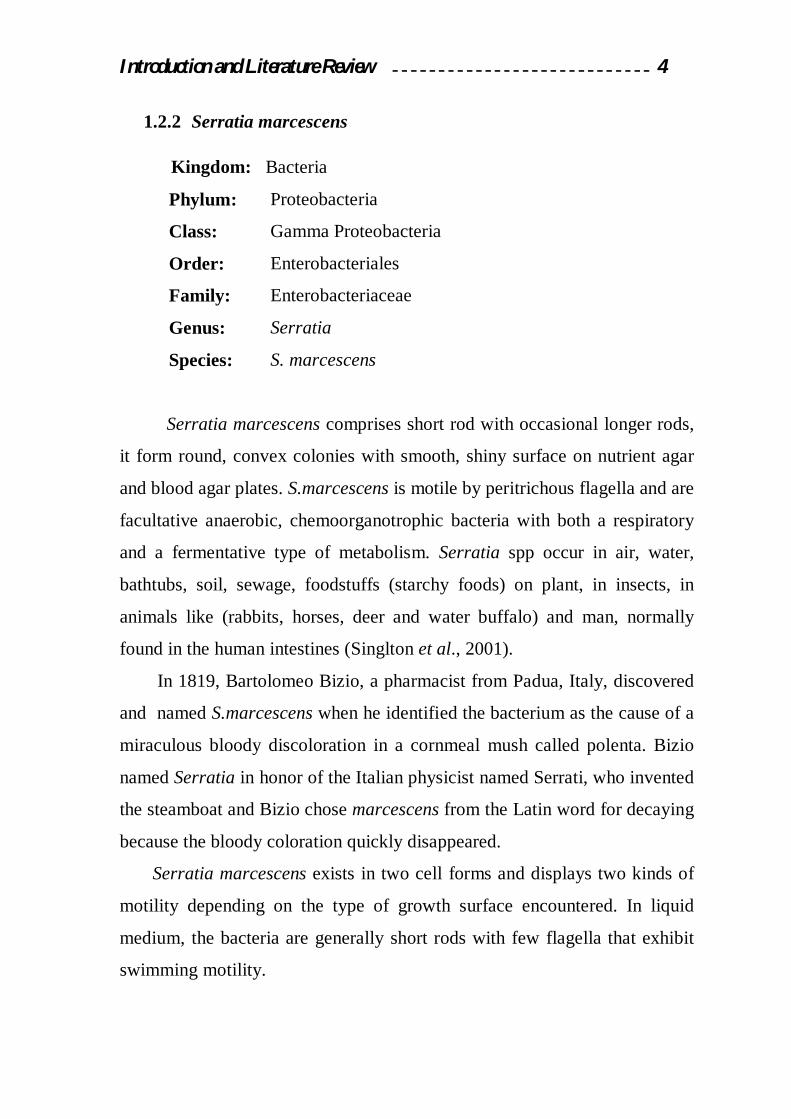
1.2.2 Serratia marcescens
Kingdom: Bacteria
Serratia marcescens comprises short rod with occasional longer rods,
it form round, convex colonies with smooth, shiny surface on nutrient agar
and blood agar plates. S.marcescens is motile by peritrichous flagella and are
facultative anaerobic, chemoorganotrophic bacteria with both a respiratory
and a fermentative type of metabolism. Serratia spp occur in air, water,
bathtubs, soil, sewage, foodstuffs (starchy foods) on plant, in insects, in
animals like (rabbits, horses, deer and water buffalo) and man, normally
found in the human intestines (Singlton et al., 2001).
In 1819, Bartolomeo Bizio, a pharmacist from Padua, Italy, discovered
and named S.marcescens when he identified the bacterium as the cause of a
miraculous bloody discoloration in a cornmeal mush called polenta. Bizio
named Serratia in honor of the Italian physicist named Serrati, who invented
the steamboat and Bizio chose marcescens from the Latin word for decaying
because the bloody coloration quickly disappeared.
Serratia marcescens exists in two cell forms and displays two kinds of
motility depending on the type of growth surface encountered. In liquid
medium, the bacteria are generally short rods with few flagella that exhibit
swimming motility.
Phylum:
Class:
Order:
Family:
Genus:
Species:
Proteobacteria
Gamma Proteobacteria
Enterobacteriales
Enterobacteriaceae
Serratia
S. marcescens

Introduction and Literature Review 5
However, upon growth on solid surface (0.7-0.85% agar), they
reportedly differentiate into elongated, hyper-flagellated cells that use a
swarming motility. These morphological changes appear to be necessary to
allow colony expansion, for swarming is a type of active surface motility
that enables bacteria to move rapidly across a semi-solid surface in a
coordinated manner (O’Rear et al., 1992).
Serratia marcescens produces extracellular enzymes such as: nuclease,
protease, haemolysin, lipase and chitinases. These factors are predicated to
play a role in bacterial environmental adaptive capacity, in either pathogenic
potential (Hejazi and Falkiner, 1997), and able to produce two types of
pigments:
Prodigiosin: a nondiffusible, water–insoluble pigment bound to the
cell envelope, prodigiosin–producing colonies are totally red or show
either a red center, a red margin or red sectors.
Pyrimine: a water–soluble, diffusible pink pigment. Ferrous iron is
required for the production of pyrimine. When the pyrimine is
produced, the agar medium turns pink while the colonies are white to
pinkish (Grimont and Grimont, 1984).
Cultures can produce two kind of odors, a fishy to urinary odor
attributed to Trimethylamine (mixed with some NH3), or a musty, potato–
like odor resembling that of 2–methoxy–3–isopropyl–pyrazine. The musty
odor is produced by S.odorifera, S.ficaria and a few strains of S.rubidaea.
All other strains and species produce the fishy–urinary odor (Grimont and
Grimont, 1984).

Introduction and Literature Review 6
Several species can grow readily at 4–5ºC (S. liquefaciens, S.
odorifera, S. plymuthica and S. ficaria) or at 40ºC (S. marcescens and
several strains of S. odorifera, S. rubidaea) however, the temperature of
37ºC is not favorable for the isolation of S. plymuthica (Forbes et al., 2002).
Most S. marcescens are resistant to several antibiotics because of the
presence of R-Factor, which a type of plasmid that carry one or more genes
that encode resistance, all are consider intrinsically resistance to ampicillin
and first – generation cephalosporins (such as cefalexin) (Carbonell et al.,
2000).
1.2.3 Ecology of Serratia marcescens
The widespread occurrence of S. marcescens indicates a strong adaptive
and survival potential and the ability to utilize wide range of nutrients. It is
able to survive and grow under extreme conditions, including antiseptics and
double-distilled water (Ajithkumar et al., 2003; Church et al., 2004;
Horkajada et al., 2006).
The bacterial cells readily colonize on surfaces and are enriched at the
air-water interface, most probably because of the pronounced cell surface
hydrophobicity of S. marcescens. In addition, S. marcescens is able to utilize
surface-bound nutrients, such as long chain fatty acids (Ulrich, 1993) and it
can be isolated from different plants such as: Eucalyptus, Pistachio, Bitter,
Cherry, Acacia, Coconuts, Sorghum, Grass, and this bacteria was also
isolated from vegetables such as: Mushrooms, tomatoes, leeks, green onions,
lettuce, broccoli, artichokes, radish spinach, carrots, and figs (Burger and
Bennett, 1985) and from different sources of foods like; fish, chicken and
their products, milk and their products (Braun et al., 2001).
Introduction and Literature Review 7
It can also grow in any moist location where phosphorous containing
materials or fatty substances accumulate. Sources of these substances
include soap residues in bathing areas and soap and food residues in pet
water dishes. Serratia can also grow in tap water in locations such as toilets
in guest bathrooms where the water is left standing long enough for the
chlorine residual disinfectant to dissipate. Serratia will not survive in
chlorinated drinking water (Ajithkumar et al., 2003; Horkajada et al., 2006).
Serratia marcescens is a well-recognized hospital acquired pathogen,
outbreaks in environmental sources associated with cross-infection which
included contaminated disinfectants, sinks, adhesive, tape, scalp vein
needles, intravenous solutions, saline bottles, mechanical respirators,
intravenous catheters, and ultrasonic nebulizers (Giles et al., 2006).
1.2.4 Quorum sensing in Serratia
Diverse environmental and cellular cues are involved and a number of
different regulatory systems have evolved to permit rapid bacterial
adaptation to fluctuating environmental conditions. These include ''quorum
sensing'', a mechanism which enables bacteria to sense their cell population
density and use this information to coordinately regulate gene expression.
Quorum sensing allows bacterial populations to efficiently adapt to changes
in the surrounding environment (Henke and Blassler, 2004; Houdt et al.,
2007). Like many other bacterial species, Serratia strains produce diffusible,
low molecular –mass signal molecules which accumulate in their
surroundings as the population increases. Two classes of quorum-sensing
systems have been described in Serratia, first, the N-acylhomoserine
lactones (AHL)-dependent LuxIR type and second, the autoinducer-2 (AI-
2)/LuxS type (Winzer et al., 2003; Wei and Lai, 2006).
Introduction and Literature Review 8
AHL-dependent quorum sensing regulates diverse phenotypes in S.
marcescens including the production of prodigiosin, carbapenem antibiotic,
extracellular and cell associated enzymes and biofilm maturation (Harris et
al., 2004; Rice et al., 2005). AHLs also regulate swarming motility in
Serratia by controlling the production of biosurfactants. For both
prodigiosin and carbapenem, environmental factors play important roles in
modulating the quorum–sensing response.
For example, phosphate availability regulates biosynthesis of prodigiosin
and carbapenem in Serratia spp. ATCC 39006 via both quorum sensing –
dependent and-independent pathways(Wei and Lai, 2006).
1.2.5 Prodigiosin
Prodigiosin is a red pigment insoluble in water, but soluble in chemical
solvent like: alcohol, chloroform. Prodigiosin is a linear tripyrrole, a
bifurcated pathway has been proposed for the biosynthesis of prodigiosin
culminating in the enzymic condensation of the terminal products of the two
pathways, the stable bipyrrole moiety 4-methoxy-2, 2-bipyrrole-5-
carbaldehyde (MBC) and the volatile monopyrrole moiety 2-methyl-3-n-
amyl-pyrrole(MAP) (Harris et al., 2004).
The prodigiosin group of natural products is a family of tripyrrole red
antibiotic(2 – methyl – 3 – pentyl – 8 – methoxy prodigiosin) (Figure1-
1).The biosynthesis of the pigment is a bifurcated process in which mono
and bipyrrole precursors are synthesized separately and then assembled to
form prodigiosin. Prodigiosin have been shown to be associated in
extracellular vesicles, cell associated or present in intracellular granules
(Kobayashi and Ichikawa, 1991).

Introduction and Literature Review 9
The molecular formula of the compound is (C20H25N3O), prodigiosin
possesses an apparent pka of 7.6 in aqueous dioxane, and 8.25 in aqueous
ethanol and that the pigment is yellow in alkali and pink in acidic media.
Interestingly, the proton affinity of the prodigiosin has now been shown to
be governed by the presence of two geometrical isomers(α and β forms) that
have very different apparent pka value, i.e. pkα = 8.23, pkβ = 5.4 and have
small molecular weight of 323.4 Dalton (Manderville, 2001).
Figure(1-1): The structure of prodigiosin (Srijith, 2006)
Introduction and Literature Review 10
1.2.5.1 Prodigiosin production
Prodigiosin is typical secondary metabolite only appearing in the later
stages (stationary phase) of bacterial growth (Williams et al., 1971;
Khanafari et al.,2006). The production of prodigiosin has been shown to be
influenced by numerous environmental factors including: inorganic
phosphate availability, media composition, incubation period, age of
bacterial colony, temperature, pH, oxygen contents (Slater et al., 2003) and
this pigment are kept to red color in dark for long time because prodigiosin
is sensitive to light (Allen, 1967; Hearn et al., 1968).
Its produced by Serratia marcescens, S.rubidaea, Pseudomonas
magneslorubra, Vibrio psychreorythrous, Vibrio gazogenes, Alteromonas
rubra, Rugamonas rubra , and various marine bacteria, including Hahella
chejeunsis KCTC 2396, and Pseudoalteromonas denitrificans, and Gram
positive Actinomycetes, such as Streptoverticillium rubrireticuli and
Streptomyces longisporus (Rowan and Fisher, 1997).
Prodigiosin has no defined role in the physiology of producing strains.
A recent study has suggested prodigiosin may be an important factor for the
trypanolytic activity of a S.marcescens strain. Other suggested roles for
prodigiosin have included a role in metabolic overflow from primary
metabolism (Srijith et al., 2006). Prodigiosin has potential clinical interest
because it is reported to have anti-fungal, anti-bacterial, anti-protozoal/anti-
malarial, immunosuppressive and anti-cancer activities (D’Alessio et al.,
2000; Montaner et al., 2000).
Previous attempts to reconstitute prodigiosin production in Escherichia
coli have been unsuccessful but in 1984, Dauenhauer isolated an
S.marcescens genomic clone capable of condensing the two prodigiosin
precursors, monopyrrole moiety 2-methyl-3-n-amyl-pyrrole (MAP) and
bipyrrole moiety 4-methoxy-2,2-bipyrrole-5-carbaldehyde (MBC), to form

Introduction and Literature Review 11
prodigiosin, so can able to demonstrate expression of prodigiosin in a
heterologous host. The pig. gene cluster from Serratia was expressed in
Erwinia carotovora subsp., though it was not expressed in several other
members of the Enterobacteriaceae, including E.coli (Thomson et al., 2000).
Many factors affects the biosynthesis of the pigment e.g. Fe(III), and
sodium dodecyl sulphate have been shown to enhance the synthesis of
prodigiosin (Geron et al., 1988), while many factors have been shown to
inhibit prodigiosin synthesis e.g. temperature, glucose, ATP, ribose,
inorganic phosphate, NaCl, KCI, polymyxin B, and streptomycin (Geron and
Rokem, 1988).
1.2.5.2 Prodigiosin biosynthesis
The biosynthesis of prodigiosin in Serratia sp. is controlled by a
complex regulatory network of both N-acyl-L-homoserine lactone quorum
sensing dependent and independent pathways. The Serratia 39006 pig.
cluster consists of 15 biosynthesis genes pig.(A-O) transcribed as a single
polycistronic mRNA (Harris et al., 2004; Fineran et al., 2005).
The biosynthesis of prodigiosin can be done by two steps:
First: Also called the cytoplasmic step to produce; MAP, MBC.
v MAP(monopyrrole moiety 2-methyl-3-n-amyl-pyrrole): Three pig. gene
can be share from total pig. genes to produce MAP: (PiB, PigD, PigE),
the first steps in the MAP pathway are thought to be performed by
enzymes of fatty acid biosynthesis to give 2–octenal. Alternatively 2–
octenal may be derived by autoxidation of unsaturated fatty acids. PigD
also decarboxylates pyruvate but that the two–carbon fragment then adds
to C-3of 2-octenal giving 3-acetyloctanal. Then transamination on the
aldehyde group of 3-acetyloctanal and the resulting aminoketone by

Introduction and Literature Review 12
PigB, would cyclize spontaneously to give the cyclic imine H2MAP.
Finally PigB is responsible for a 2-electron oxidation of H2MAP to MAP
(Harris et al., 2004; Williamson et al., 2005) as in figure (1-2).
v MBC(bipyrrolemmoietym4-methoxy-2,2-bipyrrole-5-carbaldehyde): The
precursors to produce pyrrole were shown to be acetate, serine and
proline. The first step to create this pyrrole by activate the L-proline by
ATP and then transfer the L-proline group to sulfate group in Pig G to
form prolyl-Pig G, then oxidation by PigA forming Prolyl-2-carboxyl-
Pig G, then transfer of the pyrrole-2-carboxyl unit from Pig G to the
active-site cystine of PigJ, following then transfer of a malonyl group
from malony group from malonyl CoA to the phosphopantetheinyl side-
chain of PigH and decarboxylative attack of the malonyl unit on the
pyrrole-2-carboxyl thioester giving a pyrrolyl-β-Ketothioester attached
to PigH, then condensation of serine from PigH with Pyrrole-2-carboxy
thioester forming HBM (4-hydroxy-2,2bipyrrole5-methanol). The
oxidation of the alcohol group by PigM forming HBC (4-hyroxy-2,2-
bipyrrole-5-carbaldehyde).
The final step of the MBC pathway, methylation of the hydroxy group of
HBC, involves the two enzymes PigF and PigN (Dairi et al.,2006;
Williamson et al., 2005) as in figure(1-2).
Second: the final biosynthetic step is the condensation of the terminal
products of the two parallel pathways, MAP and MBC to form prodigiosin.
This presented by PigC as the condensing enzyme (Ding and Williams ,
1983; Williamson et al., 2006).
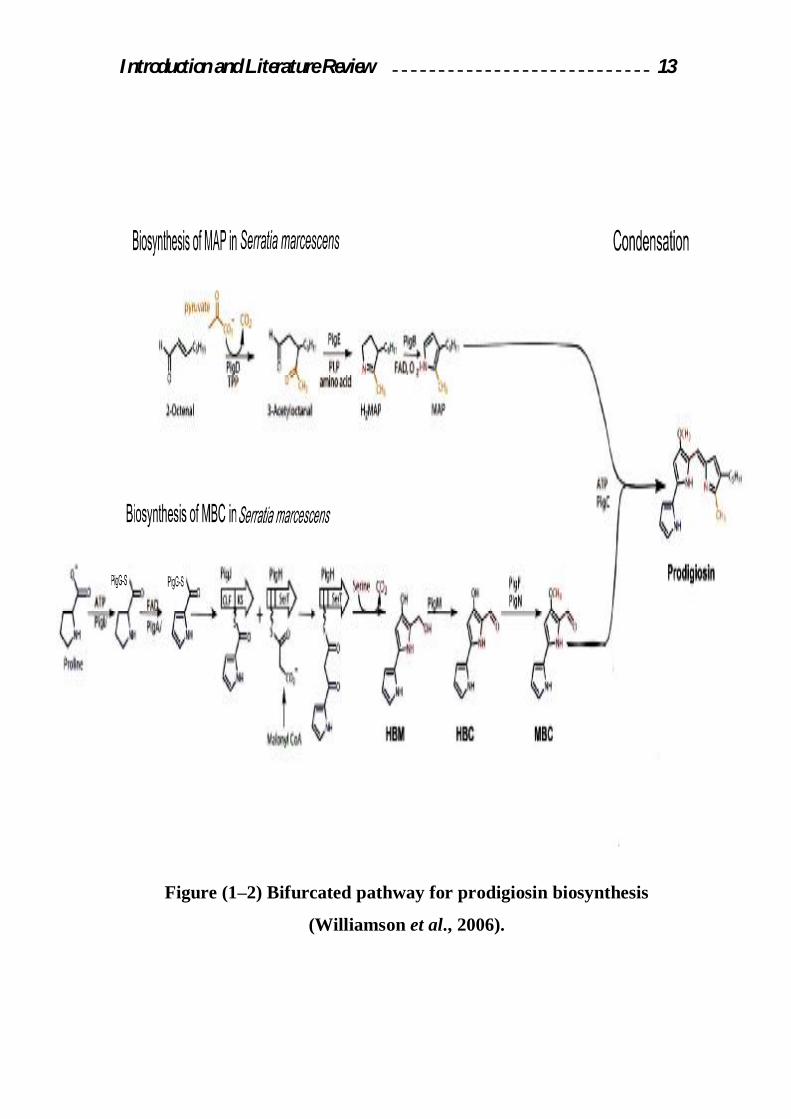
Introduction and Literature Review 13
Figure (1–2) Bifurcated pathway for prodigiosin biosynthesis
(Williamson et al., 2006).

Introduction and Literature Review 14
1.2.6 Prodigiosin gene expression
Assays of bacterial gene expression make attractive teaching tools for
several reasons. First, bacteria modulate their gene expression quickly in
response to environmental cues such as cell density, growth temperature and
growth medium. Bacterial messenger RNA molecules are typically degraded
with half-lives measured in minutes versus the hours of stability for
eukaryotic transcripts. Second, many bacteria express pigments under
certain conditions. Because most pigments absorb light at some defined
wavelength, pigment expression may be easily monitored
spectrophotometrically. Third, and perhaps most importantly, bacteria are
easy to propagate in the teaching laboratory (Khanafari and Assadi, 2006).
Prodigiosin is expressed as a secondary metabolite in the general method
of gene expression called quorum sensing. Growth in liquid culture at low
cell density allows low–level expression of a membrane permeable positive
regulator of gene expression. The intracellular concentration of the regulator
remains low at low cell density due to its diffusion across the cell membrane
after synthesis. However, as cell density increases in a closed system, the
intracellular concentration of regulator increases to a threshold needed for
activation of prodigiosin expression. Thus, high levels of prodigiosin are
expressed in liquid culture only at high cell density. A similar phenomenon
operates with colonies grown from single cells on agar plates (Haddix and
Werner, 2000).
Introduction and Literature Review 15
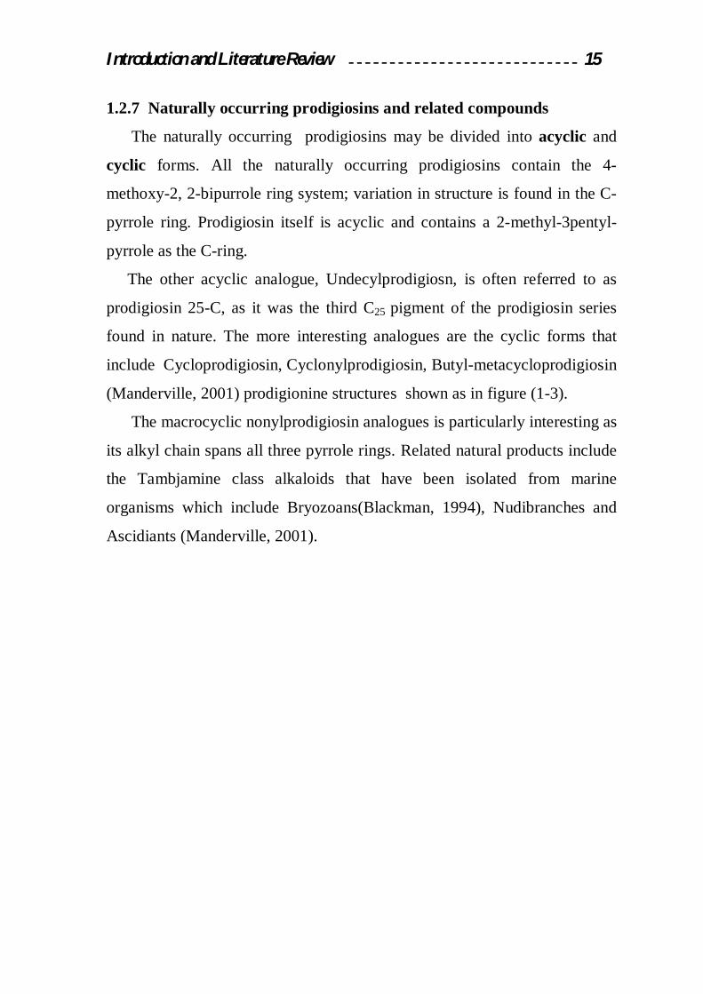
1.2.7 Naturally occurring prodigiosins and related compounds
The naturally occurring prodigiosins may be divided into acyclic and
cyclic forms. All the naturally occurring prodigiosins contain the 4-
methoxy-2, 2-bipurrole ring system; variation in structure is found in the C-
pyrrole ring. Prodigiosin itself is acyclic and contains a 2-methyl-3pentyl-
pyrrole as the C-ring.
The other acyclic analogue, Undecylprodigiosn, is often referred to as
prodigiosin 25-C, as it was the third C25 pigment of the prodigiosin series
found in nature. The more interesting analogues are the cyclic forms that
include Cycloprodigiosin, Cyclonylprodigiosin, Butyl-metacycloprodigiosin
(Manderville, 2001) prodigionine structures shown as in figure (1-3).
The macrocyclic nonylprodigiosin analogues is particularly interesting as
its alkyl chain spans all three pyrrole rings. Related natural products include
the Tambjamine class alkaloids that have been isolated from marine
organisms which include Bryozoans(Blackman, 1994), Nudibranches and
Ascidiants (Manderville, 2001).
Introduction and Literature Review 16
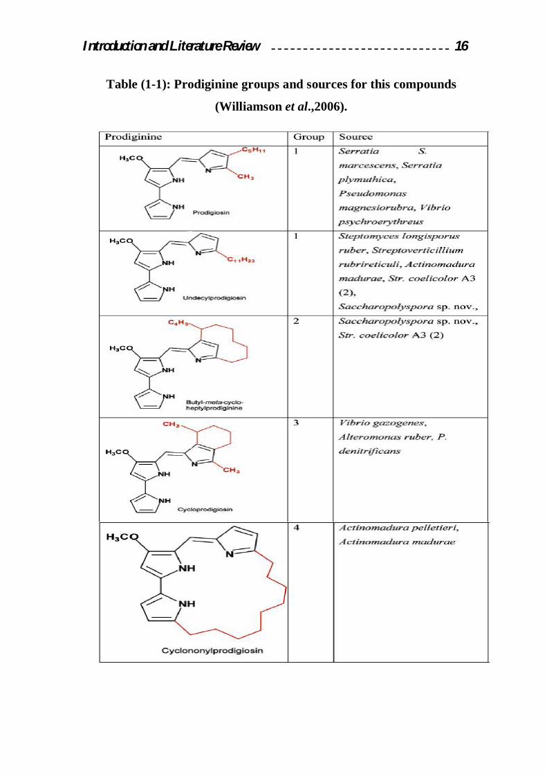
Table (1-1): Prodiginine groups and sources for this compounds
(Williamson et al.,2006).
Introduction and Literature Review 17
1.2.8 Applications of prodigiosin in Biotechnology
Prodigiosin exhibits antibacterial because this pigment showed high
inhibition activity against gram positive bacteria e.g. Staphylococcus aureus,
Streptococcus pyogenes, while showed little inhibition activity against gram
negative bacteria e.g. Klebsiella pneumonia, Proteus vulgaris (Nakashima et
al., 2005b).
This pigment, showed potent algicidal activity against various red tide
phytoplankton's H. akashiwo, H. circularisquama, C. polykrikoides, G.
impudicum and A. tamarense, in a concentration-dependent manner (Jeong
et al., 2005; Nakashima et al., 2006; Kim et al., 2007).
One such antagonistic bacterium, Serratia marcescens which produce
prodigiosin, has been isolated from the phylloplane of tomato plants
(Someya et al., 2000). This pigment effectively inhibits the in vitro growth
of several phytopathogenic fungi and suppresses cyclamen gray mold caused
by Botrytis cinerea, cyclamen soil-borne diseases caused by Rhizoctonia
solani AG-4 and fusarium oxysporum, rice sheath blight caused by
Rhizoctonia solani AG-1 AI, and rice blast caused by Pyricularia oryzae in
greenhouses (Iyozumi et al., 1996; Someya et al., 2000, 2001). S.marcescens
is particularly effective in inhibiting both hyphal growth and sclerotium
germination of R.solani (Someya et al., 2005; Someya et al., 2007).
Chagas’ disease is a protozoan infection caused by Trypanosoma cruzi. This
disease is one of the most important public health problems in many South
American countries, affecting 16–18 million people in Latin America.
Introduction and Literature Review 18
Although there is intense research on drugs for the treatment of infection
by T. cruzi , only one drug, benznidazole, has been recommended for the
treatment of acute and congenital cases (Melo et al., 2000).
However, benznidazole has severe limitations related to its efficacy and
toxicity as well as the development of parasite resistance. Studies with
bacteria and mammalian cells have demonstrated the genotoxic potential of
nifurtimox and benznidazole and rabbits treated with both drugs displayed a
high incidence of malignant lymphomas. For this reason, the development of
safer and more effective drugs, particularly against the chronic form of
Chagas’ disease, is an urgent priority (Isaka et al., 2002).
The search for new trypanocides is currently being done through the
development of in vitro screening assays. Recent studies of antifungal agents
with trypanocidal activities have shown that prodigiosin, an antimycotic
drug, can kill these parasites(Azambuja et al., 2004). Prodigiosin had a
potent trypanocidal activity against the trypomastigote forms of T. cruzi Y
strain (IC50=5 μM) compared with Nifurtimox which had an IC50 of 150 μM
and higher trypanocidal activity compared with benznidazole (IC50=19 μM).
Prodigiosin promote H+/Cl- symport and induce neutralization of the
acidic compartment of cells which in turn results in the acidification of the
cytoplasm and thus cell cycle arrest and eventually apoptosis (Yamamoto et
al., 2000). It triggers apoptosis in cancer cell lines, with no marked toxicity
in nonmalignant cell lines (Montaner and PerezTomas, 2001). Prodigiosin
exhibits selective activity against breast, colon, hematopoietic cancer, and
liver cancer cells (Soto–cerrato et al., 2004).
Introduction and Literature Review 19
1.2.9 Factors affecting the productivity of the prodigiosin pigment
There are many conditions, which affect the productivity of
prodigiosin pigment, which include pH, temperature, carbon source,
nitrogen source, and phosphate source.
1.2.9.1 Effect of pH in prodigiosin productivity
Pigment productivity and continuing of metabolic pathway is highly
affected by pH of the medium.
Optimum growth of all strains of Serratia has been observed at pH 7 and
growth of all strains of Serratia is inhibited at a pH of < 4.5, while optimum
pH for prodigiosin production is between 8.0-8.5 (Sole et al., 1997).
1.2.9.2 Effect of temperature on prodigiosin productivity
Temperature is considered as one of the most important factors affecting
pigment productivity and the growth of the microorganisms. Variable
growth has been observed at 5˚C and 40˚C. Optimum growth of all strains of
Serratia has been observed at temperatures from 20-37˚C. Growth of all
strains of Serratia is inhibited at > 45˚C and prodigiosin was produced at
temperature ranging from 12-37˚C, while the optimum temperature of
prodigiosin production was at 30˚C (Holt et al., 1994).
1.2.9.3 Effect of nutritional factors on prodigiosin production
1.2.9.3.1 Effect of carbon source
Microorganisms differ in their needs to carbon sources according to
their nutrient nature; the use of pure carbon sources e.g. (glucose, sucrose
and fructose) is expensive from the economical case, so the industrial

Introduction and Literature Review 20
fermentation try to use cheap carbon sources especially industrial and a
variety of plant seed oils have also been used as carbon substances for
prodigiosin production and displayed stimulatory effects on the production
by S.marcescens.
The optimum carbon source is olive oil while prodigiosin production
was inhibited by glucose due to catabolic repression (Giri et al., 2004).
1.2.9.3.2 Effect of nitrogen source
Nitrogen Source is one of the most important part of the components of
the culture medium for the producing microorganism such as inorganic
nitrogen salts or organic sources like amino acids and proteins and these
nitrogen sources can be used either as oxidized form (NO-3, NO-2) or as
reduced form (NH2, NH+4). Amino acids and ammonium salts considered as
a good nitrogen sources, which can be used easily by the microorganisms.
It was observed that casein hydrolysate 1.5% is the best nitrogen source
for the production of prodigiosin by S.marcescens while peptone is the best
nitrogen source for bacteria cell growth (Kim et al., 1999).
1.2.9.3.3 Effect of phosphate source
Mineral salts have an effect on the production of prodigiosin and there is
several studies demonstrated that synthesis of prodigiosin by non-
proliferating cells of S.marcescens is depended to presence of inorganic
phosphate (Pi) concentrations. A high elevation of pigment formation was
obtained at less than or equal to 0.3 mM and a broader but much lower
elevation was obtained at 10 to 250 mM Pi (Witney et al., 1977; Bennett and
Bentley, 2000).

Introduction and Literature Review 21
1.2.10 Extraction and Purification of prodigiosin
In order to make any study about the pigments to understand its
characteristics and its role in any reaction, it is very important for the
prodigiosin to be partially purified to get exact results during the study.
Pigments purification process means separation of the prodigiosin from
another products and materials presents in the crude filtrate, prodigiosin is
sometimes bound to proteins, thus, extracts may require acid treatment
before isolation of the pigment. Higher homologs of prodigiosin have been
detected by mass spectroscopy (Gerber, 1975).
The pigment was extracted from the bacterial cells by the method of
Williams's et al,. (1965). To obtain the dry pigment, the petroleum ether
extract was evaporated under vacuum at room temperature in the dark.
Thin–layer chromatography (TLC) of the extracted pigments on silica gel,
with chloroform: ethyl acetate: acetic acid (8:1:1 vol/vol). Sample of
purified pigment obtained by preparative TLC were analyzed for infrared
absorbance with a Perkin Elmer model spectrophotometer (Roberts et al.,
2007). Also this pigment can be purified by high performance liquid
chromatography (HPLC) (Nakashima et al., 2005b), was performed as
preliminary analysis for distinguishing closely related analogs of
prodigiosin.
1.2.11 Mutagenesis
Mutagenesis, the creation or the formation of a mutation can be used as
a powerful genetic tool. By inducing mutation specific ways and then
observing the phenotype of the organism, the function of the genes and even
individual nucleotides can be determined. Mutagenesis was used to ability to
overproduce a desired metabolite. So now the study of mutagenizing
microorganisms is important, interesting and potentially profitable.

Introduction and Literature Review 22
The mutation of a gene or genes under study can be achieved by first
altering the DNA of the microorganism in some fashion and then screening
or selecting for the desired phenotype (Maki, 2002).
Three general treatments can be used to mutagenize microorganism:
physical radiation, chemical mutagens and transposons. Mutation by
radiation involves exposing the microbe to high energy waves (UV light,
laser or X-ray). This procedure damages the target DNA and sometimes,
during repair, an improper base pair (or pairs) is incorporated in the DNA,
causing a mutation. Chemical mutagens are also employed. These
compounds are added to a growing culture of an organism for a given time
period and interfere with the replication of the DNA (Paustian and Kurtz,
1994).
Some mutagens achieve this by serving as base analogs, others
chemically modify the DNA, and yet another class can insert or intercalate
between the base pairs of DNA causing DNA polymerase to make mistakes.
In all cases, the mutagen causes incorrect copying of the DNA resulting in
base substitutions (exchange of one base pair for another), insertions
(additions of one base pairs), or deletions (removal of one or more base
pairs) (Paustian and Kurtz, 1994).
The third method of mutagenesis involves the use of mobile genetic
elements. The most commonly employed of these are a sub-class called
transposons. Transposons are relatively short pieces of DNA that replicate
by inserting into other pieces of DNA (plasmid, chromosomes, and viruses).
They encode two sets of functions. One set is involved in regulating and
performing the movement of the transposons from one place of "host" DNA
to the next (transposition functions). The other set of functions encode genes
that may provide an advantage for the host of example (Paustian and Kurtz,
1994).

Introduction and Literature Review 23
1.2.11.1 Physico – chemical mutagens
Physical mutagens are different types of radiations having mutagenic
properties such as UV light and ionizing radiation. The energy content of a
radiations depends upon its wavelength i.e.: shorter the wavelength, the
greater the energy value of radiation. While the ionizing radiation which is
one of the physical mutagens, has the greater penetration power than non
ionizing radiation. Ionizing radiation causes single strand breaks in DNA
and produces deletion. Ultraviolet rays are the only non–ionizing rays with
mutagenic properties (Settey and Sreekrishna, 2004).
1.2.11.1.1 Mutagenic Properties of Ultraviolet Radiation
UV light is the portion of the spectrum with wavelength of 100-400 nm,
which is just shorter than visible light (Miller et al., 1999).
UV radiation (UVR) is lethal and potentially mutagenic to all organisms
greatly dependant on the source of radiation and the time exposure. UV can
be classified into UV-A (320-400nm), UV-B (290-320nm), and UV-C(<
290). Photons of UVB and UVC wavelengths cause direct DNA damage by
inducing the formation of DNA photoproducts such as cyclobutyl
pyrimidine dimmers(CPD) and the pyrimidine (6-4) pyrimidinione. The
accumulation of DNA photoproducts can be lethal to cells through the
blockage of DNA replication and RNA transcription.
The UVA typically cause only indirect damage to cellular DNA through
catalyzing the formation of chemical intermediates such as reactive oxygen
species. Distinct differences between far-UVC and near-UV (UVB and
UVA) damage have been observed in bacteria and bacteriophage, UVC has
the most potential for directly damage DNA (Miller et al., 1999; Kim and
Sundin, 2001; Qiu et al., 2004).

Introduction and Literature Review 24
UV light is widely used as mutagen that generates a broad spectrum of
lesions in DNA. The most important mutagenic effect of UV irradiation is
believed to be stimulation of misrepair (Goodenough, 1984).
1.2.11.2 Chemical Mutagens
A chemical mutagen is a substrate that can alter a base that is already
incorporated in DNA by change its hydrogen-bonding specifically
(Freifelder, 1987). Among the most widely used mutagenic reagent with
microorganisms are the alkalating agents, ethyl methane sulfonate (EMS)
and Mitomycin C (MMC) also used as chemical mutagen which causing
cross-link formation in cellular DNA by deletion (Szybalski and Iyer, 1964).
1.2.11.2.1 Mitomycin C
Mitomycin C was identified in 1956 as an antibiotic produced by
Streptomyces lavendulae and subsequently established as an important
antitumor agent. Mitomycin C functions as a pro drugs and requires
enzymatic and chemical reduction to become a highly reactive alkylating
agent(Tomasz et al.,1988; Henderson, 1993). It has been reported that the
antibiotic mitomycin C has a specific effect on cellular DNA, but has little
or no effect on either RNA and protein formation (Suzuki and Kilgore,
1966). The ability of Mitomycin C to inhibit bacterial cell growth involves
the combined action of DNA alkylation and the formation of reactive
oxygen species (Sheldon et al., 1999). The primary action of Mitomycin C is
believed to be associated with either the inhibition of DNA biosynthesis or
the breakdown of the nuclear apparatus.
Introduction and Literature Review 25
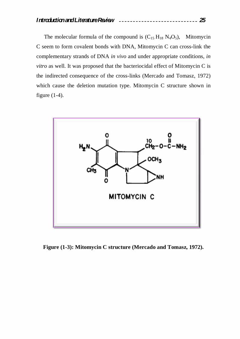
The molecular formula of the compound is (C15 H18 N4O5), Mitomycin
C seem to form covalent bonds with DNA, Mitomycin C can cross-link the
complementary strands of DNA in vivo and under appropriate conditions, in
vitro as well. It was proposed that the bacteriocidal effect of Mitomycin C is
the indirected consequence of the cross-links (Mercado and Tomasz, 1972)
which cause the deletion mutation type. Mitomycin C structure shown in
figure (1-4).
Figure (1-3): Mitomycin C structure (Mercado and Tomasz, 1972).


Results and Discussion 42
3. Results and Discussion
3.1 Isolation of Serratia species
In order to isolate S.marcescens, fifty seven samples were collected from
different environments in Baghdad governorate during the period from
2/2008 to 4/2008. Results mentioned in table (3-1) showed that from these
samples, 30 isolates were obtained from water, sewage, and soil samples.
Table (3-1): Local isolates from different environmental samples.
Source of
samples No. of
Samples
No. of
Isolates
Presence of
red pigmented
colonies
Water 11 4 4
sewage 26 16 7
Soil 20 10 4
Total 57 30 15
Among the total isolates, only 15 isolates were able to produce red
pigment, which give an indicator that these isolates are belong to Serratia
spp. These 15 isolates were further characterized and identified according to
their cultural, morphological characteristics, and biochemical tests.

Results and Discussion 43
From the results mentioned in table (3-1) it can be concluded that there
are other 15 isolates from environmental samples may belong to other
pathogenic or nonpathogenic bacteria from different genera.
3.2 Identification of bacterial isolates
Local isolates that were able to produce red pigment, were further
identified according to their cultural and morphological characteristics and
biochemical tests. For the former, colonies of each isolate that were plated
on nutrient agar medium showed different morphological characteristics of
S.marcescens such as red round, convex colonies with smooth, shiny surface
and has fishy–urinary odor. Microscopical examination of each isolate
showed that they are all having single cells, non-spore forming, gram
negative and rod shape.
Some biochemical tests were done to ensure that these 15 isolates are
S.marcescens. Results listed in table (3-2) showed that only five of these
isolates are belong to S.marcescens symbod S3, S5, S7, S11, and S13.
These isolates are able to grow at 40ºC, positive for catalase and oxidase
production, and positive for methyl red and voges-proskaure tests, and they
are motile and positive for DNase production. Results also showed that these
isolates are able to utilize sucrose as a sole source for carbon and energy,
while they are unable to produce acid from the fermentation of lactose,
xylose, raffinose, and arabinose. These results are confirmed that these five
isolates are belong to S. marcescens and as it was described by Atlas et al.,
(1995). From the other results mentioned in table (3-2), it can be concluded
that there are other 10 isolates may belong to other different genera.

Results and Discussion 44
Table (3-2): Biochemical tests of the locally isolated Serratia spp.
S15 S14
S13 S12 S11 S10 S9 S8 S7 S6 S5 S4 S3 S2 S1
Isolate
Test
- + + + + - + + + - + + + + + Growth at 40ºC
+ + + + + + + + + + + + + + + Catalase
+ + + + + + + + + + + + + + + Oxidase
- - + + + - + + + + + + + + + Methyl red
+ + + + + + - + + + + - + + - Motility
+ + + + + + - + + + + - + + - DNase
- - + + + - + + + + + + + + + Voges–proskauer
Acid production from:
+ + - + - - + + - - - - - + + lactose
+ + + - + + + - + - + + + - - sucrose
- + - - - + - - - + - + - + + xylose
+ - - + - + + + - + - + - - + raffinose
+ + - - - + + - - + - + - - + arabinose

Results and Discussion 45
3.3 Screening of S.marcescens isolates for prodigiosin production
In order to select the efficient isolate in prodigiosin production, the
ability of these local isolates in pigment production was assayed using LB
Medium by determining pigment activity (U/cell) in culture filtrate using
pigment assay procedure mentioned in (2.2.5). As it was shown in table (3-
3), S.marcescens S11 gave the maximum production of prodigiosin
according to the pigment activity (200 U/cell) in its culture medium, while
the pigment activity in the culture medium of S3, S5, S7, S13 isolates are
103.4, 89.5, 78.9, and 98.1 U/cell respectively. From these results
S.marcescens S11 regarded the efficient isolate in prodigiosin production.
This isolate was selected to study the optimum conditions for prodigiosin
production.
Table (3-3): Ability of locally isolated S.marcescens in prodigiosin
production after incubation with shaking at 150 rpm in LB medium for
24hr at 28ºC.
Prodigiosin activity (U/cell) Isolate
103.4 S.marcescens S3
89.5 S.marcescens S5
78.9 S.marcescens S7
200 S.marcescens S11
98.1 S.marcescens S13

Results and Discussion 46
3.4 Optimum conditions for prodigiosin production
Optimum conditions for prodigiosin production by the locally isolated
S.marcescens S11 were studied under the effect of different growth factors
and as follows:
3.4.1 Effect of Carbon Source
Six carbon sources (fructose, glucose, sucrose, lactose, olive oil, and
sunflower oil) were used as a sole source of carbon and energy to determine
the optimum in prodigiosin production by the locally isolated S.marcescens
S11, these carbon sources were added to the production medium in a
concentration of 2%.
Results indicated in figure (3-1) showed that the maximum production
of prodigiosin was obtained when the culture medium was supplemented
with olive oil, by using this carbon source, the prodigiosin activity in culture
medium was 440.3 U/cell, while the productivity of prodigiosin using
glucose, sunflower oil, fructose, sucrose, and lactose under the same
conditions are 82.1, 91.8, 152, 169.1, and 343.4 U/cell respectively.
From these results it can be concluded that olive oil was the best carbon
source because it contain many nutrients, fatty acids, growth factors and
variety of vitamins that supplements the growth requirements for the
microorganism, further more olive oil contain several mineral salts (K, Mn,
Ca, P, Mg, S, Zn, Na, Fe, and Cu), and vitamin such as vitamin A, K, E, B6,
B12, and C (Jama, 2008). It was well known that S.marcescens has lipase
activity and thereby it was capable for hydrolyzing oil substrates to liberate
fatty acids as a sole source for carbon and energy (Wei and Chen, 2005).
Glucose may inhibit prodigiosin production due to catabolic repression or by
lowering the medium pH during growth and fermentation (Sole at el., 1997).
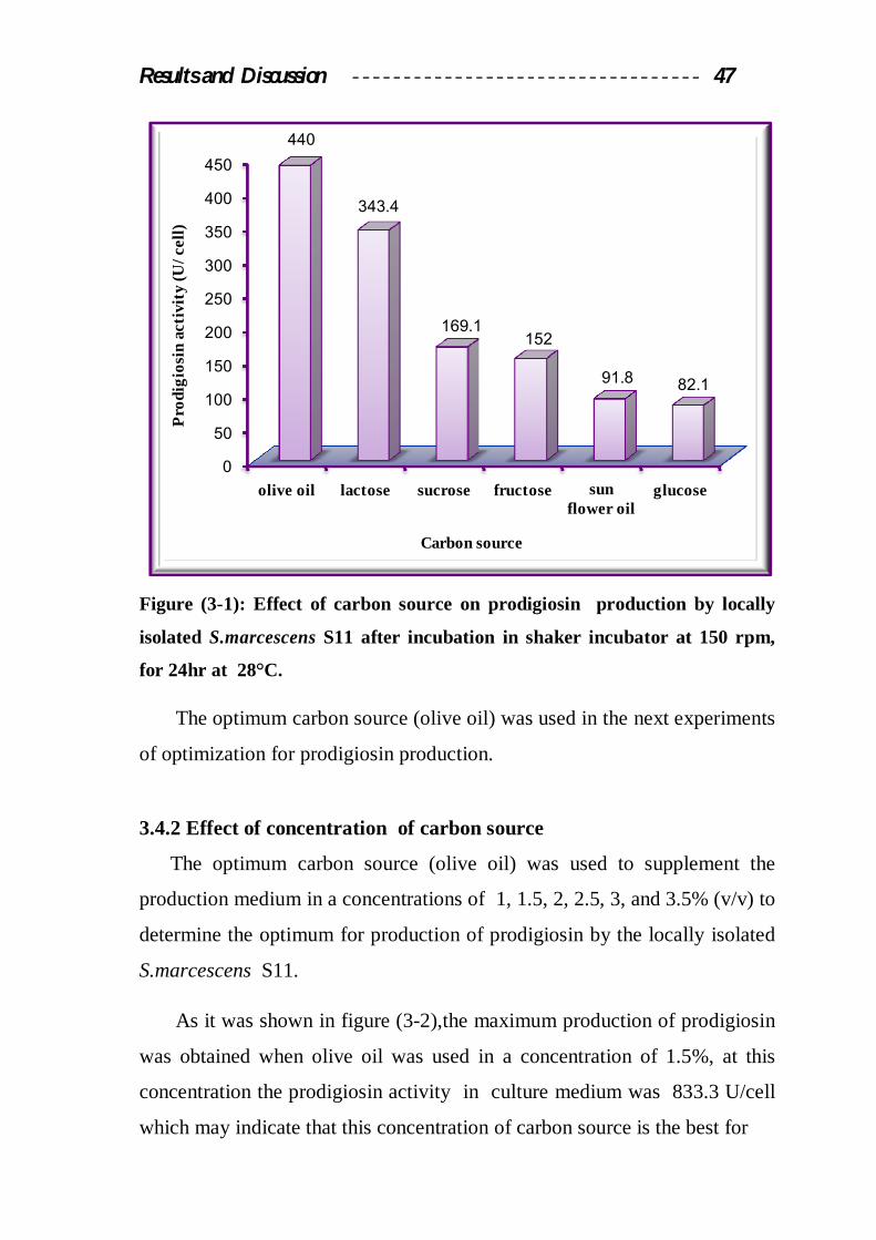
Results and Discussion 47
Figure (3-1): Effect of carbon source on prodigiosin production by locally
isolated S.marcescens S11 after incubation in shaker incubator at 150 rpm,
for 24hr at 28°C.
The optimum carbon source (olive oil) was used in the next experiments
of optimization for prodigiosin production.
3.4.2 Effect of concentration of carbon source
The optimum carbon source (olive oil) was used to supplement the
production medium in a concentrations of 1 , 1.5, 2, 2.5, 3, and 3.5% (v/v) to
determine the optimum for production of prodigiosin by the locally isolated
S.marcescens S11.
As it was shown in figure (3-2),the maximum production of prodigiosin
was obtained when olive oil was used in a concentration of 1.5%, at this
concentration the prodigiosin activity in culture medium was 833.3 U/cell
which may indicate that this concentration of carbon source is the best for
0
50
100
150
200
250
300
350
400
450
olive oil lactose sucrose fructose sun flower oil
glucose
440
343.4
169.1152
91.8 82.1
Pro
digi
osin
act
ivit
y (U
/cel
l)
Carbon source

Results and Discussion 48
Figure(3-2): Effect of different concentrations of olive oil on prodigiosin
production by the locally isolated S.marcescens S11 after incubation in
shaking incubator at 150 rpm, for 24hr at 28°C.
providing the microorganism with the needed energy for growth and
maximum production of the pigment. Other studies indicated that the
optimum carbon and energy source for the pigment production varies
between different concentration of olive oil, for example Yamashita et al.,
(2001) referred that the optimum carbon source for prodigiosin production at
4% (w/v) of S.marcescens SM∆R.
The optimum concentration of olive oil (1.5%) was used in the next
experiments of optimization for prodigiosin production.
0
100
200
300
400
500
600
700
800
900
1 1.5 2 2.5 3 3.5
714.2
833.3
440
666.6 700
600.1
Pro
digi
osin
act
ivit
y (U
/cel
l)
Concentration (%)
Results and Discussion 49
3.4.3 Effect of nitrogen source
Five nitrogen sources were used to supplement the production medium to
enhance prodigiosin production by the locally isolated S.marcescens S11,
three of these nitrogen sources are organic (peptone, tryptone, and casein
hydrolysate), and two inorganic (ammonium nitrates and ammonium
sulphate).
These sources were added to the production medium instead of tryptone
and yeast extract in a concentration of 1.5%. Results mentioned in figure
(3-3) showed that the maximum production of prodigiosin in culture
medium was obtained when the production medium was supplemented with
casein hydrolysate as an organic nitrogen source. The prodigiosin activity in
culture medium using this nitrogen sources was 922 U/cell. This result may
be attributed to the type of nitrogen source and its growth factors contents
that supplements the bacterial requirements for growth, production and
secretion of prodigiosin to culture medium and as it was mentioned by Kim
at el., (1999).
From the other results mentioned in figure (3-3), it was shown that the
production of prodigiosin was lower when peptone, tyrptone, ammonium
nitrate, ammonium sulfate were used respectively as a nitrogen sources in
the production medium. Furthermore, the production of prodigiosin in
culture medium by S.marcescens S11 using organic nitrogen sources
(peptone, tryptone, casein hydrolysate) was better than the prodigiosin
production using inorganic nitrogen sources (ammonium sulfate, ammonium
nitrate) under the same condition, these results were agreed with Brivonese
and Sutherland, (1989).

Results and Discussion 50
The increase in the production of prodigiosin using the casein
hydrolysate may be attributed to its natural component that provide the
medium with nitrogen source which contributed in the supporting of
bacterial biomass, also it contains minerals such as Ca, Mg, and
carbohydrates that provide the optimum condition for pigment activities
especially those enzymes responsible for biosynthesis of prodigiosin, and
as it was mentioned by Kim at el., (1998).
The optimum nitrogen source (casein hydrolysate) was used in the next
experiments of optimization for prodigiosin production.
Figure(3-3): Effect of nitrogen source on prodigiosin production by the locally
isolated S.marcescens S11 after incubation in shaking incubator at 150 rpm,
for 24hr at 28°C.
0
100
200
300
400
500
600
700
800
900
1000
casein hydrolysate
peptone tryptone NH4NO3 NH4(SO2)4
922
812
700
555
150
Pro
digi
osin
act
ivit
y (U
/cel
l)
Nitrogen source

Results and Discussion 51
Effect of nitrogen source concentration
To determine the optimum concentration of casein hydrolysate as it
was the optimum nitrogen source for prodigiosin production, five
concentrations (0.5, 1, 1.5, 2, and 2.5 % w/v) were used to supplement the
production medium to examine the ability of prodigiosin production by
the locally isolated S. marcescens S11.
Results indicated in figure (3-4) showed that the maximum production of
prodigiosin was obtained when casein hydrolysate was added to the
production medium in a concentration of 1.5%, the prodigiosin activity in
culture medium was 1000 U/cell, on the other hand the increase or decrease
in the concentration of casein hydrolysate above or below the optimum
concentration causing
Figure(3-4): Effect of casein hydrolysate concentration on prodigiosin
production by the locally isolated S.marcescens S11 after incubation in
shaking incubator at 150 rpm, for 24hr at 28°C.
0
100
200
300
400
500
600
700
800
900
1000
0.5 1 1.5 2 2.5
200
888
1000
300
550
Pro
digi
osin
act
ivit
y (U
/cel
l)
Concentration (%)

Results and Discussion 52
a decrease in prodigiosin production, this may be due to the change in the
C/N ratio in production medium that affects different secondary metabolites
pathways especially those responsible for prodigiosin production and as it
was mentioned by Kim at el., (1999).
The optimum concentration of casein hydrolysate (1.5%) was used in the
next experiments of optimization for prodigiosin production.
3.4.5 Effect of phosphate source
Different phosphate sources were also studied to determine the
optimum for prodigiosin production by the locally isolated S.marcescens
S11. Two types of phosphate sources (KH2PO4 and K2HPO4) were added to
the production medium
Figure(3-5): Effect of phosphate source on prodigiosin production by the
locally isolated S.marcescens S11 after incubation in shaking incubator at 150
rpm for 24hr at 28°C.
0
200
400
600
800
1000
1200
1400
1600
KH2PO4 K2HPO4:KHPO4 K2HPO4
1500
700
400
Pro
digi
osin
act
ivit
y (U
/cel
l)
Phosphate source

Results and Discussion 53
at a concentration of 0.1% and also a mixture of them (0.07% of KH2PO4
and 0.03% of K2HPO4) was used. Results indicated in figure(3-5) showed
that the maximum production of prodigiosin was obtained when the
production medium was consists of KH2PO4, which causes an increase in
prodigiosin production, the prodigiosin activity in culture medium was 1500
U/cell in comparison with K2HPO4 or the mixture of them, this is may be
due to the synthesis of two precursors of the pigment 4-methoxy-2, 2-
bipyrrole-5-carbaldehyde (MBC) and 2-methyl-3-n-amyl-pyrrole (MAP)
which was inhibited by concentration of these compounds (K2HPO4 and
K2HPO4:KH2PO4) as it was mentioned by Frank et al., (1997). The
presence of phosphate in culture medium works as a buffering capacity
when the medium become alkaline due to the biosynthesis of prodigiosin
(Frank et al., 1997).
The optimum phosphate source (KH2PO4) was used in the next
experiments of optimization for prodigiosin production.
3.4.6 Effect of Temperature
In order to determine the optimum incubation temperature for
prodigiosin production by the locally isolated S.marcescens S11, different
incubation temperatures (24, 28, 32, 36, and 40ºC) were used for this
purpose.
As it was shown in figure (3-6), it was found that the maximum
production of prodigiosin was obtained when the temperature of growth
medium was 28˚C. At this temperature, the prodigiosin activity in culture
medium was 2714 U/cell, this is may be due to the effect of this temperature
on growth of the microorganism and production of prodigiosin, on otherhand
some studies indicated that the best temperature for prodigiosin production
was 30˚C (Robert et al., 1971; Greenwood et al., 2002).

Results and Discussion 54
A block in prodigiosin production was occur above 30°C in culture
medium, while the presence of fatty acids in culture medium supported
prodigiosin production up to 42°C (Giri et al., 2004).
The increase in temperature to 40°C led to decrease the bacterial growth
rate and made the conditions unsuitable for prodigiosin production and
finally led to repress the expression of genes responsible for prodigiosin with
less effect on bacterial growth (Haddix and Werner, 2000), or may repress
the genes responsible for Prodigiosin Condensing Enzyme (PCE) which are
sensitive to high temperature (Furstner, 2003).
The optimum temperature (28°C) was used in the next experiments of
optimization for prodigiosin production.
Figure (3-6): Effect of different incubation temperature on prodigiosin
production by locally isolated S.marcescens S11 after incubation in shaking
incubator at 150 rpm, for 24hr at 28°C.
0
500
1000
1500
2000
2500
3000
24 28 32 36 40
2010
2714
1600
900
260
Pro
digi
osin
act
ivit
y (U
/cel
l)
Temperture (ºC)

Results and Discussion 55
3.4.7 Effect of pH
Different pH values were used to determine the optimum for prodigiosin
production by the locally isolated S.marcescens S11, the following pH
values were used for this purpose (7.0, 7.5, 8.0, 8.5, and 9.0).
As it was shown in figure (3-7), the maximum production of
prodigiosin was obtained when the pH value of the growth medium was
adjusted to 8.0, at this pH the prodigiosin activity in culture medium was
3000 U/cell. This result was agree with Giri et al., (2004) who noticed that
the maximum production of prodigiosin from S.marcescens was obtained
when the production medium was adjusted to alkaline pH.
pH8 may work to inhibit the activity of proline oxidase, this enzyme
causes proline inhibition which are the important amino acid precursor to
produce MBC (Solem et al., 1994).
Results mentioned in figure (3-7) showed that the increase or decrease in
the pH value of the production medium above or under the optimum pH
causes a significant decrease in prodigiosin production, this may be because
of the alteration of the activities of all genes responsible for prodigiosin
biosynthesis as it was mentioned by (Sole et al., 1997).
The effect of medium pH value on pigment productivity and activity is
due to two reasons, (Bull and Bushnel, 1976).
Its effect on the properties of the culture medium including the
solubility of the nutrients molecules, transport and ionization.
pH value affects the stability of the pigment.
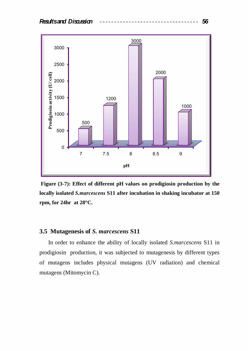
Results and Discussion 56
Figure (3-7): Effect of different pH values on prodigiosin production by the
locally isolated S.marcescens S11 after incubation in shaking incubator at 150
rpm, for 24hr at 28°C.
3.5 Mutagenesis of S. marcescens S11
In order to enhance the ability of locally isolated S.marcescens S11 in
prodigiosin production, it was subjected to mutagenesis by different types
of mutagens includes physical mutagens (UV radiation) and chemical
mutagens (Mitomycin C).
0
500
1000
1500
2000
2500
3000
7 7.5 8 8.5 9
500
1200
3000
2000
1000
Pro
digi
osin
act
ivit
y (U
/cel
l)
pH

Results and Discussion 57
According to Abbas et al., (2004); colonies screened to investigate the
genetic alteration after mutagen treatments caused 90% death (10%
survival) which may lead to over production mutants. By observing the
morphological differences, the mucoidal growth and colony size, thirty five
mutant colonies were selected after each mutagenic treatment. The
prodigiosin activity was determined as in (2.2.7) for each mutant.
3.5.1 Physical mutagenesis by UV radiation:
The results mentioned in figure (3-8) showed that the mutagenic and
lethal effect UV irradition on S. marcescens S11, there was a reduction of
total viable count of the bacterial cells from 288x109 CFU/ml in the zero
time to 199x109 CFU/ml after the exposure to the first dose of UV radiation
(1 j/m2), after subjection to this dose the survival percentage was 60%,
followed by sever reduction to 30%, 15%, and 1.6% after subjection to next
doses of UV irradiation (2, 3, and 4 j/m2).

Results and Discussion 58
Figure (3-8) Effect of different doses of UV. Radiation on the survivals of
locally isolated S.marcescens S11.
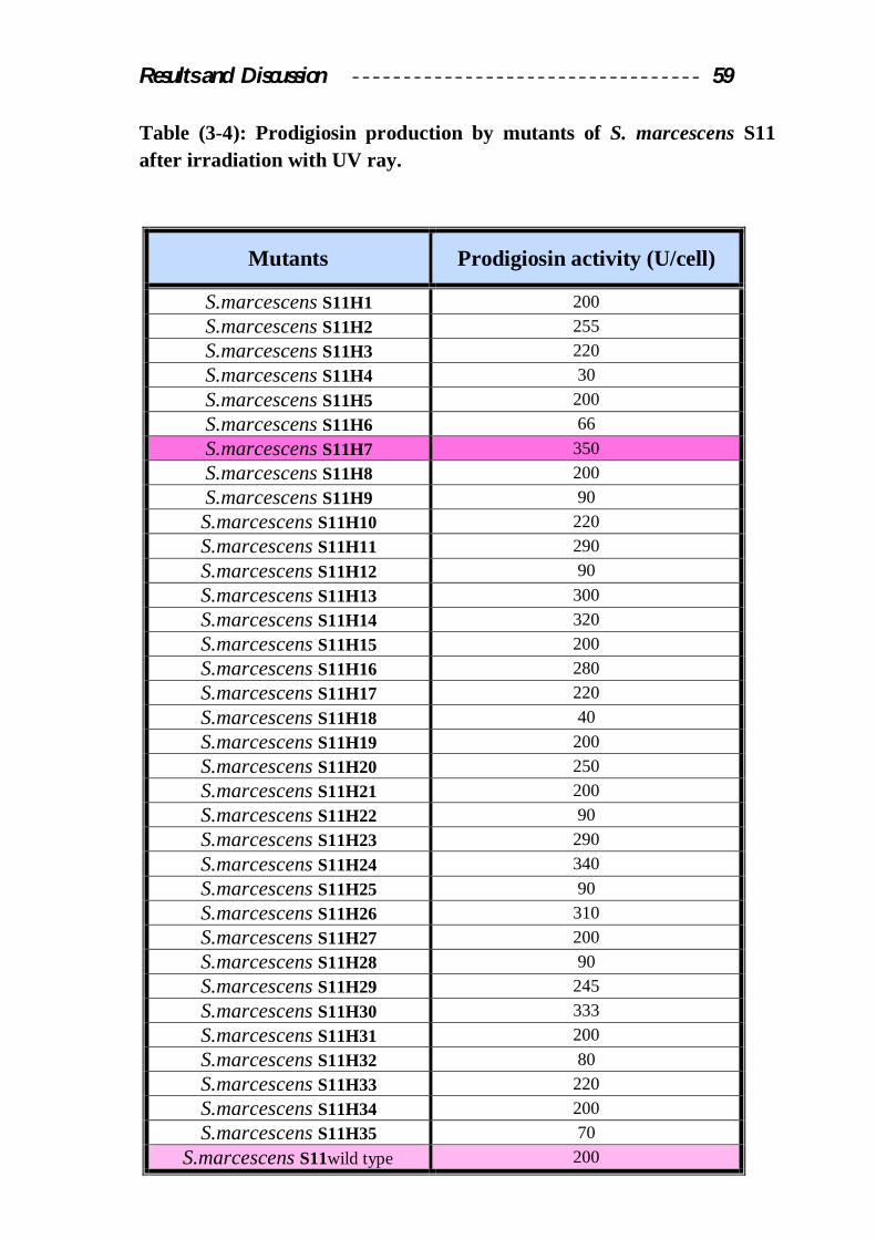
Results mentioned in table (3-4) showed that after UV radiation
sixteen mutants out of thirty five (45.7%) showed an increase in prodigiosin
production to 220U/cell for mutants S11H3, S11H10, S11H17, and S11H33
to 350U/cell for mutant S11H7 compared with the productivity of the wild
type. (200 U/cell). On the other hand there are another ten mutants 28.5%
with lower prodigiosin production than the wild type. The prodigiosin
activity produced by these mutants ranged between 30U/cell for (mutant
S11H4) to 90U/cell (mutants S11H9, S11H12, S11H22, S11H25, and
S11H28 ). UV irradiation is affected via miss repair of damaged DNA by
SOS repair system and termed indirect mutagen it was applied as mutagens
for the halotolerant Micrococcus sp., cell survival and mutability of P.
aeruginosa and P. syringae and the survival of Shewanella oneidensis were
determined after UV radiation (Qiu et al., 2004).
Results and Discussion 59
Table (3-4): Prodigiosin production by mutants of S. marcescens S11 after irradiation with UV ray.
Mutants Prodigiosin activity (U/cell)
S.marcescens S11H1 200 S.marcescens S11H2 255 S.marcescens S11H3 220 S.marcescens S11H4 30 S.marcescens S11H5 200 S.marcescens S11H6 66 S.marcescens S11H7 350 S.marcescens S11H8 200 S.marcescens S11H9 90 S.marcescens S11H10 220 S.marcescens S11H11 290 S.marcescens S11H12 90 S.marcescens S11H13 300 S.marcescens S11H14 320 S.marcescens S11H15 200 S.marcescens S11H16 280 S.marcescens S11H17 220 S.marcescens S11H18 40 S.marcescens S11H19 200 S.marcescens S11H20 250 S.marcescens S11H21 200 S.marcescens S11H22 90 S.marcescens S11H23 290 S.marcescens S11H24 340 S.marcescens S11H25 90 S.marcescens S11H26 310 S.marcescens S11H27 200 S.marcescens S11H28 90 S.marcescens S11H29 245 S.marcescens S11H30 333 S.marcescens S11H31 200 S.marcescens S11H32 80 S.marcescens S11H33 220 S.marcescens S11H34 200 S.marcescens S11H35 70
S.marcescens S11wild type 200

Results and Discussion 60
3.5.2 Chemical mutagenesis by Mitomycin C
Another type of mutagens (Mitomycin C) was used to generate over
producer mutants of prodigiosin from S.marcescens S11. Results in figure
(3-9) indicated that this mutagen has significant mutagenic and lethal effect
on the bacterial cells of S.marcescens S11. This can be noticed from the
reduction in the total viable count of bacterial cells from 15.1x10⁸ CFU/ml in
the zero time to 7.4x10⁸ CFU/ml after the incubation with the mutagen for
15 min, after subjection to this period the survival percentage was 54%,
followed by sever reduction to 26.4%, 17.8%, and 5% after subjection to
next period of incubation (30, 45, and 60 min).
Figure(3-9): Survival of locally isolated S.marcescens S11 after incubation
with Mitomycin C (30µg/ml) for different periods.

Results and Discussion 61
After Mutagenesis with Mitomycin C, the illustrated results in table
(3-5) appeared: nineteen mutants out of thirty five (54.2%) with higher
prodigiosin production ranged between 230 U/cell for (mutants S11H38,
S11H42, S11H59, S11H64 and S11H69) to 400 U/cell for (S11H54 mutant),
than the productivity of wild type (200U/cell). Another ten mutants (28.5%)
showed decreasing in prodigiosin production. The prodigiosin activity
produced by these mutants ranged between 60U/cell for (mutant S11H49)
to 120U/cell (mutants S11H36, S11H44, S11H52, S11H56, S11H61 and
S11H66). Six mutants (17.1%) have no changed in prodigiosin production
(200U/cell for the mutants S11H37, S11H40, S11H48, S11H58, S11H63
and S11H70).
The molecular basis of Mitomycin C bioactivity derived mainly from
its propensity to covalent interact with DNA sequences, causing lethal intra
and interstrand cross-links as well as alkylation and formation of reactive
oxygen species (Sheldon et al., 1999).

Results and Discussion 62
Table (3-5): Prodigiosin production by mutants of S.marcescens S11 after mutagenesis with Mitomycin C.
Bacterial Isolate Prodigiosin activity (U/cell)
S.marcescens S11H36 120 S.marcescens S11H37 200 S.marcescens S11H38 230 S.marcescens S11H39 250 S.marcescens S11H40 200 S.marcescens S11H41 330 S.marcescens S11H42 230 S.marcescens S11H43 370 S.marcescens S11H44 120 S.marcescens S11H45 266 S.marcescens S11H46 100 S.marcescens S11H47 278 S.marcescens S11H48 200 S.marcescens S11H49 60 S.marcescens S11H50 320 S.marcescens S11H51 330 S.marcescens S11H52 120 S.marcescens S11H53 360 S.marcescens S11H54 400 S.marcescens S11H55 290 S.marcescens S11H56 120 S.marcescens S11H57 295 S.marcescens S11H58 200 S.marcescens S11H59 230 S.marcescens S11H60 246 S.marcescens S11H61 120 S.marcescens S11H62 268 S.marcescens S11H63 200 S.marcescens S11H64 230 S.marcescens S11H65 90 S.marcescens S11H66 120 S.marcescens S11H67 340 S.marcescens S11H68 80 S.marcescens S11H69 230 S.marcescens S11H70 200
S.marcescens S11wild type 200
Results and Discussion 63
From the preceded results, UV radiation successfully enhanced
prodigiosin production from S. marcescens S11 since its productivity 1.8
fold higher than the prodigiosin produced of the wild type.
Also, the chemical mutagens Mitomycin C successfully developing the
prodigiosin productivity about 2.0 fold for the mutant S11H54 compared
with productivity of the wild type. By comparing the highest prodigiosin
concentration 350 U/cell for mutant (S11H7) that obtained after UV
radiation and 400 U/cell for mutant (S11H54) after treatment with
Mitomycin C, the chemical mutagens Mitomycin C was better than UV
radiation in enhancing prodigiosin productivity. The mutation either
occurred in the structural genes or in the regulatory genes that negatively
regulate prodigiosin production.
To explain the obtained results after mutagenesis with the physical
mutagens and the chemical mutagens, the increment in prodigiosin
production by these mutants may be due to the effect of the mutagens which
lead to inactivate the regulatory genes responsible for negative regulation of
prodigiosin production. The high productivity of prodigiosin by locally
isolated S. marcescens mutants after exposure to different mutagens may be
due to genetic mutations that inactivates negative regulatory gene which
causes an increasing in prodigiosin biosynthesis, Ying et al., (1998).
Reduction in prodigiosin production by the mutants may be due to the
genetic mutations induced in the structural genes responsible for prodigiosin
biosynthesis pathway (Williamson et al., 2006), or due to the mutations that
may occur in the regulatory genes which compromise the main switch
controlling the conversion to the mucoidal growth (prodigiosin production)
(Ying et al ., 1998).
Results and Discussion 64
Finally, unchangeable in prodigiosin level expected due to genetic
mutations in different genes of the chromosomal DNA of locally isolated S.
marcescens other than those responsible for production and regulation of
prodigiosin biosynthesis pathway, as it was mentioned by Ying et al.,
(1998).


Materials and Methods 26
2. Materials and Methods
2.1 Materials
2.1.1 Equipment
The following equipments and apparatus were used in this study:
Equipment Company
Autoclave Karl Kolb (Germany)
Compound Light microscope Olympus (Japan)
Laminar air flow Memmert (Germany)
Distillator Gallenkamp (England)
Incubator Gallenkamp
Electrical Oven Gallenkamp
pH-meter Gallenkamp sanyo (U.K.)
Sensitive balance Mettler (Swizerland)
Shaker incubator Sartorius (Germany)
Visible spectrophotometer Baush and Lamb (England)
Micropipettes Volac (Germany)
Vortex Stuart scientific (U.K.)
Refrigerator
Ishtar (Iraq)
Portable Centrifuge Hermle labortechnik(German)
Ultraviolet transilluminator Ultraviolet product (USA)
Millipore filter unit (0.45 µm) Millipore Corp.(U.S.A.)

Materials and Methods 27
2.1.2 Chemicals
The following chemicals were used in this study:
Chemicals
Company(origin)
Trypton, pepton
Fluka (Switzerland)
Absolute ethyl alcohol BDH ( England)
HCl
BDH
NaOH, KOH
Merck (Germany)
NaCl
Merck
Sunflower oil, olive oil
Fruee (Turkey)
Mitomycin C
Kyowa Hakko Kogyo (Japan)
KH2PO4 , K2HPO4, Glucose, Sucrose, Lactose, Raffinose, Arabinose , Xylose, Fructose, Ammonium sulphate.
BDH ( England)
Phenol, Ammonium chloride, Casein hydrolysate.
Riedel-Dehaeny-(Germany)
Yeast extract Biolife (Italy)
Agar-Agar Oxoid (England)
Glycerol
Riedel-DeHaen (Germany)
Hydrogen peroxide, , N,N,N,N-tetramethyl-p-phenylene-diamine dihydrochloride
Difco (U.S.A)
Toluidine blue Difco
α- naphthol Sigma (USA)
Materials and Methods 28
2.1. 3 Media
2.1.3.1 Ready to Use Media
These media were prepared as recommended by manufacturing
Companies, pH was adjusted to 7.0 and sterilized by autoclaving.
Brain heart infusion agar (Difco–USA).
Brain heart infusion broth (Difco).
Semi – solid agar (Difco).
DNase agar (Difco).
Methyl red-Voges Proskauer broth (Oxoid–England).
Nutrient agar (Oxoid).
2.1.3.2 Synthetic Media
Luria- Bertani medium (Maniatis et al., 1982).
This medium consists of the following components:
Component Concentration(g/L)
Tryptone 10
Yeast Extract 5
NaCl 10
All components were dissolved in 950 ml of distilled water, pH was
adjusted to 7.0, then volume was completed to 1000 ml with D.W. and
sterilized by autoclaving.
Luria- Bertani Agar (Maniatis et al., 1982).
Agar (15%) was added to LBB and autoclaved.

Materials and Methods 29
Motility medium (Macfaddin, 2000).
This medium was consist of the following components :
Concentration(g/L) Component
1 Tryptone
0.5 NaCl
o.5 Agar – Agar
All components were dissolved in 950 ml distilled water, pH was
adjusted to 7.2 then volume was completed to 1000 ml and sterilized by
autoclaving .
Sugar fermentation medium (Macfaddin, 2000).
This medium was consist of the following components :
Concentration(g/L) Component
1 Peptone
0.5 NaCl
0 .08 Phenol red
All components were dissolved in 950 ml distilled water, pH was
adjusted to 7 then volume was completed to 1000 ml and sterilized by
autoclaving.

Materials and Methods 30
Sugars (Lactose, sucrose, xylose, raffinose and arabinose) were
prepared separately by dissolving 1g of sugar to 100 ml D.W and
sterilized by autoclaving for 9 min.
2.1.4 Reagents
Catalase reagent (Atlas et al., 1995):
This reagent was prepared to be consists of (3%) hydrogen peroxide.
Oxidase reagent (Atlas et al., 1995):
This reagent is composed of 1g of N, N, N; N-tetramethyl-p-
phynylene-diamine dihydrochloride dissolved in 100 ml distilled water
and kept in dark bottle at 4°C.
Methyl red reagent (Collee et al., 1996):
It was prepared by dissoliving 0.1g of methyl red in 300ml of ethanol
(99%) then 200ml of D.W. was added, mixed thoroughly and kept at 4ºC
until use.
Vogas- Prosker reagent (Collee et al., 1996):
Solution (A): This solution was prepared by dissolving 40g of
potassium hydroxide (KOH) in 80 ml D.W., then volume was completed
to 100ml with D.W.
Solution (B): This solution was prepared by dissolving 5g of α-
napthol in 100 ml D.W.
Equal volumes of solution A and B were mixed thoroughly and kept at
4ºC until use.

Materials and Methods 31
2.1.5 Buffer and Solution
Phosphate Buffer Solution (pH7)
This solution was prepared by dissolving 9.52g of Na2HPO4 and 6g
of NaH2PO4 in 950ml of D.W., pH was adjusted to 7, then volume was
completed to 1000ml with D.W., and sterilized by autoclaving(Cruikshank
et al., 1975).
Mitomycin C stock solution
Stock solution of Mitomycin C (1000 µg/ml) was prepared by
dissolving 10 mg of Mitomycin C in 10 ml of distilled water.
2.2 Methods 2.2.1 Sterilization methods
Moist heat sterilization
Media and solutions were sterilized by autoclaving at 121°C for 15min.
Dry heat sterilization
Glasswares were sterilized using electric oven at 180°C for 3 hrs.
2.2.2 Isolation of S.marcescens
2.2.2.1 Samples collection
Different samples were collected from different sources in order to
isolate S.marcescens and as follows:
Environmental Samples
Water, soil, and sewage samples were collected from different
locations in Baghdad governorate during 2 – 4 /2008.

Materials and Methods 32
2.2.2.2 Samples preparation
Environmental samples from water, soil, and sewage were diluted
using sterilized distilled water and as follows:
Water and Sewage Samples
Serial dilutions of water and sewage samples were done by taking 1ml
aliquots of each sample and diluted with 9 ml of sterilized distilled water,
mixed thoroughly, then 100 µl aliquots from the appropriate dilution were
taken and spread on Nutrient agar plates and incubated at 28°C for 18
hours.
Soil Samples
One-gram of each soil sample was added to test tubes containing 9ml
of sterilized distilled water, mixed thoroughly and serial dilution for each
tube were done separately, then 100µl aliquots from the appropriate
dilution were taken and spread on Nutrient agar plates and incubated at
28°C for 18 hours. After incubation, the growing colonies were selected
and streaked on Brain heart infusion agar plates and incubated at 28°C for
24 hour.
2.2.3 Identification of Serratia spp.
Different isolates from environmental samples were taken and grown
on Brain heart infusion agar medium to make sure that this type of bacteria
may belong to the genus of Serratia, then these isolates were identified
according to their cultural and morphological characteristics (staining
ability, shape, color, size, production of pigment, transparency and
mucoidal properties), of the growing colonies on nutrient agar and Brain
heart infusion agar plates, then some biochemical tests were achieved to
confirm results of identification and as follows:

Materials and Methods 33
2.2.3.1 Cultural and Morphological characteristics
Colonial morphology
Shape, color and viscous growth of the colonies were studied on
Brain heart infusion agar plates medium after incubation at 28°C for 24 hr.
Gram's stain (Harely and Prescott, 1996)
Single colony of each isolate was transferred and smeared on a clean
slide and fixed by heat. The smear was stained with crystal violet (for 1
minute) and excess stain was washed then treated with iodine (for 1 min.)
and the excess was washed with distilled water, then decolorized with
absolute alcohol and the excess was washed, and then counterstained with
safranine (for 30 second) and the excess was washed, then examined under
light microscope.
Growth at 40C (Grimont et al., 1984)
One of the tests used for differentiation between species of the genus
Serratia is achieved by growing bacterial isolates at 40°C. Bacterial
isolates (or the suspected Serratia isolates) were grown on Brain heart
infusion agar plates and incubated at 40°C for 24 hr. Presence of bacterial
growth indicates a positive result.
2.2.3.2 Biochemical testes
Catalase test (Atlas et al., 1995)
Catalase is an enzyme that catalyzes the decomposition of hydrogen
peroxide to water and gaseous oxygen, this test was done by adding few
drops of 3% hydrogen peroxide on a single colony smeared on clean
glass side. Production of gaseous bubbles indicates a positive result.

Materials and Methods 34
Oxidase test (Atlas et al., 1995)
Filter paper was saturated with oxidase reagent (tetra methyl-p-
phenylene diamine dihydrochloride), then single colony of each bacterial
isolate was rubbed on the filter paper with a sterile wooden applicator
stick. An immediate color change to a deep blue indicates a positive result.
Methyl red test (Collee et al., 1996)
This test is employed to detect production of sufficient acid during
fermentation of glucose.
The MR-VP media was prepared and divided in 5 ml test tubes and
sterilized by autoclaving, then test tubes were inoculated with each
bacterial isolate and incubated at 28ºC for 48hr. 5drops of methyl red
reagent were added to each tube and mixed gently. Appearance of red
color represents positive result.
Voges – proskauer test (Collee at el., 1996)
This test was achieved to detect the production of acetoin as an
intermediary metabolite. Acetoin is also known as acetyl methyl carbinol.
MR–VP medium was prepared and distributed into sterile test tubes
(5ml/tube), then test tubes were inoculated with fresh cultures (100µl
aliquots) of bacterial isolates and incubated at 28ºC for 48hr, after
incubation 0.5ml of reagent A and 0.5 ml of reagent B were added and
shaking vigorously for 30 second. Appearance of red color represent a
positive result.
Materials and Methods 35
DNase test (Atlas, 1995)
DNase catalyzes the hydrolysis of deoxyribonucleic acid (DNA).
DNA agar plates were inoculated with fresh culture of bacterial isolates
by streaking, then incubated at 28ºC for 24hr.
After incubation plates were flooded with 0.01% of toluidine blue.
Presence of pink halo around bacterial growth indicates a positive results.
Sugar fermentation (Macfaddin, 2000)
This test was employed to detect acid and gas production from sugar
fermentation by bacterial isolates.
Sugar fermentation medium was prepared and distributed into sterile
test tubes, sugar used for this purpose are lactose, sucrose, xylose,
raffinose, and arabinose (5ml/tube), then test tubes were inoculated with
fresh cultures of bacterial isolates and incubated at 28ºC for 48hr.
Appearance of yellow color represents a positive result.
Motility test (Macfaddin, 2000)
In semi–solid agar medium, motile bacteria (swarm) and give diffuse
spreading growth that is visually recognized.
This medium was prepared by adding 0.5% of agar to nutrient broth,
then 10 ml aliquots were transferred to test tubes and left in vertical
position. All tubes were inoculated with bacterial isolates by stapping in
straight line and incubated at 28ºC for 24hr, motile bacteria would grow
and diffuse around the line of stapping.

Materials and Methods 36
2.2. 4 Maintenance of Bacterial Isolates
Maintenance of bacterial isolates was performed according to Atlas
et al., (1995), as following:
(a) Short-term storage
Bacterial isolates were maintained for period of few weeks on brain
heart infusion agar plates. The plates were tightly wrapped in parafilm and
stored at 4°C.
(b) Medium-term storage
Bacterial isolate were maintained in stab culture for period of few
months. Such cultures were prepared in screw-capped bottles containing
(5-8 ml) of agar medium and stored at 4°C.
(c) Long-term storage
Bacteria can be stored for many years in brain heart infusion broth
containing 15% glycerol at low temperature without significant loss of
viability. This was done by adding (1.5 ml) of sterilized glycerol to an
exponential growth of bacterial isolates in a screw-capped bottle with final
volume (10 ml) and stored at -20°C.
2.2.5 Ability of bacterial isolates in prodigiosin production
Ability of locally isolated S.marcescens in prodigiosin production
was examined in liquid medium by inoculating brain-heart infusion broth
with fresh cultures of bacterial isolates and incubated in shaker–incubator
(150 rpm) at 28ºC till the optical density of the growth medium was 0.75 at
620nm.

Materials and Methods 37
At the same time the optical density of the growth medium was
measured at 499nm.
Prodigiosin activity (U/cell) produced by the local isolates of
S.marcescens was determined according the following equation (Haddix et
al., 2000) using the uninoculated growth medium as blank:
[ O. D499 – (1.3831 x O.D620 )] x 1000 Prodigiosin (U/Cell) = O. D 620
O.D499 : represent pigment absorbance.
represent bacterial cell absorbance. : O.D620
1.3831 : constant.
1000 : this factor are included to avoid working with numbers smaller
than one.

Materials and Methods 38
2.2.6 Optimum conditions for prodigiosin production
In order to determine the optimum conditions for prodigiosin
production, the selected isolate which was the best among the different
isolates in pigment production, was used for this purpose. Optimum
conditions include type and concentration of carbon source, type and
concentration of nitrogen source, type of phosphate source, temperature,
and pH.
2.2.6.1 Effect of Carbon source
Six different carbon sources (fructose, glucose, sucrose ,lactose, olive
oil, and sunflower oil) were used to determine the optimum for prodigiosin
production.
These carbon sources were added to the production medium in a
concentration of 2% w/v and 2% v/v for olive oil at 28ºC, pH 7 for 24
hr.
The optimum carbon source was used to supplement the production
medium in the next experiments of optimization.
2.2.6.2 Concentration of carbon source
The optimum carbon source was used for prodigiosin production by
the selected isolate was used in different concentrations (1%, 1.5%, 2%,
2.5% , 3%, and 3.5%) to determine the optimum for production of the
pigment after incubation in a shaker incubator (150 rpm) at 28 ºC, pH 7
for 24hr.
The optimum concentration was used to supplement the production
medium in the next experiments of optimization.

Materials and Methods 39
2.2.6.3 Effect of nitrogen source
Five nitrogen sources were used to determine the optimum in
prodigiosin production [Tryptone, Peptone, Casein hydrolysate, NH4NO3,
and (NH4)2SO4]. These nitrogen sources were added to the medium at a
concentration of 1.5% w/v at 28 ºC, pH 7 for 24hr.
The optimum nitrogen sources was used to supplement the
production medium in the next experiments of optimization.
2.2.6.4 Concentration of nitrogen source
Different concentrations (0.5, 1.0, 1.5, 2.0, and 2.5%) of the optimum
nitrogen source were used to determine the optimum for prodigiosin
production by the selected isolate after incubation in a shaker incubator
(150 rpm) at 28 ºC, pH 7 for 24hr.
The optimum concentration was used to supplement the production
medium in the next experiments of optimization.
2.2.6.5 Effect of phosphate source
In order to determine the optimum phosphate source for prodigiosin
production, two types of phosphate sources (KH2PO4 and K2HPO4) and
a mixture of them (0.07% of KH2PO4 and 0.03% of K2HPO4) were used
in a concentration of (0.1% w/v), to determine the optimum for prodigiosin
production by the selected isolate after incubation in a shaker incubator
(150 rpm) at 28 ºC, pH 7 for 24hr.
The optimum phosphate source was used to supplement the
production medium in the next experiments of optimization.

Materials and Methods 40
2.2.6.6 Effect of temperature
In order to determine the optimum incubation temperature for
prodigiosin production by the selected isolate, the production medium was
inoculated with the selected isolate and incubated at different
temperatures (24, 28, 32, 36, and 40°C) in a shaker incubator (150 rpm) for
24hr.
The optimum temperature was fixed in the next experiments of
optimization.
2.2.6.7 Effect of pH
In order to determine the optimum pH for prodigiosin production
,initial medium pH was adjusted to different pH values(7.0, 7.5, 8.0, 8.5
and 9.0), then inoculated with selected isolate and incubated in a shaker
incubator (150 rpm) at 28ºC for 24hr.
2.2.7 Mutagenesis of S.marcescens
In order to study the ability of the locally isolated S.marcescens in
prodigiosin production, it was subjected to random mutagenesis using
physical and chemical mutagens to induce genetic mutations leading to
increase prodigiosin production by the selected isolate and as follows:
2.2.7.1 Physico – chemical mutagenesis
Serratia marcescens was subjected to physical mutagenesis using UV
radiation according to Kidambi et al., (1996). This was done, first by
inoculating luria broth medium with fresh culture of the selected isolate
and incubated at 28ºC till mid log phase, then cells were pelleted by
centrifugation at 6000 rpm for 15 min, washed twice with phosphate buffer
and resuspended in 5ml of the same buffer, and subjected to UV

Materials and Methods 41
irradiation in a dark place using UV photoelectric cell [ flou-link FLX ], by
transferring 5ml aliquots of the cell suspension into clean and sterile petri
dishes in a very thin layer. The distance between UV source and the
surface of the cell suspension was 11 cm.
Irradiation was achieved by subjection the suspension of bacterial cells
to different doses (0, 1, 2, 3 and 4 J/m2) of UV ray in a UV transiluminator,
then 0.1 ml aliquots was taken from the irradiated cell suspension after
each dose of irradiation, diluted properly and spread on LB agar plates and
incubated in a dark place at 28ºC for 24 hrs. Viable counts and survivals of
S.marcescens were determined after each dose treatment of incubation.
2.2.7.2 Chemical mutagenesis
Serratia marcescens was subjected to chemical mutagenesis using
Mitomycin C. Mutagenesis was achieved according to Bowring and
Morris, (1985) first by inoculating luria broth medium with fresh culture
of the selected isolate of S.marcescens and incubated at 28ºC till mid log
phase, then cells were pelleted by centrifugation at 6000 rpm for 15 min,
washed twice with phosphate buffer and resuspended in 5ml of the same
buffer and incubated with Mitomycin C, by adding 0.15 ml of Mitomycin
C stock solution to sterile test tubes containing 5 ml of cell suspension to
give final concentration of 30µg/ml then 0.1 ml aliquots of cell suspension
was taken every 15 min. of incubation with mutagen, diluted properly and
spread on LB agar plates and incubated at 28ºC for 24 hr. Viable counts
and survivals of S.marcescens were determined after each treatment of
irradiation.

Committee certification We, the examining committee certify that we have read this thesis "
Determination of the Optimum Conditions for Prodigiosin Production by
the Locally Isolated Serratia marcescens" and examined the student "Israa
Hasan Mahmood Dhahee" in its contents and that in our opinion , it is
accepted for the Degree of Master of Science in Biotechnology.
(Chairman) Signature: Name:
Scientific Degree: Date:
(Member) (Member) Signature: Signature: Name: Name: Scientific Degree: Scientific Degree: Date: Date:
(Member) (Member) Signature: Signature: Name: Dr. Hameed Majeed Name: Dr. Abdul Kareem Jasim Scientific Degree: Assistant Prof. Scientific Degree: Assistant Prof. Date: Date:
I, hereby certify upon the decision of the examining committee.
Signature: Name: Dr. Laith Abdul-Aziz AL-Ani Scientific Degree: Assist. Prof. Title: Dean of the College of Science Date:


Conclusions and Recommendations 65
Conclusions
Serratia marcescens isolated from different environmental samples
were able to produce prodigiosin.
Among different isolates, S.marcescens S11 was the best in
prodigiosin production.
Optimum conditions for prodigiosin production by the locally
isolated S. marcescens S11 involved the use of olive oil (1.5%),
casein hydrolysate (1.5%), KH2PO4, at pH 8.0, and incubation at
28ºC.
Physical and chemical mutagens may lead to prodigiosin over
producer mutants for S. marcescens S11.
Chemical mutagens using Mitomycin C are better than physical
mutagens using UV radiation in increasing the prodigiosin
productivity of S. marcescens S11.

Conclusions and Recommendations 66
Recommendations
Large scale production of prodigiosin by S.marcescens S11 under the
optimum conditions for production.
Determine the optimum conditions for prodigiosin production by the
most over-producer mutants raised after subjection to physical and
chemical mutagens.
Studying the effect of prodigiosin production by S.marcescens S11
as an antitumor agent.
Genetic study on S.marcescens S11 responsible for prodigiosin
production.

List of Abbreviations X
List of Abbreviations
Abbreviations
Full name
ATP Adenosin Tri Phosphate
D.W. Distilled Water
HBC 4-hydroxy-2,2′-bipyrrole-5-carbaldehyde
HBM 4-hydroxy-2,2′-bipyrrole-5-methanol
IC%50 Inhibitory Concentration %50
J/m2 Joule/ meter square
LB Luria Bertani
MMC Mitomycin C
O. D. Optical Density
PCE Prodigiosin Condensing Enzyme
Pig Pigment
rpm Round per minute Sp. Species
Spp. Specieses
UV Ultraviolet
UVR Ultraviolet Radiation
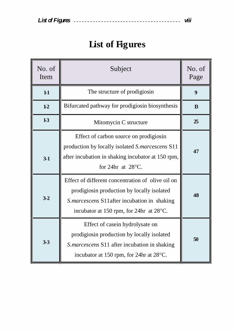
List of Figures viii
List of Figures
No. of Item
Subject
No. of Page
1-1 The structure of prodigiosin 9
1-2 Bifurcated pathway for prodigiosin biosynthesis 13
1-3 Mitomycin C structure 25
3-1
Effect of carbon source on prodigiosin
production by locally isolated S.marcescens S11
after incubation in shaking incubator at 150 rpm,
for 24hr at 28°C.
47
3-2
Effect of different concentration of olive oil on
prodigiosin production by locally isolated
S.marcescens S11after incubation in shaking
incubator at 150 rpm, for 24hr at 28°C.
48
3-3
Effect of casein hydrolysate on
prodigiosin production by locally isolated
S.marcescens S11 after incubation in shaking
incubator at 150 rpm, for 24hr at 28°C.
50

List of Figures ix
3-4
Effect of casein hydrolsyate concentration on
prodigiosin production by the locally isolated
S.marcescens S11 after incubation in shaking
incubator at 150 rpm, for 24hr at 28°C.
51
3-5
Effect of phosphate source on prodigiosin
production by locally isolated S.marcescens
S11 after incubation in shaking incubator at 150
rpm, for 24hr at 28°C.
52
3-6
Effect of different incubation temperature on
prodigiosin production by locally isolated
S.marcescens S11 after incubation in shaking
incubator at 150 rpm, for 24hr at 28°C.
54
3-7
Effect of different pH values on prodigiosin
production by locally isolated S.marcescens
S11after incubation in shaking incubator at
150 rpm, for 24hrs at 28°C.
56
3-8
Effect of Different Doses of UV. Radiation on
the Survivals of locally isolated S.marcescens
S11.
58
3-9
Survival of locally isolated S.marcescens S11
after incubation with Mitomycin C
(100µg/ml) for different periods .
60

List of Figures x

List of Tables vii
List of Tables
No. of Item Subject No. of
Page
1-1 Prodiginine groups and sources for this
compounds 16
3-1 Local isolates from different environmental
samples. 42
3-2
Biochemical tests of the locally isolated Serratia spp
44
3-3
Ability of locally isolated S.marcescens in
prodigiosin production after incubation with
shaking at 150 rpm in LB medium for 24hr at
28ºC.
45
3-4
Prodigiosin production by mutants of
S.marcescens S11 after irradiation with UV ray. 59

List of Tables viii
3-5
Prodigiosin production by mutants of
S.marcescens S11 after mutagenesis with
Mitomycin C.
62


67 References
Abbas, T.; Fatemeh, K. and Muhnaz, M. A. (2004). Improved
production of rhamnolipids by a Pseudomonas aeruginosa mutant.
Iran. J. Biomed., 8(1): 25-31.
Allen, E. G. (1967). Conditions of the color change of prodigision.
Natur., 216: 929-931.
Ajithkumar, B.; Ajithkumar, V. P.; Lriye, R.; Doi, Y. and Sakai, T.
(2003). Spore–forming Serratia marcescens subsp. Sakuensis subsp.
isolated from a domestic waste water treatment tank. Int. J. Syst.
Bacteriol., 53: 253–258.
Atlas, R. M.; Brown, A. E. and Parks, C. (1995). Laboratory Manual
Experimental of Microbiology. By Mosby – New York.
Azambuja, P.; Feder, D. and Garcia, E. S. (2004). Isolation of Serratia
marcescens in the midgut of Rhodnius prolixus: impact on the
establishment of the parasite Trypanosoma cruzi in the vector. Exp.
Parasitol., 107(1): 89–96.
Bennett, J. W. and Bentley, R. (2000). Seeing red: the story of
prodigiosin. Adv. Appl. Microbiol., 47: 1-32.
Blackman, A. J. and Li, C. (1994). Aust. J. Chem., 47: 1625.
Bowring, S. N. and Morris, J. G. (1985). Mutagenesis of Clostridium
acetobutylicum. J. Appl. Bacteriol., 58: 577-584.
Braun, P.; Balzer, G. and Fehlhaber, K. (2001). Activity of bacterial
lipases at chilling temperatures. Food Microbiol., 18: 211–215.
Brivonese, A. and Sutherland, W. I. (1989). Polymer production by a
mucoid strain of Azotobacter vinelandii in batch culture. App.
Microbiol. Biotechnol., 30: 97-102.
Bull, A. T. and Bushnel, M. E. (1976). Environmental control of fungal
growth In: The filamentous fungi. (eds. Smith, J. E. and Berry, D. E.).
2: 1-26.

68 References
Burger, S. R. and Bennett, J. W. (1985). Droplet enrichment factors of
pigmented and nonpigmented Serratia marcescens: possible selective
function for prodigiosin. Appl. Environ. Microbial., 50 (2): 487–490.
Carbonell, G. V.; Dalla Colleta, H. H. M.; Yano, T.; Levy, C. E. and
Fonseca, B. A. I. (2000). Clinical relevance and virulence factors of
pigmented Serratia marcescens. FEMS Immunol. and Medical.
Microbiol., 28 (2): 143-149.
Church, N.; Metsger, T. K.; Gowan, V.; Hillburger, V.; Adams, S. and
Rice, M. (2004). Cluster of Serratia marcescens associated with tap
water utilization in high–flow nasal cannula: identification,
investigation and correction. J. Americ. Infect. Control., 32 (3): 123–
124.
Collee, J. G.; Fraster, A.G.; Marmion, B. P. and Simons A. S. (1996).
Laboratory Strategy in the Diagnosis of infective syndromes. In
practical Med. Microbial. (eds. Mackie and Mccartiny(14thed.)
Churchill Livingstone. D.
Dairi, K.; Tripathy, S.; Attardo, G. and Lavallèe, J. F. (2006). Two
–step synthesis of the bipyrrole precursor of prodigiosin. Tetrahedron
Lett., 47 (15): 2605 – 2606.
D'Alessio, R.; Bargiotti, A.; Carlini, O. (2000). Synthesis and
immunosuppressive activity of novel prodigiosin derivatives. J. Med.
Chem., 43: 2557–2565.
Ding, M. J. and Williams, R. P. (1983). Biosynthesis of prodigiosin by
white strains of Serratia marcescens isolated from patients. J. Clin.
Microbiol., 17 (3): 476.
Fineran, P. C.; Everson, L.; Slater, H. and Salmond, G. P. C. (2005).
AGntR family transcriptional regulator (PigT) controls gluconate –
mediated repression and defines a new, independent pathway for

69 References
regulation of the tripyrrole antibiotic, prodigiosin, in Serratia.
Microbiol., 151: 3833-3845.
Forbes, B. A.; Sahm, D. F. and Weissfeld, A. S. (2002). Baily
Scott’s Diagnostic Microbiology. 11th Ed. Mosby.
Frank, R.; Witney, L.; Failla, E. D. (1997). Phosphate Inhibition of
Secondary Metabolism in Serratia marcescens. Americ. Society for
Microbio., 33 (5): 1042-1046.
Freifelder, D. (1987). Molecular Biology. Jones and Bartlett, Boston.
USA. 2nd Ed.
Furstner, A. (2003). Chemistry and biology of rosephilin and the
prodigiosin alkaloids: a survey of the last 2500 years. Angew. Chem.
Int. Ed., 42: 3582–3603.
Gerber, N. N. (1975). Prodigiosin-Like pigments. CRC Crit. Rev.
Microbiol., 3 (4): 469-485.
Geron, M.; Botershvili, I. and Rokem, J. S. (1988). Screening electron
microscopy of Serratia marcescens producing prodigiosin. J. Microb.,
54: 41-44.
Giles, M.; Harwood, H. M. and Gosling, D. A. (2006). What is the best
screening method to detect Serratia marcescens colonization during an
outbreak in a neonatal intensive care nursery. J. of Hospital Infec.,
62: 349-352.
Giri, A.V.; Anandkumar, N.; Muthukumaran, G. and Pennathur, G.
(2004). A novel medium for the enhanced cell growth and production
of prodigiosin from Serratia marcescens isolated from soil. BMC
Microbiol., 4: 11.
Goodenough, U. (1984). Genetics. Saunders College publishing,
Philadelphia. USA. 3rd Ed.

70 References
Greenwood, D.; Slack, R. C. B. and Peutherer, J. F. (2002). Med.
Microbiol., 6th Ed.
Grimont, P. A. D. and Grimont, F. (1984). Genus Serratia. In :
Bergeys manual of systematic bacteriology. 1: 477.
Haddix, P. L.; Paulsen, E. T. and Werner, T. F. (2000). Measurement
of mutation to antibiotic resistance: Ampicillin resistance in Serratia
marcescens. Bioscene: J. College Biol. Teach., 26: 17-21.
Harly, J. and Prescott, L. (1996). Laboratory excerciesion
microbiology. 3th Ed. WCB. Mcgrew Hill, Newyork.
Harris, A. K.; Harris, N. R.; Williamson, H.; Slater, A.; Cox, S.;
Abbasi, I.; Foulds, H. T.; Simonsen, F. J. and Leeper, G. P. (2004). The
Serratia gene cluster encoding biosynthesis of the red antibiotic,
prodigiosin, shows species-and strain-dependent genome context
variation. Microbiol., 150: 3547-3560.
Hearn, W. R.; Medina–Castro, J. and Elison, M. K. (1968). Color
change of prodigiosin. Natur., 220: 170–171.
Hejazi, A. and Falkiner, F. R. (1997). Serratia marcescens. J. Med.
Microbiol., 46: 903-912.
Henderson, C. I. (1993). Recent advances in the usage of mitomycin. J.
Oncol., 50: 1–84.
Henke, J. M. and Bassler, B. L. (2004). Bacterial social engagements.
Trends Cell Biol., 14: 648-656.
Holt, J. G.; Krieg, N. R.; Sneath, H. A.; Staley, J. T. and Williams, S.
T. (1994). Berge’s Manual of Determinative Bacteriology. 9th Ed
Williams and Wilkins. U.S.A.
Horkajada, J. P.; Martinaz, J. A.; Alcón, A.; Marco, F.; De Lazzari,
E.; Motas, A.; Zaragoza, M.; Salles, M.; Zavala, E. and Mensa, J.
(2006). Acquisition of multidrug–resistant Serratia marcescens by

71 References
critically ill patients who consumed tap water during receipt of oral
medication. Infect. Control Hospital Epidemiol., 27 (7): 774–777.
Houdt, R. V.; Givskov, M. and Michiels, C. W. (2007). Quorum
sensing in Serratia. FEMS Microbiol. Rev., 31: 407–424.
Isaka, M.; Jaturapat, A.; Kramyu, J.; Tanticharoen, M. and
Thebtaranonth, Y. (2002). Potent in vitro antimalarial activity of
metacycloprodigiosin isolated from Streptomyces spectabilis BCC
4785. Antimicrob. Agents Chemother., 46 (4): 1112–1113.
Iyozumi, H.; Komagata, T.; Hirayae, K. (1996). Biological control of
cyclamen gray mold (Botrytis cinerea) by Serratia marcescens B2. Ann
Phytopathol. Soc. Jpn., 62: 559–565.
Jama, A. (2008). Dietary alpha-linolenic acid is associated with
reduced risk of fatal coronary heart disease, but increased prostate
cancer risk: a meta-analysis. J. of Nutrition, 295:403-415.
Jeong, H.; Yim, J. H.; Lee, C.; Choi, S. H.; Park, Y. K.; Yoon, S. H.;
Hur, C. G. and Kang, H. Y. (2005). Genomic blue print of Hahella
chejunsis a marine microbe producing an algicidal agent. Nucleic Acids
Res., 33: 7066–7073.
Khanafari, A.; Assadi, M. M. and Fakhr, F. A. (2006). Review of
Prodigiosin, Pigmentation in Serratia marcescens. J. Bio. Sci., 6 (1): 1-
13.
Kidambi, S. P.; Sundin, G. W.; Palmer, D. A.; Chakrabarty, A. M. and
Bender, C. L. (1996). Copper as a signal for alginate synthesis in
Pseudomonas syringae pv. syringae. Appl. Environ. Microbiol., 61:
2172-2179.
Kim, J. J. and Sundin, G.W. (2001). Construction and analysis of
photolyase mutants of Pseudomonas aeruginosa and Pseudomonas
syringae: Contribution of photoreactivation, nucleotide excision repair,
72 References
and mutagenic DNA repair to cell survival and mutability following
exposure to UV-B radiation. Appl. Environ. Microbiol., 67 (4): 1405-
1411.
Kim, C. H.; Sung-Ho, K. and Suk-In, K. (1999). Isolation and
Characteristics of prodigiosin like red pigment produced by Serratia sp.
KH-95. Kor. J. Appl. Microbiol. Biotechnol., 26: 283-289.
Kim, D. ; Lee, J. S.; Park, Y. K.; Kim, J. F.; Jeong, H.; Oh, T. K.; Kim,
B. S. and Lee, C. H. (2007). Biosynthesis of antibiotic prodiginines in
the marine bacterium Hahella chejuensis KCTC 2396. J. Appl.
Microbiol., 102: 937–944.
Kim, C. H.; Kim, S. H. and Hong S. I. (1998). Isolation and
characteristics of prodigiosin-like red pigment produced by Serratia
sp.KH-95. San'oeb misaengmul haghoeji 26: 283-289.
Kobayashi, N. and Ichikawa, Y. (1991). Separation of the prodigiosin
localizing crude vesicles whichretain the activity of protease
andnuclease in Serratia marcescens. Microbiol. Immunol., 35: 607-614.
Law, R. A. (2001). Provided by Karen Fujii, Division of Outbreak
Investigation, Marylan Department of Health and Mental Hygiene. 20
(49): 1-13.
Macfaddin, J. F. (2000). Biochemical tests for identification of medical
bacteria. 3Ed lippincott Williams and Wilkins.
Maki, H. (2002). Origins of spontaneous mutations: specificity and
directionality of base-substitution, frame shift and sequence-substitution
mutagenesis. Ann. Rev. of Genet., 36: 279-303.
Manderville, R. A. (2001). Synthesis, Proton–affinity and anti-cancer
properties of the prodigiosin group natural products. Curr. Med. Chem.
Anti-Cancer Agents, 1 (2): 195 – 218.
73 References
Maniatis, T.; Fritch, E. F. and Sambrock, J. (1982). Molecular Cloning:
A Laboratory Manual. Cold spring Harbor Laboratory, New York.
Melo, P. D. S.; Durán, N. and Haun, M. (2000). Cytotoxicity of
prodigiosin and benzidazolle on V79 cells. Toxicol. Lett., 116 (3): 237–
242.
Mercado, C. M. and Tomasz, M. (1972). Inhibitory effects of
mitomycin-related compounds lacking the C1-C2 aziridine ring.
Antimicrob. Chemother., 1 (1): 73-77.
Miller, R.V.; Jefrrey, W.; Mitchell, D. and Elsari, M. (1999). Bacterial
responses to ultraviolet light. Ameri. Soc. for Microbiol., 65: 535-542.
Montaner, B.; Navarro, S.; Piqué, M.; Vilaseca, M.; Martinell, M.;
Giralt, E.; Gil, J. and Peréz-Tomás, R. (2000). Prodigiosin from the
supernatant of Serratia marcescens induces apoptosis in haematopoietic
cancer cell lines. J. Pharmacol., 131: 585–593.
Montaner, B. and Pérez–Tomas, R. (2001). Prodigiosin–induced
apoptosis in human colon cancer cells. Life Sci., 68: 2025–2036.
Nakashima, T.; Kurachi, M.; Kato, Y.; Yamaguchi, K. and Oda, T.
(2005b). Characterization of bacterium isolated from the sediment at
coastal area of omura bay in Japan and several biological activities of
pigment produced by this isolated. Microbiol. Immunol., 49 (5): 407–
415.
Nakashima, T.; Miyazaki, Y.; Matsuyama, Y.; Muraoka, W.;
Yamaguchi, K. and Odd, T. (2006). Producing mechanism of algicidal
compound against red tide phytoplankton in a marine bacterium γ–
proteobacterium. Appl. Microbiol. Biotechnol., 73 (3): 684 – 690.
O’Rear, J.; Alberti, L. and Harshey, R. M. (1992). Mutations That
Impair Swarming Motility in Serratia marcescens 274 Include but Are

74 References
Not Limited to Those Affecting Chemotaxis or Flagellar Function. J. of
Bacteriol., 174: 6125-6137.
Park, D. (1968). The ecology of terrestrial Fungi. In: the fungi (eds.
G.C. Ainsworth and A.S. Sussman). 3: 5-37. Academic press. New
Yourk.
Paustian, T. D. and Kurtz, R. S. (1994). Transposon Mutagenesis of
Rhodobacter sphaeroides. Department of Biology. Yale University.
15th Ed. 15: 45-61.
Qui, X.; Sundin, G. W.; Chai, B.; Tiedje, J. M. (2004). Survival of
Shewanella oneidensis MR-1 after UV radiation exposure. Appl.
Environ. Microbiol., 70 (11): 6435-6443.
Rice, S. A.; Kho, S. Y.; Queck, M. (2005). Biofilm formation and
sloughing in Serratia marcescens are controlled by quorum sensing
and nutrient cues. J. Bacteriol., 187: 3477-3485.
Robert, P.W.; Cora, L.G.; Hussain, S.M. and Randolph, H.S. (1971).
Influence of temperature of incubation and type of growth medium on
pigmentation in Serratia marcescens. J. Bacteriol., 106 (2): 438-443.
Roberts, D. P.; Mckenna, L. F.; Lakshman, D. K.; Meyer, S. L. F.;
Kong, H.; Souza, J. T. D.; Lydon, J.; Baker, C. J.; Buyer, J. S. and
Chung, S. (2007). Suppression of damping – off of cucumber caused by
Pythium ultimum with live cells and extracts of Serratia marcescens.
Soil Biol. and Biochem., 39: 2275–2288.
Rowan, S. and Fisher, D. E. (1997). Mechanisms of apoptotic cell
death. Leukemia, 11: 457 ± 465.
Sekiguchi, M. L. (2004). Molecular epidemiology of Serratia
marcescens in a hospital. Jpn. J. Infect. Dis., 57(2): 78-80.

75 References
Setty, R. S. and Sreekrish, V. (2004). Biotechnology including: Cell
Biology, Genetics and Microbiology. New Age International (P) Ltd.,
New Delhi. 1st ed. Pp. 212-222.
Sheldon, P. J.; Johnson D. J; August, P. R.; Liu, H.W. and Sherman,
D.H. (1999). Characterization of a Mitomycin-binding drug resistance
Mechanism from the producing organism. Bacteriol., 179 (5): 1796-
1804.
Singlton, P. and Sainsbury, D. (2001). Diction are of Microbiology and
Molecular Biology. 3rd Ed. Johan Willy and Sons Ltd.
Slater, H.; Crow, M.; Everson, L. and Salmond, G. P. (2003).
Phosphate availability regulates biosynthesis of two antibiotics,
prodigiosin and carbapenem, in Serratia via both quorum-sensing-
dependent and -independent pathways. Mol. Microbiol., 47: 303–320.
Solem, M.; Rius, N.; Francia, A. and Loren, J. G. (1994). The effect of
pH on prodigiosin production by non-proliferating cells of Serratia
marcescens. Let. in applied microbial., 19: 341-344.
Sole , M.; Francia, A.; Rius, N. and Lore n, J. G. (1997). The role of PH
in the ‘glucose effect’ on prodigiosin production by non-proliferating
cells of Serratia marcescens. Lett. Appl. Microbiol., 25: 81–84.
Someya, N.; Nakajima, M.; Hirayae, K.; Hibi, T. and Akutsu, K.
(2001). Synergistic antifungal activity of chitinolytic enzymes and
prodigiosin produced by biocontrol bacterium, Serratia marcescens
strain B2 against gray mold pathogen, Botrytis cinerea. J. Gen. Plant
Pathol., 67: 312- 317.
Someya, N.; Kataoka, N.; Hirayae, K.; Hibi, T. and Akutsu, K. (2000).
Biological control of cyclamen soilborne diseases by Serratia
marcescens strain B2. Plant. Dis., 84 (3): 334–340.

76 References
Someya, N.; Nakajima, M.; Watanabe, K.; Hibi, T. and Akutsu, K.
(2005). Potential of Serratia marcescens strain B2 for biological control
of rice sheath blight. Biocont. Sci. and Technol., 15 (1): 105–109.
Someya, N.; Tsuchiya, k.; Yoshida, T.; Noguchi, M. T.; Akutsu, K. and
Sawada, H. (2007). Co-inoculation of an antibiotic – producing
bacterium and a lytic enzyme–producing bacterium for the biocontrol of
tomato with caused by fusarim oxysporum f. sp. lycopersici. Biocontrol
Sci., 12 (1): 1–6.
Soto–Cerrato, V.; Llagostera, E.; Montaner, B.; Scheffer, G. L. and
Pere΄z– Tomas, R. (2004). Mitochondria–mediated apoptosis operating
irrespective of multidrug resistance in Brest cancer cells by the
anticancer agent prodigiosin. Biochem. Pharmacol., 68 (7): 1345-1352.
Srijith, V. M. (2006). Analysis of Serratia marcescens Genome–
identifying the biosynthetic pathway of the pigment prodigiosin. A
Computational Approach, Center for Bioinformatics, UK. 1-4.
Suzuki, H. and Kilgore, W. W. (1966). Effects of Mitomycin C on
macromolecular synthesis in E. coli. Bacteriol., 93(1): 675-682.
Szybalski, W. and Iyer, V. N. (1964). Crosslinking of DNA by
enzymatically or chemically activated mitomycins and porfiromycins,
bifunctionally "alkylating" antibiotics. Federat Proc., 23: 946-957.
Thomson, N. R.; Crow, M. A.; McGowan, S. J.; Cox, A. and Salmond,
G. P. (2000). Biosynthesis of carbapenem antibiotic and prodigiosin
pigment in Serratia is under quorum sensing control. Mol. Microbiol.,
36: 539–556.
Tomasz, M.; Chawla, A. K. and Lipman, R. (1988). Mechanism of
monofunctional and bifunctional alkylation of DNA by Mitomycin C.
Biochem., 27: 3182–3187.

77 References
Ulrich, S. R. and Thoraxel, S. (1993). Growth and survival of Serratia
marcescens under Aerobic and Anaerobic conditions in the presence of
materials from blood bags. J. of C. Microbiol., 31 (7): 1826.
Wei, J. R. and Lai, H. C. (2006). N-Acylhomoserin lactone – dependent
cell–to– cell communication and social behavior in the genus Serratia.
296: 117–124.
Wei, Y. H. and Chen, W. C. (2005a). Enhanced production of
prodigiosin–like pigment from Serratia marcescens SMAR by medium
improvement and oil–supplementation strategies. J. Biosci. Bioeng., 99
(6): 616–622.
Wei, Y. H.; Yu, W. J. and Chen, W. H. (2005b). Enhanced undecyl
prodigiosin production from Serratia marcescens SS-1 by medium
formulation and amino–acid supplementation. J. Biosci. Bioeng., 100
(4): 466–471.
Williams, R. P.; Gott, C. L. and Qadri, S. M. H. (1971). Induction of
pigmentation in nonproliferating cells of Serratia marcescens by
addition of single amino acids. J. Bacteriol., 106 (2): 444–448.
Williams, R. P.; Goldschmidt, M. E. and Gott, C. L. (1965). Inhibition
by temperature of the terminal step in biosynthesis of prodigiosin.
Biochem. Biophys. Res. Commun., 19 (2): 177–181.
Williamson, N. R.; Fineran, P. C.; Leeper, F. J. and Salmond, G. P. C.
(2006). The biosynthesis and regulation of bacterial prodiginines. Nat.
Rev. Microbiol., 4 (12): 887- 899.
Williamson, N. R.; Simonsen, H. T.; Ahmed, R. A. A.; Golder, G.;
Slater, H.; Woodley, L.; Leeper, F. J. and Salmond, G. P. C. (2005).
Biosynthesis of the red antibiotic, prodigiosin, in Serratia :
identificationof anovel 2–methyl–3–n–amyl–pyrrole(MAP) assembly
pathway, definition of the terminal condensing enzyme, and
78 References
implications for undecyl prodigiosin biosynthesis in Streptomyces. Mol.
Microbiol., 56 (4): 971–989.
Winzer, K. R.; Hardie, I. and Williams, P. (2003). LuxSand
autoinducer-2:their contribution to quorum sensing and metabolism in
bacteria. Adv. Appl. Microbiol., 53: 291-396.
Witney, F. R. (1977). Phosphate inhibition of secondary metabolism in
Serratia marcescens. Appl. Environ. Microbiol., 33: 1042-1046.
Yamashita, M.; Nakagawa, Y.; Li, H. and Matsuyama, T. (2001).
Silica gel-dependent production of prodigiosin and serrawttins by
Serratia marcescens in liquid culture. Microb. Envi., 16: 250-254.
Yammamoto, D.; Uemura, Y.; Tanaka, K.; Nakai, K.; Yamamoto, C.;
Takemoto, H.; Kamata, K.; Hirata, H. and Hioki, K. (2000).
‘‘Cycloprodigiosin hydrochloride, a H+/Cl symporter, induces
apoptosis and differentiation in HL60 cells”, Int. J. of Cancer, 88:
121-128.
Ying-Hsiu, L. A. and Wang, T. V. (1998). Involvement of recF, recO,
and recR Genes in UV-Radiation Mutagenesis of Escherichia coli.
J. Bacteriol., 180: 1766-1770.

داءـــاالھ
الرسول االعظم ... ام التقوىـمعلم الخلق وأمالـى ... الــى االمـي الذي علم االمة )صلى اهللا علیھ وسلم( مـحمـد
لب ـالقى ـال ...ي الدعاء كلما نطقت ـفاه التي اكثرت لـالش ىـال ... يدربى نبراس ــال
ى من كان صبري حین ـال ...حیاتي قدوتي ومثلي االعلى في معلمي و ىـال ....الطیب ...ى نھر العطاء وقدوة المربین واالباءـال...عجزت ان اصبر
ي العزیزــأب مرھا ـن فرشت أیام عـى مـال ... نبع الحنانى ـال ... نـن حملتني وھناعلى وھـم ىـال ى من أضاءت لي عمرھا ـال ... ت طلبا آلماليارسھرت فص ى منـال ... ألیصالي طریقا ذا ـكنت ھلما الى من لوالھا... ى من طوقتني بالحب والحنانـال...كل األزمان لتنیر لي
...األنسان العزیزةي ــأم
… سندي في الحیاة عزي والى ... التي تنبض بالوفاءالطیبة القلوب الى
أخــواتي
اسماءشیماء و
...الى من شاطرتني افراحي واحزاني...الى رفیقة دربي
اـرشي ـتـأخ
اسـراء حـسن .......المتواضع اھدي لكم ثمرة جھدي

بـسم اهللا الرمحن بـسم اهللا الرمحن الرحیمالرحیم
للــمثمث رض رض واال واال ماواتماواتــالسالس نور نور اهللاهللا باح باح ــا مص ا مص فیه فیه كاتكاتــمشمشكك ورهورهنن جاجةجاجةجاجة الزجاجة الزيف زيف ز باح باح ــاملصاملص من من د د ــوقوقی ی ري ري د د ا كوكب ا كوكب أأككا ا ونة لونة لیتیتباركة زباركة زم م رةرةج ج ششا ا ه ه یتیتزز ربیة یكاد ربیة یكاد ا غا غرقیة ولرقیة ولشش نار نار سهه سهه ــمتس متس لملم ء ولو ء ولو ~~ىىــــضیضیی ی
ورهورهلنلن هدي اهللاهدي اهللای ی ورورعلى نعلى ن نور نور االمثالاالمثال اهللاهللا ویضرب ویضرب من یشاءمن یشاء
علیمعلیم شىءشىء للبكبك اس واهللااس واهللاللنللن
دق اهللا العظیمدق اهللا العظیمــصص سورة النورسورة النور
3535االیةاالیة

ةـــالصـاخلمن مواقع و) میاه المجاري والتربة, میاه(عینة من مختلف المناطق البیئیة 57جمعت
عزلھ منھا على إنھا 15عزلة شخصت 30وقد تم الحصول على . مختلفة في محافظة بغداد
Serratia spp بعد إجراء الفحوصات . بعد التحري عن قابلیتھا على انتاج الصبغة الحمراء
عزالت منھا على انھا 5تم تشخیص ) عزلة15(الكیموحیویة على ھذه العزالت
S.marcescens .
وقد أظھرت النتائج أن البرودجیوسیناختبرت قابلیة العزالت المشخصة على إنتاج
كانت ھي األكفأ في اإلنتاج، إذ بلغت كمیة S.marcescens S11 العزلة البكتیریة
.خلیة/وحدة 200المنتج منھا البرودجیوسین
S.marcescensبوساطة العزلة البكتیریة البرودجیوسین درست الظروف المثلى إلنتاج
S11 ،لمثلى لإلنتاج تضمنت استخدام الوسط اإلنتاجيوقد أظھرت النتائج أن الظروف ا
بتركیز بائذمستخلص الحلیب ال ،)مصدرا كاربونیا(٪ 1.5بتركیز زیت الزیتون الحاوي على
، برقم )مصدرا فوسفاتیا( ات البوتاسیوم ثنائیة الھیدروجین، فوسف)مصدرا نایتروجینیا(٪ 1.5
تحت البرودجیوسیندرجة مئویة، وقد بلغت إنتاجیة 28وحضن بدرجة حرارة 8ھیدروجیني
.خلیة/ وحدة 3000ھذه الظروف
لغرض زیادة انتاجیة S.marcescens S11 خالیا بكتیریالتمت عملیة التطفیر
) االشعة فوق البنفسجیة( تعمال تطفیر فیزیائيالتطفیر العشوائي باسانجز . البرودجیوسین
S.marcescens S11 اظھرت النتائج وذلك بتعریض). Mitomycin C(وتطفیر كیمیائي
للحصول )Mitomycin C(كیمیائي التطفیر الو) االشعة فوق البنفسجیة( الى التطفیر الفیزیائي
وصلت انتاجیة . على عدة مطفرات تمیزت بقدرتھا العالیة على انتاج البرودجیوسین
بینما ارتفعت ،خلیة/وحدة 3 50الى )S11H7(البرودجیوسین بعد التطفیر الفیزیائي للطافرة
خلیة بالمقارنة /وحدة 400الى S11H5) (بعد التطفیر الكیمیائي للطافرة انتاجیة البرودجیوسین
. خلیة/وحدة 200بالنوع البري الذي كانت انتاجیتھ

جمھوریة العراق
وزارة التعلیم العالي و البحث العلمي
جامعة النھرین
كلیة العلوم
صبغة النتاج الظروف املثلى تعیني
Serratia marcescens بكتريیا الربودجیوسني من املعزولة حملیا
رسالة
جامعة النھرین كلیة العلوم مقدمة إلى /علوم في ماجستیرال متطلبات نیل درجةمن ء جزك
التقانة االحیائیة
من قبل ضاحي اسراء حسن محمود
)2006جامعة النھرین (بكالوریوس علوم تقانة احیائیة
باشراف
عبد الكریم جاسم ھاشماالستاذ الدكتور حمید مجید الدلیمي و االستاذ الدكتور
2009تشرین الثاني 1430ذوالحجة