Detection of Occult Bone Marrow Micrometastases in Patients with Operable Lung Carcinoma
9
ANNALS OF SURGERY Vol. 222, No. 4,415-425 © 1995 Lippincott-Raven Publishers Detection of Occult Bone Marrow Micrometastases in Patients with Operable Lung Carcinoma Richard J. Cote, M.D.,* Edward J. Beattie, M.D.,t Benjaporn Chaiwun, M.D.* Shan-Rong Shi, M.D.,* James Harvey, M.D.,t Su-Chiu Chen, M.S.,4 Andrew E. Sherrod, M.D.,* Susan Groshen, Ph.D.,t and Clive R. Taylor, M.D., Ph.D.* From the Departments of Pathology* and Preventive Medicine,t University of Southem California School of Medicine/Kenneth Norris Comprehensive Cancer Center, Los Angeles, California; and the Kriser Lung Cancer Center, Beth Israel Medical Center,t New York, New York Objectives A large proportion of patients with operable lung carcinoma (no evidence of systemic spread of tumor) develop metastatic disease after primary therapy. More sensitive and specific methods are needed to identify patients at highest risk for recurrence who may benefit most from adjuvant therapy, while sparing those patients who do not require such treatment. Summary Background Data Using epithelial-specific monoclonal antibodies, the authors have developed an immunocytochemical assay capable of detecting as few as 2 lung cancer cells in 1 million bone marrow cells. Methods The assay was used to test the bone marrow (from resected ribs) of 43 patients with primary non- small cell lung carcinoma who showed no clinical or pathologic evidence of systemic disease. Results Occult bone marrow micrometastases (BMMs) were detected in 40% of patients (17/43) with non-small cell lung cancer, including 29% (5/17) of patients with stage or 11 disease and 46% of whom (12/26) had stage Il disease. The median follow-up was 13.6 months. Patients with occult BMMs had significantly shorter times to disease recurrence compared with patients without BMMs (7.3 vs. >35.1 months, p = 0.0009). Furthermore, for patients with stage or 11 disease, the presence of occult BMMs was significantly associated with a higher rate of recurrence (p = 0.0004). Conclusions The detection of occult BMMs identifies patients with operable non-small cell lung carcinoma who are at significantly increased risk for recurrence, independent of tumor stage, and may be useful in evaluating patients for adjuvant treatment protocols. 415 a ............................... ................................................
Transcript of Detection of Occult Bone Marrow Micrometastases in Patients with Operable Lung Carcinoma

ANNALS OF SURGERYVol. 222, No. 4,415-425© 1995 Lippincott-Raven Publishers
Detection of Occult Bone MarrowMicrometastases in Patients with
Operable Lung CarcinomaRichard J. Cote, M.D.,* Edward J. Beattie, M.D.,t Benjaporn Chaiwun, M.D.*Shan-Rong Shi, M.D.,* James Harvey, M.D.,t Su-Chiu Chen, M.S.,4Andrew E. Sherrod, M.D.,* Susan Groshen, Ph.D.,tand Clive R. Taylor, M.D., Ph.D.*
From the Departments of Pathology* and Preventive Medicine,t University of Southem CaliforniaSchool of Medicine/Kenneth Norris Comprehensive Cancer Center, Los Angeles, California; andthe Kriser Lung Cancer Center, Beth Israel Medical Center,t New York, New York
ObjectivesA large proportion of patients with operable lung carcinoma (no evidence of systemic spread oftumor) develop metastatic disease after primary therapy. More sensitive and specific methods areneeded to identify patients at highest risk for recurrence who may benefit most from adjuvanttherapy, while sparing those patients who do not require such treatment.
Summary Background DataUsing epithelial-specific monoclonal antibodies, the authors have developed animmunocytochemical assay capable of detecting as few as 2 lung cancer cells in 1 million bonemarrow cells.
MethodsThe assay was used to test the bone marrow (from resected ribs) of 43 patients with primary non-small cell lung carcinoma who showed no clinical or pathologic evidence of systemic disease.
ResultsOccult bone marrow micrometastases (BMMs) were detected in 40% of patients (17/43) withnon-small cell lung cancer, including 29% (5/17) of patients with stage or 11 disease and 46% ofwhom (12/26) had stage Il disease. The median follow-up was 13.6 months. Patients with occultBMMs had significantly shorter times to disease recurrence compared with patients without BMMs(7.3 vs. >35.1 months, p = 0.0009). Furthermore, for patients with stage or 11 disease, thepresence of occult BMMs was significantly associated with a higher rate of recurrence (p =0.0004).
ConclusionsThe detection of occult BMMs identifies patients with operable non-small cell lung carcinoma whoare at significantly increased risk for recurrence, independent of tumor stage, and may be usefulin evaluating patients for adjuvant treatment protocols.
415
a
...............................................................................

416 Cote and Others
The incidence of cancer in the United States continuesto rise as our population increases in size and age. A totalof 1,252,000 new cancers, with an anticipated 547,000deaths, is projected for 1995.' Although lung cancer isthe third most common form of cancer, it is the leadingcause ofcancer deaths, with 157,400 deaths projected for1995.' Once a tumor has developed, surgery (either aloneor in combination with adjuvant therapy) represents theonly potentially curative modality.2 Of the four majorhistologic subtypes oflung cancer (squamous, adenocar-cinoma, small cell, large cell), only small cell carcinomais generally considered refractory to surgical therapy.However, small cell carcinoma accounts for only 22% oflung tumors overall,3 thus 78% of lung carcinomaswould be potentially curable by surgery if they were de-tected early enough. Approximately 50% of all patientswith lung carcinoma are candidates for, and will un-dergo, definitive surgical resection.2The use of the TNM staging system and the staging
map ofthe mediastinal lymph nodes4 has been very help-ful in establishing treatment plans and prognosis for re-sectable lung cancer.9 For non-small cell carcinomas,accurate staging of disease has greater prognostic signifi-cance than cell type. For a T 1, NO, MO (stage I) lung car-cinoma treated with surgical excision, preferably lobec-tomy, patients have an anticipated 5-year survival rate of60% to 85%.5.9o' For a larger carcinoma, such as T2, NO,MO (stage I), patients have a 50% to 60% 5-year survivalrate.5-9 10 However, survival rates decrease dramaticallywith increasing stage of disease. In the case of stage IIIlung cancer, survival rates as low as 5% have been re-ported for patients with stage IIIb disease, despite the ab-sence of clinically detectable systemic metastases at thetime of surgery.2The single most important determinant of prognosis
and management of lung cancer is the absence or pres-ence of micrometastatic dissemination of cancer at thetime of initial presentation and treatment, because pri-mary treatment failure is secondary to undetectable sys-temic spread of tumor. Current measures of disease ex-tent are primitive; standard prognostic indexes, althoughproviding reliable information about populations of pa-tients, cannot predict which patients will experience dis-ease progression after primary therapy. Furthermore, the
Presented at the 115th Annual Meeting ofthe American Surgical Asso-ciation, April 6-8, 1995, Chicago, Illinois.
Supported in part by grants from the State of California through theTobacco Related Disease Research Program (grant 2IT0037) andthe American Cancer Society (ACS IN-2 1-31).
Address reprint requests to Richard J. Cote, M.D., Department of Pa-thology, University of Southern California School of Medicine,2011 Zonal Avenue, HMR 204, Los Angeles, CA 90033.
Accepted for publication April 10, 1995.
Table 1. DETECTION OF LUNG CANCERMICROMETASTASES
No. of No. of BMM+Diagnosis Patients (%)
Primary (stage I-l1l) lung carcinoma 43 17 (40)Metastatic (stage IV) lung carcioma 4 4(100)Carcinoma metastatic to lung 8 4 (50)Sarcoma metastatic to lung 5 0 (0)Benign (infectious/hamartoma) 11 0 (0)All patients 71
BMM+ = bone marrow micrometastases detected.
success of adjuvant therapy is assumed to stem from itsability to eradicate microscopic metastases before theybecome clinically evident. "The ability to detect the earliest systemic spread of
lung cancer would identify several important groups ofpatients, including those with low-stage (stage I) diseasewho have evidence of occult tumor metastases and whomay therefore benefit from adjuvant systemic treatment.In addition, patients with locally advanced (stage III) dis-ease, who generally are not considered to be surgical can-didates, may be identified and thus benefit from moreaggressive local (surgical) control oftheir tumor.We have developed sensitive methods to detect the mi-
croscopic dissemination of tumor in the bone marrowof patients with cancer.'2'16 This technique is exquisitelysensitive and can detect as few as 2 cancer cells in 1 mil-lion (106) normal bone marrow cells.'5 We report hereon the use ofthis technique to detect occult bone marrowmicrometastases (BMMs) in patients with localized lungcarcinoma who have no evidence of systemic metastaseswhen routine clinical and pathologic methods are used.We have evaluated the early clinical follow-up of thisgroup of patients and have found that the presence ofBMMs is associated with a greater risk of early recur-rence.
MATERIALS AND METHODS -
Patient Population
A total of 71 patients were entered in this study. Allpatients had undergone thoracotomy at Beth Israel Hos-pital in New York between 1991 and 1993. The diagno-ses for these patients are summarized in Table 1. Forty-three patients had primary non-small cell lung carci-noma without evidence of systemic metastases (stage I-III), 12 had metastatic carcinoma (including 4 with met-astatic lung cancer), 5 had sarcomas metastatic to lung,and 11 had benign disease (e.g., infections, hamarto-
Ann. Surg. * October 1995

Lung Cancer Micrometastases 417
mas). Of the patients with primary lung carcinoma, 15had stage I, 2 had stage II, and 26 had stage III disease.Disease stage for all patients was determined accordingto the TNM system.4
All patients with primary lung cancer underwentbronchoscopy and mediastinoscopy before surgery. Sur-gery included careful mediastinal staging as well as sur-gical and pathologic TNM staging.
Bone Marrow SamplesBone marrow samples were obtained at the time ofpri-
mary surgery. Sections of rib removed routinely at thetime ofsurgery were opened immediately and bone mar-row curetted into heparinized media. This procedure re-sults in a high-quality bone marrow sample yielding ap-proximately 40 to 60 million mononuclear cells per pa-tient.Bone marrow samples were placed in an insulated
container at room temperature and shipped overnight tothe University of Southern California. Bone marrow as-pirations can be stored for 24 to 48 hours without sig-nificant adverse effects on epithelial cell antigenicity.'5Two to four routine air-dried smears were made from
the bone marrow specimen and stained with Wright-Giemsa stain for routine cytologic examination. The re-mainder of the specimen was layered onto a Ficoll-Hy-paque density gradient (Pharmacia, Piscataway, NJ) andcentrifuged at 400 X g for 20 minutes. The interface layer(which contains mononuclear cells and intact, viable ep-ithelial cells'7) was collected, and the mononuclear cellswere counted with a hemocytometer counting chamberand brought to a final concentration of 107 cells/mL.Charged glass slides (Probe On Plus; Fisher Scientific,
Pittsburgh, PA) were used for cell plating. Exactly 0.1mL (1 million mononuclear cells) of the mononuclearcell suspension was added and spread evenly onto eachslide. Typically, 20 to 40 slides were prepared per patient.These slides were allowed to air-dry overnight (or for atleast 3 hours) and then fixed in acetone for 5 minutes atroom temperature and stored at -20 C. Slides can bestored for more than 2 years.'156
Monoclonal AntibodiesTwo mouse monoclonal antibodies, AE- 1 (Hybritech,
San Diego, CA) and CAM 5.2 (Becton-Dickinson, SanJose, CA), which are specific for cytokeratin intermedi-ate filament antigens, were used in combination, each ata final concentration of 10 ,ug/mL. The specificity ofthese antibodies has been described previously.'8-2' AE-1 reacted with greater than 90% of lung carcinomas, 18"19whereas CAM 5.2 reacted with nearly 100% of lung car-cinomas.20'2 ' These antibodies are epithelial specific and
do not react with cells found normally in the bone mar-row. They did not react with normal bone marrow cellsin samples obtained from more than 65 patients withoutepithelial cancers. 15"16
Immunocytochemical Staining Procedure
The immunocytochemical procedures have been de-scribed previously. 12,13,15,16 Briefly, specimen slides werebrought to room temperature, washed, and incubatedwith suppressor serum (5.0% horse serum in phosphatebuffered saline) for 30 minutes as a blocking step. Thesuppressor serum was aspirated, and the mouse mono-clonal antibody cocktail was applied at appropriate dilu-tion (100-200 ,uL/slide). The slides were incubated in ahumid chamber for 30 minutes at room temperature andthen incubated with biotinylated horse anti-mouse anti-body (Vector Laboratories, Burlingame, CA) for 30 min-utes at room temperature. The slides were again washedand then bathed in 2% H202/phosphate buffered saline/sodium azide for 20 minutes to quench the endogenousperoxidase activity of the bone marrow mononuclearcells. After an additional wash, the slides were incubatedwith Avidin DH biotinylated horseradish peroxidase Hcomplex (Vector Laboratories) for 30 minutes at roomtemperature, washed, and bathed in a chromogenic sub-stance (diaminobenzidine) for 10 to 15 minutes at roomtemperature. They were then rinsed, counterstained withhematoxylin for 1 to 3 minutes, dehydrated, and pro-tected with a coverslip.
Five slides (i.e., 5 million bone marrow elements) fromeach patient were studied with use of the immunocyto-chemical assay. After the staining procedure, the slideswere examined microscopically for the presence of anti-gen-positive cells. These cells were marked on the slide,counted, and usually photographically documented. Allslides were screened without knowledge of patient diag-nosis or stage of disease.
Statistical AnalysisSurvival and time to the first recurrence oflung cancer
were analyzed. Survival was calculated as the number ofmonths from surgery until death or until last docu-mented contact with the patient who was known to bealive. For patients with lung cancer recurrence, time tothe first recurrence of lung cancer was calculated as thenumber ofmonths from surgery to the date offirst docu-mented recurrence of disease. Patients who died beforerecurrence of disease were censored at the date of death;patients without recurrence were censored at the date lastseen free of disease. Patients who were never disease freewere not included in the analysis oftime to recurrence.Kaplan-Meier product limit estimates22 ofoverall sur-
Vol. 222 - No. 4

418 Cote and Others
vival and recurrence-free survival were plotted. Standarderrors for the probability of surviving or of not experi-encing disease recurrence were based on Greenwood'sformula22 for the Kaplan-Meier estimates. The log-ranktest was used to compare groups of patients. All proba-bility values reported are two-sided.
RESULTS
Clinical Follow-upForty-three patients with non-small cell lung carci-
noma without clinical evidence of systemic metastaseswere evaluated. The median follow-up was 13.6 months;80% of patients (25/30) still alive at last follow-up werefollowed for more than 5.8 months. Nineteen patientsdeveloped systemic metastases; of these, 10 died. A totalof 13 deaths occurred, including 3 of non-lung cancercauses.
Detection of Bone MarrowMicrometastases
Table 1 summarizes the rate of detection ofBMMs inthe entire cohort of 71 patients. Of note is that althoughBMMs were detected in 40% ofthe patients (17/43) withprimary localized (stage I-111) lung cancer, in 100% ofpatients (4/4) with metastatic lung cancer, and in 50%of patients (4/8) with epithelial carcinoma metastatic tolung, no epithelial cells were detected in the bone mar-row of patients with metastatic sarcomas (0/5) or in thatof patients who had undergone operation for benign dis-ease (0/1 1). These results further demonstrate the speci-ficity of the immunocytochemical assay. Figure 1 showsexamples of tumor cells detected by the immunocyto-chemical assay in the bone marrow ofpatients with stageI (Fig. 1 A) and stage III (Fig. 1 B) lung cancer.
Bone Marrow Micrometastases andRecurrenceFor the 43 patients with primary lung carcinoma but
no clinical evidence of systemic metastases, the presenceof BMMs was related to stage of disease (Table 2).Twenty-nine percent of patients (5/17) with stage I or IIlung cancer had detectable BMMs, compared with 46%of patients with stage III disease ( 12/26), but this was notstatistically significant (p = 0.27). Thirteen of the 17 pa-tients (77%) with BMMs experienced recurrence, com-pared with 6 of the 26 patients (23%) without evidenceofBMMs.The presence of BMMs was significantly associated
with earlier time to recurrence (p = 0.0009) (Table 3, Fig.2). The median time to recurrence for patients with no
detectable BMMs was 35.1 months, compared with 7.3months for patients with BMMs (Table 3). Similar re-sults were seen in comparisons ofbone marrow status tooverall survival; of the 17 patients with BMMs, 7 died(41%), compared with 6 deaths (23%) among the 26 pa-tients without BMMs. The median time to death in pa-tients with no detectable BMMs was 35.1 months, com-pared with 18.6 months for patients with BMMs (Table3, Fig. 3). However, results for overall survival were notstatistically significant (p = 0.072).
Stage of Disease and Recurrence in BoneMarrow MicrometastasesWe also determined whether bone marrow status
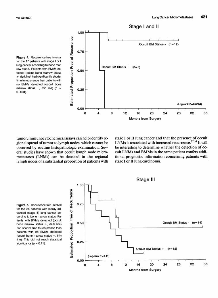
could be used to stratify patients in conventional prog-nostic groups (Figs. 4 and 5). There was a highly signifi-cant difference in recurrence rate for patients with stageI and stage II lung cancer according to bone marrow sta-tus (p = 0.0004) (Fig. 4). Similar results were seen forpatients with stage III lung cancer, although this did notreach statistical significance (p = 0.1 1) (Fig. 5). Ofpartic-ular note is that when BMMs were not detected in pa-tients with stage III disease, a substantial proportion (ap-proximately 40%) of patients remained disease free at 2years, despite the advanced stage ofdisease.A multivariable analysis evaluating bone marrow sta-
tus and controlling for stage of disease showed that thepresence ofBMMs is an independent predictor ofdiseaserecurrence (p = 0.017).
DISCUSSIONWe have shown in this study that occult BMMs can be
detected in a substantial proportion ofpatients with lungcancer who show no clinical evidence of systemic metas-tases, including patients with disease in its earliest stage(stage I). The presence of tumor cells in the bone mar-row, detected by the immunocytochemical assay de-scribed here, is significantly associated with a higher re-currence rate for patients with primary, localized (stageI-III) non-small cell lung carcinoma. In addition, pa-tients in conventional prognostic groups may be furtherstratified by their bone marrow status. Although the pres-ence ofBMMs is related to stage ofdisease, it is indepen-dent of stage regarding prediction of recurrence. Thepresence of BMMs appears to be a clinically importantpredictor oflung cancer recurrence.
Following the pioneering studies at the Ludwig Insti-tute at Royal Marsden Hospital,23 we have shown thatthe detection ofBMMs with the methods described hereis a significant predictor of recurrence for patients withearly stage operable breast cancer.'3"6 Several othergroups have also shown that the detection of BMMs is
Ann. Surg. * October 1995

Lung Cancer Micrometastases 419
Figure 1. (A) Bone marrow from a patient with stage lung cancer. A single tumor cell is identified by theimmunocytochemical technique involving the use of the epithelial-specific antibody cocktail (arrow) (originalmagnification X200). (B) Bone marrow from a patient with stage IlIl lung cancer. A cluster of tumor cells isidentified by the immunocytochemical technique (original magnification X400). In both A and B, the surroundingbone marrow mononuclear cells show no immunoreactivity; only the nuclei (counterstained with hematoxylin)are observable.
significant in predicting early recurrence for patientswith breast cancer2426 or colon cancer.27'28 Methods fordetection ofBMMs in patients with epithelial cancers are
based on the ability to distinguish extrinsic (epithelial)populations of tumor cells from normal bone marrow
elements. This involves use of monoclonal antibodies,which react with antigens expressed by epithelial (carci-noma) cells but not by normal hematopoietic ele-ments.'2'13 Studies have shown that occult BMMs (ex-trinsic, antigen-positive epithelial cells) can be detectedin the bone marrow of patients with breast or colon car-
cinoma without other evidence of metastasis.'31l6,24-28Furthermore, the presence of micrometastases identifies
Table 2. INCIDENCE OF LUNG CANCERMICROMETASTASES: ASSOCIATION
WITH STAGE OF DISEASE
No. of No. of BMM+Patients Patients (%)
Stage 15 5 (33)Stage II 2 0 (0)Stage III 26 12 (46)All patients* 43 17 (40)
BMM+ = bone marrow micrometastases detected. p = 0.27.* Patients with primary, operable non-small cell lung carcinoma.
patients with early stage (operable) breast or colon carci-nomas who are at significantly increased risk for recur-
rence. 13,16,24-28
Previous attempts to detect dissemination of lung car-
cinoma with such methods as skeletal surveys, radionu-clide scans, and bone marrow and liver biopsies havebeen disappointing regarding patients with operable(non-small cell) lung carcinoma who were not suspectedof having clinical metastases.7'2932 Recently, investiga-tors have used immunocytochemical methods similar tothose described here and have shown that occult BMMscan be detected in a substantial proportion of patientswith operable non-small cell lung carcinoma.33 34 Fur-thermore, these investigators have shown that the pres-
Table 3. ASSOCIATION OF BMM WITHRECURRENCE AND DEATH IN PATIENTS
WITH STAGE I-Ill LUNG CANCER
Median Time toOccult No. ofBMM Patients Recurrence (mo) Death (mo)
Absent 26 35.1 (10.1-35.1) 35.1 (20.2-35.1)Present 17 7.3 (3.3-9.3) 18.6 (8.6-18.6)
BMM = bone marrow micrometastases.Values in parentheses are confidence intervals.
Vol. 222 - No. 4
A
Amulff-

420 Cote and Others
1.00
0.75
0.50
0.25
Occult BM Status - (n=26)
Occult BM Status + (n=
0 4 8 12 16 20
Months from Surgery
ence ofBMMs was significantly associated with diseaserecurrence.33,34 The results of the current study are con-sistent with these findings.The immunocytochemical methods used in the cur-
rent study are exquisitely sensitive and can detect as fewas 2 tumor cells in a population of 1 million bone mar-row cells.'5 We used a cocktail of epithelial-specific anti-bodies that recognize five distinct cytokeratin sub-types' -21 to avoid overlooking extrinsic cells because of
1.00
0.75
0.50
0.25
0.00
24
Occ
(Log-rank P=0.072)
0 4 8 12 16 20
Months from Surgery
Figure 2. Recurrence-free intervalfor the 43 patients with stage I to IlIllung cancer, according to bone mar-row status. Patients with detectabletumor cells in the bone marrow (oc-cult bone marrow status +, darkline) had significantly shorter time torecurrence than patients with noBMMs detected (occult bone mar-
:17) row status -, thin line) (p = 0.0009).
28 32 36
antigenic heterogeneity. Recently, molecular assaysbased on the polymerase chain reaction have been usedto detect prostate cancer BMMs.35'36 Such assays havenot been established for other epithelial tumor types. Itis unknown whether the use of assays based on the poly-merase chain reaction for detection ofBMMs is advan-tageous to the use ofthe immunocytochemical assays de-scribed here.
Besides detecting systemic (bone marrow) spread of
:ult BM Status - (n=26)l I I I Figure 3. Survival of the 43 patients
with stage I to IlIl lung cancer accord-ing to bone marrow status. Patientswith BMMs detected (occult bonemarrow status +, dark line) diedsooner on average than patients withno BMMs detected (occult bonemarrow status -, thin line). This didnot reach statistical significance (p =0.072).
24 28 32 36
a)cJa1)
a.)L)
0
L)
a)U--
E
CD
._
0~
enla)CuE-a.'a
0)c
Cl)c0
0.0QL0
a)C.
u)w Occult BM Status + (n=17)
Ann. Surg. * October 1995
Lung Cancer Micrometastases 421
Stage I and 11
Figure 4. Recurrence-free intervalfor the 17 patients with stage or 11lung cancer according to bone mar-row status. Patients with BMMs de-tected (occult bone marrow status+, dark line) had significantly shortertime to recurrence than patients withno BMMs detected (occult bonemarrow status -, thin line) (p =0.0004).
a)
ca)
a)a)cc
%._
0a1)a1)LL
Cb
._
c
0l~0CuEC,)'a
0.75 K
0.50 F-
0.25 [-
O.0(
Occult BM Status - (n= 12)
Occult BM Status + (n=5)
(Log-rank P=0.0004)
0 4 8 12 16 20 24 28 32 36
Months from Surgery
tumor, immunocytochemical assays can help identify re-gional spread oftumor to lymph nodes, which cannot beobserved by routine histopathologic examination. Sev-eral studies have shown that occult lymph node micro-metastases (LNMs) can be detected in the regionallymph nodes ofa substantial proportion ofpatients with
stage I or II lung cancer and that the presence of occultLNMs is associated with increased recurrence.37'38 It willbe interesting to determine whether the detection of oc-cult LNMs and BMMs in the same patient confers addi-tional prognostic information concerning patients withstage I or II lung carcinoma.
Stage III
1.00
Figure 5. Recurrence-free intervalfor the 26 patients with locally ad-vanced (stage ll) lung cancer ac-cording to bone marrow status. Pa-tients with BMMs detected (occultbone marrow status +, dark line)had shorter time to recurrence thanpatients with no BMMs detected(occult bone marrow status -, thinline). This did not reach statisticalsignificance (p = 0.1 1).
a)
a.)Ca):3C)a)(C0a)a1)
C040
0-oa-~0a)
._EL'aw
0.75
0.50
0.25
0.00
Occult BM Status - (n = 14)
:P=O.11)
0 4 8 12 16 20 24 28 32 36
Months from Surgery
Vol. 222 - No. 4
1.00 I|
I

422 Cote and Others
The presence ofoccult BMMs (and LNMs) may definenot only patients who are at higher risk for recurrenceand death at worse prognosis, but may also identify bio-logically distinct mechanisms oftumor spread (e.g., lym-phatic vs. vascular dissemination). Use of these tech-niques may also allow us to identify biologically impor-tant populations of cells, that is, those cells constitutingthe earliest metastatic population of tumor cells. Thus,techniques that identify occult metastases may be valu-able in furthering our understanding of the events regu-lating tumor dissemination.The role of adjuvant chemotherapy in treating pa-
tients with stage I or II lung cancer is controversial. How-ever, identification of those patients at greatest risk forrecurrence (who, presumably, will benefit most fromsuch adjuvant approaches) may provide more rationalgrounds for making treatment decisions. For patientswith locally advanced (stage III) disease, the role of sur-gery can be controversial. Regarding this group of pa-tients, by identifying those at lowest risk for recurrence,we may be able to determine which patients will benefitmost from local (surgical) control of tumors. Bone mar-row micrometastases (and LNMs) may be importantnew prognostic factors in lung cancer that can greatlyaffect the selection of patients who may benefit mostfrom adjuvant therapy and the selection of patients withadvanced disease who may benefit from surgery. This ap-proach confers great improvement in staging accuracyand therefore allows for the formulation of truly homo-geneous treatment groups in future clinical trials.
References
1. Wingo PA, Tong T, Bolden S. Cancer statistics 1995. CA Cancer JClin 1995; 45:8-30.
2. Minna JD, Pass H, Glatstein EJ, Ihde DC. Cancer of the lung. InDevita VT, Heliman S, Rosenberg SA, eds. Cancer, Principles andPractice ofOncology. Philadelphia: JB Lippincott; 1989: 591-705.
3. Rosenow EC, Carr DT. Bronchogenic carcinoma. CA Cancer JClin 1979; 29:233-246.
4. Beahrs OH, Henson DE, Hutter RVP, Myers MH, eds. AmericanJoint Committee on Cancer Manual for Staging Cancer, 3rd ed.Philadelphia: J.B. Lippincott; 1988.
5. Martini N, Beattie EJ. Results of surgical treatment in Stage I lungcancer. J Thorac Cardiovasc Surg 1977; 74:499-506.
6. Martini N, Flehinger BJ, Nagasaki F, et al. Prognostic significanceofN 1 disease in carcinoma of the lung. J Thorac Cardiovas Surg1983; 86:646-653.
7. Martini N, Flehinger BJ, Zaman MB, Beattie EJ Jr. Results of re-section in non-oat cell carcinoma of lung with mediastinal lymphnode metastases. Ann Surg 1983; 198:386-397.
8. Melamed MR, Flehinger BJ, Zaman MB, et al. Screening for earlylung cancer: results ofthe Memorial Sloan-Kettering study in NewYork. Chest 1984; 86:44-53.
9. Mountain CF, Hermes KE. Management implications of surgicalstaging studies. Prog Cancer Res Ther 1979; 1 1:233-242.
10. Williams DE, Pairolero PC, Davis CS, et al. Survival of patients
Ann. Surg. - October 1995
surgically treated for stage I lung cancer. J Thorac Cardiovasc Surg1981; 82:70-76.
11. Schabel FM. Rationale for adjuvant chemotherapy. Cancer 1977;39:2875-2882.
12. Cote RJ, Rosen PP, Hakes TB, et al. Monoclonal antibodies detectoccult breast carcinoma metastases in the bone marrow of patientswith early stage disease. Am J Surg Pathol 1988; 12:333-340.
13. Cote RJ, Rosen PP, Old U, Osborne MP. Detection ofbone mar-row micrometastases in patients with early stage breast cancer. Di-agnOncol 1991; 1:37-42.
14. Osborne MP, Asina S, Wong G, et al. Immunofluorescent mono-clonal antibody detection of breast cancer in bone marrow: sensi-tivity in a model system. Cancer Res 1989; 49:2510-2513.
15. Chaiwun B, Saad A, Groshen S, et al. Detection of occult carci-noma cells in bone marrow and blood: efficiency of separation,preparation, and limits of detection using immunohistochemicalmethods. Diagn Oncol 1992; 2:267-276.
16. Cote RJ, Rosen PR, Lesser ML, et al. Prediction ofearly relapse inpatients with operable breast cancer by detection of occult bonemarrow micrometastases. J Clin Oncol 1991; 9:1749-1756.
17. Klein E, Vanky F, Galili U, et al. Separation and characteristics oftumor infiltrating lymphocytes in man. In Witz IP, Hanna MG,eds. Contemporary topics in immunobiology, vol. 10. New York:Plenum; 1980: 79-107.
18. Spagnolo DV, Michie SA, Crabtree GS, et al. Monoclonal anti-keratin (AE 1) reactivity in routinely processed tissue from 166 hu-man neoplasms. Am J Clin Pathol 1985; 84:697-704.
19. Tseng SCG, Jarvinen MJ, Nelson WG, et al. Correlation ofspecifickeratins with different types of epithelial differentiation: mono-clonal antibody studies. Cell 1982; 30:361-372.
20. Makin CA, Bobrow LG, Bodmer WF. A monoclonal antibody tocytokeratin for use in routine histopathology. J Clin Pathol 1984;37:975-983.
21. Bobrow LG, Makin CA, Law S, Bodmer WF. Expression of lowmolecular weight cytokeratin proteins in cervical neoplasia. J Pa-thol 1986; 148:135-140.
22. Miller RG. Survival Analysis, New York: Wiley; 1981.23. Redding WT, Coombes RC, Monaghan P, et al. Detection of mi-
crometastases in patients with primary breast cancer. Lancet 1983;2: 127 1-1274.
24. Mansi JL, Berger U, Easton D, et al. Micrometastases in bone mar-row in patients with primary breast cancer: evaluation as an earlypredictor ofbone metastases. Br Med J 1987; 295:1093-1096.
25. Dearnaley DP, Ormerod MG, Sloane JP. Micrometastases inbreast cancer: long-term follow-up ofthe first patient cohort. Eur JCancer 1991; 27:236-239.
26. Diel IJ, Kaufmann M, Goerner R, et al. Detection oftumor cells inbone marrow of patients with primary breast cancer: a prognosticfactor for distant metastasis. J Clin Oncol 1992; 10: 1534-1539.
27. Schlimok G, Funke I, Holzmann B, et al. Micrometastatic cancercells in bone marrow: in vitro detection with anti-cytokeratin andin vivo labelling with anti- 17- 1A monoclonal antibodies. Proc NatlAcad Sci USA 1987; 84:8672-8676.
28. Lindemann F, Schlimok G, Dirschedl P, et al. Prognostic signifi-cance of micrometastatic tumour cells in bone marrow of colorec-tal cancer cells. Lancet 1992; 340:685-689.
29. Ransdell JW, Peters RM, Taylor AT, et al. Multiorgan scans forstaging lung cancer: Correlation with clinical evaluation. J ThoracCardiovasc Surg 1977; 73:653-659.
30. Muggia FM, Cherun LR. Lung cancer: Diagnosis in metastaticsites. Semin Oncol 1974; 1:217-228.
31. Hansen HH, Muggia FM. Staging ofinoperable patients with bron-chogenic carcinoma with special reference to bone marrow exami-nation and peritonoscopy. Cancer 1972; 30:1395-1401.

Vol. 222 d No. 4
32. Hansen HH, Muggia FM, Selawry OS. Bone marrow examinationin 100 consecutive patients with bronchogenic carcinoma. Lancet197 1; 2:443-445.
33. Leonard RCF, Duncan LW, Hay FE. Immunocytological detec-tion of residual marrow disease at clinical remission predicts met-astatic relapse in non-small cell lung cancer. Cancer Res 1990;6545-6548.
34. Pantel K, Izbicki JR, Angstwurm M, et al. Immunocytological de-tection ofbone marrow micrometastasis in operable non-small celllung cancer. Cancer Res 1993; 53:1027-1031.
35. Moreno JG, Croce CM, Fischer R, et al. Detection of hematoge-nous micrometastasis in patients with prostate cancer. Cancer Res1992; 52:6110-6112.
36. Wood DP Jr., Banks ER, Humphreys S, Rangnekar VM. Sensitiv-ity ofimmunohistochemistry and polymerase chain reaction in de-tecting prostate cancer cells in bone marrow. J Histochem Cyto-chem 1994; 42:505-511.
37. Chen Z-L, Holmes EC, Coulson WF, et al. The frequency and dis-tribution ofoccult metastases in lymph nodes ofpatients with non-small cell lung cancer. J Natl Cancer Inst 1993; 85:493-498.
38. Passlick B, Izbicki JR, Kubuschok B, et al. Immunohistochem-ical assessment with individual tumor cells in lymph nodes ofpatients with non-small-cell lung cancer. J Clin Oncol 1994; 12:1827-1832.
Discussion
DR. TOM R. DEMEESTER (Los Angeles, California): Dr.Beattie's paper and Dr. Giuliano's paper that we heard earlierthis morning provide us with some objective evidence for whatwe always imagined to be true, that is, there are unrecognizedmicrometastases in the node removed or left behind after a re-section.
In the past, this has been difficult to measure and as a conse-quence, not much was done about it. Now, with the advent ofthe epithelial-specific clonal antibody, the assessment is possi-ble and will cause us to rethink our surgical approach to malig-nant disease in the future.The question, ofcourse, is, are these stain specific? Ifyou do
detect cells, are they viable, can they grow, and does the findingaffect survival?
It appears that Dr. Beattie has given us good evidence thatthe cells, when detected, have a dramatic effect on survival. Infact, he has shown that the observation may account for inac-curacies in staging, at least in early disease.What would be important to know is whether the finding is a
sign of systemic disease. Does he have information as to wherethe recurrences occurred? Were they systemic or local?
I would also be interested to know whether he has had anopportunity to study lymph nodes, as reported by Dr. Giuliano,and how many patients had histologically normal but histo-chemically positive lymph nodes with histochemically normalbone marrow.That would be an interesting group of patients, because if
indeed they do exist, it could be argued that our surgical resec-tion for cancer should be more extensive rather than the cur-rent emphasis on limited resections. It may be that the survivaldifference observed between limited and extensive resection fornode-negative patients may be explained by the removal of
Lung Cancer Micrometastases 423
nodes histochemically positive with the more extensive sur-gery.Now that he has developed this technique, it would be in-
teresting to know how he might use this information in pa-tient care. Would he encourage the searching for histochem-ical evidence of metastases preoperatively and then subject-ing these patients to adjuvant therapy? Or would he suggestadjuvant therapy ifthe nodes were found in the surgical spec-imen? Does he have any evidence that micrometastases de-tected by histochemical techniques are more susceptible tochemotherapy? I think these types of studies will certainlychallenge our thoughts concerning surgical oncology in the fu-ture.
DR. JOHN R. BENFIELD (Sacramento, California): Dr. Beat-tie's concept of extending the TNM nomenclature to TNMsmall "m" is heading in just the right direction, and I hope thathe intended the small "m" to stand for micrometastases andnot for marrow. I thought I might share with you some datathat we presented just 2 months ago before The Society ofTho-racic Surgeons, heading in somewhat the same direction in adifferent way (Johnson JR, et al. Successful xenotransplanta-tion of human lung cancer correlates with the metastatic phe-notype. Ann Thorac Surg 1995; 60:32-37).We studied 81 patients who had lung cancer in various stages
whose lung cancers were then propagated in xenotransplanta-tion in nude mice. The mean follow-up was 22.5 (2-61)months. Twenty-one xenotransplants successfully took andseven metastasized in the nude mice. Neither the predominantcell type nor the incidence oflymph node metastases correlatedwith the results of xenotransplantation. Of 21 patients whosenon-small-cell lung cancer (NSCLC) took in transplant, 13(61.9%) developed metastases, and 9 (42.8%) died from theircancers. Among 57 patients whose NSCLC did not take, 14(24.5%) developed metastases and 9 (15.7%) have died fromtheir cancers. The higher incidence ofmetastases in associationwith xenotransplantation take was significant (p = 0.0032). Weconcluded that patients whose NSCLC take in transplant are athigh risk for metastases, and we surmised that this method ofpropagating human lung cancers is a step toward facilitatingprecise cellular biologic definition of the metastatic propensityofsuch neoplasms.
I suggest that an extension of Dr. Beattie's approach to find-ing occult human lung cancers propagated in xenotransplanta-tion might give us some insight as to the mechanisms that areinvolved and perhaps another tool to identify patients with sys-temic disease before the usual approach of staging.We concluded that patients whose lung cancers take in our
transplantation model are at high risk for metastases. We be-lieve that this method of propagating human lung cancerswill facilitate carrying out precise cellular biologic studiesaimed at defining the metastatic potential of such neoplasmsbefore metastases become apparent. The search for evidenceof micrometastases, as described by Dr. Beattie, plus cellularbiologic markers of metastatic propensity should eventuallypermit us to identify patients whose apparently early stagelung cancers should nonetheless receive adjuvant systemictreatment.