A microphysiological system model of therapy for liver micrometastases
11
http://ebm.sagepub.com/ Experimental Biology and Medicine http://ebm.sagepub.com/content/early/2014/05/09/1535370214532596 The online version of this article can be found at: DOI: 10.1177/1535370214532596 published online 12 May 2014 Exp Biol Med (Maywood) Griffith and Alan Wells Transon Nguyen, Donna B Stolz, Jeffrey T Borenstein, Douglas A Lauffenburger, Raman Venkataramanan, Linda G Amanda M Clark, Sarah E Wheeler, Donald P Taylor, Venkateswaran C Pillai, Carissa L Young, Rachelle Prantil-Baun, A microphysiological system model of therapy for liver micrometastases Published by: http://www.sagepublications.com On behalf of: Society for Experimental Biology and Medicine can be found at: Experimental Biology and Medicine Additional services and information for http://ebm.sagepub.com/cgi/alerts Email Alerts: http://ebm.sagepub.com/subscriptions Subscriptions: http://www.sagepub.com/journalsReprints.nav Reprints: http://www.sagepub.com/journalsPermissions.nav Permissions: What is This? - May 12, 2014 OnlineFirst Version of Record >> by guest on May 13, 2014 ebm.sagepub.com Downloaded from by guest on May 13, 2014 ebm.sagepub.com Downloaded from
Transcript of A microphysiological system model of therapy for liver micrometastases
http://ebm.sagepub.com/Experimental Biology and Medicine
http://ebm.sagepub.com/content/early/2014/05/09/1535370214532596The online version of this article can be found at:
DOI: 10.1177/1535370214532596
published online 12 May 2014Exp Biol Med (Maywood)Griffith and Alan Wells
Transon Nguyen, Donna B Stolz, Jeffrey T Borenstein, Douglas A Lauffenburger, Raman Venkataramanan, Linda G Amanda M Clark, Sarah E Wheeler, Donald P Taylor, Venkateswaran C Pillai, Carissa L Young, Rachelle Prantil-Baun,
A microphysiological system model of therapy for liver micrometastases
Published by:
http://www.sagepublications.com
On behalf of:
Society for Experimental Biology and Medicine
can be found at:Experimental Biology and MedicineAdditional services and information for
http://ebm.sagepub.com/cgi/alertsEmail Alerts:
http://ebm.sagepub.com/subscriptionsSubscriptions:
http://www.sagepub.com/journalsReprints.navReprints:
http://www.sagepub.com/journalsPermissions.navPermissions:
What is This?
- May 12, 2014OnlineFirst Version of Record >>
by guest on May 13, 2014ebm.sagepub.comDownloaded from by guest on May 13, 2014ebm.sagepub.comDownloaded from
XML Template (2014) [5.5.2014–12:33pm] [1–10]//blrnas3/cenpro/ApplicationFiles/Journals/SAGE/3B2/EBMJ/Vol00000/140060/APPFile/SG-EBMJ140060.3d (EBM) [PREPRINTER stage]
Original Research
A microphysiological system model of therapy for liver
micrometastases
Amanda M Clark1, Sarah E Wheeler1, Donald P Taylor1, Venkateswaran C Pillai1, CarissaL Young2, Rachelle Prantil-Baun3, Transon Nguyen3, Donna B Stolz1, Jeffrey T Borenstein3,Douglas A Lauffenburger2, Raman Venkataramanan1, Linda G Griffith2 and Alan Wells1
1Departments of Pathology, Cell Biology, Pharmaceutical Sciences, and Bioengineering, and the McGowan Institute for Regenerative
Medicine, University of Pittsburgh and Pittsburgh VA Health System, Pittsburgh, PA 15213, USA; 2Department of Biological Engineering,
Massachusetts Institute of Technology, Cambridge, MA 02319, USA; 3Charles Stark Draper Laboratory, Cambridge, MA 02139, USA
Corresponding author: Alan Wells. Email: [email protected]
AbstractMetastasis accounts for almost 90% of cancer-associated mortality. The effectiveness of cancer therapeutics is limited by the
protective microenvironment of the metastatic niche and consequently these disseminated tumors remain incurable. Metastatic
disease progression continues to be poorly understood due to the lack of appropriate model systems. To address this gap in
understanding, we propose an all-human microphysiological system that facilitates the investigation of cancer behavior in the liver
metastatic niche. This existing LiverChip is a 3D-system modeling the hepatic niche; it incorporates a full complement of human
parenchymal and non-parenchymal cells and effectively recapitulates micrometastases. Moreover, this system allows real-time
monitoring of micrometastasis and assessment of human-specific signaling. It is being utilized to further our understanding of the
efficacy of chemotherapeutics by examining the activity of established and novel agents on micrometastases under conditions
replicating diurnal variations in hormones, nutrients and mild inflammatory states using programmable microdispensers.
These inputs affect the cues that govern tumor cell responses. Three critical signaling groups are targeted: the glucose/insulin
responses, the stress hormone cortisol and the gut microbiome in relation to inflammatory cues. Currently, the system sustains
functioning hepatocytes for a minimum of 15 days; confirmed by monitoring hepatic function (urea, a-1-antitrypsin, fibrinogen, and
cytochrome P450) and injury (AST and ALT). Breast cancer cell lines effectively integrate into the hepatic niche without detectable
disruption to tissue, and preliminary evidence suggests growth attenuation amongst a subpopulation of breast cancer cells. xMAP
technology combined with systems biology modeling are also employed to evaluate cellular crosstalk and illustrate communica-
tion networks in the early microenvironment of micrometastases. This model is anticipated to identify new therapeutic strategies
for metastasis by elucidating the paracrine effects between the hepatic and metastatic cells, while concurrently evaluating agent
efficacy for metastasis, metabolism and tolerability.
Keywords: Micrometastasis, chemotherapeutics, mammary carcinoma, liver
Experimental Biology and Medicine 2014; 0: 1–10. DOI: 10.1177/1535370214532596
Introduction
Metastasis is the leading cause of cancer-associated mor-tality. The development of metastases involves a series ofsequential biological processes that allow the spread ofcancer cells from a primary site to secondary organs.Cells escape from the primary tumor by intravasationinto the circulation followed by extravasation into theparenchyma of a distant organ.1 Those cells that success-fully disseminate may either outgrow immediately or laydormant, as small or pre-malignant micrometastases, foryears to decades before becoming clinically evident.2,3
This is especially daunting in the case of breast cancerwhere even though the primary tumor is often success-fully treated, up to 30% of women with early stage breastcancer will eventually relapse with metastatic disease.4
Due to the widespread distribution of metastatic tumorsand the protective effects of the metastatic microenviron-ment, the effectiveness of cancer therapeutics is limitedand consequently recurrent cancers remain largelyincurable.
One of the major hurdles impeding the development ofcancer therapeutics to target micrometastases is the
ISSN: 1535-3702 Experimental Biology and Medicine 2014; 0: 1–10
Copyright � 2014 by the Society for Experimental Biology and Medicine
Exp Biol Med (Maywood) OnlineFirst, published on May 12, 2014 as doi:10.1177/1535370214532596
by guest on May 13, 2014ebm.sagepub.comDownloaded from
XML Template (2014) [5.5.2014–12:33pm] [1–10]//blrnas3/cenpro/ApplicationFiles/Journals/SAGE/3B2/EBMJ/Vol00000/140060/APPFile/SG-EBMJ140060.3d (EBM) [PREPRINTER stage]
limitations of current model systems. Animal models arenot suitable for this type of study as they generally onlyprovide endpoint analyses in addition to issues of relevancefor the human condition.5,6 Typically, immunocomprom-ised murine models are used,7–9 yet studies have demon-strated that immune system is crucial to themicrometastatic microenvironment.10,11 Those animalstudies that do use syngeneic models are also not fully rep-resentative of the human situation due to interspecies dif-ferences in cytokines and metabolism.6 While in vitroculture investigations can avoid the cross species issues,the current 2D culture systems lack important aspectswhich impact tumor behavior, such as 3D architecture toprovide tissue depth for tumor intercalation; functionalaspects, including fluid flow and control of oxygen content,and do not allow for extended culture. There is also a dis-tinct absence of models capable of recreating micrometas-tasis while concurrently providing for the evaluation ofagent efficacy, toxicity and metabolism. For these reasons,a number of investigators have utilized organotypic cul-tures in bioreactors as investigative tools to overcomesuch issues.12–17
The liver as the metastatic target
The liver represents an ideal organ system to study bothmicrometastasis and the efficacy of cancer therapeutics.Firstly, it is a major site of metastasis for a wide range of
carcinomas (e.g. breast, lung, colon, prostate,brain, melanomas). Depending on the primary tumortype, 30–70% of patients dying from cancer have hepaticmetastases.18 Secondly, the liver is the major site for drugmetabolism (both activation and detoxification), a signifi-cant factor in determining efficacy and limiting toxicities ofcancer therapies.19 Further, there is evidence that metastaticdisease alters liver function, potentially increasing toxicity,as well as changing efficacy of the agent upon the tumor.20
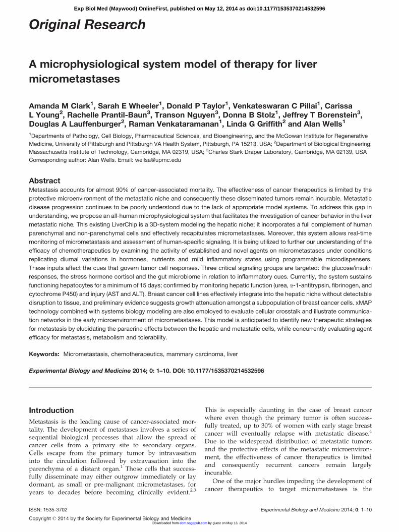
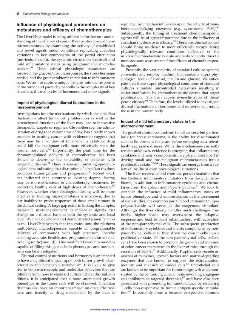
The liver architecture is suited for its diverse functionssuch as metabolism, detoxification, and regulation of multi-faceted defense mechanisms. It is composed of a complexassembly of highly specialized parenchymal andnon-parenchymal cell types (Figure 1). The parenchymalhepatocytes are responsible for most of the body’s meta-bolic functions along with the production of the majorityof the circulating plasma proteins, transporters, proteaseinhibitors, blood coagulation factors and immune modula-tors. The non-parenchymal cellular portion represents 20–40% of the liver and is primarily composed of liver sinus-oidal endothelial, Kupffer and stellate cells. The non-par-enchymal cells are essential components, releasingsubstances under both normal and pathological conditionsthat regulate many hepatocyte functions.21
As mentioned above, the liver is a major site for drugmetabolism. The metabolic processes generally involve con-verting drugs into more hydrophilic compounds in orderfor them to be excreted in the bodily fluids (e.g. urine or
Figure 1 Cellular composition of the liver. Highly specialized parenchymal and non-parenchymal cell types populate the liver. The hepatocytes form the parenchymal
portion, while the non-parenchymal portion comprises multiple cellular types, predominantly liver sinusoidal endothelial, Kupffer and stellate cells. (A color version of
this figure is available in the online journal.)
2 Experimental Biology and Medicine. . . . . . . . . . . . . . . . . . .. . . . . . . . . . . . . . . . . . .. . . . . . . . . . . . . . . . . . .. . . . . . . . . . . . . . . . . .. . . . . . . . . . . . . . . .. . . . . . . . . . . . . . . .. . . . . . . . . . . . . . .
by guest on May 13, 2014ebm.sagepub.comDownloaded from
XML Template (2014) [5.5.2014–12:33pm] [1–10]//blrnas3/cenpro/ApplicationFiles/Journals/SAGE/3B2/EBMJ/Vol00000/140060/APPFile/SG-EBMJ140060.3d (EBM) [PREPRINTER stage]
bile). The cytochrome P450 family of enzymes is primarilyresponsible for the metabolism of several essential chemo-therapy agents. There are more than 50 cytochrome P450enzymes with approximately 90% of drugs metabolizedthrough CYP3A4/5, CYP2C9, CYP2C19, CYP2D6 andCYP1A2.22 In particular, the CYP3A subfamily has beenidentified to play a predominant role in metabolizingchemotherapeutics.23,24
The liver is also one of the primary sites of systemicregulation of circadian rhythms via modulations of hor-mones, nutrients and inflammatory cytokines. Cell prolifer-ation and metabolic rhythms are regulated by circadiancycles, and often show asynchronies between normal andmalignant tissues.25 It is currently unknown how themolecular signals that change on a diurnal basis at boththe systemic and local level influence the complex micro-metastatic microenvironment. However, these circadianrhythms are known to alter the metabolic capabilities ofthe liver at the transcription, protein, and enzymaticlevels.26 Thus, it is reasonable to expect that such rhythmswould alter micrometastases and/or how chemotherapeu-tic agents are handled.
LiverChip microphysiological model
A major challenge in the field of hepatic tissue engineeringis the development of ex vivo and in vitro hepatic tissuesthat both exhibit a stable phenotype and maintain liver par-enchymal function. Further, investigating metastatic seed-ing and survival of cells requires examination over a periodof days-to-weeks. Numerous 2D and 3D in vitro liver-basedsystems have been developed and have been extensivelyreviewed.27 Extended evaluation, required for metastasisresearch, is not recreated in conventional 2D cultureswhich generally only remain viable and functional for <7days28; the addition of a second layer of collagen for a ‘sand-wich’ can extend hepatocyte functioning for longer in selectsettings.29,30 Additionally, dedifferentiation of hepatocytesin these standard 2D cultures is well established which sub-sequently causes a reduction in liver functions (e.g. down-regulation of enzymes and reduced production of plasmaproteins such as albumin).31–35 Numerous developments in
recent years, notably microfluidics and cell positioningtechniques, have overcome some of the aforementioned dis-advantages of conventional 2D cultures, primarily by con-trolling metabolite accumulation and non-steady-stateconditions allowing for extended culturing.36–38
For our investigations and as a possible approach goingforward, we utilize an all-human LiverChip microphysio-logical 3D system (Zyoxel, Ltd; Oxford, UK) that faithfullymodels both the hepatic niche and micrometastatic tumorcells.35 The liver bioreactor employed by our laboratory hasalready provided insight into the phenotypic plasticity ofboth breast and prostate carcinoma cells.39–42 Notably, thebioreactor milieu provides for a greater chemoresistancethat cannot be extrapolated from 2D studies.43 We continueto develop and improve this system as detailed below.A more detailed overview of the technical aspects of theLiverChip system and the bioengineering behind themodel is described in Wheeler et al.44
Functional hepatic niche
We aimed to replicate the fundamental physiologic func-tions and conditions of the hepatic niche, including multi-cellular composition, metabolism, protein production, andcircadian cycles (Figures 2 and 3(a) and (b)). The LiverChipbioreactor successfully recreates and maintains all-humanhepatic tissues with structural integrity and functional com-plexity for weeks in culture. It also provides adequate sam-ples for multiple downstream assays while avoidingmaterials that adsorb steroid hormones and drugs.The system incorporates fresh human hepatocytes and afull complement of non-parenchymal cells, at physiologic-ally relevant ratios. Cultures are maintained for a minimumof 15 days in defined, serum-free medium that supports celldifferentiation in culture and can be modified to reflect cir-cadian changes (i.e. insulin, glucagon, glucose, and corti-sol). The human hepatocytes and non-parenchymal cellsare sourced primarily from therapeutic partial hepatec-tomies for metastatic colorectal carcinoma or other benigndiseases such as focal nodular hyperplasia. Many of thetissue isolations are consequently from patients who previ-ously had chemotherapy for colorectal liver metastases.
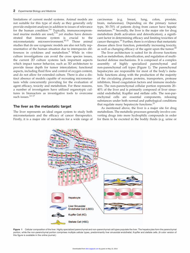
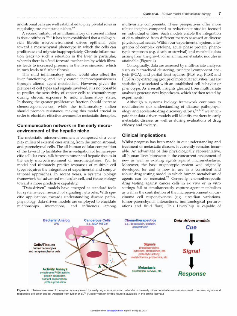
Figure 2 LiverChip experimental schematic to probe for function and tumor outgrowth. Day 0, the LiverChip system (Zyoxel, Ltd.) is seeded with hepatocytes and
non-parenchymal cells. Day 1, the medium is changed to serum-free maintenance medium. Day 3, the hepatic tissue has formed and is seeded with RFPþ breast
cancer cells (MCF-7 or MDA-MB-231). Cytochrome P450 activity is assessed on days 3 and 13. Medium is changed every two days and effluent samples taken
routinely. To monitor the influence of circadian rhythms, sampling frequency is increased to up to seven times per day. The hepatic niche is maintained for a minimum of
15 days. Assays to assess hepatocyte health and function, cytokine profiles as well as tissue morphology and cellular phenotype are performed on the tissue and
effluent samples. (A color version of this figure is available in the online journal.)
Clark et al. 3D liver model of metastasis therapy 3. . . . . . . . . . . . . . . . .. . . . . . . . . . . . . . . . . . .. . . . . . . . . . . . . . . . . . .. . . . . . . . . . . . . . . . . .. . . . . . . . . . . . . . . . . . .. . . . . . . . . . . . . . . .. . . . . . . . . . . . . .
by guest on May 13, 2014ebm.sagepub.comDownloaded from
XML Template (2014) [5.5.2014–12:33pm] [1–10]//blrnas3/cenpro/ApplicationFiles/Journals/SAGE/3B2/EBMJ/Vol00000/140060/APPFile/SG-EBMJ140060.3d (EBM) [PREPRINTER stage]
However, prior work has demonstrated that the viabilityand function of hepatocytes previously exposed to suchagents remains unaltered.45
A thorough characterization of isolated hepatocytes isessential to ensure that functionality is sustained duringculture. The investigation of morphology in combinationwith protein secretion, predominantly albumin, isfrequently used as verification of hepatocyte functionality.However, these parameters alone do not necessarily correl-ate with the existence of other hepatocyte-specific func-tions.46,47 Subsequently, a high level of importance andattention has been placed upon ensuring the health andfunctionality of the hepatic tissue in the LiverChip, and assuch a comprehensive set of biomarkers has been devel-oped (Figure 2). A key measurement of functionality forour system is active cytochrome P450 metabolism.Previously, we have developed and validated an assaymethod to simultaneously evaluate the activity of five dif-ferent cytochrome P450 enzymes with the rate of metabol-ism extrapolated as picomoles of substrate metabolized ormetabolite formed per minute per million cells.48
Functionality is further assessed through determininglevels of glucose consumption/production, urea catabolismas well as the secretion of positive acute phase proteins,such as a-1-antitrypsin (A1AT) and fibrinogen.
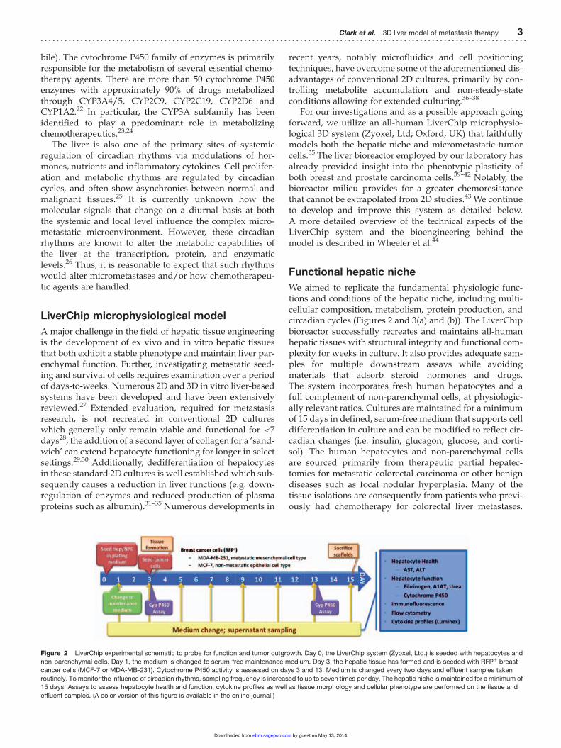
Maintained health is verified via the production of aspartatetransaminase (AST) and alanine aminotransferase (ALT),intracellular proteins released from hepatocytes duringtimes of injury and are monitored clinically. Importantly,all these measurements can be monitored in real-time overthe course of an experiment. A variety of imaging modal-ities are also employed for additional confirmation of hep-atocyte health and functionality, including confocal ormultiphoton assessment of post- immunolabeled reactorsfor viability (e.g. calcein AM/ethidium bromide, albumin),maintained presence of non-parenchymal cells (e.g. CD45,Lyve-1, CD68, desmin), cell–cell interactions (e.g. E-cad-herin, F-actin, a-smooth muscled actin), etc. Each reactorscaffold can be uniquely processed to collect a wide varietyof visual information. Some initial data pertaining to theseassays is presented in Figure 3b (i, ii, and iii).
The LiverChip system is continually being assessed forways to bring it closer to recapitulating an in vivo liver.Although the system allows for the generation of sufficienteffluent samples for the plethora of downstream assaysdescribed above, this comes with the limitation of requiringa considerable number of cells per well. Further, whilefunctionality is successfully achieved, we have not yet reca-pitulated liver structure within the present scaffoldingmatrices. The scaffolds are of a rigid nature which is
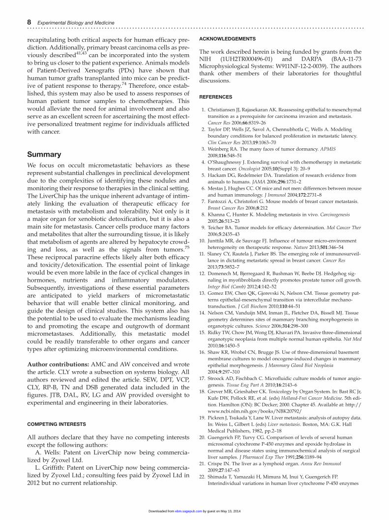
Figure 3 The LiverChip microphysiological system. (a) Aerial view of the LiverChip plate. Cells are seeded into the scaffold area, fluid flows up through this region
across the oxygenation channel and is then recirculated via the reservoir. (b) Functional and healthy hepatic tissue is formed and maintained for 15 days in the LiverChip.
i – Hepatocyte injury markers, AST and ALT, continue to decrease as tissue formation matured in the LiverChip system (n¼2 donors). Following isolation and seeding
into the system, the levels are high but as the cells establish tissue they drop to near undetectable levels. ii, iii – Hepatocytes still produce albumin (day 15) and maintain
cytochrome P450 activity (days 3 and 13) over the culture period (n¼2 donors). C) An array of microdispensers can be mounted over the LiverChip system providing for
precise delivery of drug, hormone and nutrient boluses as small as 100 nL. (d) When used in conjunction with custom-made software and electronic controllers,
programmable diurnal control of drug concentrations within the bioreactor can be achieved. (A color version of this figure is available in the online journal.)
4 Experimental Biology and Medicine. . . . . . . . . . . . . . . . . . .. . . . . . . . . . . . . . . . . . .. . . . . . . . . . . . . . . . . . .. . . . . . . . . . . . . . . . . .. . . . . . . . . . . . . . . .. . . . . . . . . . . . . . . .. . . . . . . . . . . . . . .
by guest on May 13, 2014ebm.sagepub.comDownloaded from
XML Template (2014) [5.5.2014–12:33pm] [1–10]//blrnas3/cenpro/ApplicationFiles/Journals/SAGE/3B2/EBMJ/Vol00000/140060/APPFile/SG-EBMJ140060.3d (EBM) [PREPRINTER stage]
associated with non-specific inflammatory responses in tis-sues.49 Investigations are currently underway which areperusing new functionalized matrices and scaffolding thatmore accurately reflect the rheology of the liver as well asaid in recreating liver architecture in vitro.
Recapitulating micrometastasis
In order to develop rational approaches to target growingcancer cells and promote clinically undetectable microme-tastases toward a dormant state, integrated in vitro systemsare needed that can support the initial micrometastatic nod-ules and follow such nodules for extended periods inculture.
The LiverChip, and other similar moderate-throughputorganotypic systems, offer the ability to evaluate the roleof the diversity of the human population in the microme-tastatic tumor microenvironment in an all-human systemin which the liver environment comes from individualpatients. We have previously characterized other bioreac-tor systems to examine the metastatic behaviors of breastand prostate cancer cells.39–42 These microphysiologicalsystems not only effectively recapitulate metastasis butalso concurrently allow for real-time monitoring of metas-tasis and assessment of human-specific signaling.Moreover, the system is evaluable over weeks, havingbeen tested through 30 days, with computer program-mable inputs or delivery of modifiers (e.g. hormones, pro-teins, metabolites, inhibitors) that allow one to definespecific required signals that either promote or hinderspecific metastatic properties.44 The main breast cancercell lines utilized are the well-characterized metastaticMDA-MB-231 and non-aggressive MCF-7 breast carcin-oma cell lines. In order to track and monitor tumorburden within the hepatic niche, the cell lines have beenmodified to express fluorescent labels.
Upon seeding individual carcinoma cells (the numberper well determined by cell concentration and numberintroduced into each unit) into engineered hepatic tissue,individual cells survive, form nodules, and can grow intomm-size tumor masses under the constant perfusion flow.A tissue is generated that comprises a hybrid of unaffectedhost tissue and tumor boundary, which is representative ofthe histological distinctions observed in human tumors.41,43
Importantly, the breast cancer cell lines, MDA-MB-231 andMCF-7, successfully integrate into and alongside thehepatic niche without detectable disruption to tissue.Markers of health (AST, ALT) and function (cytochromeP450 activity, glucose consumption, urea catabolism, andacute phase protein production) remain at levels compar-able to that of unburdened hepatic tissue (unpublisheddata). Furthermore, preliminary evidence suggests growthattenuation amongst a subpopulation of the highly prolif-erative and invasive MDA-MB-231 cell line when these cellsintercalate as individual cells within the tissue parenchyma(unpublished data).
Previous work also demonstrates that in the context ofthe 3D liver microbioreactor, breast carcinoma cellsundergo a phenotypic shift to a more epithelioid phenotypewith intimate connections to the hepatic cells.41,43 This was
noted not just with human cell lines, but also with primaryexplants from breast, prostate and melanoma.43 This pheno-type is often noted in small metastatic nodules of patients,wherein the metastases appear more differentiated than theprimary tumor.40,50–53 Furthermore, the reversion to a moreepithelioid phenotype in the MDA-MB-231 was associatedwith enhanced chemoresistance. This was observed thoughreduced cell death by chemotherapies in MDA-MB-231 cellsover-expressing E-cadherin compared to E-cadherin nega-tive MDA-MB-231 and MDA-MB-231-shEcad cells; thisprotection from death was abrogated by an E-cadherin anti-body. Conversely, driving the epithelial MCF-7 cells towarda proliferative mesenchymal phenotype increased theirsensitivity.43 The greater chemoresistance conferred byre-expression of E-cadherin mimics the resistance noted inpatients.40 The extent of resistance (as determined by LD50)is orders of magnitude greater for tumors grown in the 3Dliver system (unpublished data) compared to thosegrown in 2D cultures either with hepatocytes or withforced E-cadherin expression alone.43 This difference inchemotherapeutic efficacy between 3D and 2D models iswell established within the literature.54–56 Many critical sig-nals, regulators and the micrometastatic architecture are notpresent when cells are cultured under standard 2D condi-tions.57 The cellular architecture and growth conditionsrecreated by 3D cultures more accurately mimics thein vivo behavior of metastatic cancer cells and is key forthe accurate prediction of drug efficacy during the discov-ery process.
Evaluation and development of cancertherapies
The most challenging step in the development of therapiesfor metastasis relates to treating the small and pre-malig-nant micrometastases. Therapies are required which areeither directly toxic towards the proliferating metastaticcells or lock the clinically irrelevant micrometastases intheir dormant state. The different biology of the metastaticsite requires an understanding as to how the efficacy andtoxicity of chemotherapy is linked in the metastatic niche.This niche is affected directly by the molecular signalsand cellular behaviors of the cancer cells themselves aswell as the surrounding microenvironment, which is alsoaltered based upon circadian fluctuations of metabolitesand inflammatory mediators.
A fundamental element important to the investigatingthe efficacy of chemotherapies is the simple fact that thelevels of hormones, nutrients and immune regulators arenot static in nature, they fluctuate based the circadiancycles. Going forward, we therefore ask if circadian fluc-tuations that modulate metabolism and hormones alsoinfluence the efficacy and toxicity/detoxification of che-motherapeutic agents against metastatic tumors. Such ques-tions can be analyzed via the LiverChip as it intimatelylinks the efficacy and hepatic metabolism of therapeuticagents while concurrently controlling diurnal variationsas well as mild inflammatory states.
Clark et al. 3D liver model of metastasis therapy 5. . . . . . . . . . . . . . . . .. . . . . . . . . . . . . . . . . . .. . . . . . . . . . . . . . . . . . .. . . . . . . . . . . . . . . . . .. . . . . . . . . . . . . . . . . . .. . . . . . . . . . . . . . . .. . . . . . . . . . . . . .
by guest on May 13, 2014ebm.sagepub.comDownloaded from
XML Template (2014) [5.5.2014–12:33pm] [1–10]//blrnas3/cenpro/ApplicationFiles/Journals/SAGE/3B2/EBMJ/Vol00000/140060/APPFile/SG-EBMJ140060.3d (EBM) [PREPRINTER stage]
Influence of physiological parameters onmetastases and efficacy of chemotherapies
The LiverChip model is being utilized to further our under-standing of the efficacy of cancer therapeutics toward thesemicrometastases by examining the activity of establishedand novel agents under conditions replicating circadianvariations in key components of the portal circulation(nutrients, insulin), the systemic circulation (cortisol) andmild inflammatory states using programmable microdis-pensors.44 Three critical physiologic parameters areassessed: the glucose/insulin responses, the stress hormonecortisol and the gut microbiome in relation to inflammatorycues. We aim to capture unique insights into the responsesof the tumor and parenchymal cells to the complexity of keycircadian/diurnal cycles of hormones and other signals.
Impact of physiological diurnal fluctuations in themicroenvironment
Investigations into the mechanisms by which the circadianfluctuations affect tumor cell proliferation as well as theparenchymal functions of the liver may lead to innovativetherapeutic targets or regimes. Chronotherapy, the admin-istration of drugs at a certain time of day, has already shownpromise in treating cancer with evidence to suggest thatthere may be a window of time when a cytotoxic drugcould kill the malignant cells more effectively than thenormal host cells.26 Importantly, the peak time for thechronomodulated delivery of chemotherapy has beenshown to determine the tolerability of patients withmetastatic disease.58 There is also accumulating epidemio-logical data indicating that disruption of circadian rhythmspromotes tumorigenesis and progression.25 Recent workhas indicated that, contrary to existing dogma, fastingmay be more efficacious in chemotherapy treatments byprotecting healthy cells at high doses of chemotherapy.59
However, whether chronobiological dosing will be moreeffective in treating micrometastases is unknown, due toour inability to probe responses of these small tumors inthe clinical setting. A large gap exists in linking the complexmetastatic microenvironment to molecular signals thatchange on a diurnal basis at both the systemic and locallevel. We have developed and demonstrated a modificationto the LiverChip system that integrates onto the platformmultiplexed microdispensers capable of programmabledelivery of compounds with high precision, therebyenabling accurate, flexible and programmable diurnal con-trol (Figure 3(c) and (d)). This modified LiverChip model iscapable of filling this gap as both phenotypes and mechan-isms can be investigated.
Diurnal control of nutrients and hormones is anticipatedto have a significant impact upon both tumor growth char-acteristics and hepatocyte metabolism. It is likely to giverise to both macroscopic and molecular behaviors that aredifferent from those in standard culture. Under diurnal con-ditions, it is anticipated that a more attenuated growthphenotype in the tumor cells will be observed. Circadianrhythms also have an important impact on drug effective-ness and toxicity as drug metabolism by the liver is
regulated by circadian influences upon the activity of xeno-biotic-metabolizing enzymes (e.g. cytochrome P450).60
Subsequently, the timing of treatment chemotherapeuticagents will be of great importance due to the influence ofcircadian rhythms over efficacy.26 Therefore, diurnal controlshould bring us closer to more effectively recapitulatingphysiologically relevant conditions reflective of thein vivo micrometastatic nodule and subsequently direct amore accurate assessment of the efficacy of chemotherapeu-tic agents.
Presently, the vast majority of standard culture systemsconventionally employ medium that contains supra-phy-siological levels of cortisol, insulin and glucose. We antici-pate that these supra-physiological conditions of standardcultures stimulate uncontrolled metastases resulting ineasier eradication by chemotherapeutic agents that targetproliferation. This then causes overestimation of thera-peutic efficacy.61 Therefore, the levels utilized to investigatediurnal fluctuations in hormones and nutrients will mimicthose in the human body.
Impact of mild inflammatory states in themicroenvironment
The greatest clinical conundrum for all cancers, but particu-larly for breast carcinoma, is the ability for disseminatedcells to lie dormant for years before emerging as a relent-lessly aggressive disease. While the mechanisms currentlyremain unknown, evidence is emerging that inflammatorycytokines and matrix components may play at least a part indriving small and pre-malignant micrometastasis into aproliferative state.62–64 These signals can emerge from a var-iety of insults or even physiological situations.
The liver receives blood from the portal circulation thathas bacterial inflammatory initiators from the gut micro-biome, in addition to inflammatory cytokines and chemo-kines from the spleen and Peyer’s patches.21 We look toestablish the influence of mild inflammatory states ontumor phenotype and chemoresistance. In the assessmentof such studies, the common portal blood contaminant lipo-polysaccharide will serve as the exogenous stimulant.Although the liver clearly handles such challenges rou-tinely, higher loads may overwhelm the adaptiveresponse and lead to overt inflammation, with activationof the non-parenchymal cells. The subsequent productionof inflammatory cytokines and matrix components by non-parenchymal cells may then drive the cancer cells into aproliferative state. Of the non-parenchymal cells, stellatecells have been shown to promote the growth and invasionof colon cancer metastases in the liver of mice through thesecretion of SDF-1.65 Additionally, Kupffer cells secrete anarsenal of cytokines, growth factors and matrix-degradingenzymes that are known to support the extravasation,motility and invasion of cancer cells.66 Endothelial cellsare known to be important for tumor outgrowth as demon-strated by the continuing clinical trials involving angiogen-esis inhibitors as targeted therapies,67 and have also beenassociated with promoting immunotolerance by renderingT cells non-responsive to tumor antigen-specific stimula-tion.68 Importantly, from a broader perspective, immune
6 Experimental Biology and Medicine. . . . . . . . . . . . . . . . . . .. . . . . . . . . . . . . . . . . . .. . . . . . . . . . . . . . . . . . .. . . . . . . . . . . . . . . . . .. . . . . . . . . . . . . . . .. . . . . . . . . . . . . . . .. . . . . . . . . . . . . . .
by guest on May 13, 2014ebm.sagepub.comDownloaded from
XML Template (2014) [5.5.2014–12:33pm] [1–10]//blrnas3/cenpro/ApplicationFiles/Journals/SAGE/3B2/EBMJ/Vol00000/140060/APPFile/SG-EBMJ140060.3d (EBM) [PREPRINTER stage]
and stromal cells are well established to play pivotal roles inregulating pre-metastatic niches.69
A second initiator of an inflammatory or stressed milieuis tissue stiffness.30,70 It has been established that a collagen-rich fibrotic microenvironment drives epithelial cellstoward a mesenchymal phenotype in which the cells canproliferate and migrate inappropriately. Chronic inflamma-tion leads to such a situation in the liver in particular,wherein there is a feed-forward mechanism by which fibro-sis leads to increased pressure in the liver sinusoid, whichin turn leads to further fibrosis.
This mild inflammatory milieu would also affect theliver functioning, and likely cancer chemoresponsivenessthrough altered agent metabolism. However, given theplethora of cell types and signals involved, it is not possibleto predict the sensitivity of cancer cells to chemotherapyduring chronic exposure to mild inflammatory states.In theory, the greater proliferative fraction should increasechemoresponsiveness, while the inflammatory milieushould promote resistance, making this model crucial inorder to elucidate effective avenues for metastatic therapies.
Communication network in the early micro-environment of the hepatic niche
The metastatic microenvironment is composed of a com-plex milieu of external cues arising from the tumor, stromal,and parenchymal cells. The all-human cellular compositionof the LiverChip facilitates the investigation of human-spe-cific cellular cross-talk between tumor and hepatic tissues inthe early microenvironment of micrometastases. Yet, tomodel and ultimately predict responses of multiple celltypes requires the integration of experimental and compu-tational approaches. In recent years, a systems biologyframework has advanced molecular, cell, and tissue biologytoward a more predictive capability.
‘‘Data-driven’’ models have emerged as standard toolsfor systems-level research of signaling networks. With spe-cific applications towards understanding disease patho-physiology, data-driven models are employed to elucidaterelationships, interactions, and influences among
multivariate components. These perspectives offer morerobust insights compared to reductionist studies focusedon individual entities. Such models enable the integrationof data obtained from different metrics assessed at diversephysiological scales. Within our experimental system, inte-gration of complex cytokine, acute phase protein, pheno-typic responses (e.g. death or survival) and metabolic dataarising from the growth of small micrometastatic nodules isattainable (Figure 4).
Conceptually, data are assessed by multivariate analysessuch as hierarchical clustering, principal component ana-lysis (PCA), and partial least squares (PLS, e.g. PLSR andPLSDA) by extracting groups of molecular activities that arestatistically associated with an established cell behavior orphenotype. As a result, insights gleaned from multivariateanalyses generate new hypotheses, which are then tested byexperiments.
Although a systems biology framework continues torevolutionize our understanding of disease pathophysi-ology and accelerate drug discovery efforts,8,71,72 we antici-pate that data-driven models will identify markers in earlymetastatic disease, as well as during evaluations of drugefficacy and toxicity.
Clinical implications
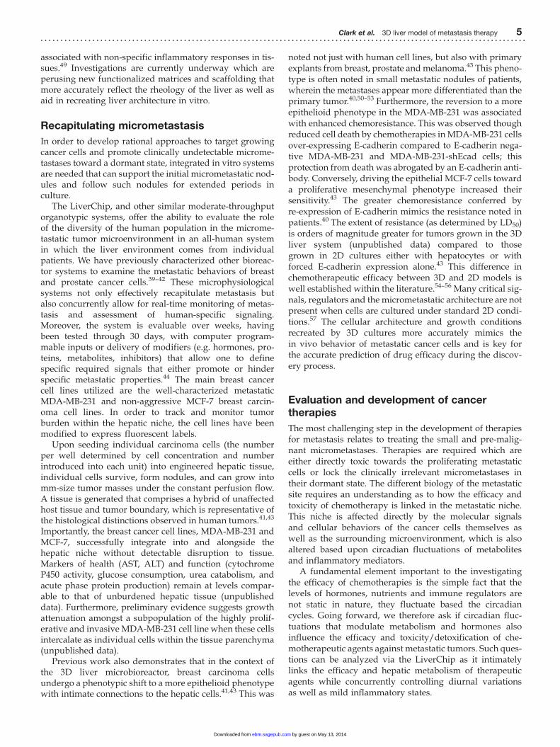
Whilst progress has been made in our understanding andtreatment of metastatic disease, it currently remains incur-able. An advantage of this physiologically representative,all-human liver bioreactor is the concurrent assessment ofnew as well as existing agents against micrometastases.Moreover, the base organotypic system was originallydeveloped for and is now in use as a consistent androbust drug testing model in which human metabolism ofagents can be recreated.73 Generally, chemotherapeuticdrug testing against cancer cells in ex vivo or in vitrosettings fail to simultaneously capture agent metabolismas well as the contribution of the microenvironment on car-cinoma cell responsiveness (e.g. circadian variations,tumor-parenchymal interactions, immunological perturb-ations and fluid flow). This LiverChip is capable of
Figure 4 General overview of the systematic approach for analyzing communication networks in the early micrometastatic microenvironment. The cues, signals and
responses are color coded. Adapted from Miller et al.76 (A color version of this figure is available in the online journal.)
Clark et al. 3D liver model of metastasis therapy 7. . . . . . . . . . . . . . . . .. . . . . . . . . . . . . . . . . . .. . . . . . . . . . . . . . . . . . .. . . . . . . . . . . . . . . . . .. . . . . . . . . . . . . . . . . . .. . . . . . . . . . . . . . . .. . . . . . . . . . . . . .
by guest on May 13, 2014ebm.sagepub.comDownloaded from
XML Template (2014) [5.5.2014–12:33pm] [1–10]//blrnas3/cenpro/ApplicationFiles/Journals/SAGE/3B2/EBMJ/Vol00000/140060/APPFile/SG-EBMJ140060.3d (EBM) [PREPRINTER stage]
recapitulating both critical aspects for human efficacy pre-diction. Additionally, primary breast carcinoma cells as pre-viously described41,43 can be incorporated into the systemto bring us closer to the patient experience. Animals modelsof Patient-Derived Xenografts (PDx) have shown thathuman tumor grafts transplanted into mice can be predict-ive of patient response to therapy.74 Therefore, once estab-lished, this system may also be used to assess responses ofhuman patient tumor samples to chemotherapies. Thiswould alleviate the need for animal involvement and alsoserve as an excellent screen for ascertaining the most effect-ive personalized treatment regime for individuals afflictedwith cancer.
Summary
We focus on occult micrometastatic behaviors as theserepresent substantial challenges in preclinical developmentdue to the complexities of identifying these nodules andmonitoring their response to therapies in the clinical setting.The LiverChip has the unique inherent advantage of intim-ately linking the evaluation of therapeutic efficacy formetastasis with metabolism and tolerability. Not only is ita major organ for xenobiotic detoxification, but it is also amain site for metastasis. Cancer cells produce many factorsand metabolites that alter the surrounding tissue, it is likelythat metabolism of agents are altered by hepatocyte crowd-ing and loss, as well as the signals from tumors.75
These reciprocal paracrine effects likely alter both efficacyand toxicity/detoxification. The essential point of linkagewould be even more labile in the face of cyclical changes inhormones, nutrients and inflammatory modulators.Subsequently, investigations of these essential parametersare anticipated to yield markers of micrometastaticbehavior that will enable better clinical monitoring, andguide the design of clinical studies. This system also hasthe potential to be used to evaluate the mechanisms leadingto and promoting the escape and outgrowth of dormantmicrometastases. Additionally, this metastatic modelcould be readily transferable to other organs and cancertypes after optimizing microenvironmental conditions.
Author contributions: AMC and AW conceived and wrotethe article. CLY wrote a subsection on systems biology. Allauthors reviewed and edited the article. SEW, DPT, VCP,CLY, RP-B, TN and DSB generated data included in thefigures. JTB, DAL, RV, LG and AW provided oversight toexperimental and engineering in their laboratories.
COMPETING INTERESTS
All authors declare that they have no competing interestsexcept the following authors:
A. Wells: Patent on LiverChip now being commercia-lized by Zyoxel Ltd.
L. Griffith: Patent on LiverChip now being commercia-lized by Zyoxel Ltd.; consulting fees paid by Zyoxel Ltd in2012 but no current relationship.
ACKNOWLEDGEMENTS
The work described herein is being funded by grants from theNIH (1UH2TR000496-01) and DARPA (BAA-11-73Microphysiological Systems: W911NF-12-2-0039). The authorsthank other members of their laboratories for thoughtfuldiscussions.
REFERENCES
1. Christiansen JJ, Rajasekaran AK. Reassessing epithelial to mesenchymal
transition as a prerequisite for carcinoma invasion and metastasis.
Cancer Res 2006;66:8319–26
2. Taylor DP, Wells JZ, Savol A, Chennubhotla C, Wells A. Modeling
boundary conditions for balanced proliferation in metastatic latency.
Clin Cancer Res 2013;19:1063–70
3. Weinberg RA. The many faces of tumor dormancy. APMIS2008;116:548–51
4. O’Shaughnessy J. Extending survival with chemotherapy in metastatic
breast cancer. Oncologist 2005;10(Suppl 3): 20–9
5. Hackam DG, Redelmeier DA. Translation of research evidence from
animals to humans. JAMA 2006;296:1731–2
6. Mestas J, Hughes CC. Of mice and not men: differences between mouse
and human immunology. J Immunol 2004;172:2731–8
7. Fantozzi A, Christofori G. Mouse models of breast cancer metastasis.
Breast Cancer Res 2006;8:212
8. Khanna C, Hunter K. Modeling metastasis in vivo. Carcinogenesis2005;26:513–23
9. Teicher BA. Tumor models for efficacy determination. Mol Cancer Ther2006;5:2435–43
10. Junttila MR, de Sauvage FJ. Influence of tumour micro-environment
heterogeneity on therapeutic response. Nature 2013;501:346–54
11. Slaney CY, Rautela J, Parker BS. The emerging role of immunosurveil-
lance in dictating metastatic spread in breast cancer. Cancer Res2013;73:5852–7
12. Domenech M, Bjerregaard R, Bushman W, Beebe DJ. Hedgehog sig-
naling in myofibroblasts directly promotes prostate tumor cell growth.
Integr Biol (Camb) 2012;4:142–52
13. Gomez EW, Chen QK, Gjorevski N, Nelson CM. Tissue geometry pat-
terns epithelial-mesenchymal transition via intercellular mechano-
transduction. J Cell Biochem 2010;110:44–51
14. Nelson CM, Vanduijn MM, Inman JL, Fletcher DA, Bissell MJ. Tissue
geometry determines sites of mammary branching morphogenesis in
organotypic cultures. Science 2006;314:298–300
15. Ridky TW, Chow JM, Wong DJ, Khavari PA. Invasive three-dimensional
organotypic neoplasia from multiple normal human epithelia. Nat Med2010;16:1450–5
16. Shaw KR, Wrobel CN, Brugge JS. Use of three-dimensional basement
membrane cultures to model oncogene-induced changes in mammary
epithelial morphogenesis. J Mammary Gland Biol Neoplasia2004;9:297–310
17. Stroock AD, Fischbach C. Microfluidic culture models of tumor angio-
genesis. Tissue Eng Part A 2010;16:2143–6
18. Grever MR, Grieshaber CK. Toxicology by Organ System. In: Bast RC Jr,
Kufe DW, Pollock RE, et al. (eds) Holland-Frei Cancer Medicine. 5th edi-
tion. Hamilton (ON): BC Decker; 2000. Chapter 45. Available at: http://
www.ncbi.nlm.nih.gov/books/NBK20792/
19. Pickren J, Tsukada Y, Lane W. Liver metastasis: analysis of autopsy data.
In: Weiss L, Gilbert L (eds) Liver metastasis. Boston, MA: G.K. Hall
Medical Publishers, 1982, pp.2–18
20. Guengerich FP, Turvy CG. Comparison of levels of several human
microsomal cytochrome P-450 enzymes and epoxide hydrolase in
normal and disease states using immunochemical analysis of surgical
liver samples. J Pharmacol Exp Ther 1991;256:1189–94
21. Crispe IN. The liver as a lymphoid organ. Annu Rev Immunol2009;27:147–63
22. Shimada T, Yamazaki H, Mimura M, Inui Y, Guengerich FP.
Interindividual variations in human liver cytochrome P-450 enzymes
8 Experimental Biology and Medicine. . . . . . . . . . . . . . . . . . .. . . . . . . . . . . . . . . . . . .. . . . . . . . . . . . . . . . . . .. . . . . . . . . . . . . . . . . .. . . . . . . . . . . . . . . .. . . . . . . . . . . . . . . .. . . . . . . . . . . . . . .
by guest on May 13, 2014ebm.sagepub.comDownloaded from
XML Template (2014) [5.5.2014–12:33pm] [1–10]//blrnas3/cenpro/ApplicationFiles/Journals/SAGE/3B2/EBMJ/Vol00000/140060/APPFile/SG-EBMJ140060.3d (EBM) [PREPRINTER stage]
involved in the oxidation of drugs, carcinogens and toxic chemicals:
studies with liver microsomes of 30 Japanese and 30 Caucasians.
J Pharmacol Exp Ther 1994;270:414–23
23. Slaughter RL, Edwards DJ. Recent advances: the cytochrome P450
enzymes. Ann Pharmacother 1995;29:619–24
24. Wilkinson GR. Drug metabolism and variability among patients in drug
response. N Engl J Med 2005;352:2211–21
25. Fu L, Lee CC. The circadian clock: pacemaker and tumour suppressor.
Nat Rev Cancer 2003;3:350–61
26. Levi F, Schibler U. Circadian rhythms: mechanisms and therapeutic
implications. Annu Rev Pharmacol Toxicol 2007;47:593–28
27. Godoy P, Hewitt NJ, Albrecht U, Andersen ME, Ansari N,
Bhattacharya S, Bode JG, Bolleyn J, Borner C, Bottger J, Braeuning A,
Budinsky RA, Burkhardt B, Cameron NR, Camussi G, Cho CS, Choi YJ,
Craig Rowlands J, Dahmen U, Damm G, Dirsch O, Donato MT, Dong J,
Dooley S, Drasdo D, Eakins R, Ferreira KS, Fonsato V, Fraczek J,
Gebhardt R, Gibson A, Glanemann M, Goldring CE, Gomez-Lechon MJ,
Groothuis GM, Gustavsson L, Guyot C, Hallifax D, Hammad S,
Hayward A, Haussinger D, Hellerbrand C, Hewitt P, Hoehme S,
Holzhutter HG, Houston JB, Hrach J, Ito K, Jaeschke H, Keitel V,
Kelm JM, Kevin Park B, Kordes C, Kullak-Ublick GA, LeCluyse EL,
Lu P, Luebke-Wheeler J, Lutz A, Maltman DJ, Matz-Soja M, McMullen P,
Merfort I, Messner S, Meyer C, Mwinyi J, Naisbitt DJ, Nussler AK,
Olinga P, Pampaloni F, Pi J, Pluta L, Przyborski SA, Ramachandran A,
Rogiers V, Rowe C, Schelcher C, Schmich K, Schwarz M, Singh B,
Stelzer EH, Stieger B, Stober R, Sugiyama Y, Tetta C, Thasler WE,
Vanhaecke T, Vinken M, Weiss TS, Widera A, Woods CG, Xu JJ,
Yarborough KM, Hengstler JG. Recent advances in 2D and 3D in vitro
systems using primary hepatocytes, alternative hepatocyte sources and
non-parenchymal liver cells and their use in investigating mechanisms
of hepatotoxicity, cell signaling and ADME. Arch Toxicol
2013;87:1315–30
28. Debnath J, Brugge JS. Modelling glandular epithelial cancers in three-
dimensional cultures. Nat Rev Cancer 2005;5:675–88
29. Dunn JC, Tompkins RG, Yarmush ML. Long-term in vitro function of
adult hepatocytes in a collagen sandwich configuration. Biotechnol Prog
1991;7:237–45
30. Khetani SR, Bhatia SN. Microscale culture of human liver cells for drug
development. Nat Biotechnol 2008;26:120–6
31. Bissell DM, Arenson DM, Maher JJ, Roll FJ. Support of cultured
hepatocytes by a laminin-rich gel. Evidence for a functionally signifi-
cant subendothelial matrix in normal rat liver. J Clin Invest
1987;79:801–12
32. Clayton DF, Harrelson AL, Darnell JE Jr. Dependence of liver-specific
transcription on tissue organization. Mol Cell Biol 1985;5:2623–32
33. Godoy P, Hengstler JG, Ilkavets I, Meyer C, Bachmann A, Muller A,
Tuschl G, Mueller SO, Dooley S. Extracellular matrix modulates sensi-
tivity of hepatocytes to fibroblastoid dedifferentiation and transforming
growth factor beta-induced apoptosis. Hepatology 2009;49:2031–43
34. Koide N, Shinji T, Tanabe T, Asano K, Kawaguchi M, Sakaguchi K,
Koide Y, Mori M, Tsuji T. Continued high albumin production by
multicellular spheroids of adult rat hepatocytes formed in the presence
of liver-derived proteoglycans. Biochem Biophys Res Commun
1989;161:385–91
35. Tong JZ, De Lagausie P, Furlan V, Cresteil T, Bernard O, Alvarez F. Long-
term culture of adult rat hepatocyte spheroids. Exp Cell Res
1992;200:326–32
36. Takeshita K, Bowen WC, Michalopoulos GK. Three-dimensional culture
of hepatocytes in a continuously flowing medium. In Vitro Cell Dev Biol
Anim 1998;34:482–85
37. Allen JW, Khetani SR, Bhatia SN. In vitro zonation and toxicity in a
hepatocyte bioreactor. Toxicol Sci 2005;84:110–19
38. Novik E, Maguire TJ, Chao P, Cheng KC, Yarmush ML. A microfluidic
hepatic coculture platform for cell-based drug metabolism studies.
Biochem Pharmacol 2010;79:1036–44
39. Chao Y, Wu Q, Acquafondata M, Dhir R, Wells A. Partial mesenchymal
to epithelial reverting transition in breast and prostate cancer metas-
tases. Cancer Microenviron 2012;5:19–28
40. Chao YL, Shepard CR, Wells A. Breast carcinoma cells re-express
E-cadherin during mesenchymal to epithelial reverting transition. MolCancer 2010;9:179
41. Yates C, Shepard CR, Papworth G, Dash A, Beer Stolz D, Tannenbaum S,
Griffith L, Wells A. Novel three-dimensional organotypic liver bio-
reactor to directly visualize early events in metastatic progression. AdvCancer Res 2007;97:225–46
42. Yates CC, Shepard CR, Stolz DB, Wells A. Co-culturing human prostate
carcinoma cells with hepatocytes leads to increased expression of
E-cadherin. Br J Cancer 2007;96:1246–52
43. Chao Y, Wu Q, Shepard C, Wells A. Hepatocyte induced re-expression
of E-cadherin in breast and prostate cancer cells increases chemoresis-
tance. Clin Exp Metastasis 2012;29:39–50
44. Wheeler S, Borenstein J, Clark AM, Ebrahimkhani M, Fox IJ, Griffith L,
Inman W, Lauffenburger DA, Nguyen T, Pillai VC, Prantil-Baun R, Stolz
DB, Taylor DP, Ulrich T, Venkataramanan R, Wells A, Young C. All-
human microphysical model of metastasis therapy. Stem Cell Res Ther2013;4(Supp 1):S11
45. Hewes JC, Riddy D, Morris RW, Woodrooffe AJ, Davidson BR, Fuller B.
A prospective study of isolated human hepatocyte function following
liver resection for colorectal liver metastases: the effects of prior
exposure to chemotherapy. J Hepatol 2006;45:263–70
46. Hengstler JG, Brulport M, Schormann W, Bauer A, Hermes M,
Nussler AK, Fandrich F, Ruhnke M, Ungefroren H, Griffin L,
Bockamp E, Oesch F, von Mach MA. Generation of human hepatocytes
by stem cell technology: definition of the hepatocyte. Expert Opin DrugMetab Toxicol 2005;1:61–74
47. Knobeloch D, Ehnert S, Schyschka L, Buchler P, Schoenberg M, Kleeff J,
Thasler WE, Nussler NC, Godoy P, Hengstler J, Nussler AK. Human
hepatocytes: isolation, culture, and quality procedures. Methods Mol Biol2012;806:99–120
48. Pillai VC, Strom SC, Caritis SN, Venkataramanan R. A sensitive and
specific CYP cocktail assay for the simultaneous assessment of human
cytochrome P450 activities in primary cultures of human hepatocytes
using LC-MS/MS. J Pharm Biomed Anal 2013;74:126–32
49. Wells RG. The role of matrix stiffness in regulating cell behavior.
Hepatology 2008;47:1394–1400
50. Chaffer CL, Brennan JP, Slavin JL, Blick T, Thompson EW, Williams ED.
Mesenchymal-to-epithelial transition facilitates bladder cancer metas-
tasis: role of fibroblast growth factor receptor-2. Cancer Res2006;66:11271–8
51. Kowalski PJ, Rubin MA, Kleer CG. E-cadherin expression in primary
carcinomas of the breast and its distant metastases. Breast Cancer Res2003;5:R217–222
52. Tarin D, Thompson EW, Newgreen DF. The fallacy of epithelial mes-
enchymal transition in neoplasia. Cancer Res 2005;65:5996–6000.
(discussion 6000-5991)
53. Wells A, Yates C, Shepard CR. E-cadherin as an indicator of mesen-
chymal to epithelial reverting transitions during the metastatic seeding
of disseminated carcinomas. Clin Exp Metastasis 2008;25:621–8
54. Horning JL, Sahoo SK, Vijayaraghavalu S, Dimitrijevic S, Vasir JK,
Jain TK, Panda AK, Labhasetwar V. 3-D tumor model for in vitro
evaluation of anticancer drugs. Mol Pharm 2008;5:849–62
55. Mitra M, Mohanty C, Harilal A, Maheswari UK, Sahoo SK,
Krishnakumar S. A novel in vitro three-dimensional retinoblastoma
model for evaluating chemotherapeutic drugs. Mol Vis 2012;18:1361–78
56. Sung JH, Shuler ML. A micro cell culture analog (microCCA) with 3-D
hydrogel culture of multiple cell lines to assess metabolism-dependent
cytotoxicity of anti-cancer drugs. Lab Chip 2009;9:1385–94
57. Lee GY, Kenny PA, Lee EH, Bissell MJ. Three-dimensional culture
models of normal and malignant breast epithelial cells. Nat Methods2007;4:359–65
58. Levi F, Focan C, Karaboue A, de la Valette V, Focan-Henrard D, Baron B,
Kreutz F, Giacchetti S. Implications of circadian clocks for the rhythmic
delivery of cancer therapeutics. Adv Drug Deliv Rev 2007;59:1015–35
59. Lee C, Raffaghello L, Brandhorst S, Safdie FM, Bianchi G, Martin-
Montalvo A, Pistoia V, Wei M, Hwang S, Merlino A, Emionite L, de
Cabo R, Longo VD. Fasting cycles retard growth of tumors and sensitize
a range of cancer cell types to chemotherapy. Sci Transl Med 2012;4:124–7
Clark et al. 3D liver model of metastasis therapy 9. . . . . . . . . . . . . . . . .. . . . . . . . . . . . . . . . . . .. . . . . . . . . . . . . . . . . . .. . . . . . . . . . . . . . . . . .. . . . . . . . . . . . . . . . . . .. . . . . . . . . . . . . . . .. . . . . . . . . . . . . .
by guest on May 13, 2014ebm.sagepub.comDownloaded from
XML Template (2014) [5.5.2014–12:33pm] [1–10]//blrnas3/cenpro/ApplicationFiles/Journals/SAGE/3B2/EBMJ/Vol00000/140060/APPFile/SG-EBMJ140060.3d (EBM) [PREPRINTER stage]
60. Ohdo S. Circadian rhythms in the CNS and peripheral clock disorders:
chronopharmacological findings on antitumor drugs. J Pharmacol Sci2007;103:155–8
61. Bernabe DG, Tamae AC, Biasoli ER, Oliveira SH. Stress hormones
increase cell proliferation and regulates interleukin-6 secretion in
human oral squamous cell carcinomas. Brain Behv Immun2011;25:574–83
62. Aguirre-Ghiso JA. Models, mechanisms and clinical evidence for cancer
dormancy. Nat Rev Cancer 2007;7:834–46
63. Barkan D, El Touny LH, Michalowski AM, Smith JA, Chu I, Davis AS,
Webster JD, Hoover S, Simpson RM, Gauldie J, Green JE. Metastatic
growth from dormant cells induced by a col-I-enriched fibrotic envir-
onment. Cancer Res 2010;70:5706–16
64. Wendt MK, Taylor MA, Schiemann BJ, Schiemann WP. Down-regula-
tion of epithelial cadherin is required to initiate metastatic outgrowth of
breast cancer. Mol Biol Cell 2011;22:2423–35
65. Matsusue R, Kubo H, Hisamori S, Okoshi K, Takagi H, Hida K,
Nakano K, Itami A, Kawada K, Nagayama S, Sakai Y. Hepatic stellate
cells promote liver metastasis of colon cancer cells by the action of SDF-
1/CXCR4 axis. Ann Surg Oncol 2009;16:2645–53
66. Paschos KA, Majeed AW, Bird NC. Role of Kupffer cells in the
outgrowth of colorectal cancer liver metastases. Hepatol Res2010;40:83–94
67. Gao D, Nolan DJ, Mellick AS, Bambino K, McDonnell K, Mittal V.
Endothelial progenitor cells control the angiogenic switch in mouse
lung metastasis. Science 2008;319:195–8
68. Hochst B, Schildberg FA, Bottcher J, Metzger C, Huss S, Turler A,
Overhaus M, Knoblich A, Schneider B, Pantelis D, Kurts C, Kalff JC,
Knolle P, Diehl L. Liver sinusoidal endothelial cells contribute to CD8 T
cell tolerance toward circulating carcinoembryonic antigen in mice.
Hepatology 2012;56:1924–33
69. Sceneay J, Smyth MJ, Moller A. The pre-metastatic niche: finding
common ground. Cancer Metastasis Rev 2013;32:449–64
70. Erler JT, Weaver VM. Three-dimensional context regulation of metas-
tasis. Clin Exp Metastasis 2009;26:35–49
71. Ideker T, Winslow LR, Lauffenburger AD. Bioengineering and systems
biology. Ann Biomed Eng 2006;34:257–64
72. Joughin BA, Naegle KM, Huang PH, Yaffe MB, Lauffenburger DA,
White FM. An integrated comparative phosphoproteomic and bio-
informatic approach reveals a novel class of MPM-2 motifs upregulated
in EGFRvIII-expressing glioblastoma cells. Mol Biosyst 2009;5:59–67
73. Dash A, Inman W, Hoffmaster K, Sevidal S, Kelly J, Obach RS,
Griffith LG, Tannenbaum SR. Liver tissue engineering in the evaluation
of drug safety. Expert Opin Drug Metab Toxicol 2009;5:1159–74
74. Siolas D, Hannon GJ. Patient-derived tumor xenografts: transforming
clinical samples into mouse models. Cancer Res 2013;73:5315–19
75. Hirschhaeuser F, Sattler UG, Mueller-Klieser W. Lactate: a metabolic
key player in cancer. Cancer Res 2011;71:692125
76. Miller MA, Barkal L, Jeng K, Herrlich A, Moss M, Griffith LG,
Lauffenburger DA. Proteolytic Activity Matrix Analysis (PrAMA) for
simultaneous determination of multiple protease activities. Integr Biol(Camb) 2011;3:422–38
10 Experimental Biology and Medicine. . . . . . . . . . . . . . . . . . .. . . . . . . . . . . . . . . . . . .. . . . . . . . . . . . . . . . . . .. . . . . . . . . . . . . . . . . .. . . . . . . . . . . . . . . .. . . . . . . . . . . . . . . .. . . . . . . . . . . . . . .
by guest on May 13, 2014ebm.sagepub.comDownloaded from