Inducible mouse models illuminate parameters influencing epigenetic inheritance
Upload
independentCategory
view
2download
0
Stem Cell Reports
ArticleDetailed Analysis of the Genetic and Epigenetic Signatures of iPSC-DerivedMesodiencephalic Dopaminergic Neurons
Reinhard Roessler,1,8 Sebastien A. Smallwood,2,7 Jesse V. Veenvliet,3,7 Petros Pechlivanoglou,4,9
Su-Ping Peng,1 Koushik Chakrabarty,6 Marian J.A. Groot-Koerkamp,5 R. Jeroen Pasterkamp,6
Evelyn Wesseling,1 Gavin Kelsey,2 Erik Boddeke,1 Marten P. Smidt,3 and Sjef Copray1,*1Department of Neuroscience, Section Medical Physiology, University Medical Center Groningen, 9713AV Groningen, the Netherlands2Epigenetics Programme, The Babraham Institute, Cambridge CB22 3AT, UK3Center for Neuroscience, Swammerdam Institute for Life Science, Science Park Amsterdam, 1098XH Amsterdam, the Netherlands4Unit of Pharmacoepidemiology and Pharmacoeconomics, Department of Pharmacy, University of Groningen, 9713AV Groningen, the Netherlands5Molecular Cancer Research, University Medical Center Utrecht, Universiteitsweg 100, 3584 CG Utrecht, the Netherlands6Department of Neuroscience and Pharmacology, Rudolf Magnus Institute of Neuroscience, University Medical Center Utrecht, Universiteitsweg 100,
3584 CG Utrecht, the Netherlands7These authors contributed equally to this work8Present address: Whitehead Institute for Biomedical Research, 9 Cambridge Center, Cambridge, MA 02142, USA9Present address: Toronto Health Economics and Technology Assessment (THETA), University of Toronto, Toronto ON M5S 3M2, Canada
*Correspondence: [email protected]
http://dx.doi.org/10.1016/j.stemcr.2014.03.001
This is an open access article under the CC BY license (http://creativecommons.org/licenses/by/3.0/).
SUMMARY
Induced pluripotent stem cells (iPSCs) hold great promise for in vitro generation of disease-relevant cell types, such as mesodiencephalic
dopaminergic (mdDA) neurons involved in Parkinson’s disease. Although iPSC-derived midbrain DA neurons have been generated,
detailed genetic and epigenetic characterizations of such neurons are lacking. The goal of this study was to examine the authenticity
of iPSC-derived DA neurons obtained by established protocols. We FACS purified mdDA (Pitx3Gfp/+) neurons derived from mouse iPSCs
and primarymdDA (Pitx3Gfp/+) neurons to analyze and compare their genetic and epigenetic features. Although iPSC-derivedDAneurons
largely adopted characteristics of their in vivo counterparts, relevant deviations in global gene expression and DNA methylation were
found. Hypermethylated genes, mainly involved in neurodevelopment and basic neuronal functions, consequently showed reduced
expression levels. Such abnormalities should be addressed because they might affect unambiguous long-term functionality and hamper
the potential of iPSC-derived DA neurons for in vitro disease modeling or cell-based therapy.
INTRODUCTION
The field of regenerative medicine experienced a powerful
impetus after the groundbreaking discovery of induced plu-
ripotency (Takahashi andYamanaka, 2006).Numerouspub-
lications have shown that mouse as well as human induced
pluripotent stem cells (iPSCs) have the potency to differ-
entiate into various clinically relevant cell types, such as
cardiomyocytes (Kuzmenkin et al., 2009; Ren et al., 2011),
hepatocytes (Espejel et al., 2010), hematopoietic progeni-
tors (Hanna et al., 2007), oligodendrocytes (Czepiel et al.,
2011), and specific subtypes of neurons (Karumbayaram
et al., 2009; Wernig et al., 2008). Such in vitro-generated
iPSC-derived cell types provide new possibilities for disease
modeling and cell replacement strategies. In particular, the
generation of autologous iPSC-derived midbrain dopami-
nergic (DA)neuronsprovides a very interesting tool to study
and treat Parkinson’s disease (PD) (Roessler et al., 2013).
However, future clinical application of iPSC-derived DA
neurons can only be considered realistic if the desired cell
population is strictly purified and completely defined.
Several groups have reported the generation of DA neu-
rons from mouse and human iPSCs (Hargus et al., 2010;
520 Stem Cell Reports j Vol. 2 j 520–533 j April 8, 2014 j ª2014 The Author
Swistowski et al., 2010; Wernig et al., 2008). In these
studies, iPSC-derived neurons displayed expression of
crucial DA markers and exhibited typical neuronal electro-
physiological properties. Furthermore, these iPSC-derived
DA neurons could functionally integrate into a rat PD
model upon transplantation. In principle, these results
indicated that a DA neuronal population could be obtained
from iPSCs and present important midbrain DA neuronal
characteristics. However, genome-wide studies comparing
the genetic and epigenetic features of iPSC-derived DAneu-
rons versus primary DA neurons are currently lacking.
Since reprogramming of somatic cells to iPSCs resets their
identity back to an embryonic stage, iPSC-derived differen-
tiated neurons should be considered freshly formed ‘‘em-
bryonic’’ neurons. Accordingly, a relevant comprehensive
comparison of iPSC-derived DA neurons can only be
donewith freshly formed embryonic and perinatal primary
mesodiencephalic DA (mdDA) neurons.
We generated iPSC lines from Pitx3Gfp/+ knockin mouse
embryonic fibroblasts. PITX3 is a highly specific mdDA
neuron marker that is required for DA neuron differentia-
tion in the substantia nigra (Jacobs et al., 2009, 2011; Smidt
et al., 2004). Specific PITX3-associated GFP expression
s
Stem Cell ReportsComprehensive Profiling of iPSC-Derived DA Neurons
allowed us to strictly identify and purify DA neurons from
either iPSCs or the ventral midbrain at specific devel-
opmental stages by fluorescence-activated cell sorting
(FACS). We then subjected these mdDA neurons to
genome-wide gene-expression analysis comparing iPSC-
derived DA neurons and primary isolated mdDA neurons.
Induction of pluripotency in somatic cells is considered
an epigenetic process that entails, among other events, a
large series of changes in DNA methylation patterns
(Bock et al., 2011; Maherali et al., 2007; Nishino et al.,
2011; Ohi et al., 2011). Furthermore, iPSC differentiation
into neural progenitor cells and subsequently into a spe-
cific neuronal subtype depends on properly established
de novo DNA methylation (Lee et al., 2010; Watanabe
et al., 2006). Therefore, it is essential not only to evaluate
gene expression but also to compare the methylome of
iPSC-derived DA neurons with their primary counterparts.
In order to obtain a comprehensive profile of the function-
ally most relevant DNA methylation sites in iPSC-derived
DA neurons versus primary mdDA neurons, we performed
a genome-wide analysis of CpG island (CGI) methylation
using reduced representation bisulfite sequencing (RRBS)
(Meissner et al., 2005; Smallwood et al., 2011) on genomic
DNA samples isolated from purified neuronal populations.
RESULTS
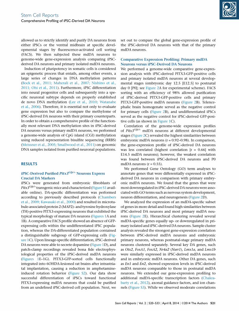
iPSC-Derived Purified Pitx3Gfp/+ Neurons Express
Crucial DA Markers
iPSCs were generated from embryonic fibroblasts of
Pitx3Gfp/+ transgenicmice andcharacterized (Figure S1avail-
able online). DA-specific differentiation was performed
according to previously described protocols (Chambers
et al., 2009; Kawasaki et al., 2000) and resulted in microtu-
bule-associatedprotein2 (MAP2)- and tyrosinehydroxylase
(TH)-positive PITX3-expressing neurons that exhibited the
typical morphology of mature DA neurons (Figures 1A and
1B). A comparative FACS profile showed an absence of GFP-
expressing cells within the undifferentiated iPSC popula-
tion, whereas the DA-differentiated population contained
a distinguishable subgroup of GFP-expressing cells (Fig-
ure 1C). Upon lineage-specific differentiation, iPSC-derived
DA neurons were able to secrete dopamine (Figure 1D), and
patch-clamp recordings revealed bona fide electrophys-
iological properties of the iPSC-derived mdDA neurons
(Figures 1E–1G). PITX3-GFP-sorted cells functionally
integrated into 6OHDA-lesioned rat brains after intrastria-
tal implantation, causing a reduction in amphetamine-
induced rotation behavior (Figure S2). Our data show
successful differentiation of iPSCs toward functional
PITX3-expressing mdDA neurons that could be purified
from an undefined iPSC-derived cell population. Next, we
Stem
set out to compare the global gene-expression profile of
the iPSC-derived DA neurons with that of the primary
mdDA neurons.
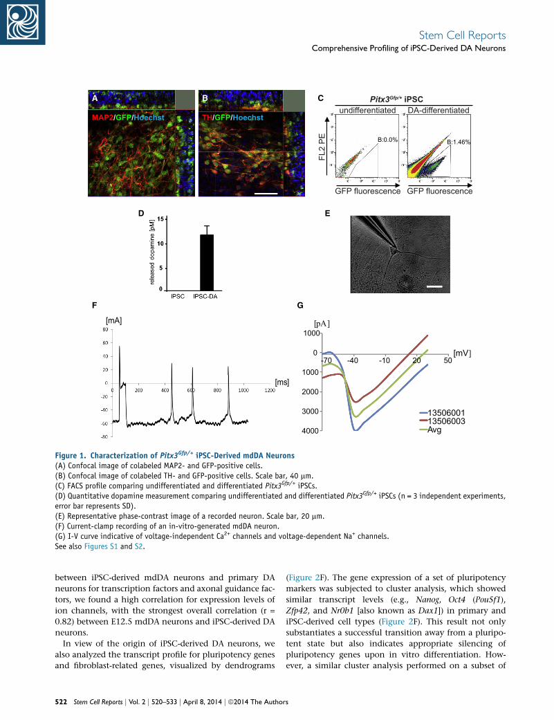
Comparative Expression Profiling: Primary mdDA
Neurons versus iPSC-Derived DA Neurons
We performed a genome-wide comparative gene-expres-
sion analysis with iPSC-derived PITX3-GFP-positive cells
and primary isolated mdDA neurons at several develop-
mental stages (embryonic day 12.5 [E12.5] to postnatal
day 0 [P0]; see Figure 2A for experimental scheme). FACS
sorting with an efficiency of 98% allowed purification
of iPSC-derived PITX3-GFP-positive cells and primary
PITX3-GFP-positive mdDA neurons (Figure 2B). Telence-
phalic brain homogenate served as the negative control
for primary cells (Figure 2B), and undifferentiated iPSCs
served as the negative control for iPSC-derived GFP-posi-
tive cells (as shown in Figure 1C).
Correlation of the genome-wide expression profiles
of Pitx3Gfp/+ mdDA neurons at different developmental
stages (Figure 2C) revealed the highest similarities between
embryonic mdDA neurons (r = 0.92–0.98). In comparison,
the gene-expression profile of iPSC-derived DA neurons
was less correlated (highest correlation [r = 0.66] with
E14.5 mdDA neurons); however, the weakest correlation
was found between iPSC-derived DA neurons and P0
mdDA neurons (r = 0.55).
We performed Gene Ontology (GO) term analyses to
annotate genes that were differentially expressed in iPSC-
derived DA neurons in comparison with primary embry-
onic mdDA neurons. We found that the genes that were
most downregulated in iPSC-derivedDAneuronswere asso-
ciatedwithGO terms such as nervous systemdevelopment,
neuron differentiation, and neurogenesis (Figure 2D).
We analyzed the expression of an mdDA-specific subset
of genes inmore detail and found high similarities between
iPSC-derived DA neurons and most primary mdDA neu-
rons (Figure 2E). Hierarchical clustering revealed several
mdDA-specific genes equally up- or downregulated in pri-
mary isolated and iPSC-derivedDAneurons. Sample cluster
analysis revealed the strongest gene-expression correlation
between iPSC-derived mdDA neurons and embryonic
primary neurons, whereas postnatal-stage primary mdDA
neurons clustered separately. Several key DA genes, such
as Otx2, FoxA1, FoxA2, Nr4a2 (Nurr1), Lmx1a, and Lmx1b
were similarly expressed in iPSC-derived mdDA neurons
and in embryonic mdDA neurons. Other DA genes, such
as En1 and En2, showed expression levels in iPSC-derived
mdDA neurons comparable to those in postnatal mdDA
neurons. We extended our gene-expression profiling to
additional mdDA-specific transcription factors (Chakra-
barty et al., 2012), axonal guidance factors, and ion chan-
nels (Figure S3). While we observed moderate correlations
Cell Reports j Vol. 2 j 520–533 j April 8, 2014 j ª2014 The Authors 521
MAP2/GFP/Hoechst TH/GFP/Hoechst
[mA]
[ms]
4000
3000
2000
1000
0
1000
13506001 13506003 Avg
[pA]
[mV]-70 50-40 20-10
D E
F
C
G
A B Pitx3Gfp/+ iPSCundifferentiated DA-differentiated
FL2
PE
GFP fluorescence GFP fluorescence
B:1.46%B:0.0%
Figure 1. Characterization of Pitx3Gfp/+ iPSC-Derived mdDA Neurons(A) Confocal image of colabeled MAP2- and GFP-positive cells.(B) Confocal image of colabeled TH- and GFP-positive cells. Scale bar, 40 mm.(C) FACS profile comparing undifferentiated and differentiated Pitx3Gfp/+ iPSCs.(D) Quantitative dopamine measurement comparing undifferentiated and differentiated Pitx3Gfp/+ iPSCs (n = 3 independent experiments,error bar represents SD).(E) Representative phase-contrast image of a recorded neuron. Scale bar, 20 mm.(F) Current-clamp recording of an in-vitro-generated mdDA neuron.(G) I-V curve indicative of voltage-independent Ca2+ channels and voltage-dependent Na+ channels.See also Figures S1 and S2.
Stem Cell ReportsComprehensive Profiling of iPSC-Derived DA Neurons
between iPSC-derived mdDA neurons and primary DA
neurons for transcription factors and axonal guidance fac-
tors, we found a high correlation for expression levels of
ion channels, with the strongest overall correlation (r =
0.82) between E12.5 mdDA neurons and iPSC-derived DA
neurons.
In view of the origin of iPSC-derived DA neurons, we
also analyzed the transcript profile for pluripotency genes
and fibroblast-related genes, visualized by dendrograms
522 Stem Cell Reports j Vol. 2 j 520–533 j April 8, 2014 j ª2014 The Author
(Figure 2F). The gene expression of a set of pluripotency
markers was subjected to cluster analysis, which showed
similar transcript levels (e.g., Nanog, Oct4 (Pou5f1),
Zfp42, and Nr0b1 [also known as Dax1]) in primary and
iPSC-derived cell types (Figure 2F). This result not only
substantiates a successful transition away from a pluripo-
tent state but also indicates appropriate silencing of
pluripotency genes upon in vitro differentiation. How-
ever, a similar cluster analysis performed on a subset of
s
Stem Cell ReportsComprehensive Profiling of iPSC-Derived DA Neurons
fibroblast-specific markers revealed differential expression
in primary and iPSC-derived neurons, suggesting rem-
nants of a still active fibroblast gene program in iPSC-
derived DA neurons.
In summary, comparative gene-expression profiling of
purified iPSC-derived DA neurons and embryonic mdDA
neurons revealed a clear correlation, but less similarity
was found between iPSC-derived DA neurons and P0 DA
neurons. Downregulated genes in iPSC-derived DA neu-
rons were mainly associated with biological functions
such as nervous system development, neurogenesis, and
neuron differentiation. These findings prompted us to
investigate the nature of this downregulation and to
extend our gene-expression analysis by performing in-
depth epigenetic profiling focused on DNA methylation.
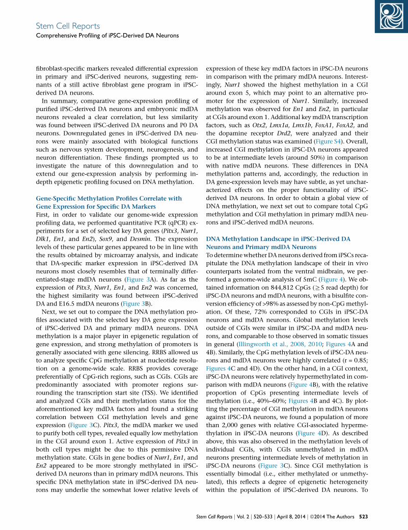
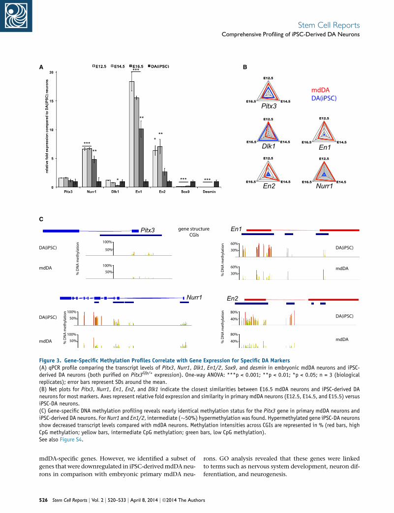
Gene-Specific Methylation Profiles Correlate with
Gene Expression for Specific DA Markers
First, in order to validate our genome-wide expression
profiling data, we performed quantitative PCR (qPCR) ex-
periments for a set of selected key DA genes (Pitx3, Nurr1,
Dlk1, En1, and En2), Sox9, and Desmin. The expression
levels of these particular genes appeared to be in line with
the results obtained by microarray analysis, and indicate
that DA-specific marker expression in iPSC-derived DA
neurons most closely resembles that of terminally differ-
entiated-stage mdDA neurons (Figure 3A). As far as the
expression of Pitx3, Nurr1, En1, and En2 was concerned,
the highest similarity was found between iPSC-derived
DA and E16.5 mdDA neurons (Figure 3B).
Next, we set out to compare the DNA methylation pro-
files associated with the selected key DA gene expression
of iPSC-derived DA and primary mdDA neurons. DNA
methylation is a major player in epigenetic regulation of
gene expression, and strong methylation of promoters is
generally associated with gene silencing. RRBS allowed us
to analyze specific CpG methylation at nucleotide resolu-
tion on a genome-wide scale. RRBS provides coverage
preferentially of CpG-rich regions, such as CGIs. CGIs are
predominantly associated with promoter regions sur-
rounding the transcription start site (TSS). We identified
and analyzed CGIs and their methylation status for the
aforementioned key mdDA factors and found a striking
correlation between CGI methylation levels and gene
expression (Figure 3C). Pitx3, the mdDA marker we used
to purify both cell types, revealed equally low methylation
in the CGI around exon 1. Active expression of Pitx3 in
both cell types might be due to this permissive DNA
methylation state. CGIs in gene bodies of Nurr1, En1, and
En2 appeared to be more strongly methylated in iPSC-
derived DA neurons than in primary mdDA neurons. This
specific DNA methylation state in iPSC-derived DA neu-
rons may underlie the somewhat lower relative levels of
Stem
expression of these key mdDA factors in iPSC-DA neurons
in comparison with the primary mdDA neurons. Interest-
ingly, Nurr1 showed the highest methylation in a CGI
around exon 5, which may point to an alternative pro-
moter for the expression of Nurr1. Similarly, increased
methylation was observed for En1 and En2, in particular
at CGIs around exon 1. Additional keymdDA transcription
factors, such as Otx2, Lmx1a, Lmx1b, FoxA1, FoxA2, and
the dopamine receptor Drd2, were analyzed and their
CGI methylation status was examined (Figure S4). Overall,
increased CGI methylation in iPSC-DA neurons appeared
to be at intermediate levels (around 50%) in comparison
with native mdDA neurons. These differences in DNA
methylation patterns and, accordingly, the reduction in
DA gene-expression levels may have subtle, as yet unchar-
acterized effects on the proper functionality of iPSC-
derived DA neurons. In order to obtain a global view of
DNA methylation, we next set out to compare total CpG
methylation and CGI methylation in primary mdDA neu-
rons and iPSC-derived mdDA neurons.
DNA Methylation Landscape in iPSC-Derived DA
Neurons and Primary mdDA Neurons
To determinewhether DAneurons derived from iPSCs reca-
pitulate the DNA methylation landscape of their in vivo
counterparts isolated from the ventral midbrain, we per-
formed a genome-wide analysis of 5mC (Figure 4). We ob-
tained information on 844,812 CpGs (R5 read depth) for
iPSC-DA neurons and mdDA neurons, with a bisulfite con-
version efficiency of >98% as assessed by non-CpGmethyl-
ation. Of these, 72% corresponded to CGIs in iPSC-DA
neurons and mdDA neurons. Global methylation levels
outside of CGIs were similar in iPSC-DA and mdDA neu-
rons, and comparable to those observed in somatic tissues
in general (Illingworth et al., 2008, 2010; Figures 4A and
4B). Similarly, the CpGmethylation levels of iPSC-DA neu-
rons and mdDA neurons were highly correlated (r = 0.85;
Figures 4C and 4D). On the other hand, in a CGI context,
iPSC-DA neurons were relatively hypermethylated in com-
parison with mdDA neurons (Figure 4B), with the relative
proportion of CpGs presenting intermediate levels of
methylation (i.e., 40%–60%; Figures 4B and 4C). By plot-
ting the percentage of CGI methylation in mdDA neurons
against iPSC-DA neurons, we found a population of more
than 2,000 genes with relative CGI-associated hyperme-
thylation in iPSC-DA neurons (Figure 4D). As described
above, this was also observed in the methylation levels of
individual CGIs, with CGIs unmethylated in mdDA
neurons presenting intermediate levels of methylation in
iPSC-DA neurons (Figure 3C). Since CGI methylation is
essentially bimodal (i.e., either methylated or unmethy-
lated), this reflects a degree of epigenetic heterogeneity
within the population of iPSC-derived DA neurons. To
Cell Reports j Vol. 2 j 520–533 j April 8, 2014 j ª2014 The Authors 523
A B
C
D
E
F
R= 0.67 0.86 0.82 0.86 0.84 (correlation rel. to mdDA.iPSCs)
mdDA specific gene subset
GO analysis of genes with reduced expression in mdDA.iPSCs
0 20 40 60 80 100 120 140 160
0.00E+00 5.00E-05 1.00E-04 1.50E-04 2.00E-04 2.50E-04 nervous system development
neurogenesis generation of neurons neuron differentiation
multicellular organismal development developmental process
system development anatomical structure development
cell development cell differentiation
neuron development central nervous system development
forebrain development cellular developmental process
multicellular organismal process brain development
neuron projection development cell projection organization
cell projection morphogenesis neuron projection morphogenesis
axonogenesis cell part morphogenesis
cellular component organization morphogenesis involved in neuron differentiation
cellular process cell morphogenesis organ development
cellular component movement cell-cell signaling
synaptic transmission regulation of multicellular organismal process
cellular component morphogenesis cell morphogenesis involved in differentiation
limbic system development telencephalon development
biological regulation transmission of nerve impulse
regulation of neurological system process radial glia guided migration of Purkinje cell
regulation of synapse structural plasticity cell communication
regulation of synaptic transmission behavior
central nervous system neuron differentiation neurotransmitter secretion
number of genes
p-value
P1 P2 P3 Midbrain
Telencephalon
Midbrain-hindbrain border
Embryonic mouse brain
vs.
Pitx3Gfp/+ neurons isolated from midbrain
iPSC-derived Pitx3Gfp/+ neurons
Telencephalon Midbrain iPSC-derived
GFP fluorescence
FL2
Pitx3Gfp/+
global correlation of gene expression (Pitx3Gfp/+ )
P0
E12
.5
E14
.5
E13
.5
E15
.5
mdD
A.iP
SC
B:1.98%
B:1.21%
Figure 2. Gene-Expression Profiling: Mesodiencephalic PITX3+ Neurons versus iPSC-Derived PITX3+ Neurons(A) Schematic of the experimental setup.(B) FACS profile of the telencephalon and midbrain of Pitx3Gfp/+ mice (E14.5) compared with iPSC-derived PITX3+ neurons.(C) Correlation matrix of global gene expression comparing all Pitx3Gfp/+ purified neurons.
(legend continued on next page)
524 Stem Cell Reports j Vol. 2 j 520–533 j April 8, 2014 j ª2014 The Authors
Stem Cell ReportsComprehensive Profiling of iPSC-Derived DA Neurons
Stem Cell ReportsComprehensive Profiling of iPSC-Derived DA Neurons
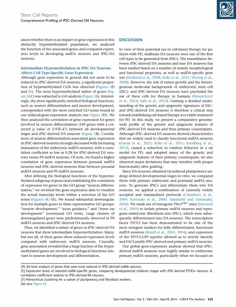
assess whether there is an impact on gene expression in this
distinctly hypermethylated population, we analyzed
the function of the associated genes and compared expres-
sion levels in developing mdDA neurons and iPSC-DA
neurons.
Intermediate Hypermethylation in iPSC-DA Neurons
Affects Cell-Type-Specific Gene Expression
Although gene expression in general did not seem to be
reduced in iPSC-derived DA neurons, a significant propor-
tion of hypermethylated CGIs was observed (Figures 4D
and S5). The most hypermethylated subset of genes (Fig-
ure S5C) was subjected to GO analysis (Figure 5A). Interest-
ingly, the most significantly enriched biological functions,
such as neuron differentiation and neuron development,
corresponded with the most enriched GO terms found in
our reduced-gene-expression analysis (see Figure 2D). We
then analyzed the correlation of gene expression for genes
involved in neuron differentiation (169 genes with a cor-
rected p value of 2.93E-47) between all developmental
stages and iPSC-derived DA neurons (Figure 5B). Correla-
tions of neuron differentiation-associated gene expression
in iPSC-derived neurons strongly decreasedwith increasing
maturation of the embryonic mdDA neurons, with a corre-
lation coefficient as low as 0.28 for iPSC-derived DA neu-
rons versus P0 mdDA neurons. Of note, we found a higher
correlation of gene expression between prenatal mdDA
neurons and iPSC-derived neurons than between prenatal
mdDA neurons and P0 mdDA neurons.
After defining the biological functions of the hyperme-
thylated subgroup of genes and establishing the correlation
of expression for genes in the GO group ‘‘neuron differen-
tiation,’’ we revisited the gene-expression data to visualize
the actual transcript levels within a selection of the GO
terms (Figures 5C–5E). We found substantial downregula-
tion for multiple genes in three representative GO groups:
‘‘neuron development,’’ ‘‘axon guidance,’’ and ‘‘inner ear
development’’ (nonneural GO term). Large clusters of
downregulated genes were predominantly observed in P0
mdDA neurons and iPSC-derived DA neurons.
Thus, we identified a subset of genes in iPSC-derived DA
neurons that show intermediate hypermethylation. Many,
but not all, of these genes show reduced expression levels
compared with embryonic mdDA neurons. Crucially,
gene annotation revealed that a large fraction of the hyper-
methylated genes are involved in biological functions rele-
vant to neuron development and differentiation.
(D) GO term analysis of genes that were most reduced in iPSC-derived(E) Expression levels of selected mdDA-specific genes, comparing decorrelation coefficient relative to iPSC-derived DA neurons.(F) Hierarchical clustering for a subset of pluripotency and fibroblastSee also Figure S3.
Stem
DISCUSSION
In view of their potential use in cell-based therapy for pa-
tients with PD, midbrain DA neurons were one of the first
cell types to be generated from iPSCs. The resemblance be-
tween iPSC-derived DA neurons and true DA neurons has
been studied based on a number of mainly morphological
and functional properties, as well as mdDA-specific gene
sets (Hedlund et al., 2008; Kriks et al., 2011; Wernig et al.,
2008). However, the risk of tumor growth and the hetero-
geneous molecular backgrounds of embryonic stem cell
(ESC)- and iPSC-derived DA neurons have precluded the
use of these cells for therapy in humans (Mom�cilovi�c
et al., 2014; Salti et al., 2013). Gaining a detailed under-
standing of the genetic and epigenetic signatures of ESC-
and iPSC-derived DA neurons is therefore a critical step
toward establishing cell-based therapy as a viable treatment
for PD. In this study, we present a comparative genome-
wide profile of the genetic and epigenetic features of
iPSC-derived DA neurons and their primary counterparts.
Although iPSC-derived DA neurons showed characteristics
that are widely used to classify functional mdDA neurons
(Ganat et al., 2012; Kriks et al., 2011; Sundberg et al.,
2013), caused a reduction in rotation behavior in a rat
model for PD, and adopted many of the genetic and
epigenetic features of their primary counterparts, we also
observed major deviations that may interfere with proper
functionality after grafting.
Since DA neurons obtained via induced pluripotency un-
dergo defined developmental stages in vitro, we compared
them with primary embryonic and postnatal mdDA neu-
rons. To generate iPSCs and differentiate them into DA
neurons, we applied a combination of currently widely
accepted and standardized protocols (Chambers et al.,
2009; Kawasaki et al., 2000; Takahashi and Yamanaka,
2006). We made use of transgenic Pitx3Gfp/+ mice (Maxwell
et al., 2005) to isolate primary mdDA neurons and repro-
gram embryonic fibroblasts into iPSCs, which were subse-
quently differentiated into DA neurons. The transcription
factor PITX3 has been demonstrated to be one of the
most stringent markers for fully differentiated, functional
mdDA neurons (Smidt et al., 2003, 2004), and expression
of the PITX3-GFP reporter allowed us to strictly identify
and FACS purify iPSC-derived and primarymdDA neurons.
Our global gene-expression analysis showed that iPSC-
derived mdDA neurons were highly similar to embryonic
primary mdDA neurons, particularly when we focused on
mdDA neurons.velopmental midbrain stages with iPSC-derived PITX3+ neurons. R,
markers.
Cell Reports j Vol. 2 j 520–533 j April 8, 2014 j ª2014 The Authors 525
E12.5
E14.5 E16.5
E12.5
E14.5 E16.5
E12.5
E14.5 E16.5
E12.5
E14.5 E16.5
E12.5
E14.5 E16.5
gene structureCGIs
Pitx3
En2
Dlk1 En1
mdDADA(iPSC)
Nurr1
A B
C
Pitx3
100%
50%DA(iPSC)
mdDA 100%
50%
DA(iPSC)
mdDA
100%
50%
100%
50%
Nurr1
60%
30%
En1
60%
30%
DA(iPSC)
mdDA
80%
40%
80%
40%
En2
DA(iPSC)
mdDA
% D
NA
met
hyla
tion
% D
NA
met
hyla
tion
% D
NA
met
hyla
tion
% D
NA
met
hyla
tion
*****
*
***
*** ***
***
**
Figure 3. Gene-Specific Methylation Profiles Correlate with Gene Expression for Specific DA Markers(A) qPCR profile comparing the transcript levels of Pitx3, Nurr1, Dlk1, En1/2, Sox9, and desmin in embryonic mdDA neurons and iPSC-derived DA neurons (both purified on Pitx3Gfp/+ expression). One-way ANOVA: ***p < 0.001; **p < 0.01; *p < 0.05; n = 3 (biologicalreplicates); error bars represent SDs around the mean.(B) Net plots for Pitx3, Nurr1, En1, En2, and Dlk1 indicate the closest similarities between E16.5 mdDA neurons and iPSC-derived DAneurons for most markers. Axes represent relative fold expression and similarity in primary mdDA neurons (E12.5, E14.5, and E15.5) versusiPSC-DA neurons.(C) Gene-specific DNA methylation profiling reveals nearly identical methylation status for the Pitx3 gene in primary mdDA neurons andiPSC-derived DA neurons. For Nurr1 and En1/2, intermediate (�50%) hypermethylation was found. Hypermethylated gene iPSC-DA neuronsshow decreased transcript levels compared with mdDA neurons. Methylation intensities across CGIs are represented in % (red bars, highCpG methylation; yellow bars, intermediate CpG methylation; green bars, low CpG methylation).See also Figure S4.
Stem Cell ReportsComprehensive Profiling of iPSC-Derived DA Neurons
mdDA-specific genes. However, we identified a subset of
genes that were downregulated in iPSC-derivedmdDAneu-
rons in comparison with embryonic primary mdDA neu-
526 Stem Cell Reports j Vol. 2 j 520–533 j April 8, 2014 j ª2014 The Author
rons. GO analysis revealed that these genes were linked
to terms such as nervous system development, neuron dif-
ferentiation, and neurogenesis.
s
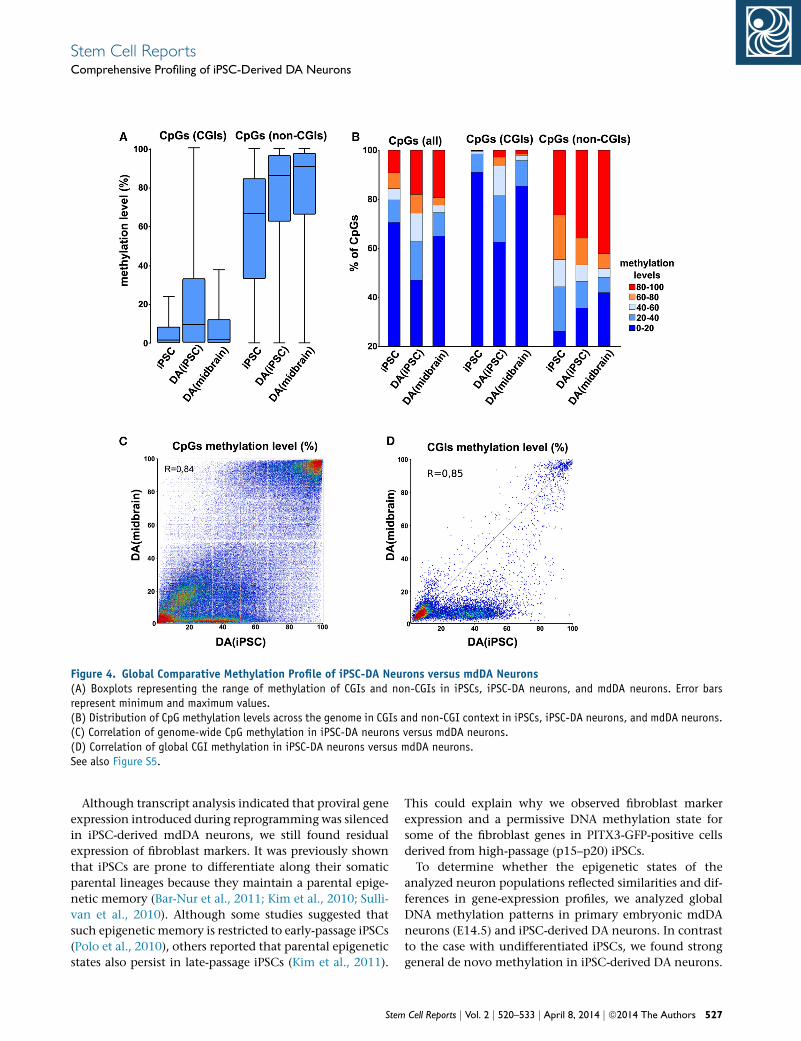
Figure 4. Global Comparative Methylation Profile of iPSC-DA Neurons versus mdDA Neurons(A) Boxplots representing the range of methylation of CGIs and non-CGIs in iPSCs, iPSC-DA neurons, and mdDA neurons. Error barsrepresent minimum and maximum values.(B) Distribution of CpG methylation levels across the genome in CGIs and non-CGI context in iPSCs, iPSC-DA neurons, and mdDA neurons.(C) Correlation of genome-wide CpG methylation in iPSC-DA neurons versus mdDA neurons.(D) Correlation of global CGI methylation in iPSC-DA neurons versus mdDA neurons.See also Figure S5.
Stem Cell ReportsComprehensive Profiling of iPSC-Derived DA Neurons
Although transcript analysis indicated that proviral gene
expression introduced during reprogramming was silenced
in iPSC-derived mdDA neurons, we still found residual
expression of fibroblast markers. It was previously shown
that iPSCs are prone to differentiate along their somatic
parental lineages because they maintain a parental epige-
netic memory (Bar-Nur et al., 2011; Kim et al., 2010; Sulli-
van et al., 2010). Although some studies suggested that
such epigenetic memory is restricted to early-passage iPSCs
(Polo et al., 2010), others reported that parental epigenetic
states also persist in late-passage iPSCs (Kim et al., 2011).
Stem
This could explain why we observed fibroblast marker
expression and a permissive DNA methylation state for
some of the fibroblast genes in PITX3-GFP-positive cells
derived from high-passage (p15–p20) iPSCs.
To determine whether the epigenetic states of the
analyzed neuron populations reflected similarities and dif-
ferences in gene-expression profiles, we analyzed global
DNA methylation patterns in primary embryonic mdDA
neurons (E14.5) and iPSC-derived DA neurons. In contrast
to the case with undifferentiated iPSCs, we found strong
general de novo methylation in iPSC-derived DA neurons.
Cell Reports j Vol. 2 j 520–533 j April 8, 2014 j ª2014 The Authors 527

A
B C
GO term analysis for hyper-methylated genes
0 50 100 150 200 250 300 350 400 450
0.00E+00 5.00E-14 1.00E-13 1.50E-13 2.00E-13 2.50E-13 3.00E-13 3.50E-13 4.00E-13 4.50E-13
neuron differentiation cell-cell signaling
neuron development embryonic morphogenesis
embryonic organ development regulation of transcription, DNA-dependent
pattern specification process embryonic organ morphogenesis
cell morphogenesis involved in differentiation regulation of RNA metabolic process
cell fate commitment neuron projection development
cell morphogenesis involved in neuron differentiation neuron projection morphogenesis
axonogenesis regionalization
cell projection organization cell projection morphogenesis
cell morphogenesis regulation of transcription from RNA polymerase II promoter
transmission of nerve impulse cell part morphogenesis
ion transport axon guidance
synaptic transmission regulation of transcription
cellular component morphogenesis sensory organ development
cell motion potassium ion transport
positive regulation of transcription, DNA-dependent ear morphogenesis
regulation of cell development positive regulation of RNA metabolic process
positive regulation of transcription from RNA polymerase II skeletal system development
regulation of nervous system development metal ion transport
ear development regulation of neurogenesis
inner ear morphogenesis behavior
inner ear development positive regulation of biosynthetic process
positive regulation of cellular biosynthetic process neuron fate commitment
positive regulation of gene expression positive regulation of transcription
anterior/posterior pattern formation positive regulation of nucleobase, nucleoside, nucleotide and
number of genes
p-value
D E
mdD
A.iP
SC P0
E12
.5
E13
.5
E14
.5
E15
.5 P0
mdD
A.iP
SC
E14
.5
E15
.5
E12
.5
E13
.5
P0
mdD
A.iP
SC
E12
.5
E15
.5
E13
.5
E14
.5
correlation of gene expression in GO subgroup ‘neuron differentiation
(legend on next page)
528 Stem Cell Reports j Vol. 2 j 520–533 j April 8, 2014 j ª2014 The Authors
Stem Cell ReportsComprehensive Profiling of iPSC-Derived DA Neurons

Stem Cell ReportsComprehensive Profiling of iPSC-Derived DA Neurons
Earlier studies reported that PSCs mostly contain methyl-
ation-free promoters, as well asmethylation-free intergenic
and orphan CGIs (Illingworth et al., 2010). De novo
methylation was only found upon loss of pluripotency,
suggesting that transcriptional repression in PSCs is estab-
lished predominantly via other mechanisms (Fouse et al.,
2008; Mohn et al., 2008). Indeed, we observed that most
de novo methylation events occurred during differentia-
tion from pluripotent cells to multipotent precursors
isolated from an iPSC-derived NESTIN-GFP reporter line
(data not shown).
General methylation states appeared to be comparable
between iPSC-derived DA neurons and primary embryonic
mdDA neurons (r = 0.85), indicating that our in-vitro-
generated DA neurons widely adopted the epigenetic
signature of their primary counterparts. Nonetheless, we
found several thousand genes hypermethylated in iPSC-
derived DA neurons. This hypermethylation was found
predominantly at an intermediate level ranging from
40% to 60% DNA methylation. At this point, we can
only speculate about whether hypermethylation is due
to retained epigenetic memory or to heterogeneous
differentiation/maturation stages within the PITX3-GFP
FACS-sorted iPSC-derived population. To our knowledge,
a specific neuronal subtype derived from iPSCs has not
been characterized to such an extent, but similar differ-
ences in methylation profiles have been observed in re-
programmed mesenchymal stromal cells compared with
ESCs (Shao et al., 2013). Interestingly, our gene-expression
data show reduced expression of Tet1 and Tet3 in iPSC-
derived DA neurons compared with E14.5 mdDA neurons.
Tet proteins have been shown to be crucial for establish-
ment of pluripotency, development, and neuronal activity
(Koh et al., 2011; Rudenko et al., 2013; Zhang et al., 2013).
Presently, very little is known about the activity of Tet
proteins in specific subtypes of neurons (primary or PSC
derived). It is becoming more and more clear, however,
that their demethylation activity is crucial for functionally
bona fide, healthy neurons (Gavin et al., 2013; Ma et al.,
2009). Our DNA methylation profiling of iPSC-derived
DA neurons versus primary mdDA neurons did not
allow a distinction between 5-methylcytosine (5mC) and
5-hydroxymethylcytosine (5hmC). Neuronal gene activa-
tion was recently shown to be mediated by specific
MeCP2 binding to 5hmC (Li et al., 2013; Mellen et al.,
2012). Therefore, we cannot rule out the possibility that
elevated 5hmC levels in iPSC-derived mdDA neurons
Figure 5. Intermediate Hypermethylation of >2,000 Genes in iPSC(A) GO term analysis of hypermethylated genes in iPSC-derived DA ne(B) Correlation matrix for gene expression in the ‘‘neuron differentiat(C–E) Heatmaps showing comparative gene expression for genes involdevelopment (E) (GO terms highlighted by red boxes in A). Blue boxe
Stem
might contribute to gene activation rather than to
silencing. However, within the population of hypermethy-
lated genes, we did observe gene clusters with substan-
tially lower transcript levels. Interestingly, we also found
clusters of genes that were upregulated compared with pri-
mary mdDA neurons. Because these genes also have been
identified by RRBS based on their intermediate hyperme-
thylation, these particular subsets might reflect groups
of genes that are activated rather than silenced upon 5-
hydroxymethylation.
In our gene-expression analyses, we found a weaker
correlation between iPSC-derived mdDA neurons and
P0 primary mdDA neurons than between iPSC-derived
mdDA neurons and embryonic primary mdDA neurons,
which prompted us to analyze in depth the underlying
methylation states of only E14.5 mdDA neurons and
iPSC-derived mdDA neurons. Interestingly, however, the
expression levels of En1 and En2 in iPSC-derived mdDA
neurons were comparable to those in P0 primary mdDA
neurons. The meaning of this in terms of functionality
is unclear and may reflect the fact that these iPSC-derived
mdDA neurons are in more of an adult state with respect
to En1 and En2. In addition, it might indicate that
iPSC-derived mdDA neurons are in an A9 state, as it has
been suggested that En1 levels are downregulated by
PITX3 in the rostral mdDA subpopulation (Veenvliet
et al., 2013).
Our findings raise two important questions: is epige-
netic memory an obstacle for exploiting the full potential
of iPSCs (e.g., personalized disease modeling), and do
aberrations in the expression profiles of iPSC-derived
mdDA neurons interfere with long-term functionality?
Although comprehensive studies have been performed
to test the in vivo functionality of human neurons
(Ganat et al., 2012; Kriks et al., 2011), these questions
still need to be addressed, especially when in-vitro-gener-
ated cells are to be considered for application in disease
modeling and cell-based therapy for PD. It remains to
be studied whether modifications in the reprogramming
process, as recently reported for the human system (Gafni
et al., 2013), as well as refined differentiation procedures
might diminish these deviations. An increased funda-
mental understanding of the genetic and epigenetic
signatures of DA neurons in vivo (Hegarty et al., 2013;
Smidt and Burbach, 2007; van Heesbeen et al., 2013)
could offer new leads to generate safe and transplantable
DA neurons in vitro.
-Derived DA Neurons Affects Cell-Type-Specific Gene Expressionurons.ion’’ GO subset (highlighted by the lowest red box in A).ved in neuron differentiation (C), axon guidance (D), and inner ears indicate the groups used for RRBS analysis.
Cell Reports j Vol. 2 j 520–533 j April 8, 2014 j ª2014 The Authors 529
Stem Cell ReportsComprehensive Profiling of iPSC-Derived DA Neurons
EXPERIMENTAL PROCEDURES
MicePitx3(Gfp/+) embryos at several developmental stages were
obtained by intercrossing C57BL6/J with Pitx3(Gfp/Gfp) mice.
Pitx3(Gfp/+) embryos are heterozygous for wild-type PITX3 and
have normal mdDA system development (Maxwell et al., 2005).
Overlap of endogenous PITX3 with GFP has been shown to be
�100% (Maxwell et al., 2005). All procedures were approved by
and performed according to the guidelines of the Dutch ethics
committees for animal experiments (UMCU and UvA).
iPSC Generation and PropagationMouse embryonic fibroblasts were isolated from E14.5 embryos of
Pitx3Gfp/+ and Nestin-Gfp mice, both of which were previously
described and characterized (Yamaguchi et al., 2000; Zhao et al.,
2004). Fibroblasts were cultured until passage 5–8 and then retro-
virally transfected with the four Yamanaka reprogramming
factors. Separate vectors containing either Oct4, Klf4, Sox2, or
cMyc were used for pluripotency induction. Retroviruses were
obtained from Phoenix Eco packaging cells transfected with the
reprogramming factors (for vector information: Addgene, http://
www.addgene.org). The detailed induction protocol was previ-
ously described (Czepiel et al., 2011). iPSC clones were char-
acterized by immunocytochemistry, RT-PCR, western blot, and
bisulfite sequencing for the promoter regions of Nanog and Oct4
(Figure S1).
Differentiation of Pitx3Gfp/+ iPSCs toward DANeuronsWe combined a stromal feeder-based protocol (Kawasaki et al.,
2000) with dual bone morphogenetic protein (BMP) and trans-
forming growth factor b (TGF-b) inhibition (Chambers et al.,
2009). iPSC colonies previously cultured on gelatin were manually
picked and seeded on MS5 stromal cells in serum replacement
medium containing Dulbecco’s modified Eagle’s medium F12
(DMEM-F12), 15% knockout serum, glutamate, and b-mercaptoe-
thanol. The cells were allowed to settle for 24 hr, and then
Noggin (300 ng/ml) and the TGF-b inhibitor SB431541 (10 mM)
were added for 4–5 days. Neural precursors (NESTIN-GFP) were
collected after 14 days of differentiation. The medium was gradu-
ally changed to N2 containing Sonic hedgehog (SHH; 200 ng/ml).
After neural induction, neuronal patterning and DA differentia-
tion were induced using a combination of brain-derived neuro-
trophic factor (BDNF), ascorbic acid, SHH, and fibroblast growth
factor 8 (FGF8) in N2 medium. Maturation was initiated by with-
drawing SHH and FGF8 in the presence of BDNF, glial cell-derived
neurotrophic factor (GDNF), ascorbic acid, and cyclic adenosine
monophosphate (Perrier et al., 2004). At about 4 weeks of differ-
entiation, Pitx3Gfp/+ neurons reached a mature state as determined
by morphology, marker expression, electrophysiology, and dopa-
mine production. Continuous culturing in these conditions
did not induce any further maturation/differentiation, i.e., there
were no changes in cell morphology, level and profile of marker
expression, electric membrane properties, or dopamine produc-
tion; however, continuous culturing resulted in an increase in
cell death. For gene expression and DNA methylation analyses,
we used Pitx3Gfp/+ mdDA neurons derived from iPSCs after 4 weeks
530 Stem Cell Reports j Vol. 2 j 520–533 j April 8, 2014 j ª2014 The Author
of differentiation. Cells were sorted and immediately subjected to
RNA or DNA isolation.
Microarray AnalysisTotal RNA was isolated from embryonic ventral midbrain tissue
at various developmental stages (four biological replicates each)
and iPSC-derived neurons (three biological replicates) using
Trizol (Invitrogen), and purified using RNeasy columns (QIAGEN).
Microarray analysis was performed in biological triplicates. For
each experimental sample, a dye swap was performed to correct
for dye effects. Agilent whole mouse genome microarray
(G4122F; Agilent) sets were used for all hybridization. The array
set is comprised of 60-mer oligonucleotide probes representing
over 41,000 mouse genes and transcripts. Hybridized slides
were scanned on an Agilent scanner (G2565AA) at 100% laser
power, 30% PMT. After data were extracted using ImaGene 8.0
(BioDiscovery), print-tip Loess normalization was performed on
mean spot intensities. Datawere analyzed using ANOVA (R version
2.2.1/MAANOVA version 0.98-7; http://www.r-project.org/), and
p values were determined by a permutation F2 test in which resid-
uals were shuffled 5,000 times globally.
RRBSRRBS was performed as described previously (Smallwood et al.,
2011). Genomic DNA from E14.5 mdDA neurons, NESTIN-GFP-
positive precursors, and iPSC-derived DA neurons was purified
using the QIAamp Micro Kit (QIAGEN) followed by MspI diges-
tion (Fermentas), end-repair/A-tailing, and ligation (T4 Ligase, Fer-
mentas) of 5mC-adapters (Illumina) performed in Tango1X buffer
without intermediate purification of the enzymatic reactions
(heat inactivation was used after each enzymatic step and compo-
nents were adjusted for the next step). Bisulfite conversion was
performed (Imprint DNA modification; Sigma) and converted
DNA was amplified (six cycles) using uracil stalling free polymer-
ase (Pfu Turbo Cx; Stratagene) followed by size selection (150–
450 bp, Qiaquick; QIAGEN) and a second round of amplification
(10–12 cycles, Platinium, Pfx polymerase; Invitrogen). Libraries
were purified (SPRI beads; Agencourt) and sequenced on an
Illumina Genome Analyzer IIx (40 bp, single read). Quality con-
trol of raw sequence data was performed using FastQC (http://
www.bioinformatics.babraham.ac.uk/projects/fastqc), and trim-
ming was done using TrimGalore (http://www.bioinformatics.
babraham.ac.uk/projects/trim_galore/). Sequence alignment and
methylation calls were performed using Bareback (http://www.
bioinformatics.babraham.ac.uk/projects/bareback/) and Bismark
(Krueger and Andrews, 2011)
ACCESSION NUMBERS
Data for the expression arrays (KC002) reported here have
been deposited in the GEO database under accession number
GSE47178. The RRBS data have been deposited in the GEO data-
base under accession number GSE55475.
SUPPLEMENTAL INFORMATION
Supplemental Information includes Supplemental Experimental
Procedures, five figures, and one table and can be found
s
Stem Cell ReportsComprehensive Profiling of iPSC-Derived DA Neurons
with this article online at http://dx.doi.org/10.1016/j.stemcr.2014.
03.001.
ACKNOWLEDGMENTS
We thankN. Brouwer andM.Meijer for their technical support and
F. Krueger (Babraham Institute) for assisting with the bioinformat-
ics. We acknowledge the help of the FACS core facility of UMCG
and thank R. Wichmann for performing animal surgeries. R.R.
was supported by the Hazewinkel-Beringer Foundation and the
Jan Kornelius de Cock Stichting. Confocal imaging was performed
at the UMCG Microscopy and Imaging Center (UMIC) and spon-
sored by NWO grants 40-00506-98-9021 and 175010-2009-23.
This work was supported by a VICI grant (No. 865.09.002) to
M.P.S. Work in G.K.’s lab is supported by the Biotechnology and
Biological Sciences Research Council and Medical Research Coun-
cil of the UK.
Received: June 21, 2013
Revised: March 4, 2014
Accepted: March 5, 2014
Published: April 3, 2014
REFERENCES
Bar-Nur, O., Russ, H.A., Efrat, S., and Benvenisty, N. (2011). Epige-
netic memory and preferential lineage-specific differentiation in
induced pluripotent stem cells derived from human pancreatic
islet beta cells. Cell Stem Cell 9, 17–23.
Bock, C., Kiskinis, E., Verstappen, G., Gu, H., Boulting, G., Smith,
Z.D., Ziller, M., Croft, G.F., Amoroso, M.W., Oakley, D.H., et al.
(2011). Reference Maps of human ES and iPS cell variation enable
high-throughput characterization of pluripotent cell lines. Cell
144, 439–452.
Chakrabarty, K., Von Oerthel, L., Hellemons, A., Clotman, F.,
Espana, A., Groot Koerkamp, M., Holstege, F.C., Pasterkamp, R.J.,
and Smidt, M.P. (2012). Genome wide expression profiling of the
mesodiencephalic region identifies novel factors involved in early
and late dopaminergic development. Biol. Open 1, 693–704.
Chambers, S.M., Fasano, C.A., Papapetrou, E.P., Tomishima, M.,
Sadelain, M., and Studer, L. (2009). Highly efficient neural conver-
sion of human ES and iPS cells by dual inhibition of SMAD
signaling. Nat. Biotechnol. 27, 275–280.
Czepiel, M., Balasubramaniyan, V., Schaafsma, W., Stancic, M.,
Mikkers, H., Huisman, C., Boddeke, E., and Copray, S. (2011). Dif-
ferentiation of induced pluripotent stem cells into functional oli-
godendrocytes. Glia 59, 882–892.
Espejel, S., Roll, G.R., McLaughlin, K.J., Lee, A.Y., Zhang, J.Y., Laird,
D.J., Okita, K., Yamanaka, S., and Willenbring, H. (2010). Induced
pluripotent stem cell-derived hepatocytes have the functional and
proliferative capabilities needed for liver regeneration in mice.
J. Clin. Invest. 120, 3120–3126.
Fouse, S.D., Shen, Y., Pellegrini, M., Cole, S., Meissner, A., Van
Neste, L., Jaenisch, R., and Fan, G. (2008). Promoter CpG methyl-
ation contributes to ES cell gene regulation in parallel with Oct4/
Nanog, PcG complex, and histone H3 K4/K27 trimethylation.
Cell Stem Cell 2, 160–169.
Stem
Gafni, O.,Weinberger, L.,Mansour, A.A.,Manor, Y.S., Chomsky, E.,
Ben-Yosef, D., Kalma, Y., Viukov, S., Maza, I., Zviran, A., et al.
(2013). Derivation of novel human ground state naive pluripotent
stem cells. Nature 504, 282–286.
Ganat, Y.M., Calder, E.L., Kriks, S., Nelander, J., Tu, E.Y., Jia, F., Bat-
tista, D., Harrison, N., Parmar, M., Tomishima, M.J., et al. (2012).
Identification of embryonic stem cell-derived midbrain dopami-
nergic neurons for engraftment. J. Clin. Invest. 122, 2928–2939.
Gavin, D.P., Chase, K.A., and Sharma, R.P. (2013). Active DNA
demethylation in post-mitotic neurons: a reason for optimism.
Neuropharmacology 75, 233–245.
Hanna, J.,Wernig,M.,Markoulaki, S., Sun, C.W.,Meissner, A., Cas-
sady, J.P., Beard, C., Brambrink, T.,Wu, L.C., Townes, T.M., and Jae-
nisch, R. (2007). Treatment of sickle cell anemiamousemodel with
iPS cells generated from autologous skin. Science 318, 1920–1923.
Hargus, G., Cooper, O., Deleidi, M., Levy, A., Lee, K., Marlow, E.,
Yow, A., Soldner, F., Hockemeyer, D., Hallett, P.J., et al. (2010).
Differentiated Parkinson patient-derived induced pluripotent
stem cells grow in the adult rodent brain and reduce motor asym-
metry in Parkinsonian rats. Proc. Natl. Acad. Sci. USA 107, 15921–
15926.
Hedlund, E., Pruszak, J., Lardaro, T., Ludwig, W., Vinuela, A., Kim,
K.S., and Isacson, O. (2008). Embryonic stem cell-derived Pitx3-
enhanced green fluorescent protein midbrain dopamine neurons
survive enrichment by fluorescence-activated cell sorting and
function in an animal model of Parkinson’s disease. Stem Cells
26, 1526–1536.
Hegarty, S.V., Sullivan, A.M., and O’Keeffe, G.W. (2013). Midbrain
dopaminergic neurons: a review of themolecular circuitry that reg-
ulates their development. Dev. Biol. 379, 123–138.
Illingworth, R., Kerr, A., Desousa, D., Jørgensen, H., Ellis, P., Stalker,
J., Jackson, D., Clee, C., Plumb, R., Rogers, J., et al. (2008). A novel
CpG island set identifies tissue-specific methylation at develop-
mental gene loci. PLoS Biol. 6, e22.
Illingworth, R.S., Gruenewald-Schneider, U., Webb, S., Kerr, A.R.,
James, K.D., Turner, D.J., Smith, C., Harrison, D.J., Andrews, R.,
and Bird, A.P. (2010). Orphan CpG islands identify numerous
conserved promoters in the mammalian genome. PLoS Genet. 6,
e1001134.
Jacobs, F.M., van Erp, S., van der Linden, A.J., von Oerthel, L.,
Burbach, J.P., and Smidt, M.P. (2009). Pitx3 potentiates Nurr1 in
dopamine neuron terminal differentiation through release of
SMRT-mediated repression. Development 136, 531–540.
Jacobs, F.M., Veenvliet, J.V., Almirza,W.H., Hoekstra, E.J., von Oer-
thel, L., van der Linden, A.J., Neijts, R., Koerkamp, M.G., van Lee-
nen, D., Holstege, F.C., et al. (2011). Retinoic acid-dependent and
-independent gene-regulatory pathways of Pitx3 in meso-dience-
phalic dopaminergic neurons. Development 138, 5213–5222.
Karumbayaram, S., Novitch, B.G., Patterson, M., Umbach, J.A.,
Richter, L., Lindgren, A., Conway, A.E., Clark, A.T., Goldman,
S.A., Plath, K., et al. (2009). Directed differentiation of human-
induced pluripotent stem cells generates active motor neurons.
Stem Cells 27, 806–811.
Kawasaki, H., Mizuseki, K., Nishikawa, S., Kaneko, S., Kuwana, Y.,
Nakanishi, S., Nishikawa, S.I., and Sasai, Y. (2000). Induction of
Cell Reports j Vol. 2 j 520–533 j April 8, 2014 j ª2014 The Authors 531
Stem Cell ReportsComprehensive Profiling of iPSC-Derived DA Neurons
midbrain dopaminergic neurons from ES cells by stromal cell-
derived inducing activity. Neuron 28, 31–40.
Kim, K., Doi, A., Wen, B., Ng, K., Zhao, R., Cahan, P., Kim, J., Aryee,
M.J., Ji, H., Ehrlich, L.I.R., et al. (2010). Epigenetic memory in
induced pluripotent stem cells. Nature 467, 285–290.
Kim, K., Zhao, R., Doi, A., Ng, K., Unternaehrer, J., Cahan, P., Huo,
H., Loh, Y.-H., Aryee, M.J., Lensch, M.W., et al. (2011). Donor cell
type can influence the epigenome and differentiation potential
of human induced pluripotent stem cells. Nat. Biotechnol. 29,
1117–1119.
Koh, K.P., Yabuuchi, A., Rao, S., Huang, Y., Cunniff, K., Nardone, J.,
Laiho, A., Tahiliani, M., Sommer, C.A., Mostoslavsky, G., et al.
(2011). Tet1 and Tet2 regulate 5-hydroxymethylcytosine produc-
tion and cell lineage specification in mouse embryonic stem cells.
Cell Stem Cell 8, 200–213.
Kriks, S., Shim, J.-W., Piao, J., Ganat, Y.M., Wakeman, D.R., Xie, Z.,
Carrillo-Reid, L., Auyeung, G., Antonacci, C., Buch, A., et al.
(2011). Dopamine neurons derived from human ES cells effi-
ciently engraft in animal models of Parkinson’s disease. Nature
480, 547–551.
Krueger, F., and Andrews, S.R. (2011). Bismark: a flexible aligner
and methylation caller for Bisulfite-Seq applications. Bioinformat-
ics 27, 1571–1572.
Kuzmenkin, A., Liang, H., Xu, G., Pfannkuche, K., Eichhorn, H.,
Fatima, A., Luo, H., Saric, T., Wernig, M., Jaenisch, R., and Hesch-
eler, J. (2009). Functional characterization of cardiomyocytes
derived frommurine induced pluripotent stem cells in vitro. FASEB
J. 23, 4168–4180.
Lee, S.-H., Jeyapalan, J.N., Appleby, V., Mohamed Noor, D.A., Sot-
tile, V., and Scotting, P.J. (2010). Dynamicmethylation and expres-
sion of Oct4 in early neural stem cells. J. Anat. 217, 203–213.
Li, Y., Wang, H., Muffat, J., Cheng, A.W., Orlando, D.A., Loven, J.,
Kwok, S.M., Feldman, D.A., Bateup, H.S., Gao, Q., et al. (2013).
Global transcriptional and translational repression in human-em-
bryonic-stem-cell-derived Rett syndrome neurons. Cell Stem Cell
13, 446–458.
Ma, D.K., Jang,M.-H., Guo, J.U., Kitabatake, Y., Chang,M.-L., Pow-
Anpongkul, N., Flavell, R.A., Lu, B., Ming, G.-L., and Song, H.
(2009). Neuronal activity-induced Gadd45b promotes epigenetic
DNA demethylation and adult neurogenesis. Science 323, 1074–
1077.
Maherali, N., Sridharan, R., Xie,W., Utikal, J., Eminli, S., Arnold, K.,
Stadtfeld, M., Yachechko, R., Tchieu, J., Jaenisch, R., et al. (2007).
Directly reprogrammed fibroblasts show global epigenetic remod-
eling and widespread tissue contribution. Cell Stem Cell 1, 55–70.
Maxwell, S.L., Ho, H.-Y., Kuehner, E., Zhao, S., and Li, M. (2005).
Pitx3 regulates tyrosine hydroxylase expression in the substantia
nigra and identifies a subgroup of mesencephalic dopaminergic
progenitor neurons during mouse development. Dev. Biol. 282,
467–479.
Meissner, A.,Gnirke, A., Bell,G.W., Ramsahoye, B., Lander, E.S., and
Jaenisch, R. (2005). Reduced representation bisulfite sequencing for
comparative high-resolution DNA methylation analysis. Nucleic
Acids Res. 33, 5868–5877.
532 Stem Cell Reports j Vol. 2 j 520–533 j April 8, 2014 j ª2014 The Author
Mellen, M., Ayata, P., Dewell, S., Kriaucionis, S., and Heintz, N.
(2012). MeCP2 binds to 5hmC enriched within active genes and
accessible chromatin in the nervous system. Cell 151, 1417–1430.
Mohn, F., Weber, M., Rebhan, M., Roloff, T.C., Richter, J., Stadler,
M.B., Bibel, M., and Schubeler, D. (2008). Lineage-specific poly-
comb targets and de novo DNA methylation define restriction
and potential of neuronal progenitors. Mol. Cell 30, 755–766.
Mom�cilovi�c, O., Liu, Q., Swistowski, A., Russo-Tait, T., Zhao, Y.,
Rao, M.S., and Zeng, X. (2014). Genome wide profiling of dopami-
nergic neurons derived from human embryonic and induced
pluripotent stem cells. Stem Cells Dev. 23, 406–420.
Nishino, K., Toyoda, M., Yamazaki-Inoue, M., Fukawatase, Y., Chi-
kazawa, E., Sakaguchi, H., Akutsu, H., and Umezawa, A. (2011).
DNA methylation dynamics in human induced pluripotent stem
cells over time. PLoS Genet. 7, e1002085.
Ohi, Y., Qin, H., Hong, C., Blouin, L., Polo, J.M., Guo, T., Qi, Z.,
Downey, S.L., Manos, P.D., Rossi, D.J., et al. (2011). Incomplete
DNA methylation underlies a transcriptional memory of somatic
cells in human iPS cells. Nat. Cell Biol. 13, 541–549.
Perrier, A.L., Tabar, V., Barberi, T., Rubio, M.E., Bruses, J., Topf, N.,
Harrison, N.L., and Studer, L. (2004). Derivation ofmidbrain dopa-
mine neurons fromhuman embryonic stem cells. Proc. Natl. Acad.
Sci. USA 101, 12543–12548.
Polo, J.M., Liu, S., Figueroa, M.E., Kulalert,W., Eminli, S., Tan, K.Y.,
Apostolou, E., Stadtfeld,M., Li, Y., Shioda, T., et al. (2010). Cell type
of origin influences the molecular and functional properties
of mouse induced pluripotent stem cells. Nat. Biotechnol. 28,
848–855.
Ren, Y., Lee,M.Y., Schliffke, S., Paavola, J., Amos, P.J., Ge, X., Ye,M.,
Zhu, S., Senyei, G., Lum, L., et al. (2011). Small molecule Wnt
inhibitors enhance the efficiency of BMP-4-directed cardiac differ-
entiation of humanpluripotent stem cells. J.Mol. Cell. Cardiol. 51,
280–287.
Roessler, R., Boddeke, E., and Copray, S. (2013). Induced pluripo-
tent stem cell technology and direct conversion: new possibilities
to study and treat Parkinson’s disease. Stem Cell Rev. 9, 505–513.
Rudenko, A., Dawlaty, M.M., Seo, J., Cheng, A.W., Meng, J., Le, T.,
Faull, K.F., Jaenisch, R., and Tsai, L.-H. (2013). Tet1 is critical for
neuronal activity-regulated gene expression and memory extinc-
tion. Neuron 79, 1109–1122.
Salti, A., Nat, R., Neto, S., Puschban, Z.,Wenning, G., andDechant,
G. (2013). Expression of early developmental markers predicts the
efficiency of embryonic stem cell differentiation into midbrain
dopaminergic neurons. Stem Cells Dev. 22, 397–411.
Shao, K., Koch, C., Gupta, M.K., Lin, Q., Lenz, M., Laufs, S.,
Denecke, B., Schmidt, M., Linke, M., Hennies, H.C., et al. (2013).
Induced pluripotent mesenchymal stromal cell clones retain
donor-derived differences in DNA methylation profiles. Mol.
Ther. 21, 240–250.
Smallwood, S.A., Tomizawa, S.-I., Krueger, F., Ruf, N., Carli, N.,
Segonds-Pichon, A., Sato, S., Hata, K., Andrews, S.R., and Kelsey,
G. (2011). Dynamic CpG island methylation landscape in oocytes
and preimplantation embryos. Nat. Genet. 43, 811–814.
Smidt, M.P., and Burbach, J.P.H. (2007). How to make a mesodien-
cephalic dopaminergic neuron. Nat. Rev. Neurosci. 8, 21–32.
s
Stem Cell ReportsComprehensive Profiling of iPSC-Derived DA Neurons
Smidt,M.P., Smits, S.M., and Burbach, J.P. (2003).Molecularmech-
anisms underlying midbrain dopamine neuron development and
function. Eur. J. Pharmacol. 480, 75–88.
Smidt, M.P., Smits, S.M., Bouwmeester, H., Hamers, F.P., van der
Linden, A.J., Hellemons, A.J., Graw, J., and Burbach, J.P. (2004).
Early developmental failure of substantia nigra dopamine neurons
in mice lacking the homeodomain gene Pitx3. Development 131,
1145–1155.
Sullivan, G.J., Bai, Y., Fletcher, J., and Wilmut, I. (2010). Induced
pluripotent stem cells: epigenetic memories and practical implica-
tions. Mol. Hum. Reprod. 16, 880–885.
Sundberg, M., Bogetofte, H., Lawson, T., Jansson, J., Smith, G.,
Astradsson, A., Moore, M., Osborn, T., Cooper, O., Spealman, R.,
et al. (2013). Improved cell therapy protocols for Parkinson’s
disease based on differentiation efficiency and safety of hESC-,
hiPSC-, and non-human primate iPSC-derived dopaminergic neu-
rons. Stem Cells 31, 1548–1562.
Swistowski, A., Peng, J., Liu, Q., Mali, P., Rao, M.S., Cheng, L., and
Zeng, X. (2010). Efficient generation of functional dopami-
nergic neurons from human induced pluripotent stem cells under
defined conditions. Stem Cells 28, 1893–1904.
Takahashi, K., and Yamanaka, S. (2006). Induction of pluripotent
stem cells from mouse embryonic and adult fibroblast cultures
by defined factors. Cell 126, 663–676.
van Heesbeen, H.J., Mesman, S., Veenvliet, J.V., and Smidt, M.P.
(2013). Epigenetic mechanisms in the development and mainte-
nance of dopaminergic neurons. Development 140, 1159–1169.
Stem
Veenvliet, J.V., Dos Santos, M.T., Kouwenhoven, W.M., von Oer-
thel, L., Lim, J.L., van der Linden, A.J., Koerkamp, M.J., Holstege,
F.C., and Smidt, M.P. (2013). Specification of dopaminergic subsets
involves interplay of En1 and Pitx3. Development 140, 3373–
3384.
Watanabe, D., Uchiyama, K., and Hanaoka, K. (2006). Transition
of mouse de novo methyltransferases expression from Dnmt3b
to Dnmt3a during neural progenitor cell development. Neurosci-
ence 142, 727–737.
Wernig, M., Zhao, J.-P., Pruszak, J., Hedlund, E., Fu, D., Soldner, F.,
Broccoli, V., Constantine-Paton, M., Isacson, O., and Jaenisch, R.
(2008). Neurons derived from reprogrammed fibroblasts function-
ally integrate into the fetal brain and improve symptoms of rats
with Parkinson’s disease. Proc. Natl. Acad. Sci. USA 105, 5856–
5861.
Yamaguchi, M., Saito, H., Suzuki, M., and Mori, K. (2000). Visuali-
zation of neurogenesis in the central nervous system using nestin
promoter-GFP transgenic mice. Neuroreport 11, 1991–1996.
Zhang, R.-R., Cui, Q.Y., Murai, K., Lim, Y.C., Smith, Z.D., Jin, S., Ye,
P., Rosa, L., Lee, Y.K., Wu, H.-P., et al. (2013). Tet1 regulates adult
hippocampal neurogenesis and cognition. Cell Stem Cell 13,
237–245.
Zhao, S., Maxwell, S., Jimenez-Beristain, A., Vives, J., Kuehner, E.,
Zhao, J., O’Brien, C., de Felipe, C., Semina, E., and Li, M. (2004).
Generation of embryonic stem cells and transgenic mice express-
ing green fluorescence protein inmidbrain dopaminergic neurons.
Eur. J. Neurosci. 19, 1133–1140.
Cell Reports j Vol. 2 j 520–533 j April 8, 2014 j ª2014 The Authors 533
Copyright © 2022 FDOKUMEN