Analysis and Proposed Organization of the Capoeira Song Repertoire
Upload
independentCategory
view
0download
0
Design and testing of PCR primers for the construction of scFvlibraries representing the immunoglobulin repertoire of rats
Jorge Sepulveda and Charles B. ShoemakerTufts Cummings School of Veterinary Medicine, North Grafton, MA 01536
AbstractRats are widely used as a model in the study of many important diseases, yet their utility in researchis limited by the lack of robust technology for the production of rat recombinant antibodies. Here wehave identified and compiled putative rat immunoglobulin sequences by bioinformatic analysis ofrecently available genomic and EST databases, and used this information to design PCR primers toamplify rat VH and VL domain coding DNA representing the expressed repertoire of the animal.These primer sets were successfully tested and used to produce a scFv phage display library fromrats that had been infected by the blood fluke, Schistosoma mansoni. The library was selected forscFvs that bind to extracellular loop epitopes on either of two known immunogenic S. mansonimembrane proteins, Sm23 and SmTsp2, both of the tetraspanin protein family. Two unique scFvswere identified that bind to each tetraspanin and shown to have excellent specificity for the target,including recognition of the native tetraspanins produced by the fluke. The primers and methodsdeveloped for rat scFvs should be of use to others seeking to characterize the antibody repertoireand/or prepare recombinant antibodies from this model.
1. IntroductionAntibodies are widely used for medical and biotechnological purposes. Since the developmentof monoclonal antibodies (Kohler and Milstein, 1975) and the subsequent generation offunctional fragments of immunoglobulin through recombinant DNA technologies (Better etal., 1988; Bird et al., 1988; Huston et al., 1988; Skerra and Pluckthun, 1988) clonal antibodieshave demonstrated a wide range of biological activities and specificities that have proved usefulfor the treatment of diseases, diagnostics and research. Immunoglobulin (Ig) proteins and geneshave been extensively characterized from human and murine B cells (IMGT database,Montpellier, France (Giudicelli et al., 2004) and these models have become the primary sourceof monoclonal antibodies and recombinant antibodies for research and therapeutic applications.More recently, other immunoglobulin sources have been utilized, such as camel (Davies andRiechmann, 1996), rabbit (Ridder et al., 1995), sheep (Li et al., 2000), chicken (Foord et al.,2007), shark (Schluter et al., 2005), etc., since these sources have been found advantageousfor particular applications. Technology exists to re-engineer these antibodies to beindistinguishable from human antibodies by grafting the CDR regions into a human antibodyframework (Jones et al., 1986), thus making it easier to develop antibodies using differentanimal model for human purposes.
Publisher's Disclaimer: This is a PDF file of an unedited manuscript that has been accepted for publication. As a service to our customerswe are providing this early version of the manuscript. The manuscript will undergo copyediting, typesetting, and review of the resultingproof before it is published in its final citable form. Please note that during the production process errors may be discovered which couldaffect the content, and all legal disclaimers that apply to the journal pertain.
NIH Public AccessAuthor ManuscriptJ Immunol Methods. Author manuscript; available in PMC 2009 March 20.
Published in final edited form as:J Immunol Methods. 2008 March 20; 332(1-2): 92–102. doi:10.1016/j.jim.2007.12.014.
NIH
-PA Author Manuscript
NIH
-PA Author Manuscript
NIH
-PA Author Manuscript

Phage display technology has been used since the early 1990s as an alternative to hybridomasfor the development of clonal antibodies that recognize specific targets (McCafferty et al.,1990). The main advantages of this methodology compared with conventional monoclonalantibodies are that it is less time consuming, less expensive and permits direct and rapidselection of antibody-based binding agents having the desired target specificity. Specifically,antibody V-domain coding regions are amplified by PCR, usually from a B cell cDNA source,and the VH and VL domains are joined together separated by a flexible spacer region. Theresulting recombinant single-chain Fv fragments (scFvs) (Bird et al., 1988; Huston et al.,1988) or Fabs (Better et al., 1988) are then fused to a bacteriophage protein such that theybecome displayed on the surface of the resulting phage. Typically, the antibody repertoire ofan immune animal can be represented as antibody fragments of rearranged Vh and V1 domainsdisplayed on a library of phage. Researchers then select the phage-displayed binding agentsthat have the desired specificity and affinity, usually by “panning” for phage able to bind tothe target. The resulting phage contain the DNA encoding the selected scFvs or Fabs. If desired,the CDRs of the single chain antibodies can be recloned into a vector expressing a full sizerecombinant monoclonal antibody. In this way, the antibody species and isotype of the finalproduct can be selected to maximize the therapeutic or functional properties that are desired.
Animal models are used extensively for studying different diseases and, in some cases, it isuseful to obtain clonal antibodies from these models to characterize the immune response orto create useful research reagents. The rat model is widely used for studying hypertension,diabetes, some autoimmune diseases, and also some tumors (seehttp://rgd.mcw.edu/tools/diseases/disease_search.cgi). Rat is also an unusual “semi-permissive” model of schistosomiasis mansoni, a helminth parasitic disease that affectshundreds of millions of people worldwide. Evidence exists that, unlike the permissive mousemodel, rats develop antibodies that contribute to the rejection of adult schistosomes about fourto five weeks post-infection (Maddison et al., 1970; Barker et al., 1985) .Obtaining clonalantibodies representing these putative “protective” antibodies could lead to identification ofschistosomiasis vaccine targets or new therapeutic avenues.
Surprisingly, despite their widespread use as a disease model, rats have not previously beenused as a source of coding DNA for the generation of recombinant antibody libraries. Indeed,little information is currently available on rat immunoglobulin sequences (Bruggemann et al.,1986; Dammers et al., 2000). As expected, mouse and rat immunoglobulin gene organizationseems to be highly conserved (Bruggemann et al., 1986; Zhao and Hammarstrom, 2003). Basedon limited sequence information, the immunoglobulin coding DNA is also well conservedbetween mice and rats suggesting that PCR primers based on mouse immunoglobulinsequences could successfully amplify many rat VH and VL domains. However, using primersbased on information from closely related animal models will likely lead to unevenamplification of the V-domains (Maass et al., 2007) and thus create antibody display librariesthat don’t well represent the source material. We wished to produce a phage display scFv librarythat represented the antibody VH and VL repertoire of rats that were in the act of rejecting anSchistosoma mansoni infection. To do this, we used mouse immunoglobulin sequences forhomology searches for V-domains in rat genomic and EST databases. Based on thisinformation, we created a set of degenerate primers and used these to produce a library of phagedisplayed rat scFvs. The success of the library was validated by the identification of multipleunique scFvs against two known schistosome tegumental membrane proteins, Sm23 (Wrightet al., 1990) and SmTsp2 (Tran et al., 2006).
Sepulveda and Shoemaker Page 2
J Immunol Methods. Author manuscript; available in PMC 2009 March 20.
NIH
-PA Author Manuscript
NIH
-PA Author Manuscript
NIH
-PA Author Manuscript

2. Materials and Methods2.1 Vector construction
The phagemid vector JSC was used for preparation of pIII phage display libraries. This vectorderives from HQ2-2 which drives pIII expression from a Lac promoter and was previouslydescribed (Maass et al., 2007). To create JSC, the signal peptide coding region in HQ2-2 wasfirst replaced by the pelB leader sequence (Better et al., 1988) by insertion of an appropriatePCR product engineered to ligate in frame between the ScaI and AscI sites to create vectorJSB. The VL and VH cloning sites of JSB were re-engineered using two large oligonucleotides,homologous to each other to leave single-stranded ends appropriate for ligation into SfiI andSgrAI. Following digestion with SfiI and SgrAI, the annealed oligonucleotide was ligated intoJSB. Sequence was obtained throughout the regions changed from HQ2-2 to ensure nomutations were present. The complete sequence of JSC was submitted to GenBank (submitted).
2.2. Primer designLittle information has yet been compiled on the amino acid sequences of rat Igs (Gilliland etal., 1996; Dammers et al., 2000). To design a set of oligonucleotide primers for PCR thatamplify a high percentage of rat VL and VH coding domains, the rat genome and EST sequencedatabases were investigated for close homologues to mouse VL or VH coding sequences. Usingthe Blast software (http://www.ncbi.nlm.nih.gov/BLAST/) we identified more than 100distinct rat VH sequences and more than 200 rat VL sequences that permitted identification ofnumerous potentially functional V domains: 65 VHs with 17 JHs, and 77 VLs with 26 JLs. PCRprimers were then designed that should be able to amplify each of these rat VL or VH codingdomains in at least one primer combination. The primers contained sequence degeneracy atsome positions to maximize amplification efficiency while minimizing the creation of stopcodons. The presence of some pseudogenes or V-like sequences in our sampling cannot beexcluded.
2.3. Rat-scFv library constructionEight male Fisher rats (~60 g) were infected with about 1000 S. mansoni cercariae. Two ratswere sacrificed four weeks post-infection and a third rat was sacrificed eight weeks post-infection, and each rat was subjected to liver perfusion (Smithers and Terry, 1965) to recoveradult worms. After four weeks, hundreds of worms were recovered while after 7 weeks onlya single, immature pair of adult worms was found. These results confirmed earlier reports thatthe Fischer rats had rejected the vast majority of the infection between four and eight weeksafter receiving a large dose of cercariae. Eight weeks post-infection, the remaining five ratsreceived a second challenge infection of 1000 cercariae and were sacrificed 4 weeks later.Serum was also obtained from the rats at this time. Spleens were removed from the immunerats and immediately used or frozen at −80°C. Approximately equal amount of each of the 5spleens (total of 300mg) where mixed, glass-homogenized and passed through QIAshreddercolumns (Qiagen). Total RNA was isolated using the RNAeasy kit according to manufacturerinstruction (QIAGEN). In four separate reactions, a total of 8 ug of total spleen RNA (asestimated by spectrophotometry and electrophoresis) was used for cDNA preparation, usingrandom hexamer and oligo(dT) as primers. First-strand cDNA synthesis was performed usingSuperScript™II RNAse H−reverse transcriptase (InVitrogen) according to the manufacture’sprotocol. The resulting cDNA was split into 50 tubes (for 24 VH and 26 VL PCR amplificationreactions) in equal amount, and subjected to amplification: 1 step of denaturation (95°C, 15min), 30 cycles (95°C, 30s; 60°C, 30s; 72°C, 30s) and a final finishing step (72°C, 10 min).PCR reactions were performed by Hotstartaq DNA polymerase (QIAGEN) using the designeddegenerated primers at a concentration of 1 uM each. All V primers were used separately witha mix of the corresponding J primers. The final VH and VL PCR products were separately
Sepulveda and Shoemaker Page 3
J Immunol Methods. Author manuscript; available in PMC 2009 March 20.
NIH
-PA Author Manuscript
NIH
-PA Author Manuscript
NIH
-PA Author Manuscript

pooled as a function of primer degeneracy in an effort to form approximately equimolar mixesof the different amplified DNAs.
Amplified V domain DNAs were digested with appropriate restriction enzymes and ligatedinto similarly digested JSC vector, introducing first the VLs and then the VHs to generate thefinal library. The restriction enzymes for cloning the VL were NgoMIV and Not1, and for theVH were Asc1 and Spe1. The ligated DNA was transfected by electroporation into highefficiency electroporation-competent TG1 cells (Stratagene) following the recommendationof the supplier, and plated on LB-amp plates (100 ug/ml ampicillin) containing 2% glucose.The total number of independent clones obtained following VL cloning was ~106 andcharacterization of random clones demonstrated that > 90% contained VL inserts. DNA wasprepared from the VL library and used as the vector for the VH cloning. The resulting librarycontained about 107 clones and a sampling of random clones indicated that >90% of the clonescontained full length scFvs. Digestion of the amplified scFv DNA from 80 random clones withBstN1 indicated that all contained different scFvs (Tomlinson et al., 1992).
Phage were prepared for panning by standard methods (Marks et al., 1992) with some minormodifications. Briefly, 100 mL of LB-amp, 2% glucose were inoculated at 37° C with 109 CFUof the amplified rat scFv library and brought to log phase, and then helper phage VCSM13 wasadded at a multiplicity of infection of ~5. After 30 min incubation, bacteria was recovered bycentrifugation and resuspended in LB-amp (100ug/mL)-kan (25ug/mL) for an overnightincubation at 30° C. The next day, supernatant was separated from bacteria and phage werepurified by PEG precipitation twice and resuspended in PBS-10% glycerol.
2.4. Expression and purification of Sm23 and SmTsp2 epitopesDNA encoding the complete, major predicted extracellular loop of the S. mansoniimmunogenic tetraspanin proteins Sm23 and of SmTsp2 was amplified from adult wormcDNA, cloned into an expression vector and the two encoded proteins were expressed asfusions with maltose binding protein (MBP) using the pMAL protein fusion and purificationsystem (Biolabs). PCR amplification of the Sm23 coding DNA utilized the following oligossets: Sm23-EC2-1 (5′-CGC GGA TCC GTT GTT TAC AAA GAT AGG ATC-3′) and Sm23-EC2-2 (5′-GGG AAG CTT CAC ACC AGG TTG CGT TTT AAG-3′). For SmTsp2, theprimers used were: SmTSP2-EC2-1 (5′-CGC GGA TCC GCT ATA GTA GAA AAG CCC-3′)and SmTSP2-EC2-2 (5′-GGG AAG CTT CAG ATA GCA TTG AGG TGC G-3′).Amplification was performed as previously described using the Phusion DNA Polymerase(Finnzymes) but using an annealing temperature of 50° C. The amplified products (each about240 bp) were ligated into pMALp2x vector following digestion with BamH1 and HindIII suchthat the coding DNA was fused in frame to MBP coding DNA. Expression and purification ofthe pMAL fusion proteins was performed as described by the manufacturer.
2.5. Screening and selection of scFvs that bind to Sm23 or SmTsp2Selection was carried out by “panning” of scFv-displayed phage libraries for phage that bindto immunotubes (Nunc) coated overnight at 4° C with one of the two MBP fusion proteins,Sm23/MBP or SmTsp2/MBP (10ug/mL each). The tubes were then washed three times withPBS (HyQ phosphate buffered saline (1x) 67mM (PO4), HyClone), and blocked with 4% non-fat dried milk in PBS (MPBS). A 1 ml suspension of phage in MPBS was prepared containing~1012 CFU and incubated in the coated immunotube at room temperature for 1h. The tubeswere washed 5 times with PBS containing 0.1% Tween 20 (PBST) followed by 5 times withPBS. For succeeding selections, 20 washes with each buffer were performed. Bound phagewere eluted with 1 ml of 100 mM HCl for 10 min, then recovered and neutralized with 0.5 mlof 1 M Tris–HCl, pH 8.0. An aliquot of 1 mL of the eluted phage was used to infect a 10 mlculture of log-phase E. coli ER2738 cells (Biolabs). A small aliquot of the infected bacteria
Sepulveda and Shoemaker Page 4
J Immunol Methods. Author manuscript; available in PMC 2009 March 20.
NIH
-PA Author Manuscript
NIH
-PA Author Manuscript
NIH
-PA Author Manuscript

was used in serial dilutions to titrate the number of phage eluted while the remainder wasprocessed as described above to amplify the phagemid DNA for further selection or analysis.Positive clones were identified by ELISA and western blotting. Individual colonies were pickedby toothpick and vortexed in 100 uL dH2O. The scFvs were amplified by PCR usingHotStartTaq DNA polymerase (Qiagen) and analyzed on a 2.5% agarose gel (Mercury).Selected clones were further analyzed by fingerprinting (Tomlinson et al., 1992) using theBstN1 enzyme (Biolabs) and the scFv coding regions of unique clones were sequenced.
2.6. Production of soluble single domain antibodiesCoding DNA for selected scFvs were engineered for expression as soluble proteins fused tosix histidines to facilitate purification. The scFv coding DNA was subcloned into the expressionvector JSC-His using the same restriction sites as in the library cloning. The JSC-His wasconstructed essentially by replacing the M13 gene III coding DNA in the JSC vector withhexahistidine coding DNA followed by a stop codon. The following oligonucleotides (oligos):6His-3 (5′-GAT CCG CTG GAA CCG CGT GCC GCA GCT CAC CAT CAC CAT CACCAT TAA TGA CA-3′) and 6His-4 (5′-TAT GTC ATT AAT GGT GAT GGT GAT GGTGAG CTG CGG CAC GCG GTT CCA GCG-3′) were annealed and ligated into JSC digestedwith BamH1-Nde1. The modified region of DNA was sequenced to confirm correct insertionof the adapter DNA.
E. coli Rosetta-gami (Novagen) and/or SORL (Stratagene) containing the various scFvexpression plasmids were grown at 37° C to an optical density of 0.7 at 600 nm. At this point,cells were pelleted and resuspended in LB-amp lacking glucose and containing 0.5 mM IPTG.The cells, induced for scFv expression, were then incubated overnight at 30° C. The cells werepelleted and supernatant scFv was quantified by western blot comparisons to a scFv standard.
2.7. Immunological detectionELISAs (Nunc Maxisorb plates) and western blotting (BioTrace PVDF membranes (PALLLife Sciences)) were performed using standard procedures. Detection of bound scFvs utilizedHRP-conjugated anti-E-tag (GE Healthcare). Detection of mouse or rat immunoglobulinsutilized anti-rat-IgG/HRP-conjugated antibody (Zymed) or anti-mouse-IgG/HRP-conjugatedantibody (Zymed). ELISA detection was with 3,3′,5,5′-tetramethyl benzidine (Sigma) andwesterns were developed using the TMB membrane peroxidase substrate system (3-C) (KPL).
3. Results3.1. Rat scFv primer design and V-domain amplification
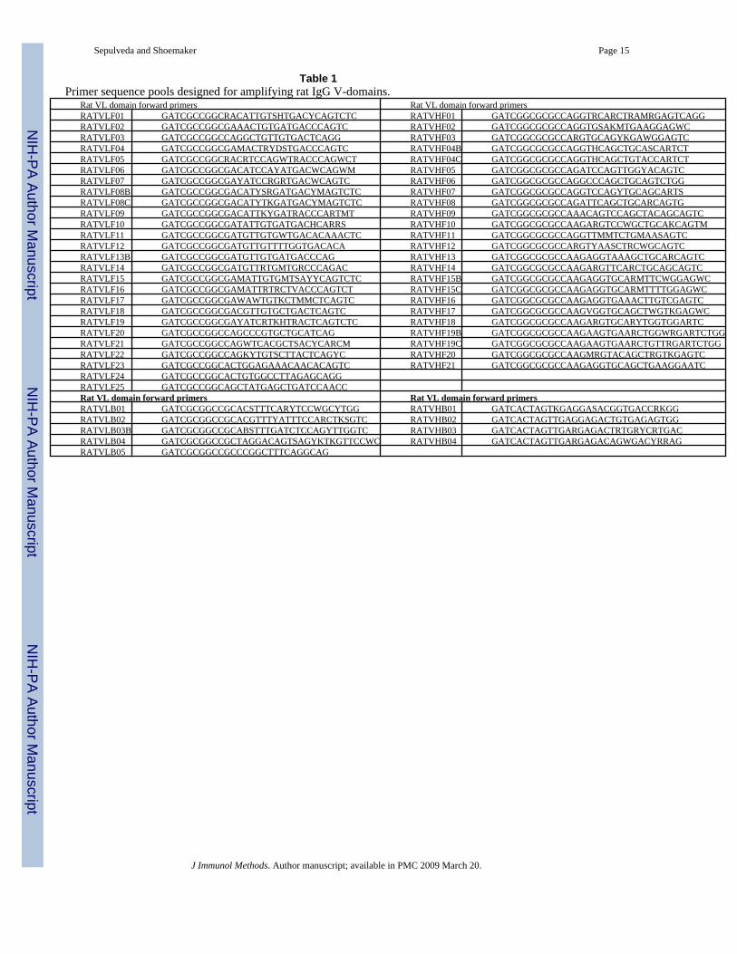
As part of an effort to identify antibodies contributing to the unusual ability of rats to rejectadult Schistosoma mansoni, we sought to obtain a scFv phage display library from schistosomeimmune rats. Unfortunately, rat scFv display libraries have not been previously reported andinformation on the coding sequences of rat immunoglobulin has not been compiled in apractical form for use in the design of appropriate PCR primers for amplification of the VL orVH coding domains. We thus used the numerous available, closely related, murine V-domainsequences as templates to perform BLAST searches of the rat genome and EST databases forVL or VH domain homologues. After analyzing more than 100 complete rat immunoglobulinV-domain homologue sequences it was clear that standard murine VH and VL primers(http://www.mrc-cpe.cam.ac.uk/index.php?module=pagemaster&PAGE_user_op=view_page&PAGE_id=26) would likely fail to amplify most of the rat immunoglobulin coding DNAs and/or introducemutations not present in the selected rat Ig sequences. Thus we designed two sets of degenerateprimers for rat immunoglobulins, one for VL and another for VH (Table 1).
Sepulveda and Shoemaker Page 5
J Immunol Methods. Author manuscript; available in PMC 2009 March 20.
NIH
-PA Author Manuscript
NIH
-PA Author Manuscript
NIH
-PA Author Manuscript

Each of the VL or VH forward oligonucleotide primers (5′-3′) was tested in combination witha pool of the four or five corresponding reverse primers by performing PCR amplification oncDNA prepared from spleen RNA obtained from S. mansoni immune rats. Because of thestrong homology between the different J sequences, using pools of the 3′–5′ primers in the PCRreactions should not significantly reduce the repertoire diversity or create scFv expressionproblems. As shown in the Figure 1A, almost all of the primer combinations yielded a PCRproduct of the expected size (arrows). In reactions yielding additional products of aberrant size,the proper sized DNA fragment was purified by excision and elution from agarose gels. ThePCR products were pooled, attempting to include approximately equal amounts of DNA fromeach reaction, and then the rat VL and VH DNAs were sequentially cloned into the JSC phagedisplay vector to produce a library with a complexity of ~107. More than 90% of the cloneshad full-length scFv inserts and a random sampling of clones was fingerprinted by digestionwith BstN1 to test for evidence of clonal diversity (Fig 1B). As observed, all the tested clonesdiffer from each other. Additional fingerprinting was performed on individual VL and VHregions which, together, indicated that the library contained a broad representation of the ratV-domain repertoire (not shown).
The ability of the rat scFvs to be displayed on phage was also examined. Several single cloneswere induced to produce phage which were then purified and analyzed for the presence of scFv/pIII fusion proteins. Western blotting was performed on extracts of individual phagepreparations using an antibody against an epitopic tag (E-tag) that is fused to all pIII proteinsexpressed from the JSC phagemid vector. The results (not shown) demonstrated that at least60% percent, and probably more, of the randomly picked clones were efficiently displayedusing the phage production methods described in the Materials and Methods. This value issimilar or better to those reported for human scFv libraries (Vaughan et al., 1996; O’Connellet al., 2002). The failure to observe scFv/pIII fusion proteins in some clones is likely causedby the poor expression levels of these scFv fusions, or by aberrant or introduced stop codons.
3.2. Validation of the librarySerum from schistosome immune rats has been shown to be highly enriched in antibodies thatrecognize antigens within the apical tegumental membrane of this parasite (manuscript inpreparation). Two schistosome tetraspanin proteins, Sm23 and SmTsp2, were selected tovalidate the utility of the rat scFv library. These proteins were chosen because they are knownto be immunogenic in schistosome infected animals (Wright et al., 1990; Tran et al., 2006) andbecause they are among the very few integral membrane antigens shown to be present withinthe host-interactive worm surface tegumental membrane (Braschi et al., 2006; Tran et al.,2006) . Four weeks after S. mansoni infection, rats have a moderate titer of serum antibodiesagainst both Sm23 and SmTsp2 (Fig. 2A). This was demonstrated by testing the serum forrecognition of the major extracellular loops of each tetraspanin protein expressed inrecombinant E. coli as a fusion protein to maltose binding protein (Sm23/MBP or SmTsp2/MBP). No recognition of these proteins was detected using serum from uninfected rats (notshown).
To identify phage displayed rat scFvs that recognize epitopes on the major extracellular loopsof S. mansoni tetraspanin Sm23 or SmTsp2, the immune rat scFv library was panned for phagethat bind to an equimolar pool of Sm23/MBP and SmTsp2/MBP. After an initial low stringencypanning, the selected phage pool was subjected to two high stringency pannings (see Materialsand Methods). Employing the same stringency and titer of input phage, we obtained about 100fold more target binding phage in the third cycle compared to the second cycle, indicatingstrong enrichment of phage recognizing the target. Random picks from the third cycle ofpanning were tested by ELISA for their ability to bind to either Sm23/MBP or SmTsp2/MBP.As shown in Fig. 2B, more than 80% of the clones from the third panning cycle produced a
Sepulveda and Shoemaker Page 6
J Immunol Methods. Author manuscript; available in PMC 2009 March 20.
NIH
-PA Author Manuscript
NIH
-PA Author Manuscript
NIH
-PA Author Manuscript
positive signal. Most of the clones clearly and exclusively recognized one or the other of thetwo MBP fusion proteins indicating that recognition was directed at schistosome epitopesrather than MBP. Clones recognizing Sm23 or SmTsp2 were about equally represented.
Individual positive clones were characterized by BstN1 fingerprinting and those with uniquefingerprints were sequenced. The results revealed two unique clusters of rat scFv clonesrecognizing Sm23/MBP and another two recognizing SmTsp2/MBP. Some sequence variationwas found within each of the four clusters but these were almost certainly mutations introducedby PCR after library construction since the VH and VL CDR (complementary determiningregion) sequences were the same. No rat scFvs were found that recognize MBP epitopes. Thefew clones in the initial screen that showed some recognition of both Sm23/MBP and SmTsp2/MBP proved to be mixtures or artifacts (Fig 2B).
A representative of each of the two Sm23-binding scFv clones (named S1 and S3) and the twoSmTsp2-binding scFv clones (T1 and T2) were selected for further study. The amino acidsequences of these four scFvs are shown in Fig. 3. Each has typical VH and VL domainsseparated by the linker region which serves as a flexible spacer to permit the two domains toseparately fold into a conformationally native form. BLAST searching finds that the mosthomologous amino acid sequences within GenBank are other known rat VH and VL domains,despite the preponderance of mouse Ig sequences in the database. These four scFvs were eachexpressed as recombinant proteins lacking the fusion to M13 pIII that exists for the scFvs inphage display vectors (see Materials and Methods). The four soluble scFvs were first testedby western blot for their recognition of either Sm23/MBP and/or SmTsp2/MBP (Fig. 4A). Asexpected, scFvs S1 and S3 recognize Sm23/MBP but not SmTsp2/MBP while scFvs T1 andT2 have the opposite specificity.
The four scFvs were next tested by ELISA for recognition of Sm23/MBP, SmTsp2/MBP andS. mansoni apical membrane extracts (Fig. 4B). As for the western blot, scFvs S1 and S3 scFvsrecognize Sm23/MBP while T1 and T2 scFvs recognize SmTsp2/MBP. Importantly, all fourof the scFvs also recognize antigens within the apical membrane fraction of S. mansoni. Whena pool of rat scFvs S1 and S3 was tested for recognition of apical membrane proteins by westernblot, a single band of about 25 kD was stained (Fig. 4C). This is precisely the SDS-PAGEmobility expected for Sm23 (Reynolds et al., 1992). Western blotting with a pool of T1 andT2 specifically recognizes an apical membrane protein of similar but not identical molecularweight to Sm23 that is expected to be native SmTsp2.
4.0. DiscussionThis study reports the first preparation of a scFv library representing the antibody VH andVL repertoire of rats, and the validation of the library through the isolation and characterizationof scFvs that bind to antigens recognized by the serum of these rats. Although the rat model ishighly valued for research in many areas, it is less studied than the mouse model and, as aresult, there is far less rat immunoglobulin sequence data available and what is available is lesswell compiled. Thus, to design primers that would amplify the vast majority of rat VL andVH coding regions, it was first necessary to perform an extensive search of existing rat genomicand EST sequence data to understand sequence variations that exist within the immunoglobulincoding DNA at the VL and VH primer design sites. Searching was done primarily for rat DNAwith homology to the closely related murine VL and VH sequences. With this information, aportfolio of primers was designed to amplify precisely the same regions of rat VL and VH thathave proven effective in the preparation of mouse scFvs. The primers were tested using ratspleen cDNA deriving from five Fisher rats immunized by multiple infections with the bloodfluke, S. mansoni. The fact that virtually all primer combinations produced products of theexpected size indicates that most of the VL and VH sequences identified by our searches are
Sepulveda and Shoemaker Page 7
J Immunol Methods. Author manuscript; available in PMC 2009 March 20.
NIH
-PA Author Manuscript
NIH
-PA Author Manuscript
NIH
-PA Author Manuscript

actually represented in the rat immunoglobulin repertoire. Thus we believe our primer designwill have utility for researchers wishing to produce recombinant antibodies from rat.
To demonstrate that the rat scFv primer design was robust, a scFv phage display library wasconstructed from an approximately equimolar pool of rat VL and VH DNA amplified by theprimer sets. The VL region was cloned first and the resulting library used as the vector for thesubsequent cloning of the VH coding region. The final library had a complexity of about 107
and most of the clones displayed a full-sized scFv. This library size should be sufficient torepresent a large portion of the antibody repertoire of the schistosome immune rat. We thenselected two different and challenging antigen targets to test the library. For several reasons,we decided to seek scFvs that bind to the Sm23 and SmTsp2 antigens present in the tegumentalmembrane syncytium that surrounds adult S. mansoni parasites. Both tetraspanin proteins werepreviously shown to be immunogenic during schistosome infection in at least one animalmodel. Each of these antigens has a large predicted extracellular loop (EC2) that has been usedpreviously to elicit antibodies against the native tetraspanins and to identify antibodies elicitedto these proteins following infection (Reynolds et al., 1992; Tran et al., 2006). Maltose bindingprotein fusions to EC2 of Sm23 and SmTsp2 were used as the target in our panning selections.Prior to panning, we demonstrated that serum from schistosome-infected rats contained goodantibody titers against both of the MBP/EC2 fusion proteins while naïve rats did not.
The robustness of our rat scFv library construction method seems to be well demonstrated byour ability, in one series of three panning cycles, to identify multiple unique scFvs recognizingeach of the two selected targets. First, the targets are only one of probably hundreds of differentantigens that are immunogenic during schistosome infection. Secondly, they derive fromproteins that are not particularly abundant in the parasites based on proteomic surveys (vanBalkom et al., 2005; Braschi et al., 2006). Thirdly, the epitopes derive from a relatively smallextracellular loop domain (~10kD) contained within these apical membrane proteins, whichare often more difficult than cytosolic or secreted proteins to study as antigens. Fourthly, it ispredicted that the selected tetraspanin EC2 domains are heavily glycosylated andconformationally-constrained which should limit the number of available epitopes that arerepresented by the linear fusion proteins that were used as the panning targets. Finally, theisolated scFvs show excellent specificity for the native tetraspanin targets.
In summary, we have designed a set of primers for the PCR amplification of rat VL and VHcoding DNA and demonstrated that the products of these primers can be used effectively toproduce a robust library of recombinant single-chain antibody fragments that represent theantibody repertoire of the rats that were used as the source of cDNA template.
AcknowledgementsWe greatly appreciate the excellent technical support of Michelle Debatis, the assistance of David Ndegwa inmaintenance of the S. mansoni life cycle and rat infections and . This research was supported by National Institute ofAllergy and Infectious Diseases grant R21-AI061517 (C. S.).
5.0. ReferencesBarker RH Jr, Srivastava BS, Suri P, Goldberg M, Knopf PM. Immunoprecipitation analysis of
radiolabelled protein antigens biosynthesized in vitro by S. mansoni. I. Identification of antigensuniquely recognized by protective antibodies. J Immunol 1985;134:1192–201. [PubMed: 3965570]
Better M, Chang CP, Robinson RR, Horwitz AH. Escherichia coli secretion of an active chimeric antibodyfragment. Science 1988;240:1041–3. [PubMed: 3285471]
Bird RE, Hardman KD, Jacobson JW, Johnson S, Kaufman BM, Lee SM, Lee T, Pope SH, Riordan GS,Whitlow M. Single-chain antigen-binding proteins. Science 1988;242:423–6. [PubMed: 3140379]
Sepulveda and Shoemaker Page 8
J Immunol Methods. Author manuscript; available in PMC 2009 March 20.
NIH
-PA Author Manuscript
NIH
-PA Author Manuscript
NIH
-PA Author Manuscript

Braschi S, Curwen RS, Ashton PD, Verjovski-Almeida S, Wilson A. The tegument surface membranesof the human blood parasite Schistosoma mansoni: a proteomic analysis after differential extraction.Proteomics 2006;6:1471–82. [PubMed: 16447162]
Bruggemann M, Free J, Diamond A, Howard J, Cobbold S, Waldmann H. Immunoglobulin heavy chainlocus of the rat: striking homology to mouse antibody genes. Proc Natl Acad Sci U S A 1986;83:6075–9. [PubMed: 3016742]
Dammers PM, Visser A, Popa ER, Nieuwenhuis P, Bos NA, Kroese FG. Immunoglobulin VH geneanalysis in rat: most marginal zone B cells express germline encoded VH genes and are ligand selected.Curr Top Microbiol Immunol 2000;252:107–17. [PubMed: 11125468]
Davies J, Riechmann L. Single antibody domains as small recognition units: design and in vitro antigenselection of camelized, human VH domains with improved protein stability. Protein Eng 1996;9:531–7. [PubMed: 8862554]
Foord AJ, Muller JD, Yu M, Wang LF, Heine HG. Production and application of recombinant antibodiesto foot-and-mouth disease virus non-structural protein 3ABC. J Immunol Methods 2007;321:142–51.[PubMed: 17328909]
Gilliland LK, Norris NA, Marquardt H, Tsu TT, Hayden MS, Neubauer MG, Yelton DE, Mittler RS,Ledbetter JA. Rapid and reliable cloning of antibody variable regions and generation of recombinantsingle chain antibody fragments. Tissue Antigens 1996;47:1–20. [PubMed: 8929708]
Giudicelli V, Chaume D, Lefranc MP. IMGT/V-QUEST, an integrated software program forimmunoglobulin and T cell receptor V-J and V-D-J rearrangement analysis. Nucleic Acids Res2004;32:W435–40. [PubMed: 15215425]
Huston JS, Levinson D, Mudgett-Hunter M, Tai MS, Novotny J, Margolies MN, Ridge RJ, BruccoleriRE, Haber E, Crea R, et al. Protein engineering of antibody binding sites: recovery of specific activityin an anti-digoxin single-chain Fv analogue produced in Escherichia coli. Proc Natl Acad Sci U S A1988;85:5879–83. [PubMed: 3045807]
Jones PT, Dear PH, Foote J, Neuberger MS, Winter G. Replacing the complementarity-determiningregions in a human antibody with those from a mouse. Nature 1986;321:522–5. [PubMed: 3713831]
Kohler G, Milstein C. Continuous cultures of fused cells secreting antibody of predefined specificity.Nature 1975;256:495–7. [PubMed: 1172191]
Li Y, Kilpatrick J, Whitelam GC. Sheep monoclonal antibody fragments generated using a phage displaysystem. J Immunol Methods 2000;236:133–46. [PubMed: 10699586]
Maass DR, Sepulveda J, Pernthaner A, Shoemaker CB. Alpaca (Lama pacos) as a convenient source ofrecombinant camelid heavy chain antibodies (VHHs). J Immunol Methods 2007;324:13–25.[PubMed: 17568607]
Maddison SE, Norman L, Geiger SJ, Kagan IG. Schistosoma mansoni infection in the rat. I. Worm burdenand serological response in infected, reexposed, and antigen-sensitized animals. J Parasitol1970;56:1058–65. [PubMed: 5504537]
Marks JD, Griffiths AD, Malmqvist M, Clackson TP, Bye JM, Winter G. By-passing immunization:building high affinity human antibodies by chain shuffling. Biotechnology (N Y) 1992;10:779–83.[PubMed: 1368267]
McCafferty J, Griffiths AD, Winter G, Chiswell DJ. Phage antibodies: filamentous phage displayingantibody variable domains. Nature 1990;348:552–4. [PubMed: 2247164]
O’Connell D, Becerril B, Roy-Burman A, Daws M, Marks JD. Phage versus phagemid libraries forgeneration of human monoclonal antibodies. J Mol Biol 2002;321:49–56. [PubMed: 12139932]
Reynolds SR, Shoemaker CB, Harn DA. T and B cell epitope mapping of SM23, an integral membraneprotein of Schistosoma mansoni. J Immunol 1992;149:3995–4001. [PubMed: 1281197]
Ridder R, Schmitz R, Legay F, Gram H. Generation of rabbit monoclonal antibody fragments from acombinatorial phage display library and their production in the yeast Pichia pastoris. Biotechnology(N Y) 1995;13:255–60. [PubMed: 9634767]
Schluter SF, Jensen I, Ramsland PA, Marchalonis JJ. Recombinant shark natural antibodies tothyroglobulin. J Mol Recognit 2005;18:404–12. [PubMed: 15954089]
Skerra A, Pluckthun A. Assembly of a functional immunoglobulin Fv fragment in Escherichia coli.Science 1988;240:1038–41. [PubMed: 3285470]
Sepulveda and Shoemaker Page 9
J Immunol Methods. Author manuscript; available in PMC 2009 March 20.
NIH
-PA Author Manuscript
NIH
-PA Author Manuscript
NIH
-PA Author Manuscript
Smithers SR, Terry RJ. The infection of laboratory hosts with cercariae of Schistosoma mansoni and therecovery of the adult worms. Parasitology 1965;55:695–700. [PubMed: 4957633]
Tomlinson IM, Walter G, Marks JD, Llewelyn MB, Winter G. The repertoire of human germline VHsequences reveals about fifty groups of VH segments with different hypervariable loops. J Mol Biol1992;227:776–98. [PubMed: 1404388]
Tran MH, Pearson MS, Bethony JM, Smyth DJ, Jones MK, Duke M, Don TA, McManus DP, Correa-Oliveira R, Loukas A. Tetraspanins on the surface of Schistosoma mansoni are protective antigensagainst schistosomiasis. Nat Med 2006;12:835–40. [PubMed: 16783371]
van Balkom BW, van Gestel RA, Brouwers JF, Krijgsveld J, Tielens AG, Heck AJ, van Hellemond JJ.Mass spectrometric analysis of the Schistosoma mansoni tegumental sub-proteome. J Proteome Res2005;4:958–66. [PubMed: 15952743]
Vaughan TJ, Williams AJ, Pritchard K, Osbourn JK, Pope AR, Earnshaw JC, McCafferty J, Hodits RA,Wilton J, Johnson KS. Human antibodies with sub-nanomolar affinities isolated from a large non-immunized phage display library. Nat Biotechnol 1996;14:309–14. [PubMed: 9630891]
Wright MD, Henkle KJ, Mitchell GF. An immunogenic Mr 23,000 integral membrane protein ofSchistosoma mansoni worms that closely resembles a human tumor-associated antigen. J Immunol1990;144:3195–200. [PubMed: 2324498]
Zhao Y, Hammarstrom L. Cloning of the complete rat immunoglobulin delta gene: evolutionaryimplications. Immunology 2003;108:288–95. [PubMed: 12603594]
Sepulveda and Shoemaker Page 10
J Immunol Methods. Author manuscript; available in PMC 2009 March 20.
NIH
-PA Author Manuscript
NIH
-PA Author Manuscript
NIH
-PA Author Manuscript

Figure 1.Testing PCR primer pairs for amplification of rat VL and VH coding DNA. A) PCRamplification of rat spleen cDNA with each of the different forward primers from Table 1 incombination with a pool of the corresponding reverse primers. For VL domain amplification,26 forward primers were tested with the pool of 5 reverse primers. For VH domainamplification, 23 forward primers were tested with the pool of 4 reverse primers. Reactionproducts were analyzed by 1% agarose gel electrophoresis. Arrows show the expected PCRproduct (~400 bp). B) Fingerprint analysis of randomly selected single clones. The ~1200 bpV region coding DNA from random clones was amplified by PCR, digested with BstN1 enzymeand analyzed by 2% agarose gel electrophoresis.
Sepulveda and Shoemaker Page 11
J Immunol Methods. Author manuscript; available in PMC 2009 March 20.
NIH
-PA Author Manuscript
NIH
-PA Author Manuscript
NIH
-PA Author Manuscript
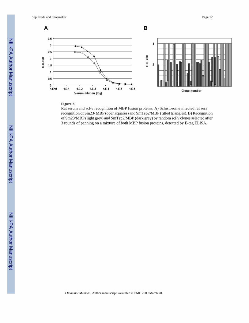
Figure 2.Rat serum and scFv recognition of MBP fusion proteins. A) Schistosome infected rat serarecognition of Sm23/ MBP (open squares) and SmTsp2/MBP (filled triangles). B) Recognitionof Sm23/MBP (light grey) and SmTsp2/MBP (dark grey) by random scFv clones selected after3 rounds of panning on a mixture of both MBP fusion proteins, detected by E-tag ELISA.
Sepulveda and Shoemaker Page 12
J Immunol Methods. Author manuscript; available in PMC 2009 March 20.
NIH
-PA Author Manuscript
NIH
-PA Author Manuscript
NIH
-PA Author Manuscript
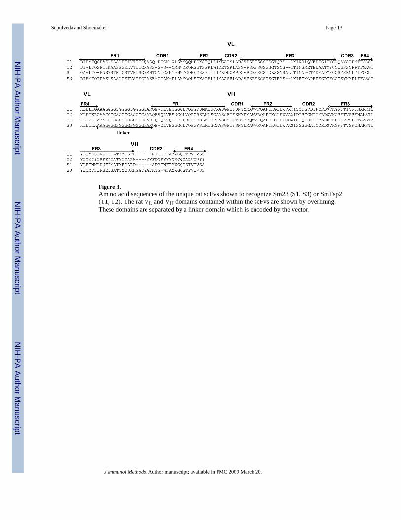
Figure 3.Amino acid sequences of the unique rat scFvs shown to recognize Sm23 (S1, S3) or SmTsp2(T1, T2). The rat VL and VH domains contained within the scFvs are shown by overlining.These domains are separated by a linker domain which is encoded by the vector.
Sepulveda and Shoemaker Page 13
J Immunol Methods. Author manuscript; available in PMC 2009 March 20.
NIH
-PA Author Manuscript
NIH
-PA Author Manuscript
NIH
-PA Author Manuscript

Figure 4.Specificity of the rat scFv for the target schistosome antigens. A) Western blot analysis of therat scFv recognition of Sm23/MBP and SmTsp2/MBP. MBP-fusion protein were separated bya 12% PAGE-SDS, incubated overnight with scFv and detected with anti-E-tag mAb. Lane 1contains purified Sm23/MBP, Lane 2 contains purified SmTsp2/MBP. The positions ofmolecular weight markers are shown to the right. B) ELISA analysis of scFv (30nM)recognition of Sm23/MBP (grey bars), SmTsp2/MBP (striped bars) and S. mansoni tegumentalextract (black bars). C) Western blot analysis of rat scFv recognition of S. mansoni tegumentalantigens. Lane 1 used a control rat scFv. Lane 2 used a pool of scFvs S1 and S3. Lane 3 useda pool of scFvs T1 and T2. The tegumental extract was resolved by 12% PAGE-SDS. BoundscFv was detected with anti-E-tag mAb.
Sepulveda and Shoemaker Page 14
J Immunol Methods. Author manuscript; available in PMC 2009 March 20.
NIH
-PA Author Manuscript
NIH
-PA Author Manuscript
NIH
-PA Author Manuscript
NIH
-PA Author Manuscript
NIH
-PA Author Manuscript
NIH
-PA Author Manuscript
Sepulveda and Shoemaker Page 15
Table 1Primer sequence pools designed for amplifying rat IgG V-domains.
Rat VL domain forward primers Rat VL domain forward primersRATVLF01 GATCGCCGGCRACATTGTSHTGACYCAGTCTC RATVHF01 GATCGGCGCGCCAGGTRCARCTRAMRGAGTCAGGRATVLF02 GATCGCCGGCGAAACTGTGATGACCCAGTC RATVHF02 GATCGGCGCGCCAGGTGSAKMTGAAGGAGWCRATVLF03 GATCGCCGGCCAGGCTGTTGTGACTCAGG RATVHF03 GATCGGCGCGCCARGTGCAGYKGAWGGAGTCRATVLF04 GATCGCCGGCGAMACTRYDSTGACCCAGTC RATVHF04B GATCGGCGCGCCAGGTHCAGCTGCASCARTCTRATVLF05 GATCGCCGGCRACRTCCAGWTRACCCAGWCT RATVHF04C GATCGGCGCGCCAGGTHCAGCTGTACCARTCTRATVLF06 GATCGCCGGCGACATCCAYATGACWCAGWM RATVHF05 GATCGGCGCGCCAGATCCAGTTGGYACAGTCRATVLF07 GATCGCCGGCGAYATCCRGRTGACWCAGTC RATVHF06 GATCGGCGCGCCAGGCCCAGCTGCAGTCTGGRATVLF08B GATCGCCGGCGACATYSRGATGACYMAGTCTC RATVHF07 GATCGGCGCGCCAGGTCCAGYTGCAGCARTSRATVLF08C GATCGCCGGCGACATYTKGATGACYMAGTCTC RATVHF08 GATCGGCGCGCCAGATTCAGCTGCARCAGTGRATVLF09 GATCGCCGGCGACATTKYGATRACCCARTMT RATVHF09 GATCGGCGCGCCAAACAGTCCAGCTACAGCAGTCRATVLF10 GATCGCCGGCGATATTGTGATGACHCARRS RATVHF10 GATCGGCGCGCCAAGARGTCCWGCTGCAKCAGTMRATVLF11 GATCGCCGGCGATGTTGTGWTGACACAAACTC RATVHF11 GATCGGCGCGCCAGGTTMMTCTGMAASAGTCRATVLF12 GATCGCCGGCGATGTTGTTTTGGTGACACA RATVHF12 GATCGGCGCGCCARGTYAASCTRCWGCAGTCRATVLF13B GATCGCCGGCGATGTTGTGATGACCCAG RATVHF13 GATCGGCGCGCCAAGAGGTAAAGCTGCARCAGTCRATVLF14 GATCGCCGGCGATGTTRTGMTGRCCCAGAC RATVHF14 GATCGGCGCGCCAAGARGTTCARCTGCAGCAGTCRATVLF15 GATCGCCGGCGAMATTGTGMTSAYYCAGTCTC RATVHF15B GATCGGCGCGCCAAGAGGTGCARMTTCWGGAGWCRATVLF16 GATCGCCGGCGAMATTRTRCTVACCCAGTCT RATVHF15C GATCGGCGCGCCAAGAGGTGCARMTTTTGGAGWCRATVLF17 GATCGCCGGCGAWAWTGTKCTMMCTCAGTC RATVHF16 GATCGGCGCGCCAAGAGGTGAAACTTGTCGAGTCRATVLF18 GATCGCCGGCGACGTTGTGCTGACTCAGTC RATVHF17 GATCGGCGCGCCAAGVGGTGCAGCTWGTKGAGWCRATVLF19 GATCGCCGGCGAYATCRTKHTRACTCAGTCTC RATVHF18 GATCGGCGCGCCAAGARGTGCARYTGGTGGARTCRATVLF20 GATCGCCGGCCAGCCCGTGCTGCATCAG RATVHF19B GATCGGCGCGCCAAGAAGTGAARCTGGWRGARTCTGGRATVLF21 GATCGCCGGCCAGWTCACGCTSACYCARCM RATVHF19C GATCGGCGCGCCAAGAAGTGAARCTGTTRGARTCTGGRATVLF22 GATCGCCGGCCAGKYTGTSCTTACTCAGYC RATVHF20 GATCGGCGCGCCAAGMRGTACAGCTRGTKGAGTCRATVLF23 GATCGCCGGCACTGGAGAAACAACACAGTC RATVHF21 GATCGGCGCGCCAAGAGGTGCAGCTGAAGGAATCRATVLF24 GATCGCCGGCACTGTGGCCTTAGAGCAGGRATVLF25 GATCGCCGGCAGCTATGAGCTGATCCAACCRat VL domain forward primers Rat VL domain forward primersRATVLB01 GATCGCGGCCGCACSTTTCARYTCCWGCYTGG RATVHB01 GATCACTAGTKGAGGASACGGTGACCRKGGRATVLB02 GATCGCGGCCGCACGTTTYATTTCCARCTKSGTC RATVHB02 GATCACTAGTTGAGGAGACTGTGAGAGTGGRATVLB03B GATCGCGGCCGCABSTTTGATCTCCAGYTTGGTC RATVHB03 GATCACTAGTTGARGAGACTRTGRYCRTGACRATVLB04 GATCGCGGCCGCTAGGACAGTSAGYKTKGTTCCWC RATVHB04 GATCACTAGTTGARGAGACAGWGACYRRAGRATVLB05 GATCGCGGCCGCCCGGCTTTCAGGCAG
J Immunol Methods. Author manuscript; available in PMC 2009 March 20.
Copyright © 2022 FDOKUMEN