Partnering for Mutual Success: DaimlerChrysler – POEMAtec ...

Vascular Immunotargeting to Endothelial DeterminantICAM-1 Enables Optimal Partnering of RecombinantscFv-Thrombomodulin Fusion with Endogenous CofactorColin F. Greineder1,2,4, Ann-Marie Chacko2,3, Sergei Zaytsev1,2, Blaine J. Zern1,2, Ronald Carnemolla1,2,
Elizabeth D. Hood1,2, Jingyan Han1,2¤a, Bi-Sen Ding1,2¤b, Charles T. Esmon5, Vladimir R. Muzykantov1,2*
1 Department of Pharmacology, Perelman School of Medicine, University of Pennsylvania, Philadelphia, Pennsylvania, United States of America, 2 Center for Targeted
Therapeutics and Translational Nanomedicine, Institute for Translational Medicine and Therapeutics, Perelman School of Medicine, University of Pennsylvania,
Philadelphia, Pennsylvania, United States of America, 3 Department of Radiology, Perelman School of Medicine, University of Pennsylvania, Philadelphia, Pennsylvania,
United States of America, 4 Department of Emergency Medicine, Perelman School of Medicine, University of Pennsylvania, Philadelphia, Pennsylvania, United States of
America, 5 Cardiovascular Biology Research Program, Oklahoma Medical Research Foundation, Howard Hughes Medical Institute, Oklahoma City, Oklahoma, United States
of America
Abstract
The use of targeted therapeutics to replenish pathologically deficient proteins on the luminal endothelial membrane hasthe potential to revolutionize emergency and cardiovascular medicine. Untargeted recombinant proteins, like activatedprotein C (APC) and thrombomodulin (TM), have demonstrated beneficial effects in acute vascular disorders, but have failedto have a major impact on clinical care. We recently reported that TM fused with an scFv antibody fragment to plateletendothelial cell adhesion molecule-1 (PECAM-1) exerts therapeutic effects superior to untargeted TM. PECAM-1 is localizedto cell-cell junctions, however, whereas the endothelial protein C receptor (EPCR), the key co-factor of TM/APC, is exposed inthe apical membrane. Here we tested whether anchoring TM to the intercellular adhesion molecule (ICAM-1) favors scFv/TMcollaboration with EPCR. Indeed: i) endothelial targeting scFv/TM to ICAM-1 provides ,15-fold greater activation of proteinC than its PECAM-targeted counterpart; ii) blocking EPCR reduces protein C activation by scFv/TM anchored to endothelialICAM-1, but not PECAM-1; and iii) anti-ICAM scFv/TM fusion provides more profound anti-inflammatory effects than anti-PECAM scFv/TM in a mouse model of acute lung injury. These findings, obtained using new translational constructs,emphasize the importance of targeting protein therapeutics to the proper surface determinant, in order to optimize theirmicroenvironment and beneficial effects.
Citation: Greineder CF, Chacko A-M, Zaytsev S, Zern BJ, Carnemolla R, et al. (2013) Vascular Immunotargeting to Endothelial Determinant ICAM-1 EnablesOptimal Partnering of Recombinant scFv-Thrombomodulin Fusion with Endogenous Cofactor. PLoS ONE 8(11): e80110. doi:10.1371/journal.pone.0080110
Editor: Michael Koval, Emory University School of Medicine, United States of America
Received August 27, 2013; Accepted October 7, 2013; Published November 14, 2013
Copyright: � 2013 Greineder et al. This is an open-access article distributed under the terms of the Creative Commons Attribution License, which permitsunrestricted use, distribution, and reproduction in any medium, provided the original author and source are credited.
Funding: This work was supported by a Junior Investigator Preliminary/Feasibility Grant from the Institute of Translational Medicine and Therapeutics (ITMAT) atthe University of Pennsylvania (CFG), as well as an American Heart Association Fellow to Faculty grant 10FTF4150053 (CFG), and National Institutes of Health GrantR01-HL091950 (VRM). The funders had no role in study design, data collection and analysis, decision to publish, or preparation of the manuscript.
Competing Interests: The authors have declared that no competing interests exist.
* E-mail: [email protected]
¤a Current address: Vascular Biology Section, Department of Medicine, Boston University, Boston, Massachusetts, United States of America¤b Current address: Howard Hughes Medical Institute, Department of Genetic Medicine, Weill Cornell Medical College, New York, New York, United States ofAmerica
Introduction
A variety of endogenous endothelial proteins project into the
vascular lumen and mediate critical homeostatic pathways,
helping to maintain blood fluidity, control vascular tone and
permeability, and regulate the innate immune response[1]. Several
decades of research have demonstrated that loss or functional
deficit of these proteins underlies the pathogenesis of a variety of
human illnesses[2–4]. Molecular therapies capable of replenishing
these proteins have the potential to achieve long-sought improve-
ments in the morbidity and mortality of conditions like sepsis and
acute lung injury. Two distinct approaches – gene therapy and
infusion of recombinant proteins – have been pursued as means to
achieve this goal. While the former may be the best suited for long-
term correction of chronic deficiencies, temporal considerations
make recombinant protein therapeutics the most useful in treating
acute vascular disorders.
Results of numerous preclinical and clinical studies support the
therapeutic potential of these strategies and provide incentives and
directions for further refinements. Since neither transfected gene
products nor infused therapeutics typically result in accumulation
of proteins at the sites where they exert the optimal effect,
achieving proper localization to selected cell types and/or sub-
cellular compartments is a key objective for both approaches[5].
Thrombomodulin (TM, CD141) is an endothelial transmem-
brane glycoprotein, which plays a critical role in regulating
inflammation and thrombosis at the vascular margin. TM binds
thrombin and blocks its pro-thrombotic and pro-coagulant
activities towards fibrinogen, Factor V and protease-activated
receptors in platelets and endothelium. Instead, the TM/thrombin
complex preferentially cleaves plasma protein C and generates
PLOS ONE | www.plosone.org 1 November 2013 | Volume 8 | Issue 11 | e80110

APC, a serine protease with multifaceted anti-thrombotic and anti-
inflammatory activities[6].
In this way, TM plays a key role in maintaining vascular
homeostasis, matching the pro-coagulant and pro-inflammatory
actions of thrombin with the anti-coagulant and anti-inflammatory
actions of APC[7–9]. This balance is disrupted in a variety of
disease states, in which TM is suppressed. Inflammatory media-
tors, oxidants, and leukocyte proteases have all been implicated in
this pathologic process, either through internalization, cleavage,
transcriptional regulation, or inactivation of TM on the endothe-
lial surface[10–13]. Endothelial TM loss has been demonstrated in
human patients suffering from sepsis, atherosclerosis, cardiopul-
monary bypass, ischemia/reperfusion injury, and cardiac ar-
rest[14–18].
Gene therapy studies in animals have demonstrated beneficial
effects of augmenting endothelial expression of TM[19–21]. In the
acute or emergent setting, where gene therapy is not feasible,
infusions of recombinant human APC or soluble TM (sTM) have
been tested clinically in patients with severe sepsis, acute lung
injury, and disseminated intravascular coagulation[22–24]. The
initial excitement generated by these untargeted therapeutics,
however, has been tempered by their lack of spatiotemporal
control, narrow therapeutic window, and limited efficacy[25].
It seems logical to try to anchor TM to its natural site, i.e., the
luminal surface of endothelial cells. This has been achieved using
vascular immunotargeting directed to a well characterized, safe,
and non-internalized endothelial determinant, the platelet endo-
thelial cell adhesion molecule-1 (PECAM-1, CD31). TM fused
with an scFv fragment of anti-PECAM binds to endothelium,
exerts therapeutic effects, and has a benefit/risk ratio superior to
untargeted TM in two separate animal models of acute lung
injury[26].
The justification for targeting recombinant TM to endothelial
cells is not simply one of pharmacokinetics. Endothelial cells
express a key co-factor of the TM/APC system, namely, the
Endothelial Protein C Receptor (EPCR, CD201). EPCR markedly
enhances APC production by the TM/thrombin complex and
mediates endothelial protective and barrier enhancing effects of
APC[27,28]. Endogenous TM and EPCR are believed to be
concentrated in the lipid rafts in the endothelial apical plasma-
lemma[29,30], whereas PECAM-1 is localized at cell-cell junc-
tions, raising questions about the ability of PECAM-anchored TM
to effectively partner with endogenous EPCR[31,32]. In contrast,
the intercellular adhesion molecule-1 (ICAM-1, CD54) is exposed
in the apical endothelial plasmalemma, in close proximity to
EPCR[33].
Given the critical role that EPCR plays in the endothelial
protective effects of the TM/APC pathway, effective partnering
with this endogenous co-factor may favor endothelial-targeted TM
therapeutics. In this study, we find that recombinant TM
anchored to endothelial PECAM-1 does not partner with EPCR,
unlike endogenous TM. We describe a new fusion protein, which
targets TM to ICAM-1, and show that this endothelial determi-
nant, unlike PECAM-1, allows partnering of recombinant TM
with endogenous EPCR. We extend these in vitro observations to a
model of acute pulmonary inflammation and find that, at an
equivalent dose, anti-ICAM scFv/TM provides more robust
protection than anti-PECAM scFv/TM. This work reveals the
importance of rational selection of cellular targets for biological
therapeutics, capitalizing on nanometer-scale membrane topology
to allow optimal interaction with endogenous partners.
Materials and Methods
Ethics StatementAnimal studies were carried out in accordance with the Guide
for the Care and Use of Laboratory Animals as adopted by the
NIH, under protocols (803320 and 804349) approved by
University of Pennsylvania IACUC.
Cell linesYN1 hybridoma and MS1 cells were purchased from ATCC
(Manassas, VA). YN1 cells were cultured in RPMI 1640
supplemented with 10% (v/v) fetal bovine serum (FBS). MS1
cells were maintained in DMEM with 10% FBS and 1X
antibiotic-antimycotic (Life technologies, Grand Island, NY).
Antibodies and other reagentsPurified anti-PECAM (390) and anti-ICAM (YN1/1.7.4)
antibodies were obtained from BioLegend (San Diego, CA).
Anti-TM polyclonal antibody (AF3894) and anti-EPCR polyclonal
antibody (AF2749) were purchased from R&D systems (Minne-
apolis, MN). Anti-EPCR blocking antibody, mAb1560, was
supplied by the Esmon laboratory. HRP-conjugated Anti-FLAG
(M2-HRP) antibody was obtained from Sigma Aldrich (St Louis,
MO). Alexa Flour-labeled secondary antibodies were purchased
from Life Technologies (Grand Island, NY). PPACK-inactivated
thrombin was a generous gift of Sriram Krishnaswamy. Bovine
thrombin, LPS (serotype B4), and mouse TNF were purchased
from Sigma. Human protein C zymogen was obtained from
Haematologic Technologies (Essex Junction, VT). APC substrate
S-2366 was purchased from Diapharma (West Chester, OH).
Endothelial cell immunofluorescence stainingMS1 cell monolayers were grown in 8 well m-slides (Ibidi,
Verona, WI) and fixed for 20 minutes at room temperature (RT)
with Histochoice (Amresco, Solon, OH). In some cases, cells were
treated with 10 ng/mL mouse TNF for 8 hours prior to fixation.
After three washes, cells were blocked with 3%(w/v) BSA in HBSS
for 1 hour at RT. Cells were stained with either anti-PECAM
(390, 15 ug/mL) or anti-ICAM (YN1, 1 ug/mL) monoclonal
antibodies, in addition to polyclonal goat anti-mouse EPCR
(0.5 ug/mL) for 2 hours at RT. Cells were washed three times
with 0.1% Tween in HBSS and then stained with Alexa Fluor 594
anti-rat (1:200) and Alexa Fluor 488 anti-goat (1:1000). After
1 hour incubation, cells were washed four times with 0.1% Tween
in HBSS and once in PBS. ProLong Gold Antifade reagent with
DAPI (Life technologies, Grand Island, NY) and a coverslip were
applied and cells were allowed to dry overnight prior to
immunofluorescence imaging.
Cloning of anti-ICAM VL and VH cDNAsTotal cellular RNA was isolated from YN1 hybridoma cells
using the RNeasy kit (Qiagen, Valencia, CA). Combined reverse
transcription and PCR was performed using SuperScript One
Step RT-PCR kit (Life Technologies, Grand Island, NY). A single
full length VH cDNA was produced using degenerate 59
framework region 1 (FR1) primers and a 39 constant region
primer[34]. This approach was not possible for the light chain, as
PCR utilizing FR1 primers produced only the previously reported
Y3-Ag 1.2.3 myeloma VL sequence[35]. An 8 amino acid peptide
unique to the ICAM-specific VL was identified using mass
spectrometry, and using degenerate primers corresponding to this
peptide, a full length VL cDNA was cloned (Figure S1).
Targeting TM to ICAM-1 Allows Partnering with EPCR
PLOS ONE | www.plosone.org 2 November 2013 | Volume 8 | Issue 11 | e80110

Assembly and expression of anti-ICAM scFv and anti-ICAM scFv/TM constructs
Completed anti-ICAM VL and VH cDNAs were assembled into
constructs encoding anti-ICAM scFv and the anti-ICAM scFv/
TM fusion protein. In each case, VH and VL sequences were
separated by a (GGGGS)3 linker, and a triple FLAG tag was
appended to the 39 end (C terminus) for purposes of purification
and detection. The anti-ICAM/TM construct was designed to be
identical to that of anti-PECAM/TM, with the anti-ICAM scFv
on the 59 end (N terminus), separated from the extracellular
domain of TM (amino acids Leu17-Ser517) by an (SSSSG)2AAA
linker. Both proteins were expressed in S2 cells and purified using
a C-terminal triple FLAG tag. Purity was assessed using SDS-
PAGE.
Generation of REN-derived Stable Cell LinesREN-mICAM cells. A full-length cDNA for mouse ICAM-1
was purchased from Thermo Scientific (Rockford, IL). The clone
was sequenced and found to contain the entire coding sequence of
mouse ICAM-1 and a portion of the 59 and 39 UTRs (nt 46-2440)
between EcoRI and XbaI restriction enzyme sites. The clone was
excised and ligated into the pcDNA3 mammalian expression
vector, and transfected into REN cells using Lipofectamine 2000
(Life Technologies, Grand Island, NY). Stably expressing cells
were selected in media containing 200 ug/mL of Geneticin (Life
Technologies, Grand Island, NY).
REN-mPECAM-mEPCR and REN-mICAM-mEPCR
cells. A vector containing the entire coding sequence of mouse
EPCR and a portion of the 59 and 39 UTRs (nt 171–1413) was
obtained from the Esmon laboratory[36]. The EPCR cDNA was
excised using XbaI and EcoRI and ligated into the pcDNA3.1/
Zeo(-) vector (Life Technologies, Grand Island, NY). Since REN-
PECAM and REN-ICAM cells already stably express the
Geneticin resistance gene, this expression vector (which confers
resistance to the antibiotic Zeocin) was utilized. Each cell type was
transfected with Lipofectamine 2000 and selected in media with
250 ug/mL of Zeocin.
Live Cell ELISA AssaysEnzyme-linked Immunosorbent Assays (ELISAs) were per-
formed on live cells as previously described[37], although in the
experiments reported here, cell monolayers were incubated with
increasing concentrations of scFv or scFv/TM fusion protein
rather than whole antibodies. Since all fusion proteins carry a C-
terminal triple FLAG tag, anti-FLAG (M2)-peroxidase (HRP)
conjugate was used as a detection antibody. In experiments
involving MS1 endothelial cells, specific binding of anti-PECAM/
TM or anti-ICAM/TM fusion proteins was assessed by co-
incubation with 10-fold excess of their parental antibodies (390
and YN1/1.7.4, respectively). ELISA binding data was analyzed
and binding parameters (EC50) were determined using PRISM 6.0
software (GraphPad, San Diego, CA)[37].
Radioimmunoassays (RIAs) using 125I-labeled AntibodiesAnti-PECAM (390) and anti-ICAM (YN1/1.7.4) monoclonal
Abs were directly radioiodinated with [125I]NaI (Perkin Elmer,
Waltham, MA) and purified using Zeba desalting spin columns
(ThermoScientific). In all cases, radiolabeling efficiency was .75%
and free iodine was ,5%, post-purification. RIAs were performed
and binding parameters (Kd, Bmax) determined as previously
reported[37].
Protein C Activation AssaysGeneration of Activated Protein C (APC) by scFv/TM fusion
was assayed as previously described[37]. In soluble APC
generation experiments, fusion proteins were mixed with 0.5 nM
thrombin and 1mM protein C in a micro-Eppendorf tube, whereas
cell-bound assays were conducted by incubating monolayers with
scFv/TM fusion protein and washing x 3 with media prior to the
addition of 1 nM thrombin and 100 nM protein C. In all cases,
protein C activation occurred @ 37C in assay buffer (20 mM Tris,
100 mM NaCl, 1 mM CaCl2, 0.1% (w/v) BSA, pH 7.5) and the
reaction was stopped by addition of an excess of hirudin. In
experiments involving MS1 cells, the monolayer was first treated
with anti-TM antibody to block endogenous TM and then washed
x 3 prior to incubation with scFv/TM fusion protein. The amount
of APC generated by cell-bound fusions in these experiments was
normalized to the number of binding sites per cell, as determined
in MS1 RIAs (approximately 240,000/cell for PECAM-1 and
12,000/cell for ICAM-1). In experiments involving EPCR
blockade, cells were incubated with 300 nM of anti-EPCR
antibody (Ab1560) for 15 minutes prior to the addition of protein
C and thrombin. This antibody has been well characterized and is
known to inhibit approximately 70% of protein C binding,
eliminating to substantial extent the ability of EPCR to accelerate
the activation of protein C by the thrombin-TM complex [38].
Intratracheal (IT) LPS ModelC57BL/6 male mice weighing 20-30 g were anaesthetized and
placed in a supine position. Acute lung injury was induced via IT
injection of 2 mg/kg of endotoxin in a volume of 100 uL of PBS.
Endotoxin injection was followed immediately by injection of
150 uL of air, to ensure even distribution of LPS throughout all
distal airspaces. Anti-PECAM scFv/TM, anti-ICAM scFv/TM, or
PBS vehicle were injected intravenously 30 minutes prior to LPS
administration. In relevant experiments, 125I-labeled albumin was
also injected intravenously 5 minutes prior to LPS administration.
6 hours after induction of lung injury, blood was withdrawn from
the inferior vena cava and animals were euthanized. In
experiments involving tracing of 125I-labeled albumin, a catheter
was placed in the pulmonary artery and the pulmonary circulation
was gently flushed with PBS prior to the harvesting of organs.
Bronchoalveolar lavage was performed via a 19-gauge stainless
steel catheter (Harvard Apparatus, Holliston, MA) placed in the
trachea and secured via a 5-0 silk suture. Each animal was lavaged
twice with 0.8cc of ice-cold PBS. The lavages were pooled and
MIP-2 was quantified using a Quantikine ELISA kit (R&D
systems, Minneapolis, MN). For quantification of VCAM-1 and E-
selectin mRNA, lungs were homogenized with steel beads (Sigma)
and a Tissue Lyser II (Qiagen). Total RNA was isolated with
RNeasy kit and cDNA was synthesized using the Transcriptor 1st
Strand cDNA Synthesis Kit (Roche Applied Science, Indianapolis,
IN). qPCR was performed using the FastStart DNA MasterPLUS
kit (SYBR green) and a Lightcycler 1.5 carousel-based system
(Roche Applied Science). Validated Quantitect primers for mouse
VCAM-1, E-selectin, and actin (housekeeping control) were
utilized (Qiagen, Valencia, CA).
Data analysis and statisticsResults are expressed as mean 6 SD unless otherwise noted.
Significant differences between means were determined using one-
way ANOVA followed by appropriate multiple comparison
(Tukey) test. P,0.05 was considered statistically significant.
Targeting TM to ICAM-1 Allows Partnering with EPCR
PLOS ONE | www.plosone.org 3 November 2013 | Volume 8 | Issue 11 | e80110

Results
PECAM-1 vs. ICAM-1: Relative proximity to EPCR andnumber of binding sites on endothelial surface
In order to assess the relative proximity of EPCR to PECAM-1
and ICAM-1, mouse MS1 endothelial cells were stained for each
antigen and imaged using a fluorescence microscope (Figure 1a).
In agreement with previous reports, most PECAM-1 staining
occurred at cell-cell borders[31], with minimal overlap with
EPCR. In contrast, there was gross overlap of staining for ICAM-1
and that for EPCR (Figure 1a).
Next, we studied binding to MS1 cells of 125Iodine-labeled
PECAM and ICAM antibodies used to produce scFv to fuse with
TM. Anti-ICAM and anti-PECAM had similar high affinities (Kd
of approx. 0.12 and 0.22 nM, respectively), although PECAM-1
provided ,20-fold more binding sites than ICAM-1 (approxi-
mately 240,000/cell vs. 12,000/cell, respectively), reflecting a
substantial difference in the level of cell surface expression of these
molecules (Figure 1b). According to these data, MS1 have a lower
number of anti-PECAM binding sites than human umbilical vein
endothelial cells (HUVECs, ,106 binding sites per cell[39]), which
may reflect innate differences between these cell lines. In
particular, MS1 cells are smaller than HUVEC, so their number
of PECAM copies per cell surface area may be fairly similar.
Binding of ICAM and PECAM-targeted scFv/TM fusionproteins
We next constructed an anti-ICAM-1 antibody fragment (scFv)
and fused it to the extracellular domain of TM (Figure S1). In
order to compare the binding of the anti-PECAM/TM and anti-
ICAM/TM fusion proteins, we utilized REN cells stably
expressing either mouse PECAM-1 or ICAM-1. The human
mesothelioma cell line REN is a useful model system, with no
expression of mouse PECAM, ICAM, TM, or EPCR at baseline
(Figure S2). Both anti-PECAM scFv and anti-ICAM scFv, as well
as their respective TM fusion proteins, demonstrated sub-
nanomolar affinity to REN cells expressing their corresponding
target molecule. Little or no non-specific binding was seen to wild
type REN cells (Figure 2).
Figure 1. Localization of PECAM-1, ICAM-1, and EPCR on MS1 mouse endothelial cells. (a) Immunofluorescence images demonstratesuperior co-localization of ICAM-1 and EPCR, as compared to PECAM-1 and EPCR. (b) Radioimmunoassay data using 125I-labeled anti-PECAM and anti-ICAM parental antibodies to determine affinity and number of binding sites per endothelial cell.doi:10.1371/journal.pone.0080110.g001
Targeting TM to ICAM-1 Allows Partnering with EPCR
PLOS ONE | www.plosone.org 4 November 2013 | Volume 8 | Issue 11 | e80110
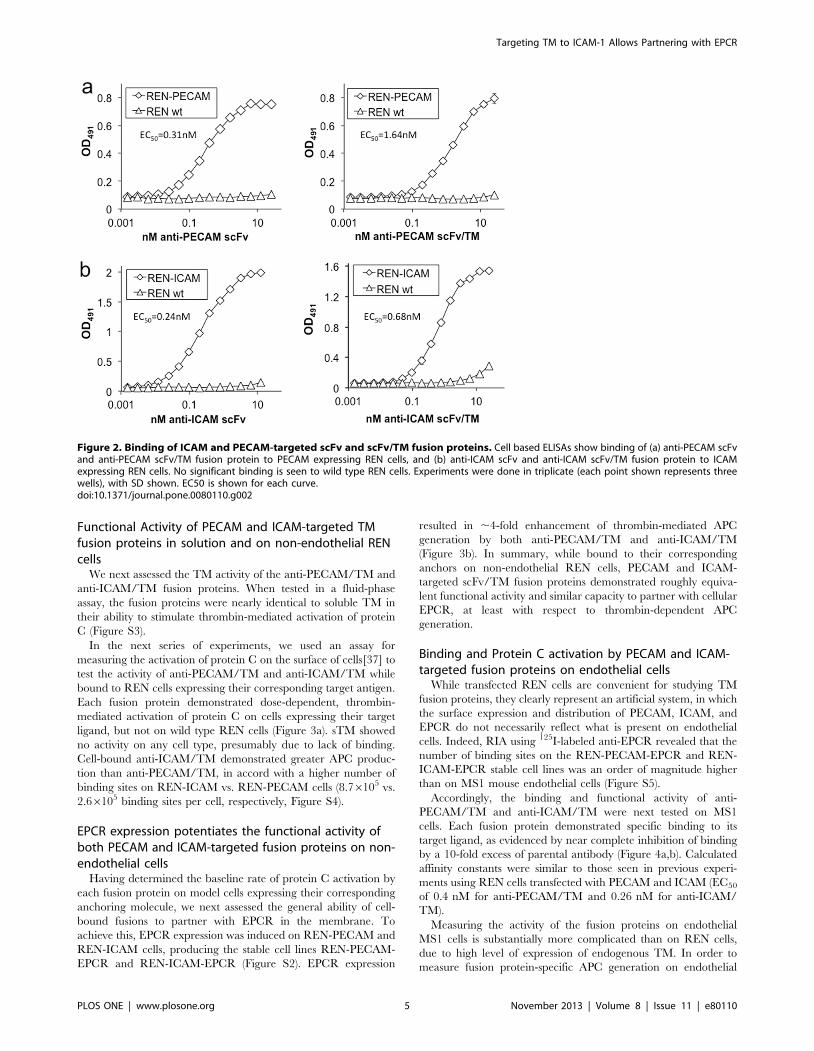
Functional Activity of PECAM and ICAM-targeted TMfusion proteins in solution and on non-endothelial RENcells
We next assessed the TM activity of the anti-PECAM/TM and
anti-ICAM/TM fusion proteins. When tested in a fluid-phase
assay, the fusion proteins were nearly identical to soluble TM in
their ability to stimulate thrombin-mediated activation of protein
C (Figure S3).
In the next series of experiments, we used an assay for
measuring the activation of protein C on the surface of cells[37] to
test the activity of anti-PECAM/TM and anti-ICAM/TM while
bound to REN cells expressing their corresponding target antigen.
Each fusion protein demonstrated dose-dependent, thrombin-
mediated activation of protein C on cells expressing their target
ligand, but not on wild type REN cells (Figure 3a). sTM showed
no activity on any cell type, presumably due to lack of binding.
Cell-bound anti-ICAM/TM demonstrated greater APC produc-
tion than anti-PECAM/TM, in accord with a higher number of
binding sites on REN-ICAM vs. REN-PECAM cells (8.76105 vs.
2.66105 binding sites per cell, respectively, Figure S4).
EPCR expression potentiates the functional activity ofboth PECAM and ICAM-targeted fusion proteins on non-endothelial cells
Having determined the baseline rate of protein C activation by
each fusion protein on model cells expressing their corresponding
anchoring molecule, we next assessed the general ability of cell-
bound fusions to partner with EPCR in the membrane. To
achieve this, EPCR expression was induced on REN-PECAM and
REN-ICAM cells, producing the stable cell lines REN-PECAM-
EPCR and REN-ICAM-EPCR (Figure S2). EPCR expression
resulted in ,4-fold enhancement of thrombin-mediated APC
generation by both anti-PECAM/TM and anti-ICAM/TM
(Figure 3b). In summary, while bound to their corresponding
anchors on non-endothelial REN cells, PECAM and ICAM-
targeted scFv/TM fusion proteins demonstrated roughly equiva-
lent functional activity and similar capacity to partner with cellular
EPCR, at least with respect to thrombin-dependent APC
generation.
Binding and Protein C activation by PECAM and ICAM-targeted fusion proteins on endothelial cells
While transfected REN cells are convenient for studying TM
fusion proteins, they clearly represent an artificial system, in which
the surface expression and distribution of PECAM, ICAM, and
EPCR do not necessarily reflect what is present on endothelial
cells. Indeed, RIA using 125I-labeled anti-EPCR revealed that the
number of binding sites on the REN-PECAM-EPCR and REN-
ICAM-EPCR stable cell lines was an order of magnitude higher
than on MS1 mouse endothelial cells (Figure S5).
Accordingly, the binding and functional activity of anti-
PECAM/TM and anti-ICAM/TM were next tested on MS1
cells. Each fusion protein demonstrated specific binding to its
target ligand, as evidenced by near complete inhibition of binding
by a 10-fold excess of parental antibody (Figure 4a,b). Calculated
affinity constants were similar to those seen in previous experi-
ments using REN cells transfected with PECAM and ICAM (EC50
of 0.4 nM for anti-PECAM/TM and 0.26 nM for anti-ICAM/
TM).
Measuring the activity of the fusion proteins on endothelial
MS1 cells is substantially more complicated than on REN cells,
due to high level of expression of endogenous TM. In order to
measure fusion protein-specific APC generation on endothelial
Figure 2. Binding of ICAM and PECAM-targeted scFv and scFv/TM fusion proteins. Cell based ELISAs show binding of (a) anti-PECAM scFvand anti-PECAM scFv/TM fusion protein to PECAM expressing REN cells, and (b) anti-ICAM scFv and anti-ICAM scFv/TM fusion protein to ICAMexpressing REN cells. No significant binding is seen to wild type REN cells. Experiments were done in triplicate (each point shown represents threewells), with SD shown. EC50 is shown for each curve.doi:10.1371/journal.pone.0080110.g002
Targeting TM to ICAM-1 Allows Partnering with EPCR
PLOS ONE | www.plosone.org 5 November 2013 | Volume 8 | Issue 11 | e80110
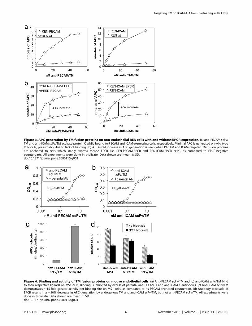
Figure 3. APC generation by TM fusion proteins on non-endothelial REN cells with and without EPCR expression. (a) anti-PECAM scFv/TM and anti-ICAM scFv/TM activate protein C while bound to PECAM and ICAM-expressing cells, respectively. Minimal APC is generated on wild typeREN cells, presumably due to lack of binding. (b) A ,4-fold increase in APC generation is seen when PECAM and ICAM-targeted TM fusion proteinsare anchored to cells which stably express mouse EPCR (i.e. REN-PECAM-EPCR and REN-ICAM-EPCR cells), as compared to EPCR-negativecounterparts. All experiments were done in triplicate. Data shown are mean 6 SD.doi:10.1371/journal.pone.0080110.g003
Figure 4. Binding and activity of TM fusion proteins on mouse endothelial cells. (a) Anti-PECAM scFv/TM and (b) anti-ICAM scFv/TM bindto their respective ligands on MS1 cells. Binding is inhibited by excess of parental anti-PECAM-1 and anti-ICAM-1 antibodies. (c) Anti-ICAM scFv/TMdemonstrates ,15-fold greater activity per binding site on MS1 cells, as compared to its PECAM-anchored counterpart. (d) Antibody blockade ofEPCR results in a ,50% decrease in APC generation by endogenous TM and anti-ICAM scFv/TM, but not anti-PECAM scFv/TM. All experiments weredone in triplicate. Data shown are mean 6 SD.doi:10.1371/journal.pone.0080110.g004
Targeting TM to ICAM-1 Allows Partnering with EPCR
PLOS ONE | www.plosone.org 6 November 2013 | Volume 8 | Issue 11 | e80110

Targeting TM to ICAM-1 Allows Partnering with EPCR
PLOS ONE | www.plosone.org 7 November 2013 | Volume 8 | Issue 11 | e80110

cells, we blocked the activation of protein C by endogenous TM.
Both PPACK-inactivated thrombin and a polyclonal anti-TM
antibody were able to fully inhibit the activity of endogenous TM.
Ultimately, we used the antibody as a blocking agent since it had a
sustained effect after washing. Sustained blockade of 60-70% of
endogenous TM activity, provided by anti-TM antibody, enabled
measurement of dose responsive, fusion protein-dependent protein
C activation (Figure S6).
We employed these conditions to assess protein C activation by
anti-PECAM/TM vs. anti-ICAM/TM anchored to MS1 cells.
The amount of APC generated by cell-bound fusions was
normalized to the number of binding sites per cell. This analysis
revealed ,15-fold greater APC generation by anti-ICAM/TM vs.
anti-PECAM/TM (Figure 4c). To assess the role of EPCR in this
marked difference in fusion protein activity, we blocked EPCR
interaction with murine Protein C using a monoclonal antibody
that thereby inhibits its ability to accelerate APC production by
the thrombin-TM complex[38]. Indeed, we found that treatment
of MS1 cells with the anti-EPCR antibody resulted in approxi-
mately 50% reduction in thrombin-dependent activation of
protein C by endogenous TM (Figure 4d). We observed a similar
50% reduction in the activation of protein C by anti-ICAM/TM
on MS1 cells, following blockade of endogenous TM. In contrast,
there was no significant effect on the activation of protein C by
anti-PECAM/TM (Figure 4d).
These results, together with the 15-fold difference in APC
generation by the fusion proteins on endothelial cells, indicate that
recombinant scFv/TM partners more effectively with endogenous
EPCR when anchored to ICAM, as opposed to PECAM.
Endothelial protective effects of PECAM and ICAM-targeted fusion proteins in a mouse model of acute lunginjury
Anti-inflammatory effects of anti-PECAM/TM and anti-
ICAM/TM were then compared in a model of lung inflammation,
in which mice receive an intratracheal injection of endotoxin. The
fusion proteins, or PBS vehicle, were injected intravenously 30
minutes prior to LPS challenge (Figure 5a). Relevant indices of
lung inflammation and injury were measured, including the level
of MIP-2 in bronchoalveolar lavage fluid (Figure 5b), expression of
cell adhesion molecules VCAM-1 and E-selectin in lung tissue
homogenate (Figure 5c), and extravascular leakage of radiolabeled
albumin injected intravenously and detected in the lungs
(Figure 5d). While both anti-PECAM/TM and anti-ICAM/TM
showed evidence of protection, the ICAM-targeted fusion protein
was more effective in all cases.
Discussion
Thrombomodulin is a multifaceted mediator of vascular
homeostasis. For reasons that are not fully understood, humans
and other mammals have evolved mechanisms by which the TM/
protein C pathway is suppressed in a variety of settings, tipping the
balance towards thrombosis, leukocyte adhesion, and vascular
leak. While endothelial TM loss may have selective advantage in
some situations, such as localized infection or trauma, there is
substantial evidence to suggest that it has a role in the pathogenesis
of a variety of contemporary human illnesses[14–18].
The direct reversal of this process through gene therapy has
provided proof-of-principle for replenishing endothelial TM[19–
21]. In chronic settings such as atherosclerosis and vascular bypass
grafting, genetic approaches may be preferable[40], but they are
unlikely to have a role in unforeseeable and emergent conditions
like sepsis, stroke, and acute lung injury. The landmark discovery
that recombinant human APC could protect baboons from a
otherwise lethal infusion of E Coli raised expectations that this
approach could restore the critical TM/APC pathway in sepsis
and other diseases[41]. However, infusion of an activated
zymogen lacks the ‘‘on demand’’ nature of the endogenous
TM/protein C response, and clinical utility has been limited by a
narrow therapeutic window and serious systemic side effects[25].
Likewise, infusions of soluble TM – or various genetic mutants –
have shown initial promise in animal models and human clinical
trials[24,42], but do not allow recombinant TM to capitalize on
the unique factors of the endothelial luminal microenvironment
critical to its function.
Vascular immunotargeting is a modern strategy to direct the
delivery and therapeutic effect of drugs via their conjugation to
specific affinity ligands of determinants on the luminal surface of
endothelial cells. Several promising endothelial targets have been
explored, including cell adhesion molecules, caveolar aminopep-
tidase P, angiotensin converting enzyme, and aminopeptidase
N[43–48]. This strategy has allowed marked improvement of
therapeutic interventions in numerous animal models of human
diseases, but further improvements are warranted. Specifically, it
has become clear that binding to target cells is necessary, but not
always sufficient, for optimal results. In many cases, the cargo
needs even more precise addressing at sub-cellular level, such as
internalization and accumulation in selected organelles[5,49].
Protein therapeutics that exert their activity in the vascular
lumen, such as thrombomodulin, are a special case in which the
cargo must be retained on the surface, instead of entering the cells.
Ligands such as PECAM-1 and ICAM-1, endothelial adhesion
molecules with limited internalization, represent preferable targets.
Figure 5. Anti-ICAM scFv/TM provides enhanced endothelial protection in a mouse model of lung inflammation/injury. (a) Timelineof intratracheal LPS lung injury model. In experiments assessing endothelial barrier dysfunction, a tracer amount of 125I-labeled albumin was injected5 minutes prior to LPS administration. (b) Concentration of the chemokine MIP-2 in bronchoalveolar lavage (BAL) fluid. (c) mRNA transcript levels ofpro-inflammatory cell adhesion molecules, VCAM-1 and E-selectin, in lung homogenate. (d) Endothelial barrier dysfunction, as measured by leakageof 125I-labeled albumin from blood into lung interstitium and/or alveolar space. All data shown are mean 6 SD, with number of animals as shown.doi:10.1371/journal.pone.0080110.g005
Figure 6. Schematic representation of TM fusion proteinsanchored to the endothelial plasmalemma. The proximity ofICAM-targeted TM to endogenous EPCR/Protein C may account for itsenhanced activity in vitro and in vivo.doi:10.1371/journal.pone.0080110.g006
Targeting TM to ICAM-1 Allows Partnering with EPCR
PLOS ONE | www.plosone.org 8 November 2013 | Volume 8 | Issue 11 | e80110

We have now devised scFv/TM fusions directed to each of these
determinants. The results of the present study, for the first time
comparing these fusions, indicate that scFv/TM functionality and
protective effect benefit from a nanoscale level of sub-cellular
localization – not only targeting the surface membrane, but a
particular determinant which allows cooperation of the cell-
anchored fusion with endogenous endothelial cofactor(s).
The most important cofactor, in the case TM and APC, is the
endothelial protein C receptor[27]. In addition to accelerating the
activation of protein C, EPCR plays an critical role in the
endothelial protective signaling of APC and is necessary for the
protective effects of APC in diseases of endothelial injury and
dysfunction, such as severe sepsis[28,50]. The current data set
supports several conclusions: 1. the ability of scFv/TM fusion
protein to interact with endogenous EPCR depends on which
surface determinant is targeted (Figure 4), and 2. this variable has
significant therapeutic implications, with the ICAM-targeted
scFv/TM fusion demonstrating more potent protective effects in
vivo (Figure 5). Our data, along with prior reports regarding the
distribution of ICAM and PECAM on the endothelial mem-
brane[31–33], suggest that the proximity of the TM fusion to
EPCR may be the critical factor. Figure 6 shows a simplified
model of an endothelial cell with the TM fusion proteins bound to
their target ligands. Of note, the figure accurately depicts the
fusion proteins binding the domains of PECAM-1 and ICAM-1
which lie furthest from the plasma membrane, consistent with the
location of their target epitopes[37,51]. The schematic highlights
the proposed difference in proximity to the EPCR/Protein C
complex, which may account for our experimental observations.
It is not known if partnering between anti-ICAM/TM and
EPCR is limited to the stimulation of protein C activation, or what
role EPCR may have in the endothelial protective effects of TM
fusion proteins. In theory, one would expect the requirement for
molecular proximity to be less stringent in mediating signaling by
APC generated by endothelium-anchored fusion protein, which
might interact with and signal through EPCR in a paracrine
manner. This intriguing issue is worth further investigation.
Likewise, a rigorous appraisal of the benefit/risk ratio of the TM
fusion proteins is needed in relevant animal models of human
diseases, with administration both prior to and after the onset of
injury.
It is worth noting that the results of this study align with the
general notion that anchoring agents to distinct determinants on
the same target cell may produce distinct outcomes, due to the
differing functions, location, and trafficking of these surface
molecules. For example, our laboratory previously reported that
binding of the H2O2-producing enzyme, glucose oxidase (GOX),
to endothelial cells induced varying degrees of vascular damage,
depending on whether PECAM or TM was chosen as the surface
target[52]. The variation in outcome in those experiments was
attributed to the substantially different function of these two
surface molecules and the consequences of their blockade by GOX
conjugates. In contrast, it is difficult to attribute the current results
to any functional difference between ICAM and PECAM, two
closely related proteins which both support leukocyte adhesion,
pro-inflammatory signaling, and uptake of antibody conjugates via
a similar endocytic mechanism[53,54]. For this reason, we believe
that the most logical explanation for our current experimental
results is the distinct localization of ICAM and PECAM on the
endothelial membrane and their differing capacity to allow
interaction of anchored scFv/TM with EPCR.
In summary, we report the creation of a novel ICAM-targeted
TM fusion protein and evidence for it’s partnering with
endogenous endothelial EPCR in the activation of protein C.
Delivery of recombinant TM to the endothelial membrane in a
way that mimics its natural distribution and allows interaction with
endogenous co-factors represents a new strategy for restoration
and/or augmentation of the TM/Protein C pathway and may
provide effective treatment of a wide variety of human illnesses.
Supporting Information
Figure S1 Cloning and assembly of anti-ICAM scFv andscFv/TM fusion protein.
(PDF)
Figure S2 Creation of REN cells stably expressingPECAM and EPCR.
(PDF)
Figure S3 APC generation by TM fusion proteins influid phase assay.
(PDF)
Figure S4 Quantification of ICAM and PECAM bindingsites on transfected REN cells.
(PDF)
Figure S5 Quantification of EPCR binding sites ontransfected REN cells.
(PDF)
Figure S6 APC generation on MS1 cells followingblockade of endogenous TM.
(PDF)
Author Contributions
Conceived and designed the experiments: CFG VRM. Performed the
experiments: CFG AMC SZ RC BJZ EDH JH. Analyzed the data: CFG.
Contributed reagents/materials/analysis tools: BD CTE. Wrote the paper:
CFG VRM.
References
1. Pober JS, Sessa WC (2007) Evolving functions of endothelial cells in
inflammation. Nature Reviews Immunology 7: 803–815. doi:10.1038/nri2171.
2. Aird WC (2003) The role of the endothelium in severe sepsis and multiple organ
dysfunction syndrome. Blood 101: 3765–3777. doi:10.1182/blood-2002-06-
1887.
3. Finigan JH (2009) The coagulation system and pulmonary endothelial function
in acute lung injury. Microvasc Res 77: 35–38. doi:10.1016/j.mvr.2008.09.002.
4. Neumar RW, Nolan JP, Adrie C, Aibiki M, Berg RA, et al. (2008) Post–Cardiac
Arrest Syndrome Epidemiology, Pathophysiology, Treatment, and Prognostication.
Circulation 118: 2452–2483. doi:10.1161/CIRCULATIONAHA.108.190652.
5. Muzykantov VR (2005) Biomedical aspects of targeted delivery of drugs to
pulmonary endothelium. Expert Opinion on Drug Delivery 2: 909–926.
doi:10.1517/17425247.2.5.909.
6. Esmon CT (1989) The roles of protein C and thrombomodulin in the regulation
of blood coagulation. J Biol Chem 264: 4743–4746.
7. Esmon CT (2001) The normal role of Activated Protein C in maintaining
homeostasis and its relevance to critical illness. Crit Care 5: S7–12.
8. Wouwer MV de, Collen D, Conway EM (2004) Thrombomodulin-Protein C-
EPCR System Integrated to Regulate Coagulation and Inflammation. Arterioscler
Thromb Vasc Biol 24: 1374–1383. doi:10.1161/01.ATV.0000134298.25489.92.
9. Weiler H, Lindner V, Kerlin B, Isermann BH, Hendrickson SB, et al. (2001)
Characterization of a Mouse Model for Thrombomodulin Deficiency.
Arterioscler Thromb Vasc Biol 21: 1531–1537. doi:10.1161/hq0901.094496.
10. Nawroth PP, Handley DA, Esmon CT, Stern DM (1986) Interleukin 1 induces
endothelial cell procoagulant while suppressing cell-surface anticoagulant
activity. Proc Natl Acad Sci U S A 83: 3460–3464.
11. Moore KL, Esmon CT, Esmon NL (1989) Tumor necrosis factor leads to the
internalization and degradation of thrombomodulin from the surface of bovine
aortic endothelial cells in culture. Blood 73: 159–165.
Targeting TM to ICAM-1 Allows Partnering with EPCR
PLOS ONE | www.plosone.org 9 November 2013 | Volume 8 | Issue 11 | e80110

12. MacGregor IR, Perrie AM, Donnelly SC, Haslett C (1997) Modulation of
human endothelial thrombomodulin by neutrophils and their release products.
Am J Respir Crit Care Med 155: 47–52.
13. Glaser CB, Morser J, Clarke JH, Blasko E, McLean K, et al. (1992) Oxidation of
a specific methionine in thrombomodulin by activated neutrophil products
blocks cofactor activity. A potential rapid mechanism for modulation ofcoagulation. J Clin Invest 90: 2565–2573. doi:10.1172/JCI116151.
14. Faust SN, Levin M, Harrison OB, Goldin RD, Lockhart MS, et al. (2001)
Dysfunction of endothelial protein C activation in severe meningococcal sepsis.
N Engl J Med 345: 408–416. doi:10.1056/NEJM200108093450603.
15. Lin S-M, Wang Y-M, Lin H-C, Lee K-Y, Huang C-D, et al. (2008) Serum
thrombomodulin level relates to the clinical course of disseminated intravascular
coagulation, multiorgan dysfunction syndrome, and mortality in patients withsepsis. Crit Care Med 36: 683–689. doi:10.1097/CCM.0B013E31816537D8.
16. Laszik ZG, Zhou XJ, Ferrell GL, Silva FG, Esmon CT (2001) Down-Regulation
of Endothelial Expression of Endothelial Cell Protein C Receptor andThrombomodulin in Coronary Atherosclerosis. Am J Pathol 159: 797–802.
17. Sido B, Datsis K, Mehrabi A, Kraus T, Klar E, et al. (1995) Soluble
thrombomodulin—a marker of reperfusion injury after orthotopic livertransplantation. Transplantation 60: 462–466.
18. Adrie C, Monchi M, Laurent I, Um S, Yan SB, et al. (2005) Coagulopathy after
successful cardiopulmonary resuscitation following cardiac arrest: implication ofthe protein C anticoagulant pathway. J Am Coll Cardiol 46: 21–28.
doi:10.1016/j.jacc.2005.03.046.
19. Waugh JM, Yuksel E, Li J, Kuo MD, Kattash M, et al. (1999) Local
Overexpression of Thrombomodulin for In Vivo Prevention of ArterialThrombosis in a Rabbit Model. Circulation Research 84: 84–92.
doi:10.1161/01.RES.84.1.84.
20. Kim AY, Walinsky PL, Kolodgie FD, Bian C, Sperry JL, et al. (2002) Early Lossof Thrombomodulin Expression Impairs Vein Graft Thromboresistance
Implications for Vein Graft Failure. Circulation Research 90: 205–212.
doi:10.1161/hh0202.105097.
21. Tabuchi N, Shichiri M, Shibamiya A, Koyama T, Nakazawa F, et al. (2004)
Non-viral in vivo thrombomodulin gene transfer prevents early loss of
thromboresistance of grafted veins. Eur J Cardiothorac Surg 26: 995–1001.
doi:10.1016/j.ejcts.2004.07.028.
22. Bernard GR, Vincent JL, Laterre PF, LaRosa SP, Dhainaut JF, et al. (2001)
Efficacy and safety of recombinant human activated protein C for severe sepsis.
N Engl J Med 344: 699–709. doi:10.1056/NEJM200103083441001.
23. Liu KD, Levitt J, Zhuo H, Kallet RH, Brady S, et al. (2008) Randomized
Clinical Trial of Activated Protein C for the Treatment of Acute Lung Injury.
Am J Respir Crit Care Med 178: 618–623. doi:10.1164/rccm.200803-419OC.
24. Saito H, Maruyama I, Shimazaki S, Yamamoto Y, Aikawa N, et al. (2007)
Efficacy and safety of recombinant human soluble thrombomodulin (ART-123)
in disseminated intravascular coagulation: results of a phase III, randomized,double-blind clinical trial. J Thromb Haemost 5: 31–41. doi:10.1111/j.1538-
7836.2006.02267.x.
25. Gentry CA, Gross KB, Sud B, Drevets DA (2009) Adverse outcomes associatedwith the use of drotrecogin alfa (activated) in patients with severe sepsis and
baseline bleeding precautions. Crit Care Med 37: 19–25. doi:10.1097/
CCM.0b013e318192843b.
26. Ding B-S, Hong N, Christofidou-Solomidou M, Gottstein C, Albelda SM, et al.
(2009) Anchoring fusion thrombomodulin to the endothelial lumen protects
against injury-induced lung thrombosis and inflammation. Am J Respir Crit
Care Med 180: 247–256. doi:10.1164/rccm.200809-1433OC.
27. Stearns-Kurosawa DJ, Kurosawa S, Mollica JS, Ferrell GL, Esmon CT (1996)
The endothelial cell protein C receptor augments protein C activation by the
thrombin-thrombomodulin complex. Proc Natl Acad Sci USA 93: 10212–10216.
28. Feistritzer C, Riewald M (2005) Endothelial barrier protection by activated
protein C through PAR1-dependent sphingosine 1–phosphate receptor-1crossactivation. Blood 105: 3178–3184. doi:10.1182/blood-2004-10-3985.
29. Teasdale MS, Bird CH, Bird P (1994) Internalization of the anticoagulant
thrombomodulin is constitutive and does not require a signal in the cytoplasmic
domain. Immunol Cell Biol 72: 480–488. doi:10.1038/icb.1994.72.
30. Bae J-S, Yang L, Rezaie AR (2007) Receptors of the protein C activation and
activated protein C signaling pathways are colocalized in lipid rafts of
endothelial cells. PNAS 104: 2867–2872. doi:10.1073/pnas.0611493104.
31. Albelda SM, Muller WA, Buck CA, Newman PJ (1991) Molecular and cellular
properties of PECAM-1 (endoCAM/CD31): a novel vascular cell-cell adhesion
molecule. J Cell Biol 114: 1059–1068. doi:10.1083/jcb.114.5.1059.
32. Newman PJ (1997) The biology of PECAM-1. J Clin Invest 99: 3–8.
doi:10.1172/JCI119129.33. Van Buul JD, van Rijssel J, van Alphen FPJ, Hoogenboezem M, Tol S, et al.
(2010) Inside-Out Regulation of ICAM-1 Dynamics in TNF-a-Activated
Endothelium. PLoS ONE 5: e11336. doi:10.1371/journal.pone.0011336.34. Dubel S, Breitling F, Fuchs P, Zewe M, Gotter S, et al. (1994) Isolation of IgG
antibody Fv-DNA from various mouse and rat hybridoma cell lines using thepolymerase chain reaction with a simple set of primers. Journal of
Immunological Methods 175: 89–95. doi:10.1016/0022-1759(94)90334-4.
35. Crowe JS, Smith MA, Cooper HJ (1989) Nucleotide sequence of Y3-Ag 1.2.3.rat myeloma immunoglobulin kappa chain cDNA. Nucleic Acids Res 17: 7992.
36. Fukudome K, Esmon CT (1995) Molecular Cloning and Expression of Murineand Bovine Endothelial Cell Protein C/Activated Protein C Receptor (EPCR)
The Structural and Functional Conservation in Human, Bovine, and MurineEPCR. J Biol Chem 270: 5571–5577. doi:10.1074/jbc.270.10.5571.
37. Chacko A-M, Nayak M, Greineder CF, DeLisser HM, Muzykantov VR (2012)
Collaborative Enhancement of Antibody Binding to Distinct PECAM-1Epitopes Modulates Endothelial Targeting. PLoS ONE 7: e34958.
doi:10.1371/journal.pone.0034958.38. Li W, Zheng X, Gu J, Hunter J, Ferrell GL, et al. (2005) Overexpressing
endothelial cell protein C receptor alters the hemostatic balance and protects
mice from endotoxin. Journal of Thrombosis and Haemostasis 3: 1351–1359.doi:10.1111/j.1538-7836.2005.01385.x.
39. Newman PJ (1994) The Role of PECAM-1 in Vascular Cell Biologya. Annals ofthe New York Academy of Sciences 714: 165–174. doi:10.1111/j.1749-
6632.1994.tb12041.x.40. Newby A, Baker A (1999) Targets for gene therapy of vein grafts. [Review] [37
refs]. Current Opinion in Cardiology 14: 489–494.
41. Taylor FB, Chang A, Esmon CT, D’Angelo A, Vigano-D’Angelo S, et al. (1987)Protein C prevents the coagulopathic and lethal effects of Escherichia coli
infusion in the baboon. J Clin Invest 79: 918–925.42. Su EJ, Geyer M, Wahl M, Mann K, Ginsburg D, et al. (2011) The
thrombomodulin analog Solulin promotes reperfusion and reduces infarct
volume in a thrombotic stroke model. J Thromb Haemost 9: 1174–1182.doi:10.1111/j.1538-7836.2011.04269.x.
43. Kowalski PS, Leus NGJ, Scherphof GL, Ruiters MHJ, Kamps JAAM, et al.(2011) Targeted siRNA delivery to diseased microvascular endothelial cells—
Cellular and molecular concepts. IUBMB Life 63: 648–658. doi:10.1002/iub.487.
44. Zhang N, Chittasupho C, Duangrat C, Siahaan TJ, Berkland C (2008) PLGA
nanoparticle—peptide conjugate effectively targets intercellular cell-adhesionmolecule-1. Bioconjug Chem 19: 145–152. doi:10.1021/bc700227z.
45. Simone E, Ding B-S, Muzykantov V (2009) Targeted delivery of therapeutics toendothelium. Cell Tissue Res 335: 283–300. doi:10.1007/s00441-008-0676-7.
46. Chrastina A, Valadon P, Massey KA, Schnitzer JE (2010) Lung vascular
targeting using antibody to aminopeptidase P: CT-SPECT imaging, biodistribu-tion and pharmacokinetic analysis. J Vasc Res 47: 531–543. doi:10.1159/
000313880.47. Danilov SM, Gavrilyuk VD, Franke FE, Pauls K, Harshaw DW, et al. (2001)
Lung uptake of antibodies to endothelial antigens: key determinants of vascularimmunotargeting. Am J Physiol Lung Cell Mol Physiol 280: L1335–1347.
48. Pasqualini R, Koivunen E, Kain R, Lahdenranta J, Sakamoto M, et al. (2000)
Aminopeptidase N Is a Receptor for Tumor-homing Peptides and a Target forInhibiting Angiogenesis. Cancer Res 60: 722–727.
49. Torchilin VP (2006) Recent approaches to intracellular delivery of drugs andDNA and organelle targeting. Annu Rev Biomed Eng 8: 343–375. doi:10.1146/
annurev.bioeng.8.061505.095735.
50. Kerschen EJ, Fernandez JA, Cooley BC, Yang XV, Sood R, et al. (2007)Endotoxemia and sepsis mortality reduction by non-anticoagulant–activated
protein C. J Exp Med 204: 2439–2448. doi:10.1084/jem.20070404.51. Staunton DE, Dustin ML, Erickson HP, Springer TA (1990) The arrangement
of the immunoglobulin-like domains of ICAM-1 and the binding sites for LFA-1
and rhinovirus. Cell 61: 243–254. doi:10.1016/0092-8674(90)90805-O.52. Christofidou-Solomidou M, Kennel S, Scherpereel A, Wiewrodt R, Solomides
CC, et al. (2002) Vascular Immunotargeting of Glucose Oxidase to theEndothelial Antigens Induces Distinct Forms of Oxidant Acute Lung Injury.
Am J Pathol 160: 1155–1169.53. Muro S, Koval M, Muzykantov V (2004) Endothelial endocytic pathways: gates
for vascular drug delivery. Curr Vasc Pharmacol 2: 281–299.
54. Muro S, Wiewrodt R, Thomas A, Koniaris L, Albelda SM, et al. (2003) A novelendocytic pathway induced by clustering endothelial ICAM-1 or PECAM-1.
J Cell Sci 116: 1599–1609. doi:10.1242/jcs.00367.
Targeting TM to ICAM-1 Allows Partnering with EPCR
PLOS ONE | www.plosone.org 10 November 2013 | Volume 8 | Issue 11 | e80110
Copyright © 2022 FDOKUMEN