06-01-2022 06:12a Page: 1 Stafford County Month End Deeds ...
Upload
independentCategory
view
5download
0
Systematic Parasitology 59: 199–210, 2004.© 2004 Kluwer Academic Publishers. Printed in the Netherlands.
199
Description of two new species of Glypthelmins Stafford, 1905 (Digenea:Macroderoididae) in Rana spp. from Mexico, based on morphology andmtDNA and rDNA sequences
Ulises J. Razo-Mendivil, Virginia Leon-Regagnon & Gerardo Perez-Ponce de Leon∗Laboratorio de Helmintologıa, Instituto de Biologıa, Universidad Nacional Autonoma de Mexico. Ap. Postal 70-153. Del. Coyoacan. C. P. 04510 Mexico, D.F. Mexico
Accepted for publication 1st March, 2004
Abstract
Glypthelmins Stafford, 1905 includes 29 putative species commonly found in the intestine and liver of anuransfrom all over the world but mainly in the Americas. Partial sequences of the cytochrome c oxidase subunit 1(cox1), ribosomal internal transcribed spacer region 2 (ITS2) and the large subunit 28S rDNA gene were obtainedand analysed using pairwise distance matrices and parsimony methods in order to characterise the interrelationshipsbetween 14 isolates of four nominal species of Glypthelmins recognised on morphological grounds. The highestintra-specific sequence divergence occurred in the cox1 (18.53%) sequence, followed by that of the ITS2 (5.44%)and 28S (4.63%). Genetic variability was detected between the three isolates originally identified as G. facioiBrenes et al., 1959 from two localities in Mexico and one locality in Costa Rica. Sequence divergence exhibitedamong these isolates ranged from 10.70 to 11.22%, from 0.48 to 0.97% and from 1.33 to 1.88% for cox1, ITS2and 28S, respectively. Phylogenetic analysis combining all three data-sets generated a single most parsimonioustree. The three isolates of G. facioi form a clade, with an isolate collected from frogs in Veracruz State as thesister group to an isolate from Tabasco State + G. facioi from Costa Rica. The information derived from pairwisedistance of independent data-sets plus the phylogenetic information indicate that each of the two isolates fromMexico, identified a priori as G. facioi, represent separate species. A re-examination of specimens was carried outand a re-evaluation made of the morphological characters to find reliable differences that had been overlooked. Asa consequence, G. brownorumae n. sp. from Tabasco and G. tuxtlasensis n. sp. from Veracruz are described basedon molecular and morphological differences.
Introduction
Helminth parasites of amphibians have been surveyedin Mexico since the 1940s, but more intensively duringthe last 10 years. Current data indicate that 119 speciesof helminths have been described, 26% of which werenew species (Pérez-Ponce de León et al., 2002). Oneof the most species-rich groups of helminths in Mex-ican anurans is represented by Glypthelmins Stafford,1905. This genus was established by Stafford (1905)to include Distomum quietum Stafford, 1900, para-sitic in Rana catesbeiana Shaw, R. viriscens Kalm andHyla pickeringii Holb, all from Canada. Twenty-ninenominal species have been described from around the
∗Author for correspondence (E-mail: [email protected])
world (Yamaguti, 1971; Sullivan, 1976; Prudhoe &Bray, 1982; Razo-Mendivil & León-Règagnon, 2001).Descriptions have been based on morphological char-acters. However, diagnostic traits in some specieshave shown several degrees of intra-specific variability(Miller, 1930; Rankin, 1944.
Recent studies have demonstrated that morpholo-gical data alone may not provide enough informa-tion to establish clear limits between species of di-geneans, and the use of molecular markers has beenproposed (Tkach et al., 2000a). Closely related spe-cies of digeneans and monogeneans, without obviousmorphological differentiation (cryptic species), havebeen characterised and discriminated by data fromsequences of mitochondrial DNA (mtDNA) and ri-
200
bosomal DNA (rDNA) (Anderson & Barker, 1998;van Herwerden et al., 1998, 1999; Iwagami et al.,2000; Cunningham et al., 2001; Jousson and Bartoli,2001; León-Règagnon & Paredes-Calderón, 2002).
Species of Glypthelmins in Mexican anurans havebeen intensively surveyed over the past four years inorder to obtain new information which would enablea re-examination of the an existing phylogenetic hy-pothesis for the group (Brooks & McLennan, 1993) inlight of new evidence from both molecular and mor-phological data. Based on a combination of resultsfrom both sources, we determined the presence of twonew species of Glypthelmins infecting anurans fromthe lowlands of the Gulf of Mexico slope. These newtaxa are described below.
Materials and methods
SamplingAs a part of an ongoing systematic study ofGlypthelmins, specimens of 29 species of frogs andtoads were collected from 35 localities in Mexicobetween July, 1996 and April, 2002. Anurans werecaptured by hand or with seine nets and kept alive priorto parasitological examination. Hosts were killed withan overdose of sodium pentobarbitol and all organswere examined under a stereo-microscope.
Digeneans belonging to Glypthelmins were ini-tially placed in a 0.65% saline solution, and someworms from each host were mounted as semiperman-ent slides in saline and allocated to morphospecies invivo.
Molecular studyFor molecular work, the following specimens weremorphologically identified as: Glypthelmins facioiBrenes, Jiménez-Quirós, Arroyo-Sancho & Delgado-Flores, 1959 from Veracruz and Tabasco, Mexico, andfrom Guanacaste, Costa Rica; G. quieta (Stafford,1900) from Estado de Mexico; G. californiensis (Cort,1919) from Estado de Mexico and Michoacán, Mex-ico; and G. hyloreus Martin, 1969 from Nebraska,USA. They were fixed in 100% ethanol (Table 1)and all gravid worms were digested. In most cases,two or three individuals per species were sequenced.Worms were digested with proteinase K (25 mg/ml)in 500 µl of STE buffer, 75 µl of 10% SDS and in-cubated during 12–24 h at 55 ◦C. Genomic DNA wasextracted with phenol/chloroform, precipitated with96% ethanol and dissolved in 100 µl of deionised
sterile distilled water (Hillis et al., 1996). We chosecox1, 28S and ITS2 following (Anderson & Barker,1998; Itagaki & Tsutsumi, 1998; Bell et al., 2001;León-Règagnon et al., 2001), where these molecularmarkers demonstrated their utility to differentiate di-genean species. Mitochondrial and nuclear fragmentswere amplified by PCR in a final volume of 25 µl(2.5 µl 10× PCR buffer, 0.5 µl 10mM dNTP mix-ture (200 µM each), 0.8 µl 50 mM MgCl2, 1 µl ofeach primer (10 pmol), 1 µl template DNA, 0.5 µlTaq DNA polymerase (5 units) and 17.7 µl of steriledistilled water. Primers used to generate partial frag-ments of mitochondrial and nuclear genes were: cox1,JB3 (5′-TTT TTT GGG CAT CCT GAG GTT TAT-3′) and JB4.5 (5′-TAA AGA AAG AAC ATA ATGAAA ATG-3′) (Morgan & Blair, 1998); 5.8S + ITS2,3S (5′-GGT ACC GGT GGA TCA CGT GGC TAGTG-3′) and BD2 (5′-TAT GCT TAA ATT CAG CGGGT-3′) (Bowles et al., 1995); and 28S, 28Sy (5′ CTAACC AGG ATT CCC TCA GTA ACG GCG AGT 3′)(Palumbi, 1996) and LO (5′-GCT ATC CTG AG(AG)GAA ACT TCG-3′) (Tkach et al., 2000b). With theexception of annealing temperatures, reaction condi-tions used were the same regardless of primer setemployed. An initial denaturation at 95 ◦C for 5 minwas followed by 30-35 cycles of 94 ◦C for 1 min,primers annealing for 45 s at 50 ◦C (cox1 and 28S)or at 55 ◦C (5.8S and ITS2) and extension at 72 ◦Cfor 1 min; the mixes were held at 72 ◦C for 10 minto complete elongation and then dropped to 4 ◦C. PCRproducts were visualised on ethidium-bromide-stained1% TRIS-acetate-EDTA (ethylenediaminetetraaceticacid) agarose gels and subsequently purified withthe QIAquick Gel Extraction Kit (Qiagen) accord-ing to manufacturer’s instructions. The purified PCRproducts were sequenced on an ABI PRISM 310 auto-mated DNA sequencer (Applied Biosystems) usingthe Big Dye Terminator� chemistry and incorporat-ing the same primers as those used in previous PCRs.Sense and anti-sense strands were sequenced. Chro-matogram files were initially checked using the com-puter program Chromas (version 1.43). Subsequently,sense and anti-sense sequences were assembled, andaligned using the software Bioedit (Hall, 1999).
Sequence analysisPairwise distance matrices and phylogenetic analyseswere performed using PAUP (version 4.0b10 Swof-ford, 2002). Trees were found using a branch andbound search for the separate and combined data sets(cox1, ITS2 and 28S). The partition-homogeneity test

201
Table 1. Hosts and localities of isolates of Glypthelmins spp. collected in Mexico, Costa Rica and the USA.
Isolate Host Locality
G. facioi Rana vaillanti Brocchi Guanacaste, Costa Rica
G. facioi Rana brownorum Sanders Km. 50 Carretera 180, Villahermosa-Frontera, Tabasco, Mexico
G. facioi Rana vaillanti Laguna Escondida, Los Tuxtlas, Veracruz, Mexico
G. quieta Rana montezumae Baird Cienaga de Lerma, Estado de Mexico, Mexico
G. californiensis Rana dunni Zweifel Lago de Zacapu, Michoacan, Mexico
G. californiensis Rana montezumae Cienaga de Lerma, Estado de Mexico
G. hyloreus Pseudacris triseriata (Wied-Neuwied) Nebraska, USA
was used to demonstrate whether or not distinct char-acter sets could be combined (Farris et al., 1995).Branch support was evaluated using bootstrap andjack-knife analyses with 1,000 replicates and Jac emu-lation, respectively, and also Bremer support wascalculated for each internal branch (Bremer, 1994).The sequences obtained in this study were submit-ted to GenBank and assigned the following accessionnumbers AY278046-AY 278052 (28S), AY278053-AY278059 (cox1) and AY278060-AY278066 (ITS2).
Morphological studySeveral specimens were fixed by sudden immer-sion in hot formalin (4%) and stored in 70% eth-anol. Unflattened worms were stained with Ehrlich’shaematoxylin and Mayer’s paracarmine, dehydrated,cleared in methyl salicylate and mounted in Canadabalsam. Some of the specimens were permanentlymounted between cover-slips and held in Cobb slides.Measurements are given in micrometres, unless oth-erwise stated. Illustrations were made with a drawingtube.
For comparison, the following species of Glypthel-mins were examined: G. quieta, Colección Nacionalde Helmintos, Mexico (CNHE) 1461, 1562-1563,3275-3279, 3346, 3416, 3406, vouchers; UnitedStates National Parasite Collection, Beltsville, Mary-land (USNPC), 51635, 72268, 84184, vouchers,and Harold W. Manter Laboratory, Lincoln, Neb-raska (HWML) 20174 to 20201, vouchers; G. hy-loreus, (USNPC) 70464, paratypes; G. californiensis,(CNHE) 1181,1561, 2495, 3280-3283, 3294, vouch-ers; and G. facioi, Colección de Helmintos de CostaRica, San José (CHCR) 202-22, lectotype; (USNPC)72275, vouchers; (CNHE) 1514, 3285, 4083, vouch-ers; and specimens from Costa Rica borrowed from DrDaniel R. Brooks.
Specimens of the present material were depositedon some of the above-mentioned collections and TheNatural History Museum, London [BM(NH)].
Results
All targeted DNA was successfully amplified andsequenced. The analysed data-sets of the cox1,ITS2 (5.8S + ITS2) and 28S rDNA of 14 isol-ates of four nominal species of Glypthelminsspp. produced a total alignment of 2,077 nuc-leotide sites, 231 of which were variable and138 of which were phylogenetically informative.The alignment is available from FTP.EBI.AC.UKin the directory /pub/databases/embl/align, or athttp://www3ebi.ac.uk/Services/align/listali.html underthe number ALIGN_000530. Pairwise distancematrices are presented in Tables 2–4. In cases wheremore than one individual worm was sequenced, thesequences obtained were identical, except for G. cali-forniensis, where some variation was observed.
cox1
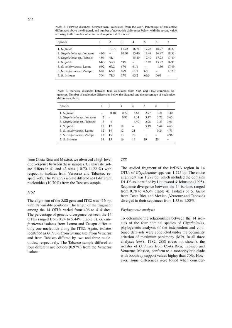
A total of 383 bp of cox1 were sequenced and alignedfor four nominal species, one isolate of G. californi-ensis and two isolates identified as G. facioi fromTabasco and Veracruz, Mexico. No gaps were requiredto align this gene. The sequence divergence betweenG. facioi (Costa Rica), G. californiensis, G. quieta andG. hyloreus ranged from 15.92 to 18.53%, with thehighest disparity between the Veracruz isolate and G.hyloreus (71 sites, 55 of which were in the third codonposition); these species present three different encodedamino acids (Table 2). Intra-specific variability wasobserved between two isolates of G. californiensisat six nucleotides (1.56%). When comparing the se-quences of the distinct samples identified as G. facioi
202
Table 2. Pairwise distances between taxa, calculated from the cox1. Percentage of nucleotidedifferences above the diagonal, and number of nucleotide differences below, with the second valuereferring to the number of amino acid sequence differences.
Species 1 2 3 4 5 6 7
1. G. facioi - 10.70 11.22 16.71 17.23 16.97 18.27
2. Glypthelmins sp., Veracruz 41/0 – 10.70 15.40 17.49 16.97 18.53
3. Glypthelmins sp., Tabasco 43/1 41/1 – 15.40 17.49 17.23 17.49
4. G. quieta 64/3 59/3 59/2 – 15.92 15.92 16.97
5. G. californiensis, Lerma 66/2 67/2 67/1 61/1 – 1.56 17.49
6. G. californiensis, Zacapu 65/1 65/2 66/1 61/1 6/0 – 17.23
7. G. hyloreus 70/4 71/3 67/3 65/2 67/3 66/3 –
Table 3. Pairwise distances between taxa calculated from 5.8S and ITS2 combined se-quences. Number of nucleotide differences below the diagonal and the percentage of nucleotidedifferences above.
Species 1 2 3 4 5 6 7
1. G. facioi – 0.48 0.72 3.65 2.97 3.21 3.40
2. Glypthelmins sp., Veracruz 2 – 0.97 4.14 3.47 3.72 3.65
3. Glypthelmins sp., Tabasco 3 4 – 4.40 2.98 3.23 3.91
4. G. quieta 15 17 18 – 5.19 5.44 4.63
5. G. californiensis, Lerma 12 14 12 21 – 0.24 4.71
6. G. californiensis, Zacapu 13 15 13 22 1 – 4.96
7. G. hyloreus 14 15 16 19 19 20 –
from Costa Rica and Mexico, we observed a high levelof divergence between these samples. Guanacaste isol-ate differs in 41 and 43 sites (10.70-11.22 %) withrespect to isolates from Veracruz and Tabasco, re-spectively. The Veracruz isolate differed at 41 differentnucleotides (10.70%) from the Tabasco sample.
ITS2
The alignment of the 5.8S gene and ITS2 was 416 bp,with 38 variable positions. The length of the fragmentamong the 14 OTUs varied from 406 to 414 sites.The percentage of genetic divergence between the 14OTUs ranged from 0.24 to 5.44% (Table 3). G. cali-forniensis isolates from Lerma and Zacapu differ atonly one nucleotide along the ITS2. Again, isolatesidentified as G. facioi from Guanacaste, from Veracruzand from Tabasco differed by two and three nucle-otides, respectively. The Tabasco sample differed atfour different nucleotides (0.97%) from the Veracruzisolate.
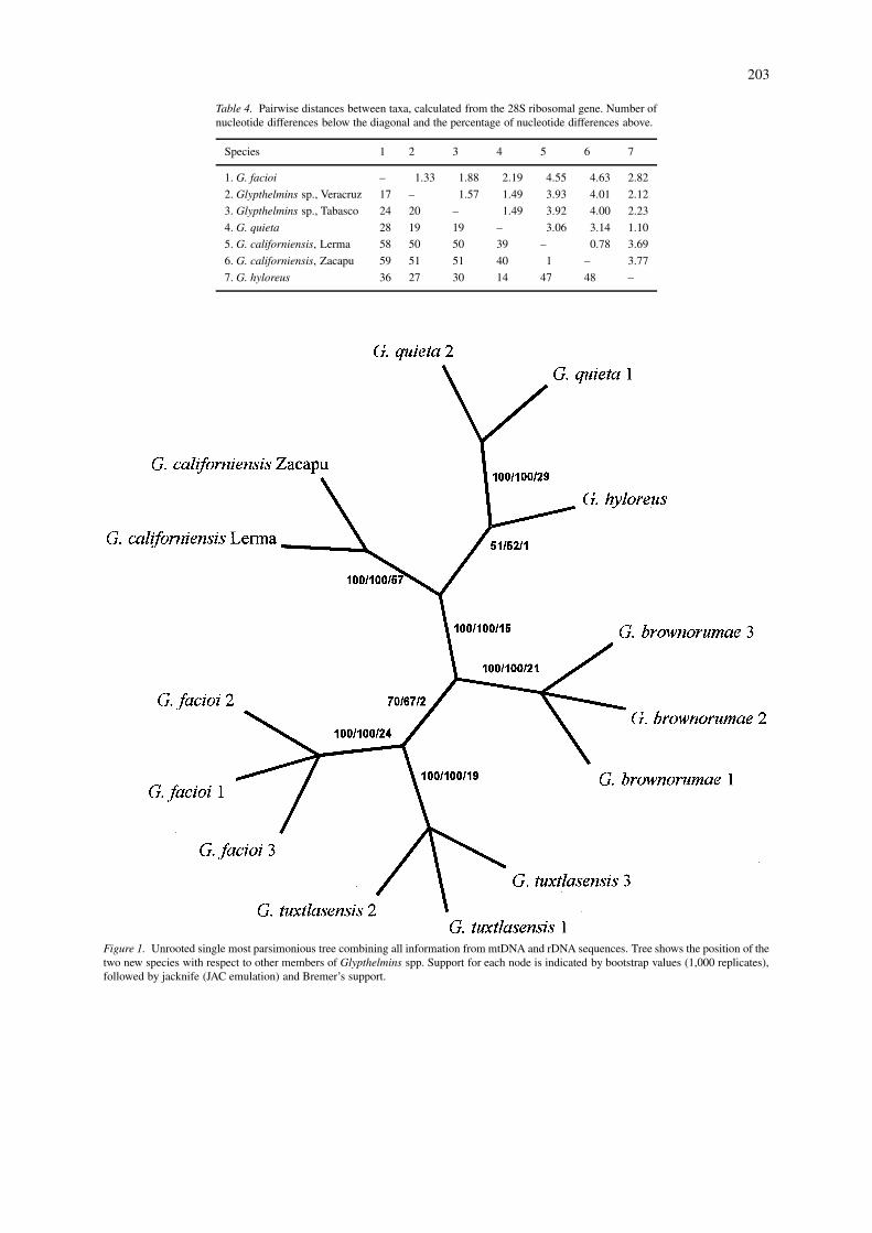
28S
The studied fragment of the lsrDNA region in 14OTUs of Glypthelmins spp. was 1,275 bp. The entirealignment was 1,278 bp, which included the domainsD1-D3 as identified by Littlewood & Johnston (1995).Sequence divergence between the 14 isolates rangedfrom 0.78 to 4.63% (Table 4). Isolates of G. facioifrom Costa Rica and Mexico (Veracruz and Tabasco)diverged in their sequences from 1.33 to 1.88%.
Phylogenetic analysis
To determine the relationships between the 14 isol-ates of the four nominal species of Glypthelmins,phylogenetic analyses of the independent and com-bined data-sets were conducted under the optimalitycriterion of maximum parsimony (MP). In all threeanalyses (cox1, ITS2, 28S) (trees not shown), theisolates of G. facioi from Costa Rica, Tabasco andVeracruz, Mexico, conform to a monophyletic cladewith bootstrap support values higher than 70%. How-ever, some differences were found when consider-
203
Table 4. Pairwise distances between taxa, calculated from the 28S ribosomal gene. Number ofnucleotide differences below the diagonal and the percentage of nucleotide differences above.
Species 1 2 3 4 5 6 7
1. G. facioi – 1.33 1.88 2.19 4.55 4.63 2.82
2. Glypthelmins sp., Veracruz 17 – 1.57 1.49 3.93 4.01 2.12
3. Glypthelmins sp., Tabasco 24 20 – 1.49 3.92 4.00 2.23
4. G. quieta 28 19 19 – 3.06 3.14 1.10
5. G. californiensis, Lerma 58 50 50 39 – 0.78 3.69
6. G. californiensis, Zacapu 59 51 51 40 1 – 3.77
7. G. hyloreus 36 27 30 14 47 48 –
Figure 1. Unrooted single most parsimonious tree combining all information from mtDNA and rDNA sequences. Tree shows the position of thetwo new species with respect to other members of Glypthelmins spp. Support for each node is indicated by bootstrap values (1,000 replicates),followed by jacknife (JAC emulation) and Bremer’s support.
204
ing sister-group relationships within this clade. Apartition-homogeneity test supported combining all ofthe molecular data partitions (p ≥ 0.05). When gapswere treated as missing characters, the combined ana-lysis generated a single most parsimonious tree (L =232, CI = 0.78, RI = 0.73) with strongly supportedbranch topology (Figure 1). Treating alignment gapsas fifth characters produced an identical tree. The treeshows that the three isolates of G. facioi (formerlyrecognised on morphological grounds) form a mono-phyletic group, with the isolates from Tabasco as thesister group to the Veracruz isolate + G. facioi (CostaRica, from which the original description of the spe-cies was made), albeit with a low bootstrap support(70%). Information on other molecular markers, aswell as the inclusion of the remainder Glypthelminsmembers in a phylogenetic analysis is needed in orderto clearly define the affinities of these taxa.
These results, in addition to the information de-rived from pairwise distance matrices of independentdata-sets (cox1, ITS2 and 28 srDNA) show that theisolates from Mexico identified a priori as G. fa-cioi in fact represent separate species from the CostaRican isolate [i.e. G. facioi (sensu stricto)]. Basedon these results, we re-examined specimens, and are-evaluation of the morphological characters was car-ried out to determine whether there were reliabledifferences which might have been overlooked. As aconsequence of this study, the description of these twonew species is presented below.
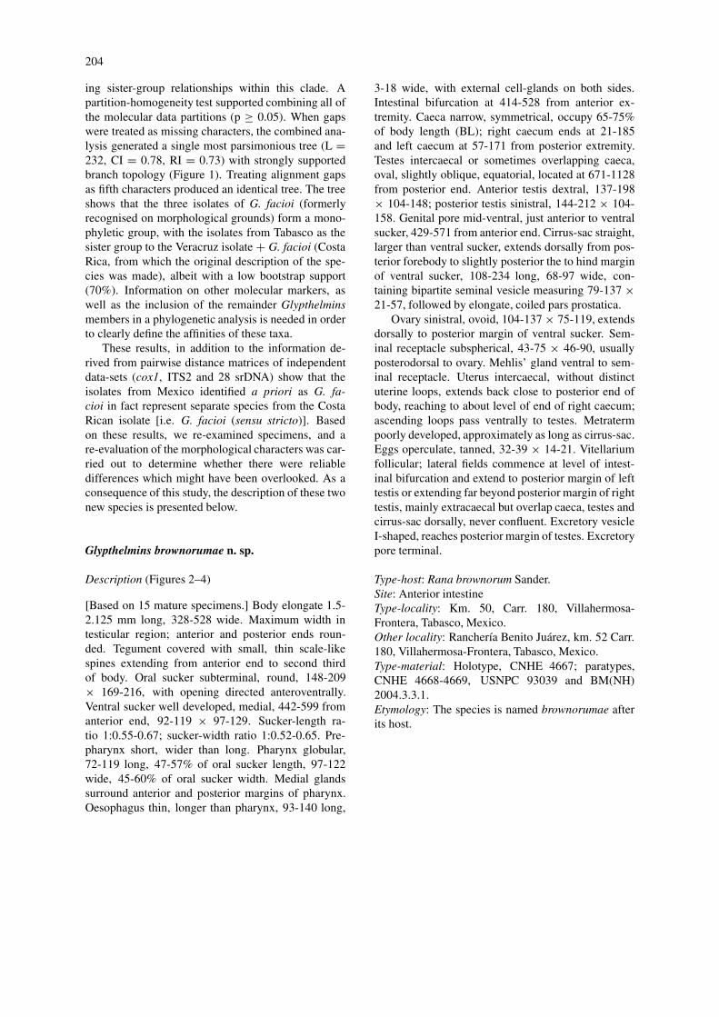
Glypthelmins brownorumae n. sp.
Description (Figures 2–4)
[Based on 15 mature specimens.] Body elongate 1.5-2.125 mm long, 328-528 wide. Maximum width intesticular region; anterior and posterior ends roun-ded. Tegument covered with small, thin scale-likespines extending from anterior end to second thirdof body. Oral sucker subterminal, round, 148-209× 169-216, with opening directed anteroventrally.Ventral sucker well developed, medial, 442-599 fromanterior end, 92-119 × 97-129. Sucker-length ra-tio 1:0.55-0.67; sucker-width ratio 1:0.52-0.65. Pre-pharynx short, wider than long. Pharynx globular,72-119 long, 47-57% of oral sucker length, 97-122wide, 45-60% of oral sucker width. Medial glandssurround anterior and posterior margins of pharynx.Oesophagus thin, longer than pharynx, 93-140 long,
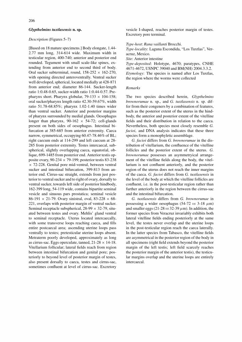
3-18 wide, with external cell-glands on both sides.Intestinal bifurcation at 414-528 from anterior ex-tremity. Caeca narrow, symmetrical, occupy 65-75%of body length (BL); right caecum ends at 21-185and left caecum at 57-171 from posterior extremity.Testes intercaecal or sometimes overlapping caeca,oval, slightly oblique, equatorial, located at 671-1128from posterior end. Anterior testis dextral, 137-198× 104-148; posterior testis sinistral, 144-212 × 104-158. Genital pore mid-ventral, just anterior to ventralsucker, 429-571 from anterior end. Cirrus-sac straight,larger than ventral sucker, extends dorsally from pos-terior forebody to slightly posterior the to hind marginof ventral sucker, 108-234 long, 68-97 wide, con-taining bipartite seminal vesicle measuring 79-137 ×21-57, followed by elongate, coiled pars prostatica.
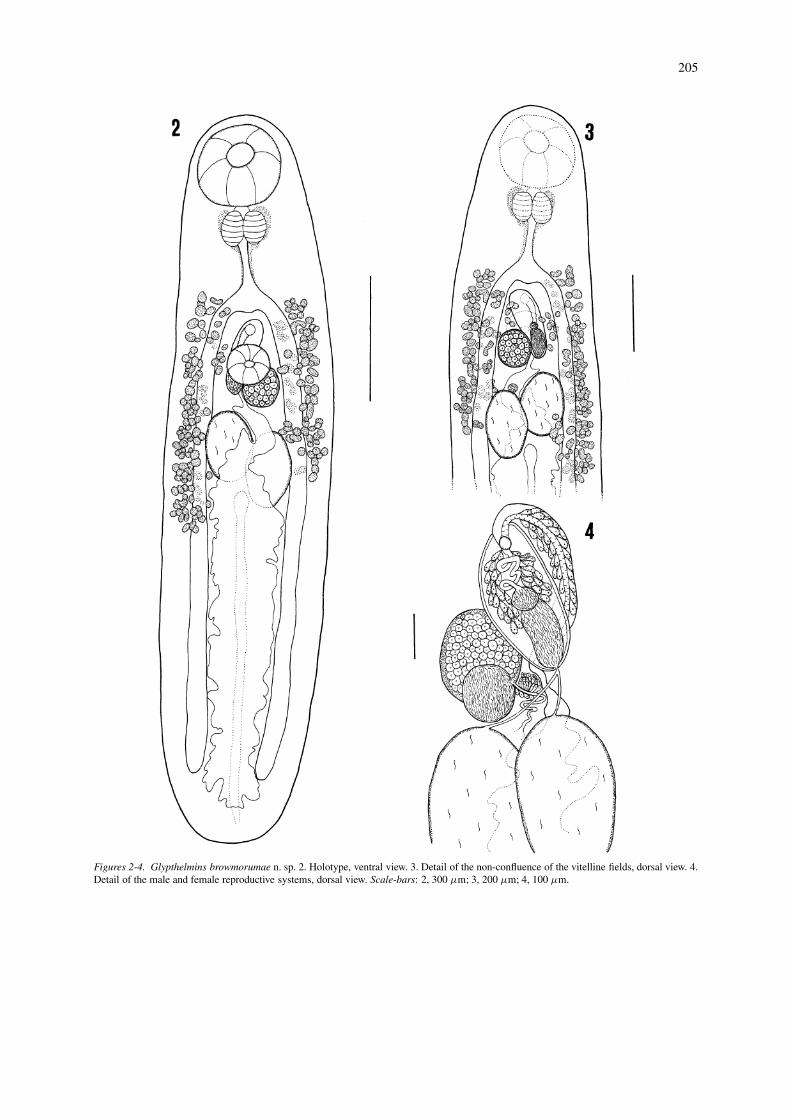
Ovary sinistral, ovoid, 104-137 × 75-119, extendsdorsally to posterior margin of ventral sucker. Sem-inal receptacle subspherical, 43-75 × 46-90, usuallyposterodorsal to ovary. Mehlis’ gland ventral to sem-inal receptacle. Uterus intercaecal, without distinctuterine loops, extends back close to posterior end ofbody, reaching to about level of end of right caecum;ascending loops pass ventrally to testes. Metratermpoorly developed, approximately as long as cirrus-sac.Eggs operculate, tanned, 32-39 × 14-21. Vitellariumfollicular; lateral fields commence at level of intest-inal bifurcation and extend to posterior margin of lefttestis or extending far beyond posterior margin of righttestis, mainly extracaecal but overlap caeca, testes andcirrus-sac dorsally, never confluent. Excretory vesicleI-shaped, reaches posterior margin of testes. Excretorypore terminal.
Type-host: Rana brownorum Sander.Site: Anterior intestineType-locality: Km. 50, Carr. 180, Villahermosa-Frontera, Tabasco, Mexico.Other locality: Ranchería Benito Juárez, km. 52 Carr.180, Villahermosa-Frontera, Tabasco, Mexico.Type-material: Holotype, CNHE 4667; paratypes,CNHE 4668-4669, USNPC 93039 and BM(NH)2004.3.3.1.Etymology: The species is named brownorumae afterits host.
205
Figures 2-4. Glypthelmins browmorumae n. sp. 2. Holotype, ventral view. 3. Detail of the non-confluence of the vitelline fields, dorsal view. 4.Detail of the male and female reproductive systems, dorsal view. Scale-bars: 2, 300 µm; 3, 200 µm; 4, 100 µm.
206
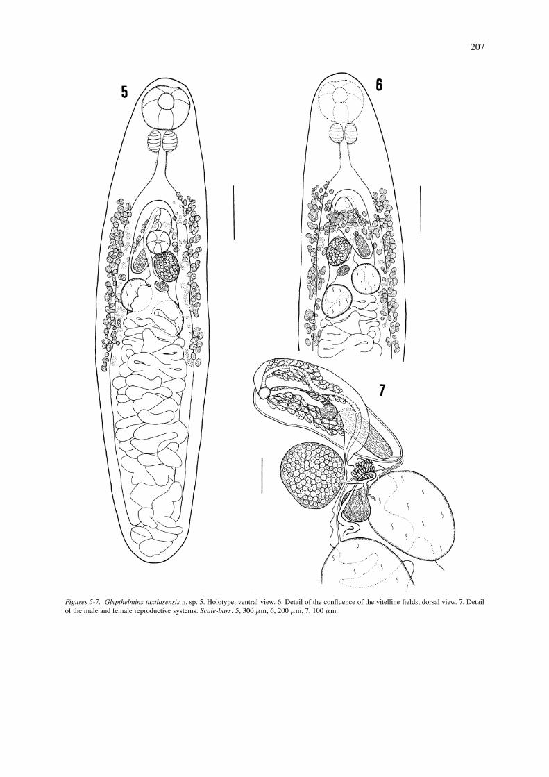
Glypthelmins tuxtlasensis n. sp.
Description (Figures 5–7)
[Based on 18 mature specimens.] Body elongate, 1.44-2.77 mm long, 314-614 wide. Maximum width intesticular region, 400-740; anterior and posterior endrounded. Tegument with small scale-like spines, ex-tending from anterior end to second third of body.Oral sucker subterminal, round, 158-252 × 162-270,with opening directed anteroventrally. Ventral suckerwell developed, spherical, located medially at 428-871from anterior end; diameter 86-144. Sucker-lengthratio 1:0.48-0.65, sucker-width ratio 1:0.44-0.57. Pre-pharynx short. Pharynx globular, 79-133 × 104-158;oral sucker/pharynx length ratio 42.30-59.67%, widthratio 51.78-68.85%; pharynx 1.02-1.40 times widerthan ventral sucker. Anterior and posterior marginsof pharynx surrounded by medial glands. Oesophaguslonger than pharynx, 90-162 × 54-72; cell-glandspresent on both sides of oesophagus. Intestinal bi-furcation at 385-685 from anterior extremity. Caecanarrow, symmetrical, occupying 60.47-78.46% of BL;right caecum ends at 114-299 and left caecum at 28-285 from posterior extremity. Testes intercaecal, sub-spherical, slightly overlapping caeca, equatorial, ob-lique, 699-1485 from posterior end. Anterior testis op-posite ovary, 90-234 × 79-199; posterior testis 83-238× 72-228. Genital pore mid-ventral, between ventralsucker and intestinal bifurcation, 399-813 from an-terior end. Cirrus-sac straight, extends from just pos-terior to ventral sucker and to right of ovary, dorsally toventral sucker, towards left side of posterior hindbody,162-399 long, 54-119 wide, contains bipartite seminalvesicle and sinuous pars prostatica; seminal vesicle86-191 × 21-79. Ovary sinistral, oval, 83-228 × 68-221, overlaps with posterior margin of ventral sucker.Seminal receptacle subspherical, 28-99 × 32-79, situ-ated between testes and ovary. Mehlis’ gland ventralto seminal receptacle. Uterus located intercaecally,with some transverse loops reaching caeca, and fillsentire postcaecal area; ascending uterine loops passventrally to testes; pretesticular uterine loops absent.Metraterm poorly developed, approximately as longas cirrus-sac. Eggs operculate, tanned, 21-28 × 14-18.Vitellarium follicular; lateral fields reach from regionbetween intestinal bifurcation and genital pore; pos-teriorly to beyond level of posterior margin of testes,also present dorsally to caeca, testes and cirrus-sac,sometimes confluent at level of cirrus-sac. Excretory
vesicle I-shaped, reaches posterior margin of testes.Excretory pore terminal.
Type-host: Rana vaillanti Brocchi.Type-locality: Laguna Escondida, “Los Tuxtlas”, Ver-acruz, Mexico.Site: Anterior intestineType-deposited: Holotype, 4670; paratypes, CNHE4671-4672, USNPC 39040 and BM(NH) 2004.3.3.2.Etymology: The species is named after Los Tuxtlas,the region where the worms were collected
Remarks
The two species described herein, Glypthelminsbrownorumae n. sp., and G. tuxtlasensis n. sp. dif-fer from their congeners by a combination of features,such as the posterior extent of the uterus in the hind-body, the anterior and posterior extent of the vitellinefields and their distribution in relation to the caeca.Nevertheless, both species most closely resemble G.facioi, and DNA analysis indicates that these threespecies form a monophyletic assemblage.
G. facioi differs from G. brownorumae in the dis-tribution of vitellarium, the confluence of the vitellinefollicles and the posterior extent of the uterus. G.brownorumae possesses an asymmetryical arrange-ment of the vitelline fields along the body, the vitel-larium is not confluent anteriorly, and the posteriorregion of the uterus does not reach the inner marginsof the caeca. G. facioi differs from G. tuxtlasensis inthe level of the body at which the vitelline follicles areconfluent, i.e. in the post-testicular region rather thanfurther anteriorly in the region between the cirrus-sacand the intestinal bifurcation.
G. tuxtlasensis differs from G. brownorumae inpossessing a wider oesophagus (54-72 vs 3-18 µm)and smaller eggs (21-28 vs 32-39 µm). In addition, theformer species from Veracruz invariably exhibits bothlateral vitelline fields ending posteriorly at the samelevel, the testes never overlap and the uterine loopsin the post-testicular region reach the caeca laterally.In the latter species from Tabasco, the vitelline fieldsare asymmetrical in the posterior region of the body inall specimens (right field extends beyond the posteriormargin of the left testis; left field scarcely reachesthe posterior margin of the anterior testis), the testicu-lar margins overlap and the uterine loops are entirelyintercaecal.
207
Figures 5-7. Glypthelmins tuxtlasensis n. sp. 5. Holotype, ventral view. 6. Detail of the confluence of the vitelline fields, dorsal view. 7. Detailof the male and female reproductive systems. Scale-bars: 5, 300 µm; 6, 200 µm; 7, 100 µm.

208
Discussion
The application of molecular tools combined witha detailed morphological study of specimens fromdifferent localities has permitted a reliable differen-tiation of the two new species, G. brownorumae andG. tuxtlasensis, from the neotropical region of Mex-ico. These two species exhibit a morphological re-semblance to G. facioi; however, molecular evidenceclearly shows that they are distinct lineages. Addition-ally, G. facioi is distributed in Central America (CostaRica) and parasitises Rana vaillanti, whereas the twonew species are found in R. vaillanti from Los Tuxtlas,Veracruz and R. brownorum from the state of Tabasco,respectively. All three species are found in the neo-tropical region within the distributional limits of theirtype-hosts.
Sequence comparison across the cox1 and 28Sgenes and ITS2 (Tables 2-4) indicate that the highestinter-specific divergence among the six putative spe-cies of Glypthelmins which we analysed occurs incox1 (18.53%), followed by ITS2 (5.44%) and 28S(4.63%).
Mitochondrial DNA sequencing has proved to bea useful marker for differentiating species withinGlypthelmins. Intraspecific variability was found to bevery low between isolates of G. californiensis fromtwo localities (1.56%). The intra-specific genetic vari-ation found in G. californiensis is comparable withthat shown between populations of digeneans usingthe same gene, e.g. in isolates of Paragonimus west-ermani (Kerbert, 1878) from China, Taiwan, Koreaand Japan. Iwagami et al. (2000) found a variation of0.26-1.82%. Samples of Ichthyocotylurus variegatus(Creplin, 1825) from distinct localities in Scotlandshowed null variation, and only one had a single nuc-leotide difference when compared with isolates fromFinland (Bell et al., 2001).
When comparing variation in closely related spe-cies, higher values of sequence divergence were foundbetween G. facioi, G. brownorumae and G. tuxtlasen-sis (10.70-11.22%). This level of variation is similarto that found between congeneric species of otherdigeneans, such as Echinostoma Rudolphi, 1809, Ich-thyocotylurus Odening, 1969 and Paragonimus Braun,1899, i.e. genera in which species differentiation canbe achieved using non-molecular criteria. Morgan& Blair (1998) observed a variability of 6.3-14.8%when studying a fragment of 257 sites of the cox1of seven species of Echinostoma. Similarly, I. varie-gatus, I. platycephalus (Creplin, 1825) and I. erraticus
(Rudolphi, 1809) showed divergences of 9.56-12.84%along 366 bp of cox1 (Bell et al., 2001). Finally,three species of Paragonimus (P. westermani, P. ohiraiMiyazaki, 1939 and P. miyazakii Kamo, Nishida,Hatsushika & Tomimura, 1961) exhibited a variationof 9.39-15.92% (Iwagami et al., 2000). This studyhas also shown that the mitochondrial gene used isvariable enough to achieve species differentiation.
ITS2 was also used to discriminate intra- and inter-specific genetic variation between isolates of G. cali-forniensis and closely related species of Glypthelmins.Only one variable nucleotide was found in the ITS2sequences of two isolates of G. californiensis fromtwo localities. These results concur with the variationlevels displayed in Fasciola gigantica Cobbold, 1855isolates from geographically distant localities (Adlardet al., 1993). The application of ITS2 sequences todiscriminate closely related species of Glypthelminswas limited because the percentage of sequence di-vergence found between G. facioi, G. brownorumaeand G. tuxtlasensis was strikingly low (<1%). Thismay be due to the fact that this gene is conservativein this particular group of species, or, alternatively,that these species diverged recently. Similar levels ofvariation have been found in other digeneans such asIndodidymozoon Madhavi, 1982, Fasciola Linnaeus,1869 and Ichthyocotylurus (see Anderson & Barker,1998; Itagaki & Tsutsumi, 1998; Bell et al., 2001).Considering the low intra- and inter-specific variab-ility shown by ITS2, the apparent discrimination ofclosely related species should be treated with caution.
The 28S ribosomal gene has been demonstrated tobe a good molecular marker for studies at differenttaxonomic levels, because it contains several variableand conservative fragments (Tkach et al., 2000b). Inthis case, the intra-specific variation between G. cali-forniensis isolates is also very low (only one variablesite), while divergence values between G. facioi andthe two new species ranged from 1.33 to 1.88%. Thisrange is higher than that between G. quieta and G.hyloreus (1.1%, Table 4), species that can be read-ily differentiated on morphological grounds. Usingthe 28S ribosomal gene, León-Règagnon et al. (2001)and León-Règagnon & Paredes Calderón (2002) foundsimilar values of sequence divergence in closely re-lated species of Haematoloechus (H. meridionalisLeón-Règagnon, Brooks & Zelmer, 2001 and H. dan-brooksi León-Règagnon & Paredes-Calderón, 2002).Therefore, the 28S gene is slightly more useful thanthe ITS2 for differentiating species belonging to thesame monophyletic group.
209
Based solely on morphology, several authors haverecognised the species from Veracruz (G. tuxtlasen-sis) as either G. californiensis or G. facioi. Guillén-Hernández et al. (2000) reported G. californiensis(CNHE 1514) in Rana berlandieri and R. vaillantifrom Los Tuxtlas, Veracruz State. Razo-Mendivil et al.(1999) and Pérez-Ponce de León et al. (2000) identi-fied specimens from Los Tuxtlas as G. facioi (CNHE3285), and re-assigned specimens of Guillén et al.(2000) to this species. Finally, Goldberg et al. (2002),in a study of the helminth parasites from six speciesof anurans in the Los Tuxtlas region, reported G. fa-cioi from R. vaillanti and Leptodactylus melanonotus.These authors indicated that the specimens were de-posited in the CNHE, but they never were, and theyare not available for examination. In this paper, wehave provided molecular data showing that species ofGlypthelmins from that particular region of the coun-try represent an independent monophyletic group. There-evaluation of morphological characters also indic-ates that they represent a new species, which we havenamed G. tuxtlasensis. Considering this, the voucherspecimens from the CNHE labelled as G. facioi (Nos1514, 3285 and 4013) should, to in future, be referredas G. tuxtlasensis. Corroboration of the identificationof the specimens reported by Goldberg and co-workerswill need to await the collection of fresh material.
Acknowledgements
We are grateful to Marcelo Paxtián, Agustín Jiménez,Laura Paredes, Rosario Mata, Berenit Mendoza andSerapio López for their help in the collection of hostsand worms, and Edmundo Pérez and Adrián Nietofor the identification of the hosts. Special thanks aredue to Gabriela Parra Olea and Rosario Mata fortheir help performing Bremer support analysis andhomogeneity tests, respectively. We thank Patricia Pil-lit, USNPC, Beltsville, Maryland, and Luis García,CNHE, Mexico City, for the loan of specimens. Weare especially grateful to Daniel Brooks, Universityof Toronto, Toronto, Canada, for his thoughtful re-vision of the specimens. This study was undertakenat the Molecular Biology Lab. No. 3, Instituto deBiología, UNAM, and at the Molecular SystematicsLab., Centro de Investigaciones Biológicas, Univer-sidad Autónoma del Estado de Hidalgo. Thanks toLaura Márquez V. (IBUNAM) for her technical as-sistance and the sequencing of the samples, and toDave Gernandt for his review of the manuscript. URM
thanks PAEP Nos 101329 and 201326 for financialsupport, and DGEP and CONACyT for a doctoralscholarship. This study was partly funded by CON-ACYT Proj. J27985-N to VLR, PAPIIT-UNAM No.IN205501 to GPPL and NSF grant no. DEB0102383to Jonathan Campbell and VLR.
References
Adlard, R.D., Barker, S.C., Blair, D. & Cribb, T.H. (1993) Com-parison of the second internal transcribed spacer (ribosomalDNA) from populations and species of Fasciolidae (Digenea).International Journal of Parasitology, 23, 423–425.
Anderson, G.R. & Barker, S.C. (1998) Inference of phylogeny andtaxonomy within the Didymozoidae (Digenea) from the secondinternal transcribed spacer (ITS2) of ribosomal DNA. SystematicParasitology, 41, 87–94.
Bell, A.S., Sommerville, C. & Valtonen, E.T. (2001) A molecularphylogeny of the genus Ichthyocotylurus (Digenea, Strigeidae).International Journal for Parasitology, 31, 833–842.
Bowles, J., Blair, D. & McManus, D.P. (1995) A molecular phylo-geny of the human schistosomes. Molecular Phylogenetics andEvolution, 2, 103–109.
Bremer, K. (1994) Branch support and tree stability. Cladistics, 10,295–304.
Brooks, D. & McLennan, D.A. (1993) Parascript. Parasites and thelanguage of evolution. Smithsonian Institution Press, Washing-ton. 429 pp.
Cunningham, C.O., Mo, T.A., Collins, C.M., Buchmann, K., Thiery,R., Blanc, G. & Lautraite, A. (2001) Redescription of Gyrodac-tylus teuchis Lautraite, Blanc, Thiery, Daniel & Vigneulle, 1999(Monogenea: Gyrodactylidae); a species identified by ribosomalRNA sequences. Systematic Parasitology, 48, 141–150.
Farris, J.S., Källersjö, M., Kluge, A.G. & Bult, C. (1995) Testingsignificance of incongruence. Cladistics, 10, 315–319.
Goldberg, S.R., Bursey, C.R., Salgado-Maldonado, G., Báez-Vale,R. & Cañeda-Guzmán, C. (2002) Helminth parasites of six spe-cies of anurans from los Tuxtlas and Catemaco Lake, Veracruz,Mexico. The Southwestern Naturalist, 47, 293–299.
Guillén-Hernández, S., Salgado-Maldonado, G. & Lamothe-Argumedo, R. (2000) Digeneans (Plathelminthes: Trematoda)of seven sympatric species of anurans from Los Tuxtlas, Verac-ruz, Mexico. Studies on Neotropical Fauna & Environment, 35,10–13.
Hall, T.A. (1999) Bioedit: A user friendly biological sequencealignment editor and analysis program for Windows 95/98/NT.Nucleic Acids Symposium Series, 41, 95–98.
Hillis, D., Mable, B.K. & Moritz, C. (1996) Nucleic acids IV:Sequencing and cloning. In Hillis, D., Moritz, C. & MableB.K. (Eds) Molecular systematics. Sunderland, Massachusetts:Sinauer, pp. 321–383.
Itagaki, T. & Tsutsumi, K. (1998) Triploid form of Fasciola inJapan: genetic relationships between Fasciola hepatica and Fas-ciola gigantica determined by ITS–2 sequence of nuclear rDNA.International Journal for Parasitology, 28, 777–781.
Iwagami, M., Ho, L.Y., Su, K., Lai, P.F., Fukushima, M., Nakano,M., Blair, D., Kawashima, K. & Agatsuma, T. (2000) Molecularphylogeographic studies on Paragonimus westermani in Asia.Journal of Helminthology, 74, 315–322.
Jousson, O. & Bartoli, P. (2001) Molecules, morphology andmorphometrics of Cainocreadium labracis and Cainocreadium
210
dentecis n. sp. (Digenea: Opecoelidae) parasitic in marine fishes.International Journal for Parasitology, 31, 706–714.
León-Règagnon, V., Brooks, D.R. & Zelmer, D.A. (2001) Mor-phological and molecular description of Haematoloechus meridi-onalis n. sp. (Digenea: Plagiorchioidea: Haematoloechidae) fromRana vaillanti Brocchi of Guanacaste, Costa Rica. Journal ofParasitology, 87, 1423–1427.
León-Règagnon, V. & Paredes-Calderón, L. (2002) Haemato-loechus danbrooksi n. sp. (Digenea: Plagiorchioidea) from Ranavaillanti from Los Tuxtlas, Veracruz, Mexico. Journal of Para-sitology, 88, 1215–1221.
Littlewood, D.T. & Johnston, D.A. (1995) Molecular phylogeneticsof the four Schistosoma species groups determined with partial28S ribosomal RNA gene sequences. Parasitology, 111, 167–175.
Miller, E.L. (1930) Studies on Glypthelmins quieta Stafford.Journal of Parasitology, 16, 237–243.
Morgan, J.A.T. & Blair, D. (1998) Relative merits of nuclear ri-bosomal internal transcribed spacers and mitochondrial CO1and ND1 genes for distinguishing among Echinostoma species(Trematoda). Parasitology, 116, 289–297.
Palumbi, S.R. (1996) Nucleic acids II. The polymerase chain reac-tion. In: Hillis, D., Moritz, C. & Mable B.K. (Eds) Molecularsystematics. Sunderland, Massachusetts: Sinauer, pp. 205–247.
Pérez-Ponce de León, G., García-Prieto, L. & Razo-Mendivil, U.(2002) Species richness of helminth parasites in mexican amphi-bians and reptiles. Diversity and Distributions, 8, 211–218.
Pérez-Ponce de León, G., León-Règagnon, V., García-Prieto, L.,Razo-Mendivil, U. & Sánchez-Alvarez, A. (2000) Digeneanfauna of amphibians from central Mexico: nearctic and neotrop-ical influences. Comparative Parasitology, 67, 92–106.
Prudhoe, S. & Bray, R.A. (1982) Platyhelminth parasites of theAmphibia. Oxford: Oxford University Press, 217 pp.
Rankin J.S., Jr (1944) A review of the trematode genusGlypthelmins Stafford, 1905, with an account of the life cycleof G. quieta (Stafford, 1900) Stafford, 1905. Transactions of theAmerican Microscopical Society, 63, 30–43
Razo-Mendivil, U., Laclette, J. P. & Pérez-Ponce de León, G. (1999)New host and locality records for three species of Glypthelmins(Digenea: Macroderoididae) in anurans of Mexico. Journal ofthe Helminthological Society of Washington, 66, 197–201.
Razo-Mendivil, U. & León-Règagnon, V. (2001) Glypthelminsponcedeleoni n. sp. (Trematoda: Macroderoididae) of amphibi-ans from neotropical region of México. Journal of Parasitology,87, 686–691.
Stafford, J. (1905) Trematodes from Canadian vertebrates. Zoolo-gischer Anzeiger, 28, 681–694.
Sullivan, J. J. (1976) The trematode genus Glypthelmins Stafford,1905 (Plagiorchioidea: Macroderoididae) with a redescription ofG. facioi from Costa Rican frogs. Proccedings of Helmintholo-gical Society of Washington, 43, 116–125.
Swofford, D. L. (2002) PAUP*. Phylogenetic analysis using parsi-mony (*and other methods). Version 4.0b10. Sunderland, Mas-sachusetts: Sinauer Associates.
Tkach, V., Pawlowski, J. & Sharpilo, V. (2000a) Molecularand morphological differentiation between species of the Pla-giorchis vespertilionis group (Digenea, Plagiorchiidae) occur-ring in European bats, with a re-description of P. vespertilionis(Müller, 1780). Systematic Parasitology, 47, 9–22.
Tkach, V., Pawlowski, J. & Mariaux, J. (2000b) Phylogenetic ana-lysis of the suborder Plagiorchiata (Platyhelminthes, Digenea)based on partial lsrDNA sequences. International Journal forParasitology, 30, 83–93.
van Herwerden, L., Blair, D. & Agatsuma, T. (1998) Intra- and inter-specific variation in nuclear ribosomal internal transcribed spacer1 of the Schistosoma japonicum species complex. Parasitology,116, 311–317.
van Herwerden, L., Blair, D. & Agatsuma, T. (1999) Intra- andinterindividual variation in ITS1 of Paragonimus westermani(Trematoda: Digenea) and related species: Implications forPhylogenetic Studies. Molecular Phylogenetics and Evolution,12, 67–73.
Yamaguti, S. (1971) Synopsis of the digenetic trematodes of verteb-rates. Tokyo: Keigaku Publishing Co.,. Vol. 1, 1074 pp.
Copyright © 2022 FDOKUMEN