Description and comparison of the skin and ear canal ... - PLOS
21
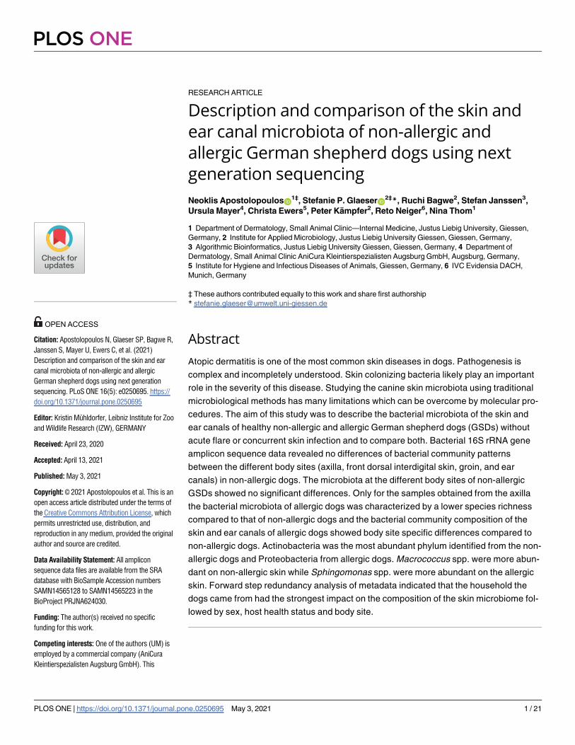
RESEARCH ARTICLE Description and comparison of the skin and ear canal microbiota of non-allergic and allergic German shepherd dogs using next generation sequencing Neoklis Apostolopoulos ID 1‡ , Stefanie P. Glaeser ID 2‡ *, Ruchi Bagwe 2 , Stefan Janssen 3 , Ursula Mayer 4 , Christa Ewers 5 , Peter Ka ¨ mpfer 2 , Reto Neiger 6 , Nina Thom 1 1 Department of Dermatology, Small Animal Clinic—Internal Medicine, Justus Liebig University, Giessen, Germany, 2 Institute for Applied Microbiology, Justus Liebig University Giessen, Giessen, Germany, 3 Algorithmic Bioinformatics, Justus Liebig University Giessen, Giessen, Germany, 4 Department of Dermatology, Small Animal Clinic AniCura Kleintierspezialisten Augsburg GmbH, Augsburg, Germany, 5 Institute for Hygiene and Infectious Diseases of Animals, Giessen, Germany, 6 IVC Evidensia DACH, Munich, Germany ‡ These authors contributed equally to this work and share first authorship * [email protected] Abstract Atopic dermatitis is one of the most common skin diseases in dogs. Pathogenesis is complex and incompletely understood. Skin colonizing bacteria likely play an important role in the severity of this disease. Studying the canine skin microbiota using traditional microbiological methods has many limitations which can be overcome by molecular pro- cedures. The aim of this study was to describe the bacterial microbiota of the skin and ear canals of healthy non-allergic and allergic German shepherd dogs (GSDs) without acute flare or concurrent skin infection and to compare both. Bacterial 16S rRNA gene amplicon sequence data revealed no differences of bacterial community patterns between the different body sites (axilla, front dorsal interdigital skin, groin, and ear canals) in non-allergic dogs. The microbiota at the different body sites of non-allergic GSDs showed no significant differences. Only for the samples obtained from the axilla the bacterial microbiota of allergic dogs was characterized by a lower species richness compared to that of non-allergic dogs and the bacterial community composition of the skin and ear canals of allergic dogs showed body site specific differences compared to non-allergic dogs. Actinobacteria was the most abundant phylum identified from the non- allergic dogs and Proteobacteria from allergic dogs. Macrococcus spp. were more abun- dant on non-allergic skin while Sphingomonas spp. were more abundant on the allergic skin. Forward step redundancy analysis of metadata indicated that the household the dogs came from had the strongest impact on the composition of the skin microbiome fol- lowed by sex, host health status and body site. PLOS ONE PLOS ONE | https://doi.org/10.1371/journal.pone.0250695 May 3, 2021 1 / 21 a1111111111 a1111111111 a1111111111 a1111111111 a1111111111 OPEN ACCESS Citation: Apostolopoulos N, Glaeser SP, Bagwe R, Janssen S, Mayer U, Ewers C, et al. (2021) Description and comparison of the skin and ear canal microbiota of non-allergic and allergic German shepherd dogs using next generation sequencing. PLoS ONE 16(5): e0250695. https:// doi.org/10.1371/journal.pone.0250695 Editor: Kristin Mu ¨hldorfer, Leibniz Institute for Zoo and Wildlife Research (IZW), GERMANY Received: April 23, 2020 Accepted: April 13, 2021 Published: May 3, 2021 Copyright: © 2021 Apostolopoulos et al. This is an open access article distributed under the terms of the Creative Commons Attribution License, which permits unrestricted use, distribution, and reproduction in any medium, provided the original author and source are credited. Data Availability Statement: All amplicon sequence data files are available from the SRA database with BioSample Accession numbers SAMN14565128 to SAMN14565223 in the BioProject PRJNA624030. Funding: The author(s) received no specific funding for this work. Competing interests: One of the authors (UM) is employed by a commercial company (AniCura Kleintierspezialisten Augsburg GmbH). This
-
Upload
khangminh22 -
Category
Documents
-
view
1 -
download
0
Transcript of Description and comparison of the skin and ear canal ... - PLOS
RESEARCH ARTICLE
Description and comparison of the skin and
ear canal microbiota of non-allergic and
allergic German shepherd dogs using next
generation sequencing
Neoklis ApostolopoulosID1‡, Stefanie P. GlaeserID
2‡*, Ruchi Bagwe2, Stefan Janssen3,
Ursula Mayer4, Christa Ewers5, Peter Kampfer2, Reto Neiger6, Nina Thom1
1 Department of Dermatology, Small Animal Clinic—Internal Medicine, Justus Liebig University, Giessen,
Germany, 2 Institute for Applied Microbiology, Justus Liebig University Giessen, Giessen, Germany,
3 Algorithmic Bioinformatics, Justus Liebig University Giessen, Giessen, Germany, 4 Department of
Dermatology, Small Animal Clinic AniCura Kleintierspezialisten Augsburg GmbH, Augsburg, Germany,
5 Institute for Hygiene and Infectious Diseases of Animals, Giessen, Germany, 6 IVC Evidensia DACH,
Munich, Germany
‡ These authors contributed equally to this work and share first authorship
Abstract
Atopic dermatitis is one of the most common skin diseases in dogs. Pathogenesis is
complex and incompletely understood. Skin colonizing bacteria likely play an important
role in the severity of this disease. Studying the canine skin microbiota using traditional
microbiological methods has many limitations which can be overcome by molecular pro-
cedures. The aim of this study was to describe the bacterial microbiota of the skin and
ear canals of healthy non-allergic and allergic German shepherd dogs (GSDs) without
acute flare or concurrent skin infection and to compare both. Bacterial 16S rRNA gene
amplicon sequence data revealed no differences of bacterial community patterns
between the different body sites (axilla, front dorsal interdigital skin, groin, and ear
canals) in non-allergic dogs. The microbiota at the different body sites of non-allergic
GSDs showed no significant differences. Only for the samples obtained from the axilla
the bacterial microbiota of allergic dogs was characterized by a lower species richness
compared to that of non-allergic dogs and the bacterial community composition of the
skin and ear canals of allergic dogs showed body site specific differences compared to
non-allergic dogs. Actinobacteria was the most abundant phylum identified from the non-
allergic dogs and Proteobacteria from allergic dogs. Macrococcus spp. were more abun-
dant on non-allergic skin while Sphingomonas spp. were more abundant on the allergic
skin. Forward step redundancy analysis of metadata indicated that the household the
dogs came from had the strongest impact on the composition of the skin microbiome fol-
lowed by sex, host health status and body site.
PLOS ONE
PLOS ONE | https://doi.org/10.1371/journal.pone.0250695 May 3, 2021 1 / 21
a1111111111
a1111111111
a1111111111
a1111111111
a1111111111
OPEN ACCESS
Citation: Apostolopoulos N, Glaeser SP, Bagwe R,
Janssen S, Mayer U, Ewers C, et al. (2021)
Description and comparison of the skin and ear
canal microbiota of non-allergic and allergic
German shepherd dogs using next generation
sequencing. PLoS ONE 16(5): e0250695. https://
doi.org/10.1371/journal.pone.0250695
Editor: Kristin Muhldorfer, Leibniz Institute for Zoo
and Wildlife Research (IZW), GERMANY
Received: April 23, 2020
Accepted: April 13, 2021
Published: May 3, 2021
Copyright: © 2021 Apostolopoulos et al. This is an
open access article distributed under the terms of
the Creative Commons Attribution License, which
permits unrestricted use, distribution, and
reproduction in any medium, provided the original
author and source are credited.
Data Availability Statement: All amplicon
sequence data files are available from the SRA
database with BioSample Accession numbers
SAMN14565128 to SAMN14565223 in the
BioProject PRJNA624030.
Funding: The author(s) received no specific
funding for this work.
Competing interests: One of the authors (UM) is
employed by a commercial company (AniCura
Kleintierspezialisten Augsburg GmbH). This
Introduction
Several next generation sequencing (NGS) studies in the last few years have shown that the
skin of dogs, similar to humans, contains a higher diversity of bacterial taxa than previously
believed [1–4]. Bacteria play an important role in both, health and disease, and changes in bac-
terial community composition of the skin are associated with many skin diseases in both
humans and animals [2,5].
Canine atopic dermatitis (cAD) is a common skin disease in dogs characterized by a geneti-
cally predisposed inflammatory, IgE-associated, pruritic allergic disease, affecting certain body
sites and ear canals [6,7]. CAD has been proposed as an animal model for human atopic der-
matitis [8,9]. Environmental allergens are the most common cause of cAD [6]. But, food aller-
gens (cutaneous adverse food reactions; CAFRs) can also cause identical clinical signs or be a
flare factor of a cAD, making a clinical differentiation impossible [10,11]. The final diagnosis
must be obtained through a systematic workup [12]. Studies in dogs have shown an association
between the skin microbiota and allergic skin diseases. The skin of six allergic dogs without
signs of pyoderma or Malassezia dermatitis revealed that there was a lower bacterial commu-
nity diversity compared to the skin of 12 healthy dogs, but the bacterial community composi-
tion did not differ significantly [13]. Recently, a longitudinal study showed reduced diversity
and different bacterial community composition in dogs with cAD and secondary pyoderma
compared with healthy dogs [14]. After antimicrobial therapy and remission of skin lesions,
the diversity was restored and the clustering difference of the bacterial communities was
reduced [14]. Neither study examined the ear canal, which is a commonly affected body site in
allergic dogs. The only study using NGS for evaluating the microbiota of asymptomatic ear
canals of dogs with cAD showed no difference in the diversity but a significant difference in
bacteria community composition [1]. All previously mentioned studies involved dogs from
various breeds. It is well documented that cAD has breed predispositions and that the pheno-
type of the disease differs between breeds [15]. It is unclear if the different phenotypes of cAD
affect the bacterial community composition between the breeds. To date no study has evalu-
ated the microbiota of the skin and ear canal of only a single breed in both healthy and allergic
skin disease states, thus minimizing potential bias due to allergy phenotype effect on the
microbiota. The German shepherd dog (GSD) was chosen in our investigation because it is a
high-risk breed for cAD [15], possibly due to an altered expression of the plakophilin 2 gene
and other genes of the chromosome 27 [16].
The goal of the study was to describe and compare bacterial microbiomes of four body sites
(axilla, front dorsal interdigital skin, groin, and ear canal) of healthy (non-allergic) and allergic
GSDs using a 16S rRNA gene amplicon-based Illumina sequencing approach. We hypothe-
sized that atopic dermatitis and/or CAFR influence the microbiota of the skin and ear canal of
GSDs resulting in reduced bacterial diversity and significantly different bacterial community
composition.
Material and methods
Study subjects
The clinical study was performed at the small animal clinic of the Justus Liebig University
(JLU), Giessen, Germany. Samples were collected at the small animal clinic (JLU) during
appointments specifically for this study. All owners were informed of the procedures and
signed a consent form for sample collection. The Animal Welfare Committee of the Justus Lie-
big University of Giessen was informed about the study protocol and especially the sampling
method was discussed. As there is neither pain, harm nor damage caused by gently rolling a
PLOS ONE Skin and ear canal microbiota in non-allergic and allergic German shepherd dogs
PLOS ONE | https://doi.org/10.1371/journal.pone.0250695 May 3, 2021 2 / 21
company provided the salary for the clinical duty of
the author (UM). Her clinical duty is not dependent
on the study. Furthermore, the company did not
have any additional role in the study design, data
collection and analysis, decision to publish, or
preparation of the manuscript. The specific role of
these author is articulated in the ‘author
contributions’ section. This commercial affiliation
does not alter our adherence to PLOS ONE policies
on sharing data and materials.
cotton swab on skin, they assured us that ethical approval by the responsible authority is not
required. Two groups of GSDs were studied. The control group included 12 GSDs without any
history of allergic conditions or any clinical skin/ear canal lesions compatible with allergy
(they will be referred to as "non-allergic" in the rest of the manuscript) at the time of examina-
tion and sampling. In order to minimize the risk of including a dog with subclinical allergy,
only dogs older than four years were involved, as allergic conditions most often develop in
young dogs from 6 months to 3 years [17]. In order to investigate a possible influence of the
household conditions to the microbiota, two non-allergic GSDs per household were included.
Neither systemic antibiotics nor any immunomodulatory or anti-inflammatory drugs were
allowed six months prior to sampling. Bathing with shampoo and the use of ear cleaners was
not allowed seven days prior to sampling. Twelve adult allergic GSDs were diagnosed with
cAD, either due to cutaneous food reactions or environmental allergens. Standard diagnostic
and therapeutic methods were used, including fulfillment of at least five of Favrot´s criteria
and excluding other pruritic dermatosis (e.g. flea saliva hypersensitivity, sarcoptic mange)
[12,18]. A combination of cAD and/or CAFRs with flea saliva hypersensitivity was allowed.
Dogs with any secondary bacterial or fungal skin or ear infection were excluded. Systemic and
topical antibacterial or antifungal agents were not allowed 30 and 14 days prior to sampling,
respectively. Systemic administration of any immunomodulatory or anti-inflammatory drugs,
with the exception of oclacitinib, were equally not allowed two months prior to sampling. Top-
ical immunomodulatory or anti-inflammatory drugs had to be withdrawn 14 days prior to the
study. Any shampooing and ear cleaners were not allowed seven days prior to sampling. All
owners filled out a questionnaire regarding their dog’s housing, partner animals, current dis-
eases and treatment, food supplements, frequency of bathing and type of shampoo. Owners of
allergic dogs were asked about gastrointestinal signs and to score their dog’s pruritic signs
using a pruritus visual analogue scale (pVAS), as previously validated [19,20].
Assessment of lesions
All dogs were examined clinically and dermatologically including otoscopic examination
using sterile powder-free gloves and an autoclavable metal otoscope. A validated site-specific
lesion and scoring scale of lesions‘ severity, the Canine Atopic Dermatitis Extent and Severity
Index (CADESI-4), was performed for the atopic dogs in order to evaluate any relationship of
cutaneous microbiota and the severity of atopic dermatitis [21]. Skin and ear canal cytology
was performed as described [22,23].
Sample collection
Prior to sampling and in between sampling of the GSDs, the examination table was cleaned
with PCR Clean™ Wipes (Minerva Biolabs, Berlin, Germany) according to the manufacturer´s
manual, to avoid DNA cross-contamination. In order to minimize microbial contamination
from the clinic´s floor, dogs were brought directly into the examination room and placed onto
the examination table. After physical examination, the left axilla (A), left front dorsal interdigi-
tal region (Int), left side of the groin (L) and the left ear canal (O) were sampled. These body
sites appear to be most commonly affected by atopy in this breed [15]. For sampling of micro-
biome studies we used 70% ethylene oxide sterilized forensic swabs with transport tube, poly-
styrene stem material and viscose swab material (Forensic Swab, Nr 80.629, Sarstedt,
Nuembrecht, Germany) to ensure the absence of DNA contamination and to avoid cotton or
wood mitochondria from the swab or the stem. The swabs were rubbed 40 times on the desired
region, rotating one-quarter of the swab´s site (90˚) for 10 times each. All samples were
obtained in duplicates. Subsequently, a sample was taken for cytological purposes using a
PLOS ONE Skin and ear canal microbiota in non-allergic and allergic German shepherd dogs
PLOS ONE | https://doi.org/10.1371/journal.pone.0250695 May 3, 2021 3 / 21
sterile cotton swab. The samples were transported immediately at 8˚C to the Institute of
Applied Microbiology, JLU Giessen. Samples for microbiome studies were stored at -20˚C
until further processing (DNA extraction).
DNA extraction and 16S rRNA gene amplicon sequencing
Total DNA was extracted from DNA free swabs stored at -20˚C using the NucleoSpin1 96 Soil
kit (96-well extraction system, Macherey Nagel AG, Oesingen, Switzerland) which can effi-
ciently extract DNA from Gram-negative and Gram-positive bacteria including spores.
According to manufacturer’s instruction using vacuum processing (NucleoVac 96 vacuum
manifold, Macherey Nagel AG, Oesingen, Switzerland) with slight modifications, extraction
was started using lysis buffer SL1 and afterward steps 1 to 5 were repeated with lysis buffer
SL2, thus samples were lysed twice. Total DNA was eluted with 80 μL PCR water (1x 30 μL, 1x
50 μL) instead of SE buffer. DNA was quantified spectrophotometrically using a NanoDrop
spectrophotometer (Thermo Scientific) and subsequently checked for the presence and
amplifiability of 16S rRNA gene sequences for selected samples.
The 16S rRNA gene sequences of Bacteria were amplified for Illumina amplicon sequencing
(LGC Genomics, Berlin, Germany) using a nested PCR approach with a first PCR with the
primer system 341F (50-CCTACGGGAGGCAGCAG-3´) and 1061R (50-CRRCACGAGCTGACGAC-3´) (V3-V6) [24] (20 cycles) followed by a second PCR with primer system 515F (50-GTGYCAGCMGCCGCGGTAA-3´)-Y and 926R (50-CCGYCAATTYMTTTRAGTTT-3´)-jed (V4-V5)
[25] (20 cycles), because according to previous studies, only a low amount of microbial DNA
was detected on human skin analyzed by PCR [26,27]. For each sample, forward and reverse
primers of the second PCR had the same 10-nt barcode sequences. The first round of PCR was
carried out for 20 cycles, using the following parameters: 2 minutes 96˚C pre-denaturation;
96˚C for 15 seconds (s), 50˚C for 30 s, 70˚C for 90 s and primers without inline barcodes were
used (341F/1061R). For the second round, 1 μl PCR product from the first PCR was used and
the PCR conditions were the same as before. In this case, barcoded primers were added
(515F-Y/926R-jed). DNA concentration of amplicons of interest was determined by gel elec-
trophoresis. About 20 ng amplicon DNA of each sample were pooled for up to 48 samples car-
rying different barcodes. The amplicon pools were purified with one volume AMPure XP
beads (Agencourt) to remove primer dimer and other small misspriming products, followed
by an additional purification on MiniElute columns (Qiagen). About 100 ng of each purified
amplicon pool DNA was used to construct Illumina libraries using the Ovation Rapid DR
Multiplex System 1–96 (NuGEN). Illumina libraries were pooled, and size selected by prepara-
tive gel electrophoresis. Sequencing was done on a Illumina MiSeq using V3 Chemistry (Illu-
mina). Raw sequence reads are available in the Sequence Read Archive (SRA) with BioSample
Accession numbers SAMN14565128 to SAMN14565223 in the BioProject PRJNA624030.
Amplicon sequence data analysis
The NGS analysis pipeline (https://www.arb-silva.de/ngs) of the SILVA rRNA gene database
(SILVAngs 1.3) was used for sequence analysis [28]. For this, datasets of all combined sequence
reads (adaptor and primer clipped) were uploaded to the database. All reads were aligned
using the SILVA Incremental Aligner (SINA v1.2.10 for ARB SVN revision21008) [29], against
the SILVA SSU rRNA SEED and quality controlled [28]. Reads shorter than 50 aligned nucleo-
tides and reads with more than 2% of ambiguities, or 2% of homopolymers, were excluded
from further processing. Reads with a low alignment quality (50 alignment identity, 40 align-
ment score reported by SINA) as well as putative contaminations and artifacts, were identified
and excluded from downstream analysis. The next process step was dereplication and
PLOS ONE Skin and ear canal microbiota in non-allergic and allergic German shepherd dogs
PLOS ONE | https://doi.org/10.1371/journal.pone.0250695 May 3, 2021 4 / 21
clustering with cd-hit-est (version 3.1.2; http://www.bioinformatics.org/cd-hit) using accuratemode, ignoring overhangs and applying identity criteria of 1.00 and 0.98, respectively [30]. For
classification a local nucleotide BLAST search was performed against the non-redundant ver-
sion of the SILVA SSU Ref dataset (release 128; http://www.arb-silva.de) using blastn (version
2.2.30+; http://blast.ncbi.nlm.nih.gov/Blast.cgi) with standard settings [31]. Unique reads were
clustered in operational taxonomic unit (OTU) on a per sample basis under the criterium of
98% sequence identity to each other (pairwise distance and single linkage clustering). The lon-
gest read in each cluster was classified as the reference for each OTU and was mapped onto all
reads that were assigned to the respective OTU. OTUs were assigned to taxonomic paths
(genus level). Several OTUs were thereby assigned to the same taxonomic path/genus. This
process resulted in quantitative information (number of individual reads of all OTUs per taxo-
nomic path), despite the PCR limitations, possible sequencing technique biases and multiple
rRNA operons. Reads without any BLAST hits or reads with weak BLAST hits, where the func-
tion “(% sequence identity + % alignment coverage)/2” did not exceed the value of 93,
remained unclassified and were assigned in the virtual taxonomical group “No Relative” in the
SILVAngs fingerprint and Krona charts [32], as previously reported [33,34].
Alpha and beta diversity analyses were performed in PAST3 (https://folk.uio.no/ohammer/
past) [35] at the level of genera (phylogenetic groups). Alpha diversity was measured by calculat-
ing different diversity indices considering the number of phylogenetic groups per samples and
the number of reads per phylogenetic group. The Shannon (overall alpha diversity) and Chao1
(richness; number of taxa corrected by the presence of singleton) indices were calculated. Signif-
icant differences between the alpha diversity indices determined for non-allergic vs allergic dogs
were further evaluated. For two groups, single body sites of non-allergic versus allergic dogs,
unpaired t-tests were performed in SigmaPlot 13 (Systat Software Inc.). First, a normality test
(Shapiro-Wilk) was performed [36]. If the data passed the normality test, a two-tailed p-value
was obtained from the t-test analysis. If the normality test failed, the Mann-Whitney Rank Sum
Test was applied to test for the presence of significant differences. For more than two groups
(all body sites), the significant differences of the alpha diversity values were evaluated by per-
forming a Kruskal Wallis Test [37]. The beta diversity (comparison of the phylogenetic compo-
sition of the bacterial communities of the different samples) was studied based on relative
abundance data analyzed by non-metric multidimensional scaling (NMDS; [38]) based on a
Bray-Curtis similarity matrix [39]. Environmental variables, e.g. body sites, home (living in the
same household) and sex, were included to the NMDS plot displayed as biplot vector. One way
ANOSIMs (9999 permutations) [39] and One way PERMANOVA (9999 permutations) [40]
were performed to determine significant differences (evaluating the sequential Bonferroni p-
values) among samples. SIMPER (Similarity Percentage) analysis [39] also based on a Bray Cur-
tis similarity matrix, was used to determine the average percent contribution of the different
taxa to the dissimilarity among samples. Based on relative abundance patterns and contribu-
tions to community differences (SIMPER analysis) a selection of bacterial taxa was further stud-
ied with respect to significant differences in relative abundance in non-allergic vs allergic dogs.
T-test were performed as described above. For correction for multiple hypothesis testing, p val-
ues were divided through the number of performed tests.
We quantified the effect size of multiple metadata fields (collection timestamp, sex, oclaciti-
nib, host health status, host subject id) by combining them in a linear model and performed
forward step redundancy analysis (RDA) as previously described [41], with the rda and
ordiR2step functions in the vegan package in R 3.6.1 [42].
By systematically testing metadata fields for correlation with alpha- (using the two metrics
’Shannon’, ’chao1’) or beta-diversity (using metric ’Bray-Curtis’ [43]) via Kruskal-Wallis [44],
Spearman- [45], and Pearson-correlation [46] (for alpha diversity) and PERMANOVA [40]
PLOS ONE Skin and ear canal microbiota in non-allergic and allergic German shepherd dogs
PLOS ONE | https://doi.org/10.1371/journal.pone.0250695 May 3, 2021 5 / 21
and ANOSIM [39] (for beta diversity), we found that the following fields were significant in at
least one of the tests: collection timestamp, sex, oclacitinib, host health status, host subject id
and household id.
To illustrate covariance among these metadata fields, we examined the correlation between
individual metadata variables. Categorical metadata fields (all but collection timestamp) were
compared using a modified CramerV statistic [47]). Continuous (collection timestamp) and
categorical covariates were compared with a Welchttest [47]. Resulting correlation ratios were
visualized as a heat map.
Results
Study subjects
Overall 29 GSDs were sampled but five were excluded according to the exclusion criteria
(Table 1 in S1 File). Twelve non-allergic (6 male, 1 male castrated, 4 intact female, and 1 spayed
female) and twelve allergic (2 intact male, 1 male castrated, and 9 intact female) GSDs were
further analyzed (Table 1 in S1 File). The dogs were living in a radius of max 131 kilometers
away from Giessen (Germany). The age of the non-allergic dogs (mean: 7.5 ± 1.9 years) was
significantly higher (t-Test, p< 0.01) than the age of allergic GSDs (mean: 4.8 ± 2.2 years)
(Table 2 in S1 File). All allergic dogs had mild lesions with a median CADESI-04 score of six
(range 0–11) and a median PVAS score of four (range 0–8). Two dogs had a PVAS score of 7/
10 and 8/10, respectively, with only mild skin lesions. Six allergic GSDs received labeled doses
of oclacitinib (Apoquel, Zoetis Deutschland GmbH, Berlin, Germany).
Analysis of the phylogenetic composition of the GSDs microbiota
In total, 4,038,850 paired end sequences with an average sequence length of 373 nucleotides (nt)
were obtained. Briefly, 2,579 sequences (0.06%) were rejected because they failed the SILVAngs
pipeline quality control. Finally, 4,036,271 sequences (2,334–246,525 per sample) were further
analyzed (Table 3 in S1 File). In total 241,114 unique reads (5.97% of the finally analyzed
sequences) were assigned to OTUs. Additionally, 1,129,221 (27.96%) sequences (number of
sequence reads with 98% sequence identity to each other; defined as "clustered"), and 2,665,936
(66.01%) sequences (number of sequence reads with 100% identity to another; defined as "repli-
cates") were assigned to the OTUs. Each OTU was classified in the SILVA database with a taxo-
nomic paths with maximum resolution at the genus level (named as ´phylogenetic groups‘for
unnamed genera). Chloroplast, mitochondrial, and archaeal 16S rRNA gene sequences were
detected with a relative abundance of 20.13% (190,605 sequences), 0.07% (9,430 sequences),
0.02% (1,064 sequences) of the total analyzed sequences, respectively (Table 3 in S1 File).
Sequences that did not match any known taxa (sequence similarity<93% to the next known
taxon) were classified as “no relative” and had 0.3% relative abundance (5,131 sequences) (Table 3
in S1 File). Sequences assigned to Archaea, chloroplasts, mitochondria, and no relative groups
were excluded from further analyses. Only sequences assigned to the domain Bacteria (3,830,041
in total; 2,334 to 232,445 per sample) were further analyzed and were set to 100% (per sample).
Skin Microbiome of non-allergic German shepherd dogs
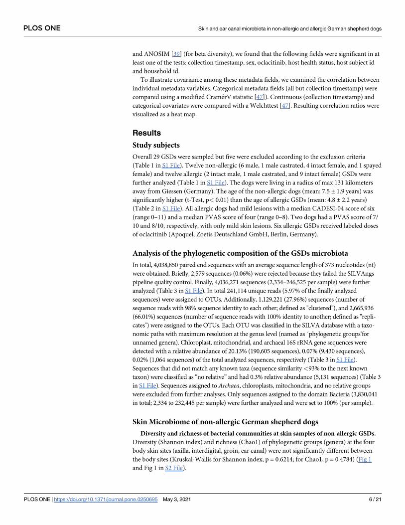
Diversity and richness of bacterial communities at skin samples of non-allergic GSDs.
Diversity (Shannon index) and richness (Chao1) of phylogenetic groups (genera) at the four
body skin sites (axilla, interdigital, groin, ear canal) were not significantly different between
the body sites (Kruskal-Wallis for Shannon index, p = 0.6214; for Chao1, p = 0.4784) (Fig 1
and Fig 1 in S2 File).
PLOS ONE Skin and ear canal microbiota in non-allergic and allergic German shepherd dogs
PLOS ONE | https://doi.org/10.1371/journal.pone.0250695 May 3, 2021 6 / 21
However, a greater number of individual samples from the interdigital region and ear canal
had a more homogeneous composition of the microbiota, showing a lower richness (Chao1
values; Fig 1 in S2 File).
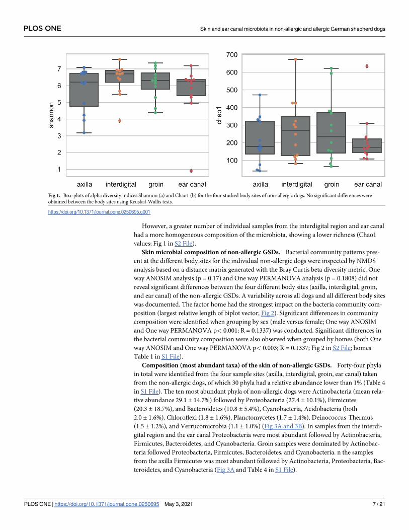
Skin microbial composition of non-allergic GSDs. Bacterial community patterns pres-
ent at the different body sites for the individual non-allergic dogs were inspected by NMDS
analysis based on a distance matrix generated with the Bray Curtis beta diversity metric. One
way ANOSIM analysis (p = 0.17) and One way PERMANOVA analysis (p = 0.1808) did not
reveal significant differences between the four different body sites (axilla, interdigital, groin,
and ear canal) of the non-allergic GSDs. A variability across all dogs and all different body sites
was documented. The factor home had the strongest impact on the bacteria community com-
position (largest relative length of biplot vector; Fig 2). Significant differences in community
composition were identified when grouping by sex (male versus female; One way ANOSIM
and One way PERMANOVA p< 0.001; R = 0.1337) was conducted. Significant differences in
the bacterial community composition were also observed when grouped by homes (both One
way ANOSIM and One way PERMANOVA p< 0.003; R = 0.1337; Fig 2 in S2 File; homes
Table 1 in S1 File).
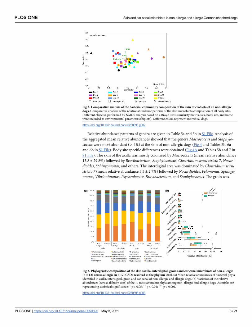
Composition (most abundant taxa) of the skin of non-allergic GSDs. Forty-four phyla
in total were identified from the four sample sites (axilla, interdigital, groin, ear canal) taken
from the non-allergic dogs, of which 30 phyla had a relative abundance lower than 1% (Table 4
in S1 File). The ten most abundant phyla of non-allergic dogs were Actinobacteria (mean rela-
tive abundance 29.1 ± 14.7%) followed by Proteobacteria (27.4 ± 10.1%), Firmicutes
(20.3 ± 18.7%), and Bacteroidetes (10.8 ± 5.4%), Cyanobacteria, Acidobacteria (both
2.0 ± 1.6%), Chloroflexi (1.8 ± 1.6%), Planctomycetes (1.7 ± 1.4%), Deinococcus-Thermus
(1.5 ± 1.2%), and Verrucomicrobia (1.1 ± 1.0%) (Fig 3A and 3B). In samples from the interdi-
gital region and the ear canal Proteobacteria were most abundant followed by Actinobacteria,
Firmicutes, Bacteroidetes, and Cyanobacteria. Groin samples were dominated by Actinobac-
teria followed Proteobacteria, Firmicutes, Bacteroidetes, and Cyanobacteria. n the samples
from the axilla Firmicutes was most abundant followed by Actinobacteria, Proteobacteria, Bac-
teroidetes, and Cyanobacteria (Fig 3A and Table 4 in S1 File).
Fig 1. Box-plots of alpha diversity indices Shannon (a) and Chao1 (b) for the four studied body sites of non-allergic dogs. No significant differences were
obtained between the body sites using Kruskal-Wallis tests.
https://doi.org/10.1371/journal.pone.0250695.g001
PLOS ONE Skin and ear canal microbiota in non-allergic and allergic German shepherd dogs
PLOS ONE | https://doi.org/10.1371/journal.pone.0250695 May 3, 2021 7 / 21
Relative abundance patterns of genera are given in Table 5a and 5b in S1 File. Analysis of
the aggregated mean relative abundances showed that the genera Macrococcus and Staphylo-coccus were most abundant (> 4%) at the skin of non-allergic dogs (Fig 4 and Tables 5b, 6a
and 6b in S1 File). Body site specific differences were obtained (Fig 4A and Tables 5b and 7 in
S1 File). The skin of the axilla was mostly colonized by Macrococcus (mean relative abundance
13.8 ± 29.8%) followed by Brevibacterium, Staphylococcus, Clostridium sensu stricto 7, Nocar-dioides, Sphingomonas, and others. The interdigital area was dominated by Clostridium sensustricto 7 (mean relative abundance 3.5 ± 2.7%) followed by Nocardioides, Pelomonas, Sphingo-monas, Vibrionimonas, Psychrobacter, Bravibacterium, and Staphylococcus. The groin was
Fig 2. Comparative analysis of the bacterial community composition of the skin microbiota of all non-allergic
dogs. Comparative analysis of the relative abundance patterns of the skin microbiota composition of all body sites
(different objects), performed by NMDS analysis based on a Bray-Curtis similarity matrix. Sex, body site, and home
were included as environmental parameters (biplots). Different colors represent individual dogs.
https://doi.org/10.1371/journal.pone.0250695.g002
Fig 3. Phylogenetic composition of the skin (axilla, interdigital, groin) and ear canal microbiota of non-allergic
(n = 12) versus allergic (n = 12) GSDs resolved at the phylum level. (a) Mean relative abundances of bacterial phyla
identified in axilla, interdigital, groin and ear canal of non-allergic and allergic dogs. (b) Variation of the relative
abundances (across all body sites) of the 10 most abundant phyla among non-allergic and allergic dogs. Asterisks are
representing statistical significance: � p< 0.05; �� p< 0.01; ��� p< 0.001.
https://doi.org/10.1371/journal.pone.0250695.g003
PLOS ONE Skin and ear canal microbiota in non-allergic and allergic German shepherd dogs
PLOS ONE | https://doi.org/10.1371/journal.pone.0250695 May 3, 2021 8 / 21
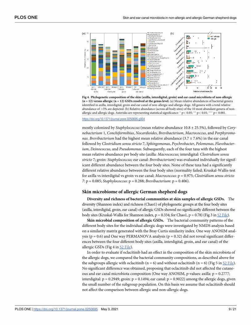
mostly colonized by Staphylococcus (mean relative abundance 10.8 ± 25.5%), followed by Cory-nebacterium 1, Conchiformibius, Nocardioides, Brevibacterium, Macrococcus, and Porphyromo-nas. Brevibacterium had the highest mean relative abundance (3.7 ± 7.6%) in the ear canal
followed by Clostridium sensu stricto 7, Sphingomonas, Psychrobacter, Pelomonas, Flavobacter-ium, Deinococcus, and Pseudomonas. Subsequently, each of the four taxa with the highest
mean relative abundance per body site (axilla: Macrococcus; interdigital: Clostridium sensustricto 7; groin: Staphylococcus; ear canal: Brevibacterium) was evaluated individually for signif-
icant different abundance between the four body sites. None of these taxa had a significantly
different relative abundance between the four body sites (normality failed; Kruskal-Wallis-test
for axilla vs interdigital vs groin vs ear canal; Macrococcus: p = 0.975; Clostridium sensu stricto7: p = 0.085; Staphylococcus: p = 0.288; Brevibacterium: p = 0.406).
Skin microbiome of allergic German shepherd dogs
Diversity and richness of bacterial communities at skin samples of allergic GSDs. The
diversity (Shannon index) and richness (Chao1) of phylogenetic groups at the four body sites
(axilla, interdigital, groin, ear canal) of allergic GSDs showed no significantly different between the
body sites (Kruskal-Wallis for Shannon index, p = 0.334; for Chao1, p = 0.78) (Fig 3 in S2 File).
Skin microbial composition of allergic GSDs. The bacterial community patterns of the
different body sites for the individual allergic dogs were investigated by NMDS analysis based
on a similarity matrix generated with the Bray Curtis similarity index. One way ANOSIM anal-
ysis (p = 0.6) and One way PERMANOVA analysis (p = 0.32) did not reveal significant differ-
ences between the four different body sites (axilla, interdigital, groin, and ear canal) of the
allergic GSDs (Fig 4 in S2 File).
In order to evaluate if oclacitinib had an effect in the composition of the skin microbiota of
the allergic dogs, we compared the bacterial community compositions, as described above for
the subgroups allergic with oclacitinib (n = 6) and without oclacitinib (n = 6) (Fig 5 in S2 File).
No significant difference was obtained, proposing that oclacitinib did not affected the cutane-
ous and ear canal microbiota composition (One way ANOSIM, p-values: axilla: p = 0.2777;
interdigital: p = 0.2949; groin: p = 0.1494; ear canal: p = 0.9022) among the allergic dogs, given
the small number of the subgroup population. On this basis we assume that oclacitinib should
not affect the comparison between allergic and non-allergic dogs.
Fig 4. Phylogenetic composition of the skin (axilla, interdigital, groin) and ear canal microbiota of non-allergic
(n = 12) versus allergic (n = 12) GSDs resolved at the genus level. (a) Mean relative abundances of bacterial genera
identified in axilla, interdigital, groin and ear canal of non-allergic and allergic dogs. All genera with a total relative
abundance of>5% are depicted. (b) Relative abundance (across all body sites) of the 10 most abundant genera of non-
allergic and allergic dogs. Asterisks are representing statistical significance: � p< 0.05; �� p< 0.01; ��� p< 0.001.
https://doi.org/10.1371/journal.pone.0250695.g004
PLOS ONE Skin and ear canal microbiota in non-allergic and allergic German shepherd dogs
PLOS ONE | https://doi.org/10.1371/journal.pone.0250695 May 3, 2021 9 / 21
Composition (most abundant taxa) of the skin of allergic GSDs. Proteobacteria (mean
relative abundance 35.5 ± 10.9%) and Actinobacteria (22.4 ±10.2%) were the most abundant
phyla of the allergic GSDs and dominated in each of the four sample sites (Fig 3A and 3B and
Table 8 in S1 File). They were followed by Firmicutes (17.0 ± 14.6%), Bacteroidetes
(14.1 ± 5.2%), Cyanobacteria (2.5 ± 3.0%), Planctomycetes (1.8 ±1.2%), Chloroflexi
(1.7 ± 1.5%), Acidobacteria (1.7 ± 1.6%), Verrucomicrobia (0.7 ± 0.9%), and Deinococcus-
Thermus (0.6 ± 0.7%).
Overview of relative abundance patterns of genera present in the microbiota at different
body sites of allergic dogs is given in Table 5a and 5b in S1 File. Aggregated mean relative abun-
dance data showed that Sphingomonas, Staphylococcus, Clostridium sensu stricto 7, and Nocar-dioides dominated the skin microbiota of allergic GSDs (each genus> 4% aggregated relative
abundance) (Fig 4 and Tables 5b, 6a and 6b in S1 File). Analysis of the mean relative abun-
dances at the genus level revealed some body site specific differences (Fig 4A and Tables 5b and
7 in S1 File). Clostridium sensu stricto 7 (mean relative abundance 6.2 ± 7.8%) was the most
abundant phylogenetic group on the skin of the axilla, followed by Staphylococcus, Vibrionimo-nas, Sphingomonas, Pelomonas, and Nocardioides. The interdigital skin was dominated by
Nocardioides (mean relative abundance 5.3 ± 4.4%), followed by Sphingomonas, Pelomonas,Clostridium sensu stricto 7, Vibrionimonas, Bradyrhizobium, Staphylococcus, and Hymenobacter.The skin of the groin was dominated by Sphingomonas (mean relative abundance 4.9 ± 2.4%),
followed by Clostridium sensu stricto 7, Nocardioides, Porphyromonas, Conchiformibius, Pelomo-nas, and Vibrionimonas. Finally, the ear canal of the allergic GSDs was dominated by Staphylo-coccus (mean relative abundance 9.4 ± 25.9%), followed by Sphingomonas, Clostridium sensustricto 7, Nocardioides, Vibrionimonas, Pelomonas, Propionibacterium, and Paracoccus.
Comparative analysis of the skin microbiome of non-allergic versus allergic
German shepherd dogs
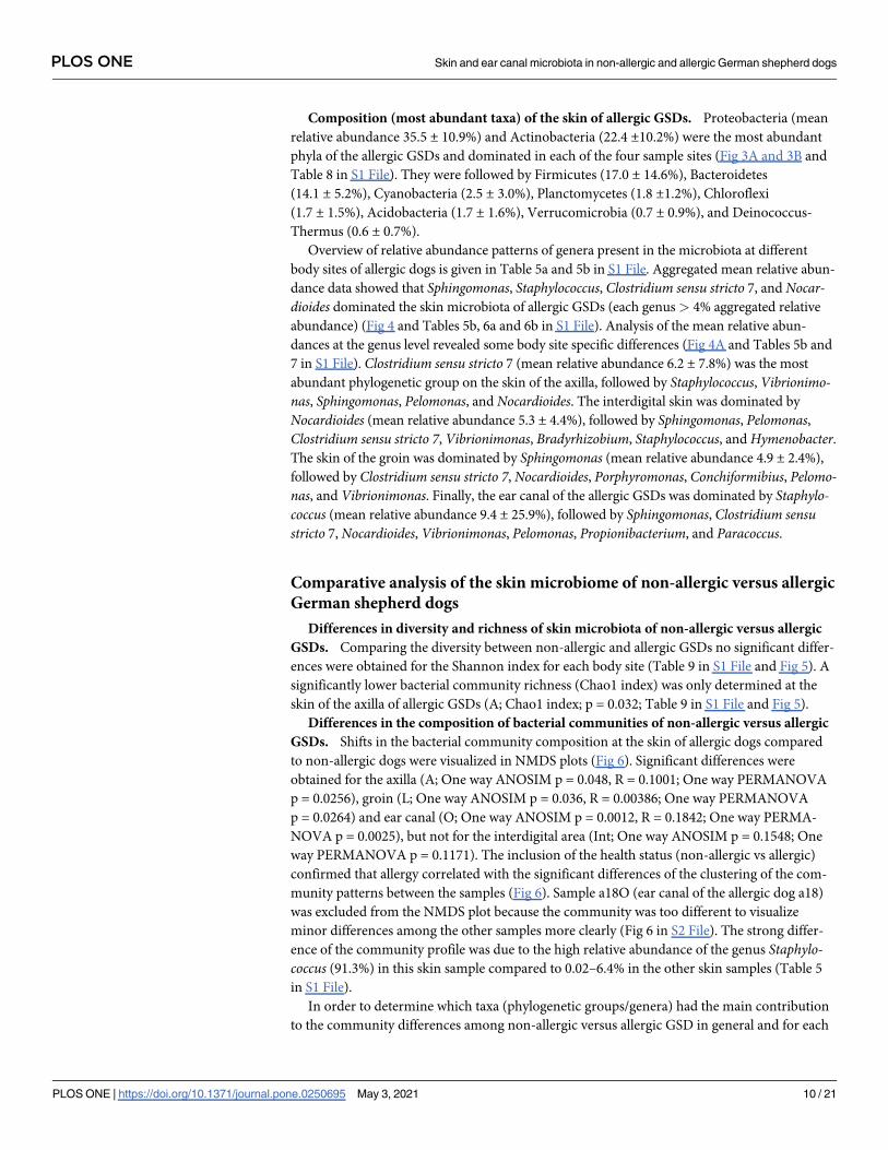
Differences in diversity and richness of skin microbiota of non-allergic versus allergic
GSDs. Comparing the diversity between non-allergic and allergic GSDs no significant differ-
ences were obtained for the Shannon index for each body site (Table 9 in S1 File and Fig 5). A
significantly lower bacterial community richness (Chao1 index) was only determined at the
skin of the axilla of allergic GSDs (A; Chao1 index; p = 0.032; Table 9 in S1 File and Fig 5).
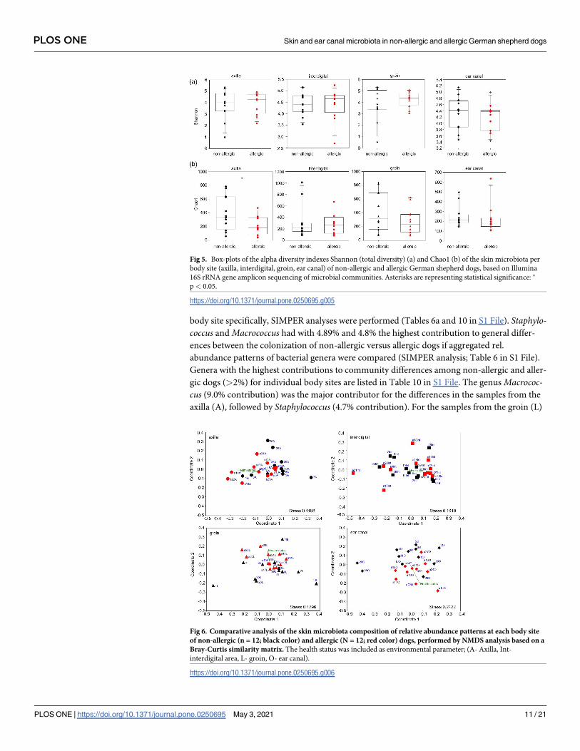
Differences in the composition of bacterial communities of non-allergic versus allergic
GSDs. Shifts in the bacterial community composition at the skin of allergic dogs compared
to non-allergic dogs were visualized in NMDS plots (Fig 6). Significant differences were
obtained for the axilla (A; One way ANOSIM p = 0.048, R = 0.1001; One way PERMANOVA
p = 0.0256), groin (L; One way ANOSIM p = 0.036, R = 0.00386; One way PERMANOVA
p = 0.0264) and ear canal (O; One way ANOSIM p = 0.0012, R = 0.1842; One way PERMA-
NOVA p = 0.0025), but not for the interdigital area (Int; One way ANOSIM p = 0.1548; One
way PERMANOVA p = 0.1171). The inclusion of the health status (non-allergic vs allergic)
confirmed that allergy correlated with the significant differences of the clustering of the com-
munity patterns between the samples (Fig 6). Sample a18O (ear canal of the allergic dog a18)
was excluded from the NMDS plot because the community was too different to visualize
minor differences among the other samples more clearly (Fig 6 in S2 File). The strong differ-
ence of the community profile was due to the high relative abundance of the genus Staphylo-coccus (91.3%) in this skin sample compared to 0.02–6.4% in the other skin samples (Table 5
in S1 File).
In order to determine which taxa (phylogenetic groups/genera) had the main contribution
to the community differences among non-allergic versus allergic GSD in general and for each
PLOS ONE Skin and ear canal microbiota in non-allergic and allergic German shepherd dogs
PLOS ONE | https://doi.org/10.1371/journal.pone.0250695 May 3, 2021 10 / 21
body site specifically, SIMPER analyses were performed (Tables 6a and 10 in S1 File). Staphylo-coccus and Macrococcus had with 4.89% and 4.8% the highest contribution to general differ-
ences between the colonization of non-allergic versus allergic dogs if aggregated rel.
abundance patterns of bacterial genera were compared (SIMPER analysis; Table 6 in S1 File).
Genera with the highest contributions to community differences among non-allergic and aller-
gic dogs (>2%) for individual body sites are listed in Table 10 in S1 File. The genus Macrococ-cus (9.0% contribution) was the major contributor for the differences in the samples from the
axilla (A), followed by Staphylococcus (4.7% contribution). For the samples from the groin (L)
Fig 6. Comparative analysis of the skin microbiota composition of relative abundance patterns at each body site
of non-allergic (n = 12; black color) and allergic (N = 12; red color) dogs, performed by NMDS analysis based on a
Bray-Curtis similarity matrix. The health status was included as environmental parameter; (A- Axilla, Int-
interdigital area, L- groin, O- ear canal).
https://doi.org/10.1371/journal.pone.0250695.g006
Fig 5. Box-plots of the alpha diversity indexes Shannon (total diversity) (a) and Chao1 (b) of the skin microbiota per
body site (axilla, interdigital, groin, ear canal) of non-allergic and allergic German shepherd dogs, based on Illumina
16S rRNA gene amplicon sequencing of microbial communities. Asterisks are representing statistical significance: �
p< 0.05.
https://doi.org/10.1371/journal.pone.0250695.g005
PLOS ONE Skin and ear canal microbiota in non-allergic and allergic German shepherd dogs
PLOS ONE | https://doi.org/10.1371/journal.pone.0250695 May 3, 2021 11 / 21
the genus Staphylococcus contributed the most (7.0% contribution). For the ear canal (O), Bre-vibacterium contributed the most to the differences (2.5% contribution) with Staphylococcus of
the sample a18O being excluded as dominated all the other, as mentioned above, affecting the
comparison.
Composition (most abundant taxa) of the skin of non-allergic versus allergic GSDs.
The order of the 10 most abundant phyla of allergic GSDs was different compared with non-
allergic GSDs (Tables 4 and 8 in S1 File; Fig 3). Between the two groups, Proteobacteria (p<
0.001) and Bacteroidetes (p = 0.003) had a significantly higher mean relative abundance at the
skin of allergic dogs, whereas Actinobacteria (p = 0.012), Deinococcus-Thermus (p< 0.001)
and Verrucomicrobia (p = 0.016) occurred in a significantly lower relative abundance in aller-
gic dogs. Firmicutes showed no significant differences between allergic and non-allergic GSDs
(Fig 3B).
Evaluating the mean relative abundances of the phylogenetic groups for significant differ-
ences between non-allergic and allergic GSDs showed that Sphingomonas was significantly
higher abundant in allergic GSDs (p<0.001; mean rel. abundance of non-allergic 2.5 ± 1.4%
versus 4.9 ± 2.3% of allergic dogs), as well as Nocardioides (p = 0.034; mean rel. abundance of
2.5 ± 1.8% for non-allergic dogs versus 4.3 ± 3.4% for allergic dogs) (Fig 4B). No significant
difference was observed for Staphylococcus (p = 0.8, Table 11a in S1 File) and Clostridiumsensu stricto 7 (p = 0.062) between non-allergic and allergic GSDs. Interestingly, allergic dogs
had significantly lower mean relative abundance of Macrococcus (p< 0.001; mean rel. abun-
dance of 4.8 ± 15.5% for non- allergic dogs versus 0.1 ± 0.4% for allergic dogs; Table 11b in S1
File) and Brevibacterium (p = 0.016; mean rel. abundance of 2.7 ± 6.3% for non- allergic dogs
versus 0.1 ±0.3% for allergic dogs) than non-allergic GSDs.
A more detailed comparison between non-allergic and allergic GSDs of the most abundant
taxa per body site revealed important findings (Fig 4). Clostridium sensu stricto 7 occurred in a
significantly higher relative abundance on the axilla of allergic dogs (p = 0.026; mean relative
abundance of 2.7 ± 5.0% for non-allergic dogs versus 6.2 ± 7.8% for allergic dogs). Nocar-dioides of the interdigital skin showed no significant difference (p = 0.138). Sphingomonas was
significantly more abundant in the groin of the allergic dogs (p = 0.002; mean relative abun-
dance of 2.0 ± 1.5% for non-allergic dogs versus 4.9 ± 2.4% for allergic dogs). In addition,
Sphingomonas was significantly more abundant in the non-allergic versus allergic GSDs for
multiple sites (axilla: p = 0.013; interdigital: p = 0.014; ear canal: p = 0.017, Table 7 in S1 File).
Staphylococcus did not show any significant difference for any site between the allergic and
non-allergic GSDs (Table 11a in S1 File). On the contrary, a significantly lower relative abun-
dance of Macrococcus was obtained from samples of the interdigital skin, the groin, and the
ear canal of the allergic dogs but there was no significant difference for the axilla (Table 11b in
S1 File). In addition, Brevibacterium was also evaluated for significant difference for the ear
canal samples because it was the main contributor to the difference of the bacterial community
composition between the two groups in SIMPER analysis (Table 10 in S1 File), with the allergic
group having a significantly lower mean relative abundance (p = 0.041; mean relative abun-
dance in ear channel microbiota (Table 7 in S1 File) of 3.6 ± 7.7% for the non-allergic dogs ver-
sus 0.1 ± 0.4% for the allergic dogs).
Evaluation of the impact of metadata on the alpha and beta diversity
analysis of skin microbiota
Finally, the impact of metadata (collection timestamp, sex, oclacitinib, host health status, host
subject id) on alpha and beta diversity data across all studied dogs and body sites was evaluated
by RDA analysis. We found that the following fields were significant in at least one of the tests:
PLOS ONE Skin and ear canal microbiota in non-allergic and allergic German shepherd dogs
PLOS ONE | https://doi.org/10.1371/journal.pone.0250695 May 3, 2021 12 / 21
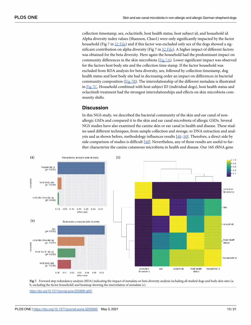
collection timestamp, sex, oclacitinib, host health status, host subject id, and household id.
Alpha diversity index values (Shannon, Chao1) were only significantly impacted by the factor
household (Fig 7 in S2 File) and if this factor was excluded only sex of the dogs showed a sig-
nificant contribution on alpha diversity (Fig 7 in S2 File). A higher impact of different factors
was obtained for the beta diversity. Here again the household had the predominant impact on
community differences in the skin microbiota (Fig 7A). Lower significant impact was observed
for the factors host body site and the collection time stamp. If the factor household was
excluded from RDA analysis for beta diversity, sex, followed by collection timestamp, dog
health status and host body site had in decreasing order an impact on differences in bacterial
community composition (Fig 7B). The interrelationship of the different metadata is illustrated
in Fig 7C. Household combined with host subject ID (individual dogs), host health status and
oclacitinib treatment had the strongest interrelationships and effects on skin microbiota com-
munity shifts.
Discussion
In this NGS study, we described the bacterial community of the skin and ear canal of non-
allergic GSDs and compared it to the skin and ear canal microbiota of allergic GSDs. Several
NGS studies have also examined the canine skin or ear canal in health and disease. These stud-
ies used different techniques, from sample collection and storage, to DNA extraction and anal-
ysis and as shown before, methodology influences results [48–50]. Therefore, a direct side by
side comparison of studies is difficult [48]. Nevertheless, any of those results are useful to fur-
ther characterize the canine cutaneous microbiota in health and disease. Our 16S rRNA gene
Fig 7. Forward step redundancy analysis (RDA) indicating the impact of metadata on beta diversity analysis including all studied dogs and body skin sites (a;
b, excluding the factor household) and heatmap showing the interrelation of metadata (c).
https://doi.org/10.1371/journal.pone.0250695.g007
PLOS ONE Skin and ear canal microbiota in non-allergic and allergic German shepherd dogs
PLOS ONE | https://doi.org/10.1371/journal.pone.0250695 May 3, 2021 13 / 21
amplicon approach (nested PCR; V3-V6 & V4-V5) showed high individual and body site vari-
ability between the different study subjects, without significant differences between the body
sites, in contrast to the first NGS-based study in veterinary medicine [13]. In our study the
body skin (axilla, interdigital, groin) showed significantly higher species richness than the ear
canal in non-allergic dogs. Possible explanations may include the special ear canal construc-
tion (chamber-like) and histology, with a comparatively thin epidermis and dermis containing
single hair follicles, sebaceous glands and ceruminal glands, providing lipid-rich cerumen
determining it´s microenvironment with a relatively high humidity [51,52]. Bactericidal activ-
ity of cerumen could explain the lower species richness compared to the other body sites.
We identified Actinobacteria as the most dominant phylum on the non-allergic dogs, simi-
lar to another study [53]. This finding is also in line with the bacterial composition of human
skin [3]. In contrast, the first NGS veterinary study describing healthy canine cutaneous bacte-
rial composition showed predominately Proteobacteria [13]. As mentioned above, different
methodology could explain such differences as studies have shown [48,50,51,54]. Thus, this
phenomenon is not unique to dogs. Furthermore, studies have shown that environmental fac-
tors can influence the skin microbiome in humans like contact with soil or plant material
[53,55–57]. Therefore, it can be assumed that different habits or environment of the dogs
could explain differences in results. In addition, temporal changes of microbiome can also
influence the results and lead to such disagreements between studies. To date temporal stabil-
ity of cutaneous microbiome in animals remains largely unknown. In rats healthy skin showed
temporal instability (11 days) [58], whereas the healthy canine ear canal remained stable for 28
days. [1]. Both, living in the same household and sex affected the bacterial community patterns
among the samples of the healthy dogs, in agreement with other studies [53,59]. Interestingly
in our study, Proteobacteria predominated in the ear canal and the interdigital area, as in
another study for the interdigital area and the pinna [13]. It is not clear why these two areas
showed this difference in comparison with the groin and the axilla of the GSDs. One possible
explanation for the interdigital area could be direct and repeated contact with soil, where it is
shown that Proteobacteria predominate its bacterial composition [60]. Currently there are
only two other NGS studies describing the canine ear canal. In the first study, Proteobacteria
were the most abundant phyla followed by Actinobacteria, Firmicutes, Bacteroidetes, and
Fusobacteria with Escherichia as the most abundant genus [1]. The second study showed also
similar most abundant phyla but in a different order, with Firmicutes being most abundant
followed by Proteobacteria, Bacteroidetes, and Actinobacteria with Romboutsia as the most
abundant genus [61]. Our findings were consistent with the first study [1], as we had the same
order of the most common phyla, except that we documented Cyanobacteria instead of Fuso-
bacteria. At the genus level we identified Brevibacterium as the most abundant taxon. The
most abundant genus found on non-allergic skin was Macrococcus with the highest abundance
in the axilla. The second most common genus was Staphylococcus with the highest abundance
in the groin followed by the axilla, the interdigital area, and the ear canal. Clostridium sensustricto 7 was the most common genus of the interdigital area. In contrast, one previous study
found that healthy canine skin (axilla, pinna, and groin) and mouth was predominantly colo-
nized by Porphyromonas, Staphylococcus, Streptococcus, and Propionibacterium, with Porphyr-omonas significantly colonizing the axilla [14]. In another study, analysis of healthy canine
skin (dorsal nose, nasal mucosa, lip commissure, conjunctiva, periocular skin, ear canal, con-
cave pinna, dorsal lumbar area, axilla, groin, interdigital skin, and perianal area), revealed Ral-stonia as the most abundant genus in most of the samples [13]. A recent exploratory study
with six healthy dogs showed that on the skin (inguinal, axilla, periocular and trunk), Pseudo-monas was most abundant followed by Kocuria, Porphyromonas, and Corynebacterium [62].
While another one identified Propionibacterium acnes, Corynebacterium, and Porphyromonas
PLOS ONE Skin and ear canal microbiota in non-allergic and allergic German shepherd dogs
PLOS ONE | https://doi.org/10.1371/journal.pone.0250695 May 3, 2021 14 / 21
as most abundant in the skin (dorsal neck, axilla, and abdomen) of 40 healthy dogs [53]. These
findings are in contrast to our study. Macrococcus is a Gram-positive coccoid bacterium, previ-
ously placed into the Staphylococcus genus but since 1998 assigned to its own genus [64]. It is
composed of eight species that are closely related with species of the genus Staphylococcus [63].
Current information on the distribution of Macrococcus is limited. This genus is described pri-
marily as part of the microbiota of mammals and in milk and meat according to a current
review paper [63]. Even though it is considered as a non-pathogenic bacterium, there are a few
reports of infections associated with Macrococcus caseolyticus and M. canis [64–67]. A recent
study found that of 162 dogs, 13 carried M. canis and six M. caseolyticus predominately in cuta-
neous (axilla and groin) non-infectious sites. Six M. canis and one M. caseolyticus strains were
isolated from animals with rhinitis, otitis externa, dermatitis, and mastitis [64]. As both healthy
and infected skin was colonized, it was concluded that Macrococcus is an important opportu-
nistic bacterium of the canine skin [64]. Because Macrococcus contributed mainly to the differ-
ence in bacterial community composition of the axilla between non-allergic and allergic GSDs,
and was the genus with the highest relative abundance found on the skin of non-allergic dogs a
potential protective role can be speculated, too. Thus, further studies regarding the distribution
and role of bacteria of this genus in healthy and diseased dogs are needed.
An important finding of our study was that the site-specific bacterial composition signif-
icantly differs with allergic skin disease. The SIMPER analysis revealed the taxa with the
major contribution to these changes, even though the mean relative abundance of some of
those taxa (e.g., Staphylococcus) did not differ significantly between non-allergic and allergic
GSDs. A possible explanation for this is that SIMPER analysis identified genera that had the
highest contribution to differences between the groups without being necessarily signifi-
cantly different. Furthermore, the axilla of allergic dogs showed a significantly decreased
diversity (species richness) indicating dysbiosis (due to higher relative abundance of Clos-tridium sensu stricto 7 and Sphingomonas), even though the atopic dogs showed no clinical
flares and had no pyoderma at the time of the sampling. This is consistent with another
study of atopic dogs with pyoderma, which demonstrated different bacterial communities
and reduced diversity in the allergic dogs [14]. The first 16S rRNA gene amplicon based
NGS study using allergic dogs without flares, like our study population, did not show signif-
icant differences of the skin microbiota community composition in comparison with
healthy dogs in contrast to our findings, but did show that allergic skin had lower diversity
(richness), similar to our findings [13]. The ear canal of allergic dogs without signs of otitis
also had a bacterial composition significantly different from healthy dogs’ ear canal, and
without a significant difference in diversity between the groups, similar to our results [1]. A
trend of dysbiosis in association with allergy was shown with significantly increased abun-
dance of Staphylococcus and Ralstonia in the atopic ear canals [1]. In general, we were able
to show a trend of dysbiosis for all the sites evaluated in total, with significantly reduction of
Macrococcus and Brevibacterium and increase of the phylum Proteobacteria and the genera
of Sphingomonas and Nocardioides.Six out of 12 allergic dogs received oclacitinib, which is a Janus kinase inhibitor with
anti-pruritic and anti-inflammatory properties [68]. Until now there is no study investigat-
ing the impact of oclacitinib on the skin microbiota. One study documented that treatment
with cyclosporine or corticosteroids did not affect the cutaneous microbiota in dogs, evalu-
ating these dogs before, during, and after treatment [69]. This is in line with our findings,
bacterial composition of allergic dogs with and without oclacitinib did not show any signifi-
cant differences. Thus, we suggest that oclacitinib may not influence the overall comparison
between the non-allergic and allergic dogs, but future studies with larger population should
confirm our finding.
PLOS ONE Skin and ear canal microbiota in non-allergic and allergic German shepherd dogs
PLOS ONE | https://doi.org/10.1371/journal.pone.0250695 May 3, 2021 15 / 21
One limitation of our study was the small sample size. However, our sample size is larger
than in previous studies [1,13,62,69]. Furthermore, our study adds to a growing corpus of
research that helps us better understand the microbiota inhabiting the skin and ear canal of dogs
and can be used to design larger confirmatory studies. The study was a cross-sectional analysis
and therefore it remains unclear if findings are a cause or a result of allergy. Regarding the
description of the cutaneous microbiota only a longitudinal study could clarify if the composi-
tion of the microbiota is stable or only transient. Further studies including also non-allergic dogs
from other breeds are required, in order to evaluate if the skin microbiota of non-allergic GSDs
is breed specific or not. Because previous studies of humans’ skin microbiota detected a low
amount of microbial DNA when standard PCR was used, a nested PCR was chosen in our study
[26,27]. A bias in alpha diversity and community structure (beta diversity = has been docu-
mented due to a nested PCR in stool samples but not in vaginal swabs and might be considered
as a possible limitation factor of our study [70]. However, the microbiota and DNA quantity
obtained from the skin and in the ear channels of dogs by swab-based sampling is more similar
to the output obtained from vaginal swabs than form stool samples which have in general a
higher load of bacteria/DNA which make a nested approach for those samples unnecessary. Cur-
rently, no standard protocol exists for the methodology, and as consequence different primers
are used in different studies [48]. Because there is very limited systematic comparison of the
primers, and no "perfect" primer exists [71], most commonly, primers are selected based recom-
mendations and the experimental experience of the laboratory [54]. Our second primer system
targeted the V4-V5 hypervariable region, which is currently used for 16S rRNA gene sequenc-
ing-based microbial profiling [25,54,72]. Our choice was based on studies that suggest an accu-
rate estimation of multiple taxa and an efficient phylogenetic resolution with this hypervariable
region [25,54]. These advantages, were again demonstrated recently [73]. On the other hand, as
it was shown in the same study, targeting this region (or targeting other regions) might have an
effect on the results, because different primers do not always detect as expected specific bacterial
communities, resulting in a bias when quantification of those communities is assessed [73]. But
the primer system amplifying the V4-V5 was one of the primer systems with was less biased, and
probably these effects may be subtle [73]. It was also shown that the analysis of bacterial commu-
nity composition (beta diversity studies) were robust for the use of different primers [71,73]. In
order to minimize the possibility to include a dog with a subclinical cAD into the non-allergic
group, the age of the healthy dogs had to be more than four years old. Consequently, the age of
dogs of two groups was significantly different. This might be a limitation, as it has been shown
in humans that the skin microbiota evolves with age (first years after birth or with sexual matu-
ration) [74–76]. In addition, all dogs were living in the same area of Germany and the environ-
ment affects skin microbiota [77], possibly leading to differences between results of studies from
other countries. In our study, we did not evaluate the association of rural or urban environment
with the dogs’ cutaneous microbiota. A last limitation of our study is that the 16S rRNA gene
sequencing cannot differentiate dead from alive bacteria [2]. Larger studies including other
breeds, several countries, as well as dogs with and without allergy flares could elucidate the
canine skin microbiota in health and disease.
Conclusion
This study describes and compares the cutaneous and ear canal microbiota of non-allergic and
allergic GSDs, showing that allergic dogs have a significantly different bacterial community
composition and significantly lower species richness (alpha diversity). The skin and ear canal
microbiota of non-allergic GSDs is highly variable, and the ear canal has a significantly lower
species richness than the body skin.
PLOS ONE Skin and ear canal microbiota in non-allergic and allergic German shepherd dogs
PLOS ONE | https://doi.org/10.1371/journal.pone.0250695 May 3, 2021 16 / 21
Supporting information
S1 File.
(XLSX)
S2 File.
(DOCX)
Acknowledgments
We thank Katja Grebing and Anna Baum (Applied Microbiology, JLU Giessen) for their excel-
lent technical assistance.
Author Contributions
Conceptualization: Neoklis Apostolopoulos, Stefanie P. Glaeser, Reto Neiger, Nina Thom.
Data curation: Neoklis Apostolopoulos, Stefanie P. Glaeser.
Formal analysis: Neoklis Apostolopoulos, Stefanie P. Glaeser, Ruchi Bagwe.
Investigation: Neoklis Apostolopoulos, Ruchi Bagwe, Stefan Janssen.
Methodology: Stefanie P. Glaeser.
Project administration: Neoklis Apostolopoulos, Nina Thom.
Resources: Peter Kampfer, Reto Neiger.
Software: Stefanie P. Glaeser.
Supervision: Stefanie P. Glaeser, Reto Neiger, Nina Thom.
Validation: Neoklis Apostolopoulos, Stefanie P. Glaeser, Stefan Janssen, Nina Thom.
Visualization: Neoklis Apostolopoulos, Stefanie P. Glaeser, Stefan Janssen.
Writing – original draft: Neoklis Apostolopoulos, Stefanie P. Glaeser, Nina Thom.
Writing – review & editing: Stefanie P. Glaeser, Stefan Janssen, Ursula Mayer, Christa Ewers,
Peter Kampfer, Reto Neiger, Nina Thom.
References1. Ngo J, Taminiau B, Fall PA, Daube G, Fontaine J. Ear canal microbiota—a comparison between healthy
dogs and atopic dogs without clinical signs of otitis externa. Vet Dermatol. 2018; 29:425–e140. https://
doi.org/10.1111/vde.12674 PMID: 30084115.
2. Rodrigues Hoffmann A. The cutaneous ecosystem. The roles of the skin microbiome in health and its
association with inflammatory skin conditions in humans and animals. Vet Dermatol. 2017; 28:60–e15.
https://doi.org/10.1111/vde.12408 PMID: 28133874.
3. Costello EK, Lauber CL, Hamady M, Fierer N, Gordon JI, Knight R. Bacterial community variation in
human body habitats across space and time. Science. 2009; 326:1694–7. https://doi.org/10.1126/
science.1177486 PMID: 19892944.
4. Grice EA, Segre JA. The skin microbiome. Nat Rev Microbiol. 2011; 9:244–53. https://doi.org/10.1038/
nrmicro2537 PMID: 21407241.
5. Karkman A, Lehtimaki J, Ruokolainen L. The ecology of human microbiota: dynamics and diversity in
health and disease. Ann N Y Acad Sci. 2017; 1399:78–92. https://doi.org/10.1111/nyas.13326 PMID:
28319653.
6. Halliwell R. Revised nomenclature for veterinary allergy. Vet Immunol and Immunopathol. 2006;
114:207–8. https://doi.org/10.1016/j.vetimm.2006.08.013 PMID: 17005257.
PLOS ONE Skin and ear canal microbiota in non-allergic and allergic German shepherd dogs
PLOS ONE | https://doi.org/10.1371/journal.pone.0250695 May 3, 2021 17 / 21
7. Hillier A, Griffin CE. The ACVD task force on canine atopic dermatitis (I): incidence and prevalence. Vet
Immunol and Immunopathol. 2001; 81:147–51. https://doi.org/10.1016/s0165-2427(01)00296-3 PMID:
11553375
8. Freudenberg JM, Olivry T, Mayhew DN, Rubenstein DS, Rajpal DK. The comparison of skin transcrip-
tomes confirms canine atopic dermatitis is a natural homologue to the human disease. J Invest Derma-
tol. 2018. https://doi.org/10.1016/j.jid.2018.10.018 PMID: 30393079.
9. Marsella R, Girolomoni G. Canine models of atopic dermatitis: a useful tool with untapped potential. J
Invest Dermatol. 2009; 129:2351–7. https://doi.org/10.1038/jid.2009.98 PMID: 19516261.
10. Jackson HA, Murphy KM, Tater KC, Olivry T, Hummel JB, Itensen J, et al. The pattern of allergen hyper-
sensitivity (dietary or environmental) of dogs with non-seasonal atopic dermatitis cannot be differenti-
ated on the basis of historical or clinical information: a prospective evaluation 2003–2004. Abstracts
from the joint meeting of the American College of Veterinary Dermatology and the American Academy
of Veterinary Dermatology 6–10 April 2005, Sarasota, Florida; 16; 2005. pp. 192–211.
11. Olivry T, Deboer DJ, Prelaud P, Bensignor E. Food for thought: pondering the relationship between
canine atopic dermatitis and cutaneous adverse food reactions. Vet Dermatol. 2007; 18:390–1. https://
doi.org/10.1111/j.1365-3164.2007.00625.x PMID: 17991155.
12. Hensel P, Santoro D, Favrot C, Hill P, Griffin C. Canine atopic dermatitis: detailed guidelines for diagno-
sis and allergen identification. BMC Vet Res. 2015; 11. https://doi.org/10.1186/s12917-015-0515-5
PMID: 26260508.
13. Hoffmann AR, Patterson AP, Diesel A, Lawhon SD, Ly HJ, Stephenson CE, et al. The Skin Microbiome
in Healthy and Allergic Dogs. PLOS ONE. 2014; 9:e83197. https://doi.org/10.1371/journal.pone.
0083197 PMID: 24421875
14. Bradley CW, Morris DO, Rankin SC, Cain CL, Misic AM, Houser T, et al. Longitudinal Evaluation of the
Skin Microbiome and Association with Microenvironment and Treatment in Canine Atopic Dermatitis. J
Invest Dermatol. 2016; 136:1182–90. https://doi.org/10.1016/j.jid.2016.01.023 PMID: 26854488.
15. Wilhem S, Kovalik M, Favrot C. Breed-associated phenotypes in canine atopic dermatitis. Vet Dermatol.
2011; 22:143–9. https://doi.org/10.1111/j.1365-3164.2010.00925.x PMID: 20887404.
16. Tengvall K, Kierczak M, Bergvall K, Olsson M, Frankowiack M, Farias FHG, et al. Genome-Wide Analy-
sis in German Shepherd Dogs Reveals Association of a Locus on CFA 27 with Atopic Dermatitis. PLOS
Genetics. 2013; 9:e1003475. https://doi.org/10.1371/journal.pgen.1003475 PMID: 23671420
17. Griffin C, DeBoer D. The ACVD task force on canine atopic dermatitis (XIV): clinical manifestations of
canine atopic dermatitis. Veterinary Immunology and Immunopathology. 2001; 81:255–69. https://doi.
org/10.1016/s0165-2427(01)00346-4 PMID: 11553388
18. Favrot C, Steffan J, Seewald W, Picco F. A prospective study on the clinical features of chronic canine
atopic dermatitis and its diagnosis. Vet Dermatol. 2010; 21:23–31. https://doi.org/10.1111/j.1365-3164.
2009.00758.x PMID: 20187911.
19. Hill PB, Lau P, Rybnicek J. Development of an owner-assessed scale to measure the severity of pruri-
tus in dogs. Vet Dermatol. 2007; 18:301–8. https://doi.org/10.1111/j.1365-3164.2007.00616.x PMID:
17845617.
20. Rybnıcek J, Lau-Gillard PJ, Harvey R, Hill PB. Further validation of a pruritus severity scale for use in dogs.
Vet Dermatol. 2009; 20:115–22. https://doi.org/10.1111/j.1365-3164.2008.00728.x PMID: 19171021.
21. Olivry T, Saridomichelakis M, Nuttall T, Bensignor E, Griffin CE, Hill PB. Validation of the Canine Atopic
Dermatitis Extent and Severity Index (CADESI)-4, a simplified severity scale for assessing skin lesions
of atopic dermatitis in dogs. Vet Dermatol. 2014; 25:77–85, e25. https://doi.org/10.1111/vde.12107
PMID: 24461108.
22. Ginel PJ, Lucena R, Rodriguez JC, Ortega J. A semiquantitative cytological evaluation of normal and
pathological samples from the external ear canal of dogs and cats. Vet Dermatol. 2002; 13:151–6.
https://doi.org/10.1046/j.1365-3164.2002.00288.x PMID: 12074704
23. Budach SC, Mueller RS. Reproducibility of a semiquantitative method to assess cutaneous cytology.
Vet Dermatol. 2012; 23:426–e80. https://doi.org/10.1111/j.1365-3164.2012.01075.x PMID: 22809453
24. Ong SH, Kukkillaya VU, Wilm A, Lay C, Ho EXP, Low L, et al. Species identification and profiling of com-
plex microbial communities using shotgun Illumina sequencing of 16S rRNA amplicon sequences. PLOS
ONE. 2013; 8:e60811. Epub 2013/04/08. https://doi.org/10.1371/journal.pone.0060811 PMID: 23579286.
25. Parada AE, Needham DM, Fuhrman JA. Every base matters: assessing small subunit rRNA primers for
marine microbiomes with mock communities, time series and global field samples. Environ Microbiol.
2016; 18:1403–14. https://doi.org/10.1111/1462-2920.13023 PMID: 26271760.
26. Zeeuwen PL, Boekhorst J, Bogaard, Ellen H van den, Koning HDd, Kerkhof, Peter MC van de, Saulnier
DM, et al. Microbiome dynamics of human epidermis following skin barrier disruption. Genome Biol.
2012; 13:1–18. https://doi.org/10.1186/gb-2012-13-11-r101 PMID: 23153041
PLOS ONE Skin and ear canal microbiota in non-allergic and allergic German shepherd dogs
PLOS ONE | https://doi.org/10.1371/journal.pone.0250695 May 3, 2021 18 / 21
27. Li W, Han L, Yu P, Ma C, Wu X, Moore JE, et al. Molecular characterization of skin microbiota between
cancer cachexia patients and healthy volunteers. Microb Ecol. 2014; 67:679–89. https://doi.org/10.
1007/s00248-013-0345-6 PMID: 24402361.
28. Quast C, Pruesse E, Yilmaz P, Gerken J, Schweer T, Yarza P, et al. The SILVA ribosomal RNA gene
database project: improved data processing and web-based tools. Nucleic Acids Res. 2013; 41:D590–
D596. https://doi.org/10.1093/nar/gks1219 PMID: 23193283
29. Pruesse E, Peplies J, Glockner FO. SINA: Accurate high-throughput multiple sequence alignment of
ribosomal RNA genes. Bioinformatics. 2012; 28:1823–9. https://doi.org/10.1093/bioinformatics/bts252
PMID: 22556368
30. Li W, Godzik A. Cd-hit: a fast program for clustering and comparing large sets of protein or nucleotide
sequences. SINA: Accurate high-throughput multiple sequence alignment of ribosomal RNA genes.
Bioinformatics. 2012; 28:1823–9. https://doi.org/10.1093/bioinformatics/bts252 PMID: 22556368
31. Camacho C, Coulouris G, Avagyan V, Ma N, Papadopoulos J, Bealer K, et al. BLAST+: architecture
and applications. BMC Bioinformatics. 2009; 10:1–9. https://doi.org/10.1186/1471-2105-10-1 PMID:
19118496
32. Ondov BD, Bergman NH, Phillippy AM. Interactive metagenomic visualization in a Web browser. BMC
Bioinformatics. 2011; 12:1–10. https://doi.org/10.1186/1471-2105-12-1 PMID: 21199577
33. Ionescu D, Siebert C, Polerecky L, Munwes YY, Lott C, Hausler S, et al. Microbial and chemical charac-
terization of underwater fresh water springs in the Dead Sea. PLoS ONE. 2012; 7:e38319. https://doi.
org/10.1371/journal.pone.0038319 PMID: 22679498.
34. Klindworth A, Pruesse E, Schweer T, Peplies J, Quast C, Horn M, et al. Evaluation of general 16S ribo-
somal RNA gene PCR primers for classical and next-generation sequencing-based diversity studies.
Nucleic Acids Res. 2013; 41:e1. https://doi.org/10.1093/nar/gks808 PMID: 22933715.
35. Hammer Oyvind, Harper David A.T., and Ryan Paul D.. PAST: Paleontological Statistics Software
Package for Education and Data Analysis. Palaeontologia Electronica, 4(1):9pp.
36. Shapiro SS, Wilk MB. An Analysis of Variance Test for Normality (Complete Samples). Biometrika.
1965; 52:591. https://doi.org/10.2307/2333709
37. Zar JH. Biostatistical analysis. 3rd ed. Upper Saddle River, N.J.: Prentice-Hall; 1996.
38. Taguchi Y-H, Oono Y. Relational patterns of gene expression via non-metric multidimensional scaling
analysis. Bioinformatics. 2005; 21:730–40. https://doi.org/10.1093/bioinformatics/bti067 PMID:
15509613.
39. CLARKE KR. Non-parametric multivariate analyses of changes in community structure. Austral Ecol.
1993; 18:117–43. https://doi.org/10.1111/j.1442-9993.1993.tb00438.x
40. Anderson MJ. A new method for non-parametric multivariate analysis of variance. Austral Ecol. 2001;
26:32–46. https://doi.org/10.1111/j.1442-9993.2001.01070.pp.x
41. Thomas RL, Jiang L, Adams JS, Xu ZZ, Shen J, Janssen S, et al. Vitamin D metabolites and the gut
microbiome in older men. Nat Commun. 2020; 11:5997. Epub 2020/11/26. https://doi.org/10.1038/
s41467-020-19793-8 PMID: 33244003.
42. vegan: Community Ecology Package. R package version 2.4–6. Vegan 2.4–5 (2017).
43. Bray JR, Curtis JT. An Ordination of the Upland Forest Communities of Southern Wisconsin. Ecological
Monographs. 1957; 27:325–49. https://doi.org/10.2307/1942268
44. Kruskal WH, Wallis WA. Use of Ranks in One-Criterion Variance Analysis. Journal of the American Sta-
tistical Association. 1952; 47:583–621. https://doi.org/10.2307/2280779
45. Spearman C. The proof and measurement of association between two things. By C. Spearman, 1904.
Am J Psychol. 1987; 100:441–71. PMID: 3322052
46. Pearson K. VII. Note on regression and inheritance in the case of two parents. Proc R Soc Lond. 1895;
58:240–2. https://doi.org/10.1098/rspl.1895.0041
47. Cramer H. Mathematical methods of statistics. Princeton: Princeton University Press; 1999.
48. Clooney AG, Fouhy F, Sleator RD, O’ Driscoll A, Stanton C, Cotter PD, et al. Comparing Apples and
Oranges?: Next Generation Sequencing and Its Impact on Microbiome Analysis. PLoS ONE. 2016; 11:
e0148028. https://doi.org/10.1371/journal.pone.0148028 PMID: 26849217.
49. Kong HH. Details Matter: Designing Skin Microbiome Studies. J Invest Dermatol. 2016; 136:900–2.
https://doi.org/10.1016/j.jid.2016.03.004 PMID: 27107375.
50. Pollock J, Glendinning L, Wisedchanwet T, Watson M. The Madness of Microbiome: Attempting To
Find Consensus “Best Practice” for 16S Microbiome Studies. Appl Environ Microbiol. 2018; 84. https://
doi.org/10.1128/AEM.02627-17 PMID: 29427429.
51. Cole LK. Anatomy and physiology of the canine ear. Vet Dermatol. 2009; 20:412–21. https://doi.org/10.
1111/j.1365-3164.2009.00849.x PMID: 20178478.
PLOS ONE Skin and ear canal microbiota in non-allergic and allergic German shepherd dogs
PLOS ONE | https://doi.org/10.1371/journal.pone.0250695 May 3, 2021 19 / 21
52. William H. Miller J, Craig E Griffin DV, Campbell KL. Muller and Kirk’s Small Animal Dermatology. Else-
vier Health Sciences; 2012.
53. Torres S, Clayton JB, Danzeisen JL, Ward T, Huang H, Knights D, et al. Diverse bacterial communities
exist on canine skin and are impacted by cohabitation and time. PeerJ. 2017; 5:e3075. https://doi.org/
10.7717/peerj.3075 PMID: 28289569.
54. Claesson MJ, Wang Q, O’Sullivan O, Greene-Diniz R, Cole JR, Ross RP, et al. Comparison of two
next-generation sequencing technologies for resolving highly complex microbiota composition using
tandem variable 16S rRNA gene regions. Nucleic Acids Res. 2010; 38:e200. https://doi.org/10.1093/
nar/gkq873 PMID: 20880993.
55. Song SJ, Lauber C, Costello EK, Lozupone CA, Humphrey G, Berg-Lyons D, et al. Cohabiting family
members share microbiota with one another and with their dogs. Elife. 2013; 2:e00458. https://doi.org/
10.7554/eLife.00458 PMID: 23599893.
56. Dominguez-Bello MG, Costello EK, Contreras M, Magris M, Hidalgo G, Fierer N, et al. Delivery mode
shapes the acquisition and structure of the initial microbiota across multiple body habitats in newborns. Proc
Natl Acad Sci U S A. 2010; 107:11971–5. https://doi.org/10.1073/pnas.1002601107 PMID: 20566857.
57. Gronroos M, Parajuli A, Laitinen OH, Roslund MI, Vari HK, Hyoty H, et al. Short-term direct contact with
soil and plant materials leads to an immediate increase in diversity of skin microbiota. Microbiologyo-
pen. 2018:e00645. https://doi.org/10.1002/mbo3.645 PMID: 29808965.
58. Sanjar F, Weaver AJ, Peacock TJ, Nguyen JQ, Brandenburg KS, Leung KP. Identification of Metage-
nomics Structure and Function Associated With Temporal Changes in Rat (Rattus norvegicus) Skin
Microbiome During Health and Cutaneous Burn. J Burn Care Res. 2020; 41:347–58. https://doi.org/10.
1093/jbcr/irz165 PMID: 31665423.
59. Cusco A, Belanger JM, Gershony L, Islas-Trejo A, Levy K, Medrano JF, et al. Individual signatures and
environmental factors shape skin microbiota in healthy dogs. Microbiome. 2017; 5:139. https://doi.org/
10.1186/s40168-017-0355-6 PMID: 29029635.
60. Janssen PH. Identifying the dominant soil bacterial taxa in libraries of 16S rRNA and 16S rRNA genes.
Appl Environ Microbiol. 2006; 72:1719–28. https://doi.org/10.1128/AEM.72.3.1719-1728.2006 PMID:
16517615.
61. Korbelik J, Singh A, Rousseau J, Weese JS. Characterization of the otic bacterial microbiota in dogs
with otitis externa compared to healthy individuals. Vet Dermatol. 2019. https://doi.org/10.1111/vde.
12734 PMID: 30828896.
62. Chermprapai S, Ederveen THA, Broere F, Broens EM, Schlotter YM, van Schalkwijk S, et al. The bacte-
rial and fungal microbiome of the skin of healthy dogs and dogs with atopic dermatitis and the impact of
topical antimicrobial therapy, an exploratory study. Vet Microbiol. 2019; 229:90–9. https://doi.org/10.
1016/j.vetmic.2018.12.022 PMID: 30642603.
63. Mazhar S, Hill C, McAuliffe O. The Genus Macrococcus: An Insight Into Its Biology, Evolution, and Rela-
tionship With Staphylococcus. Adv Appl Microbiol. 2018; 105:1–50. https://doi.org/10.1016/bs.aambs.
2018.05.002 PMID: 30342720.
64. Cotting K, Strauss C, Rodriguez-Campos S, Rostaher A, Fischer NM, Roosje PJ, et al. Macrococcus
canis and M. caseolyticus in dogs: occurrence, genetic diversity and antibiotic resistance. Vet Dermatol.
2017; 28:559–e133. https://doi.org/10.1111/vde.12474 PMID: 28748533.
65. Gobeli Brawand S, Cotting K, Gomez-Sanz E, Collaud A, Thomann A, Brodard I, et al. Macrococcus
canis sp. nov., a skin bacterium associated with infections in dogs. Int J Syst Evol Microbiol. 2017;
67:621–6. https://doi.org/10.1099/ijsem.0.001673 PMID: 27902286.
66. La Fuente R de, Suarez G, Ruiz Santa Quiteria JA, Meugnier H, Bes M, Freney J, et al. Identification of
coagulase negative staphylococci isolated from lambs as Staphylococcus caseolyticus. Comp Immunol
Microbiol Infect Dis. 1992; 15:47–52. https://doi.org/10.1016/0147-9571(92)90101-v PMID: 1547620
67. Gomez-Sanz E, Schwendener S, Thomann A, Gobeli Brawand S, Perreten V. First Staphylococcal
Cassette Chromosome mec Containing a mecB-Carrying Gene Complex Independent of Transposon
Tn6045 in a Macrococcus caseolyticus Isolate from a Canine Infection. Antimicrob Agents Chemother.
2015; 59:4577–83. https://doi.org/10.1128/AAC.05064-14 PMID: 25987634.
68. Gonzales AJ, Bowman JW, Fici GJ, Zhang M, Mann DW, Mitton-Fry M. Oclacitinib (APOQUEL(®)) is a
novel Janus kinase inhibitor with activity against cytokines involved in allergy. J Vet Pharmacol Ther.
2014; 37:317–24. https://doi.org/10.1111/jvp.12101 PMID: 24495176.
69. Widmer G, Ferrer L, Favrot C, Paps J, Huynh K, Olivry T. Glucocorticosteroids and ciclosporin do not
significantly impact canine cutaneous microbiota. BMC Vet Res. 2018; 14:51. https://doi.org/10.1186/
s12917-018-1370-y PMID: 29471815.
70. Yu G, Fadrosh D, Goedert JJ, Ravel J, Goldstein AM. Nested PCR Biases in Interpreting Microbial
Community Structure in 16S rRNA Gene Sequence Datasets. PLoS ONE. 2015; 10:e0132253. https://
doi.org/10.1371/journal.pone.0132253 PMID: 26196512.
PLOS ONE Skin and ear canal microbiota in non-allergic and allergic German shepherd dogs
PLOS ONE | https://doi.org/10.1371/journal.pone.0250695 May 3, 2021 20 / 21
71. Tremblay J, Singh K, Fern A, Kirton ES, He S, Woyke T, et al. Primer and platform effects on 16S rRNA
tag sequencing. Front Microbiol. 2015; 6:771. https://doi.org/10.3389/fmicb.2015.00771 PMID:
26300854.
72. Walters W, Hyde ER, Berg-Lyons D, Ackermann G, Humphrey G, Parada A, et al. Improved Bacterial
16S rRNA Gene (V4 and V4-5) and Fungal Internal Transcribed Spacer Marker Gene Primers for Micro-
bial Community Surveys. mSystems. 2016; 1. https://doi.org/10.1128/mSystems.00009-15 PMID:
27822518.
73. Wear EK, Wilbanks EG, Nelson CE, Carlson CA. Primer selection impacts specific population abun-
dances but not community dynamics in a monthly time-series 16S rRNA gene amplicon analysis of
coastal marine bacterioplankton. Environ Microbiol. 2018; 20:2709–26. https://doi.org/10.1111/1462-
2920.14091 PMID: 29521439.
74. Capone KA, Dowd SE, Stamatas GN, Nikolovski J. Diversity of the human skin microbiome early in life.
J Invest Dermatol. 2011; 131:2026–32. https://doi.org/10.1038/jid.2011.168 PMID: 21697884.
75. Scotti E, Boue S, Lo Sasso G, Zanetti F, Belcastro V, Poussin C, et al. Exploring the microbiome in
health and disease. Toxicology Research and Application. MicrobiologyOpen. 2017;
1:239784731774188. https://doi.org/10.1177/2397847317741884
76. Oh J, Conlan S, Polley EC, Segre JA, Kong HH. Shifts in human skin and nares microbiota of healthy
children and adults. Genome Med. 2012; 4:1–11. https://doi.org/10.1186/gm300 PMID: 22257447
77. Lehtimaki J, Sinkko H, Hielm-Bjorkman A, Salmela E, Tiira K, Laatikainen T, et al. Skin microbiota and
allergic symptoms associate with exposure to environmental microbes. Proc Natl Acad Sci U S A. 2018;
115:4897–902. https://doi.org/10.1073/pnas.1719785115 PMID: 29686089.
PLOS ONE Skin and ear canal microbiota in non-allergic and allergic German shepherd dogs
PLOS ONE | https://doi.org/10.1371/journal.pone.0250695 May 3, 2021 21 / 21