Derivation of goat embryonic stem cell-like cell lines from in vitro produced parthenogenetic...
9
Small Ruminant Research 113 (2013) 145–153 Contents lists available at SciVerse ScienceDirect Small Ruminant Research jou r n al homep age : w w w . elsevier.com/locate/smallrumres Derivation of goat embryonic stem cell-like cell lines from in vitro produced parthenogenetic blastocysts Arun Kumar De a,∗∗ , Shweta Garg a , Dinesh Kumar Singhal a , Hrudananda Malik a , Ayan Mukherjee a , Manoj Kumar Jena a , Sudarshan Kumar a , Jai Kumar Kaushik a , Ashok Kumar Mohanty a , Bikash Chandra Das b , Sadhan Bag b , Subrata Kumar Bhanja c , Dhruba Malakar a,∗ a Animal Biotechnology Centre, National Dairy Research Institute, Karnal, Haryana, India b Veterinary Physiology and Climatology, Indian Veterinary Research Institute, Izatnagar, Bareilly, India c Central Avian Research Institute, Izatnagar, Bareilly, India a r t i c l e i n f o Article history: Received 21 March 2012 Received in revised form 1 January 2013 Accepted 16 January 2013 Available online 28 February 2013 Keywords: Parthenogenesis Embryonic stem cell-like cells Blastocysts Goat a b s t r a c t Parthenogenesis is the biological phenomenon by which embryonic development is ini- tiated without male contribution. Parthenogenesis is a very useful method of derivation of embryonic stem cells (ESCs), which may be an important source of histocompatible cells and tissues for cell therapy. The aim of the present study was to derive and char- acterize goat embryonic stem cell-like cells from in vitro developed blastocysts following parthenogenetic activation of goat oocytes. Two parthenogenetic embryonic stem cell-like cell (gPESC) lines were established. The gPES cell-like cell colonies showed typical ESC morphology and expressed ESC specific markers such as alkaline phosphatase, Oct-4, Sox- 2, Nanog, TRA-1-60, SSEA-4 and TRA-1-81. Genetic stability of the cell lines was proven to be preserved via karyotype analysis which showed normal karyotype. In suspension cul- ture in absence of feeder layer and without LIF, the embryonic stem cell-like cells produced embryoid bodies and following prolonged culture differentiated into several types of cells including neuron like, epithelium like and cardiac muscle cells. Cell specific markers were observed all cells expressed positive marker genes. All of these results demonstrated the feasibility to isolate and establish goat parthenogenetic ES cell-like cell lines, which pro- vides an important tool for studying epigenetic effects in ESCs and developmental biology as well as gene targeting to produce genetically modified livestock. © 2013 Elsevier B.V. All rights reserved. 1. Introduction The ES cells have tremendous potential applications, including the application in therapeutic medicine in ∗ Corresponding author. Tel.: +91 9416741839. ∗∗ Corresponding author. Current address: Central Agricultural Research Institute, Port Blair, A & N Islands, India. Tel.: +91 9679515260. E-mail addresses: [email protected] (A.K. De), [email protected] (D. Malakar). human, study of developmental biology, analysis of the characteristics of totipotent cells and gene targeting to produce genetically modified livestock. Parthenogenetic embryonic stem cells are alternative to embryonic stem cells derived from embryos produced by somatic cell nuclear transfer or by in vitro fertilization. Parthenogenesis is the process by which an oocyte develops into an embryo without being fertilized by a sper- matozoon. Such embryos lack the potential to develop to full term but they can be used to establish partheno- genetic embryonic stem (PES) cells. Successful generation 0921-4488/$ – see front matter © 2013 Elsevier B.V. All rights reserved. http://dx.doi.org/10.1016/j.smallrumres.2013.01.018
-
Upload
independent -
Category
Documents
-
view
0 -
download
0
Transcript of Derivation of goat embryonic stem cell-like cell lines from in vitro produced parthenogenetic...

Di
AHSBDa
b
c
ARRAA
KPEBG
1
i
I
d
0h
Small Ruminant Research 113 (2013) 145– 153
Contents lists available at SciVerse ScienceDirect
Small Ruminant Research
jou r n al homep age : w w w . elsev ier .com/ locate /smal l rumres
erivation of goat embryonic stem cell-like cell lines fromn vitro produced parthenogenetic blastocysts
run Kumar Dea,∗∗, Shweta Garga, Dinesh Kumar Singhala,rudananda Malika, Ayan Mukherjeea, Manoj Kumar Jenaa,udarshan Kumara, Jai Kumar Kaushika, Ashok Kumar Mohantya,ikash Chandra Dasb, Sadhan Bagb, Subrata Kumar Bhanjac,hruba Malakara,∗
Animal Biotechnology Centre, National Dairy Research Institute, Karnal, Haryana, IndiaVeterinary Physiology and Climatology, Indian Veterinary Research Institute, Izatnagar, Bareilly, IndiaCentral Avian Research Institute, Izatnagar, Bareilly, India
a r t i c l e i n f o
rticle history:eceived 21 March 2012eceived in revised form 1 January 2013ccepted 16 January 2013vailable online 28 February 2013
eywords:arthenogenesismbryonic stem cell-like cellslastocystsoat
a b s t r a c t
Parthenogenesis is the biological phenomenon by which embryonic development is ini-tiated without male contribution. Parthenogenesis is a very useful method of derivationof embryonic stem cells (ESCs), which may be an important source of histocompatiblecells and tissues for cell therapy. The aim of the present study was to derive and char-acterize goat embryonic stem cell-like cells from in vitro developed blastocysts followingparthenogenetic activation of goat oocytes. Two parthenogenetic embryonic stem cell-likecell (gPESC) lines were established. The gPES cell-like cell colonies showed typical ESCmorphology and expressed ESC specific markers such as alkaline phosphatase, Oct-4, Sox-2, Nanog, TRA-1-60, SSEA-4 and TRA-1-81. Genetic stability of the cell lines was proven tobe preserved via karyotype analysis which showed normal karyotype. In suspension cul-ture in absence of feeder layer and without LIF, the embryonic stem cell-like cells producedembryoid bodies and following prolonged culture differentiated into several types of cells
including neuron like, epithelium like and cardiac muscle cells. Cell specific markers wereobserved all cells expressed positive marker genes. All of these results demonstrated thefeasibility to isolate and establish goat parthenogenetic ES cell-like cell lines, which pro-vides an important tool for studying epigenetic effects in ESCs and developmental biologyas well as gene targeting to produce genetically modified livestock.. Introduction
The ES cells have tremendous potential applications,ncluding the application in therapeutic medicine in
∗ Corresponding author. Tel.: +91 9416741839.∗∗ Corresponding author. Current address: Central Agricultural Researchnstitute, Port Blair, A & N Islands, India. Tel.: +91 9679515260.
E-mail addresses: [email protected] (A.K. De),[email protected] (D. Malakar).
921-4488/$ – see front matter © 2013 Elsevier B.V. All rights reserved.ttp://dx.doi.org/10.1016/j.smallrumres.2013.01.018
© 2013 Elsevier B.V. All rights reserved.
human, study of developmental biology, analysis of thecharacteristics of totipotent cells and gene targeting toproduce genetically modified livestock. Parthenogeneticembryonic stem cells are alternative to embryonic stemcells derived from embryos produced by somatic cellnuclear transfer or by in vitro fertilization.
Parthenogenesis is the process by which an oocyte
develops into an embryo without being fertilized by a sper-matozoon. Such embryos lack the potential to developto full term but they can be used to establish partheno-genetic embryonic stem (PES) cells. Successful generation
ant Rese
146 A.K. De et al. / Small Ruminof parthenogenetic stem cell lines have been reported inmouse (Kaufman et al., 1983), non-human primate mod-els (Cibelli et al., 2002; Vrana et al., 2003) and in humans(Revazova et al., 2007; Kim et al., 2007; Mai et al., 2007).Differentiation of PES cells into all three embryonic germcell lines in vitro has been demonstrated in primates(Hernandez et al., 2003) and differentiation of partheno-genetic stem cells into neuronal cell in vitro was observedin a defined differentiation system in monkey (Cibelli et al.,2002).
In this study, we reported the establishment of two goatembryonic stem cell-like cell lines from in vitro producedparthenogenetically activated embryos and subsequentobservation of those cells over more than 15 passages. Weperformed characterization of the pluripotency of the linesby expression study of stem cell specific surface markersalkaline phosphatase, TRA-1-60, SSEA-4 and TRA-1-81 aswell as intracellular markers Oct-4, Sox-2 and Nanog.
2. Materials and methods
All the present experiments comply with all relevant institutionaland national animal welfare guidelines, policies and ethics committeeapproval.
All chemicals and media were purchased from Sigma Chemical Co. (St.Louis, MO, USA), and disposable plastic wares were from Nunc (Roskilde,Denmark) unless specified otherwise.
2.1. In vitro maturation of oocytes
In vitro maturation of oocytes was performed according to the meth-ods described by De et al. (2011). Oocytes were aspirated from ovariescollected from local slaughter house by puncturing the visible follicleswith an 18-gauge needle in the oocyte collection medium (OCM) con-taining TCM 199 (HEPES modification), 100 �g/ml l-glutamine, 10% FBS(Hyclone, Logan, UT, Cat No. CH30160.02), 50 �g/ml gentamicin and3 mg/ml BSA (Fraction-V). The cumulus-oocyte-complexes (COCs) having≥3 layers of compact cumulus cells were picked up under stereo zoommicroscope for in vitro maturation. COCs were washed 2 times with thematuration medium (De et al., 2011). Four drops of 100 �l maturationmedium were made in 35 mm Petri dishes and covered with mineral oil.These dishes were placed in the incubator with 5% CO2 in air at 38.5 ◦C,1 h prior to use for equilibration. Then 15–16 oocytes were placed in eachdrop of maturation medium. The Petri dishes were incubated at 38.5 ◦Cunder 5% CO2 in air with maximum humidity for 24 h.
2.2. Parthenogenetic activation and embryo culture
Matured oocytes with expanded cumulus cells were transferredinto micro centrifuge tube containing 0.5 mg/ml hyaluronidase in T2(T denotes TCM-199 supplemented with 2.0 mM l-glutamine, 0.2 mMsodium pyruvate, 50 �g/ml gentamicin and the number denotes percent-age of FBS) and incubated for 1 min at 38.5 ◦C under 5% CO2 in air. Thenvortexing was done for 2–3 min. The contents of the tube were transferredto a 35 mm Petri dish containing T2 and completely denuded oocytes wereselected and washed twice in fresh T2 for removal of cumulus cells.
Chemical activation of the in vitro matured oocytes was done by incu-bating in T20 medium (T denotes TCM-199 supplemented with 2.0 mMl-glutamine, 0.2 mM sodium pyruvate, 50 �g/ml gentamicin and thenumber denotes percentage of FBS) containing 5 �M Calcium ionophore(A23187) for 5 min at 38.5 ◦C under 5% CO2 in air. Then the oocytes werewashed thrice in T20 medium and incubated in T20 medium contain-ing 2 mM 6-Dimethylaminopurine (6-DMAP) at 38.5 ◦C under 5% CO2
in air for 4 h. The activated oocytes were cultured in embryo develop-ment medium (EDM) containing TCM 199 (HEPES modification), 30 �g/mlsodium pyruvate, 100 �g/ml l-glutamine, 50 �g/ml gentamicin, 10 �l/mlessential amino acids, 5 �l/ml non-essential amino acids, 10 mg/ml BSA(Fraction-V) and 10% FCS under flat culture system (De et al., 2012).
arch 113 (2013) 145– 153
2.3. Embryonic stem cells isolation and culture
Embryos developed to blastocyst stage after 7 days of culture andhatched blastocysts were observed after 8 days of culture. For isolationof inner cell masses (ICMs) from blastocysts and hatched blastocystsmechanical isolation was performed.
The zona pellucida of blastocysts was removed by treatment of 1%pronase in DPBS (w/v). Then to inhibit further action of pronase, the blas-tocysts were immediately given 4–5 washing in DPBS. The ICM cells weredissected out with the help of two fine glass needles under zoom stereomi-croscope (Olympus SZ 61, Japan) (De et al., 2011). The ICM cells of hatchedblastocysts were easily isolated mechanically as they were clearly visibleunder zoom stereomicroscope.
The isolated ICMs were seeded on mitomycin-C (10 �g/ml) inactiv-ated goat fetal fibroblast feeder layers in ES cell medium containing DMEMsupplemented with 20% FCS, 1000 IU/ml murine leukemia inhibitory fac-tor (mLIF), 1% nonessential amino acids, 0.1 mM �-mercaptoethanol, and2 mM l-glutamine. The medium was changed every 48 h interval and theformation of colony was observed routinely under inverted microscope(Nikon, Japan).
The primary colonies were cultured for 5–9 days and the ES cell-likecells were selected, picked up, and replated on new feeder layer cells.When the ES cell-like cells appeared to proliferate stably, the ES cell-likecell colonies were dissociated every 4–5 days by mechanical methods.
2.4. Characterization of goat parthenogenetic ES cell-like cells
The putative embryonic stem cell-like cells were characterized byexpression study of surface markers like alkaline phosphatase, TRA-1-60,SSEA-4, TRA-1-81 and intracellular markers Oct-4, Sox-2 and Nanog.
2.4.1. Alkaline phosphatase (AP) stainingFor alkaline phosphatase staining, the medium was removed and the
colonies were fixed in 3.7% paraformaldehyde in DPBS for 15 min. Thenthe fixed colonies were washed 4–5 times with DPBS and incubated in APsubstrate solution containing 25 mM Tris–maleate (pH 9.5), 8 mM MgCl2,0.4 mg/ml sodium alphanaphthyl phosphate and 1 mg/ml Fast red for30 min. The cells were washed 2–3 times in DPBS and response of thecells to AP staining was observed under inverted microscope.
2.4.2. Gene expression profile analysisPassage 10 and passage 15 parthenogenetic embryonic stem cell-like
cells were used for gene expression studies. Total RNA was isolated fromputative goat ES cell colonies according to methods reported by De et al.(2011). Then cDNA was prepared by taking 10 �l of the cell lysate usingrandom primers. The PCR cycle included denaturation (94 ◦C for 2 min)followed by repeated cycles of denaturation (94 ◦C for 30 s), annealing (for30 s at temperature 58 ◦C) and extension (72 ◦C for 45 s) followed by a finalextension (at 72 ◦C for 10 min). A negative RT reaction (i.e., RT reaction butwithout MMLV enzyme) was set up. A house keeping marker gene � actinwas also amplified at each stage of PCR. The PCR primers used for thestudy were same as reported by De et al. (2011). Similarly neuron like,epithelium like and cardiac muscle cells specific markers were studiedspecific primers of respective marker genes.
Primer Sequences Annealing/cycles Size
Nestin AGTGTGAAGGCAAAGATAGC 58/34 245 bpTCTGTCAGGATTGGGATGGG
Keratin CACCCAATCCACCTTCTCCAA 58/35 287 bpGGTCTTGCATGGTCTCCTTCT
2.4.3. Immunofluorescence staining of putative ES cellsThe expression of surface markers TRA-1-60, SSEA-4 and TRA-1-81 as
well as intracellular marker oct-4 was examined by immunofluorescencestaining of colonies of goat PES cell-like cells. The ES cell colonies of pas-sage 10 were fixed in 4% paraformaldehyde in DPBS for 30 min, washed3 times with DPBS and then permeabilized by treatment with 0.1% Tri-ton X-100 in DPBS for 30 min. After thorough washing with DPBS, goat ES
cell-like cell colonies were incubated with the blocking solution (4% nor-mal goat serum) for 30 min and then with the primary antibody (Millipore,USA) at a dilution of 1:10–1:20 for 1 h. After washing 3 times with DPBS, EScell colonies were incubated with the appropriate FITC-labeled secondaryantibody goat anti-mouse IgG (Millipore, USA) diluted 1:100–1:200 for
A.K. De et al. / Small Ruminant Research 113 (2013) 145– 153 147
Table 1Development of parthenogenetic goat embryos.
Oocytes Cleaved (n) % Morulae (n) % Blastocyst (n) % Hatched blastocyst (n) %
(65)
D
2m
2
mpi4sfirrccfm
2
ctmwtLdc2
2
cs
3
MtaahaaIfmIwflwppws
sage 13 without mLIF and in the absence of feeder layerresulted in embryoid body (EBs) formation within 3 days(Fig. 7A). These EBs when cultured in 0.1% gelatin coated
Fig. 1. Morphology of in vitro produced parthenogenetic goat embryos:(A) Morula stage goat embryo (400×). (B) Blastocysts of goat (200×). (C)Blastocyst just before hatching (400×). (D) Hatched blastocyst of goat(400×).
Table 2Primary colony formation from goat parthenogenetic blastocysts andhatched blastocysts.
ICM source Number ofembryos
No of primary EScell colonies, n (%)
Blastocysts 27 11 (40.74%)a
154 75.31 ± 0.842 (116) 42.09 ± 2.57
ata from 4 trials.
h. The goat ES cell colonies were then examined under a fluorescenceicroscope (Nikon, Tokyo, Japan).
.5. Karyotyping
The cells were subjected to chromosomal analysis according to theethod described by Dyban (1983) with slight modifications. Briefly,
assage 10 parthenogenetic embryonic stem cell-like cell colonies werencubated in DMEM supplemented with 0.1 mg/ml colcemid at 38.5 ◦C for
h. The cells were then washed, trypsinized and resuspended in hypotonicolution (75 mM KCl) for 30 min at 38.5 ◦C. They were washed and thenxed in chilled fixative (3:1 methanol/glacial acetic acid) for 30 min atoom temperature and centrifuged at 200 × g for 8 min. The pellets wereesuspended in 5 ml of ice-chilled fixative for another 10 min and thenentrifuged again. The metaphase spreads were prepared by dropping theells onto ice cold glass slides. Chromosomes were stained with 2% Giemsaor 6 min and observed under oil immersion (1000×) using a compound
icroscope (Nikon, Microphot-FXA, Japan).
.6. Evaluation of in vitro differentiation
For in vitro differentiation, passage 12 goat parthenogenetic ES cellolonies were removed from the feeder layer as small clumps and wereransferred to suspension culture dishes containing ES medium without
LIF and in the absence of feeder layer. After 3 days, embryoid bodiesere produced and subsequently seeded in a 4-well gelatin (0.1%)-coated
issue culture dish in differentiation medium (ES cell medium withoutIF and feeder layer) for spontaneous in vitro differentiation. For directedifferentiation of ES cell-like cells to cardiomyocetes, the ES cells wereultured in ES cell medium containing 100 ng/ml Activin-A, 10 ng/ml FGF-
and 100 ng/ml BMP-4.
.7. Statistical analysis
The differences in the number of embryos giving rise to primary cellolony were revealed by Chi-square (�2) test. A value of P < 0.05 was con-idered to be statistically significant.
. Results
Cleavage was found after 36–48 h post activation.orula (Fig. 1A), blastocysts (Fig. 1B) and hatched blas-
ocysts (Fig. 1C and D) were obtained on day 5, day 7nd day 8 post activation respectively. Cleavage percent-ge was 75.31 ± 0.842% (Mean ± SEM) and blastocyst andatched blastocyst development rate was 17.49 ± 0.95%nd 6.45 ± 1.187% respectively (Table 1). 27 blastocystsnd 10 hatched blastocysts were used for isolation ofCMs (Fig. 2A). Primary ES cell-like cell colonies (Fig. 2B)ormed after 5 days of seeding of ICMs on feeder layers. Pri-
ary colony formation rate was significantly higher whenCMs were isolated from hatched blastocysts (60.00%) than
hen isolated from blastocysts (40.74%) (Table 2). Con-uent layer of PES cell-like cell colonies (Fig. 2C and D)as found after 10 days of culture. The colonies were
assaged by mechanical disaggregation method and twoarthenogenetic ES cell-like cell lines (gPES-1, gPES-2)ere established which maintained their undifferentiatedtate up to 15th successive passages. The other primary
17.49 ± 0.95 (27) 6.45 ± 1.187 (10)
colonies which were not used to establish ES cell-like celllines were lost to differentiation.
Parthenogenetic embryonic stem (PES) cell-like cellsshowed stem cell morphology and displayed traits ofnormal stem cells. The ES cell-like cell colonies weredensely packed, flat and had higher nucleus: cytoplasmratio with prominent nucleus and clear boarder (Fig. 2Cand D). These colonies showed expression of AP activity(Fig. 3) and expressed stem cell positive markers TRA-1-81(Fig. 5A), TRA-1-60 (Fig. 5B), SSEA-4 (Fig. 5C) and Oct-4 (Fig. 5D). Expression of important pluripotency relatedgenes was detected by RT-PCR and positive signals forOct-4, Sox2 and Nanog were observed (Fig. 4A and B).Normal chromosomal profile was observed by karyotypeanalysis (Fig. 6). Suspension culture of ES cells of pas-
Hatched blastocysts 10 6 (60.00%)b
Data from 4 trials.Values within the same column with different superscripts (a and b) differsignificantly (P < 0.05).

148 A.K. De et al. / Small Ruminant Research 113 (2013) 145– 153
: (A) ICMc ES cel
Fig. 2. Morphology of goat parthenogenetic embryonic stem cell-like cells(B) Attachment of ICMs after 4 days of culture (100×). (C) Parthenogeneticells after 10 days of culture (400×).
tissue culture dishes without feeder layer and mLIF spon-
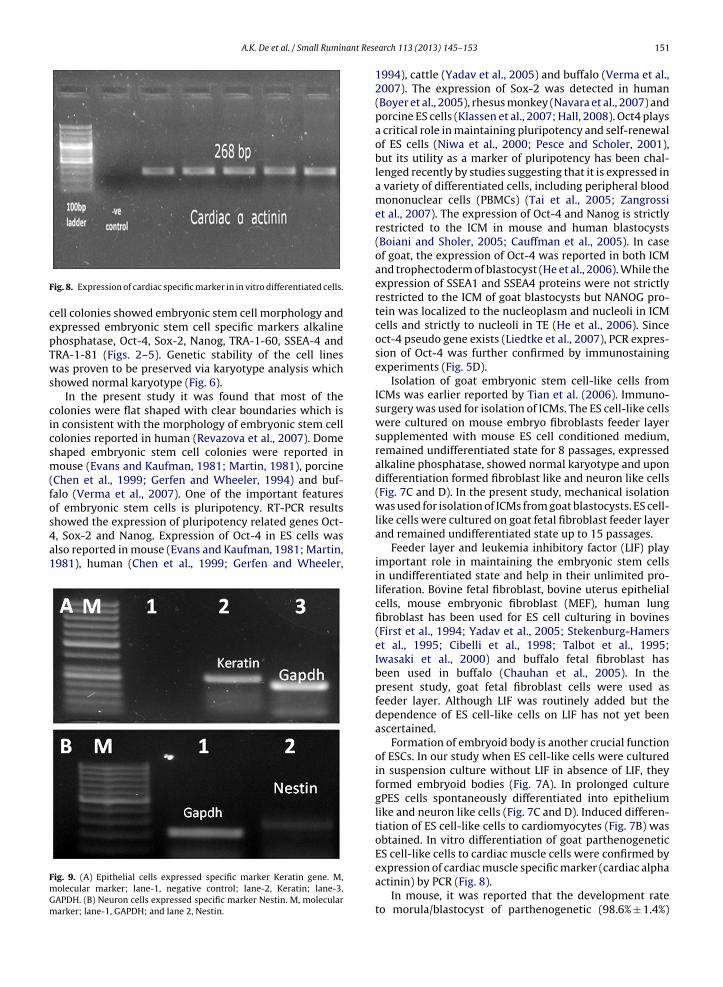
taneously differentiated into epithelium like and neuronlike cells (Fig. 7C and D). Epithelium cells and neuron cellsspecific marker genes Nestin and Keratin were expressedFig. 3. Expression of alkaline phosphatase in parthenogenetic ES cell-
s isolated from parthenogenetically activated hatched blastocyst (400×).l-like cells after 7 days of culture (200×). (D) Parthenogenetic ES cell-like
respectively during their growth and confirmed by PCR
(Fig. 9A and B).When ES cell-like cells were cultured in differentiationmedium containing 100 ng/ml Activin-A, 10 ng/ml FGF-2
like cells (200×): (A) On 10th passages. (B) On 15th passages.

A.K. De et al. / Small Ruminant Research 113 (2013) 145– 153 149
F rthenog1
a(dc
ig. 4. Expression of intracellular markers Oct-4, Sox-2 and Nanog in pa5th passages.
nd 100 ng/ml BMP-4 cardiac muscle cells were obtained
Fig. 7B). Expression of a cardiac muscle specific marker car-iac alpha actinin in induced differentiated cardiac muscleells (Fig. 8) was confirmed by PCR.Fig. 5. Immunofluorescence staining of goat parthenogenetic ES cell-
enetic goat embryonic stem cell-like cells: (A) On 10th passages. (B) On
4. Discussion
Two goat parthenogenetic stem cell-like cell lines weresuccessfully derived from parthenogenetic blastocysts.
like cells: (A) TRA-1-81. (B) TRA-1-60. (C) SSEA-4. (D) Oct-4.

150 A.K. De et al. / Small Ruminant Research 113 (2013) 145– 153
Norma
Fig. 6. Chromosomal profile of goat parthenogenetic ES cell-like cells: (A)cells (1000×). (B) Normal karyotyping.After chemical activation, 27 out of 154 oocytes developed
into blastocysts and among 27 blastocysts 10 hatched. Theprimary colony formation was 60.00% when ICMs wereisolated from hatched blastocysts and 40.74% when ICMsFig. 7. In vitro differentiation of goat parthenogenetic ES cell-like cells: (A) Embrlike cells (400×). (D) Epithelium like cells (400×).
l no. of metaphase chromosome in passage 8 parthenogenetic ES cell-like
were isolated from blastocysts (Table 2). Primary colony
formation was higher from hatched blastocysts becausethe ICM was more clearly visible in case of hatched blasto-cysts than early or expanded blastocysts. The gPES cell-likeyoid body formation (400×). (B) Cardiac muscle cells (200×). (C) Neuron
A.K. De et al. / Small Ruminant Rese
F
cepTws
cicsm(fos4a1
FmGm
ig. 8. Expression of cardiac specific marker in in vitro differentiated cells.
ell colonies showed embryonic stem cell morphology andxpressed embryonic stem cell specific markers alkalinehosphatase, Oct-4, Sox-2, Nanog, TRA-1-60, SSEA-4 andRA-1-81 (Figs. 2–5). Genetic stability of the cell linesas proven to be preserved via karyotype analysis which
howed normal karyotype (Fig. 6).In the present study it was found that most of the
olonies were flat shaped with clear boundaries which isn consistent with the morphology of embryonic stem cellolonies reported in human (Revazova et al., 2007). Domehaped embryonic stem cell colonies were reported inouse (Evans and Kaufman, 1981; Martin, 1981), porcine
Chen et al., 1999; Gerfen and Wheeler, 1994) and buf-alo (Verma et al., 2007). One of the important featuresf embryonic stem cells is pluripotency. RT-PCR results
howed the expression of pluripotency related genes Oct-, Sox-2 and Nanog. Expression of Oct-4 in ES cells waslso reported in mouse (Evans and Kaufman, 1981; Martin,981), human (Chen et al., 1999; Gerfen and Wheeler,ig. 9. (A) Epithelial cells expressed specific marker Keratin gene. M,olecular marker; lane-1, negative control; lane-2, Keratin; lane-3,APDH. (B) Neuron cells expressed specific marker Nestin. M, moleculararker; lane-1, GAPDH; and lane 2, Nestin.
arch 113 (2013) 145– 153 151
1994), cattle (Yadav et al., 2005) and buffalo (Verma et al.,2007). The expression of Sox-2 was detected in human(Boyer et al., 2005), rhesus monkey (Navara et al., 2007) andporcine ES cells (Klassen et al., 2007; Hall, 2008). Oct4 playsa critical role in maintaining pluripotency and self-renewalof ES cells (Niwa et al., 2000; Pesce and Scholer, 2001),but its utility as a marker of pluripotency has been chal-lenged recently by studies suggesting that it is expressed ina variety of differentiated cells, including peripheral bloodmononuclear cells (PBMCs) (Tai et al., 2005; Zangrossiet al., 2007). The expression of Oct-4 and Nanog is strictlyrestricted to the ICM in mouse and human blastocysts(Boiani and Sholer, 2005; Cauffman et al., 2005). In caseof goat, the expression of Oct-4 was reported in both ICMand trophectoderm of blastocyst (He et al., 2006). While theexpression of SSEA1 and SSEA4 proteins were not strictlyrestricted to the ICM of goat blastocysts but NANOG pro-tein was localized to the nucleoplasm and nucleoli in ICMcells and strictly to nucleoli in TE (He et al., 2006). Sinceoct-4 pseudo gene exists (Liedtke et al., 2007), PCR expres-sion of Oct-4 was further confirmed by immunostainingexperiments (Fig. 5D).
Isolation of goat embryonic stem cell-like cells fromICMs was earlier reported by Tian et al. (2006). Immuno-surgery was used for isolation of ICMs. The ES cell-like cellswere cultured on mouse embryo fibroblasts feeder layersupplemented with mouse ES cell conditioned medium,remained undifferentiated state for 8 passages, expressedalkaline phosphatase, showed normal karyotype and upondifferentiation formed fibroblast like and neuron like cells(Fig. 7C and D). In the present study, mechanical isolationwas used for isolation of ICMs from goat blastocysts. ES cell-like cells were cultured on goat fetal fibroblast feeder layerand remained undifferentiated state up to 15 passages.
Feeder layer and leukemia inhibitory factor (LIF) playimportant role in maintaining the embryonic stem cellsin undifferentiated state and help in their unlimited pro-liferation. Bovine fetal fibroblast, bovine uterus epithelialcells, mouse embryonic fibroblast (MEF), human lungfibroblast has been used for ES cell culturing in bovines(First et al., 1994; Yadav et al., 2005; Stekenburg-Hamerset al., 1995; Cibelli et al., 1998; Talbot et al., 1995;Iwasaki et al., 2000) and buffalo fetal fibroblast hasbeen used in buffalo (Chauhan et al., 2005). In thepresent study, goat fetal fibroblast cells were used asfeeder layer. Although LIF was routinely added but thedependence of ES cell-like cells on LIF has not yet beenascertained.
Formation of embryoid body is another crucial functionof ESCs. In our study when ES cell-like cells were culturedin suspension culture without LIF in absence of LIF, theyformed embryoid bodies (Fig. 7A). In prolonged culturegPES cells spontaneously differentiated into epitheliumlike and neuron like cells (Fig. 7C and D). Induced differen-tiation of ES cell-like cells to cardiomyocytes (Fig. 7B) wasobtained. In vitro differentiation of goat parthenogeneticES cell-like cells to cardiac muscle cells were confirmed by
expression of cardiac muscle specific marker (cardiac alphaactinin) by PCR (Fig. 8).In mouse, it was reported that the development rateto morula/blastocyst of parthenogenetic (98.6% ± 1.4%)

ant Rese
152 A.K. De et al. / Small Rumingroups was significantly higher than nuclear transferredoocytes (27.9% ± 5.9%) and the embryonic stem cell lineestablishment rate was also higher from parthenogenet-ically activated oocytes (15.7%) than nuclear transferred(4.3%) oocytes (Ju et al., 2008). Cell colonies displayedtypical morphology of mice embryonic stem cells andcould be maintained successfully with undifferentiatedmorphology after continuous proliferation for more than120 passages still maintaining normal karyotype (Ju et al.,2008). In the present study the efficiency of embry-onic stem cell derivation was not compared with anyother sources but morphologically the PES cell-like cellcolonies showed embryonic stem cell specific morphology,expressed embryonic stem cell specific markers and wasmaintained successfully for more than 15 passages withundifferentiated state maintaining normal karyotype.
Teratoma formation and germ-line transmission areimportant characteristics of ES cell-like cells. A caprinechimera produced by injection of embryonic germ cellsinto blastocysts was reported by Jia et al. (2008). Success-ful isolation and culture of goat primodial germ cells, theirpluripotency and differentiation potency into three germlayers were reported (Jia et al., 2008).
In conclusion, we report the production of embryonicstem cell-like cell lines from in vitro produced partheno-genetic blastocysts. Several properties associated withundifferentiated state and pluripotency of ES cells like AP,TRA-1-60, TRA-1-81, Oct-4, Sox-2 and Nanog expression,EBs formation and in vitro differentiation were demon-strated.
Acknowledgement
Authors are very thankful to NAIP, Indian Council ofAgricultural Research (ICAR), India for supporting the work.
References
Boiani, M., Sholer, H., 2005. Regulatory networks in embryo-derivedpluripotent stem cells. Nat. Rev. Mol. Cell Biol. 6, 872–884.
Boyer, L.A., Lee, T.I., Cole, M.F., Johnstone, S.E., Levine, S.S., Zucker,J.P., Guenther, M.G., Kumar, R.S., Murray, H.L., Jenner, R.G., Gifford,D.K., Melton, D.A., Jaenisch, R., Young, R.A., 2005. Core transcrip-tional regulatory circuitry in human embryonic stem cells. Cell 122,947–956.
Cauffman, G., Van de Velde, H., Liebaers, I., Van Steirteghem, A., 2005.Oct-4 mRNA and protein expression during human preimplantationdevelopment. Mol. Hum. Reprod. 11, 173–181.
Chauhan, M.S., Verma, V., Manik, R.S., Palta, P., Singla, S.K., Goswami, S.L.,2005. Development of inner cell mass and formation of embroid bod-ies on a gelatin coated dish and on the feeder layer in buffalo (Bubalusbubalis). Reprod. Fertil. Dev. 18, 205–206.
Chen, L.R., Shiue, Y.L., Bertolini, L., Medrano, J.F., BonDurant, R.H., Ander-son, G.B., 1999. Establishment of pluripotent cell lines from porcinepreimplantation embryos. Theriogenology 52, 195–212.
Cibelli, J.B., Stice, S.L., Golueke, P.J., Kane, J.J., Jerry, J., Blackwell, C., Poncede Leon, F.A., Robl, J.M., 1998. Transgenic bovine chimeric offspringproduced from somatic cell-derived stem-like cells. Nat. Biotechnol.16 (7), 642–646.
Cibelli, J.B., Grant, K.A., Chapman, K.B., Cunniff, K., Worst, T., Green, H.L.,Walker, S.J., Gutin, P.H., Vilner, L., Tabar, V., Dominko, T., Kane, J.,Wettstein, P.J., Lanza, R.P., Studer, L., Vrana, K.E., West, M.D., 2002.Parthenogenetic stem cells in nonhuman primates. Science 295,
779–780.De, A.K., Malakar, D., Akshey, Y.S., Jena, M.K., Dutta, R., 2011. Isolationand characterization of embryonic stem cell-like cells from in vitroproduced goat (Capra hircus) embryos. Anim. Biotechnol. 22 (4),181–196.
arch 113 (2013) 145– 153
De, A.K., Malakar, D., Akshey, Y.S., Jena, M.K., Dutta, R., 2012. Zona-freeand with zona parthenogenetic 1 embryo production in goat (Caprahircus) – effect of activation methods, culture systems and culturemedia. Livest. Sci. 143, 35–42.
Dyban, A.P., 1983. An improved method for chromosome preparationfrom preimplantation mammalian embryos, oocytes or isolated blas-tomeres. Stain Technol. 58, 69–72.
Evans, M.J., Kaufman, M., 1981. Establishment in culture of pluripotentcells from mouse embryos. Nature 292, 154–156.
First, N.L., Sims, M.M., Park, S.P., Kent-First, M.J., 1994. Systems for pro-duction of calves from cultured embryonic cells. Reprod. Fertil. Dev.6, 533–562.
Gerfen, R.W., Wheeler, M.B., 1994. Isolation of embryo cell lines fromprocine blastocysts. Anim. Biotechnol. 6, 1–14.
Hall, V., 2008. Porcine embryonic stem cells: a possible source for cellreplacement therapy. Stem Cell Rev. Rep. 4 (4), 275–282.
He, S., Pant, D., Schiffmacher, A., Bischoff, S., Melican, D., Gavin, W., Keefer,C., 2006. Developmental expression of pluripotency determining fac-tors in caprine embryos: novel pattern of NANOG protein localizationin the nucleolus. Mol. Reprod. Dev. 73 (12), 1512–1522.
Hernandez, L., Kozlov, S., Piras, G., Stewart, C.L., 2003. Paternal and mater-nal genomes confer opposite effects on proliferation, cellcycle length,senescence, and tumor formation. Proc. Natl. Acad. Sci. U.S.A. 100,13344–13349.
Iwasaki, S., Campbell, K.H., Gallic, A.K., Akiyama, K., 2000. Production oflive calves derived from embryonic stem-like cells aggregated withtetraploid embryos. Biol. Reprod. 62, 470–475.
Jia, W., Yang, W., Lei, A., Gao, Z., Yang, C., Hua, J., Huang, W., Ma,X., Wang, H., Dou, Z., 2008. A caprine chimera produced by injec-tion of embryonic germ cells into a blastocyst. Theriogenology 69,340–348.
Ju, J.Y., Park, C.Y., Gupta, M.K., Uhm, S.J., Paik, E.C., Ryoo, Z.Y., Cho, Y.H.,Chung, K.S., Lee, H.T., 2008. Establishment of stem cell lines fromnuclear transferred and parthenogenetically activated mouse oocytesfor therapeutic cloning. Fertil. Steril. 89, 1314–1323.
Kaufman, M.H., Robertson, E.J., Handyside, A.H., Evans, M.J., 1983. Estab-lishment of pluripotential cell lines from haploid mouse embryos. J.Embryol. Exp. Morphol. 73, 249–261.
Kim, K., Ng, K., Rugg-Gunn, P.J., Shieh, J.H., Jaenisch, R., Wakayama, T.,Moore, M.A., Pedersen, R.A., Daley, G.Q., 2007. Recombination signa-tures distinguish embryonic stem cells derived by parthenogenesisand somatic cell nuclear transfer. Cell Stem Cell 1, 346–352.
Klassen, H., Kiilgaard, J.F., Zahir, T., Ziaeian, B., Kirov, I., Scherfig, E., War-fvinge, K., Young, M.J., 2007. Progenitor cells from the porcine neuralretina express photoreceptor markers after transplantation to the sub-retinal space of allorecipients. Stem Cells 25 (5), 1222–1230.
Liedtke, S., Enczmann, J., Waclawczyk, S., Wernet, P., Kögler, G., 2007. Oct4and its pseudogenes confuse stem cell research. Cell Stem Cell 1 (4),364–366.
Mai, Q., Yu, Y., Li, T., Wang, L., Chen, M.J., Huang, S.Z., Zhou, C., Zhou, Q.,2007. Derivation of human embryonic stem cell lines from partheno-genetic blastocysts. Cell Res. 17, 1008–1019.
Martin, G.R., 1981. Isolation of a pluripotent cell line from early mouseembryos cultured in medium conditioned by teratocarcinoma stemcells. Proc. Natl. Acad. Sci. U.S.A. 78, 7634–7638.
Navara, C.S., Mich-Basso, J.D., Redinger, C.J., Ben-Yehudah, A., Jacoby,E., Kovkarova-Naumovski, E., Sukhwani, M., Orwig, K., Kaminski,N., Castro, C.A., Simerly, C.R., Schatten, G., 2007. Pedigreed primateembryonic stem cells express homogeneous familial gene profiles.Stem Cells 25 (11), 2695–2704.
Niwa, H., Miyazaki, J., Smith, A.G., 2000. Quantitative expression of Oct-3/4defines differentiation, dedifferentiation or self-renewal of ES cells.Nat. Genet. 24, 372–376.
Pesce, M., Scholer, H.R., 2001. Oct-4: gatekeeper in the beginnings of mam-malian development. Stem Cells 19, 271–278.
Revazova, E.S., Turovets, N.A., Kochetkova, O.D., Kindarova, L.B.,Kuzmichev, L.N., Janus, J.D., Pryzhkova, M.V., 2007. Patient-specificstem cell lines derived from human parthenogenetic blastocysts.Cloning Stem Cells 9, 1–9.
Stekenburg-Hamers, A.E., Van Achterberg, T.A., Rebel, H.G., Flechon, J.E.,Campbell, K.H., Weima, S.M., Mummery, C.L., 1995. Isolation and char-acterization of permanent cell lines from inner cell mass cells of bovineblastocysts. Mol. Reprod. Dev. 40, 444–454.
Tai, M.H., Chang, C.C., Kiupel, M., Webster, J.D., Olson, L.K., Trosko, J.E.,
2005. Oct4 expression in adult human stem cells: evidence in sup-port of the stem cell theory of carcinogenesis. Carcinogenesis 26,495–502.Talbot, N.C., Powell, A.M., Rexroad, C.E., 1995. In vitro pluripotency of epi-blasts derived from bovine blastocysts. Mol. Reprod. Dev. 42, 35–52.

ant Rese
T
V
V
A.K. De et al. / Small Rumin
ian, H.B., Wang, H., Sha, H.Y., Xu, X.J., Zhu, M., Wu, Y.B., Cheng, S.H., Chen,J.Q., Shi, Y.X., Bai, Z.L., Cheng, G.X., 2006. Factors derived from mouseembryonic stem cells promote self-renewal of goat embryonic stem-like cells. Cell Biol. Int. 30, 452–458.
erma, V., Gautam, S.K., Singh, B., Manik, R.S., Palta, P., Singla, S.K.,
Goswami, S.L., Chauhan, M.S., 2007. Isolation and characterization ofembryonic stem cell-like cells from in vitro-produced buffalo (Bubalusbubalis) embryos. Mol. Reprod. Dev. 74, 520–529.rana, K.E., Hipp, J.D., Goss, A.M., McCool, B.A., Riddle, D.R., Walker,S.J., Wettstein, P.J., Studer, L.P., Tabar, V., Cunniff, K., Chapman,
arch 113 (2013) 145– 153 153
K., Vilner, L., West, M.D., Grant, K.A., Cibelli, J.B., 2003. Nonhumanprimate parthenogenetic stem cells. Proc. Natl. Acad. Sci. U.S.A. 100,11911–11916.
Yadav, P.S., Wilfried, A.K., Herrmann, D., Carnwath, J.W., Niemann, H.,2005. Bovine ICM derived cells express Oct4 ortholog. Mol. Reprod.
Dev. 72 (2), 182–190.Zangrossi, S., Marabese, M., Broggini, M., Giordano, R., D’Erasmo, M., Mon-telatici, E., Intini, D., Neri, A., Pesce, M., Rebulla, P., Lazzari, L., 2007.Oct-4 expression in adult human differentiated cells challenges itsrole as a pure stem cell marker. Stem Cells 25, 1675–1680.