Deletions and duplications of developmental pathway genes in 5q31 contribute to abnormal phenotypes
Upload
telethonkidsCategory
view
4download
0
ARTICLE
Delineation of large deletions of the MECP2 genein Rett syndrome patients, including a familialcase with a male proband
Simon A Hardwick1, Kirsten Reuter2, Sarah L Williamson3,4, Vidya Vasudevan3,4,Jennifer Donald1, Katrina Slater4, Bruce Bennetts3,4, Ami Bebbington5, Helen Leonard5,Simon R Williams6, Robert L Smith6,7, Desiree Cloosterman3 and John Christodoulou*,3,4
1Department of Biological Sciences, Macquarie University, Sydney, Australia; 2Fachhochschule Mannheim, MannheimUniversity of Applied Sciences, Mannheim, Germany; 3Western Sydney Genetics Program, The Children’s Hospitalat Westmead, Sydney, Australia; 4Discipline of Paediatrics and Child Health, University of Sydney, Sydney, Australia;5Centre for Child Health Research, Telethon Institute for Child Health Research, Perth, Australia; 6John HunterChildren’s Hospital, Newcastle, Australia; 7Discipline of Paediatrics and Child Health, University of Newcastle,Newcastle, Australia
Comprehensive genetic screening programs have led to the identification of pathogenic methyl-CpG-binding protein 2 (MECP2) mutations in up to 95% of classical Rett syndrome (RTT) patients. This highrate of mutation detection can partly be attributed to specialised techniques that have enabled thedetection of large deletions in a substantial fraction of otherwise mutation-negative patients. These caseswould normally be missed by the routine PCR-based screening strategies. Here, we have identified largemulti-exonic deletions in 12/149 apparently mutation-negative RTT patients using multiplex ligation-dependent probe amplification (MLPA). These deletions were subsequently characterised using real-timequantitative PCR (qPCR) and long-range PCR with the ultimate aim of defining the exact nucleotidepositions of the breakpoints and rearrangements. We detected an apparent deletion in one further patientusing MLPA; however, this finding was contradicted by subsequent qPCR and long-range PCR results. Thepatient group includes an affected brother and sister with a large MECP2 deletion also present in theircarrier mother. The X chromosome inactivation pattern of all female patients in this study was determined,which, coupled with detailed clinical information, allowed meaningful genotype–phenotype correlationsto be drawn. This study reaffirms the view that large MECP2 deletions are an important cause of bothclassical and atypical RTT syndrome, and cautions that apparent deletions detected using high-throughputdiagnostic techniques require further characterisation.European Journal of Human Genetics (2007) 15, 1218–1229; doi:10.1038/sj.ejhg.5201911; published online 22 August 2007
Keywords: rett syndrome; MECP2; large deletion; rearrangement; qPCR; MLPA
IntroductionRett syndrome (RTT; OMIM no. 312750) is an X-linked
neurodevelopmental disorder affecting females almost
exclusively. RTT affects approximately 1 in 8300 females
before the age of 15 years,1 making it one of the major
causes of severe mental retardation in females. RTT is
characterised by a distinctive set of clinical features,Received 3 April 2007; revised 26 June 2007; accepted 6 July 2007;
published online 22 August 2007
*Correspondence: Professor J Christodoulou, Western Sydney Genetics
Program, Children’s Hospital at Westmead, Locked Bag 4001, Westmead,
NSW 2145, Australia.
Tel: þ 612 9845 3452; Fax: þ612 9845 1864;
E-mail: [email protected]
European Journal of Human Genetics (2007) 15, 1218–1229& 2007 Nature Publishing Group All rights reserved 1018-4813/07 $30.00
www.nature.com/ejhg

including a loss of motor skills and communicative
abilities, acquired microcephaly and the development of
stereotypical hand movements.2 These features typically
manifest between 6–18 months of age after a period of
apparently normal development.
RTT is caused by loss-of-function mutations in the
methyl-CpG-binding protein 2 (MECP2) gene on Xq28;3
over 99% of cases are sporadic and represent de novo
mutations.4 Familial cases, although rare, are usually
explained by germline mosaicism or skewed X chromo-
some inactivation (XCI) in the carrier mother. The
development of comprehensive diagnostic screening
strategies has led to the identification of MECP2 mutations
in up to 95% of classical RTT patients worldwide.5 Many
groups have reported on the MECP2 mutation spectrum
observed in their respective patient cohorts. The eight most
common missense and nonsense mutations, all located
within exons 3 and 4, constitute approximately two-thirds
of all mutations (RettBASE: http://mecp2.chw.edu.au).6
Small deletions (20–500 bp) in the 30 end of exon 4 make
up approximately a further 10% of all mutated alleles.
To account for the small proportion of RTT patients in
whom no pathogenic mutations were being identified,
some groups postulated that a second locus was involved
and searched for new candidate genes associated with RTT.7
Other groups speculated that larger deletions might occur,
but would be missed by the routine non-quantitative
PCR-based screening strategies because of exclusive ampli-
fication of the normal allele.8,9 Accordingly, several groups
have developed techniques to screen apparently mutation-
negative RTT patients for the presence of large multi-
exonic deletions.10 – 21 These groups have detected large
MECP2 deletions in 56/202 (27.7%) classical RTT patients
in whom prior analysis of coding sequences revealed a
seemingly wild-type result. These deletions typically
affect exons 3 and 4, with many breakpoints falling
within a common deletion-prone region (DPR) at the 30
end of exon 4.15
Here, we use a real-time quantitative PCR (qPCR)
approach to characterise large MECP2 deletions in 12/149
apparently mutation-negative RTT patients initially
detected using multiplex ligation-dependent probe ampli-
fication (MLPA). These deletions were then fine-mapped to
the nucleotide level using long-range PCR for six patients.
The patient group includes six females with classical RTT,
five females with atypical RTT and one male with a severe
neonatal encephalopathy. In one additional case (Patient
13), MLPA detected a deletion, which was not confirmed
by subsequent qPCR and long-range PCR results. We report
here a family with an affected brother and sister, both of
whom have a large B8.5 kb MECP2 double deletion, which
is also present in their carrier mother. Subsequent analysis
revealed that the mother has highly skewed XCI leading to
preferential silencing of the mutant allele, whereas her
classical RTT daughter has a random pattern of XCI. This
novel family represents not only the first genetically
confirmed case of familial RTT in Australia, but also the
first male patient worldwide with a large deletion of the
MECP2 gene.
Materials and methodsPatients and DNA samples
This study involved 149 patients in whom no pathogenic
mutations were identified using routine PCR-based
screening of the four MECP2 exons. Of the 149 patients,
48 (including Patients 1–12 in this study) are registered
with the Australian Rett Syndrome Database (ARSD).22 As
at mid 2007, total ARSD registration includes 312 verified
Australian cases, which have been well characterised
clinically as classical or atypical RTT and were born in
1976 and subsequently. As has been described previously,1
verification of cases in the ARSD (as either classical or
atypical) currently requires the presence of at least six of
the eight necessary criteria originally set out by the Rett
Syndrome Diagnostic Criteria Work Group23 and modified
in 2002,24 provided either criterion five (loss of hand
skills) or seven (stereotypic hand movements) is present.
A minimum of three primary inclusion criteria from
Hagberg’s variant delineation model also have to be
present.25 Categorisation of cases as classical requires the
presence of all the criteria as revised in 2002.24 Clinical
information was gathered on patients from the ARSD and
through direct contact with clinicians. Where possible,
patients were assigned overall clinical severity scores for
the Kerr, Percy and Pineda scales, as described previously.22
Genotype–phenotype correlations were made using SPSS v
13.0 software (SPSS Inc., Chicago, IL, USA).
The remaining patients had also been referred to the
Department of Molecular Genetics at the Children’s
Hospital, Westmead, for MECP2 mutation testing, but
had not been reported or did not meet the criteria for
eligibility to the ARSD. The total patient group includes
one male (Patient 8), who is the brother of Patient 9. DNA
samples were obtained from all patients with informed
consent, as well as the mother of Patients 8 and 9 (the
children have different fathers, neither of whom were
available for testing). DNA was extracted from peripheral
blood leucocytes using the ‘salting out’ method26 and
diluted appropriately. Appropriate ethical approval for this
study has been obtained from the Ethics Department at the
Children’s Hospital, Westmead.
Case report: affected RTT family
Patient 9, currently aged 5 years, has classical RTT.
Pregnancy and delivery were normal. Developmental delay
was first noted at the age of 6 months; by 1 year of age,
global developmental regression was evident. Deceleration
in head growth was noticed at 1 year of age. Her head
circumference is currently below the second percentile. At
Large deletions in Rett syndromeSA Hardwick et al
1219
European Journal of Human Genetics

the most recent assessment, her weight was at the third
percentile. She had lost all purposeful hand movements by
the age of 18 months, and has since developed a range
of stereotypical hand movements such as frequent hand
wringing and chest tapping. She has experienced seizures,
which are easily controlled with sodium valproate. She
has never walked, never developed any meaningful speech,
is wheelchair-bound and is dependent for all care. She
has frequent bruxism, constipation, scoliosis and irregular
breathing patterns. EEG showed intermittent asynchrony
between the two hemispheres of the brain with potentially
epileptogenic changes, whereas MRI was suggestive
of mild generalised atrophy. She had a normal 46XX
karyotype, and basic metabolic investigations of blood and
urine were normal.
Patient 8, currently aged 18 months, presented with a
severe neonatal encephalopathy phenotype. The preg-
nancy was complicated by PV bleeding, which was
originally assumed to have been a spontaneous abortion
until pregnancy was ‘re-diagnosed’ at 20 weeks. He was
born via caesarean section at 38 weeks for a low-lying
placenta. He weighed 2810 g and subsequently spent 3
weeks in a special care nursery due to poor feeding, failure
to thrive and apnoea. He has had poor somatic and head
growth; head circumference currently 1 cm below the
second percentile; weight 1.6 kg below the third percentile
and length on the third percentile. He has a scaphocephaly
and myopathic-looking facies. He has a prominent rotary
nystagmus, which has attenuated with age, and striking
bilateral pupillary constriction (miosis). He has periodic
breathing while awake and asleep. He has also developed
hand mannerisms, with his fists invariably clenched and
placed in his mouth or ear, but has no dystonia. He has
marked central hypotonia with poor muscle bulk, early
flexion contractures at the knees and brisk deep tendon
reflexes and a crossed adductor response. He has no
obvious visual function, speech or eye contact. MRI shows
a structurally normal brain, and basic metabolic tests have
all been normal. He developed generalised motor seizures
at the age of 18 months. To date, he has not reached any
clear developmental milestones, but his condition does not
appear to be deteriorating. The mother identified atten-
tional problems at school, but subsequently obtained a
diploma in clerical skills. She has longstanding severe
anxiety and depression. She also has infrequent hand
tremors, but none of the criteria normally associated with
RTT seen in her children.
MLPA analysis
All 149 patients who tested negative for MECP2 mutations
in coding sequences were analysed using MLPA. MECP2-
MLPA was performed using kit P015C (MRC-Holland,
Amsterdam, The Netherlands) as described previously.14,27
This assay covers all four MECP2 exons and the flanking
genes IRAK1, L1CAM and SYBL1.
qPCR analysis
To narrow down the deletion breakpoints in each patient,
we used real-time qPCR to test the relative copy number of
various strategically designed amplicons located along the
MECP2 gene. Primers were designed from the genomic
clone AF030876 with the help of the Primer3 program28
(primer sequences and annealing sites available upon
request). Briefly, our qPCR strategy was based on generat-
ing standard curves for each MECP2 amplicon, and also for
an autosomal reference gene (GAPDH); these standard
curves define the relationship between the input DNA
concentration and the Ct value. Copy number standards
were produced by amplifying the respective fragment from
genomic DNA on a PCR Express thermocycler (Hybaid,
Franklin, MA, USA) using Brilliant SYBR Master Mix
(Stratagene, La Jolla, CA, USA). Amplicons were gel purified
using the QIAquick Gel Extraction Kit (Qiagen, Hilden,
Germany) and then serially diluted based on concentration
determined by spectrophotometry (Beckman Coulter,
Fullerton, CA, USA). All protocols were carried out
essentially in accordance with the manufacturer’s instruc-
tions with some optimisation.
Real-time qPCR was performed using a Rotor-Gene
3000 A thermocycler (Corbett Research, Sydney, Australia).
All reactions were conducted in triplicate, with the average
of each triplicate group used for quantitative analysis.
Product specificity was assessed using melt curve analysis
and gel electrophoresis of qPCR products. The MECP2
amplicon of interest and the GAPDH reference amplicon
were amplified separately for each patient and also for a
normal female control, yielding a copy number for each.
The copy number of the MECP2 amplicon was divided by
the reference GAPDH amplicon to normalise for slight
differences in input DNA concentrations. The normalised
copy number for each patient was calibrated to the normal
female control included in each run. An RTT deletion-
positive female or a normal (hemizygous) male was
included in each qPCR run to act as a positive control.
Long-range PCR amplification and sequencing ofdeletion junctions
When deletion breakpoints had been narrowed down to a
sufficiently small region using qPCR, primer sites in the
dizygous regions immediately flanking the breakpoints
were selected for long-range PCR amplification across
the deletion junction. As the precise size of the junction
fragment in each patient was unknown, several different
PCR conditions were tested and optimised. If the expected
fragment size was less than 2 kb, long-range PCR was
performed using AmpliTaq Gold (Applied Biosystems,
Foster City, CA, USA); the Expand Long Template PCR
System (Roche, Mannheim, Germany) was used for larger
products. Both protocols were carried out in accordance
with the manufacturer’s instructions on a PCR Express
thermocycler.
Large deletions in Rett syndromeSA Hardwick et al
1220
European Journal of Human Genetics
When a specific PCR product was generated, it was
treated with 1 U shrimp alkaline phosphatase (Promega,
Madison, WI, USA) and 5 U exonuclease I (Epicentre,
Madison, WI, USA) at 371C for 30 min, followed by a
deactivation step at 801C for 15 min. Sequencing was
carried out on an ABI Prism 3100 Genetic Analyser using
BigDye Terminator v 3.1 chemistry (Applied Biosystems).
Sequences were aligned with the normal MECP2 genomic
sequence (obtained from the genomic clone AF030876) to
determine the exact positions of breakpoints and rearran-
gements; we define nucleotide 1 as the transcriptional start
site of MECP2 exon 1. Sequences surrounding the deletion
junctions were examined for the presence of repetitive
elements and other factors that may shed light upon the
mechanisms involved in mutagenesis.
X Chromosome inactivation assay
The XCI status of all 12 female patients, as well as the
mother of Patients 8 and 9, was determined by investiga-
tion of the methylation status of the highly polymorphic
X-linked androgen receptor (AR) locus.29 For each subject,
500 ng of genomic DNA was digested separately with
HhaI and McrBC restriction enzymes (New England Biolabs,
Beverly, MA, USA) in accordance with the manufacturer’s
instructions. Enzyme digests were carried out in duplicate
to control for incomplete digestion. A B280 bp region
of the AR locus was PCR amplified from digested and
undigested DNA using fluorochrome-labelled primers.
Samples were electrophoresed on an ABI Prism 3100
Genetic Analyser and peak areas quantified using Gene-
Scan v 3.7 software (Applied Biosystems). A known highly
skewed RTT female and a normal male were included in the
analysis to act as controls.
ResultsDetection of deletions using MLPA
Of the 149 patients without a detected mutation in MECP2
coding regions, MLPA detected one or more missing exonic
probes in 13 patients. For some of the remaining patients,
available DNA was of insufficient quality. The same missing
probes from exons 3 and 4 were detected in all three
available members of the affected family.
Characterisation of deletions in patients using qPCR
In all patients with suspected deletions on the basis of
MLPA, qPCR analysis of the respective region yielded
results compatible with a deletion, except in the case of
Patient 13. In this case, qPCR analysis of the suspected
region in exon 4 produced results suggestive of a normal
dizygous genotype. In all other patients, the PCR-jumping
strategy was successfully applied; relative ratios of
0.35–0.73 were suggestive of a deletion, whereas ratios of
0.82–1.28 were indicative of a normal dizygous copy
number for that region. Deletion breakpoints were
narrowed down to less than 1-kb regions in all cases except
Patients 3 and 12. For these two patients, the upstream
breakpoint was localised within intron 2, whereas the
downstream breakpoints extended well into the IRAK1
locus beyond the coverage of the AF030876 genomic clone.
In each run, relative ratios suggestive of a deletion
were reliably obtained for the hemizygous control (data
not shown), confirming the accuracy of our qPCR assay.
For Patient 8, the only male in the study, simple (non-
quantitative) PCR was performed to localise the deletion
breakpoints. Subsequent long-range PCR results (see below)
made it unnecessary to conduct qPCR on the mother or
sister of Patient 8.
Long-range PCR amplification across deletionjunctions
Long-range PCR across the breakpoints was used to further
characterise the deletions in the 9 patients for whom the
approximate positions of the upstream and downstream
breakpoints were defined by qPCR. Long-range PCR ap-
proaches were successful in five cases (Patients 1, 7, 8, 10 and
11; Figure 1). In many cases, several different forward/reverse
primer combinations were tested before success was achieved
(data not shown). For Patients 2, 4, 5 and 6, multiple
long-range PCR approaches were designed and tested but
ultimately failed. In the case of the affected family, long-
range PCR was first performed and optimised on the son
(Patient 8). The same PCR assay was then used as a diagnostic
tool to assess his sister (Patient 9) and mother for the
presence of a familial deletion. The same junction fragment
was observed in all three family members (Figure 2).
Long-range PCR was also attempted for Patient 13,
despite the fact that qPCR analysis found no evidence of
a deletion. MLPA screening suggested that this patient had
a hemizygous copy number for MLPA probe 3026-L2372;
this suspected deletion was detected in two independent
MLPA runs. This probe is located in the DPR in the 30 end of
exon 4, making it plausible that this patient had a small
deletion affecting the MLPA probe binding site. Multiple
long-range PCR approaches were implemented, in which
forward and reverse primers were chosen on either side of
the two neighbouring nondeleted MLPA probe-binding
sites. The PCR product obtained from Patient 13 was
always the same size as that observed from a normal female
control. Subsequent sequencing of an approximately 2 kb
fragment spanning the probe-binding site from Patient 13
revealed a wild-type sequence, with no small deletions or
polymorphisms that might affect the binding of the MLPA
probe in question. Based on these results, we conclude that
this patient does not have a deletion, making the MLPA
diagnosis a false positive.
Fine mapping of deletions and rearrangements
We succeeded in sequencing the junction fragments
obtained from long-range PCR in the five patients
Large deletions in Rett syndromeSA Hardwick et al
1221
European Journal of Human Genetics

mentioned above. The precise nucleotide positions of the
breakpoints and rearrangements were identified and con-
firmed by both forward and reverse sequencing reactions.
As most PCR products were too large to sequence using one
primer on each strand, new nested sequencing primers
were designed and used. The sizes of the amplified junction
fragments corresponded to those expected from the
sequencing results in all cases. The nature of the deletions
and rearrangements in each patient are summarised in
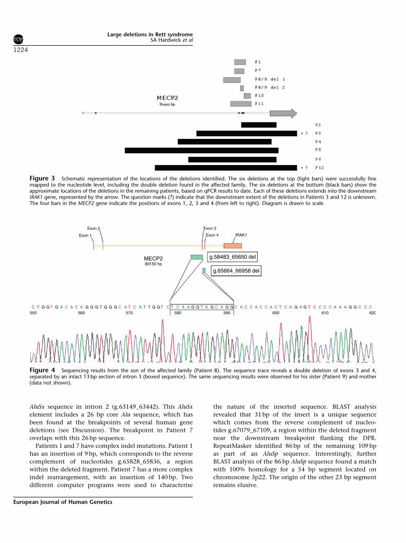
Table 1 and Figure 3.
For the affected family, the 1.5 kb junction fragments
were sequenced in all three family members, revealing the
presence of a double deletion involving exons 3 and 4
(Figure 4). The most upstream breakpoint is located in
intron 2, with the most downstream breakpoint in the
DPR of exon 4. The two deletions are separated by an intact
13-bp section of intron 3. The same four breakpoints were
observed in all three cases. Two other patients (1 and 7)
had complex insertion/deletion (indel) mutations.
Analysis of junction sequences
In the six rearranged alleles that were fine mapped to
the nucleotide level, we looked for repetitive elements
surrounding the deletion breakpoints and also for regions
of homology between the upstream and downstream wild-
type sequences. All six alleles had either the w sequence
or the GCTGG pentanucleotide present in the region
surrounding at least one breakpoint. In Patient 11, the
upstream breakpoint is positioned within an L2 repeat
element (g.61315_61538). The upstream breakpoints in
Patients 1 and 7 are located 45 bp apart within the same
Figure 2 Long-range PCR results for the affected family. The gelshows the B1.5 kb PCR product spanning the deletion breakpoints inall three family members. The primers used to generate this fragmentare normally B10 kb apart in a wild-type allele. The mother (M) is acarrier of the B8.5 kb deletion, which she has passed on to heraffected son (P8) and daughter (P9). Lane 6 (�)¼no template control.
Figure 1 Long-range PCR results. The gel shows PCR products spanning the deletion breakpoints in Patients 1, 7, 8, 10 and 11. Different forward/reverse primer combinations were used to generate these fragments. In lanes 3 and 5, the normal allele has also amplified in the normal female control.L, DNA ladder; P1, Patient 1; N, normal female control DNA. The numbers on the left and right refer to the sizes of the DNA ladder fragments in kb.
Large deletions in Rett syndromeSA Hardwick et al
1222
European Journal of Human Genetics
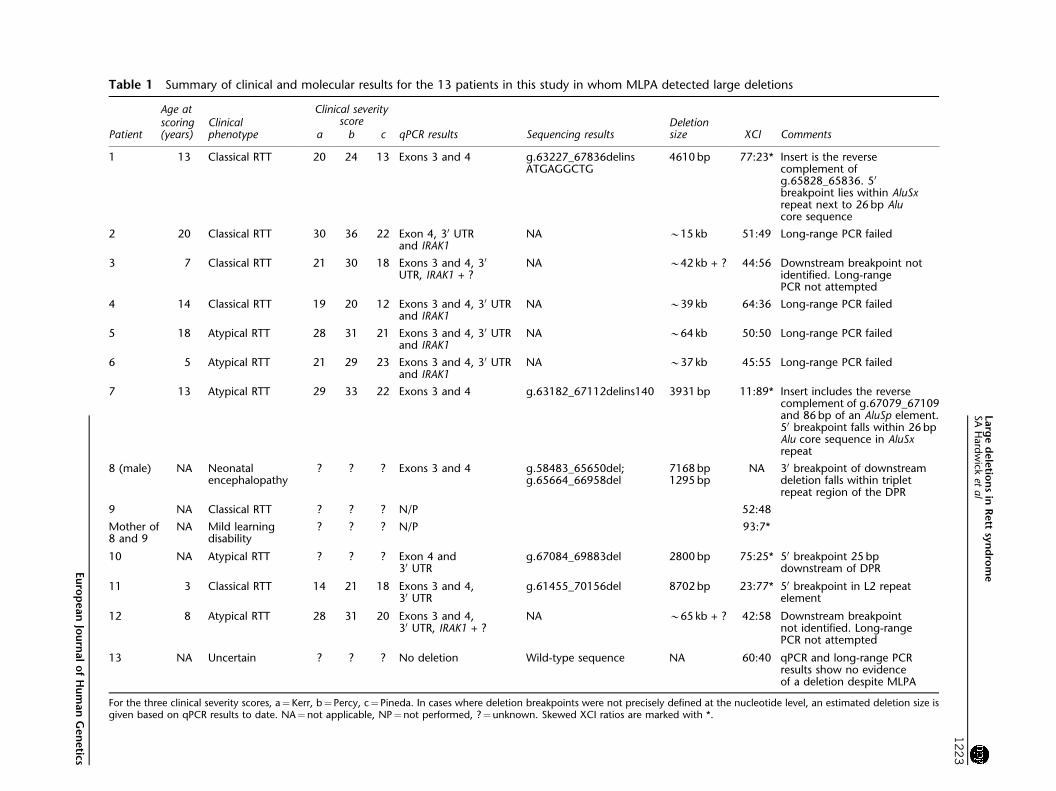
Table 1 Summary of clinical and molecular results for the 13 patients in this study in whom MLPA detected large deletions
Age at Clinical severityscore
Patientscoring(years)
Clinicalphenotype a b c qPCR results Sequencing results
Deletionsize XCI Comments
1 13 Classical RTT 20 24 13 Exons 3 and 4 g.63227_67836delinsATGAGGCTG
4610 bp 77:23* Insert is the reversecomplement ofg.65828_65836. 50
breakpoint lies within AluSxrepeat next to 26 bp Alucore sequence
2 20 Classical RTT 30 36 22 Exon 4, 30 UTRand IRAK1
NA B15 kb 51:49 Long-range PCR failed
3 7 Classical RTT 21 30 18 Exons 3 and 4, 30
UTR, IRAK1 + ?NA B42 kb + ? 44:56 Downstream breakpoint not
identified. Long-rangePCR not attempted
4 14 Classical RTT 19 20 12 Exons 3 and 4, 30 UTRand IRAK1
NA B39 kb 64:36 Long-range PCR failed
5 18 Atypical RTT 28 31 21 Exons 3 and 4, 30 UTRand IRAK1
NA B64 kb 50:50 Long-range PCR failed
6 5 Atypical RTT 21 29 23 Exons 3 and 4, 30 UTRand IRAK1
NA B37 kb 45:55 Long-range PCR failed
7 13 Atypical RTT 29 33 22 Exons 3 and 4 g.63182_67112delins140 3931 bp 11:89* Insert includes the reversecomplement of g.67079_67109and 86 bp of an AluSp element.50 breakpoint falls within 26 bpAlu core sequence in AluSxrepeat
8 (male) NA Neonatalencephalopathy
? ? ? Exons 3 and 4 g.58483_65650del;g.65664_66958del
7168 bp1295 bp
NA 30 breakpoint of downstreamdeletion falls within tripletrepeat region of the DPR
9 NA Classical RTT ? ? ? N/P 52:48
Mother of8 and 9
NA Mild learningdisability
? ? ? N/P 93:7*
10 NA Atypical RTT ? ? ? Exon 4 and30 UTR
g.67084_69883del 2800 bp 75:25* 50 breakpoint 25 bpdownstream of DPR
11 3 Classical RTT 14 21 18 Exons 3 and 4,30 UTR
g.61455_70156del 8702 bp 23:77* 50 breakpoint in L2 repeatelement
12 8 Atypical RTT 28 31 20 Exons 3 and 4,30 UTR, IRAK1 + ?
NA B65 kb + ? 42:58 Downstream breakpointnot identified. Long-rangePCR not attempted
13 NA Uncertain ? ? ? No deletion Wild-type sequence NA 60:40 qPCR and long-range PCRresults show no evidenceof a deletion despite MLPA
For the three clinical severity scores, a¼Kerr, b¼ Percy, c¼ Pineda. In cases where deletion breakpoints were not precisely defined at the nucleotide level, an estimated deletion size isgiven based on qPCR results to date. NA¼not applicable, NP¼not performed, ?¼unknown. Skewed XCI ratios are marked with *.
Larg
ed
ele
tion
sin
Rett
syn
dro
me
SA
Hard
wick
etal
1223
Eu
rop
ean
Jou
rnal
of
Hu
man
Gen
etics
AluSx sequence in intron 2 (g.63149_63442). This AluSx
element includes a 26 bp core Alu sequence, which has
been found at the breakpoints of several human gene
deletions (see Discussion). The breakpoint in Patient 7
overlaps with this 26 bp sequence.
Patients 1 and 7 have complex indel mutations. Patient 1
has an insertion of 9 bp, which corresponds to the reverse
complement of nucleotides g.65828_65836, a region
within the deleted fragment. Patient 7 has a more complex
indel rearrangement, with an insertion of 140 bp. Two
different computer programs were used to characterise
the nature of the inserted sequence. BLAST analysis
revealed that 31 bp of the insert is a unique sequence
which comes from the reverse complement of nucleo-
tides g.67079_67109, a region within the deleted fragment
near the downstream breakpoint flanking the DPR.
RepeatMasker identified 86 bp of the remaining 109 bp
as part of an AluSp sequence. Interestingly, further
BLAST analysis of the 86 bp AluSp sequence found a match
with 100% homology for a 54 bp segment located on
chromosome 3p22. The origin of the other 23 bp segment
remains elusive.
Figure 3 Schematic representation of the locations of the deletions identified. The six deletions at the top (light bars) were successfully finemapped to the nucleotide level, including the double deletion found in the affected family. The six deletions at the bottom (black bars) show theapproximate locations of the deletions in the remaining patients, based on qPCR results to date. Each of these deletions extends into the downstreamIRAK1 gene, represented by the arrow. The question marks (?) indicate that the downstream extent of the deletions in Patients 3 and 12 is unknown.The four bars in the MECP2 gene indicate the positions of exons 1, 2, 3 and 4 (from left to right). Diagram is drawn to scale.
C T G G T G A C A C A G G G T G G G C A T C A T T G G T C T C A A G G T A G C A G G C A C C A C C A C T C A G A G T C C C C A A A G G C C C550 560 570 580 590
g.65664_66958 del
g.58483_65650 del
IRAK1
600 610
Exon 3
Exon 4
Exon 2
MECP289150 hp
Exon 1
620
Figure 4 Sequencing results from the son of the affected family (Patient 8). The sequence trace reveals a double deletion of exons 3 and 4,separated by an intact 13 bp section of intron 3 (boxed sequence). The same sequencing results were observed for his sister (Patient 9) and mother(data not shown).
Large deletions in Rett syndromeSA Hardwick et al
1224
European Journal of Human Genetics
XCI results
All 13 patients were heterozygous at the AR locus and
were thus informative for the assay. The results for each
subject are listed in Table 1, along with a summary of all
other results obtained. Interestingly, Patient 9 has
random XCI (52:48), whereas her mother is highly
skewed (93:7). Patients 1, 7, 10 and 11 also have
skewed XCI (defined here as Z75% activity of one X
chromosome).
Genotype–phenotype correlations
All 12 patients with deletions in this study have been
clinically diagnosed with RTT with the exception of Patient
8, who has a severe neonatal encephalopathy. Clinical
severity scores, for the deletion patients in which they were
available, are shown in Table 1. Several statistical compari-
sons were made. For severity scores and deletion sizes, the
assumption of normally distributed populations could not
be made, and so nonparametric statistical tests were used.
The clinical score for the patients with all of exons 3 and
4 deleted (n¼7) was not significantly different from
those for patients with some of this region intact (n¼2;
Mann–Whitney test; Kerr: 20.9 vs 29.5, P¼0.057, Percy:
29.3 vs 34.4, P¼0.057, Pineda: 18.2 vs 21.9, P¼0.188).
The patients with deletions extending into IRAK1 (n¼6)
did not have significantly higher clinical scores than
those patients whose deletions were confined to within
MECP2 (n¼3; Mann–Whitney test; Kerr: 24.5 vs 20.0,
P¼0.519, Percy: 30.6 vs 24.0, P¼0.699, Pineda:
20.5 vs 18.0, P¼0.699). Furthermore, the average deletion
size for the patients with classical RTT (n¼6) was
not significantly different from that of the atypical RTT
patients (n¼5; Mann–Whitney test; 11.9 vs 37.0 kb,
P¼0.927).
There was no significant correlation between deletion
size and clinical score (Kerr: Spearman’s rho¼0.083,
P¼0.831). Large deletions were found in 6/23 classical
RTT patients (26.1%) and in 5/25 atypical patients
(20.0%) in whom no mutations were previously detected;
these proportions were not significantly different (w2 test,
df¼2, P¼0.882). The proportion of females with dele-
tions in this study with skewed XCI (5/13; 38.5%; 95%
confidence interval: 13.9–68.4%) was significantly greater
than the proportion of skewed females in the general
population (B10% for females o60 years30). Finally, the
average severity scores for the patients in this study
(n¼9) were compared with the average scores for all
mutation-positive patients in the ARSD without large
deletions for whom clinical scores were available
(n¼151). The patients in this study had higher average
scores for all three clinical scales, although these differ-
ences were not significant (T test, df¼158; Kerr: 23.4 vs.
20.8, P¼0.211, Percy: 28.3 vs 24.4, P¼0.067, Pineda: 18.8
vs 16.1, P¼0.077).
DiscussionIn this study, we have screened a cohort of apparently
mutation-negative RTT patients for large MECP2 deletions
using MLPA. One or more missing exonic probes were
detected in 13/149 patients; subsequent qPCR analysis has
confirmed the presence of large deletions in 12 of these
patients. In these patients, the deletion breakpoints
were further characterised using qPCR and long-range
PCR with the ultimate aim of defining the precise end-
points at the nucleotide level. The latter was achieved for 6
of these 12 patients.
Previous studies have reported deletions in 71/450
(15.8%) RTT patients in whom a wild-type sequence was
previously found for all exons, including both classical and
atypical cases. The lower proportion in this study (8.1%;
12/149) is probably due to the inclusion of a wider
spectrum of patients. Amongst our cohort of patients
screened in the diagnostic laboratory, many were selected
only on the basis of referral for MECP2 analysis. Hence, not
all patients from this cohort have been clinically diagnosed
with RTT, and re-evaluation of their clinical diagnoses is
being considered. On the other hand, if we consider the
proportion in those cases confirmed by the ARSD, it is
considerably higher at 25% (12/48).
Before this study, there were only five reported atypical
RTT patients with large MECP2 deletions.16,20 This study
has identified a further five atypical RTT cases with large
deletions, along with a male patient with a severe neonatal
encephalopathy. Our rate of detection of large deletions in
classical and atypical RTT patients previously thought to be
mutation negative were not significantly different, similar
to previous findings.15 These findings would support
the view that the relative lack of large deletions found in
atypical RTT patients could merely reflect a patient
selection bias in that classic RTT patients constitute the
most intensively studied group in terms of MECP2 gene
analysis.20 Our study reaffirms the view that large MECP2
deletions are an important and not infrequent cause of
atypical RTT.
All the deletions reported in this study involve exon 3
and/or 4, and in most cases both. This is in accordance
with previous findings, although a small number of
deletions have been reported affecting exons 1 and
2.14,18,20 We have identified one additional patient with
a large deletion of MECP2 exons 1 and 2 using MLPA
(unpublished data), although this is a rare phenomenon in
our experience. Whereas it has been suggested that this
could be due to an ascertainment bias introduced by
the genomic location of amplicons used,15 this can be
excluded in our case, as the MLPA assay includes amplicons
covering all four MECP2 exons and also three neighbouring
genes. We were unable to further characterise the deletion
in this patient using our qPCR approach due to an
insufficiency of DNA, hence this patient was not included
in the present study.
Large deletions in Rett syndromeSA Hardwick et al
1225
European Journal of Human Genetics
For the five rearranged alleles which we successfully fine
mapped to the nucleotide level, only one had a breakpoint
in the DPR, as defined by Laccone et al.15 One other patient
had a potential breakpoint in the DPR (Patient 2). Five out
of our 12 deletion patients had a breakpoint in the
immediate vicinity of the DPR. Combining our findings
with the three previous studies which have defined the
precise endpoints of large MECP2 deletions,13,15,18 12/21
(57.1%) rearranged alleles have a breakpoint in the DPR.
It is quite remarkable that the breakpoints of these
large multi-kilobase deletions are restricted to a common
B150-bp region; interestingly, this region is also the
hotspot for the smaller deletions (20–500 bp) confined
within exon 4. Several explanations have been proposed
to account for the intrinsic instability of this region;
these include the presence of direct and inverted repeats,8
the abundance of polypurine residues in the antisense
strand31 and the presence of a so-called w sequence,15
which has been found to be highly recombinogenic
in the Escherichia coli genome.32 However, the w sequence
is found in an additional four locations in the
AF030876 genomic clone, none of which have been
reported to be recombination hotspots. It is likely
that the mutagenic nature of the DPR is caused by
synergistic interactions between several different recombi-
nogenic factors.
It has been proposed that large MECP2 deletions are
often caused by potent mutagenic interactions between
w-like sequences and Alu repeats in intron 2.15 The
rearrangements in Patients 1 and 7 in the present study
lend support to this hypothesis. The upstream breakpoint
in these patients is positioned within the same AluSx
element in intron 2, whereas an internal element of the wsequence (GCTGG) is present near the downstream break-
point. Interestingly, a 26 bp core Alu sequence which has
been found at the breakpoints of many pathogenic human
gene deletions33 is also present at the upstream breakpoint
in these two patients. These are the same two patients in
whom complex indel mutations were identified, suggest-
ing that they may be the result of Alu-mediated indel
events. Interestingly, the GCTGG pentanucleotide is
found in both the w sequence and in the 26 bp core
Alu sequence.15 Analysis of the AF030876 genomic clone
with the RepeatMasker program (http://www.repeatmasker.
org) revealed that 22.8% of its sequence consists of Alu
elements. Accordingly, one would expect 22.8% of deletion
breakpoints to fall within an Alu element purely by chance.
Taking our findings together with the three above
mentioned studies, 11/21 (52.4%; 95% confidence interval:
29.8–74.3%) rearranged alleles have had at least one
breakpoint within an Alu sequence. This observed propor-
tion is significantly greater than what would be expected
due to chance alone, suggesting that Alu repeats do indeed
play a role in mutagenesis, probably by facilitating
illegitimate recombination.
As all of the deletions in this study affect exon 3 and/or
4, they affect crucial regions of the MeCP2 protein and can
be assumed to be pathogenic. The methyl-CpG-binding
domain (MBD) spans exons 3 and 4, whereas the
transcription repression domain (TRD) is located within
exon 4. These two functional domains are completely
deleted in 10/12 patients with deletions in this study.
For these patients, it is highly unlikely that the protein
produced from the mutant allele (if any) would have
residual function; the putative proteins would be unable to
enter the nucleus, let alone bind target genes or repress
transcription. It might be expected that the patients with
the MBD and TRD partially or wholly intact would present
with a relatively mild phenotype. However, our patients
with all of exons 3 and 4 deleted did not have significantly
different phenotype scores from the patients with some of
this region intact. The deletion in Patient 2 leaves exon 3
intact, with the upstream breakpoint located in exon 4.
Surprisingly, she has the most severe phenotype of all the
patients in the group for whom clinical scores were
available; she also has random XCI in blood leucocytes.
In her case, however, the entire 30 UTR is deleted, along
with the first B2 kb of the IRAK1 gene, which probably
adds to the severity of her phenotype. It should be noted
that she was also the eldest of the deletion patients and
therefore her clinical scores are likely to be associated with
some age-related changes.
In Patient 10, both the MBD and the TRD are left
completely intact. The last 229 bp of exon 4 are deleted,
including the stop codon, along with a further 2571 bp
of the 30 UTR. This would, theoretically, produce a MeCP2
protein truncated after residue 409. This leads to the loss of
almost half of the recently characterised WW domain.
Based on functional studies conducted by others, this
would lead to severely reduced, if not totally abolished,
binding activity of this domain.34 Unfortunately, total
clinical severity scores were not available for this patient,
although she did appear to have a relatively mild
phenotype based on the limited scores that were available
for individual scoring criteria. Notably, she has not
experienced seizures, which is unusual amongst our group
of patients. She has been diagnosed with atypical RTT.
Whether large MECP2 deletions invariably result in a total
loss of protein function is yet to be determined. This could
be further explored through transcript analysis or, ideally,
functional proteomic studies.
We have attempted to draw genotype–phenotype corre-
lations in our large deletion patient cohort. One group
has recently proposed that deletions extending into the
downstream IRAK1 gene may make the phenotype more
severe.20 Three other groups have also reported such
deletions,15,18,21 although none of the affected patients
have had additional clinical features. In our study, six
patients have deletions extending into the IRAK1 gene; the
average severity score for these patients was higher than in
Large deletions in Rett syndromeSA Hardwick et al
1226
European Journal of Human Genetics

the other six patients whose deletions were located wholly
within MECP2. This was observed for all three clinical
scores, although the differences were not statistically
significant. In Patients 2, 4, 5 and 6, the downstream
breakpoints were identified in the proximal part of IRAK1,
and so any disruption to IRAK1 would be minimal. In
Patients 3 and 12, however, it appears that much more of
the IRAK1 gene is deleted. These two patients have random
XCI in blood leucocytes. Patient 12, now deceased, had a
particularly severe phenotype that was not fully reflected
in her clinical scores at 8 years of age. It is believed that her
death at the age of 9 years was associated with complica-
tions related to her condition, although the nature of her
additional clinical features is not known in detail. Patient 3
has a fairly moderate phenotype despite the involvement
of the IRAK1 gene. IRAK1 is essential for the regulation of
innate immunity,35 and has been associated with athero-
sclerosis36 and myocardial contractile dysfunction.37 Due
to the involvement of the IRAK1 locus in cardiac function,
we suggest that patients with deletions extending into
IRAK1 should be considered for clinical assessment of
underlying cardiovascular abnormalities.
The average deletion size in atypical RTT patients was
larger than that in classical cases, but this difference was
not significant. This is not surprising as atypical patients
can be either more or less severely affected than classical
patients; even if larger deletions did have a tendency to
lead to more severe phenotypes this would not necessarily
lead to either the classical or atypical phenotype predomi-
nating. There did appear to be a positive trend between
deletion size and clinical severity, although no significant
relationship was observed for any of the three severity
scores. We acknowledge that this may well be due to poor
statistical power associated with our sample size.
Archer et al20 noted that their deletion group were
indistinguishable from other mutation-positive RTT
patients in terms of average clinical severity. The patients
in our study had higher clinical severity scores, on average,
than all the mutation-positive patients without large
deletions in the ARSD. This was true for all three severity
scores, although the differences were not significant at the
5% level. Whereas the numbers in this study are too small
to draw definite conclusions, it seems plausible that RTT
patients with large deletions are more severely affected
than their mutation-positive counterparts without large
deletions. It is quite likely that large multi-exonic deletions
result in a greater loss of protein function than would be
expected for other common types of mutations such as
missense, nonsense and frameshift mutations. The only
type of mutation that would conceivably be more severe
than a large deletion would be a nonsense mutation that
leads to premature truncation of the protein before exon 3.
In this study, we have reported a family in which
multiple members harbour a large MECP2 deletion. The
carrier mother has passed on the mutation to her affected
son and daughter, as demonstrated by our long-range PCR
results in all three family members. To our knowledge, this
is the first male patient reported worldwide with a large
MECP2 deletion, although recently some male subjects
have been reported with duplications of the Xq28
chromosomal band.38 – 43 He is only the 16th male in
which a pathogenic MECP2 mutation associated with early
post-natal encephalopathy has been found.44 As this is a
severe mutation, and the possibility of mosaicism can be
excluded due to the familial inheritance of the allele, this
case provides strong evidence that an absence of functional
MeCP2 is compatible with human life. Although this
individual has a severe neonatal encephalopathy, his
condition does not appear to be deteriorating at present.
The finding of a mutation present in all three family
members indicated that the mother either has highly
skewed XCI or is a germline mosaic; one of these has been
the case in other instances of familial RTT.45 – 52 We found
a highly skewed pattern of XCI in the mother’s blood
leucocytes, whereas her daughter has random XCI. By
comparing the sizes of the AR alleles in the mother and
daughter, we were able to establish that the mutant allele is
preferentially silenced in the mother. This family illustrates
the role that XCI plays in modulating the RTT pheno-
type. We have noticed some evidence of a mild learning
disability and fine hand tremor in the mother, although
these abnormalities were only detected in retrospect
after a familial mutation was identified. A family with
two RTT daughters with large deletions of exons 3 and 4
has recently been reported (F Ariani et al, personal
communication), but due to an absence of the mutation
in either parent, it was hypothesised that one of the
parents is a germline mosaic.
In the past, many laboratories have overlooked large
heterozygous deletions due to their reliance on PCR-based
screening methods. This study, combined with the findings
of previous reports, highlights the importance of screening
both classical and atypical RTT patients for large MECP2
deletions. Large deletions of exon 3 and/or 4 accounted for
12/211 (5.7%) of all pathogenic mutations in the ARSD
patient cohort. This makes large deletions as a group one
of the most frequent MECP2 mutations in the cohort, a
phenomenon also reported by Archer et al.20 Using MLPA,
we identified deletions in 13/149 apparently mutation-
negative patients, although in one patient (Patient 13),
subsequent qPCR and long-range PCR analysis found no
evidence of a deletion. As the missing exon 4 probe was
detected in two independent MLPA runs, it is likely to be
a reproducible MLPA artefact. We cannot, however, rule
out the possibility that this patient harbours a complex
underlying mutation that destroys the MLPA probe
binding site leading to exclusive amplification of the
normal allele during long-range PCR. We have recently
come across a similar case in which an insertion of genetic
material disturbed an MLPA probe-binding site; in this
Large deletions in Rett syndromeSA Hardwick et al
1227
European Journal of Human Genetics

latter case, however, we succeeded in amplifying a larger
product from the mutant allele (unpublished data). Our
results in the case of Patient 13 show that, if a similar
phenomenon has occurred, the mutant allele is beyond the
scope of normal PCR amplification. We are considering
screening this patient with a third independent method
such as Southern blotting to resolve the issue. Our findings
in this patient demonstrate the need to further characterise
apparent deletions detected by high-throughput diagnostic
methods such as MLPA.
AcknowledgementsWe thank Julianne Jackson and Gemma Jenkins for screeningdiagnostic patients for MECP2 mutations before MLPA analysis. HLis funded by National Health and Medical Research Council (NHMRC)program Grant 353514. JC is funded by NHMRC project Grants346603 and 457238. We also acknowledge the funding of AustralianRTT research by the National Institutes of Health (1 R01 HD43100-01A1) and the NHMRC under project Grant 303189. Finally, weexpress our gratitude to the Rett Syndrome Australian Research Fund,the Rotary Club of Narellan, and the Country Women’s Association ofNSW for financial support.
References1 Laurvick CL, de Klerk N, Bower C et al: Rett syndrome in
Australia: a review of the epidemiology. J Pediatr 2006; 148:347–352.
2 Weaving LS, Ellaway CJ, Gecz J, Christodoulou J: Rett syndrome:clinical review and genetic update. J Med Genet 2005; 42: 1–7.
3 Amir RE, Van den Veyver IB, Wan M, Tran CQ, Francke U, ZoghbiHY: Rett syndrome is caused by mutations in X-linked MECP2,encoding methyl-CpG-binding protein 2. Nat Genet 1999; 23:185–188.
4 Trappe R, Laccone F, Cobilanschi J et al: MECP2 mutations insporadic cases of Rett syndrome are almost exclusively of paternalorigin. Am J Hum Genet 2001; 68: 1093–1101.
5 Amir R, Sutton V, Van den Veyver I: Newborn screening andprenatal diagnosis for Rett syndrome: implications for therapy.J Child Neurol 2005; 20: 779–783.
6 Christodoulou J, Grimm A, Maher T, Bennetts B: RettBASE: theIRSA MECP2 variation databaseFa new mutation database inevolution. Hum Mutat 2003; 21: 466–472.
7 Renieri A, Meloni I, Longo I et al: Rett syndrome: the complexnature of a monogenic disease. J Mol Med 2003; 81: 346–354.
8 De Bona C, Zappella M, Hayek G et al: Preserved speechvariant is allelic of classic Rett syndrome. Eur J Hum Genet 2000;8: 325–330.
9 Laccone F, Huppke P, Hanefeld F, Meins M: Mutation spectrum inpatients with Rett syndrome in the German population: evidenceof hot spot regions. Hum Mutat 2001; 17: 183–190.
10 Bourdon V, Philippe C, Grandemenge A, Reichwald K, JonveauxP: Deletion screening by fluorescence in situ hybridization in Rettsyndrome patients. Ann Genet 2001; 44: 191–194.
11 Bourdon V, Philippe C, Labrune O, Amsallem D, Arnould C,Jonveaux P: A detailed analysis of the MECP2 gene: prevalence ofrecurrent mutations and gross DNA rearrangements in Rettsyndrome patients. Hum Genet 2001; 108: 43–50.
12 Yaron Y, Ben Zeev B, Shomrat R, Bercovich D, Naiman T, Orr-Urtreger A: MECP2 mutations in Israel: implications for mole-cular analysis, genetic counseling, and prenatal diagnosis in Rettsyndrome. Hum Mutat 2002; 20: 323–324.
13 Schollen E, Smeets E, Deflem E, Fryns J, Matthijs G: Grossrearrangements in the MECP2 gene in three patients with Rett
syndrome: implications for routine diagnosis of Rett syndrome.Hum Mutat 2003; 22: 116–120.
14 Erlandson A, Samuelsson L, Hagberg B, Kyllerman M, Vujic M,Wahlstrom J: Multiplex ligation-dependent probe amplification(MLPA) detects large deletions in the MECP2 gene of Swedish Rettsyndrome patients. Genet Test 2003; 7: 329–332.
15 Laccone F, Junemann I, Whatley S et al: Large deletions of theMECP2 gene detected by gene dosage analysis in patients withRett syndrome. Hum Mutat 2004; 23: 234–244.
16 Ariani F, Mari F, Pescucci C et al: Real-time quantitative PCR as aroutine method for screening large rearrangements in Rettsyndrome: report of one case of MECP2 deletion and one caseof MECP2 duplication. Hum Mutat 2004; 24: 172–177.
17 Huppke P, Ohlenbusch A, Brendel C, Laccone F, Gartner J:Mutation analysis of the HDAC 1, 2, 8 and CDKL5 genes in Rettsyndrome patients without mutations in MECP2. Am J Med Genet2005; 137A: 136–138.
18 Ravn K, Nielsen JB, Skjeldal OH, Kerr A, Hulten M, Schwartz M:Large genomic rearrangements in MECP2. Hum Mutat 2005;25: 324.
19 Shi J, Shibayama A, Liu Q et al: Detection of heterozygousdeletions and duplications in the MECP2 gene in Rett syndromeby Robust Dosage PCR (RD-PCR). Hum Mutat 2005; 25: 505.
20 Archer HL, Whatley SD, Evans JC et al: Gross rearrangements ofthe MECP2 gene are found in both classical and atypical Rettsyndrome patients. J Med Genet 2006; 43: 451–456.
21 Pan H, Li MR, Nelson P, Bao XH, Wu XR, Yu S: Large deletionsof the MECP2 gene in Chinese patients with classical Rettsyndrome. Clin Genet 2006; 70: 418–419.
22 Colvin L, Fyfe S, Leonard S et al: Describing the phenotype inRett syndrome using a population database. Arch Dis Child 2003;88: 38–43.
23 The Rett Syndrome Diagnostic Criteria Work Group: Diagnosticcriteria for Rett syndrome. Ann Neurol 1988; 23: 425–428.
24 Hagberg B, Hanefeld F, Percy A, Skjeldal O: An update onclinically applicable diagnostic criteria in Rett syndrome. Com-ments to Rett Syndrome Clinical Criteria Consensus PanelSatellite to European Paediatric Neurology Society Meeting,Baden Baden, Germany, 11 September 2001. Eur J Paediatr Neurol2002; 6: 293–297.
25 Hagberg B: Clinical delineation of Rett syndrome variants.Neuropediatrics 1995; 26: 62.
26 Miller S, Dykes D, Polesky H: A simple salting out procedure forextracting DNA from human nucleated cells. Nucleic Acids Res1988; 16: 1215.
27 Schouten JP, McElgunn CJ, Waaijer R, Zwijnenburg D, Diepvens F,Pals G: Relative quantification of 40 nucleic acid sequences bymultiplex ligation-dependent probe amplification. Nucleic AcidsRes 2002; 30: e57.
28 Rozen S, Skaletsky H: Primer3 on the WWW for general users andfor biologist programmers. Methods Mol Biol 2000; 132: 365–386.
29 Allen R, Zoghbi H, Moseley A, Rosenblatt H, Belmont J:Methylation of HpaII and HhaI sites near the polymorphic CAGrepeat in the human androgen-receptor gene correlates with Xchromosome inactivation. Am J Hum Genet 1992; 51: 1229–1239.
30 Busque L, Mio R, Mattioli J et al: Nonrandom X-inactivationpatterns in normal females: lyonization ratios vary with age.Blood 1996; 88: 59–65.
31 Lebo RV, Ikuta T, Milunsky JM, Milunsky A: Rett syndrome fromquintuple and triple deletions within the MECP2 deletionhotspot region. Clin Genet 2001; 59: 406–417.
32 Stahl F, Stahl M: Recombination pathway specificity of Chi.Genetics 1977; 86: 715–725.
33 Rudiger N, Gregersen N, Kielland-Brandt M: One short wellconserved region of Alu-sequences is involved in human generearrangements and has homology with prokaryotic chi. NucleicAcids Res 1995; 23: 256–260.
34 Buschdorf J, Stratling W: A WW domain binding region inmethyl-CpG-binding protein MeCP2: impact on Rett syndrome.J Mol Med 2004; 82: 135–143.
Large deletions in Rett syndromeSA Hardwick et al
1228
European Journal of Human Genetics
35 Su K, Richter K, Zhang C, Gu Q, Li L: Differential regulation ofinterleukin-1 receptor associated kinase 1 (IRAK1) splice variants.Mol Immunol 2007; 44: 900–905.
36 Huang Y, Li T, Sane DC, Li L: IRAK1 serves as a novel regulatoressential for lipopolysaccharide-induced interleukin-10 geneexpression. J Biol Chem 2004; 279: 51697–51703.
37 Thomas JA, Haudek SB, Koroglu T et al: IRAK1 deletion disruptscardiac Toll/IL-1 signaling and protects against contractiledysfunction. Am J Physiol Heart Circ Physiol 2003; 285: 597–606.
38 Friez MJ, Jones JR, Clarkson K et al: Recurrent infections, hypo-tonia, and mental retardation caused by duplication of MECP2and adjacent region in Xq28. Pediatrics 2006; 118: e1687–e1695.
39 Lugtenberg D, de Brouwer APM, Kleefstra T et al: Chromosomalcopy number changes in patients with non-syndromic X linkedmental retardation detected by array CGH. J Med Genet 2006; 43:362–370.
40 Sanlaville D, Prieur M, de Blois M-C et al: Functional disomy of theXq28 chromosome region. Eur J Hum Genet 2005; 13: 579–585.
41 Meins M, Lehmann J, Gerresheim F et al: Submicroscopicduplication in Xq28 causes increased expression of the MECP2gene in a boy with severe mental retardation and features of Rettsyndrome. J Med Genet 2005; 42: e12.
42 del Gaudio D, Fang P, Scaglia F et al: Increased MECP2 gene copynumber as the result of genomic duplication in neurodevelop-mentally delayed males. Genet Med 2006; 8: 784–792.
43 Van Esch H, Bauters M, Ignatius J et al: Duplication of the MECP2region is a frequent cause of severe mental retardation andprogressive neurological symptoms in males. Am J Hum Genet2005; 77: 442–453.
44 Kankirawatana P, Leonard H, Ellaway C et al: Early progressiveencephalopathy in boys and MECP2 mutations. Neurology 2006;67: 164–166.
45 Sirianni N, Naidu S, Pereira J, Pillotto R, Hoffman E: Rettsyndrome: confirmation of X-linked dominant inheritance, andlocalization of the gene to Xq28. Am J Hum Genet 1998; 63:1552–1558.
46 Wan M, Lee S, Zhang X et al: Rett syndrome and beyond:recurrent spontaneous and familial MECP2 mutations at CpGhotspots. Am J Hum Genet 1999; 65: 1520–1529.
47 Bienvenu T, Carrie A, de Roux N et al: MECP2 mutations accountfor most cases of typical forms of Rett syndrome. Hum Mol Genet2000; 9: 1377–1384.
48 Villard L, Kpebe A, Cardoso C, Chelly J, Tardieu M, Fontes M: Twoaffected boys in a Rett syndrome family: clinical and molecularfindings. Neurology 2000; 55: 1188–1193.
49 Villard L, Levy N, Xiang F et al: Segregation of a totally skewedpattern of X chromosome inactivation in four familial cases ofRett syndrome without MECP2 mutation: implications for thedisease. J Med Genet 2001; 38: 435–442.
50 Gill H, Cheadle JP, Maynard J et al: Mutation analysis in theMECP2 gene and genetic counselling for Rett syndrome. J MedGenet 2003; 40: 380–384.
51 Evans JC, Archer HL, Whatley SD, Clarke A: Germline mosaicismfor a MECP2 mutation in a man with two Rett daughters. ClinGenet 2006; 70: 336–338.
52 Dayer A, Bottani A, Bouchardy I et al: MECP2 mutant allele in aboy with Rett syndrome and his unaffected heterozygous mother.Brain Dev 2007; 29: 47–50.
Large deletions in Rett syndromeSA Hardwick et al
1229
European Journal of Human Genetics
Copyright © 2022 FDOKUMEN