Mitochondrial DNA deletions are associated with non-B DNA conformations
Upload
independentCategory
view
0download
0
TITLE PAGE
High frequency of COH1 intragenic deletions and duplications detected by MLPA in patients
with Cohen syndrome.
Parri V1, Katzaki E1, Uliana V1, Scionti F1, Tita R1, Artuso R1, Longo I1, Boschloo R2, Vijzelaar
R2, Selicorni A3, Brancati F4,5, Dallapiccola B6, Zelante L7, Hamel CP8, Sarda P8, Lalani SR9,
Grasso R10, Buoni S11, Hayek J11, Servais L12, de Vries BBA 13, Georgoudi N14, Nakou S15, Petersen
MB16, Mari F1, Renieri A1, Ariani F1.
1. Medical Genetics, University of Siena, Siena, Italy
2. MRC-Holland, Amsterdam, The Netherlands
3. Paediatric Unit, University of Milan, Milan, Italy
4. Ospedale Casa Sollievo della Sofferenza-Mendel Insitute, Rome, Italy
5. Department of Biomedical Sciences, Gabriele D'Annunzio University, Chieti-Pescara, Italy
6. Ospedale Pediatrico Bambino Gesù, IRCCS, Rome, Italy
7. Ospedale Casa Sollievo della Sofferenza, IRCCS, San Giovanni Rotondo, Italy
8. Centre Hospitalier Régional et Universitaire, Service de Génétique Médicale, Montpellier, France
9. Department of Molecular and Human Genetics, Baylor College of Medicine, Houston, USA
10. IRCSS MEDEA, Bosisio Parini, Lecco, Italy
11. Child Neuropsychiatric Unit, University of Siena, Siena, Italy
12. Institut de Myologie, Hôpital de la Pitié Salpétrière, Paris, France
13. Department of Human Genetics, Radboud University Nijmegen Medical Centre, Nijmegen, The
Netherlands

13. Department of Mental Health and Social Welfare, Institute of Child Health, Athens, Greece
14. Department of Social and Developmental Pediatrics, Institute of Child Health, Athens, Greece
15. Department of Genetics, Institute of Child Health, Athens, Greece
Corresponding author:
Alessandra Renieri, MD, Ph.D
Medical Genetics, Department of Molecular Biology
University of Siena, V. Le Bracci 2 – 53100 Siena (Italy)
tel +39 0577 233303, fax +39 0577 233259
e-mail [email protected]

ABSTRACT
Cohen syndrome is a rare clinically variable autosomal recessive disorder characterized by mental
retardation, postnatal microcephaly, facial dysmorphisms, ocular abnormalities, and intermittent
neutropenia. Mutations in the COH1 gene have been found in patients from different ethnic origins.
However, a high percentage of patients has only one or no mutated allele. In order to investigate
whether COH1 copy number changes account for missed mutations, we used multiplex ligation-
dependent probe amplification (MLPA) to test a group of 14 patients with Cohen syndrome. This
analysis has allowed to identify multi-exonic deletions in 11 alleles and duplications in 4 alleles.
Considering our previous study, COH1 copy number variations represent 42% of total mutated
alleles. To our knowledge, COH1 intragenic duplications have never been reported in Cohen
syndrome. The three duplications encompassed exons 4-13, 20-30 and 57-60, respectively.
Interestingly, four deletions showed the same exons coverage (exons 6-16) respect to a deletion
recently reported in a large Greek consanguineous family. Haplotype analysis suggested a possible
founder effect in the Mediterranean basin. The use of MLPA was therefore crucial in identifying
mutated alleles undetected by traditional techniques and in defining the extent of the
deletions/duplications. Given the high percentage of identified copy number variations, we suggest
that this technique could be used as the initial screening method for molecular diagnosis of Cohen
syndrome.
KEYWORDS: Cohen syndrome, COH1, MLPA.

INTRODUCTION
Cohen syndrome (OMIM #216550) is an autosomal recessive disorder first described in 1973 by
Cohen et al. 1. It is characterized by non-progressive mental retardation, characteristic facial
features, hypotonia, pigmentary retinopathy, myopia and intermittent neutropenia 1-3. The peculiar
craniofacial features of Cohen syndrome include microcephaly, downslanting and wave shaped
palpebral fissures, short philtrum and prominent upper central incisors 1-3.
In 2003, mutations in the COH1 gene were identified as causative of Cohen syndrome in the
Finnish population 4. The COH1 gene maps to chromosome 8q22 and consists of 62 exons encoding
for a potential transmembrane protein presumably involved in vesicle mediated sorting and
intracellular protein transport 4,5.
The phenotypic spectrum in Finnish patients is highly homogeneous and molecular analysis
revealed a founder effect with a common ancestral mutation causative of the majority of cases 4. On
the other hand, Cohen syndrome was found to be associated with mutations in the COH1 gene in
different populations with a broader clinical spectrum than the Finnish subtype 4,6-10. About one
hundred mutations in COH1 gene have been identified so far 9. Most of them are truncating
mutations resulting in a null allele, while missense mutations and in-frame deletions are less
frequent 9.
Methods for the detection of point mutations in the COH1 gene are well established in our
laboratory and consists of DHPLC followed by automatic sequencing 10. Until now, we employed
real time quantitative PCR (qPCR) for the detection of large COH1 deletions/duplications 10.
However, since COH1 is a large gene, spanning 846 kb of genomic DNA and composed by 62
exons, qPCR assays designed on a limited number of target regions are prone to miss a high fraction
of intragenic rearrangements and do not allow the characterization of the extent of the

deletions/duplications. Very recently, a targeted oligonucleotide array was designed, enabling the
detection of COH1 copy number changes with higher resolution 11. The authors analyzed 35
patients (from 26 families) with unexplained Cohen syndrome and identified deletions in 9 patients
from 7 families, showing that large deletions are an important cause of Cohen syndrome 11.
In order to detect COH1 copy number variations, we employed Multiple Ligation-
dependent Probe Amplification (MLPA), a technique that has greatly improved mutation screening
allowing the relative quantification of up to 40 different nucleic acid sequences in a single reaction
tube at a relatively low cost 12. By the use of two MLPA assays designed to screen copy number
changes in almost all coding exons (60/62) of COH1, we analyzed a group of patients with a
clinical diagnosis of Cohen syndrome where traditional tests failed to identify mutations in both
alleles.

MATERIALS AND METHODS
Patients
Clinical geneticists from Italy, France, Holland and United States assessed patients and diagnosed
Cohen syndrome on the bases of published criteria 13. Patients were considered as having Cohen
syndrome when 6 of the following 8 criteria were fulfilled: developmental delay, microcephaly,
typical facial features, truncal obesity with slender extremities, sociable behavior, joint
hypermobility, retinopathy or myopia, and intermittent neutropenia. Our series includes three
children younger than 5 years (Table 1). Since the chorioretinal dystrophy not manifest in young
patients, the diagnosis of Cohen syndrome in children is considered when learning disabilities are
associated with two of the following features: typical facial gestalt, pigmentary retinopathy, or
neutropenia 14.
Overall, we collected 14 patients from 11 families, ranging in age from 18 months to 52
years. This group included four patients (1, 8, 9A, 9B) originally described by Katzaki et al. 10 and
10 newly ascertained cases. The main clinical features are summarized in Table 1. Enrolled cases
included one consanguineous family with an affected child (8) and 10 non-consanguineous families:
7 with one affected child (1, 2, 3, 4, 5, 6, 7), one with two affected sisters (9A, 9B), one with two
affected brothers (10A, 10B) and one with an affected brother (11A) and sister (11B). A distinct
phenotype was present in two affected brothers (10A and 10B), presenting five of eight diagnostic
criteria (Table 1); these patients were classified as Cohen-like 13.
COH1 molecular analysis
Genomic DNA was isolated using QIAamp DNA blood maxi kit, according to the manufacturer’s
protocol (Qiagen, Hilden Germany). PCR amplification of the 62 exons was carried out using
published primers 4 10. Mutation analysis was performed by Denaturing High Performance Liquid
Chromatography (DHPLC) using the Transgenomic WAVETM (Transgenomic, San Jose, CA,
USA)10. Quantitative PCR was also performed in one familiar case (9A, 9B) and one sporadic case
(8) with a Custom TaqMan Assay designed on exon 16 (Applied Biosystems,
https://products.appliedbiosystems.com) 10.
MLPA analysis was performed using two distinct SALSA MLPA kits (P321-A1/P322-
A1) designed by MRC-Holland (Amsterdam, The Netherlands). The two assays include 69 COH1
probes to screen copy number changes in almost all coding gene exons (60/62) and 16 control
probes. No probe was present for exons 6 and 14. For exons 3, 16, 17, 24, 31, 34, 35 and 36, two
distinct probes were designed. The analysis was carried out as previously described 12. Briefly, 100
ng of genomic DNA was diluted with TE buffer to 5 μl, denatured at 98°C for 5 minutes and
hybridized with SALSA Probe-mix at 60°C overnight. Ligase-65 mix was then added and ligation
was performed at 54°C for 15 minutes. The ligase was successively inactivated by heating the
samples at 98°C for 5 minutes. PCR reaction was performed in a 50 μl volume. Primers, dNTPs and
polymerase were added and amplification was carried out for 35 cycles (30 seconds at 95°C, 30
seconds at 60°C and 60 seconds at 72 °C). The amplification products were separated on an ABI
Prism 310 automatic sequencer and analyzed using the GenScan software ver.3.1. For data analysis
the values of peak sizes and areas were exported to an Excel table and compared with a normal
control (MRC-Holland, Amsterdam, The Netherlands). Dosage alterations were considered
significant if sample values deviated more than 30% from the control.
For exons 6 and 14, we designed two specific qPCR assays (Supplementary Table 1). In
addition MLPA results were confirmed by qPCR using probes located in exon 16, 24, 34, 42, 48
and 58 (Supplementary Table 1) 10. Reactions were performed in a 96-well optical plate with a final
reaction volume of 50 µl using an ABI prism 7000 (Applied Biosystems, Foster City California). A

total of 100 ng of DNA (10 µl) was dispensed in each of the four sample wells for quadruplicate
reactions. Thermal cycling conditions included a pre-run of 2 min at 50°C and 10 min at 95°C.
Cycle conditions were 40 cycles at 95°C for 15 sec and 60°C for 1 min, according to the TaqMan
Universal PCR Protocol (PE Applied Biosystems, Foster City, CA, USA). The TaqMan Universal
PCR Master Mix and Microamp reaction tubes were supplied by Applied Biosystems. The starting
copy number of the unknown samples was determined using the comparative Ct method, as
previously described 15.
In case 11, long-range PCR was performed with the Expand Long Template PCR kit as
specified by the manufacturer (Roche Diagnostics, Basel, Switzerland), using a forward primer
located in intron 59 (ggatggctctgaacagatga) and a reverse primer located in intron 56
(agaagcaattggcaagaggt). These primers are divergent in the normal genome and they do not amplify
the control’s DNA. PCR conditions were: 300 nM of each primer, 350 μM of dNTPs, 2.0 mM
MgCl2, 0.75 μl of enzyme mix and 1X Buffer II, and the following cycling parameters: 94◦C 5 min;
94◦C 10 sec, 59◦C 30 sec, 68◦C 5 min, 10 cycles; 94◦C 15 sec, 59◦ C 30 sec, 68◦C 5 min
+20sec/cycle, 25 cycles; final extension 68◦C 30 min.
Haplotype analysis
A set of 10 markers covering a region of about 4 Mb encompassing the COH1 gene were used for
haplotype analysis (Supplementary Table 2) in three of our cases with the 6-16 deletion (case 5, 8
and 9A) and one member of the large Greek consanguineous family reported by Bugiani et al.
harbouring the 6-16 deletion in homozygous state 16. Haplotype analysis was also performed in all
available family members of the 6-16 deleted patients and in 50 Italian control individuals. The
forward primers were fluorescently labelled with FAM. Markers were amplified by polymerase
chain reaction. Conditions were optimized for individual primer pairs in a 9600 thermocycler

(Applied Biosystems). The programs used were 95°C for 12 min, followed by 30 cycles of melting
at 94°C for 15 s, annealing at the optimal temperature for 15 s, and then extension at 72°C for 30 s.
A final extension was performed at 72°C for 10 min. PCR products were run on an ABI 3130
sequencer (Applied Biosystems) and analysed with GeneMapper v.4.0. The size of the PCR
products of the microsatellite markers were compared among the families carrying the recurrent
deletion 6-16 in heterozygous or homozygous state, in order to define the haplotype co-segregating
with the deletion.
RESULTS
Phenotype
All fourteen patients displayed the typical Cohen facial gestalt, narrow extremities and truncal
adiposity even if not all cases were obese (7/14) (Table 1, Figure 1) 10. Microcephaly was present in
the majority of patients (9/14) (Table 1) 10. The retinopathy was absent in one family with two
affected children younger than five years (11A, 11B) (Table 1). Neutropenia was absent in one
patient (3) and one case did not show joint hyperextensibility (8). Among the 14 patients, two
brothers (10A, 10B) presented an atypical phenotype, lacking microcephaly and truncal obesity.
However, the diagnosis of Cohen syndrome was suggested based on the association of retinopathy,
neutropenia and facial appearance (Figure 1).
COH1 molecular analysis
The 14 patients (11 families) with a clinical diagnosis of Cohen syndrome were analysed for the
presence of COH1 point mutations by DHPLC followed by sequencing of the samples with an
abnormal elution profile 10. This analysis led to the detection of 12 different mutations, including 6

frame-shift, 3 splice site, 2 nonsense and one complex rearrangement (Table 2). Moreover, in one
family (9A and 9B) and in one sporadic patient (8) a partial heterozygous COH1 gene deletion was
already detected by qPCR using a TaqMan probe designed on exon 16 10. In order to identify
missed mutated alleles and to characterize the extent of the deletions/duplications, we employed
two MLPA assays (P321-A1/P322-A1) designed to detect COH1 copy number changes in 60 out of
62 exons of the gene. This method led us to identify 5 different multi-exonic deletions in 11 alleles
and 3 different duplications in 4 alleles (Table 2). In particular, MLPA characterized heterozygous
copy number variations in 9 patients (7 families) displaying a point mutation previously identified
by DHPLC on the other allele (1, 2, 4, 7, 8, 10A, 10B, 11A and 11B), two different compound
heterozygous deletions in two affected sisters (9A and 9B) and one homozygous deletion in one
sporadic patient (case 5) (Table 2, Figure 2, Supplementary Figure 1, Supplementary Figure 2).
In four patients, MLPA showed the presence of a deletion spanning from exon 7 to exon 16
(Figure 2, Table 2). Since the MLPA assays contain 69 probes not including exon 6, we designed a
targeted qPCR probe assay for this exon (Supplementary Table 1). This analysis showed that the
four deletions spanned indeed from exon 6 to exon 16 (Figure 3, Table 2).
In two sporadic patients (cases 2 and 4), MLPA detected a significant increase in the
fluorescent signals corresponding to exons 4-13 and 20-30, indicating the presence of two
differently sized duplications (Table 2, Supplementary Figure 1). In case 2, a specific qPCR assays
indicated that exon 14 is not included in the duplication (data not shown). In a familial case (11A,
11B) in which DHPLC followed by sequencing had already detected a complex rearrangement in
exon 56 (c.1088insTTdelCTGCGAGGCAGCTTGTGCAC; p.T3627_H3633delinsI), MLPA also
disclosed a significant increase in peak heights 57-60, suggesting the presence of a heterozygous
duplication (Table 2, Figure 4). Analysis of the parental DNA indicated that the rearrangement
p.T3627_H3633delinsI was in cis with the duplication detected by MLPA (Table 2).

In order to better characterise the 57-60 duplication, we performed long-range PCR using
a forward primer in intron 59 and a reverse primer in intron 56 (Figure 5a). We obtained a product
of ~1 kb in the two affected sibs and in the carrier father. Automatic sequencing of the PCR product
permitted to characterize the junction sequence of the duplicated segment (Figure 5), 95 bp
downstream respect to the rearrangement. The duplicated segment, starting within intron 56, is
inserted within exon 61 in position g.100,953,994 (NM_017890) (Figure 5). According to
prediction softwares, this insertion interrupts the protein product creating a premature stop codon
after 10 new aminoacids.
Not all parental DNAs were available for testing (Table 2). For patients 9A and 9B,
DNAs of two healthy sibs have been analyzed to determine whether the two rearrangements were in
cis or trans. MLPA revealed that the brother and the sister were carriers of the deletions spanning
respectively exons 6-16 and exons 46-50, confirming that the rearrangements were in compound
heterozygousity. In the cases where parental DNAs have been tested, all mutations were inherited
but in one patient (case 2) harboring a de novo point mutation (c.11695delAGTG; p.S3899fs3941X)
(Table 2).
All copy number changes identified by MLPA were confirmed by qPCR using specific
probes for exon 16, 24, 34, 42, 48 and 58 (data not shown).
Haplotype analysis
In order to investigate a founder effect for the recurrent deletion of exons 6-16, we performed
haplotype analysis in three of our cases and one additional case belonging to the large Greek
consanguineous family reported by Bugiani et al. 16. A founder effect is expected to result in sharing
of allelic sequence polymorphisms in the vicinity of the deletion. We examined 10 microsatellite
markers within a region of about 4 Mb encompassing the COH1 gene (Supplementary Table 2,

Table 3). For heterozygous markers, the phase was assigned by genotyping other family members:
parents in case 8 (carrier mother and noncarrier father) and sibs in case 9 (one carrier and one
noncarrier sister) (data not shown).
In order to determine how frequently alleles of the same size can be obtained by chance in
a general population, we genotyped DNA from 50 Italian control samples using primers for the
same 8 microsatellite markers (minimal common haplotype, Table 3). None of the healthy controls
and none of noncarrier family members showed the minimal common haplotype (data not shown).

DISCUSSION
In this study, we report the first application of the MLPA technique to screen for COH1 large
deletions and duplications. In a group of 14 patients (11 families) with a clinical diagnosis of Cohen
syndrome, MLPA allowed us to obtain rapid and high quality results disclosing 11 deleted and 4
duplicated COH1 alleles. The use of MLPA led us to identify all COH1 mutations undetected by
conventional screening, suggesting that this technique is an important tool for the molecular
characterization of Cohen syndrome.
Our series included 12 patients with true Cohen syndrome and two brothers with an atypical
phenotype, lacking microcephaly and truncal obesity. However, the association of retinopathy,
neutropenia and facial appearance addressed the clinical diagnosis. Their facial features although
not typical, were not in disagreement with the diagnosis of Cohen syndrome consisting of long face,
heavy eyebrows, mildly down-slanting palpebral fissures, prominent root of the nose, normal
philtrum and prognatism (Figure 1). Three patients from two families were children with less than 5
years. They presented the typical facial features of younger patients, including round face with full
lower lip, not excessively short philtrum, slightly downward slanting eyes with wave-shaped eyelids
and less prominent nasal bridge (Figure 1) 10.
Copy number changes in COH1 have been previously investigated in patients with Cohen
syndrome by qPCR using probes designed on a limited number of exons 10, 16. Only recently a
targeted oligonucleotide array with a median resolution of 200 bp was designed within the gene,
considerably increasing mutation detection rate 11. Using this technique, the authors identified
COH1 large deletions in 9 patients from 7 families, demonstrating that they represent an important
cause of Cohen syndrome 11. The present results and our previous study on a group of 18 patients
disclosed a total of 21 alleles with point mutations (58%) and 15 with copy number variations

(42%), confirming that deletions and duplications account for a significant percentage of COH1
mutations 10.
In 4 patients from three families, MLPA identified a COH1 large deletion sharing the same
extent with one previously reported in an isolated Greek Island population, spanning from exon 6 to
exon 16 16. In our patients, the deletion was heterozygous in two families and homozygous in an
apparently non consanguineous family 10. Interestingly, this latter patient displays the same
constellation of facial features reported in Greek patients with the homozygous deletion including
thick hair with low hair-line, strabism, lack of nasofrontal angle, short upturned philtrum and
prominent maxillary central incisors (patient 5, Figure 1) 16. Moreover, they show milder
microcephaly and more severe visual impairment compared to the original phenotype described in
the Finnish population 4,16.
Our three families with the same deletion encompassing exons 6-16 come from different
Italian regions, two in Central and one in the Southern Italy. The results obtained by haplotype
analysis in these families, in one member of the large Greek consanguineous family previously
reported by Bugiani et al. harbouring the 6-16 deletion in homozygous state and in 50 healthy
Italian controls, suggest that the recurrent deletion is due to an ancestral founder effect in the
Mediterranean area 16 (Table 3).
In the present study we also identified two deletions spanning exons 4-16 and 40-43, sharing
the same exons coverage with two deletions already reported in the Northern European
population11. Also in these cases we can not exclude a founder effect for the deleted alleles.
Alternatively, these could be independent mutations favored by the presence of repeated elements
located at the breakpoints. Accordingly, RepeatMasker software analysis of the genomic region
containing COH1 revealed a higher frequency of LINEs, SINEs and DNA repeat elements in
comparison to the average for autosomal sequences 11. In a previous study, it was suggested that

the most likely mechanism for genomic rearrangements in the COH1 gene is the Non Homologous
End Joining (NHEJ), leading to non recurrent deletions 11. Considering our latest results, the Non
Allelic Homologous Recombination mechanism (NAHR) cannot be ruled out.
In four patients from three families, MLPA identified three different size duplications
spanning exons 4-13, 20-30 and 57-60, respectively. To our knowledge COH1 intragenic
duplications have never been reported in Cohen syndrome.
In one family with two affected sibs (cases 11A/B), we identified a complex rearrangement
(p.T3627_H3633delinsI) in cis with the downstream duplication detected by MLPA. We initially
hypothesized that this rearrangement could be located at the breakpoint of the duplication within
exon 56. However, sequencing analysis of the long PCR product using a forward primer in intron
59 and a reverse primer in intron 56 indicated that the duplication effectively starts in intron 56, 95
bp after the rearrangement (Figure 5). This sequence is joined to exon 61 in position g.100,953,994
(NM_152564) (Figure 5). Since the MLPA probe of exon 61 is located upstream the junction point
(Figure 5a) and its signal doesn’t result increased, we can suppose that the duplication is not in
tandem. The insertion of the duplicated segments within exon 61 creates a premature stop codon
after 10 new aminoacids of the protein product. Even if detailed mapping of the extent of all the
duplications has not yet been undertaken, these rearrangements probably led to a frameshift and a
premature truncation of the protein at different levels.
In conclusion, our study confirms that COH1 copy number variations are a frequent cause of
Cohen syndrome and consists of intragenic deletions as well as duplications. Therefore,
incorporation of detection tools for COH1 copy number variations is mandatory in the molecular
diagnosis of Cohen syndrome.

ACKNOWLEDGEMENT
We thank all the families for their participation in this study. We also thank MRC-Holland for the
technical support.
REFERENCES
1 Cohen MM, Jr., Hall BD, Smith DW, Graham CB, Lampert KJ: A new syndrome with
hypotonia, obesity, mental deficiency, and facial, oral, ocular, and limb anomalies. J Pediatr
1973; 83: 280-284.
2 Carey JC, Hall BD: Confirmation of the Cohen syndrome. J Pediatr 1978; 93: 239-244.
3 Norio R, Raitta C, Lindahl E: Further delineation of the Cohen syndrome; report on
chorioretinal dystrophy, leukopenia and consanguinity. Clin Genet 1984; 25: 1-14.
4 Kolehmainen J, Black GC, Saarinen A et al: Cohen syndrome is caused by mutations in a
novel gene, COH1, encoding a transmembrane protein with a presumed role in vesicle-
mediated sorting and intracellular protein transport. Am J Hum Genet 2003; 72: 1359-1369.
5 Velayos-Baeza A, Vettori A, Copley RR, Dobson-Stone C, Monaco AP: Analysis of the
human VPS13 gene family. Genomics 2004; 84: 536-549.
6 Falk MJ, Feiler HS, Neilson DE et al: Cohen syndrome in the Ohio Amish. Am J Med Genet
A 2004; 128: 23-28.
7 Hennies HC, Rauch A, Seifert W et al: Allelic heterogeneity in the COH1 gene explains
clinical variability in Cohen syndrome. Am J Hum Genet 2004; 75: 138-145.
8 Mochida GH, Rajab A, Eyaid W et al: Broader geographical spectrum of Cohen syndrome
due to COH1 mutations. J Med Genet 2004; 41: e87.
9 Seifert W, Holder-Espinasse M, Spranger S et al: Mutational spectrum of COH1 and clinical
heterogeneity in Cohen syndrome. J Med Genet 2006; 43: e22.
10 Katzaki E, Pescucci C, Uliana V et al: Clinical and molecular characterization of Italian
patients affected by Cohen syndrome. J Hum Genet 2007; 52: 1011-1017.
11 Balikova I, Lehesjoki AE, de Ravel TJ et al: Deletions in the VPS13B (COH1) gene as a
cause of Cohen syndrome. Hum Mutat 2009.
12 Schouten JP, McElgunn CJ, Waaijer R, Zwijnenburg D, Diepvens F, Pals G: Relative
quantification of 40 nucleic acid sequences by multiplex ligation-dependent probe
amplification. Nucleic Acids Res 2002; 30: e57.
13 Kolehmainen J, Wilkinson R, Lehesjoki AE et al: Delineation of Cohen syndrome following
a large-scale genotype-phenotype screen. Am J Hum Genet 2004; 75: 122-127.
14 Chandler KE, Kidd A, Al-Gazali L et al: Diagnostic criteria, clinical characteristics, and
natural history of Cohen syndrome. J Med Genet 2003; 40: 233-241.

15 Ariani F, Mari F, Pescucci C et al: Real-time quantitative PCR as a routine method for
screening large rearrangements in Rett syndrome: Report of one case of MECP2 deletion
and one case of MECP2 duplication. Hum Mutat 2004; 24: 172-177.
16 Bugiani M, Gyftodimou Y, Tsimpouka P et al: Cohen syndrome resulting from a novel large
intragenic COH1 deletion segregating in an isolated Greek island population. Am J Med
Genet A 2008; 146A: 2221-2226.

1
Table 1. Summary of the clinical features in Cohen patients.
Case 1 2 3 4 5 6 7 8 9A 9B 10A 10B 11A 11B Patients ID C8* C91 C104 C145 C155 C167 C185 R111* C42* C43* C160 C161 C164 C268 Sex M F F F F F F M F F M M M F Consanguineous parents
no no no no no no no yes no no no no no no
Age of assesment (years)
5y 20 y 10y6m 19y 17y 19y 3y6m 6y3m 52y 51y 45y 40y 4y6m 2y4m
Mental retardation (degree)
Yes Severe Mild-moderate
Moderate Moderate Mild- moderate
Moderate Moderate Yes Yes Moderate Moderate Moderate Moderate
Microcephaly + + + + 3° cnt + + + + + - - + + Typical Facial Gestalt
+ + + + + + + + + + + + + +
Truncal obesity - - + + + + - + + + - - - - Narrow H/F; slender /tapering fingers
+ + + + + + + + + + + + + +
Retinopathy + + + + + + + + + + + + - - Myopia (diaptres) + + + + + + + + + + + + + + Neutropenia + + - + + + + + + + + + + +
Joints Hyperlaxity + + + + + + + - + + + + + + Sociable Behaviour
- n.r. + + n.r. + + + + + n.r. n.r. + +
Other Pes varus
Mild mitral insufficiency
Leg asymmetry
IUGR Hip asymmetry
Neonatal hypotonia
Syndactyly (II-III toes)
Breast cancer, bilateral catarct
Breast cancer, bilateral catarct
Mitralic insufficiency
n.r.: not reported * Patients already reported (Katzaki et al, 2007)

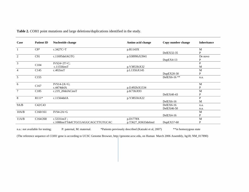
Table 2. COH1 point mutations and large deletions/duplications identified in the study. Case
Patient ID
Nucleotide change Amino acid change Copy number change Inheritance
1 C8* c.3427C>T p.R1143X DelEX32-35
M P
2 C91 c.11695delAGTG p.S3899fsX3941 DupEX4-13
De novo P
3 C104 IVS24+2T>C; c.11556insT
p.V3853fsX32 P
M 4 C145 c.402insT p.L135fsX145
DupEX20-30 M P
5 C155 DelEX6-16 ** n.a.
6 C167 IVS14-2A>G; c.4474delA
p.I1492fsX1534 M
P 7 C185 c.219_20delACinsT p.K73fsX93
DelEX40-43 M P
8 R111* c.11564delA p.Y3855fsX22 DelEX6-16
P M
9A/B C42/C43
DelEX6-16 DelEX46-50
n.a. n.a.
10A/B C160/161 IVS4-2A>G DelEX4-16
M P
11A/B C164/268 c.5331insT ; c.1088insTTdelCTGCGAGGCAGCTTGTGCAC
p.D1778X p.T3627_H3633delinsI
DupEX57-60
M P
n.a.: not available for testing; P: paternal, M: maternal. *Patients previously described (Katzaki et al, 2007) **in homozygous state (The reference sequence of COH1 gene is according to UCSC Genome Browser, http://genome.ucsc.edu, on Human March 2006 Assembly, hg18; NM_017890)


Table 3. Haplotype analysis in patients harbouring the recurrent exons 6-16 deletion.
Marker Position (Mb) C37 (Greek) Case 5 (Italian) Case 9A (Italian) Case 8 (Italian)
D8S1018 97,598 315 319 315 315 319 323 319 315 D8S257 99,451 109 109 109 109 109 - 109 113 8-23TC 99,924 214 214 214 214 214 218 214 204 8-25GT 100,056 353 353 353 353 353 379 353 351 8-20TG 100,601 169 169 169 169 169 173 169 173 VPS13B - del6_16 del6_16 del6_16 del6_16 del6_16 del46_49 del6_16 Y3855fsX22
D8S1789* 100,738 255 255 255 255 255 255 255 255 D8S470* 100,743 226 226 226 226 226 226 226 226 D8S300 100,987 485 485 485 485 485 499 485 499 8-18AC 101,066 95 95 95 95 95 97 95 97 D8S398 101,588 141 141 141 141 137 141 137 141
* intragenic markers grey columns: haplotype co-segregating with the deletion (The reference sequence of COH1 gene is according to UCSC Genome Browser, http://genome.ucsc.edu, on Human March 2006 Assembly, hg18; NM_017890)

TITLES AND LEGENDS TO FIGURES
Figure 1. Clinical features of Cohen syndrome patients. Note the typical facial gestalt of patients 3,
4, 5, 6, 10A, 10B and 11A. Frontal views of patients 2, 3, 4 and 6, showing truncal obesity.
Figure 2. MLPA analysis results showing the recurrent deletion in heterozygous (Case 8) and
homozygous state (Case 5). A) Electropherograms obtained with P321-A1 kit (on the left) and
P322-A1 kit (on the right) for a normal control sample, patient 8 and patient 5. Numbers and arrows
indicate the exon probes with reduced fluorescence signals respect to the control sample. In patient
8, the signal is half-reduced for probes 7-16, while in patient 5 there is no signal for the same
probes. B) Peak area histograms for patient 8 and 5 normalized with the control sample. Exon
dosage is reported on the y axis (normal values spannning from 0.7 to 1.3 are indicated with broken
lines). MLPA analysis shows reduced peak area for exons 7-16, compatible with a heterozygous
deletion in patient 8 and with a homozygous deletion in patient 5. Deletions are indicated with the
heavy black line.
Figure 3. Quantitative PCR results for exon 6 of COH1. ddCT ratios and standard deviations of a
normal control sample (c1), a deleted control sample (c2) and patients 5, 8, 9A and 10A. Compared
to controls, patients 8, 9A and 10A show ddCT ratio values of about 0.5, indicating a deletion in
heterozygous state, while patient 5 shows ddCT ratio values of about 0.0, indicating a deletion in
homozygous state.
Figure 4. MLPA analysis results showing the duplication spanning exons 57-60 in the familial case
with an affected brother (11A) and sister (11B). A) Electropherograms obtained with P321-A1 kit

(on the left) and P322-A1 kit (on the right) for a normal control sample and patient 11A. Numbers
and arrows indicate the exon probes with increased fluorescence signals respect to the control
sample. B) Peak area histograms for patient 11A normalized with the control sample. The exon
dosage is reported on the y axis (normal values spannning from 0.7 to 1.3 are indicated with broken
lines). The consistent increase of the peak area for exons 57-60 is compatible with a duplication of
these exons (indicated with the heavy black line).
Figure 5. Characterisation of duplication 57-60 in familial case 11 a) Schematic drawing of the
duplicated region. The star indicates the position of the MLPA probe in exon 61, while the thunder
represents the insertion point of the duplicated segment. Arrows indicate the primers located within
intron 59 and 56 used in the long-range PCR experiment. b) Sequence analysis showing the junction
between intron 56 and exon 61. c) Aligned exon 61 and intron 56 sequences at the duplication
junction. Region of homology across the duplication junction is boxed.
Supplementary Figure 1. COH1 multi-exon deletions and duplications detected by MLPA in
patients 1, 2 and 4. Normalised relative peak areas of all COH1 gene-specific probes are shown.
Sequences present in two copies of the genome have a relative peak area value of approximately
1.0. A reduction in the peak area values to <0.7 indicates a deletion (heavy black line) while an
increase in the peak area values to > 1.3 indicates a duplication (heavy black line). Normal values
are indicated with broken lines.
Supplementary Figure 2. COH1 multi-exon deletions detected by MLPA in patients 7, 9A and
10A. Normalised relative peak areas of all COH1 gene-specific probes are shown. Sequences
present in two copies of the genome have a relative peak area value of approximately 1.0. A

reduction in the peak area values to <0.7 indicates a deletion (heavy black line). Normal values
spanning from 0.7 to 1.3 are indicated with broken lines. In patient 7, the half-reduced signal for
exon 3 is due to the point mutation c.219_20delACinsT located exactly on the probe sequence.
Copyright © 2022 FDOKUMEN