Elevated Hypothalamic TCPTP in Obesity Contributes to Cellular Leptin Resistance
Upload
independentCategory
view
3download
0
Deletion of Inducible Nitric-Oxide Synthase in Leptin-Deficient Mice Improves Brown Adipose Tissue FunctionSara Becerril1,3, Amaia Rodrıguez1,3, Victoria Catalan1,3, Neira Sainz1,3, Beatriz Ramırez1,3, Marıa
Collantes4, Ivan Penuelas4, Javier Gomez-Ambrosi1,3, Gema Fruhbeck1,2,3*
1 Metabolic Research Laboratory, Clınica Universidad de Navarra, Pamplona, Spain, 2 Department of Endocrinology, Clınica Universidad de Navarra, Pamplona, Spain,
3 CIBER Fisiopatologıa de la Obesidad y Nutricion, Instituto de Salud Carlos III, Madrid, Spain, 4 MicroPET Research Unit CIMA-CUN, University of Navarra, Pamplona, Spain
Abstract
Background: Leptin and nitric oxide (NO) on their own participate in the control of non-shivering thermogenesis. However,the functional interplay between both factors in this process has not been explored so far. Therefore, the aim of the presentstudy was to analyze the impact of the absence of the inducible NO synthase (iNOS) gene in the regulation of energybalance in ob/ob mice.
Methods and Findings: Double knockout (DBKO) mice simultaneously lacking the ob and iNOS genes were generated, andthe expression of molecules involved in the control of brown fat cell function was analyzed by real-time PCR, western-blotand immunohistochemistry. Twelve week-old DBKO mice exhibited reduced body weight (p,0.05), decreased amounts oftotal fat pads (p,0.05), lower food efficiency rates (p,0.05) and higher rectal temperature (p,0.05) than ob/ob mice.Ablation of iNOS also improved the carbohydrate and lipid metabolism of ob/ob mice. DBKO showed a marked reduction inthe size of brown adipocytes compared to ob/ob mutants. In this sense, in comparison to ob/ob mice, DBKO rodents showedan increase in the expression of PR domain containing 16 (Prdm16), a transcriptional regulator of brown adipogenesis.Moreover, iNOS deletion enhanced the expression of mitochondria-related proteins, such as peroxisome proliferator-activated receptor c coactivator-1 a (Pgc-1a), sirtuin-1 (Sirt-1) and sirtuin-3 (Sirt-3). Accordingly, mitochondrial uncouplingproteins 1 and 3 (Ucp-1 and Ucp-3) were upregulated in brown adipose tissue (BAT) of DBKO mice as compared to ob/obrodents.
Conclusion: Ablation of iNOS improved the energy balance of ob/ob mice by decreasing food efficiency through an increasein thermogenesis. These effects may be mediated, in part, through the recovery of the BAT phenotype and brown fat cellfunction improvement.
Citation: Becerril S, Rodrıguez A, Catalan V, Sainz N, Ramırez B, et al. (2010) Deletion of Inducible Nitric-Oxide Synthase in Leptin-Deficient Mice Improves BrownAdipose Tissue Function. PLoS ONE 5(6): e10962. doi:10.1371/journal.pone.0010962
Editor: Jose A. L. Calbet, University of Las Palmas de Gran Canaria, Spain
Received January 8, 2010; Accepted May 16, 2010; Published June 4, 2010
Copyright: � 2010 Becerril et al. This is an open-access article distributed under the terms of the Creative Commons Attribution License, which permitsunrestricted use, distribution, and reproduction in any medium, provided the original author and source are credited.
Funding: SB was recipient of doctoral training grants from the Departments of Industry and Education of the Gobierno de Navarra. This work was supported bygrants from the Instituto de Salud Carlos III, Fondo de Investigacion Sanitaria (FIS) del Ministerio de Sanidad y Consumo to G.F. and J.G.A; from the Ministerio deCiencia y Tecnologi-a to G.F.; from the Department of Health of the Gobierno de Navarra to G.F. and J.G.A.; and from PIUNA foundation to G.F. CIBER deFisiopatologia de la Obesidad y Nutricion (CIBEROBN) is an initiative of the Instituto de Salud Carlos III, Spain. The funders had no role in study design, datacollection and analysis, decision to publish, or preparation of the manuscript.
Competing Interests: The authors have declared that no competing interests exist.
* E-mail: [email protected]
Introduction
Energy homeostasis is a highly regulated process that requires a
tight balance between caloric intake and energy expenditure [1].
The latter is a key determinant of energy balance and includes
three components: basal metabolic rate, physical activity, and
adaptive thermogenesis [2,3]. In this sense, brown adipose tissue
(BAT) constitutes a highly active metabolic organ that plays a
crucial role in non-shivering thermogenesis, defined as the heat
production in response to cold or overfeeding [4]. Until recently,
BAT was thought to be important only in small mammals and
newborn humans. However, functional BAT was recently
identified in adults, suggesting a role in human metabolism
[5,6]. In brown adipocytes, thermogenesis is mainly mediated by
sympathetically innervated b3-adrenergic receptors, leading to the
activation of the BAT-specific uncoupling protein-1 (Ucp-1). This
protein is a proton transporter located in the inner mitochondrial
membrane that diverts the energy from the mitochondrial
respiratory chain from ATP synthesis to heat production [7].
The Ucp-1 promoter is regulated by several transcriptional
coactivators, including the peroxisome proliferator-activated
receptor c (PPARc) coactivator-1 a (Pgc-1a), being also involved
in the regulation of crucial aspects of energy metabolism [8,9].
Pgc-1a is strongly induced in murine BAT during cold exposure
activating the thermogenic gene program of brown fat through the
control of the gene expression levels of Ucp-1 and Pgc-1a itself. In
this regard, it has been recently described that during BAT
differentiation PR domain containing 16 (Prdm16) directly binds
to Pgc-1a, allowing the activation of Ucp-1 and other brown fat-
specific genes [10,11]. Moreover, it has been demonstrated that
the NAD-dependent deacetylase sirtuin-1 (Sirt-1) deacetylates and
activates Pgc-1a in the liver and BAT [12,13], allowing its union
PLoS ONE | www.plosone.org 1 June 2010 | Volume 5 | Issue 6 | e10962
to target genes and increasing the rate of gene transcription. The
key role of the correpresor of nuclear receptor-interacting protein
1 (Nrip), also known as receptor-interacting protein 140 (Rip140), in
energy homeostasis by suppressing the transcription of Ucp-1 and
other metabolic genes has been also reported [14,15].
Leptin, the product of the ob gene, plays a key role in the control
of body weight by suppressing food intake through actions on
hypothalamic receptors and by increasing energy expenditure via
the activation of the sympathetic nerve activity and the turnover of
norepinephrine in BAT [16,17]. Leptin induces the gene
expression of Pgc-1a and Ucp-1 through the stimulation of b3-
adrenergic receptors, thereby leading to an increased thermogen-
esis [18–21]. In this sense, it has been shown that leptin-deficient
ob/ob mice are obese, hyperphagic and exhibit reduced non-
shivering thermogenesis as well as low UCP-1 levels in BAT [22].
Previous studies showed that norepinephrine increases the blood
flow in BAT by stimulating the production of nitric oxide (NO), a
potent vasodilator [23]. NO is produced by NO synthase (NOS),
and three isoforms have been identified: the endothelial (eNOS)
and neuronal (nNOS), which are constitutively expressed, together
with the inducible NOS (iNOS), which is primarily transcription-
ally regulated by immunologic as well as inflammatory stimuli
[24]. Both eNOS and iNOS isoforms have been shown to be
expressed in brown adipocytes [25], providing evidence for the
involvement of NO in BAT function regulation.
The deletion of the iNOS gene reportedly prevents high-fat diet-
induced insulin resistance [26]. Furthermore, leptin and iNOS on
their own participate in multiple common physiological processes,
with a functional relationship between both factors having been
described earlier by our group [27–29] and others [30,31]. In
order to explore the functional interplay between both factors and
to better understand the regulatory pathways that govern energy
metabolism, we examined the effects of iNOS gene disruption in
genetically obese ob/ob mice on the diverse elements of energy
balance focusing particularly on the expression of non-shivering
thermogenesis-related molecules. Our study shows that deletion of
the iNOS gene decreases food efficiency through an increase in
thermogenesis, thus improving the energy balance of ob/ob mice.
Materials and Methods
Generation of double-knockout mice lacking the ob andiNOS genes
A double knockout (DBKO) mouse simultaneously lacking the
ob and the iNOS genes was generated by intercrossing male ob/ob
mice with female iNOS knockout mice (iNOS2/2) on a C57BL/6J
background (Jackson Laboratories, Bar Harbor, ME, USA).
Noteworthy, ob/ob male mice were placed under caloric restriction
(2 g standard chow diet/day) and were daily injected with
recombinant leptin (2 g/kg body weight) (Bachem, Bubendorf,
Switzerland) in order to overcome the infertility problems of the
leptin-deficient rodents [32]. Genotyping for ob and iNOS was
performed as previously described [32,33]. Briefly, genomic DNA
was extracted from ear clips by using the DNeasy Mini kit
(Qiagen, Valencia, CA, USA) according to the manufacturer’s
instructions. The genotyping strategy utilized the Dde I restriction
site generated by the ob mutation. To identify the presence of the
wild type or the disrupted iNOS allele, three different primers were
used. One primer complementary to both the genomic and
disrupted alleles that amplified a 108-bp fragment with the second
primer specific for the genomic sequence and a 275-bp fragment
with the third primer complementary to a region of the neomycin
resistance insert specific to the disrupted iNOS allele [34]. The
PCR was performed as described elsewhere [33] and the PCR
products were separated on a 1.5% agarose gel and visualized with
ethidium bromide staining.
Male mice were weaned at 21 days of age, genotyped, and
maintained at a room temperature of 2262uC on a 12:12 light-
dark cycle (lights on at 08:00 am) with a relative humidity of
50610% and under pathogen-free conditions. Animals had free
access to tap water and were fed ad libitum with a normal chow diet
(2014S Teklad Global 14% Protein Rodent Maintenance Diet,
Harlan, Barcelona, Spain). Body weight and food intake were
registered twice weekly. The food efficiency was determined as
body weight gained per week divided by total energy (kilocalories)
consumed over this period [35]. Body temperature was assessed by
measuring rectal temperature using a rectal thermoprobe (YSI
4600 Series Precision Thermometers, YSI Temperature, Dayton,
OH, USA). The diameter of adipocytes was determined by direct
microscopy, and the cell size was obtained using digital
photographs with the Axiovision 4.6 program (Zeiss). Mice were
injected in the tail vein with 18F-fluorodeoxyglucose (18F-FDG)
(200 mCi) after a short isoflurane (4%) anesthesia period. The
uptake of 18F-FDG was analyzed by a Mosaic (Philips Electronics,
Amsterdam, The Netherland) small-animal dedicated imaging
tomograph as previously described [36].
Blood and tissue collectionTwelve-week-old mice were fasted for 6 hours and sacrificed by
CO2 inhalation. Blood samples were obtained by cardiac
puncture, and sera collected after cold centrifugation (4uC) at
700 g for 15 min and stored at 220uC. Epididymal, subcutaneous
and perirenal white adipose tissue together with brown fat from
the interscapular depots were carefully excised. Tissue samples
were immediately frozen at 280uC. Biopsies of BAT were also
formalin-fixed for immunohistochemical analyses. All experimen-
tal procedures conformed to the European Guidelines for the Care
and Use of Laboratory Animals (Directive 86/609) and the study
was approved by the Ethical Committee for Animal Experimen-
tation of the University of Navarra (042/03, 041/08).
Blood measurementsAn intraperitoneal glucose tolerance test was performed after an
overnight fasting period (12 h). Mice were injected intraperitoneally
with glucose (2 g/kg of body weight). Glucose concentrations were
measured before and 15, 30, 60, 90 and 120 min after the glucose
challenge. Glucose was determined by an automatic glucose sensor
(Ascensia Elite, Bayer, Barcelona, Spain) from whole blood obtained
from the tail vein. Serum glucose was measured by a glucometer
(Ascensia Elite). Serum concentrations of triglycerides, total cholesterol
(Infinity, Thermo Electron Corporation, Melbourne, Australia), free
fatty acids (FFA) (Wako Chemicals, GmbH, Neuss, Germany) and
glycerol (Sigma, St. Louis, MO, USA) were measured by enzymatic
methods using commercially available kits. Insulin and adiponectin
were determined by ELISA (Crystal Chem, Inc., Chicago, IL, USA
and BioVendor Laboratory Medicine, Inc., Modrice, Czech
Republic, respectively). Intra- and inter-assay coefficients of variation
for measurements of insulin and adiponectin were 3.5% and 6.3%,
respectively, for the former, and 5.6% and 7.2%, for the latter.
Western blot studiesTissues were homogenized and protein content was measured
as described earlier [37]. Equal amounts of protein (30 mg) were
run in 8% SDS-PAGE, subsequently transferred to nitrocellulose
membranes (Bio-Rad Laboratories, Inc., Hercules, CA, USA), and
blocked in Tris-buffered saline (TBS) with Tween 20 containing
5% non-fat dry milk for 1 h at room temperature (RT). Blots were
then incubated overnight at 4uC with primary antibodies against
ob/ob-iNOS2/2 & BAT Function
PLoS ONE | www.plosone.org 2 June 2010 | Volume 5 | Issue 6 | e10962
Ucp-1 and Ucp-3 (Abcam) at 1:10,000 and 1:8,000, respectively;
Pgc-1a (Cell Signalling Technology, Inc., Danvers, MA, USA) at
1:1,000; Sirt-1 (Abcam Ltd., Cambridge, UK) at 1:1,000; Sirt-3
(Cell Signalling Technology, Inc) at 1:1,000; or b-actin (Sigma) at
1:5,000. The antigen-antibody complexes were visualized using
peroxidase-conjugated anti-rabbit or anti-mouse IgG antibodies
(1:5,000) and the enhanced chemiluminescence ECL detection
system (Amersham Biosciences, Buckinghamshire, UK). The
intensity of the bands was determined by densitometric analysis
with the Gel DocTM gel documentation system and the Quantity One
4.5.0 software (Bio-Rad) and normalized with b-actin densitomet-
ric values. All assays were performed in duplicate.
Immunohistochemistry of Ucp-1 and Ucp-3The immunohistochemistry was carried out using the indirect
immunoperoxidase method. Sections (6 mm) of formalin-fixed
paraffin-embedded BAT were dewaxed in xylene, rehydrated in
decreasing concentrations of ethanol and treated with 3% H2O2
(Sigma) in absolute methanol for 10 min at RT to quench
endogenous peroxidase activity. Then, sections were immersed in
10 mmol/l citrate buffer (pH 6.00) and heated using a microwave
oven at 800 W for 15 min to enhance antigen retrieval. After
cooling, slides were blocked during 1 h with 1% murine serum
(Sigma) diluted in Tris-buffer saline (TBS) (50 mmol/l Tris,
0.5 mol/l NaCl; pH 7.36) for preventing non-specific adsorption.
Sections were incubated overnight at 4uC with rabbit monoclonal
anti-Ucp-1 or Ucp-3 antibodies (Abcam) diluted 1:100 in TBS. After
three washes (5 min each) with TBS, sections were incubated with
horseradish peroxidase-conjugated anti-rabbit IgG antibody (Amer-
sham Biosciences) diluted 1:200 in TBS for 30 min at RT. After
washing in TBS, the peroxidase reaction was visualized with a 3,39-
diaminobenzidine (DAB, Amersham Biosciences)/H2O2 solution
(0.5 mg/ml DAB, 0.03% H2O2 diluted in 50 mmol/l Tris-HCl,
pH 7.36) as chromogen, and Harris hematoxylin solution (Sigma) as
counterstaining. Sections were dehydrated, coverslipped and
observed under a Zeiss Axiovert 40 CFL optic microscope (Zeiss,
Gottingen, Germany). Negative control slides without primary
antibody were included for the assessment of non-specific staining.
RNA extraction and Real-Time PCRTotal RNA was extracted from BAT samples by homogeniza-
tion with an ULTRA-TURRAXH T 25 basic (IKAH Werke
GmbH, Staufen, Germany) using TRIzolH Reagent (Invitrogen,
Barcelona, Spain). Samples were purified with the RNeasy Mini
kit (Qiagen) according to the manufacturer’s instructions and
treated with DNase I (RNase-free DNase Set, Qiagen). For first
strand cDNA synthesis constant amounts of 2 mg of total RNA
were reverse transcribed in a 40 ml final volume using random
hexamers (Roche Molecular Biochemicals, Mannheim, Germany)
as primers and 400 units of M-MLV reverse transcriptase
(Invitrogen) as described earlier [38].
The transcript levels for genes involved in brown fat cell
differentiation and function (Prdm16, Rip140, Bmp7, Sirt-1, Sirt-3,
Pgc-1a, Ucp-1 and Ucp-3) were quantified by Real-Time PCR (7300
Real Time PCR System, Applied Biosystems, Foster City, CA,
USA). Primers and probes were designed using the software Primer
Express 2.0 (Applied Biosystems) (Table 1) and purchased from
Genosys (Sigma). TaqManH probes encompassing fragments of
areas from the extremes of two exons were designed to ensure the
detection of the corresponding transcript avoiding genomic DNA
amplification. The cDNA was amplified at the following conditions:
95uC for 10 min, followed by 45 cycles of 15 s at 95uC and 1 min at
59uC, using the TaqManH Universal PCR Master Mix (Applied
Biosystems). The primer and probe concentrations for gene
amplification were 300 nmol/l and 200 nmol/l, respectively. All
results were normalized to the levels of 18S rRNA (Applied
Biosystems) and relative quantification was calculated using the
DDCt formula [38]. Relative mRNA expression was expressed as
fold expression over the calibrator sample (average of gene
expression corresponding to the wild type group) [39]. All samples
were run in triplicate and the average values were calculated.
Statistical analysisData are presented as the mean 6 SEM. Differences between
groups were assessed by two-way ANOVA. In case of interaction
between factors (lack of iNOS or ob genes), one-way ANOVA
followed by Tukey’s post hoc tests were applied. Statistics were
calculated by the SPSS/Windows version 15.0 software (SPSS,
Inc., Chicago, IL, USA). A P value less than 0.05 was considered
statistically significant.
Results
Ablation of iNOS reduces the positive energy balance ofob/ob mice
As expected, leptin deficiency was associated with increased
(p,0.001) body weight, higher fat depots, hyperphagia and
hypothermia, whereas iNOS deficiency resulted in reduced
(p,0.01) body weight and lower fat content as well as increased
(p,0.01) body temperature as compared to control mice (Fig. 1).
The ob/ob mice lacking the iNOS gene (DBKO) exhibited a
decreased (p,0.01) body weight accompanied by significantly
(p,0.01) smaller epididymal, subcutaneous and total fat depots as
compared to ob/ob mice. The weight gain of DBKO mice during
the study was significantly reduced (p,0.0001) as compared to ob/
ob mice (25.160.6 g vs 30.860.7 g). Twelve week-old DBKO mice
showed a reduced (p,0.01) food intake (Fig 1C) exhibiting a lower
food efficiency as compared to ob/ob mice (Fig 1D). Basal rectal
temperature was analyzed showing that iNOS deficiency improved
the reduced rectal temperature of ob/ob mice (p,0.05) (Fig 1E). In
a subset of mice, a glucose tolerance test was performed. The
glucose areas under the curves (AUC) were measured using the
trapezoidal method. The glucose AUC in ob/ob mice was
significantly higher (p,0.001) than that of wild type mice.
Moreover, deletion of the iNOS gene significantly decreased the
glucose AUC (p,0.05) in wild type and ob/ob mice (wild type
540627, iNOS2/2 474629, ob/ob 706671, DBKO 618690). As
can be observed in Table 2, absence of leptin was associated with
insulin resistance as evidenced by the increased concentrations of
glucose, insulin, glucose AUC and HOMA index as well as by low
adiponectin levels (p,0.01). iNOS deletion significantly reduced
FFA and cholesterol concentrations in wild type and ob/ob mice
(p,0.01). DBKO mice exhibited a tendency towards an improved
insulin sensitivity as compared to ob/ob mice as evidenced by the
reductions in insulin, HOMA index (p = 0.071 and p = 0.069,
respectively) and glucose AUC.
Brown adipose tissue phenotype of ob/ob mice lackingthe iNOS gene
The weight of interscapular BAT was increased (p,0.001) in
leptin-deficient mice (wild type 0.3160.02, ob/ob 0.8060.04 g/
100 g body weight). Deletion of iNOS slightly decreased the weight
of BAT in wild type (wild type 0.3160.02, iNOS2/2 0.2960.01 g/
100 g body weight) and ob/ob mice (ob/ob 0.8060.04, DBKO
0.7560.04 g/100 g body weight) although the differences did not
reach statistical significance (Fig. 2B). However, the cross-sectional
area of brown adipocytes of experimental animals was determined
and as expected showed small and multilocular lipid droplets in
ob/ob-iNOS2/2 & BAT Function
PLoS ONE | www.plosone.org 3 June 2010 | Volume 5 | Issue 6 | e10962

control mice, whereas ob/ob mutants exhibited large and
unilocular lipid droplets (Fig. 2A). The deletion of the iNOS gene
in ob/ob mice dramatically reduced the size of brown adipocytes.
In this sense, DBKO mice displayed a higher proportion of small
multilocular adipocytes together with a lower proportion of larger
unilocular ‘‘white-like’’ adipocytes as corroborated by the cell
surface area (p,0.001) (Fig. 2C). As seen in Fig. 2D, microPET
scans revealed a markedly enhanced 18F-FDG uptake in the
interscapular BAT of DBKO mice compared to ob/ob animals.
Up-regulation of brown adipocyte function markers inob/ob mice lacking the iNOS gene
The mRNA and protein expression levels of molecules involved
in the regulation of thermogenesis and mitochondrial function
were analyzed in BAT of the experimental animals. As shown in
Fig. 3, the gene and protein expression levels of Ucp-1 and Ucp-3
were down-regulated in ob/ob mice and up-regulated in iNOS-
deficient mice as compared to those of wild type mice.
Noteworthy, DBKO mice simultaneously lacking the ob and iNOS
genes showed a statistically significant increase in Ucp-1 (p,0.05)
and Ucp-3 (p,0.05) transcripts and proteins compared to ob/ob
mice. Immunohistological analyses showed a high expression of
Ucp-1 and Ucp-3 in BAT in all experimental groups. Nonetheless,
the immunostaining of both proteins was markedly increased in
iNOS knockout and DBKO mice, and decreased in ob/ob animals
as compared to wild types.
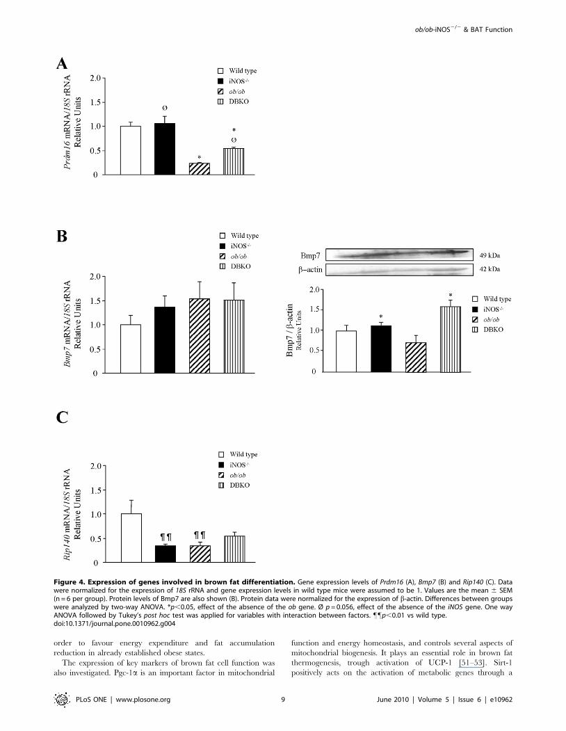
ob/ob mice lacking iNOS display changes in geneexpression levels of molecules involved in brown fat celldifferentiation
To gain further insight into the mechanisms underlying the
improved energy expenditure of DBKO mice, the gene expression
levels of key molecules involved in brown fat cell differentiation
were examined (Fig. 4). The gene expression levels of Prdm16, a
zinc-finger protein that stimulates brown fat-selective gene
expression, was significantly down-regulated (p,0.001) in ob/ob
mice, while the deletion of iNOS increased mRNA expression
levels although only a marginal statistical significance was found
(p = 0.056). No changes in the mRNA expression levels of Bmp7, a
protein involved in the activation of the program of brown
adipogenesis, was observed. Nevertheless, protein expression levels
of Bmp7 were increased in mice lacking the iNOS gene. Moreover,
gene expression levels of Rip140, a nuclear receptor involved in the
differentiation of white adipocytes, was significantly decreased
(p,0.01) in iNOS-deficient mice.
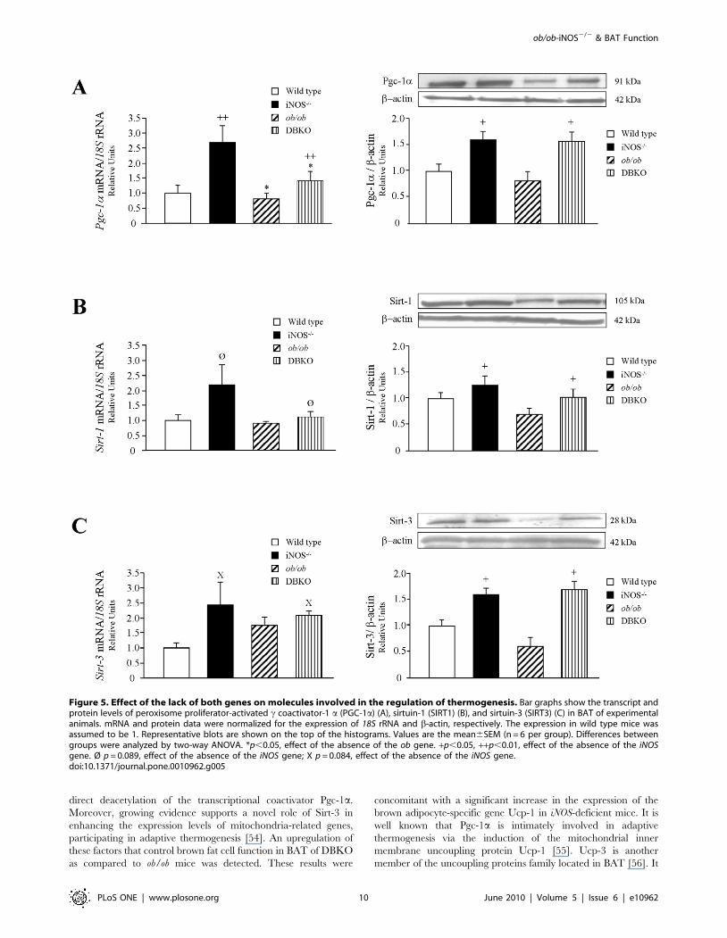
The mRNA and protein expression levels of molecules involved
in the regulation of mitochondrial function and thermogenesis
were also analyzed. Leptin deficiency was associated with a
reduction of Pgc-1a transcript levels, together with a tendency
towards a decrease in Sirt-1 transcript levels, without changes in
the gene expression levels of Sirt-3 (Fig. 5). On the contrary, iNOS
knockout mice showed an increase in mRNA levels of Pgc-1 a at
the same time as an increase in Sirt-1 and Sirt-3 transcript levels,
although only a marginal statistical significance was found. The
DBKO mice showed an up-regulation of Pgc-1 a (p,0.01) and a
marginal increase in Sirt-1 and Sirt-3 as compared to the ob/ob
group, although in the case of the sirtuins differences were not
statistically significant. The protein expression of Pgc-1a, Sirt-1
and Sirt-3 in BAT exhibited a similar pattern to that observed in
the gene expression analyses.
Discussion
The involvement of leptin and iNOS in the control of energy
balance through actions on food intake, body weight and energy
expenditure has been previously reported [16,33,40]. Leptin-
deficient mice exhibit marked obesity, hyperphagia, insulin
resistance, hypothermia and increased food efficiency [40], whereas
iNOS knockout mice are resistant to diet-induced obesity, showing
reduced epididymal fat pads and increased body temperature
[26,33]. Although the iNOS deficiency did not completely restore
the phenotype of the absence of leptin, our results show that deletion
of the iNOS gene exerts a significant impact on energy homeostasis
via increasing energy expenditure and decreasing food intake. We
observed that the absence of leptin leads to obesity even in the
context of iNOS deficiency. The DBKO mice showed a modest, but
consistent lower body weight than that of ob/ob mice at the end of
the study. The data shows that from the eleventh week of the study
Table 1. Sequences of the primers and TaqManH probes.
Gene (GenBankaccession number) Oligonucleotide sequence (59-39)
Prdm16 (NM_027504)
Forward GATGGGAGATGCTGACGGATAC
Reverse CTCGCTACCCAAGTCTTCAGAGAT
TaqManH Probe FAM- CATCCCAGGAGAGCTGATCAAAAAGC-TAMRA
Rip140 (NM_173440)
Forward TCAGCTTCCTTTCCCACATAGC
Reverse TCATCTTTCGTTGCTCACCAAA
TaqManH Probe FAM-AGGCTCAGGCTGAGGCAGACGATACT-TAMRA
Bmp7 (NM_07557)
Forward CAAGACGCCAAAGAACCAAGAG
Reverse GGTCTCGGAAGCTGACGTACAG
TaqManH Probe FAM-ATGGCCAGTGTGGCAGAAAACAGCA-TAMRA
Sirt-1 (NM_019812)
Forward AGCAGGTTGCAGGAATCCAA
Reverse CACGAACAGCTTCACAATCAACTT
TaqManH Probe FAM-CCTTCAGTGTCATGGTTCCTTTG-TAMRA
Sirt-3 (NM_001127351)
Forward CTGACTTCGCTTTGGCAGATCT
Reverse CCCCACCAAGTCTCGATTGAT
TaqManH Probe FAM-CTGGAGGTGGAGCCTTTTGCCAGCT-TAMRA
Pgc-1a (NM_008904)
Forward TGAACGCACCTTAAGTGTGGAA
Reverse GGGTTATCTTGGTTGGCTTTATGA
TaqManH Probe FAM-ATCGCAGGCCTAACTCCACCCACCA -TAMRA
Ucp1 (NM_009463)
Forward CGATGTCCATGTACACCAAGGA
Reverse ACCCGAGTCGCAGAAAAGAAG
TaqManH Probe FAM-ACCGACGGCCTTTTTCAAAGGGTTTG-TAMRA
Ucp3 (NM_001030877)
Forward GACCTACGACATCATCAAGGAGAAGT
Reverse CTCCAAAGGCAGAGACAAAGTGA
TaqManH Probe FAM-6TCTCACCTGTTTACTGACAACTTCCC-TAMRA
Prdm16, PR domain containing 16; Rip140, receptor-interacting protein 140;Bmp7, bone morphogenetic protein 7; Sirt-1, sirtuin-1, Sirt-3, sirtuin-3; Pgc-1a,peroxisome proliferative activated receptor c coactivator 1 a; Ucp-1, uncouplingprotein 1, Ucp-3, uncoupling protein 3.doi:10.1371/journal.pone.0010962.t001
ob/ob-iNOS2/2 & BAT Function
PLoS ONE | www.plosone.org 4 June 2010 | Volume 5 | Issue 6 | e10962

Figure 1. Growth and metabolic variables of mice of the four experimental groups. Growth curves of 4–12 week-old-mice (A) togetherwith epididymal (EWAT), subcutaneous (SCWAT) and whole-body fat content (B) of the experimental animals. Cumulative food intake (C), foodefficiency (D) and rectal temperature (E) are also shown. Representative images illustrating the differences in size between 12-week-old ob/ob andDBKO mice (F). Values are the mean 6 SEM (n = 10 per group). Differences between groups were analyzed by two-way ANOVA. ***p,0.001, effect ofthe absence of the ob gene. +p,0.05, ++p,0.01, effect of the absence of the iNOS gene. One-way ANOVA followed by Tukey’s post hoc test wasapplied for variables with interaction between factors. """p,0.001 vs wild type, #p,0.05 vs ob/ob mice. EWAT, epididymal white adipose tissue;SCWAT, subcutaneous white adipose tissue; WAT, white adipose tissue; bw, body weight.doi:10.1371/journal.pone.0010962.g001
ob/ob-iNOS2/2 & BAT Function
PLoS ONE | www.plosone.org 5 June 2010 | Volume 5 | Issue 6 | e10962
onwards DBKO mice weigh significantly less than ob/ob mutants.
Moreover, these differences were maintained and increased from
this time point throughout longer experimental periods (32 weeks,
data not shown). The possibility that DBKO mice weigh less at the
initial weeks of the study and therefore, the increase in body weight
may be similar to that of ob/ob mice at the end of the experimental
period is ruled out since from the beginning the DBKO exhibited a
slightly higher body weight than ob/ob animals. Thereafter, the
growth curve of ob/ob mice continues to increase steadily while that
of DBKO slows down. Body weights of both experimental groups
are superimposed from 8–10 weeks. During that period the growth
curves of both groups intercross with ob/ob mice becoming heavier
than DBKO mutants. This leads towards the end of the study to a
more evident reduction in body weight meaning that the lack of
iNOS is exerting an impact on body weight in ob/ob mice. The
differences in body weight were attributable in part to a reduction in
food intake. We describe, for the first time, that the disruption of the
iNOS gene reduces the elevated food intake and food efficiency,
partially ameliorating the obesity of ob/ob mice. It has been
proposed that leptin-deficient mice show higher food efficiency rates
at least in part due to an impaired capacity of BAT to produce heat
[2,41]. In this sense, our data confirm this observation and further
show that iNOS-deficient and DBKO mice exhibit an increase in
rectal temperature. Taken together, ablation of iNOS improves the
energy balance of ob/ob mice by decreasing energy intake (reduced
food intake), and by increasing energy expenditure (increased rectal
temperature). It is well known that ob/ob mice have an increased
respiratory quotient (RQ) due to a reduced fat oxidation with leptin
replacement normalizing the metabolic rate in these animals. iNOS
knockout mice probably exhibit a reduced RQ due to their
increased b-oxidation in brown adipose tissue, as reflected by their
reduction in circulating free fatty acids. It seems reasonable that the
absence of iNOS may be associated to an increase in energy
expenditure given that iNOS and DBKO mice exhibit a reduced
body weight and fat mass as compared to their respective controls.
This is supported by published studies [42,43] evidencing that nitric
oxide inhibits cytochrome c oxidase, thus inhibiting the mitochon-
drial respiratory chain. The participation of NO in thermoregula-
tion is based on its vasodilator properties and its regulatory role in
non-shivering thermogenesis. Previous studies were focused on the
existence of two isoforms of NOS in BAT: eNOS and iNOS. A role
for iNOS in the regulation of sympathetically-mediated blood flow
in brown adipose tissue has been described, arguing against an
increased energy expenditure in this tissue in iNOS knockout mice
[23]. Furthermore, eNOS-deficient mice show a reduced energy
expenditure and an increased body weight [44]. On the other hand,
it has been reported that NO downregulates the expression of UCPs
in adipocytes [45] with UCPs being heavily involved in energy
expenditure regulation. The lack of iNOS may decrease NO
production in adipose tissue in an autocrine/paracrine way, thereby
increasing the expression of UCPs and hence energy expenditure.
The observed increase in the expression of Ucp-1 and Ucp-3 in
BAT of iNOS-deficient mice in the present study supports this
mechanism. However, NO involvement in the regulation of energy
expenditure is complex and may exhibit NOS-specific differences.
On the other hand, ob/ob mice exhibit a reduced locomotor activity,
which is normalized after leptin administration. The fact that the
proposed rise in energy expenditure in iNOS-deficient mice is due to
an increased locomotor activity may be excluded given the fact that
NO has been reported to induce locomotor activity in mice [46]
while eNOS-deficient mice show normal locomotor activity [44].
Regular observation of the DBKO and iNOS2/2 mice during the
whole experimental period of our study did not identify qualitive or
semiquantitative changes in locomotor activity and behaviour
between the different rodents. In this sense, the preponderance of an
effect on body temperature in the absence of changes in locomotor
activity may be put forward. Undoubtedly, the detailed analysis of
the potential impact on both energy expenditure and locomotor
activity with sophisticated equipment to pick up slight differences
would merit a study on its own to clarify the exact contribution of
each component.
The activation of BAT has been evidenced to play an important
role in energy expenditure [47]. Previous studies have reported a
‘‘white-like’’ appearance of BAT in ob/ob mice, suggesting a
crucial role of leptin in the development of brown adipocytes [22].
In the histological analyses, large unilocular lipid droplets were
observed in BAT of leptin-deficient mice. We also showed that in
the DBKO mice the characteristic features of BAT tissue (both
macro and microscopically as well as molecularly) are partially
restored. To gain further insight into the mechanisms underlying
the change in the phenotype of BAT of the experimental models,
we focused on the transcriptional control of the metabolic
pathways, achieved by the coordinated actions of numerous
transcription factors and associated coregulators or corepressors.
In this sense, an increase in the gene expression levels of the
recently identified transcription factor PRDM16, a positive
transcriptional regulator of the brown fat cell gene program [10]
was observed in iNOS-deficient mice. We also studied the
expression of a member of the family of the bone morphogenetic
proteins (BMPs), Bmp7, that reportedly regulates energy homeo-
stasis by activating a full program of brown adipogenesis [48].
iNOS deficiency enhanced Bmp7 protein expression. Moreover,
the corepressor Rip140, that plays a key role in energy homeostasis
by repressing metabolic gene networks [49], was dramatically
reduced in iNOS knockout and ob/ob mice. These data are in line
with previous studies in humans [50], supporting the notion that
downregulation of Rip140 may be a compensatory mechanism in
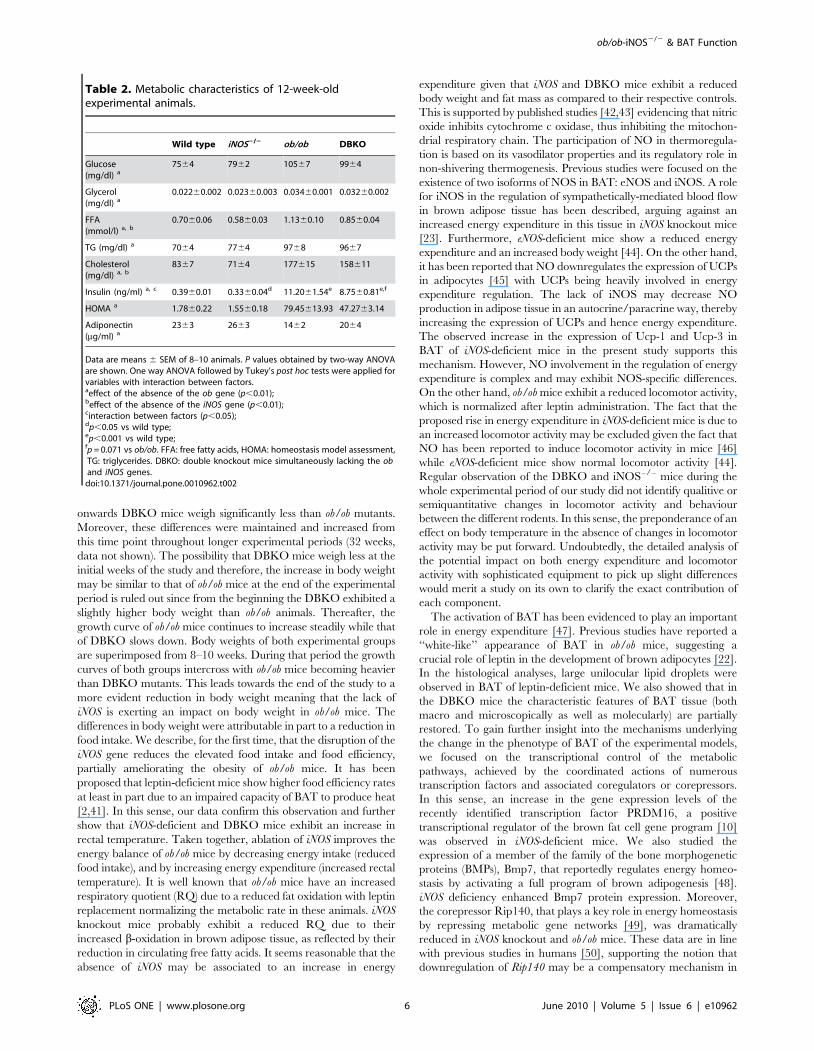
Table 2. Metabolic characteristics of 12-week-oldexperimental animals.
Wild type iNOS2/2 ob/ob DBKO
Glucose(mg/dl) a
7564 7962 10567 9964
Glycerol(mg/dl) a
0.02260.002 0.02360.003 0.03460.001 0.03260.002
FFA(mmol/l) a, b
0.7060.06 0.5860.03 1.1360.10 0.8560.04
TG (mg/dl) a 7064 7764 9768 9667
Cholesterol(mg/dl) a, b
8367 7164 177615 158611
Insulin (ng/ml) a, c 0.3960.01 0.3360.04d 11.2061.54e 8.7560.81e,f
HOMA a 1.7860.22 1.5560.18 79.45613.93 47.2763.14
Adiponectin(mg/ml) a
2363 2663 1462 2064
Data are means 6 SEM of 8–10 animals. P values obtained by two-way ANOVAare shown. One way ANOVA followed by Tukey’s post hoc tests were applied forvariables with interaction between factors.aeffect of the absence of the ob gene (p,0.01);beffect of the absence of the iNOS gene (p,0.01);cinteraction between factors (p,0.05);dp,0.05 vs wild type;ep,0.001 vs wild type;fp = 0.071 vs ob/ob. FFA: free fatty acids, HOMA: homeostasis model assessment,TG: triglycerides. DBKO: double knockout mice simultaneously lacking the oband iNOS genes.
doi:10.1371/journal.pone.0010962.t002
ob/ob-iNOS2/2 & BAT Function
PLoS ONE | www.plosone.org 6 June 2010 | Volume 5 | Issue 6 | e10962

Figure 2. Phenotype of BAT of the experimental groups. (A) Representative histological sections of BAT stained with hematoxylin-eosin. MagnificationX100 (scale bar = 50 mm). BAT weight, general cell surface area (B), and mean values (C) of the cell surface area in relation to the percentage of brownadipocytes contributing to the final cell size in each of the experimental groups. Values are the mean 6 SEM (n = 6 per group). (D) MicroPET scans depictinginterscapular BAT uptake of experimental animals using 18F-FDG as a probe; signals are shown in %ID/g at the region of interest over the background.Differences between groups were analyzed by one-way ANOVA followed by Tukey’s post hoc test. """p,0.001 vs wild type, ###p,0.001 vs ob/ob mice.doi:10.1371/journal.pone.0010962.g002
ob/ob-iNOS2/2 & BAT Function
PLoS ONE | www.plosone.org 7 June 2010 | Volume 5 | Issue 6 | e10962

Figure 3. Gene and protein expression levels of genes involved in thermogenesis. Expression levels of UCP-1 (A) and UCP-3 (B) in BAT.mRNA and protein data were normalized for the expression of 18S rRNA and b-actin, respectively. The expression in wild type mice was assumed tobe 1. Representative blots are shown on top of the histograms. Immunohistochemistry of UCP-1 and UCP-3 in BAT corresponding to eachexperimental groups is shown at the bottom of the histograms. Magnification X100 (scale bar = 50 mm). Values are the mean6SEM (n = 6 per group).Differences between groups were analyzed by two-way ANOVA. *p,0.05, **p,0.01, effect of the absence of the ob gene. +p,0.05, ++p,0.01, effectof the absence of the iNOS gene.doi:10.1371/journal.pone.0010962.g003
ob/ob-iNOS2/2 & BAT Function
PLoS ONE | www.plosone.org 8 June 2010 | Volume 5 | Issue 6 | e10962
order to favour energy expenditure and fat accumulation
reduction in already established obese states.
The expression of key markers of brown fat cell function was
also investigated. Pgc-1a is an important factor in mitochondrial
function and energy homeostasis, and controls several aspects of
mitochondrial biogenesis. It plays an essential role in brown fat
thermogenesis, trough activation of UCP-1 [51–53]. Sirt-1
positively acts on the activation of metabolic genes through a
Figure 4. Expression of genes involved in brown fat differentiation. Gene expression levels of Prdm16 (A), Bmp7 (B) and Rip140 (C). Datawere normalized for the expression of 18S rRNA and gene expression levels in wild type mice were assumed to be 1. Values are the mean 6 SEM(n = 6 per group). Protein levels of Bmp7 are also shown (B). Protein data were normalized for the expression of b-actin. Differences between groupswere analyzed by two-way ANOVA. *p,0.05, effect of the absence of the ob gene. Ø p = 0.056, effect of the absence of the iNOS gene. One wayANOVA followed by Tukey’s post hoc test was applied for variables with interaction between factors. ""p,0.01 vs wild type.doi:10.1371/journal.pone.0010962.g004
ob/ob-iNOS2/2 & BAT Function
PLoS ONE | www.plosone.org 9 June 2010 | Volume 5 | Issue 6 | e10962
direct deacetylation of the transcriptional coactivator Pgc-1a.
Moreover, growing evidence supports a novel role of Sirt-3 in
enhancing the expression levels of mitochondria-related genes,
participating in adaptive thermogenesis [54]. An upregulation of
these factors that control brown fat cell function in BAT of DBKO
as compared to ob/ob mice was detected. These results were
concomitant with a significant increase in the expression of the
brown adipocyte-specific gene Ucp-1 in iNOS-deficient mice. It is
well known that Pgc-1a is intimately involved in adaptive
thermogenesis via the induction of the mitochondrial inner
membrane uncoupling protein Ucp-1 [55]. Ucp-3 is another
member of the uncoupling proteins family located in BAT [56]. It
Figure 5. Effect of the lack of both genes on molecules involved in the regulation of thermogenesis. Bar graphs show the transcript andprotein levels of peroxisome proliferator-activated c coactivator-1 a (PGC-1a) (A), sirtuin-1 (SIRT1) (B), and sirtuin-3 (SIRT3) (C) in BAT of experimentalanimals. mRNA and protein data were normalized for the expression of 18S rRNA and b-actin, respectively. The expression in wild type mice wasassumed to be 1. Representative blots are shown on the top of the histograms. Values are the mean6SEM (n = 6 per group). Differences betweengroups were analyzed by two-way ANOVA. *p,0.05, effect of the absence of the ob gene. +p,0.05, ++p,0.01, effect of the absence of the iNOSgene. Ø p = 0.089, effect of the absence of the iNOS gene; X p = 0.084, effect of the absence of the iNOS gene.doi:10.1371/journal.pone.0010962.g005
ob/ob-iNOS2/2 & BAT Function
PLoS ONE | www.plosone.org 10 June 2010 | Volume 5 | Issue 6 | e10962
has been recently shown that, in addition to participating in lipid
metabolism and defense against reactive oxygen species, Ucp-3 is
also implicated in thermogenesis, and its absence is associated with
impaired cold tolerance and decreases expression of metabolic
genes [57]. In this sense, levels of UCP-3 were reduced in the
absence of leptin and were significantly upregulated by the
deficiency of the iNOS gene. Further in vitro studies would help to
confirm the effect of iNOS on brown adipocyte differentiation.
Our data reveals that iNOS ablation improves the brown-like
phenotype and the molecular function of brown fat in ob/ob mice,
thus improving the energy balance and increasing the thermogenic
activity of these animals. Interestingly, mitochondrial NOS
(mtNOS) has been shown to modulate bioenergetics regulating
oxygen uptake by reversible inhibition of cytochrome oxidase [58].
Thus, NO produced by mtNOS is involved in setting the oxygen
uptake level in the cell as a metabolic adaptation. In the setting of
iNOS deficiency a potential compensatory upregulation of the
mtNOS isoform may take place. From a teleological point of view
the coupling of thermogenesis with the metabolic response during
infection via the different NOS isoforms is justified to warrant an
adequate immunologic response at the same time as avoiding an
exaggerated thermogenic effect in a catabolic setting. An iNOS-
induced NO production after an infection has been shown to be
an important mediator of the febrile response. Previous studies
[59] have reported that iNOS knockout mice respond with lower
fever after LPS administration. However, the febrile response and
non-shivering thermogenesis are mechanistically different. The
present study does not allow to conclude whether the observed
effects are due to an independent effect of leptin and NO via the
sympathetic nervous system or due to an interaction between the
signalling cascades of both molecules. Noteworthy, several
interactions in different physiological systems have been further
described. NO is involved in the effects of leptin and neuropeptide
Y on food intake, as well as in other biological actions, such as
glucose [26] and lipid [28] homeostasis, vascular tone regulation
[29,60,61], reproduction [62] or immune response [33,63].
Consistent with its pleiotropic role, leptin interacts with many
signalling pathways including those involving NO [64]. A
functional relation between leptin and NO in many cell types
and biological processes has been established. In this context, our
group was the first one to identify that NO represents the key
molecule for the depressor response induced by leptin in the
control of blood pressure [27] via iNOS-mediated signalling [29].
Interestingly, leptin was shown to play a dual role on blood
pressure, whereby it increased arterial pressure through its
sympathoexcitatory activity at the same time as exerting a
depressor response attributable to NO release. Therefore, it is
foreseeable that the inhibition of NOS leads to a predominant
effect on the sympathetic activation.
Obesity is characterized by a low-grade chronic inflammatory
state; it causes the activation of an inflammatory process in
metabolically active sites such as adipose tissue, liver or immune
cells and the altered production of immunomodulators and pro-
inflammatory molecules that contribute to the induction of iNOS
[26,65]. This enzyme plays a crucial role against microbial
pathogens and tumor cells, immunopathologies and in immune
regulation, since NO contributes to local immune defence during
inflammatory processes. In this sense, it has been reported that
mice lacking iNOS are more susceptible to viral infections [66].
Moreover, the obese state is associated with alterations in immune
function since obesity interferes with the ability of the immune
system to appropriately respond to infections [67]. Furthermore,
several studies suggest that iNOS induction is involved in cytokine-
induced insulin resistance, given that increased iNOS expression is
related to an impaired insulin-stimulated glucose uptake [68]
revealing a profound involvement of iNOS in both immunologic
and metabolic systems. In this line, the present study shows that
ablation of the iNOS gene in ob/ob mice improves the impaired
carbohydrate metabolism, as previously described [69,70], de-
creasing glycemia and insulinemia as well as increasing adipo-
nectinemia, at the same time as it ameliorates lipid homeostasis
through a decrease in serum triglycerides and total cholesterol
levels.
Taken together, deletion of the iNOS gene improves the brown-
like phenotype and the molecular function of brown fat in ob/ob
mice. iNOS deficiency results in decreased body weight and
reduced white fat pads, not only in wild type but also in leptin-
deficient obese mice. The anti-obesity effect of the absence of iNOS
is probably due to changes in pathways promoting the differen-
tiation of brown fat cells, and changes in genes involved in brown
fat function, such as Sirt1, Sirt-3 and Pgc-1a. Moreover, ablation
of iNOS increases Ucp-1 and Ucp-3 expression, which, in turn,
may increase the rate of b-oxidation, leading to the increased
consumption of FFA in BAT as fuel for adaptive thermogenesis.
These data suggest that attenuated adiposity and improved energy
expenditure in iNOS-deficient wild type and ob/ob mice are
functionally related to increased thermogenesis in BAT.
Acknowledgments
We gratefully acknowledge the valuable collaboration of all the staff of the
breeding house of the University of Navarra, in particular, to Javier
Guillen, Elena Ciordia and Juan Percaz.
Author Contributions
Conceived and designed the experiments: SB JGA GF. Performed the
experiments: SB AR VC NS BR MC IP. Analyzed the data: SB AR VC
NS BR MC IP JGA GF. Contributed reagents/materials/analysis tools:
JGA GF. Wrote the paper: SB AR VC JGA GF.
References
1. Galgani J, Ravussin E (2008) Energy metabolism, fuel selection and body weight
regulation. Int J Obes 32 Suppl 7: S109–119.
2. Webber J (2003) Energy balance in obesity. Proc Nutr Soc 62: 539–543.
3. Fruhbeck G (2005) Does a NEAT difference in energy expenditure lead to
obesity? Lancet 366: 615–616.
4. Cannon B, Nedergaard J (2004) Brown adipose tissue: function and
physiological significance. Physiol Rev 84: 277–359.
5. Virtanen KA, Lidell ME, Orava J, Heglind M, Westergren R, et al. (2009)Functional brown adipose tissue in healthy adults. N Engl J Med 360: 1518–1525.
6. Fruhbeck G, Becerril S, Sainz N, Garrastachu P, Garcıa-Velloso MJ (2009)BAT: a new target for human obesity? Trends Pharmacol Sci 30: 387–396.
7. Klingenberg M (1990) Mechanism and evolution of the uncoupling protein of
brown adipose tissue. Trends Biochem Sci 15: 108–112.
8. Handschin C, Spiegelman BM (2006) Peroxisome proliferator-activated receptor
c coactivator 1 coactivators, energy homeostasis, and metabolism. Endocr Rev
27: 728–735.
9. Gomez-Ambrosi J, Fruhbeck G, Martınez JA (2001) Rapid in vivo PGC-1 mRNAupregulation in brown adipose tissue of Wistar rats by a b3-adrenergic agonist
and lack of effect of leptin. Mol Cell Endocrinol 176: 85–90.
10. Seale P, Kajimura S, Yang W, Chin S, Rohas LM, et al. (2007) Transcriptional
control of brown fat determination by PRDM16. Cell Metab 6: 38–54.
11. Fruhbeck G, Sesma P, Burrell MA (2009) PRDM16: the interconvertible adipo-myocyte switch. Trends Cell Biol 19: 141–146.
12. Rodgers JT, Lerin C, Haas W, Gygi SP, Spiegelman BM, et al. (2005) Nutrientcontrol of glucose homeostasis through a complex of PGC-1a and SIRT1.
Nature 434: 113–118.
13. Feige JN, Lagouge M, Canto C, Strehle A, Houten SM, et al. (2008) Specific
SIRT1 activation mimics low energy levels and protects against diet-inducedmetabolic disorders by enhancing fat oxidation. Cell Metab 8: 347–358.
14. Christian M, Kiskinis E, Debevec D, Leonardsson G, White R, et al. (2005)
RIP140-targeted repression of gene expression in adipocytes. Mol Cell Biol 25:
9383–9391.
ob/ob-iNOS2/2 & BAT Function
PLoS ONE | www.plosone.org 11 June 2010 | Volume 5 | Issue 6 | e10962

15. White R, Morganstein D, Christian M, Seth A, Herzog B, et al. (2008) Role of
RIP140 in metabolic tissues: connections to disease. FEBS Lett 582: 39–45.
16. Ahima RS, Flier JS (2000) Leptin. Annu Rev Physiol 62: 413–437.
17. Sahu A (2004) Minireview: A hypothalamic role in energy balance with special
emphasis on leptin. Endocrinology 145: 2613–2620.
18. Scarpace PJ, Matheny M, Pollock BH, Tumer N (1997) Leptin increases
uncoupling protein expression and energy expenditure. Am J Physiol 273:
E226–230.
19. Scarpace PJ, Matheny M (1998) Leptin induction of UCP1 gene expression is
dependent on sympathetic innervation. Am J Physiol 275: E259–264.
20. Kakuma T, Wang ZW, Pan W, Unger RH, Zhou YT (2000) Role of leptin in
peroxisome proliferator-activated receptor c coactivator-1 expression. Endocri-
nology 141: 4576–4582.
21. Gullicksen PS, Flatt WP, Dean RG, Hartzell DL, Baile CA (2002) Energy
metabolism and expression of uncoupling proteins 1, 2, and 3 after 21 days of
recovery from intracerebroventricular mouse leptin in rats. Physiol Behav 75:
473–482.
22. Commins SP, Watson PM, Padgett MA, Dudley A, Argyropoulos G, et al.
(1999) Induction of uncoupling protein expression in brown and white adipose
tissue by leptin. Endocrinology 140: 292–300.
23. Nisoli E, Tonello C, Briscini L, Carruba MO (1997) Inducible nitric oxide
synthase in rat brown adipocytes: implications for blood flow to brown adipose
tissue. Endocrinology 138: 676–682.
24. Nathan C (1997) Inducible nitric oxide synthase: what difference does it make?
J Clin Invest 100: 2417–2423.
25. Giordano A, Tonello C, Bulbarelli A, Cozzi V, Cinti S, et al. (2002) Evidence for
a functional nitric oxide synthase system in brown adipocyte nucleus. FEBS Lett
514: 135–140.
26. Perreault M, Marette A (2001) Targeted disruption of inducible nitric oxide
synthase protects against obesity-linked insulin resistance in muscle. Nat Med 7:
1138–1143.
27. Fruhbeck G (1999) Pivotal role of nitric oxide in the control of blood pressure
after leptin administration. Diabetes 48: 903–908.
28. Fruhbeck G, Gomez-Ambrosi J (2001) Modulation of the leptin-induced white
adipose tissue lipolysis by nitric oxide. Cell Signal 13: 827–833.
29. Rodrıguez A, Fortuno A, Gomez-Ambrosi J, Zalba G, Dıez J, et al. (2007) The
inhibitory effect of leptin on angiotensin II-induced vasoconstriction in vascular
smooth muscle cells is mediated via a nitric oxide-dependent mechanism.
Endocrinology 148: 324–331.
30. Shiuchi T, Nakagami H, Iwai M, Takeda Y, Cui T, et al. (2001) Involvement of
bradykinin and nitric oxide in leptin-mediated glucose uptake in skeletal muscle.
Endocrinology 142: 608–612.
31. Dixit VD, Mielenz M, Taub DD, Parvizi N (2003) Leptin induces growth
hormone secretion from peripheral blood mononuclear cells via a protein kinase
C- and nitric oxide-dependent mechanism. Endocrinology 144: 5595–5603.
32. Chehab FF, Mounzih K, Lu R, Lim ME (1997) Early onset of reproductive
function in normal female mice treated with leptin. Science 275: 88–90.
33. Gomez-Ambrosi J, Becerril S, Oroz P, Zabalza S, Rodrıguez A, et al. (2004)
Reduced adipose tissue mass and hypoleptinemia in iNOS deficient mice: effect
of LPS on plasma leptin and adiponectin concentrations. FEBS Lett 577:
351–356.
34. Laubach VE, Shesely EG, Smithies O, Sherman PA (1995) Mice lacking
inducible nitric oxide synthase are not resistant to lipopolysaccharide-induced
death. Proc Natl Acad Sci U S A 92: 10688–10692.
35. Densmore VS, Morton NM, Mullins JJ, Seckl JR (2006) 11 b-hydroxysteroid
dehydrogenase type 1 induction in the arcuate nucleus by high-fat feeding: A
novel constraint to hyperphagia? Endocrinology 147: 4486–4495.
36. Mazo M, Planat-Benard V, Abizanda G, Pelacho B, Leobon B, et al. (2008)
Transplantation of adipose derived stromal cells is associated with functional
improvement in a rat model of chronic myocardial infarction. Eur J Heart Fail
10: 454–462.
37. Rodrıguez A, Catalan V, Becerril S, Gil MJ, Mugueta C, et al. (2008) Impaired
adiponectin-AMPK signalling in insulin-sensitive tissues of hypertensive rats.
Life Sci 83: 540–549.
38. Gomez-Ambrosi J, Catalan V, Ramırez B, Rodrıguez A, Colina I, et al. (2007)
Plasma osteopontin levels and expression in adipose tissue are increased in
obesity. J Clin Endocrinol Metab 92: 3719–3727.
39. Catalan V, Gomez-Ambrosi J, Rotellar F, Silva C, Rodrıguez A, et al. (2007)
Validation of endogenous control genes in human adipose tissue: relevance to
obesity and obesity-associated type 2 diabetes mellitus. Horm Metab Res 39:
495–500.
40. Zhang Y, Proenca R, Maffei M, Barone M, Leopold L, et al. (1994) Positional
cloning of the mouse obese gene and its human homologue. Nature 372:
425–432.
41. Ueno N, Oh-ishi S, Segawa M, Nishida M, Fukuwatari Y, et al. (1998) Effect of
age on brown adipose tissue activity in the obese (ob/ob) mouse. Mech Ageing
Dev 100: 67–76.
42. Brown GC (2001) Regulation of mitochondrial respiration by nitric oxide
inhibition of cytochrome c oxidase. Biochim Biophys Acta 1504: 46–57.
43. Brown GC, Borutaite V (2002) Nitric oxide inhibition of mitochondrial
respiration and its role in cell death. Free Radic Biol Med 33: 1440–1450.
44. Nisoli E, Clementi E, Paolucci C, Cozzi V, Tonello C, et al. (2003)
Mitochondrial biogenesis in mammals: the role of endogenous nitric oxide.Science 299: 896–899.
45. Merial C, Bouloumie A, Trocheris V, Lafontan M, Galitzky J (2000) Nitric
oxide-dependent downregulation of adipocyte UCP-2 expression by tumornecrosis factor-alpha. Am J Physiol Cell Physiol 279: C1100–1106.
46. Zarrindast MR, Gholami A, Sahraei H, Haeri-Rohani A (2003) Role of nitricoxide in the acquisition and expression of apomorphine- or morphine-induced
locomotor sensitization. Eur J Pharmacol 482: 205–213.
47. Nicholls DG, Locke RM (1984) Thermogenic mechanisms in brown fat. PhysiolRev 64: 1–64.
48. Tseng YH, Kokkotou E, Schulz TJ, Huang TL, Winnay JN, et al. (2008) Newrole of bone morphogenetic protein 7 in brown adipogenesis and energy
expenditure. Nature 454: 1000–1004.49. Debevec D, Christian M, Morganstein D, Seth A, Herzog B, et al. (2007)
Receptor interacting protein 140 regulates expression of uncoupling protein 1 in
adipocytes through specific peroxisome proliferator activated receptor isoformsand estrogen-related receptor a. Mol Endocrinol 21: 1581–1592.
50. Catalan V, Gomez-Ambrosi J, Lizanzu A, Rodrıguez A, Silva C, et al. (2009)RIP140 Gene and protein expression levels are downregulated in visceral
adipose tissue in human morbid obesity. Obes Surg 19: 771–776.
51. Uldry M, Yang W, St-Pierre J, Lin J, Seale P, et al. (2006) Complementaryaction of the PGC-1a coactivators in mitochondrial biogenesis and brown fat
differentiation. Cell Metab 3: 333–341.52. Nemoto S, Fergusson MM, Finkel T (2005) SIRT1 functionally interacts with
the metabolic regulator and transcriptional coactivator PGC-1a. J Biol Chem280: 16456–16460.
53. Chen W, Yang Q, Roeder RG (2009) Dynamic interactions and cooperative
functions of PGC-1a and MED1 in TRa-mediated activation of the brown-fat-specific UCP-1 gene. Mol Cell 35: 755–768.
54. Shi T, Wang F, Stieren E, Tong Q (2005) SIRT3, a mitochondrial sirtuindeacetylase, regulates mitochondrial function and thermogenesis in brown
adipocytes. J Biol Chem 280: 13560–13567.
55. Puigserver P, Spiegelman BM (2003) Peroxisome proliferator-activated receptor-c coactivator 1 a (PGC-1a): transcriptional coactivator and metabolic regulator.
Endocr Rev 24: 78–90.56. Villarroya F, Brun S, Giralt M, Camara Y, Solanes G, et al. (2001) Gene
expression of leptin and uncoupling proteins: molecular end-points of fetaldevelopment. Biochem Soc Trans 29: 76–80.
57. Nau K, Fromme T, Meyer CW, von Praun C, Heldmaier G, et al. (2008) Brown
adipose tissue specific lack of uncoupling protein 3 is associated with impairedcold tolerance and reduced transcript levels of metabolic genes. J Comp Physiol
178: 269–277.58. Finocchietto PV, Franco MC, Holod S, Gonzalez AS, Converso DP, et al.
(2009) Mitochondrial nitric oxide synthase: a masterpiece of metabolic
adaptation, cell growth, transformation, and death. Exp Biol Med (Maywood)234: 1020–1028.
59. Soszynski D, Chelminiak M (2007) Intracerebroventricular injection of neuronaland inducible nitric oxide synthase inhibitors does not influence febrile response
in rats during turpentine abscess. J Physiol Pharmacol 58: 657–667.60. Fortuno A, Rodrıguez A, Gomez-Ambrosi J, Muniz P, Salvador J, et al. (2002)
Leptin inhibits angiotensin II-induced intracellular calcium increase and
vasoconstriction in the rat aorta. Endocrinology 143: 3555–3560.61. Rodrıguez A, Fruhbeck G, Gomez-Ambrosi J, Catalan V, Sainz N, et al. (2006)
The inhibitory effect of leptin on angiotensin II-induced vasoconstriction isblunted in spontaneously hypertensive rats. J Hypertens 24: 1589–1597.
62. White V, Gonzalez E, Pustovrh C, Capobianco E, Martınez N, et al. (2007)
Leptin in embryos from control and diabetic rats during organogenesis: amodulator of nitric oxide production and lipid homeostasis. Diabetes Metab Res
Rev 23: 580–588.63. Raso GM, Pacilio M, Esposito E, Coppola A, Di Carlo R, et al. (2002) Leptin
potentiates IFN-c-induced expression of nitric oxide synthase and cyclo-
oxygenase-2 in murine macrophage J774A.1. Br J Pharmacol 137: 799–804.64. Fruhbeck G (2006) Intracellular signalling pathways activated by leptin.
Biochem J 393: 7–20.65. Weisberg SP, McCann D, Desai M, Rosenbaum M, Leibel RL, et al. (2003)
Obesity is associated with macrophage accumulation in adipose tissue. J ClinInvest 112: 1796–1808.
66. MacLean A, Wei XQ, Huang FP, Al-Alem UA, Chan WL, et al. (1998) Mice
lacking inducible nitric-oxide synthase are more susceptible to herpes simplexvirus infection despite enhanced Th1 cell responses. J Gen Virol 79 (Pt 4):
825–830.67. Hotamisligil GS (2006) Inflammation and metabolic disorders. Nature 444:
860–867.
68. Sugita H, Kaneki M, Tokunaga E, Sugita M, Koike C, et al. (2002) Induciblenitric oxide synthase plays a role in LPS-induced hyperglycemia and insulin
resistance. Am J Physiol Endocrinol Metab 282: E386–394.69. Sugita H, Fujimoto M, Yasukawa T, Shimizu N, Sugita M, et al. (2005)
Inducible nitric-oxide synthase and NO donor induce insulin receptor substrate-1 degradation in skeletal muscle cells. J Biol Chem 280: 14203–14211.
70. Fujimoto M, Shimizu N, Kunii K, Martyn JA, Ueki K, et al. (2005) A role for
iNOS in fasting hyperglycemia and impaired insulin signaling in the liver ofobese diabetic mice. Diabetes 54: 1340–1348.
ob/ob-iNOS2/2 & BAT Function
PLoS ONE | www.plosone.org 12 June 2010 | Volume 5 | Issue 6 | e10962
Copyright © 2022 FDOKUMEN