Molecular Mechanisms Involved in Insulin- and Leptin - TSpace

Allen-Gipson and Vrushank DaveMalaney, Waise Quarni, Ashish Pandit, Diane Ravi Ramesh Pathak, Aditya Grover, Prerna LungA Feed-Forward Loop Operating in theLeptin Mediated Leptin Gene Expression: Loss of Tumor Suppressor PTEN InducesGene Regulation:
published online August 20, 2013J. Biol. Chem.
10.1074/jbc.M113.481523Access the most updated version of this article at doi:
.JBC Affinity SitesFind articles, minireviews, Reflections and Classics on similar topics on the
Alerts:
When a correction for this article is posted•
When this article is cited•
to choose from all of JBC's e-mail alertsClick here
http://www.jbc.org/content/early/2013/08/20/jbc.M113.481523.full.html#ref-list-1
This article cites 0 references, 0 of which can be accessed free at
at UN
IV O
F SOU
TH
FLO
RID
A on M
arch 14, 2014http://w
ww
.jbc.org/D
ownloaded from
at U
NIV
OF SO
UT
H FL
OR
IDA
on March 14, 2014
http://ww
w.jbc.org/
Dow
nloaded from

1
Loss of Tumor Suppressor PTEN Induces Leptin Mediated Leptin Gene Expression: A Feed-Forward Loop Operating in the Lung
Ravi Ramesh Pathak1, Aditya Grover3 Prerna Malaney1, Waise Quarni1, Ashish Pandit4, Diane Allen-Gipson4, Vrushank Davé*1,2
1Morsani College of Medicine, Department of Pathology and Cell Biology, Tampa, Florida 33612, 2Department of Molecular Oncology, H. Lee Moffitt Cancer Center and Research Institute, Tampa, FL 33612, 3College of Arts and Sciences Undergraduate Honors Program and 4College of Pharmacy, University of South Florida, Tampa, FL 33620
*Running Title: PTEN loss induces leptin expression and adipokine signaling
To whom correspondence should be addressed: Vrushank Davé, MS/Ph.D., Department of Pathology and Cell Biology, Morsani College of Medicine, MDC 64, 12901 Bruce B Downs Blvd., Tampa, FL, 33612, USA, Tel: 813-974-0930; Fax: 813-974-5536 Email: [email protected] Key Words: PTEN; Leptin signaling; obesity; lung diseases; lung cancer Background: Leptin expression is induced in lung diseases and lung cancer but the mechanism of leptin gene expression remains elusive. Result: Leptin mediates leptin and leptin receptor expression, setting-up a feed-forward loop. Conclusion: DNA elements and intracellular signals activating leptin gene expression were identified. Significance: Mechanism of leptin/leptin receptor gene regulation will aid in targeting leptin signaling in lung pathologies. ABSTRACT Elevated levels of systemic and pulmonary leptin are associated with diseases related to lung injury and lung cancer. However, the role of leptin in lung biology and pathology, including the mechanism of leptin gene expression in the pathogenesis of lung diseases, including lung cancer remains elusive. Herein, using conditional deletion of tumor suppressor gene Pten in the lung epithelium in vivo in transgenic mice and human PTEN-null lung epithelial cells, we identify leptin driven feed-forward signaling loop in the lung epithelial cells. Leptin mediated leptin/leptin-receptor gene expression, likely amplifying leptin signaling that may contribute to the pathogenesis and severity of lung diseases, resulting in poor clinical outcomes. Loss of Pten in the lung epithelial cells in vivo, activated adipokine signaling, and induced leptin synthesis as
ascertained by genome-wide mRNA profiling and pathway analysis. Leptin gene transcription was mediated by binding of transcription factors NRF-1 and C/EBP-δ to the proximal and STAT3 to the distal promoter regions as revealed by leptin promoter-mutation, chromatin immuno-precipitation (ChIP) and gain- and loss-of- function studies in lung epithelial cells. Leptin treatment induced expression of leptin/leptin-receptor in the lung epithelial cells via activation of MEK/ERK, PI3K/AKT/mTOR and JAK2/STAT3 signaling pathways. Expression of constitutively active MEK-1, AKT and STAT3 proteins increased, while treatment with MEK, PI3K, AKT and mTOR inhibitors decreased LEP expression, indicating that leptin via MAPK/ERK1/2, PI3K/AKT/mTOR and JAK2/STAT3 pathways, in turn, further induces its own gene expression. Thus, targeted inhibition of the leptin mediated feed-forward loop provides a novel rationale for pharmacotherapy of disease associated with lung injury and remodeling, including lung cancer.
INTRODUCTION Leptin (LEP) is a 16 kDa pleotropic hormone and a pro-inflammatory adipokine/cytokine. LEP binds to the leptin receptor (LEPR) and activates multiple intracellular signaling pathways(1,2). Elevated levels of LEP in the lung and serum are associated with, and potentially exacerbate severity and progression
http://www.jbc.org/cgi/doi/10.1074/jbc.M113.481523The latest version is at JBC Papers in Press. Published on August 20, 2013 as Manuscript M113.481523
Copyright 2013 by The American Society for Biochemistry and Molecular Biology, Inc.
at UN
IV O
F SOU
TH
FLO
RID
A on M
arch 14, 2014http://w
ww
.jbc.org/D
ownloaded from

2
of lung diseases including acute lung injury (ALI), acute respiratory distress syndrome (ARDS), chronic obstructive pulmonary disease (COPD), airway remodeling associated with asthma and lung cancer(3-10). In patients, circulating and airway LEP concentrations negatively correlate with lung function(11). Increased LEP expression and secretion following lung injury promotes fibroproliferation, contributing to pulmonary fibrosis(12), particularly in the setting of hyperoxia-induced ALI(13,14). Pulmonary LEP is also increased in asymptomatic smokers and in mice exposed to cigarette smoke where it modulates innate and adaptive immune cell recruitment(5,8). In contrast, resistance to the effects of LEP attenuates lung disease pathology, while reduction in LEP levels is a strong predictive factor in the improvement of lung function(15-17). Thus, accumulating evidence indicates that LEP is causally linked to the pathogenesis of many lung diseases associated with injury as well as lung cancer. ALI/ARDS and COPD cause considerable morbidity and mortality(18), while close to 160,000 people die of lung cancer every year in the U.S alone, imposing a major healthcare burden(19). Although clinical interventions do improve alveolar functions marginally in some patients, deterioration of lung function cannot be prevented in these diseases, leading to respiratory failure and death(20). Despite advancements in understanding the pathophysiology of ALI, ARDS, COPD, asthma and lung cancer, how LEP is induced and contributes to severity and progression of these lung diseases remains poorly understood(21). LEP and adiponectin (ADIPOQ) and their respective receptors are expressed by human lung bronchiolar and type II epithelial cells(7). Airway LEP concentrations are high in COPD patients, while increased LEP is associated with greater airway inflammation and disease severity in asthma patients; however, these data remain conflicting(7,11). LEP signals are pro-angiogenic, pro-inflammatory and mitogenic, mediated via multiple cross-regulatory pathways involving oncogenes, cytokines and growth factors,
driving growth of solid tumors(22,23). LEP activates JAK2/STAT3, MAPK/ERK1/2 and PI3K/AKT signaling pathways(2,24). As a pro-angiogenic factor, LEP up-regulates VEGF and its receptor VEGFR2 via activation of the IL-1 signaling pathway(1,25,26). However, despite the importance well-established LEP signaling pathways outside the adipose tissue and their roles in disease pathology, mechanisms of regulation and induction of Leptin (LEP) gene expression largely remains limited to adipocytes. While the LEP gene proximal promoter was defined(27,28), its role in transcriptional regulation of LEP expression remains limited to adipocytes, including the roles of transcription factors SP1, GR, CREB, PPARγ, C/EBPα, AP-2 and SREBP1c (29-34). Given the emerging role of LEP in the structural and functional maintenance of the normal and injured lung as well as in the progression of lung cancer(6,7), it is imperative that a transcriptional regulatory mechanism, especially induction of LEP gene expression in lung epithelial cells be elucidated. In the present work, we demonstrate that loss of Pten in the lung epithelium in vivo in transgenic mice and in PTEN-null human lung epithelial cells induced LEP signaling in lung epithelial cells. LEP mediated transcription of LEP and LEPR was mediated by binding of transcription factors NRF-1 and C/EBP-δ to the proximal and STAT3 to the distal LEP gene promoter in lung epithelial cells. Increased LEP expression in Pten∆∆ respiratory epithelial cells elicited an autocrine feed-forward loop via up-regulation of LEPR on the lung epithelial cells. LEP/LEPR signaling loop was driven by activation of PI3K/AKT/mTOR, MEK/ERK and JAK/STAT3 pathways. These three signaling pathways activated expression of both, LEP and LEPR, setting up a positive feed-forward LEP/LEPR signaling loop in the lung epithelium. Taken together, aberrant amplification of the LEP mediated LEP signaling loop potentially deregulates modulatory role of LEP, likely exacerbating the severity of lung diseases, including cancer, leading to poor clinical outcomes.
at UN
IV O
F SOU
TH
FLO
RID
A on M
arch 14, 2014http://w
ww
.jbc.org/D
ownloaded from
3
MATERIALS AND METHODS Generation of Transgenic Mouse Lines- Compound-transgenic mice harboring Pten gene with loxP-flanked exon-V (Ptenflox/flox), SP-C–rtTAtg/−; and TetO-Cretg/- were generated and genotyped as described previously(35)with mice harboring SPC-rtTA/Ptenflox/flox, TetO-Cretg/-
/Ptenflox/flox, SPC-rtTA, or TetO-Cretg/- as controls. Likewise, Dox treatment (1 month) in 4 week old mice induced tumors in CCSP-rtTA/TetO-Cretg/-/LSL-KrasG12D/PtenΔ/Δ mice between 10-12 weeks of age. Mice expressing rtTA, or bearing TetO-Cretg/- alone, were normal controls. Animal studies were reviewed and approved by the Institutional Animal Care and Use Committee of University of South Florida. Mice were housed in humidity- and temperature-controlled rooms on a 12-hour light/12-hour dark cycle with food and water ad libitum. There was no serologic or histologic evidence of either pulmonary pathogens or infections in sentinel mouse colonies. Gestation was dated E0.5 by vaginal plug. Mice were killed by injection of anesthetic to obtain lung tissue at approximately 12 weeks when tachypnea associated with lethargy was observed.
RNA microarray analysis– Lung cRNA was hybridized to the murine genome MOE430 chips (Affymetrix) according to the manufacturer’s protocol. Affymetrix Microarray Suite 5.0 was used to scan and quantitate the gene chips under default settings. Normalization was performed using the robust multichip average model (36,37). Data were analyzed using Genespring 7.2 (Silicon Genetics). A volcano plot was used to identify significance (negative log of P values from Welch’s approximate t test on y axis) and magnitude of change (log2 of fold change on the x axis) in the expression of a set of genes between PtenΔ/Δ mice and control littermates(38). The selection criteria included a P value of 0.05 or less by 2-tailed Student’s t test, false discovery rate(39,40) (FDR) of no more than 10% (41), and fold change of at least 1.5. Differentially expressed genes were subjected to an additional filter and classified according to Gene Ontology classification on Biological Process using the publicly available
web-based tool David (42). The Fisher exact test was used to calculate the probability of each gene ontology category that was overrepresented in the selected list, using the entire MOE430 mouse genome as a reference data set. Differentially expressed genes (P < 0.05, 2-tailed Student’s t test; fold change, >1.5) were compared, and correlations of transcript changes among 3 microarray experiments were measured.
Bioinformatic analyses of differentially regulated genes– The differentially regulated genes were enriched into different functional clusters using the DAVID Bioinformatic resources 6.7 and quantitatively measured by statistical methods, including X2, Fisher's exact test, Binomial probability and Hypergeometric distribution. Pathway analysis was performed on the enriched clusters using DAVID pathway viewer, GeneGo and Ingenuity software suites and top scoring pathways were considered for biological interpretation. Analysis of the promoter regions of the top 20 PTEN responsive genes (up and down regulated) was performed using the MatInspector tool (default settings) of the Genomatix software suite.
Cell Culture, Transfection, and Reporter Gene Assays– H1650 cells (ATCC# CRL-5883), a gift from Dr. Chellappan (H. Lee Moffitt Cancer Center and Research Institute, University of South Florida, Tampa FL) were cultured in RPMI medium (Invitrogen) with 10% fetal bovine serum and 5% mixture of penicillin G, streptomycin and Plasmocin (Invitrogen) in a 5% CO2 incubator at 37 °C. A series of lep promoter-luciferase constructs were used in transient transfection assays using the PEI method(43). Briefly, 6-well plates at 30–50% confluence were transfected with a fixed amount
of lep promoter-luciferase plasmid and various amounts of CMV-based cDNA expressing transactivator plasmids. Total DNA was normalized with corresponding CMV-empty vectors, and transfection efficiency was normalized to β-galactosidase activity using 100 ng/well of pCMV β-galactosidase. Two days after transfection, luciferase and β-galactosidase assays were performed using 50 µl of the supernatant. The light units were assayed by
at UN
IV O
F SOU
TH
FLO
RID
A on M
arch 14, 2014http://w
ww
.jbc.org/D
ownloaded from

4
luminometry (MLX, Microtiter Luminometer, DYNEX). Data obtained represent the average of three transfection experiments, each carried out in triplicate and depicted as means ± S.E. unless stated otherwise. Primer sequence for lep promoter-luciferase constructs: BGL-HU-LEP-B1-R: cggaacagatcttgcaaccgctggcgctg; 800MluHuLEP-F3:gcgagcacgcgttgacaaaaa cgtg gctacatctggg; 620MluHuLep-F4:gcgagcacgcgtg aggcttggaactcgattctccg; 399MluHuLep-F5: gcga gcacgcgtcggagcccctcacagcca; 150MluHuLep-F6:gcgagcacgcgtcggcacgtcgctaccctgag; 89MluHuLep-F7: gcga gcacgcgtcggggcagttgcgc aagt and 52MluHuLep-F8: gcgagcacgcgtagt tgtgatcgggccgctataagag.
RNA isolation and Real Time PCR Assays – Total RNA was isolated from 60-80% confluent H1650 cells grown in RPMI medium using TRIZOL reagent (Ambion) as per manufacturer’s instructions. Total RNA was treated with RQ1 RNase-Free DNase (Promega) and purified using the RNeasy MinElute Cleanup Kit (Qiagen). Purified RNA was converted into cDNA using the SuperScript® III Reverse Transcriptase kit (Invitrogen) and used for Real Time PCR assays. cDNA samples were mixed with 10 µl of 2× Fast SYBR Green Real-Time PCR Master Mix containing gene specific primers. The reaction mixture was denatured at 95 °C for 3 min, followed by 40 cycles of PCR reactions with the following settings: 95 °C for 15 s, 60 °C for 15 s and 72 °C for 20 s. The PCR reaction was monitored by the ABI StepOnePlus™ Real-Time PCR System (ABI PRISM 7700; Applied Biosystems, Foster City, CA, USA), and the results were analyzed with the ABI StepOnePlus™ Real-Time PCR v2.0 software (ABI PRISM 7700). Sequences for primers used are as follows: Lep-R: caccaaaaccctcatcaagaca; Lep-F: gatagaggcccagg catttttta; LEPR-F: tagagaaggccagcacgtgaa, LEPR-R: acaccactctctctctttttgattga, GAPDH-F: tgttgccatcaatgacccctt; GAPDH-R: ctccacgacgtac tcagcg, NRF-1 F: ccgaggacacctcttacgatg, NRF-1 R: tacatgaggccgtttccgttt.
RNA Interference Assay – Short hairpin RNAs (shRNAs) specific to human NRF1 (Hu-SH-29, 29mer shRNA constructs in retroviral GFP vector) were purchased from OriGene
Technologies, Inc. (Rockville, MD). A non-effective 29-mer scrambled shRNA cassette in pGFP-V-RS was used as control. NRF1 shRNA and scrambled control (3 μg/ml) was transfected into H1650 cells and RNA was isolated from cells after 48 hours. The efficiency of shRNA-based interference of NRF1 was monitored via Real time PCR analysis and gene-specific NRF1 primers.
Site-directed mutagenesis– The C/EBP-δ and NRF-1 mutant plasmids were generated using site directed mutagenesis (QuikChange Lightening Site-Directed Mutagenesis Kit, Agilent Technologies). Briefly, Lep150 was used as a template with oligos containing mutations in the CEBP-δ and NRF-1 sites respectively (listed below) to generate PCR products. An annealing temperature of 51 deg Celsius was employed for 18 cycles with an extension time of 3 minutes at 68 deg Celsius. This was followed by DpnI digestion of the parental DNA and transformation in XL10-Gold Ultracompetent cells using beta-mercaptoethanol provided in the kit. This was followed by routine plating and colony culture procedures. The mutants were confirmed by sequencing. Sequences for primers used are as follows: C/EBP-delta (-60/-53): Forward primer: 5’-ggcagttcagta cgttgtgatcg-3’; Reverse primer: 5’-acaacgt actgaactgcccg-3’; NRF-1 (-81/-78): Forward primer: 5’-tagaaatacaccggggcctg-3’; Reverse primer: 5’-caggccccggtgtatttcta-3’.
Chromatin Immunoprecipitation (ChIP) Assay– ChIP lysates were made using the ChIP-IT Express Magnetic Chromatin Immunoprecipitation kits (Active Motif, Carlsbad, CA). H1650 cells that were 90% confluents were treated with formaldehyde solution and the chromatin isolated, digested and immunoprecipitated as per manufacturer’s instructions. The sheared chromatin was incubated with antibody directed against NRF-1, C/EBPδ and STAT-3, and the antibody-bound protein/DNA complexes were precipitated using
at UN
IV O
F SOU
TH
FLO
RID
A on M
arch 14, 2014http://w
ww
.jbc.org/D
ownloaded from
5
magnetic Protein G-coupled beads. The captured chromatin was eluted, the uncross-linked, and the DNA was recovered ChIP DNA were subjected to RT-PCR using specific primers flanking DNA binding sites for NRF-1, C/EBPδ and STAT3. Sequences for primers used are as follows:NRF-1/CEBP-D-ChIP-R: cggaacagatctt gcaaccgctggcgctg; NRF-1/CEBP-D-ChIP-F: gcgagcacgcgtcggcacgtcgctaccctgag; STAT3-ChIP-R: tcctctctttgtactctctctctttatttctcagc; STAT3-ChIP-F: ccagatgcagtggctcatgcttgta: GAPDH-ChIP-R: tactagcggttttacgggcg, GAPDH-ChIP-F: tcgaacaggaggagcagagagcga.
Immunohistochemistry– Lungs from experimental mice PtenΔ/Δ (n = 10 total) and control littermates (n =8 total) were inflation-fixed by gravity (25 cm of water pressure) with 4% paraformaldehyde in PBS, removed from the chest, and immersed in fixative overnight at 4°C. The tissue samples were rinsed in PBS, dehydrated, and then embedded in paraffin blocks. Sections were cut at 5-μm intervals and antigen retrieval was done using pepsin. DAB was used as a substrate and sections were counter stained with Mayer’s hematoxylin (BioGenex, Fremont, CA, U.S.A.) to assess histologic changes.
LEP treatments and protein analysis– PTEN deficient H1650 lung cancer cell lines were plated at 1x10⁶ cells per well of a 6-well plate and allowed to attach overnight. Cells then were serum-deprived for 24 hours followed by treatment with 100 ng/mL of human recombinant LEP (R&D Systems, Minneapolis, MN) for 48 and 72 hours in serum free medium. For measurements of ERK/MAPK activity after LEP treatment cells were treated with 100ng/ml of human recombinant LEP (R&D Systems, Minneapolis, MN) for 15 and 30 minutes. Proteins were isolated post LEP treatment and separated by SDS-PAGE on 10% and electroblotted to nitrocellulose membranes (0.1 μm; Invitrogen). Blots were blocked with 5% nonfat dry milk in TBST (10 mM Tris, pH 8, 150 mM NaCl, 0.1% Tween 20) and incubated with 1:1000 diluted specific primary antibodies to P-mTOR (#2971), P-AKT (#9271) and Phospho-p44/42 Erk1/2 (Thr202/Tyr204, #4370) from Cell Signaling Technology. While P-
STAT3 (Y-705, #2236-1) was from EPITOMICS, ERK1/2 (Abcam; #AB17942) and β-actin (A5060; Sigma-Aldrich) Peroxidase-conjugated AffiniPure Goat Anti-Rabbit IgG secondary antibody (Jackson ImmunoResearch Laboratories, INC.) was used at 1:10,000 concentration. Blots were developed by chemiluminescence (Pierce Biotechnology) and autoradiographed.
Cell Proliferation Assay– Cell proliferation assays were performed using Cell Counting Kit-8 (Fluka,Biochemika). Cells were plated in 96-well plates at increasing density ranging from 5 x 10³ to 1.2 x 10⁴ cells/well and cultured in RPMI growth medium as described above. The cells were transferred to serum free medium for 16 hours and replenished with serum free medium containing increasing concentrations (50-200ng/ml) of human recombinant LEP (R&D Systems Minneapolis, MN). At the indicated time points, the cell numbers in triplicate wells were measured as a function of absorbance (450 nm) of reduced WST-8 (2-(2-methoxy-4-nitrophenyl)-3-(4-nitrophenyl)-5-(2,4-disulfophenyl)-2H-tetrazolium, monosodium salt).
Inhibitor studies– Dose response studies for pathway specific inhibitors of PI3K/mTOR (BEZ-235, LY294002), MEK1/2 (U0126) and AKT 1/2/3 (MK-2206) (Selleck Chemicals) were carried out on 30-40% confluent H1650 cells grown in RPMI medium for 24 hours in the presence of LEP promoter fragment (150bp). Post transfection, luciferase and β-galactosidase assays were performed using 50 µl of the supernatant on MLX, Microtiter Luminometer, (DYNEX). Data obtained represent the average of three transfection experiments, each carried out in triplicate and depicted as mean ± S.E. unless stated otherwise.
Electric Cell Substrate Impedance Sensing Wounding (Migration) Assay– H1650 cells were grown on electric cell substrate impedance sensing (ECIS) 8-well plate arrays (8W1E; Applied Biophysics, Troy, NY) in growth media with serum until fully confluent, after which the media were replaced with serum-free media for 24 h. Serum-deprived cells were treated with
at UN
IV O
F SOU
TH
FLO
RID
A on M
arch 14, 2014http://w
ww
.jbc.org/D
ownloaded from
6
100 ng/mL of human recombinant LEP (R&D Systems, Minneapolis, MN) for 2 hours prior to wounding. Cells were wounded using an elevated field pulse of 1,400 mA at 32,000 Hz applied for 20 seconds, producing a uniform circular lesion of 250 mm in size, and wounds were tracked over a period of 24 hours. The impedance (Z) was measured at 4,000 Hz, normalized to its value at the initiation of data acquisition, and plotted as a function of time. Assays were performed in triplicates and reported as mean ± S.E. unless stated otherwise (P Value≤ 0.05).
RESULTS
Loss of Pten induces LEP signaling in the lung epithelial cells in vivo and in vitro– Triple transgenic mice harboring a conditional Pten allele were developed (Figure 1A) and Pten was conditionally deleted in vivo from the lung epithelial cells using the doxycycline dependent Cre/LoxP approach (Figure 1Ai) as described previously(44). Pten was selectively deleted in the respiratory epithelial cells after administration of doxycycline (Dox) to the dam (Figure 1Aii). At birth, the transmission of all of the genes followed Mendelian inheritance as confirmed by genotyping (Figure 1Aiii). Selective deletion of Pten gene in the lung resulted in epithelial hyperplasia at 20 weeks as compared to control littermates, the bronchial epithelium in PtenΔ/Δ mice was hyper-cellular (denoted by red arrow) (Figure 1Aiv). Microarray analysis of lung RNA isolated from Pten-deleted lung epithelial cells (PtenΔ/Δ) and control mice revealed that expression of 1389 genes was altered significantly (≥2-fold change, P-Value ≤ 0.05) (Figure 1B). Network enrichment analysis of the Pten responsive genes using the MetaCore software suite(45,46) (MetaCore from GeneGo Inc. NY, USA) revealed that LEP signal transduction is among the most significantly perturbed networks (Figure 2C). This was independently confirmed by the disease enrichment analysis using the Ingenuity Pathway Analysis suite(47) (IPA CA, USA), which identified LEP signaling in obesity as one of the most significantly altered pathways (Figure 2D). Loss of Pten in the lung epithelial cells up regulated the adipocyte
signaling pathway including a number of genes involved in the LEP pathway as assessed by KEGG signaling pathway database(48) (Figure 2A). Robust expression of LEP expression was observed after deletion of Pten (PtenΔ/Δ) in lung adenocarcinomas (Figure 2B) developed in mice. In order to identify a PTEN null cancer cell line, we compared endogenous PTEN protein expression in A549, H292 and H1650 human lung epithelial cell lines (Figure 3A). Since H1650 did not show any expression of PTEN as opposed to the other 2 cell lines, we used it for all further experiments. Significant expression of LEP and LEPR mRNAs (Figure 3B) and LEP/LEPR proteins (Figure 3A and 3C) was observed in PTEN deficient human lung cancer cells (NCI-H1650), consistent with the concept that LEP/LEPR signaling pathway is operational and may be up-regulated following loss of PTEN in lung epithelial cells, including in lung cancer.
LEP directly influences lung epithelial cell physiology by inducing cell proliferation and wound healing– Pathophysiological alterations in the lung after Pten loss in the respiratory epithelium have been characterized in great detail (35,49-52). We hypothesized that increase in LEP signaling will likely alter lung epithelial cell physiology and behavior, and as a hallmark of LEP signaling, treatment with LEP should enhance proliferation of H1650 cells. To test this premise, we treated exponentially growing H1650 cells with increasing concentration of LEP. There was approximately 3-fold increase in cell proliferation in a dose dependent manner (Figure 3D), indicating a physiological response to LEP in H1650 lung epithelial cells, consistent with studies on other cancer cells (53). Since leptin signaling pathway was up regulated in H1650 PTEN null cells, we sought to determine the physiological role of leptin on wound healing using Electric Cell-substrate Impedance Sensing (ECIS) method(54-57). For the wound-healing assays, confluent H1650 cells were serum starved for 24 hours on ECIS 8W1E plates were treated with 100ng/ml of leptin and impedance values were measured. Real time measurements of impedance values prior to wounding clearly indicated that cells treated with leptin reached confluence earlier than the
at UN
IV O
F SOU
TH
FLO
RID
A on M
arch 14, 2014http://w
ww
.jbc.org/D
ownloaded from
7
ones without treatment (Figure 3E). As shown in Figure 3E, the application of the high-field pulse led to a drastic drop of cell impedance. Post wounding (as represented by red dotted line), control H1650 cells (no leptin treatment) had a lower impedance value as compared to leptin treated H1650 cells (Figure. 3E), indicating that Pten-null H1650 cells showed a physiological response to leptin in the medium. Taken together, leptin treatment increased cell proliferation (Figure 3D) and accelerated wound healing due to increased cell migration (Figure 3E). Consequently, it is highly likely that pathological conditions associated with increased LEP synthesis and secretion may accompany LEP binding to LEP receptor on lung epithelial cells, activating its own synthesis and setting up a self-sustaining feed-forward auto-regulatory loop that may further drive pathogenic events in the lung following injury or in lung cancer. Therefore, we sought to define the molecular mechanisms underlying LEP-mediated LEP gene expression.
LEP gene expression is regulated by a proximal enhancer via transcription factors NRF-1 and C/EBP-δ– To define potential proximal enhancer elements that drive LEP gene expression in PTEN-null the lung epithelial cells, luciferase reporter plasmids comprising various lengths of the 5’-upstream regulatory regions of the LEP gene promoter were sub-cloned. Transcription activity of each promoter-deletion plasmid was evaluated in transient transfection assays using H1650 PTEN-null lung epithelial cells (Figure 3F). The promoter region spanning -149 to +21 (Luc-150) was found to be transcriptionally most active, while further deletion of the promoter up to -52 bps (Luc-50) decreased the activity to basal levels, indicating the presence of a proximal enhancer element in the region between –149 to –50 bps (Figure 3F). Consensus sites for NRF-I (pink solid box) and C/EBP (green solid box) were identified in the region spanning –83 to –78 bps and –60 to –53 bps from the transcription start site respectively (Figure 3G) using MatInspector 7.0(58), a transcription factor binding site identification software derived from the Genomatix Suite (Genomatix Software GmbH, Munich, Germany).
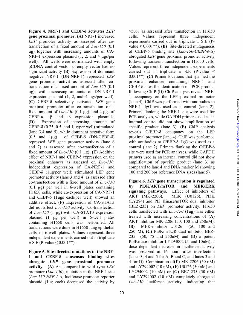
To ascertain the role of transcription factors NRF-1 and C/EBP in transcriptional regulation of LEP gene via the proximal enhancer, constitutively active NRF-1 (CA-NRF-1) was co-transfected with Luc-150 in H1650 cells. Dose dependent expression of CA-NRF-1 increased the transcriptional activity from Luc-150 (Figure 4A, lanes 3-5). In contrast, co-transfection of Luc-150 with increasing amount of dominant negative NRF-1 (DN-NRF-1) in H1650 cells decreased Luc-150 activity (Figure 4B, lanes 3-5), indicating that NRF-1 is able to transcriptionally activate the LEP gene via the proximal enhancer that contains an NRF-1 binding element. Role of C/EBP site within the proximal enhancer (–60/–53 bps) was examined by co-transfection of Luc-150 together with either C/EBP-α, C/EBP-β or C/EBP-δ expressing plasmids since all three forms bind similar DNA consensus sites and are highly expressed in lung epithelial cells(59,60). Plasmid expressing C/EBP-δ selectively up regulated Luc-150 activity by approximately 20 fold (Figure 4C, lane 5) as opposed to C/EBP-α and C/EBP-β, which showed 4 to 5 fold activation (Figure 3C, lanes 3 and 4), indicating that C/EBP-δ likely plays a major regulatory role in transcriptional activation of LEP gene. Indeed, dose dependent co-transfection of C/EBP-δ and fixed amount of Luc-150 activated (Figure 4D, Lanes 3-5), while co-transfection of dominant negative C/EBP-δ (DN-C/EBP-δ) abrogated Luc-150 activity (Figure 4D, Lanes 6-7).
Since NRF-1 and C/EBP-δ both up-regulated LEP gene promoter activity, we tested the possibility that NRF-1 and C/EBP-δ might have potential synergistic or additive effect on the proximal enhancer in the induction of LEP transcription. Co-transfection of Luc-150 in combination with CA-NRF-1 and C/EBP-δ expressing plasmids showed approximately 8 fold increase in Luc-150 activity (Figure 4E, lane 5) as compared to 4 fold increase after expression of CA-NRF-1 (Figure 4E, lane 3) and 2.5 fold increase after expression of C/EBP-δ alone (Figure 4E, lane 4), indicating additive effect of the two transcription factors. Since canonical LEP signaling is mediated via binding of the transcription factor STAT3 to various
at UN
IV O
F SOU
TH
FLO
RID
A on M
arch 14, 2014http://w
ww
.jbc.org/D
ownloaded from

8
gene promoter elements(61,62) we tested the hypothesis whether STAT3 itself may play a critical role in transcriptional activation of Luc-150. However, co-transfection of Luc-150 with plasmids expressing constitutively active STAT3 (CA-STAT3) did not activate transcription from Luc-150, suggesting that the proximal LEP gene enhancer functions independently of STAT3 and that LEP transcription may be regulated by STAT3 binding elements present further upstream in the LEP gene promoter. Together, NRF-1 and C/EBP-δ activated LEP gene transcription utilizing the LEP gene proximal enhancer element.
LEP gene proximal enhancer binds transcription factors NRF-1 and C/EBP-δ– The NRF-1 and C/EBP-δ DNA-binding elements that were identified by MatInspector were matched with true consensus elements defined by JASPAR database(58,63). While C/EBP site showed complete conservation (5’-TTGCGCAAC-3’), NRF-1 site deviated by one nucleotide at position 2 (5’-AAATGCGCN-3’). Site-directed mutagenesis of the NRF-1 and C/EBP-δ binding-sites within the proximal enhancer abrogated transcription activity, indicating that both these transcription factors directly bind to the enhancer element and transcriptionally regulates LEP gene expression (Figure 5A, lane 3 and 5B, lane3). Indeed, chromatin immuno-precipitation (ChIP) of NRF-1 and C/EBP-δ using the DNA primers spanning the 5′-regulatory region containing the proximal LEP gene enhancer (Figure 5C) readily detected bound form of NRF-1 and C/EBP-δ in the chromatin in vivo in H1650 cells (Figure 5D, lane 4 and 5E, lane 4). In contrast, NRF-1 and C/EBP-δ failed to bind GAPDH proximal promoter (Figure 5D lane 3 and 5E, lane 3), implicating NRF-1 and C/EBP-δ as direct transcriptional activators of LEP gene. Taken together, these experiments confirm that LEP gene transcription is mediated by binding of NRF-1 and C/EBP-δ to the LEP gene proximal enhancer in the chromatin context in lung epithelial cells.
PI3K/AKT and MEK pathways regulate LEP gene transcription via a proximal enhancer– When LEP binds to its receptor LEPR, it triggers the activation of PI3K/AKT, MEK/ERK and JAK2/STAT3 pathways in many cell types(24). However, whether LEP expression is controlled and induced by its own signaling via these three signaling pathways remains unknown. It is plausible that in order to maintain continuous LEP signaling, mainly in an autocrine loop, LEP itself may regulate its own expression via up-regulating LEPR and its downstream signaling pathways. To test this hypothesis, we transfected H1650 cells with Luc-150 and subsequently subjected these cells to PI3K/AKT and MEK pathway specific inhibitors. MK-2206, an AKT inhibitor, U0126 a MEK inhibitor, LY-294002 a pan-PI3K inhibitor and BEZ-235 a dual-PI3K/mTOR kinase inhibitor reduced LEP promoter activity in a dose dependent manner respectively (lanes 3-5 in Figure 6A, 6B, 6C and lanes 3-4 in Figure 6D). A combination of PI3K/AKT, PI3K/MEK and MEK/mTOR pathway inhibitors further reduced LEP promoter activity, indicating that signals from these pathways independently regulate LEP gene promoter (lane 5 in Figure 6E, 6F and 6G). In all these experiments, using Trypan blue staining, we made sure that cell-viability was not compromised. The highest concentrations used in our experiments were consistent or lower than previously reported studies (64-67) without any observable toxicity.
To further support the role of PI3K/AKT/mTOR and MEK/ERK signaling pathways we performed co-transfection of Luc-150 with plasmids expressing constitutively active AKT (CA-AKT) and constitutively active MEK (CA-MEK) in lung epithelial cells H1650. Indeed, expression of CA-AKT and CA-MEK induced LEP gene promoter activation in H1650 cells (Figure 9C). In summary, the LEP gene proximal promoter was regulated by PI3K/AKT/mTOR and MEK/ERK signaling pathways in lung epithelial cells. These observations raise the possibility that LEP via PI3K/AKT/mTOR and MEK/ERK signaling likely mediates its own transcription followed by increased LEP synthesis in lung epithelial cells
at UN
IV O
F SOU
TH
FLO
RID
A on M
arch 14, 2014http://w
ww
.jbc.org/D
ownloaded from

9
that are exposed to extracellular LEP, reinforcing an auto-regulatory LEP signaling loop. NRF-1, C/EBP-δ and STAT3 up-regulates endogenous LEP and LEP receptor gene transcripts– Although the potential role of NRF-1 and C/EBP-δ in the regulation of LEP gene transcription was established in transient transfection assays using the Luc-150 as a reporter, whether NRF-1 and C/EBP-δ can directly activate endogenous LEP gene transcription in the native chromatin context in H1650 lung epithelial cells was not determined. Therefore, following expression of CA-NRF-1 and CA-C/EBP-δ in H1650 cells, induction of LEP mRNA transcription was evaluated by quantitative real-time PCR (qRT-PCR). Indeed, expression of CA-NRF-1 and C/EBP-δ significantly increased LEP and its receptor LEPR mRNA expression (Figure 7A and 7B, lanes 3-4 and lanes 2-3 of insets). To further confirm that endogenous NRF-1 does activate LEP and its receptor LEPR transcription in lung epithelial cells, plasmid vectors expressing small hairpin RNA (shRNA) targeting the NRF-1 mRNA were expressed in H1650 cells. NRF-1 shRNA expression vectors 1A and 1B resulted in ~60–80% decrease in levels of NRF-1 transcripts as measured by RT-qPCR (Figure 7C, lanes 2 and 3 and inset lanes 3 and 4). Expression of shRNA-1B in H1650 cells significantly down-regulated endogenous LEP and LEPR gene mRNA transcripts as measured by RT-qPCR analysis (Figure 7D and 7E), thereby validating the role of NRF-1 in the transcriptional regulation of LEP and LEP receptor expression in lung epithelial cells. LEP mediated LEP and LEPR gene expression– LEP as a cytokine and a paracrine factor activates JAK2/STAT3, PI3K/AKT/mTOR and MEK/ERK signaling pathways that are directly involved in cancer progression(24). LEP also activates expression of several gene targets that participate in cancer progression, including pro-inflammatory cytokines and factors promoting angiogenesis(68). However, whether LEP signaling modulates the transcription of its own gene (LEP) and receptor (LEPR), amplifying its
function has not been studied. When H1650 cells were treated with recombinant LEP for 48 and 72 hours, JAK2/STAT3 and PI3K/AKT/mTOR signaling pathways were activated as assessed by increase in phosphorylation of STAT3 AKT and mTOR, (Figure 8 A, B and C). Within 15 minutes after treatment with leptin, increase in phosphorylation of ERK was observed (Figure 8D) Likewise, 30 minutes of treatment with leptin regulated P38 and JNK signaling pathways as assessed by increase in p-P38, p-P54 SNP/JNK and p-P46 SNP/JNK (Figure 8 E F and G). These results clearly indicate that the LEP/LEPR signaling pathway was operational in the lung epithelial cells. Furthermore, treatment of LEP significantly increased LEP and LEP receptor gene expression as assessed by measurement of their mRNA transcripts by RT-qPCR (Figure 9A and 9B). Taken together, LEP via LEP receptor up-regulated the expression of LEP and LEPR genes in lung epithelial cells, likely driving a feed-forward LEP-signaling loop, that may potentially be required for sustained LEP signaling as observed in chronic lung injury diseases and lung cancer. STAT3 binds to a distal enhancer and activates LEP gene transcription –When CA-STAT3 was expressed in H1650 cells, transcription of LEP and its receptor LEPR mRNA were significantly induced (Figure 9D and 9E) as assessed by RT-qPCR. This result, indicated that while STAT3 did not function via the proximal enhancer, it certainly regulated LEP and LEP gene transcription from the endogenous promoter in H1650 cells, likely via an upstream STAT3 responsive element located distally. To ascertain whether distal enhancer element comprising STAT3 binding sites is present within the LEP gene promoter, we scanned up to -2Kb of the human LEP gene 5’-upstream regulatory promoter sequence. The STAT3 binding sites were identified by homology search using softwares such as JASPAR, GENOMATIX and published STAT3 consensus DNA-binding sequence(69). While STAT3 site was not completely conserved at every nucleotide as per JASPAR, it did show the classical TT-N3-6-GG STAT3 binding sequence(69). Chromatin
at UN
IV O
F SOU
TH
FLO
RID
A on M
arch 14, 2014http://w
ww
.jbc.org/D
ownloaded from

10
immuno-precipitation (ChIP) of STAT3 using DNA primers spanning the 5′-regulatory region (-1610 to -1493 bps) containing the STAT3 site (Figure 9F) readily detected bound form of STAT3 on the chromatin in vivo in H1650 cells (Figure 9G lane 5). In contrast, STAT3 antibodies failed to bind GAPDH proximal promoter (Figure 9G lane 4), implicating STAT3 as a direct transcriptional activator of LEP gene expression. Taken together, these experiments confirm that transcriptional regulation of LEP and its receptor LEPR gene expression is mediated by STAT3 in lung epithelial cells. DISCUSSION LEP and LEP receptor are synthesized by several non-adipose tissues, wherein, LEP functions as a pleiotropic cytokine, modulating a variety of physiological and pathological functions(70). Increased pulmonary and circulating LEP levels are observed with several lung diseases associated with injury/repair and remodeling, including lung cancer(6,71-73). LEP is also involved in fetal lung development and pulmonary homeostasis (74,75). Emerging evidence indicates that LEP as a pro-inflammatory and pro-angiogenic cytokine may play critical roles in exacerbating acute and chronic pulmonary pathologies and drive lung cancer as an inflammatory molecule(7,12,76). However, the molecular mechanism of LEP gene expression in lung diseases and lung cancer remains elusive. In the present study, we demonstrate that Cre/LoxP mediated conditional deletion of Pten (Pten∆∆) activated adipocyte signaling in the respiratory epithelium that was associated with increased expression of LEP and its receptor. Using PTEN-null lung epithelial cells, we show that LEP gene was transcriptionally activated by a proximal enhancer element via binding of NRF-1 and C/EBP-δ transcription factors, while STAT3 bound a distal promoter element in the LEP gene and activated its expression. Transcription of the active form of the LEP receptor (LEPR), was also induced by NRF-1, C/EBP-δ and STAT3, suggesting that these three factors concertedly activate LEP/LEPR signaling pathway in the lung epithelial cells.
C/EBP-δ and STAT3 play critical roles during inflammatory responses in the lung(59,77,78). Since lung epithelial cells, particularly type II alveolar epithelial cells (Type II AECs) have high lipid metabolic activity and turnover that is required for surfactant synthesis(79), it is plausible that LEP may play a critical role in surfactant homeostasis following lung epithelial injury. Indeed, several studies have demonstrated that LEP directly stimulates proliferation of Type II AECs(80) up-regulating type IV collagen synthesis, that reinforce the alveolar walls(81,82). Extra lipid accumulation in Type II pneumocytes may lead to lipotoxicity as observed in many non-adipose tissues(83,84). Since LEP stimulates fatty acid oxidation via activation of AMP-activated protein kinase (AMPK)(85), it participates in reducing lipid stores, thereby reducing lipotoxicity(85-87). Consistent with this concept, sustained LEP signaling, as seen in many inflammatory conditions, causes chronic activation of AMPK(85), which activates the transcription factor NRF-1(88). Activated NRF-1 binds gene promoters involved in enhancing oxidative capacity and mitochondrial biogenesis, including LEP as demonstrated in the present work, increasing energy metabolism via known LEP signaling pathways(88,89). Thus, our results presented here are consistent with the role of NRF-1 in PTEN-null H1650 lung epithelial cells. In addition, loss of PTEN in lung epithelial cells drives rapid cell proliferation that would indeed be associated with increased mitochondriogenesis(35). Supporting these observations, our LEP promoter-reporter deletion analysis, site-directed mutagenesis, ChIP and NRF-1 over-expression studies demonstrated increased activity of NRF-1 on the LEP promoter itself, indicating that LEP mediated activation of NRF-1 contributes to transcriptional activation and expression of the LEP gene in lung epithelial cells. In adipocytes, LEP gene transcription is regulated by C/EBP-α(32,90), however, transcriptional regulation of LEP gene and the roles of C/EBP isoforms, including C/EBP-β and C/EBP-δ in non-adipocyte tissues remain
at UN
IV O
F SOU
TH
FLO
RID
A on M
arch 14, 2014http://w
ww
.jbc.org/D
ownloaded from

11
unclear. The present study revealed that C/EBP-δ, but not C/EBP-α or C/EBP-β, strongly activated LEP gene transcription in lung epithelial cells, suggesting a critical and selective role for C/EBP-δ in LEP gene regulation. Given the important role of LEP in inflammatory processes, it is highly likely that concerted modulation of pro-inflammatory genes including LEP is under the control of C/EBP-δ in the lung epithelium(60). Recent studies demonstrate that C/EBP-β and C/EBP-δ participate in inflammatory responses following lung injury and infection (91), consistent with the identified role of C/EBP-δ in the transcriptional activation of LEP gene expression in the lung epithelial cells. LEP signaling is transduced via the activation of canonical PI3K/AKT, MEK/ERK and JAK2/STAT3 pathways in many cell types(24). However, whether LEP itself is activated by these three signaling pathways and regulates its own expression has not been explored.
Tumors maintain continuous LEP signaling that facilitate cancer progression and metastasis, while inhibition of LEP-signaling results in efficient anti-tumor activity(92); therefore, it is likely that LEP itself may regulate its own gene expression When H1650 PTEN-null lung epithelial cells were treated with LEP, activation of PI3K/AKT/mTOR, MEK/ERK and JAK2/STAT3, P38 and JNK signaling pathways was detected by immune-blotting for p-mTOR, p-AKT, p-STAT, p-ERK, p38 MAPK as well as the active and inactive forms of JNK (p54 JNK and p46 JNK). This is consistent with previous findings where leptin mediated activation of canonical (PI3K, ERK) and non-canonical (p38 MAPK, JNK, and PKC) signaling pathways has been observed(93,94) Together, our results support the concept that loss of PTEN in lung adenocarcinoma would activate PI3K/AKT/mTOR, MEK/ERK and the p38/JNK MAPK signaling pathways, which would in turn, contribute to the induction of LEP gene expression and subsequent secretion.
To test this hypothesis in vivo, we developed a mouse model with conditional deletion of PTEN in an oncogenic K-RAS background (95). Exuberant LEP secretion was indeed detected in
tumors generated after PTEN loss in oncogenic KRAS background (Figure 2B), indicating an important role for LEP signaling in lung cancer progression. Since treatment of H1650 cells with inhibitors of PI3K, AKT, mTOR, and MEK abrogated LEP gene transcription while constitutive expression of AKT and MEK activated LEP gene promoter activity, we proposed in our conceptual model that LEP mediated LEP gene expression is directly influenced by PI3K/AKT/mTOR, MEK/ERK and P38 JNK signaling pathways (Figure 10). The major hallmarks of LEP signaling are activation of STAT3 via JAK2 phosphorylation, and increased cell proliferation. Over-expression of constitutively active STAT3 induced LEP gene expression while treatment with LEP enhanced proliferation and wound healing of H1650 cells, consistent with promotion of invasion and migration in cancer cells(96). Taken together, our findings demonstrate that LEP itself regulate its own expression via LEP receptor mediated downstream signaling in lung epithelial cells. The present study supports the concept that therapies that can abrogate LEP signaling in lung pathologies, may reduce disease morbidity.
REFERENCES:
1. Procaccini, C., Jirillo, E., and Matarese, G. (2012) Leptin as an immunomodulator. Molecular aspects of medicine 33, 35-45
2. Morris, D. L., and Rui, L. (2009) Recent advances in understanding leptin signaling and leptin resistance. American journal of physiology. Endocrinology and metabolism 297, E1247-1259
3. Bruno, A., Chanez, P., Chiappara, G., Siena, L., Giammanco, S., Gjomarkaj, M., Bonsignore, G., Bousquet, J., and Vignola, A. M. (2005) Does leptin play a cytokine-like role within the airways of COPD patients? European Respiratory Journal 26, 398-405
4. Vernooy, J. H. J., Drummen, N. E. A., van Suylen, R. J., Cloots, R. H. E., Möller, G. M., Bracke, K. R., Zuyderduyn, S., Dentener, M. A.,
at UN
IV O
F SOU
TH
FLO
RID
A on M
arch 14, 2014http://w
ww
.jbc.org/D
ownloaded from

12
Brusselle, G. G., Hiemstra, P. S., and Wouters, E. F. M. (2009) Enhanced pulmonary leptin expression in patients with severe COPD and asymptomatic smokers. Thorax 64, 26-32
5. Vernooy, J. H., Drummen, N. E., van Suylen, R. J., Cloots, R. H., Moller, G. M., Bracke, K. R., Zuyderduyn, S., Dentener, M. A., Brusselle, G. G., Hiemstra, P. S., and Wouters, E. F. (2009) Enhanced pulmonary leptin expression in patients with severe COPD and asymptomatic smokers. Thorax 64, 26-32
6. Ribeiro, R., Araujo, A., Lopes, C., and Medeiros, R. (2007) Immunoinflammatory mechanisms in lung cancer development: is leptin a mediator? Journal of thoracic oncology : official publication of the International Association for the Study of Lung Cancer 2, 105-108
7. Vernooy, J. H., Ubags, N. D., Brusselle, G. G., Tavernier, J., Suratt, B. T., Joos, G. F., Wouters, E. F., and Bracke, K. R. (2013) Leptin as regulator of pulmonary immune responses: Involvement in respiratory diseases. Pulmonary pharmacology & therapeutics
8. Vernooy, J. H., Bracke, K. R., Drummen, N. E., Pauwels, N. S., Zabeau, L., van Suylen, R. J., Tavernier, J., Joos, G. F., Wouters, E. F., and Brusselle, G. G. (2010) Leptin modulates innate and adaptive immune cell recruitment after cigarette smoke exposure in mice. J Immunol 184, 7169-7177
9. Broekhuizen, R., Vernooy, J. H., Schols, A. M., Dentener, M. A., and Wouters, E. F. (2005) Leptin as local inflammatory marker in COPD. Respiratory medicine 99, 70-74
10. Aleman, M. R., Santolaria, F., Batista, N., de La Vega, M., Gonzalez-Reimers, E., Milena, A., Llanos, M., and Gomez-Sirvent, J. L. (2002) Leptin role in advanced lung cancer. A mediator of the acute phase response or a marker of the status of nutrition? Cytokine 19, 21-26
11. Sood, A. (2010) Obesity, adipokines, and lung disease. J Appl Physiol 108, 744-753
12. Jain, M., Budinger, G. R., Lo, A., Urich, D., Rivera, S. E., Ghosh, A. K., Gonzalez, A., Chiarella, S. E., Marks, K., Donnelly, H. K., Soberanes, S., Varga, J., Radigan, K. A., Chandel, N. S., and Mutlu, G. M. (2011) Leptin promotes fibroproliferative acute respiratory distress syndrome by inhibiting peroxisome proliferator-activated receptor-gamma. American journal of respiratory and critical care medicine 183, 1490-1498
13. Barazzone-Argiroffo, C., Muzzin, P., Donati, Y. R., Kan, C. D., Aubert, M. L., and Piguet, P. F. (2001) Hyperoxia increases leptin production: a mechanism mediated through endogenous elevation of corticosterone. Am J Physiol Lung Cell Mol Physiol 281, L1150-1156
14. Jain, M., Budinger, G. R. S., Lo, A., Urich, D., Rivera, S. E., Ghosh, A. K., Gonzalez, A., Chiarella, S. E., Marks, K., Donnelly, H. K., Soberanes, S., Varga, J., Radigan, K. A., Chandel, N. S., and Mutlu, G. M. (2011) Leptin Promotes Fibroproliferative Acute Respiratory Distress Syndrome by Inhibiting Peroxisome Proliferator-activated Receptor-{gamma}. Am. J. Respir. Crit. Care Med. 183, 1490-1498
15. Bellmeyer, A., Martino, J. M., Chandel, N. S., Scott Budinger, G. R., Dean, D. A., and Mutlu, G. M. (2007) Leptin resistance protects mice from hyperoxia-induced acute lung injury. Am J Respir Crit Care Med 175, 587-594
16. Leao da Silva, P., Tulio de Mello, M., Cheik, N. C., Lima Sanches, P., Munhoz da Silveira Campos, R., Carnier, J., Inoue, D., do Nascimento, C. M., Oyama, L. M., Tock, L., Tufik, S., and Damaso, A. R. (2012) Reduction in the Leptin Concentration as a Predictor of Improvement in Lung Function in Obese Adolescents. Obesity facts 5, 806-820
17. Leao da Silva, P., de Mello, M. T., Cheik, N. C., Sanches, P. L., Munhoz da
at UN
IV O
F SOU
TH
FLO
RID
A on M
arch 14, 2014http://w
ww
.jbc.org/D
ownloaded from

13
Silveira Campos, R., Carnier, J., Inoue, D., do Nascimento, C. M., Oyama, L. M., Tock, L., Tufik, S., and Damaso, A. R. (2012) Reduction in the leptin concentration as a predictor of improvement in lung function in obese adolescents. Obesity facts 5, 806-820
18. Malli, F., Papaioannou, A. I., Gourgoulianis, K. I., and Daniil, Z. (2010) The role of leptin in the respiratory system: an overview. Respiratory research 11, 152
19. MacCallum, N. S., and Evans, T. W. (2005) Epidemiology of acute lung injury. Current Opinion in Critical Care 11, 43-49
20. Matthay, M. A., and Zimmerman, G. A. (2005) Acute lung injury and the acute respiratory distress syndrome: four decades of inquiry into pathogenesis and rational management. Am J Respir Cell Mol Biol 33, 319-327
21. Calfee, C. S., Matthay, M. A., Eisner, M. D., Benowitz, N., Call, M., Pittet, J. F., and Cohen, M. J. (2011) Active and passive cigarette smoking and acute lung injury after severe blunt trauma. Am J Respir Crit Care Med 183, 1660-1665
22. Sierra-Honigmann, M. R., Nath, A. K., Murakami, C., Garcia-Cardena, G., Papapetropoulos, A., Sessa, W. C., Madge, L. A., Schechner, J. S., Schwabb, M. B., Polverini, P. J., and Flores-Riveros, J. R. (1998) Biological action of leptin as an angiogenic factor. Science 281, 1683-1686
23. Vansaun, M. N. (2013) Molecular pathways: adiponectin and leptin signaling in cancer. Clinical cancer research : an official journal of the American Association for Cancer Research 19, 1926-1932
24. Sweeney, G. (2002) Leptin signalling. Cellular signalling 14, 655-663
25. Zhou, W., Guo, S., and Gonzalez-Perez, R. R. (2011) Leptin pro-angiogenic signature in breast cancer is linked to IL-1 signalling. Br J Cancer 104, 128-137
26. Gonzalez-Perez, R. R., Xu, Y., Guo, S., Watters, A., Zhou, W., and Leibovich, S. J. (2010) Leptin upregulates VEGF in breast cancer via canonic and non-canonical signalling pathways and NFkappaB/HIF-1alpha activation. Cell Signal 22, 1350-1362
27. de la Brousse, F. C., Shan, B., and Chen, J. L. (1996) Identification of the promoter of the mouse obese gene. Proc Natl Acad Sci U S A 93, 4096-4101
28. Gong, D. W., Bi, S., Pratley, R. E., and Weintraub, B. D. (1996) Genomic structure and promoter analysis of the human obese gene. J Biol Chem 271, 3971-3974
29. Hwang, C. S., Mandrup, S., MacDougald, O. A., Geiman, D. E., and Lane, M. D. (1996) Transcriptional activation of the mouse obese (ob) gene by CCAAT/enhancer binding protein alpha. Proceedings of the National Academy of Sciences 93, 873-877
30. He, Y., Chen, H., Quon, M. J., and Reitman, M. (1995) The Mouse obese Gene. Journal of Biological Chemistry 270, 28887-28891
31. de la Brousse, F. C., Shan, B., and Chen, J. L. (1996) Identification of the promoter of the mouse obese gene. Proceedings of the National Academy of Sciences 93, 4096-4101
32. Miller, S. G., De Vos, P., Guerre-Millo, M., Wong, K., Hermann, T., Staels, B., Briggs, M. R., and Auwerx, J. (1996) The adipocyte specific transcription factor C/EBPalpha modulates human ob gene expression. Proc Natl Acad Sci U S A 93, 5507-5511
33. Fuke, T., Yoshizaki, T., Kondo, M., Morino, K., Obata, T., Ugi, S., Nishio, Y., Maeda, S., Kashiwagi, A., and Maegawa, H. (2010) Transcription factor AP-2beta inhibits expression and secretion of leptin, an insulin-sensitizing hormone, in 3T3-L1 adipocytes. Int J Obes (Lond) 34, 670-678
34. Kim, J. B., Sarraf, P., Wright, M., Yao, K. M., Mueller, E., Solanes, G., Lowell, B. B., and Spiegelman, B. M. (1998) Nutritional and insulin regulation of
at UN
IV O
F SOU
TH
FLO
RID
A on M
arch 14, 2014http://w
ww
.jbc.org/D
ownloaded from

14
fatty acid synthetase and leptin gene expression through ADD1/SREBP1. J Clin Invest 101, 1-9
35. Dave, V., Wert, S. E., Tanner, T., Thitoff, A. R., Loudy, D. E., and Whitsett, J. A. (2008) Conditional deletion of Pten causes bronchiolar hyperplasia. Am J Respir Cell Mol Biol 38, 337-345
36. Dranoff, G., Crawford, A. D., Sadelain, M., Ream, B., Rashid, A., Bronson, R. T., Dickersin, G. R., Bachurski, C. J., Mark, E. L., Whitsett, J. A., and et al. (1994) Involvement of granulocyte-macrophage colony-stimulating factor in pulmonary homeostasis. Science 264, 713-716
37. Irizarry, R. A., Bolstad, B. M., Collin, F., Cope, L. M., Hobbs, B., and Speed, T. P. (2003) Summaries of Affymetrix GeneChip probe level data. Nucleic Acids Res 31, e15
38. Dinu, I., Potter, J. D., Mueller, T., Liu, Q., Adewale, A. J., Jhangri, G. S., Einecke, G., Famulski, K. S., Halloran, P., and Yasui, Y. (2007) Improving gene set analysis of microarray data by SAM-GS. BMC Bioinformatics 8, 242
39. Verhoeven, K. J. F., Simonsen, K. L., and McIntyre, L. M. (2005) Implementing false discovery rate control: increasing your power. Oikos 108, 643-647
40. Storey, J. D. (2002) A direct approach to false discovery rates. Journal of the Royal Statistical Society: Series B (Statistical Methodology) 64, 479-498
41. Benjamini, Y., Drai, D., Elmer, G., Kafkafi, N., and Golani, I. (2001) Controlling the false discovery rate in behavior genetics research. Behav Brain Res 125, 279-284
42. Dennis, G., Jr., Sherman, B. T., Hosack, D. A., Yang, J., Gao, W., Lane, H. C., and Lempicki, R. A. (2003) DAVID: Database for Annotation, Visualization, and Integrated Discovery. Genome Biol 4, P3
43. Boussif, O., Lezoualc'h, F., Zanta, M. A., Mergny, M. D., Scherman, D., Demeneix, B., and Behr, J. P. (1995) A
versatile vector for gene and oligonucleotide transfer into cells in culture and in vivo: polyethylenimine. Proceedings of the National Academy of Sciences of the United States of America 92, 7297-7301
44. Davé, V., Wert, S. E., Tanner, T., Thitoff, A. R., Loudy, D. E., and Whitsett, J. A. (2008) Conditional Deletion of Pten Causes Bronchiolar Hyperplasia. American Journal of Respiratory Cell and Molecular Biology 38, 337-345
45. Ekins, S., Nikolsky, Y., Bugrim, A., Kirillov, E., and Nikolskaya, T. (2007) Pathway mapping tools for analysis of high content data. Methods Mol Biol 356, 319-350
46. Ekins, S., Nikolsky, Y., Bugrim, A., Kirillov, E., and Nikolskaya, T. (2006) Pathway Mapping Tools for Analysis of High Content Data. in High Content Screening (Taylor, D. L., Haskins, J., and Giuliano, K. eds.), Humana Press. pp 319-350
47. Jimenez-Marin, A., Collado-Romero, M., Ramirez-Boo, M., Arce, C., and Garrido, J. J. (2009) Biological pathway analysis by ArrayUnlock and Ingenuity Pathway Analysis. BMC Proc 3 Suppl 4, S6
48. Kanehisa, M., Goto, S., Sato, Y., Furumichi, M., and Tanabe, M. (2012) KEGG for integration and interpretation of large-scale molecular data sets. Nucleic Acids Res 40, D109-114
49. Yanagi, S., Kishimoto, H., Kawahara, K., Sasaki, T., Sasaki, M., Nishio, M., Yajima, N., Hamada, K., Horie, Y., Kubo, H., Whitsett, J. A., Mak, T. W., Nakano, T., Nakazato, M., and Suzuki, A. (2007) Pten controls lung morphogenesis, bronchioalveolar stem cells, and onset of lung adenocarcinomas in mice. J Clin Invest 117, 2929-2940
50. Miyoshi, K., Yanagi, S., Kawahara, K., Nishio, M., Tsubouchi, H., Imazu, Y., Koshida, R., Matsumoto, N., Taguchi, A., Yamashita, S., Suzuki, A., and Nakazato, M. (2013) Epithelial Pten
at UN
IV O
F SOU
TH
FLO
RID
A on M
arch 14, 2014http://w
ww
.jbc.org/D
ownloaded from

15
controls acute lung injury and fibrosis by regulating alveolar epithelial cell integrity. Am J Respir Crit Care Med 187, 262-275
51. Mihai, C., Bao, S., Lai, J. P., Ghadiali, S. N., and Knoell, D. L. (2012) PTEN inhibition improves wound healing in lung epithelia through changes in cellular mechanics that enhance migration. Am J Physiol Lung Cell Mol Physiol 302, L287-299
52. Tiozzo, C., De Langhe, S., Yu, M., Londhe, V. A., Carraro, G., Li, M., Li, C., Xing, Y., Anderson, S., Borok, Z., Bellusci, S., and Minoo, P. (2009) Deletion of Pten expands lung epithelial progenitor pools and confers resistance to airway injury. Am J Respir Crit Care Med 180, 701-712
53. Okumura, M., Yamamoto, M., Sakuma, H., Kojima, T., Maruyama, T., Jamali, M., Cooper, D. R., and Yasuda, K. (2002) Leptin and high glucose stimulate cell proliferation in MCF-7 human breast cancer cells: reciprocal involvement of PKC-alpha and PPAR expression. Biochimica et biophysica acta 1592, 107-116
54. Keese, C. R., Wegener, J., Walker, S. R., and Giaever, I. (2004) Electrical wound-healing assay for cells in vitro. Proc Natl Acad Sci U S A 101, 1554-1559
55. Schiller, K. R., Maniak, P. J., and O'Grady, S. M. (2010) Cystic fibrosis transmembrane conductance regulator is involved in airway epithelial wound repair. Am J Physiol Cell Physiol 299, C912-921
56. Wu, N. L., Chiang, Y. C., Huang, C. C., Fang, J. Y., Chen, D. F., and Hung, C. F. (2010) Zeaxanthin inhibits PDGF-BB-induced migration in human dermal fibroblasts. Exp Dermatol 19, e173-181
57. Gorshkova, I., He, D., Berdyshev, E., Usatuyk, P., Burns, M., Kalari, S., Zhao, Y., Pendyala, S., Garcia, J. G., Pyne, N. J., Brindley, D. N., and Natarajan, V. (2008) Protein kinase C-epsilon regulates sphingosine 1-phosphate-mediated migration of human lung
endothelial cells through activation of phospholipase D2, protein kinase C-zeta, and Rac1. J Biol Chem 283, 11794-11806
58. Quandt, K., Frech, K., Karas, H., Wingender, E., and Werner, T. (1995) MatInd and MatInspector: new fast and versatile tools for detection of consensus matches in nucleotide sequence data. Nucleic Acids Res 23, 4878-4884
59. Roos, A. B., and Nord, M. (2012) The emerging role of C/EBPs in glucocorticoid signaling: lessons from the lung. The Journal of endocrinology 212, 291-305
60. Cassel, T. N., and Nord, M. (2003) C/EBP transcription factors in the lung epithelium. American journal of physiology. Lung cellular and molecular physiology 285, L773-781
61. Buettner, C., Pocai, A., Muse, E. D., Etgen, A. M., Myers, M. G., Jr., and Rossetti, L. (2006) Critical role of STAT3 in leptin's metabolic actions. Cell metabolism 4, 49-60
62. Myers, M. G., Cowley, M. A., and Munzberg, H. (2008) Mechanisms of leptin action and leptin resistance. Annual review of physiology 70, 537-556
63. Sandelin, A., Alkema, W., Engstrom, P., Wasserman, W. W., and Lenhard, B. (2004) JASPAR: an open-access database for eukaryotic transcription factor binding profiles. Nucleic acids research 32, D91-94
64. Maira, S. M., Stauffer, F., Brueggen, J., Furet, P., Schnell, C., Fritsch, C., Brachmann, S., Chene, P., De Pover, A., Schoemaker, K., Fabbro, D., Gabriel, D., Simonen, M., Murphy, L., Finan, P., Sellers, W., and Garcia-Echeverria, C. (2008) Identification and characterization of NVP-BEZ235, a new orally available dual phosphatidylinositol 3-kinase/mammalian target of rapamycin inhibitor with potent in vivo antitumor activity. Mol Cancer Ther 7, 1851-1863
65. DeSilva, D. R., Jones, E. A., Favata, M. F., Jaffee, B. D., Magolda, R. L.,
at UN
IV O
F SOU
TH
FLO
RID
A on M
arch 14, 2014http://w
ww
.jbc.org/D
ownloaded from

16
Trzaskos, J. M., and Scherle, P. A. (1998) Inhibition of Mitogen-Activated Protein Kinase Kinase Blocks T Cell Proliferation But Does Not Induce or Prevent Anergy. The Journal of Immunology 160, 4175-4181
66. Hirai, H., Sootome, H., Nakatsuru, Y., Miyama, K., Taguchi, S., Tsujioka, K., Ueno, Y., Hatch, H., Majumder, P. K., Pan, B.-S., and Kotani, H. (2010) MK-2206, an Allosteric Akt Inhibitor, Enhances Antitumor Efficacy by Standard Chemotherapeutic Agents or Molecular Targeted Drugs In vitro and In vivo. Molecular Cancer Therapeutics 9, 1956-1967
67. Semba, S., Itoh, N., Ito, M., Harada, M., and Yamakawa, M. (2002) The in Vitro and in Vivo Effects of 2-(4-Morpholinyl)-8-phenyl-chromone (LY294002), a Specific Inhibitor of Phosphatidylinositol 3′-Kinase, in Human Colon Cancer Cells. Clinical Cancer Research 8, 1957-1963
68. Aleffi, S., Petrai, I., Bertolani, C., Parola, M., Colombatto, S., Novo, E., Vizzutti, F., Anania, F. A., Milani, S., Rombouts, K., Laffi, G., Pinzani, M., and Marra, F. (2005) Upregulation of proinflammatory and proangiogenic cytokines by leptin in human hepatic stellate cells. Hepatology 42, 1339-1348
69. Seidel, H. M., Milocco, L. H., Lamb, P., Darnell, J. E., Jr., Stein, R. B., and Rosen, J. (1995) Spacing of palindromic half sites as a determinant of selective STAT (signal transducers and activators of transcription) DNA binding and transcriptional activity. Proceedings of the National Academy of Sciences of the United States of America 92, 3041-3045
70. Margetic, S., Gazzola, C., Pegg, G. G., and Hill, R. A. (2002) Leptin: a review of its peripheral actions and interactions. Int J Obes Relat Metab Disord 26, 1407-1433
71. Karakas, S., Karadag, F., Karul, A. B., Gurgey, O., Gurel, S., Guney, E., and Cildag, O. (2005) Circulating leptin and body composition in chronic obstructive
pulmonary disease. International journal of clinical practice 59, 1167-1170
72. Shen, Y., Wang, Q., Zhao, Q., and Zhou, J. (2009) Leptin promotes the immune escape of lung cancer by inducing proinflammatory cytokines and resistance to apoptosis. Molecular medicine reports 2, 295-299
73. Carpagnano, G. E., Spanevello, A., Curci, C., Salerno, F., Palladino, G. P., Resta, O., Di Gioia, G., Carpagnano, F., and Foschino Barbaro, M. P. (2007) IL-2, TNF-alpha, and leptin: local versus systemic concentrations in NSCLC patients. Oncology research 16, 375-381
74. Henson, M. C., Swan, K. F., Edwards, D. E., Hoyle, G. W., Purcell, J., and Castracane, V. D. (2004) Leptin receptor expression in fetal lung increases in late gestation in the baboon: a model for human pregnancy. Reproduction 127, 87-94
75. Bergen, H. T., Cherlet, T. C., Manuel, P., and Scott, J. E. (2002) Identification of leptin receptors in lung and isolated fetal type II cells. American journal of respiratory cell and molecular biology 27, 71-77
76. Xu, Y. J., Shao, Y. F., Zhao, X., Geng, Y. T., Wang, K., and Yin, Y. M. (2011) Expression and clinical significance of leptin, the functional receptor of leptin (OB-Rb) and HER-2 in non-small-cell lung cancer: a retrospective analysis. Journal of cancer research and clinical oncology 137, 1841-1848
77. Gao, H., Guo, R. F., Speyer, C. L., Reuben, J., Neff, T. A., Hoesel, L. M., Riedemann, N. C., McClintock, S. D., Sarma, J. V., Van Rooijen, N., Zetoune, F. S., and Ward, P. A. (2004) Stat3 activation in acute lung injury. J Immunol 172, 7703-7712
78. Hokuto, I., Ikegami, M., Yoshida, M., Takeda, K., Akira, S., Perl, A. K., Hull, W. M., Wert, S. E., and Whitsett, J. A. (2004) Stat-3 is required for pulmonary homeostasis during hyperoxia. The Journal of clinical investigation 113, 28-37
at UN
IV O
F SOU
TH
FLO
RID
A on M
arch 14, 2014http://w
ww
.jbc.org/D
ownloaded from
17
79. Dobbs, L. G. (1989) Pulmonary surfactant. Annual review of medicine 40, 431-446
80. Torday, J. S., Sun, H., Wang, L., Torres, E., Sunday, M. E., and Rubin, L. P. (2002) Leptin mediates the parathyroid hormone-related protein paracrine stimulation of fetal lung maturation. Am J Physiol Lung Cell Mol Physiol 282, L405-410
81. Wolf, G., and Ziyadeh, F. N. (2006) Leptin and renal fibrosis. Contributions to nephrology 151, 175-183
82. Maina, J. N., and West, J. B. (2005) Thin and strong! The bioengineering dilemma in the structural and functional design of the blood-gas barrier. Physiological reviews 85, 811-844
83. Unger, R. H., Zhou, Y. T., and Orci, L. (1999) Regulation of fatty acid homeostasis in cells: novel role of leptin. Proc Natl Acad Sci U S A 96, 2327-2332
84. van Herpen, N. A., and Schrauwen-Hinderling, V. B. (2008) Lipid accumulation in non-adipose tissue and lipotoxicity. Physiology & behavior 94, 231-241
85. Minokoshi, Y., Kim, Y. B., Peroni, O. D., Fryer, L. G., Muller, C., Carling, D., and Kahn, B. B. (2002) Leptin stimulates fatty-acid oxidation by activating AMP-activated protein kinase. Nature 415, 339-343
86. Muoio, D. M., Dohm, G. L., Fiedorek, F. T., Jr., Tapscott, E. B., and Coleman, R. A. (1997) Leptin directly alters lipid partitioning in skeletal muscle. Diabetes 46, 1360-1363
87. Minokoshi, Y., and Kahn, B. B. (2003) Role of AMP-activated protein kinase in leptin-induced fatty acid oxidation in muscle. Biochemical Society transactions 31, 196-201
88. Bergeron, R., Ren, J. M., Cadman, K. S., Moore, I. K., Perret, P., Pypaert, M., Young, L. H., Semenkovich, C. F., and Shulman, G. I. (2001) Chronic activation of AMP kinase results in NRF-1 activation and mitochondrial biogenesis. American journal of
physiology. Endocrinology and metabolism 281, E1340-1346
89. Porter, R. K., and Andrews, J. F. (1998) Effects of leptin on mitochondrial 'proton leak' and uncoupling proteins: implications for mammalian energy metabolism. The Proceedings of the Nutrition Society 57, 455-460
90. Hwang, C. S., Mandrup, S., MacDougald, O. A., Geiman, D. E., and Lane, M. D. (1996) Transcriptional activation of the mouse obese (ob) gene by CCAAT/enhancer binding protein alpha. Proc Natl Acad Sci U S A 93, 873-877
91. Yan, C., Johnson, P. F., Tang, H., Ye, Y., Wu, M., and Gao, H. (2013) CCAAT/Enhancer-Binding Protein delta Is a Critical Mediator of Lipopolysaccharide-Induced Acute Lung Injury. Am J Pathol 182, 420-430
92. Rene Gonzalez, R., Watters, A., Xu, Y., Singh, U. P., Mann, D. R., Rueda, B. R., and Penichet, M. L. (2009) Leptin-signaling inhibition results in efficient anti-tumor activity in estrogen receptor positive or negative breast cancer. Breast cancer research : BCR 11, R36
93. Ando, S., and Catalano, S. (2012) The multifactorial role of leptin in driving the breast cancer microenvironment. Nat Rev Endocrinol 8, 263-275
94. Onuma, M., Bub, J. D., Rummel, T. L., and Iwamoto, Y. (2003) Prostate cancer cell-adipocyte interaction: leptin mediates androgen-independent prostate cancer cell proliferation through c-Jun NH2-terminal kinase. J Biol Chem 278, 42660-42667
95. Johnson, L., Mercer, K., Greenbaum, D., Bronson, R. T., Crowley, D., Tuveson, D. A., and Jacks, T. (2001) Somatic activation of the K-ras oncogene causes early onset lung cancer in mice. Nature 410, 1111-1116
96. Saxena, N. K., Sharma, D., Ding, X., Lin, S., Marra, F., Merlin, D., and Anania, F. A. (2007) Concomitant activation of the JAK/STAT, PI3K/AKT, and ERK signaling is involved in leptin-mediated promotion
at UN
IV O
F SOU
TH
FLO
RID
A on M
arch 14, 2014http://w
ww
.jbc.org/D
ownloaded from

18
of invasion and migration of hepatocellular carcinoma cells. Cancer Res 67, 2497-2507
Foot Note: R. R. P. A.G, W. Q. and P. M. performed the experiments, A. P. and D.A.G. designed the ECIS experiments. The project was conceived and designed by V. D. and written by R. R. P. and V. D.
List of abbreviations:
PTEN: Phosphatase and tensin homolog, LEP: Leptin, LEPR: Leptin receptor, MEK: Mitogen Activated Protein Kinase, ERK: Extracellular signal-regulated kinase, PI3K: Phosphatidylinositide 3-kinase,AKT: Protein Kinase B, mTOR: mammalian target of rapamycin,STAT3: Signal transducer and activator of transcription 3,C/EBP: CCAAT-enhancer-binding proteins, ChIP: Chromatin Immunoprecipitation, JAK2: Janus kinase 2,VEGF: Vascular endothelial growth factor,VEGFR2: Vascular endothelial growth factor receptor 2, KEGG: Kyoto Encyclopedia of Genes and Genomes, Dox: Doxycycline, PEI: Polyethylenimine, JNK: c-Jun N-terminal kinases
Acknowledgements
We thank Dr. Todd Gulick, M.D, Sanford-Burnham Medical Research Institute, Lake Nano Orlando for the NRF-1 plasmids, Dr. Steven McKnight, Ph.D., UT-Southwestern Medical Center , Dallas for C/EBP- δ plasmid and Dr. Jim Darnell, The Rockefeller University, New York for CA-STAT3 expression plasmid. We would also like to thank Dr. Jeffrey Whitsett MD (Cincinnati Children’s Hospital, Cincinnati OH) for the use of microarray core facility. This work was supported by American Heart Association Scientist Development Grant SDG-0830101N (V. D.) and NIH-Lung SPORE Career Development Grant, Moffitt Cancer Center (V. D.).
at UN
IV O
F SOU
TH
FLO
RID
A on M
arch 14, 2014http://w
ww
.jbc.org/D
ownloaded from

19
FIGURE LEGENDS
Figure 1. Generation of PtenΔ/Δ mice and microarray profiling of PtenΔ/Δ lung mRNA identifies 1389 Pten responsive genes. (A) Generation of the PtenΔ/Δ mice. Triple-transgenic mice containing 3 alleles: loxP-flanked exon V (Ptenflox/flox); SP-C–rtTAtg/−; TetO-Cretg/- (orange oval) were produced (i, and ii) and selected by genotyping (iii). Mice harboring SPC-rtTAtg/-/Ptenflox/flox, TetO-Cretg/-
/Ptenflox/flox, or Ptenflox/flox were used as controls. Lungs were harvested from 20-week old mice. Hematoxylin/eosin staining of lung sections from PtenΔ/Δ mice demonstrated normal branching morphogenesis (indicated by blue arrows) and postnatal lung formation with increased hyperplasia (indicated by red arrows) (iv). (B) Microarray analysis of lung RNA isolated from Pten-deleted lung epithelial cells (PtenΔ/Δ) and control mice revealed that expression of 1389 genes was altered significantly (≥2-fold change, P-Value ≤ 0.05).
Figure 2. Activation of the adipocytokine signaling pathway in PTENΔ/Δ mouse lungand expression of LEP and LEP receptor in lung epithelial cells. (A) KEGG (Kyoto Encyclopedia of Genes and Genomes) pathway analysis of PTEN responsive genes identified adipocytokine signaling as being significantly up-regulated. LEP, LEPR and adiponectin (ADIPQ) were up-regulated in the PTENΔ/Δ mice and overlapped (red asterisks) on the canonical KEGG adipocyte signaling pathway, suggesting that loss of PTEN activated adipokine synthesis and signaling in the lung epithelium. (B) Immuno-staining with LEP antibodies confirmed exuberant induction and secretion of LEP in K-RASG12D(95) driven PtenΔ/Δlung tumors in mice. (C) Enrichment for gene networks in PTENΔ/Δ mice. Biological networks were enriched from the PTEN responsive gene set using standard software tools from MetaCoreTM. LEP signaling was identified as a significant metabolic pathway activated in the PTENΔ/Δ enriched list (P value = 0.007). (D) Likewise, application of Ingenuity Systems software tool called Intelligent Pathway Analysis (IPATM)) independently enriched LEP signaling pathway
(P value = 0.04). The linear plot shows enhanced threshold calculated using the right-tailed Fisher Exact Test for different diseases. Diseases that fall below the threshold are not statistically significant. Enrichment score (y axis) is reported as the minus log transformation on the geometric mean of P-values from the enriched annotation terms associating with one or more of the gene group members. The genes are clustered into significantly enriched groups for specific disease pathways.
Figure 3. H1650 human lung adenocarcinoma cells are PTEN deficient and express high leptin and leptin receptor mRNA. (A) Comparison of different lung cancer cell lines (A549, H292 and H1650) reveals H1650 cells are PTEN deficient and (B) express highest levels of LEP and its receptor LEPR mRNA (C) Leptin and leptin receptor protein levels as a result of PTEN deletion as assessed in H1650 cells, suggesting the likely presence of a functional LEP signaling pathway in lung epithelial cells. (D) Dose dependent increase in H1650 cell proliferation (~3 fold) was observed when cells were treated for 48 hours with increasing concentration (50, 100 and 200ng/ml; lane 2-4) of human recombinant LEP. (E) Continuous impedance sensing measurements identifies greater wound closure efficiency in cells treated with 100ng/ml leptin prior to wounding as compared to control cells without leptin. (F) Identification of the LEP gene core promoter region in H1650 lung cancer cell line revealed a proximal enhancer containing NRF-1 and C/EBP binding sites. Sub-confluent cultures were transiently transfected with various promoter-reporter deletion constructs derived from 5’-upstream regulatory sequence of LEP gene. Luciferase activity was expressed relative to the base-line luciferase activity of a promoter-less luciferase reporter construct (pGL3-Basic) set to unity. Data are represented from 4 independent experiments performed in triplicate (± S.E ; P-value ≤ 0.05*). (G) Diagrammatic representation of the most active promoter region -149 to +21bp of the LEP gene (Luc-150), comprising proximal enhancer depicting the positions of NRF-1 and C/EBP sites.
at UN
IV O
F SOU
TH
FLO
RID
A on M
arch 14, 2014http://w
ww
.jbc.org/D
ownloaded from
20
Figure 4. NRF-1 and CEBP-δ activates LEP gene proximal promoter. (A) NRF-1 increased LEP promoter activity as assessed after co-transfection of a fixed amount of Luc-150 (0.1 μg) together with increasing amounts of CA-NRF-1 expression plasmid (1, 2, and 4 μgs/per well). All wells were normalized with empty pCDNA control vector as empty vector had no significant activity (B) Expression of dominant negative NRF-1 (DN-NRF-1) repressed LEP gene promoter activit as assessed after co-transfection of a fixed amount of Luc-150 (0.1 μg), with increasing amounts of DN-NRF-1 expression plasmid (1, 2, and 4 μgs/per well). (C) CEBP-δ selectively activated LEP gene proximal promoter after co-transfection of a fixed amount of Luc-150 (0.1 μg), and 4 μgs of CEBP-α, -β and -δ expression plasmids. (D) Expression of increasing amounts of CEBP-δ (0.25, 0.5, and 1μg/per well) stimulated (lane 3,4 and 5), while dominant negative form (0.5 and 1μg) of CEBP-δ (DN-CEBP-δ) repressed LEP gene promoter activity (lane 6 and 7) as assessed after co-transfection of a fixed amount of Luc-150 (0.1 μg). (E) Additive effect of NRF-1 and CEBP-δ expression on the proximal enhancer as assessed on Luc-150. Independent expression of CA-NRF-1 and CEBP-δ (1μg/per well) stimulated LEP gene promoter activity (lane 3 and 4) as assessed after co-transfection with a fixed amount of Luc-150 (0.1 μg) per well in 6-well plates containing H1650 cells, while co-expression of CA-NRF-1 and CEBP-δ (1μgs each/per well) showed an additive effect. (F) Expression of CA-STAT3 did not affect Luc-150 activity. Co-transfection of Luc-150 (1 μg) with CA-STAT3 expression plasmid (1 μg per well) in 6-well plates containing H1650 cells was performed. All transfections were done in H1650 lung epithelial cells in 6-well plates. Values represent three independent experiments carried out in triplicate ± S.E (P-value ≤ 0.001**).
Figure 5. Site-directed mutations to the NRF-1 and CEBP-δ consensus binding sites abrogate LEP gene proximal promoter activity. (A) As compared to wild–type LEP promoter (Luc-150), mutation in the NRF-1 site (Luc-150-NRF-1-∆) luciferase promoter-reporter plasmid (1ug each) decreased the activity by
>50% as assessed after transfection in H1650 cells. Values represent three independent experiments carried out in triplicate ± S.E (P-value ≤ 0.001**). (B) Site-directed mutagenesis of CEBP-δ binding site (Luc-150-CEBP-δ-∆) abrogated LEP gene proximal promoter activity following transient transfection in H1650 cells. Values represent three independent experiments carried out in triplicate ± S.E (P-value ≤ 0.001**). (C) Primer locations that spanned the proximal enhancer containing NRF-1 and CEBP-δ sites for identification of PCR product following ChIP (D) ChIP analysis reveals NRF-1 occupancy on the LEP proximal promoter (lane 4). ChIP was performed with antibodies to NRF-1. IgG was used as a control (lane 2). Primers flanking the NRF-1 site were used for PCR analyses, while GAPDH primers used as an internal control did not show amplification of specific product (lane 3). (E) ChIP analysis reveals C/EBP-δ occupancy on the LEP proximal promoter (lane 4). ChIP was performed with antibodies to C/EBP-δ. IgG was used as a control (lane 2). Primers flanking the C/EBP-δ site were used for PCR analyses, while GAPDH primers used as an internal control did not show amplification of specific product (lane 3) as compared to lane 4 and DNA marker M showing 100 and 200 bps reference DNA sizes (lane 5).
Figure 6. LEP gene transcription is regulated by PI3K/AKT/mTOR and MEK/ERK signaling pathways. Effect of inhibitors of AKT (MK-2206), MEK (U0126), PI3K (LY294) and PI3 Kinase/mTOR dual inhibitor (BEZ-235) on LEP promoter activity. H1650 cells transfected with Luc-150 (1ug) was either treated with increasing concentrations of (A) AKT inhibitor MK-2206 (50, 100 and 250nM), (B) MEK-inhibitor U0126 (50, 100 and 250nM), (C) PI3K/mTOR dual inhibitor BEZ-235 (50, 75 and 250nM) and (D) a potent PI3Kinase inhibitor LY294002 (5, and 10nM), a dose dependent decrease in luciferase activity was observed at 16 hours after transfection (lanes 3, 4 and 5 for A, B and C, and lanes 3 and 4 for D). Combination of(E) MK-2206 (50 nM) and LY294002 (10 nM), (F) U0126 (50 nM) and LY294002 (10 nM) or (G) BEZ-235 (50 nM) and LY294002 (10 nM) completely abrogated Luc-150 luciferase activity, indicating that
at UN
IV O
F SOU
TH
FLO
RID
A on M
arch 14, 2014http://w
ww
.jbc.org/D
ownloaded from

21
PI3K/AKT/mTOR and MEK/ERK pathways independently modulate the LEP proximal promoter activity, likely via posttranslational modification of NRF-1 and CEBP-δ, influencing their transcription activity. Throughout, values represent three independent experiments carried out in triplicate. Data are presented as ± S.E. (P-value ≤ 0.05*).
Figure 7. LEP and LEPR mRNA levels are up-regulated by NRF-1 and CEBP-δ. (A) When H1650 lung epithelial cells growing in 10 cm plates were transfected with expression plasmids (8µgs) containing CA-NRF-1 or CEBP-δ, endogenous expression of LEP mRNA was increased by 8 and 4 fold respectively after 48 hours (lane 3 and 4, and inset lane 2 and 3). mRNA expression was quantitated by real time qPCR using LEP exon-specific primers as described in methods section. (B) Likewise, transfection of H1650 cells with CA-NRF-1 and CEBP-δ increased endogenous LEPR mRNA by approximately 3.5 and 2.5 fold respectively after 48 hours, suggesting a direct role for CA-NRF-1 and CEBP- δ in modulating LEP and LEPR gene expression. (C) Robust expression of endogenous NRF-1 mRNA was detected in H1650 cells. Two distinct shRNAs abrogated NRF-1 mRNA, shRNA-1B being more effective (lane 3). (D) Over-expression of shNRF-1B significantly decreased LEP and (E) LEPR mRNA expression in H1650, confirming a critical transcriptional role for NRF-1 in LEP and LEPR expression in lung epithelial cells. Throughout, values represent three independent experiments carried out in triplicate. Data are presented as ± S.E. (P-value ≤ 0.05*).
Figure 8. Leptin mediated activation of AKT/mTOR, MEK/ERK and JAK2/STAT3 signaling pathways. H1650 cells were serum starved for 16 h and then replenished with either 10% FCS, or 10% FCS with LEP (100 ng/ml). After 48 and 72 hours, total protein was isolated and immuno-blotted to detect activation of PI3K/AKT/mTOR, MEK/ERK and JAK2/STAT3 pathways via phosphorylation of AKT, mTOR and STAT3. Increase in (A) P-STAT3, (B) P-AKT and (C) P-mTOR and was readily observed at 48 and 72 hours after treatment with LEP (lane 2 and
4), suggesting an active LEP signaling pathway in the lung epithelial cells that operates via downstream activation of AKT, MEK and STAT3 pathways. β-actin was used as a biological and loading control. All data are representative of at least 3 independent experiments. Likewise, following serum starvation, when H1650 cells were treated with LEP (100 ng/ml), activation of (D) P-ERK (E) P-P38 (F) P-P54 SNP/JNK and (G) P-P46 SNP/JNK was transient but detectable at 15 min, for P-ERK and at 30 min, for P-P38,P-P54 SNP/JNK and P-P46SNP/JNK respectively . Throughout, values represent three independent experiments carried out in triplicate. Data are presented as ± S.E. (P-value ≤ 0.05*).
Figure 9. LEP treatment increases endogenous LEP and LEPR mRNA levels. (A) H1650 cells were serum starved for 16 h and then replenished with either 10% fetal calf serum (FCS), or 10% FCS containing LEP (100 ng/ml) for 24 and 72 hours (lane 2 and 3). Endogenous levels of LEP mRNA were measured using RT-qPCR analysis as described in the methods section. (B) When LEP treatment was performed on H1650 cells and expression of LEPR mRNA was analyzed by RT qPCR a modest but significant increase in LEPR mRNA was detected. Exon-specific primers for LEP and LEPR were used as described in Materials and Methods. Values represent three independent experiments carried out in triplicate. Data are presented as ± S.E. (P-value ≤ 0.05*). (C) Co-transfection of Luc-150 (1 μg) together with expression plasmids CA-AKT (6ug) or CA-MEK (6 μg) in 6-well plates containing H1650 cells stimulated LEP promoter activity by approximately 3 and 4 fold respectively (lane 3 and 4), indicating a direct role for AKT and MEK pathways in the modulation of LEP proximal promoter activity. Values represent three independent experiments carried out in triplicate. Data are presented as ± S.E. (P-value ≤ 0.001**). (D) When H1650 cells growing in 10 cm plates were transfected with plasmid (8 µg) expressing constitutively active STAT3 (CA-STAT3), LEP and (E) LEPR mRNA expression was induced by approximately 2.5 fold as measured by RT-QPCR using gene-specific primers. Values represent three
at UN
IV O
F SOU
TH
FLO
RID
A on M
arch 14, 2014http://w
ww
.jbc.org/D
ownloaded from
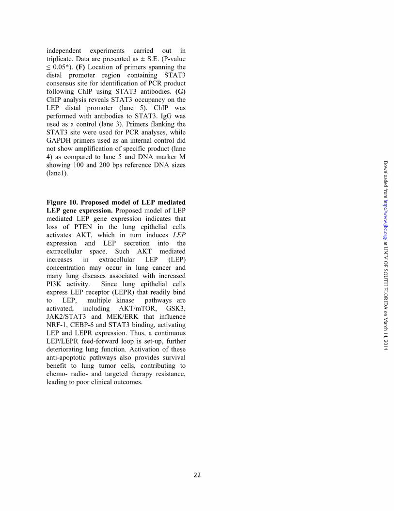
22
independent experiments carried out in triplicate. Data are presented as ± S.E. (P-value ≤ 0.05*). (F) Location of primers spanning the distal promoter region containing STAT3 consensus site for identification of PCR product following ChIP using STAT3 antibodies. (G) ChIP analysis reveals STAT3 occupancy on the LEP distal promoter (lane 5). ChIP was performed with antibodies to STAT3. IgG was used as a control (lane 3). Primers flanking the STAT3 site were used for PCR analyses, while GAPDH primers used as an internal control did not show amplification of specific product (lane 4) as compared to lane 5 and DNA marker M showing 100 and 200 bps reference DNA sizes (lane1).
Figure 10. Proposed model of LEP mediated LEP gene expression. Proposed model of LEP mediated LEP gene expression indicates that loss of PTEN in the lung epithelial cells activates AKT, which in turn induces LEP expression and LEP secretion into the extracellular space. Such AKT mediated increases in extracellular LEP (LEP) concentration may occur in lung cancer and many lung diseases associated with increased PI3K activity. Since lung epithelial cells express LEP receptor (LEPR) that readily bind to LEP, multiple kinase pathways are activated, including AKT/mTOR, GSK3, JAK2/STAT3 and MEK/ERK that influence NRF-1, CEBP-δ and STAT3 binding, activating LEP and LEPR expression. Thus, a continuous LEP/LEPR feed-forward loop is set-up, further deteriorating lung function. Activation of these anti-apoptotic pathways also provides survival benefit to lung tumor cells, contributing to chemo- radio- and targeted therapy resistance, leading to poor clinical outcomes.
at UN
IV O
F SOU
TH
FLO
RID
A on M
arch 14, 2014http://w
ww
.jbc.org/D
ownloaded from

BA
Pten∆/∆Ptenflox/flox
Figure 1
1 2 3 4 5 6
(i)
(ii)
(iii)
(iv)
E 0.5 E 6.5 E 14.5lungs harvested
(20 weeks)DOX
E 18.5
at UNIV OF SOUTH FLORIDA on March 14, 2014 http://www.jbc.org/ Downloaded from

1
2
3
4
5
78
9
10
6
1 2 3 -log(pValue)4
Development_Neurogenesis_Synaptogenesis
Proteolysis _Connective tissue degradation
Proliferation _Cell Proliferation
Neurophysiologic process _nerve impulse
Signal transduction_Leptin signaling
Proteolysis_ECM remodeling
Anti-Apoptosis_PI3K/AKT signaling
Inflammation_IL-6 signaling
Reproduction_Feeding/Neurohormone signaling
Cell adhesion_Cell-matrix interactions
Networks
KEGG: Adipocyte Signaling PathwayA
B
C
Lung AdenocarcinomaNo Primary AntibodiesLeptin Antibodies
Control lungLeptin Antibodies
D
Figure 2
at UNIV OF SOUTH FLORIDA on March 14, 2014 http://www.jbc.org/ Downloaded from

H1
65
0
A5
49
H2
92
PTEN
β-actin
LEP
LEPR
Human LEP gene-Proximal Enhancer
1 2 3 4Leptin ngs/ml:
0
Ce
ll n
um
be
r (f
old
)
3
1
2
4
*
* *
- 200
A B C
D
Figure 3
10050
*
*
**
Fold Activation
‐‐‐‐‐‐‐ (‐) Leptin‐‐‐‐‐‐‐ (+) Leptin
Wounding
E
Whole Cell Extract
Protein (μg):
H1650 PTEN-null Cells
10 20 40
A5
49
H1
65
0
H2
92
LEPLEPR
GAPDH
GF
at UNIV OF SOUTH FLORIDA on March 14, 2014 http://www.jbc.org/ Downloaded from

**
**
**
** **
****
**
0
1
2
1 2 3+
+ ++
**
+
****
CA-STAT3Luc-150
PGL3
A B C
D E F
Figure 4
at UNIV OF SOUTH FLORIDA on March 14, 2014 http://www.jbc.org/ Downloaded from
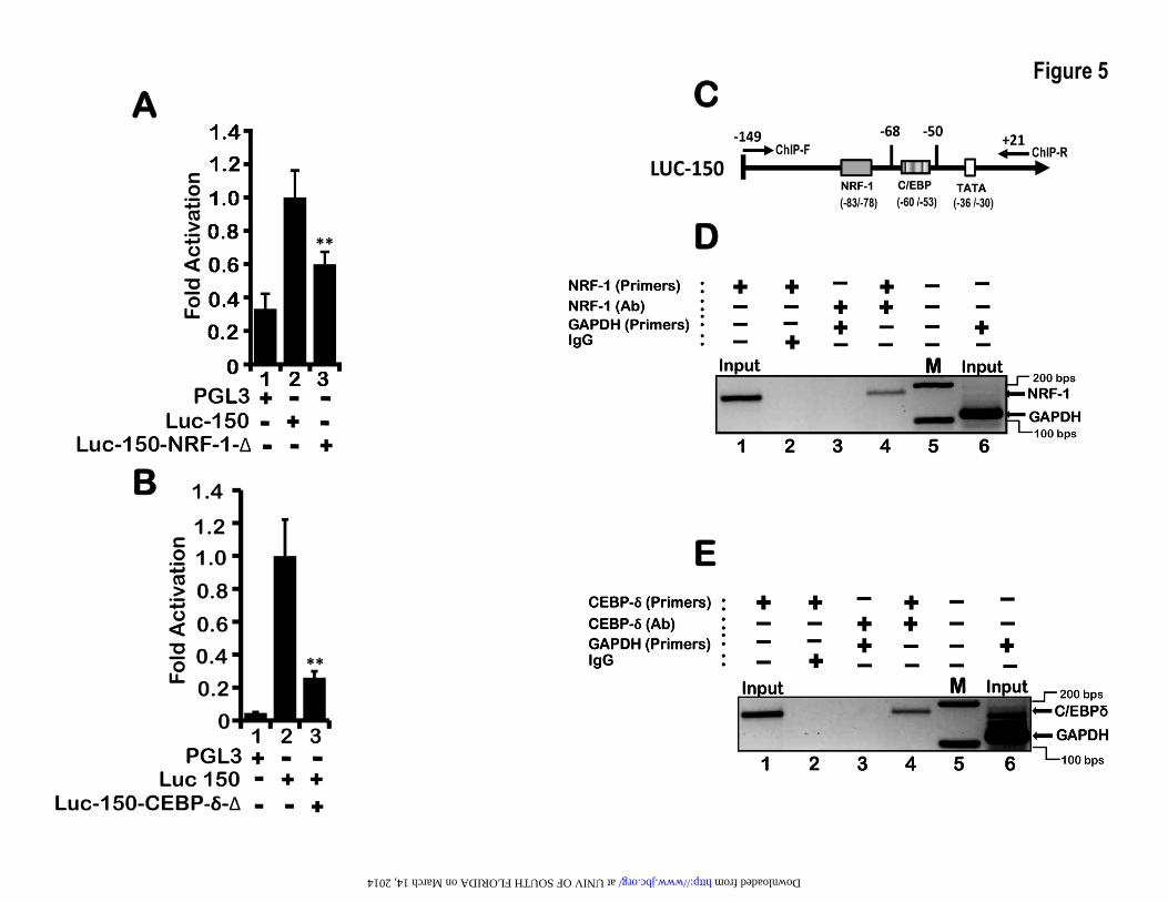
**
**
**
A
B
C
D
E
Figure 5
at UNIV OF SOUTH FLORIDA on March 14, 2014 http://www.jbc.org/ Downloaded from
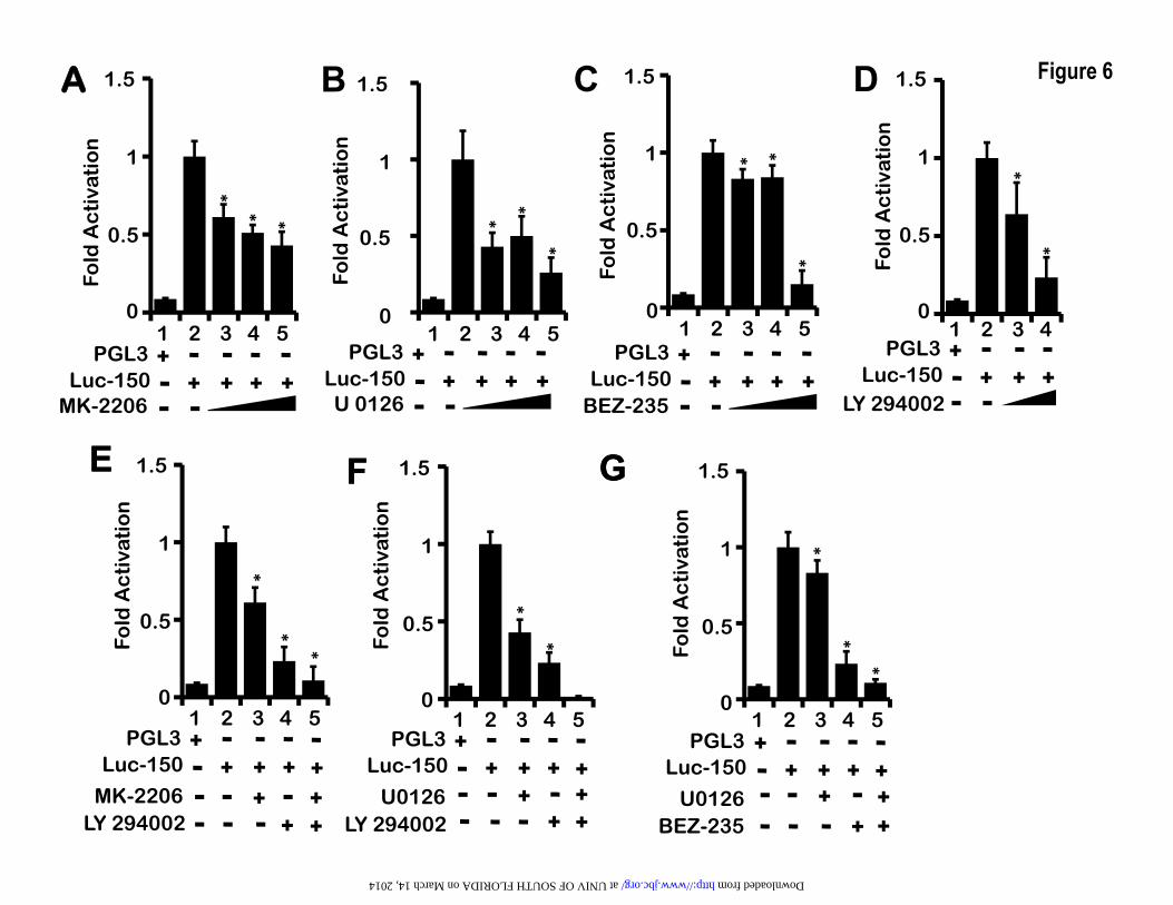
0
0.5
1
1.5
1 2 3 4 5
Luc-150U 0126
+ + ++PGL3 +
Fold
Ac
tiva
tio
n0
0.5
1
1.5
1 2 3 4 5
Luc-150MK-2206
+ + ++PGL3 +
Fold
Ac
tiva
tio
n
*
* * *
*
*
1.5
0
0.5
1
1 2 3 4 5
Luc-150 + + ++PGL3 +
Fold
Ac
tiva
tio
n
0
0.5
1
1.5
1 2 3 4
Luc-150LY 294002
+ + +PGL3 +
Fold
Ac
tiva
tio
n
BEZ-235* *
*
*
*
*
E 1.5
0
0.5
1
1 2 3 4 5
Luc-150 + + +PGL3 +
MK-2206 +LY 294002 +
+
Fold
Ac
tiva
tio
n
0
0.5
1
1.5
1 2 3 4 5
Luc-150 + + +PGL3 +
U0126+
Fold
Ac
tiva
tio
n
++ +
0
0.5
1
1.5
1 2 3 4 5
Luc-150 + + +PGL3 +
U0126BEZ-235
+
Fold
Ac
tiva
tio
n
++++
F G
+LY 294002
++
**
*
*
*
*
*
B C D
E F G
A Figure 6
at UNIV OF SOUTH FLORIDA on March 14, 2014 http://www.jbc.org/ Downloaded from

**
**
1.0
0
2.0
1 2Control +
shNRF-1B +
1 20
Control +shNRF-1B +
1.0
2.0
LEP
-mR
NA
(fo
ld)
LEP
R-m
RN
A (
fold
)
*
*
LEP
-mR
NA
(fo
ld)
LEP
-mR
NA
(fo
ld)
A B
1 2 3
0.5
1
1.5
0shControl
shNRF-1A
shNRF-1B
++
+
NR
F-1
mR
NA
(fo
ld)
shC
on
tro
l
sh N
RF
-1 B
shN
RF
-1 A
Ma
rke
r
NRF1
1 2 3 4
*
*
C
D E
Figure 7
at UNIV OF SOUTH FLORIDA on March 14, 2014 http://www.jbc.org/ Downloaded from

A B C
1
2
3
4
00
0.5
1
1.5
2
0
0.5
1
1.5
2
0
0.5
1
1.5
2
Fold
Act
ivat
ion
Fold
Act
ivat
ion
* *
*
*
48 hrs 72 hrs 48 hrs 72 hrs
Fold
Act
ivat
ion
*
*
1
2
3
0 0
0.5
1
1.5
2
48 hrs 72 hrs
Leptin:48 hrs 72 hrs
Leptin:48 hrs 72 hrs
Leptin:48 hrs 72 hrs
P-mTORP-AKTP-STAT- -+ +
β-actin
- -+ +
β-actin
- -+ +
β-actin
Figure 8
D E F GLeptin: (Min)
P-ERK P-P38 P-54 SNP/JNK
0
1
2 **
0
1
2
3
0 15 30 0
1
2
3
*
0 15 30 600
1
2
3
*
0 15 30 60 0 15 30 60
β-actin β-actin
Leptin: (Min)
β-actin
P-46 SNP/JNK
β-actin
Leptin: (Min)0 15 30
Leptin: (Min)0 15 30 60 0 15 30 60 0 15 30 60
Fold
Act
ivat
ion
Fold
Act
ivat
ion
Fold
Act
ivat
ion
Fold
Act
ivat
ion
Min Min Min Min
at UNIV OF SOUTH FLORIDA on March 14, 2014 http://www.jbc.org/ Downloaded from
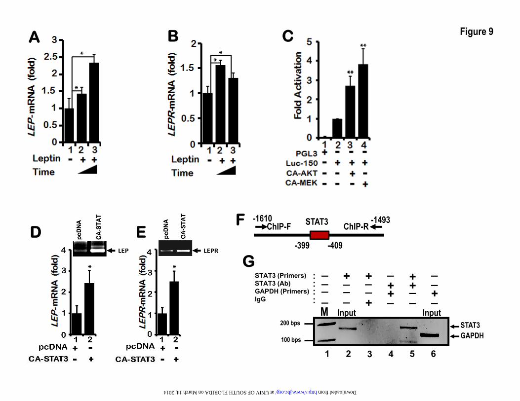
**
*
* ** *
*M InputInput
STAT3 (Primers) STAT3 (Ab) GAPDH (Primers)IgG
::
:: _
_
__ +_
_+
___
+ _+_+
+
__+ +_
__
200 bps
100 bps
STAT3GAPDH
1 2 3 4 5 6
ChIP-F ChIP-R-1493-1610
-399 -409
STAT3
A B
F
C
G
D E
Figure 9
* *
D E
at UNIV OF SOUTH FLORIDA on March 14, 2014 http://www.jbc.org/ Downloaded from

ERKP38JNK
Figure 10
at UNIV OF SOUTH FLORIDA on March 14, 2014 http://www.jbc.org/ Downloaded from
Copyright © 2022 FDOKUMEN