Identification of Unknown Homeopathic Remedies by Delayed Luminescence
Upload
independentCategory
view
6download
0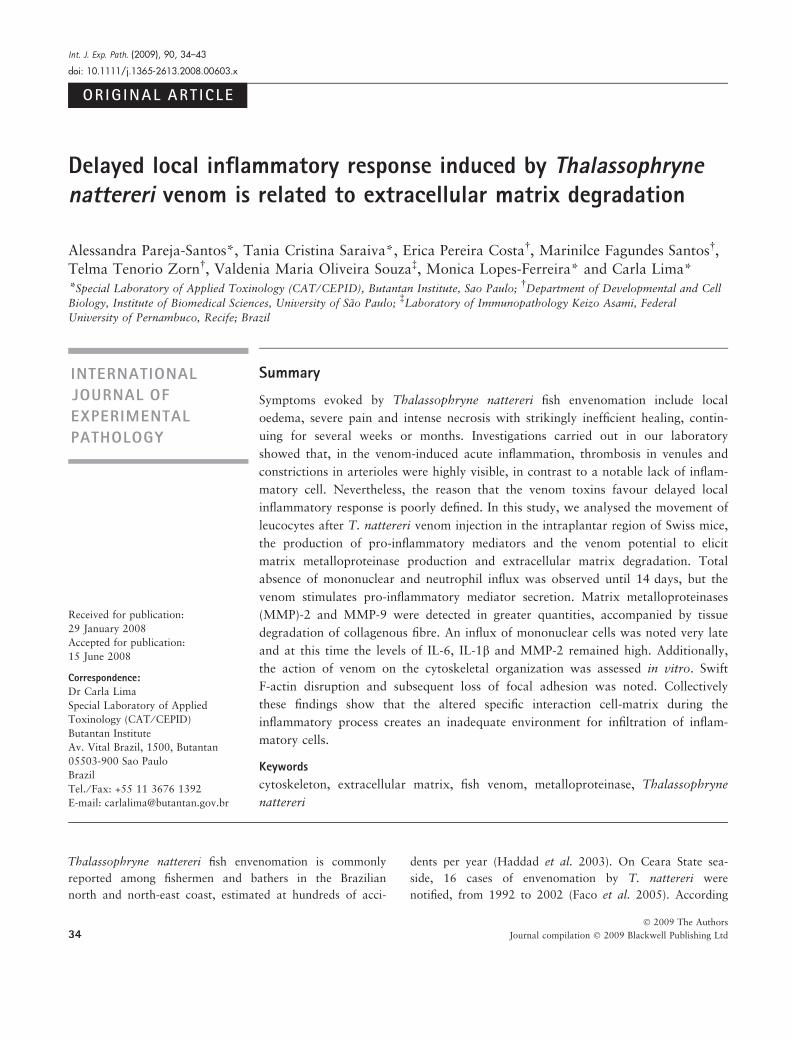
ORIG INAL ART ICLE
Delayed local inflammatory response induced by Thalassophrynenattereri venom is related to extracellular matrix degradation
Alessandra Pareja-Santos*, Tania Cristina Saraiva*, Erica Pereira Costa�, Marinilce Fagundes Santos�,
Telma Tenorio Zorn�, Valdenia Maria Oliveira Souza�, Monica Lopes-Ferreira* and Carla Lima*
*Special Laboratory of Applied Toxinology (CAT ⁄ CEPID), Butantan Institute, Sao Paulo;�Department of Developmental and Cell
Biology, Institute of Biomedical Sciences, University of Sao Paulo;�Laboratory of Immunopathology Keizo Asami, Federal
University of Pernambuco, Recife; Brazil
Thalassophryne nattereri fish envenomation is commonly
reported among fishermen and bathers in the Brazilian
north and north-east coast, estimated at hundreds of acci-
dents per year (Haddad et al. 2003). On Ceara State sea-
side, 16 cases of envenomation by T. nattereri were
notified, from 1992 to 2002 (Faco et al. 2005). According
INTERNATIONAL
JOURNAL OF
EXPERIMENTAL
PATHOLOGY
Received for publication:
29 January 2008
Accepted for publication:
15 June 2008
Correspondence:
Dr Carla Lima
Special Laboratory of Applied
Toxinology (CAT ⁄ CEPID)
Butantan Institute
Av. Vital Brazil, 1500, Butantan
05503-900 Sao Paulo
Brazil
Tel. ⁄ Fax: +55 11 3676 1392
E-mail: [email protected]
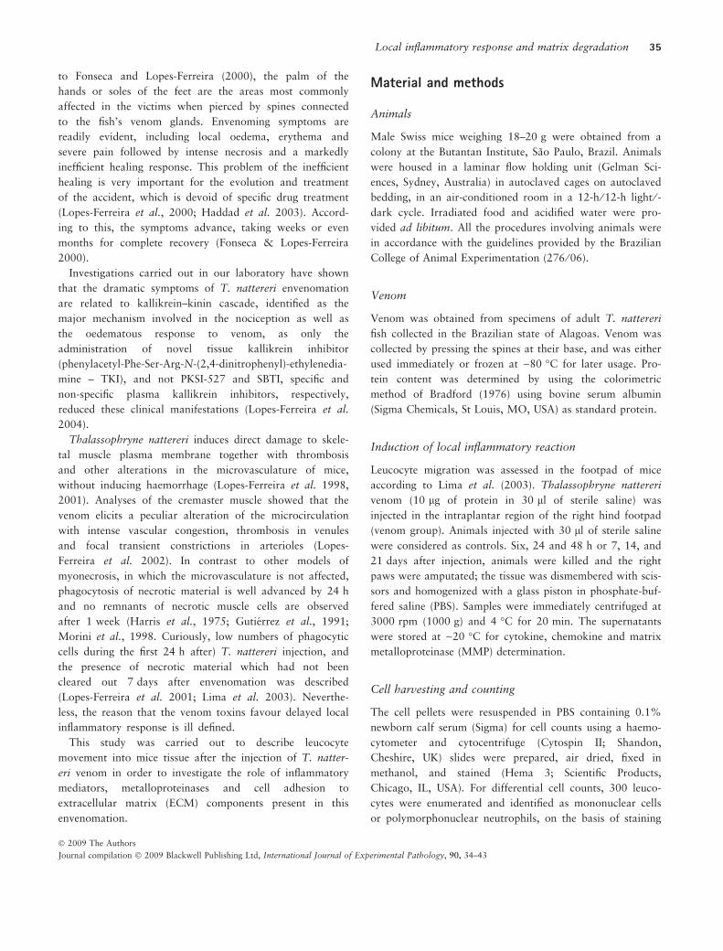
Summary
Symptoms evoked by Thalassophryne nattereri fish envenomation include local
oedema, severe pain and intense necrosis with strikingly inefficient healing, contin-
uing for several weeks or months. Investigations carried out in our laboratory
showed that, in the venom-induced acute inflammation, thrombosis in venules and
constrictions in arterioles were highly visible, in contrast to a notable lack of inflam-
matory cell. Nevertheless, the reason that the venom toxins favour delayed local
inflammatory response is poorly defined. In this study, we analysed the movement of
leucocytes after T. nattereri venom injection in the intraplantar region of Swiss mice,
the production of pro-inflammatory mediators and the venom potential to elicit
matrix metalloproteinase production and extracellular matrix degradation. Total
absence of mononuclear and neutrophil influx was observed until 14 days, but the
venom stimulates pro-inflammatory mediator secretion. Matrix metalloproteinases
(MMP)-2 and MMP-9 were detected in greater quantities, accompanied by tissue
degradation of collagenous fibre. An influx of mononuclear cells was noted very late
and at this time the levels of IL-6, IL-1b and MMP-2 remained high. Additionally,
the action of venom on the cytoskeletal organization was assessed in vitro. Swift
F-actin disruption and subsequent loss of focal adhesion was noted. Collectively
these findings show that the altered specific interaction cell-matrix during the
inflammatory process creates an inadequate environment for infiltration of inflam-
matory cells.
Keywords
cytoskeleton, extracellular matrix, fish venom, metalloproteinase, Thalassophryne
nattereri
Int. J. Exp. Path. (2009), 90, 34–43
doi: 10.1111/j.1365-2613.2008.00603.x
34� 2009 The Authors
Journal compilation � 2009 Blackwell Publishing Ltd
to Fonseca and Lopes-Ferreira (2000), the palm of the
hands or soles of the feet are the areas most commonly
affected in the victims when pierced by spines connected
to the fish’s venom glands. Envenoming symptoms are
readily evident, including local oedema, erythema and
severe pain followed by intense necrosis and a markedly
inefficient healing response. This problem of the inefficient
healing is very important for the evolution and treatment
of the accident, which is devoid of specific drug treatment
(Lopes-Ferreira et al., 2000; Haddad et al. 2003). Accord-
ing to this, the symptoms advance, taking weeks or even
months for complete recovery (Fonseca & Lopes-Ferreira
2000).
Investigations carried out in our laboratory have shown
that the dramatic symptoms of T. nattereri envenomation
are related to kallikrein–kinin cascade, identified as the
major mechanism involved in the nociception as well as
the oedematous response to venom, as only the
administration of novel tissue kallikrein inhibitor
(phenylacetyl-Phe-Ser-Arg-N-(2,4-dinitrophenyl)-ethylenedia-
mine – TKI), and not PKSI-527 and SBTI, specific and
non-specific plasma kallikrein inhibitors, respectively,
reduced these clinical manifestations (Lopes-Ferreira et al.
2004).
Thalassophryne nattereri induces direct damage to skele-
tal muscle plasma membrane together with thrombosis
and other alterations in the microvasculature of mice,
without inducing haemorrhage (Lopes-Ferreira et al. 1998,
2001). Analyses of the cremaster muscle showed that the
venom elicits a peculiar alteration of the microcirculation
with intense vascular congestion, thrombosis in venules
and focal transient constrictions in arterioles (Lopes-
Ferreira et al. 2002). In contrast to other models of
myonecrosis, in which the microvasculature is not affected,
phagocytosis of necrotic material is well advanced by 24 h
and no remnants of necrotic muscle cells are observed
after 1 week (Harris et al., 1975; Gutierrez et al., 1991;
Morini et al., 1998. Curiously, low numbers of phagocytic
cells during the first 24 h after) T. nattereri injection, and
the presence of necrotic material which had not been
cleared out 7 days after envenomation was described
(Lopes-Ferreira et al. 2001; Lima et al. 2003). Neverthe-
less, the reason that the venom toxins favour delayed local
inflammatory response is ill defined.
This study was carried out to describe leucocyte
movement into mice tissue after the injection of T. natter-
eri venom in order to investigate the role of inflammatory
mediators, metalloproteinases and cell adhesion to
extracellular matrix (ECM) components present in this
envenomation.
Material and methods
Animals
Male Swiss mice weighing 18–20 g were obtained from a
colony at the Butantan Institute, Sao Paulo, Brazil. Animals
were housed in a laminar flow holding unit (Gelman Sci-
ences, Sydney, Australia) in autoclaved cages on autoclaved
bedding, in an air-conditioned room in a 12-h ⁄ 12-h light ⁄ -dark cycle. Irradiated food and acidified water were pro-
vided ad libitum. All the procedures involving animals were
in accordance with the guidelines provided by the Brazilian
College of Animal Experimentation (276 ⁄ 06).
Venom
Venom was obtained from specimens of adult T. nattereri
fish collected in the Brazilian state of Alagoas. Venom was
collected by pressing the spines at their base, and was either
used immediately or frozen at )80 �C for later usage. Pro-
tein content was determined by using the colorimetric
method of Bradford (1976) using bovine serum albumin
(Sigma Chemicals, St Louis, MO, USA) as standard protein.
Induction of local inflammatory reaction
Leucocyte migration was assessed in the footpad of mice
according to Lima et al. (2003). Thalassophryne nattereri
venom (10 lg of protein in 30 ll of sterile saline) was
injected in the intraplantar region of the right hind footpad
(venom group). Animals injected with 30 ll of sterile saline
were considered as controls. Six, 24 and 48 h or 7, 14, and
21 days after injection, animals were killed and the right
paws were amputated; the tissue was dismembered with scis-
sors and homogenized with a glass piston in phosphate-buf-
fered saline (PBS). Samples were immediately centrifuged at
3000 rpm (1000 g) and 4 �C for 20 min. The supernatants
were stored at )20 �C for cytokine, chemokine and matrix
metalloproteinase (MMP) determination.
Cell harvesting and counting
The cell pellets were resuspended in PBS containing 0.1%
newborn calf serum (Sigma) for cell counts using a haemo-
cytometer and cytocentrifuge (Cytospin II; Shandon,
Cheshire, UK) slides were prepared, air dried, fixed in
methanol, and stained (Hema 3; Scientific Products,
Chicago, IL, USA). For differential cell counts, 300 leuco-
cytes were enumerated and identified as mononuclear cells
or polymorphonuclear neutrophils, on the basis of staining
Local inflammatory response and matrix degradation 35
� 2009 The Authors
Journal compilation � 2009 Blackwell Publishing Ltd, International Journal of Experimental Pathology, 90, 34–43
and morphologic characteristics using a conventional light
microscope (Axio Imager A1; Carl Zeiss, Meditec, Dublin,
CA, USA).
Quantification of cytokines and chemokines
Cytokines and chemokines were measured in footpad tissue
homogenates by a specific two-site sandwich ELISA, using
the OpEIA Kit (BD Pharmingen, San Diego, CA, USA).
Detection limits were 7.8 pg ⁄ ml for IL-1b, IL-6, TNF-a and
MCP-1 or 15.62 pg ⁄ ml for keratinocyte-derived chemokine
or cytokine-induced neutrophil chemoattractant (KC).
Determination of MMP-2 and MMP-9 levels using
ELISA
Matrix metalloproteinase levels in footpad homogenate sam-
ples were analysed using mouse ELISA kits following manu-
facturer’s instructions (R&D Systems, Minneapolis, MN,
USA). Detection limit was 0.75 or 0.65 ng ⁄ ml for MMP-2
and MMP-9 respectively.
Determination of MMPs in footpad after venom injection
using gelatin zymography
Enzymatic activity in footpad homogenates of mice injected
or not with T. nattereri venom was analysed using zymo-
graphy gel containing 1 mg ⁄ ml gelatin, in the presence of
sodium dodecyl sulphate (SDS), under non-reducing condi-
tions as previously described (Hibbs et al. 1985; Kleiner &
Stetler-Stevenson 1994). After electrophoresis, the gels were
stained with Coomassie Brilliant blue and destained using
acetic acid in methanol and H2O (1:3:6), both for 2 h, to
visualize bands with gelatinolytic activity. Gels were then
photographed by means of a digital camera (Nikon Coolpix
S1; Nikon Instruments Inc, Melville, NY, USA). The molec-
ular mass (kDa) of the gelatinases was estimated against
markers of known recombinant proteins (R&D Systems).
Footpad total collagen measurement
Total collagen content was determined using the Sircol colla-
gen assay as previously described (Muro et al. 2008; Saito
et al. 2008). Briefly, frozen right footpad of each animal was
homogenized in 500 ll of radio-immunoprecipitation assay
buffer containing protease inhibitors. Homogenates were
centrifuged at 13,000 g for 10 min at 4 �C. Sirius red
reagent (50 ll) was added to each footpad homogenate
(50 ll) and mixed for 30 min. The collagen–dye complex
was precipitated by centrifugation at 16,000 g for 5 min,
washed with ethanol, and dissolved in 0.5 M NaOH.
Finally, absorbance was measured at 540 nm and compared
with a calibration curve generated using the collagen
standard provided by the manufacturer (Biocolor Ltd,
Carrickfergus, UK).
Tissue staining
Mice were killed and injected footpads were removed and
immediately fixed in 10% buffered formalin. The tissue was
then processed and embedded in paraffin. Five-micrometre
tissue sections were prepared and stained using haematoxy-
lin and eosin (H&E) methods. The stained, paraffin-embed-
ded sections were examined and qualitatively evaluated
using light photomicroscopy for inflammatory and architec-
tural changes to evaluate general morphology. All slides
were examined with light microscopy at a magnification of
·40 (Axio Imager A1; Carl Zeiss) calibrated with a reference
micrometre slide. For each group of six mice, four stained
footpad sections from each mouse were analysed.
Stimulation of epithelial cells with Thalassophryne
nattereri venom
The IEC-6 cells (ATCC # CRL 1592) were cultured in Dul-
becco’s Modified Eagle’s Medium with 5% FBS plus 10 lg
of insulin, 100 units of penicillin, 100 lg of streptomycin
sulphate and 0.29 mg of l-glutamine ⁄ ml (DMEM-FBS)
according to Quaroni et al. (1979). Approximately 2 · 105
cells were plated per 35-mm dish (each containing a glass
cover slip) and incubated with T. nattereri venom at a con-
centration of 5 lg ⁄ ml to the experimental dishes. Cells were
incubated for 1, 5 or 60 min at 37 �C, 5% CO2. Polylysine
(Sigma)-coated slides were used as a control for the effects
of non-specific adhesion.
F-actin staining
After culture, cells were fixed and permeabilized with 2%
formaldehyde plus 0.2% Triton X-100 in PEM buffer
(10 mM PIPES, 5 mM EGTA, 2 mM MgCl2, pH 6.8) for
10 min at RT and postfixed with 95% ethanol for 5 min at
)20 �C. In fibroblasts and most adherent cells, focal adhe-
sion involves interactions between adhesion molecules and
cytoskeletonal proteins, such as actin (Richardson & Parsons
1996). F-actin was detected using rhodamine–phalloidin
staining according to manufacturer’s instructions. Samples
were imaged with a Nikon PCM 2000 Laser Scanning
Confocal microscope (Nikon Instruments Inc, Melville, NY,
USA).
36 A. Pareja-Santos et al.
� 2009 The Authors
Journal compilation � 2009 Blackwell Publishing Ltd, International Journal of Experimental Pathology, 90, 34–43
Statistical analysis
All values were expressed as mean ± SEM. Parametric data
were evaluated using an analysis of variance, followed by the
Bonferroni test. Non-parametric data were assessed using the
Mann–Whitney test. Differences were considered statistically
significant at P < 0.05. The spss statistical package (Release
13.0, Evaluation Version, 2004; SPSS Inc, Chicago, Illinois,
USA) was employed. Experiments were repeated at least
three times.
Results
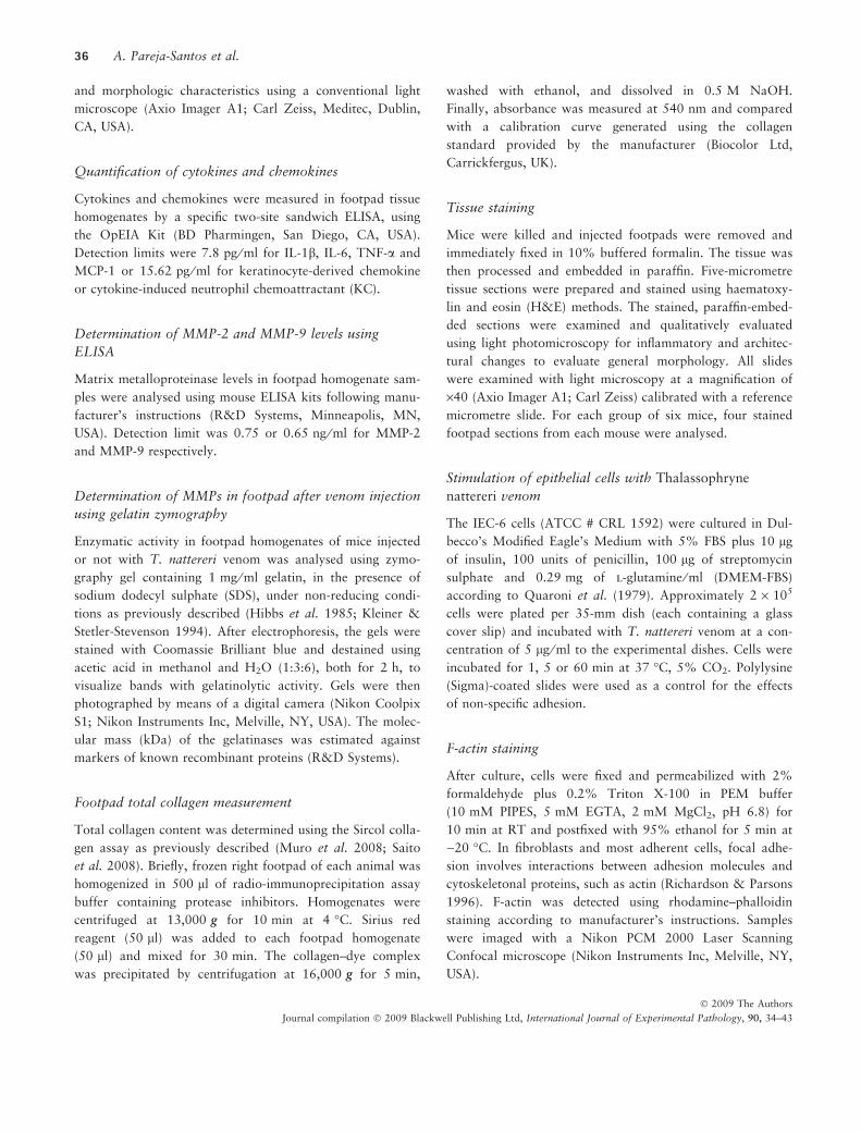
Impaired early leucocyte migration induced by
Thalassophryne nattereri venom
Rapid accumulation of neutrophils to a site of inflammation
is a defining early event of innate immunity. Thalassophryne
nattereri venom does not induce an increase in neutrophils
in footpad during the first 24 h, but, by contrast, caused a
marked decrease in mononuclear cell recruitment at 48 h
(Figure 1c,d). A drastic reduction in neutrophil number com-
pared with control mice provoked by the venom was
observed at 14 and 21 days after injection (Figure 1b). Sig-
nificant influx of leucocytes to the footpad was only
observed 21 days after venom injection with augmented
number of mononuclear cells (Figure 1a,c,d). Thus, T. nat-
tereri venom causes inappropriate early polymorphonuclear
(PMN) recruitment in the footpad of mice.
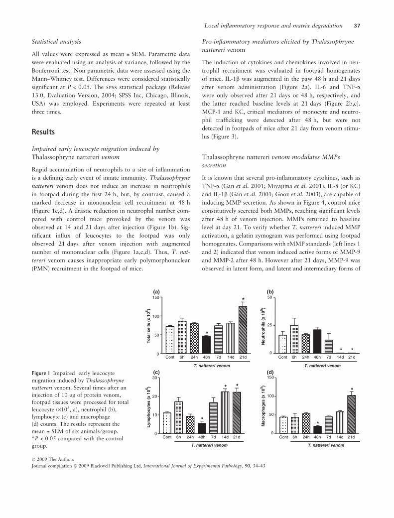
Pro-inflammatory mediators elicited by Thalassophryne
nattereri venom
The induction of cytokines and chemokines involved in neu-
trophil recruitment was evaluated in footpad homogenates
of mice. IL-1b was augmented in the paw 48 h and 21 days
after venom administration (Figure 2a). IL-6 and TNF-a
were only observed after 21 days or 48 h, respectively, and
the latter reached baseline levels at 21 days (Figure 2b,c).
MCP-1 and KC, critical mediators of monocyte and neutro-
phil trafficking were detected after 48 h, but were not
detected in footpads of mice after 21 day from venom stimu-
lus (Figure 3).
Thalassophryne nattereri venom modulates MMPs
secretion
It is known that several pro-inflammatory cytokines, such as
TNF-a (Gan et al. 2001; Miyajima et al. 2001), IL-8 (or KC)
and IL-1b (Gan et al. 2001; Gooz et al. 2003), are capable of
inducing MMP secretion. As shown in Figure 4, control mice
constitutively secreted both MMPs, reaching significant levels
after 48 h of venom injection. MMPs returned to baseline
level at day 21. To verify whether T. nattereri induced MMP
activation, a gelatin zymogram was performed using footpad
homogenates. Comparisons with rMMP standards (left lines 1
and 2) indicated that venom induced active forms of MMP-9
and MMP-2 after 48 h. However after 21 days, MMP-9 was
observed in latent form, and latent and intermediary forms of
100
150 (a) (b)
(c) (d)
*
21d 0
50 *
To
tal c
ells
(x
105 )
T. nattereri venom
Cont 6h 24h 48h 7d 14d
50
Neu
tro
ph
ils (
x 10
5 )
0
25
* *
T. nattereri venom
21d Cont 6h 24h 48h 7d 14d
Lym
ph
ocy
tes
(x 1
05 )
20
30
* *
0
10 *
T. nattereri venom
21d Cont 6h 24h 48h 7d 14d
Mac
rop
hag
es (
x 10
5 )
100
150
*
0
50
*
T. nattereri venom
21d Cont 6h 24h 48h 7d 14d
Figure 1 Impaired early leucocyte
migration induced by Thalassophryne
nattereri venom. Several times after an
injection of 10 lg of protein venom,
footpad tissues were processed for total
leucocyte (·105, a), neutrophil (b),
lymphocyte (c) and macrophage
(d) counts. The results represent the
mean ± SEM of six animals ⁄ group.
*P < 0.05 compared with the control
group.
Local inflammatory response and matrix degradation 37
� 2009 The Authors
Journal compilation � 2009 Blackwell Publishing Ltd, International Journal of Experimental Pathology, 90, 34–43
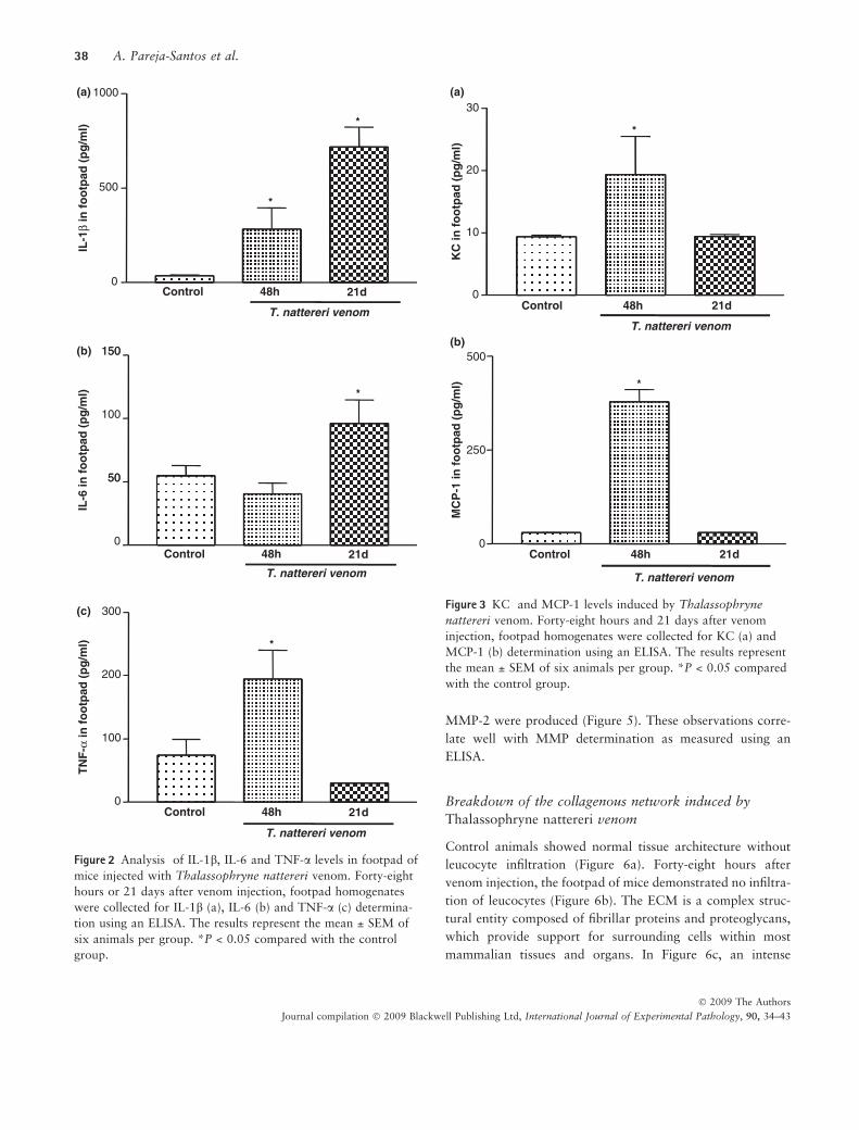
MMP-2 were produced (Figure 5). These observations corre-
late well with MMP determination as measured using an
ELISA.
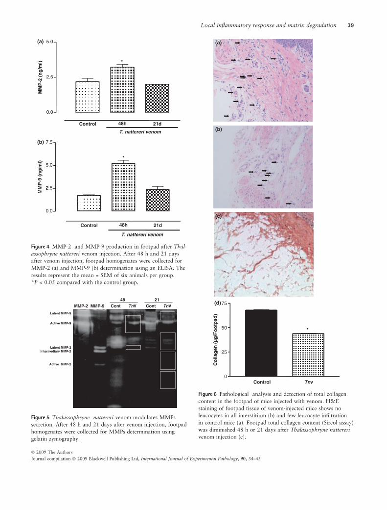
Breakdown of the collagenous network induced by
Thalassophryne nattereri venom
Control animals showed normal tissue architecture without
leucocyte infiltration (Figure 6a). Forty-eight hours after
venom injection, the footpad of mice demonstrated no infiltra-
tion of leucocytes (Figure 6b). The ECM is a complex struc-
tural entity composed of fibrillar proteins and proteoglycans,
which provide support for surrounding cells within most
mammalian tissues and organs. In Figure 6c, an intense
1000 (a)
(b)
(c)
*
500 *
IL-1
β in
fo
otp
ad (
pg
/ml)
21d 0
T. nattereri venom
Control 48h
IL-6
in f
oo
tpad
(p
g/m
l)
150
50
100
150
*
0
50
T. nattereri venom
21dControl 48h
TN
F-α
in f
oo
tpad
(p
g/m
l)
200
300
*
0
100
T. nattereri venom
21dControl 48h
Figure 2 Analysis of IL-1b, IL-6 and TNF-a levels in footpad of
mice injected with Thalassophryne nattereri venom. Forty-eight
hours or 21 days after venom injection, footpad homogenates
were collected for IL-1b (a), IL-6 (b) and TNF-a (c) determina-
tion using an ELISA. The results represent the mean ± SEM of
six animals per group. *P < 0.05 compared with the control
group.
20
30 (a)
(b)
*
10
KC
in f
oo
tpad
(p
g/m
l)
21d 0
T. nattereri venom
Control 48h
MC
P-1
in f
oo
tpad
(p
g/m
l)
500
*
0
250
T. nattereri venom
21d Control 48h
Figure 3 KC and MCP-1 levels induced by Thalassophryne
nattereri venom. Forty-eight hours and 21 days after venom
injection, footpad homogenates were collected for KC (a) and
MCP-1 (b) determination using an ELISA. The results represent
the mean ± SEM of six animals per group. *P < 0.05 compared
with the control group.
38 A. Pareja-Santos et al.
� 2009 The Authors
Journal compilation � 2009 Blackwell Publishing Ltd, International Journal of Experimental Pathology, 90, 34–43
MM
P-9
(n
g/m
l)
2.5
5.0
7.5
*
Control 21d
0.0
2
T. nattereri venom
48h
5.0(a)
(b)
*
0.0
2.5
MM
P-2
(n
g/m
l)
T. nattereri venom
Control 21d48h
Figure 4 MMP-2 and MMP-9 production in footpad after Thal-
assophryne nattereri venom injection. After 48 h and 21 days
after venom injection, footpad homogenates were collected for
MMP-2 (a) and MMP-9 (b) determination using an ELISA. The
results represent the mean ± SEM of six animals per group.
*P < 0.05 compared with the control group.
MMP-2 Cont TnV Cont TnVMMP-9
Latent MMP-2Intermediary MMP-2
Active MMP-2
Latent MMP-9
Active MMP-9
48 21
Figure 5 Thalassophryne nattereri venom modulates MMPs
secretion. After 48 h and 21 days after venom injection, footpad
homogenates were collected for MMPs determination using
gelatin zymography.
50
75
(a)
(b)
*
Control Tnv0
25
Co
llag
en (
µg
/Fo
otp
ad)
(c)
(d)
Figure 6 Pathological analysis and detection of total collagen
content in the footpad of mice injected with venom. H&E
staining of footpad tissue of venom-injected mice shows no
leucocytes in all interstitium (b) and few leucocyte infiltration
in control mice (a). Footpad total collagen content (Sircol assay)
was diminished 48 h or 21 days after Thalassophryne nattereri
venom injection (c).
Local inflammatory response and matrix degradation 39
� 2009 The Authors
Journal compilation � 2009 Blackwell Publishing Ltd, International Journal of Experimental Pathology, 90, 34–43
rupture can be seen in the major ECM protein, such as colla-
gen fibres, accompanied by a significant decrease in the
amounts of total collagen content in the footpad after 48 h
(Figure 6d).
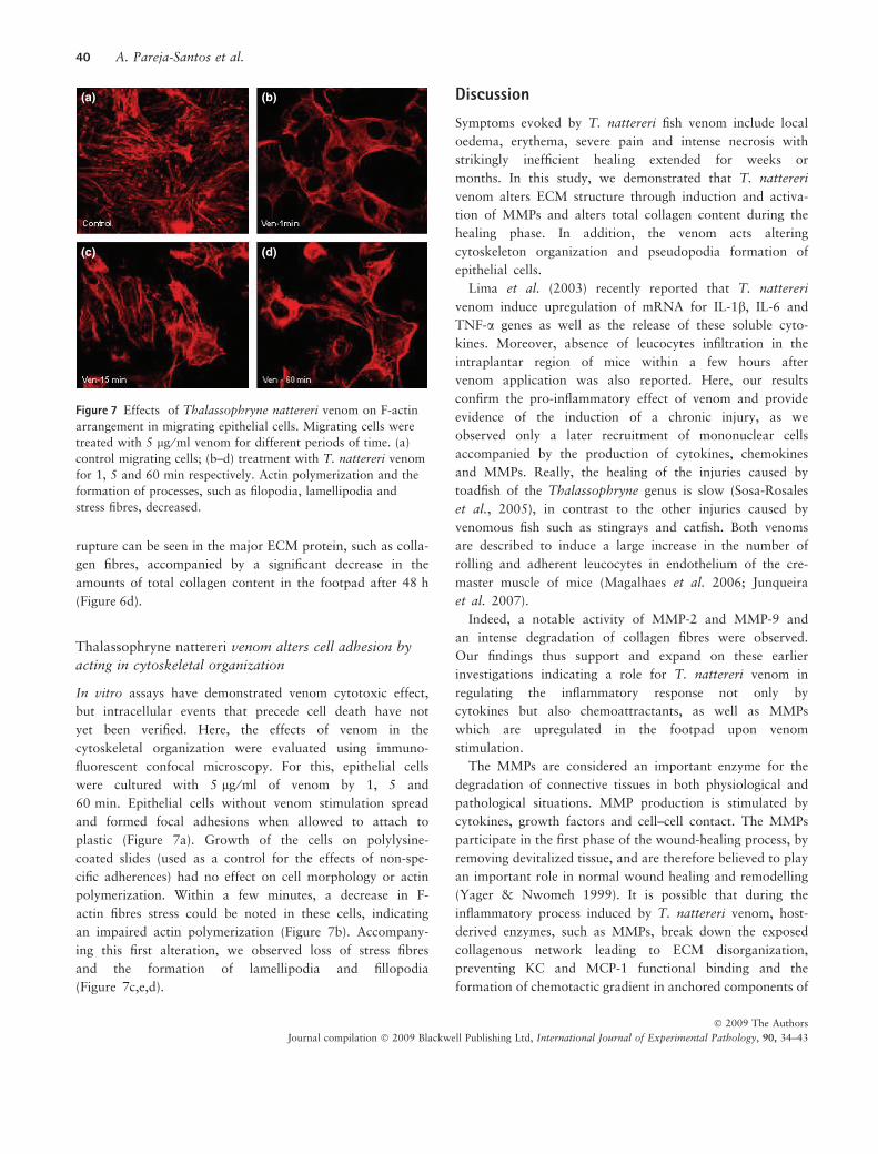
Thalassophryne nattereri venom alters cell adhesion by
acting in cytoskeletal organization
In vitro assays have demonstrated venom cytotoxic effect,
but intracellular events that precede cell death have not
yet been verified. Here, the effects of venom in the
cytoskeletal organization were evaluated using immuno-
fluorescent confocal microscopy. For this, epithelial cells
were cultured with 5 lg ⁄ ml of venom by 1, 5 and
60 min. Epithelial cells without venom stimulation spread
and formed focal adhesions when allowed to attach to
plastic (Figure 7a). Growth of the cells on polylysine-
coated slides (used as a control for the effects of non-spe-
cific adherences) had no effect on cell morphology or actin
polymerization. Within a few minutes, a decrease in F-
actin fibres stress could be noted in these cells, indicating
an impaired actin polymerization (Figure 7b). Accompany-
ing this first alteration, we observed loss of stress fibres
and the formation of lamellipodia and fillopodia
(Figure 7c,e,d).
Discussion
Symptoms evoked by T. nattereri fish venom include local
oedema, erythema, severe pain and intense necrosis with
strikingly inefficient healing extended for weeks or
months. In this study, we demonstrated that T. nattereri
venom alters ECM structure through induction and activa-
tion of MMPs and alters total collagen content during the
healing phase. In addition, the venom acts altering
cytoskeleton organization and pseudopodia formation of
epithelial cells.
Lima et al. (2003) recently reported that T. nattereri
venom induce upregulation of mRNA for IL-1b, IL-6 and
TNF-a genes as well as the release of these soluble cyto-
kines. Moreover, absence of leucocytes infiltration in the
intraplantar region of mice within a few hours after
venom application was also reported. Here, our results
confirm the pro-inflammatory effect of venom and provide
evidence of the induction of a chronic injury, as we
observed only a later recruitment of mononuclear cells
accompanied by the production of cytokines, chemokines
and MMPs. Really, the healing of the injuries caused by
toadfish of the Thalassophryne genus is slow (Sosa-Rosales
et al., 2005), in contrast to the other injuries caused by
venomous fish such as stingrays and catfish. Both venoms
are described to induce a large increase in the number of
rolling and adherent leucocytes in endothelium of the cre-
master muscle of mice (Magalhaes et al. 2006; Junqueira
et al. 2007).
Indeed, a notable activity of MMP-2 and MMP-9 and
an intense degradation of collagen fibres were observed.
Our findings thus support and expand on these earlier
investigations indicating a role for T. nattereri venom in
regulating the inflammatory response not only by
cytokines but also chemoattractants, as well as MMPs
which are upregulated in the footpad upon venom
stimulation.
The MMPs are considered an important enzyme for the
degradation of connective tissues in both physiological and
pathological situations. MMP production is stimulated by
cytokines, growth factors and cell–cell contact. The MMPs
participate in the first phase of the wound-healing process, by
removing devitalized tissue, and are therefore believed to play
an important role in normal wound healing and remodelling
(Yager & Nwomeh 1999). It is possible that during the
inflammatory process induced by T. nattereri venom, host-
derived enzymes, such as MMPs, break down the exposed
collagenous network leading to ECM disorganization,
preventing KC and MCP-1 functional binding and the
formation of chemotactic gradient in anchored components of
(a) (b)
(c) (d)
Figure 7 Effects of Thalassophryne nattereri venom on F-actin
arrangement in migrating epithelial cells. Migrating cells were
treated with 5 lg ⁄ ml venom for different periods of time. (a)
control migrating cells; (b–d) treatment with T. nattereri venom
for 1, 5 and 60 min respectively. Actin polymerization and the
formation of processes, such as filopodia, lamellipodia and
stress fibres, decreased.
40 A. Pareja-Santos et al.
� 2009 The Authors
Journal compilation � 2009 Blackwell Publishing Ltd, International Journal of Experimental Pathology, 90, 34–43
the matrix, such as syndecan-1 and proteoglycans respectively
(Parks et al. 2004).
This scenario indicates an ambiguous role of the venom in
the inflammatory process, where it displays a potent stimu-
lus ability in contrast to an inefficient capacity for support-
ing infiltrated cells in inflamed tissue due to MMP
upregulation. Furthermore, we did not rule out the possibil-
ity that enzymatic toxins in the venom can act directly as
degrading factors of ECM. Indeed, previous characterization
of proteins in the T. nattereri venom showed that Natterins
family presented intense kininogenase activity, although not
sharing the molecular motif with MMPs or other already
known protease present in databanks (Magalhaes et al.
2005).
Interestingly, after 21 days when the level of active
MMP-2 was minimal and inactive MMP-9 was present,
mononuclear cell influx was replenished, indicating a par-
tial recovery of the specific cell matrix component and tis-
sue healing. This is, however, unlikely, as neutrophil
numbers are dramatically reduced. The total absence of
TNF-a, a potent signal for neutrophil influx and activation
could explain this phenomenon (Paul & Ruddle 1988;
Flynn et al. 1995). In addition, IL-1b has been associated
with impairment of normal homeostasis of cartilage ECM
through inhibition of proteoglycans and collagen synthesis
(Fernandes et al. 2002). In our model, elevated levels of
IL-1b were detected after venom application remained
high until 3 weeks.
Collectively, these results reinforce the view that the
most efficient therapy for controlling T. nattereri venom
poisoning can be based on neutralizing the toxins and
inducing polymorphonuclear recruitment in the lesion. A
vital role for neutrophil in injury and regenerative process
was recently described by Teixeira et al. (2003) showing
that mice treated by intraperitoneal injections of antisera
to neutrophils and monocytes and injected with snake
venom show a deficient regenerative response suggesting
the importance of neutrophils for normal muscle repair.
Another interesting observation from this study was that
animals depleted of neutrophils and monocytes also
showed more tissue debris in the injured muscles, raising
the possibility at least that the impaired capacity to
remove tissue debris by phagocytes could slow the regen-
erative process.
Inflammatory responses constitute a potential link
between acute injury and chronic remodelling and wound
healing. Some studies clearly demonstrate the essential
function of neutrophils in clearing antigens and toxins for
effective injury resolution (Savill et al. 1989; Teixeira
et al. 2003; Toumi et al. 2006). After entering tissues,
granulocytes promote the switch of arachidonic acid-
derived prostaglandins and leucotrienes to lipoxins, resolv-
ins and protectins which initiate the termination sequence,
leading to neutrophil clearance and release of anti-inflam-
matory and reparative cytokines, such as transforming
growth factor-b1, essential for later initiation of ECM
deposition and remodelling (Serhan & Savill 2005). We
can speculate that for the efficient clearance of the toxins,
polymorphonuclear recruitment would be an essential step
for resolving inflammation and initiation of healing.
The secondary aim of this study was to explore the effect
of venom on focal adhesion. Focal adhesion is now regarded
as an important route of signal transduction in cell growth
and migration (Valitutti et al. 1995), and is influenced by
ECM proteins including collagen, fibronectin and laminin
(Juliano & Haskill 1993). Additionally, disruption of actin
has been associated with MMP-2 production (Sanka et al.
2007) fostering the extreme degradation of ECM. Venom-
induced ECM degradation was associated with a loss of
stress fibres and the formation of lamellipodia and fillopodia
by epithelial cells, which are critically involved in the regula-
tion of cell migration. This well-established in vitro assay
could mimic the early alteration induced by the venom in
endothelial cells previously described (Lopes-Ferreira et al.
2002) and shows that the actin cytoskeleton signals play
important roles in the communication between different cell
types in inflammatory sites.
Collectively these findings show that the altered availabil-
ity of the specific cell matrix component during the early
inflammatory process induced by T. nattereri venom create
an inadequate environment for infiltration or survival of
inflammatory cells, damaging the healing phase and inflam-
mation resolution. Understanding the mechanism used by T.
nattereri venom to reduce the early neutrophilic influx to
injured tissue is essential for establishing an adequate ther-
apy for uncommon inflammatory response observed in
envenomation.
Acknowledgements
This work was supported by the Fundacao de Amparo a
Pesquisa do Estado de Sao Paulo (FAPESP) and CNPq.
References
Bradford M.M. (1976) A rapid and sensitive method for quanti-
tation of microgram quantities of protein utilizing the princi-
ple of protein dye binding. Anal. Biochem. 72, 248–254.
Faco P.E., Bezerra G.P., Barbosa P.S. et al. (2005) Epidemiology
of the injuries caused by Thalassophryne nattereri (niquim) in
Local inflammatory response and matrix degradation 41
� 2009 The Authors
Journal compilation � 2009 Blackwell Publishing Ltd, International Journal of Experimental Pathology, 90, 34–43
Ceara State (1992–2002). Rev. Soc. Bras. Med. Trop. 38,
479–482.
Fernandes J.C., Martel-Pelletier J., Pelletier J.P. (2002) The role
of cytokines in osteoarthritis pathophysiology. Biorheology
39, 237–246.
Flynn J.L., Goldstein M.M., Chan J. et al. (1995) Tumor necro-
sis factor-a is required in the protective immune response
against Mycobacterium tuberculosis in mice. Immunity 2,
561–572.
Fonseca L.A. & Lopes-Ferreira M. (2000) Clinical and experi-
mental studies regarding poisoning caused by a fish Thalas-
sophryne nattereri (niquim). An. Bras. Dermatol. 75, 435–
443.
Gan X., Wong B., Wright S.D., Cai T.Q. (2001) Production
of matrix metalloproteinase-9 in CaCO-2 cells in response
to inflammatory stimuli. J. Interferon Cytokine Res. 21,
93–98.
Gooz M., Shaker M., Gooz P., Smolka A.J. (2003) Interleukin
1beta induces gastric epithelial cell matrix metalloproteinase
secretion and activation during Helicobacter pylori infection.
Gut 52, 1250–1256.
Gutierrez J.M., Nunez J., Dıaz C. et al. (1991) Skeletal muscle
degeneration and regeneration after injection of bothrop-
stoxin II, a phospholipase A2 isolated from the venom of the
snake. Bothrops Jararacussu. Exp. Mol. Pathol. 55, 217–229.
Haddad V. Jr, Pardal P.P., Cardoso J.L., Martins I.A. (2003)
The venomous toadfish Thalassophryne nattereri (niquim or
miquim): report of 43 injuries provoked in fishermen of Sali-
nopolis (Para State) and Aracaju (Sergipe State), Brazil. Rev.
Inst. Med. Trop. 45, 221–223.
Harris J.B., Johnson M.A., Karlsson E. (1975) Pathological
responses of rat skeletal muscle to a single subcutaneous
injection of a toxin isolated from the venom of the Australian
tiger snake, Notechis scutatus scutatus. Clin. Exp. Pharmac.
Physiol. 2, 383–404.
Hibbs M.S., Hasty K.A., Seyer J.M., Kang A.H., Mainardi C.L.
(1985) Biochemical and immunological characterization of
the secreted forms of human neutrophil gelatinase. J. Biol.
Chem. 260, 2493–2500.
Juliano R.L. & Haskill S. (1993) Signal transduction from the
extracellular matrix. J. Cell Biol. 120, 577–585.
Junqueira M., Grund L.Z., Orii N.M. et al. (2007) Analysis of
the inflammatory reaction induced by the catfish (Cathorops
spixii) venoms. Toxicon 49, 909–919.
Kleiner D.E. & Stetler-Stevenson W.G. (1994) Quantitative
zymography: detection of picogram quantities of gelatinases.
Anal. Biochem. 218, 325–329.
Lima C., Clissa B.P., Piran-Soares A.A., Tanjoni I., Moura-da-
Silva A.M., Lopes-Ferreira M. (2003) Characterisation of
local inflammatory response induced by Thalassophryne nat-
tereri fish venom in a mouse model of tissue injury. Toxicon
42, 499–507.
Lopes-Ferreira M., Barbaro K.C., Cardoso D.F., Moura-da-Silva
A.M., Mota I. (1998) Thalassophryne nattereri fish venom:
biological and biochemical characterization and serum neu-
tralization of its toxic activities. Toxicon 36, 405–410.
Lopes-Ferreira M., Moura-da-Silva A.M., Mota I., Takehara
H.A. (2000) Neutralization of Thalassophryne nattereri
(niquim) fish venom by an experimental antivenom. Toxicon
38, 1149–1156.
Lopes-Ferreira M., Nunez J., Rucavado A. et al. (2001) Skeletal
muscle necrosis and regeneration after injection of Thalas-
sophryne nattereri (niquim) fish venom in mice. Int. J. Exp.
Pathol. 82, 55–64.
Lopes-Ferreira M., Moura-da-Silva A.M., Piran-Soares A.A.
et al. (2002) Hemostatic effects induced by Thalassophryne
nattereri fish venom: a model of endothelium-mediated blood
flow impairment. Toxicon 40, 1141–1147.
Lopes-Ferreira M., Emim J.A., Oliveira V. et al. (2004) Kinino-
genase activity of Thalassophryne nattereri fish venom. Bio-
chem. Pharmacol. 68, 2151–2157.
Magalhaes G.S., Lopes-Ferreira M., Junqueira-de-Azevedo I.L.
et al. (2005) Natterins, a new class of proteins with kinino-
genase activity characterized from Thalassophryne nattereri
fish venom. Biochimie 87, 687–699.
Magalhaes K.W., Lima C., Piran-Soares A.A., Marques E.E.,
Hiruma-Lima C.A., Lopes-Ferreira M. (2006) Biological and
biochemical properties of the Brazilian Potamotrygon
stingrays: Potamotrygon cf. scobina and Potamotrygon gr.
orbignyi. Toxicon 47, 575–583.
Miyajima S., Akaike T., Matsumoto K. et al. (2001) Matriz
metalloproteinases induction by pseudomonal virulence
factors and inflammatory cytokines in vitro. Microb. Pathog.
31, 271–281.
Morini C.C., Pereira E.C.L., Ownby C.L., Salvini T.F. (1998)
Injury and recovery of fast and slow skeletal muscle fibers
affected by ACL myotoxin isolated from Agkistrodon contor-
trix laticinctus (broad-banded copperhead) venom. Toxicon
36, 1007–1024.
Muro A.F., Moretti F.A., Moore B.B. et al. (2008) An essential
role for fibronectin extra type III domain A in pulmonary
fibrosis. J. Respir. Crit. Care Med. 177, 638–645.
Parks W.C., Wilson C.L., Lopez-Boado Y.S. (2004) Matrix
metalloproteinases as modulators of inflammation and innate
immunity. Nat. Rev. Immunol. 4, 617–629.
Paul N.L. & Ruddle N.H., (1988) Lymphotoxin. Annu. Rev.
Immunol. 6, 407–438.
Quaroni A., Wands J., Trelstad R.L., Isselbacher K.J. (1979)
Epithelioid cell cultures from rat small intestine. Characteriza-
tion by morphologic and immunologic criteria. J. Cell Biol.
80, 248–265.
Richardson A. & Parsons J.T. (1996) A mechanism for regula-
tion of the adhesion-associated protein tyrosine kinase
pp125FAK. Nature 380, 538–540.
42 A. Pareja-Santos et al.
� 2009 The Authors
Journal compilation � 2009 Blackwell Publishing Ltd, International Journal of Experimental Pathology, 90, 34–43
Saito F., Tasaka S., Inoue K. et al. (2008) Role of interleukin-6
in bleomycin-induced lung inflammatory changes in mice.
Am. J. Respir. Cell Mol. Biol. 38, 566–571.
Sanka K., Maddala R., Epstein D.L., Rao P.V. (2007) Influence
of actin cytoskeletal integrity on matrix metalloproteinase-2
activation in cultured human trabecular meshwork cells.
Invest. Ophthalmol. Vis. Sci. 48, 2105–2114.
Savill J.S., Wyllie A.H., Henson J.E., Walport M.J., Henson
P.M., Haslett C. (1989) Macrophage phagocytosis of aging
neutrophils in inflammation. Programmed cell death in the
neutrophil leads to its recognition by macrophages. J. Clin.
Invest. 83, 865–875.
Serhan C.N. & Savill J. (2005) Resolution of inflammation: the
beginning programs the end. Nat. Immunol. 6, 1191–1197.
Sosa-Rosales J.I., D’Suze G., Salazar V., Fox J., Sevcik. (2005)
Purification of a myotoxin from the toadfish Thalassophryne
maculosa (Gunter) venom. Toxicon 45, 147–153.
Teixeira C.F.P., Chaves F., Zamuner S.R. et al. (2003) Effects
of neutrophil depletion in the local pathological alterations
and muscle regeneration in mice injected with Bothrops jara-
raca snake venom. Int. J. Exp. Pathol. 86, 107–115.
Toumi H., F’guyer S., Best T.M. (2006) The role of neutrophils
in injury and repair following muscle stretch. J. Anat. 208,
459–470.
Valitutti S., Dessing M., Aktories K., Gallati H., Lanzavecchia
A. (1995) Sustained signaling leading to T cell activation
results from prolonged T cell receptor occupancy. Role of T
cell actin cytoskeleton. J. Exp. Med. 181, 577–584.
Yager D.R. & Nwomeh B.C. (1999) The proteolytic environ-
ment of chronic wounds. Wound Repair Regen. 7, 433–441.
Local inflammatory response and matrix degradation 43
� 2009 The Authors
Journal compilation � 2009 Blackwell Publishing Ltd, International Journal of Experimental Pathology, 90, 34–43
Copyright © 2022 FDOKUMEN