Decreased 13-S-hydroxyoctadecadienoic acid levels and 15-lipoxygenase-1 expression in human colon...
11
Carcinogenesis vol.20 no.10 pp.1985–1995, 1999 Decreased 13-S-hydroxyoctadecadienoic acid levels and 15-lipoxygenase-1 expression in human colon cancers Imad Shureiqi 1,9 , Kirk J.Wojno 2 , Judy A.Poore 6 , Ramesh G.Reddy 7 , Micheline J.Moussalli 8 , Stephen A.Spindler 7 , Joel K.Greenson 2 , Daniel Normolle 5 , Ahmed A.K.Hasan 2 , Theodore S.Lawrence 3 and Dean E.Brenner 1,4,6 1 Division of Hematology and Oncology, Department of Internal Medicine, 2 Department of Pathology, 3 Department of Radiation Oncology, 4 Department of Pharmacology and 5 Biostatistic Core, Comprehensive Cancer Center, University of Michigan Medical School, Ann Arbor, MI 48109, 6 The Ann Arbor Veteran Affairs Medical Center, 2215 Fuller Avenue, Ann Arbor, MI 48105, 7 Oxford Biomedical Research Inc., 2165 Avon Industrial Drive, Rochester Hills, MI 48309 and 8 The Howard Hughes Medical Institute, Ann Arbor, MI 48109, USA 9 To whom correspondence should be addressed at present address: The University of Texas, M.D. Anderson Cancer Center, Department of Clinical Cancer Prevention, Box 236, 1515 Holcombe Boulevard, Houston, TX 77030, USA Email: [email protected] 13-S-Hydroxyoctadecadienoic acid (13-S-HODE), the prod- uct of 15-lipoxygenase (15-LOX) metabolism of linoleic acid, enhances cellular mitogenic responses to certain growth factors. Other observations have questioned whether 13-S-HODE has tumorigenic effects. Our study evaluated the hypothesis that 15-LOX-1 is overexpressed in colon cancers resulting in an increase in intracellular 13-S-HODE. 15-LOX-1 and 13-S-HODE were quantified using western blots, ELISA and immunohistochemistry in 18 human colon cancers with paired normal colonic mucosa. Additionally, 15-LOX-1 expression was measured by west- ern blots in three transformed colonic cell lines and in a human umbilical vein endothelial cell line. Next, we evalu- ated 13-S-HODE effects on cellular proliferation, cell cycle distribution and apoptosis in a transformed colonic cell line (RKO). Cell cycle distributions were measured by flow cytometry and apoptosis was assessed by phase contrast microscopy, electron microscopy, flow cytometry and DNA fragmentation assay. 15-LOX-1 immunohistochemistry staining scores were reduced in tumor tissues (P ≤ 0.0001) and 15-LOX-1 expression was absent in three transformed colonic cell lines. 13-S-HODE levels were also reduced in tumors tissues compared with normal controls by ELISA (median 3.3-fold, P J 0.02) and by immunohistochemistry (P ≤ 0.0001). In vitro 13-S-HODE inhibited RKO cell proliferation and induced cell cycle arrest and apoptosis. 13-S-HODE produced similar effects in HT-29 cells. Our observations indicate that: (i) human colon cancers are associated with a down-regulation in 15-LOX-1 expres- Abbreviations: DAB, 3,39-diaminobenzidine; EGF, epidermal growth factor; H&E, hematoxylin and eosin; HRP, horseradish peroxidase; HUVEC, human umbilical vein endothelial cells; 12-S-HETE, 12-S-hydroxyeicosatetraenoic acid; 13-S-HODE, 13-S-hydroxyoctadecadienoic acid; 15-LOX, 15-lipoxygen- ase; NDGA, nordihydroguaiaretic acid; PBS, phosphate-buffered saline; NaBT, sodium butyrate; SHE, Syrian hamster embryo; TBS, Tris-buffered saline. Disclosure statement: Dr Ramesh G.Reddy and Mr Stephen A.Spindler are previous employees of Oxford Biomedical Research Inc., which manufactures the 13-S-HODE kits. © Oxford University Press 1985 sion and a reduction in 13-S-HODE intracellular levels; (ii) 13-S-HODE can suppress cell proliferation and induce apoptosis in transformed colonic epithelial cells. Introduction A diet high in fats increases the risk of colorectal cancer, as shown in several epidemiological studies (1). Among dietary fats, polyunsaturated fatty acids in particular enhance carcino- gen induction of intestinal tumors (2,3). The major polyunsatur- ated fatty acid in the human diet is linoleic acid (4). Linoleic acid promotes carcinogenesis in azoxymethane rat models and the growth of murine adenocarcinomas in nude mice (5,6). Oxidation of polyunsaturated fatty acids is necessary for them to stimulate mitogenesis in rat rectal epithelia (7,8). Linoleic acid oxidation is predominantly mediated by 15-lipoxygenase (15-LOX) and produces 13-S-hydroxyoctadecadienoic acid (13-S-HODE) in humans (9,10). Recently, a second lipoxy- genase has been described and classified as 15-LOX-2 (11), while the formerly known reticulocyte 15-LOX has been renamed 15-LOX-1. Human colon tissues have been found to express 15-LOX-1 but not 15-LOX-2 (11,12). Several lines of evidence suggest a link between lipoxygen- ase metabolism of linoleic acid and tumor promotion by linoleic acid. First, lipoxygenase inhibitors attenuate linoleic acid stimulation of murine adenocarcinoma growth (13). Second, 15-LOX formation of 13-S-HODE enhances the mitogenic response to epidermal growth factor (EGF) in BALB/c3T3 fibroblasts (14) and Syrian hamster embryo (SHE) cells (15). Similarly, EGF and transforming growth factor α stimulate DNA synthesis and 13-S-HODE production in transformed breast cancer cells (BT-20) (16). Third, 13-S-HODE production increases when normal fibroblasts are transfected with c-erbB-2 (a proto-oncogene similar to EGF) (17). Finally, studies of 13-HODE dehydrogenase, the enzyme that converts 13-S-HODE to 13-oxoctadienoic acid, have also suggested an association between 13-S-HODE metabolism and colon carcinogenesis. Colonic tissues are one of two sites that have the highest level of 13-HODE dehydrogenase activity in rats (4) and 13-HODE dehydrogenase activity levels correlate positively with the degree of colonic cell differentiation (18). Correspondingly, human colonic cancer tissues have lower levels of 13-HODE dehydrogenase activity compared with normal tissues (19). Based on these observations, it has been proposed that 13-S-HODE promotes colonic cell proliferation, while 13-HODE dehydrogenase inactivates 13-S-HODE to allow colonic cellular differentiation (19). Despite these observations which imply that linoleic acid might enhance tumorigenesis through 13-S-HODE, a direct link between 13-S-HODE and colonic carcinogenesis has not been demonstrated. Additionally, some observations have questioned whether 13-S-HODE has tumorigenic effects (20), including studies on transformed SHE cells which had lost a
-
Upload
independent -
Category
Documents
-
view
0 -
download
0
Transcript of Decreased 13-S-hydroxyoctadecadienoic acid levels and 15-lipoxygenase-1 expression in human colon...

Carcinogenesisvol.20 no.10 pp.1985–1995, 1999
Decreased 13-S-hydroxyoctadecadienoic acid levels and15-lipoxygenase-1 expression in human colon cancers
Imad Shureiqi1,9, Kirk J.Wojno 2, Judy A.Poore6,Ramesh G.Reddy7, Micheline J.Moussalli8,Stephen A.Spindler7, Joel K.Greenson2, Daniel Normolle5,Ahmed A.K.Hasan2, Theodore S.Lawrence3 andDean E.Brenner1,4,6
1Division of Hematology and Oncology, Department of Internal Medicine,2Department of Pathology,3Department of Radiation Oncology,4Department of Pharmacology and5Biostatistic Core, ComprehensiveCancer Center, University of Michigan Medical School, Ann Arbor,MI 48109, 6The Ann Arbor Veteran Affairs Medical Center, 2215 FullerAvenue, Ann Arbor, MI 48105,7Oxford Biomedical Research Inc.,2165 Avon Industrial Drive, Rochester Hills, MI 48309 and8The HowardHughes Medical Institute, Ann Arbor, MI 48109, USA9To whom correspondence should be addressed at present address:The University of Texas, M.D. Anderson Cancer Center, Department ofClinical Cancer Prevention, Box 236, 1515 Holcombe Boulevard, Houston,TX 77030, USAEmail: [email protected]
13-S-Hydroxyoctadecadienoic acid (13-S-HODE), the prod-uct of 15-lipoxygenase (15-LOX) metabolism of linoleicacid, enhances cellular mitogenic responses to certaingrowth factors. Other observations have questionedwhether 13-S-HODE has tumorigenic effects. Our studyevaluated the hypothesis that 15-LOX-1 is overexpressedin colon cancers resulting in an increase in intracellular13-S-HODE. 15-LOX-1 and 13-S-HODE were quantifiedusing western blots, ELISA and immunohistochemistry in18 human colon cancers with paired normal colonic mucosa.Additionally, 15-LOX-1 expression was measured by west-ern blots in three transformed colonic cell lines and in ahuman umbilical vein endothelial cell line. Next, we evalu-ated 13-S-HODE effects on cellular proliferation, cell cycledistribution and apoptosis in a transformed colonic cellline (RKO). Cell cycle distributions were measured by flowcytometry and apoptosis was assessed by phase contrastmicroscopy, electron microscopy, flow cytometry and DNAfragmentation assay. 15-LOX-1 immunohistochemistrystaining scores were reduced in tumor tissues (P ≤ 0.0001)and 15-LOX-1 expression was absent in three transformedcolonic cell lines. 13-S-HODE levels were also reduced intumors tissues compared with normal controls by ELISA(median 3.3-fold,P J 0.02) and by immunohistochemistry(P ≤ 0.0001). In vitro 13-S-HODE inhibited RKO cellproliferation and induced cell cycle arrest and apoptosis.13-S-HODE produced similar effects in HT-29 cells. Ourobservations indicate that: (i) human colon cancers areassociated with a down-regulation in 15-LOX-1 expres-
Abbreviations: DAB, 3,39-diaminobenzidine; EGF, epidermal growth factor;H&E, hematoxylin and eosin; HRP, horseradish peroxidase; HUVEC, humanumbilical vein endothelial cells; 12-S-HETE, 12-S-hydroxyeicosatetraenoicacid; 13-S-HODE, 13-S-hydroxyoctadecadienoic acid; 15-LOX, 15-lipoxygen-ase; NDGA, nordihydroguaiaretic acid; PBS, phosphate-buffered saline; NaBT,sodium butyrate; SHE, Syrian hamster embryo; TBS, Tris-buffered saline.
Disclosure statement: Dr Ramesh G.Reddy and Mr Stephen A.Spindler areprevious employees of Oxford Biomedical Research Inc., which manufacturesthe 13-S-HODE kits.
© Oxford University Press 1985
sion and a reduction in 13-S-HODE intracellular levels;(ii) 13-S-HODE can suppress cell proliferation and induceapoptosis in transformed colonic epithelial cells.
Introduction
A diet high in fats increases the risk of colorectal cancer, asshown in several epidemiological studies (1). Among dietaryfats, polyunsaturated fatty acids in particular enhance carcino-gen induction of intestinal tumors (2,3). The major polyunsatur-ated fatty acid in the human diet is linoleic acid (4). Linoleicacid promotes carcinogenesis in azoxymethane rat models andthe growth of murine adenocarcinomas in nude mice (5,6).Oxidation of polyunsaturated fatty acids is necessary for themto stimulate mitogenesis in rat rectal epithelia (7,8). Linoleicacid oxidation is predominantly mediated by 15-lipoxygenase(15-LOX) and produces 13-S-hydroxyoctadecadienoic acid(13-S-HODE) in humans (9,10). Recently, a second lipoxy-genase has been described and classified as 15-LOX-2 (11),while the formerly known reticulocyte 15-LOX has beenrenamed 15-LOX-1. Human colon tissues have been found toexpress 15-LOX-1 but not 15-LOX-2 (11,12).
Several lines of evidence suggest a link between lipoxygen-ase metabolism of linoleic acid and tumor promotion bylinoleic acid. First, lipoxygenase inhibitors attenuate linoleicacid stimulation of murine adenocarcinoma growth (13).Second, 15-LOX formation of 13-S-HODE enhances themitogenic response to epidermal growth factor (EGF) inBALB/c3T3 fibroblasts (14) and Syrian hamster embryo (SHE)cells (15). Similarly, EGF and transforming growth factorα stimulate DNA synthesis and 13-S-HODE production intransformed breast cancer cells (BT-20) (16). Third,13-S-HODE production increases when normal fibroblasts aretransfected with c-erbB-2 (a proto-oncogene similar to EGF)(17). Finally, studies of 13-HODE dehydrogenase, the enzymethat converts 13-S-HODE to 13-oxoctadienoic acid, have alsosuggested an association between 13-S-HODE metabolism andcolon carcinogenesis. Colonic tissues are one of two sites thathave the highest level of 13-HODE dehydrogenase activity inrats (4) and 13-HODE dehydrogenase activity levels correlatepositively with the degree of colonic cell differentiation (18).Correspondingly, human colonic cancer tissues have lowerlevels of 13-HODE dehydrogenase activity compared withnormal tissues (19). Based on these observations, it has beenproposed that 13-S-HODE promotes colonic cell proliferation,while 13-HODE dehydrogenase inactivates 13-S-HODE toallow colonic cellular differentiation (19).
Despite these observations which imply that linoleic acidmight enhance tumorigenesis through 13-S-HODE, a directlink between 13-S-HODE and colonic carcinogenesis hasnot been demonstrated. Additionally, some observations havequestioned whether 13-S-HODE has tumorigenic effects (20),including studies on transformed SHE cells which had lost a
I.Shureiqi et al.
tumor suppresser gene function. In these cells 13-S-HODEfailed to augment EGF-dependent DNA synthesis as in thenormal phenotype SHE cells (21). Furthermore, the epitheliallevels of 13-S-HODE in cancerous and normal human colontissues are still unknown. Therefore, this study has examinedthe hypothesis that 15-LOX-1 is up-regulated in the trans-formed colonic epithelium, which leads to an increase inintracellular levels of 13-S-HODE. We expected 15-LOX-1expression and 13-S-HODE levels to be higher in coloniccancer tissues than in their normal counterparts.
Materials and methods
Sample acquisition
Following institutional review board approval, samples were obtained throughthe University of Michigan Cancer Center tissue procurement program fromsurgical tissue specimens of 18 subjects who underwent colonic resection forcolon cancers at the University of Michigan Medical Center. In each case,samples were procured from both the tumor area and from normal-appearingmucosa. The distance between the sampled normal-appearing mucosa and thecenter of the tumor was.10 cm for 16 of 18 subjects (except for cases 13and 17). Tissue blocks were frozen at –70°C until the time of processing.Frozen sections were cut at –20°C. A staff pathologist at the University ofMichigan Medical Center (J.G.) confirmed the locations of normal andcancerous epithelia in each sample on hematoxylin and eosin (H&E) stainedsections. Samples for ELISA and western blotting were dissected at –20°Cfrom normal and tumorous epithelia, which were localized on each tissueblock by H&E staining.
Materials
Rabbit polyclonal antiserum to human recombinant 15-LOX-1 and standardsof recombinant 15-LOX-1 were a generous gift from Dr Elliot Sigal andDr Mary Mulkins (Roche Bioscience, Palo Alto, CA) (22). The specificity ofthe antibody against recombinant human reticulocyte 15-LOX-1 has been wellcharacterized and the antibody has been used extensively in published studieson 15-LOX-1 in humans (22–26). Sheep polyclonal antibody against rabbitreticulocyte 15-LOX-1 and standard 13-S-HODE solution were obtained fromCayman Chemical (Ann Arbor, MI). 10% SDS–PAGE pre-cast gels werepurchased from Bio-Rad (Richmond, CA). Antiprotease cocktail tablets wereobtained from Boehringer Mannheim (Indianapolis, IN). Vectastain ABC kitsand secondary anti-rabbit and anti-goat antibodies were purchased from VectorLaboratories (Burlingame, CA). 13-S-HODE antibody raised in goat and13-S-HODE kits were supplied by Oxford Biomedical Research (Oxford, MI).The specificity of the 13-S-HODE antibody has been well demonstrated inprior studies (27). Transformed colonic cell lines HT-29 and SW-620 wereobtained from American Type Culture Collection (Rockville, MD). The RKOrectal carcinoma cell line was kindly provided by Dr Albert J. Fornace (NCI,Bethesda, MD). The human umbilical vein endothelial cell (HUVEC) linewas purchased from Clonetics (San Diego, CA). Other reagents, moleculargrade solvents and chemicals were obtained from regular commercial manufac-turers.
Determination of 13-S-HODE levels by ELISA
13-S-HODE was extracted from each tissue sample at 4°C as follows. Eachfrozen tissue specimen was placed in 250µl phosphate-buffered saline (PBS),homogenized at low speed for 1 min and centrifuged at 2100g for 30 s.Protein concentration was determined by the method of Bradford (28). Thesolution was acidified to a pH of 3.5–4.0 with 0.2 M HCl solution. Theorganic phase of the solution was extracted using water-saturated ethyl acetate.Samples were dried completely in a centrifuge dryer, then reconstituted witha mixture of 25µl methanol, 975µl dilution buffer (provided with the 13-S-HODE kits) and 50µl chloroform. The pH was adjusted to 7.2 and thesamples were stored at –20°C. 13-S-HODE commercial plates pre-coated withanti-13-HODE antibodies were used to measure 13-S-HODE levels in tissueextracts at room temperature. Serial dilutions of 13-S-HODE tissue extractswere prepared and 100µl volumes of each dilution were added to one of fivewells. An aliquot of 100µl of a 13-S-HODE–horseradish peroxidase (HRP)conjugate (1:1000) was added to each well and the plates were incubated for2 h at room temperature. Wells were washed twice with wash buffer, then200 µl of 3,39,5,59-tetramethylbenzidine reagent was added. The plates werethen incubated for 20 min at room temperature. The reaction was terminatedby adding 50µl of 1 N sulfuric acid. The absorbance was measured using amicrotiter plate reader at 450 nm. Concentrations of 13-S-HODE werenormalized perµg of protein of the tissue sample.
1986
Western blot of 15-LOX-1 protein
The method was modified from Lei and Rao (24). The tissue samples wereprocessed at 4°C. Each sample was placed in 500µl of antiprotease cocktailsolution, homogenized at low speed for ~1 min, sonicated for 10 s three timesand then centrifuged at 10 000g for 15 min. Total protein concentration wasdetermined using the method of Bradford (28). An aliquot of 100µg of crudeprotein from each specimen was immunoprecipitated using 15-LOX-1 antibodyagainst rabbit reticulocyte 15-LOX-1 (Cayman Chemical). Equal amounts(50 µg of crude protein) of each immunoprecipitate were subjected toelectrophoresis on a SDS–polyacrylamide gel (10%) under reducing conditionsusing a Bio-Rad Miniprotein Gel Electrophoresis Unit. Tumor and normalsamples from the same subject were separated on the same gel. To verify theidentification of 15-LOX-1 proteins, recombinant human 15-LOX-1 standard(50 ng) and molecular markers were run on each gel, while one or two laneswere loaded with sample buffer solution only, as a negative control. Theseparated proteins were electroblotted onto nitrocellulose membrane using aBio-Rad Mini-Transfer Unit. The blots were incubated with 20% (w/v) non-fat dry milk in PBS overnight. After three washes with Tris-buffered saline(TBS) containing 0.05% Tween 20, the membranes were incubated in a 1:2000dilution of 15-LOX-1 antibody (Roche Bioscience) at room temperature for3 h with slow agitation. The blots were washed with TBS/Tween (0.05%)three times and re-incubated for 1 h at room temperature with anti-rabbitIgG–HRP conjugate. Blots were washed in TBS/Tween (0.05%) three times andthe films were developed by the Enhanced Chemiluminescence (Amersham,Arlington Heights, IL) method. Resulting bands were quantified using NIHImage (NIH Freeware). The blots were digitized using a scanner linked to aMacintosh computer. The digitized blots were then uploaded into Image andeach sample autograph was quantified by using the Integrated Density function.This function automatically subtracts background density from the blot densityand provides a quantified gray scale for each pixel in the outlined band. Theratio of areas under the curve of the unknown to the standard on eachmembrane was used to estimate the amount of the unknown. Separateexperiments showed a linear relationship between known amounts of15-LOX-1 and areas under the curve within the measured concentration range.
Preparation of cell line samples for western blotting
HUVEC were grown to confluence using medium and growth factors fromClonetics on fibronectin-coated flasks. Cells were dislodged and lysed in asolution of 150 mM NaCl, 50 mM Tris–HCl, 0.5% Triton X-100 (pH 7.4)and antiprotease. Transformed cell lines (RKO, HT-29 and SW-620) weregrown in 100 mm tissue culture dishes to confluence. The cells were lysedusing an NP-40 buffer solution containing protease inhibitors. Protein wasmeasured in all cell lines by the Bradford method (28). Western blotting wasperformed as described for tissue samples.
15-LOX-1 protein expression by immunohistochemistry
Frozen sections of 5µm thickness were cut at –20°C, air dried and fixed inacetone for 30 s. At the time of staining the sections were incubated with 3%H2O2 in ethanol for 30 min to inactivate endogenous peroxides. Non-specificantibody binding sites were blocked using 20% goat serum in PBS for 30 min.Tissue sections were incubated in (1:8000) 15-LOX-1 primary antibodysolution in a humidified chamber at 4°C. The next morning they were washedwith PBS and then incubated for 30 min with the secondary antibody solution(1:100 biotinylated anti-rabbit antibody; Vector Laboratories). An avidin–biotin–immunoperoxidase complex solution (ABC complex; Vector Laborat-ories) was applied for 15 min at room temperature, washed with PBS andslides were re-incubated in a solution of 0.1 M 3,39-diaminobenzidine (DAB)in 0.05 M TBS with 0.5 ml 3% H2O2 DAB solution (Vector Laboratories)for 5 min. Slides were counterstained with hematoxylin. The primary antibodysolution was substituted with PBS or rabbit serum in negative controlexperiments. Frozen lung sections were stained for positive control experi-ments. Staining was evaluated and rated by a pathologist with an extensiveexperience in immunohistochemistry (K.W.). The staining intensity was ratedon a scale of 0–4. The area of maximal intensity was used for grading aslong as it comprised at least 10% of the region of interest. In general thestaining was homogeneous without tremendous variations in staining intensityin the regions of interest. Therefore, quantitation of the proportion of cellsstaining was not undertaken.
Immunohistochemistry protocol for the detection of 13-S-HODE in humancolorectal tissues
Staining was similar to that described for 15-LOX-1 with the followingmodifications: (i) non-specific antibody binding sites were blocked using 20%rabbit serum in PBS for 30 min; (ii) tissue sections were incubated in (1:6000)goat anti-13-S-HODE antibody solution at room temperature for 2 h; (iii) thesecondary antibody solution was a 1:100 biotinylated anti-goat antibodysolution (Vector Laboratories). In negative control experiments the primaryantibody solution was substituted with PBS or goat serum. Frozen sections
13-S-hydroxyoctadecadienoic acid and colon cancer
containing cancerous and hyperplastic prostatic tissues were used as a positivecontrol, because the anti-13-S-HODE antibody used in our study was reportedpreviously to react with 13-S-HODE in human prostatic tissues (29).
Transformed colonic cell treatment with 13-S-HODE in vitro
RKO and HT-29 cells were plated in 60 mm tissue culture dishes(53105 cells/dish for RKO and 1.43105 cells/dish for HT-29) and grown inRPMI 1640 medium supplemented with 10% fetal bovine serum, penicillinand streptomycin (Gibco BRL, Grand Island, NY). Serial dilutions of standard13-S-HODE were made using PBS. 13-S-HODE was added to the mediumon the next day of cell plating (pre-confluent conditions). Cells were incubatedfor 48 h in five culture dishes. To each culture dish one of the following serialdilutions of 13-S-HODE was added twice a day during 48 h incubation:(i) 0 µM (control); (ii) 17 µM; (iii) 34 µM; (iv) 67 µM; (v) 135 µM.
Cell cycle distribution analysis
Measurements of cell cycle were conducted 14 h after the last dose of 13-S-HODE (30). Cells were washed with cold PBS and fixed with ice-cold 70%ethanol in PBS at 4°C overnight. Fixed cells were washed twice with PBS,incubated with RNase type IIA (20µg/ml) (Sigma, St Louis, MO) for 30 minat 37°C and then labeled with propidium iodide (50µg/ml) (Sigma). DNAcontents were measured with a Coulter ELITE flow cytometer. A multicyclesoftware program (Phoenix Flow System, San Diego, CA) was used to producehistograms of DNA content frequency.
Cellular apoptosis
Apoptosis was measured using the following methods.Flow cytometry analysis.Sub-diploid DNA peaks were quantified from thecell cycle distribution data to measure apoptosis.Phase contrast microscopy.RKO and HT-29 cells were assessed in culturedishes prior to harvesting by phase contrast microscopy (Nikon) for morpholo-gical changes typical of apoptosis: cytoplasmic and nuclear shrinkage,chromatin condensation, membrane blebbing and formation of small apoptoticbodies leading to fragmentation of the cells (31,32).Electronmicroscopy. Cultured RKO cells were pelleted and fixed with 2.5%glutaraldehyde in 0.1 M PBS for 24 h at 4°C. Cells were afterwards rinsedwith PBS and post-fixed with 1% osmium tetroxide for 1 h, then dehydratedthrough a series of graded alcohol concentrations and, later, with propyleneoxide. Cells were embedded in Epon resin. Ultrathin sections were cut, stainedwith lead citrate and uranyl acetate and inspected with a Phillips CM-100transmission electron microscope.
DNA gel electrophoresis
Harvested RKO cells were suspended in 0.5 ml lysis buffer (10 mMTris–HCl, pH 7.4, 1 mM EDTA, 1% SDS, 400 mM NaCl) (33). Cell lysateswere incubated at 50°C for 2 h with 0.2 mg/ml proteinase K. DNA wasextracted with phenol/chloroform/isoamyl alcohol (25:24:1) and re-extractedwith chloroform/isoamyl alcohol and then precipitated with isopropanol at–70°C overnight. Precipitated DNA was centrifuged at 14 000g for 30 min,washed with ice-cold 70% ethanol, resuspended in Tris–EDTA buffer(pH 8.0) and treated with 20µg/ml DNase-free RNase (BoehringerMannheim). DNA samples were applied to 1.6% agarose gels and visualizedusing ethidium bromide staining.
Statistical analysis
Data were analyzed using SAS software (SAS Institute, Cary, NC). Summarystatistics are presented as the median and 95% confidence interval. 13-S-HODE values, measured by ELISA, were log10 transformed due to their non-normal distribution. Pairedt-tests were used to test for significance of thedifferences between tumor and normal tissue levels (log10 transformed values).Differences in 13-S-HODE concentration were expressed as ratios of normalto tumor (mean and 95% confidence interval). 15-LOX-1 protein measurementswere non-normally distributed, therefore, a non-parametric test (Wilcoxonsigned rank test) was applied to compare levels among normal and tumoroustissues. Differences (normal – tumor) were expressed as median and 95%confidence intervals. Similarly, data for the immunohistochemical staining of13-S-HODE and 15-LOX-1 had non-normal distributions (categorical data)and were analyzed analogously.
Results
The median age for the patients was 69.5 years (range 49–79)and the male to female ratio was 13:5 (2.6). Tumor character-istics are shown in Table I.
Tissue levels of 13-S-HODE as measured by ELISA13-S-HODE levels were higher in the normal-appearing colontissues than the tumor areas (normal, median 0.37 ng/µg
1987
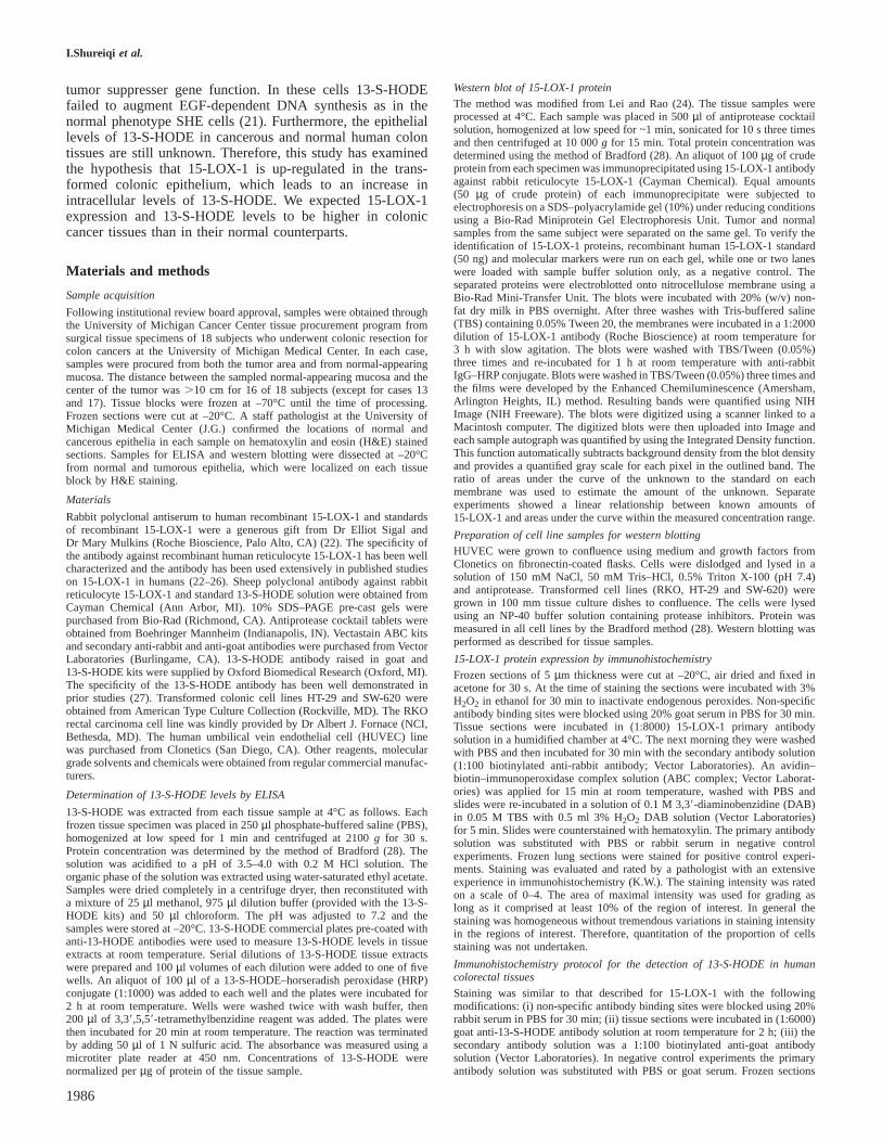
Fig. 1. Ratios of 13-S-HODE levels in paired tumor and normal colonicepithelia. 13-S-HODE levels were measured using an ELISA method andwere normalized to protein content in each sample. The ratio of normal totumor tissue concentrations was calculated for each patient and isrepresented by closed bars in the figure. Median ratio 3.33 (95% CI 1.28–8.677,P 5 0.0167).
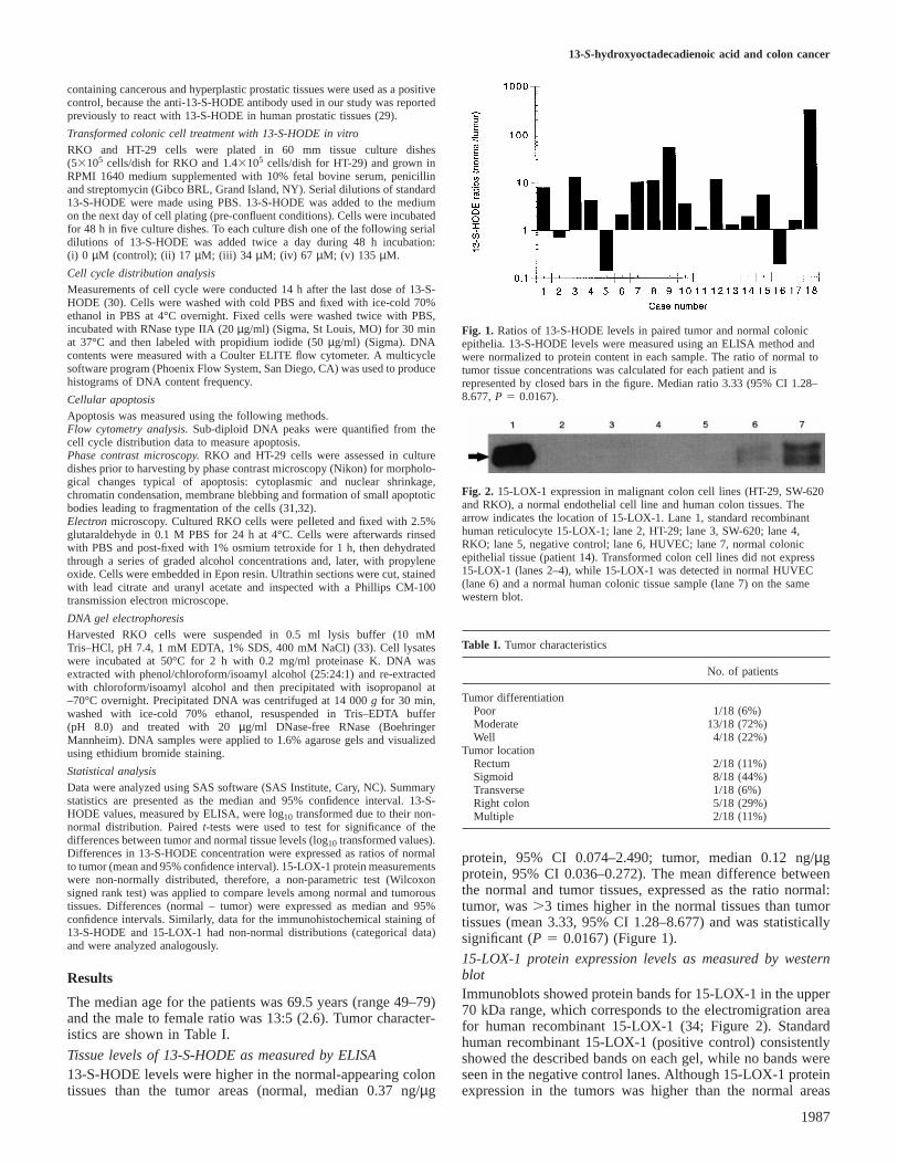
Fig. 2. 15-LOX-1 expression in malignant colon cell lines (HT-29, SW-620and RKO), a normal endothelial cell line and human colon tissues. Thearrow indicates the location of 15-LOX-1. Lane 1, standard recombinanthuman reticulocyte 15-LOX-1; lane 2, HT-29; lane 3, SW-620; lane 4,RKO; lane 5, negative control; lane 6, HUVEC; lane 7, normal colonicepithelial tissue (patient 14). Transformed colon cell lines did not express15-LOX-1 (lanes 2–4), while 15-LOX-1 was detected in normal HUVEC(lane 6) and a normal human colonic tissue sample (lane 7) on the samewestern blot.
Table I. Tumor characteristics
No. of patients
Tumor differentiationPoor 1/18 (6%)Moderate 13/18 (72%)Well 4/18 (22%)
Tumor locationRectum 2/18 (11%)Sigmoid 8/18 (44%)Transverse 1/18 (6%)Right colon 5/18 (29%)Multiple 2/18 (11%)
protein, 95% CI 0.074–2.490; tumor, median 0.12 ng/µgprotein, 95% CI 0.036–0.272). The mean difference betweenthe normal and tumor tissues, expressed as the ratio normal:tumor, was.3 times higher in the normal tissues than tumortissues (mean 3.33, 95% CI 1.28–8.677) and was statisticallysignificant (P 5 0.0167) (Figure 1).
15-LOX-1 protein expression levels as measured by westernblotImmunoblots showed protein bands for 15-LOX-1 in the upper70 kDa range, which corresponds to the electromigration areafor human recombinant 15-LOX-1 (34; Figure 2). Standardhuman recombinant 15-LOX-1 (positive control) consistentlyshowed the described bands on each gel, while no bands wereseen in the negative control lanes. Although 15-LOX-1 proteinexpression in the tumors was higher than the normal areas
I.Shureiqi et al.
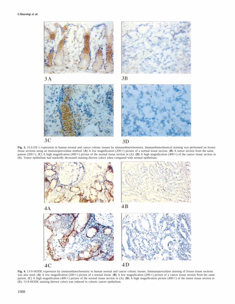
Fig. 3. 15-LOX-1 expression in human normal and cancer colonic tissues by immunohistochemistry. Immunohistochemical staining was performed on frozentissue sections using an immunoperoxidase method. (A) A low magnification (2003) picture of a normal tissue section. (B) A tumor section from the samepatient (2003). (C) A high magnification (4003) picture of the normal tissue section in (A). (D) A high magnification (4003) of the cancer tissue section in(B). Tumor epithelium had markedly decreased staining (brown color) when compared with normal epithelium.
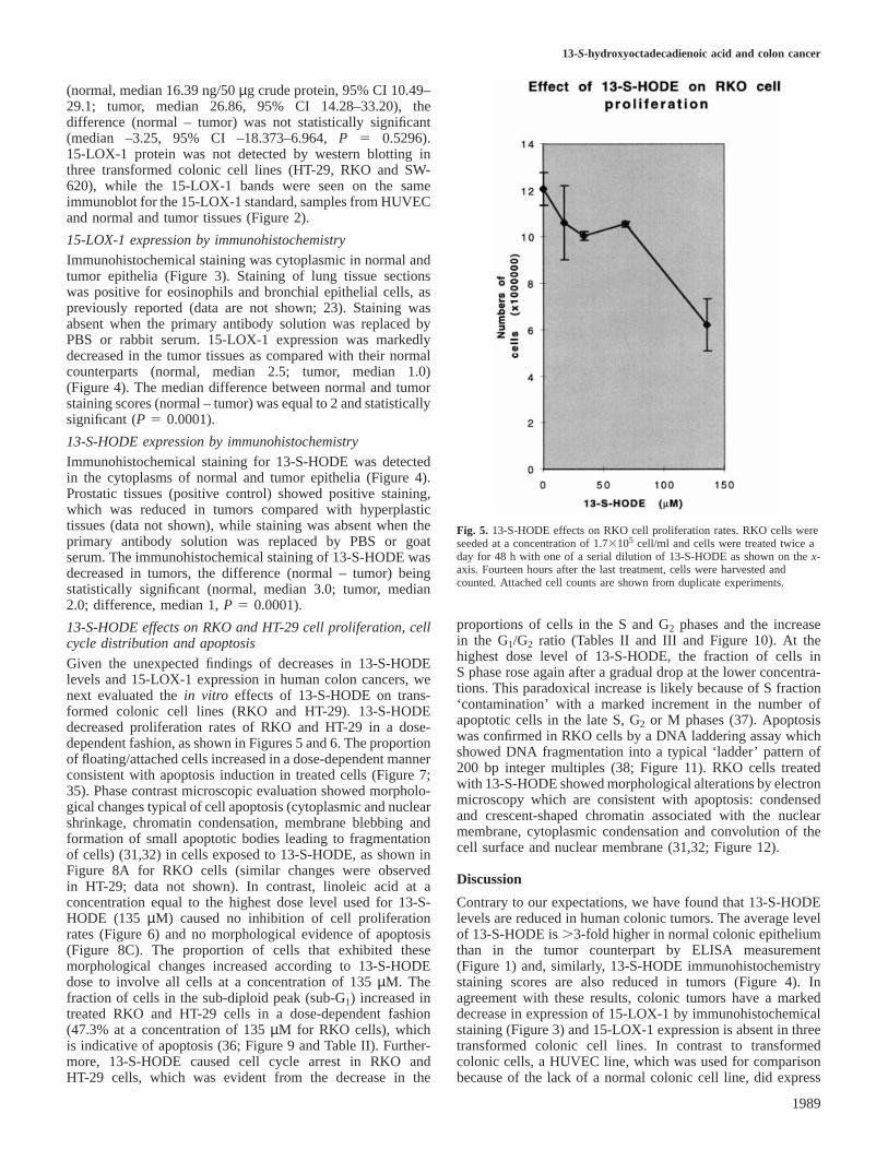
Fig. 4. 13-S-HODE expression by immunohistochemistry in human normal and cancer colonic tissues. Immunoperoxidase staining of frozen tissue sectionswas also used. (A) A low magnification (2003) picture of a normal tissue. (B) A low magnification (2003) picture of a cancer tissue section from the samepatient. (C) A high magnification (4003) picture of the normal tissue section in (A). (D) A high magnification picture (4003) of the tumor tissue section in(B). 13-S-HODE staining (brown color) was reduced in colonic cancer epithelium.
1988
13-S-hydroxyoctadecadienoic acid and colon cancer
(normal, median 16.39 ng/50µg crude protein, 95% CI 10.49–29.1; tumor, median 26.86, 95% CI 14.28–33.20), thedifference (normal – tumor) was not statistically significant(median –3.25, 95% CI –18.373–6.964,P 5 0.5296).15-LOX-1 protein was not detected by western blotting inthree transformed colonic cell lines (HT-29, RKO and SW-620), while the 15-LOX-1 bands were seen on the sameimmunoblot for the 15-LOX-1 standard, samples from HUVECand normal and tumor tissues (Figure 2).
15-LOX-1 expression by immunohistochemistry
Immunohistochemical staining was cytoplasmic in normal andtumor epithelia (Figure 3). Staining of lung tissue sectionswas positive for eosinophils and bronchial epithelial cells, aspreviously reported (data are not shown; 23). Staining wasabsent when the primary antibody solution was replaced byPBS or rabbit serum. 15-LOX-1 expression was markedlydecreased in the tumor tissues as compared with their normalcounterparts (normal, median 2.5; tumor, median 1.0)(Figure 4). The median difference between normal and tumorstaining scores (normal – tumor) was equal to 2 and statisticallysignificant (P 5 0.0001).
13-S-HODE expression by immunohistochemistry
Immunohistochemical staining for 13-S-HODE was detectedin the cytoplasms of normal and tumor epithelia (Figure 4).Prostatic tissues (positive control) showed positive staining,which was reduced in tumors compared with hyperplastictissues (data not shown), while staining was absent when theprimary antibody solution was replaced by PBS or goatserum. The immunohistochemical staining of 13-S-HODE wasdecreased in tumors, the difference (normal – tumor) beingstatistically significant (normal, median 3.0; tumor, median2.0; difference, median 1,P 5 0.0001).
13-S-HODE effects on RKO and HT-29 cell proliferation, cellcycle distribution and apoptosis
Given the unexpected findings of decreases in 13-S-HODElevels and 15-LOX-1 expression in human colon cancers, wenext evaluated thein vitro effects of 13-S-HODE on trans-formed colonic cell lines (RKO and HT-29). 13-S-HODEdecreased proliferation rates of RKO and HT-29 in a dose-dependent fashion, as shown in Figures 5 and 6. The proportionof floating/attached cells increased in a dose-dependent mannerconsistent with apoptosis induction in treated cells (Figure 7;35). Phase contrast microscopic evaluation showed morpholo-gical changes typical of cell apoptosis (cytoplasmic and nuclearshrinkage, chromatin condensation, membrane blebbing andformation of small apoptotic bodies leading to fragmentationof cells) (31,32) in cells exposed to 13-S-HODE, as shown inFigure 8A for RKO cells (similar changes were observedin HT-29; data not shown). In contrast, linoleic acid at aconcentration equal to the highest dose level used for 13-S-HODE (135 µM) caused no inhibition of cell proliferationrates (Figure 6) and no morphological evidence of apoptosis(Figure 8C). The proportion of cells that exhibited thesemorphological changes increased according to 13-S-HODEdose to involve all cells at a concentration of 135µM. Thefraction of cells in the sub-diploid peak (sub-G1) increased intreated RKO and HT-29 cells in a dose-dependent fashion(47.3% at a concentration of 135µM for RKO cells), whichis indicative of apoptosis (36; Figure 9 and Table II). Further-more, 13-S-HODE caused cell cycle arrest in RKO andHT-29 cells, which was evident from the decrease in the
1989
Fig. 5. 13-S-HODE effects on RKO cell proliferation rates. RKO cells wereseeded at a concentration of 1.73105 cell/ml and cells were treated twice aday for 48 h with one of a serial dilution of 13-S-HODE as shown on thex-axis. Fourteen hours after the last treatment, cells were harvested andcounted. Attached cell counts are shown from duplicate experiments.
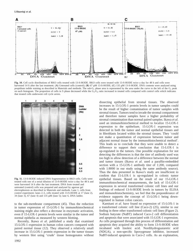
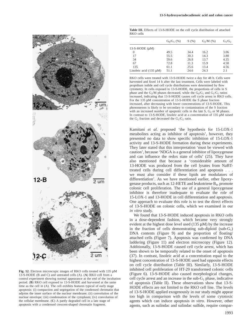
proportions of cells in the S and G2 phases and the increasein the G1/G2 ratio (Tables II and III and Figure 10). At thehighest dose level of 13-S-HODE, the fraction of cells inS phase rose again after a gradual drop at the lower concentra-tions. This paradoxical increase is likely because of S fraction‘contamination’ with a marked increment in the number ofapoptotic cells in the late S, G2 or M phases (37). Apoptosiswas confirmed in RKO cells by a DNA laddering assay whichshowed DNA fragmentation into a typical ‘ladder’ pattern of200 bp integer multiples (38; Figure 11). RKO cells treatedwith 13-S-HODE showed morphological alterations by electronmicroscopy which are consistent with apoptosis: condensedand crescent-shaped chromatin associated with the nuclearmembrane, cytoplasmic condensation and convolution of thecell surface and nuclear membrane (31,32; Figure 12).
Discussion
Contrary to our expectations, we have found that 13-S-HODElevels are reduced in human colonic tumors. The average levelof 13-S-HODE is.3-fold higher in normal colonic epitheliumthan in the tumor counterpart by ELISA measurement(Figure 1) and, similarly, 13-S-HODE immunohistochemistrystaining scores are also reduced in tumors (Figure 4). Inagreement with these results, colonic tumors have a markeddecrease in expression of 15-LOX-1 by immunohistochemicalstaining (Figure 3) and 15-LOX-1 expression is absent in threetransformed colonic cell lines. In contrast to transformedcolonic cells, a HUVEC line, which was used for comparisonbecause of the lack of a normal colonic cell line, did express
I.Shureiqi et al.
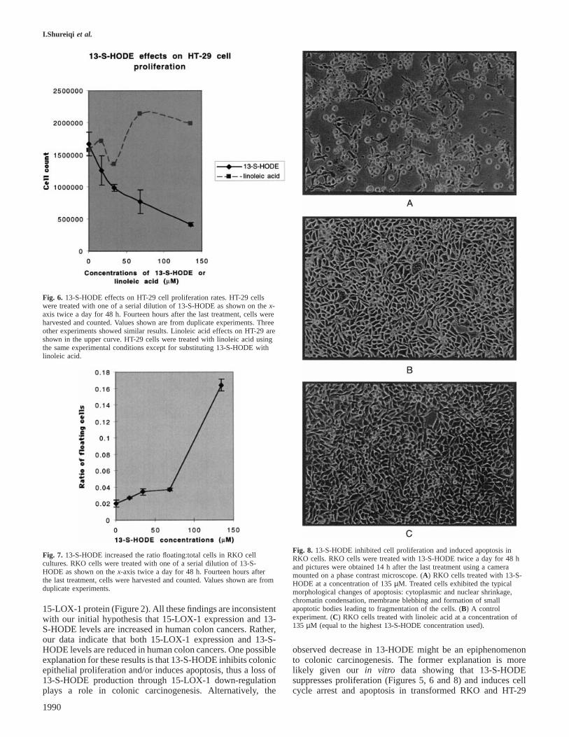
Fig. 6. 13-S-HODE effects on HT-29 cell proliferation rates. HT-29 cellswere treated with one of a serial dilution of 13-S-HODE as shown on thex-axis twice a day for 48 h. Fourteen hours after the last treatment, cells wereharvested and counted. Values shown are from duplicate experiments. Threeother experiments showed similar results. Linoleic acid effects on HT-29 areshown in the upper curve. HT-29 cells were treated with linoleic acid usingthe same experimental conditions except for substituting 13-S-HODE withlinoleic acid.
Fig. 7. 13-S-HODE increased the ratio floating:total cells in RKO cellcultures. RKO cells were treated with one of a serial dilution of 13-S-HODE as shown on thex-axis twice a day for 48 h. Fourteen hours afterthe last treatment, cells were harvested and counted. Values shown are fromduplicate experiments.
15-LOX-1 protein (Figure 2). All these findings are inconsistentwith our initial hypothesis that 15-LOX-1 expression and 13-S-HODE levels are increased in human colon cancers. Rather,our data indicate that both 15-LOX-1 expression and 13-S-HODE levels are reduced in human colon cancers. One possibleexplanation for these results is that 13-S-HODE inhibits colonicepithelial proliferation and/or induces apoptosis, thus a loss of13-S-HODE production through 15-LOX-1 down-regulationplays a role in colonic carcinogenesis. Alternatively, the
1990
Fig. 8. 13-S-HODE inhibited cell proliferation and induced apoptosis inRKO cells. RKO cells were treated with 13-S-HODE twice a day for 48 hand pictures were obtained 14 h after the last treatment using a cameramounted on a phase contrast microscope. (A) RKO cells treated with 13-S-HODE at a concentration of 135µM. Treated cells exhibited the typicalmorphological changes of apoptosis: cytoplasmic and nuclear shrinkage,chromatin condensation, membrane blebbing and formation of smallapoptotic bodies leading to fragmentation of the cells. (B) A controlexperiment. (C) RKO cells treated with linoleic acid at a concentration of135 µM (equal to the highest 13-S-HODE concentration used).
observed decrease in 13-HODE might be an epiphenomenonto colonic carcinogenesis. The former explanation is morelikely given our in vitro data showing that 13-S-HODEsuppresses proliferation (Figures 5, 6 and 8) and induces cellcycle arrest and apoptosis in transformed RKO and HT-29
13-S-hydroxyoctadecadienoic acid and colon cancer
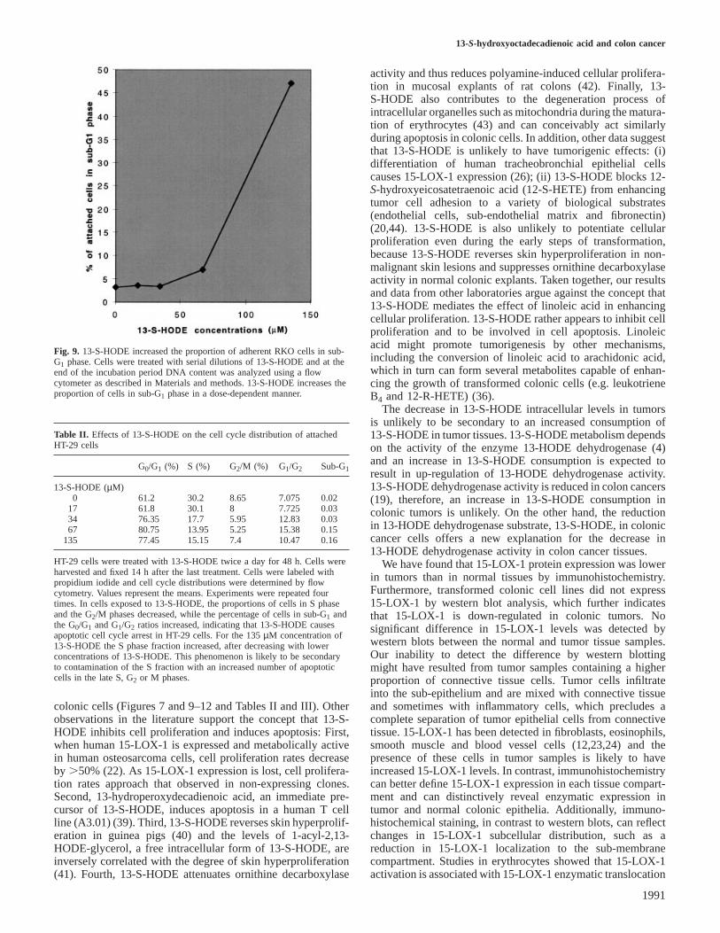
Fig. 9. 13-S-HODE increased the proportion of adherent RKO cells in sub-G1 phase. Cells were treated with serial dilutions of 13-S-HODE and at theend of the incubation period DNA content was analyzed using a flowcytometer as described in Materials and methods. 13-S-HODE increases theproportion of cells in sub-G1 phase in a dose-dependent manner.
Table II. Effects of 13-S-HODE on the cell cycle distribution of attachedHT-29 cells
G0/G1 (%) S (%) G2/M (%) G1/G2 Sub-G1
13-S-HODE (µM)0 61.2 30.2 8.65 7.075 0.02
17 61.8 30.1 8 7.725 0.0334 76.35 17.7 5.95 12.83 0.0367 80.75 13.95 5.25 15.38 0.15
135 77.45 15.15 7.4 10.47 0.16
HT-29 cells were treated with 13-S-HODE twice a day for 48 h. Cells wereharvested and fixed 14 h after the last treatment. Cells were labeled withpropidium iodide and cell cycle distributions were determined by flowcytometry. Values represent the means. Experiments were repeated fourtimes. In cells exposed to 13-S-HODE, the proportions of cells in S phaseand the G2/M phases decreased, while the percentage of cells in sub-G1 andthe G0/G1 and G1/G2 ratios increased, indicating that 13-S-HODE causesapoptotic cell cycle arrest in HT-29 cells. For the 135µM concentration of13-S-HODE the S phase fraction increased, after decreasing with lowerconcentrations of 13-S-HODE. This phenomenon is likely to be secondaryto contamination of the S fraction with an increased number of apoptoticcells in the late S, G2 or M phases.
colonic cells (Figures 7 and 9–12 and Tables II and III). Otherobservations in the literature support the concept that 13-S-HODE inhibits cell proliferation and induces apoptosis: First,when human 15-LOX-1 is expressed and metabolically activein human osteosarcoma cells, cell proliferation rates decreaseby .50% (22). As 15-LOX-1 expression is lost, cell prolifera-tion rates approach that observed in non-expressing clones.Second, 13-hydroperoxydecadienoic acid, an immediate pre-cursor of 13-S-HODE, induces apoptosis in a human T cellline (A3.01) (39). Third, 13-S-HODE reverses skin hyperprolif-eration in guinea pigs (40) and the levels of 1-acyl-2,13-HODE-glycerol, a free intracellular form of 13-S-HODE, areinversely correlated with the degree of skin hyperproliferation(41). Fourth, 13-S-HODE attenuates ornithine decarboxylase
1991
activity and thus reduces polyamine-induced cellular prolifera-tion in mucosal explants of rat colons (42). Finally, 13-S-HODE also contributes to the degeneration process ofintracellular organelles such as mitochondria during the matura-tion of erythrocytes (43) and can conceivably act similarlyduring apoptosis in colonic cells. In addition, other data suggestthat 13-S-HODE is unlikely to have tumorigenic effects: (i)differentiation of human tracheobronchial epithelial cellscauses 15-LOX-1 expression (26); (ii) 13-S-HODE blocks 12-S-hydroxyeicosatetraenoic acid (12-S-HETE) from enhancingtumor cell adhesion to a variety of biological substrates(endothelial cells, sub-endothelial matrix and fibronectin)(20,44). 13-S-HODE is also unlikely to potentiate cellularproliferation even during the early steps of transformation,because 13-S-HODE reverses skin hyperproliferation in non-malignant skin lesions and suppresses ornithine decarboxylaseactivity in normal colonic explants. Taken together, our resultsand data from other laboratories argue against the concept that13-S-HODE mediates the effect of linoleic acid in enhancingcellular proliferation. 13-S-HODE rather appears to inhibit cellproliferation and to be involved in cell apoptosis. Linoleicacid might promote tumorigenesis by other mechanisms,including the conversion of linoleic acid to arachidonic acid,which in turn can form several metabolites capable of enhan-cing the growth of transformed colonic cells (e.g. leukotrieneB4 and 12-R-HETE) (36).
The decrease in 13-S-HODE intracellular levels in tumorsis unlikely to be secondary to an increased consumption of13-S-HODE in tumor tissues. 13-S-HODE metabolism dependson the activity of the enzyme 13-HODE dehydrogenase (4)and an increase in 13-S-HODE consumption is expected toresult in up-regulation of 13-HODE dehydrogenase activity.13-S-HODE dehydrogenase activity is reduced in colon cancers(19), therefore, an increase in 13-S-HODE consumption incolonic tumors is unlikely. On the other hand, the reductionin 13-HODE dehydrogenase substrate, 13-S-HODE, in coloniccancer cells offers a new explanation for the decrease in13-HODE dehydrogenase activity in colon cancer tissues.
We have found that 15-LOX-1 protein expression was lowerin tumors than in normal tissues by immunohistochemistry.Furthermore, transformed colonic cell lines did not express15-LOX-1 by western blot analysis, which further indicatesthat 15-LOX-1 is down-regulated in colonic tumors. Nosignificant difference in 15-LOX-1 levels was detected bywestern blots between the normal and tumor tissue samples.Our inability to detect the difference by western blottingmight have resulted from tumor samples containing a higherproportion of connective tissue cells. Tumor cells infiltrateinto the sub-epithelium and are mixed with connective tissueand sometimes with inflammatory cells, which precludes acomplete separation of tumor epithelial cells from connectivetissue. 15-LOX-1 has been detected in fibroblasts, eosinophils,smooth muscle and blood vessel cells (12,23,24) and thepresence of these cells in tumor samples is likely to haveincreased 15-LOX-1 levels. In contrast, immunohistochemistrycan better define 15-LOX-1 expression in each tissue compart-ment and can distinctively reveal enzymatic expression intumor and normal colonic epithelia. Additionally, immuno-histochemical staining, in contrast to western blots, can reflectchanges in 15-LOX-1 subcellular distribution, such as areduction in 15-LOX-1 localization to the sub-membranecompartment. Studies in erythrocytes showed that 15-LOX-1activation is associated with 15-LOX-1 enzymatic translocation
I.Shureiqi et al.
Fig. 10. Cell cycle distributions of RKO cells treated with 13-S-HODE. RKO cells were treated with 13-S-HODE twice a day for 48 h and cells wereharvested 14 h after the last treatment. (A) Untreated cells (control); (B) 67 µM 13-S-HODE; (C) 135 µM 13-S-HODE. DNA contents were analyzed usingpropidium iodide staining as described in Materials and methods. The sub-G1 phase area is represented by the area under the curve to the left of the G1 peakon each histogram. The proportion of cells in S phase decreased while the G1/G2 ratio increased in treated cells compared with control cells which indicatesthat treated cells underwent cell cycle arrest.
Fig. 11. 13-S-HODE induced DNA fragmentation in RKO cells. Cells weretreated with one of a serial dilution of 13-S-HODE twice a day for 48 h andwere harvested 14 h after the last treatment. DNA from treated anduntreated (control) cells was prepared and analyzed by agarose gelelectrophoresis as described in Materials and methods. Lane 1, cells fromcontrol experiment; lanes 2–5, cells treated with 13-S-HODE at 17 (lane 2),34 (lane 3), 67 (lane 4) and 135µM (lane 5); lane 6, DNA ladder.
to the sub-membrane compartment (45). Thus the reductionin tumor expression of 15-LOX-1 by immunohistochemicalstaining might also reflect a decrease in enzymatic activation,even if 15-LOX-1 protein levels were similar in the tumor andnormal epithelia as measured by western blotting.
Recently, Ikawaet al. published a study that examined15-LOX-1 expression in human colon cancers compared withpaired normal tissue (12). They observed a relatively smallincrease in 15-LOX-1 protein expression in the tumor tissuesby western blot using ‘crude’ tissue homogenates without
1992
dissecting epithelial from stromal tissues. The observedincreases in 15-LOX-1 protein levels in tumor samples couldbe the result of higher contamination of tumor samples withstromal tissues. Tumors tend to invade the stromal compartmentand therefore tumor samples have a higher probability ofstromal contamination than normal paired samples. Ikawaet al.used an immunohistochemical method to localize 15-LOX-1expression to the epithelium. 15-LOX-1 expression wasdetected in both the tumor and normal epithelial tissues andin fibroblasts located within the stromal tissues. They ‘couldnot make a quantitation of expression between tumor andadjacent normal tissue by the immunohistochemical method’.This leads us to conclude that they were unable to detect adifference to support their conclusion that 15-LOX-1 isup-regulated in the tumors. One possible explanation for notdetecting the differences is that the titer of antibody used wastoo high to allow detection of a difference between the normaland tumor tissues (Ikawaet al. used a paraffin-embeddedsection with a 15-LOX-1 antibody titer of 1:200, comparedwith 1:8000 in our study using the same antibody source).Thus the data presented in Ikawa’s study are insufficient toconfirm that 15-LOX-1 is up-regulated in colonic tumorepithelial tissues. Based on the result of our 15-LOX-1immunohistochemical measurements, the lack of 15-LOX-1expression in several transformed colonic cell lines and ourfindings of reduced 13-S-HODE levels in tumors by ELISAand immunohistochemistry, we feel that the preponderance ofevidence supports the concept of 15-LOX-1 being down-regulated in human colon cancers.
Kamitani et al. have found no expression of 15-LOX-1 ina transformed colonic cell line (Caco-2) (25), similar to ourfinding in three other transformed colonic cell lines (Figure 2).Sodium butyrate (NaBT) induced Caco-2 cell differentiationand apoptosis that were associated with 15-LOX-1 expression.As cells progressed through this process of differentiation andapoptosis they acquired the ability to form 13-S-HODE whenincubated with linoleic acid. Nordihydroguaiaretic acid(NDGA), a non-specific lipoxygenase inhibitor, increasedNaBT-induced apoptosis in Caco-2 cells. As an explanation,
13-S-hydroxyoctadecadienoic acid and colon cancer
Fig. 12. Electron microscopic images of RKO cells treated with 135µM13-S-HODE (B and C) and untreated cells (A). (A) RKO cell from acontrol experiment showing normal appearance at the end of the incubationperiod. (B) RKO cell exposed to 13-S-HODE and harvested at the sametime as the cell in (A). The cell exhibits features typical of early stageapoptosis: (i) compaction and segregation of the condensed chromatin thatadjoins the inner surface of the nuclear membrane; (ii) convolution of thenuclear envelope; (iii) condensation of the cytoplasm; (iv) convolution ofthe cellular membrane. (C) A partly degraded cell in a late stage ofapoptosis with a condensed crescent-shaped chromatin fragment.
1993
Table III. Effects of 13-S-HODE on the cell cycle distribution of attachedRKO cells
G0/G1 (%) S (%) G2/M (%) G1/G2
13-S-HODE (µM)0 49.5 34.4 16.2 3.0617 55.5 30.3 14.3 3.8834 59.6 26.8 13.7 4.3567 72.8 11.3 15.9 4.58135 61.1 25.6 13.4 4.56
Linoleic acid (135µM) 51.1 24.6 24.3 2.1
RKO cells were treated with 13-S-HODE twice a day for 48 h. Cells wereharvested and fixed 14 h after the last treatment. Cells were labeled withpropidium iodide and cell cycle distributions were determined by flowcytometry. In cells exposed to 13-S-HODE, the proportions of cells in Sphase and the G2/M phases decreased, while the G0/G1 and G1/G2 ratiosincreased, indicating that 13-S-HODE causes cell cycle arrest in RKO cells.For the 135µM concentration of 13-S-HODE the S phase fractionincreased, after decreasing with lower concentrations of 13-S-HODE. Thisphenomenon is likely to be secondary to contamination of the S fractionwith an increased number of apoptotic cells in the late S, G2 or M phases.In contrast to 13-S-HODE, linoleic acid at a concentration of 135µM raisedthe G2 fraction and decreased the G1/G2 ratio.
Kamitani et al. proposed ‘the hypothesis for 15-LOX-1metabolites acting as inhibitor of apoptosis’, however, theypresented no data to show specific inhibition of 15-LOX-1activity and 13-S-HODE formation during these experiments.They later stated that this interpretation ‘must be viewed withcaution’, because ‘NDGA is a general inhibitor of lipoxygenaseand can influence the redox state of cells’ (25). They havealso mentioned that because a ‘considerable amount of13-HODE was produced from the cell lysates from NaBT-treated cells during cell differentiation and apoptosis . . .we must also consider if these lipids are modulators ofdifferentiation’. As we have mentioned earlier, other lipoxy-genase products, such as 12-HETE and leukotriene B4, promotecolonic cell proliferation. The use of a general lipoxygenaseinhibitor is therefore inadequate to evaluate the role of15-LOX-1 and 13-HODE in cell differentiation and apoptosis.One approach to evaluate this role is to test the direct effectsof 13-S-HODE on colonic cells, which we examined in ourin vitro study.
We found that 13-S-HODE induced apoptosis in RKO cellsin a dose-dependent fashion, which became very stronglyevident at the highest dose level used (135µM) by the increasein the fraction of cells demonstrating sub-diploid (sub-G1)DNA contents (Figure 9) and the proportion of floating/attached cells (Figure 7). Apoptosis was confirmed by DNAladdering (Figure 11) and electron microscopy (Figure 12).Additionally, 13-S-HODE caused cell cycle arrest, which hasbeen shown to be temporally related to the onset of apoptosis(37). In contrast, linoleic acid at a concentration equal to thehighest concentration of 13-S-HODE used had opposite effectson cell cycle distribution (Table III). Similarly, 13-S-HODEinhibited cell proliferation of HT-29 transformed colonic cells(Figure 6). 13-S-HODE also caused morphological changes,cell cycle arrest and an increase in the sub-G1 phase, indicativeof apoptosis (Table II). These observations show that 13-S-HODE effects are not limited to the RKO cell line. The levelsof 13-S-HODE applied exogenously in our study might appeartoo high in comparison with the levels of some cytotoxicagents which can induce apoptosisin vitro. However, otheragents, such as sulindac and sulindac sulfide, require compar-
I.Shureiqi et al.
able concentrations to the 13-S-HODE concentrations used inour study to trigger apoptosis in transformed colonic cells(sulindac, 120µM; sulindac sulfone, 240µM) (46). Further-more, 13-S-HODE exists in normal resting cells at a concentra-tion of 34106 340 ng/106 cells, as measured in HUVEC (47).Currently, the intracellular levels of 13-S-HODE in normalcolonic cells in vitro is unknown, because no normal coloncell line is available. If the levels of 13-S-HODE in restingHUVEC is used as an approximation to that expected innormal colonic cells, a 13-S-HODE concentration of 57µMin the culture medium is required to reach that level. 13-S-HODE levels are expected to increase above the levels inresting cells for 13-S-HODE to activate apoptosis. In ourin vitro studies the exogenous application of 13-S-HODEactivated apoptosis markedly in RKO cells at concentrations,2.5-fold of the level in resting HUVEC. Additionally, 13-S-HODE is considered to be relatively unstable in culture mediaand sensitive to common elements such as light and oxygenwhich limit cell exposure to this compound when appliedexogenously (Cayman Chemical Catalog, Vol. VIII, 1996).Therefore, the observed effects of 13-S-HODE are unlikely tohave resulted from using concentrations that are far fromphysiological levels. Finally, in control experiments linoleicacid, the parent compound of 13-S-HODE, failed to inhibitthe growth of RKO and HT-29 cells or induce cell cycle arrest(Figures 6 and 8C) at a concentration equal to the highest13-S-HODE concentrations used in our study (135µM). Thisfurther indicates that a non-specific fatty acid toxicity isunlikely to have produced the 13-S-HODE effects.
In summary, our study demonstrates that 15-LOX-1 proteinexpression and 13-S-HODE intracellular levels are decreasedin human colonic tumors and that 13-S-HODE inhibits cellproliferation and induces cell cycle arrest and apoptosis inRKO transformed colonic cells. These findings contradict thenotion that 13-S-HODE promotes colonic carcinogenesis andindicate that 13-S-HODE might have a role in the inhibitionof colonic cell proliferation and the induction of apoptosis.
Acknowledgements
We thank Dr Alnawaz Rehemtulla and Joseph Cornicelli for reviewing themanuscript and for their helpful scientific comments and Daniel Cutler forhis efforts in preparing the immunohistochemistry pictures. This work hasbeen supported in part by an institutional grant from the American CancerSociety (ACS374639).
References
1.Schottenfeld,D. and Winawer,S.J. (1996) Cancer by tissue of origin.In Schottenfeld,D. and Fraumeni,J.F. (eds)Cancer Epidemiology andPrevention, 2nd Edn. Oxford University Press, New York, NY, p. 828.
2.Nigro,N.D., Singh,D.V., Campbell,R.L. and Pak,M.S. (1975) Effect ofdietary beef fat on intestinal tumor formation by azoxymethane in rats.J. Natl Cancer Inst., 54, 439–442.
3.Broitman,S.A., Vitale,J.J., Jakuba,E.V. and Gottlieb,L.S. (1977)Polyunsaturated fat, cholesterol and large bowel tumorigenesis.Cancer(Philad.), 40, 2455–2463.
4.Earls,S.M., Bronstein,J.C., Winner,D.L. and Bull,A.W. (1991) Metabolismof oxidized linoleic acid: characterization of 13-hydroxyoctadecadienoicacid dehydrogenase activity from rat colonic tissue.Biochim. Biophys.Acta, 1081, 174–180.
5.Sakaguchi,M., Hiramatsu,Y., Takada,H., Yamamura,M., Hioki,K., Saito,K.and Yamamoto,M. (1984) Effect of dietary unsaturated and saturated fatson azoxymethane-induced colon carcinogenesis in rats.Cancer Res., 44,1472–1477.
6.Hussey,H.J. and Tisdale,M. (1994) Effect of polyunsaturated fatty acidson the growth of murine colon adenocarcinomasin vitro and in vivo. Br.J. Cancer, 70, 6–10.
1994
7.Bull,A.W., Nigro,N.D., Golembieski,W.A., Crissman,J.D. and Marnett,L.J.(1984) In vivo stimulation of DNA synthesis and induction of ornithinedecarboxylase in rat colon by fatty acid hydroperoxides, autoxidationproducts of unsaturated fatty acids.Cancer Res., 44, 4924–4928.
8.Bull,A.W., Nigro,N.D. and Marnett,L.J. (1988) Structural requirement ofstimulation of colonic cell proliferation by oxidized fatty acids.CancerRes., 48, 1771–1776.
9.Baer,A.N., Costello,P.B. and Green,F.A. (1991)In vivo activation of anω-6 oxygenase in human skin.Biochem. Biophys. Res. Commun., 180, 93–104.
10.Daret,D., Blin,P. and Larrue,J. (1989) Synthesis of hydroxy fatty acidsfrom linoleic acid by human blood platelets.Prostaglandins, 38, 203–214.
11.Brash,A., Boeglin,W. and Chang,M. (1997) Discovery of a second 15S-lipoxygenase in humans.Proc. Natl Acad. Sci. USA, 94, 6148–6152.
12. Ikawa,H., Kamitani,H., Calvo,B., Foley,J. and Eling,T. (1999) Expression of15-lipoxygenase-1 in human colorectal cancer.Cancer Res., 59, 360–366.
13.Hussey,H.J. and Tisdale,M.J. (1997) Mechanism of the anti-tumour effectof 2,3,5-trimethyl-6-(3-pyridylmethyl) 1,4-benzoquinone (CV-6504).Br.J. Cancer, 75, 845–849.
14.Glasgow,W.C. and Eling,T.E. (1990) Epidermal growth factor stimulteslinoleic acid metabolism in BALB/c 3T3 fibroblast.Mol. Pharmacol., 38,503–510.
15.Glasgow,W.C., Afshari,C.A., Barrett,J.C. and Eling,T.E. (1992) Modulationof the epidermal growth factor mitogenic response by metabolites oflinoleic and arachidonic acid in Syrian hamster embryo fibroblasts.Differential effects in tumor suppressor gene (1) and (–) phenotypes.J. Biol. Chem., 267, 10771–10779.
16.Reddy,N., Everhart,A., Eling,T. and Glasgow,W. (1997) Characterizationof 15-lipoxygenase in human breast carcinoma BT-20 cells: stimulationof 13-HODE formation by TGFα/EGF.Biochem. Biophys. Res. Commun.,231, 111–116.
17.Glasgow,W.C. and Everhart,A.L. (1996) Linoleic acid metabolites modulatemitogenic signal transduction in cells overexpressing c-erbB-2.Proc.Annu. Meet. Am. Assoc. Cancer Res., 37, A3395.
18.Bronstein,J.C. and Bull,A.W. (1993) The correlation between 13-hydroxyoctadecadienic acid dehydrogenase (13-HODE dehydrogenase)and intestinal cell differentiation.Prostaglandins, 46, 387–395.
19.Silverman,A.L., Bronstein,J.C., Krymgold,S., Kalon,D. and Bull,A. (1996)Decreased levels of 13-hydroxyoctadecadeinoic acid (13-HODE)dehydrogenase in neoplastic tissue of human colon biopsies.CancerEpidemiol. Biomarkers Prev., 5, 53–56.
20.Liu,B., Khan,W.A., Hannun,Y.A., Timar,J., Taylor,J.D., Lundy,S.,Butovich,I. and Honn,K.V. (1995) 12(S)-Hydroxyeicostetraenoic acid and13(S)-hydroxyoctadecadienoic acid regulation of protein kinase C-α inmelanoma cells: role of receptor-mediated hydrolysis of inositolphospholipids.Proc. Natl Acad. Sci. USA, 92, 9323–9327.
21.Hui,R., Everhart,A.L. and Glasgow,W.C. (1997) Epidermal growth factor-stimulated production of esterified 13(S)-hydroxyoctadecadienoic acid isassociated with tumor suppressor phenotype in Syrian hamster embryofibroblasts.J. Lipid Res., 38, 46–60.
22.Sigal,E., Grunberger,D., Highland,E., Gross,C., Dixon,R. and Craik,C.(1990) Expression of cloned human reticulocyte 15-lipoxygenase andimmunological evidence that 15-lipoxygenases of different cell types arerelated.J. Biol. Chem., 265, 5113–5120.
23.Nadel,J.A., Conrad,D.J., Ueki,I.F., Schuster,A. and Sigal,E. (1991)Immunocytochemical localization of arachidonate 15-lipoxygenase inerythrocytes, leukocytes and airway cells.J. Clin. Invest., 87, 1139–1145.
24.Lei,Z.M. and Rao,C.V. (1992) The expression of 15-lipoxygenase geneand the presence of functional enzyme in cytoplasm and nuclei ofpregnancy human myometria.Endocrinology, 130, 861–870.
25.Kamitani,H., Geller,M. and Eling,T. (1998) Expression of 15-lipoxygenaseby human colorectal carcinoma Caco-2 cell lines during apoptosis andcell differentiation.J. Biol. Chem., 273, 21569–21577.
26.Hill,E.M., Eling,T. and Nettesheim,P. (1998) Changes in expression of15-lipoxygenase and prostaglandin-H synthase during differentiation ofhuman tracheobronchial epithelial cells.Am. J. Respir. Cell Mol. Biol.,18, 662–669.
27.Spindler,S.A., Clark,K.S., Callewaert,D.M. and Reddy,R.G. (1996)Significance and Immunoassay of 9- and 13-hydroxyoctadecadienoic acids.Biochem. Biophys. Res. Commun., 218, 187–191.
28.Bradford,M. (1976) A rapid and sensitive method for the quantitation ofmicrogram quantities of protein utilizing the principle of protein–dyebinding.Anal. Biochem., 72, 248–254.
29.Spindler,S., Sarkar,F., Sakr,W., Blackburn,M., Bull,A., LaGattuta,M. andReddy,R. (1997) Production of 13-hydroxyoctadecadienoic acid(13-HODE) by prostate tumors and cell lines.Biochem. Biophys. Res.Commun., 239, 775–781.
13-S-hydroxyoctadecadienoic acid and colon cancer
30.Srivastava,S.P., Kumar,K. and Kaufman,R.J. (1998) Phosphorylation ofeukaryotic translation initiation factor 2 mediates apoptosis in response toactivation of the double-stranded RNA-dependent protein kinase.J. Biol.Chem., 273, 2416–2423.
31.Kerr,J.F.R. and Harmon,B.V. (1991) Definition and incidence of apoptosis:an historical perspective. In Tomei,L.D. and Cope,F.O. (eds)Apoptosis:The Molecular Basis of Cell Death.Cold Spring Harbor Laboratory Press,Plainview, NY, pp. 5–6.
32.Gschwind,M. and Huber,G. (1997) Detection of apoptotic or necrotic deathin neuronal cells by morphological, biochemical and molecular analysis.In Poirier,J. (ed.)Apoptosis Techniques and Protocols, Vol. 29. HumanaPress, Totowa, NJ, pp. 16–17.
33.Srivastava,S.P., Kumar,K.U. and Kufman,R. (1998) Phosphorylation ofeukayotic translation initiation factor 2 mediates apoptosis in response toactivation of the double-stranded RNA-dependent protein kinase.J. Biol.Chem., 273, 2416–2423.
34.Kuhn,H., Barnett,J., Grunberger,D., Baecker,P., Chow,J., Nguyen,B.,Burstyn-Pettergrew,H., Chan,H. and Sigal,E. (1993) Overexpression,purification and characterization of human recombinant 15-lipoxygenase.Biochim. Biophys. Acta, 1169, 80–89.
35.Soldatenkov,V.A., Prasad,S., Notario,V. and Dritschilo,A. (1995) Radiation-induced apoptosis of Ewing’s sarcoma cells: DNA fragmentation andproteolysis of poly(ADP-ribose) polymerase.Cancer Res., 55, 4240–4242.
36.Bortuzzo,C., Hanif,R., Kashfi,K., Staiano-Coico,L., Shiff,S.J. and Rigas,B.(1996) The effect of leukotrienes B and selected HETES on the proliferationof colon cancer cells.Biochim. Biophys. Acta, 1300, 240–246.
37.Sherwood,S.W. and Schimke,R.T. (1995) Cell cycle analysis of apoptosisusing flow cytometry. In Schwartz,L.M. and Osborne,B.A. (eds)Methodsin Cell Biology, Vol. 46, Cell Death.Academic Press, San Diego, CA, pp.77–97.
38.Wyllie,A.H. (1980) Glucocorticoid-induced thymocyte apoptosis isassociated with endogenous endonuclease activation.Nature, 284, 555–556.
1995
39.Sandstrom,P.A., Pardi,D., Tebbey,P.W., Dudek,R.W., Terrain,D.M.,Folks,T.M. and Buttke,T.M. (1995) Lipid hyperperoxidase-inducedapoptosis: lack of inhibition by Bcl-2 over-expression.FEBS Lett., 365,66–77.
40.Miller,C.C. and Ziboh,V.A. (1990) Induction of epidermalhyperproliferation by topical n-3 polyunsaturated fatty acids on guinea pigskin linked to decrease levels of 13-hydroxyocatdecadienoic acid(13-HODE).J. Invest. Dermatol., 94, 354–358.
41.Cho,Y. and Ziboh,V.A. (1995) Nutritional modulation of guinea pig skinhyperproliferation by essential fatty acid deficiency is associated withselective down regulation of protein kinase C-b.J. Nutr., 125, 2741–2750.
42.Bull,A.W., Earls,S.M. and Blackburn,M.L. (1993) Regulation of theinduction of ornithine decaroxylase in short-term rat colon organ cultureby dexamethasone and 13-hydroxyoctadecadienoic acid (13-HODE).LifeSci., 53, 337–385.
43.Kuhn,H., Belkner,J., Wienser,R. and Brash,A.R. (1990) Oxygenation ofbiological membranes by the pure reticulocyte lipoxygenase.J. Biol.Chem., 265, 18351–18361.
44.Grossi,M.I., Fitzgerald,A.L., Umbrager,K.K.N., Diglio,A.C.A., Taylor,D.J.and Honn,V.K. (1989) Bidirectional control of membrane expression and/or activation of the tumor cell IRGPIIB/IIIa receptors and tumor celladhesion by lipoxygenase products of arachidonic acid and linoleic acid.Cancer Res., 49, 1029–1037.
45.Watson,A. and Doherty,J.F. (1994) Calcium promotes membraneassociation of reticulocyte 15-lipoxygenase.Biochem. J., 298, 377–383.
46.Piazza,G., Rahm,A., Krutzsch,M.et al. (1995) Antineoplastic drugssulindac sulfide and sulfone inhibit cell growth by inducing apoptosis.Cancer Res., 55, 3110–3116.
47.Buchanan,M.R., Haas,T.A., Lagarde,M. and Guichardant,M. (1985)13-Hydoxyoctadecadienoic acid is the vessel wall chemorepellant factor,LOX. J. Biol. Chem., 260, 16056–16059.
Received February 26, 1999; revised June 8, 1999; accepted June 25, 1999