Screen-detected multiple primary lung cancers in the ... - FloRE
10
© Journal of Thoracic Disease. All rights reserved. J Thorac Dis 2018;10(2):1058-1066 jtd.amegroups.com Short Communication Screen-detected multiple primary lung cancers in the ITALUNG trial Mario Mascalchi 1 , Camilla E. Comin 2 , Elena Bertelli 1 , Lapo Sali 1 , Cristina Maddau 3 , Stefania Zuccherelli 1 , Giulia Picozzi 3 , Laura Carrozzi 4 , Michela Grazzini 5 , Gabriella Fontanini 6 , Luca Voltolini 7 , Alessandra Vella 8 , Francesca Castiglione 2 , Francesca Carozzi 3 , Eugenio Paci 3 , Maurizio Zompatori 9 , Andrea Lopes Pegna 10 , Fabio Falaschi 11 ; for the ITALUNG Study Research Group * 1 “Mario Serio” Department of Experimental and Clinical Biomedical Sciences, University of Florence, Florence, Italy; 2 Division of Pathological Anatomy, Department of Medical and Surgical Critical Care, University of Florence, Florence, Italy; 3 Institute for Cancer Research and Prevention (ISPO), Florence, Italy; 4 Cardiopulmonary Department, Pisa University Hospital, Pisa, Italy; 5 Pulmonology Department, Hospital of Pistoia, Pistoia, Italy; 6 Pathology Department, University Hospital of Pisa, Pisa, Italy; 7 Division of Thoracic Surgery, Careggi University Hospital, Florence, Italy; 8 Service of Nuclear Medicine, Le Scotte Hospital, Siena, Italy; 9 Radiology Department, Multimedica Group, IRCCS, Sesto San Giovanni, Italy; 10 Pulmonology, Cardio-Thoracic-Vascular Department, Careggi Hospital, Florence, Italy; 11 2nd Radiology Unit, University Hospital of Pisa, Pisa, Italy Correspondence to: Lapo Sali, MD, PhD. “Mario Serio” Department of Experimental and Clinical Biomedical Sciences, University of Florence, Viale Morgnagni 50, 50134 Florence, Italy. Email: lapo.sali@unifi.it. Abstract: Occurrence of multiple primary lung cancers (MPLC) in individuals undergoing low-dose computed tomography (LDCT) screening has not been thoroughly addressed. We investigated MPLC in subjects recruited in the ITALUNG randomized clinical trial. Cases of cytologically/histologically proven MPLC detected at screening LDCT or follow-up CT were selected and pathologically re-evaluated according to the WHO 2015 classification. Overall 16 MPLC were diagnosed at screening LDCT (n=14, all present at baseline) or follow-up CT (n=2) in six subjects (4 in one subject, 3 in two and 2 in three subjects), representing 0.43% of the 1,406 screenees and 15.8% of the 38 subjects with at least one screen-detected primary lung cancer. MPLC included 9 adenocarcinomas in three subjects and a combination of 7 different tumour histotypes in three subjects. MPLC, mostly adenocarcinomas, are not uncommon in smokers and ex-smokers with at least one LDCT screen detected primary lung cancer. * Members of the ITALUNG Study Research group: Andrea Lopes Pegna (MD), Roberto Bianchi (MD), Cristina Ronchi (MD), Pulmonology Cardio-Thoracic-Vascular Department, Careggi Hospital, Florence, Italy; Laura Carrozzi (MD), Ferruccio Aquilini (BSc), Stella Cini (MD), Mariella De Santis, Francesco Pistelli (MD), Filomena Baliva, Antonio Chella (MD), Laura Tavanti (MD), Cardiopulmonary Department, University Hospital of Pisa, Italy; Michela Grazzini (MD), Florio Innocenti (MD), Ilaria Natali (BSc), Pulmunonology Department, Hospital of Pistoia, Italy; Mario Mascalchi (MD, PhD), Maurizio Bartolucci (MD), Elena Crisci (MD), Agostino De Francisci (MD), Massimo Falchini (MD), Silvia Gabbrielli (MD), Giulia Picozzi (MD), Giuliana Roselli (MD), Andrea Masi (MD), Radiology Department, Careggi Hospital, Florence, Italy; Fabio Falaschi (MD), Luigi Battola (MD), Anna Lisa De Liperi (MD), Cheti Spinelli (MD), 2 nd Radiology Unit, University Hospital of Pisa, Italy; Letizia Vannucchi (MD), Alessia Petruzzelli (MD), Davide Gadda (MD), Anna Talina Neri (MD), Franco Niccolai (MD) Radiology Department, Hospital of Pistoia, Italy; Luca Vaggelli (MD), Nuclear Medicine Department, Careggi Hospital, Florence, Italy; Alessandra Vella (MD, PhD), Nuclear Medicine Department, Le Scotte University Hospital, Siena; Francesca Maria Carozzi (BSc), Cristina Maddau (BSc), Laboratory Unit, Institute for Oncological Study and Prevention, Florence, Italy; Alberto Janni (MD), Luca Voltolini (MD), Thoracic Surgery Department, Careggi Hospital, Florence, Italy; Alfredo Mussi (MD), Marco Lucchi (MD), Thoracic Surgery Department, Cisanello Hospital, University of Pisa, Italy; Camilla Comin (MD), Division of Pathological Anatomy, Department of Medical and Surgical Critical Care, University of Florence, Italy; Gabriella Fontanini (MD), Adele Renza Tognetti (MD), Pathology Department, University Hospital of Pisa, Italy; Eugenio Paci (MD, PhD), Giovanna Cordopatri (BSc), Francesco Giusti (BSc, PhD), Ida Esposito (BSc), Department of Epidemiology Institute for Oncological Study and Prevention, Florence, Italy.
-
Upload
khangminh22 -
Category
Documents
-
view
0 -
download
0
Transcript of Screen-detected multiple primary lung cancers in the ... - FloRE
© Journal of Thoracic Disease. All rights reserved. J Thorac Dis 2018;10(2):1058-1066jtd.amegroups.com
Short Communication
Screen-detected multiple primary lung cancers in the ITALUNG trial
Mario Mascalchi1, Camilla E. Comin2, Elena Bertelli1, Lapo Sali1, Cristina Maddau3, Stefania Zuccherelli1, Giulia Picozzi3, Laura Carrozzi4, Michela Grazzini5, Gabriella Fontanini6, Luca Voltolini7, Alessandra Vella8, Francesca Castiglione2, Francesca Carozzi3, Eugenio Paci3, Maurizio Zompatori9, Andrea Lopes Pegna10, Fabio Falaschi11; for the ITALUNG Study Research Group*
1“Mario Serio” Department of Experimental and Clinical Biomedical Sciences, University of Florence, Florence, Italy; 2Division of Pathological
Anatomy, Department of Medical and Surgical Critical Care, University of Florence, Florence, Italy; 3Institute for Cancer Research and Prevention
(ISPO), Florence, Italy; 4Cardiopulmonary Department, Pisa University Hospital, Pisa, Italy; 5Pulmonology Department, Hospital of Pistoia,
Pistoia, Italy; 6Pathology Department, University Hospital of Pisa, Pisa, Italy; 7Division of Thoracic Surgery, Careggi University Hospital, Florence,
Italy; 8Service of Nuclear Medicine, Le Scotte Hospital, Siena, Italy; 9Radiology Department, Multimedica Group, IRCCS, Sesto San Giovanni,
Italy; 10Pulmonology, Cardio-Thoracic-Vascular Department, Careggi Hospital, Florence, Italy; 112nd Radiology Unit, University Hospital of Pisa,
Pisa, Italy
Correspondence to: Lapo Sali, MD, PhD. “Mario Serio” Department of Experimental and Clinical Biomedical Sciences, University of Florence, Viale
Morgnagni 50, 50134 Florence, Italy. Email: [email protected].
Abstract: Occurrence of multiple primary lung cancers (MPLC) in individuals undergoing low-dose computed tomography (LDCT) screening has not been thoroughly addressed. We investigated MPLC in subjects recruited in the ITALUNG randomized clinical trial. Cases of cytologically/histologically proven MPLC detected at screening LDCT or follow-up CT were selected and pathologically re-evaluated according to the WHO 2015 classification. Overall 16 MPLC were diagnosed at screening LDCT (n=14, all present at baseline) or follow-up CT (n=2) in six subjects (4 in one subject, 3 in two and 2 in three subjects), representing 0.43% of the 1,406 screenees and 15.8% of the 38 subjects with at least one screen-detected primary lung cancer. MPLC included 9 adenocarcinomas in three subjects and a combination of 7 different tumour histotypes in three subjects. MPLC, mostly adenocarcinomas, are not uncommon in smokers and ex-smokers with at least one LDCT screen detected primary lung cancer.
1066
* Members of the ITALUNG Study Research group: Andrea Lopes Pegna (MD), Roberto Bianchi (MD), Cristina Ronchi (MD),
Pulmonology Cardio-Thoracic-Vascular Department, Careggi Hospital, Florence, Italy; Laura Carrozzi (MD), Ferruccio Aquilini (BSc), Stella Cini (MD), Mariella De Santis, Francesco Pistelli (MD), Filomena Baliva, Antonio Chella (MD), Laura Tavanti (MD), Cardiopulmonary Department, University Hospital of Pisa, Italy; Michela Grazzini (MD), Florio Innocenti (MD), Ilaria Natali (BSc), Pulmunonology Department, Hospital of Pistoia, Italy; Mario Mascalchi (MD, PhD), Maurizio Bartolucci (MD), Elena Crisci (MD), Agostino De Francisci (MD), Massimo Falchini (MD), Silvia Gabbrielli (MD), Giulia Picozzi (MD), Giuliana Roselli (MD), Andrea Masi (MD), Radiology Department, Careggi Hospital, Florence, Italy; Fabio Falaschi (MD), Luigi Battola (MD), Anna Lisa De Liperi (MD), Cheti Spinelli (MD), 2nd Radiology Unit, University Hospital of Pisa, Italy; Letizia Vannucchi (MD), Alessia Petruzzelli (MD), Davide Gadda (MD), Anna Talina Neri (MD), Franco Niccolai (MD) Radiology Department, Hospital of Pistoia, Italy; Luca Vaggelli (MD), Nuclear Medicine Department, Careggi Hospital, Florence, Italy; Alessandra Vella (MD, PhD), Nuclear Medicine Department, Le Scotte University Hospital, Siena; Francesca Maria Carozzi (BSc), Cristina Maddau (BSc), Laboratory Unit, Institute for Oncological Study and Prevention, Florence, Italy; Alberto Janni (MD), Luca Voltolini (MD), Thoracic Surgery Department, Careggi Hospital, Florence, Italy; Alfredo Mussi (MD), Marco Lucchi (MD), Thoracic Surgery Department, Cisanello Hospital, University of Pisa, Italy; Camilla Comin (MD), Division of Pathological Anatomy, Department of Medical and Surgical Critical Care, University of Florence, Italy; Gabriella Fontanini (MD), Adele Renza Tognetti (MD), Pathology Department, University Hospital of Pisa, Italy; Eugenio Paci (MD, PhD), Giovanna Cordopatri (BSc), Francesco Giusti (BSc, PhD), Ida Esposito (BSc), Department of Epidemiology Institute for Oncological Study and Prevention, Florence, Italy.
1059Journal of Thoracic Disease, Vol 10, No 2 February 2018
© Journal of Thoracic Disease. All rights reserved. J Thorac Dis 2018;10(2):1058-1066jtd.amegroups.com
Introduction
Smoking makes pulmonary tissue diffusely prone to cancer development (“field cancerization” theory) (1,2). Accordingly, smokers and former smokers can develop multiple lung cancers in their lives. Available data on multiple primary lung cancers (MPLC) derive mainly from surgical series (3). However, low-dose computed tomography (LDCT) screening can provide an alternative source of information about MPLC. To the best of our knowledge, so far this has not been thoroughly addressed. We reviewed the occurrence and type of MPLC in subjects undergoing LDCT screening in the ITALUNG randomized clinical trial (4).
Methods
ITALUNG is a randomized clinical trial carried out in Italy evaluating efficacy of LDCT screening in reducing lung cancer mortality as compared to “usual care” (5). The study was conducted in compliance with the Helsinki Declaration (http://www.wma.net/en/30publications/10policies/b3/index.html) and the study protocol was approved by the Local Ethic Committees of the participating centers (Firenze, approval number 29–30, 30 September 2003; Pisa, number 23, 27 October 2003; Pistoia, number 00028543, 13 May 2004). Each subject provided an informed written consent to participate to the study.
The ITALUNG study design and protocol were previously reported (6,7). Briefly, 3,206 smokers or former smokers identified by general practitioners and invited by mail were randomized to receive four annual LDCT (n=1,613) or usual care (n=1,593). Management protocol for positive LDCT examinations included follow-up LDCT, 2-[18F]fluoro-2-deoxy-D glucose positron emission tomography, and CT-guided fine-needle aspiration biopsy (FNAB).
Follow-up data at 8.5 years in ITALUNG indicated that LDCT screening could reduce lung cancer and overall
mortality (4). In the actively screened arm, 1,406 smokers or former smokers (910 men with mean age of 61.1 years and 496 women with mean age of 60.6 years) underwent annual screening LDCT at baseline and in the next 3 years.
In ITALUNG the subjects with screen-detected primary lung cancer entered follow-up with contrast-enhanced full-dose head, chest and abdomen CT that was performed every 6 months for the first 2 years and annually for 3 years thereafter. All subjects with screen-detected primary lung cancer had completed the 5 years of follow-up CT at the time of writing.
For identification of MPLC in ITALUNG we applied a three step procedure (see below) and the criteria proposed by Shen et al. (8) that define 3 types of lesions: (I) those that share the same histology but are distributed in different pulmonary lobes, in absence of N2, N3 or systemic metastases; (II) those that show different histological or molecular-genetic characteristics and arise separately from foci of carcinoma in situ; (III) those that share the same histology but are separated by at least 4 years interval and without systemic metastases between the detection of multiple tumors.
The three step procedure included the following:(I) Step 1: records of all screened subjects with
diagnosis of lung cancer based on the results of FNAB or surgical pathology during LDCT screening or ful l-dose CT follow-up were reviewed searching for cases of multiple primary or secondary lung cancer.
(II) Step 2: one experienced lung pathologist (C.E.C) reviewed and classified all the surgical or fine needle aspiration specimens of the selected cases according to the 2015 WHO criteria taking into account morphology and molecular/genetic features (9,10). The ITALUNG pathology protocol referred to the EU-US shared pathology protocol for CT-screening trials (EU-US Spiral CT Collaborative Group) and is detailed in supplementary material. Pathologic or clinical staging of MPLC was performed according
Keywords: Multiple primary lung cancer; lung cancer; low-dose CT screening; adenocarcinoma
Submitted Jul 19, 2017. Accepted for publication Dec 04, 2017.
doi: 10.21037/jtd.2018.01.95
View this article at: http://dx.doi.org/10.21037/jtd.2018.01.95

1060 Mascalchi et al. Screen-detected multiple primary lung cancers
© Journal of Thoracic Disease. All rights reserved. J Thorac Dis 2018;10(2):1058-1066jtd.amegroups.com
to the 7th Edition of the American Joint Committee on Cancer Staging Manual (11).
(III) Step 3: two senior chest radiologists (M.M. and F.F.) reviewed all the LDCT and follow-up full-dose CT examinations of the subjects with pathologically confirmed MPLC. They established the first LDCT or follow-up CT showing focal abnormalities that were ultimately diagnosed as lung cancer and described them (12).
Results
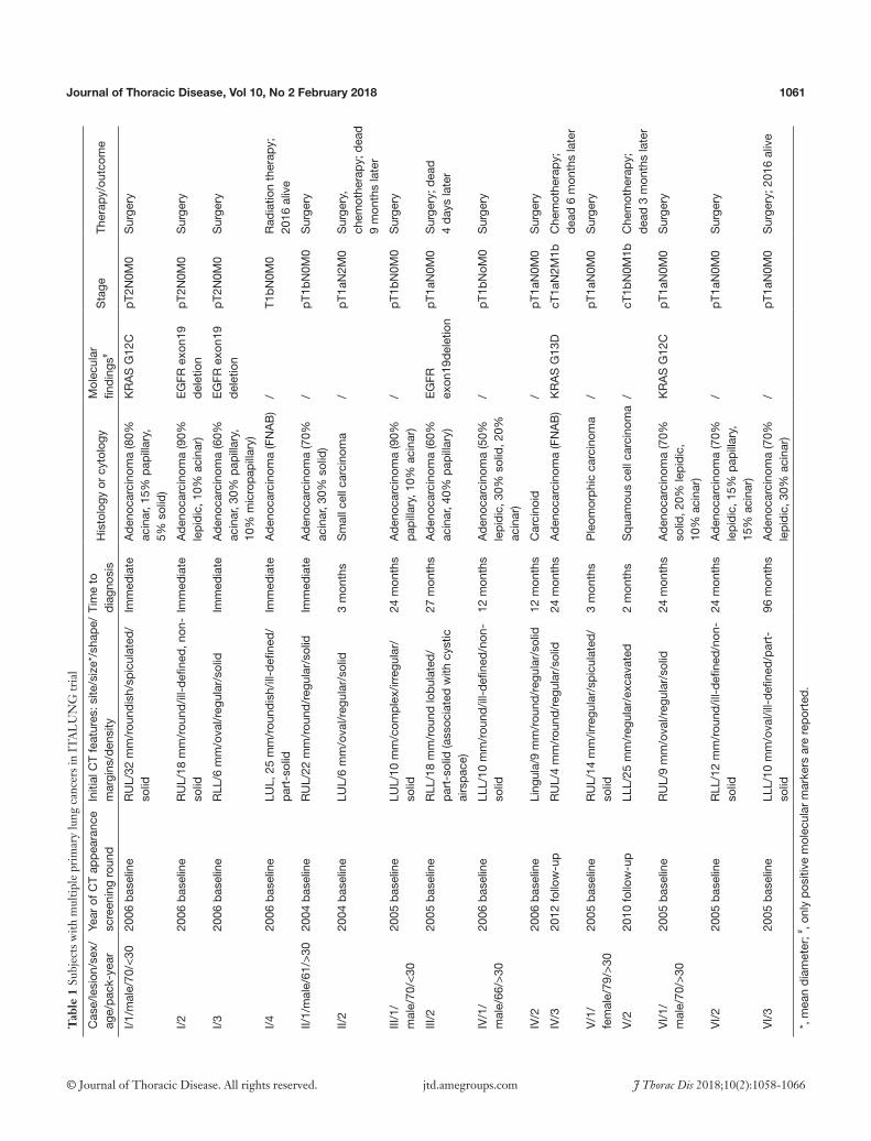
Eight subjects were diagnosed with multiple lung cancers during LDCT screening or full-dose CT follow-up. Two had multiple secondary lesions (from renal cancer and colorectal cancer) detected at LDCT and diagnosed at FNAB. Six subjects were ultimately diagnosed with MPLC (overall =16:2 in three subjects, 3 in two subjects and 4 in one subject) (Table 1 and Figures 1,2). They represented 15.8% (6/38) of all subjects with at least one screen-detected primary lung cancer.
The 16 MPLC included 9 morphologically and molecularly heterogeneous adenocarcinomas in three subjects (Figure 1) and combination of different tumor histotypes (2 adenocarcinomas plus 1 carcinoid, 1 adenocarcinoma plus 1 small cell carcinoma, 1 pleomorphic carcinoma plus 1 squamous cell carcinoma) in three subjects (Figure 2). Adenocarcinomas accounted for 75% (12/16) of MPLC.
Fourteen of 16 MPLC were observed during LDCT screening in four subjects and 2 during full-dose CT follow-up in two subjects. All the former were already present at baseline LDCT examination (Figures 1,2) and may be considered as synchronous lesions. The latter appeared during follow-up CT 5 and 6 years after surgical removal of the first lesions and may be considered metachronous lesions. Overall, MPLC appeared in the LDCT or follow-up CT in which they were retrospectively identified for the first time as solid (n=9), partially solid (n=3), non-solid (n=3) or excavated (n=1) lesions with regular (n=7), ill-defined (n=5), spiculated (n=2), irregular or lobulated (n=2) margins. Their initial size ranged between 4 and 32 mm (mean diameter 14.4±8.1 mm).
Of the three subjects with 2 lesions, one died of surgical complication and two of advanced disease. Of the three subjects with more than 2 lesions, two are alive (10 and 11 years after treatment of multifocal adenocarcinomas) and one died of advanced disease.
Discussion
Data about occurrence of MPLC in observational or randomized LDCT screening studies are fragmentary and summarized in Table 2. At least one subject with MPLC was reported in 11/33 studies (5,13-18,24-35), with a mean cumulative frequency of 0.18% (136 subjects) in 71,901 screenees and of 11.6% in 1,170 subjects with at least one screen-detected lung cancer (19-23,36-44). The MPLC were considered synchronous in 123/136 (90%) subjects and metachronous in 13 (10%). Unfortunately, in many cases the time of lesions appearance and histological diagnoses were not available. Moreover uncertainties and intervening modifications of the staging system of lung cancer and unavailability of morphologic and genetic/molecular features hinder recollection of information about MPLC in previous reports of LDCT screening (and surgical series). In particular, multiple pulmonary nodules may have been considered stage III or IV tumors (18), and this may have led to under-reporting of cases of MPLC, especially in case of multifocal adenocarcinoma.
In the active arm of ITALUNG, after the 4 years of active screening and 5 years of follow-up we observed few subjects (n=6; 0.43% of the screenees) harboring or developing MPLC. However they represented 15.8% of all those subjects with at least one screen-detected primary lung cancer. These percentages are in line with those reported in LDCT screening studies (Table 2).
In our series, adenocarcinoma was the most frequent histotype. This is in line with the type of primary lung cancers that are discovered by LDCT (Table 1) and in a previous study (18). Despite the small numbers of MPLC cases in our study that indicates need of further studies, two scenarios may be drawn. The first deals with morphological and molecular/genetically heterogeneous multifocal adenocarcinomas. The second deals with combination of adenocarcinoma with others histological types including small cell lung cancer, carcinoid and squamous cell cancer. Only one case of double squamous lung cancer was reported (23). As in previous reports (19,22), most (14/16 lesions in five subjects) of the screen-detected MPLC in ITALUNG were present at baseline and were diagnosed after further LDCT screening rounds, because of increased size or density (12,17,27).
Remarkably, our two patients with synchronous mult i focal adenocarcinoma with 4 and 3 les ions, respectively, were alive many years after lesion treatment. This i s consis tent with the v iew that mult i focal
1061Journal of Thoracic Disease, Vol 10, No 2 February 2018
© Journal of Thoracic Disease. All rights reserved. J Thorac Dis 2018;10(2):1058-1066jtd.amegroups.com
Tab
le 1
Sub
ject
s w
ith m
ultip
le p
rim
ary
lung
can
cers
in I
TA
LU
NG
tria
l
Cas
e/le
sion
/sex
/ag
e/pa
ck-y
ear
Year
of C
T ap
pear
ance
sc
reen
ing
roun
dIn
itial
CT
feat
ures
: site
/siz
e*/s
hape
/m
argi
ns/d
ensi
tyTi
me
to
diag
nosi
sH
isto
logy
or
cyto
logy
Mol
ecul
ar
findi
ngs#
Sta
geTh
erap
y/ou
tcom
e
I/1/m
ale/
70/<
3020
06 b
asel
ine
RU
L/32
mm
/rou
ndis
h/sp
icul
ated
/so
lidIm
med
iate
Ade
noca
rcin
oma
(80%
ac
inar
, 15%
pap
illar
y,
5% s
olid
)
KR
AS
G12
CpT
2N0M
0S
urge
ry
I/220
06 b
asel
ine
RU
L/18
mm
/rou
nd/il
l-de
fined
, non
-so
lidIm
med
iate
Ade
noca
rcin
oma
(90%
le
pidi
c, 1
0% a
cina
r)E
GFR
exo
n19
dele
tion
pT2N
0M0
Sur
gery
I/320
06 b
asel
ine
RLL
/6 m
m/o
val/r
egul
ar/s
olid
Imm
edia
teA
deno
carc
inom
a (6
0%
acin
ar, 3
0% p
apill
ary,
10
% m
icro
papi
llary
)
EG
FR e
xon1
9 de
letio
npT
2N0M
0S
urge
ry
I/420
06 b
asel
ine
LUL,
25
mm
/rou
ndis
h/ill
-defi
ned/
part
-sol
idIm
med
iate
Ade
noca
rcin
oma
(FN
AB
)/
T1bN
0M0
Rad
iatio
n th
erap
y;
2016
aliv
e
II/1/
mal
e/61
/>30
2004
bas
elin
eR
UL/
22 m
m/r
ound
/reg
ular
/sol
idIm
med
iate
Ade
noca
rcin
oma
(70%
ac
inar
, 30%
sol
id)
/pT
1bN
0M0
Sur
gery
II/2
2004
bas
elin
eLU
L/6
mm
/ova
l/reg
ular
/sol
id3
mon
ths
Sm
all c
ell c
arci
nom
a/
pT1a
N2M
0S
urge
ry,
chem
othe
rapy
; dea
d 9
mon
ths
late
r
III/1
/m
ale/
70/<
3020
05 b
asel
ine
LUL/
10 m
m/c
ompl
ex/ir
regu
lar/
so
lid24
mon
ths
Ade
noca
rcin
oma
(90%
pa
pilla
ry, 1
0% a
cina
r)/
pT1b
N0M
0S
urge
ry
III/2
2005
bas
elin
eR
LL/1
8 m
m/r
ound
lobu
late
d/pa
rt-s
olid
(ass
ocia
ted
with
cys
tic
airs
pace
)
27 m
onth
sA
deno
carc
inom
a (6
0%
acin
ar, 4
0% p
apill
ary)
EG
FR
exon
19de
letio
npT
1aN
0M0
Sur
gery
; dea
d
4 da
ys la
ter
IV/1
/m
ale/
66/>
3020
06 b
asel
ine
LLL/
10 m
m/r
ound
/ill-
defin
ed/n
on-
solid
12 m
onth
sA
deno
carc
inom
a (5
0%
lepi
dic,
30%
sol
id, 2
0%
acin
ar)
/pT
1bN
oM0
Sur
gery
IV/2
2006
bas
elin
eLi
ngul
a/9
mm
/rou
nd/r
egul
ar/s
olid
12 m
onth
sC
arci
noid
/pT
1aN
0M0
Sur
gery
IV/3
2012
follo
w-u
pR
UL/
4 m
m/r
ound
/reg
ular
/sol
id24
mon
ths
Ade
noca
rcin
oma
(FN
AB
)K
RA
S G
13D
cT
1aN
2M1b
Che
mot
hera
py;
dead
6 m
onth
s la
ter
V/1
/fe
mal
e/79
/>30
2005
bas
elin
eR
UL/
14 m
m/ir
regu
lar/
spic
ulat
ed/
solid
3 m
onth
sP
leom
orph
ic c
arci
nom
a/
pT1a
N0M
0S
urge
ry
V/2
2010
follo
w-u
pLL
L/25
mm
/reg
ular
/exc
avat
ed2
mon
ths
Squ
amou
s ce
ll ca
rcin
oma
/cT
1bN
0M1b
Che
mot
hera
py;
dead
3 m
onth
s la
ter
VI/1
/m
ale/
70/>
3020
05 b
asel
ine
RU
L/9
mm
/ova
l/reg
ular
/sol
id24
mon
ths
Ade
noca
rcin
oma
(70%
so
lid, 2
0% le
pidi
c,
10%
aci
nar)
KR
AS
G12
CpT
1aN
0M0
Sur
gery
VI/2
2005
bas
elin
eR
LL/1
2 m
m/r
ound
/ill-
defin
ed/n
on-
solid
24 m
onth
sA
deno
carc
inom
a (7
0%
lepi
dic,
15%
pap
illar
y,
15%
aci
nar)
/pT
1aN
0M0
Sur
gery
VI/3
2005
bas
elin
eLL
L/10
mm
/ova
l/ill-
defin
ed/p
art-
solid
96 m
onth
sA
deno
carc
inom
a (7
0%
lepi
dic,
30%
aci
nar)
/pT
1aN
0M0
Sur
gery
; 201
6 al
ive
*, m
ean
diam
eter
; # , onl
y po
sitiv
e m
olec
ular
mar
kers
are
repo
rted
.

1062 Mascalchi et al. Screen-detected multiple primary lung cancers
© Journal of Thoracic Disease. All rights reserved. J Thorac Dis 2018;10(2):1058-1066jtd.amegroups.com
A
E F G H
B C D
Figure 1 Case I (Table 1). Four primary adenocarcinomas in a 70-year-old smoker, which were all detected at baseline LDCT screening round. They appeared as a spiculated lung nodule in the right upper lobe (RUL) (A), a ground glass opacity in the same lobe (B), a small solid nodule in the right lower lobe (RLL) (arrow in C) and a ground glass opacity with a small solid component in the left upper lobe (LUL) (D). Haematoxylin and eosin histologic staining (original magnification ×200) demonstrate an invasive adenocarcinoma, acinar predominant (E) in the RUL lesion corresponding to (A), an invasive adenocarcinoma, lepidic predominant (F) in the RUL lesion corresponding to (B) and an invasive adenocarcinoma, acinar predominant (G) in the RLL lesion corresponding to (C). Papanicolaou stain (original magnification ×40) of fine needle aspiration biopsy shows papillary pattern of uniform malignant cells with irregular nuclei consistent with adenocarcinoma (H) in the LUL lesion corresponding to (D).
adenocarcinoma can behave as indolent lesion that should not be confused with aggressive primary lung cancers with intrapulmonary metastases (18). Awareness of this possibility may have significant impact on management of multiple lesions detected at LDCT screening that is currently recommended in the US (39) and is under evaluation in Europe (4).
In ITALUNG after a median follow-up time of 8.5 years, we observed 2 metachronous cancers during full-dose CT follow-up, which were both fatal. Obviously, longer surveillance is expected to increase the yield of metachronous MPLC. In fact one LDCT study reported 6 cases of metachronous MPLC in 2,989 screenees followed for 14 years (23).
Prevalence of MPLC in 18 surgical series outside screening (mean range, 1.1–8.6%) (3) was lower than the frequency in LDCT screening studies reporting at least one
such a case. This is not surprising since subjects undergoing LDCT are asymptomatic and prone to show developing cancers in their earlier stages.
Occurrence of MPLC in a single subject per se suggests the possibility of genetic predisposition. However search of genetic features associated with lung cancer (45) was beyond the scope of the present report.
Admittedly, since our study is based on cytopathologically or histopathologically proven primary lung cancer, it is possible that we underestimated MPLC in the cohort of subjects undergoing LDCT screening. In particular, lesions presenting as pure ground glass opacity that can be associated with minimally invasive adenocarcinoma (9) may be missed.
In conclusion, MPLC, mostly adenocarcinomas, are not uncommon in smokers and former smokers with at least one LDCT screen detected primary lung cancer. The

1063Journal of Thoracic Disease, Vol 10, No 2 February 2018
© Journal of Thoracic Disease. All rights reserved. J Thorac Dis 2018;10(2):1058-1066jtd.amegroups.com
Table 2 Multiple primary lung cancers (MPLC) in low-dose CT (LDCT) screening studies
StudyNo. of
screeneesNo. of LDCT
rounds
Subjects with screen-detected
primary lung cancer (%)
No. of adenocarcinomas
Subjects with MPLC
Subjects with MPLC/subjects
with screen-detected primary lung cancer (%)
Nawa et al. 2002 (13) 7,956 1 40 (0.5) 39 1 (synchronous) 2.5
Diederich et al. 2002 (14) 817 4 11 (1.3) 5 1 (synchronous) 9.0
Flieder et al. 2006 (15) 2,968 11 77 (2.6) 81 16 (synchronous) 20.8
Carter et al. 2007 (16) 10,056 11 250 (2.5) 177 31 (synchronous), 5 (metachronous)
14.4
Lindell et al. 2007 (17) 1,520 5 59 (3.9) 34 1 (synchronous), 1 (metachronous)
3.4
Pelosi et al. 2008 (18) 5,202 3 89 (1.7) 72 10 (synchronous) 11.2
Vazquez et al. 2009 (19) 27,456 12 338 (1.2) 279 49 (synchronous) 14.5
van Klaveren et al. 2009 (20) 7,557 3 124 (1.6) NA 5 (synchronous) 4.0
Infante et al. 2009 (21) 1,276 4 60 (4.7) 27 2 (synchronous), 1 (metachronous)
5.0
Saghir et al. 2012 (22) 4,104 5 69 (1.7) 48 6 (synchronous) 8.7
Sanchez-Salcedo et al. 2015 (23) 2,989 14 53 (1.8%) 33 1 (synchronous), 6 (metachronous)
13.2
A
B C
Figure 2 Case II (Table 1). Adenocarcinoma and small cell lung cancer in a 61-year-old smoker both detected at baseline LDCT screening round. They appeared as a rounded lung nodule in the RUL and a subpleural small solid nodule (arrow) in the LUL (A). Haematoxylin and eosin histologic staining demonstrate invasive adenocarcinoma, acinar predominant (original magnification ×200) (B) in the RUL lesion and small cell lung carcinoma (original magnification ×100) (C) in the LUL lesion.

1064 Mascalchi et al. Screen-detected multiple primary lung cancers
© Journal of Thoracic Disease. All rights reserved. J Thorac Dis 2018;10(2):1058-1066jtd.amegroups.com
potential distinction of two subtypes of MPLC, represented by multifocal adenocarcinomas and by combination of adenocarcinoma with others histological types, having different prognoses and treatment implications warrants further study.
Acknowledgements
Funding: The study was supported by the local government of Tuscany (Decree N. 1014 of 02.25.2004) and by a Research Grant of the Italian Ministry of Education University and Research (PRIN 2003) to Professor Mario Mascalchi. The funding source had no role in study design, in the collection, analysis and interpretation of data; in the writing of the report; and in the decision to submit the article for publication.
Footnote
Conflicts of Interest: The authors have no conflicts of interest to declare.
Ethical Statement: The study was conducted in complian-ce with the Helsinki Declaration (http://www.wma.net/en/30publications/10policies/b3/index.html) and the study protocol was approved by the Local Ethic Committees of the participating centers (Firenze, approval number 29–30, 30 September 2003; Pisa, number 23, 27 October 2003; Pistoia, number 00028543, 13 May 2004). Each subject provided an informed written consent to participate to the study.
References
1. Slaughter DP, Southwich HW, Smejkal W. “Field cancerization” in oral stratified squamous epithelium: clinical implications of multi-centric origin. Cancer 1953;6:963-8.
2. Kadara H, Wistuba II. Field cancerization in non-small cell lung cancer: implications in disease pathogenesis. Proc Am Thorac Soc 2012;9:38-42.
3. Voltolini L, Rapicetta C, Luzzi L. et al. Surgical treatment of synchronous multiple lung cancer located in a different lobe or lung: high survival in node negative subgroup. Eur J Cardiothorac Surg 2010;37:1198-204.
4. Paci E, Puliti D, Lopes Pegna A, et al. Mortality, survival and incidence rates in the ITALUNG randomised lung cancer screening trial. Thorax 2017;72:825-31.
5. Picozzi G, Paci E, Lopes-Pegna A, et al. Screening of lung cancer with low-dose spiral CT: results of a three-year pilot study and design of the randomized controlled trial “Italung-CT”. Radiol Med 2005;109:17-26.
6. Lopes Pegna A, Picozzi G, Mascalchi M, et al. ITALUNG Study Research Group. Design, recruitment and baseline results of the ITALUNG trial for lung cancer screening with low-dose CT. Lung Cancer 2009;64:34-40.
7. Lopes Pegna A, Picozzi G, Falaschi F, et al. ITALUNG Study Research Group. Four-year results of low-dose CT screening and nodule management in the ITALUNG trial. J Thorac Oncol 2013;8:866-75.
8. Shen KR, Meyers BF, Larner JM, et al. Special treatment issues in lung cancer: ACCP evidence-based clinical practice guidelines (2nd edition). Chest 2007;132:290-305.
9. Travis WD, Brambilla E, Burke AP, et al. WHO classification of tumours of the lung, pleura, thymus and heart. Lyon, France: IARC Press, 2015.
10. Travis WD, Brambilla E, Nicholson AG, et al. The 2015 World Health Organization classification of lung tumors. Impact of genetic, clinical and radiologic advances since the 2004 classification. J Thorac Oncol 2015;10:1243-60.
11. Edge SB, Compton CC. The American Joint Committee on Cancer: the 7th edition of the AJCC cancer staging manual and the future of TNM. Ann Surg Oncol 2010;17:1471-4.
12. Mascalchi M, Picozzi G, Falchini M, et al. Initial LDCT appearance of screen-detected lung cancers in the ITALUNG trial. Eur J Radiol 2014;83:2080-6.
13. Nawa T, Nakagawa T, Kusano S, et al. Lung cancer screening using low-dose spiral CT. Results of baseline and 1-year follow-up studies. Chest 2002;122:15-20.
14. Diederich S, Wormanns D, Semik M et al. Screening for early lung cancer with low-dose spiral CT: prevalence in 817 asymptomatic smokers. Radiology 2002;222:773-81.
15. Flieder DB, Vazquez M, Carter D, et al. Pathologic findings of lung tumors diagnosed on baseline CT screening. Am J Surg Pathol 2006;30:606-13.
16. Carter D, Vazquez M, Flieder D, et al Comparison of pathologic findings of baseline and annual repeat cancers diagnosed on CT screening. Lung Cancer 2007;56:193-9.
17. Lindell RM, Hartman TE, Swensen SJ, et al. Five-year lung cancer screening experience: CT appearance, growth rate, location, and histologic features of 61 lung cancers. Radiology 2007;242:555-62.
18. Pelosi G, Sonzogni A, Veronesi G, et al. Pathologic and molecular features of screening low-dose computed tomography (LDCT)-detected lung cancer: A baseline and
1065Journal of Thoracic Disease, Vol 10, No 2 February 2018
© Journal of Thoracic Disease. All rights reserved. J Thorac Dis 2018;10(2):1058-1066jtd.amegroups.com
2-year repeat study. Lung Cancer 2008;62:202-14. 19. Vazquez M, Carter D, Brambilla E, et al. Solitary and
multiple resected adenocarcinomas after CT screening for lung cancer: histopathologic features and their prognostic implications. Lung Cancer 2009;64:148-54.
20. van Klaveren RJ, Ouderk M, Prokop M, et al. Management of lung nodules detected by volume CT screening. N Engl J Med 2009;361:2221-9.
21. Infante M, Cavuto S, Lutman FR, et al. A randomized study of lung cancer screening with spiral computed tomography.Three-year results from the DANTE trial. Am J Respir Crit Care Med 2009;180:445-53.
22. Saghir Z, Dirksen A, Ashraf H, et al. CT screening for lung cancer brings forward early disease. The randomised Danish Lung Cancer Screening Trial: status after five annual screening rounds with low-dose CT. Thorax 2012;67:296-301.
23. Sanchez-Salcedo P, Berto J, de-Torres JP, et al. Lung cancer screening: fourteen year experience of the Pamplona early detection program (P-IELCAP). Arch Bronconeumol 2015;51:169-76.
24. Sone S, Li F, Yang ZG, et al. Results of three-year mass screening programme for lung cancer using mobile low-dose spiral computed tomography scanner. Br J Cancer 2001;84:25-32.
25. Sobue T, Moriyama N, Kaneko M et al. Screening for lung cancer with low-dose helical computed tomography: anti-lung cancer association project. J Clin Oncol 2002;20:911-20.
26. Garg K, Keith RL, Byers T, et al. Randomized controlled trial with low-dose spiral CT for lung cancer screening: feasibility study and preliminary results. Radiology 2002;225:506-10.
27. Pastorino U, Bellomi M, Landoni C, et al. Early lung-cancer detection with spiral CT and positron emission tomography in heavy smokers: 2-year results. Lancet 2003;362:593-7.
28. Gohagan JK, Marcus PM, Fagerstrom RM, et al. Final results of the Lung Screening Study, a randomised feasibility study of spiral CT versus chest X-ray screening for lung cancer. Lung Cancer 2005;47:9-15.
29. Chong S, Lee KS, Jin M, et al. Lung cancer screening with low-dose helical CT in Korea: experiences at the Samsung Medical Center. J Korean Med Sci 2005;20:402-8.
30. Stephenson SM, Mech KF, Sardi A. Lung cancer screening with low-dose spiral computed tomography. Am Surg 2005;71:1015-7.
31. Bastarrika G, García-Velloso MJ, Lozano MD, et al. Early
lung cancer detection using spiral computed tomography and positron emission tomography. Am J Respir Crit Care Med 2005;171:1378-83.
32. Novello S, Fava C, Borasio P, et al. Three-year findings of an early lung cancer detection feasibility study with low-dose spiral computed tomography in heavy smokers. Ann Oncol 2005;16:1662-6.
33. MacRedmond R, McVey G, Lee M, et al. Screening for lung cancer using low dose CT scanning: results of 2 year follow up. Thorax 2006;61:54-6.
34. Blanchon T, Bréchot JM, Grenier PA, et al. Baseline results of the Depiscan study: a French randomized pilot trial of lung cancer screening comparing low dose CT scan (LDCT) and chest X-ray (CXR). Lung Cancer 2007;58:50-8.
35. Wilson DO, Weissfeld J, Fuhrman C, et al. The Pittsburgh Lung Screening Study (PLuSS): outcomes within 3 years of a first computed tomography scan. Am J Respir Crit Care Med 2008;178:956-61.
36. Menezes RJ, Roberts HC, Paul NS, et al. Lung cancer screening using low-dose computed tomography in at-risk individuals: The Toronto experience. Lung Cancer 2010;67:177-83.
37. Pastorino U, Rossi M, Rosato V, et al. Annual or biennial CT screening versus observation in heavy smokers: 5-year results of the MILD trial. Eur J Cancer Prev 2012;21:308-15.
38. Veronesi G, Maisonneuve P, Rampinelli C, et al. Computed tomography screening for lung cancer: results of ten years of annual screening and validation of COSMOS prediction model. Lung Cancer 2013;82:426-30.
39. National Lung Screening Trial Research Team, Church TR, Black WC, et al. Results of initial low-dose computed tomographic screening for lung cancer. N Engl J Med 2013;368:1980-91.
40. Becker N, Motsch E, Gross ML, et al. Randomized study on early detection of lung cancer with MSCT in Germany: results of the first 3 years of follow-up after randomization. J Thorac Oncol 2015;10:890-6.
41. Rzyman W, Dziedzic R, Jelitto-Górska M, et al. Results of an open-access lung cancer screening program with low-dose computed tomography: the Gdańsk experience. Pol Arch Med Wewn 2015;125:232-9.
42. Yi CA, Lee KS, Shin MH, et al. Low-dose CT screening in an Asian population with diverse risk for lung cancer: A retrospective cohort study. Eur Radiol 2015;25:2335-45.
43. Field JK, Duffy SW, Baldwin DR, et al. UK lung
1066 Mascalchi et al. Screen-detected multiple primary lung cancers
© Journal of Thoracic Disease. All rights reserved. J Thorac Dis 2018;10(2):1058-1066jtd.amegroups.com
Cite this article as: Mascalchi M, Comin CE, Bertelli E, Sali L, Maddau C, Zuccherelli S, Picozzi G, Carrozzi L, Grazzini M, Fontanini G, Voltolini L, Vella A, Castiglione F, Carozzi F, Paci E, Zompatori M, Lopes Pegna A, Falaschi F; for the ITALUNG Study Research Group. Screen-detected multiple primary lung cancers in the ITALUNG trial. J Thorac Dis 2018;10(2):1058-1066. doi: 10.21037/jtd.2018.01.95
cancer RCT pilot screening trial: baseline findings from the screening arm provide evidence for the potential implementation of lung cancer screening. Thorax 2016;71:161-70.
44. Sverzellati N, Silva M, Calareso G, et al. Low-dose computed tomography for lung cancer screening:
comparison of performance between annual and biennial screen. Eur Radiol 2016;26:3821-9.
45. Liu Y, Kheradmand F, Davis CF, et al. Focused analysis of exome sequencing data for rare germline mutations in familial and sporadic lung cancer. J Thorac Oncol 2016;11:52-61.
Supplementary
ITALUNG pathology protocol
Tumors were sampled as fresh tissue immediately after surgery. The entire tumors, fragments of distant non-neoplastic parenchyma, all detectable lymph nodes, and mediastinal lymph nodes were sampled. Specimens were fixed in buffered formalin for 16–24 hours and processed for standard histological examination. Multiple adenocarcinoma cases underwent molecular analysis for the identification of ALK gene translocation and of the known driver mutations in the following genes: EGFR, KRAS, NRAS, BRAF, PIKCA, ERBB2, DDR2, MAP2K1 and RET. A representative paraffin-embedded block containing about 80% viable tumor was selected for each case. Immunohistochemical staining procedures were conducted on 4 µm-thick sections of paraffin-embedded tissue using a Ventana Benchmark Ultra automated immunostainer (Ventana Medical System, Tucson, AZ, USA) with the
anti-ALK monoclonal antibody (clone D5F3, ready to use, Ventana Medical System). The OptiView DAB Detection kit was used as revelation system, adding the OptiView Amplification kit (Ventana Medical System) for ALK detection. Three consecutive 10 µm-thick sections were performed from the same blocks for molecular analysis. These were conducted by a mass spectrometry-based multiplex assay (MassArray technology, Sequenom, San Diego, CA) using the “Myriapod Lung Status” kit (Diatech Pharmacogenetics, AN, IT) capable of identifying the major mutations of the following genes: EGFR (exons 18, 19, 20, 21), KRAS (codons 12, 13, 61), NRAS (codons 12, 61), BRAF (codons 466, 469, 594, 597, 600), PIKCA (codons 542, 545, 1043, 1047), ERBB2 (exon 20), DDR2 (codons 239, 638, 768), MAP2K1 (codons 56, 57, 67), RET (codon 918).