5Hydroxy l-tryptophan suppresses food intake in food-deprived and stressed rats
Upload
independentCategory
view
2download
0
Biochem. J. (2010) 425, 265–274 (Printed in Great Britain) doi:10.1042/BJ20090856 265
Coactosin-like protein functions as a stabilizing chaperonefor 5-lipoxygenase: role of tryptophan 102Julia ESSER*1, Marija RAKONJAC*1, Bettina HOFMANN†‡, Lutz FISCHER*†, Patrick PROVOST§, Gisbert SCHNEIDER‡,Dieter STEINHILBER†, Bengt SAMUELSSON* and Olof RADMARK*2
*Department of Medical Biochemistry and Biophysics, Division of Physiological Chemistry II, Karolinska Institutet, S-171 77 Stockholm, Sweden, †Institute of PharmaceuticalChemistry/ZAFES, University of Frankfurt, Max-von-Laue-Str. 9, D-60438 Frankfurt am Main, Germany, ‡Institute of Organic Chemistry and Chemical Biology, Johann WolfgangGoethe-University, Siesmayerstr. 70, D-60323 Frankfurt am Main, Germany, and §CHUL Research Center/CHUQ and Faculty of Medicine, Universite Laval, Quebec, QC, G1V 4G2,Canada
The activity of 5-LO (5-lipoxygenase), which catalyses twoinitial steps in the biosynthesis of pro-inflammatory LTs(leukotrienes), is strictly regulated. One recently discoveredfactor, CLP (coactosin-like protein), binds 5-LO and promotesLT formation. In the present paper we report that CLP alsostabilizes 5-LO and prevents non-turnover inactivation of theenzyme in vitro. Mutagenesis of tryptophan residues in the 5-LO β-sandwich showed that 5-LO-Trp102 is essential for bindingto CLP, and for CLP to support 5-LO activity. In addition,the stabilizing effect also depended on binding between CLPand 5-LO. After mutations which prevent interaction (5-LO-W102A or CLP-K131A), the protective effect of CLP wasabsent. A calculated 5-LO–CLP docking model indicates thatCLP may bind to additional residues in both domains of 5-LO,
thus possibly stabilizing the 5-LO structure. To obtain furthersupport for binding between CLP and 5-LO in a living cell,subcellular localization of CLP and 5-LO in the monocyticcell line Mono Mac 6 was determined. In these cells, 5-LO asso-ciates with a nuclear fraction only when differentiated cells areprimed with phorbol ester and stimulated with ionophore. Thesame pattern of redistribution was found for CLP, indicating thatthe two proteins associate with the nucleus in a co-ordinatedfashion. The results of the present study support a role for CLP asa chaperoning scaffold factor, influencing both the stability andthe activity of 5-LO.
Key words: arachidonic acid, eicosanoid, leukotriene, oxylipin.
INTRODUCTION
5-LO (5-lipoxygenase) catalyses two initial steps in LT (leuko-triene) biosynthesis, oxygenation of AA (arachidonic acid) to 5-HPETE [5(S)-hydroperoxy-6-trans-8,11,14-cis-eicosatetraenoicacid] and subsequent dehydration into the epoxide LTA4 [1].LTs are inflammatory lipid mediators which cause leucocytechemotaxis and increased vascular permeability. The effects ofLTs are well established in the pathogenesis of asthma, andaccumulating data also indicate a role for LTs in atherosclerosis[2]. In addition, 5-LO products are implicated in cancer cellsurvival [3], recently for leukaemia stem cells [4].
A model of the 5-LO structure, based on the crystal structure ofthe ferrous form of rabbit reticulocyte 15-LO [5,6], consistsof an N-terminal β-sandwich (residues 1–114) and a largerC-terminal catalytic domain containing prosthetic iron (residues121–673). The validity of this 5-LO model structure is supportedby various mutagenesis studies (reviewed in [7]). Several factorswhich influence 5-LO enzyme activity bind to the C2-likeβ-sandwich, e.g. Ca2+ and PC (phosphatidylcholine), and whencells are stimulated to produce LTs, 5-LO is typically associatedwith the nuclear membrane [8]. However, recent observationsindicate a different subcellular distribution in neutrophils frommales [9].
CLP (coactosin-like protein) is similar to coactosin [10], amember of the ADF (actin-depolymerizing factor)/cofilin groupof actin-binding proteins. By two-hybrid screening with 5-LO
as bait, we found that CLP can bind to 5-LO [11]. Bindingwas also demonstrated by co-immunoprecipitation from lysatesof transfected cells, and in vitro assays [GST (glutathionetransferase) pull-down assay, native PAGE and chemical cross-linking] showed binding with a 1:1 molar stoichiometry [12].Human CLP also binds F-actin, and was found to co-localizewith actin stress fibres in transfected CHO (Chinese-hamsterovary) and COS-7 cells [13]. Also mouse CLP was found to bind5-LO with a 1:1 stoichiometry [14]. Mutagenesis showed theinvolvement of CLP-Lys75 and -Lys131 in binding to F-actin and5-LO respectively [12]. In the CLP structure [15] Lys75
and Lys131 are close, indicating overlapping binding sites, whichcould explain why a ternary complex of F-actin–CLP–5-LO hasnot been found. We have previously described that CLP can serveas a scaffold for Ca2+-induced activation of 5-LO [16]. CLP hasappeared in several array/proteome analyses, connecting CLPwith cancer and inflammatory disease, e.g. rheumatoid arthritis[17]. Recently, hyperforin, an anti-inflammatory compound fromSt John’s wort which inhibits 5-LO activity, was found to impairthe binding between CLP and 5-LO [18].
In the present study we show that CLP stabilizes 5-LO againstnon-turnover inactivation, and that one particular residue inthe 5-LO β-sandwich (Trp102) is essential for binding of CLPto 5-LO, and for the effects of CLP on 5-LO activity andstability. A model of the docking complex was calculated (DOTalgorithm) taking the available mutagenesis results into consi-deration.
Abbreviations used: AA, arachidonic acid; CLP, coactosin-like protein; GST, glutathione transferase; 5-HETE, 5(S)-hydroxy-6-trans-8,11,14-cis-eicosatetraenoic acid; 5-HPETE, 5(S)-hydroperoxy-6-trans-8,11,14-cis-eicosatetraenoic acid; 13-HPODE, 13(S)-hydroperoxy-9-cis-11-trans-octadecadienoic acid; 5-LO, 5-lipoxygenase; LT, leukotriene; MM6, Mono Mac 6; PC, phosphatidylcholine; PGC buffer, PBS containing 1 mg/ml glucoseand 1 mM CaCl2; TGF-β, transforming growth factor-β; wt, wild-type.
1 These authors contributed equally.2 To whom correspondence should be addressed (email [email protected]).
c© The Authors Journal compilation c© 2010 Biochemical Society
www.biochemj.org
Bio
chem
ical
Jo
urn
al

266 J. Esser and others
MATERIALS AND METHODS
Expression of CLP and 5-LO
Recombinant human CLP was expressed as a GST fusion protein,using the plasmid pGEX-5X-1-CLP [12,15]. The fusion partnerwas removed by digestion with Factor Xa, and anion-exchangechromatography on MonoQ. Recombinant human 5-LO wasexpressed from the plasmid pT3-5-LO and purified on ATP-agarose (Sigma A2767) [19,20]. Two Escherichia coli strainswere used. Expression in BL21 at 27 ◦C using a rich medium(TB) gave 5-LO preparations with specific activities of approx.10 μmol of 5-H(P)ETE/mg, whereas expression in MV1190 at27 ◦C using minimal medium resulted in specific activities ofapprox. 20 μmol of 5-H(P)ETE/mg. Mutated plasmids (W13A,W75A and W102A) were constructed from pT3-5-LO using theQuikChange® kit (Stratagene).
Time and heat inactivation
Solutions of purified 5-LO (14 μg/ml, total volume 500 μl), withor without purified wt (wild-type)-CLP or CLP mutants (1:1stoichiometry), were prepared in AB+ buffer [50 mM Tris/HCl(pH 7.5), 2 mM EDTA and 0.1% 2-mercaptoethanol], and kept inclosed Eppendorf tubes. For time-dependent inactivation at roomtemperature (22 ◦C), the tubes were kept on the laboratory bench,and at the indicated intervals, aliquots (10 μl) were removed forthe 5-LO activity assay (described below). For heat inactivation,tubes were immersed in a water bath at 50, 55 and 60 ◦C. At theindicated intervals, aliquots (10 μl) were removed and assayedfor 5-LO activity.
HPLC assay of 5-LO enzyme activity
Incubations were performed in Eppendorf tubes at roomtemperature for 10 min. Buffer AB [50 mM Tris/HCl (pH7.5) and 2 mM EDTA] was added to tubes, followed bythe addition of substrate mix containing Tris/HCl (pH 7.5),CaCl2, PC (Sigma P-3556), AA (Nu-Chek Prep), 13-HPODE[13(S)-hydroperoxy-9-cis-11-trans-octadecadienoic acid] andATP. Tubes were immersed in a sonication bath for 1 min.The reaction was initiated by addition of aliquots of 5-LOfrom inactivation experiments containing approx. 150 ng of5-LO. When CLP (1:1 stoichiometry) was present duringinactivation experiments, it was also present in the assay. Finalconcentrations in the 100 μl incubation volume were: 77.4 mMTris/HCl (pH 7.5), 100 μM AA, 1.2 mM EDTA, 1.9 mM Ca2+,25 μg/ml PC, 10 μM 13-HPODE and 4.9 mM ATP.
Incubations were terminated with 300 μl of ice-coldstop solution (acetonitrile/water/acetic acid; 60:40:0.2, byvol) containing 3.3 μM 17(S)-hydroxy-(7Z,10Z,13Z,15E)-docosatetraenoic acid (internal standard at 235 nm, a gift fromDr Mats Hamberg, Department of Medical Biochemistry andBiophysics, Karolinska Institute, Stockholm, Sweden). Aliquots(100 μl) were injected on to a C18 HPLC column (Waters NovaRadial Pak) and AA metabolites were isocratically eluted withacetonitrile/water/acetic acid (60:40:0.2, v/v) at a flow rate of1.2 ml/min. The eluate was monitored at 235 nm for 5-HPETEand 5-HETE [5(S)-hydroxy-6-trans-8,11,14-cis-eicosatetraenoicacid]. 5-LO enzymatic activity was calculated from the sum ofpeak areas, relative to internal standards.
GST pull-down assay
For binding studies in vitro, 20 μg of the GST–CLP fusionprotein linked to glutathione–Sepharose 4B beads was incubated
with purified 5-LO protein (5 μg) in the presence of BSA(50 μg) in 200 μl of buffer A [2 mM Tris/HCl (pH 8.0), 0.2 mMATP, 0.2 mM CaCl2, 2 mM MgCl2, 50 mM KCl and0.5 mM 2-mercaptoethanol]. After a 30-min gentle rotation atroom temperature, beads were washed five times in buffer A(without BSA). Bound proteins were eluted with 150 μl of elutionbuffer [10 mM GSH in 50 mM Tris/HCl (pH 8.0)] during 60–90 min rotation at room temperature. Beads were sedimented,and aliquots of the supernatant eluate were assayed by SDS/PAGEfollowed by 5-LO Western blot analysis.
Pulse proteolysis with thermolysin
A stock solution of thermolysin (Sigma T7902) was preparedin 2.5 M NaCl and 10 mM CaCl2 and the concentration wasdetermined by spectroscopy as described previously [21]. Purified5-LO (4 μg) with or without CLP (0.8 μg, 1:1 stoichiometry) waspre-incubated for 10 min at room temperature in PBS containing10 mM CaCl2 and 50 mM NaCl. Thermolysin (4 μg) was added,and after 20–120 s incubations were stopped by the addition of3.5 μl of EDTA (50 mM) and cooling on ice. Sample loadingbuffer (10× concentration) was added and 4 μl aliquots weresubjected to SDS/PAGE on a Pharmacia FAST system, followedby Coomassie Blue staining.
Subcellular fractionation after detergent lysis
MM6 (Mono Mac 6) cells were grown in cell culture, anddifferentated with TGF-β (transforming growth factor-β; 5 ng/ml)and calcitriol (50 nM) for 96 h as described previously [22].Cells (10 × 106) were resuspended in 1 ml of PGC buffer(PBS containing 1 mg/ml glucose and 1 mM CaCl2). After pre-incubation for 10 min at 37 ◦C, cells were primed with PMA(100 nM for 10 min) and activated with ionophore (5 μM)and AA (40 μM) for another 10 min, as indicated. Cells werethen chilled on ice for 5 min, lysed with Nonidet P40, andsubcellular fractionation was performed as described previously[22]. Aliquots (45 μl, derived from approx. 1 million cells) of pair-wise nuclear and non-nuclear fractions were immediately mixedwith 9 μl of concentrated SDS loading buffer, heated for 5 min at95 ◦C, and analysed for 5-LO and CLP protein by SDS/PAGE andimmunoblotting. In-house antisera for 5-LO (1551) and CLP wereused. Antibodies against lamin B (nuclear membrane marker, acontrol for the fractionation procedure) and against β-actin werefrom Santa Cruz Biotechnology.
Protein–protein docking of CLP and 5-LO
Protein–protein docking of CLP and 5-LO was performed usingthe ClusPro server [23] and applying the DOT algorithm [24,25].Since an experimentally determined three-dimensional structurefor 5-LO is not available, we used our previous homology modelbased on the crystal structure of rabbit 15-LO [26]. For CLPthe averaged structure of 20 NMR determinations (PDB: 1WNJ;[15]) was used as the starting conformation for protein–proteindocking. The flexible ends (residues 1–4 and 137–145) weretruncated. Parameters for the docking algorithm were: radius ofclustering = 9, number of electrostatic hits to be clustered = 1500,and number of retained output-structures = 10. Hydrogen atomswere added to both input structures and the proteins wereminimized with a constrained backbone using the CHARMMforce field [27]. DOT performs a systematic rigid-body search ofone molecule translated and rotated relative to a fixed secondmolecule by evaluating conformations during docking with a
c© The Authors Journal compilation c© 2010 Biochemical Society

Coactosin-like protein functions as a stabilizing chaperone for 5-lipoxygenase 267
Figure 1 Enzyme activity of wt-5-LO and three tryptophan mutants in the presence of CLP and/or PC
(A) The Ca2+-activated formation of 5-HPETE plus 5-HETE was determined in 10 min incubations of purifed 5-LO proteins (expressed in BL21) as described in the Materials and methods section. PC(25 μg/ml) or CLP (at a 1:1 stoichiometry) were present as indicated. (B) The formation of LTA4 hydrolysis products was determined in the same incubations as in (A). (C) Total products (5-HPETE,5-HETE and LTA4 hydrolysis products) in the same incubations. All values are means+−S.E.M. for three independent experiments. n.s., not significant.
shape complementarity function. Intermolecular energies of allconfigurations generated are calculated as the sum of electrostaticand van-der-Waals energies. These energy terms are evaluated ascorrelation functions and used to rank the docking solution [24].The obtained docked complexes were filtered by our experimentaldata retaining only those complexes where CLP-Lys131 couldinteract with 5-LO and 5-LO-Trp102 with CLP. CLP-Lys131,5-LO-Trp102 and nearby amino acids in the resulting complexwere finally relaxed using the CHARMM force field to optimizethe modelled interacting geometry.
Data analysis
Origin 8 was used for data analysis, and a Student’s t testfor two independent samples was performed to determinestatistical differences between the means of groups of independentexperiments. P values <0.05 were considered statisticallysignificant.
RESULTS
Enzyme activity of wt-5-LO and three tryptophan mutants in thepresence of CLP and/or PC
Efficient Ca2+-induced dioxygenase and LTA4 synthase activitiesof wt-5-LO requires the presence of a scaffold factor to which5-LO can bind. Binding of 5-LO to PC has been shown toinvolve three surface-exposed tryptophan residues in the 5-LOβ-sandwich (Trp13, Trp75 and Trp102). When these were exchanged
for alanine, mutagenesis of Trp102 gave the most prominent effect,reducing the affinity of the isolated 5-LO C2-like domain to PCby approx. 20-fold [28]. Because of the similar effects of PC andCLP (both support Ca2+-induced 5-LO activity) we performed thesame three single-point mutations in intact 5-LO, and determinedthe effect on enzyme activity in the presence of CLP and/or PC.As shown in Figure 1(A), in the presence of PC (25 μg/ml) theformation of dioxygenase products (5-HPETE and 5-HETE) wasrather similar for wt-5-LO and the mutants. Enzyme activitywas much lower in the absence of scaffold factors. In assaysfrom which PC was excluded, addition of CLP (1:1 stoichiometry)increased dioxygenase activity of wt-5-LO and two of the mutants(5-LO-W13A and 5-LO-W75A). In contrast, for 5-LO-W102A,CLP did not support the dioxygenase activity, which was barelydetectable. When both PC and CLP were added, activities wereagain quite similar for all four proteins. Concomitantly, for wt-5-LO, 5-LO-W13A and 5-LO-W75A, addition of CLP resulted inan increased 5-HETE/5-HPETE ratio (results not shown).
Formation of LTA4 was determined as the non-enzymatichydrolysis products 6-trans-LTB4 and 12(S)-6-trans-LTB4. Forwt-5-LO, inclusion of both CLP and PC led to a 6-foldup-regulation of LTA4 production, compared with incubationsincluding only PC (Figure 1B). Similar up-regulation of LTproduction was found for 5-LO-W13A and 5-LO-W75A, but notfor 5-LO-W102A. For all four 5-LO proteins, LT biosynthesis wassmall when PC was absent. Thus, as observed previously [16],CLP alone cannot support formation of LTA4, but CLP togetherwith PC leads to a considerable increase in LTA4. Figure 1(C)shows the total 5-LO product formation, with and without PC and
c© The Authors Journal compilation c© 2010 Biochemical Society

268 J. Esser and others
Figure 2 Enzyme activity of wt-5-LO and three tryptophan mutants at lowconcentrations of PC and AA
The Ca2+-activated formation of 5-HPETE plus 5-HETE was determined in 10 min incubationsof purifed 5-LO proteins (expressed in BL21) as described in the Materials and methods section.The AA and PC concentration was 100 μM and 25 μg/ml, or 20 μM and 5 μg/ml, as indicated.All values are means+−S.E.M. for three independent experiments.
CLP. Taken together, the activity results show that Trp102 in 5-LOis essential for the effects of CLP on 5-LO activity.
Enzyme activity of wt-5-LO and three tryptophan mutants atreduced concentrations of PC and AA
To study the role of the three tryptophan residues on 5-LOactivation (by Ca2+) in the presence of only PC as a scaffold factor,activity assays were performed at reduced concentrations of AAand PC (20 μM AA and 5 μg/ml PC). Only at such relativelylow concentrations were differences in activities detected for thetryptophan mutants, in comparison with wt-5-LO (Figure 2). Thusat high PC and AA (our standard conditions: 100 μM AA and25 μg/ml PC) no significant reduction in oxygenase activity wasobserved for the mutants in relation to wt-5-LO (compare withFigure 1A). However, reduced amounts of PC and AA resulted ina significantly lower formation of 5-HPETE and 5-HETE, whichwas observed for all three 5-LO mutants (Figure 2). Thus all threetryptophan residues contribute to PC support of Ca2+-induced5-LO activation. None appears to be of particular importance.Accordingly, when all three tryptophan residues were mutated toalanine, there was a more pronounced reduction in 5-LO activityat reduced concentrations of PC and AA [16].
Mutant 5-LO-W102A does not bind GST–CLP
To ascertain whether the effects of CLP on 5-LO enzyme activitymirror specific binding of 5-LO proteins to CLP, pull-down assays,using the GST–CLP fusion protein, were performed. wt-5-LO,5-LO-W13A and 5-LO-W75A all bound to GST–CLP, wher-eas 5-LO-W102A did not (Figure 3). There was no association of5-LO to GST, used as a negative control. The enzyme activityof 5-LO-W102A was about the same as for wt-5-LO when assayedin the presence of PC (25 μg/ml) (Figure 1). This indicates thatthe loss of binding of 5-LO-W102A to CLP should not be due tocompromised overall structure of this 5-LO mutant.
CLP prevents inactivation of 5-LO over time
In addition to supporting Ca2+-induced enzyme activity, we foundthat CLP prevents inactivation of 5-LO. Solutions of purified
Figure 3 Mutant 5-LO-W102A does not bind GST–CLP
A GST pull-down assay was performed as described in the Materials and methods section.Aliquots of bead eluates were analysed for 5-LO by Western blotting. Similar results wereobtained in four separate experiments. stand., standard.
5-LO were kept at room temperature (on the laboratory bench) forup to 5 days in sealed Eppendorf tubes under normal atmosphere.At intervals, aliquots were removed and subjected to the enzymeactivity assay. 5-LO alone was gradually inactivated, withhalf activity remaining after 24 h, and approx. 20% after 120 h.However, in the presence of CLP (1:1 molar stoichiometry),enzyme activity was preserved, considering that approx. 2/3 of theinitial activity remained after 120 h (Figure 4A). Since no substratewas present during the 5 days, CLP protected 5-LO against non-turnover inactivation. Exposure to oxygen is an important factorpreviously shown to lead to non-turnover inactivation of 5-LO(see [29] for a review).
The effect of CLP was also tested with regard to inactivationof 5-LO during turnover. For this purpose 5-LO, with or withoutCLP (1:1), was subjected to repeated addition of substrate. 5-LOwas incubated under standard assay conditions (see the Materialsand methods section), including 100 μM AA. After 10 min, halfof the assay mixture was removed, and analysed for 5-HETE and5-HPETE. To the remaining half, AA (100 μM) was added again,and incubated for an additional 10 min. Prominent formation of5-HETE and 5-HPETE was observed after the first addition ofsubstrate (24 and 23.7 μmol/mg of protein) in both cases (sampleswith and without CLP). There were only minor increases inproducts after the second substrate additions, going up to 27 and24 μmol/mg of protein respectively. This result showed that CLPcould not prevent turnover-related inactivation of 5-LO.
CLP prevents heat-inactivation of 5-LO
Samples of 5-LO, with or without CLP (1:1 stoichiometry), weresubjected to different temperatures for 10 min. Samples were thencooled on ice, and aliquots were taken to perform a 5-LOstandard assay. At 37 ◦C there was no inactivation of 5-LO,whereas exposure to 80 ◦C resulted in complete inactivation. CLPcould not protect at 80 ◦C, probably due to denaturation ofCLP itself. However, at 50, 55 and at 60 ◦C, CLP had a protectiveeffect. The protective effect was most prominent at 55 ◦C, andthis temperature was chosen for a time curve. In this experiment,samples of 5-LO, with or without the presence of an equimolar(1:1) amount of CLP, were kept at 55 ◦C for up to 90 min. Aliquotswere removed at intervals for an activity assay. As shown in Figure4(B), the presence of CLP gave a considerable reduction of heat-inactivation of 5-LO at 55 ◦C.
Mutant 5-LO-W102A is not protected by CLP
The protective effect of CLP against heat inactivation was alsoinvestigated for the three tryptophan mutants. Heat treatment(55 ◦C) of 5-LO proteins for 80 min resulted in reduced5-LO product formation in subsequent oxygenase activity assays(formation of 5-HPETE and 5-HETE) compared with room
c© The Authors Journal compilation c© 2010 Biochemical Society

Coactosin-like protein functions as a stabilizing chaperone for 5-lipoxygenase 269
Figure 4 CLP protects 5-LO activity
(A) CLP prevents inactivation of 5-LO over time. Solutions of purified 5-LO (expressed in MV1190, 14 μg/ml, total volume 500 μl), with or without CLP (1:1 stoichiometry), were prepared in AB +buffer, and kept in closed Eppendorf tubes at room temperature. At the indicated intervals, aliquots (10 μl) were removed and assayed in triplicate for 5-LO activity (as described in the Materialsand methods section). Values are means+−S.E.M. for three independent experiments. (B) CLP prevents heat-inactivation of 5-LO. Solutions of purified 5-LO (expressed in MV1190, 14 μg/ml, totalvolume 500 μl), with or without CLP (1:1 stoichiometry), were prepared in AB + buffer, and kept in closed Eppendorf tubes immersed in a water bath at 55◦C. At the indicated intervals, aliquots(10 μl), were removed and assayed in triplicate for 5-LO activity (as described in the Materials and methods section). Results are means+−S.E.M. for two independent experiments.
Figure 5 Mutant 5-LO-W102A is not protected by CLP
(A) Solutions of purified 5-LO proteins (wt and mutants expressed in BL21, 14 μg/ml, total volume 500 μl), with or without CLP (1:1 stoichiometry), were prepared in AB + buffer, and kept inclosed Eppendorf tubes immersed in a water bath at 55◦C for 80 min. Before and after heat treatment, aliquots (10 μl) were removed and assayed in triplicate for 5-LO activity (as described in theMaterials and methods section). Relative activities are shown, for each 5-LO protein the activity obtained before heat treatment, in the presence of PC (no CLP added), is set as 1. When CLP waspresent during heat treatment, the assay was performed in the presence of PC plus CLP. (B) Inactivation of wt-5-LO and 5-LO-W102A over time. Solutions of purified 5-LO (wt and 5-LO-W102Aexpressed in BL21 cells, 14 μg/ml, total volume 500 μl), with or without CLP (1:1 stoichiometry), were prepared, in AB + buffer, and kept in closed Eppendorf tubes at room temperature. At theindicated intervals, aliquots (10 μl) were removed and assayed in triplicate for 5-LO activity (as described in the Materials and methods section). All values are means+−S.E.M. for three independentexperiments. n.s, not significant.
temperature controls (Figure 5A). These heat-induced reductionsin activity were statistically significant [P<0.001 for all fourproteins (Figure 5A)]. However, in the presence of CLP, heattreatment did not have any effect on the enzyme activities ofwt-5-LO, 5-LO-W13A and 5-LO-W75A. It should be observedthat the formation of 5-HPETE and 5-HETE for wt-5-LO, 5-LO-W13A and 5-LO-W75A was generally reduced in the presenceof CLP, due to a shift of the 5-LO product profile in favour ofLTs (compare with Figure 1). In contrast, CLP failed to protectthe 5-LO-W102A mutant against heat-inactivation. In addition,among the four 5-LO proteins, this 5-LO mutant was the mostsensitive to heat treatment, with a 90% reduction in activity.In comparison, heat exposure reduced the activity of wt-5-LO by65%, but only by 35% for 5-LO-W75A, suggesting that mutationof Trp75 renders 5-LO less susceptible.
Inactivation at room temperature over time was determined forthe mutant 5-LO-W102A, in comparison with wt-5-LO. As shownin Figure 5(B), without CLP both 5-LO enzymes lost activitysimilarly over 5 days. On the other hand, in the presence ofCLP (1:1 stoichiometry) wt-5-LO maintained approx. 90% of itsstarting activity after 5 days (similar to Figure 4B), whereas CLPdid not prevent inactivation of 5-LO-W102A over time.
CLP mutant K131A does not protect 5-LO
Two CLP mutants, K131A with reduced binding to 5-LO andK75A which binds to 5-LO (but not to F-actin), were tested withregard to protection of 5-LO against non-turnover inactivation atroom temperature. Unlike wt-CLP and CLP-K75A which reduced
c© The Authors Journal compilation c© 2010 Biochemical Society
270 J. Esser and others
Figure 6 Effects of CLP mutants on inactivation of 5-LO over time
Solutions of purified 5-LO (expressed in MV1190, 14 μg/ml, total volume 500 μl), with orwithout wt or mutated CLP proteins (1:1 stoichiometry), were prepared in AB + buffer, and keptin closed Eppendorf tubes at room temperature. At the indicated intervals, aliquots (10 μl) wereremoved and assayed in triplicate for 5-LO activity (as described in the Materials and methodssection). Values are means+−S.E.M. for three independent experiments. n.s., not signficant.
5-LO inactivation over time, the mutant CLP-K131A did nothave this protective effect (Figure 6). The two CLP mutantswere also tested with regard to protection of 5-LO against heatinactivation (55 ◦C for 80 min). Again it was found that CLP-K75A prevented inactivation of 5-LO, whereas CLP-K131A wasineffective (results not shown).
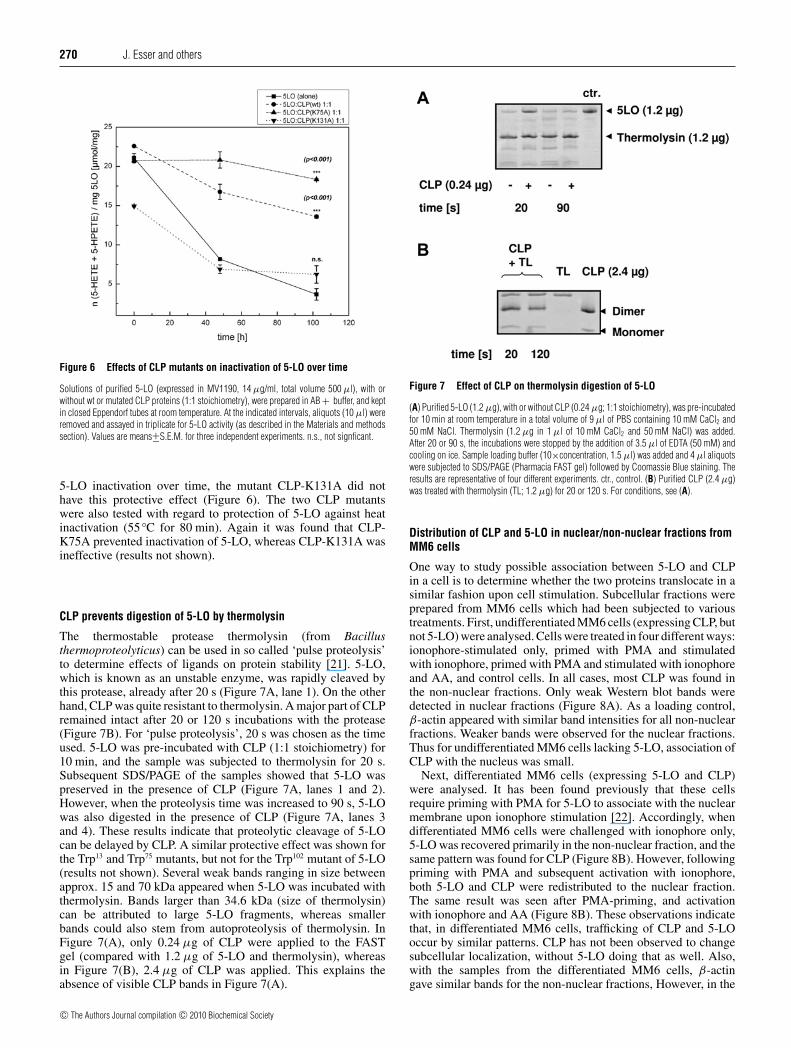
CLP prevents digestion of 5-LO by thermolysin
The thermostable protease thermolysin (from Bacillusthermoproteolyticus) can be used in so called ‘pulse proteolysis’to determine effects of ligands on protein stability [21]. 5-LO,which is known as an unstable enzyme, was rapidly cleaved bythis protease, already after 20 s (Figure 7A, lane 1). On the otherhand, CLP was quite resistant to thermolysin. A major part of CLPremained intact after 20 or 120 s incubations with the protease(Figure 7B). For ‘pulse proteolysis’, 20 s was chosen as the timeused. 5-LO was pre-incubated with CLP (1:1 stoichiometry) for10 min, and the sample was subjected to thermolysin for 20 s.Subsequent SDS/PAGE of the samples showed that 5-LO waspreserved in the presence of CLP (Figure 7A, lanes 1 and 2).However, when the proteolysis time was increased to 90 s, 5-LOwas also digested in the presence of CLP (Figure 7A, lanes 3and 4). These results indicate that proteolytic cleavage of 5-LOcan be delayed by CLP. A similar protective effect was shown forthe Trp13 and Trp75 mutants, but not for the Trp102 mutant of 5-LO(results not shown). Several weak bands ranging in size betweenapprox. 15 and 70 kDa appeared when 5-LO was incubated withthermolysin. Bands larger than 34.6 kDa (size of thermolysin)can be attributed to large 5-LO fragments, whereas smallerbands could also stem from autoproteolysis of thermolysin. InFigure 7(A), only 0.24 μg of CLP were applied to the FASTgel (compared with 1.2 μg of 5-LO and thermolysin), whereasin Figure 7(B), 2.4 μg of CLP was applied. This explains theabsence of visible CLP bands in Figure 7(A).
Figure 7 Effect of CLP on thermolysin digestion of 5-LO
(A) Purified 5-LO (1.2 μg), with or without CLP (0.24 μg; 1:1 stoichiometry), was pre-incubatedfor 10 min at room temperature in a total volume of 9 μl of PBS containing 10 mM CaCl2 and50 mM NaCl. Thermolysin (1.2 μg in 1 μl of 10 mM CaCl2 and 50 mM NaCl) was added.After 20 or 90 s, the incubations were stopped by the addition of 3.5 μl of EDTA (50 mM) andcooling on ice. Sample loading buffer (10×concentration, 1.5 μl) was added and 4 μl aliquotswere subjected to SDS/PAGE (Pharmacia FAST gel) followed by Coomassie Blue staining. Theresults are representative of four different experiments. ctr., control. (B) Purified CLP (2.4 μg)was treated with thermolysin (TL; 1.2 μg) for 20 or 120 s. For conditions, see (A).
Distribution of CLP and 5-LO in nuclear/non-nuclear fractions fromMM6 cells
One way to study possible association between 5-LO and CLPin a cell is to determine whether the two proteins translocate in asimilar fashion upon cell stimulation. Subcellular fractions wereprepared from MM6 cells which had been subjected to varioustreatments. First, undifferentiated MM6 cells (expressing CLP, butnot 5-LO) were analysed. Cells were treated in four different ways:ionophore-stimulated only, primed with PMA and stimulatedwith ionophore, primed with PMA and stimulated with ionophoreand AA, and control cells. In all cases, most CLP was found inthe non-nuclear fractions. Only weak Western blot bands weredetected in nuclear fractions (Figure 8A). As a loading control,β-actin appeared with similar band intensities for all non-nuclearfractions. Weaker bands were observed for the nuclear fractions.Thus for undifferentiated MM6 cells lacking 5-LO, association ofCLP with the nucleus was small.
Next, differentiated MM6 cells (expressing 5-LO and CLP)were analysed. It has been found previously that these cellsrequire priming with PMA for 5-LO to associate with the nuclearmembrane upon ionophore stimulation [22]. Accordingly, whendifferentiated MM6 cells were challenged with ionophore only,5-LO was recovered primarily in the non-nuclear fraction, and thesame pattern was found for CLP (Figure 8B). However, followingpriming with PMA and subsequent activation with ionophore,both 5-LO and CLP were redistributed to the nuclear fraction.The same result was seen after PMA-priming, and activationwith ionophore and AA (Figure 8B). These observations indicatethat, in differentiated MM6 cells, trafficking of CLP and 5-LOoccur by similar patterns. CLP has not been observed to changesubcellular localization, without 5-LO doing that as well. Also,with the samples from the differentiated MM6 cells, β-actingave similar bands for the non-nuclear fractions, However, in the
c© The Authors Journal compilation c© 2010 Biochemical Society

Coactosin-like protein functions as a stabilizing chaperone for 5-lipoxygenase 271
Figure 8 Distribution of CLP and 5-LO in nuclear/non-nuclear fractions fromMM6 cells
Undifferentiated and differentiated (TGF-β and calcitriol for 96 h) MM6 cells (10 × 106 cells in1 ml of PGC buffer) were primed with PMA (100 nM) for 10 min, and activated with ionophore(iono; 5 μM) and AA (40 μM) for another 10 min, as indicated. After lysis with Nonidet P40,pair-wise nuclear (Nucl.) and non-nuclear (Non-nucl.) fractions were prepared as describedin the Materials and methods section. Aliquots (40 μl, corresponding to approx. 106 cells)of pair-wise nuclear and non-nuclear fractions were analysed for 5-LO, CLP and β-actin byWestern blotting. Aliquots of a 10 000 g supernatant (104 × g sup.) derived from non-stimulated(Non-stim.) cells was analysed as a control for adequate expression of CLP and 5-LO in the cellbatch used. Similar results were obtained in two other experiments.
nuclear fractions from PMA-primed cells, β-actin was increased,similar to 5-LO and CLP. Interestingly, β-actin can bind both toCLP [13] and to 5-LO (reviewed in [29]).
Protein–protein docking of the CLP–5-LO interaction
The combination of computational docking results from DOTand experimental data has been established as a useful tool forunderstanding potential molecular interactions [30]. To betterunderstand the interaction between CLP and 5-LO, docking wasperformed using our 5-LO model structure and the CLP NMRstructure (PDB: 1WNJ; [15]). Assuming that the impairmentof complex formation by the two mutations CLP-K131A and5-LO-W102A points to a close interaction of CLP-Lys131
with 5-LO, and of 5-LO-Trp102 with CLP, only one model complexremained (Figure 9). In particular, in this model CLP-Lys131 isdirected towards 5-LO and 5-LO-Trp102 points towards CLP. Thismodel suggests a direct interaction via a cation–pi interactionbetween the side chains of CLP-Lys131 and 5-LO-Phe14. 5-LO-Trp102 could be involved in an interaction network also including
5-LO-Arg165, and the carbonyl oxygen of CLP-Lys131. In such aninteraction network, the 5-LO-Trp102 side chain might engage ina cationic–pi interaction with 5-LO-Arg165 which in turn wouldform a hydrogen bond with the backbone carbonyl oxygen ofCLP-Lys131 (Figure 9).
Several other amino acids, located both in the 5-LO β-sandwichand in the catalytic domain, may potentially form hydrogenbonds stabilizing the complex [5-LO-Glu70–CLP-Arg91, 5-LO-Asp106–CLP-Val113, 5-LO-Glu108–CLP-Arg91, 5-LO-Thr137–CLP-Lys102 and 5-LO-Lys140–CLP-Gln106]. Four of these five residuesare identical in other mammalian 5-LOs, one is conserved. Allpredicted interacting residues of CLP are identical in mouse CLP,which is also known to bind 5-LO [14].
CLP also binds F-actin [13], and F-actin-binding residues weredetermined [31]. Many of these are located on the predicted CLP–5-LO interface, indicating overlapping binding sites. This is in linewith the observation that a ternary complex of F-actin–CLP–5-LOhas not been found [12].
DISCUSSION
5-LO has always been considered to be an unstable enzymeand 5-LO is inactivated both during turnover and during storage(non-turnover inactivation) (for a review see [29]). Inactivationof 5-LO during turnover can be due to reactions with theenzyme products 5-HPETE and LTA4. Non-turnover inactivationis thought to depend on oxygen, and deliberate exposure to oxygeninactivated 5-LO, due to loss of the prosthetic iron. Also treatmentwith H2O2 inactivated purified 5-LO, and catalase or glutathioneperoxidase can protect against such inactivation.
CLP was previously found to bind 5-LO by co-immuno-precipitation from lysates of transfected cells. In addition, GSTpull-down assays, native PAGE and chemical cross-linkingshowed binding in vitro with a 1:1 molar stoichiometry [14,12].CLP serves as a scaffold for 5-LO activity [16], apparently byreplacing or complementing membrane (PC) in this role. WhenCLP was present in the in vitro assays (no PC added) Ca2 +
activation led to formation of 5-HPETE plus 5-HETE, whereasthe presence of CLP together with PC gave considerable up-regulation of LTA4 formation. In the present study, we confirmthese effects of CLP on 5-LO activity and we show that CLPalso has another effect, i.e. to prevent non-turnover inactivationof 5-LO. Thus CLP, present in a 1:1 molar stoichiometry, couldprevent both inactivation over time and during heat treatment.This stabilizing effect of CLP was found to be specific, since itstrictly depended on binding to 5-LO. The CLP mutant K131A,with considerably reduced binding ability to 5-LO, did not protect5-LO. Also, the 5-LO mutant W102A, which did not bind CLP,was neither stabilized nor activated by CLP. The 5-LO tryptophanmutants studied in the present report were as active as wt-5-LO(in the presence of PC; Figure 1) indicating that these mutationsdid not compromise the overall structure of 5-LO.
PC is the major phospholipid constituent of the nuclearmembrane. In addition to its role as a scaffold for Ca2+-induced5-LO activity, PC can also stabilize 5-LO. In fact, PC has beenused as a stabilizing agent during purification of 5-LO [32,33],and membrane binding of 5-LO stabilized the structures of both5-LO and the membrane [34]. Addition of leucocyte proteinfractions improved stability and activity of purified 5-LO [32,35].In addition to the ‘macromolecular crowding’ provided by mixedproteins at relatively high concentrations, it appears possible thatthese protein fractions contained CLP. The observation that CLPis effective already at a 1:1 stoichiometry supports a specificinteraction between 5-LO and CLP, different in nature from the
c© The Authors Journal compilation c© 2010 Biochemical Society

272 J. Esser and others
Figure 9 Protein–protein docking of the CLP–5-LO complex
Model of the CLP–5-LO complex. Left-hand side: side-view of the complex. Right-hand side: close-up of the interface region (rotated 90◦ about the y axis). The side-view shows 5-LO in surfacerepresentation (light blue and dark blue) and CLP as the backbone (dark purple). CLP-Lys131 is shown as spheres (green, coloured by element). The location of 5-LO-Trp102 is indicated in orange,as are the other two tryptophan residues (5-LO-Trp13 and 5-LO-Trp75). The close-up shows the interaction network in the interface region. Possible hydrogen bonds formed between amino acids ofthe complex partners are indicated: 5-LO-Glu70–CLP-Arg91, 5-LO-Asp106–CLP-Val113, 5-LO-Glu108–CLP-Arg91, 5-LO-Thr137–CLP-Lys102, 5-LO-Lys140–CLP-Gln106 and 5-LO-Arg165–CLP-Lys131.These hydrogen bonds link CLP to both domains in 5-LO. The proposed cation–pi interactions of CLP-Lys131 and 5-LO-Phe14 and of 5-LO-Trp102 and 5-LO-Arg165 are indicated (red lines).
protective effect of proteins in general. Also, the dependenceon particular amino acid residues (both in 5-LO and in CLP)strongly suggests that the protective effect of CLP is due to specificinteraction between these two proteins.
Binding of PC to the 5-LO β-sandwich involves threetryptophan residues (Trp13, Trp75 and Trp102) [28]. Mutagenesisof these residues (to alanine) reduced the affinity of the isolated5-LO C2-like domain to PC, substitution of Trp102 gave the mostprominent effect with a 20-fold reduced affinity [28]. Whenall of these residues were mutated to alanine in intact 5-LO,Ca2+-induced enzyme activity in the presence of relatively lowconcentrations of PC and AA was reduced to approx. 25% of wt-5-LO [16]. In the present study we show that, for the three singletryptophan mutants, the activity at low PC and AA was approx.50% of wt-5-LO. Thus all three residues seem to contribute, toabout the same extent, for PC to support 5-LO enzyme activity.Similarly to rabbit 15-LO [36] it has been suggested that forintact 5-LO residues also in the catalytic domain contribute tomembrane binding, and it was noted that the Kd for binding ofintact 5-LO to PC-rich vesicles was considerably lower than forbinding of the isolated β-sandwich [37]. Thus it appears that,for intact 5-LO, Trp102 is not as influential for PC binding, as forthe isolated 5-LO β-sandwich. Also, it has been proposed that5-LO can bind to membranes in different modes, non-productiveand productive [34].
Presumably, our determinations of Ca2+-induced enzymeactivity in the presence of PC reflect a productive binding mode.In view of this model, it thus appears that Trp102 is more importantfor non-productive binding of 5-LO to PC, in comparison withthe productive binding. Similar considerations can also apply to
the role of 5-LO Trp75 for binding to PC. In the 5-LO modelstructure [7] this residue is located on the very tip of a surface-exposed loop of the β-sandwich, and in a model of a 5-LO–phospholipid interaction, this residue inserts into the hydrophobicpart of the membrane [37]. However, the present mutagenesisdata (Figure 2) as well as previous findings (Trp75 mutated also toarginine, phenylalanine and serine) [38] do not point to a particularrole for Trp75 with regard to PC-supported 5-LO activity.
On the other hand, for CLP to support 5-LO, Trp102 in 5-LO isessential, and mutagenesis of this residue obliterated binding ofCLP to 5-LO. When both PC and CLP were added to 5-LO, total5-LO product formation was about the same as with onlyPC present (Figure 1C), but strikingly the LT productionwas prominently increased (approx. 5-fold; Figure 1B). Assuggested previously [16], this indicates a three-partner complex,comprising 5-LO, PC and CLP. Our findings show that Trp102 in5-LO is important for binding to CLP. Apparently, Trp13 and Trp75
are free to bind PC, possibly Trp102 can also bind to PC (at thesame time as binding CLP).
Trp102 is part of a stretch of conserved surface residues(FPCYRW) [39], and in the 5-LO model structure Trp102 ispartially hidden in the cleft between the two domains, aspreviously demonstrated for the corresponding residue (Trp100)in 12/15-LO [40]. Thus it appears possible that binding of CLPto 5-LO may have an allosteric effect on the association of thetwo domains in 5-LO, these may open up for CLP to directlyaccess Trp102. Alternatively, as suggested by our model of theCLP–5-LO complex, 5-LO-Trp102 may influence CLP bindingvia an interaction network involving 5-LO-Arg165. The modelalso suggests a direct pi–cation interaction of CLP-Lys131 with
c© The Authors Journal compilation c© 2010 Biochemical Society

Coactosin-like protein functions as a stabilizing chaperone for 5-lipoxygenase 273
5-LO-Phe14, as well as six hydrogen bonds between 5-LO andCLP, three of which involve residues in the 5-LO catalytic domain,whereas the other three involve residues in the β-sandwich. Ineither case, binding of CLP to 5-LO may influence the relativepositions of the two domains in 5-LO. We would like to pointout that, in our modelling of the CLP–5-LO complex, Ca2+ wasnot included. Ca2+ is known to bind the 5-LO β-sandwich anddifferent sets of Ca2+ ligands have been suggested [19,28,39,41].Although CLP can bind to 5-LO in the absence of Ca2+ [12], themode of binding will possibly be different in the presence of Ca2+.
CLP could protect 5-LO against thermolysin-catalysedproteolysis. Thermolysin is a Ca2+-dependent endopeptidasewith preference for hydrophobic target sites, which can be usedto study protein stability and ligand binding by so called ‘pulseproteolysis’ [21]. In short time incubations (20 s), CLP protected5-LO against thermolysin; however, when the incubation timewas extended to 90 s, protection was not observed. This indicatesthat proteolysis of 5-LO was delayed in the presence of CLP, andnot entirely prevented. An attractive explanation for this effectof CLP is that, by binding to 5-LO, it renders the 5-LO structuremore compact, and consequently bulky hydrophobic residues(preferred cleavage sites of thermolysin; [42]) may become lessaccessible. In soy bean LO-1, hydrophobic interactions contributeto association of the two domains [43], and hydrophobic residuesare present between the two domains in 5-LO model structures[39]. The importance of 5-LO-Trp102 suggests that CLP binds ator near the domain interface of 5-LO. Possibly, CLP preventsaccess of thermolysin to a target site on hydrophobic interdomainareas of 5-LO.
When cells are activated to produce LTs, 5-LO typicallymigrates to the nuclear membrane. In previous studies with humanneutrophils, CLP displayed the same changes in subcellulardistribution as 5-LO [9,16], i.e. following ionophore stimulationboth 5-LO and CLP were associated with a nuclear fraction.Recently, a gender difference regarding subcellular localizationof 5-LO was found [9]. It was shown that for male neutrophilsa substantial amount of 5-LO was associated with the nucleusalready in non-stimulated cells, and the same was found forCLP. In the present study we analysed subcellular fractionsfrom MM6 cells, which offer some experimental options.First, undifferentiated MM6 cells express CLP, but not 5-LO.Regardless of stimulation, for these undifferentiated cells almostall CLP was recovered in the non-nuclear fractions. Secondly,differentiated MM6 cells (expressing 5-LO) required primingwith PMA for 5-LO to associate with the nuclear membrane inresponse to ionophore [22], in accordance with a role for kinasesin 5-LO translocation [7]. Accordingly, when differentiated MM6cells were stimulated with ionophore only, 5-LO was recoveredprimarily in the non-nuclear fraction, and the same was foundfor CLP (Figure 8B). However, following priming with PMA andsubsequent activation with ionophore, substantial amounts of both5-LO and CLP were found in the nuclear fraction. Thus, also inMM6 cells, it appears that migration of CLP is connected withmigration of 5-LO.
In summary, the findings of the present study show that Trp102 inthe 5-LO β-sandwich is important for binding of CLP. Our resultsalso provide further support for CLP functioning as a chaperonefor 5-LO, stabilizing 5-LO in the resting cell and functioning as ascaffold during Ca2+-induced 5-LO activity.
AUTHOR CONTRIBUTIONS
Julia Esser, Marija Rakonjac, Gisbert Schneider, Dieter Steinhilber, Bengt Samuelsson andOlof Radmark designed the research. Julia Esser, Marija Rakonjac, Bettina Hofmann
and Lutz Fischer performed the studies. Patrick Provost provided analytical tools. JuliaEsser, Marija Rakonjac, Bettina Hofmann, Gisbert Schneider and Olof Radmark analysedthe results. Julia Esser, Marija Rakonjac, Bettina Hofmann, Gisbert Schneider, PatrickProvost and Olof Radmark wrote the paper.
ACKNOWLEDGEMENTS
We thank Dr Anders Wetterholm (Department of Medical Biochemistry and Biophysics,Karolinska Institute, Stockholm, Sweden) for all of his help.
FUNDING
This work was supported by the Swedish Research Council [grant number 03X-217]; bythe European Union [grant number LSHM-CT-2004–00533, FP7-Health-201668]; and bythe Karolinska Institutet. P.P. is a Senior Scholar from the Fonds de la Recherche en Santedu Quebec.
REFERENCES
1 Samuelsson, B. (1983) Leukotrienes: mediators of immediate hypersensitivity reactionsand inflammation. Science 220, 568–575
2 Peters-Golden, M. and Henderson, Jr, W. R. (2007) Leukotrienes. N. Engl. J. Med. 357,1841–1854
3 Werz, O. and Steinhilber, D. (2006) Therapeutic options for 5-lipoxygenase inhibitors.Pharmacol. Ther. 112, 701–718
4 Chen, Y., Hu, Y., Zhang, H., Peng, C. and Li, S. (2009) Loss of the Alox5 gene impairsleukemia stem cells and prevents chronic myeloid leukemia. Nat. Genet. 41, 783–792
5 Choi, J., Chon, J. K., Kim, S. and Shin, W. (2008) Conformational flexibility in mammalian15S-lipoxygenase: reinterpretation of the crystallographic data. Proteins 70, 1023–1032
6 Gillmor, S. A., Villasenor, A., Fletterick, R., Sigal, E. and Browner, M. F. (1997) Thestructure of mammalian 15-lipoxygenase reveals similarity to the lipases and thedeterminants of substrate specificity. Nat. Struct. Biol. 4, 1003–1009
7 Radmark, O., Werz, O., Steinhilber, D. and Samuelsson, B. (2007) 5-Lipoxygenase:regulation of expression and enzyme activity. Trends Biochem. Sci. 32, 332–341
8 Peters-Golden, M. and Brock, T. G. (2003) 5-lipoxygenase and FLAP. ProstaglandinsLeukot. Essent. Fatty Acids 69, 99–109
9 Pergola, C., Dodt, G., Rossi, A., Neunhoeffer, E., Lawrenz, B., Northoff, H., Samuelsson,B., Radmark, O., Sautebin, L. and Werz, O. (2008) ERK-mediated regulation of leukotrienebiosynthesis by androgens: a molecular basis for gender differences in inflammation andasthma. Proc. Natl. Acad. Sci. U.S.A. 105, 19881–19886
10 de Hostos, E. L., Bradtke, B., Lottspeich, F. and Gerisch, G. (1993) Coactosin, a 17 kDaF-actin binding protein from Dictyostelium discoideum. Cell Motil. Cytoskeleton 26,181–191
11 Provost, P., Samuelsson, B. and Radmark, O. (1999) Interaction of 5-lipoxygenase withcellular proteins. Proc. Natl. Acad. Sci. U.S.A. 96, 1881–1885
12 Provost, P., Doucet, J., Hanmmarberg, T., Gerisch, G., Samuelsson, B. and Radmark, O.(2001) 5-Lipoxygenase interacts with coactosin-like protein. J. Biol. Chem. 276,16520–16527
13 Provost, P., Doucet, J., Stock, A., Gerisch, G., Samuelsson, B. and Radmark, O. (2001)Coactosin-like protein, a human F-actin-binding protein: critical role of lysine-75.Biochem. J. 359, 255–263
14 Doucet, J., Provost, P., Samuelsson, B. and Radmark, O. (2002) Molecular cloning andfunctional characterization of mouse coactosin-like protein. Biochem. Biophys. Res.Commun. 290, 783–789
15 Liepinsh, E., Rakonjac, M., Boissonneault, V., Provost, P., Samuelsson, B., Radmark, O.and Otting, G. (2004) NMR structure of human coactosin-like protein. J. Biomol. NMR30, 353–356
16 Rakonjac, M., Fischer, L., Provost, P., Werz, O., Steinhilber, D., Samuelsson, B. andRadmark, O. (2006) Coactosin-like protein supports 5-lipoxygenase enzyme activity andup-regulates leukotriene A4 production. Proc. Natl. Acad. Sci. U.S.A. 103, 13150–13155
17 Jin, E. H., Shim, S. C., Kim, H. G., Chae, S. C. and Chung, H. T. (2009) Polymorphisms ofCOTL1 gene identified by proteomic approach and their association with autoimmunedisorders. Exp. Mol. Med. 41, 354–361
18 Feisst, C., Pergola, C., Rakonjac, M., Rossi, A., Koeberle, A., Dodt, G., Hoffmann, M.,Hoernig, C., Fischer, L., Steinhilber, D. et al. (2009) Hyperforin is a novel type of5-lipoxygenase inhibitor with high efficacy in vivo. Cell. Mol. Life Sci. 66, 2759–2771
19 Hammarberg, T., Provost, P., Persson, B. and Radmark, O. (2000) The N-terminal domainof 5-lipoxygenase binds calcium and mediates calcium stimulation of enzyme activity.J. Biol. Chem. 275, 38787–38793
c© The Authors Journal compilation c© 2010 Biochemical Society

274 J. Esser and others
20 Zhang, Y. Y., Hammarberg, T., Radmark, O., Samuelsson, B., Ng, C. F., Funk, C. D. andLoscalzo, J. (2000) Analysis of a nucleotide-binding site of 5-lipoxygenase by affinitylabelling: binding characteristics and amino acid sequences. Biochem. J. 351, 697–707
21 Park, C. and Marqusee, S. (2005) Pulse proteolysis: a simple method for quantitativedetermination of protein stability and ligand binding. Nat. Methods 2, 207–212
22 Werz, O., Klemm, J., Samuelsson, B. and Radmark, O. (2001) Phorbol ester up-regulatescapacities for nuclear translocation and phosphorylation of 5-lipoxygenase in Mono Mac6 cells and human polymorphonuclear leukocytes. Blood 97, 2487–2495
23 Comeau, S. R., Gatchell, D. W., Vajda, S. and Camacho, C. J. (2004) ClusPro: anautomated docking and discrimination method for the prediction of protein complexes.Bioinformatics 20, 45–50
24 Mandell, J. G., Roberts, V. A., Pique, M. E., Kotlovyi, V., Mitchell, J. C., Nelson, E.,Tsigelny, I. and Ten Eyck, L. F. (2001) Protein docking using continuum electrostatics andgeometric fit. Protein Eng. 14, 105–113
25 Ten Eyck, L. F., Mandell, J. G., Roberts, V. A. and Pique, M. E. (1995) SurveyingMolecular Interactions With DOT, Proceedings of the 1995 ACM/IEEE SupercomputingConference, San Diego, IEEE Computer Society Press, Los Alamitos, CA
26 Werz, O., Tretiakova, I., Michel, A., Ulke-Lemee, A., Hornig, M., Franke, L., Schneider, G.,Samuelsson, B., Radmark, O. and Steinhilber, D. (2005) Caspase-mediated degradationof human 5-lipoxygenase in B lymphocytic cells. Proc. Natl. Acad. Sci. U.S.A. 102,13164–13169
27 Brooks, B. R., Bruccoleri, R. E., Olafson, B. D., States, D. J., Swaminathan, S. andKarplus, M. (1983) CHARMM: a program for macromolecular energy, minimization, anddynamics calculations. Comp. Chem. 4, 187–217
28 Kulkarni, S., Das, S., Funk, C. D., Murray, D. and Cho, W. (2002) Molecular basis of thespecific subcellular localization of the C2-like domain of 5-lipoxygenase. J. Biol. Chem.277, 13167–13174
29 Radmark, O. (2002) Arachidonate 5-lipoxygenase. Prostaglandins Other Lipid Mediat.68–69, 211–234
30 Law, D. S., Ten Eyck, L. F., Katzenelson, O., Tsigelny, I., Roberts, V. A., Pique, M. E. andMitchell, J. C. (2003) Finding needles in haystacks: reranking DOT results by using shapecomplementarity, cluster analysis, and biological information. Proteins 52, 33–40
31 Dai, H., Huang, W., Xu, J., Yao, B., Xiong, S., Ding, H., Tang, Y., Liu, H., Wu, J. and Shi, Y.(2006) Binding model of human coactosin-like protein with filament actin revealed bymutagenesis. Biochim. Biophys. Acta 1764, 1688–1700
32 Denis, D., Falgueyret, J. P., Riendeau, D. and Abramovitz, M. (1991) Characterization ofthe activity of purified recombinant human 5-lipoxygenase in the absence and presence ofleukocyte factors. J. Biol. Chem. 266, 5072–5079
33 Hogaboom, G. K., Cook, M., Newton, J. F., Varrichio, A., Shorr, R. G., Sarau, H. M. andCrooke, S. T. (1986) Purification, characterization, and structural properties of a singleprotein from rat basophilic leukemia (RBL-1) cells possessing 5-lipoxygenase andleukotriene A4 synthetase activities. Mol. Pharmacol. 30, 510–519
34 Pande, A. H., Moe, D., Nemec, K. N., Qin, S., Tan, S. and Tatulian, S. A. (2004) Modulationof human 5-lipoxygenase activity by membrane lipids. Biochemistry 43, 14653–14666
35 Rouzer, C. A. and Samuelsson, B. (1985) On the nature of the 5-lipoxygenase reaction inhuman leukocytes: enzyme purification and requirement for multiple stimulatory factors.Proc. Natl. Acad. Sci. U.S.A. 82, 6040–6044
36 Walther, M., Wiesner, R. and Kuhn, H. (2004) Investigations into calcium-dependentmembrane association of 15-lipoxygenase-1. Mechanistic roles of surface-exposedhydrophobic amino acids and calcium. J. Biol. Chem. 279, 3717–3725
37 Pande, A. H., Qin, S. and Tatulian, S. A. (2005) Membrane fluidity is a key modulator ofmembrane binding, insertion, and activity of 5-lipoxygenase. Biophys. J. 88,4084–4094
38 Okamoto, H., Hammarberg, T., Zhang, Y. Y., Persson, B., Watanabe, T., Samuelsson, B.and Radmark, O. (2005) Mutation analysis of the human 5-lipoxygenase C-terminus:Support for a stabilizing C-terminal loop. Biochim. Biophys. Acta 1749, 123–131
39 Allard, J. B. and Brock, T. G. (2005) Structural organization of the regulatory domain ofhuman 5-lipoxygenase. Curr. Protein Pept. Sci. 6, 125–131
40 Hammel, M., Walther, M., Prassl, R. and Kuhn, H. (2004) Structural flexibility of theN-terminal β-barrel domain of 15-lipoxygenase-1 probed by small angle X-rayscattering. Functional consequences for activity regulation and membrane binding.J. Mol. Biol. 343, 917–929
41 Bindu, P. H., Sastry, G. M. and Sastry, G. N. (2004) Characterization of calcium andmagnesium binding domains of human 5-lipoxygenase. Biochem. Biophys. Res.Commun. 320, 461–467
42 Heinrikson, R. L. (1977) Applications of thermolysin in protein structural analysis.Methods Enzymol. 47, 175–189
43 Sudharshan, E., Srinivasulu, S. and Rao, A. G. A. (2000) pH-induced domain interactionand conformational transitions of lipoxygenase-1. Biochim. Biophys. Acta 1480,13–22
Received 5 June 2009/24 September 2009; accepted 6 October 2009Published as BJ Immediate Publication 6 October 2009, doi:10.1042/BJ20090856
c© The Authors Journal compilation c© 2010 Biochemical Society
Copyright © 2022 FDOKUMEN