Impact of Genome Reduction on Bacterial Metabolism and Its Regulation
Upload
khangminh22Category
view
0download
0
Bacterial gene diversity related to tryptophanmetabolism and indole-3-acetic acid (IAA)
production in the rhizosphere
Pablo Suárez PérezMáster en Bioinformática y BioestadísticaÁREA 4 – Aula 1
Nombre Consultor/a: Paloma María Pizarro TobíasNombre Profesor/a responsable de la asignatura: Carles Ventura Royo
8/01/2020

Esta obra está sujeta a una licencia deReconocimiento-NoComercial-SinObraDerivada 3.0 España de CreativeCommons
FICHA DEL TRABAJO FINAL
Título del trabajo:Bacterial gene diversity related to tryptophanmetabolism and indole-3-acetic acid (IAA)production in the rhizosphere
Nombre del autor: Pablo Suárez Pérez
Nombre del consultor/a: Paloma María Pizarro Tobías
Nombre del PRA: Carles Ventura Royo
Fecha de entrega (mm/aaaa): 01/2020
Titulación:: Máster en Bioinformática y Bioestadística
Área del Trabajo Final: Microbiología, biotecnología, filogenética
Idioma del trabajo: Inglés
Palabras claveTriptófano, ácido indoleacético, enzima,gen, operón, ruta metabótica, filogenética,análisis filogenético
Resumen del Trabajo:
Desde hace años, las rizobacterias han suscitado interés por su capacidad depromover el crecimiento vegetal. Una de las hormonas que estas bacteriasproducen y que tiene uno de los principales papeles en esta promoción es elácido indoleacético.
En este trabajo, se recoge toda la evidencia registrada hasta el momento de lasrutas de biosintesís de esta hormona en rizobacterias, así como de suprecursor, el aminoácido triptófano. Para cada una de las rutas y especiesidentificadas, se obtuvieron los genes y proteinas que intervienen en losdiferentes puntos de cada ruta. A fin de analizar su diversidad, se realizó unestudio filogético de cada una de las enzimas bajo estudio, se documentó superfil proteico y se analizó la coocurrencia de rutas. Para estudiar su origen yevolución, se realizó a su vez una comparación con un marcador filogenéticoconocido (gyrB).
Los resultados muestran una clara co-occurencia de enzimas y rutas, que enalgunos casos puede ser explicada por la presencia de operones, así comociertas divergencias en algunos casos en las comparaciones con losmarcadores.
Abstract:
For years, growth-promoting rizhobacteria have been a topic of interest.. Indole-3-acetic acid or IAA is one of the principal hormones that can be find in thesespecies.
i

In this study, we have gathered all the evidence so far about IAA byosinthesis.Besides, we have followed the same procedure for the substrate of the IAApathways (tryptophan). For each one of the identified species and pathways,genes and proteins sequences that are involved have been obtained. We haveanalyzed these gene pool diversity through phylogenetic analysis, and at thesame time we have documented their protein profile and co-ocurrence level. Inorder to study their origin and evolution, comparisons using the phylogeneticmarker gyrB have been performed.
Results show high co-occurence (both at enzyme/pathway level), that in somecases can be justified attending to the existence of operons. Besides, severaldifferences of interest have been found in the gyrB comparisons.
ii
Table of contents
1. Introduction.......................................................................................................11.1 Context of the project and justification.........................................................11.2 Objectives....................................................................................................21. Identify the main bacterial species which take part in the tryptophan/IAAmetabolism in the rizhosphere...........................................................................2...........................................................................................................................31.3 Materials and methods................................................................................31.4 Project planning...........................................................................................41.5 Breve sumario de productos obtenidos.......................................................41.5 Breve sumario de los productos obtenidos.................................................51.6 Breve descripción de los otros capítulos de la memoria.............................5
2. Chapters............................................................................................................62.1. Plant growth promoting rhizobacteria and the importance of thetryptophan metabolism/IAA production in the rhizosphere................................62.2. Description of the metabolic pathways.......................................................7 2.2.1. Indole-3-pyruvate pathway (IPA)..........................................................8 2.2.2. Indole-3-acetamide pathway (IAM).......................................................8 2.2.3. Indole-3-acetonitrile pathway (IAN)......................................................9 2.2.4. Tryptophan biosynthesis.....................................................................102.3. Main genes and protein profiles...............................................................11 2.3.1. IPA......................................................................................................11 2.3.1.1. Tryptophan aminotransferase.......................................................11 2.3.1.2. Indole-3-pyruvate decarboxylase.................................................12 2.3.1.3. Indole-3-acetaldehyde dehydrogenase........................................13 2.3.2. IAM......................................................................................................14 2.3.2.1. Tryptophan 2-monooxygenase.....................................................14 2.3.2.2. Indole-3-acetamide hydrolase......................................................15 2.3.3. IAN......................................................................................................16 2.3.3.1. Nitrile hydratase............................................................................16 2.3.3.2. Nitrilase.........................................................................................17 2.3.4. Tryptophan biosynthesis.....................................................................18 2.3.4.1. Anthranilate synthase...................................................................18 2.3.4.2. Tryptophan synthase....................................................................192.4. Main IAA rhizobacteria species, known pathways and coocurrence.......202.5. Results: diversity analysis I.......................................................................242.6. Results: diversity analysis II......................................................................38
3.Conclussions....................................................................................................514. Glossary..........................................................................................................535. References......................................................................................................546. Annexes..........................................................................................................65
6.1. Annex 1: Protein accesion numbers list...................................................656.2. Annex 2: R pipeline...................................................................................756.3. Annex 3: Tanglegram abbreviation...........................................................78
iii
Figures
Figure 1. Fungal and bacterial rhizosphere microbiome …………………………1Figure 2. Project timeline (Gant diagram)…………………………………………..4Figure 3. Indole-3-acetic acid (IAA)…………………………………………………6Figure 4. IAA and tryptohan biosynthesis…….…………………………………….7Figure 5. Indole-3-pyruvate (IPA) pathway…………………………………………8Figura 6. Indole-3-acetamide (IAM) pathway………………………………………9Figure 7. Indole-3-acetonitrile (IAN) pathway………………………………………9Figure 8. Tryptophan biosynthesis pathway………………………………………10Figure 9. Tryptophan aminotransferase reaction………………………………...11Figure 10. Indole-3-pyruvate decarboxylase reaction…………………………...12Figure 11. Indole-3-acetaldehyde dehydrogenase reaction…………………….13Figure 12. Tryptophan-2-monooxygenase reaction……………………………...14Figure 13. Indole-3-acetamide hydrolase reaction……………………………….15Figure 14. Nitrile hydratase reaction………………………………………………16Figure 15. Nitrilase reaction………………………………………………………...17Figure 16. Anthranilate synthase reaction………………………………………...18Figure 17. Tryptophan synthase reaction…………………………………………19Figure 18. Anthranilate synthase phylogeny……………………………………...25Figure 19. Tryptophan synthase (alpha subunit) phylogeny……………………26Figure 20. Tryptophan synthase (beta subunit) phylogeny……………………..28Figure 21. Trp operon……………………………………………………………….29Figure 22. Genetic region trpF, trpE, trpD, trpC, trpB and trpA from Rhodococcus jostii………………………………………………………..29Figure 23. trpA/trpB region comparison…………………………………………..30Figure 24. Tryptophan transaminase phylogeny…………………………………31Figure 25. Indole-3-pyruvate decarboxylase phylogeny………………………...32Figure 26. Indole-3-acetaldehyde dehydrogenase phylogeny………………….33Figure 27. Tryptophan-2-monooxygenase phylogeny…………………………...34Figure 28. Indole-3-acetamide hydrolase phylogeny……………………………35Figure 29. Nitrile hydratase (alpha subunit) phylogeny…………………………36Figure 30. Nitrile hydratase (beta subunit) phylogeny…………………………..37Figure 31. Nitrilase phylogeny……………………………………………………..38Figure 32. gyrB marker phylogeny………………………………………………..39Figure 33. Anthranilate synthase vs gyrB………………………………………..40Figure 34. Tryptophan synthase(alpha subunit) vs gyrB………………………..42Figure 35. Tryptophan synthase(beta subunit) vs gyrB…………………………43Figure 36. Tryptophan transaminase vs gyrB…………………………………….45Figure 37. Indole-3-pyruvate decarboxylase vs gyrB……………………………46Figure 38. Indole-3-acetaldehyde dehydrogenase vs gyrB……………………..46Figure 39. Tryptophan-2-monooxygenase vs gyrB………………………………47Figure 40. Indole-3-acetamide hydrolase vs gyrB……………………………….48Figure 41. Nitrile hydratase (alpha subunit) vs gyrB……………………………48Figure 42. Nitrile hydratase (beta subunit) vs gyrB……………………………..49Figure 43. Nitrilase vs gyrB………………………………………………………..50
Tables
Table 1. Tryptophan aminotransferase (protein profile)…………………………12Table 2. Indole-3-pyruvate decarboxylase (protein profile)……………………..13Table 3. Indole-3-acetaldehyde (protein profile)………………………………….14Table 4. Tryptophan-2-monooxygenase (protein profile)………………………..15Table 5. Indole-3-acetamide hydrolase (protein profile)…………………………16Table 6. Nitrile hydratase (protein profile)…………………………………………17Table 7. Anthranilate synthase (protein profile)………………………………......19Table 8. Tryptophan synthase (protein profile)…………………………………...20Table 9. IAA rhizobacteria species and known pathways……………………….23Table 10. Co-occurrence matrix……………………………………………………23
1. Introduction
1.1 Context of the project and justification
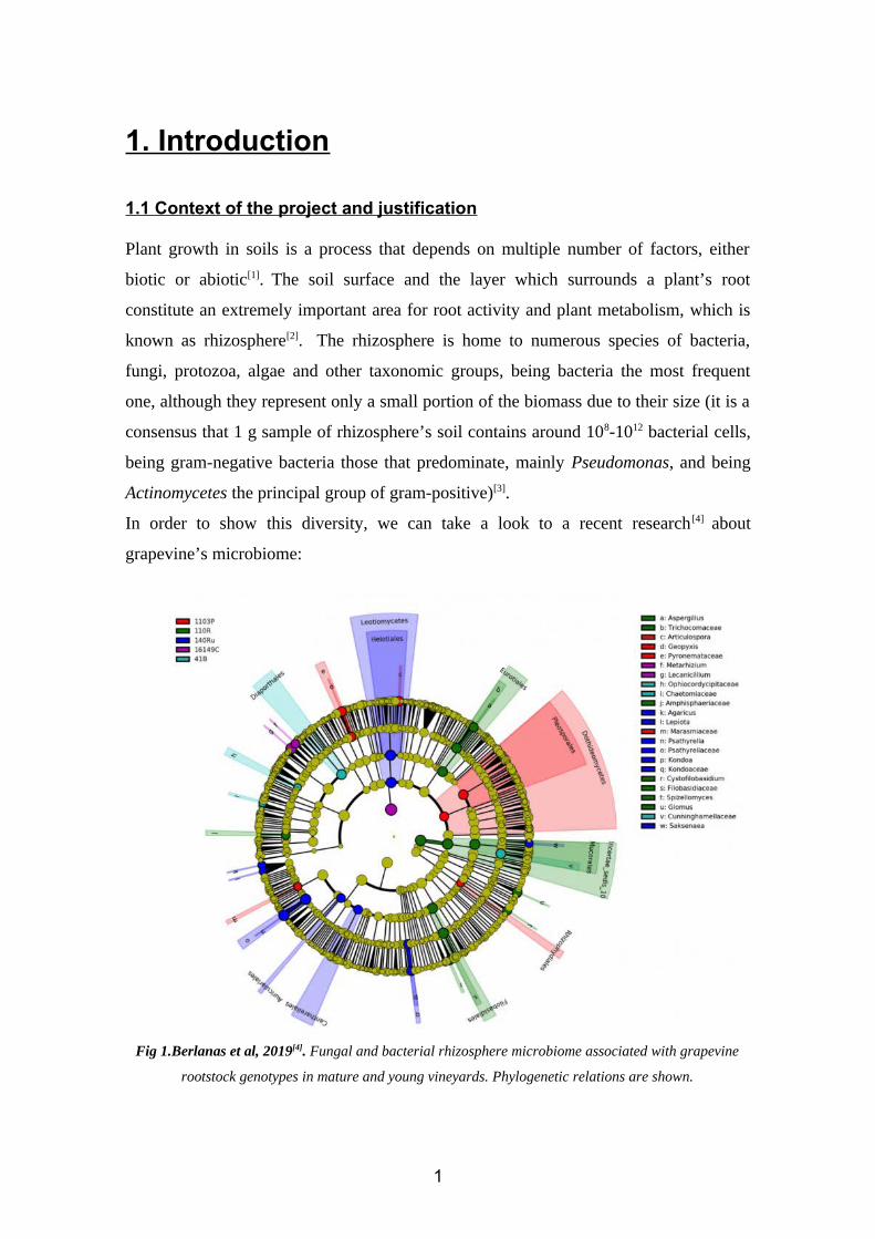
Plant growth in soils is a process that depends on multiple number of factors, either
biotic or abiotic[1]. The soil surface and the layer which surrounds a plant’s root
constitute an extremely important area for root activity and plant metabolism, which is
known as rhizosphere[2]. The rhizosphere is home to numerous species of bacteria,
fungi, protozoa, algae and other taxonomic groups, being bacteria the most frequent
one, although they represent only a small portion of the biomass due to their size (it is a
consensus that 1 g sample of rhizosphere’s soil contains around 108-1012 bacterial cells,
being gram-negative bacteria those that predominate, mainly Pseudomonas, and being
Actinomycetes the principal group of gram-positive)[3].
In order to show this diversity, we can take a look to a recent research [4] about
grapevine’s microbiome:
Fig 1.Berlanas et al, 2019[4]. Fungal and bacterial rhizosphere microbiome associated with grapevine
rootstock genotypes in mature and young vineyards. Phylogenetic relations are shown.
1

Rhizosphere bacterial species (or rhizobacteria) are known for several interactions that
affect both plants and soils[5]. Some of these are activities that ensure the stability and
productivity of numerous systems, such as agricultural and natural systems. In this
sense, it has been proved that a bunch of bacterial activities could have potential
industrial applications, mainly as biotechnological tools in sustainable agriculture and
other agrotechnological areas.
One of the activities that promote plant growth in the rhizosphere is the secretion of
growth hormones, being IAA one of them[5]. Thus, identifying the gene diversity
associated to the metabolic pathway of this hormone could be a crucial step in order to
understand and develop new strategies concerning sustainable agriculture and plant
other activities. According to this, this project aims to analyze the gene diversity
associated to the tryptophan and indole-3-acetic acid (hereinafter, IAA) metabolic
pathways. As a first step, we will perform an exhaustive bibliographic research of all the
available information about these pathways and the main bacterial species that take part
in these processes in the rhizosphere. Then, we will gather information about the
substrates and products of the pathways, analyzing every element that may be involved
in each step, such as enzymes and co-factors. Therefore, we will also get every gene and
protein sequence of the identified enzymes, being these the biological data we will use
as elements of comparison to study the diversity. After this preliminary process, we will
perform a phylogenetic analysis of each coding gene, in order to establish an
evolutionary relationship among the identified bacterial species that share that gene. We
will also give more information about protein profiles, distribution and co-ocurrence.
We will lately modify this approach using gene markers instead, allowing us to get a
general overview of the evolutionary diversification for each analyzed gene through the
comparison of the phylogenetic relationship obtained using markers and those obtained
by using the selected coding genes. Finally, we will discuss our results.
1.2 Objectives
The objectives of this research are:
1. To identify the main bacterial species which take part in the tryptophan/IAA
metabolism in the rizhosphere.
2
• To collect all the information available concerning the identified genes and
proteins that are known for taking part in a step of the tryptophan/IAA
biosynthesis and, in some cases, also those that are not yet identified but that
have been proposed.
• To describe the identified genes and proteins and their implications in the
different metabolic pathways.
• To describe the metabolic pathways.
2. To perform a comparison of the selected proteins (diversity analysis I).
• To determine the protein profile, co-ocurrence and distribution of each protein.
• To make a phylogenetic study of each protein according to the identified
sequences.
3. To perform a comparison of the bacterial species using the selected markers
(diversity analysis II).
• To study the phylogenetic differences according to gyrase B (gyrB) marker.
• To compare the results from the two approaches (diversity analysis I and II) in
order to find differences and similarities.
1.3 Materials and methods
We have obtained all our data (species, known pathways and enzymes) from the
information available in books, papers and metabolic databases, mainly KEEG
pathway(Kyoto encyclopedia of genes and genomes, described as “a collection of
manually drawn pathway maps representing the knowledge on the molecular
interaction, reaction and relation networks for metabolism and other topics”)[106]. Then,
we have created a list with these species, gene references and protein accession numbers
(annex 1). After that, and following the same criteria, we have identified for each
species which IAA biosynthesis pathways were present (since there are several, as we
will discuss) and which were absent.
In order to perform the phylogenetic analysis that we have proposed, we have used R as
the main tool. The R pipeline we have designed has been built using an amount of well-
known packages in these evolutionary studies, such as ape (used in the construction and
plotting of the trees), seqinr (used when computing distance matrices), biostrings (for a
3
faster manipulation of large sets of biological sequences), msa (used in order to perform
the required multiple sequence alignment) and reutils (to retrieve biological sequences).
Besides, comparisons of trees have been performed using the dendextend package. The
pipeline structure and a step by step guidance can be found in the annex 2. Finally, we
have determined the presence of operons of interest using Softberry[107], and then we
have compared some genomic regions related to these operons using Easyfig [108].
1.4 Project planning
The resources that we have used in this project are:
• Publications (books and papers about different topics, such as rhizobacteria,
rhizosphere, IAA, tryptophan, R packages and software tools).
• NCBI (National Center for Biotechnology Information)[109], from where we have
collected the available information of interest about the identified species. This
includes protein accession number, obtained from Refseq through NCBI.
• KEGG (Kyoto encyclopedia of genes and genomes), where the metabolic
pathways of the species are described, which have served us as a tool to check if
any doubtful gene or protein obtained through bibliographic research has been
finally identified as a part of the metabolic pathway that we are studying.
• BRENDA[110] and PDB[111] databases, for information about enzymes.
• R packages (ape, seqinr, reutils, phylotools, dendextend, bionstrings and msa).
• Softberry, for the identification of operons, and Easyfig, for comparing similar
genetic regions among a group of species.
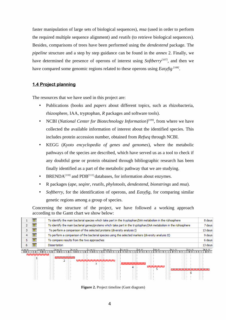
Concerning the structure of the project, we have followed a working approachaccording to the Gantt chart we show below:
Figure 2. Project timeline (Gant diagram)
4
1.5 Products summary• List of rhizobacteria that show evidence of IAA biosynthesis (starting from
tryptophan) and tryptophan biosynthesis (starting from chorismate).
• List of enzymes that catalyze each step in each described pathway.
• R pipeline (for creating and comparing trees).
• Phylogeny of each enzyme under study.
1.6 Chapters (brief description)
• Chapter 1: Introduction of the topic. Description of the rizhosphere, its
characteristics and importance. Description of the associated microbiome,
characteristics and importance. Tryptophan and IAA characterization:
Importance and contributions to the rhizosphere ecosystem. Economic and
industrial applications.
• Chapter 2: Description of the metabolic pathways. Genes and proteins of
interest.
• Chapter 3: Gene characterization. Protein structure and functions.
• Chapter 4: List of bacterial species that are able to produce IAA. Relationship
among them. Co-occurence matrix.
• Chapter 5: Diversity analysis I. Phylogeny of each enzyme under study.
• Chapter 6: Diversity analysis I. gyrB marker phylogeny. Comparisons enzyme
vs marker.
5
2. Chapters2.1. CHAPTER 1. Plant growth promoting rhizobacteria and theimportance of the «tryptophan metabolism - IAA biosynthesis» in therhizosphere
As mentioned before, rhizobacteria play essential roles in plant nutrition, growth
promotion and disease interactions[3]. Plants select these bacteria using specific organic
compounds including those that exudate through their roots, creating a selective
environment where only a few bacterial species can survive.
Thus, rhizosphere ecosystems behave as an ecological niche for each and every plant
and those beneficial bacterial species to which they are associated. When referrring to
growth promotion activities, these bacterial species are know as plant growth promoting
rhizobacteria (PGPR)[5] or simply plant growth promoting bacteria (PGPB). In recent
years, the utilization of PGPR as fertilizers and pesticides has started to be a topic of
interest related to agriculture and biological production[6], since these bacterial species
play a role in enhancing nutrient use efficiency and ensuring their availability [7]. PGPR
have a high potential in the production of several plant hormones (know as
phytohormones), such as auxins (involved in growth and behavioral processes in plant
life cycles, such as phototropism, geotropism, hydrotropism, wound response and root
growth and development), gibberellins (stem elongation, germination, dormancy,
flowering, flower development, and leaf and fruit senescence), cytokinins (promoting
cell division), ethylene (ripening of fruits) and absisic acid (seed and bud dormancy,
control of organ size and stomatal closure)[8].
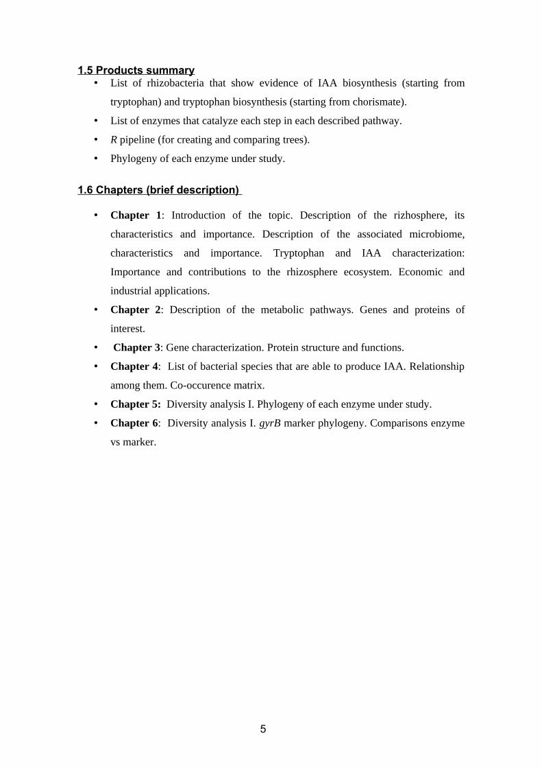
The main auxin we can find in plants is indole-3-acetic acid (IAA)[9]. This hormone can
be synthesized by the plant itself using tryptophan as substrate, but can be also provided
by rhizobacteria and some other groups of microbes[9].
Figure 3.
Indole-3-acetic acid (BRENDA
database)
6
Therefore, the implications of IAA produced by rhizobacteria are known as a key part
in plant-growth processes, from cell elongation to cell division an even tissue
differentiation[10]. This, in addition to the inherent elevation of size and surface area of
root systems in contact with soils by rhizobacteria, which leads to an increased in
nutrients and water uptake[11], makes IAA PGPR one of the most important species of
every soil ecosystem.
Since IAA has been proved to be one of the most important hormones in plants,
understanding tryptophan metabolism and IAA biosynthesis in rhizobacteria has
become one topic of economic interest due to huge impacts that the use of these species
could make in agrotechnological industries as potential natural fertilizers.
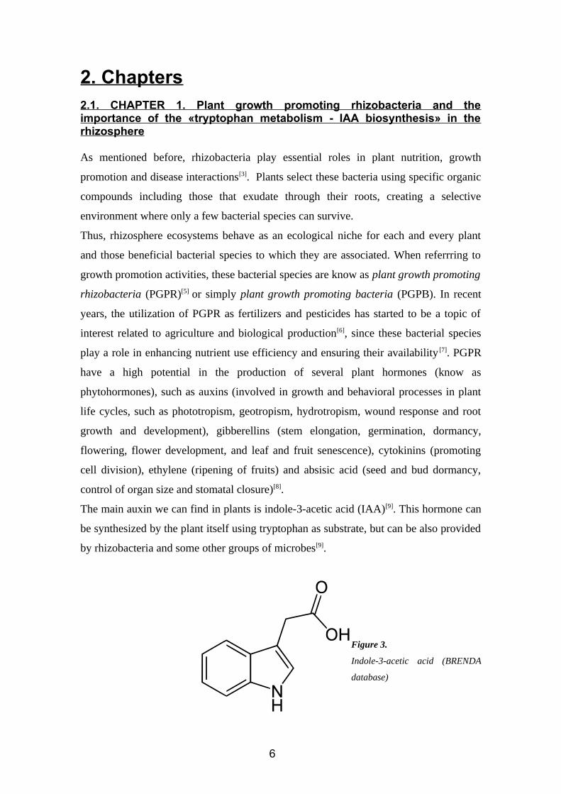
2.2. CHAPTER 2: Description of the metabolic pathwaysAccording to the evidence, tryptophan and IAA biosynthesis are two related processes,
since tryptophan is the substrate of almost every IAA pathway that has been
described[12].
Figure 4. Spaepen & Vanderleyden (2011)[15]. IAA and tryptophan biosynthesis pathways , starting from
chorismate.
7
There are several pathways that start with tryptophan and end with IAA. According to
Spaepen & Vanderleyden (2011)[15], these are the indole-3-pyruvate pathway (IPA), the
indole-3-acetonitrile pathway (IAN), the indole-3-acetamide pathway (IAM), the
tryptamine pathway and the tryptophan side-oxidase pathway. Besides, the existence of
a tryptophan independent pathway has been suggested, but there are no evidence so far.
Since only three of them (IPA, IAN and IAM) are present in rhizobacteria, these are the
ones we are going to study and describe.
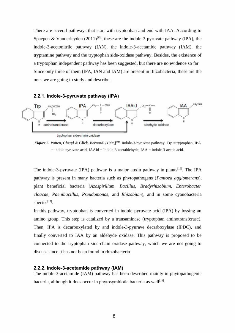
2.2.1. Indole-3-pyruvate pathway (IPA)
Figure 5. Patten, Cheryl & Glick, Bernard. (1996)[14]. Indole-3-pyruvate pathway. Trp =tryptophan, IPA
= indole pyruvate acid, IAAId = Indole-3-acetaldehyde, IAA = indole-3-acetic acid.
The indole-3-pyruvate (IPA) pathway is a major auxin pathway in plants[15]. The IPA
pathway is present in many bacteria such as phytopathogens (Pantoea agglomerans),
plant beneficial bacteria (Azospirillum, Bacillus, Bradyrhizobium, Enterobacter
cloacae, Paenibacillus, Pseudomonas, and Rhizobium), and in some cyanobacteria
species[15].
In this pathway, tryptophan is converted in indole pyruvate acid (IPA) by lossing an
amino group. This step is catalized by a transaminase (tryptophan aminotransferase).
Then, IPA is decarboxylated by and indole-3-pyurave decarboxylase (IPDC), and
finally converted to IAA by an aldehyde oxidase. This pathway is proposed to be
connected to the tryptophan side-chain oxidase pathway, which we are not going to
discuss since it has not been found in rhizobacteria.
2.2.2. Indole-3-acetamide pathway (IAM)The indole-3-acetamide (IAM) pathway has been described mainly in phytopathogenic
bacteria, although it does occur in phytosymbiotic bacteria as well[14].
8
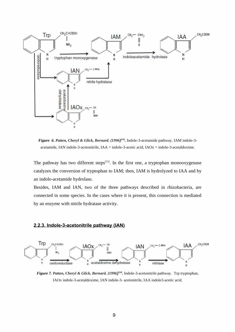
Figure 6. Patten, Cheryl & Glick, Bernard. (1996)[14]. Indole-3-acetamide pathway. IAM indole-3-
acetamide, IAN indole-3-acetonitrile, IAA = indole-3-acetic acid, IAOx = indole-3-acetaldoxime.
The pathway has two different steps[15]. In the first one, a tryptophan monooxygenase
catalyzes the conversion of tryptophan to IAM; then, IAM is hydrolyzed to IAA and by
an indole-acetamide hydrolase.
Besides, IAM and IAN, two of the three pathways described in rhizobacteria, are
connected in some species. In the cases where it is present, this connection is mediated
by an enzyme with nitrile hydratase activity.
2.2.3. Indole-3-acetonitrile pathway (IAN)
Figure 7. Patten, Cheryl & Glick, Bernard. (1996)[14]. Indole-3-acetonitrile pathway. Trp tryptophan,
IAOx indole-3-acetaldoxime, IAN indole-3- acetonitrile, IAA indole3-acetic acid.
9
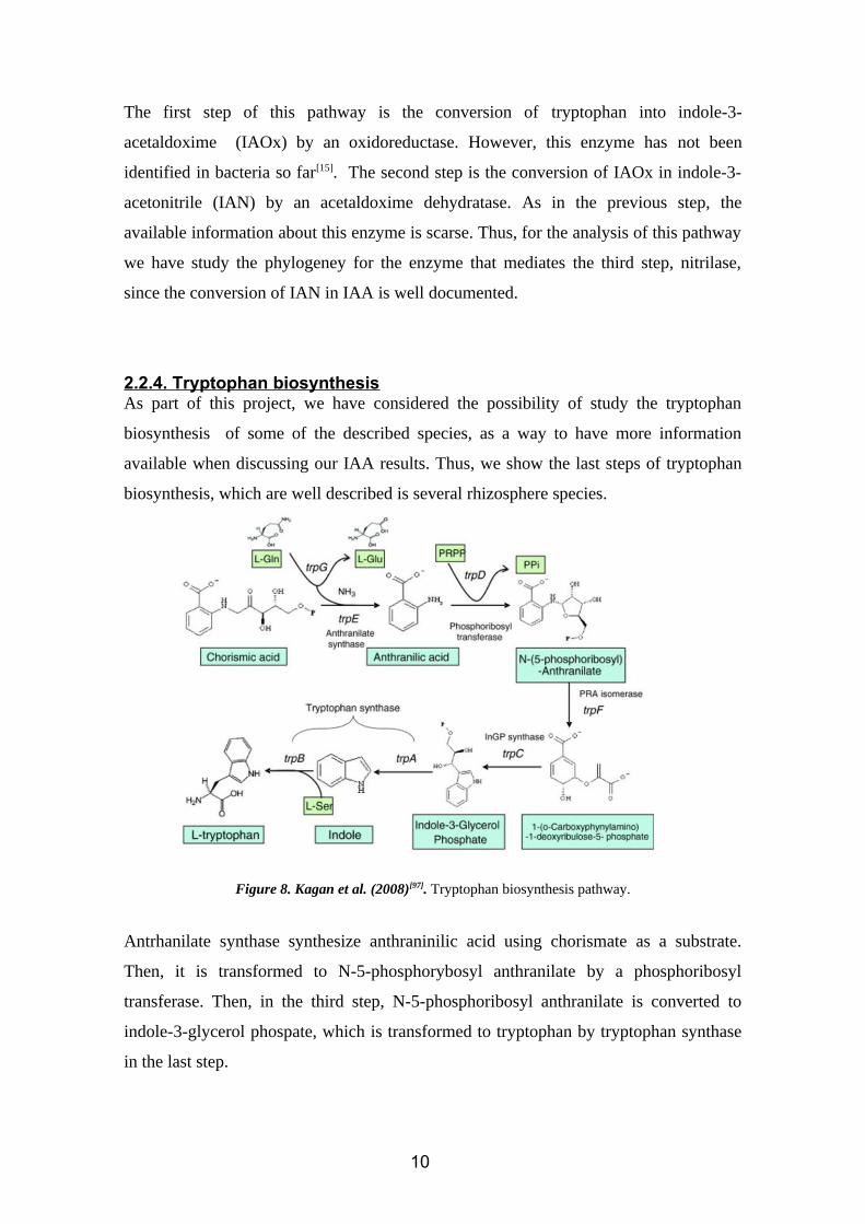
The first step of this pathway is the conversion of tryptophan into indole-3-
acetaldoxime (IAOx) by an oxidoreductase. However, this enzyme has not been
identified in bacteria so far[15]. The second step is the conversion of IAOx in indole-3-
acetonitrile (IAN) by an acetaldoxime dehydratase. As in the previous step, the
available information about this enzyme is scarse. Thus, for the analysis of this pathway
we have study the phylogeney for the enzyme that mediates the third step, nitrilase,
since the conversion of IAN in IAA is well documented.
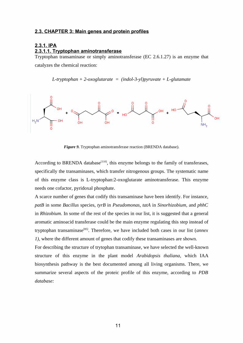
2.2.4. Tryptophan biosynthesisAs part of this project, we have considered the possibility of study the tryptophan
biosynthesis of some of the described species, as a way to have more information
available when discussing our IAA results. Thus, we show the last steps of tryptophan
biosynthesis, which are well described is several rhizosphere species.
Figure 8. Kagan et al. (2008)[97]. Tryptophan biosynthesis pathway.
Antrhanilate synthase synthesize anthraninilic acid using chorismate as a substrate.
Then, it is transformed to N-5-phosphorybosyl anthranilate by a phosphoribosyl
transferase. Then, in the third step, N-5-phosphoribosyl anthranilate is converted to
indole-3-glycerol phospate, which is transformed to tryptophan by tryptophan synthase
in the last step.
10
2.3. CHAPTER 3: Main genes and protein profiles
2.3.1. IPA2.3.1.1. Tryptophan aminotransferaseTryptophan transaminase or simply aminotransferase (EC 2.6.1.27) is an enzyme that
catalyzes the chemical reaction:
L-tryptophan + 2-oxoglutarate = (indol-3-yl)pyruvate + L-glutamate
Figure 9. Tryptophan aminotransferase reaction (BRENDA database).
According to BRENDA database[110], this enzyme belongs to the family of transferases,
specifically the transaminases, which transfer nitrogenous groups. The systematic name
of this enzyme class is L-tryptophan:2-oxoglutarate aminotransferase. This enzyme
needs one cofactor, pyridoxal phosphate.
A scarce number of genes that codify this transaminase have been identify. For instance,
patB in some Bacillus species, tyrB in Pseudomonas, tatA in Sinorhizobium, and phhC
in Rhizobium. In some of the rest of the species in our list, it is suggested that a general
aromatic aminoacid transferase could be the main enzyme regulating this step instead of
tryptophan transaminase[85]. Therefore, we have included both cases in our list (annex
1), where the different amount of genes that codify these transaminases are shown.
For describing the structure of trytophan transaminase, we have selected the well-known
structure of this enzyme in the plant model Arabidopsis thaliana, which IAA
biosynthesis pathway is the best documented among all living organisms. There, we
summarize several aspects of the proteic profile of this enzyme, according to PDB
database:
11
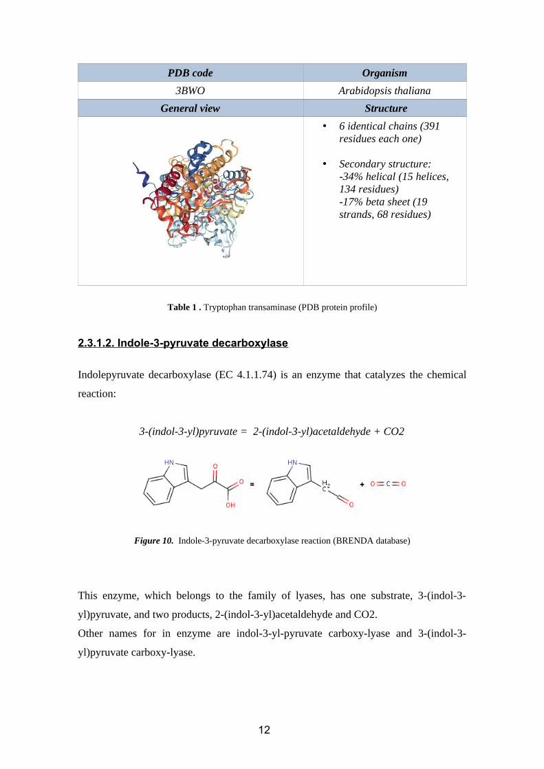
PDB code Organism
3BWO Arabidopsis thaliana
General view Structure
• 6 identical chains (391 residues each one)
• Secondary structure:-34% helical (15 helices, 134 residues)-17% beta sheet (19 strands, 68 residues)
Table 1 . Tryptophan transaminase (PDB protein profile)
2.3.1.2. Indole-3-pyruvate decarboxylase
Indolepyruvate decarboxylase (EC 4.1.1.74) is an enzyme that catalyzes the chemical
reaction:
3-(indol-3-yl)pyruvate = 2-(indol-3-yl)acetaldehyde + CO2
Figure 10. Indole-3-pyruvate decarboxylase reaction (BRENDA database)
This enzyme, which belongs to the family of lyases, has one substrate, 3-(indol-3-
yl)pyruvate, and two products, 2-(indol-3-yl)acetaldehyde and CO2.
Other names for in enzyme are indol-3-yl-pyruvate carboxy-lyase and 3-(indol-3-
yl)pyruvate carboxy-lyase.
12
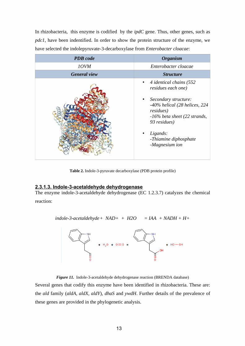
In rhizobacteria, this enzyme is codified by the ipdC gene. Thus, other genes, such as
pdc1, have been indentified. In order to show the protein structure of the enzyme, we
have selected the indolepyruvate-3-decarboxylase from Enterobacter cloacae:
PDB code Organism
1OVM Enterobacter cloacae
General view Structure
• 4 identical chains (552 residues each one)
• Secondary structure:-40% helical (28 helices, 224residues)-16% beta sheet (22 strands, 93 residues)
• Ligands:-Thiamine diphosphate-Magnesium ion
Table 2. Indole-3-pyruvate decarboxylase (PDB protein profile)
2.3.1.3. Indole-3-acetaldehyde dehydrogenaseThe enzyme indole-3-acetaldehyde dehydrogenase (EC 1.2.3.7) catalyzes the chemical
reaction:
indole-3-acetaldehyde+ NAD+ + H2O = IAA + NADH + H+
Figure 11. Indole-3-acetaldehyde dehydrogenase reaction (BRENDA database)
Several genes that codify this enzyme have been identified in rhizobacteria. These are:
the ald family (aldA, aldX, aldY), dhaS and ywdH. Further details of the prevalence of
these genes are provided in the phylogenetic analysis.
13
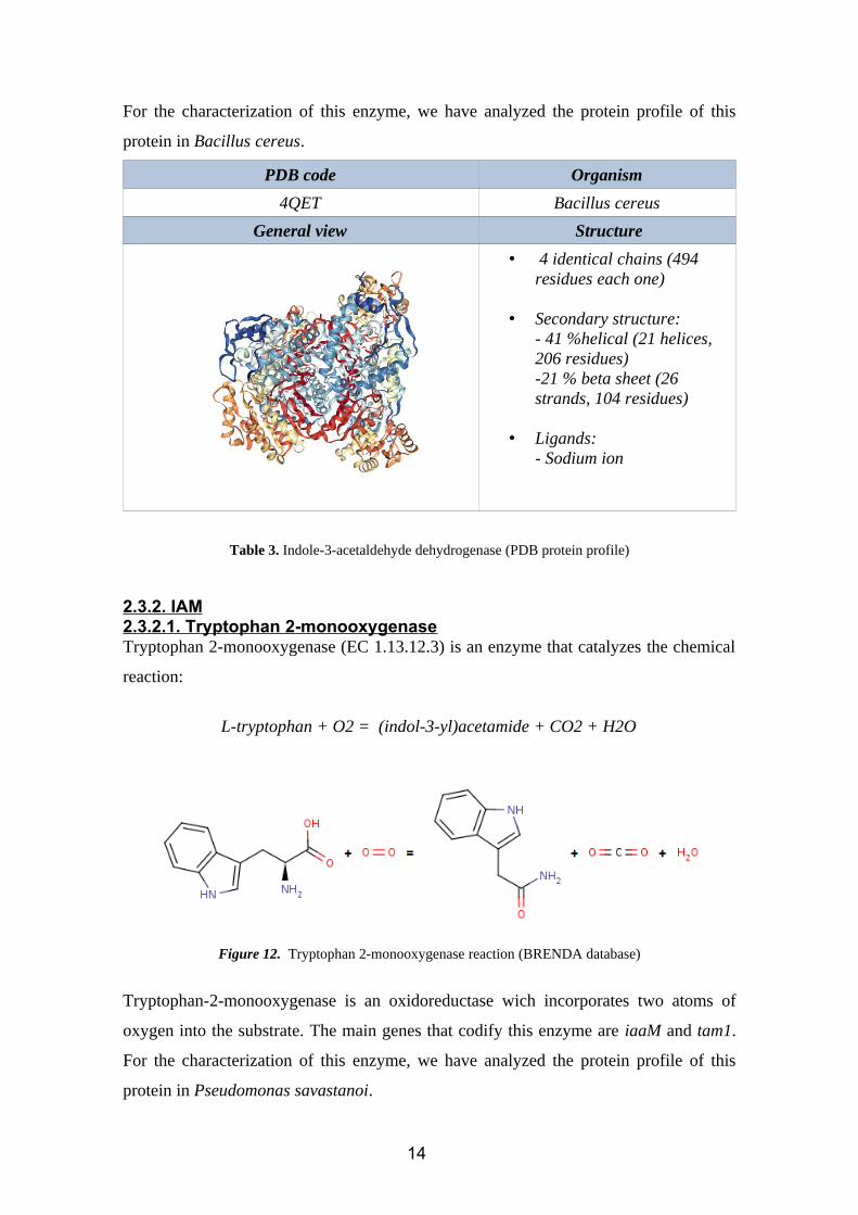
For the characterization of this enzyme, we have analyzed the protein profile of this
protein in Bacillus cereus.
PDB code Organism
4QET Bacillus cereus
General view Structure
• 4 identical chains (494 residues each one)
• Secondary structure:- 41 %helical (21 helices, 206 residues)-21 % beta sheet (26 strands, 104 residues)
• Ligands:- Sodium ion
Table 3. Indole-3-acetaldehyde dehydrogenase (PDB protein profile)
2.3.2. IAM2.3.2.1. Tryptophan 2-monooxygenaseTryptophan 2-monooxygenase (EC 1.13.12.3) is an enzyme that catalyzes the chemical
reaction:
L-tryptophan + O2 = (indol-3-yl)acetamide + CO2 + H2O
Figure 12. Tryptophan 2-monooxygenase reaction (BRENDA database)
Tryptophan-2-monooxygenase is an oxidoreductase wich incorporates two atoms of
oxygen into the substrate. The main genes that codify this enzyme are iaaM and tam1.
For the characterization of this enzyme, we have analyzed the protein profile of this
protein in Pseudomonas savastanoi.
14
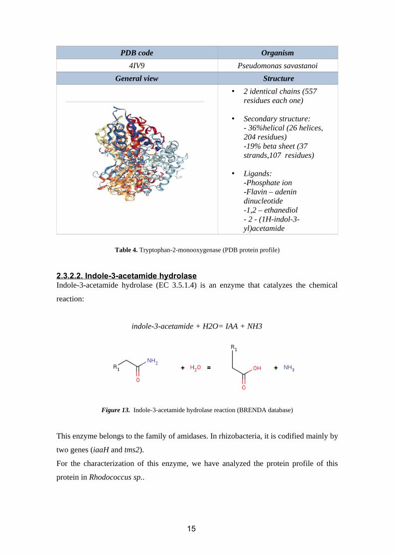
PDB code Organism
4IV9 Pseudomonas savastanoi
General view Structure
• 2 identical chains (557 residues each one)
• Secondary structure:- 36%helical (26 helices, 204 residues)-19% beta sheet (37 strands,107 residues)
• Ligands:-Phosphate ion-Flavin – adenin dinucleotide-1,2 – ethanediol- 2 - (1H-indol-3-yl)acetamide
Table 4. Tryptophan-2-monooxygenase (PDB protein profile)
2.3.2.2. Indole-3-acetamide hydrolaseIndole-3-acetamide hydrolase (EC 3.5.1.4) is an enzyme that catalyzes the chemical
reaction:
indole-3-acetamide + H2O= IAA + NH3
Figure 13. Indole-3-acetamide hydrolase reaction (BRENDA database)
This enzyme belongs to the family of amidases. In rhizobacteria, it is codified mainly by
two genes (iaaH and tms2).
For the characterization of this enzyme, we have analyzed the protein profile of this
protein in Rhodococcus sp..
15
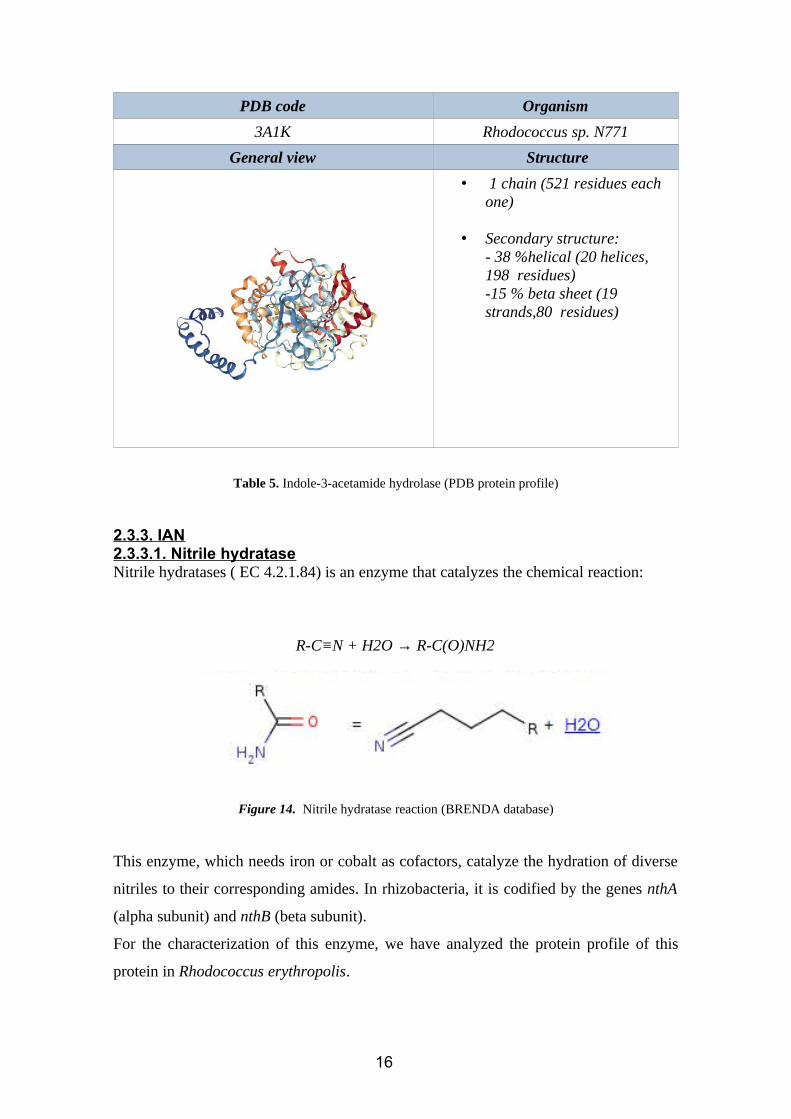
PDB code Organism
3A1K Rhodococcus sp. N771
General view Structure
• 1 chain (521 residues each one)
• Secondary structure:- 38 %helical (20 helices, 198 residues)-15 % beta sheet (19 strands,80 residues)
Table 5. Indole-3-acetamide hydrolase (PDB protein profile)
2.3.3. IAN2.3.3.1. Nitrile hydrataseNitrile hydratases ( EC 4.2.1.84) is an enzyme that catalyzes the chemical reaction:
R-C≡N + H2O → R-C(O)NH2
Figure 14. Nitrile hydratase reaction (BRENDA database)
This enzyme, which needs iron or cobalt as cofactors, catalyze the hydration of diverse
nitriles to their corresponding amides. In rhizobacteria, it is codified by the genes nthA
(alpha subunit) and nthB (beta subunit).
For the characterization of this enzyme, we have analyzed the protein profile of this
protein in Rhodococcus erythropolis.
16
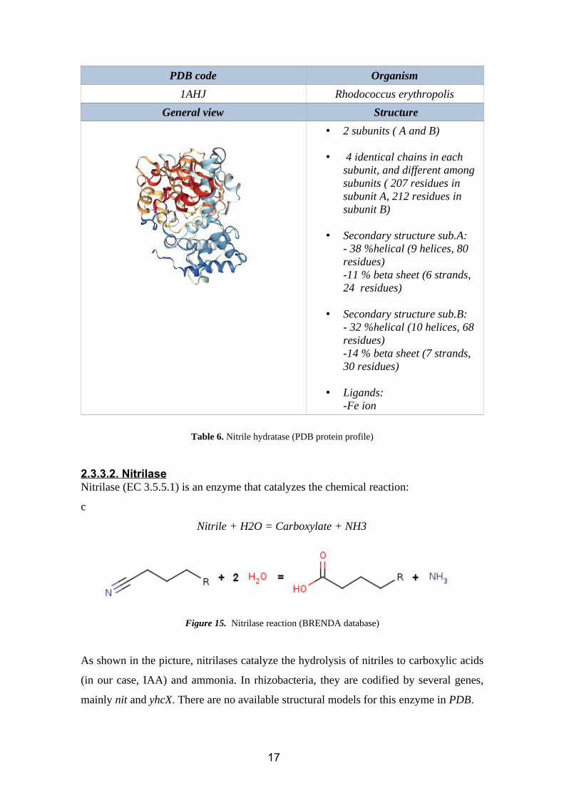
PDB code Organism
1AHJ Rhodococcus erythropolis
General view Structure
• 2 subunits ( A and B)
• 4 identical chains in each subunit, and different among subunits ( 207 residues in subunit A, 212 residues in subunit B)
• Secondary structure sub.A:- 38 %helical (9 helices, 80 residues)-11 % beta sheet (6 strands, 24 residues)
• Secondary structure sub.B:- 32 %helical (10 helices, 68 residues)-14 % beta sheet (7 strands, 30 residues)
• Ligands:-Fe ion
Table 6. Nitrile hydratase (PDB protein profile)
2.3.3.2. NitrilaseNitrilase (EC 3.5.5.1) is an enzyme that catalyzes the chemical reaction:
c
Nitrile + H2O = Carboxylate + NH3
Figure 15. Nitrilase reaction (BRENDA database)
As shown in the picture, nitrilases catalyze the hydrolysis of nitriles to carboxylic acids
(in our case, IAA) and ammonia. In rhizobacteria, they are codified by several genes,
mainly nit and yhcX. There are no available structural models for this enzyme in PDB.
17
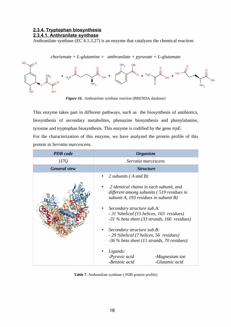
2.3.4. Tryptophan biosynthesis2.3.4.1. Anthranilate synthaseAnthranilate synthase (EC 4.1.3.27) is an enzyme that catalyzes the chemical reaction:
chorismate + L-glutamine = anthranilate + pyruvate + L-glutamate
Figure 16. Anthranilate synthase reaction (BRENDA database)
This enzyme takes part in different pathways, such as the biosynthesis of antibiotics,
biosynthesis of secondary metabolites, phenazine biosynthesis and phenylalanine,
tyrosine and tryptophan biosynthesis. This enzyme is codified by the gene trpE.
For the characterization of this enzyme, we have analyzed the protein profile of this
protein in Serratia marcescens.
PDB code Organism
1I7Q Serratia marcescens
General view Structure
• 2 subunits ( A and B)
• 2 identical chains in each subunit, and different among subunits ( 519 residues in subunit A, 193 residues in subunit B)
• Secondary structure sub.A:- 31 %helical (15 helices, 163 residues)-31 % beta sheet (33 strands, 166 residues)
• Secondary structure sub.B:- 29 %helical (7 helices, 56 residues)-36 % beta sheet (11 strands, 70 residues)
• Ligands:-Pyruvic acid -Magnesium ion-Benzoic acid -Glutamic acid
Table 7. Anthranilate synthase ( PDB protein profile)
18
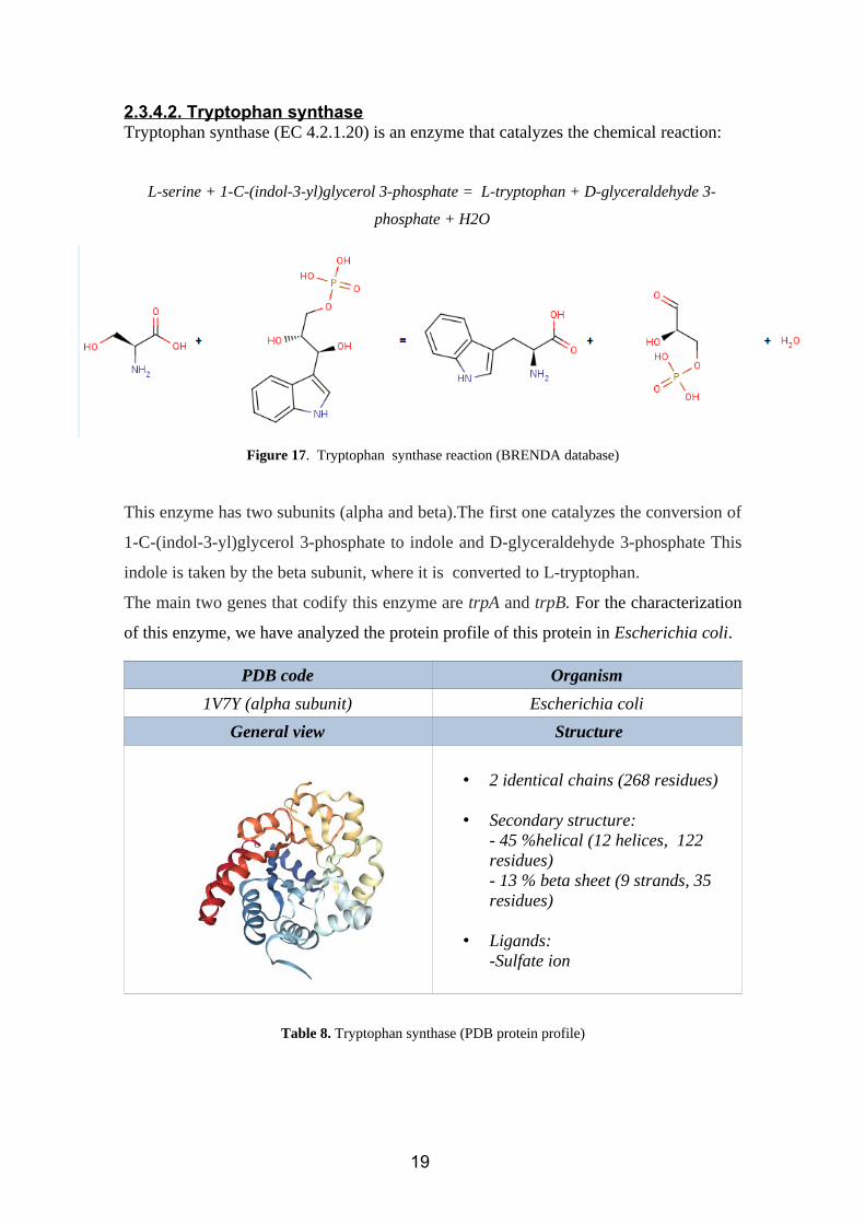
2.3.4.2. Tryptophan synthaseTryptophan synthase (EC 4.2.1.20) is an enzyme that catalyzes the chemical reaction:
L-serine + 1-C-(indol-3-yl)glycerol 3-phosphate = L-tryptophan + D-glyceraldehyde 3-
phosphate + H2O
Figure 17. Tryptophan synthase reaction (BRENDA database)
This enzyme has two subunits (alpha and beta).The first one catalyzes the conversion of
1-C-(indol-3-yl)glycerol 3-phosphate to indole and D-glyceraldehyde 3-phosphate This
indole is taken by the beta subunit, where it is converted to L-tryptophan.
The main two genes that codify this enzyme are trpA and trpB. For the characterization
of this enzyme, we have analyzed the protein profile of this protein in Escherichia coli.
PDB code Organism
1V7Y (alpha subunit) Escherichia coli
General view Structure
• 2 identical chains (268 residues)
• Secondary structure:- 45 %helical (12 helices, 122 residues)- 13 % beta sheet (9 strands, 35 residues)
• Ligands:-Sulfate ion
Table 8. Tryptophan synthase (PDB protein profile)
19
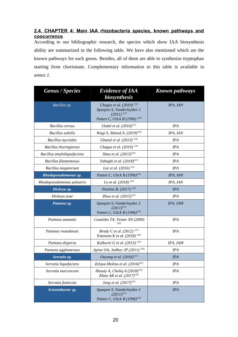
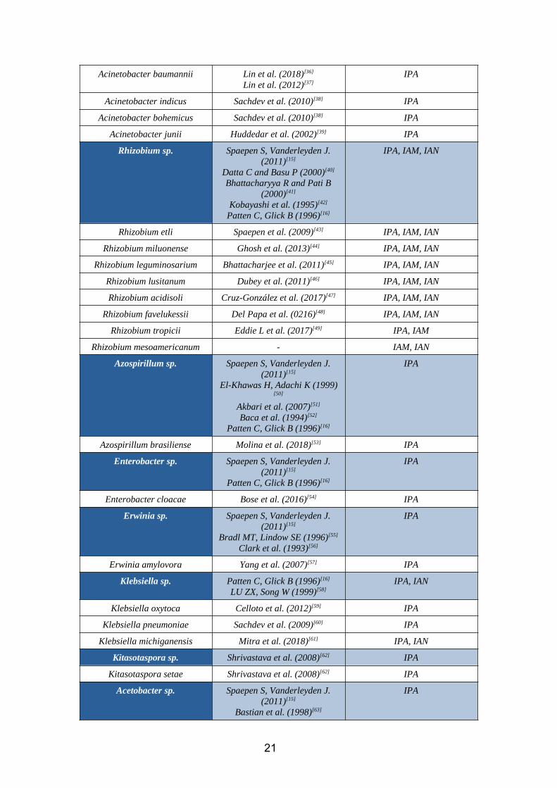
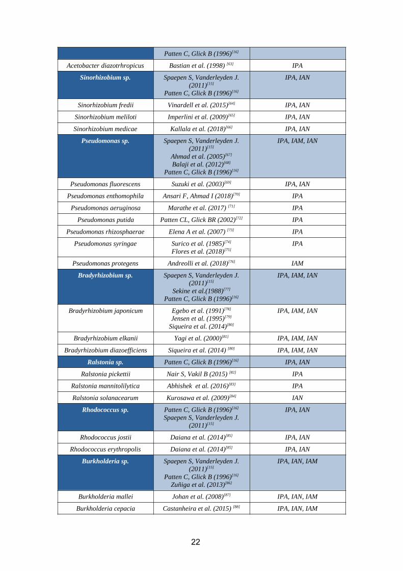
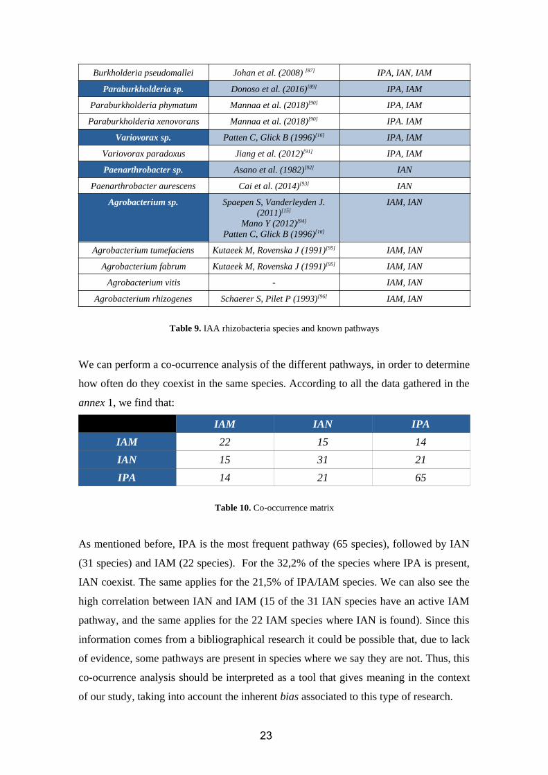
2.4. CHAPTER 4: Main IAA rhizobacteria species, known pathways andcoocurrenceAccording to our bibliographic research, the species which show IAA biosynthesis
ability are summarized in the following table. We have also mentioned which are the
known pathways for each genus. Besides, all of them are able to synthesize tryptophan
starting from chorismate. Complementary information to this table is available in
annex 1.
Genus / Species Evidence of IAAbiosynthesis
Known pathways
Bacillus sp. Chagas et al. (2019) [14]
Spaepen S, Vanderleyden J.(2011) [15]
Patten C, Glick B (1996) [16]
IPA, IAN
Bacillus cereus Ozdal et al. (2016)[17] IPA
Bacillus subtilis Wagi S, Ahmed A. (2019)[18] IPA, IAN
Bacillus mycoides Ghazal et al. (2013) [19] IPA
Bacillus thuringiensis Chagas et al. (2019) [14] IPA
Bacillus amyloliquefaciens Shao et al. (2015)[20] IPA
Bacillus filamentosus Yahaghi et al. (2018)[21] IPA
Bacillus megaterium Lee et al. (2016) [22] IPA
Rhodopseudomonas sp. Patten C, Glick B (1996)[16] IPA, IAN
Rhodopseudomonas palustris Lo et al. (2018) [23] IPA, IAN
Dickeya sp. Pauline B. (2017) [24] IPA
Dickeya zeae Zhou et al. (2015)[25] IPA
Pantoea sp. Spaepen S, Vanderleyden J.(2011)[15]
Patten C, Glick B (1996)[16]
IPA, IAM
Pantoea ananatis Coutinho TA, Venter SN (2009)[26]
IPA
Pantoea rwandensis Brady C et al. (2012) [27]
Estenson K et al. (2018) [28]IPA
Pantoea dispersa Kulkarni G et al. (2013) [29] IPA, IAM
Pantoea agglomerans Apine OA, Jadhav JP (2011) [30] IPA
Serratia sp. Ouyang et al. (2016)[31] IPA
Serratia liquefaciens Zelaya-Molina et al. (2016)[32] IPA
Serratia marcescens Hasuty A, Choliq A (2018)[33]
Khan AR et al. (2017)[34]IPA
Serratia fonticola Jung et al. (2017)[35] IPA
Acinetobacter sp. Spaepen S, Vanderleyden J.(2011)[15]
Patten C, Glick B (1996)[16]
IPA
20
Acinetobacter baumannii Lin et al. (2018)[36]
Lin et al. (2012)[37]IPA
Acinetobacter indicus Sachdev et al. (2010)[38] IPA
Acinetobacter bohemicus Sachdev et al. (2010)[38] IPA
Acinetobacter junii Huddedar et al. (2002)[39] IPA
Rhizobium sp. Spaepen S, Vanderleyden J.(2011)[15]
Datta C and Basu P (2000)[40]
Bhattacharyya R and Pati B(2000)[41]
Kobayashi et al. (1995)[42]
Patten C, Glick B (1996)[16]
IPA, IAM, IAN
Rhizobium etli Spaepen et al. (2009)[43] IPA, IAM, IAN
Rhizobium miluonense Ghosh et al. (2013)[44] IPA, IAM, IAN
Rhizobium leguminosarium Bhattacharjee et al. (2011)[45] IPA, IAM, IAN
Rhizobium lusitanum Dubey et al. (2011)[46] IPA, IAM, IAN
Rhizobium acidisoli Cruz-González et al. (2017)[47] IPA, IAM, IAN
Rhizobium favelukessii Del Papa et al. (0216)[48] IPA, IAM, IAN
Rhizobium tropicii Eddie L et al. (2017)[49] IPA, IAM
Rhizobium mesoamericanum - IAM, IAN
Azospirillum sp. Spaepen S, Vanderleyden J.(2011)[15]
El-Khawas H, Adachi K (1999)[50]
Akbari et al. (2007)[51]
Baca et al. (1994)[52]
Patten C, Glick B (1996)[16]
IPA
Azospirillum brasiliense Molina et al. (2018)[53] IPA
Enterobacter sp. Spaepen S, Vanderleyden J.(2011)[15]
Patten C, Glick B (1996)[16]
IPA
Enterobacter cloacae Bose et al. (2016)[54] IPA
Erwinia sp. Spaepen S, Vanderleyden J.(2011)[15]
Bradl MT, Lindow SE (1996)[55]
Clark et al. (1993)[56]
IPA
Erwinia amylovora Yang et al. (2007)[57] IPA
Klebsiella sp. Patten C, Glick B (1996)[16]
LU ZX, Song W (1999)[58]IPA, IAN
Klebsiella oxytoca Celloto et al. (2012)[59] IPA
Klebsiella pneumoniae Sachdev et al. (2009)[60] IPA
Klebsiella michiganensis Mitra et al. (2018)[61] IPA, IAN
Kitasotaspora sp. Shrivastava et al. (2008)[62] IPA
Kitasotaspora setae Shrivastava et al. (2008)[62] IPA
Acetobacter sp. Spaepen S, Vanderleyden J.(2011)[15]
Bastian et al. (1998)[63]
IPA
21
Patten C, Glick B (1996)[16]
Acetobacter diazotrhropicus Bastian et al. (1998) [63] IPA
Sinorhizobium sp. Spaepen S, Vanderleyden J.(2011)[15]
Patten C, Glick B (1996)[16]
IPA, IAN
Sinorhizobium fredii Vinardell et al. (2015)[64] IPA, IAN
Sinorhizobium meliloti Imperlini et al. (2009)[65] IPA, IAN
Sinorhizobium medicae Kallala et al. (2018)[66] IPA, IAN
Pseudomonas sp. Spaepen S, Vanderleyden J.(2011)[15]
Ahmad et al. (2005)[67]
Balaji et al. (2012)[68]
Patten C, Glick B (1996)[16]
IPA, IAM, IAN
Pseudomonas fluorescens Suzuki et al. (2003)[69] IPA, IAN
Pseudomonas enthomophila Ansari F, Ahmad I (2018)[70] IPA
Pseudomonas aeruginosa Marathe et al. (2017) [71] IPA
Pseudomonas putida Patten CL, Glick BR (2002)[72] IPA
Pseudomonas rhizosphaerae Elena A et al. (2007) [73] IPA
Pseudomonas syringae Surico et al. (1985)[74]
Flores et al. (2018)[75]IPA
Pseudomonas protegens Andreolli et al. (2018)[76] IAM
Bradyrhizobium sp. Spaepen S, Vanderleyden J.(2011)[15]
Sekine et al.(1988)[77]
Patten C, Glick B (1996)[16]
IPA, IAM, IAN
Bradyrhizobium japonicum Egebo et al. (1991)[78]
Jensen et al. (1995)[79]
Siqueira et al. (2014)[80]
IPA, IAM, IAN
Bradyrhizobium elkanii Yagi et al. (2000)[81] IPA, IAM, IAN
Bradyrhizobium diazoefficiens Siqueira et al. (2014) [80] IPA, IAM, IAN
Ralstonia sp. Patten C, Glick B (1996)[16] IPA, IAN
Ralstonia pickettii Nair S, Vakil B (2015) [82] IPA
Ralstonia mannitolilytica Abhishek et al. (2016)[83] IPA
Ralstonia solanacearum Kurosawa et al. (2009)[84] IAN
Rhodococcus sp. Patten C, Glick B (1996)[16]
Spaepen S, Vanderleyden J.(2011)[15]
IPA, IAN
Rhodococcus jostii Daiana et al. (2014)[85] IPA, IAN
Rhodococcus erythropolis Daiana et al. (2014)[85] IPA, IAN
Burkholderia sp. Spaepen S, Vanderleyden J.(2011)[15]
Patten C, Glick B (1996)[16]
Zuñiga et al. (2013)[86]
IPA, IAN, IAM
Burkholderia mallei Johan et al. (2008)[87] IPA, IAN, IAM
Burkholderia cepacia Castanheira et al. (2015) [88] IPA, IAN, IAM
22
Burkholderia pseudomallei Johan et al. (2008) [87] IPA, IAN, IAM
Paraburkholderia sp. Donoso et al. (2016)[89] IPA, IAM
Paraburkholderia phymatum Mannaa et al. (2018)[90] IPA, IAM
Paraburkholderia xenovorans Mannaa et al. (2018)[90] IPA. IAM
Variovorax sp. Patten C, Glick B (1996)[16] IPA, IAM
Variovorax paradoxus Jiang et al. (2012)[91] IPA, IAM
Paenarthrobacter sp. Asano et al. (1982)[92] IAN
Paenarthrobacter aurescens Cai et al. (2014)[93] IAN
Agrobacterium sp. Spaepen S, Vanderleyden J.(2011)[15]
Mano Y (2012)[94]
Patten C, Glick B (1996)[16]
IAM, IAN
Agrobacterium tumefaciens Kutaeek M, Rovenska J (1991)[95] IAM, IAN
Agrobacterium fabrum Kutaeek M, Rovenska J (1991)[95] IAM, IAN
Agrobacterium vitis - IAM, IAN
Agrobacterium rhizogenes Schaerer S, Pilet P (1993)[96] IAM, IAN
Table 9. IAA rhizobacteria species and known pathways
We can perform a co-ocurrence analysis of the different pathways, in order to determine
how often do they coexist in the same species. According to all the data gathered in the
annex 1, we find that:
IAM IAN IPA
IAM 22 15 14
IAN 15 31 21
IPA 14 21 65
Table 10. Co-occurrence matrix
As mentioned before, IPA is the most frequent pathway (65 species), followed by IAN
(31 species) and IAM (22 species). For the 32,2% of the species where IPA is present,
IAN coexist. The same applies for the 21,5% of IPA/IAM species. We can also see the
high correlation between IAN and IAM (15 of the 31 IAN species have an active IAM
pathway, and the same applies for the 22 IAM species where IAN is found). Since this
information comes from a bibliographical research it could be possible that, due to lack
of evidence, some pathways are present in species where we say they are not. Thus, this
co-ocurrence analysis should be interpreted as a tool that gives meaning in the context
of our study, taking into account the inherent bias associated to this type of research.
23
2.5. CHAPTER 5. Results: diversity analysis IIn order to perform the diversity analysis I described in the chapter Objectives, we have
obtained all the information available concerning genes and proteins of the identified
rhizobacteria species. Therefore, protein accession numbers of the sequences we are
working with can be found in the annex 1. Since the protein sequence is under selective
constraint for protein function and protein structure, and these are conserved over much
longer periods than the individual codon choices, we have decided to carry out our
diversity analysis using the protein sequences instead of DNA sequences. If we were
looking for differences within a closely related group of species (for instance, a bunch
of species from the same strain), DNA would be a better option, but in our case we are
working with species that are far away from each other from an evolutionary point of
view, and the higher conservation status of protein sequences in comparison to DNA
sequences will be a plus in our analysis.
In order to run our analysis, we have created several csv document, each of them with
the accession numbers of the identified proteins for each enzyme under study. These csv
worked as an input for the R pipeline we have designed. Further information about the
structure of these csv documents and the R pipeline can be found in annex 2.
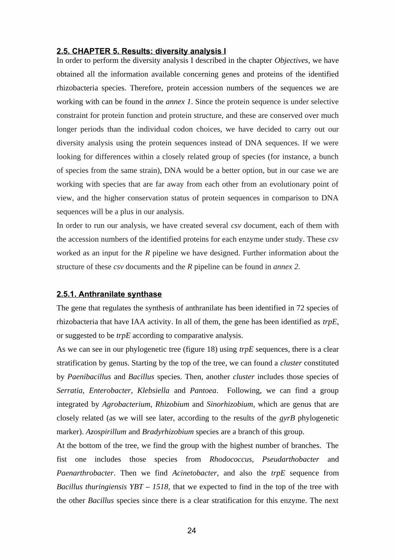
2.5.1. Anthranilate synthase
The gene that regulates the synthesis of anthranilate has been identified in 72 species of
rhizobacteria that have IAA activity. In all of them, the gene has been identified as trpE,
or suggested to be trpE according to comparative analysis.
As we can see in our phylogenetic tree (figure 18) using trpE sequences, there is a clear
stratification by genus. Starting by the top of the tree, we can found a cluster constituted
by Paenibacillus and Bacillus species. Then, another cluster includes those species of
Serratia, Enterobacter, Klebsiella and Pantoea. Following, we can find a group
integrated by Agrobacterium, Rhizobium and Sinorhizobium, which are genus that are
closely related (as we will see later, according to the results of the gyrB phylogenetic
marker). Azospirillum and Bradyrhizobium species are a branch of this group.
At the bottom of the tree, we find the group with the highest number of branches. The
fist one includes those species from Rhodococcus, Pseudarthobacter and
Paenarthrobacter. Then we find Acinetobacter, and also the trpE sequence from
Bacillus thuringiensis YBT – 1518, that we expected to find in the top of the tree with
the other Bacillus species since there is a clear stratification for this enzyme. The next
24
branch also goes according to the expected evolutionary relation observed, and is
configurated by Ralstonia, Burkholderia and Paraburkholderia species. Finally, the last
branch gather all the species from the genus Pseudomonas.
Figure 18.
Anthranilate
synthase
phylogeny.
25
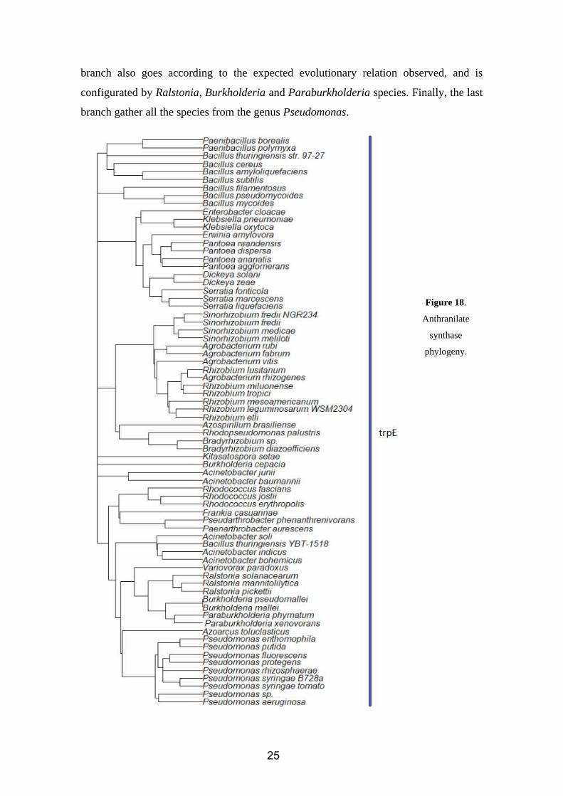
2.5.2. Tryptophan synthase (alpha subunit)
Figure 19.
Tryptophan
synthase (alpha
subunit)
phylogeny.
26
The gene that regulates the synthesis of the tryptophan synthase’s alpha subunit has
been identified in 76 species of rhizobacteria that also show IAA activity. In all of them,
this gene has been identified as trpA, or suggested to be trpA according to comparative
analysis. The reason why we have four 76 instead of 72 as in the previous case is
because there are four species where trpE sequences have not been identified or at least
mentioned in the bibliography.
This phylogenetic tree (figure 19) is similar to the one observed for anthranilate
synthase. As we well explain later after showing the beta subunit’s tree, there is reason
for this resemblance. Starting from the top of the tree, we observe a well-defined cluster
constituted by the species of the genus Pseudomonas. In the next branch, we find that
Azospirillum, Rhodopseudomonas, Bradyrhizobium, Sinorhizobium, Rhizobium and
Agrobacterium species are close from a phylogenetic point of view according to trpA.
These two branches were unrelated in the anthranilate synthase tree.
The first branch of the next cluster contains all the Acinetobacter species under analysis.
Then, Variovorax, Ralstonia, Burkholderia and Paraburkholderia appear in the next
branch. If we continue our path to the bottom of the tree, we will find once again the
cluster of Enterobacter, Klebsiella, Dickeya, Pantoea, Serratia and Erwinia. Once
again, we have our group of Bacillus and Paenibacillus (this time, with the expected
result of Bacillus thuringiensis YBT – 1518 being close to Bacillus thuringiensis str.97-
27) and, as the last branch of the tree, the group Kitasatospora, Frankia,
Pseudarthrobacter, Paenarthrobacter and Rhodococcus.
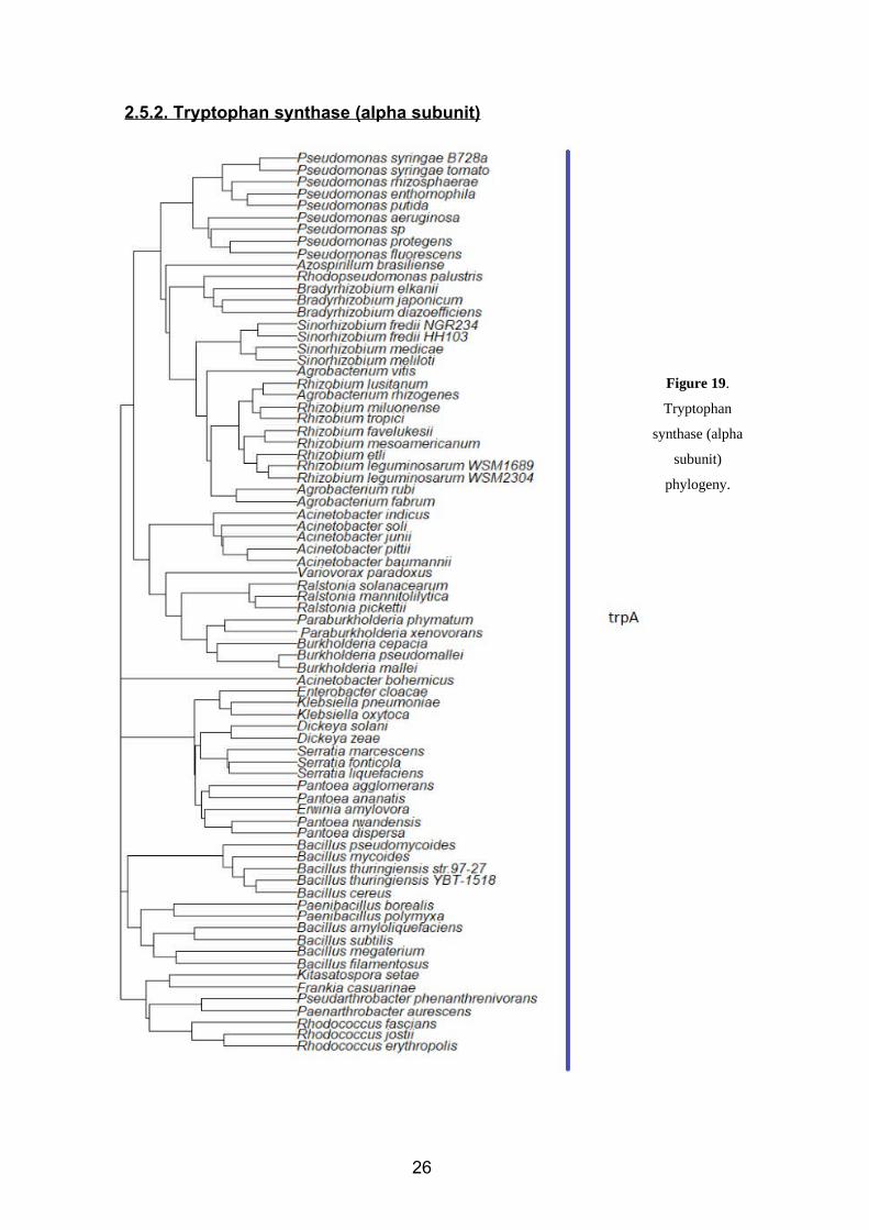
2.5.3. Tryptophan synthase (beta subunit)
The gene that regulates the synthesis of the tryptophan synthase’s beta subunit has been
identified in 76 species of rhizobacteria which also show IAA activity. In all of them,
this gene has been identified as trpB, or suggested to be trpB according to comparative
analysis.
As the tree shows (figure 20), the groups or clusters observed are very similar to those
in the alpha subunit, with slight differences. Starting from the top of the tree, we find
the group Erwinia – Pantoea -Klebsiella – Serratia – Dickeya. Then, there is a new
cluster similar to those observed before, constituted by Paenarthrobacer,
Pseudarthrobacter, Kitasatosporae, Rhodococcus and Frankia. After that, we find the
big group of Agrobacterium, Sinorhizobium, Rhizobium, followed by the Pseudomonas
group, the Bradyrhizobium-Azospirillum-Rhodopseudomonas group and then by the
27
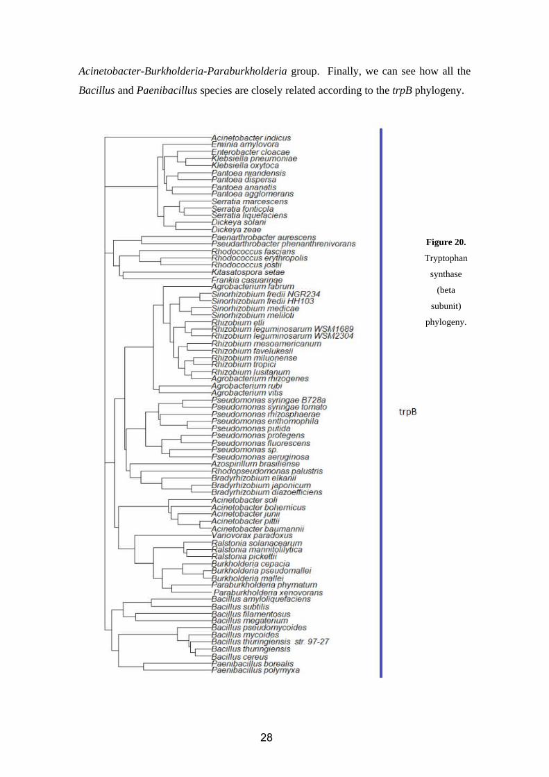
Acinetobacter-Burkholderia-Paraburkholderia group. Finally, we can see how all the
Bacillus and Paenibacillus species are closely related according to the trpB phylogeny.
Figure 20.
Tryptophan
synthase
(beta
subunit)
phylogeny.
28
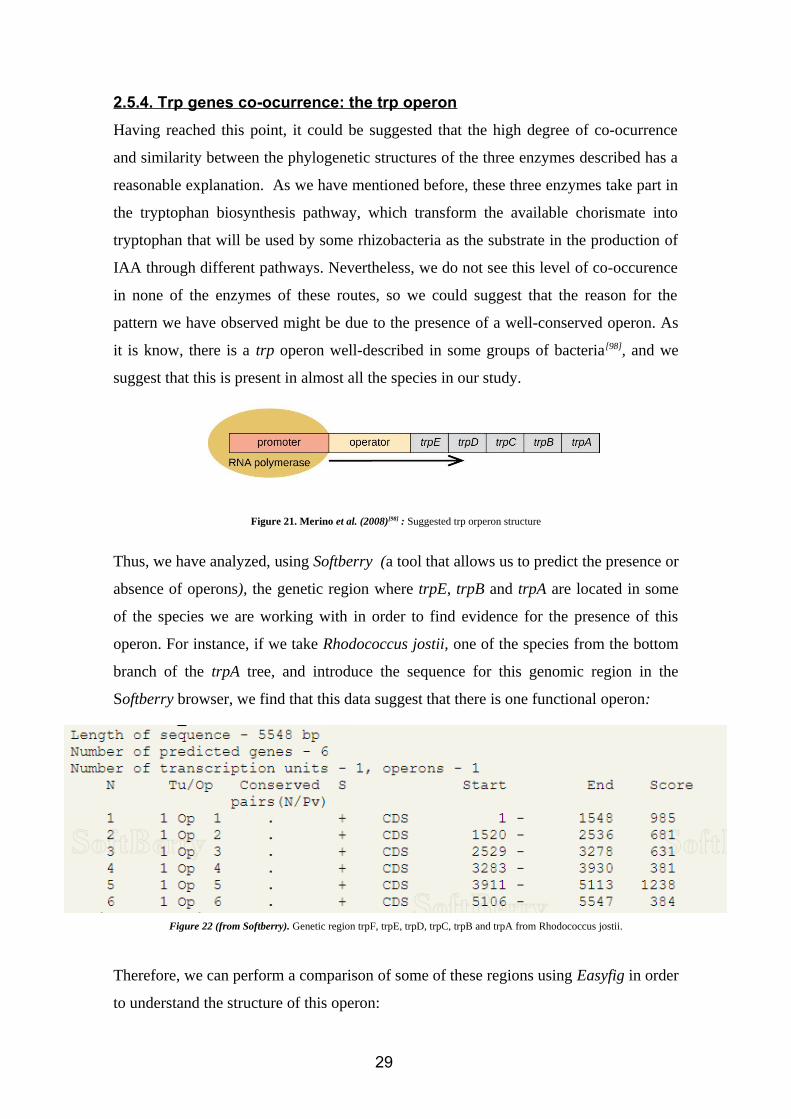
2.5.4. Trp genes co-ocurrence: the trp operon
Having reached this point, it could be suggested that the high degree of co-ocurrence
and similarity between the phylogenetic structures of the three enzymes described has a
reasonable explanation. As we have mentioned before, these three enzymes take part in
the tryptophan biosynthesis pathway, which transform the available chorismate into
tryptophan that will be used by some rhizobacteria as the substrate in the production of
IAA through different pathways. Nevertheless, we do not see this level of co-occurence
in none of the enzymes of these routes, so we could suggest that the reason for the
pattern we have observed might be due to the presence of a well-conserved operon. As
it is know, there is a trp operon well-described in some groups of bacteria[98], and we
suggest that this is present in almost all the species in our study.
Figure 21. Merino et al. (2008)[98] : Suggested trp orperon structure
Thus, we have analyzed, using Softberry (a tool that allows us to predict the presence or
absence of operons), the genetic region where trpE, trpB and trpA are located in some
of the species we are working with in order to find evidence for the presence of this
operon. For instance, if we take Rhodococcus jostii, one of the species from the bottom
branch of the trpA tree, and introduce the sequence for this genomic region in the
Softberry browser, we find that this data suggest that there is one functional operon:
Figure 22 (from Softberry). Genetic region trpF, trpE, trpD, trpC, trpB and trpA from Rhodococcus jostii.
Therefore, we can perform a comparison of some of these regions using Easyfig in order
to understand the structure of this operon:
29
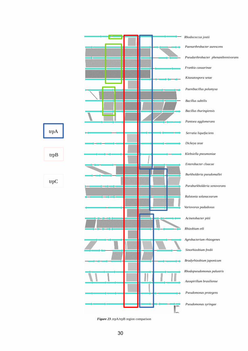
Figure 23 .trpA/trpB region comparison
30
trpA
trpB
trpC
Rhodococcus jostii
Paenarthrobacter aurescens
Pseudarthrobacter phenanthrenivorans
Frankia casuarinae
Kitasatospora setae
Paenibacillus polumyxa
Bacillus subtilis
Bacillus thuringiensis
Pantoea agglomerans
Serratia liquefaciens
Dickeya zeae
Klebsiella pneumoniae
Enterobacter cloacae
Burkholderia pseudomallei
Paraburkholderia xenovorans
Ralstonia solanacearum
Variovorax padadoxus
Acinetobacter pitii
Rhizobium etli
Agrobacterium rhizogenes
Sinorhizobium fredii
Bradyrhizobium japonicum
Rhodopseudomonas palustris
Azospirillum brasiliense
Pseudomonas protegens
Pseudomonas syringae
As we can see in almost all the species tested, with the exceptions of Burkholderia,
Paraburkholderia, Variovorax and Ralstonia, trpB is located immediately near to trpA.
Nevertheless, in these exceptions only one gene is among them.
We see that, from the top to the bottom of the comparison matrix (figure 23), the
distance from the trpB-trpA tandem to the rest of the operon (trpC, trpE and trpD) tends
to get bigger. The pattern observed for this changes has some similarities to that
observed for the structure of both trpB and trpA trees, were species where trpB-trpA are
near to trpC-trpD are closer in these trees, and the same works for those were trpB-trpA
region is far away from the rest of the operon’s genes.
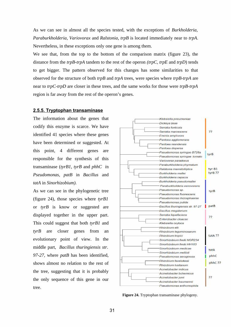
2.5.5. Tryptophan transaminase
The information about the genes that
codify this enzyme is scarce. We have
identified 41 species where these genes
have been determined or suggested. At
this point, 4 different genes are
responsible for the synthesis of this
transaminase (tyrB1, tyrB and phhC in
Pseudomonas, patB in Bacillus and
tatA in Sinorhizobium).
As we can see in the phylogenetic tree
(figure 24), those species where tyrB1
or tyrB is know or suggested are
displayed together in the upper part.
This could suggest that both tyrB1 and
tyrB are closer genes from an
evolutionary point of view. In the
middle part, Bacillus thuringiensis str.
97-27, where patB has been identified,
shows almost no relation to the rest of
the tree, suggesting that it is probably
the only sequence of this gene in our
tree.
Figure 24. Tryptophan transaminase phylogeny.
31
Then, Sinorhizobium species where tatA is know as the gene that codifies this
transaminase (Sinorhizobium meliloti and Sinorhizobium medicae) are grouped together,
as expected. They form a cluster with the rest of the species from the same genus, and
also with those of Rhizobium. Thus, and due to the evolutionary proximity suggested by
the marker phylogeny, we could think that the suggested genes in Rhizobium are also
tatA. Nevertheless, and since some species of Rhizobium where the transaminase gene
has been suggested but not identified are in the same branch as Pseudomonas
aeruginosa, we could think that phhC is also an active transaminase that takes part in
this step of the IPA patwhay.
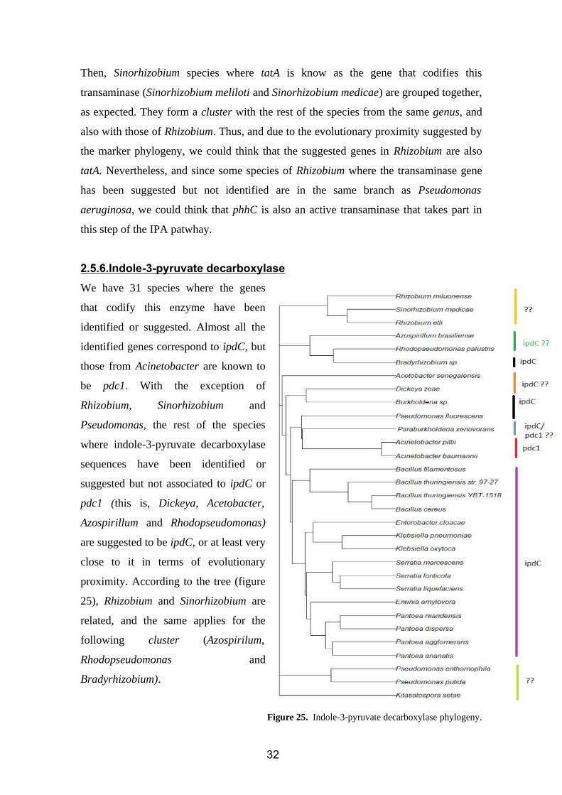
2.5.6.Indole-3-pyruvate decarboxylase
We have 31 species where the genes
that codify this enzyme have been
identified or suggested. Almost all the
identified genes correspond to ipdC, but
those from Acinetobacter are known to
be pdc1. With the exception of
Rhizobium, Sinorhizobium and
Pseudomonas, the rest of the species
where indole-3-pyruvate decarboxylase
sequences have been identified or
suggested but not associated to ipdC or
pdc1 (this is, Dickeya, Acetobacter,
Azospirillum and Rhodopseudomonas)
are suggested to be ipdC, or at least very
close to it in terms of evolutionary
proximity. According to the tree (figure
25), Rhizobium and Sinorhizobium are
related, and the same applies for the
following cluster (Azospirilum,
Rhodopseudomonas and
Bradyrhizobium).
Figure 25. Indole-3-pyruvate decarboxylase phylogeny.
32
Then, there is a big cluster of ipdC/pdc1 genes integrated by Acetobacter, Dickeya,
Burkholderia, Acinetobacter, Bacillus, Klebsiella, Enterobacter, Serratia, Erwinia and
Pantoea. Finally, indole-3-pyruvate decarboxylases from Pseudomonas and
Kitasatosporae seem to have evolved different from those of the rest of species codified
by ipdC/pdc1, suggesting another gene to be defined.
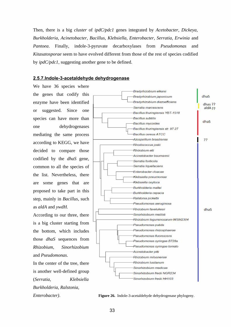
2.5.7.Indole-3-acetaldehyde dehydrogenase
We have 36 species where
the genes that codify this
enzyme have been identified
or suggested. Since one
species can have more than
one dehydrogenases
mediating the same process
according to KEGG, we have
decided to compare those
codified by the dhaS gene,
common to all the species of
the list. Nevertheless, there
are some genes that are
proposed to take part in this
step, mainly in Bacillus, such
as aldA and ywdH.
According to our three, there
is a big cluster starting from
the bottom, which includes
those dhaS sequences from
Rhizobium, Sinorhizobium
and Pseudomonas.
In the center of the tree, there
is another well-defined group
(Serratia, Klebsiella
Burkholderia, Ralstonia,
Enterobacter). Figure 26. Indole-3-acetaldehyde dehydrogenase phylogeny.
33
In the upper part, there are two groups, constituted each one of them for only one genus
(Bradyrhizobium and Bacillus). Serratia marcescens, Pseudomonas aeruginosa and
Azospirillum brasiliense did not show the expected relation observed previously in the
rest of the trees. This could mean that there is a problem with the sequence (an error in
its identification as dhaS) or more probably that they have evolved differently due to
natural selection.
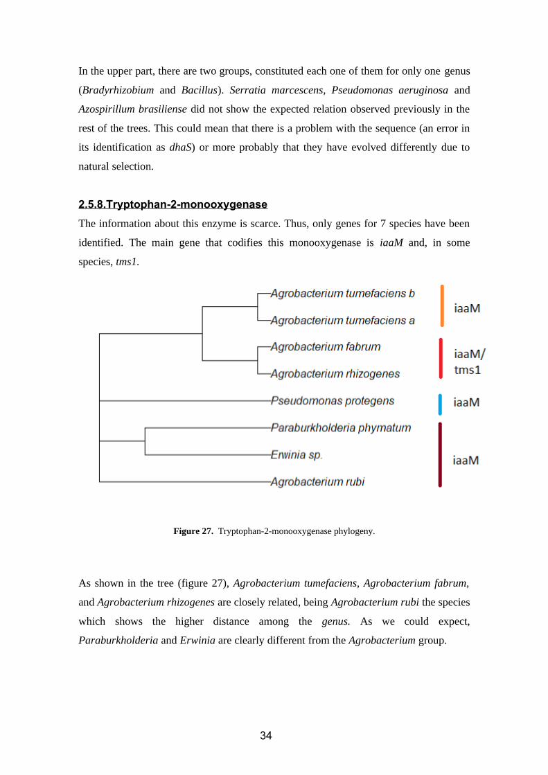
2.5.8.Tryptophan-2-monooxygenase
The information about this enzyme is scarce. Thus, only genes for 7 species have been
identified. The main gene that codifies this monooxygenase is iaaM and, in some
species, tms1.
Figure 27. Tryptophan-2-monooxygenase phylogeny.
As shown in the tree (figure 27), Agrobacterium tumefaciens, Agrobacterium fabrum,
and Agrobacterium rhizogenes are closely related, being Agrobacterium rubi the species
which shows the higher distance among the genus. As we could expect,
Paraburkholderia and Erwinia are clearly different from the Agrobacterium group.
34
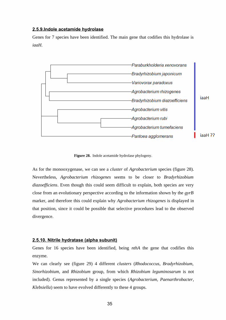
2.5.9.Indole acetamide hydrolase
Genes for 7 species have been identified. The main gene that codifies this hydrolase is
iaaH.
Figure 28. Indole acetamide hydrolase phylogeny.
As for the monooxygenase, we can see a cluster of Agrobacterium species (figure 28).
Nevertheless, Agrobacterium rhizogenes seems to be closer to Bradyrhizobium
diazoefficiens. Even though this could seem difficult to explain, both species are very
close from an evolutionary perspective according to the information shown by the gyrB
marker, and therefore this could explain why Agrobacterium rhizogenes is displayed in
that position, since it could be possible that selective procedures lead to the observed
divergence.
2.5.10. Nitrile hydratase (alpha subunit)
Genes for 16 species have been identified, being nthA the gene that codifies this
enzyme.
We can clearly see (figure 29) 4 different clusters (Rhodococcus, Bradyrhizobium,
Sinorhizobium, and Rhizobium group, from which Rhizobium leguminosarum is not
included). Genus represented by a single species (Agrobacterium, Paenarthrobacter,
Klebsiella) seem to have evolved differently to these 4 groups.
35
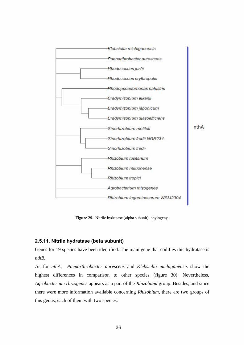
Figure 29. Nitrile hydratase (alpha subunit) phylogeny.
2.5.11. Nitrile hydratase (beta subunit)
Genes for 19 species have been identified. The main gene that codifies this hydratase is
nthB.
As for nthA, Paenarthrobacter aurescens and Klebsiella michiganensis show the
highest differences in comparison to other species (figure 30). Nevertheless,
Agrobacterium rhizogenes appears as a part of the Rhizobium group. Besides, and since
there were more information available concerning Rhizobium, there are two groups of
this genus, each of them with two species.
36
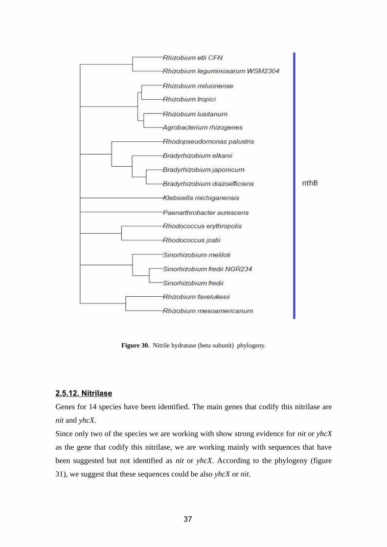
Figure 30. Nitrile hydratase (beta subunit) phylogeny.
2.5.12. Nitrilase
Genes for 14 species have been identified. The main genes that codify this nitrilase are
nit and yhcX.
Since only two of the species we are working with show strong evidence for nit or yhcX
as the gene that codify this nitrilase, we are working mainly with sequences that have
been suggested but not identified as nit or yhcX. According to the phylogeny (figure
31), we suggest that these sequences could be also yhcX or nit.
37
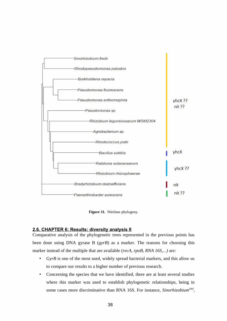
Figure 31. Nitrilase phylogeny.
2.6. CHAPTER 6: Results: diversity analysis IIComparative analysis of the phylogenetic trees represented in the previous points has
been done using DNA gyrase B (gyrB) as a marker. The reasons for choosing this
marker instead of the multiple that are available (recA, rpoB, RNA 16S,...) are:
• GyrB is one of the most used, widely spread bacterial markers, and this allow us
to compare our results to a higher number of previous research.
• Concerning the species that we have identified, there are at least several studies
where this marker was used to establish phylogenetic relationships, being in
some cases more discriminative than RNA 16S. For instance, Sinorhizobium[99],
38
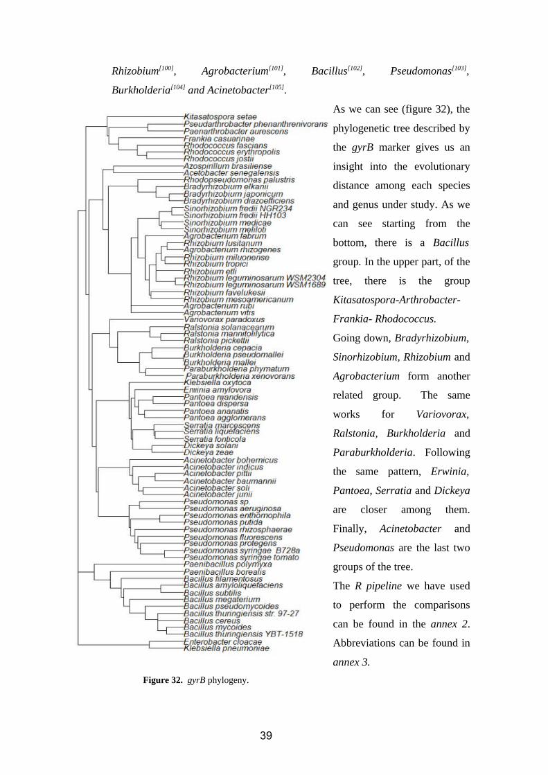
Rhizobium[100], Agrobacterium[101], Bacillus[102], Pseudomonas[103],
Burkholderia[104] and Acinetobacter[105].
As we can see (figure 32), the
phylogenetic tree described by
the gyrB marker gives us an
insight into the evolutionary
distance among each species
and genus under study. As we
can see starting from the
bottom, there is a Bacillus
group. In the upper part, of the
tree, there is the group
Kitasatospora-Arthrobacter-
Frankia- Rhodococcus.
Going down, Bradyrhizobium,
Sinorhizobium, Rhizobium and
Agrobacterium form another
related group. The same
works for Variovorax,
Ralstonia, Burkholderia and
Paraburkholderia. Following
the same pattern, Erwinia,
Pantoea, Serratia and Dickeya
are closer among them.
Finally, Acinetobacter and
Pseudomonas are the last two
groups of the tree.
The R pipeline we have used
to perform the comparisons
can be found in the annex 2.
Abbreviations can be found in
annex 3.
Figure 32. gyrB phylogeny.
39
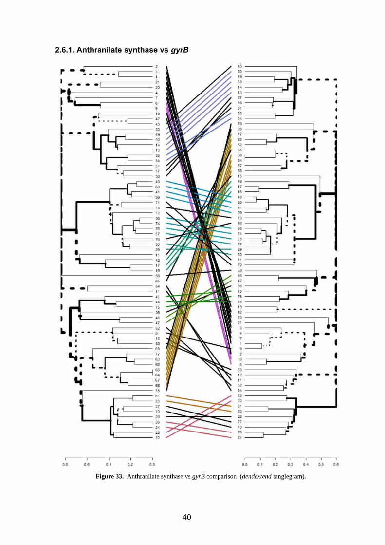
2.6.1. Anthranilate synthase vs gyrB
Figure 33. Anthranilate synthase vs gyrB comparison (dendextend tanglegram).
40
Agrobacterium (71,72) Pseudomonas (27, 28, 70)
Bacillus (1,2,3,4,7,9) Rhizobium (76)
Klebsiella (42,43) Acinetobacter (12,53)
Serratia (37,38,51) Pseudarthrobacter (47)
While color lines show that a species is in the same branch in both trees, black lines
show species and genus which have different locations in both trees (figure 33). Genus
and species that have different classifications depending on the tree are summarized in
the previous chart.
Nevertheless, an as we can see, these differences are not huge, since the main part of
them refer to a different position of these species but inside the same clusters in both
trees.
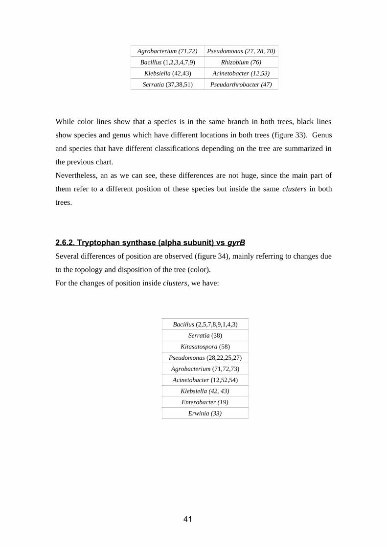
2.6.2. Tryptophan synthase (alpha subunit) vs gyrB
Several differences of position are observed (figure 34), mainly referring to changes due
to the topology and disposition of the tree (color).
For the changes of position inside clusters, we have:
Bacillus (2,5,7,8,9,1,4,3)
Serratia (38)
Kitasatospora (58)
Pseudomonas (28,22,25,27)
Agrobacterium (71,72,73)
Acinetobacter (12,52,54)
Klebsiella (42, 43)
Enterobacter (19)
Erwinia (33)
41
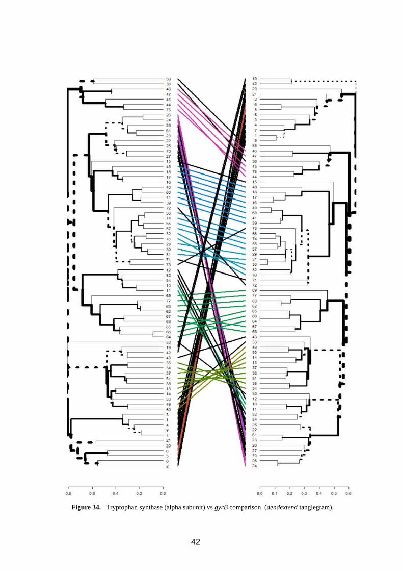
Figure 34. Tryptophan synthase (alpha subunit) vs gyrB comparison (dendextend tanglegram).
42
2.6.3. Tryptophan synthase (beta subunit) vs gyrB
Figure 35. Tryptophan synthase (beta subunit) vs gyrB comparison (dendextend tanglegram).
43
Several differences of position are observed (figure 35), mainly referring to changes due
to the topology and disposition of the tree (color).
For the changes of position inside clusters, we have:
Bacillus (2,5,7,8,9,1,4,3) Klebsiella (42, 43)
Erwinia (33) Pseudomonas (28, 27, 70)
Enterobacter (19) Azospirillum (15)
Serratia (38) Acinetobacter (52, 53, 54)
Kitasatospora (58) Paenibacillus (21,20)
Agrobacterium (73) Klebsiella (42, 43)
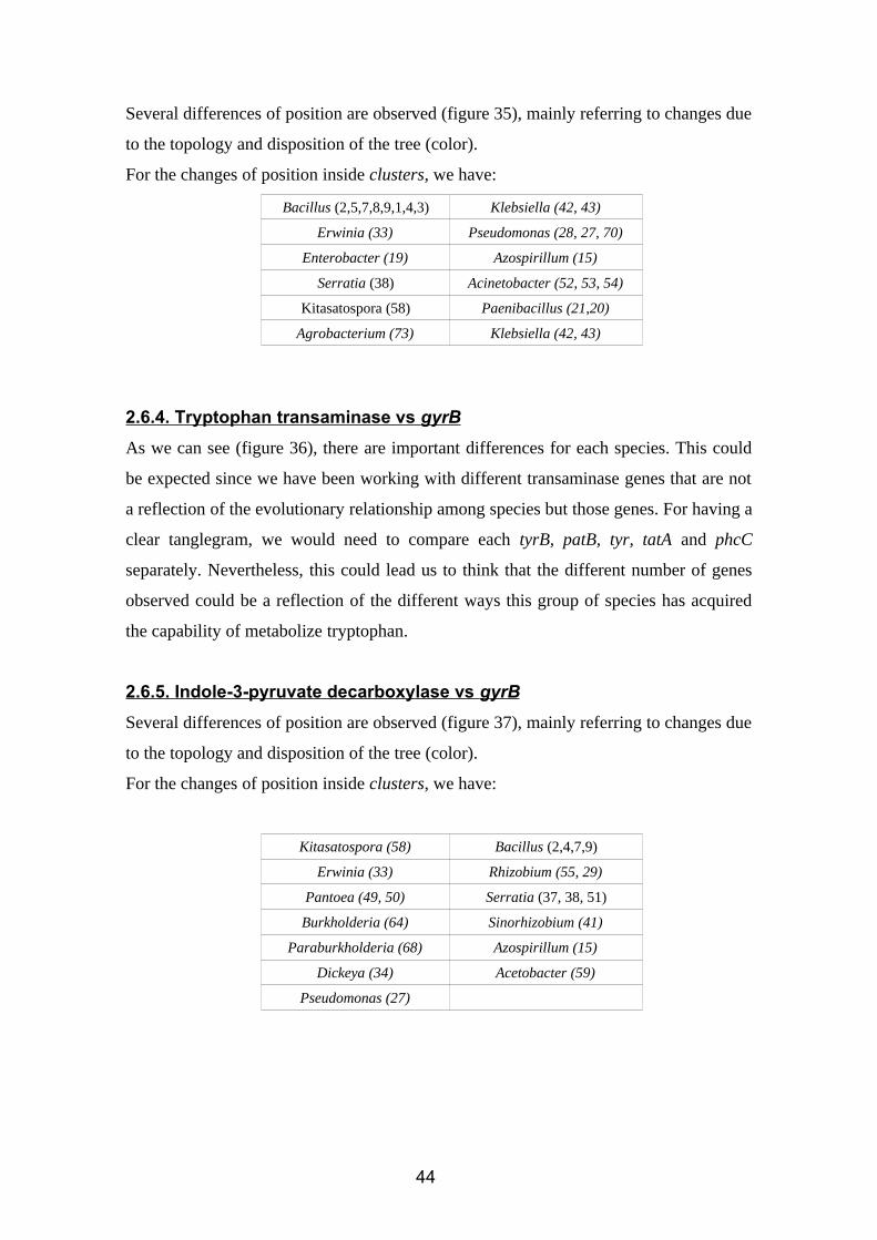
2.6.4. Tryptophan transaminase vs gyrB
As we can see (figure 36), there are important differences for each species. This could
be expected since we have been working with different transaminase genes that are not
a reflection of the evolutionary relationship among species but those genes. For having a
clear tanglegram, we would need to compare each tyrB, patB, tyr, tatA and phcC
separately. Nevertheless, this could lead us to think that the different number of genes
observed could be a reflection of the different ways this group of species has acquired
the capability of metabolize tryptophan.
2.6.5. Indole-3-pyruvate decarboxylase vs gyrB
Several differences of position are observed (figure 37), mainly referring to changes due
to the topology and disposition of the tree (color).
For the changes of position inside clusters, we have:
Kitasatospora (58) Bacillus (2,4,7,9)
Erwinia (33) Rhizobium (55, 29)
Pantoea (49, 50) Serratia (37, 38, 51)
Burkholderia (64) Sinorhizobium (41)
Paraburkholderia (68) Azospirillum (15)
Dickeya (34) Acetobacter (59)
Pseudomonas (27)
44
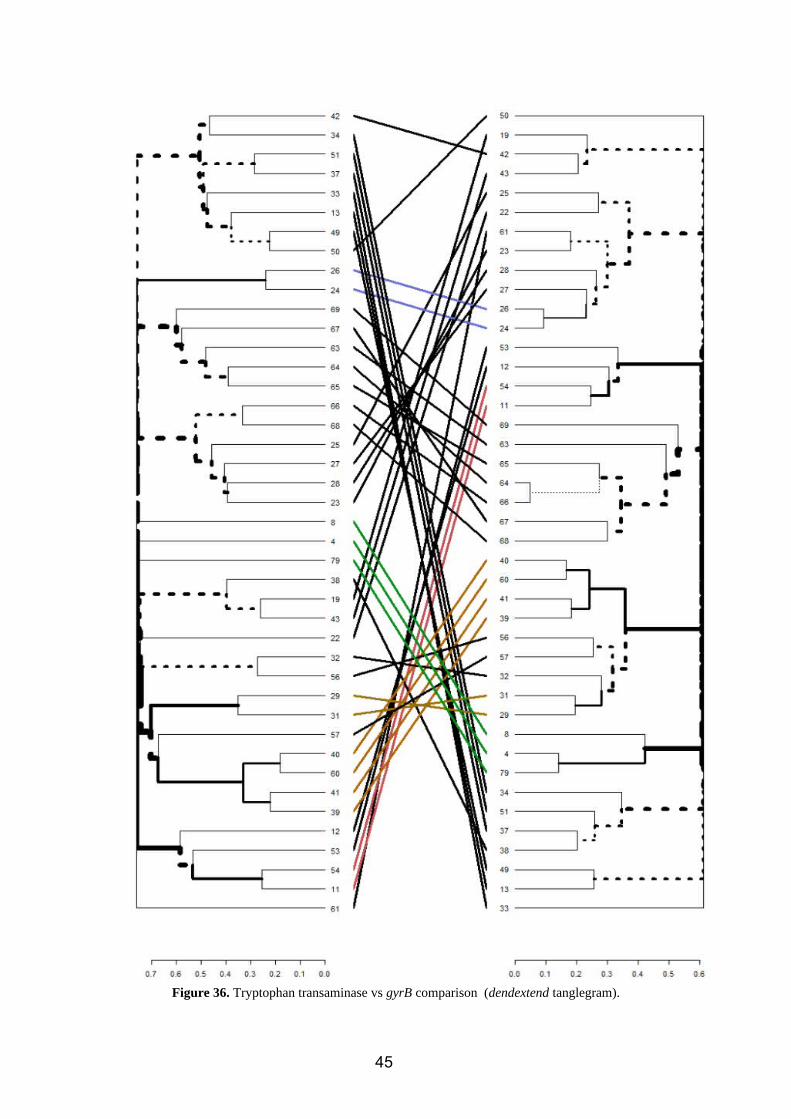
Figure 36. Tryptophan transaminase vs gyrB comparison (dendextend tanglegram).
45
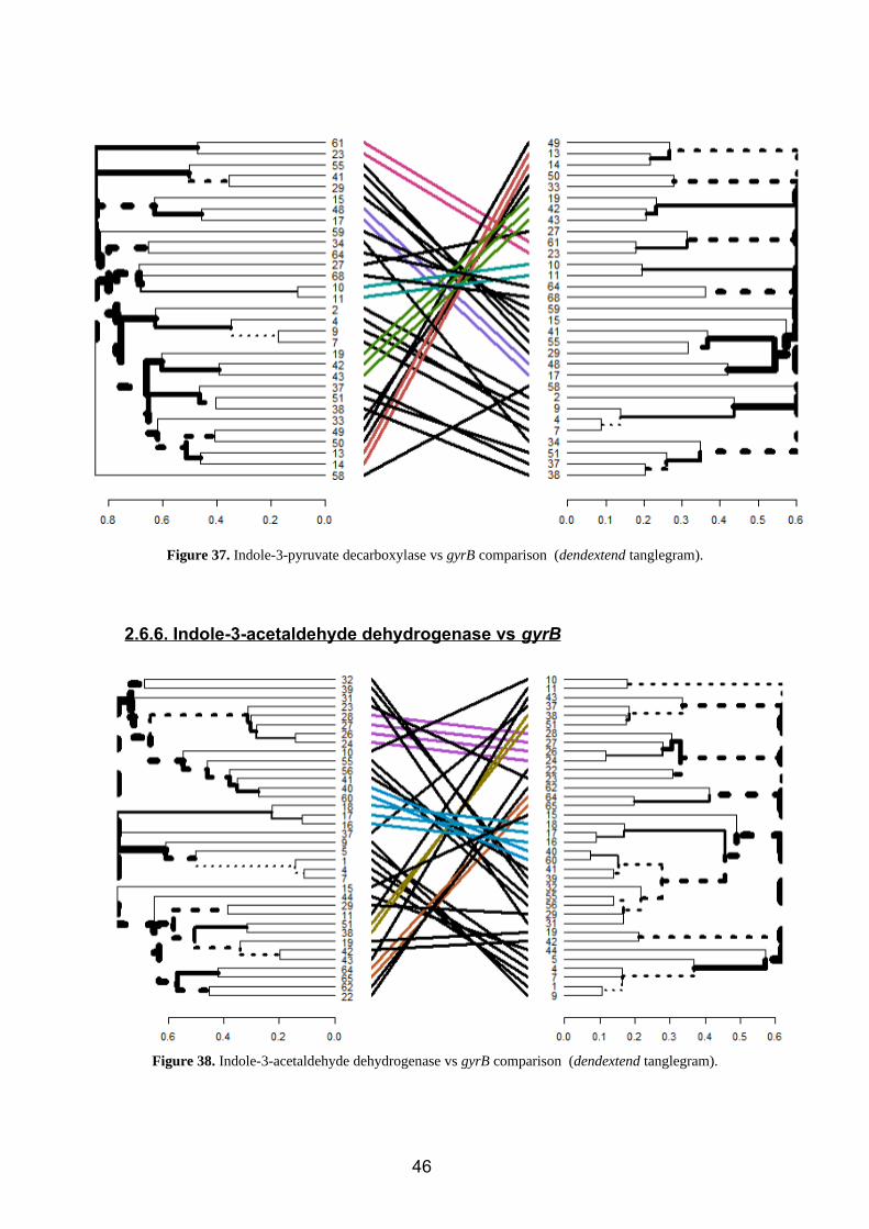
Figure 37. Indole-3-pyruvate decarboxylase vs gyrB comparison (dendextend tanglegram).
2.6.6. Indole-3-acetaldehyde dehydrogenase vs gyrB
Figure 38. Indole-3-acetaldehyde dehydrogenase vs gyrB comparison (dendextend tanglegram).
46
Several differences of position are observed (figure 38), mainly referring to changes due
to the topology and disposition of the tree (color).
For the changes of position inside clusters, we have:
Rhizobium (32,31,55, 56)
Sinorhizobium (39, 41)
Pseudomonas(23,22)
Acinetobacter (10)
Serratia (37)
Bacillus (9,4,1,5,7)
Klebsiella (19,42,43)
Ralstonia (62)
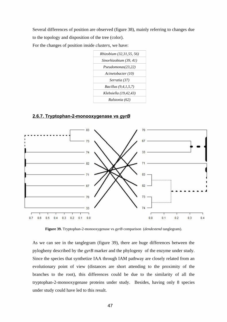
2.6. 7 . Tryptophan-2-monooxygenase vs gyrB
Figure 39. Tryptophan-2-monooxygenase vs gyrB comparison (dendextend tanglegram).
As we can see in the tanglegram (figure 39), there are huge differences between the
pylogheny described by the gyrB marker and the phylogeny of the enzyme under study.
Since the species that synthetize IAA through IAM pathway are closely related from an
evolutionary point of view (distances are short attending to the proximity of the
branches to the root), this differences could be due to the similarity of all the
tryptophan-2-monooxygenase proteins under study. Besides, having only 8 species
under study could have led to this result.
47
2.6.8. Indole-3-acetamide hydrolase vs gyrB
Figure 40. Indole-3-acetamide hydrolase vs gyrB comparison (dendextend tanglegram).
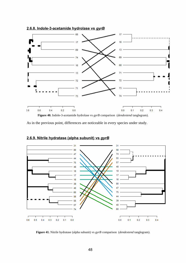
As in the previous point, differences are noticeable in every species under study.
2.6. 9 . Nitrile hydratase (alpha subunit) vs gyrB
Figure 41. Nitrile hydratase (alpha subunit) vs gyrB comparison (dendextend tanglegram).
48
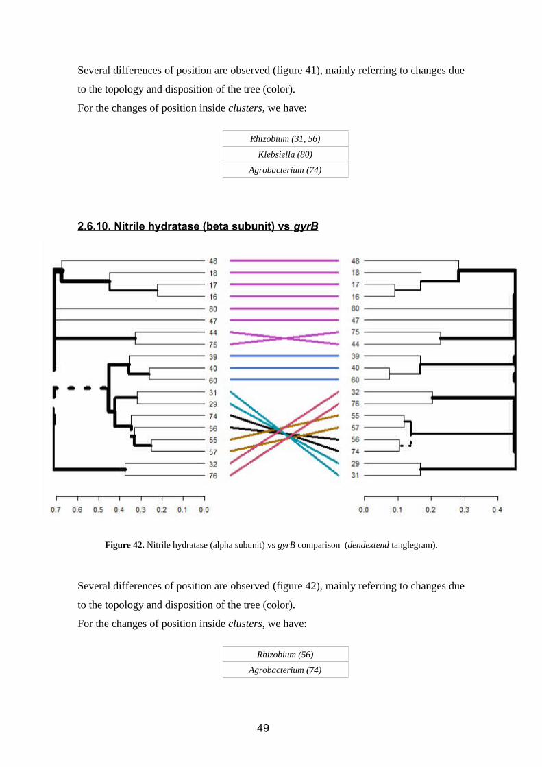
Several differences of position are observed (figure 41), mainly referring to changes due
to the topology and disposition of the tree (color).
For the changes of position inside clusters, we have:
Rhizobium (31, 56)
Klebsiella (80)
Agrobacterium (74)
2.6.10. Nitrile hydratase (beta subunit) vs gyrB
Figure 42. Nitrile hydratase (alpha subunit) vs gyrB comparison (dendextend tanglegram).
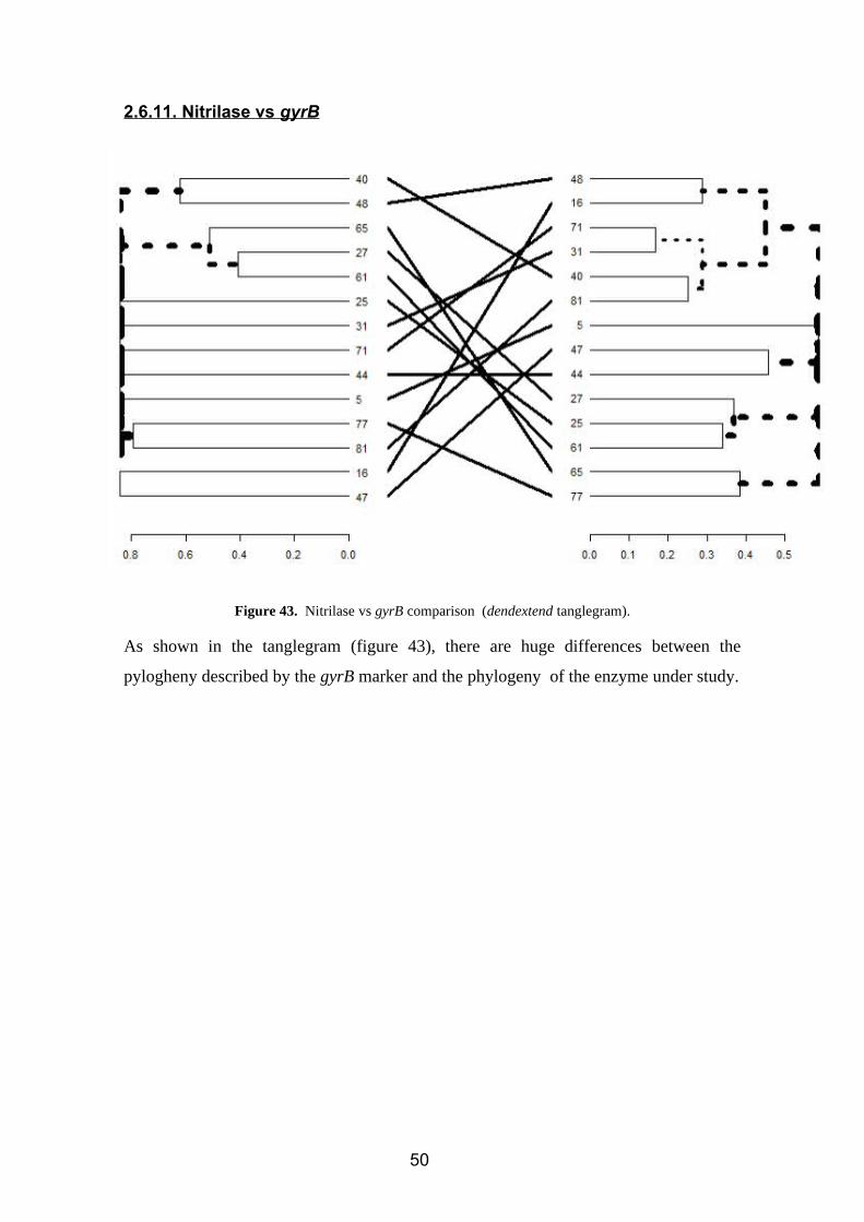
Several differences of position are observed (figure 42), mainly referring to changes due
to the topology and disposition of the tree (color).
For the changes of position inside clusters, we have:
Rhizobium (56)
Agrobacterium (74)
49
2.6.11. Nitrilase vs gyrB
Figure 43. Nitrilase vs gyrB comparison (dendextend tanglegram).
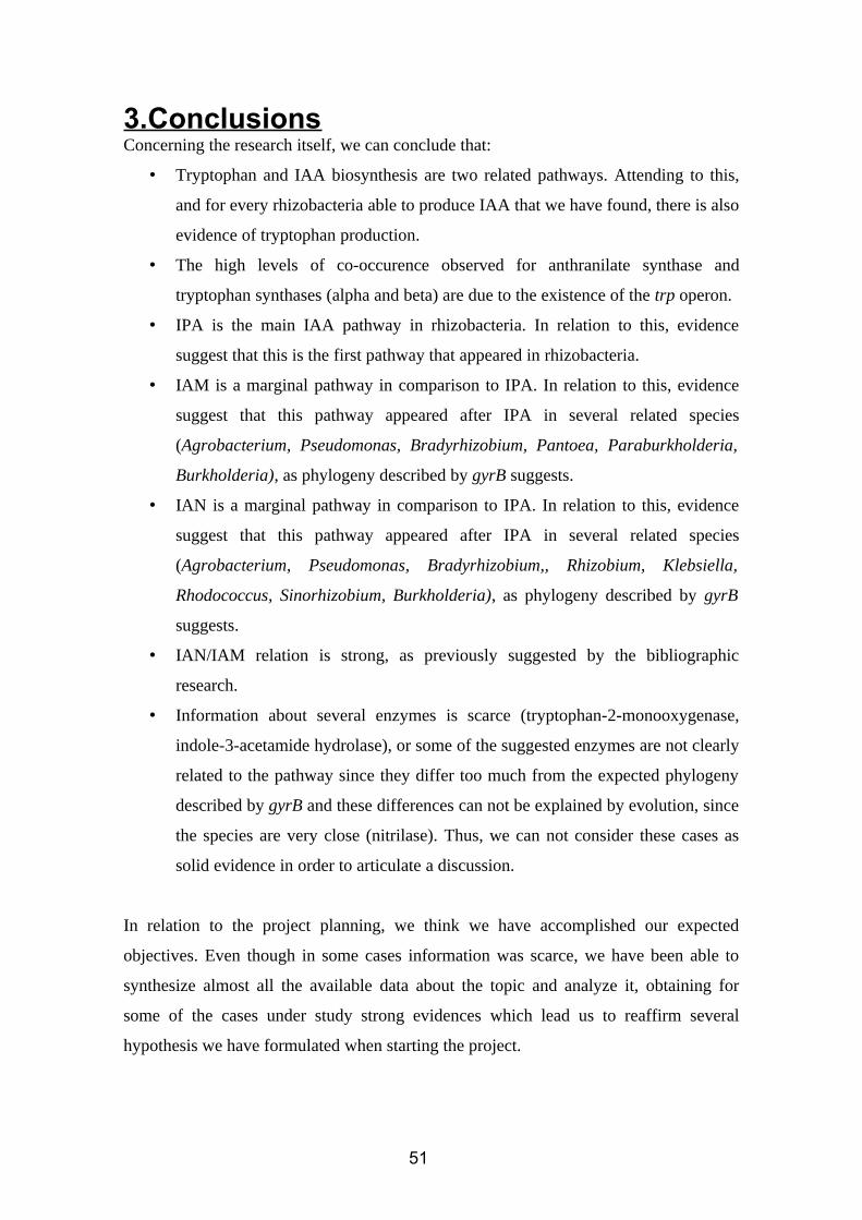
As shown in the tanglegram (figure 43), there are huge differences between the
pylogheny described by the gyrB marker and the phylogeny of the enzyme under study.
50
3.ConclusionsConcerning the research itself, we can conclude that:
• Tryptophan and IAA biosynthesis are two related pathways. Attending to this,
and for every rhizobacteria able to produce IAA that we have found, there is also
evidence of tryptophan production.
• The high levels of co-occurence observed for anthranilate synthase and
tryptophan synthases (alpha and beta) are due to the existence of the trp operon.
• IPA is the main IAA pathway in rhizobacteria. In relation to this, evidence
suggest that this is the first pathway that appeared in rhizobacteria.
• IAM is a marginal pathway in comparison to IPA. In relation to this, evidence
suggest that this pathway appeared after IPA in several related species
(Agrobacterium, Pseudomonas, Bradyrhizobium, Pantoea, Paraburkholderia,
Burkholderia), as phylogeny described by gyrB suggests.
• IAN is a marginal pathway in comparison to IPA. In relation to this, evidence
suggest that this pathway appeared after IPA in several related species
(Agrobacterium, Pseudomonas, Bradyrhizobium,, Rhizobium, Klebsiella,
Rhodococcus, Sinorhizobium, Burkholderia), as phylogeny described by gyrB
suggests.
• IAN/IAM relation is strong, as previously suggested by the bibliographic
research.
• Information about several enzymes is scarce (tryptophan-2-monooxygenase,
indole-3-acetamide hydrolase), or some of the suggested enzymes are not clearly
related to the pathway since they differ too much from the expected phylogeny
described by gyrB and these differences can not be explained by evolution, since
the species are very close (nitrilase). Thus, we can not consider these cases as
solid evidence in order to articulate a discussion.
In relation to the project planning, we think we have accomplished our expected
objectives. Even though in some cases information was scarce, we have been able to
synthesize almost all the available data about the topic and analyze it, obtaining for
some of the cases under study strong evidences which lead us to reaffirm several
hypothesis we have formulated when starting the project.
51

Besides, we successfully reached every objective and deadline we set in PEC 0 and PEC
1 on scheduled time. Moreover, we have been able to introduce new perspectives, such
as the operon analysis, which was incorporated as a part of the project after PEC 2
attending to the results of the co-ocurrence matrix draft.
Finally, we consider this project as a first step into further analysis in the near future, as
we expect that the lack of information about some enzymes decreases in a few years.
52
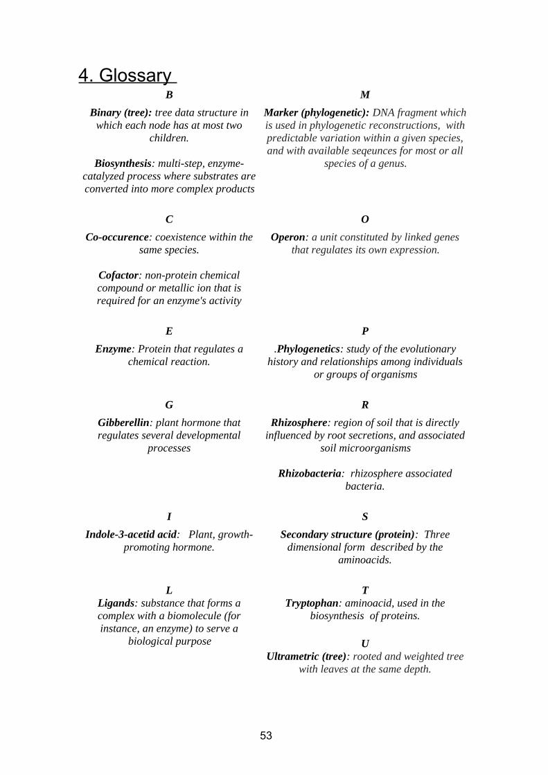
4. Glossary B M
Binary (tree): tree data structure inwhich each node has at most two
children.
Biosynthesis: multi-step, enzyme-catalyzed process where substrates areconverted into more complex products
Marker (phylogenetic): DNA fragment whichis used in phylogenetic reconstructions, withpredictable variation within a given species,and with available seqeunces for most or all
species of a genus.
C O
Co-occurence: coexistence within thesame species.
Cofactor: non-protein chemicalcompound or metallic ion that isrequired for an enzyme's activity
Operon: a unit constituted by linked genesthat regulates its own expression.
E P
Enzyme: Protein that regulates achemical reaction.
.Phylogenetics: study of the evolutionaryhistory and relationships among individuals
or groups of organisms
G R
Gibberellin: plant hormone thatregulates several developmental
processes
Rhizosphere: region of soil that is directlyinfluenced by root secretions, and associated
soil microorganisms
Rhizobacteria: rhizosphere associatedbacteria.
I S
Indole-3-acetid acid: Plant, growth-promoting hormone.
Secondary structure (protein): Threedimensional form described by the
aminoacids.
LLigands: substance that forms acomplex with a biomolecule (forinstance, an enzyme) to serve a
biological purpose
TTryptophan: aminoacid, used in the
biosynthesis of proteins.
UUltrametric (tree): rooted and weighted tree
with leaves at the same depth.
53
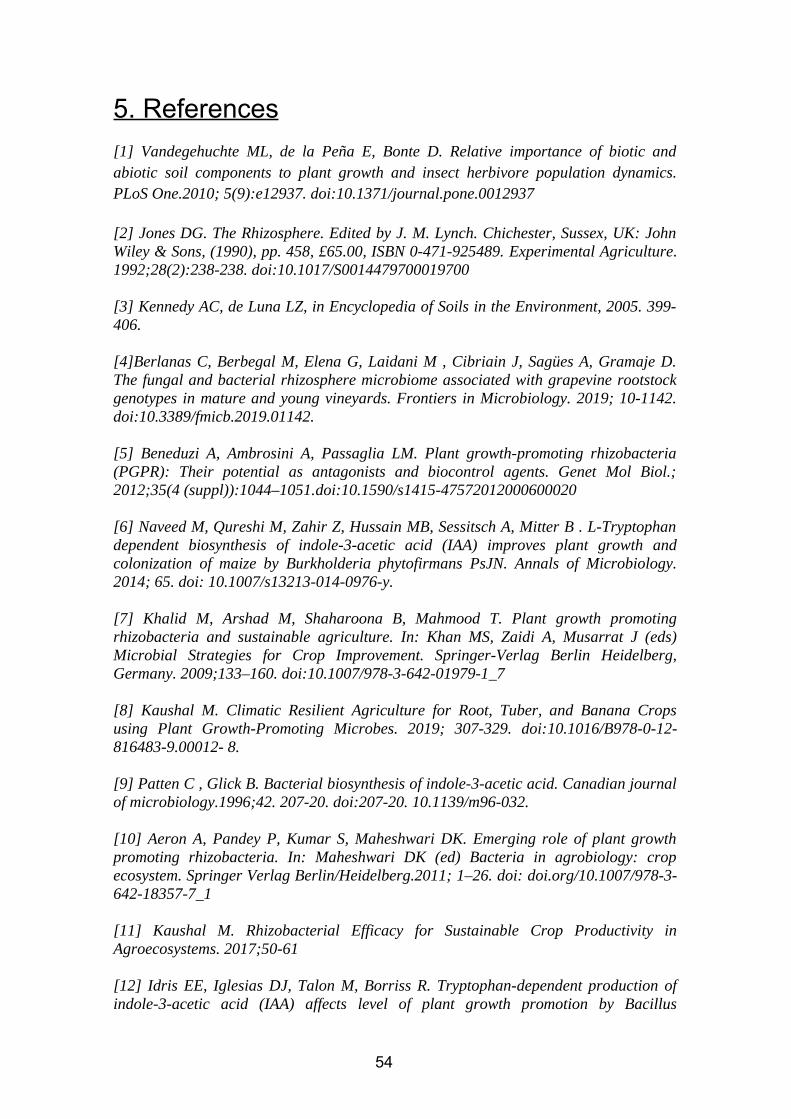
5. References
[1] Vandegehuchte ML, de la Peña E, Bonte D. Relative importance of biotic andabiotic soil components to plant growth and insect herbivore population dynamics.PLoS One.2010; 5(9):e12937. doi:10.1371/journal.pone.0012937
[2] Jones DG. The Rhizosphere. Edited by J. M. Lynch. Chichester, Sussex, UK: JohnWiley & Sons, (1990), pp. 458, £65.00, ISBN 0-471-925489. Experimental Agriculture.1992;28(2):238-238. doi:10.1017/S0014479700019700
[3] Kennedy AC, de Luna LZ, in Encyclopedia of Soils in the Environment, 2005. 399-406.
[4]Berlanas C, Berbegal M, Elena G, Laidani M , Cibriain J, Sagües A, Gramaje D.The fungal and bacterial rhizosphere microbiome associated with grapevine rootstockgenotypes in mature and young vineyards. Frontiers in Microbiology. 2019; 10-1142.doi:10.3389/fmicb.2019.01142.
[5] Beneduzi A, Ambrosini A, Passaglia LM. Plant growth-promoting rhizobacteria(PGPR): Their potential as antagonists and biocontrol agents. Genet Mol Biol.;2012;35(4 (suppl)):1044–1051.doi:10.1590/s1415-47572012000600020
[6] Naveed M, Qureshi M, Zahir Z, Hussain MB, Sessitsch A, Mitter B . L-Tryptophandependent biosynthesis of indole-3-acetic acid (IAA) improves plant growth andcolonization of maize by Burkholderia phytofirmans PsJN. Annals of Microbiology.2014; 65. doi: 10.1007/s13213-014-0976-y.
[7] Khalid M, Arshad M, Shaharoona B, Mahmood T. Plant growth promotingrhizobacteria and sustainable agriculture. In: Khan MS, Zaidi A, Musarrat J (eds)Microbial Strategies for Crop Improvement. Springer-Verlag Berlin Heidelberg,Germany. 2009;133–160. doi:10.1007/978-3-642-01979-1_7
[8] Kaushal M. Climatic Resilient Agriculture for Root, Tuber, and Banana Cropsusing Plant Growth-Promoting Microbes. 2019; 307-329. doi:10.1016/B978-0-12-816483-9.00012- 8.
[9] Patten C , Glick B. Bacterial biosynthesis of indole-3-acetic acid. Canadian journalof microbiology.1996;42. 207-20. doi:207-20. 10.1139/m96-032.
[10] Aeron A, Pandey P, Kumar S, Maheshwari DK. Emerging role of plant growthpromoting rhizobacteria. In: Maheshwari DK (ed) Bacteria in agrobiology: cropecosystem. Springer Verlag Berlin/Heidelberg.2011; 1–26. doi: doi.org/10.1007/978-3-642-18357-7_1
[11] Kaushal M. Rhizobacterial Efficacy for Sustainable Crop Productivity inAgroecosystems. 2017;50-61
[12] Idris EE, Iglesias DJ, Talon M, Borriss R. Tryptophan-dependent production ofindole-3-acetic acid (IAA) affects level of plant growth promotion by Bacillus
54
amyloliquefaciens FZB42. Mol Plant- Microbe Interact. 2007;20: 619-626.doi:10.1094/MPMI-20-6-0619.
[13] Wang B, Chu J, Yu T, et al. Tryptophan-independent auxin biosynthesiscontributes to early embryogenesis in Arabidopsis. Proc Natl Acad SciUSA.2015;112(15):4821–4826. doi:10.1073/pnas.1503998112
[14] Chagas A, Oliveira A, Costa J. Production of indole-3-acetic acid by bacillusisolated from different soils. Bulgarian Journal of Agricultural Science. 2015;21. 282-287.
[15] Spaepen S, Vanderleyden J. Auxin and plant-microbe interactions. Cold SpringHarb Perspect Biol. 2011;3(4):a001438. doi:10.1101/cshperspect.a001438
[16] Patten C & Glick B. Bacterial biosynthesis of indole-3-acetic acid. Canadianjournal of microbiology. 1996;42. 207-20. doi:10.1139/m96-032.
[17] Ozdal M, Özlem O, Sezen A, Algur O. Biosynthesis Of Indole-3-Acetic Acid ByBacillus cereus Immobilized Cells. Cumhuriyet Science Journal. 2016;37. 212.doi:10.17776/csj.34085.
[18] Wagi S, Ahmed A. Bacillus spp.: potent microfactories of bacterial IAA. PeerJ.2019;7:e7258. doi:10.7717/peerj.7258
[19] Ghazal M, Moussa L, Fayed S, Mostfa A. The use of bacillus mycoides andbacillus subtlis as biocontrol agents for the fungi causing root rot disease in commonbean . Egypt J. Biotechnol. 2013;44. 37-43. doi:10.13140/RG.2.1.2674.5841.
[20] Shao J, Li S, Zhang N. Analysis and cloning of the synthetic pathway of thephytohormone indole-3-acetic acid in the plant-beneficial Bacillus amyloliquefaciensSQR9. Microb Cell Fact. 2015;14:130. doi:10.1186/s12934-015-0323-4.
[21] Yahaghi Z, Shirvani M, Nourbakhsh F, Coba T, Pueyo J, Talebi M. Isolation andCharacterization of Pb-Solubilizing Bacteria and Their Effects on Pb Uptake byBrassica juncea: Implications for Microbe-Assisted Phytoremediation. Journal ofmicrobiology and biotechnology. 2018;28. doi:10.4014/jmb.1712.12038.
[22] Lee JC, Whang KS. Optimization of Indole-3-acetic Acid (IAA) Production byBacillus megaterium BM5. Korean Journal of Soil Science and Fertilizer. 2016;49:461-468. doi:10.7745/KJSSF.2016.49.5.461.
[23] Lo KJ, Lin SS, Lu CW, Kuo CH, Liu CT. Whole-genome sequencing andcomparative analysis of two plant-associated strains of Rhodopseudomonas palustris(PS3 and YSC3).Sci Rep. 2018;8(1):12769. doi:10.1038/s41598- 018-31128-8
[24] Blin P. Dynamique des interactions entre Dickeya dianthicola, Dickeya solani etleur hôte Solanum tuberosum. 2017.
55
[25] Zhou J, Cheng Y, Lv M. The complete genome sequence of Dickeya zeae EC1reveals substantial divergence from other Dickeya strains and species. BMCGenomics.2015;16(1):571. doi:10.1186/s12864-015-1545-x
[26] Coutinho TA, Venter SN. Pantoea ananatis: an unconventional plant pathogen.Mol Plant Pathol. 2009;10(3):325–335. doi:10.1111/j.1364-3703.2009.00542.x
[27] Brady CL, Cleenwerck I, Westhuizen L, Venter SN, Coutinho TA, Vos PD.Pantoea rodasii sp. nov., Pantoea rwandensis sp. nov. and Pantoea wallisii sp. nov.,isolated from eucalyptus. Int J Syst Evol Micr.2012;62: 1457–1464. doi:10.1099/ijs.0.032615-0.
[28] Estenson K, Hurst G, Standaert R, Bible A, Garcia D, Chourey K, & Doktycz,Mitchel & Morrell-Falvey, Jennifer. Characterization of Indole-3-acetic AcidBiosynthesis and the Effects of This Phytohormone on the Proteome of the Plant-Associated Microbe Pantoea sp. YR343. Journal of Proteome Research.2018;17.doi:10.1021/acs.jproteome.7b00708.
[29] Kulkarni G, Nayak A, Sajjan S, Oblesha A, Karegoudar, TB. Indole‐3‐acetic acidbiosynthetic pathway and aromatic amino acid aminotransferase activities in Pantoeadispersa strain GPK. Letters in applied microbiology.2013;56. doi:10.1111/lam.12053.
[30] Apine OA, Jadhav JP. Optimization of medium for indole-3-acetic acid productionusing Pantoea agglomerans strain PVM. J Appl Microbiol.2011;110:1235–1244.doi:10.1111/j.1365-2672.2011.04976.x
[31] Ouyang LM, Pei H, Xu Z. Low nitrogen stress stimulating the indole-3-acetic acidbiosynthesis of Serratia sp. ZM is vital for the survival of the bacterium and its plantgrowth-promoting characteristic. Archives of microbiology.2016;199.doi:10.1007/s00203-016-1312-7
[32] Zelaya-Molina L, Hernandez-Soto LM, Guerra-Camacho J, Monterrubio R ,Patiño-Siciliano A, Villa‐Tanaca L, Hernández-Rodríguez C. Ammonia-Oligotrophicand Diazotrophic Heavy Metal-Resistant Serratia liquefaciens Strains from PioneerPlants and Mine Tailings. Microbial Ecology.2016;72. doi:10.1007/s00248-016- 0771-3.
[33] Hasuty A, Choliq A. Production of Indole Acetic Acid (IAA) by serratiamarcescens subsp. marcescens and rhodococcus aff. qingshengii. International Journalof Agricultural Technology.2018;14. 299-312.
[34] Khan AR, Park GS, Asaf S, Hong SJ, Jung BK, Shin JH. Complete genomeanalysis of Serratia marcescens RSC-14: A plant growth-promoting bacterium thatalleviates cadmium stress in host plants. PLoS One.2017;12(2):e0171534. doi:10.1371/journal.pone.0171534
[35] Jung BK, Khan AR, Hong SJ, Park GS, Park YJ, Park CE, Jeon HJ, Lee SE, ShinJH. Genomic and phenotypic analyses of Serratia fonticola strain GS2: arhizobacterium isolated from sesame rhizosphere that promotes plant growth and
56
produces N-acyl homoserine lactone. Journal of Biotechnology.2017;241:158-162.doi: 10.1016/j.jbiotec.2016.12.002.
[36] Lin HR, Shu HY, Lin GH. Biological roles of indole-3-acetic acid in Acinetobacterbaumannii. Microbiological Research.2018;216. 10.1016/j.micres.2018.08.004.
[37] Lin G, Chen H, Huang J, Liu T, Lin T, Wang S, Tseng C, Shu H. Identification andcharacterization of an indigoproducing oxygenase involved in indole 3-acetic acidutilization by Acinetobacter baumannii. Antonie Van Leeuwenhoek.2012;101:881–890.doi:doi: 10.1007/s10482-012-9704-4
[38] Sachdev D, Nema P, Dhakephalkar P, Zinjarde S, Chopade B. Assessment of 16SrRNA gene-based phylogenetic diversity and promising plant growth-promoting traitsof Acinetobacter community from the rhizosphere of wheat. Microbiologicalresearch.2010;165. 627-38. doi:10.1016/j.micres.2009.12.002.
[39] Huddedar S, Shete A, Tilekar J, Gore S, Dhavale D, Chopade B. Isolation,Characterization, and Plasmid pUPI126-Mediated Indole-3-Acetic Acid Production inAcinetobacter Strains from Rhizosphere of Wheat. Applied biochemistry andbiotechnology.2002;102-103. 21-39. doi:10.1385/ABAB:102-103:1-6:021.
[40] Datta C, Basu P. Indole acetic acid production by a Rhizobium species from rootnodules of a leguminous shrub, Cajanus cajan. Microbiological research.2000;155.123-7. doi:10.1016/S0944-5013(00)80047-6.
[41] Bhattacharyya R, Pati B. Growth behaviour and indole acetic acid (IAA)production by a Rhizobium isolated from root nodules of Alysicarpus vaginalis DC.Acta microbiologica et immunologica Hungarica.2000;47. 41-51.
[42] Kobayashi M, Suzuki T, Fujita T, Masuda M, Shimizu S. Occurrence of enzymesinvolved in biosynthesis of indole-3-acetic acid from indole-3-acetonitrile inplantassociated bacteria, Agrobacterium and Rhizobium. Proc Natl Acad SciUSA.1195; 92:714–718. doi:10.1073/pnas.92.3.714
[43] Spaepen S, Das F, Luyten E, Michiels J, Vanderleyden J. Indole-3-acetic acid-regulated genes in Rhizobium etli CNPAF512. FEMS microbiology letters.2009;291.195-200. doi:10.1111/j.1574-6968.2008.01453.x.
[44] Ghosh P, Saha P, Mayilraj S, Maiti T. Role of IAA metabolizing enzymes onproduction of IAA in root, nodule of Cajanus cajan and its PGP Rhizobium sp.Biocatalysis and Agricultural Biotechnology.2013;2. 234-239. doi:10.1016/j.bcab.2013.04.002
[45] Bhattacharjee R, Jourand P, Chaintreuil C, Dreyfus B, Singh A, Mukhopadhyay S.Indole acetic acid and ACC deaminase-producing Rhizobium leguminosarum bv. trifoliiSN10 promote rice growth, and in the process undergo colonization and chemotaxis.Biology and Fertility of Soils.2011;48. doi:10.1007/s00374-011-0614-9.
57
[46] Dubey RC, Maheshwari D, Kumar H, Choure K. Assessment of diversity andplant growth promoting attributes of rhizobia isolated from Cajanus cajan L. AfricanJournal of Biotechnology.2011;9. 8619-8629. doi:10.5897/AJB10.1127
[47] Cruz-González X, Laza-Pérez N, Mateos PF, Rivas R. Analysis and effect of theuse of biofertilizers on Trifolium rubens L., a preferential attention species in Castileand Leon, Spain, with the aim of increasing the plants conservation status. AIMSMicrobiol.2017;3(4) 733-746.doi:10.3934/microbiol.2017.4.733
[48] Del Papa F, Schlueter A, Althabegoiti M, Rogel M, Martínez-Romero E, NilssonJF, Ormeño-Orrillo E, Pistorio M, Pühler A, Niehaus K, Tejerizo G, Lagares A.Rhizobium favelukesii sp. nov., isolated from the root nodules of alfalfa (Medicagosativa L). International Journal of Systematic and Evolutionary Microbiology.2016;66.doi:10.1099/ijsem.0.001373.
[49] Eddie LI, Alves de Paiva Rolla dos Santos A, Luiz Martinez de Oliveira A,Hungria M, Pains Rodrigues E. Indole-3-acetic acid production via the indole-3-pyruvate pathway by plant growth promoter Rhizobium tropici CIAT 899 is stronglyinhibited by ammonium, Research in Microbiology2017;168(3), 283-292.doi:10.1016/j.resmic.2016.10.010.
[50] El-Khawas H, Adachi K. Identification and quantification of auxins in culturemedia of Azospirillum and Klebsiella and their effect on rice roots. Biol FertilitySoils.1999;28:377–38. doi:10.1007/s003740050507
[51] Akbari GA, Arab SM, Alikhani HA, Allahdadi I, Arzanesh MH. Isolation andselection of indigenous Azospirillum spp. and the IAA of superior strains effects onwheat roots. World J Agric Sci.2007;3:523–529.
[52] Baca BE, Soto-Urzua L, Xochihua-Corona YG, Cuervo-Garcia A.Characterization of two aromatic amino acidaminotransferases and production ofindoleacetic acid in Azospirillum strains. Soil Biol Biochem.1194;26:57–63.doi:10.1016/0038-0717(94)90195-3
[53] Molina R, Rivera D, Mora V, Lopez G, Rosas S, Spaepen S,Vanderleyden J,Cassan F. Regulation of IAA Biosynthesis in Azospirillum brasilense UnderEnvironmental Stress Conditions. Current Microbiology.2018;75. doi:10.1007/s00284-018- 1537-6.
[54] Bose A, Kher M, Nataraj M, Keharia H. Phytostimulatory effect of indole-3-acetic acid by Enterobacter cloacae SN19 isolated from Teramnus labialis (L. f.)Spreng rhizosphere. Biocatalysis and Agricultural Biotechnology.2016;6.doi:10.1016/j.bcab.2016.03.005.
[55] Brandl MT, Lindow SE. Cloning and characterization of a locus encoding anindolepyruvate decarboxylase involved in indole-3-acetic acid synthesis in Erwiniaherbicola. Appl Environ Microbiol.1996;62:4121–4128.
58
[56] Clark E, Manulis S, Ophir Y, Barash I, Gafni Y. Cloning and characterization ofiaaM and iaaH from Erwinia herbicola pathovar gypsophilae.Phytopathology.1993;83:234– 240.
[57] Yang S, Zhang Q, Guo JH, Charkowski A, Glick B, Ibekwe A, Cooksey D, YangCH. Global Effect of Indole-3-Acetic Acid Biosynthesis on Multiple Virulence Factorsof Erwinia chrysanthemi 3937. Applied and environmental microbiology.2007;73.1079-88. doi:10.1128/AEM.01770-06.
[58] Lü Z, Song W. Research of indole-3-acetic acid biosynthetic pathway of Klebsiellaoxytoca SG-11 by HPLC and GC-MS. Se pu = Chinese journal of chromatography /Zhongguo hua xue hui. 2000;18:328-31.
[59] Celloto V, Oliveira A, Gonçalves J, Watanabe C, Matioli G, Gonçalves R.Biosynthesis of Indole-3-Acetic Acid by New Klebsiella oxytoca Free and ImmobilizedCells on Inorganic Matrices. TheScientificWorldJournal. 2012. doi:10.1100/2012/495970. doi:10.1100/2012/495970.
[60] Sachdev D, Chaudhari H, Kasture V, Dhavale D, Chopade B. Isolation andcharacterization of indole acetic acid producing Klebsiella pneumoniae strains fromthe rhizosphere of wheat (Triticum aestivum) and their effect on plant growth. Indian J.Exp. Biol.2009;47: 993-1000.
[61] Mitra S Pramanik K, & Ghosh P, Soren T, Sarkar A, Dey R, Pandey S, Maiti T.Characterization of Cd-resistant Klebsiella michiganensis MCC3089 and its potentialfor rice seedling growth promotion under Cd stress. MicrobiologicalResearch.2018;210. 12-25. doi:10.1016/j.micres.2018.03.003.
[62] Shrivastava S, D'Souza S, Desai P. Production of indole-3-acetic acid byimmobilized actinomycetes (Kitasatospora Sp.) for soil application. Curr. Sci.2008;94.
[63] Bastian F, Cohen A, Piccoli P, Luna M, Bottini R, Baraldi R. Production ofindole-3-acetic acid and gibberellins A_1 and A_3 by Acetobacter diazotrophicus andHerbaspirillum seropedicae in chemically-defined culture media. Plant GrowthRegulation.1998;24:7–11. doi: 10.1023/a:1005964031159
[64] Vinardell J. The Sinorhizobium fredii HH103 Genome: A Comparative AnalysisWith S. fredii Strains Differing in Their Symbiotic Behavior With Soybean. Molecularplant-microbe interaction.2015;28(7):811-24. doi:10.1094/MPMI-12-14-0397-FI
[65] Imperlini E, Bianco C, Lonardo E, Camerini S, Cermola M, Moschetti G, Defez R.Effects of indole-3-acetic acid on Sinorhizobium meliloti survival and on symbioticnitrogen fixation and stem dry weight production. Applied microbiology andbiotechnology.2009;83. 727-38. doi:10.1007/s00253-009-1974-z.
[66] Kallala N, M’sehli W, Jelali K, Kais Z, Mhadhbi H. Inoculation with EfficientNitrogen Fixing and Indoleacetic Acid Producing Bacterial Microsymbiont EnhanceTolerance of the Model Legume Medicago truncatula to Iron Deficiency. BioMedResearch International. 2018. 1-14. doi:10.1155/2018/9134716.
59
[67] Ahmad F, Ahmad I, Khan MS. Indole acetic acid production by the indigenousisolates of Azotobacter and fluorescent Pseudomonas in the presence and absence oftryptophan. Turk J Biol.2005;29:29–34.
[68] Balaji N, Lavanya SS, Muthamizhselvi S, Tamilarasan K. Optimization offermentation conditions for indole acetic acid production by Pseudomonas species. IntJ Adv Biotechnol Res.2012;3:797–803.
[69] Suzuki S, He Y, Oyaizu H. Indole-3-acetic acid production in Pseudomonasfluorescens HP72 and its association with suppression of creeping bentgrass brownpatch. Curr Microbiol.2003;47:138–143. doi:10.1007/s00284-002-3968-2
[70] Ansari F, Ahmad I. (2018). Biofilm Development, Plant Growth Promoting Traitsand Rhizosphere Colonization by Pseudomonas entomophila FAP1: A PromisingPGPR. Advances in Microbiology. 08. 235-251. doi:10.4236/aim.2018.83016.
[71] Marathe R, Phatake YB , Shaikh A, Shinde BP, Gajbhiye M. Effect of IAAproduced by Pseudomonas aeruginosa 6a (bc4) on seed germination and plant growthof Glycin max. Journal of Experimental Biology and Agricultural Sciences.2017;5. 351-358. doi:10.18006/2017.5(3).351.358. doi:10.18006/2017.5(3).351.358
[72] Patten CL, Glick BR. Role of Pseudomonas putida indoleacetic acid indevelopment of the host plant root system. Appl Environ Microbiol.2002;68(8):3795–3801. doi:10.1128/aem.68.8.3795-3801.2002
[73] Tsavkelova EA, Cherdyntseva TA, Botina SG, Netrusov AI.Bacteria associatedwith orchid roots and microbial production of auxin, MicrobiologicalResearch.2007;162(1),69-76. doi:10.1016/ j.micres.2006.07.014.
[74] Surico G, Iacobellis NS, Sisto A. Studies on the role of indole-3-acetic acid andcytokinins in the formation of knots on olive and oleander plants by Pseudomonassyringae pv. savastanoi. Physiol Plant Pathol.1985;26:309–320. Doi:10.1016/0048-4059(85)90006-2.
[75] Flores O, Prince C, Nuñez M, Vallejos A, Mardones C, Yáñez C, Besoain X,Bastías R. Genetic and Phenotypic Characterization of Indole-Producing Isolates ofPseudomonas syringae pv. actinidiae Obtained From Chilean Kiwifruit Orchards.Frontiers in Microbiology.2018;9. doi:10.3389/fmicb.2018.01907.
[76] Andreolli M, Zapparoli G, Angelini E, Lucchetta G, Lampis S, Vallini G.Pseudomonas protegens MP12: A plant growth-promoting endophytic bacterium withbroad-spectrum antifungal activity against grapevine phytopathogens. MicrobiologicalResearch.2018;219. doi:10.1016/j.micres.2018.11.003.
[77] Sekine M, Ichikawa T, Kuga N, Kobayashi M, Sakurai A, Syono K. Detection ofthe IAA biosynthetic pathway from tryptophan via indole-3-acetamide inBradyrhizobium spp. Plant Cell Physiol.1988;29:867–874.doi:10.1093/oxfordjournals.pcp.a077574.
60
[78] Egebo L, Nielsen S, Jochimsen B. Oxygen-dependent catabolism of indole-3-aceticacid in Bradyrhizobium japonicum. J Bacteriol.1991;173:4897–4901.doi:10.1128/jb.173.15.4897-4901.1991
[79] Jensen JB, Egsgaard H, Van Onckelen H, Jochimsen BU. Catabolism of indole-3-acetic acid and 4- and 5-chloroindole-3-acetic acid in Bradyrhizobium japonicum. JBacteriol. 1995;177(20):5762–5766. doi:10.1128/jb.177.20.5762-5766.1995
[80] Siqueira AF, Ormeño-Orrillo E, Souza RC, et al. Comparative genomics ofBradyrhizobium japonicum CPAC 15 and Bradyrhizobium diazoefficiens CPAC 7: elitemodel strains for understanding symbiotic performance with soybean.BMC Genomics.2014;15(1):420. doi:10.1186/1471-2164-15-420
[81] Yagi K, Matsumoto T, Chujo T, Nojiri H, Omori T, Minamisawa K, NishiyamaM, Yamane H. Isolation and Characterization of Low-indole-3-acetic Acid-producingMutants from Bradyrhizobium elkanii. Bioscience, biotechnology, andbiochemistry.2000;64. 1359-64. doi:10.1271/bbb.64.1359.
[82] Kailasan N, Vamanrao VB. Isolation and Characterization of Ralstonia Pickettii -A Novel Phosphate Solubilizing Bacterium from Pomegranate Rhizosphere fromWestern India, International Journal of Life Sciences Biotechnology and PharmaResearch.2015;4(1),1-9.
[83] Paul A, Datta B. Cadmium-resistant Ralstonia mannitolilytica Relieved CadmiumToxicity in Mustard Plant Through Root Colonization and Growth Promoting Activity.Research Journal of Microbiology.2016;11. 157-168. doi:10.3923/jm.2016.157.168.
[84] Kurosawa N, Hirata T, Suzuki H. Characterization of putative tryptophanmonooxygenase from Ralstonia solanasearum. J Biochem.2009;146:23–32. doi:10.1093/jb/mvp040.
[85] Duca D, Lorv J, Patten C, Rose D, Glick B. Indole-3-acetic acid in plant-microbeinteractions. Antonie van Leeuwenhoek.2014;106. doi:10.1007/s10482-013-0095-y.
[86] Zuñiga A, Poupin MJ, Donoso R, Ledger T, Guiliani N, Gutierrez RA, Gonzalez B.Quorum sensing and indole-3-acetic acid degradation play a role in colonization andplant growth promotion of Arabidopsis thaliana by Burkholderia phytofirmans PsJN.Mol Plant-Microbe Interact.2013;26:546–553. doi:10.1094/MPMI-10-12-0241-R.
[87] Johan HJ, Gerards SL. Discovery of a bacterial gene cluster for catabolism of theplant hormone indole 3-acetic acid, FEMS Microbiology Ecology.2008;65(2):238–250.doi: 10.1111/j.1574-6941.2008.00436.x.
[88] Castanheira N, Dourado AC, Kruz S, Alves PIL, Delgado-Rodríguez AI, Pais I,Semedo JN, Campos P, Sánchez C, Borges N, Carvalho G, Crespo MT, Fareleira P.Plant growth promoting Burkholderia species isolated from annual ryegrass inPortuguese soils. Journal of Applied Microbiology.2015;120. doi:10.1111/jam.13025.
[89] Donoso R, Leiva-Novoa P, Zúñiga A, Timmermann T, Recabarren G, GonzalezB. Biochemical and Genetic Bases of Indole-3-Acetic Acid (Auxin Phytohormone)
61
Degradation by the Plant-Growth-Promoting Rhizobacterium Paraburkholderiaphytofirmans PsJN. Applied and Environmental Microbiology.2016;83.doi:10.1128/AEM.01991-16.
[90] Mannaa M, Park I, Seo YS. Genomic Features and Insights into the Taxonomy,Virulence, and Benevolence of Plant-Associated Burkholderia Species. Int J Mol Sci.2018;20(1):121. doi:10.3390/ijms20010121.
[91] Jiang F, Chen L, Belimov AA, et al. (2012). Multiple impacts of the plant growth-promoting rhizobacterium Variovorax paradoxus 5C-2 on nutrient and ABA relationsof Pisum sativum. J Exp Bot.63(18):6421–6430. doi:10.1093/jxb/ers301.
[92] Asano Y, Fujishiro K, Tani Y, Yamada H. Aliphatic nitrile hydratase fromArthrobacter sp. J-1 purification and characterization. Agric BiolChem.1982;46:1165–1174. doi:10.1271/bbb1961.46.1165.
[93] Cai W, Su E, Zhu S, Ren Y, Wei D. Characterization of a nitrilase fromArthrobacter aurescens CYC705 for synthesis of iminodiacetic acid. The Journal ofGeneral and Applied Microbiology. 2014;60(6):207–14. doi:10.2323/jgam.60.207.
[94] Mano Y, Nemoto K. The pathway of auxin biosynthesis in plants. Journal ofexperimental botany.2012;63:2853-72. doi:10.1093/jxb/ers091.
[95] Kutáèek M, Rovenská J. Auxin synthesis in Agrobacterium tumefaciens and A.tumefaciens-transformed plant tissue. Plant Growth Regul. 1991;10:313–327doi:10.1007/BF00024591.
[96] Schaerer S, Pilet PE. Quantification of indole-3-acetic acid in untransformed andAgrobacterium rhizogenes-transformed pea roots using gas chromatography massspectrometry. Planta.1993;189:55-59. doi:10.1007/BF00201343.
[97] Kagan J, Sharon I Beja O, Kuhn J. The tryptophan pathway genes of the SargassoSea metagenome: New operon structures and the prevalence of non-operonorganization. Genome biology.2008;9. doi:10.1186/gb2008-9-1-r20.
[98] Merino E, Jensen RA, Yanofsky C. Evolution of bacterial trp operons and theirregulation. Curr Opin Microbiol.2008;11(2):78–86. doi:10.1016/j.mib.2008.02.005.
[99] Martens M, Dawyndt P, Coopman R, Gillis M, De Vos P, Willems A. Advantagesof multilocus sequence analysis for taxonomic studies: A case study using 10housekeeping genes in the genus Ensifer (including former Sinorhizobium).International journal of systematic and evolutionary microbiology.2008;58. 200-14.doi:10.1099/ijs.0.65392-0.
[100] Roberts R, Jackson RW, Mauchline TH. Is there sufficient Ensifer and Rhizobiumspecies diversity in UK farmland soils to support red clover (Trifolium pratense), whiteclover (T. repens), lucerne (Medicago sativa) and black medic (M. lupulina) Appl SoilEcol.2017;120:35–43. doi:10.1016/j.apsoil.2017.06.030.
62
[101] Puławska J, Kałużna M.. Phylogenetic relationship and genetic diversity ofAgrobacterium spp. isolated in Poland based on gyrB gene sequence analysis andRAPD. European Journal of Plant Pathology.2011;133. doi:10.1007/s10658-011-9911-2.
[102] Duc M, Satomi M, Agata N, Venkateswaran K. GyrB as a phylogeneticdiscriminator for members of the Bacillus anthracis-cereus-thuringiensis group.Journal of microbiological methods. 2004; 56:383-94. doi:10.1016/j.mimet.2003.11.004.
[103] Yamamoto S, Harayama S. PCR Amplification and Direct Sequencing of gyrBGenes with Universal Primers and Their Application to the Detection and TaxonomicAnalysis of Pseudomonas putida Strains. Applied and environmentalmicrobiology.1995;61(10):3768. doi:10.1128/AEM.61.3.1104-1109.1995.
[104] Tabacchioni S, Ferri L, Manno G, Mentasti M, Cocchi P, Campana S, RavenniN, Taccetti G, Dalmastri C, Chiarini L, Bevivino A, Fani R. Use of the gyrB gene todiscriminate among species of the Burkholderia cepacia complex. FEMS microbiologyletters. 2018;281:175-82. doi:10.1111/j.1574-6968.2008.01105.x.
[105] Yamamoto S, Bouvet P, Harayama S. Phylogenetic structures of the genusAcinetobacter based on gyrB sequences: comparison with the grouping by DNA-DNAhybridization. International journal of systematic bacteriology. 1999;49(1):87-95 .doi:10.1099/00207713-49-1-87.
Websites and databases
[106] Kanehisa M, Goto S.; KEGG: Kyoto Encyclopedia of Genes and Genomes.Nucleic Acids Res.2000;28:27-30 . https://www.genome.jp/kegg/pathway.html Last access: 06/01/2020.
[107] Solovyev V, Salamov A. Automatic Annotation of Microbial Genomes andMetagenomic Sequences. In Metagenomics and its Applications in Agriculture,Biomedicine and Environmental Studies (Ed. R.W. Li), Nova Science Publishers. 2011;61-78. http://www.softberry.com/berry.phtml?topic=fgenesb&group=help&subgroup=gfindb Last access: 06/01/2020
[108] Sullivan MJ, Petty NK, Beatson SA. Easyfig: a genome comparison visualiser.Bioinformatics.2011;27(7):1009-1010. https://mjsull.github.io/Easyfig/ Last access: 06/01/2020
[109]O'Leary NA, Wright MW, Brister JR, Ciufo S, Haddad D, McVeigh R, Rajput B,Robbertse B, Smith-White B, Ako-Adjei D, Astashyn A, Badretdin A, Bao Y, Blinkova O,Brover V, Chetvernin V, Choi J, Cox E, Ermolaeva O, Farrell CM, Goldfarb T, GuptaT, Haft D, Hatcher E, Hlavina W, Joardar VS, Kodali VK, Li W, Maglott D, MastersonP, McGarvey KM, Murphy MR, O'Neill K, Pujar S, Rangwala SH, Rausch D, RiddickLD, Schoch C, Shkeda A, Storz SS, Sun H, Thibaud-Nissen F, Tolstoy I, Tully RE,Vatsan AR, Wallin C, Webb D, Wu W, Landrum MJ, Kimchi A, Tatusova T, DiCuccio
63

M, Kitts P, Murphy TD, Pruitt KD. Reference sequence (RefSeq) database at NCBI:current status, taxonomic expansion, and functional annotation. Nucleic Acids Res.2016;4;44(D1):D733-45. https://www.ncbi.nlm.nih.gov/ Last access: 06/01/2020
[110] Schomburg I, Chang A, Schomburg D. BRENDA, enzyme data and metabolicinformation. Nucleic Acids Res. 2002;30(1):47–49. https://www.brenda-enzymes.org/Last access: 06/01/2020.
[111] The Protein Data Bank HM Berman, Westbrook J, Feng Z, Gilliland G, BhatTN, Weissig H, Shindyalov IN, Bourne PE. Nucleic Acids Research.2000;28: 235-242. doi:10.1093/nar/28.1.235 http://www.rcsb.org/ Last access: 06/01/2020.
64
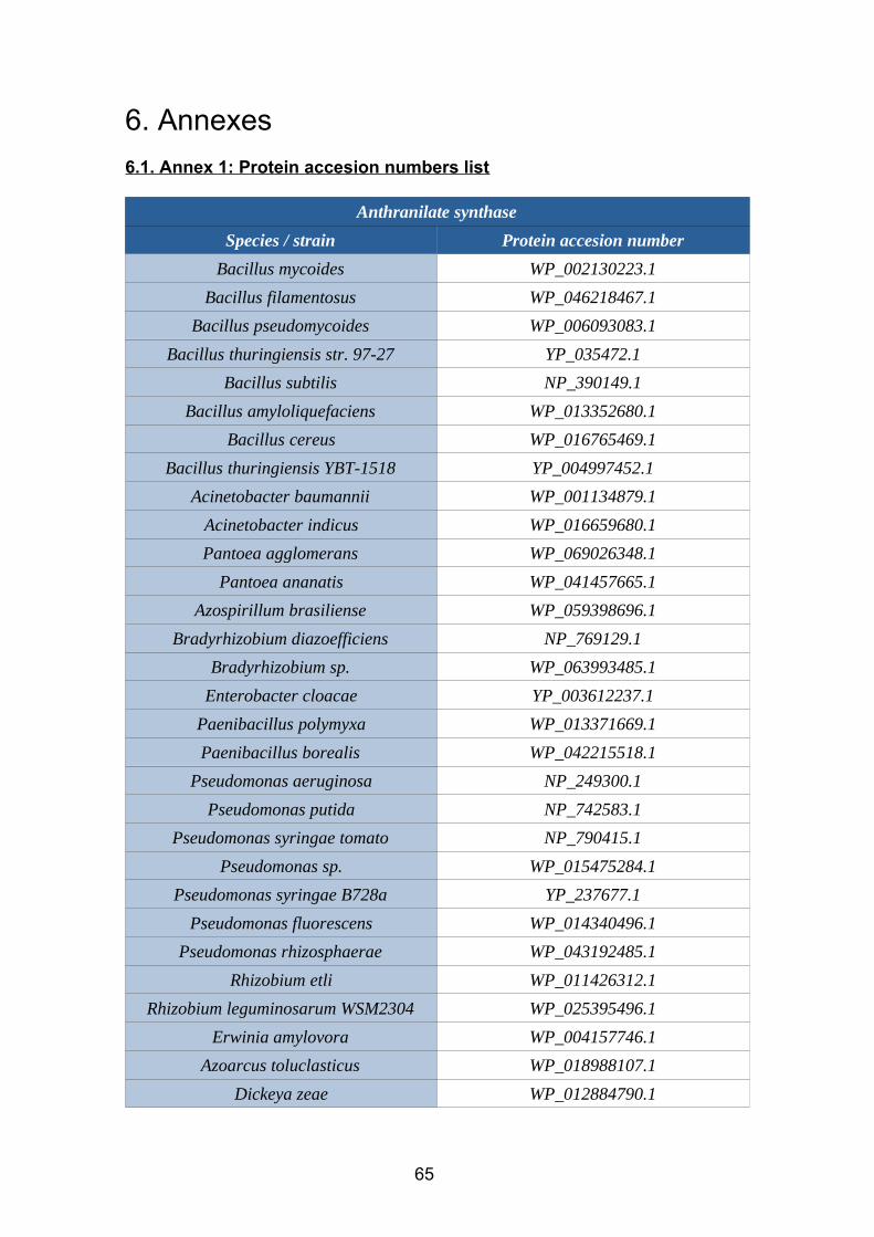
6. Annexes
6.1. Annex 1: Protein accesion numbers list
Anthranilate synthase
Species / strain Protein accesion number
Bacillus mycoides WP_002130223.1
Bacillus filamentosus WP_046218467.1
Bacillus pseudomycoides WP_006093083.1
Bacillus thuringiensis str. 97-27 YP_035472.1
Bacillus subtilis NP_390149.1
Bacillus amyloliquefaciens WP_013352680.1
Bacillus cereus WP_016765469.1
Bacillus thuringiensis YBT-1518 YP_004997452.1
Acinetobacter baumannii WP_001134879.1
Acinetobacter indicus WP_016659680.1
Pantoea agglomerans WP_069026348.1
Pantoea ananatis WP_041457665.1
Azospirillum brasiliense WP_059398696.1
Bradyrhizobium diazoefficiens NP_769129.1
Bradyrhizobium sp. WP_063993485.1
Enterobacter cloacae YP_003612237.1
Paenibacillus polymyxa WP_013371669.1
Paenibacillus borealis WP_042215518.1
Pseudomonas aeruginosa NP_249300.1
Pseudomonas putida NP_742583.1
Pseudomonas syringae tomato NP_790415.1
Pseudomonas sp. WP_015475284.1
Pseudomonas syringae B728a YP_237677.1
Pseudomonas fluorescens WP_014340496.1
Pseudomonas rhizosphaerae WP_043192485.1
Rhizobium etli WP_011426312.1
Rhizobium leguminosarum WSM2304 WP_025395496.1
Erwinia amylovora WP_004157746.1
Azoarcus toluclasticus WP_018988107.1
Dickeya zeae WP_012884790.1
65
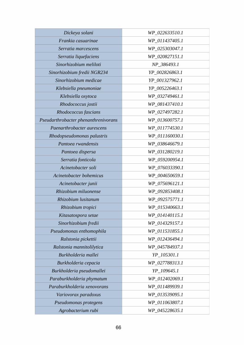
Dickeya solani WP_022633510.1
Frankia casuarinae WP_011437405.1
Serratia marcescens WP_025303047.1
Serratia liquefaciens WP_020827151.1
Sinorhizobium meliloti NP_386493.1
Sinorhizobium fredii NGR234 YP_002826863.1
Sinorhizobium medicae YP_001327962.1
Klebsiella pneumoniae YP_005226463.1
Klebsiella oxytoca WP_032749461.1
Rhodococcus jostii WP_081437410.1
Rhodococcus fascians WP_027497282.1
Pseudarthrobacter phenanthrenivorans WP_013600757.1
Paenarthrobacter aurescens WP_011774530.1
Rhodopseudomonas palustris WP_011160030.1
Pantoea rwandensis WP_038646679.1
Pantoea dispersa WP_031280219.1
Serratia fonticola WP_059200954.1
Acinetobacter soli WP_076033390.1
Acinetobacter bohemicus WP_004650659.1
Acinetobacter junii WP_075696121.1
Rhizobium miluonense WP_092853408.1
Rhizobium lusitanum WP_092575771.1
Rhizobium tropici WP_015340663.1
Kitasatospora setae WP_014140115.1
Sinorhizobium fredii WP_014329157.1
Pseudomonas enthomophila WP_011531855.1
Ralstonia pickettii WP_012436494.1
Ralstonia mannitolilytica WP_045784937.1
Burkholderia mallei YP_105301.1
Burkholderia cepacia WP_027788313.1
Burkholderia pseudomallei YP_109645.1
Paraburkholderia phymatum WP_012402069.1
Paraburkholderia xenovorans WP_011489939.1
Variovorax paradoxus WP_013539095.1
Pseudomonas protegens WP_011063807.1
Agrobacterium rubi WP_045228635.1
66
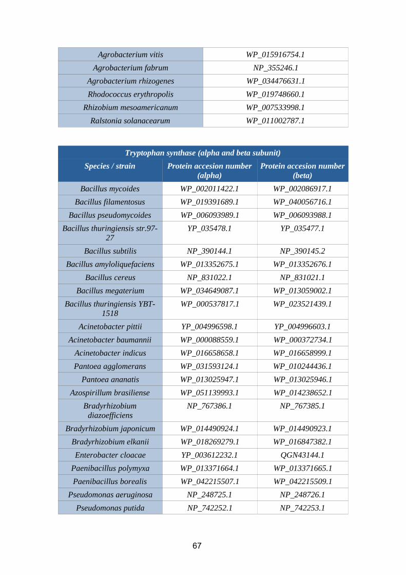
Agrobacterium vitis WP_015916754.1
Agrobacterium fabrum NP_355246.1
Agrobacterium rhizogenes WP_034476631.1
Rhodococcus erythropolis WP_019748660.1
Rhizobium mesoamericanum WP_007533998.1
Ralstonia solanacearum WP_011002787.1
Tryptophan synthase (alpha and beta subunit)
Species / strain Protein accesion number(alpha)
Protein accesion number(beta)
Bacillus mycoides WP_002011422.1 WP_002086917.1
Bacillus filamentosus WP_019391689.1 WP_040056716.1
Bacillus pseudomycoides WP_006093989.1 WP_006093988.1
Bacillus thuringiensis str.97-27
YP_035478.1 YP_035477.1
Bacillus subtilis NP_390144.1 NP_390145.2
Bacillus amyloliquefaciens WP_013352675.1 WP_013352676.1
Bacillus cereus NP_831022.1 NP_831021.1
Bacillus megaterium WP_034649087.1 WP_013059002.1
Bacillus thuringiensis YBT-1518
WP_000537817.1 WP_023521439.1
Acinetobacter pittii YP_004996598.1 YP_004996603.1
Acinetobacter baumannii WP_000088559.1 WP_000372734.1
Acinetobacter indicus WP_016658658.1 WP_016658999.1
Pantoea agglomerans WP_031593124.1 WP_010244436.1
Pantoea ananatis WP_013025947.1 WP_013025946.1
Azospirillum brasiliense WP_051139993.1 WP_014238652.1
Bradyrhizobiumdiazoefficiens
NP_767386.1 NP_767385.1
Bradyrhizobium japonicum WP_014490924.1 WP_014490923.1
Bradyrhizobium elkanii WP_018269279.1 WP_016847382.1
Enterobacter cloacae YP_003612232.1 QGN43144.1
Paenibacillus polymyxa WP_013371664.1 WP_013371665.1
Paenibacillus borealis WP_042215507.1 WP_042215509.1
Pseudomonas aeruginosa NP_248725.1 NP_248726.1
Pseudomonas putida NP_742252.1 NP_742253.1
67
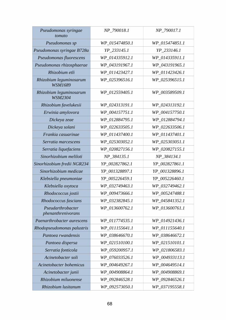
Pseudomonas syringaetomato
NP_790018.1 NP_790017.1
Pseudomonas sp WP_015474850.1 WP_015474851.1
Pseudomonas syringae B728a YP_233145.1 YP_233146.1
Pseudomonas fluorescens WP_014335912.1 WP_014335911.1
Pseudomonas rhizosphaerae WP_043191967.1 WP_043191965.1
Rhizobium etli WP_011423427.1 WP_011423426.1
Rhizobium leguminosarumWSM1689
WP_025396516.1 WP_025396515.1
Rhizobium leguminosarumWSM2304
WP_012559405.1 WP_003589509.1
Rhizobium favelukesii WP_024313191.1 WP_024313192.1
Erwinia amylovora WP_004157751.1 WP_004157750.1
Dickeya zeae WP_012884795.1 WP_012884794.1
Dickeya solani WP_022633505.1 WP_022633506.1
Frankia casuarinae WP_011437400.1 WP_011437401.1
Serratia marcescens WP_025303052.1 WP_025303051.1
Serratia liquefaciens WP_020827156.1 WP_020827155.1
Sinorhizobium meliloti NP_384135.1 NP_384134.1
Sinorhizobium fredii NGR234 YP_002827862.1 YP_002827861.1
Sinorhizobium medicae YP_001328897.1 YP_001328896.1
Klebsiella pneumoniae YP_005226459.1 YP_005226460.1
Klebsiella oxytoca WP_032749463.1 WP_032749462.1
Rhodococcus jostii WP_009473666.1 WP_005247488.1
Rhodococcus fascians WP_032382845.1 WP_045841352.1
Pseudarthrobacterphenanthrenivorans
WP_013600762.1 WP_013600761.1
Paenarthrobacter aurescens WP_011774535.1 WP_014921436.1
Rhodopseudomonas palustris WP_011155641.1 WP_011155640.1
Pantoea rwandensis WP_038646670.1 WP_038646672.1
Pantoea dispersa WP_021510100.1 WP_021510101.1
Serratia fonticola WP_059200957.1 WP_021806583.1
Acinetobacter soli WP_076033526.1 WP_004933113.1
Acinetobacter bohemicus WP_004649267.1 WP_004649514.1
Acinetobacter junii WP_004908864.1 WP_004908869.1
Rhizobium miluonense WP_092846528.1 WP_092846526.1
Rhizobium lusitanum WP_092573050.1 WP_037195558.1
68
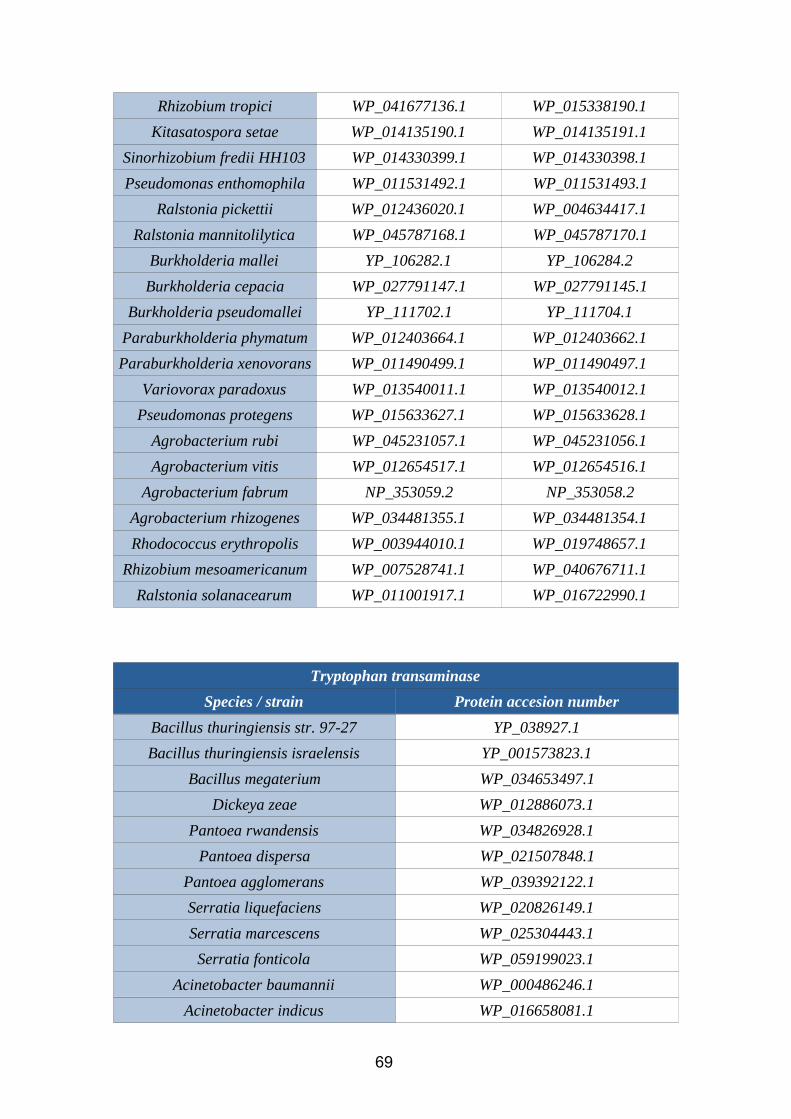
Rhizobium tropici WP_041677136.1 WP_015338190.1
Kitasatospora setae WP_014135190.1 WP_014135191.1
Sinorhizobium fredii HH103 WP_014330399.1 WP_014330398.1
Pseudomonas enthomophila WP_011531492.1 WP_011531493.1
Ralstonia pickettii WP_012436020.1 WP_004634417.1
Ralstonia mannitolilytica WP_045787168.1 WP_045787170.1
Burkholderia mallei YP_106282.1 YP_106284.2
Burkholderia cepacia WP_027791147.1 WP_027791145.1
Burkholderia pseudomallei YP_111702.1 YP_111704.1
Paraburkholderia phymatum WP_012403664.1 WP_012403662.1
Paraburkholderia xenovorans WP_011490499.1 WP_011490497.1
Variovorax paradoxus WP_013540011.1 WP_013540012.1
Pseudomonas protegens WP_015633627.1 WP_015633628.1
Agrobacterium rubi WP_045231057.1 WP_045231056.1
Agrobacterium vitis WP_012654517.1 WP_012654516.1
Agrobacterium fabrum NP_353059.2 NP_353058.2
Agrobacterium rhizogenes WP_034481355.1 WP_034481354.1
Rhodococcus erythropolis WP_003944010.1 WP_019748657.1
Rhizobium mesoamericanum WP_007528741.1 WP_040676711.1
Ralstonia solanacearum WP_011001917.1 WP_016722990.1
Tryptophan transaminase
Species / strain Protein accesion number
Bacillus thuringiensis str. 97-27 YP_038927.1
Bacillus thuringiensis israelensis YP_001573823.1
Bacillus megaterium WP_034653497.1
Dickeya zeae WP_012886073.1
Pantoea rwandensis WP_034826928.1
Pantoea dispersa WP_021507848.1
Pantoea agglomerans WP_039392122.1
Serratia liquefaciens WP_020826149.1
Serratia marcescens WP_025304443.1
Serratia fonticola WP_059199023.1
Acinetobacter baumannii WP_000486246.1
Acinetobacter indicus WP_016658081.1
69
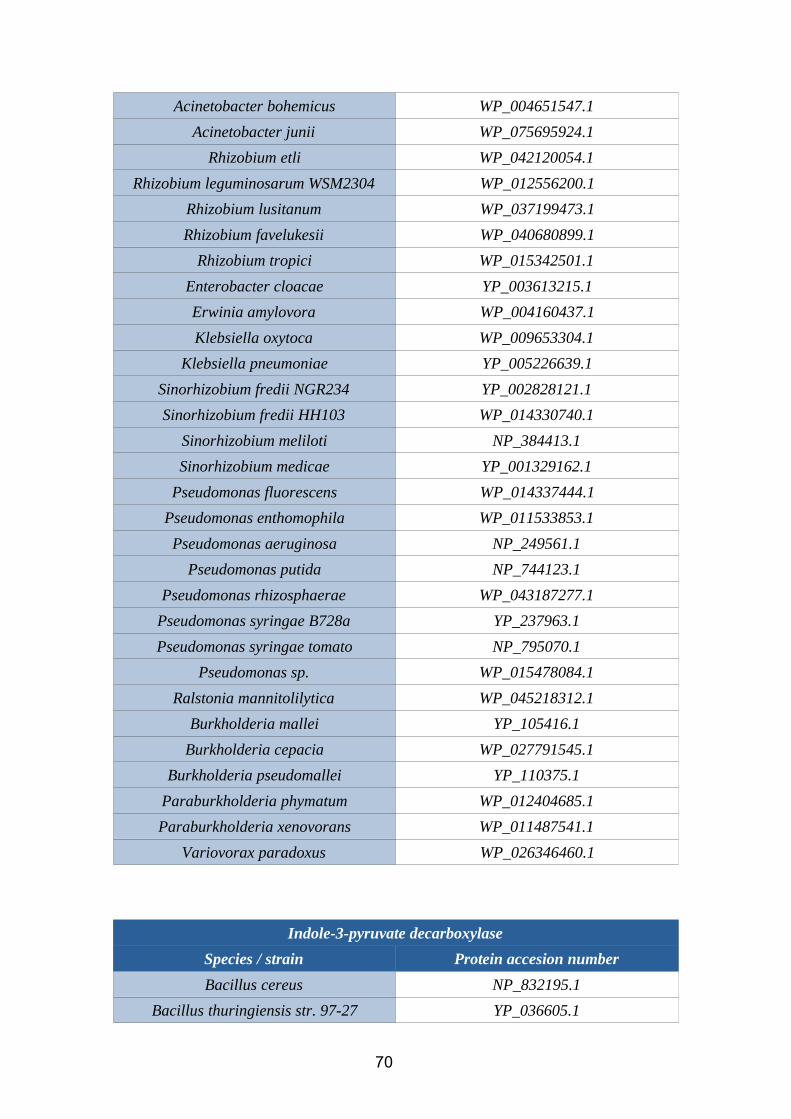
Acinetobacter bohemicus WP_004651547.1
Acinetobacter junii WP_075695924.1
Rhizobium etli WP_042120054.1
Rhizobium leguminosarum WSM2304 WP_012556200.1
Rhizobium lusitanum WP_037199473.1
Rhizobium favelukesii WP_040680899.1
Rhizobium tropici WP_015342501.1
Enterobacter cloacae YP_003613215.1
Erwinia amylovora WP_004160437.1
Klebsiella oxytoca WP_009653304.1
Klebsiella pneumoniae YP_005226639.1
Sinorhizobium fredii NGR234 YP_002828121.1
Sinorhizobium fredii HH103 WP_014330740.1
Sinorhizobium meliloti NP_384413.1
Sinorhizobium medicae YP_001329162.1
Pseudomonas fluorescens WP_014337444.1
Pseudomonas enthomophila WP_011533853.1
Pseudomonas aeruginosa NP_249561.1
Pseudomonas putida NP_744123.1
Pseudomonas rhizosphaerae WP_043187277.1
Pseudomonas syringae B728a YP_237963.1
Pseudomonas syringae tomato NP_795070.1
Pseudomonas sp. WP_015478084.1
Ralstonia mannitolilytica WP_045218312.1
Burkholderia mallei YP_105416.1
Burkholderia cepacia WP_027791545.1
Burkholderia pseudomallei YP_110375.1
Paraburkholderia phymatum WP_012404685.1
Paraburkholderia xenovorans WP_011487541.1
Variovorax paradoxus WP_026346460.1
Indole-3-pyruvate decarboxylase
Species / strain Protein accesion number
Bacillus cereus NP_832195.1
Bacillus thuringiensis str. 97-27 YP_036605.1
70
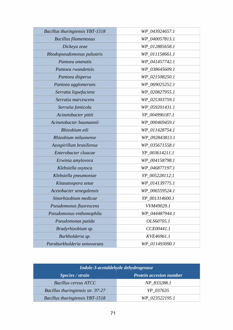
Bacillus thuringiensis YBT-1518 WP_043924657.1
Bacillus filamentosus WP_040057813.1
Dickeya zeae WP_012885658.1
Rhodopseudomonas palustris WP_011158661.1
Pantoea ananatis WP_041457742.1
Pantoea rwandensis WP_038645609.1
Pantoea dispersa WP_021508250.1
Pantoea agglomerans WP_069025252.1
Serratia liquefaciens WP_020827955.1
Serratia marcescens WP_025303759.1
Serratia fonticola WP_059201431.1
Acinetobacter pittii YP_004996187.1
Acinetobacter baumannii WP_000469459.1
Rhizobium etli WP_011428754.1
Rhizobium miluonense WP_092843813.1
Azospirillum brasiliense WP_035671558.1
Enterobacter cloacae YP_003614211.1
Erwinia amylovora WP_004158798.1
Klebsiella oxytoca WP_046877197.1
Klebsiella pneumoniae YP_005228112.1
Kitasatospora setae WP_014139775.1
Acetobacter senegalensis WP_006559524.1
Sinorhizobium medicae YP_001314600.1
Pseudomonas fluorescens VVM49029.1
Pseudomonas enthomophila WP_044487944.1
Pseudomonas putida OLS60705.1
Bradyrhizobium sp. CCE00441.1
Burkholderia sp. KVE46961.1
Paraburkholderia xenovorans WP_011493090.1
Indole-3-acetaldehyde dehydrogenase
Species / strain Protein accesion number
Bacillus cereus ATCC NP_833288.1
Bacillus thuringiensis str. 97-27 YP_037635
Bacillus thuringiensis YBT-1518 WP_023522195.1
71
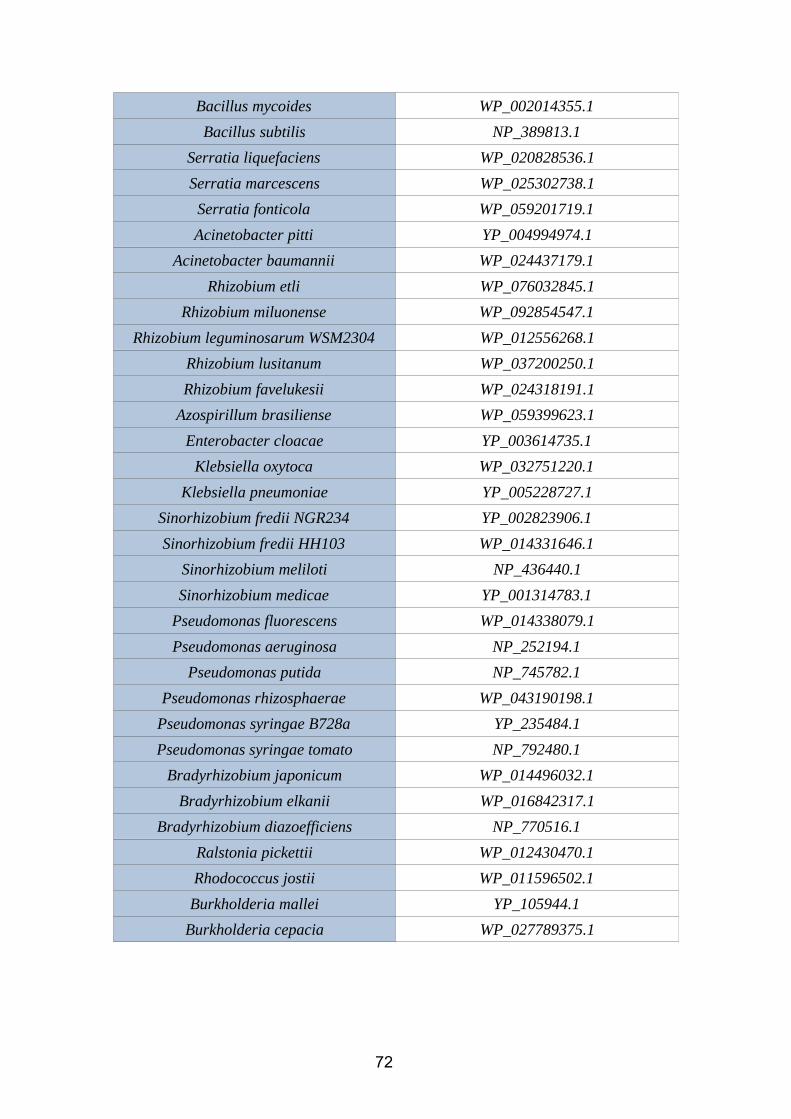
Bacillus mycoides WP_002014355.1
Bacillus subtilis NP_389813.1
Serratia liquefaciens WP_020828536.1
Serratia marcescens WP_025302738.1
Serratia fonticola WP_059201719.1
Acinetobacter pitti YP_004994974.1
Acinetobacter baumannii WP_024437179.1
Rhizobium etli WP_076032845.1
Rhizobium miluonense WP_092854547.1
Rhizobium leguminosarum WSM2304 WP_012556268.1
Rhizobium lusitanum WP_037200250.1
Rhizobium favelukesii WP_024318191.1
Azospirillum brasiliense WP_059399623.1
Enterobacter cloacae YP_003614735.1
Klebsiella oxytoca WP_032751220.1
Klebsiella pneumoniae YP_005228727.1
Sinorhizobium fredii NGR234 YP_002823906.1
Sinorhizobium fredii HH103 WP_014331646.1
Sinorhizobium meliloti NP_436440.1
Sinorhizobium medicae YP_001314783.1
Pseudomonas fluorescens WP_014338079.1
Pseudomonas aeruginosa NP_252194.1
Pseudomonas putida NP_745782.1
Pseudomonas rhizosphaerae WP_043190198.1
Pseudomonas syringae B728a YP_235484.1
Pseudomonas syringae tomato NP_792480.1
Bradyrhizobium japonicum WP_014496032.1
Bradyrhizobium elkanii WP_016842317.1
Bradyrhizobium diazoefficiens NP_770516.1
Ralstonia pickettii WP_012430470.1
Rhodococcus jostii WP_011596502.1
Burkholderia mallei YP_105944.1
Burkholderia cepacia WP_027789375.1
72
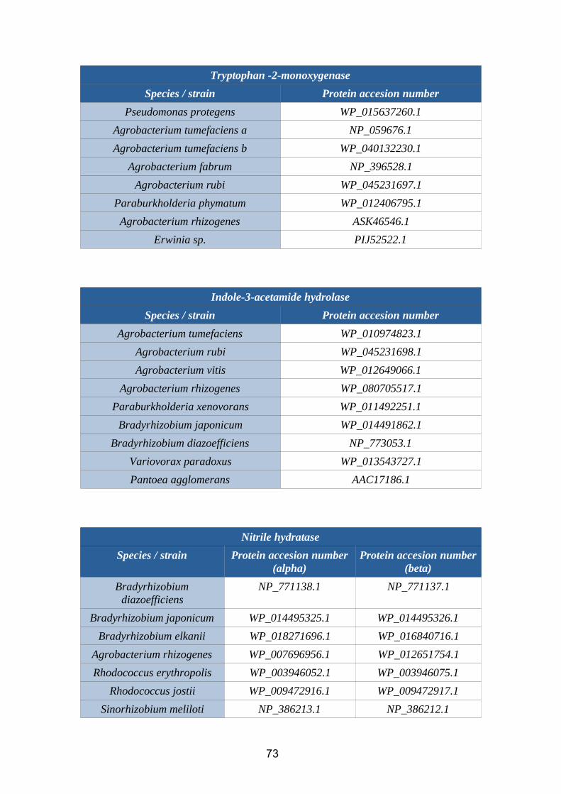
Tryptophan -2-monoxygenase
Species / strain Protein accesion number
Pseudomonas protegens WP_015637260.1
Agrobacterium tumefaciens a NP_059676.1
Agrobacterium tumefaciens b WP_040132230.1
Agrobacterium fabrum NP_396528.1
Agrobacterium rubi WP_045231697.1
Paraburkholderia phymatum WP_012406795.1
Agrobacterium rhizogenes ASK46546.1
Erwinia sp. PIJ52522.1
Indole-3-acetamide hydrolase
Species / strain Protein accesion number
Agrobacterium tumefaciens WP_010974823.1
Agrobacterium rubi WP_045231698.1
Agrobacterium vitis WP_012649066.1
Agrobacterium rhizogenes WP_080705517.1
Paraburkholderia xenovorans WP_011492251.1
Bradyrhizobium japonicum WP_014491862.1
Bradyrhizobium diazoefficiens NP_773053.1
Variovorax paradoxus WP_013543727.1
Pantoea agglomerans AAC17186.1
Nitrile hydratase
Species / strain Protein accesion number(alpha)
Protein accesion number(beta)
Bradyrhizobiumdiazoefficiens
NP_771138.1 NP_771137.1
Bradyrhizobium japonicum WP_014495325.1 WP_014495326.1
Bradyrhizobium elkanii WP_018271696.1 WP_016840716.1
Agrobacterium rhizogenes WP_007696956.1 WP_012651754.1
Rhodococcus erythropolis WP_003946052.1 WP_003946075.1
Rhodococcus jostii WP_009472916.1 WP_009472917.1
Sinorhizobium meliloti NP_386213.1 NP_386212.1
73
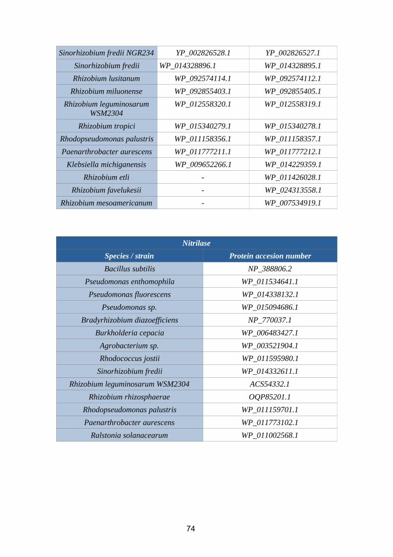
Sinorhizobium fredii NGR234 YP_002826528.1 YP_002826527.1
Sinorhizobium fredii WP_014328896.1 WP_014328895.1
Rhizobium lusitanum WP_092574114.1 WP_092574112.1
Rhizobium miluonense WP_092855403.1 WP_092855405.1
Rhizobium leguminosarumWSM2304
WP_012558320.1 WP_012558319.1
Rhizobium tropici WP_015340279.1 WP_015340278.1
Rhodopseudomonas palustris WP_011158356.1 WP_011158357.1
Paenarthrobacter aurescens WP_011777211.1 WP_011777212.1
Klebsiella michiganensis WP_009652266.1 WP_014229359.1
Rhizobium etli - WP_011426028.1
Rhizobium favelukesii - WP_024313558.1
Rhizobium mesoamericanum - WP_007534919.1
Nitrilase
Species / strain Protein accesion number
Bacillus subtilis NP_388806.2
Pseudomonas enthomophila WP_011534641.1
Pseudomonas fluorescens WP_014338132.1
Pseudomonas sp. WP_015094686.1
Bradyrhizobium diazoefficiens NP_770037.1
Burkholderia cepacia WP_006483427.1
Agrobacterium sp. WP_003521904.1
Rhodococcus jostii WP_011595980.1
Sinorhizobium fredii WP_014332611.1
Rhizobium leguminosarum WSM2304 ACS54332.1
Rhizobium rhizosphaerae OQP85201.1
Rhodopseudomonas palustris WP_011159701.1
Paenarthrobacter aurescens WP_011773102.1
Ralstonia solanacearum WP_011002568.1
74
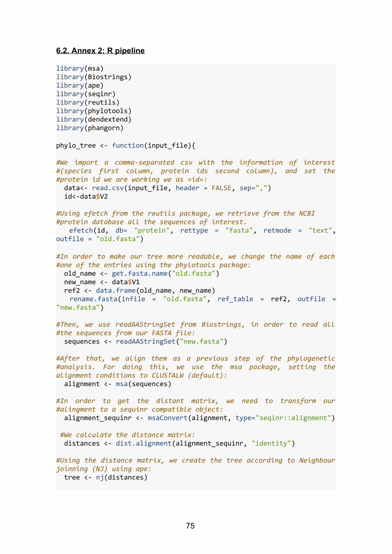
6.2. Annex 2: R pipeline
library(msa)library(Biostrings)library(ape)library(seqinr)library(reutils)library(phylotools)library(dendextend)library(phangorn)
phylo_tree <- function(input_file){ #We import a comma-separated csv with the information of interest#(species first column, protein ids second column), and set the#protein id we are working we as «id»: data<- read.csv(input_file, header = FALSE, sep=",") id<-data$V2 #Using efetch from the reutils package, we retrieve from the NCBI #protein database all the sequences of interest. efetch(id, db= "protein", rettype = "fasta", retmode = "text",outfile = "old.fasta") #In order to make our tree more readable, we change the name of each#one of the entries using the phylotools package: old_name <- get.fasta.name("old.fasta") new_name <- data$V1 ref2 <- data.frame(old_name, new_name) rename.fasta(infile = "old.fasta", ref_table = ref2, outfile ="new.fasta") #Then, we use readAAStringSet from Biostrings, in order to read all#the sequences from our FASTA file: sequences <- readAAStringSet("new.fasta") #After that, we align them as a previous step of the phylogenetic#analysis. For doing this, we use the msa package, setting thealignment conditions to CLUSTALW (default): alignment <- msa(sequences) #In order to get the distant matrix, we need to transform our#alingment to a sequinr compatible object: alignment_sequinr <- msaConvert(alignment, type="seqinr::alignment") #We calculate the distance matrix: distances <- dist.alignment(alignment_sequinr, "identity") #Using the distance matrix, we create the tree according to Neighbourjoinning (NJ) using ape: tree <- nj(distances)
75

}#We will use nitrilase.csv and gyrB_nitrilase.csv as examples:
a<-phylo_tree("csv_nitrilase.csv") b<-phylo_tree("csv_gyrB_nitrilase.csv")
#Since we are using dendextend to compare trees, we are going to need#to convert our phylo objects to dendextend compatible ojects. Thus,#we need to make sure that our phylo trees are binary, ultrametric androoted. We can check if the conditions are true as follows:is.binary(a)is.ultrametric(a)is.rooted(a)is.binary(b)is.ultrametric(b)is.rooted(b)
#If not, we can force every one of the three conditions:
#If root is absent, we can set a root:a$root.edge <- 0b$root.edge <- 0
#If the tree is not ultrametric, we can convert it to ultrametric. In#this step, we are going to use the phangorn package:
convert_to_ultra<-function(nonultra,method=c("nnls","extend")){ method<-method[1] if(method=="nnls") nonultra←nnls.tree(cophenetic(nonultra),nonultra, rooted=TRUE,trace=0) else if(method=="extend"){ h<-diag(vcv(nonultra)) d<-max(h)-h ii<-sapply(1:Ntip(nonultra),function(x,y) which(y==x), y=nonultra$edge[,2]) nonultra$edge.length[ii]<-nonultra$edge.length[ii]+d } else cat("impossible to convert to ultrametric") nonultra}
a<- convert_to_ultra(a)b<- convert_to_ultra(b)
#Finally, we can force the binary condition too:a<-multi2di(a)b<-multi2di(b)
76
#After the transformation, we can check the three conditions onceagain:is.binary(a)## [1] TRUEis.ultrametric(a)## [1] TRUEis.rooted(a)## [1] TRUEis.binary(b)## [1] TRUEis.ultrametric(b)## [1] TRUEis.rooted(b)## [1] TRUE
#Besides, we can plot our trees:plot(a)plot(b)
#Now, we can convert our phylo trees to dendrograms and perform allthe comparisons needed.dend1 <- as.dendrogram(a)dend2 <- as.dendrogram(b)
#As mentioned before, we use dendextend to compare dendrograms:tanglegram(dend1,dend2)
77
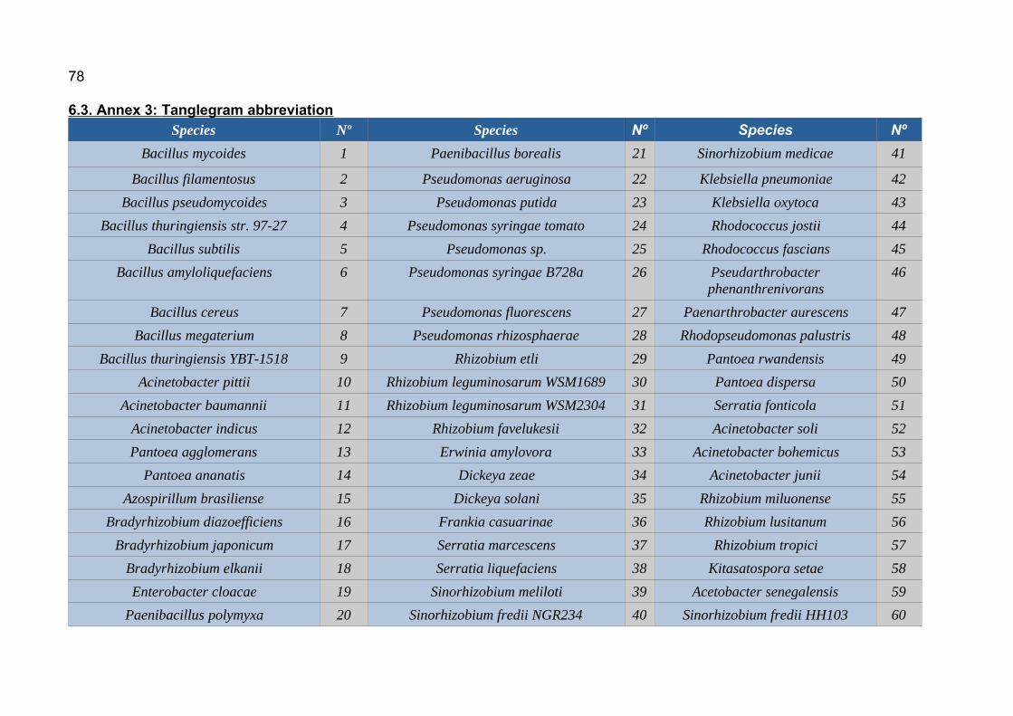
78
6.3. Annex 3: Tanglegram abbreviation Species Nº Species Nº Species Nº
Bacillus mycoides 1 Paenibacillus borealis 21 Sinorhizobium medicae 41
Bacillus filamentosus 2 Pseudomonas aeruginosa 22 Klebsiella pneumoniae 42
Bacillus pseudomycoides 3 Pseudomonas putida 23 Klebsiella oxytoca 43
Bacillus thuringiensis str. 97-27 4 Pseudomonas syringae tomato 24 Rhodococcus jostii 44
Bacillus subtilis 5 Pseudomonas sp. 25 Rhodococcus fascians 45
Bacillus amyloliquefaciens 6 Pseudomonas syringae B728a 26 Pseudarthrobacterphenanthrenivorans
46
Bacillus cereus 7 Pseudomonas fluorescens 27 Paenarthrobacter aurescens 47
Bacillus megaterium 8 Pseudomonas rhizosphaerae 28 Rhodopseudomonas palustris 48
Bacillus thuringiensis YBT-1518 9 Rhizobium etli 29 Pantoea rwandensis 49
Acinetobacter pittii 10 Rhizobium leguminosarum WSM1689 30 Pantoea dispersa 50
Acinetobacter baumannii 11 Rhizobium leguminosarum WSM2304 31 Serratia fonticola 51
Acinetobacter indicus 12 Rhizobium favelukesii 32 Acinetobacter soli 52
Pantoea agglomerans 13 Erwinia amylovora 33 Acinetobacter bohemicus 53
Pantoea ananatis 14 Dickeya zeae 34 Acinetobacter junii 54
Azospirillum brasiliense 15 Dickeya solani 35 Rhizobium miluonense 55
Bradyrhizobium diazoefficiens 16 Frankia casuarinae 36 Rhizobium lusitanum 56
Bradyrhizobium japonicum 17 Serratia marcescens 37 Rhizobium tropici 57
Bradyrhizobium elkanii 18 Serratia liquefaciens 38 Kitasatospora setae 58
Enterobacter cloacae 19 Sinorhizobium meliloti 39 Acetobacter senegalensis 59
Paenibacillus polymyxa 20 Sinorhizobium fredii NGR234 40 Sinorhizobium fredii HH103 60

79
Species Nº
Pseudomonas enthomophila 61
Ralstonia pickettii 62
Ralstonia mannitolilytica 63
Burkholderia mallei 64
Burkholderia cepacia 65
Burkholderia pseudomallei 66
Paraburkholderia phymatum 67
Paraburkholderia xenovorans 68
Variovorax paradoxus 69
Pseudomonas protegens 70
Agrobacterium rubi 71
Agrobacterium vitis 72
Agrobacterium fabrum 73
Agrobacterium rhizogenes 74
Rhodococcus erythropolis 75
Rhizobium mesoamericanum 76
Ralstonia solanacearum 77
Azoarcus toluclasticus 78
Bacillus thuringiensis israelensis 79
klebsiella michiganensis 80
Rhizobium rhizosphaerae 81
Copyright © 2022 FDOKUMEN