Decoding the perception of pain from fMRI using multivariate pattern analysis
9
Decoding the perception of pain from fMRI using multivariate pattern analysis Kay H. Brodersen a, b, c, ⁎ , 1 , Katja Wiech a, ⁎⁎ , 1 , Ekaterina I. Lomakina b, c , Chia-shu Lin a , Joachim M. Buhmann c , Ulrike Bingel a, d , Markus Ploner a, e , Klaas Enno Stephan b, f , Irene Tracey a a Centre for Functional Magnetic Resonance Imaging of the Brain (FMRIB), Nuffield Department of Clinical Neurosciences, Nuffield Division Anaesthetics, University of Oxford, John Radcliffe Hospital, Oxford OX3 9DU, UK b Translational Neuromodeling Unit (TNU), Institute for Biomedical Engineering, University of Zurich & ETH Zurich, Wilfriedstrasse 6, CH 8032 Zurich, Switzerland c Department of Computer Science, ETH Zurich, Universitaetstrasse 6, CH 8092 Zurich, Switzerland d NeuroImage Nord, Department of Neurology, University Medical Center Hamburg-Eppendorf, Martinistr. 52, 20246 Hamburg, Germany e Department of Neurology, Technische Universität München, 81675 Munich, Germany f Wellcome Trust Centre for Neuroimaging, University College London, UK abstract article info Article history: Accepted 13 August 2012 Available online 18 August 2012 Keywords: Pain Decoding Support vector machine Permutation test Classification accuracy Pain is known to comprise sensory, cognitive, and affective aspects. Despite numerous previous fMRI studies, however, it remains open which spatial distribution of activity is sufficient to encode whether a stimulus is per- ceived as painful or not. In this study, we analyzed fMRI data from a perceptual decision-making task in which participants were exposed to near-threshold laser pulses. Using multivariate analyses on different spatial scales, we investigated the predictive capacity of fMRI data for decoding whether a stimulus had been perceived as pain- ful. Our analysis yielded a rank order of brain regions: during pain anticipation, activity in the periaqueductal gray (PAG) and orbitofrontal cortex (OFC) afforded the most accurate trial-by-trial discrimination between pain- ful and non-painful experiences; whereas during the actual stimulation, primary and secondary somatosensory cortex, anterior insula, dorsolateral and ventrolateral prefrontal cortex, and OFC were most discriminative. The most accurate prediction of pain perception from the stimulation period, however, was enabled by the combined activity in pain regions commonly referred to as the ‘pain matrix’. Our results demonstrate that the neural rep- resentation of (near-threshold) pain is spatially distributed and can be best described at an intermediate spatial scale. In addition to its utility in establishing structure-function mappings, our approach affords trial-by-trial pre- dictions and thus represents a step towards the goal of establishing an objective neuronal marker of pain perception. © 2012 Elsevier Inc. All rights reserved. Introduction The perception of pain is a multi-factorial experience that comprises sensory, cognitive, and affective aspects. Accordingly, pain is thought to result from a complex interplay between many regions in the human brain, including the thalamus, insula, primary and secondary somato- sensory, anterior cingulate cortex, and prefrontal cortex (Apkarian et al., 2005). The specific characteristics of regions underlying the percep- tion of pain have been described in some detail using conventional uni- variate analysis methods for functional magnetic resonance imaging (fMRI). By contrast, there have been almost no attempts at examining the distributed representation of pain and how it is encoded jointly by activity within and across the set of regions commonly associated with pain. Statistical methods for examining distributed coding schemes have undergone rapid progress over the past years. One particularly versatile approach, termed multivariate pattern analysis (MVPA), is based on the use of a classification algorithm to infer a perceptual or cognitive state from brain activity. The underlying multivariate decoding models differ in important ways from univariate encoding models, such as the gener- al linear model (GLM). Univariate analyses have proven powerful for in- ference on structure-function mappings in the brain when activations are expressed in terms of local peaks or clusters of activity (Friston et al., 1995). However, they are less suitable for assessing the amount of information encoded in spatially distributed (multivoxel) patterns of activity underlying specific perceptual or cognitive states. This informa- tion can be estimated using multivariate decoding models (Friston et al., 2008; Haynes and Rees, 2006; Norman et al., 2006; O'Toole et al., 2007; Pereira et al., 2009). These models consider several voxels at the same time and may therefore be more sensitive than univariate models (for an analysis of the conditions under which this is the case, see Guyon and Elisseeff, 2003). NeuroImage 63 (2012) 1162–1170 ⁎ Correspondence to: K.H. Brodersen, Department of Computer Science, ETH Zurich, Universitaetstrasse 6, CH 8092 Zurich, Switzerland. Fax: +41 4463 44 907. ⁎⁎ Correspondence to: K. Wiech, Centre for Functional Magnetic Resonance Imaging of the Brain (FMRIB), Nuffield Department of Clinical Neurosciences, Nuffield Division Anaesthetics, University of Oxford, Oxford, OX3 9DU, UK. Fax: +44 1865 234541. E-mail addresses: [email protected] (K.H. Brodersen), [email protected] (K. Wiech). 1 These authors contributed equally. 1053-8119/$ – see front matter © 2012 Elsevier Inc. All rights reserved. http://dx.doi.org/10.1016/j.neuroimage.2012.08.035 Contents lists available at SciVerse ScienceDirect NeuroImage journal homepage: www.elsevier.com/locate/ynimg
Transcript of Decoding the perception of pain from fMRI using multivariate pattern analysis
NeuroImage 63 (2012) 1162–1170
Contents lists available at SciVerse ScienceDirect
NeuroImage
j ourna l homepage: www.e lsev ie r .com/ locate /yn img
Decoding the perception of pain from fMRI using multivariate pattern analysis
Kay H. Brodersen a,b,c,⁎,1, Katja Wiech a,⁎⁎,1, Ekaterina I. Lomakina b,c, Chia-shu Lin a, Joachim M. Buhmann c,Ulrike Bingel a,d, Markus Ploner a,e, Klaas Enno Stephan b,f, Irene Tracey a
a Centre for Functional Magnetic Resonance Imaging of the Brain (FMRIB), Nuffield Department of Clinical Neurosciences, Nuffield Division Anaesthetics, University of Oxford,John Radcliffe Hospital, Oxford OX3 9DU, UKb Translational Neuromodeling Unit (TNU), Institute for Biomedical Engineering, University of Zurich & ETH Zurich, Wilfriedstrasse 6, CH 8032 Zurich, Switzerlandc Department of Computer Science, ETH Zurich, Universitaetstrasse 6, CH 8092 Zurich, Switzerlandd NeuroImage Nord, Department of Neurology, University Medical Center Hamburg-Eppendorf, Martinistr. 52, 20246 Hamburg, Germanye Department of Neurology, Technische Universität München, 81675 Munich, Germanyf Wellcome Trust Centre for Neuroimaging, University College London, UK
⁎ Correspondence to: K.H. Brodersen, Department ofUniversitaetstrasse 6, CH 8092 Zurich, Switzerland. Fax:⁎⁎ Correspondence to: K. Wiech, Centre for Functiona
of the Brain (FMRIB), Nuffield Department of Clinical NeAnaesthetics, University of Oxford, Oxford, OX3 9DU, UK
E-mail addresses: [email protected] (K.H. [email protected] (K. Wiech).
1 These authors contributed equally.
1053-8119/$ – see front matter © 2012 Elsevier Inc. Allhttp://dx.doi.org/10.1016/j.neuroimage.2012.08.035
a b s t r a c t
a r t i c l e i n f oArticle history:Accepted 13 August 2012Available online 18 August 2012
Keywords:PainDecodingSupport vector machinePermutation testClassification accuracy
Pain is known to comprise sensory, cognitive, and affective aspects. Despite numerous previous fMRI studies,however, it remains open which spatial distribution of activity is sufficient to encode whether a stimulus is per-ceived as painful or not. In this study, we analyzed fMRI data from a perceptual decision-making task in whichparticipants were exposed to near-threshold laser pulses. Using multivariate analyses on different spatial scales,we investigated the predictive capacity of fMRI data for decodingwhether a stimulus had beenperceived as pain-ful. Our analysis yielded a rank order of brain regions: during pain anticipation, activity in the periaqueductalgray (PAG) and orbitofrontal cortex (OFC) afforded themost accurate trial-by-trial discrimination between pain-ful and non-painful experiences; whereas during the actual stimulation, primary and secondary somatosensorycortex, anterior insula, dorsolateral and ventrolateral prefrontal cortex, and OFC were most discriminative. Themost accurate prediction of pain perception from the stimulation period, however, was enabled by the combinedactivity in pain regions commonly referred to as the ‘pain matrix’. Our results demonstrate that the neural rep-resentation of (near-threshold) pain is spatially distributed and can be best described at an intermediate spatialscale. In addition to its utility in establishing structure-functionmappings, our approach affords trial-by-trial pre-dictions and thus represents a step towards the goal of establishing an objective neuronal marker of painperception.
© 2012 Elsevier Inc. All rights reserved.
Introduction
The perception of pain is amulti-factorial experience that comprisessensory, cognitive, and affective aspects. Accordingly, pain is thought toresult from a complex interplay between many regions in the humanbrain, including the thalamus, insula, primary and secondary somato-sensory, anterior cingulate cortex, and prefrontal cortex (Apkarian etal., 2005). The specific characteristics of regions underlying the percep-tion of pain have been described in some detail using conventional uni-variate analysis methods for functional magnetic resonance imaging(fMRI). By contrast, there have been almost no attempts at examiningthe distributed representation of pain and how it is encoded jointly by
Computer Science, ETH Zurich,+41 4463 44 907.l Magnetic Resonance Imagingurosciences, Nuffield Division. Fax: +44 1865 234541.rodersen),
rights reserved.
activity within and across the set of regions commonly associatedwith pain.
Statistical methods for examining distributed coding schemes haveundergone rapid progress over the past years. One particularly versatileapproach, termedmultivariate pattern analysis (MVPA), is based on theuse of a classification algorithm to infer a perceptual or cognitive statefrom brain activity. The underlying multivariate decodingmodels differin important ways from univariate encodingmodels, such as the gener-al linearmodel (GLM). Univariate analyses have provenpowerful for in-ference on structure-function mappings in the brain when activationsare expressed in terms of local peaks or clusters of activity (Friston etal., 1995). However, they are less suitable for assessing the amount ofinformation encoded in spatially distributed (multivoxel) patterns ofactivity underlying specific perceptual or cognitive states. This informa-tion can be estimatedusingmultivariate decodingmodels (Friston et al.,2008; Haynes and Rees, 2006; Norman et al., 2006; O'Toole et al., 2007;Pereira et al., 2009). These models consider several voxels at the sametime and may therefore be more sensitive than univariate models (foran analysis of the conditions under which this is the case, see Guyonand Elisseeff, 2003).
painful?
yes no
cue
(4-8 s)
delay
(4-6 s)
decision
(until response)
inter-trial interval
(10-12 s)
stimulus button press
+
time
blaser intensity
… …120 trials
(subject 1)120 trials
(subject 2)
subject-specific
pain thresholds
a
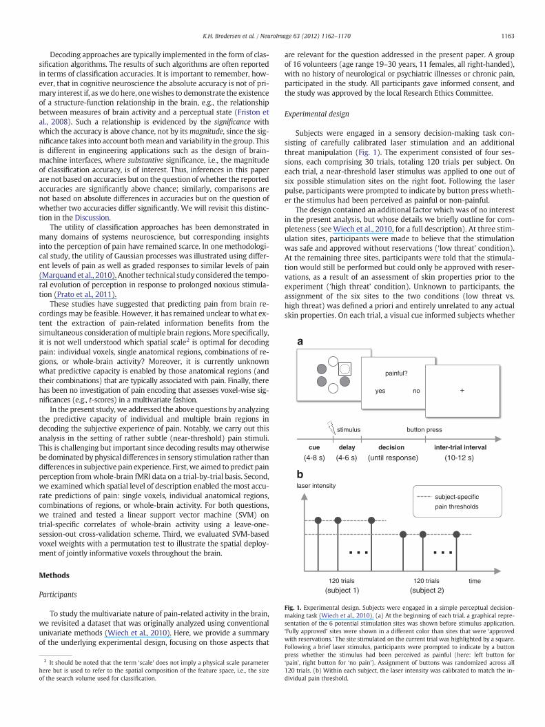
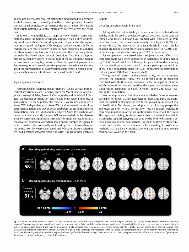
Fig. 1. Experimental design. Subjects were engaged in a simple perceptual decision-making task (Wiech et al., 2010). (a) At the beginning of each trial, a graphical repre-sentation of the 6 potential stimulation sites was shown before stimulus application.‘Fully approved’ sites were shown in a different color than sites that were ‘approvedwith reservations.’ The site stimulated on the current trial was highlighted by a square.Following a brief laser stimulus, participants were prompted to indicate by a button
1163K.H. Brodersen et al. / NeuroImage 63 (2012) 1162–1170
Decoding approaches are typically implemented in the form of clas-sification algorithms. The results of such algorithms are often reportedin terms of classification accuracies. It is important to remember, how-ever, that in cognitive neuroscience the absolute accuracy is not of pri-mary interest if, aswe dohere, onewishes to demonstrate the existenceof a structure-function relationship in the brain, e.g., the relationshipbetween measures of brain activity and a perceptual state (Friston etal., 2008). Such a relationship is evidenced by the significance withwhich the accuracy is above chance, not by itsmagnitude, since the sig-nificance takes into account bothmean and variability in the group. Thisis different in engineering applications such as the design of brain-machine interfaces, where substantive significance, i.e., the magnitudeof classification accuracy, is of interest. Thus, inferences in this paperare not based on accuracies but on the question ofwhether the reportedaccuracies are significantly above chance; similarly, comparisons arenot based on absolute differences in accuracies but on the question ofwhether two accuracies differ significantly. We will revisit this distinc-tion in the Discussion.
The utility of classification approaches has been demonstrated inmany domains of systems neuroscience, but corresponding insightsinto the perception of pain have remained scarce. In one methodologi-cal study, the utility of Gaussian processes was illustrated using differ-ent levels of pain as well as graded responses to similar levels of pain(Marquand et al., 2010). Another technical study considered the tempo-ral evolution of perception in response to prolonged noxious stimula-tion (Prato et al., 2011).
These studies have suggested that predicting pain from brain re-cordings may be feasible. However, it has remained unclear to what ex-tent the extraction of pain-related information benefits from thesimultaneous consideration of multiple brain regions. More specifically,it is not well understood which spatial scale2 is optimal for decodingpain: individual voxels, single anatomical regions, combinations of re-gions, or whole-brain activity? Moreover, it is currently unknownwhat predictive capacity is enabled by those anatomical regions (andtheir combinations) that are typically associated with pain. Finally, therehas been no investigation of pain encoding that assesses voxel-wise sig-nificances (e.g., t-scores) in a multivariate fashion.
In the present study, we addressed the above questions by analyzingthe predictive capacity of individual and multiple brain regions indecoding the subjective experience of pain. Notably, we carry out thisanalysis in the setting of rather subtle (near-threshold) pain stimuli.This is challenging but important since decoding results may otherwisebe dominated by physical differences in sensory stimulation rather thandifferences in subjective pain experience. First, we aimed to predict painperception fromwhole-brain fMRI data on a trial-by-trial basis. Second,we examined which spatial level of description enabled the most accu-rate predictions of pain: single voxels, individual anatomical regions,combinations of regions, or whole-brain activity. For both questions,we trained and tested a linear support vector machine (SVM) ontrial-specific correlates of whole-brain activity using a leave-one-session-out cross-validation scheme. Third, we evaluated SVM-basedvoxel weights with a permutation test to illustrate the spatial deploy-ment of jointly informative voxels throughout the brain.
Methods
Participants
To study the multivariate nature of pain-related activity in the brain,we revisited a dataset that was originally analyzed using conventionalunivariate methods (Wiech et al., 2010). Here, we provide a summaryof the underlying experimental design, focusing on those aspects that
2 It should be noted that the term ‘scale’ does not imply a physical scale parameterhere but is used to refer to the spatial composition of the feature space, i.e., the sizeof the search volume used for classification.
are relevant for the question addressed in the present paper. A groupof 16 volunteers (age range 19–30 years, 11 females, all right-handed),with no history of neurological or psychiatric illnesses or chronic pain,participated in the study. All participants gave informed consent, andthe study was approved by the local Research Ethics Committee.
Experimental design
Subjects were engaged in a sensory decision-making task con-sisting of carefully calibrated laser stimulation and an additionalthreat manipulation (Fig. 1). The experiment consisted of four ses-sions, each comprising 30 trials, totaling 120 trials per subject. Oneach trial, a near-threshold laser stimulus was applied to one out ofsix possible stimulation sites on the right foot. Following the laserpulse, participants were prompted to indicate by button press wheth-er the stimulus had been perceived as painful or non-painful.
The design contained an additional factor which was of no interestin the present analysis, but whose details we briefly outline for com-pleteness (see Wiech et al., 2010, for a full description). At three stim-ulation sites, participants were made to believe that the stimulationwas safe and approved without reservations (‘low threat’ condition).At the remaining three sites, participants were told that the stimula-tion would still be performed but could only be approved with reser-vations, as a result of an assessment of skin properties prior to theexperiment (‘high threat’ condition). Unknown to participants, theassignment of the six sites to the two conditions (low threat vs.high threat) was defined a priori and entirely unrelated to any actualskin properties. On each trial, a visual cue informed subjects whether
press whether the stimulus had been perceived as painful (here: left button for‘pain’, right button for ‘no pain’). Assignment of buttons was randomized across all120 trials. (b) Within each subject, the laser intensity was calibrated to match the in-dividual pain threshold.
1164 K.H. Brodersen et al. / NeuroImage 63 (2012) 1162–1170
the laser stimulus was about to target a ‘low threat’ or a ‘high threat’site.
Data acquisition and preprocessing
Using a 3T MRI scanner (Oxford Magnet Technology, Oxford, UK),whole-brain functional T2*-weighted echo-planar images (EPI) wereacquired with BOLD contrast (TR 3 s; TE 30 ms; flip angle 90°; matrix64×64; field of view 192 mm×192 mm; 41 axial slices; slice thickness3 mm). The first 4 volumes were discarded to compensate for T1 satu-ration effects. Using SPM8 (http://www.fil.ion.ucl.ac.uk/spm), imageswere realigned to the first volume and unwarped. Images of all sessionswere spatially normalized to the standard EPI template included inSPM, using a fourth-degree B-spline interpolation.
Univariate analysis
Prior to the classification-based analyses described below, weperformed several conventional univariate analyses for comparison.For these analyses, images were spatially smoothed with an isotropicGaussian kernel (FWHM8 mm). First, we investigated themain effect ofpain during anticipation and stimulation. To this end, we constructed a(first-level) GLM for each subject with a design matrix that includedseparate ‘pain’ and ‘no pain’ regressors for the anticipation and the stim-ulation period (4 regressors), collapsing across ‘low threat’ and ‘highthreat’ trials, whose distinction was of no interest in the present study.Anticipation periods were modeled according to their trial-specific du-rations (i.e., 4–8 s), while the stimulus duration was modeled as 1 s. Se-rial autocorrelation and low-frequency drifts were accounted for using afirst-order autoregressive model and a high-pass filter (cut-off 128 s),respectively. Group-level inferences for the anticipation and stimulationperiod were made by entering the appropriate contrast into an ANOVA,using the following two contrasts: (1) pain vs. no pain during anticipa-tion; and (2) pain vs. no pain during stimulation.
Multivariate analysis
In contrast to univariate analyses, multivariate approaches explicitlyaccount for dependencies between voxels, which allows for inferenceon distributed responses. In this study, we trained and tested a linearsupport vector machine (SVM) on trial-wise fMRI data. In order toavoid a potential bias resulting from serial autocorrelations, we usedleave-one-session-out cross-validation. Specifically, we trained anSVM on trials from three sessions and tested it on trials from the fourth(left-out) session, repeating this process four times. To obtain trial-wisedata for classification, we constructed a GLM with a design matrix thatincluded separate boxcar regressors for the anticipation phase and thestimulation phase of each individual trial (240 regressors). We usedthis GLM as a filter to obtain separate parameter-estimate images(beta images) for the anticipation phase and the stimulation phase ofeach trial. These images were processed further in two ways. First, westandardized the parameter estimates within each voxel (implyingmean=0 and standard deviation=1). Second, we scaled all imagessuch that within each trial the l2-norm of parameter estimates became1. The resulting images were used in two sets of classification analyses,as described next (for a structured list of individual analysis steps, seeSection C in the Supplemental Material).
In the first analysis, we investigated whether fMRI data containedsufficient information to predict, on a trial-by-trial basis, the perceptionof pain. For this purpose, a linear SVMwas trained and tested on differ-ent anatomical scales. These independent analyses were based on(i) the single most discriminative voxel (which was determined usinga t-contrast as described in the second analysis below, and whoseidentity was allowed to vary both between cross-validation folds andbetween subjects), (ii) combinations of discriminative voxels (i.e., dif-ferently sized groups of voxels that were individually discriminative,
as determined using a t-contrast as described below), (iii) single ana-tomical regions typically associated with pain processing (see below),(iv) combinations of the most predictive anatomical regions, and(v) whole-brain data.
Within each cross-validation fold, we used another (nested) levelof (leave-one-trial-out) cross-validation on the training data to opti-mize the regularization hyperparameter C. In this way, test data wereneither used for training nor for the optimization of hyperparameters,guaranteeing a non-circular analysis. Furthermore, to ensure that theanalysis was not confounded by differences between ‘high threat’ and‘low threat’ trials, we ran two separate decoding analyses on the twotrial types and considered the mean accuracy. This procedure was re-peated for every subject to obtain an estimate of mean classification ac-curacy in the group (cf. Section C in the Supplement).
We used a nonparametric permutation test to evaluate the null hy-pothesis that there was no statistical link between fMRI data and theperception of pain. This null hypothesis corresponds to a mean popula-tion accuracy at the level of chance (i.e., 0.5). Thus, we repeated eachclassification analysis N times using labels that were randomly permut-ed within sessions, preserving the assumption of exchangeabilityunderlying the permutation test for our leave-one-session-out cross-validation scheme. For each analysis, we computed a p-value as: therank of the original sample accuracy in the distribution of permutation-based sample accuracies, divided by the number of permutations. Wegenerally used N=1000. In the case of ROI-specific analyses with their26-fold multiple-comparison correction, we used N=2600 to allow forthe detection of significance at the 0.05 level (see below).
In the second analysis, we characterized the spatial deploymentof jointly informative voxels across the brain by combining an SVMwith a permutation test on voxel weights (LaConte et al., 2005;Mourao-Miranda et al., 2005). Specifically, we trained a linear SVM onwhole-brain data (using all 120 trials in each subject) and reconstructedthe spatial deployment of voxel-wise weight coefficients. These coeffi-cients may heavily depend on task-unrelated sources of variance inthe data and are generally not interpretable as such. One way ofaddressing this issue is to relate voxel weights to their empirical nulldistributions, i.e., those distributions that one would obtain if no statis-tical relationship between BOLD activity and pain perception existed(Mourao-Miranda et al., 2005; Wang et al., 2007).
To obtain these distributions, we randomly permuted trial-specificlabels and re-estimated the model based on the new labels. Unlike inthe case of all other multivariate analyses presented in this paper, anonparametric permutation is computationally intractable if onewishes to obtain a whole-brain FWE-corrected map with fine-graineddiscriminability eve among top-scoring voxels. For this particular anal-ysis, we therefore resorted to a parametric approach. Using a Kolmogo-rov–Smirnov test (test size α=0.05), we found that less than 0.01% ofall voxel-specific null distributions were not Gaussian. Thus, we sum-marized each null distribution in terms of the mean and variance of aGaussian. Using these null distributions, we evaluated the probabilitywith which the weight wv in voxel v would have been observed underthe null. Formally, this test is based on a t-score, defined as
tv ¼wv−μ̂ v
σ̂ ve
tN−1;
where μ̂ v and σ̂ v denote the sample mean and standard deviation ofvoxel weights in voxel v across all random permutations, and tN−1
is Student's t-distribution on N−1 degrees of freedom. We used N=2000 permutations and corrected the resulting map for multiple com-parisons using a conservativewhole-brain family-wise error (FWE) cor-rection (α=0.05). This correctionwas based on the same application ofrandom-field theory to estimate the smoothness of the data aswas usedfor thresholding the (mass-univariate) SPMs (see above).
In summary, we obtained whole-brain FWE-corrected maps oft-scores by relating voxel-wise SVM weights to their null distributions,
1165K.H. Brodersen et al. / NeuroImage 63 (2012) 1162–1170
as obtained by repeatedly re-estimating the model based on permutedlabels. In comparison to searchlight methods, this approach is of similarcomputational complexity but extends the search space from locallymultivariate patterns to jointly informative patterns across the entirebrain.
It is worth emphasizing that maps of voxel weights come withmethodological limitations which may diminish their utility for draw-ing conclusions about the encoding of pain. In particular, the regionsthat are assigned the highest SVM weights may not necessarily be theregions that are most strongly related to pain responses. In addition,the above t-scores are based on the assumption that voxel weights arenormally distributed under the null. Deviations from this assumptionmay be particularly severe in the far tails of the distributions, leadingto inaccuracies among high t-scores. Thus, the spatial deployment offeature weights will serve illustrative purposes only, while conclusionsabout the representation of pain will first and foremost be based on re-gional analyses of classification accuracy, as described next.
Region-of-interest analysis
Using published reference atlases (Harvard–Oxford cortical and sub-cortical structural atlases, Harvard Center for Morphometric Analysis;Jülich Histological Atlas, Research Centre Jülich) and individual T1 im-ages, we defined 26 masks for pain-related brain regions (see Fig. S1and Section B in the Supplemental material). We trained and tested alinear SVM independently on these ROIs and evaluated the resultingperformance in the sameway as described before (using nonparametricpermutation tests; see ‘Multivariate analysis’). Since this analysis wascarried out independently for each ROI, we controlled the family-wiseerror by correcting significance thresholds for multiple testing using aconservative Bonferroni correction based on the number of regions. Inorder to reduce the potentially confounding effect of smoothing onthe comparison between voxel-based and ROI-based feature selection,we used a smaller smoothing kernel (FWHM 5 mm) in these analyses.
a Decoding pain during anticipation (p < 0.05 FWE)
b Decoding pain during stimulation (p < 0.05 FWE)
overall accuracy
58%
overallaccuracy
61%
non-painful
–5.09–8 +5.09
t
LR
122–8–18–28
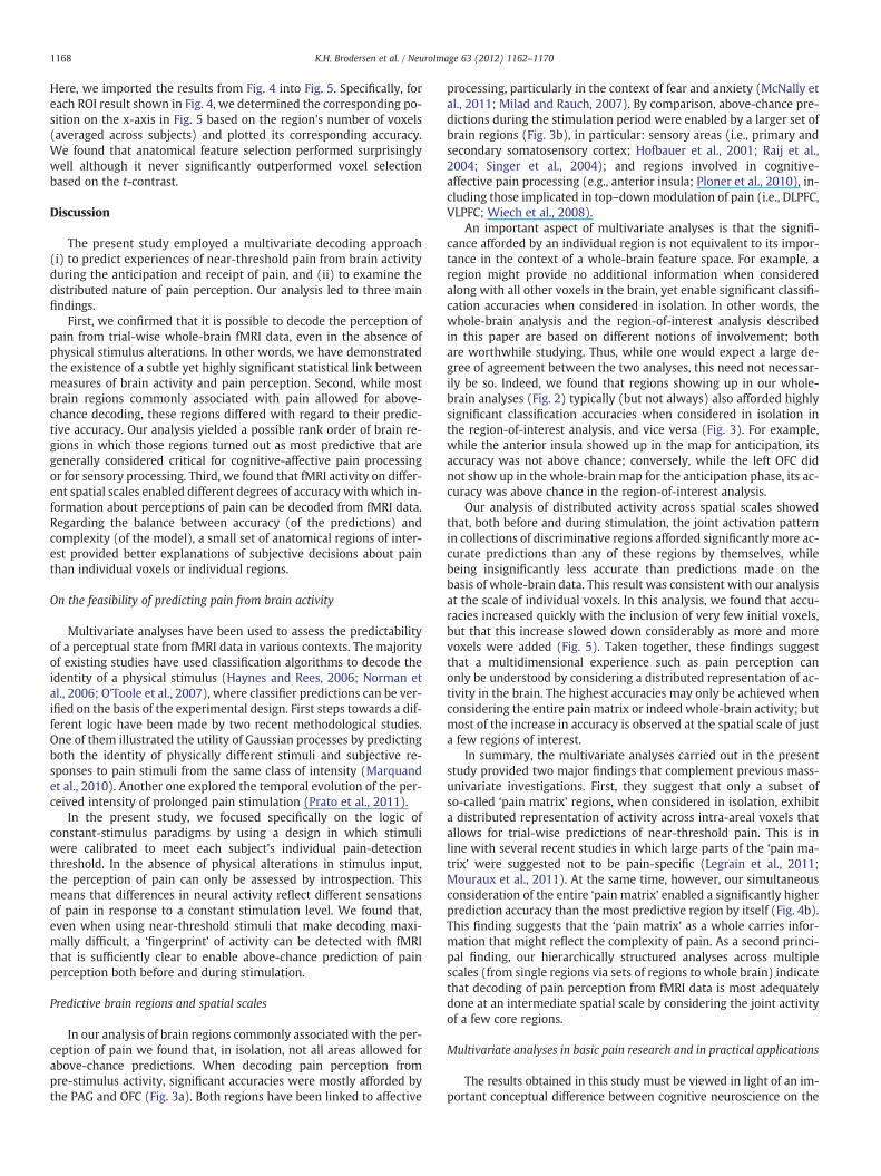
Fig. 2. Pain perception in individual voxels. This discriminative map shows the statistical ssubjects). Separate analyses were conducted based on brain activity (a) before and (b) durhigher on subjectively painful trials than on non-painful trials, whereas blue regions re(pb0.05, FWE-corrected, see Methods section). Results are overlaid onto a standard structuraccuracies when using a whole-brain feature space. Both are significantly above chance (pb0the reader is referred to the web version of this article.)
Results
Decoding pain from whole-brain data
Asking whether subtle trial-by-trial variations in distributed brainactivity could be used to predict perceptual decisions about pain, wetrained and tested a linear SVM on trial-wise correlates of fMRIdata. We found that whole-brain activity both before (57.6%) andduring (61.4%) the application of a near-threshold laser stimulusenabled predictions significantly above chance level (pb0.001; non-parametric permutation test using N=1000 permutations).
For completeness, we tested which subjects showed effects thatwere significant even when considered in isolation (see SupplementalFig. S1).We found that 1 out of 16 subjects by itself showed an accuracythat was significantly above chance in the anticipation phase, and 3 outof 16 in the stimulation phase (pb0.05; nonparametric permutationtest using N=1000; Bonferroni-corrected).
Though not of interest in the present study, we also examinedwhether the condition (‘threat’ vs. ‘no threat’) could be predictedfrom trial-wise fMRI data, in particular in the anticipation phase inwhich the condition was disclosed on the screen; we obtained meanclassification accuracies of 55.7% (pb0.05) before and 52.2% (n.s.)during the stimulation.
In order to provide an intuition about which data features were re-sponsible for above-chance accuracies in predicting pain, we exam-ined the spatial deployment of voxels that played an important rolein classification. To this end, we adopted an exploratory perspectiveand used an SVM with a permutation test on feature weights tomap discriminative information continuously throughout the brain.This approach highlights those voxels that are most influential inshaping the separating hyperplane used by the SVM to distinguish be-tween painful and non-painful trials (Fig. 2). In contrast to other com-monly used multivariate techniques, such as searchlight classificationmethods that are locally multivariate, our approach simultaneouslyconsiders all voxels in the brain.
painful
+8
6252423222
ignificance of voxel weights obtained by training a linear support vector machine (16ing stimulus application. Regions highlighted in red represent voxels whose activity ispresent voxels whose activity is higher on non-painful trials than on painful trialsal scan in MNI152 space. The percentages on the left indicate the resulting classification.001; see main text). (For interpretation of the references to color in this figure legend,

1166 K.H. Brodersen et al. / NeuroImage 63 (2012) 1162–1170
We found that in the anticipation period, the bilateral insulaproved most predictive for the perception of pain. In the stimulationperiod, brain activity in the mid cingulate cortex (MCC), SI/SII, bilater-al insula and orbitofrontal cortex (OFC) allowed for significant predic-tions of the perception of pain (for a comparison with a conventionalmass-univariate map, see Fig. S2 in the Supplemental material).
Decoding pain from individual regions of interest
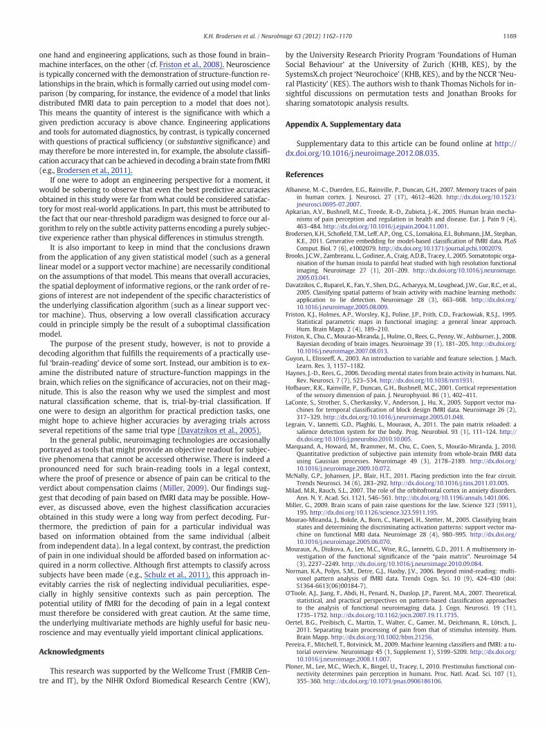
Having predicted pain perception based onwhole-brain activity, wenext asked whether it is possible to predict pain perception based onfMRI activity within individual brain regions. To investigate this, weattempted to decode the anticipation and perception of pain from 26predefined brain areas typically reported in the context of pain (plus 2control areas). It should be noted that these regions were defined apriori on anatomical grounds, independently from the results of theabovewhole-brain analysis. We assessed the importance of each regionin terms of the significance withwhich it enabled above-chance predic-tions. This approach allowed us to propose a rank order of pain-relatedregions. Critically, this rank order is based on significance rather thanaccuracy, and thus takes into account not only the mean accuracy butalso its between-subjects variability (Fig. 3).
It should be noted that the whole-brain analysis in the previoussection and the region-of-interest analysis in this section are basedon different notions of involvement. Thus, one would expect their re-sults to share the most important, but not necessarily all, characteris-tics. We will expand on this point in the Discussion.
Of all brain regions commonly associated with the perception ofpain, our analysis revealed that only a subset was predictive of painon a trial-by-trial basis. The most predictive regions during the anti-cipation period were the right and left periaqueductal gray (PAG)and right orbitofrontal cortex (OFC; all pb0.05; permutation testusing N=2600; Bonferroni-corrected for multiple testing acrossROIs; Fig. 3a). During the stimulation period, the most predictive re-gions were the right and left primary somatosensory cortex (SI),right anterior insula, right secondary somatosensory cortex, right
50% 52% 54% 56% 58% 60%
VLPFC.RHG.LPI.L
rACC.RSII.R
DLPFC.LrACC.L
dACC.RDLPFC.RVLPFC.L
HG.RTHA.RTHA.L
PI.RMI.R
AMYG.LdACC.L
MI.LSII.LAI.RSI.RAI.LSI.L
AMYG.ROFC.LPAG.LOFC.RPAG.R
a Decoding pain during anticipation
prediction acc
p < 0.05n.s.
Fig. 3. Pain perception in individual regions of interest. The figure shows prediction accuraciepainful or non-painful (a) before and (b) during stimulation (plus two control regions, HG.L abased on 16 subjects. Statistical inference is based on a nonparametric permutation test wigions are sorted by the significance of prediction accuracies (p-values), not by their magnitrostral anterior cingulate cortex; DLPFC/VLPFC=dorsolateral/ventrolateral prefrontal cortexprimary/secondary somatosensory cortex; THA = thalamus; *.R=right; *.L=left.
and left dorsolateral prefrontal cortex (DLPFC), left ventrolateral cortex(VLPFC), and right OFC (all pb0.05; N=2600; corrected; Fig. 3b). Interms of magnitudes, in the anticipation period, the highest classifica-tion accuracy was afforded by the right PAG, while the most accuratepredictions for the stimulation period were enabled by activity in theleft SI. By contrast, no above-chance performance was obtained whenusing gray-matter control masks of regions not involved in pain pro-cessing (left and right Heschl's gyrus, HG.L and HG.R).
It should be kept in mind that all of the above numbers are sampleaccuracies which serve as estimates of their corresponding unknownpopulation mean accuracies. The uncertainty associated with theseestimates (i.e., their standard error) is non-negligible since it reflectsthe between-subjects variability in the population (i.e., random ef-fects). The rank order of regions is particularly sensitive to this vari-ability and should therefore only be interpreted as an approximateguide to true differences in regional informativeness.
Onefinding, perhaps surprising atfirst,was the lack of above-chanceprediction accuracy obtained in the signal of the mid and posteriorinsula during stimulation. Thiswould be in contrast to previousfindingsin which both insula subdivisions have been implicated instimulus-dependent processing (Albanese et al., 2007; Raij et al.,2005; Singer et al., 2004). The posterior insula in particular is consid-ered a key region for nociceptive processing (Oertel et al., 2011). It isconceivable that this seeming discrepancy is a result of the way inwhich our anatomical maskswere defined. As highlighted before, all re-gions of interest were defined on the basis of anatomical landmarks, notfunctional contrasts, and all voxels within a given area entered the re-spective multivariate analysis. Some regions, such as the posteriorinsula, are known to be somatotopically organized (Brooks et al.,2005), suggesting that predictions from the entire region might notfaithfully reflect the impact of a particular somatotopically relevant sub-region. Under this view, the surprisingly low accuracies in the posteriorinsula might be a consequence of the functional heterogeneity acrossanatomical subdivisions included in our anatomical mask.
We tested this hypothesis in an additional post-hoc analysis inwhich we utilized results from a previous study investigating the
50% 52% 54% 56% 58% 60%
HG.RPAG.R
PI.LHG.L
PAG.LPI.RMI.L
rACC.RAMYG.R
rACC.LdACC.R
MI.RAI.L
THA.LdACC.L
SII.LAMYG.L
VLPFC.RTHA.ROFC.LOFC.R
VLPFC.LDLPFC.LDLPFC.R
SII.RAI.RSI.RSI.L
b Decoding pain during stimulation
uracy +/–s.e.m.
s obtained in 26 regions of interest for the differentiation between trials experienced asnd HG.R). Results are given in terms of mean accuracy +/− standard error of the mean,th N=2600 permutations and Bonferroni correction for multiple testing. Note that re-ude. AI/MI/PI=anterior/mid/posterior insula; AMYG=amygdala; dACC/rACC=dorsal/; HG=Heschl's gyrus; OFC=orbitofrontal cortex; PAG=periaqueductal gray; SI/SII=

1167K.H. Brodersen et al. / NeuroImage 63 (2012) 1162–1170
somatotopic organization of the insula (Brooks et al., 2005) to consid-er those portions of the posterior insula that had been implicated inthe processing of somatosensory stimuli applied to the same site asin the present experiment (main effect of pain, N=14, thresholdedat p=0.001). Contrary to our initial hypothesis, the prediction accu-racy obtained in foot-specific portions of the (left) posterior insulawas not significantly above chance during the stimulation period.
Comparison of different spatial scales
As indicated above, activity patterns in several pain-related regionsallow for the prediction of pain, both during anticipation and duringstimulation. However, this does not necessarily imply that predictionsbecome evenmore accurate when considering several brain regions si-multaneously. To investigatewhether thismight be the case, we carriedout two additional analyses in which we examined increasing spatialscales of encoding.
In the first analysis, we focused on pain-related brain regions andtested whether prediction accuracies would benefit from increasingthe search space (i.e., increasing the potential complexity of themodel) from the most predictive single region to combinations ofmultiple regions (Fig. 4). In the anticipation period, the most predictivesingle ROI (i.e., the right PAG) yielded a prediction accuracy of 56%(pb0.01; nonparametric permutation test using N=1000; Fig. 4a). Wefound that this accuracy increased continuously when jointly consider-ing additional regions and reached a significantly higher level (pb0.05;N=1000) when using the five most predictive regions (i.e., PAG, OFC,and right amygdala). Following this, the inclusion of additional regionsdid not yield further improvements in prediction accuracy, suggestingthat the activation patterns in small combinations of regions, such asthese five, encode sufficient complementary information to predict theperception of pain. Similarly, we observed no (significant) further im-provement in prediction accuracy by moving to a whole-brain analysis.
50% 55% 60%
whole brain
pain matrix
+DLPFC.R
+SII.R
+AI.R
SI.L
prediction accuracy +/–s.e.m.
anticipation
b stimulation
50% 55% 60%
whole brain
pain matrix
+AMYG.R
+OFC.L
+PAG.L
+OFC.R
PAG.R
*
n.s.
*
n.s.
+SI.R
a
Fig. 4. Pain perception in combinations of highly predictive regions. The figure showsprediction accuracies for the classification of painful versus non-painful trials, usingdifferent sizes of search space, (a) before and (b) during stimulation, based on 16 sub-jects. Results are given in terms of mean accuracy +/− standard error of the mean. Allaccuracies are significantly above chance (pb0.01; nonparametric permutation test;N=1000). Additional significances are indicated between accuracies on different setsof regions (*pb0.05; permutation test).
These findings agreed nicely with the results that emerged from ouranalysis of the stimulation period. While being exposed to a stimulus,pain predictions from left SI, which was the most significant region,reached an accuracy of 59% (pb0.01; nonparametric permutation testusing N=1000). Predictions from the combination of left SI and otherbrain regions that allowed for prediction of pain when considered inisolation (i.e., right SI, AI, SII, and DLPFC) did not lead to a significant in-crease in prediction accuracy. However, adding in the entire ‘pain ma-trix’ yielded a significant increase (pb0.05; N=1000) in predictionaccuracy (to 62%) (Fig. 4b). This observation indicates that, while theoverall accuracy is still well below 100%, the joint activation pattern ofregions commonly summarized as the ‘pain matrix’ might enable thebest predictions about pain perception that can be made on the basisof fMRI activity measures. Thus, both in the anticipation phase and inthe stimulation phase, accuracies reached an optimum on the basis ofa set of anatomical regions, with no further improvement enabled byconsidering whole-brain activity.
In the second analysis, we examined a search space that was notbased on anatomical regions of interest; instead, we considered indi-vidual voxels without anatomical constraints. We began with the sin-gle most predictive voxel, then tripled the number of voxels in eachstep, until the search space corresponded to a whole-brain analysis.Voxel-wise predictive strength was measured, independently withineach cross-validation fold, in terms of t-scores, using a between-conditions two-tailed t-test. Using these discriminative scores, wefound that the resulting prediction accuracies increased near-monotonically with the number of voxels considered, both in the an-ticipation and in the stimulation period, and leveled off towards theend (Fig. 5). Prediction of pain from the anticipation period increasedfrom 52% (using a single voxel) to 58% (using all voxels; significantlyabove chance, pb0.001; nonparametric permutation test; N=1000).For the stimulation period, this accuracy increased from 54% (usinga single voxel) to 61% (using all voxels, pb0.001; N=1000). Critically,the most rapid increase in accuracy was observed at small voxel num-bers, while subsequent additions made very little contributions com-pared to the number of additional model parameters.
Overall, using a whole-brain search for the most informativevoxels yielded higher prediction accuracies than the combinationsof anatomical regions analyzed before. One might ask, of course,whether the two approaches could be compared on a finer scale.
45%
50%
55%
60%
number of voxels30 31 32 33 34 35 36 37 38 39 whole
brain
most informative voxelsanatomical ROIs
most informative voxelsanatomical ROIs
anticipation
stimulationchance
pred
ictio
n ac
cura
cy +
/– s
.e.m
.
Fig. 5. Pain perception across different scales. The two diagrams show the role of differentspatial scales in relating brain activity to the perception of pain (a) before (blue) and(b) during (red) stimulus application, based on 16 subjects. Results are given interms of mean accuracy +/− standard error of the mean. The gray horizontal barindicates chance level (50%). All accuracies from 31 (3) voxels onwards are significantlyabove chance (pb0.05; nonparametric permutation test; N=1000). For direct compari-son of different strategies for feature selection,we imported the results from Fig. 4. Specif-ically, for each ROI shown in Fig. 4, we determined the number of voxels in the underlyinganatomical mask (averaged across subjects).We then plotted the ROI-based accuracies atthe corresponding locations on the x-axis (green and yellow lines). (For interpretation ofthe references to color in thisfigure legend, the reader is referred to theweb version of thisarticle.)
1168 K.H. Brodersen et al. / NeuroImage 63 (2012) 1162–1170
Here, we imported the results from Fig. 4 into Fig. 5. Specifically, foreach ROI result shown in Fig. 4, we determined the corresponding po-sition on the x-axis in Fig. 5 based on the region's number of voxels(averaged across subjects) and plotted its corresponding accuracy.We found that anatomical feature selection performed surprisinglywell although it never significantly outperformed voxel selectionbased on the t-contrast.
Discussion
The present study employed a multivariate decoding approach(i) to predict experiences of near-threshold pain from brain activityduring the anticipation and receipt of pain, and (ii) to examine thedistributed nature of pain perception. Our analysis led to three mainfindings.
First, we confirmed that it is possible to decode the perception ofpain from trial-wise whole-brain fMRI data, even in the absence ofphysical stimulus alterations. In other words, we have demonstratedthe existence of a subtle yet highly significant statistical link betweenmeasures of brain activity and pain perception. Second, while mostbrain regions commonly associated with pain allowed for above-chance decoding, these regions differed with regard to their predic-tive accuracy. Our analysis yielded a possible rank order of brain re-gions in which those regions turned out as most predictive that aregenerally considered critical for cognitive-affective pain processingor for sensory processing. Third, we found that fMRI activity on differ-ent spatial scales enabled different degrees of accuracy with which in-formation about perceptions of pain can be decoded from fMRI data.Regarding the balance between accuracy (of the predictions) andcomplexity (of the model), a small set of anatomical regions of inter-est provided better explanations of subjective decisions about painthan individual voxels or individual regions.
On the feasibility of predicting pain from brain activity
Multivariate analyses have been used to assess the predictabilityof a perceptual state from fMRI data in various contexts. The majorityof existing studies have used classification algorithms to decode theidentity of a physical stimulus (Haynes and Rees, 2006; Norman etal., 2006; O'Toole et al., 2007), where classifier predictions can be ver-ified on the basis of the experimental design. First steps towards a dif-ferent logic have been made by two recent methodological studies.One of them illustrated the utility of Gaussian processes by predictingboth the identity of physically different stimuli and subjective re-sponses to pain stimuli from the same class of intensity (Marquandet al., 2010). Another one explored the temporal evolution of the per-ceived intensity of prolonged pain stimulation (Prato et al., 2011).
In the present study, we focused specifically on the logic ofconstant-stimulus paradigms by using a design in which stimuliwere calibrated to meet each subject's individual pain-detectionthreshold. In the absence of physical alterations in stimulus input,the perception of pain can only be assessed by introspection. Thismeans that differences in neural activity reflect different sensationsof pain in response to a constant stimulation level. We found that,even when using near-threshold stimuli that make decoding maxi-mally difficult, a ‘fingerprint’ of activity can be detected with fMRIthat is sufficiently clear to enable above-chance prediction of painperception both before and during stimulation.
Predictive brain regions and spatial scales
In our analysis of brain regions commonly associated with the per-ception of pain we found that, in isolation, not all areas allowed forabove-chance predictions. When decoding pain perception frompre-stimulus activity, significant accuracies were mostly afforded bythe PAG and OFC (Fig. 3a). Both regions have been linked to affective
processing, particularly in the context of fear and anxiety (McNally etal., 2011; Milad and Rauch, 2007). By comparison, above-chance pre-dictions during the stimulation period were enabled by a larger set ofbrain regions (Fig. 3b), in particular: sensory areas (i.e., primary andsecondary somatosensory cortex; Hofbauer et al., 2001; Raij et al.,2004; Singer et al., 2004); and regions involved in cognitive-affective pain processing (e.g., anterior insula; Ploner et al., 2010), in-cluding those implicated in top–downmodulation of pain (i.e., DLPFC,VLPFC; Wiech et al., 2008).
An important aspect of multivariate analyses is that the signifi-cance afforded by an individual region is not equivalent to its impor-tance in the context of a whole-brain feature space. For example, aregion might provide no additional information when consideredalong with all other voxels in the brain, yet enable significant classifi-cation accuracies when considered in isolation. In other words, thewhole-brain analysis and the region-of-interest analysis describedin this paper are based on different notions of involvement; bothare worthwhile studying. Thus, while one would expect a large de-gree of agreement between the two analyses, this need not necessar-ily be so. Indeed, we found that regions showing up in our whole-brain analyses (Fig. 2) typically (but not always) also afforded highlysignificant classification accuracies when considered in isolation inthe region-of-interest analysis, and vice versa (Fig. 3). For example,while the anterior insula showed up in the map for anticipation, itsaccuracy was not above chance; conversely, while the left OFC didnot show up in the whole-brain map for the anticipation phase, its ac-curacy was above chance in the region-of-interest analysis.
Our analysis of distributed activity across spatial scales showedthat, both before and during stimulation, the joint activation patternin collections of discriminative regions afforded significantly more ac-curate predictions than any of these regions by themselves, whilebeing insignificantly less accurate than predictions made on thebasis of whole-brain data. This result was consistent with our analysisat the scale of individual voxels. In this analysis, we found that accu-racies increased quickly with the inclusion of very few initial voxels,but that this increase slowed down considerably as more and morevoxels were added (Fig. 5). Taken together, these findings suggestthat a multidimensional experience such as pain perception canonly be understood by considering a distributed representation of ac-tivity in the brain. The highest accuracies may only be achieved whenconsidering the entire pain matrix or indeed whole-brain activity; butmost of the increase in accuracy is observed at the spatial scale of justa few regions of interest.
In summary, the multivariate analyses carried out in the presentstudy provided two major findings that complement previous mass-univariate investigations. First, they suggest that only a subset ofso-called ‘pain matrix’ regions, when considered in isolation, exhibita distributed representation of activity across intra-areal voxels thatallows for trial-wise predictions of near-threshold pain. This is inline with several recent studies in which large parts of the ‘pain ma-trix’ were suggested not to be pain-specific (Legrain et al., 2011;Mouraux et al., 2011). At the same time, however, our simultaneousconsideration of the entire ‘pain matrix’ enabled a significantly higherprediction accuracy than the most predictive region by itself (Fig. 4b).This finding suggests that the ‘pain matrix’ as a whole carries infor-mation that might reflect the complexity of pain. As a second princi-pal finding, our hierarchically structured analyses across multiplescales (from single regions via sets of regions to whole brain) indicatethat decoding of pain perception from fMRI data is most adequatelydone at an intermediate spatial scale by considering the joint activityof a few core regions.
Multivariate analyses in basic pain research and in practical applications
The results obtained in this study must be viewed in light of an im-portant conceptual difference between cognitive neuroscience on the
1169K.H. Brodersen et al. / NeuroImage 63 (2012) 1162–1170
one hand and engineering applications, such as those found in brain–machine interfaces, on the other (cf. Friston et al., 2008). Neuroscienceis typically concerned with the demonstration of structure-function re-lationships in the brain, which is formally carried out usingmodel com-parison (by comparing, for instance, the evidence of a model that linksdistributed fMRI data to pain perception to a model that does not).This means the quantity of interest is the significance with which agiven prediction accuracy is above chance. Engineering applicationsand tools for automated diagnostics, by contrast, is typically concernedwith questions of practical sufficiency (or substantive significance) andmay therefore be more interested in, for example, the absolute classifi-cation accuracy that can be achieved in decoding a brain state from fMRI(e.g., Brodersen et al., 2011).
If one were to adopt an engineering perspective for a moment, itwould be sobering to observe that even the best predictive accuraciesobtained in this study were far fromwhat could be considered satisfac-tory for most real-world applications. In part, this must be attributed tothe fact that our near-threshold paradigmwas designed to force our al-gorithm to rely on the subtle activity patterns encoding a purely subjec-tive experience rather than physical differences in stimulus strength.
It is also important to keep in mind that the conclusions drawnfrom the application of any given statistical model (such as a generallinear model or a support vector machine) are necessarily conditionalon the assumptions of that model. This means that overall accuracies,the spatial deployment of informative regions, or the rank order of re-gions of interest are not independent of the specific characteristics ofthe underlying classification algorithm (such as a linear support vec-tor machine). Thus, observing a low overall classification accuracycould in principle simply be the result of a suboptimal classificationmodel.
The purpose of the present study, however, is not to provide adecoding algorithm that fulfills the requirements of a practically use-ful ‘brain-reading’ device of some sort. Instead, our ambition is to ex-amine the distributed nature of structure-function mappings in thebrain, which relies on the significance of accuracies, not on their mag-nitude. This is also the reason why we used the simplest and mostnatural classification scheme, that is, trial-by-trial classification. Ifone were to design an algorithm for practical prediction tasks, onemight hope to achieve higher accuracies by averaging trials acrossseveral repetitions of the same trial type (Davatzikos et al., 2005).
In the general public, neuroimaging technologies are occasionallyportrayed as tools that might provide an objective readout for subjec-tive phenomena that cannot be accessed otherwise. There is indeed apronounced need for such brain-reading tools in a legal context,where the proof of presence or absence of pain can be critical to theverdict about compensation claims (Miller, 2009). Our findings sug-gest that decoding of pain based on fMRI data may be possible. How-ever, as discussed above, even the highest classification accuraciesobtained in this study were a long way from perfect decoding. Fur-thermore, the prediction of pain for a particular individual wasbased on information obtained from the same individual (albeitfrom independent data). In a legal context, by contrast, the predictionof pain in one individual should be afforded based on information ac-quired in a norm collective. Although first attempts to classify acrosssubjects have been made (e.g., Schulz et al., 2011), this approach in-evitably carries the risk of neglecting individual peculiarities, espe-cially in highly sensitive contexts such as pain perception. Thepotential utility of fMRI for the decoding of pain in a legal contextmust therefore be considered with great caution. At the same time,the underlying multivariate methods are highly useful for basic neu-roscience and may eventually yield important clinical applications.
Acknowledgments
This research was supported by the Wellcome Trust (FMRIB Cen-tre and IT), by the NIHR Oxford Biomedical Research Centre (KW),
by the University Research Priority Program ‘Foundations of HumanSocial Behaviour’ at the University of Zurich (KHB, KES), by theSystemsX.ch project ‘Neurochoice’ (KHB, KES), and by the NCCR ‘Neu-ral Plasticity’ (KES). The authors wish to thank Thomas Nichols for in-sightful discussions on permutation tests and Jonathan Brooks forsharing somatotopic analysis results.
Appendix A. Supplementary data
Supplementary data to this article can be found online at http://dx.doi.org/10.1016/j.neuroimage.2012.08.035.
References
Albanese, M.-C., Duerden, E.G., Rainville, P., Duncan, G.H., 2007. Memory traces of painin human cortex. J. Neurosci. 27 (17), 4612–4620. http://dx.doi.org/10.1523/jneurosci.0695-07.2007.
Apkarian, A.V., Bushnell, M.C., Treede, R.-D., Zubieta, J.-K., 2005. Human brain mecha-nisms of pain perception and regulation in health and disease. Eur. J. Pain 9 (4),463–484. http://dx.doi.org/10.1016/j.ejpain.2004.11.001.
Brodersen, K.H., Schofield, T.M., Leff, A.P., Ong, C.S., Lomakina, E.I., Buhmann, J.M., Stephan,K.E., 2011. Generative embedding for model-based classification of fMRI data. PLoSComput. Biol. 7 (6), e1002079. http://dx.doi.org/10.1371/journal.pcbi.1002079.
Brooks, J.C.W., Zambreanu, L., Godinez, A., Craig, A.D.B., Tracey, I., 2005. Somatotopic orga-nisation of the human insula to painful heat studied with high resolution functionalimaging. Neuroimage 27 (1), 201–209. http://dx.doi.org/10.1016/j.neuroimage.2005.03.041.
Davatzikos, C., Ruparel, K., Fan, Y., Shen, D.G., Acharyya, M., Loughead, J.W., Gur, R.C., et al.,2005. Classifying spatial patterns of brain activity with machine learning methods:application to lie detection. Neuroimage 28 (3), 663–668. http://dx.doi.org/10.1016/j.neuroimage.2005.08.009.
Friston, K.J., Holmes, A.P., Worsley, K.J., Poline, J.P., Frith, C.D., Frackowiak, R.S.J., 1995.Statistical parametric maps in functional imaging: a general linear approach.Hum. Brain Mapp. 2 (4), 189–210.
Friston, K., Chu, C., Mourao-Miranda, J., Hulme, O., Rees, G., Penny,W., Ashburner, J., 2008.Bayesian decoding of brain images. Neuroimage 39 (1), 181–205. http://dx.doi.org/10.1016/j.neuroimage.2007.08.013.
Guyon, I., Elisseeff, A., 2003. An introduction to variable and feature selection. J. Mach.Learn. Res. 3, 1157–1182.
Haynes, J.-D., Rees, G., 2006. Decoding mental states from brain activity in humans. Nat.Rev. Neurosci. 7 (7), 523–534. http://dx.doi.org/10.1038/nrn1931.
Hofbauer, R.K., Rainville, P., Duncan, G.H., Bushnell, M.C., 2001. Cortical representationof the sensory dimension of pain. J. Neurophysiol. 86 (1), 402–411.
LaConte, S., Strother, S., Cherkassky, V., Anderson, J., Hu, X., 2005. Support vector ma-chines for temporal classification of block design fMRI data. Neuroimage 26 (2),317–329. http://dx.doi.org/10.1016/j.neuroimage.2005.01.048.
Legrain, V., Iannetti, G.D., Plaghki, L., Mouraux, A., 2011. The pain matrix reloaded: asalience detection system for the body. Prog. Neurobiol. 93 (1), 111–124. http://dx.doi.org/10.1016/j.pneurobio.2010.10.005.
Marquand, A., Howard, M., Brammer, M., Chu, C., Coen, S., Mourão-Miranda, J., 2010.Quantitative prediction of subjective pain intensity from whole-brain fMRI datausing Gaussian processes. Neuroimage 49 (3), 2178–2189. http://dx.doi.org/10.1016/j.neuroimage.2009.10.072.
McNally, G.P., Johansen, J.P., Blair, H.T., 2011. Placing prediction into the fear circuit.Trends Neurosci. 34 (6), 283–292. http://dx.doi.org/10.1016/j.tins.2011.03.005.
Milad, M.R., Rauch, S.L., 2007. The role of the orbitofrontal cortex in anxiety disorders.Ann. N. Y. Acad. Sci. 1121, 546–561. http://dx.doi.org/10.1196/annals.1401.006.
Miller, G., 2009. Brain scans of pain raise questions for the law. Science 323 (5911),195. http://dx.doi.org/10.1126/science.323.5911.195.
Mourao-Miranda, J., Bokde, A., Born, C., Hampel, H., Stetter, M., 2005. Classifying brainstates and determining the discriminating activation patterns: support vector ma-chine on functional MRI data. Neuroimage 28 (4), 980–995. http://dx.doi.org/10.1016/j.neuroimage.2005.06.070.
Mouraux, A., Diukova, A., Lee, M.C., Wise, R.G., Iannetti, G.D., 2011. A multisensory in-vestigation of the functional significance of the “pain matrix”. Neuroimage 54(3), 2237–2249. http://dx.doi.org/10.1016/j.neuroimage.2010.09.084.
Norman, K.A., Polyn, S.M., Detre, G.J., Haxby, J.V., 2006. Beyond mind-reading: multi-voxel pattern analysis of fMRI data. Trends Cogn. Sci. 10 (9), 424–430 (doi:S1364-6613(06)00184-7).
O'Toole, A.J., Jiang, F., Abdi, H., Penard, N., Dunlop, J.P., Parent, M.A., 2007. Theoretical,statistical, and practical perspectives on pattern-based classification approachesto the analysis of functional neuroimaging data. J. Cogn. Neurosci. 19 (11),1735–1752. http://dx.doi.org/10.1162/jocn.2007.19.11.1735.
Oertel, B.G., Preibisch, C., Martin, T., Walter, C., Gamer, M., Deichmann, R., Lötsch, J.,2011. Separating brain processing of pain from that of stimulus intensity. Hum.Brain Mapp. http://dx.doi.org/10.1002/hbm.21256.
Pereira, F., Mitchell, T., Botvinick, M., 2009. Machine learning classifiers and fMRI: a tu-torial overview. Neuroimage 45 (1, Supplement 1), S199–S209. http://dx.doi.org/10.1016/j.neuroimage.2008.11.007.
Ploner, M., Lee, M.C., Wiech, K., Bingel, U., Tracey, I., 2010. Prestimulus functional con-nectivity determines pain perception in humans. Proc. Natl. Acad. Sci. 107 (1),355–360. http://dx.doi.org/10.1073/pnas.0906186106.

1170 K.H. Brodersen et al. / NeuroImage 63 (2012) 1162–1170
Prato, M., Favilla, S., Zanni, L., Porro, C.A., Baraldi, P., 2011. A regularization algorithmfor decoding perceptual temporal profiles from fMRI data. Neuroimage 56,258–267. http://dx.doi.org/10.1016/j.neuroimage.2011.01.074.
Raij, T.T., Forss, N., Stancák, A., Hari, R., 2004. Modulation of motor-cortex oscillatoryactivity by painful Adelta- and C-fiber stimuli. Neuroimage 23 (2), 569–573.http://dx.doi.org/10.1016/j.neuroimage.2004.06.036.
Raij, T.T., Numminen, J., Närvänen, S., Hiltunen, J., Hari, R., 2005. Brain correlates of sub-jective reality of physically and psychologically induced pain. Proc. Natl. Acad. Sci.102 (6), 2147–2151. http://dx.doi.org/10.1073/pnas.0409542102.
Schulz, E., Zherdin, A., Tiemann, L., Plant, C., Ploner, M., 2011. Decoding an individual'ssensitivity to pain from the multivariate analysis of EEG data. Cereb. Cortex. http://dx.doi.org/10.1093/cercor/bhr186.
Singer, T., Seymour, B., O'Doherty, J., Kaube, H., Dolan, R.J., Frith, C.D., 2004. Empathy forpain involves the affective but not sensory components of pain. Science 303(5661), 1157–1162. http://dx.doi.org/10.1126/science.1093535.
Wang, Z., Childress, A.R., Wang, J., Detre, J.A., 2007. Support vector machine learning-based fMRI data group analysis. Neuroimage 36 (4), 1139–1151. http://dx.doi.org/10.1016/j.neuroimage.2007.03.072.
Wiech, K., Ploner, M., Tracey, I., 2008. Neurocognitive aspects of pain perception.Trends Cogn. Sci. 12 (8), 306–313. http://dx.doi.org/10.1016/j.tics.2008.05.005.
Wiech, K., Lin, C.-S., Brodersen, K.H., Bingel, U., Ploner, M., Tracey, I., 2010. Anteriorinsula integrates information about salience into perceptual decisions aboutpain. J. Neurosci. 30 (48), 16324–16331. http://dx.doi.org/10.1523/jneurosci.2087-10.2010.