Declaration - PolyU Electronic Theses
91
-
Upload
khangminh22 -
Category
Documents
-
view
1 -
download
0
Transcript of Declaration - PolyU Electronic Theses


Declaration
I hereby declare that this thesis represents my own research work which has been done
within the period from September 2003 to August 2005 for the degree of Master of
Philosophy. This thesis has not been previously included in a thesis, dissertation or report
submitted to this or any other institutions for a degree, a diploma or other qualifications.
Levina Suk Mi Lam
April 2006

Contents
Contents
Acknowledgments i Abbreviations ii List of Figures and Tables iv Abstract vi I Introduction 1 1.1 Oxidative stress and aging 1 1.2 Thioredoxin system 3 1.3 Biological roles of thioredoxin system 5 1.3.1 DNA synthesis 5 1.3.2 Antioxidation 5 1.3.3 Regulation of apoptosis 6 1.3.4 Hormone action and cytokine functions involvement 6 1.3.5 Regulation of transcriptional factors 7 1.4 Local concentration, tissue distribution and subcellular localization of Trx 8
and TR 1.5 Thioredoxin system and oxidative stress 9 1.6 Potential applications of thioredoxin system in medical aspects 11 1.7 Objective 13
II Methodology 15 Section I Methods and materials 15 Section II Preparation of young and old erythrocytes 16 2.1 Separation of young and old erythrocytes from porcine whole blood 17 2.1.1 Separation of erythrocytes into young and old population 17
2.2 Assessment of the separation of young and old erythrocytes 18 2.2.1 Red blood cell counting 18 2.2.2 Determination of haemoglobin concentration of young and old 18
erythrocytes 2.2.3 Determination of mean corpuscular volume in young and old 19
erythrocytes 2.2.4 Determination of membrane acetylcholinesterase activity of young 19 and old erythrocytes 2.3 Preparation of young and old red cell lysates for analysis 20 2.3.1 Removal of haemoglobin from red cell haemolysates by cation 20 exchange chromatography
2.3.2 Concentration of young and old sample by ultrafiltration 21 2.4 Analysis of young and old sample 21 2.4.1 Western Blot analysis of Trx in young and old sample 21 2.4.2 Western Blot analysis of TR in young and old sample 22

Contents
2.4.3 Determination of Trx activity by insulin dependent DTNB activity 22 assay 2.4.4 Determination of TR activity by insulin dependent DTNB activity 23 assay 2.4.5 Functional studies of thioredoxin system by IgG reduction assay 24 Section III Preparation of HeLa and HT-29 lysates 24 2.5 Culture of HeLa and HT-29 cell line 24 2.6 Oxidative challenge on HeLa and HT-29 cell line 25 2.7 Cell viability by Trypan Blue dye exclusion assay 25 2.8 Cell proliferation by MTS tetrazolium assay 25 2.9 Cell extracts preparation 26 2.10 Analysis of cell lysates after oxidative challenge 26 2.10.1 Western Blot analysis of Trx in cell lysates after oxidative challenge 27 2.10.2 Western Blot analysis of TR in cell lysates after oxidative challenge 27 2.10.3 Functional studies of thioredoxin system in HeLa and HT-29 28 III Results 29 3.1 Separation of young and old erythrocytes from porcine whole blood 29 3.1.1 Red blood cell counting 29 3.1.2 Haemoglobin concentration determination 30 3.1.3 Mean corpuscular volume determination 31 3.1.4 Membrane acetylcholinesterase activity determination 32 3.2 Partial purification and concentration of young and old haemolysates 33 3.3 Analysis of young and old sample 34 3.3.1 Western blotting analysis of TR in young and old sample 34 3.3.2 Western blotting analysis of Trx in young and old sample 35 3.3.3 Trx and TR activity assay 36 3.3.4 IgG reduction assay of young and old haemolysates upon oxidative 38 challenge 3.4 Culture of HeLa and HT-29 cell line 40 3.4.1 Cell viability of HeLa after oxidative stress by Trypan Blue dye 41 exclusion assay 3.4.2 Cell viability of HeLa after oxidative challenge by MTS tetrazolium 43
assay 3.4.3 Western blotting analysis of Trx and TR in HeLa cells upon oxidative 45 challenge
3.4.4 Western blotting analysis of Trx and TR in HeLa cells upon oxidative 46 challenge and with prior treatment of cycloheximide
3.3.5 Activity of thioredoxin system in HeLa cells upon oxidative 48 challenge without prior treatment of cycloheximide
3.3.6 Activity of thioredoxin system in HeLa cells upon oxidative challenge 51 with prior treatment of cycloheximide 3.4.7 Cell viability of HT-29 after oxidative challenge by Trypan Blue dye 55 exclusion assay

Contents
3.4.8 Cell viability of HT-29 after oxidative challenge by MTS tetrazolium 57 assay
3.4.9 Activity of thioredoxin system in HT-29 cells upon oxidative challenge 58 with or without prior treatment of cycloheximide IV Discussion 62 V Conclusion 73 References 75
Acknowledgment
Acknowledgments
I would like to extend my sincere gratitude and appreciation to my supervisor
Dr. K. S. Lee for his valuable advice, patient guidance and kind encouragement
throughout my study. I am indebted to my co-supervisor Dr. T. Leung for his support.
Many thanks also go to Dr. N. S. Wong of The University of Hong Kong, who provided
the human cancer cell line for my research study.
Sincere thanks to my friends and colleagues, K. K. Cheung, C. K. Ng, Y. C.
Cheng, C. H. Yu, W. F. Lee, P. L. Chan, X. Peng and C. W. Yiu, who have given me
endless support throughout my study.
I would also like to express many thanks to technicians and staff of Applied
Biology and Chemical Technology Department of The Hong Kong Polytechnic
University, especially Miss Sarah Yeung and Mr. C. H. Cheng for their technical support,
which assist my research project.
Finally, I would like to express my deep and sincere gratitude to my beloved one
and family members. Thanks for their understanding, encouraging and endless love. I
would like to dedicate this thesis to them.
i

Abbreviation
List of Abbreviation
Abbreviation Full Name
AchE acetylcholinesterase
BSA bovine serum albumin
CM carboxylmethyl
ddH2O de-ionized distilled water
DNA deoxyribonucleic acid
DTNB 5,5'-dithiobis-(2-nitrobenzoic) acid
DTT dithiothreitol
EDTA ethylenediaminetetraacetic acid
FAD flavin adenine dinucleotide
H2O2 hydrogen peroxide
Hb haemoglobin
HEPES (N-[2-hydroxyethl]piperazine-N'-[2-
ethanesulfonic acid])
HRP horseradish peroxidase
IgG immunoglobulin
kDa kilodaltons
MCHC mean cell haemoglobin concentration
mRNA messenger ribonucleic acid
MTS 3-(4,5-dimethylthiazol-2-yl)-5-
(3-carboxymethoxyphenyl)-2-
(4-sulfophenyl)-2H-tetrazolium
ii

Abbreviation
NADPH nicotinamide adenine dinucleotide phosphate
NDP nucleoside diphosphate
NF-κB nuclear factor-kappaB
PAGE polyacrylamide gel electrophoresis
PBS phosphate buffered saline
PCV packed cell volume
PMSF phenylmethanesulfonyl fluoride
PVDF polyvinylidene fluoride
ROS reactive oxygen species
SDS sodium dodecyl sulphate
TEMED N,N,N,N-tetramethylethylene diamine
TPBS 0.05% (v/v) Tween 20 in phosphate buffered
saline
TR thioredoxin reductase
Tris(HCl) tris(Hydroxymethyl)aminomethane
Trx thioredoxin
Tween poly(oxyethylene) sorbitan monolaurate
iii

List of Figures and Tables
List of Figures and Tables
List of Figures:
Fig.1.2.1 Scheme of oxidoreductase activities of the thioredoxin system 4
Fig.3.3.1 Western blotting analysis of TR in young and old sample 34
Fig.3.3.2 Western blotting analysis of Trx in young and old sample 35
Fig.3.3.4 Reduction of mouse IgG by the thioredoxin system in Y and O sample 38
after oxidative stress
Fig.3.4.1 Trypan Blue dye exclusion assay by HeLa cells treated with 0-0.4 mM 41
H2O2 for 8 hr with or without prior treatment of cycloheximide
Fig.3.4.2 MTS assay by HeLa cells treated with 0-0.4 mM H2O2 for 8 h with or 43
without prior treatment of cycloheximide
Fig.3.4.3 Western blotting analysis of Trx and TR in HeLa cells upon oxidative 45
challenge
Fig.3.4.4 Western blotting analysis of Trx and TR in HeLa cells upon oxidative 46
challenge and with prior treatment of cycloheximide
Fig.3.4.4.1 The level of Trx and TR in HeLa cells upon oxidative stress with or 47
without prior treatment of cycloheximide
Fig.3.4.5 Reduction of mouse IgG by thioredoxin system in HeLa cells after 49
oxidative stress
Fig.3.4.5.1 Reduction capability of Trx system in HeLa cells upon treated with 50
0-0.4 mM H2O2 for 8 h
Fig.3.4.6 Reduction of mouse IgG by the thioredoxin system in HeLa cells after 52
oxidative stress with prior treatment of cycloheximide
iv

List of Figures and Tables
Fig.3.4.6.1 Reduction capability of thioredoxin system in HeLa cells upon treated 54 with 0-0.4 mM H2O2 for 8 h and prior treatment with cycloheximide for 4 h Fig.3.4.7 Trypan Blue dye exclusion assay by HT-29 cells treated with 0-1 mM 55
H2O2 for 8 hr with or without prior treatment of cycloheximide
Fig.3.4.8 MTS assay by HT-29 cells treated with 0-1 mM H2O2 for 8 h with or 57
without prior treatment of cycloheximide for 4 h
Fig.3.4.9 Reduction of mouse IgG by the thioredoxin system in HT-29 cells after 59
oxidative stress with or without prior treatment of cycloheximide
Fig.3.4.9.1 Reduction capability of thioredoxin system in HT-29 cells upon treated 61
with 0-1 mM H2O2 for 8 h with or without prior treatment of
cycloheximide for 4 h
List of Tables
Table3.1.1 Red blood cell counting of young and old erythrocytes 29
Table3.1.2 Mean cell haemoglobin concentration of young and old cells 30
Table3.1.3 Mean corpuscular volume of young and old erythrocytes 31
Table3.1.4 Specific activity of acetylcholinesterase in young and old cells membrane 32
Table3.3.3 Insulin-dependent DTNB assay of Trx and TR on young and old 36
erythrocytes
v

Abstract
Abstract
Aging is an inevitable part of the life natural process that is governed by
decreasing in physiological functions that ultimately result in mortality. Reactive oxygen
species (ROS) are believed to be one of the casual factors in aging. Consequently, the
ability to respond appropriately to oxidative challenge is likely to be an important factor
in combating diseases and disabilities of aging. Unique amongst others, mammalian
erythrocytes are continuously subject to oxidative damage but devoid of protein synthesis
machinery, which implies the repairing mechanism is most important for cell survival. To
defend the attack, there are quite a number of pathways and mechanisms. Thioredoxin
system, comprising that of the enzyme thioredoxin (Trx), thioredoxin reductase (TR) and
NADPH is one of the best representative systems for its protective function against
oxidative stress in various cells. While the existence and importance of thioredoxin
system in erythrocytes are least understood, it would therefore be informative to use
erythrocytes as a study model to find out the relationship of cell aging and oxidative
stress.
By means of their difference in density, young and old populations of red cells
were separated, characterized and used in the analysis of thioredoxin system components.
By immunoblotting, thioredoxin was found in young cells but not in old cells. The
activity assay also revealed similar pattern that there was 0.19 U thioredoxin/mg total
protein in young cells but no activity can be observed in old cells. Our findings suggest
that the thioredoxin system may be involved in the aging process of erythrocyte. To
further elucidate the role of thioredoxin system, young and old erythrocytes were subject
vi

Abstract
to oxidative challenge with hydrogen peroxide. We found an increase of 63.5%
thioredoxin system activity in young cells challenged with 4 mM H2O2. Though the level
and activity of thioredoxin in old cells were not detectable, an enhancement of the whole
thioredoxin system activity was noticed after oxidative challenge. Such an enhancement
of the system activity implies a regulatory system of the thioredoxin system may exist,
which can respond to oxidative stress but de novo protein synthesis is not required. It
therefore becomes important to extend our study to nucleated cells to investigate if
endogenous regulatory system of thioredoxin system does exist universally.
HeLa and HT-29 cell cultures were employed to study the thioredoxin system
activity upon oxidative challenge with and without prior treatment of cycloheximide, by
which de novo protein synthesis of proteins would be inhibited. In both cell cultures,
enhancement of thioredoxin system activity was observed upon oxidative challenge
(8.2% and 19.7 % in HeLa cells; 13.5 % and 18.9 % increase in HT-29 cells, with and
without the treatment of cycloheximide respectively). In this study, we have
demonstrated enhancement of thioredoxin system activity upon oxidative challenge in
both nucleated and anucleated cell models and that enhancement was not necessarily
dependent on de novo protein synthesis. Our notion is that both de novo protein synthesis
and a direct activation of the thioredoxin system may be involved in response to oxidative
stress. Together with the yet to be characterized regulatory system, the full thioredoxin
system appears to play important roles in cell aging and combat against oxidative stress.
vii

Introduction
I Introduction
1.1 Oxidative stress and aging
Aging is a highly emotive and health issue for human beings. It is the main
underlying basis of most major human diseases such as atherosclerosis, cardiovascular
defects, neurodegeneration, cataract and cancer (Ratten, 2005). Much evidence have
shown that the maintenance and repairing capacities of cells determine the natural
survival of a species. As a result, the maintenance of intrinsic homeodynamics
characteristic becomes ultimately important. In addition, previous studies illustrated that
wide ranges of biochemical pathways are actively involved in the aging process such as
kinases, the transcriptional factors and the cell cycle pathways. By modulating the basic
process of aging, the onset of those age-related diseases can be delayed.
The process of aging is not completely understood due to the involvement of an
incalculable number of biological mechanisms and pathways (Barry, 2002). However,
considerable progress has been made in explaining the aging process. Oxidative damage
accumulation is one of the phenomena of aging in most cell types. Previous studies
indicated that most of the age related alterations are generally related to oxidative stress
with increased reactive oxygen species (ROS) formation due to aerobic metabolism
(Kang, 2005). ROS are inevitable products in cells, which are relatively high energy,
unstable free radicals produced by aerobic metabolism in cells. These include hydroxyl
radicals, the superoxide radical anions, singlet oxygen, peroxyl radicals and hydrogen
peroxide as well (Stuart, 2001). Indeed, the majority of intracellular ROS production is
derived from mitochondria (Toren, 2000) as a result of electron escape from the electron
1

Introduction
transport chain and the Fenton chemistry within the mitochondria matrix (Chen, 2005).
Upon the generation of high levels of ROS from exogenous or endogenous sources, the
increment of intracellular oxidant levels disturbs the redox balance and cells therefore
undergo oxidative stress. Due to their high reactivity, the excessive ROS therefore cause
damage to all major groups of biomolecules such as lipids, DNA and proteins. And
finally results in preliminary cell dysfunction.
Despite of the damaging effects of ROS, they also play essential physiological
role in various cells (Nordberg, 2001). It is found that the intracellular reactive oxygen
species may act as signaling molecules and mediators in cell senescence. And mostly the
reaction is transcriptional mediated (Ronata, 2005). ROS serve as second messengers of
several cytokines, growth factors, hormones and neurotransmitters, thereby involving in
the intracellular signal transduction (Nordberg, 2001). Taken together, the influence of
ROS is therefore in a complex fashion (Toren, 2000).
Indeed, the ability of cells to defend, counteract and respond to oxidative stress
becomes one of the factors affecting the lifespan of the organism. Numerous forms of
ROS are generated by diverse cellular processes, which might have different reactivities
and result in disastrous effects on cell viability. The survival is dependent on the
antioxidant capacity of cells (Chen, 2005). As a result, controls and defences against
ROS are of ultimately importance to cells. At the same time, the repairing and
maintenance mechanisms also take important roles in the redox regulation.
2

Introduction
ROS are known to oxidize, and therefore damage macromolecules. The effects
are more pronounced on proteins. The oxidation of amino acid residues of proteins is one
of the characteristics of oxidative damage (Stuart, 2001). Due to a less efficient removal
through proteolytic cleavage in the aging cells, oxidized proteins are therefore
accumulated. It finally leads to age-related diseases due to the structural alterations of
proteins, the interferences of regulatory functions and the inhibitions of the enzyme
activity (Barry, 2002). Progressive accumulation of molecular damage should be avoided
to diminish the harmful effects onto cells. Despite the requirement of an efficient protein
degradation system, repairing mechanisms are therefore essential to contribute to rescue
the oxidized proteins, whereas the thioredoxin system is one of the best representatives in
repairing proteins by a redox regulation.
1.2 Thioredoxin system
The thioredoxin system, which is comprised of thioredoxin (Trx), thioredoxin
reductase (TR) and NADPH, is ubiquitous from Archea to man (Arnér, 2000). Trx was
first described in 1964 as a small redox protein of Escherichia coli (Laurent, 1964). It is a
ubiquitous protein with a molecular weight of 12 kDa, and is characterized as a heat
stable protein that presents in cells, tissues and subcellular fragments (Nordberg, 2001).
The presence of conserved dithol/disulphide active site, Trp-Cys32-Gly-Pro-Cys35-Lys,
which consists of Cys32 and Cys35 catalytic residues that acts as a nucleophile for thiol-
sulfide conversion reaction (Arnér, 2000). As a result, the conserved active site serves as
the major cellular protein disulfide reductase. Such active center enables Trx to
participate in thiol-dependent redox reactions. Thus the thioredoxin system becomes one
3

Introduction
of the most important systems that facilitates the cellular thiol redox control and
antioxidant defense (Nordberg, 2001). The most studied form is the cytosolic thioredoxin,
Trx-1, which is ubiquitously expressed in mammalian cells (Haendeler, 2004).
Thioredoxin reductase, on the other hand, is a selenium dependent dimeric
flavoprotein with a molecular size of 58 kDa. The mechanism of TR involves the transfer
of the reducing equivalents from NADPH to a disulfide bond of TR within its conserved
active site: -Cys-Ala-Thr-Cys- via FAD (Holmgren, 1995). The redox active site
dithiol/disulphide of TR has a board range of substrate specificity, while Trx is one of the
partners that work with TR. However, TR is the only enzyme that is known to be able to
reduce the active site of Trx (Powis, 2001).
The reaction of the thioredoxin system is described as follows:
NADPH + H+ NADP+
Trx-S2 Trx-(SH)2 TR
(Oxidized) (Reduced)
Trx-(SH)2 + Protein-S2 Trx-S2 + protein-(SH)2
(Reduced) (Oxidized)
Fig. 1.2.1 Scheme of oxidoreductase activities of the thioredoxin system
The thioredoxin system is a NADPH-dependent protein disulfide reductase
system (Nordberg, 2001). In the presence of NADPH (by acting as an electron donor),
oxidized thioredoxin (Trx-S2) is reduced to dithiol thioredoxin, Trx-(SH)2 by thioredoxin
4

Introduction
reductase. Reduced thioredoxin is highly efficient in reducing disulfides in proteins and
peptides. As a result, Trx system plays a central role for redox control of cellular
function.
1.3 Biological roles of thioredoxin system
The thioredoxin system is a highly conserved, ubiquitous system that plays an
important role in the redox regulation of several intracellular processes, including DNA
synthesis, antioxidation, hormone action and cytokine functions involvement, apoptosis
and redox regulations. Some of these are described as follows:
1.3.1 DNA synthesis
One of the earliest known functions of Trx is that it serves as a source of reducing
equivalents for ribonucleotide reductase. DNA synthesis requires deoxyribonucleotides.
Trx acts as reducing equivalents for ribonucleotide reductase which is important for
catalyzing the conversion of nucleotides to deoxynucleotides (Holmgren, 1989). Thus
Trx is involved in the first unique step of DNA synthesis and an important step for
cellular proliferation (Powis, 2001).
NDP + Trx-(SH)2 dNDP + H2O + Trx-S2
1.3.2 Antioxidation
The thioredoxin system is also involved directly in the antioxidation function.
Most cells must keep their cell homeostasis for survival. At the time of aerobic
5
Introduction
metabolism, inevitable by-products such as reactive oxygen species (ROS) are generated.
ROS such as hydrogen peroxide, superoxide, singlet oxygen derived from the unwanted
side reactions inflict oxidative damage to proteins, lipids and genetic materials in cells.
Some kinds of damage are irreversible. Trx can maintain its function continuous by the
help of TR to reduce the ROS concentration.
1.3.3 Regulation of apoptosis
ASK-1 activates the c-Jun amino-terminal kinase (JNK) and p38 MAP kinase
pathways, and consequently apoptosis occurs. It is found that the reduced Trx forms
inactive complex with apoptosis signal-regulating kinase (ASK-1), which prevents the
downstream signaling for apoptosis. The findings also illustrated that the inhibition of
ASK-1 by Trx is highly dependent on the redox status of Trx (Watson, 2003). Recent
data also suggest that dissociation of Trx from ASK-1 does occur when Trx is oxidized
by the stress-induced ROS and TNFα (Watson, 2003). Endogenous ASK-1 is therefore
activated and it leads to an ASK-1 dependent apoptosis (Arnér, 2000).
1.3.4 Hormone actions and cytokine functions involvement
The thioredoxin system is involved in various aspects of hormone and cytokine
actions. It is reported that the reduced Trx is secreted from cells extracellularly and it
functions as an autocrine growth-factor synergizing with IL-1 and IL-2. The findings
illustrate that the redox properties of Trx are involved in the autocrine stimulation process,
yet its mechanism still needs to be investigated (Wakasugi, 1990).
6
Introduction
1.3.5 Regulation of transcriptional factors
Redox regulation is one of the important systems in controlling cell function.
Indeed, cysteines present in the active site of proteins are usually the main residues for
activity. It is impossible to sustain the reducing cytosolic environment at all time, which
implies that the formation of oxidized species is unavoidable. The thioredoxin system, on
the other hand, helps redox-regulate those primary response mechanisms, such as the
activation or deactivation of specific enzymes or transcription factors (Sun, 2002). Trx
specifically activates a number of transcriptional factors involving DNA binding. One of
the well known transcriptional factors, NF-κB, which takes part in the cell responses to
oxidative stress, apoptosis and tumorigenesis as well, is directly redox-regulated by Trx
(Powis, 2001). Trx catalyzes the reduction of Cys62 of the NF-κB p50 subunit, thus
results in the enhancement of its DNA binding capability.
The transcription factor AP-1 (Fos and Jun homo- and heterodimers), with its
activation closely correlated with increased cell growth, is also redox-regulated by Trx
(Powis, 2001). The reduction of a single conserved Cys residue in the DNA binding
domain of each of the homodimers results in an increment of the DNA binding of AP-1
(Jordan, 1998). It was shown that cells transfected with human Trx showed an increase in
AP-1 activity measured by a reporter construct (Freemerman, 1999). However, Trx is not
directly involved in the process. Trx does not reduce AP-1 directly but does so through
another nuclear redox protein Ref-1 (37-kDa). Sequences in the N-terminal domain of
Ref-1 are necessary for the redox activity, while C-terminal sequences are required for
7
Introduction
the DNA repairing activity (Powis, 2001). Thereby, the N-terminal domain of Ref-1 is
first reduced by the active Trx, then the reduced Ref-1 domain is able to reduce the Cys
residue of AP-1, thus its DNA binding ability is further increased. It shows that Trx
becomes the main thiol redox control center for cell functioning, with either the
activation or inactivation of the transcription factors.
1.4 Local concentration, tissue distribution and subcellular
localization of Trx and TR
It was reported that Trx and TR occur in all subcellular compartments. Trx exists
in cells with concentration ranging from 1 to 20 μM. It can be up to 100 μM in some
particular sites (Follmann, 1995). It was also shown that the concentration of TR in cells
would be 1 μM, while the μM range of Trx and TR is thought to be in a functional
physiological range (Nordberg, 2001). Due to the species difference and also deviations
in different methods employed, the tissue specific expression of Trx and TR varies.
However, the mRNA level and protein level should be taken into account for the
evaluation of the distribution. Interestingly, it was found that Trx and TR have affinity
towards all cellular membranes which implies they are membrane associated. Taken
together, it is noteworthy that the abundance of the thioredoxin system with
multifunctions implies the importance of the system in cells. However, the definitive
localization and role of the thioredoxin system in nucleated and anucleated cells still need
to be investigated.
8

Introduction
1.5 Thioredoxin system and oxidative stress
In order to combat against oxidative challenge, various antioxidation mechanisms
are present in cells. Cells have enzymatic and non-enzymatic systems to defend against
oxidative stress (Stadtman, 1992). The thioredoxin system is thought to be involved in
the cellular response to ROS. Previous studies showed that the thioredoxin system
protects ordinary cells against oxidative stress (Powis, 2001). However, whether the
thioredoxin system plays a similar protective role in specialized and/or anucleated cells is
still largely unknown.
Erythrocytes offer a number of advantages for the study of the response of the
thioredoxin system upon oxidative challenge. Circulatory mammalian erythrocytes are
devoid of a protein synthesis machinery and continuously subject to oxidative damage. In
many previous studies, evidence showed that increased oxidative stress would cause
adverse effects on the proper functioning and integrity of the mammalian erythrocytes
(Mauro, 1991). It would finally result in the oxidation of the protein sulfihydryl group
(Snyder, 1988), enhancement of proteolysis (Daves, 1987) and erythrocytes membrane
instability (Mauro, 1991; Deuticke, 1987). The survival of erythrocytes would therefore
largely rely on effective cellular anti-oxidation and/or protein repairing systems.
Amongst all these, the thioredoxin system, which comprises of the enzyme thioredoxin
reductase (TR), thioredoxin (Trx) and NADPH has been known to be one of the essential
protective systems in various cells (Arnér, 2000; Mustacich, 2000). Yet its importance
and existence in erythrocytes are not very well understood. It would therefore be
9
Introduction
informative to use erythrocytes as a study model to find out the relationship between cell
aging and oxidative stress.
It was reported that repair of proteins inactivated by oxidative challenge requires
the help of the thioredoxin system (Fernando, 1995). The thioredoxin system assists the
repair of proteins in a direct or indirect manner. Protein refolding is one of the well
known functions of the thioredoxin system. Many eukaryotic proteins are produced in
Escherichia coli as insoluble aggregates, thus undesirable products with abnormal
functions are formed. Recent findings suggested that Trx acts as a chaperone to help
refolding of various proteins. The findings also illustrated that Trx can refold proteins
without disulfide bridges, which implies an active site of Trx may not be involved in the
refolding process (Yu, 2003). Despite the refolding function, Trx and TR can also serve
to recycle antioxidants (Nordberg, 2001). TR assists the recycle of dehydroascorbate to
ascorbate, which is a water soluble antioxidant in human plasma. It helps reduce
peroxides and ROS such as superoxides (Nordberg, 2001).
Surprisingly, the system involves more than 50 % of the repairing function
(Gromer, 2004). The thioredoxin system is therefore one of the most important protein
repairing mechanisms from prokaryotes to eukaryotes. Taken together, the thioredoxin
system is essential in defending against oxidative stress. However, the distinct role of the
thioredoxin system in erythrocytes is still not well understood. A further investigation on
the biochemical and biophysical changes, level, activity and integrity of the thioredoxin
system in both dividing and non-dividing cells will then be the most essential and urgent
10

Introduction
in elucidating the role of the system in the cell aging process. The understanding of the
cellular response to oxidative challenge will in the end provide important insight into
cellular aging.
1.6 Potential applications of thioredoxin system in medical
aspects
The application of the thioredoxin system in medical science has been far-
reaching in the past several years. The thioredoxin system takes part in various cellular
mechanisms which implies potential medical applications. Indeed, the thioredoxin system
plays an important role in fighting against infectious diseases and non-infectious disease.
These applications of the thioredoxin system have led to more research in the treatment
of inflammatory disease and virus disease.
Transmittable pathogens are the main cause of infectious disease. In order to
survive in a hostile environment, an effective defense mechanism such as the thioredoxin
system is necessary to defend against the host immune system. It was shown that Trx is
able to inactivate immunoglobulins to some degree (Magnusson, 1997). Indeed,
thioredoxin system should be investigated to fight against infectious disease. Due to the
species difference of the thioredoxin system, it is possible to make an antiparasitic drug
with few side effects.
The thioredoxin system may play a role of defense against non-infectious diseases
such as cancer. The thioredoxin system employs its antioxidative properties to reduce
11

Introduction
ROS formed by carcinogens (Clark, 1996). It was found that with the supplement of
selenium in the therapy, the activity of selenium dependent TR increases, which in turn
enhances the formation of a tumor preventing compound methyselenol, which can
penetrate membrane easily and helps in detoxifying ROS. Such applications imply the
importance of studying the thioredoxin system in response to oxidative challenge.
12

Introduction
1.7 Objectives
The thioredoxin system, which comprises of the Trx, TR and NADPH, is a
multifunctional proteins with a well-known protective mechanism in many cells. In some
previous studies, this ubiquitous system was shown to exhibit an oxidoreductase activity
with a redox regulatory role. Moreover, the anti-oxidation function of the thioredoxin
system in dividing cells has been previously described. Recent findings suggested that
TR could be expressed at elevated levels under oxidative stress and the process could be
transcription factors mediated. Nevertheless, the direct relationship between the
thioredoxin system and ROS is still not well understood.
Such findings prompt us to investigate the true role of the whole system in
defense against oxidative stress and cell aging. In this research project, two cell types:
non-dividing cells and dividing cells would be investigated. Porcine erythrocytes were
used as a non-dividing cell model. The thioredoxin system of young and old erythrocytes
was investigated by western blotting analysis, insulin DTNB activity assay and
immunoglobulin reduction assay. Furthermore, hydrogen peroxide was used as oxidative
challenge agent to erythrocytes to study the action of the thioredoxin system in the red
cell model. At the same time, the level and activity of TR and Trx were studied in HeLa
and HT-29 cells challenged with H2O2 and a prior treatment of cycloheximide. The aim
of the comparative study between the dividing and non-dividing cells is to find out
whether de novo protein synthesis and/or possibly a direct activation of the thioredoxin
system are involved in response to oxidative stress. As a long term goal, the findings will
not only provide some basic understandings of the role of the thioredoxin system in
13

Introduction
combating against oxidative stress but also opening up new areas for potential
applications. We hope that this study contributes to the understanding of the aging
problems.
14

Methodology
II Methodology
Section I Methods and Materials Biological samples
Porcine blood was freshly collected from slaughterhouse. The blood samples were
collected in heparin containing bottles.
Cell cultures
Human carcinoma cell lines (HeLa and HT-29) were a gift kindly provided by
Dr. N. S. Wong of the Department of Biochemistry, The University of Hong Kong.
Materials
Thioredoxin, thioredoxin reductase and antibody against thioredoxin reductase
were previously prepared by Dr. K.S. Lee's research group. Antibody against thioredoxin
was obtained from Bio-Synthesis Corp. Trypsin-ethylenediaminetetraacetic acid (EDTA),
Penicillin-Streptomycin-Glutamine, Dulbecco's modified Eagle's medium and fetal
bovine serum were obtained from Gibco. 4-Chloro-1-naphthol, 5,5'-dithiobis-(2-
nitrobenzoic) acid (DTNB), NADPH, bovine insulin and horseradish peroxidase-linked
antibody against IgG were from Sigma. CM SephadexTM C-50 and dithiothreitol (DTT)
were purchased from Amersham Biosciences. Protein A Sepharose® CL-4B was obtained
from Pharmacia Biotech. SuperSignal® West Pico Chemiluminescent Substrate was
purchased from Pierce. Micro Biospin® 6 chromatography column was obtained from
BioRad.
15
Methodology
Ultrafiltration membranes with 1000 Da cut-off, ImmobilonTM transfer
polyvinylidene fluoride (PVDF) membranes and Amicon® Ultra were purchased from
Millipore.
All buffer reagents were prepared in deionized water. All other chemicals were of
analytical reagent grade.
Section II Preparation of young and old erythrocytes
Erythrocytes experience continuous oxidative insults by being exposed to reactive
oxygen species. Although there are various protective systems in erythrocytes, oxidative
damage of proteins is one of the most prominent features of erythrocytes senescence. To
protect them from premature destruction erythrocytes depends on various repairing
mechanisms. Though the thioredoxin system is one of the important repairing
mechanisms in various cells, the level and activity of the thioredoxin system in
erythrocytes are least understood. In this study, erythrocyte was therefore employed as
an anucleated cell model for the study of the thioredoxin system in cell aging. Since
porcine blood is readily available and that the cell's morphology and physiology are
comparable with that of human, pig erythrocyte therefore serves as a good model for this
study.
In order to study the role of the thioredoxin system in cell aging, young and old
erythrocytes were first separated from porcine blood by means of centrifugation. The
separation of young and old cells was then assessed by various haematological and
16

Methodology
biochemical methods including red blood cells counting, mean cell haemoglobin
concentration, mean corpuscular volume and membrane acetylcholinesterase activity.
Part 2.1 Separation of young and old erythrocytes from porcine
whole blood
2.1.1 Separation of erythrocytes into young and old cell populations
Erythrocytes were separated into young cells and old cells by density gradient
centrifugation (Murphy, 1973). Fresh heparinized pig whole blood was centrifuged at
4,000 g at 4 ºC for 10 min to obtain packed red cells, which were resuspended to 90 %
haematocrit, and then centrifuged at 39,000 g at 30 ºC for 1 h with a JA 20 rotor
(Beckman). The young and old cells were separated as the upper 10 % packed red cells
and bottom 10 % cells respectively. Cells were then re-suspended and washed three more
times with phosphate buffered saline (PBS). The young and old cells were divided into
three aliquots, each of them challenged with different concentrations of H2O2 (0, 4 and 8
mM) for 60 min at 37 ºC. After the challenge, cells were immersed in an ice bath for 60 s.
Subsequently, 10 % haemolysates were prepared by the addition of a defined volume of
PBS with saponin. Membranes of the red cells were separated from lysed erythrocytes
by centrifugation at 39,000 g at 4 ºC for 30 min. Membranes were then washed 2 times
with 50 mM Tris-HCl with 1 mM EDTA at pH 7.4 and 3 more times with 50 mM Tris-
HCl at pH 7.4 while 10 % haemolysates (membrane-free) were collected and
concentrated by ultrafiltration with Amicon® Ultra at 3,500 g for 30 min.
17

Methodology
Part 2.2 Assessment of the separation of young and old
erythrocytes
To verify the successful separation of the young and old cells, red blood cell
counting, haemoglobin concentration, mean corpuscular volume as well as membrane
acetylcholinesterase activity of the young cells and old cells were determined. This
ensured good yield of the young and old cells samples for further investigation of the
thioredoxin system in different aged group of erythrocytes.
2.2.1 Red blood cell counting
Red blood cell counting was performed by using an improved Neubauer
hemocytometer (Chanarin, 1989). Cell suspensions (20 µL) were transferred to 4 mL cell
count diluents. The cell number within a definite area of known depth was counted and
cell concentrations were derived from the counting.
2.2.2 Determination of haemoglobin concentration of young and old erythrocytes
The mean cell haemoglobin concentration is one of the parameters to differentiate
the structural difference in the two erythrocytes fractions. Haemoglobin concentration
was determined by the Cyanomethaemoglobin method (Dacie & Lewis, 1975). Cell
suspensions (20 µL) were mixed with 4 mL Drabkin's reagent (200 mg potassium
ferricyanide, 50 mg potassium cyanide and 140 mg potassium dihydrogen phosphate/L).
Absorbance of the mixture at 540 nm was determined after standing for 20 min with
cyanomethaemoglobin (57.2 mg/100 mL) as standard.
18

Methodology
2.2.3 Determination of mean corpuscular volume in young and old erythrocytes
The mean corpuscular volume (MCV) is one of the best age parameters to assess the
age of the red cells. The MCV of red cells is known to decrease progressively with age.
Erythrocytes in young and old cell suspension were counted while haematocrit of the
young and old cells was determined. Then, mean corpuscular volume of young and old
cells could be calculated (Dacie & Lewis, 1975).
2.2.4 Determination of membrane acetylcholinesterase activity of young and old
erythrocytes
During the in vivo aging process, contents of the erythrocytes membranes change
with age. The membrane protein, acetylcholinesterase is one of the aging markers. The
membrane acetylcholinesterase activity of young and old cell membrane was determined
using a previously described method with some minor modifications (Ellman, 1961).
Membrane (40 µL) was added to 3 mL 0.1 M sodium phosphate buffer at pH 8.0. 20 µL
of 75 mM acetylthiocholine iodide, and 100 µL DTNB reagent (10mM DTNB/10mM
sodium hydrogen carbonate in 0.1 M sodium phosphate buffer at pH 7.0 were
subsequently added. Absorbance was measured and monitored at 412 nm for 10 min.
Membrane protein concentration was determined by the Lowry method with some minor
modifications (Lowry, 1951).
19

Methodology
Part 2.3 Preparation of young and old red cell lysates for analysis
In the study, the interfering protein, haemoglobin, was removed by cation
exchange chromatography. The young and old cell lysates were then subjected to further
analysis.
Both young and old haemolysates were treated as follows.
2.3.1 Removal of haemoglobin from red cell haemolysates by cation exchange
chromatography
This method was developed in our laboratory. A column of 3 cm × 10 cm, bed
volume (70 mL) was packed with CM SephadexTM C-50 previously swollen in
equilibrating buffer of 25 mM Tris-HCl, pH 6.8. The packed column was then further
washed with at least four bed volume of the equilibrating buffer until the column was
well equilibrated to pH 6.8. The capacity of CM SephadexTM C-50 for haemoglobin is
140 mg/mL drained gel.
Before loading onto the column, haemolysates were concentrated and extensively
dialysed against the equilibrating buffer. Then 5 mL of haemolysate was added onto the
column. After the sample was loaded, the column was eluted with four bed volume of
the equilibrating buffer to elute the unbound proteins. Thus, the target proteins (Trx and
TR) together with other unbound proteins were collected. The column was washed with 4
bed volume of the equilibrating buffer. The unbound proteins were collected at 2 mL per
fraction. Eluted proteins were concentrated to 2 mL by ultra-filtration. Activity assays
and western blots were performed for characterization.
20
Methodology
2.3.2. Concentration of young and old sample by ultrafiltration
All the eluents prepared in the previous step were subjected to ultrafiltration with
Amicon® Ultra and YM-1 membranes (1000 MWCO) for subsequent studies. 4 Ba
pressure was applied to it. All steps were carried out at 4 ºC.
Part 2.4 Analysis of young and old sample
2.4.1 Western Blot analysis of Trx in young and old sample
The existence of Trx was detected by the immunoblotting method. 30 µg of
concentrated haemoglobin deficient cell lysates of the young and old samples were
heated in the reducing sample buffer at 100 ºC for 5 min and subjected to SDS PAGE
analysis. After electrophoresis, the separated proteins were then electroblotted onto the
polyvinylidene difluoride (PVDF) membrane at 100 V for 2 hours. The membranes were
blocked with 5 % skimmed milk in 1 × TPBS (1 × PBS + 0.05 % Tween 20) for 1 hr.
Then, the membrane was incubated overnight with custom-made Trx antibody (1: 4000)
in 1 × TPBS containing 2 % BSA. The Trx antibodies were polyclonal, probed for the
whole molecule of Trx. After washing three times with 1 × TPBS, the membrane was
incubated in horseradish peroxidase (HRP) conjugated goat against rabbit secondary
antibody (1: 5000) which was in conjunction with chemiluminescent substrate. Following
1 hr incubation, the membrane was washed three times with 1 × TBS. The detection was
performed by the addition of Supersignal® West Pico Chemiluminescent substrate and
visualized by Lumicap 3.1 software.
21
Methodology
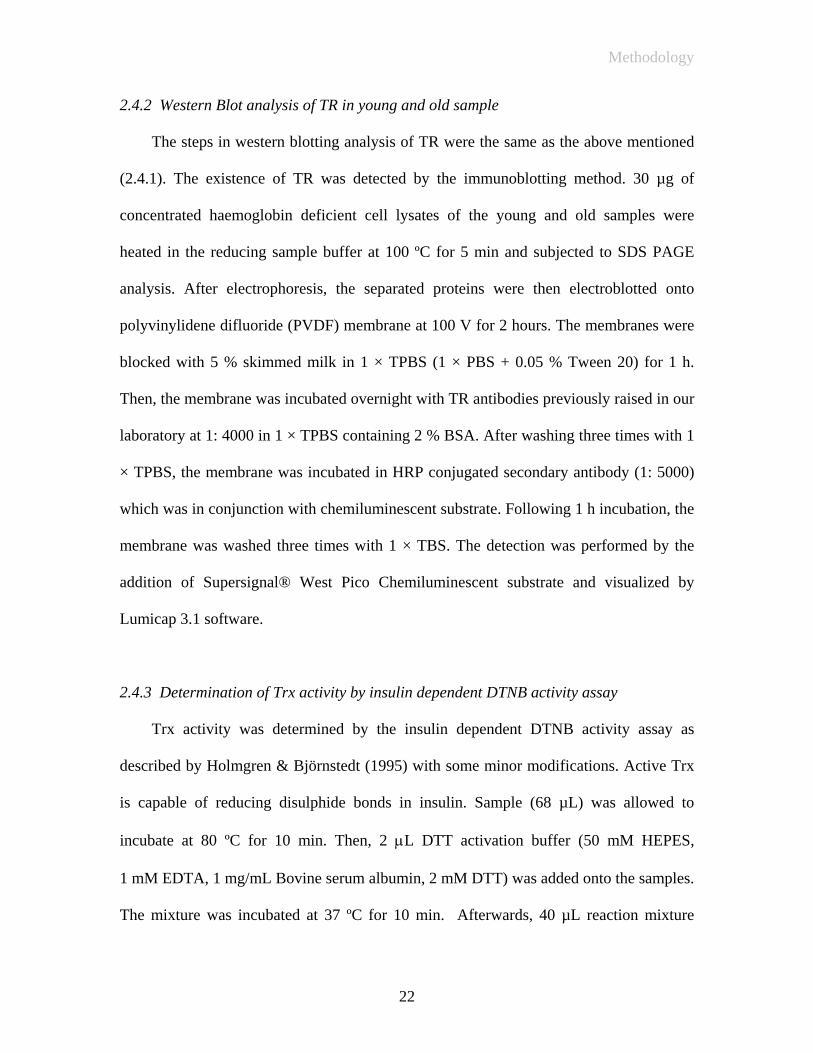
2.4.2 Western Blot analysis of TR in young and old sample
The steps in western blotting analysis of TR were the same as the above mentioned
(2.4.1). The existence of TR was detected by the immunoblotting method. 30 µg of
concentrated haemoglobin deficient cell lysates of the young and old samples were
heated in the reducing sample buffer at 100 ºC for 5 min and subjected to SDS PAGE
analysis. After electrophoresis, the separated proteins were then electroblotted onto
polyvinylidene difluoride (PVDF) membrane at 100 V for 2 hours. The membranes were
blocked with 5 % skimmed milk in 1 × TPBS (1 × PBS + 0.05 % Tween 20) for 1 h.
Then, the membrane was incubated overnight with TR antibodies previously raised in our
laboratory at 1: 4000 in 1 × TPBS containing 2 % BSA. After washing three times with 1
× TPBS, the membrane was incubated in HRP conjugated secondary antibody (1: 5000)
which was in conjunction with chemiluminescent substrate. Following 1 h incubation, the
membrane was washed three times with 1 × TBS. The detection was performed by the
addition of Supersignal® West Pico Chemiluminescent substrate and visualized by
Lumicap 3.1 software.
2.4.3 Determination of Trx activity by insulin dependent DTNB activity assay
Trx activity was determined by the insulin dependent DTNB activity assay as
described by Holmgren & Björnstedt (1995) with some minor modifications. Active Trx
is capable of reducing disulphide bonds in insulin. Sample (68 µL) was allowed to
incubate at 80 ºC for 10 min. Then, 2 μL DTT activation buffer (50 mM HEPES,
1 mM EDTA, 1 mg/mL Bovine serum albumin, 2 mM DTT) was added onto the samples.
The mixture was incubated at 37 ºC for 10 min. Afterwards, 40 µL reaction mixture
22

Methodology
(260 mM HEPES at pH 7.6, 10 mM EDTA , 2 mg/mL NADPH , 6 mg/mL insulin) was
added. The reaction was started by the addition of 30 µL TR and was incubated at 37 ºC
for for 20 min. Free thiols generated were assayed by the addition of 500 µL DTNB (0.4
mg/mL) in 6 M Guanidine HCl. Absorbance at 412 nm was measured while enzyme
activity in the assay was expressed as μmol of TNB formation by using the molar
extinction coefficient of 13600 M-1cm-1.
2.4.4 Determination of TR activity by insulin dependent DTNB activity assay
This was determined by the insulin dependent reduction assay as described by
Luthman & Holmgren (1982). A stock reaction mixture was prepared by mixing 200 µL
1.0 M HEPES at pH 7.6, 40 µL NADPH (40 mg/mL), 40 µL 0.2 M EDTA and 500 µL
insulin (10 mg/mL). To each microtubes, 40 µL reaction mixture, 10 µL 0.36 mM Trx
and 70 µL sample were mixed and incubated at 37 ºC for 20 min. The reaction was
stopped by the addition of 500 µL 0.4mg/mL DTNB in 6 M Guanidine HCl. Absorbance
at 412 nm was measured while enzyme activity in the assay was expressed as μmol of
TNB formation by using a molar extinction coefficient of 13600 M-1cm-1.
23

Methodology
2.4.5 Functional studies of thioredoxin system by IgG reduction assay
The assay measures the capability of the thioredoxin system in breaking down IgG
light chains from heavy chains (Magnusson, 1997; Windle, 2000). Concentrated
haemolysates from the young and old cell population were first incubated with 1 µL 0.1
M DTT at 37 ºC for 30 min. DTT was then removed by a Bio-spin column. Mouse IgG (5
µg), 500 µM NADPH and DTT activated concentrated haemolysates were incubated at
30 °C for two hours. After incubation, 15 µL (50 % gel) Protein A Sepharose was added
into the sample mixture. Binding buffer (50 mM Tris-HCl at pH 7.5) was added onto it. It
was then centrifuged at 8000 g for 20 seconds to spin down the gel. Supernatants were
discarded, and the washings were repeated twice. Samples were then run in a non-
reducing electrophoresis system using 10 % polyacrylamide gel. Western blotting was
then applied with horseradish peroxidase-conjugated anti-IgG as antibody. Split IgG
heavy chains were visualized with the addition of 4-chloro-1-naphthol. Intensity of intact
IgG and split chains were measured which reflected the thioredoxin system activity of the
tested samples.
Section III Preparation of HeLa and HT-29 lysates
To further delineate the role of the thioredoxin system, it is crucial to use other
cell model to compare with results obtained from that of a specialized anucleated cell,
erythrocyte. HeLa and HT-29 cells were chosen as the nucleated cell model for the
studying of the thioredoxin system. These were used to investigate the activity of the
thioredoxin system upon oxidative challenge.
24
Methodology
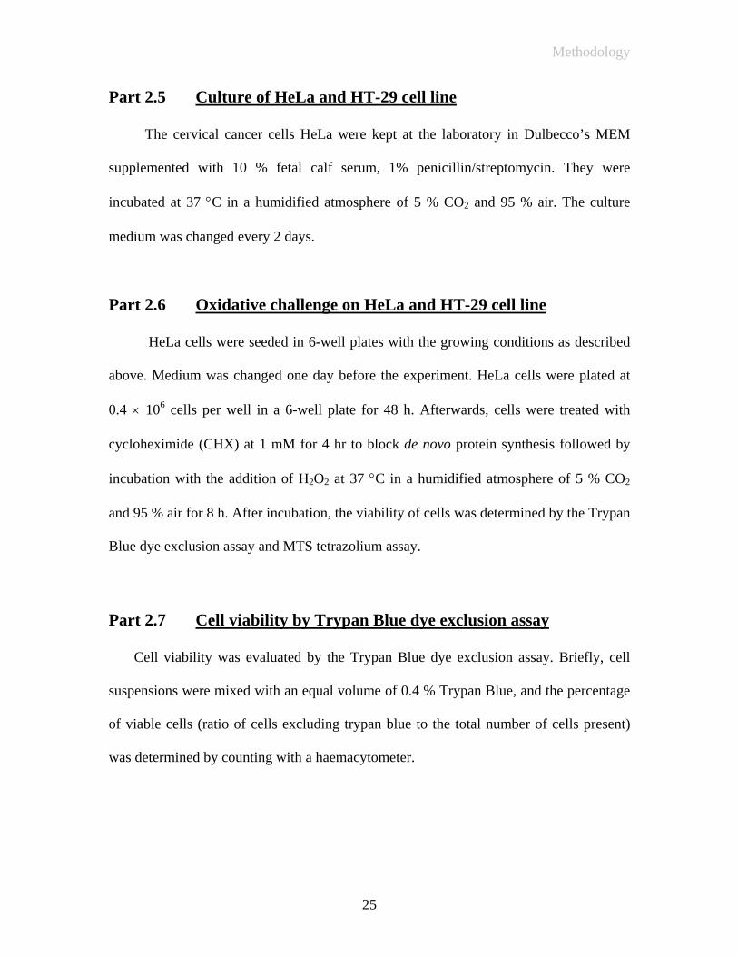
Part 2.5 Culture of HeLa and HT-29 cell line
The cervical cancer cells HeLa were kept at the laboratory in Dulbecco’s MEM
supplemented with 10 % fetal calf serum, 1% penicillin/streptomycin. They were
incubated at 37 °C in a humidified atmosphere of 5 % CO2 and 95 % air. The culture
medium was changed every 2 days.
Part 2.6 Oxidative challenge on HeLa and HT-29 cell line
HeLa cells were seeded in 6-well plates with the growing conditions as described
above. Medium was changed one day before the experiment. HeLa cells were plated at
0.4 × 10⁶ cells per well in a 6-well plate for 48 h. Afterwards, cells were treated with
cycloheximide (CHX) at 1 mM for 4 hr to block de novo protein synthesis followed by
incubation with the addition of H2O2 at 37 °C in a humidified atmosphere of 5 % CO2
and 95 % air for 8 h. After incubation, the viability of cells was determined by the Trypan
Blue dye exclusion assay and MTS tetrazolium assay.
Part 2.7 Cell viability by Trypan Blue dye exclusion assay
Cell viability was evaluated by the Trypan Blue dye exclusion assay. Briefly, cell
suspensions were mixed with an equal volume of 0.4 % Trypan Blue, and the percentage
of viable cells (ratio of cells excluding trypan blue to the total number of cells present)
was determined by counting with a haemacytometer.
25

Methodology
Part 2.8 Cell proliferation by MTS tetrazolium assay
The MTS assay for cell viability is based on the conversion of a tetrazolium salt into
a coloured, aqueous soluble formazan product by the mitochondrial activity of living
cells at 37 ºC (Malich & Boban, 1997). MTS activity assays were performed on cells
treated with hydrogen peroxide for 8 h and also prior to treatment with cycloheximide.
Cells were cultured overnight at 5,000 cells/well in a 96-well plate. Cycloheximide (1
mM) was added into the cell medium for 4 h followed by the addition of hydrogen
peroxide for 8 h. The tetrazolium assay reagent (10 µL/well) was added immediately
after the treatment, and the assay plates were incubated at 37 ºC. Absorbance at 490 nm
was then recorded.
Part 2.9 Cell extracts preparation
Adherent cells were washed once with Dulbecco’s PBS. They were harvested by
trypsinization. Cell pellets were lysed in lysis buffer (20 mM Tris-HCl containing 1 %
Triton X-100, 1 mM EDTA, 1 mM PMSF) in an ice bath for 20 min. Then, it was
centrifuged at 13 000 g for 15 min at 4 °C. Afterwards, supernatants were collected and
protein concentration was determined by the Bio-Rad Dc Protein Assay using bovine
serum albumin as standard. Cell lysates were stored at -80 °C and thawed immediately
before use.
26

Methodology
Part 2.10 Analysis of cell lysates after oxidative challenge
2.10.1 Western Blot analysis of Trx in cell lysates after oxidative challenge
The level of Trx was detected by the immunoblotting method. Cell lysates (10 µg)
were heated in the reducing sample buffer at 100 ºC for 5 min and subjected to SDS
PAGE analysis. After electrophoresis, the separated proteins were then electroblotted
onto the polyvinylidene difluoride (PVDF) membrane at 100 V for 2 hours. The
membranes were blocked with 5 % skimmed milk in 1 × TPBS (1 × PBS + 0.05 %
Tween 20) for 1 h. Then, the membrane was incubated overnight with custom-made Trx
antibody (1: 4000) in 1 × TPBS containing 2 % BSA. The Trx antibodies were polyclonal,
probed for the whole molecule of Trx. After washing three times with 1 × TPBS, the
membrane was incubated in the horseradish peroxidase (HRP) conjugated goat against
rabbit secondary antibody (1: 5000) which was in conjunction with chemiluminescent
substrate. Following 1 h incubation, the membrane was washed three times with 1 ×
TPBS. The detection was performed by the addition of Supersignal® West Pico
Chemiluminescent substrate and visualized by Lumicap 3.1 software.
2.10.2. Western Blot analysis of TR in cell lysates after oxidative challenge
The steps in western blotting analysis of TR were the same as the above mentioned
(2.10.1). The level of TR was detected by the immunoblotting method. 10 µg of cell
lysates were heated in the reducing sample buffer at 100 ºC for 5 min and subjected to
SDS PAGE analysis. After electrophoresis, the separated proteins were then
electroblotted onto the polyvinylidene difluoride (PVDF) membrane at 100 V for 2 hours.
The membranes were blocked with 5 % skimmed milk powder in 1 × TPBS (1 × PBS +
27
Methodology
0.05 % Tween 20) for 1 h. Then, the membrane was incubated overnight with TR
antibodies previously raised in our laboratory at 1: 4000 in 1 × TPBS containing 2 %
BSA. After washing three times with 1 × TPBS, the membrane was incubated in HRP
conjugated secondary antibody (1: 5000) which was in conjunction with
chemiluminescent substrate. Following 1 h incubation, the membrane was washed three
times with 1 × TPBS. The detection was performed by the addition of Supersignal® West
Pico Chemiluminescent substrate and visualized by Lumicap 3.1 software.
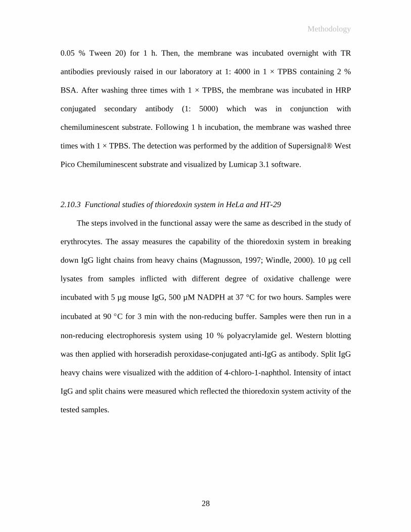
2.10.3 Functional studies of thioredoxin system in HeLa and HT-29
The steps involved in the functional assay were the same as described in the study of
erythrocytes. The assay measures the capability of the thioredoxin system in breaking
down IgG light chains from heavy chains (Magnusson, 1997; Windle, 2000). 10 µg cell
lysates from samples inflicted with different degree of oxidative challenge were
incubated with 5 µg mouse IgG, 500 µM NADPH at 37 °C for two hours. Samples were
incubated at 90 °C for 3 min with the non-reducing buffer. Samples were then run in a
non-reducing electrophoresis system using 10 % polyacrylamide gel. Western blotting
was then applied with horseradish peroxidase-conjugated anti-IgG as antibody. Split IgG
heavy chains were visualized with the addition of 4-chloro-1-naphthol. Intensity of intact
IgG and split chains were measured which reflected the thioredoxin system activity of the
tested samples.
28

Results
III Results
A. Study of thioredoxin system in young and old erythrocytes
3.1 Separation of young and old erythrocytes from porcine whole blood
The young and old cell separation was performed by the centrifugation method.
The achievement of separation was checked by red blood cell counting, mean cell
haemoglobin concentration (MCHC), mean corpuscular volume (MCV) as well as
membrane acetylcholinesterase activity, which are the established parameters for the
assessment of the age of red cells.
3.1.1 Red blood cell counting
Red blood cell counting is an estimation of the number of red blood cells per litre
of blood. It is one of the parameters to differentiate between the young and old cells. Both
young and old erythrocytes were counted by means of a hematocytometer. The value
illustrated that the samples of old cells had a higher number than that of young cells (by
32 %) per unit volume.
Table 3.1.1
Red blood cell counting of young and old erythrocytes Young cells Old cells
Red blood cell counting (no./L)
4.47 ×10¹² ± 0.3 5.9×10¹² ± 0.2
Results are the mean ± SD of four experiments. Data are analyzed as the percentage
difference between the young and old erythrocytes.
29
Results
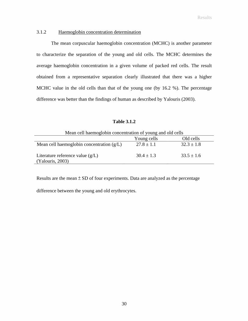
3.1.2 Haemoglobin concentration determination
The mean corpuscular haemoglobin concentration (MCHC) is another parameter
to characterize the separation of the young and old cells. The MCHC determines the
average haemoglobin concentration in a given volume of packed red cells. The result
obtained from a representative separation clearly illustrated that there was a higher
MCHC value in the old cells than that of the young one (by 16.2 %). The percentage
difference was better than the findings of human as described by Yalouris (2003).
Table 3.1.2
Mean cell haemoglobin concentration of young and old cells Young cells Old cells
Mean cell haemoglobin concentration (g/L) Literature reference value (g/L) (Yalouris, 2003)
27.8 ± 1.1
30.4 ± 1.3
32.3 ± 1.8
33.5 ± 1.6
Results are the mean ± SD of four experiments. Data are analyzed as the percentage
difference between the young and old erythrocytes.
30

Results
3.1.3 Mean corpuscular volume determination
The mean corpuscular volume was the average volume of the erythrocytes. It
could be found from the haematocrit (%) and erythrocytes counting analysis. The results
obtained from a representative separation indicated the average size of the young
erythrocytes was larger than the old one (by 32.6 %). The percentage difference was in
good agreement with the literature reference range (Fausta, 2003).
Table 3.1.3
Mean corpuscular volume of young and old erythrocytes Young erythrocytes Old erythrocytes
Mean corpuscular volume (fL) Literature reference value (fL) (Fausta, 2003)
61 ± 0.5
93 ± 0.7
46 ± 0.2
81 ± 0.5
Results are the mean ± SD of four experiments. Data are analyzed as the percentage
difference between the young and old erythrocytes.
31

Results
3.1.4 Membrane acetylcholinesterase activity determination
Acetylcholinesterase is a glycoprotein on the erythrocytes membrane. The results
indicated a higher activity in the young than that of old (by 15 %) which implied that the
old erythrocytes are more vulnerable to osmotic shock. In comparison with the literature
reference value, it showed a good separation between the young and old erythrocytes.
Table 3.1.4
Specific activity of acetylcholinesterase in young and old cells membrane Young cell membrane Old cell membrane
Specific activity (U/mg) Literature reference value (U/mg) (Fausta, 2003)
0.6 ± 0.13
4.9 ± 1.2
0.51 ± 0.12
3.1 ± 0.9
Results are the mean ± SD of four experiments. Data are analyzed as the percentage
difference between the young and old erythrocytes.
32
Results
3.2 Partial purification and concentration of young and old haemolysates
The oxygen carrying protein, haemoglobin, constitutes 95 % of the erythrocytes
proteins. The heme groups on haemoglobin present the characteristic intense red colour.
It is therefore difficult to analysis the young and old erythrocytes samples with methods
involving colorimetric measurement. However, haemoglobin can be removed by means
of chromatography. The young and old haemolysates were subjected to cation exchange
chromatography for the partial purification of the thioredoxin system components. The
cation exchanger was used to remove the most interfering protein, haemoglobin. After
entrapment of haemoglobin from each cell systems on a cation exchange column, the
flow throughs were further concentrated by ultrafiltration. Presumably about 97 % of
haemoglobin was removed by the cation exchanger.
33

Results
3.3 Analysis of young and old sample
3.3.1 Western blotting analysis of TR in young and old sample
In a quantitative measurement of TR present in young and old sample, western
blotting analysis was performed.
After removing haemoglobin from the young and old haemolysate, all the eluents
prepared in the previous step were subjected to ultrafiltration with Amicon® Ultra and
YM-1 membranes (1000 MWCO) for concentration purposes. From each sample, they
were concentrated to approximately 2 mL and the protein concentration was checked by
the Lowry assay (Lowry, 1951). Western blot analysis was then performed. 30 μg protein
was applied onto each lane (Lane 2 and Lane 3). As shown in the results below, TR was
found in both young and old samples, while porcine TR was used as a positive control
(Fig.3.3.1).
62 kDa TR
1 2 3
Fig.3.3.1 Western blotting analysis of TR in young and old sample
Lane 1: control TR
Lane 2: young sample
Lane 3: old sample
The densitometric measurement showed that the TR protein levels decreased by
32.6 % in the old cells compared with that of the young. Results are the mean ± SD of
two experiments. Data are analyzed by comparing the percentage difference between the
young and old erythrocytes.
34

Results
3.3.2 Western blotting analysis of Trx in young and old sample
Determination of Trx in young and old cell population by Western Blotting
To examine the presence and the level of Trx in the young and old cells, the
immunoblotting of Trx was carried out. Custom made anti-Trx was used as primary
antibody while HRP conjugated secondary antibody was used in conjunction with
chemiluminescent substrate (Supersignal®). Sample from the young cells showed one
band (11.6 kDa) while it showed no bands in the old sample (Fig.3.3.2).
1 2 3 kDa 113 93 50.3 35.5 28.8 214
11.6 kDa
Fig.3.3.2. Western blotting analysis of Trx in young and old sample
Lane 1: old cell sample
Lane 2: young cell sample
Lane 3: prestained SDS-PAGE Standards (not shown in chemiluminescent detection)
The results revealed that Trx was present in the young but not in the old sample. It
clearly showed that the amount of Trx was substantially higher in the young sample while
there would be no Trx in the old sample or the concentration was beyond the detection
limit. Results are the mean ± SD of two experiments.
35

Results
3.3.3 Thioredoxin and thioredoxin reductase activity assay
Determination of thioredoxin and thioredoxin reductase activity by insulin dependent
reduction assay.
To further investigate the individual components of the thioredoxin system, two
key components, the Trx and TR activities were monitored.
Table 3.3.3
Insulin-dependent DTNB assay of Trx and TR on young and old erythrocytes Unit of enzyme (U) Specific activity (U/mg) Trx from young sample Trx from old sample TR from young sample TR from old sample
0.028 ± 0.002
N.D.
0.039 ± 0.014
0.029 ± 0.010
0.19 ± 0.01
N.D.
0.27 ± 0.14
0.19 ± 0.07 *N.D. Not detectable
The insulin dependent DTNB reduction assay was carried out. The results showed
the age-related changes in the activity of both Trx and TR in the young and old
erythrocytes (Table 3.3.3). The results as shown above, illustrated that both Trx and TR
activity in the young cells were higher than that of the old cells. Trx in the young sample
showed some activity while no activity in the old sample was shown. The findings were
consistent with the result that no Trx was detected by the western blotting analysis
(Fig.3.3.2).
The findings were in good agreement with previous findings which showed an
age-related decrease in the Trx activity in other tissues (Cho, 2003). Results are the mean
± SD of two experiments. Data are analyzed by comparing the percentage difference
between the young and old erythrocytes.
36
Results
3.3.4 IgG reduction assay of young and old haemolysates upon oxidative challenge
Functional studies of thioredoxin system of erythrocytes after oxidative stress by IgG
reduction assay
To investigate the effect of age on the susceptibility of the thioredoxin system in
the young and old erythrocytes towards H2O2 oxidative challenge, erythrocytes separated
from young and old cells were exposed to various concentrations of H2O2 (4 and 8 mM).
IgG reduction assay was carried out to monitor the response of the thioredoxin system
upon oxidative challenge.
37
Results
1 2 3 4 5 6 7 8 9 10
kDa
205 112 87 69 56 38.5 33.5
IgG heavy chain
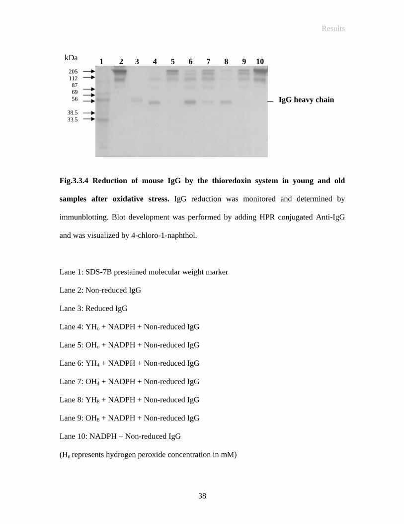
Fig.3.3.4 Reduction of mouse IgG by the thioredoxin system in young and old
samples after oxidative stress. IgG reduction was monitored and determined by
immunblotting. Blot development was performed by adding HPR conjugated Anti-IgG
and was visualized by 4-chloro-1-naphthol.
Lane 1: SDS-7B prestained molecular weight marker
Lane 2: Non-reduced IgG
Lane 3: Reduced IgG
Lane 4: YHo + NADPH + Non-reduced IgG
Lane 5: OHo + NADPH + Non-reduced IgG
Lane 6: YH4 + NADPH + Non-reduced IgG
Lane 7: OH4 + NADPH + Non-reduced IgG
Lane 8: YH8 + NADPH + Non-reduced IgG
Lane 9: OH8 + NADPH + Non-reduced IgG
Lane 10: NADPH + Non-reduced IgG
(Hn represents hydrogen peroxide concentration in mM)
38
Results
Results are the mean ± SD of two experiments. Data are analyzed by comparing
the percentage difference between the young and old erythrocytes. Lane 4 and 5 show the
thioredoxin system activity of the young and old cells respectively with no addition of
H2O2. Lane 6 and 7 indicate the result of the thioredoxin system activity of Y and O cells
after applying 4 mM H2O2 at 37ºC for 1 hour. We found an increase of 63.5 % ± 11.3 %
thioredoxin system activity in the young cells challenged with 4 mM H2O2. Though the
level and activity of thioredoxin in the old cells were not detectable, an enhancement of
the whole thioredoxin system activity was observed after oxidative challenge. Both
results showed an increase in the the IgG reduction capability of the thioredoxin system,
while the capability was further decreased when the samples were treated with
8 mM H2O2.
39

Results
B. Study of thioredoxin system in nucleated cell model
3.4 Culture of HeLa and HT-29 cell lines
The cells were cultured under the condition as described in part 2.2.2. The
experiment would be carried out until HeLa cells were grown to 106 cells per well. All
cultures used in the experiment exhibited similar viabilities of about 94 %. In the study,
cell lysates were isolated from the control and hydrogen peroxide treated HeLa cell
populations. In addition, samples were analyzed for the thioredoxin system activity by the
IgG reduction assay. Equal concentration of protein was used as sample for analysis.
40

Results
3.4.1 Cell viability of HeLa after oxidative challenge by Trypan Blue dye exclusion
assay
Cell viability was determined by the Trypan Blue dye exclusion assay in which
cells were treated with different defined concentrations of H2O2 for 8 hr (0-0.4 mM). The
experiments were carried out in duplicates while the results were expressed as the mean ±
S.D. Statistical calculations were done using the student’s t test. A p value < 0.05 was
considered statistically significant.
Determination of cell viability of HeLa by Trypan Blue dye exclusion assay
Trypan Blue dye exclusion assay of HeLa cells treated withhydrogen peroxide
0
20
40
60
80
100
120
0 0.025 0.05 0.1 0.2 0.4
Hydrogen peroxide (mM)
cel
l via
bilit
y (%
)
* * ***
* **
*
Fig.3.4.1 Trypan Blue dye exclusion assay by HeLa cells treated with 0-0.4 mM hydrogen peroxide for 8 hr with (■) or without (♦) prior treatment of cycloheximide * p< 0.05 when it was compared with H2O2 treated cells and untreated control.
41

Results
The results showed a dose-dependent trend of hydrogen peroxide toxicity, that
was comparable with the later MTS assay. However, the findings also illustrated that the
cycloheximide treated cells were significantly more sensitive to the cytotoxic effect of
H2O2, which were not shown by the MTS assays, indicating that de novo protein
synthesis is essential for cell survival under oxidative stress.
42

Results
3.4.2 Cell viability of HeLa after oxidative challenge by MTS tetrazolium assay
MTS tetrazolium was reduced to formazan by HeLa cells treated with 0-0.4 mM
hydrogen peroxide for 8 h and a prior treatment with cycloheximide for 4 h. HeLa cells
were cultured overnight at 5,000 cells/well in a 96-well plate. Cycloheximide (1 mM)
was added into the cell medium for 4 h followed by the addition of hydrogen peroxide for
8 h. The tetrazolium assay reagent (10 µL/well) was added immediately after the
treatment, and the assay plates were incubated at 37 ºC prior to the recording of
absorbance at 490 nm. The absorbance value for the control of culture medium without
cells was 0.38 ± 0.05 at 490 nm (not shown).
Determination of cell viability of HeLa by MTS tetrazolium assay
MTS tetrazolium reduction to formazan by HeLa cells treated with 0-0.4 mM hydrogen peroxide
00.20.40.60.8
11.2
0 0.025 0.05 0.1 0.2 0.4
Hydrogen peroxide (mM)
Abs
orba
nce
(490
nm
)
Fig.3.4.2 MTS tetrazolium reduction to formazan by HeLa cells treated with 0-0.4 mM hydrogen peroxide for 8 h with (■) or without (♦) prior treatment of cycloheximide for 4 h
*p< 0.05 when it was compared with H2O2 treated cells and untreated control.
43

Results
The MTS tetrazolium reduction assay was used to measure the cell viability of
HeLa cells treated with hydrogen peroxide in the presence or absence of cycloheximide.
The results showed a dose-dependent trend of hydrogen peroxide toxicity, implying the
validity of this method for measuring viability comparing with the Trypan Blue dye
exclusion assay.
Because of the necessity for viable cells to reduce the tetrazolium compound to a
water soluble formazan product, a 3 hr incubation period is needed. It is interesting to
point out that there was a slight increase in absorbance at 490 nm at 0.025 mM hydrogen
peroxide for the 8 hr exposure period. Such interesting phenomenon is consistent with
previous observations (Haendeler, 2004). However, a significant decrease of cell viability
was observed in cycloheximide treated HeLa cells at the same concentration of hydrogen
peroxide, indicating cycloheximide treated cells were more sensitive to the cytotoxic
effect of hydrogen peroxide.
44

Results
3.4.3 Western blotting analysis of Trx and TR in HeLa cells upon oxidative challenge
Determination of Trx and TR in HeLa cells upon oxidative challenge by Western Blotting
To address the possible effect of transcription of Trx and TR upon oxidative
challenge, the level of Trx and TR in HeLa cells challenged with various concentrations of H2O2 was determined. Results are the mean ± SD of two experiments.
1 2 3 4 5 6 7 8
Trx
TR
Actin
Fig.3.4.3 Western blotting analysis of Trx and TR in HeLa cells upon oxidative
challenge
Lane 1: standard Trx
Lane 2: standard TR
Lane 3: HeLa lysate (control)
Lane 4: HeLa lysate (pretreated with 0.025 mM H2O2)
Lane 5: HeLa lysate (pretreated with 0.05 mM H2O2)
Lane 6: HeLa lysate (pretreated with 0.1 mM H2O2)
Lane 7: HeLa lysate (pretreated with 0.2 mM H2O2)
Lane 8: HeLa lysate (pretreated with 0.4 mM H2O2)
45
Results
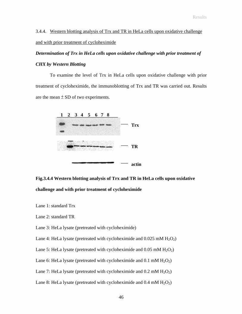
3.4.4. Western blotting analysis of Trx and TR in HeLa cells upon oxidative challenge
and with prior treatment of cycloheximide
Determination of Trx in HeLa cells upon oxidative challenge with prior treatment of
CHX by Western Blotting
To examine the level of Trx in HeLa cells upon oxidative challenge with prior
treatment of cycloheximide, the immunoblotting of Trx and TR was carried out. Results
are the mean ± SD of two experiments.
1 2 3 4 5 6 7 8
TR
actin
Trx
Fig.3.4.4 Western blotting analysis of Trx and TR in HeLa cells upon oxidative
challenge and with prior treatment of cycloheximide
Lane 1: standard Trx
Lane 2: standard TR
Lane 3: HeLa lysate (pretreated with cycloheximide)
Lane 4: HeLa lysate (pretreated with cycloheximide and 0.025 mM H2O2)
Lane 5: HeLa lysate (pretreated with cycloheximide and 0.05 mM H2O2)
Lane 6: HeLa lysate (pretreated with cycloheximide and 0.1 mM H2O2)
Lane 7: HeLa lysate (pretreated with cycloheximide and 0.2 mM H2O2)
Lane 8: HeLa lysate (pretreated with cycloheximide and 0.4 mM H2O2)
46

Results
The level of Trx and TR in HeLa cells upon oxidative stress
0
0.2
0.4
0.6
0.8
1
1.2
0 0.025 0.05 0.1 0.2 0.4
H2O2 (mM)
Inte
nsity
Trx
TR
Trx (CHXtreated)TR (CHXtreated)
Fig.3.4.4.1 The level of Trx and TR in HeLa cells upon oxidative stress with or
without prior treatment of cycloheximide
The results of these experiments showed that there was no significance difference
in the level of Trx and TR in HeLa cells upon oxidative challenge with and without the
prior treatment of cycloheximide.
47

Results
3.4.5 Activity of thioredoxin system in HeLa cells upon oxidative challenge without prior treatment of of cycloheximide
Recent studies have indicated that short term exposure of low doses of H2O2
induced Trx mRNA (Haendeler, 2004). To find out the relationship between mRNA
expression and Trx system activity, human epithelial carcinoma, HeLa cells were
exposed to 0-0.4 mM H2O2 for 8 hr. Trx system activity was then demonstrated by the
IgG reduction assay. Results are the mean ± SD of two experiments. Statistical
calculations were done using the student’s t test. A p value < 0.05 was considered
statistically significant.
48

Results
Functional studies of thioredoxin system of HeLa cells after oxidative stress by IgG
reduction assay
1 2 3 4 5 6 7 8 9 10
kDa 205 112 87 69 56 38.5 33.5
IgG heavy chain
Fig.3.4.5 Reduction of mouse IgG by the thioredoxin system in HeLa cells after
oxidative stress
* Equal loading according to its protein concentration
Lane 1: SDS 7B Markers
Lane 2: Non-reduced IgG
Lane 3: Reduced IgG
Lane 4: H0 + Non-reduced IgG + NADPH
Lane 5: H0.025 + Non-reduced IgG + NADPH
Lane 6: H0.05 + Non-reduced IgG + NADPH
Lane 7: H0.1 + Non-reduced IgG + NADPH
Lane 8: H0.2 + Non-reduced IgG + NADPH
Lane 9: H0.4 + Non-reduced IgG + NADPH
Lane 10: Non-reduced IgG + NADPH
(Hn represents hydrogen peroxide concentration in mM)
49
Results
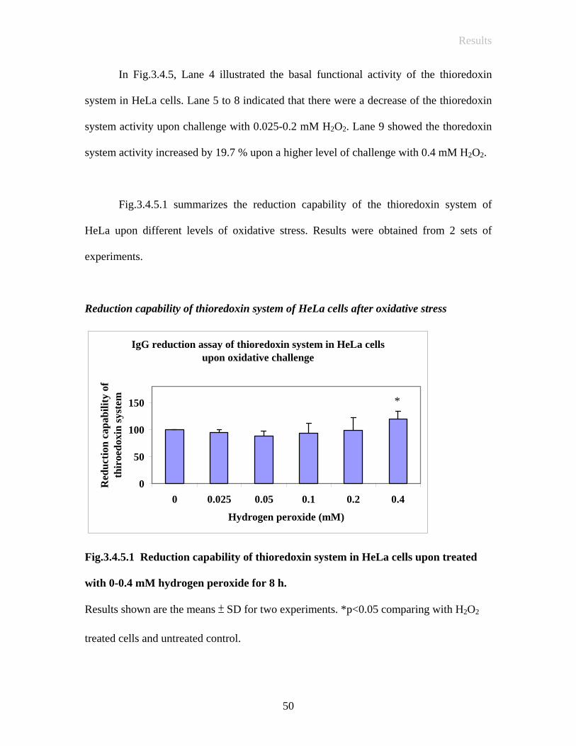
In Fig.3.4.5, Lane 4 illustrated the basal functional activity of the thioredoxin
system in HeLa cells. Lane 5 to 8 indicated that there were a decrease of the thioredoxin
system activity upon challenge with 0.025-0.2 mM H2O2. Lane 9 showed the thoredoxin
system activity increased by 19.7 % upon a higher level of challenge with 0.4 mM H2O2.
Fig.3.4.5.1 summarizes the reduction capability of the thioredoxin system of
HeLa upon different levels of oxidative stress. Results were obtained from 2 sets of
experiments.
Reduction capability of thioredoxin system of HeLa cells after oxidative stress
IgG reduction assay of thioredoxin system in HeLa cellsupon oxidative challenge
0
50
100
150
0 0.025 0.05 0.1 0.2 0.4
Hydrogen peroxide (mM)
Red
uctio
n ca
pabi
lity
ofth
iroe
doxi
n sy
stem
*
Fig.3.4.5.1 Reduction capability of thioredoxin system in HeLa cells upon treated with 0-0.4 mM hydrogen peroxide for 8 h. Results shown are the means ± SD for two experiments. *p<0.05 comparing with H2O2
treated cells and untreated control.
50

Results
(It was assessed by densitometric measurement, in which 0 mM hydrogen peroxide was
represented as 100 for comparison)
3.4.6 Activity of thioredoxin system in HeLa cells upon oxidative challenge with prior treatment of cycloheximide
To further elucidate whether this increase in the thioredoxin system activity was
indeed due to Trx expression and/or with other enhancement pathways, HeLa cells were
pretreated with 1 mM cycloheximide to inhibit the de novo protein synthesis. The
blockage of de novo protein synthesis allowed us to have a better understanding on how
the thioredoxin system responses upon oxidative stress.
51

Results
Functional studies of thioredoxin system of HeLa cells after oxidative stress by IgG
reduction assay
kDa 1 2 3 4 5 6 7 8 9 10
205 112 87 69 56 38.5 33.5
IgG heavy chain
Fig.3.4.6 Reduction of mouse IgG by the thioredoxin system in HeLa cells after
oxidative stress with prior treatment of cycloheximide
* Equal loading according to its protein concentration
Lane 1: SDS 7B Marker
Lane 2: Non-reduced IgG
Lane 3: Reduced IgG
Lane 4: H0 + Non-reduced IgG + NADPH
Lane 5: H0.025’+ Non-reduced IgG + NADPH
Lane 6: H0.05 + Non-reduced IgG + NADPH
Lane 7: H0.1 + Non-reduced IgG + NADPH
Lane 8: H0.2 + Non-reduced IgG + NADPH
Lane 9: H0.4 + Non-reduced IgG + NADPH
Lane 10: Non-reduced IgG + NADPH
(Hn represents hydrogen peroxide concentration in mM)
52
Results
As shown in Fig.3.4.6, Lane 4 showed the basal functional activity of the
thioredoxin system in HeLa cells. Lane 5 indicated that there was a 8.2 % increase of the
thioredoxin system activity upon challenge with 0.025 mM H2O2. The thioredoxin system
activity then further decreased upon higher level of challenge with 0.05-0.4 mM H2O2.
Fig.3.4.6.1 summarizes the reduction capability of HeLa cells after oxidative
stress with prior treatment of cycloheximide. As compared with the results obtained from
Fig.3.4.5.1, the enhancement of the thioredoxin system activity by 8.2 % occurred at
lower concentration of H2O2 (0.025 mM). Since cycloheximide acted as inhibitors for de
novo protein synthesis, the results interestingly suggest that the thioredoxin system
activity is enhanced during oxidative challenge which is not related to translation.
53

Results
Reduction capability of thioredoxin system of HeLa cells after oxidative stress with
prior treatment of cycloheximide
IgG reduction assay of thioredoxin system in HeLa cellsupon oxidative challenge with prior treatment of
cycloheximide
0
50
100
150
0 0.025 0.05 0.1 0.2 0.4
Hydrogen peroxide (mM)
Red
uctio
n ca
pabi
lity
of th
iroe
doxi
n sy
stem
** * *
Fig.3.4.6.1 Reduction capability of thioredoxin system in HeLa cells upon treated with 0-0.4 mM hydrogen peroxide for 8 h and prior treatment with cycloheximide for 4 h
Results shown are the means ± SD for two experiments. * p< 0.05 when it was compared
with H2O2 treated cells and untreated control.
54

Results
3.4.7 Cell viability of HT-29 after oxidative challenge by Trypan Blue dye exclusion assay
To further study the effect of oxidative challenge and response of the thioredoxin
system in nucleated cells, another cell line HT-29 was investigated. Cell viability was
determined by the Trypan Blue dye exclusion assay in which HT-29 cells were treated
with different defined concentrations of H2O2 for 8 hr (0-1 mM). The experiments were
carried out in duplicates while the results were expressed as the mean ± S.D.
Determination of cell viability of HT-29 by Trypan Blue dye exclusion assay
Trypan Blue dye exclusion assay of HT-29 treated with hydrogen peroxide
0102030405060708090
100
0 0.5 1
Hydrogen peroxide (mM)
cel
l via
bilit
y (%
)
Fig.3.4.7 Trypan Blue dye exclusion assay by HT-29 cells treated with 0-1 mM hydrogen peroxide for 8 h with (■) or without (♦) prior treatment of cycloheximide
55

Results
The results showed a dose dependent trend of hydrogen peroxide toxicity, that is
comparable with the MTS assay. However, the findings also illustrated that
cycloheximide treated cells were significantly more sensitive to the cytotoxic effect of
H2O2, and that de novo protein synthesis is essential for cell survival under oxidative
stress.
56

Results
3.4.8 Cell viability of HT-29 after oxidative challenge by MTS tetrazolium assay
The results also illustrated a dose dependent trend of HT-29 cells towards
hydrogen peroxide toxicity as compared with the HeLa cells. A significant decrease of
cell viability was observed in cycloheximide treated HeLa cells at the same concentration
of hydrogen peroxide, indicating cycloheximide treated cells were more sensitive to the
cytotoxic effect of hydrogen peroxide under oxidative stress.
Determination of cell viability of HT-29 by MTS tetrazolium assay
MTS tetrazolium reduction to formazan of HT-29 cells treated with hydrogen peroxide
00.10.20.30.40.50.60.70.8
0 0.5 1
Hydrogen peroxide (mM)
Abs
orba
nce
(490
nm
)
Fig.3.4.8 MTS tetrazolium reduction to formazan by HT-29 cells treated with 0-1 mM hydrogen peroxide for 8 h with (■) or without (♦) prior treatment of cycloheximide for 4 h
57

Results
3.4.9 Activity of thioredoxin system in HT-29 cells upon oxidative challenge with or without prior treatment of cycloheximide
Previous studies showed the activation of the thioredoxin system activity upon
oxidative challenge in HeLa cells (Fig.3.4.5.1 and Fig.3.4.6.1). To examine whether the
induction was cell type independent, another nucleated cell line, HT-29 cells were
examined and pretreated with 1 mM cycloheximide to inhibit the de novo protein
synthesis. As compared with the HeLa cells, higher concentrations of H2O2 (0-1 mM)
were used in the oxidative challenge. The experiments were carried out in triplicate while
the results were expressed as the mean ± S.D.
58

Results
Functional studies of thioredoxin system of HT-29 cells after oxidative stress by IgG
reduction assay
1 2 3 4 5 6 7 8 9 10
kDa 205 112 87 69 56 38.5 33.5
IgG heavy chain
Fig.3.4.9 Reduction of mouse IgG by the thioredoxin system in HT-29 cells after
oxidative stress with or without prior treatment of cycloheximide
* Equal loading according to its protein concentration
Lane 1: SDS 7B Marker
Lane 2: Non-reduced IgG
Lane 3: Reduced IgG
Lane 4: H0 + Non-reduced IgG + NADPH
Lane 5: H0.5 + Non-reduced IgG + NADPH
Lane 6: H1 + Non-reduced IgG + NADPH
Lane 7: H0 + Non-reduced IgG + NADPH (CHX treated)
Lane 8: H0.5 + Non-reduced IgG + NADPH (CHX treated)
Lane 9: H1 + Non-reduced IgG + NADPH (CHX treated)
Lane 10: Non-reduced IgG + NADPH
(Hn represents hydrogen peroxide concentration in mM)
59

Results
We examined the reduction capability of the thioredoxin system in HT-29 cells
upon oxidative challenge. Fig.3.4.9.1 showed that the thioredoxin system activity
increased by 16.3 % and 18.5 % after addition of 0.5 and 1 mM H2O2 respectively. Such
progressive increase of the thioredoxin system activity could also be observed in the cells
pretreated with cycloheximide with a smaller increment of 8.1 % and 13.9 %
respectively. Since cycloheximide acted as inhibitors for de novo protein synthesis, the
results also revealed that the thioredoxin system activity was interestingly enhanced
during oxidative challenge that was not necessarily related to translation.
60

Results
Reduction capability of thioredoxin system of HT-29 cells after oxidative stress with or
without prior treatment of cycloheximide
Percentage increase of reduction capability of thioredoxinsystem of HT-29
0
5
10
15
20
25
30
0.5 1
Hydrogen peroxide (mM)
perc
enta
ge in
crea
se o
fre
duct
ion
capa
bilit
y
Without CHXtreatedCHX treated
* *
*
*
Fig.3.4.9.1 Reduction capability of thioredoxin system in HT-29 cells upon treated
with 0-1 mM hydrogen peroxide for 8 h with or without prior treatment of
cycloheximide for 4 h
Results shown are the means ± SD for three experiments. *p< 0.001 when it was
compared with H2O2 treated cells and untreated control, with or without prior treatment
of CHX.
61

Discussion
V Discussion
Aging is an inevitable part of the life natural process that is governed by the
decreasing physiological functions which ultimately results in mortality. Previous studies
showed that ROS induced apoptosis. In addition, ROS are believed to be one of the
causal factors of aging. The balance between ROS generation, antioxidant defenses and
repairing mechanisms is therefore of paramount importance. There are numerous
biochemical pathways and mechanisms involved in the defence against oxidative stress.
Unique amongst others, the thioredoxin system is one of the best representative systems
for its protective function against oxidative stress. It can be found in different cell types
ranging from prokaryotes to eukaryotes. However, the existence of the thioredoxin
system in erythrocytes has not been reported. Previous studies illustrated that Trx
involves in the first unique step of DNA synthesis and an important step for cellular
proliferation (Powis, 2001). However, the anucleated feature of erythrocytes implies no
DNA synthesis in the mature state. From our studies, Trx and TR were found surprisingly
high in erythrocytes. It is noteworthy that besides the involvement in DNA synthesis, the
thioredoxin system has other roles and functions. Oxidative damage to erythrocytes has
known to cause destructive events in many haematological disorders and aging.
Erythrocytes encounter high oxidative stress due to its oxygen carrying function. Indeed,
the anucleated feature of erythrocytes implies no protein synthesis. The repairing
mechanism is therefore becoming the determinant factor in keeping cell homeostasis and
most importantly the cell viability.
62

Discussion
Consequently, erythrocyte can be used as a cell aging model. We attempted to
find out the level of the thioredoxin system components in erythrocytes, the role they
played, and in particular how they might vary in the aging process and how they
responded to oxidative challenge. Through this study, it is hoped that a better
understanding on the role of the thioredoxin system and its relationship with aging in
erythrocytes can be obtained.
During cellular aging, erythrocytes become smaller and denser. Young and old
populations of cells were first separated by means of density, while the success of the
separation was further verified by the difference in erythrocytes counting, mean
haemoglobin concentration, mean corpuscular volume and membrane acetylcholinestase
activity between the young and old erythrocytes. The findings showed an age related
decrease in MCV and AchE compared with that of the corresponding young cells. The
results were in agreement with published data (Fausta, 2003). The results obtained were
consistent with the nature of the young and old erythrocytes. Previous studies illustrated
the significant structural and membrane deviation between the young and old human
erythrocytes (Fausta, 2003). In the present study, pig erythrocytes were employed and
comparable results to that of human erythrocytes were obtained. It indicates clearly that
pig red cells serve as a good substitution for human erythrocytes.
The successfully separated cells were then used in the subsequent analysis of the
thioredoxin system. It has been the first attempt to compare the thioredoxin system in the
young and old erythrocytes. In the study, western blotting analysis showed the presence
63
Discussion
of Trx in the young but not in the old cells. The molecular weight of Trx was found to be
11.6 kDa. The findings were consistent with the molecular weight of the mammalian Trx
(Arnér, 2000). Such findings indicated the existence of Trx in porcine erythrocytes while
no Trx could be found in the old erythrocytes or the concentration of Trx was beyond the
detection limit. Immunoblotting of TR was also carried out in the young and old cells.
The level of TR in the young cells was higher than that of the old cells by 32.6 %. It is
worth noting that there was still some TR found in the old erythrocytes. The findings
were consistent with the relatively stable nature of TR when compared with Trx. In
addition, the enzyme activity study of Trx of the young and old erythrocytes was also
consistent with such findings. The results illustrated that there was 0.19 U thioredoxin /
mg total protein in young cells but no activity could be observed in the old cells. Only the
concentrated young sample, but not the old sample, was able to reduce the disulfide-
containing protein, insulin. The results showed the age-related changes in the activity of
both Trx and TR in the young and old erythrocytes. In general, both level and activity of
Trx and TR in the young cells were substantially higher than that of the old cells. Such
deviations illustrated that the thioredoxin system may be essential in the cell aging
process of erythrocytes.
To further confirm our preliminary hypothesis, immunoglobulin (IgG) reduction
assay was employed to study the functional role of the whole thioredoxin system in both
cell populations. Mouse immunoglobulin was chosen as the substrate, which contains
interchain and intrachain disulphide bridges between the heavy and light chain of
immunoglobulin to maintain the conformation of the whole structure. Previous studies
64

Discussion
showed that functioning thioredoxin system is able to reduce the interchain disulphide
bridges between the heavy chain and light chain of IgG (Magnusson, 1997). The
advantage of using the assay was to illustrate the disulfide reducing capacity of the whole
thioredoxin system, which overcomes the limitations of the above insulin dependent
DTNB assay that can only measure either component in the whole thioredoxin system
independently. Indeed, with the excessive supplement of NADPH, sufficient amount of
hydrogen ions could be supplied to the whole system, thus the system was kept in an
active form for carrying its oxidoreductase function continuously.
In the experiment, with the supplement of NADPH, the results clearly illustrated
that both TR and Trx in the young haemolysates could reduce the interchain disulfide
bridges of IgG. TR activity in the old cells, though measurable by the insulin reduction
activity assay, was not detected in the IgG reduction activity assay. The result was
consistent with no Trx activity and a lower TR activity found in the old cells in the
previous experiments.
The findings indicated the difference of Trx and TR activity between the young
and old cells in erythrocytes. Such deviations were significantly pronounced in the
different ages of cells, thereby suggesting an interesting relationship between the
thioredoxin system and cell aging in erythrocytes. To further elucidate the role of
thioredoxin system in erythrocytes, the young and old erythrocytes were exposed to
different levels of oxidative stress. In this experiment, hydrogen peroxide acted as
oxidative challenge which mimicked the ROS attack on cells. Previous studies showed
65
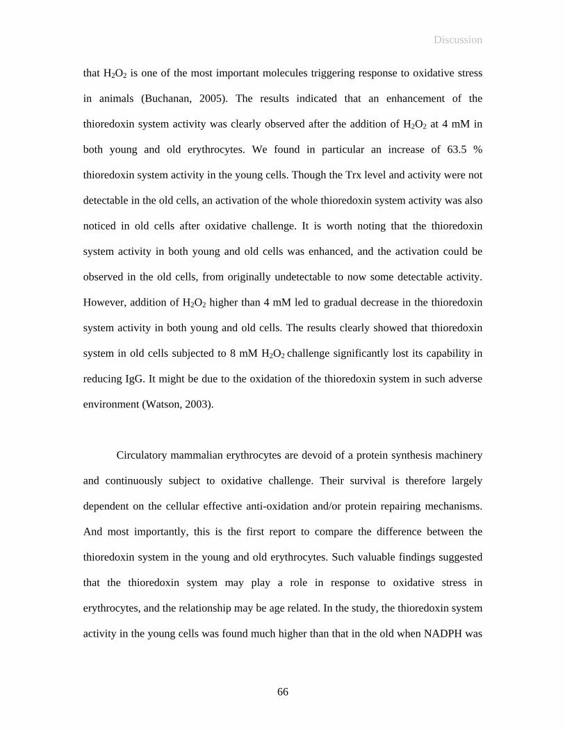
Discussion
that H2O2 is one of the most important molecules triggering response to oxidative stress
in animals (Buchanan, 2005). The results indicated that an enhancement of the
thioredoxin system activity was clearly observed after the addition of H2O2 at 4 mM in
both young and old erythrocytes. We found in particular an increase of 63.5 %
thioredoxin system activity in the young cells. Though the Trx level and activity were not
detectable in the old cells, an activation of the whole thioredoxin system activity was also
noticed in old cells after oxidative challenge. It is worth noting that the thioredoxin
system activity in both young and old cells was enhanced, and the activation could be
observed in the old cells, from originally undetectable to now some detectable activity.
However, addition of H2O2 higher than 4 mM led to gradual decrease in the thioredoxin
system activity in both young and old cells. The results clearly showed that thioredoxin
system in old cells subjected to 8 mM H2O2 challenge significantly lost its capability in
reducing IgG. It might be due to the oxidation of the thioredoxin system in such adverse
environment (Watson, 2003).
Circulatory mammalian erythrocytes are devoid of a protein synthesis machinery
and continuously subject to oxidative challenge. Their survival is therefore largely
dependent on the cellular effective anti-oxidation and/or protein repairing mechanisms.
And most importantly, this is the first report to compare the difference between the
thioredoxin system in the young and old erythrocytes. Such valuable findings suggested
that the thioredoxin system may play a role in response to oxidative stress in
erythrocytes, and the relationship may be age related. In the study, the thioredoxin system
activity in the young cells was found much higher than that in the old when NADPH was
66
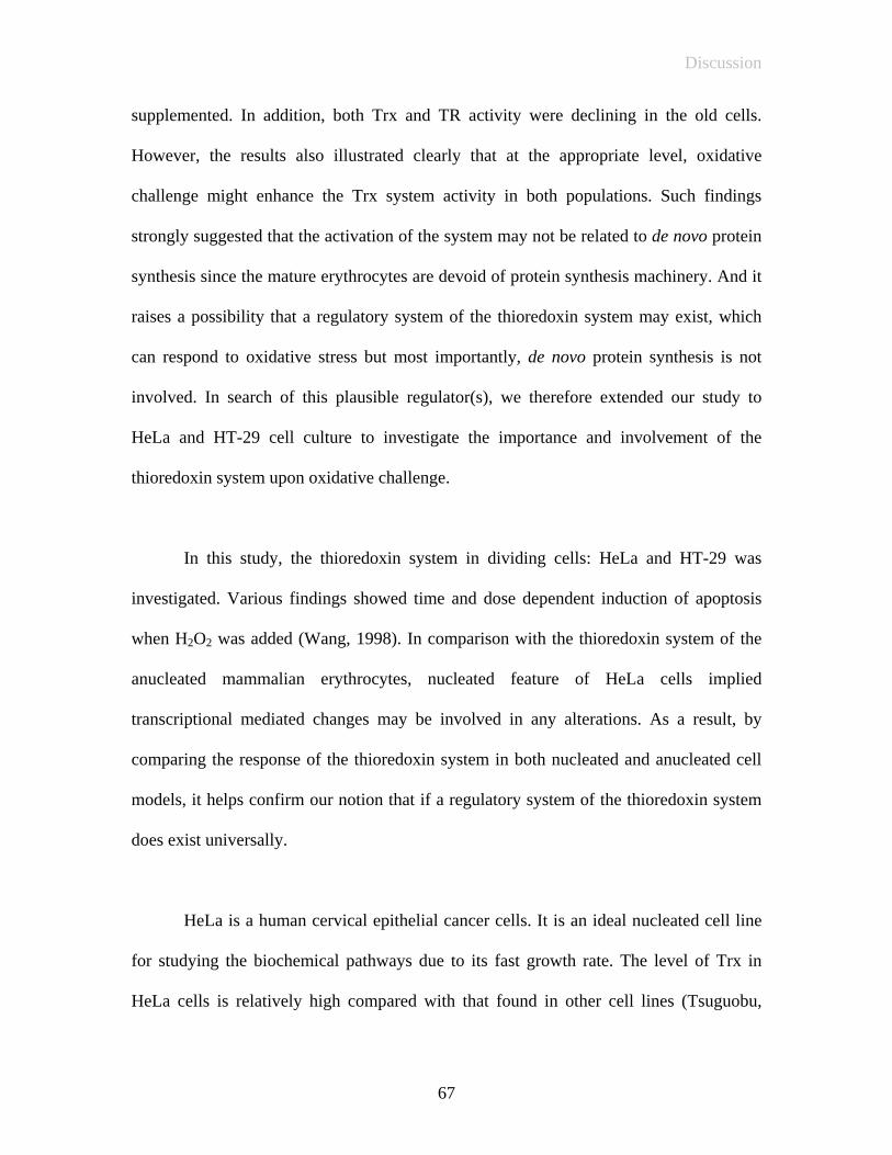
Discussion
supplemented. In addition, both Trx and TR activity were declining in the old cells.
However, the results also illustrated clearly that at the appropriate level, oxidative
challenge might enhance the Trx system activity in both populations. Such findings
strongly suggested that the activation of the system may not be related to de novo protein
synthesis since the mature erythrocytes are devoid of protein synthesis machinery. And it
raises a possibility that a regulatory system of the thioredoxin system may exist, which
can respond to oxidative stress but most importantly, de novo protein synthesis is not
involved. In search of this plausible regulator(s), we therefore extended our study to
HeLa and HT-29 cell culture to investigate the importance and involvement of the
thioredoxin system upon oxidative challenge.
In this study, the thioredoxin system in dividing cells: HeLa and HT-29 was
investigated. Various findings showed time and dose dependent induction of apoptosis
when H2O2 was added (Wang, 1998). In comparison with the thioredoxin system of the
anucleated mammalian erythrocytes, nucleated feature of HeLa cells implied
transcriptional mediated changes may be involved in any alterations. As a result, by
comparing the response of the thioredoxin system in both nucleated and anucleated cell
models, it helps confirm our notion that if a regulatory system of the thioredoxin system
does exist universally.
HeLa is a human cervical epithelial cancer cells. It is an ideal nucleated cell line
for studying the biochemical pathways due to its fast growth rate. The level of Trx in
HeLa cells is relatively high compared with that found in other cell lines (Tsuguobu,
67

Discussion
2002). On the other hand, HT-29, a human colorectal carcinoma cell line, which is an
adherent epithelial cell growing as a monolayer and in large colonies, has been reported
to have a good response in the TR promoter site . HeLa and HT-29 cells therefore serve
as good tools for exploring functions of the thioredoxin system.
In this study, oxidative stress elicited by the addition of H2O2 to HeLa cells was
performed. We employed IgG as the substrate to assay the full thioredoxin system
activity of HeLa cells after different degree of oxidative stress, both with and without
prior treatment of cycloheximide. In the cell viability assay, the MTS assay showed a
dose dependent trend of H2O2 toxicity, which was in good agreement with the results in
the Trypan Blue dye exclusion assay. It is however interesting to note that there was an
increase in absorbance at 490 nm at a low H2O2 concentration treatment (0.025 mM) in
the MTS assay. Such interesting phenomenon had also been demonstrated in other
workers’ observations which suggested that upon challenges at low concentrations of
oxidants, cells may respond with a stimulated mitochondrial activity. In addition, a
significant decrease of cell viability was also observed in cycloheximide treated HeLa
cells at the corresponding concentration of H2O2, indicating that the cycloheximide
treated cells were more sensitive to the cytotoxic effect of H2O2. In our subsequent
studies, we therefore employed a sub-lethal dose of hydrogen peroxide to ensure
sufficient cell viability for the investigation of the thioredoxin system response under
oxidative challenges, which also acted as a stimulant to the cells.
68
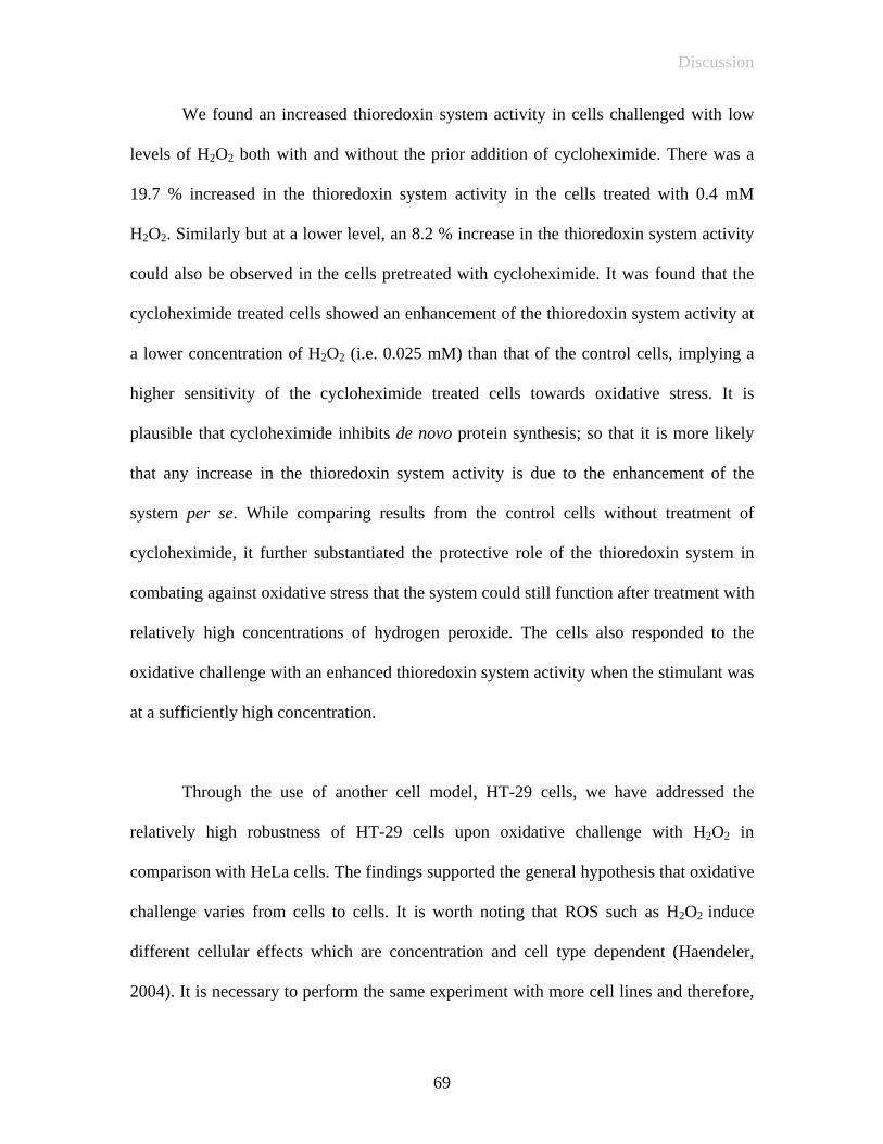
Discussion
We found an increased thioredoxin system activity in cells challenged with low
levels of H2O2 both with and without the prior addition of cycloheximide. There was a
19.7 % increased in the thioredoxin system activity in the cells treated with 0.4 mM
H2O2. Similarly but at a lower level, an 8.2 % increase in the thioredoxin system activity
could also be observed in the cells pretreated with cycloheximide. It was found that the
cycloheximide treated cells showed an enhancement of the thioredoxin system activity at
a lower concentration of H2O2 (i.e. 0.025 mM) than that of the control cells, implying a
higher sensitivity of the cycloheximide treated cells towards oxidative stress. It is
plausible that cycloheximide inhibits de novo protein synthesis; so that it is more likely
that any increase in the thioredoxin system activity is due to the enhancement of the
system per se. While comparing results from the control cells without treatment of
cycloheximide, it further substantiated the protective role of the thioredoxin system in
combating against oxidative stress that the system could still function after treatment with
relatively high concentrations of hydrogen peroxide. The cells also responded to the
oxidative challenge with an enhanced thioredoxin system activity when the stimulant was
at a sufficiently high concentration.
Through the use of another cell model, HT-29 cells, we have addressed the
relatively high robustness of HT-29 cells upon oxidative challenge with H2O2 in
comparison with HeLa cells. The findings supported the general hypothesis that oxidative
challenge varies from cells to cells. It is worth noting that ROS such as H2O2 induce
different cellular effects which are concentration and cell type dependent (Haendeler,
2004). It is necessary to perform the same experiment with more cell lines and therefore,
69

Discussion
HT-29 was employed. In the study, higher concentrations (0.5 and 1 mM) of H2O2 were
employed to act as oxidative challenge onto HT-29 cells. In agreement with the HeLa
cells findings, an enhancement of the thioredoxin system activity could also be observed
in cells challenged with H2O2 both with and without the prior addition of cycloheximide.
There were a 16.3 % and 18.5 % increase in the thioredoxin system activity when 0.5 and
1 mM H2O2 were added respectively, while in the cells pretreated with cycloheximide, an
8.1 % and 13. 9 % increased in the thioredoxin system activity could be observed after
the addition of 0.5 and 1 mM H2O2 respectively. Results from both HeLa and HT-29 cells
support the notion that thioredoxin system may play active role in defense against
oxidative challenge. And interestingly, the enhancement was not necessarily dependent
on de novo protein synthesis.
In many previous studies, ROS were shown to act as stress signals that activated
specific redox sensitive signaling pathways to respond to such oxidative challenge
(Toren, 2000). The initiating events leading to the activation of the pathways upon
oxidative challenge are still not completely understood. Recent findings suggested that
TR would be expressed at elevated levels and the process could be transcriptional
mediated (Smart, 2004). However in our studies, both nucleated and anucleated cells
showed an enhancement of the thioredoxin activity that was not necessarily dependent on
transcription and indeed we have also demonstrated that there were no significant
changes of expression and level of Trx and TR upon oxidative challenge. The findings
supported a very important role of the thioredoxin system in cells. Taken together, the
results of these experiments not only showed the involvement of the thioredoxin system
70
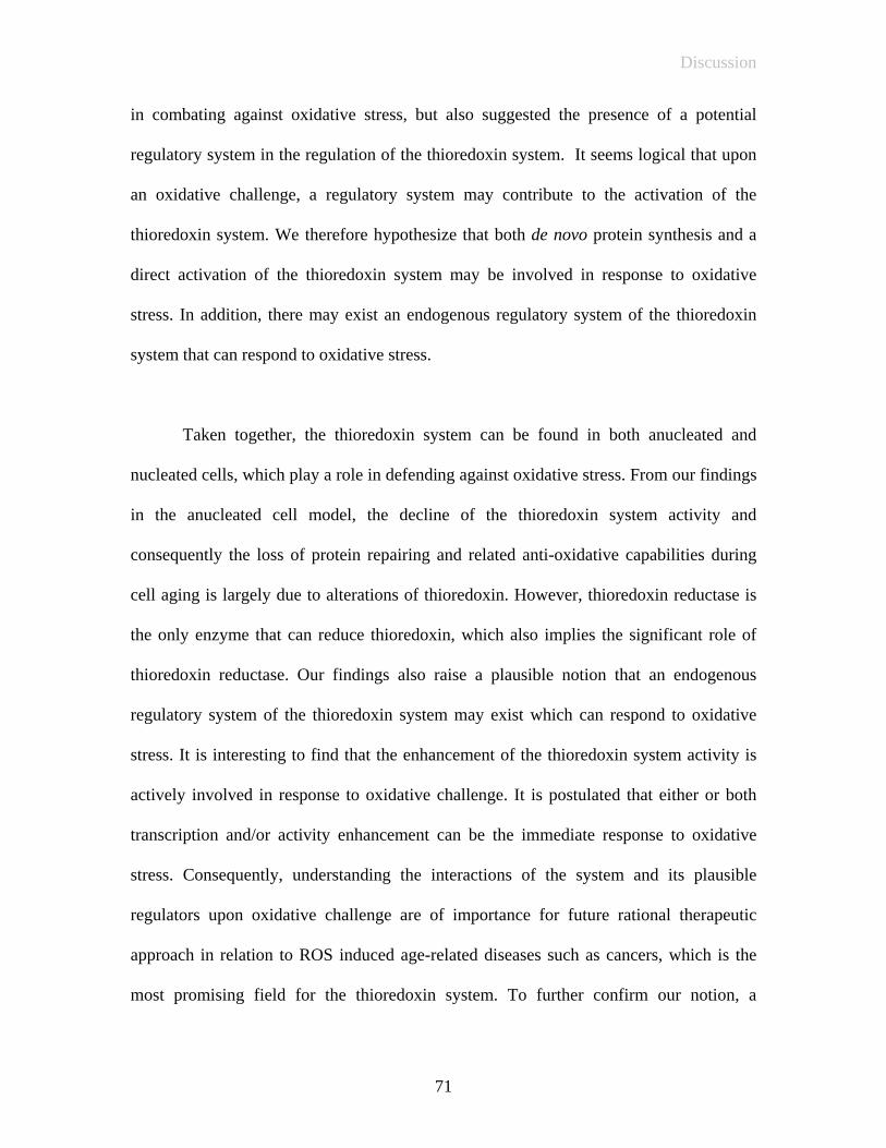
Discussion
in combating against oxidative stress, but also suggested the presence of a potential
regulatory system in the regulation of the thioredoxin system. It seems logical that upon
an oxidative challenge, a regulatory system may contribute to the activation of the
thioredoxin system. We therefore hypothesize that both de novo protein synthesis and a
direct activation of the thioredoxin system may be involved in response to oxidative
stress. In addition, there may exist an endogenous regulatory system of the thioredoxin
system that can respond to oxidative stress.
Taken together, the thioredoxin system can be found in both anucleated and
nucleated cells, which play a role in defending against oxidative stress. From our findings
in the anucleated cell model, the decline of the thioredoxin system activity and
consequently the loss of protein repairing and related anti-oxidative capabilities during
cell aging is largely due to alterations of thioredoxin. However, thioredoxin reductase is
the only enzyme that can reduce thioredoxin, which also implies the significant role of
thioredoxin reductase. Our findings also raise a plausible notion that an endogenous
regulatory system of the thioredoxin system may exist which can respond to oxidative
stress. It is interesting to find that the enhancement of the thioredoxin system activity is
actively involved in response to oxidative challenge. It is postulated that either or both
transcription and/or activity enhancement can be the immediate response to oxidative
stress. Consequently, understanding the interactions of the system and its plausible
regulators upon oxidative challenge are of importance for future rational therapeutic
approach in relation to ROS induced age-related diseases such as cancers, which is the
most promising field for the thioredoxin system. To further confirm our notion, a
71

Discussion
comparison of the activity enhancement and the transcriptional mRNA level of the
thioredoxin system may provide a better understanding of the true role of the thioredoxin
system in cell aging and oxidative stress. The search for the regulatory system(s) should
also be actively pursued.
72
Conclusion
VI Conclusion
The thioredoxin system activity in the young erythrocytes was found substantially
higher than that in the old ones when NADPH was supplemented. Both Trx and TR
activity were found declining in the old erythrocytes. Moreover, TR in old cells still
showed some degrees of activity as illustrated by the insulin assay while it showed no
Trx activity in the old cells. In addition, the thioredoxin system activity of the H2O2-
treated erythrocytes increased with an increasing H2O2 level at up to 4 mM H2O2.
Beyond this level, the thioredoxin system activity progressively decreased. As a result,
we propose that low concentrations of ROS may exert a stimulating effect on the
signaling, receptors and enzymatic response. The addition of oxidative stress exceeding
the maximum tolerance level would lead to gradually degeneration of the thioredoxin
system due to excessive insults. The decline of the thioredoxin system activity and
consequently loss of the protein repairing and related anti-oxidative capabilities during
cell aging thus largely look attributed to the decline of the function of the important
protective protein co-factor of the system, which is thioredoxin.
In conclusion, our study confirms the ability of the thioredoxin system in
combating against oxidative stress in both erythrocytes and cell culture (HeLa and HT-29
cells). The enhancement of the thioredoxin system activity, observed after oxidative
stress, is in concert with the data describing the role of the thioredoxin system. Oxidative
stress elicited by H2O2, which mimics the conditions of cellular aging process, trigger
cells to initiate apoptotic signaling. Recently, the term hormesis has been widely
discussed. It is postulated that low level of stress can exert beneficial effects to cell, while
73
Conclusion
it has stimulatory effects on the signaling, receptor stimulatory and enzyme stimulatory
effects (Radak, 2004). In this study, we demonstrated that there was an enhancement of
the thioredoxin system activity upon oxidative challenge in both young and old
erythrocytes, and such an activation could also be observed in nucleated HeLa and HT-29
cell models. The results clearly illustrated that the enhancement was not necessarily
dependent on de novo protein synthesis. Our notion is that both de novo protein synthesis
and a direct activation of the thioredoxin system may be involved in response to oxidative
stress. Together with the yet to be characterized regulatory system, the full thioredoxin
system appears to play important roles in cell aging and the combat against oxidative
stress. To confirm our hypothesis, further studies concerning the regulators of the
thioredoxin system may provide a better understanding on the response and involvement
of the thioredoxin system upon oxidative challenge. Our findings have provided more
information on the regulation of redox balance and hopefully a step forward to unravel
the mystery of aging.
74
References
References Arnér E. S. J., Holmgren A., Physiological functions of thioredoxin and thioredoxin reductase, Eur. J. Biochem. 267 (2000) 6102-6109 Barry D., Christiaan L., Aging and the role of reactive nitrogen species, Ann. N. Y. Acad. Sci. 959 (2002) 66-81 Buchanan B. B., Balmer Y., Redox regulation: a broadening horizon, Annu. Rev. Plant Biol. 56 (2005) 187-220 Chanarin I., Laboratory Haematology, Churchill Livingstone, 1989 Chen Q. G., Ding Q. X. , Jeffrey N. K., The stationary phase model of aging in yeast for the study of oxidative stress and age-related neurodegeneration, Biogerontology. 6 (2005) 1-13 Cho C. G., Kim H. J., Chung S. W., Jung K. J., Shim K. H., Yu B. P., Yodoi J., Chung H. Y., Modulation of glutathione and thioredoxin system by calorie restriction during the aging process, Experimental gerontology. 38 (2003) 539-548 Clark L. C., Combs G. F., Jr., Turnbull B. W., Stlate E. H., Chalker D. K., Chow J., Davis L. S., Glover R. A., Graham G. F., Gross E. G., et al, Effects of selenium supplementation for cancer prevention in patients with carcinoma of the skin. A randomized controlled trial, nutritional prevention of cancer study group, JAMA. 276 (1996) 1957-1963 Dacie J. V., Lewis S. M., Practical Haematology, Churchill Livingston, 1975 Daves K. J. A., Goldberg A. L., Oxygen radicals stimulate intracellular proteolysis and lipid peroxidation by independent mechanisms in erythrocytes, J. Biol. Chem. 262 (1987) 8220-8226 Deuticke B., Heller K. B., Haest C. W. M., Progressive oxidative membrane damage in erythrocytes after pulse treatment with t-butylhydroperoxide, Biochem. Biophy. Acta. 899 (1987) 113-124 Ellman G. L., Courtney K. D., Andres V., Featherstone R. M., A new and rapid colorimetric determination of acetylcholinesterase activity, Biochem. Pharmacol. 2 (1961) 88-95 Fausta O. S., Anna M., Nicoletta B., Silvia P., Piero O., Donatella T., Accelerated senescence of human erythrocytes cultured with Plasmodium falciparum, Blood. 102 (2003) 705-711
75

References
Fernando M. R., Nanri H., Yoshitake S., Nagata-Kuno K., Minakami S., Thioreodixin regenerates proteins inactivated by oxidative stress in endothelial cells, Eur. J. Biochem. 209 (1992) 917-922 Follmann H., Haberlein I., Thioredoxin: universal, yet specific thiol-disulpfide redox cofactors, Biofactors. 5 (1995) 147-156 Freemerman A. J., Gallegos A., Powis G., NF-kappaB transactivation is increased but not involved in the proliferation effects of thioredoxin overexpression in MCF-7 breast cancer cells, Cancer Res. 59 (1999) 4090-94 Gromer S., Urig S., Becker K., The thioredoxin system-from science to clinic, Med. Res. Reviews. Vol 24 (2004) 40-89 Haendeler J., Tischler V., Hoffmann J., Zeiher A. M., Dimmeler S., Low doses of reactive oxygen species protect endothelial cells from apoptosis by increasing thioredoxin-1 expression, FEBS Letters. 577 (2004) 427-433 Holmgren A., Björnstedt M., Thioredoxin and thioredoxin reductase, Methods. Enzymol. Vol.252 (1995) 199-208 Holmgren A., Thioredoxin and Glutaredoxin system, J. Biol. Chem. 264 (1989) 13963-13966 Jordan A., Reichard P., Ribonucleotide reductase, Annu. Rev. Biochem. 67 (1998) 71-98 Kang M. J., Kim H. J., Chung H. Y., The effect of age and calories restriction on HIF-1-responsive genes in aged liver, Biogerontology. 6 (2005) 27-37 Laurent T. C., Moore E. C., Reichard P., Enzymatic synthesis of deoxyribonlucleotides VI. Isolation and characterization of thioredoxin, the hydrogen donor from Escherichia coli B, J. Biol. Chem. 239 (1964) 3436-44 Lowry, N. J. Rosebrough, A. L., Farr R. J., Randall J. Biol. Chem. 193 (1951) 265 Luthman M., Holmgren A., Rat liver thioredoxin and thioredoxin reductase: purification and characterization, Biochem. 21 (1982) 6628-6633 Magnusson C. G., Björnstedt M., Holmgren A., Human IgG is substrate for the thioredoxin system: Differential cleavage pattern of interchain disulphide bridges in IgG subclasses, Mol. Immunol. 34 (1997) 709-717 Malich G., Boban M., Chris W., The sensitivity and specificity of the MTS tetrazolium assay for detecting the in vitro cytotoxicity of 20 chemicals using human cell lines, Toxicol. 124 (1997) 179-192
76

References
Mauro M., Antonio D. F., Red blood cell aging, Plenum Press, New York, Vol.37, (1991) Murphy J. R., Influence of temperature and method of centrifugation on the separation of erythrocytes, J. Lab. Clin. Med. 82 (1973) 334-41 Mustacich D., Powis G., Thioredoxin reductase, Biochem. J. 346 (2000) 1-8 Nordberg J., Arner E. S. J., Reactive oxygen species, antioxidants, and the mammalian thioredoxin system, Free Radic. Biol. Med. 31 (2001) 1287-1312 Powis G., Montfort W. R., Properties and biological activities of thioredoxins, Annu. Rev. Pharmacol. Toxicol. 41 (2001) 261-95 Radak Z., Chung H.Y., Goto S., Exercise and hormesis: oxidative stress-related adaptation for successful aging, Biogerontology. 6 (2005) 71-75 Ratten S. I S., Clark B. F. C., Understanding and modulating aging, IUBMB Life. 57 (2005) 297-304 Richard A., Lockshin, Zahra Z., The biology of cell death and its relationship to aging, Wiley-Liss, New York. (1996) 167-180 Ronata C., Toren F., Reactive oxygen species as mediators of cellular senescence, IUBMB Life. 57 (2005) 277-281 Smart D. K., Ortiz K. L., Mattson D., Bradbury M., Bisht K. S., Sieck L. K., Brechbiel M. W., Gius D., Thioredoxin reductase as a potential molecular target for anticancer agents that induce oxidative stress, Cancer Research. 64 (2004) 6716-6724 Snyder L. M., Fortier N. L., Mohandas N., The role of membrane protein sulfhydryl groups in hydrogen peroxide-mediated membrane damage in human erythrocytes, Biochim. Biophys. 937 (1988) 229-240 Stadtman E. R., Protein oxidation and aging, Science. 257 (1992) 1220-1224 Stuart L., Michael J. D., Roger T. D., Protein oxidation and aging, Experimental gerontology. 36 (2001) 1503-1518 Sun Q. A., Gladyshev V. N., Redox regulation of cell signaling by thioredoxin reductase, Methods Enzymol. 347 (2002) 451-461 Tavazzi B., Pierro D. D., Lazzarino G., Energy metabolism and lipid peroxidation of human erythrocytes as a function of increased oxidative stress, Eur. J. Biochem. 267 (2000) 684-689
77
References
Toren N., Nikki J. H., Oxidants, oxidative stress and the biology of ageing, Nature. 408 (2000) 239-247 Tsugnuobu A., Chock P. B., Chuang C. C., The roles of thioredoxin in protection against oxidative stress-induced apoptosis in SH-SY5Y cells, J. Biol. Chem. 277 (2002) 9655-9660 Wakasugi N., TagayaY., Wakasugi H., Mitsiu A., Maeda M., Yodoi J., Tursz T., Adult T-cell leukemia-derived factor/thioredoxin, produced by both human T-lymphophytic virus type I- and Epstein-Barr virus-transformed lymphocytes, acts as an autocrine growth factor and synergizes with interleukin 1 and interleukin 2, Proc. Natl. Sci. USA. 87 (1990) 8282-8286 Wang X. T., Liu M. Y., Holbrook N. J., The cellular response to oxidative stress: influences of mitogen-activated protein kinase signaling pathways on cell survivals, Biochem J. 333 (1998) 291-300 Watson H. W., Jones D. P., Oxidative of nuclear thioredoxin during oxidative stress, FEBS. 543 (2003) 144-147 Windle H. J., Fox Á., Eidhim D. N., Kelleher D., The thioredoxin system of helicobacter pylori, J. Biol. Chem. Vol.275, No.7 (2000) 5081-5089 Yu G. W., Leung Y. C. and Lee K. S., Non-catalytic activity of thioredoxin in protein refolding, Experimental Biology, New Orleans, L.A., Late-Breaking Abstract. (2002) 34
78