De Novo Design and Experimental Characterization of Ultrashort Self-Associating Peptides
17
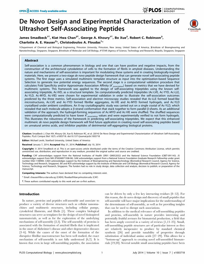
De Novo Design and Experimental Characterization of Ultrashort Self-Associating Peptides James Smadbeck 1" , Kiat Hwa Chan 2" , George A. Khoury 1" , Bo Xue 3 , Robert C. Robinson 3 , Charlotte A. E. Hauser 2 *, Christodoulos A. Floudas 1 * 1 Department of Chemical and Biological Engineering, Princeton University, Princeton, New Jersey, United States of America, 2 Institute of Bioengineering and Nanotechnology, Singapore, Singapore, 3 Institute of Molecular and Cell Biology, A*STAR (Agency of Science, Technology and Research), Biopolis, Singapore, Singapore Abstract Self-association is a common phenomenon in biology and one that can have positive and negative impacts, from the construction of the architectural cytoskeleton of cells to the formation of fibrils in amyloid diseases. Understanding the nature and mechanisms of self-association is important for modulating these systems and in creating biologically-inspired materials. Here, we present a two-stage de novo peptide design framework that can generate novel self-associating peptide systems. The first stage uses a simulated multimeric template structure as input into the optimization-based Sequence Selection to generate low potential energy sequences. The second stage is a computational validation procedure that calculates Fold Specificity and/or Approximate Association Affinity (K * association ) based on metrics that we have devised for multimeric systems. This framework was applied to the design of self-associating tripeptides using the known self- associating tripeptide, Ac-IVD, as a structural template. Six computationally predicted tripeptides (Ac-LVE, Ac-YYD, Ac-LLE, Ac-YLD, Ac-MYD, Ac-VIE) were chosen for experimental validation in order to illustrate the self-association outcomes predicted by the three metrics. Self-association and electron microscopy studies revealed that Ac-LLE formed bead-like microstructures, Ac-LVE and Ac-YYD formed fibrillar aggregates, Ac-VIE and Ac-MYD formed hydrogels, and Ac-YLD crystallized under ambient conditions. An X-ray crystallographic study was carried out on a single crystal of Ac-YLD, which revealed that each molecule adopts a b-strand conformation that stack together to form parallel b-sheets. As an additional validation of the approach, the hydrogel-forming sequences of Ac-MYD and Ac-VIE were shuffled. The shuffled sequences were computationally predicted to have lower K * association values and were experimentally verified to not form hydrogels. This illustrates the robustness of the framework in predicting self-associating tripeptides. We expect that this enhanced multimeric de novo peptide design framework will find future application in creating novel self-associating peptides based on unnatural amino acids, and inhibitor peptides of detrimental self-aggregating biological proteins. Citation: Smadbeck J, Chan KH, Khoury GA, Xue B, Robinson RC, et al. (2014) De Novo Design and Experimental Characterization of Ultrashort Self-Associating Peptides. PLoS Comput Biol 10(7): e1003718. doi:10.1371/journal.pcbi.1003718 Editor: Michael Levitt, Stanford University, United States of America Received January 7, 2014; Accepted May 31, 2014; Published July 10, 2014 Copyright: ß 2014 Smadbeck et al. This is an open-access article distributed under the terms of the Creative Commons Attribution License, which permits unrestricted use, distribution, and reproduction in any medium, provided the original author and source are credited. Funding: CAF acknowledges support from the National Institutes of Health (5R01 GM052032) and the National Science Foundation (CBET-091143). JS acknowledges support from NIH (P50GM071508-06). GAK acknowledges support from a National Science Foundation Graduate Research Fellowship under grant number DGE-1148900. CAEH acknowledges support by the Institute of Bioengineering and Nanotechnology (Biomedical Research Council, Agency for Science, Technology and Research, Singapore). BX and RCR acknowledge support by the Institute of Molecular and Cell Biology (Biomedical Research Council, Agency for Science, Technology and Research, Singapore. The funders had no role in study design, data collection and analysis, decision to publish, or preparation of the manuscript. Competing Interests: The authors have declared that no competing interests exist. * Email: [email protected] (CAEH); [email protected] (CAF) " These authors contributed equally to this work and are co-first authors. Introduction In nature, proteins and peptides self-assemble and associate to produce a variety of diverse structures such as cellular nanoma- chines and multimeric structures, including cellular pumps, cytoskeletal filaments, and fibrils [1]. These complex biological structures can serve as templates for the design of novel bioinspired nanomaterials, as well as for the exploration of the underlying mechanisms of self-assembly [2,3]. The self-assembly of proteins is associated with the formation of amyloid fibrils that is implicated in the onset of Alzheimer’s disease and other degenerative diseases [3–6]. While the causes of the onset of the formation of the disruptive fibrillar macrostructure has been well studied, the exact mechanism of self-assembly is not fully understood [6,7]. It is known that even in large self-assembling peptides, the association can be driven by only a few key interacting residues [8–12]. For this reason, the de novo design and discovery of small peptides that self-assemble will have major implications for the understanding of the determinants of self-assembly, as well as for providing insights that can be used to disrupt such associations. In addition to the medical relevance of self-assembling peptides and proteins, self-assembly in nature provides interesting and potentially fruitful avenues for biomaterial production, a field that has been amply covered in a variety of reviews [1,13–25]. Small, self-assembling peptide structures are of particular interest as they are relatively inexpensive to produce by standard chemical synthesis [26] and provide tunability of properties through substitution of individual amino acids [27–29]. This allows for a ‘‘bottom-up’’ approach to creating novel self-assembled biomate- rials [19,20]. Several notable small associating peptides have been PLOS Computational Biology | www.ploscompbiol.org 1 July 2014 | Volume 10 | Issue 7 | e1003718
Transcript of De Novo Design and Experimental Characterization of Ultrashort Self-Associating Peptides
De Novo Design and Experimental Characterization ofUltrashort Self-Associating PeptidesJames Smadbeck1", Kiat Hwa Chan2", George A. Khoury1", Bo Xue3, Robert C. Robinson3,
Charlotte A. E. Hauser2*, Christodoulos A. Floudas1*
1 Department of Chemical and Biological Engineering, Princeton University, Princeton, New Jersey, United States of America, 2 Institute of Bioengineering and
Nanotechnology, Singapore, Singapore, 3 Institute of Molecular and Cell Biology, A*STAR (Agency of Science, Technology and Research), Biopolis, Singapore, Singapore
Abstract
Self-association is a common phenomenon in biology and one that can have positive and negative impacts, from theconstruction of the architectural cytoskeleton of cells to the formation of fibrils in amyloid diseases. Understanding thenature and mechanisms of self-association is important for modulating these systems and in creating biologically-inspiredmaterials. Here, we present a two-stage de novo peptide design framework that can generate novel self-associating peptidesystems. The first stage uses a simulated multimeric template structure as input into the optimization-based SequenceSelection to generate low potential energy sequences. The second stage is a computational validation procedure thatcalculates Fold Specificity and/or Approximate Association Affinity (K*
association) based on metrics that we have devised formultimeric systems. This framework was applied to the design of self-associating tripeptides using the known self-associating tripeptide, Ac-IVD, as a structural template. Six computationally predicted tripeptides (Ac-LVE, Ac-YYD, Ac-LLE,Ac-YLD, Ac-MYD, Ac-VIE) were chosen for experimental validation in order to illustrate the self-association outcomespredicted by the three metrics. Self-association and electron microscopy studies revealed that Ac-LLE formed bead-likemicrostructures, Ac-LVE and Ac-YYD formed fibrillar aggregates, Ac-VIE and Ac-MYD formed hydrogels, and Ac-YLDcrystallized under ambient conditions. An X-ray crystallographic study was carried out on a single crystal of Ac-YLD, whichrevealed that each molecule adopts a b-strand conformation that stack together to form parallel b-sheets. As an additionalvalidation of the approach, the hydrogel-forming sequences of Ac-MYD and Ac-VIE were shuffled. The shuffled sequenceswere computationally predicted to have lower K*
association values and were experimentally verified to not form hydrogels.This illustrates the robustness of the framework in predicting self-associating tripeptides. We expect that this enhancedmultimeric de novo peptide design framework will find future application in creating novel self-associating peptides basedon unnatural amino acids, and inhibitor peptides of detrimental self-aggregating biological proteins.
Citation: Smadbeck J, Chan KH, Khoury GA, Xue B, Robinson RC, et al. (2014) De Novo Design and Experimental Characterization of Ultrashort Self-AssociatingPeptides. PLoS Comput Biol 10(7): e1003718. doi:10.1371/journal.pcbi.1003718
Editor: Michael Levitt, Stanford University, United States of America
Received January 7, 2014; Accepted May 31, 2014; Published July 10, 2014
Copyright: � 2014 Smadbeck et al. This is an open-access article distributed under the terms of the Creative Commons Attribution License, which permitsunrestricted use, distribution, and reproduction in any medium, provided the original author and source are credited.
Funding: CAF acknowledges support from the National Institutes of Health (5R01 GM052032) and the National Science Foundation (CBET-091143). JSacknowledges support from NIH (P50GM071508-06). GAK acknowledges support from a National Science Foundation Graduate Research Fellowship under grantnumber DGE-1148900. CAEH acknowledges support by the Institute of Bioengineering and Nanotechnology (Biomedical Research Council, Agency for Science,Technology and Research, Singapore). BX and RCR acknowledge support by the Institute of Molecular and Cell Biology (Biomedical Research Council, Agency forScience, Technology and Research, Singapore. The funders had no role in study design, data collection and analysis, decision to publish, or preparation of themanuscript.
Competing Interests: The authors have declared that no competing interests exist.
* Email: [email protected] (CAEH); [email protected] (CAF)
" These authors contributed equally to this work and are co-first authors.
Introduction
In nature, proteins and peptides self-assemble and associate to
produce a variety of diverse structures such as cellular nanoma-
chines and multimeric structures, including cellular pumps,
cytoskeletal filaments, and fibrils [1]. These complex biological
structures can serve as templates for the design of novel bioinspired
nanomaterials, as well as for the exploration of the underlying
mechanisms of self-assembly [2,3]. The self-assembly of proteins is
associated with the formation of amyloid fibrils that is implicated
in the onset of Alzheimer’s disease and other degenerative diseases
[3–6]. While the causes of the onset of the formation of the
disruptive fibrillar macrostructure has been well studied, the exact
mechanism of self-assembly is not fully understood [6,7]. It is
known that even in large self-assembling peptides, the association
can be driven by only a few key interacting residues [8–12]. For
this reason, the de novo design and discovery of small peptides that
self-assemble will have major implications for the understanding of
the determinants of self-assembly, as well as for providing insights
that can be used to disrupt such associations.
In addition to the medical relevance of self-assembling peptides
and proteins, self-assembly in nature provides interesting and
potentially fruitful avenues for biomaterial production, a field that
has been amply covered in a variety of reviews [1,13–25]. Small,
self-assembling peptide structures are of particular interest as they
are relatively inexpensive to produce by standard chemical
synthesis [26] and provide tunability of properties through
substitution of individual amino acids [27–29]. This allows for a
‘‘bottom-up’’ approach to creating novel self-assembled biomate-
rials [19,20]. Several notable small associating peptides have been
PLOS Computational Biology | www.ploscompbiol.org 1 July 2014 | Volume 10 | Issue 7 | e1003718

discovered by derivation of natural systems (e.g., Alzheimer’s b-
amyloid protein) and through rational design [13,14,25].
The design of self-assembling peptides for biomedical and
biomaterial purposes has most commonly been performed through
rational design and large-scale screening. The discovery of a self-
assembling dipeptide [30–32] has demonstrated the applicability
of methods to such a problem. However, the size of the peptide is
limiting in this design process, since the immense sequence space
(20N possible designed sequences, where N is the number of design
positions) that must be searched may, in many cases, overstretch
the combinatorial capabilities of such experimental methods. Due
to the considerable cost and time involved in synthesizing and
testing a large number of candidate peptides, it is highly desirable
to screen computationally for self-assembly properties prior to
experimental testing of peptides. For this reason, the application of
computational methods to the design of self-assembling peptides is
highly desirable.
Computational protein design methods have become increas-
ingly prevalent in the field of protein engineering. These design
methods include those that employ probabilistic algorithms like
Monte Carlo (MC) methods [33–36] and genetic algorithms [37],
as well as deterministic algorithms like dead end elimination
(DEE) [38–42], self-consistent mean field (SCMF) methods [43–
48], or quadratic assignment-like global optimization for
sequence selection followed by fold specificity and approximate
binding affinity [49–55]. Such computational methods allow for
the consideration of large numbers of amino acid-amino acid
interactions simultaneously. Computational design has been used
to design inhibitors against H1N1 influenza hemagglutinin [56],
to switch cofactor specificity of an enzyme [57], for generalized
antibody design for recognition of a target epitope [58], for the
design of entry inhibitors of HIV-1 gp41 [59], for the design of
C3a receptor agonists for medicinal use [54], and for the design
of inhibitors of the histone methyltransferase EZH2 [55]. See
Fung et al. [51], Pantazes et al. [60], Samish et al. [61], and
Khoury et al. [62] for reviews of the recent advances and
successes in the area.
As computational methods for single peptides and protein-
peptide complexes have improved, the general interest in the design
of multimeric protein assemblies for therapeutic and biomaterial
applications [63–65] has also increased. Recently, there have been a
number of successful computational designs carried out to create
unique multimeric protein structures [66–69], Here we present a de
novo protein/peptide design framework applicable to multimeric
systems and its application to the design of self-associating
tripeptides. This framework utilizes a computationally-generated
multimeric assembly [51,70] of the self-assembling tripeptide Ac-
IVD [11] as the template for an optimization-based Sequence
Selection method [49,50,71,72]. Selected sequences are then
computationally screened via a Fold Specificity calculation [70]
and/or calculation of Association Affinity via molecular dynamics
(MD) simulations. The Association Affinity metric is based on
statistical mechanics [59,73] and is used to select a small set of high
confidence peptide sequences from the candidate set. To experi-
mentally validate the framework, six in silico designed sequences
were selected for experimental assessment based on the metrics
described. We found that two of these tripeptides (Ac-VIE, Ac-
MYD) formed hydrogels on time scales and at concentrations
comparable to the template peptide Ac-IVD. Shuffled control
sequences of these designed hydrogelating peptides were further
experimentally and computationally assessed to validate the
approach. Remarkably, Ac-YLD was capable of rapidly associating
into large crystals under ambient conditions, which led to the
elucidation of its crystal structure. The structural data obtained from
the crystal are invaluable in refining the framework for improved
accuracy in the design of self-associating systems.
Results
Computational ResultsThe outcomes of the optimization and simulation (Stages One
and Two) are tabulated in Tables 1–3 (full results provided in
Table S1). The Sequence Selection table shows that there is a high
frequency of double aromatic residues (Trp, Tyr) present in the
top ten sequences exhibiting the lowest potential energies (Pot.E),
whereas there is a high frequency of Met and Ile being present in
the last ten tripeptides with the highest potential energies (Table
S2). Stage One calculates the pairwise interaction energies
between residues. A fully extended polypeptide chain would result
in side-chains of adjacent residues being on opposite planes of the
polypeptide backbone. The fact that double aromatic residue
sequences have been calculated to possess the lowest potential
energies suggests that the backbones of these tripeptides are
twisted to promote pairwise interactions between residues.
Aromatic residues are known to associate via p-p/CH-p stacking,
a prominent example being diphenylalanine [30–32], so the high
ranking of double aromatic residue sequences (lowest potential
energies) enhances confidence in Stage One results. The high
frequency of linear aliphatic residues (Met, Ile) in the sequences of
highest potential energies reflects that van der Waals interactions
between adjacent aliphatic side-chains of Met/Ile are weak
compared to the aromatic residues.
In order to improve confidence in the sequences to be selected
for experimental validation, the full set of sequences was screened
by Fold Specificity (FSpec) and ranked again. In turn, the top
twenty sequences ranked by Fold Specificity were also assessed by
the Approximate Association Affinity metric, K*association. This
double-ranked set of peptide sequences is shown as ‘‘Run 1’’ in
Table 2.
In order to separately assess the capabilities of the newly
developed metric, 109 of the 128 tripeptide candidates were also
Author Summary
The self-association of peptides and proteins plays animportant role in many serious diseases, such as Alzhei-mer’s disease. A complete understanding of how peptidesand proteins self-associate is important in creatingtherapeutics for such diseases. Additionally, self-associat-ing peptides can be used as templates for bioinspirednanomaterials. With these goals in mind, we haveproposed a de novo peptide design methodology capableof producing peptides that self-associate. We haveexperimentally tested the framework through the designof several self-associating tripeptides. Using the frameworkwe designed six self-associating peptides, including twopeptides, Ac-MYD and Ac-VIE, which readily formedhydrogels and one peptide, Ac-YLD, which readily formeda crystal. An X-ray crystallographic study was performed onAc-YLD to determine its crystal structure. The top-rankeddesigned sequences were shuffled and computationallyand experimentally characterized in order to validate thatthe approach can differentiate the self-associating oftripeptides, which are derived from the same amino acids.Through the analysis of the experimental results wedetermine which metrics are most important in the self-association of peptides. Additionally, the crystallographicstructure of the tripeptide Ac-YLD provides a structuraltemplate for future self-association design experiments.
De Novo Design of Ultrashort Self-Associating Peptides
PLOS Computational Biology | www.ploscompbiol.org 2 July 2014 | Volume 10 | Issue 7 | e1003718

directly assessed by K*association. 19 peptides were excluded since
their Sequence Selection and/or Fold Specificity rankings were
among the lowest ranking and thus did not warrant re-evaluation.
The set of top ten sequences using this metric is given as ‘‘Run 2’’
in Table 3. Unlike for Sequence Selection in which sequences with
double aromatic residues dominate the top of the rank, no
outstanding trends were observed with regard to the residues of the
top-ranked sequences for either Fold Specificity or Approximate
Association Affinity, despite the expectation that sequences with
aromatic residues might exhibit higher association affinity. This
illustrates the ability of the Approximate Association Affinity
metric to discern tripeptides that are strong candidates for
association, but would have otherwise been difficult to identify
through rational design.
The top-ranked tripeptide in both Runs 1 and 2, Ac-LVE, was
selected for validation. Compared to Ac-LVE, Ac-YLD has
similar Pot.E and FSpec, but different K*association, so it was also
selected. Similarly, Ac-LLE and Ac-YYD were selected because
they have similar FSpec and K*association, but different Pot.E. The
Ac-LVE/Ac-YLD and Ac-LLE/Ac-YYD pairings might allow
the respective effects of K*association and Pot.E on self-association
outcomes to be discerned. Lastly, Ac-MYD and Ac-VIE were
selected as they have similar Pot.E to Ac-IVD. This allows the
effects of FSpec and K*association on hydrogelation to be assessed.
Thus, the tripeptides chosen for experimental validation were Ac-
IVD, Ac-LVE, Ac-YYD, Ac-LLE, Ac-YLD, Ac-MYD, and Ac-
VIE (Table 4).
It should be noted that the Fold Specificity and Approximate
Association Affinity are used strictly as metrics for selecting which
peptides should be experimentally tested. We are not attempting to
compare the calculated values to exact, experimental Fold
Specificity or Association Affinity values. Rather, we aim to
produce metrics capable of ranking a set of peptides to increase the
probability that the top ranked peptides are positive hits, in this
case, self-associating peptides. For this reason, it is of little concern
whether the properties of the produced peptides match exactly the
ranking shown in the tables.
The inter-peptide interactions that are observed in the
simulations of favorably self-associating sequences are predicted
to have a higher tendency to self-associate (form hydrogels or
crystals) and this forms the hypothesis being tested in this work. In
the computational calculations, there is currently no metric that
can distinguish whether the peptides could potentially form
crystals or hydrogels.
Table 1. Summary of results from Stage One: Sequence Selection.
Seq. Sel. Rank P1 P2 P3 Potential Energy
1 TRP TRP ASP 20.1255
2 TRP TRP GLU 20.1193
3 TRP TYR GLU 20.0912
4 TRP TYR ASP 20.0851
5 TYR TRP GLU 20.0801
6 TYR TRP ASP 20.0788
7 TRP LEU GLU 20.0755
8 TRP LEU ASP 20.0687
9 LEU TRP ASP 20.0664
10 LEU TRP GLU 20.0663
The top ten candidates, ranked by potential energy (Pot.E), are given in the table.doi:10.1371/journal.pcbi.1003718.t001
Table 2. Summary of results for Run 1.
Seq. Sel. Rank Pot.E FSpec Rank FSpec K*association Rank K*
association Sequence
54* 20.0324 4 6.07 1 1.66E-03 LVE
49* 20.0340 7 5.18 2 4.97E-50 YLD
55 20.0323 8 5.17 3 1.12E-54 VLE
48 20.0342 19 2.66 4 1.34E-61 YIE
93* 20.0173 18 2.69 5 5.39E-64 VIE
12* 20.0618 13 3.54 6 4.31E-64 LLE
46* 20.0361 10 3.89 7 2.65E-70 YYD
33 20.0367 20 2.39 8 2.67E-72 LYD
27 20.0483 1 8.48 9 1.13E-79 YLE
120 20.0051 14 3.02 10 4.23E-90 VME
The top twenty sequences by Fold Specificity were re-ranked by Approximate Association Affinity. The top ten candidates after re-ranking are shown in the table. Five ofthe top sequences (*) were chosen from this re-ranked set for experimental validation.doi:10.1371/journal.pcbi.1003718.t002
De Novo Design of Ultrashort Self-Associating Peptides
PLOS Computational Biology | www.ploscompbiol.org 3 July 2014 | Volume 10 | Issue 7 | e1003718

Experimental Validation of Designed Tripeptides: Self-Association and Rheological Studies
The six high-ranking tripeptides that were chosen to be
evaluated based on their predicted abilities to self-associate can
be divided into two classes: (1) the aliphatic class of Ac-LVE, Ac-
VIE, Ac-LLE and (2) the aromatic class of Ac-MYD, Ac-YLD,
and Ac-YYD. The ability of the tripeptides to associate was
assessed across a concentration range from 5 mg/mL to the upper
limit of 40 mg/mL, in steps of 5 mg/mL. Such a concentration
series enables one to compare the association properties of the
evaluated tripeptides at 20 mg/mL (concentration at which the
simulations were run), as well as bracket the concentration in
which there is a change in the association state of the tripeptide.
Of the three aliphatic tripeptides, the top-ranked sequence, Ac-
LVE, was able to form a gelatinous precipitate between 5 and
10 mg/mL. This precipitation persisted up to 30 mg/mL, with
hydrogelation of Ac-LVE observed at 35 and 40 mg/mL. The
second-ranked sequence, Ac-VIE was able to form a hydrogel at
5 mg/mL over 48 h; at 10 mg/mL, hydrogelation proceeded
within 10 min (Figure 1A). The third-ranked sequence, Ac-LLE,
formed a clear solution up to 40 mg/mL, even after standing for
two weeks. This indicates that either there is no self-association, or
that any association formed by Ac-LLE is still soluble in water.
Of the three aromatic tripeptides, the top-ranked sequence, Ac-
MYD, was able to form a hydrogel at 5 mg/mL over 24 h; at
10 mg/mL, hydrogelation proceeded within 1 min (Figure 1A).
The second-ranked sequence, Ac-YLD, spontaneously crystallized
in water, even at the lowest concentration of 5 mg/mL, to furnish
large crystals of diffraction quality under ambient conditions
(Figure 1B). This indicates that the self-association of Ac-YLD
proceeded in an orderly manner to produce the well-defined
packing of a crystal. The third-ranked sequence, Ac-YYD, was
readily soluble in water, but over time, a small amount of
gelatinous precipitate was observed. The amount of gelatinous
precipitate scales approximately with concentration up to 40 mg/
mL. These observations indicate that the propensity of Ac-YYD to
aggregate and entrap water is low.
The viscoelasticity of the hydrogels formed from Ac-VIE and
Ac-MYD were assessed experimentally at 20 mM. Ac-MYD
formed the stiffer hydrogel with a storage modulus (G9) of 20 kPa
compared to Ac-VIE (G9 = 8 kPa) (Figure 2). The loss modulus
graph also shows that Ac-MYD possessed the larger loss modulus.
The loss modulus is a measure of the viscosity of the system, so a
substrate with large loss modulus would be very viscous, and less
likely to ‘‘slip’’. Indeed, while the hydrogels of Ac-VIE
(G0 = 1 kPa) collapsed within two days, the hydrogel of Ac-MYD
(G0 = 9 kPa) was able to maintain its physical form over more than
10 months (Figure 2 inset: note the hydrogel suspended on the
wall). The storage modulus values are comparable to those
previously reported for the template tripeptide, Ac-IVD [74].
Experimental Results for Shuffled Control Sequences ofHydrogel Forming Peptides
Control experiments were performed to illustrate the ability of
the procedure to compare the relative self-association of analogous
tripeptides. Calculations of K*association for analogous tripeptides of
Ac-MYD and Ac-VIE, based on shuffling the amino acid residues
in the tripeptide sequence, were performed. The calculations show
that the optimal position of the polar headgroup is at the C-
terminal position, which was previously proposed by Hauser et al.
[11]. Four tripeptides (Ac-YMD, Ac-DMY, Ac-IVE, Ac-EVI)
were chosen from the shuffled sequences and assessed experimen-
tally. As Table 5 shows, the shuffled sequences of Ac-MYD, i.e.
Ac-YMD and Ac-DMY, formed clear solutions with no signs of
self-association, in agreement with the computed lower K*association
values (Table 5). While the shuffled sequences of Ac-VIE (i.e. Ac-
IVE and Ac-EVI) precipitated with fibrillar nanostructures,
hydrogels were not formed (Table 5 and Figure S1). This could
be related to the ability of the de novo protein design method to
predict differently for aliphatic and aromatic tripeptides. It should
be noted that the peptides Ac-EVI and Ac-DMY are the only two
cases where the self-association motif detailed in Hauser et al. [11]
is not incorporated in a tested peptide. The fact that both such
peptides are predicted and experimentally validated to not form
self-associating structures supports the use of the motif in this and
future studies.
Morphological and Structural Studies of Self-AssociatingTripeptides
Aliphatic peptides. To gain information on the morphology
that the tripeptides assume on self-association, field emission
scanning electron microscopy (FE-SEM) was used to examine the
tripeptides after they had been dissolved in water and subsequently
freeze-dried. The electron micrographs of the hydrogels of Ac-
LVE (40 mg/mL) and Ac-VIE showed the presence of mesh-like
fibers (Figure 3A, 3B), the latter being very similar to that observed
Table 3. Summary of results for Run 2.
Seq. Sel. Rank Pot.E FSpec Rank FSpec K*association Rank K*
association Sequence
54* 20.0324 4 6.07 1 1.66E-03 LVE
101** 20.0151 35 1.37 2 3.05E-15 MYD
103 20.0150 66 0.71 3 1.33E-24 AAD
47 20.0349 62 0.84 4 3.97E-30 IYE
61 20.0304 86 0.43 5 2.23E-30 IYD
100* 20.0153 51 1.00 6 4.87E-32 IVD
20 20.0517 70 0.65 7 7.80E-33 WVD
73 20.0272 85 0.44 8 1.04E-33 LAD
33 20.0467 111 0.18 9 8.25E-36 AWD
1 20.1255 126 0.06 10 2.15E-36 WWD
The top ten sequences ranked by Approximate Association Affinity (K*association) from the 109-peptide set. The top sequence not chosen in Run 1 was chosen from this
re-ranked set for experimental validation. The newly chosen peptide sequence is highlighted with (**). Previously selected peptides are highlighted with (*).doi:10.1371/journal.pcbi.1003718.t003
De Novo Design of Ultrashort Self-Associating Peptides
PLOS Computational Biology | www.ploscompbiol.org 4 July 2014 | Volume 10 | Issue 7 | e1003718

for Ac-LIVAGD and Ac-IVD [11]. This indicates that Ac-VIE
self-assembled into fibers akin to that of Ac-IVD. In contrast, the
electron micrographs of Ac-LLE showed no fibrillar features at 5
or 40 mg/mL (Figure 3C, 3D). Instead, bead-like structures
(diameter: 4–10 mm) could be observed at 5 and 40 mg/mL
(Figure 3C, 3D), as well as at intermediate concentrations (data not
shown). This suggests that Ac-LLE associates to form spherical
microstructures instead of fibrillar nanostructures.
Aromatic peptides. The electron micrographs of Ac-MYD
showed fibers densely packed together (Figure 4A), akin to that
observed for various aromatic peptides that have been previously
studied [75]. This suggests that it is packed in a different manner
to the aliphatic tripeptides. The electron micrographs of Ac-YLD
showed the presence of thin rectangular plates with well-defined
edges (Figure 4B), as expected of crystals. This reflects the high
propensity of Ac-YLD to associate in an orderly manner. Analysis
of the gelatinous precipitate of Ac-YYD that had formed slowly
showed fibrillar structures (Figure 4C). In contrast to the
microstructures observed in the solutions of Ac-LLE, only
amorphous structures were observed in the supernatant of Ac-
YYD (Figure 4D). This indicates that Ac-YYD possesses the
tendency to associate into fibers, but that this tendency is weak.
It should be noted that the use of FE-SEM to visualize
nanostructures is not meant to be a direct reflection of the
outcomes of the computational simulations, but rather to
characterize and compare the nanostructures that can be obtained
through the self-association of the designed sequences with the
highest calculated tendency to self-associate as quantified by their
K*association values.
Crystallographic study of Ac-YLD. An X-ray diffraction
study was carried out on a single crystal of Ac-YLD. Ac-YLD
adopts the typical conformation of a b-strand, which stacks
together to form parallel b-sheets (Figure 5). The b-sheets associate
laterally to form the crystal. The side-chain of Leu2 protrudes
from one side of the b-sheet while the side-chains of Tyr1 and
Asp3 protrude from the opposite side of the b-sheet. A water
molecule, anchored by the protonated carboxyl side chain of Asp3,
is critical for the formation of a hydrogen bond network that
contributes to both intra- and inter-b-sheet interactions. The
network is composed of the water molecule and three oxygen
atoms (one each) from three adjacent Ac-YLD molecules: Asp3(O-
H)-OH2, 1.75 A; Asp3(H-O)-H2O, 2.20 A; Ac(C = O)-H2O,
1.90 A. Other significant hydrogen-bond donor-acceptor pairs
include those formed by the protonated C-terminal carboxyl of
Asp3 and the amide oxygen of Tyr1 (inter-b-sheet, 1.76 A), and by
the hydroxyl of Tyr1 and the carbonyl oxygen of the N-terminal
acetyl (inter-b-sheet, 2.01 A), as well as two inter-b-strand
hydrogen bonds commonly seen in a b-sheet (intra-b-sheet, 2.40
and 2.49 A, respectively). In addition to these hydrogen bonds,
hydrophobic interactions among the clustered hydrophobic
moieties, that is, the methyl of the N-terminal acetyl and the
side-chain of Leu2, also promote both intra- and inter-b-sheet
associations. A third type of observed interaction is p-p stacking,
which engages the aromatic side chain of Tyr1 in a parallel-
displaced manner, and favors the stacking of Ac-YLD into a b-
sheet. As Figure 5 illustrates, the 4-phenoyl rings lie ‘‘stepped’’
with respect to each other. The detailed statistics of the
crystallization and refinement parameters are shown in Table 6.
The structural data obtained from an X-ray crystallographic
study of Ac-YLD, which formed crystals at ambient conditions,
has important implications in the further development of the self-
association de novo design method. The crystallographic structure
could be used as a starting template in future designs in order to
increase the accuracy of the Sequence Selection stage and open
Ta
ble
4.
Sum
mar
yo
fp
ep
tid
es
cho
sen
for
exp
eri
me
nta
lva
lidat
ion
.
Po
t.E
Ra
nk
Po
t.E
FS
pe
cR
an
kF
Sp
ec
K* a
sso
cia
tio
nS
eq
.R
un
Pe
pti
de
pro
pe
rty
Ex
p.
Ob
serv
ati
on
Sto
rag
em
od
ulu
s(G
9,k
Pa
)L
oss
mo
du
lus
(G0,
kP
a)
54
*2
0.0
32
44
6.0
71
.66
E-0
3LV
E1
,2al
iph
atic
hyd
rog
el
N.D
.N
.D.
10
1**
20
.01
51
35
1.3
73
.05
E-1
5M
YD
2ar
om
atic
hyd
rog
el
20
9
10
0*
20
.01
53
51
14
.87
E-3
2IV
D2
ali
ph
ati
ch
yd
rog
el
[87
]3
[87
]N
.A.
49
*2
0.0
34
07
5.1
84
.97
E-5
0Y
LD1
aro
mat
iccr
ysta
lN
.D.
N.D
.
93
*2
0.0
17
31
82
.69
5.3
9E-
64
VIE
1al
iph
atic
hyd
rog
el
81
12
*2
0.0
61
81
33
.54
4.3
1E-
64
LLE
1al
iph
atic
be
ad-l
ike
stru
ctu
reN
.D.
N.D
.
46
*2
0.0
36
11
03
.89
2.6
5E-
70
YY
D1
aro
mat
icfi
bri
llar
stru
ctu
reN
.D.
N.D
.
Seq
ue
nce
sar
era
nke
din
ord
er
of
K* a
sso
cia
tio
nva
lue
.T
em
pla
tese
qu
en
cekn
ow
nto
hyd
rog
el,
Ac-
IVD
,is
hig
hlig
hte
din
bo
ld.
do
i:10
.13
71
/jo
urn
al.p
cbi.1
00
37
18
.t0
04
De Novo Design of Ultrashort Self-Associating Peptides
PLOS Computational Biology | www.ploscompbiol.org 5 July 2014 | Volume 10 | Issue 7 | e1003718

the possibility of alternate design applications, such as the design of
peptides to inhibit crystal formation. Additionally, the structure
could be used to gain insight into the physical basis of how Ac-
YLD associates to form the crystal which could be used to improve
the metrics used in choosing the candidate peptides to more
accurately predict which peptides will self-associate into hydrogel
or crystal structures.
Discussion
Connections between Computational Metrics andExperimental Observations
When evaluating a newly developed multimeric de novo peptide
design framework that relies on several validation stages, it is
important to be able to critically assess each stage separately. The
experimental results aim to confirm/disconfirm the predictions
that the proposed computational framework makes, thus providing
an essential test of the approach. Run 1 utilizes Sequence
Selection, Fold Specificity and Approximate Association Affinity
to select sequences for experimental validation, whereas Run 2
utilizes only Sequence Selection and Approximate Association
Affinity. In order to utilize the framework for reliable prediction of
self-associating peptides, it is pertinent to understand the
properties that each of Sequence Selection (Pot.E), Fold Specificity
(FSpec), and Approximate Association Affinity (K*association) may
influence.
The potential energy used in the Sequence Selection stage,
Pot.E, which measures the pairwise interaction energies of residues
within the tripeptide, may be indirectly related to the extent to
which the tripeptide interacts with the solvent. For instance, if the
residues of the tripeptides interact in a highly favorable manner
with each other (large negative Pot.E) they may correspondingly
interact to a lower extent with the solvent. The converse would
also be true. Such substrate interaction with the solvent is known
to critically determine the nano-/microstructural form adopted by
the substrate. The tripeptides can be grouped into three potential
energy classes: low (Ac-LLE; Pot.E = 20.0618), medium (Ac-LVE,
Ac-YLD, Ac-YYD; Pot.E = 20.0324, 20.0340, 20.036, respec-
tively), and high (Ac-MYD, Ac-VIE, Ac-IVD; Pot.E = 20.0151, 2
0.0173, 20.0153, respectively). Ac-LLE (FSpec = 3.54, K*association
= 4.31610264) and Ac-YYD (FSpec = 3.89, K*association
= 2.65610270) have similar FSpec and K*association, so the effect of
Pot.E on their self-association can be gleaned. With the lower
Pot.E, Ac-LLE can interact to a lower extent with water, which
may account for the formation of bead-like microstructures. With
a higher Pot.E, Ac-YYD can interact to a greater extent with
water, which accounts for its high water solubility. The high Pot.E
of Ac-MYD/Ac-VIE/Ac-IVD suggests they can interact to the
relatively highest extent with water, which accounts for their
ability to entrap water in forming hydrogels.
FSpec, which is derived from an ensemble of 500 models with
varying backbone conformations, can be construed as sampling
conformations that are amenable to self-associating into nano- and
microstructures. Indeed, the chosen tripeptides, which all have
FSpec more than one, are capable of self-associating into either
Figure 1. Experimental characterization of successful self-associating peptide design. (A) (From left to right) Hydrogels formed from: Ac-VIE, 5 mg/mL; Ac-VIE, 10 mg/mL; Ac-MYD, 5 mg/mL; Ac-MYD, 10 mg/mL (B) Crystals of Ac-YLD viewed under a light microscope.doi:10.1371/journal.pcbi.1003718.g001
Figure 2. Comparison of the viscoelastic properties of tripep-tide. Comparison of the viscoelastic properties of tripeptide hydrogelsvia frequency sweep studies (strain = 0.1%) at 25uC: Ac-MYD (20 mM,13 mg/mL), Ac-VIE (20 mM, 12 mg/mL). Every data point represents themean of 10 repetitions. The error bars reflect the standard deviation of10 repetitions. The data show that at 20 mM, Ac-MYD formed the stifferhydrogel compared to Ac-VIE. The inset illustrates the physical forms ofthe hydrogels (left: Ac-VIE, right: Ac-MYD) on prolonged standing underambient conditions.doi:10.1371/journal.pcbi.1003718.g002
De Novo Design of Ultrashort Self-Associating Peptides
PLOS Computational Biology | www.ploscompbiol.org 6 July 2014 | Volume 10 | Issue 7 | e1003718

fibrillar structures (Ac-LVE, Ac-YYD, Ac-MYD, Ac-VIE), crystals
(Ac-YLD), or bead-like microstructures (Ac-LLE) to varying
extents. This illustrates the capability of the new Fold Specificity
metric for multimeric systems.
The Approximate Association Affinity (K*association) reflects the
affinity of the tripeptide to self-associate into multimeric structures.
By comparing Ac-LVE (Pot.E = 20.0324, FSpec = 6.09) and Ac-
YLD (Pot.E = 20.0340, FSpec = 5.18), which have similar Pot.E
and FSpec, the effect of K*association on self-association can be
assessed. With a greater K*association, Ac-LVE (1.6661023) has a
higher tendency to self-associate than Ac-YLD (4.97610250). The
higher tendency of Ac-LVE to associate might pre-dispose it to
form disorderly aggregates whereas the lower tendency of Ac-YLD
to associate could allow it to pack orderly and form crystals. The
effect of K*association is also borne out by an inspection of the
K*association of the seven tripeptides: Ac-YYD, which has the smallest
K*association (2.65610270) relative to the other six tripeptides,
certainly exhibited the lowest affinity to self-associate. Given that
Ac-YYD possesses the highest aromatic content of the seven
tripeptides, and that aromatic residues are known to self-associate
readily via either p-p or CH-p stacking, it is surprising that Ac-
YYD would have the lowest tendency to self-associate. Addition-
ally, the negative controls for Ac-VIE and Ac-MYD (i.e., Ac-IVE,
Ac-EVI, Ac-YMD, and Ac-DMY) presented in the results
demonstrate how the self-association properties of peptides with
similar amino acid content can be adequately predicted by the
calculated Approximate Association Affinity. These two examples
aptly illustrate the capability of the new Approximate Association
Affinity metric presented here.
However, it would be remiss to consider that Pot.E, FSpec, and
K*association independently impact on the self-association outcome of
the tripeptides. The group of Ac-MYD/Ac-VIE/Ac-IVD
(Pot.E = 20.0151, 20.0173, 20.0153, respectively) provides a
case in point. Ac-MYD (K*association = 3.05610215) was observed to
possess a higher tendency to gel than Ac-IVD (K*association
= 4.87610232), and this could be related to the larger K*association
of the former. However, although Ac-VIE (K*association =
5.39610264) has a smaller K*association than Ac-IVD, it was also
observed to gel faster than Ac-IVD. The larger FSpec of Ac-VIE
(FSpec = 2.69) compared to Ac-IVD (FSpec = 1.00) suggests that
Ac-VIE may adopt conformations that are more amenable to self-
association than Ac-IVD, leading to faster gelling. These
considerations illustrate how the interplay between FSpec and
K*association influences the self-association outcome. Naturally, it can
Table 5. Summary of Approximate Association Affinity (K*association) values for shuffled sequences of the designed stiff
hydrogelating sequences Ac-MYD and Ac-VIE.
Sequence K*association Observation Sequence K*
association Observation
MYD 3.05 E-15 Opaque gel VIE 5.39 E-64 Translucent hydrogel
YMD 1.23 E-68 Clear solution IVE 7.60 E-230 Precipitate
DMY 4.91 E -160 Clear solution EVI 1.40 E-273 Precipitate
doi:10.1371/journal.pcbi.1003718.t005
Figure 3. Electron micrographs depicting structures of aliphat-ic tripeptides after dissolving in water. (A) Ac-LVE; 40 mg/mL,magnification: 20006, (B) Ac-VIE; 5 mg/mL, magnification: 800006, (C)Ac-LLE; 5 mg/mL, magnification: 20006, and (D) Ac-LLE; 40 mg/mL,magnification: 20006. In the hydrogels of Ac-LVE and Ac-VIE, fibrillarstructures were present (thickness: ,1 mm and ,30 nm, respectively).In the solutions of Ac-LLE, bead-like structures (diameter: 4–10 mm)were observed at 5 and 40 mg/mL.doi:10.1371/journal.pcbi.1003718.g003
Figure 4. Electron micrographs depicting structures of aromat-ic tripeptides after dissolving in water. (A) Ac-MYD; 5 mg/mL,magnification: 200006, (B) Ac-YLD; 40 mg/mL, magnification: 2506, (C)Ac-YYD (precipitate); 40 mg/mL, magnification: 200006, (D) Ac-YYD(supernatant); 40 mg/mL, magnification: 100006. In the hydrogel, Ac-MYD formed densely packed fibers (thickness: ,150 nm). Ac-YLDreadily formed large micrometer-sized crystals. In the two phases of Ac-YYD, fibrillar structures (thickness: ,50 nm) were present in thegelatinous precipitate whereas only amorphous structures were presentin the solution.doi:10.1371/journal.pcbi.1003718.g004
De Novo Design of Ultrashort Self-Associating Peptides
PLOS Computational Biology | www.ploscompbiol.org 7 July 2014 | Volume 10 | Issue 7 | e1003718

be expected that Pot.E would also influence self-association
outcome although this is not exemplified in this case. These
results demonstrate that both the filtered (Run 1) and unfiltered
(Run 2) stages produced experimentally validated tripeptide
sequences.
Interpretation of Single Amino Acid SubstitutionsWith an interpretation of Pot.E, FSpec, and K*
association, the
effects of point mutations in (Ac-LVE«Ac-LLE) and (Ac-
MYD«Ac-YYD«Ac-YLD) might be assessed. In all four cases,
all three metrics change drastically upon the point mutations. As
our results indicate, switching the amino acid from Val to Leu in
(Ac-LVERAc-LLE) caused the tripeptide to convert from fibrillar
structures to bead-like microstructures. Switching the amino acid
from the aliphatic methionine (Ac-MYD) to the aromatic tyrosine
(Ac-YYD) abolished hydrogelating ability of the tripeptide. This is
unlike the aliphatic-to-aromatic residue switch of the amyloid-
forming fragment of the human islet polypeptide, in which
changing the residue from alanine (NAGAIL) to the native
phenylalanine (NFGAIL) led to a gain in amyloid-forming ability
[76]. Conversely, switching the amino acid residue from the
aromatic tyrosine (Ac-YYD) to the aliphatic leucine (Ac-YLD) led
to the facile crystallization of the tripeptide. It is remarkable that
such apparently small changes can result in major effects on Pot.E,
FSpec, K*association, and physical properties of the designed peptides.
It is tempting to suggest that these changes affect the multimeric
Figure 5. Model of Ac-YLD crystal structure. Stick modelillustrating the network of hydrogen bonding linking one molecule ofwater and three molecules of Ac-YLD together. The intermolecularhydrogen bonds are labeled accordingly. Residue identities are labeledfor the first row of peptides only. Most of the hydrogen atoms havebeen omitted for clarity. The diagram illustrates that the aromatic ringsof tyrosine engage in p-p stacking interactions.doi:10.1371/journal.pcbi.1003718.g005
Table 6. Details of crystallization, data collection and refinement.
Crystal data
Chemical formula C21H31N3O9
Mr 469.49
Crystal system, space group Monoclinic, P21
Temperature (K) 100
a, b, c (A) 5.0366 (3), 18.0741 (11), 12.6825 (7)
a, b, c (u) 90, 101.45, 90
V (A3) 1131.52 (11)
Z 2
Radiation type Cu Ka radiation, l= 1.54178 A
m (mm21) 0.91
Data collection
Diffractometer Bruker Kappa APEX-II CCD diffractometer
hmin, hmax (u) 3.6, 56.7
No. of measured, independent and observed [I.2s(I)] reflections 3781, 2111, 2071
Rint 0.020
Refinement
R[F2.2(F2)], wR(F2), S 0.025, 0.065, 1.00
No. of reflections 2111
No. of parameters 327
No. of restraints 13
Drmax, min (e A23) 0.24, 20.19
Absolute structure Flack6parameter determined using 581 quotients [(I+)2(I2)]/[(I+)+(I2)] (Parsons et al., 2013)
Absolute structure parameter 0.09 (12)
Absolute structure Flack6parameter determined using 581 quotients [(I+)2(I2)]/[(I+)+(I2)] (Parsons et al., 2013)
Computer programs: APEX2 (Bruker, 2010), SAINT (Bruker, 2010), SHELXD (Sheldrick, 2008), SHELXL2013 (Sheldrick, 2008), Wincoot (Emsley, 2010) and publCIF (Westrip,2010).
doi:10.1371/journal.pcbi.1003718.t006
De Novo Design of Ultrashort Self-Associating Peptides
PLOS Computational Biology | www.ploscompbiol.org 8 July 2014 | Volume 10 | Issue 7 | e1003718

structures of the tripeptides, which in turn affect the interaction of
the multimeric structures with water [77].
There could be two reasons for the change observed in (Ac-
YLDRAc-YYD): (1) the (4-phenol)methylenyl side-chain of Tyr2
in Ac-YYD would hinder the tight packing of the tripeptide and (2)
hydrophobic interactions among the (2-methyl)propyl side-chain
of Leu2 in Ac-YLD facilitate the lateral packing of Ac-YLD. Such
lateral association of aliphatic side-chains has been noted to be
important in the self-assembly of b-hairpin structures that form
hydrogels [78]. From the crystal structures of diphenylalanine
[79,80], it can be observed that both intramolecular CH-pinteractions [81] and intermolecular p-p stacking [82,83] are
involved in the formation of the nanotubular structure of
diphenylalanine. It has often been considered that aromatic
groups play a critical role in the key interactions that drive peptide
self-assembly, however the extent to which this is true is still
unknown [12].
Comparison of Ac-YLD Crystal Structure to Known PDBStructures
Analysis of the crystal structure of Ac-YLD in comparison to
known crystal structures of small self-associating peptides allows
for detailed analysis of the interactions that are important for self-
association, and more specifically, those interactions that lead to
the formation of ordered crystals. Crystal structures of small, self-
associating peptides are rare in the PDB. A total of 96 structures in
the PDB are classified as ‘‘Protein Fibril’’. Of these structures,
many have characteristics that make it difficult to compare to the
crystal structure of Ac-YLD, such as the presence of modified
amino acids, peptide lengths greater than 20 amino acids, presence
of stabilizing small molecules, and elucidation by NMR rather
than crystallography. Removing structures that contain these
characteristics, we are left with 35 PDB structures of associating
peptides (Table S3) [9,10,84–90]. Through analysis of these
structures we can identify a consistent motif for crystal stabilization
that is also present in the newly determined crystal structure of Ac-
YLD. A clear pattern of alternating hydrophobic zipper-like
regions and hydrophilic regions stabilized through immobilized
water molecules can be found throughout the crystal structure of
Ac-YLD (Figure 6). Figure 7A–D provides examples of peptide
fibril crystals showing similar patterns, despite their difference in
peptide length, sequence, associating properties, and backbone
orientation (parallel or antiparallel b-sheet). This suggests that
sequences that are amenable to forming such patterns may have a
higher tendency for crystal formation. Additionally, the impor-
tance of the immobilized water molecule in such peptidic crystals
points to the possibility that the inclusion of explicit water
molecules in the approximate association energy simulations could
improve the prediction of whether a peptide will self-associate into
a hydrogel or crystal structure.
Comparison of Ac-YLD Crystal Structure to SimulationTrajectory
The simulation trajectory of Ac-YLD was compared to the
crystal structure of Ac-YLD using VMD [91]. Specifically, key
intra- and inter-chain atom distances present in the crystal
structure were compared with those sampled in the simulation
trajectory. In Figure 8A, one periodic cell consisting of four
peptides was extracted from the crystal structure of Ac-YLD. The
intra-chain Tyr1:OH to Asp3:OD2 distance was 3.09 A, the
inter-chain Tyr1:OH to Tyr1:N distance was 5.02 A, and the
inter-chain Leu2:CG to Asp3:CG distance was 4.75 A. Each of
these distances were assessed for each of the 5000 frames in the
10 ns trajectory and are shown in Figures 8B, 8C, and 8D,
respectively. In the calculation of the inter-chain distances, the
corresponding atom on each chain that is closest to the starting
chain was used for the calculation. Generally the intra-chain
contacts between Tyr1 and Asp3 observed in the crystal structure
were not sampled in all of the chains. Conversely, the inter-chain
contacts were sampled for a subset of the chains (Figures 8C and
8D). The overall structure at the beginning of the simulation was
in a ‘‘box-like’’ configuration with an RMSD to the native of
9.35 A (Figure 8E). Throughout the simulation trajectory the
states sampled became closer to the crystal reaching the
minimum distance of 5.47 A before finding another stable
configuration which the multimeric system remained until the
end of the simulation at 7.05 A from the crystal (Figure 8E). The
differences in the models sampled and the crystal structure may
be due to the initial configuration, or because the models were
sampled at a constant temperature. Since we were assessing their
strength of interactions, the simulations provide fair comparisons
between different sequences of the same length. It is possible that
enhanced sampling techniques such as replica-exchange [92] may
have allowed for a larger sampling population and should be
explored in future work.
While computational de novo design methodologies have
advanced in their ability to use simulated structures as input
models, as was carried out in this study, it is highly preferable to
use experimentally determined structures for design. For this
reason the elucidation of a crystal structure for Ac-YLD provides
an exciting opportunity for future de novo design studies; in
particular, for the potential design of inhibiting peptides that may
prevent the observed crystal formation. Designs of this category
have biomedical implications for the design of inhibitors of
amyloid formation. If the formation of such structures can be
prevented by the addition of another small peptide, then the
interactions important for such inhibition can be determined and
Figure 6. Stick models of the Ac-YLD crystal. Stick model of theAc-YLD crystal showing a pattern of alternating hydrophobic zipperregions with hydrophilic, water-stabilized regions. Hydrophobic regionsare highlighted in green. Water stabilized regions are highlighted in red.doi:10.1371/journal.pcbi.1003718.g006
De Novo Design of Ultrashort Self-Associating Peptides
PLOS Computational Biology | www.ploscompbiol.org 9 July 2014 | Volume 10 | Issue 7 | e1003718

exploited for research into the prevention of the onset of
degenerative diseases.
ConclusionsIn this study, we have introduced a new computational de novo
peptide design framework for multimeric systems and demon-
strated its capability to predict self-associating tripeptides based
on the metrics of Pot.E, FSpec, and K*association. Out of the six
tripeptides that were computationally predicted to self-associate,
all tripeptides formed aggregates of different forms and to
different extents, as illustrated by self-association and electron
microscopy studies. Two of the six proposed tripeptides, Ac-VIE
and Ac-MYD, formed hydrogels at concentrations and on time
scales comparable to the template peptide, Ac-IVD. The
hydrogel of Ac-MYD showed surprising stability, remaining
intact after 10 months, as perhaps might be expected by the
computed large association affinity. We were able to use the
experimental results to determine how the metrics devised in this
work could potentially be used to discriminate between peptides
that can and cannot self-associate. Additionally, several negative
controls were used to demonstrate the strength of the Approx-
imate Association Affinity metric in distinguishing between
closely related peptide sequences that have different self-
associating behaviors in nature. These negative controls also
support the use of the self-association sequence motif detailed in
Hauser et al. [11] as biological constraints in designs of this kind.
It is also important to highlight that the aforementioned
successful predictions were obtained having as a starting point
a simulated initial multimeric structure of IVD and not an
experimentally elucidated structure.
Importantly, Ac-YLD produced large crystals at ambient
conditions and low concentrations. It is often advantageous to
use an experimentally elucidated protein structure as the starting
template, rather than a simulated multimeric structure in peptide
design. Hence, the Ac-YLD crystal structure can serve as a
template basis for the design of additional crystal forming
peptides or alternately to design peptidic inhibitors of its crystal
formation. The use of a crystal structure as a template in future
design will improve the accuracy of the first stage and increase
the confidence in the designs produced through the subsequent
stages. The crystal structure also provided direct observation of
the important interactions for the peptide self-association and
common packing features between the crystal structure of Ac-
YLD and crystal structures of other small, fibril-forming peptides.
It was observed that particular intra-molecular interactions were
observed in both the MD simulation and the crystal structure,
which may point to which interactions are important for crystal
formation and can be used to predict which peptides will form
crystals. Furthermore, it was determined that a pattern of
alternating hydrophobic and water-stabilized hydrophilic regions
exists in many small, peptidic crystals, which may indicate that
the inclusion of explicit waters in the simulations may improve
the accuracy of the simulations used in the calculation of the
Approximate Association Affinity. These types of observations
can be used as a guide in refining the de novo design framework
which currently has no metric to determine whether gelation or
crystal formation takes place.
Materials and Methods
Computational Design MethodologyThe de novo protein/peptide design framework applicable to
multimeric systems consists of two stages [49,50,52,53,59,70–72].
The framework has been developed to handle flexible backbone
templates, since experimental structures are not often available for
Figure 7. Stick models of small peptide fibril crystals from the PDB. Stick models of small peptide fibril crystals showing a similar pattern toAc-YLD of alternating hydrophobic zipper regions with hydrophilic, water-stabilized regions. Hydrophobic regions are highlighted in green. Waterstabilized regions are highlighted in red. Note that these regions can greatly vary in size and shape across crystal structures. (A) PDB: 2OMM,GNNQQNY (parallel) (B) PDB:3LOZ, LSFSKD (antiparallel) (C) PDB:2Y29, KLVFFA (antiparallel) (D) PDB:3SGS, GDVIEV (parallel).doi:10.1371/journal.pcbi.1003718.g007
De Novo Design of Ultrashort Self-Associating Peptides
PLOS Computational Biology | www.ploscompbiol.org 10 July 2014 | Volume 10 | Issue 7 | e1003718

multimeric systems. As such, a flexible backbone template must be
created through simulation. In the current design of self-
associating tripeptides, MD simulations were performed for this
purpose, which produced many snapshots of the plausible
multimeric complex. These snapshots were then used to produce
a flexible backbone template. The flexible backbone template was
subsequently used as the input for the design framework. The first
stage of the framework is Sequence Selection, which is based on a
global optimization method that minimizes the potential energy of
a designed sequence in the flexible template structure. The
potential energy used can either be based on an 8-bin Ca-Ca force
field or an 8-bin centroid-centroid force field [93,94]. A novel
aspect of this method is the mathematical connection of residues in
the design framework, so that identical chains in the template
structure remain identical throughout the design procedure. The
optimized sequences are then subjected to a Fold Specificity
calculation and screening. Fold Specificity assesses how energet-
ically favorable it is for the designed sequence to adopt the target
multimeric structure in comparison to the native sequence. In
cases where the native sequence is known to associate, Fold
Specificity aims to produce designed sequences that are more
energetically favorable in the target multimeric structure than the
native sequence. In cases where the native sequence does not
assemble, sequences with higher Fold Specificity are considered to
have a higher chance of adopting the novel multimeric structure.
Finally, a subset of high confidence sequences is subjected to an
Figure 8. Molecular dynamics simulations vs. crystallography analysis. (A) 4 structures from the periodic structure of Ac-YLD were extractedand key intra and interchain distances were evaluated. They were compared to the distances present in the simulations for (B) intrachain Tyr1:OH-Asp3:OD2 (C) interchain Tyr1:OH-Tyr1:N and (D) interchain Leu2:CG-Asp3:CG. (E) The time-series of the RMSD to the crystal structure of the Ac-YLD isshown with figures representing the initial state, the lowest RMSD state, and the final state in the trajectory. All the states were used for the analysisof the association affinity.doi:10.1371/journal.pcbi.1003718.g008
De Novo Design of Ultrashort Self-Associating Peptides
PLOS Computational Biology | www.ploscompbiol.org 11 July 2014 | Volume 10 | Issue 7 | e1003718

additional validation step whereby MD simulations are used to
dynamically assess the energetics of each designed sequence and its
potential to self-associate. In this type of design problem, the
binding of several peptides into a multimeric structure has to be
considered, which is tackled by the novel Association Affinity
metric. All the steps in the design framework, which are presented
in a workflow diagram (Figure 9), are defined in full detail in the
following section. This framework is a general methodology that
can be applied to a variety of multimeric protein/peptide design
problems.
Design TemplatePyRosetta [95] was used to generate the initial tripeptide models
for the template sequence Ac-IVD through a Monte Carlo (MC)
Figure 9. Overview of the de novo protein/peptide design framework for multimeric systems. The method is a two-stage method.Design inputs are used as constraints in an initial optimization sequence selection stage. The sequences identified by the sequence selection stageare then validated computationally by fold specificity and approximate association affinity calculations. High ranking sequences can then bevalidated experimentally.doi:10.1371/journal.pcbi.1003718.g009
De Novo Design of Ultrashort Self-Associating Peptides
PLOS Computational Biology | www.ploscompbiol.org 12 July 2014 | Volume 10 | Issue 7 | e1003718

conformational search. The function ‘‘make_pose_from_se-
quence’’ was used in conjunction with the ‘‘fa_standard’’ Rosetta
force field [96]. A SmallMover object was constructed with the
backbone being allowed to move, with 5 MC perturbations per
cycle. The model was subjected to 60,000 MC cycles, with the
Metropolis criterion determining whether a move was accepted or
rejected. This procedure was used to generate 200 low energy
decoys for the template. The models were then clustered in
Rosetta. The four lowest energy models from the densest cluster
were centered at the origin.
In CHARMM, the four tripeptides were translated 8 A in both
the y- and z- directions so as to form a square box with the
distance from the center of the box to the center of each peptide
being 11.31 A. Each tripeptide was rotated randomly. ‘‘Hbuild’’
was used to construct the hydrogen atoms. Periodic boundary
conditions, which determine the length and (consequently) volume
of the box, were applied in CHARMM so that the concentration
of the system was 20 mg/mL. The nonbonded cutoffs in
CHARMM were set using the following options and values:
ctonnb 20, ctofnb 20, cutnb 24, and cutim 24. Implicit solvent was
invoked using the generalized Born with simple switching model
[97]. A half smoothing length of 0.3 A, a non-polar surface tension
coefficient of 0.03kcal
molA2, and a grid spacing of 1.5 A were used.
The system was subjected to 2,000 steps of steepest descent,
followed by 2,000 steps of adopted basis-set Newton-Raphson, and
finally an additional 2,000 steps of steepest descent minimization.
The system was heated to 300 K over 10 ps (stepsize: 0.5 fs) and
with harmonic constraints on all heavy atoms with force constant
5.0kcal
molA2. The system (N,V,T ensemble) was equilibrated for
1 ns (stepsize: 1 fs) with a force constant of 1.0kcal
molA2on all heavy
atoms. The system was subjected to 10 ns of molecular dynamics
(MD) at 300 K, with SHAKE constraints applied to all bonds
involving a hydrogen atom with a tolerance of 1–10. All
simulations were performed using Langevin dynamics with the
leapfrog integration scheme. The last 5 ns of the simulation
trajectory were processed into pdb files, which were used as the
flexible template for design.
Biological Constraints, Mutation Set, and Force FieldIn accordance to the amphiphilic profile of the template
sequence Ac-IVD, the motif [hydrophobic]-[hydrophobic]-[E/D]
was applied in the computational method. The hydrophobic
residues were allowed to mutate to Leu, Ile, Val, Ala as utilized in
the self-assembling hexapeptide Ac-LIVAGD [11], as well as Met,
Phe, Tyr, and Trp. Aromatic moieties have been observed to be
important for association due to p-stacking, so aromatic residues
were included to expand the scope of tripeptides available for
comparison. This resulted in a total of 128 candidate tripeptide
sequences, which is a small enough pool that no further biological
or mutational constraints were required. Previously developed 8-
bin Ca-Ca and centroid-centroid force fields [93,94] were
available for use as the potential energy function in the Sequence
Selection stage. In this study, the 8-bin centroid-centroid force
field was employed, since unlike the Ca-Ca force fields, the
centroid-centroid force field implicitly includes side-chain direc-
tionality in the potential energy calculation.
Sequence SelectionSince the template for tripeptide association was determined
through MD simulations, the optimization-based Sequence
Selection method was used with a flexible template rather than
a rigid backbone template. Two previous methods were developed
for flexible template protein design: a Weighted-average method
and a Distance Bin method [70–72]. The Weighted-average
method takes into account each flexible template, such that the
potential energy of the system is the average of all the determined
templates in the flexible ensemble. The Distance Bin method
allows for the optimization method to design not only for the
sequence, but also for the optimal interaction distances for each
residue-residue interaction. The Distance Bin method represents
the most rigorous way of designing with a flexible backbone. For
this reason, the Distance Bin Sequence Selection framework for
multimeric system design was the chosen framework that was used
in this study.
miny
ji,yl
k
Xn{1
i~1
Xmi
j~1
Xn
k~iz1
Xmk
l~1
Xd:disbinikd ~1
Ejlikd w
jlikbikd
Subject to
Xmi
j~1
yji~1Vi
Xmi
j~1
wjlik~yl
kVi,kwi,l
Xmi
l~1
wjlik~y
jiVi,kwi,j
Xd:disbinikd ~1
bikd~1Vi,kwi
SConnexikyji~SConnexiky
jkVi, kwi, j
yji ,y
lk,w
jlik,bikd~ 0,1f gVi, j, kwi, l, d ð1Þ
The model minimizes the summation of pairwise interaction
energies Ejlik, which is the interaction between residue types j and l
in residue positions i and k whose distance apart falls in distance
bin d. The binary variable yji equals 1 if residue type j is in residue
position i, and 0 otherwise. The binary variable wjlik equals 1 if, and
only if, yji and yl
k are both equal to 1, and is 0 otherwise. This
represents an exact linearization of the problem. The final binary
variable bikd is allowed to equal 1, if and only if, the distance
between positions i and k fall into distance bin d in at least one of
the flexible models in the template. In this way, the model is
allowed to select one, and only one, distance bin in which two
residues can fall, from the set of distance bins observed in the
flexible template. A new element of this model is the addition of a
mathematical parameter denoted here as SConnexik to connect
design constraints between multiple chains. This parameter is
defined as 1 if two design positions (i and k) are identical positions
in a design system. For example, in the design of a dimer, two
De Novo Design of Ultrashort Self-Associating Peptides
PLOS Computational Biology | www.ploscompbiol.org 13 July 2014 | Volume 10 | Issue 7 | e1003718

identical positions in the two proteins will not allow the model to
design for one of the positions without designing for the other
position as well. It is also important to emphasize that the objective
function is a pairwise interaction potential energy, which takes into
account the possible structural flexibility and mutational con-
straints through a series of linear constraints. The minimization of
this objective function aims to improve the stability of the designed
sequences in the target structure. This model was used to
energetically evaluate all possible tripeptide sequences that fit to
the defined design motif. This constituted a total of 128 possible
designed sequences, a small enough pool to allow for an exhaustive
design search and validation for each sequence. This provides an
ideal test system for the new design method as all possible design
sequences could be evaluated at each stage before experimental
validation.
Fold SpecificityTo further validate and analyze the 128 possible tripeptides, a
method capable of calculating the Fold Specificity [70] for sequences
in multimeric systems was developed. This method uses a constrained
annealing simulation in CYANA [98,99] to produce a set of initial
models. A local AMBER energy minimization using TINKER [100]
is then performed on each model to produce a set of 500 final models,
along with corresponding AMBER ff94 energy values [101]. Using
these AMBER energy values the Fold Specificity value is calculated as
follows:
fSpec~
Xi[New
e{bEi
Xi[Native
e{bEið2Þ
where ‘‘New’’ is the set of models produced for the new sequence,
‘‘native’’ is the set of models produced for the reference sequence Ac-
IVD, and Ei is the AMBER energy value calculated for model i.
Physically, Fold Specificity assesses how energetically favorable it is
for the designed sequences to adopt the target multimeric structure in
comparison to the native sequence. The aim is to assess the specificity
of the designed sequences for the target structure using a more
detailed, atomistic potential energy than in the Sequence Selection
stage. Since the metric compares the energy values of the designed
sequence directly to those of the native sequence, a ‘‘favorable’’
sequence is one that has a Fold Specificity value greater than 1.
Approximate Association Affinity CalculationPyRosetta was used to construct initial coordinates of the subset
of the 128 tripeptide sequences that ranked highly in the Fold
Specificity metric. The MC and MD protocols, which were
described previously in the Template Generation section, were
used to generate a trajectory for each candidate sequence. The
ensemble of models generated through this dynamics run could
then be used in the calculation of an Approximate Association
Affinity of four tripeptides associating together in the simulations.
Since the tripeptides have high flexibility, they do not have a single
stable state. Thus, the simulations did not attempt to reproduce
the three-dimensional structure of an associate precursor [11], but
to provide an estimation of the affinity of a particular sequence to
itself through physics-based intermolecular interactions.
For the equilibrium association of two species A and B in
solution, the binding affinity can be calculated as:
K~AB½ �
A½ � B½ � ð3Þ
Lilien et al. [73] proposed an approach for the calculation of
approximate binding affinities of protein-ligand complexes. It was
based on generating rotamerically-based ensembles of the protein,
ligand, and protein-ligand complex. These ensembles were used
then to calculate partition functions. This approximate binding
affinity was denoted as K� and defined by Eq. 4:
K�~qPL
qPqL
ð4Þ
qPL~Xb[B
e{EbRT ,qP~
Xf [F
e{EfRT ,qL~
Xl[L
e{ElRT ð5Þ
where qPL is the partition function of the protein-ligand complex,
qP is the partition function of the free protein, and qL is the
partition function of the free ligand. The partition functions are
defined in Eq. 5, where the sets B, F, and L contain the
rotamerically-based conformations of the bound protein-ligand
complex, free protein, and free ligand, respectively. The value En
is the energy of conformation n, R is the gas constant, and T is the
temperature.
A similar metric can be defined for the association of 4
monomeric peptides into a homogeneous multimeric system. This
metric, referred to as the Approximate Association Affinity, is
defined as:
K�association~
Xi[association
e{bEi
Xi[monomer
e{bEi
h i4ð6Þ
This metric was used in conjunction with the Jacobi logarithm
[102] to avoid numerical overflow in the calculation. K*association was
calculated for each candidate sequence and rank-ordered from the
highest (most favorable spontaneous association) to the lowest
(Table S1). The simulation of each design was then visually
inspected to assess whether the tripeptides associated during the
simulation and thus fit to the model. Sequences that did not
associate were not considered regardless of the value of the
metric. The final set of designed sequences picked for experimen-
tal assessment was selected via a combination of Potential
Energy, Fold Specificity, and Approximate Association Affinity.
The criteria for this selection are provided in more detail in the
Results.
Experimental MethodsMaterials and preparations. The tripeptides (Ac-LVE, Ac-
YYD, Ac-LLE, Ac-YLD, Ac-MYD, Ac-VIE) were purchased from
American Peptide Company (Sunnyvale, CA, USA), while Ac-
YMD, Ac-DMY, Ac-IVE, and Ac-EVI were manually synthesized
via solid phase peptide synthesis [103] and purified to .95% via
HPLC. Amino acid content (AA %) analysis was performed and
the actual peptide content of each sample was determined by
calculating the net weight (gross weight6AA %). The peptides
were dissolved by vortexing in deionized water. The tripeptide
samples were prepared from 5 to 40 mg/mL, in steps of 5 mg/
mL, to assess and compare the association properties of the
tripeptides. To allow for proper self-assembly, all samples were left
untouched for 24 h before further analyses.
Field emission scanning electron microscopy
studies. The peptide samples were flash-frozen at 280uC and
subsequently freeze-dried. The dried samples were adhered onto
copper conductive tape on a sample holder and sputtered with
De Novo Design of Ultrashort Self-Associating Peptides
PLOS Computational Biology | www.ploscompbiol.org 14 July 2014 | Volume 10 | Issue 7 | e1003718

platinum for 60 seconds in a JEOL JFC-1600 High Resolution
Sputter Coater operating at a coating current of 20 mA.
Examination of sample morphology was carried out on a JEOL
JSM-7400F Field Emission Scanning Electron Microscopy system
with an accelerating voltage of 5 kV and emission current of
10 mA. Electron micrographs were acquired in the lower
secondary electron imaging (LEI) mode using a working distance
of 8–9 mm.Rheological measurements. The peptide solutions (20 mM,
250 mL) were pipetted into a polystyrene ring mold as described
previously [104]. The solutions were allowed to gel at ambient
temperature (22uC) over 24 hours before rheological measurements
were carried out. Ten hydrogel samples of each peptide candidate
were prepared and measured. The viscoelastic properties of the
hydrogels were measured at 25uC on an ARES-G2 rheometer (TA
Instruments, Piscataway, NJ) equipped with an 8.0 mm-diameter
titanium serrated plate. Upon loading the hydrogel on the rheometer
platform and adjusting the height of the measurement plate, the
system was allowed to equilibrate for 300 s. The oscillatory frequency
sweep study was performed across 0.1–100rad
sat a constant strain of
0.1%, which was followed by an equilibration period of 300 s and
subsequently, an amplitude sweep study performed across 0.1–100%
at a constant oscillatory frequency of 6.6rad
s.
X-ray crystallography studies. Ac-YLD in a glass vial was
dissolved in water to 5 mg/mL, and was allowed to crystallize
spontaneously at ambient temperature for 24 h. The crystals were
then transferred into 25% (v/v) glycerol for 5 min before being
flash frozen in liquid nitrogen. X-ray diffraction data was collected
at 2173uC on a Bruker X8 PROTEUM system consisting of a
MICROSTAR micro-focus X-ray generator, a PLATINUM135
CCD detector, and a 4-circle KAPPA goniometer. Data reduction
was carried out using SAINT, SADABS, and XPREP, which are
part of the Bruker PROTEUM2 program suite (Bruker AXS inc.)
[105]. Ab initio structural solution was achieved using SHELXD
[106], and the model obtained was further refined using SHELXL
[107] (through the ShelXle graphical user interface [108]). Details
of crystallization, data collection and refinement are listed in
Table 5. The final structure was deposited at the Cambridge
Crystallographic Data Centre with the deposition numbers CCDC
974865 (Data S1 and Data S2).
Supporting Information
Figure S1 Pictures of peptides (shuffled sequences) inwater. Row 1: (From left to right) Ac-YMD and Ac-DMY in
water at various concentrations. Row 2: (From left to right) Ac-
IVE and Ac-EVI in water at various concentrations.
(PDF)
Table S1 Full Stage II computational validation results.All 109 peptide sequences tested for Fold Specificity and
Approximate Association Affinity are provided. The table is
ordered by Approximate Association Affinity as this was the metric
used in selecting the final experimentally validated peptide
sequences. Additionally, average and median interaction
energies of the self-associating peptides were calculated and are
provided.
(PDF)
Table S2 Full Stage I sequence selection results.Sequence Selection Potential Energy results were calculated for
all 128 peptides possible given the design constraints and are
provided here. The table is ordered by Potential Energy.
(PDF)
Table S3 PDB structures and references for self-associating peptides. All 35 comparable PDB structures of
self-associating peptides are reference. A subset of these peptides
were used to compare to the crystal structure of Ac-YLD.
(PDF)
Data S1 YLD.pdb: X-ray crystallography structure ofAc-YLD in PDB format. The x-ray crystallography structure of
Ac-YLD is provided in PDB format. This file can be viewed in
programs such as Chimera, PyMOL, Jmol, or VMD.
(PDB)
Data S2 YLD.cif: X-ray crystallography structure of Ac-YLD in CIF format. The x-ray crystallography structure of Ac-
YLD is provided in Crystallographic Information File (CIF)
format. This file can be viewed in programs such as enCIFer,
Jmol, or RasMol. The final structure was deposited at the
Cambridge Crystallographic Data Centre with the deposition
number CCDC 974865.
(CIF)
Author Contributions
Conceived and designed the experiments: JS KHC GAK BX RCR CAEH
CAF. Performed the experiments: JS KHC GAK BX. Analyzed the data:
JS KHC GAK BX RCR CAEH CAF. Contributed reagents/materials/
analysis tools: JS KHC GAK BX RCR CAEH CAF. Wrote the paper: JS
KHC GAK BX RCR CAEH CAF.
References
1. Ulijn RV, Smith AM (2008) Designing peptide based nanomaterials. Chem SocRev 37: 664–675.
2. Whitesides GM, Grzybowski B (2002) Self-Assembly at All Scales. Science 295:2418–2421.
3. Chiti F, Dobson CM (2006) Protein Misfolding, Functional Amyloid, andHuman Disease. Annu Rev Biochem 75: 333–366.
4. Makin OS, Serpell LC (2005) Structures for amyloid fibrils. FEBS J 272: 5950–5961.
5. Lansbury PT, Lashuel HA (2006) A century-old debate on protein aggregationand neurodegeneration enters the clinic. Nature 443: 774–779.
6. Uversky VN (2008) Amyloidogenesis of natively unfolded proteins. CurrentAlzheimer Research 5: 260.
7. Neudecker P, Robustelli P, Cavalli A, Walsh P, Lundstrom P, et al. (2012)Structure of an intermediate state in protein folding and aggregation. Science
336: 362–366.
8. Reches M, Porat Y, Gazit E (2002) Amyloid Fibril Formation by Pentapeptide
and Tetrapeptide Fragments of Human Calcitonin. J Biol Chem 277: 35475–
35480.
9. Nelson R, Sawaya MR, Balbirnie M, Madsen AO, Riekel C, et al. (2005)
Structure of the cross-b spine of amyloid-like fibrils. Nature 435: 773–778.
10. Sawaya MR, Sambashivan S, Nelson R, Ivanova MI, Sievers SA, et al. (2007)
Atomic structures of amyloid cross-b spines reveal varied steric zippers. Nature
447: 453–457.
11. Hauser CAE, Deng R, Mishra A, Loo Y, Khoe U, et al. (2011) Natural tri- to
hexapeptides self-assemble in water to amyloid b-type fiber aggregates by
unexpected a-helical intermediate structures. Proc Natl Acad Sci USA 108:
1361–1366.
12. Lakshmanan A, Cheong DW, Accardo A, Di Fabrizio E, Riekel C, et al. (2013)
Aliphatic peptides show similar self-assembly to amyloid core sequences,
challenging the importance of aromatic interactions in amyloidosis. Proc Natl
Acad Sci USA 110: 519–524.
13. Colombo G, Soto P, Gazit E (2007) Peptide self-assembly at the nanoscale: a
challenging target for computational and experimental biotechnology. Trends
Biotechnol 25: 211–218.
14. Zhao X, Pan F, Xu H, Yaseen M, Shan H, et al. (2010) Molecular self-
assembly and applications of designer peptide amphiphiles. Chem Soc Rev 39:
3480–3498.
15. Zhang S (2002) Emerging biological materials through molecular self-assembly.
Biotechnol Adv 20: 321–339.
De Novo Design of Ultrashort Self-Associating Peptides
PLOS Computational Biology | www.ploscompbiol.org 15 July 2014 | Volume 10 | Issue 7 | e1003718

16. Zhang S, Marini DM, Hwang W, Santoso S (2002) Design of nanostructuredbiological materials through self-assembly of peptides and proteins. Curr Opin
Chem Biol 6: 865–871.
17. Zhao X, Zhang S (2007) Designer Self-Assembling Peptide Materials.
Macromol Biosci 7: 13–22.
18. Bong DT, Clark TD, Granja JR, Ghadiri MR (2001) Self-Assembling Organic
Nanotubes. Angew Chem Int Ed 40: 988–1011.
19. Zhang S (2003) Fabrication of novel biomaterials through molecular self-
assembly. Nat Biotech 21: 1171–1178.
20. Yang Y, Khoe U, Wang X, Akihiro H, Hidenori Y, et al. (2009) Designer self-
assembling peptide nanomaterials. Nano Today 4: 193–210.
21. Zhao X, Zhang S (2006) Molecular designer self-assembling peptides. Chem
Soc Rev 35: 1105–1110.
22. Lu JR, Zhao XB, Yaseen M (2007) Biomimetic amphiphiles: Biosurfactants.
Curr Opin Colloid Interface Sci 12: 60–67.
23. Mart RJ, Osborne RD, Stevens MM, Ulijn RV (2006) Peptide-based stimuli-
responsive biomaterials. Soft Matter 2: 822–835.
24. Carlsen A, Lecommandoux S (2009) Self-assembly of polypeptide-based block
copolymer amphiphiles. Curr Opin Colloid Interface Sci 14: 329–339.
25. Lakshmanan A, Zhang S, Hauser CAE (2012) Short self-assembling peptides as
building blocks for modern nanodevices. Trends Biotechnol 30: 155–165.
26. Gazit E (2007) Self-assembled peptide nanostructures: the design of molecular
building blocks and their technological utilization. Chem Soc Rev 36: 1263–1269.
27. Chen C-L, Rosi NL (2010) Peptide-Based Methods for the Preparation ofNanostructured Inorganic Materials. Angew Chem Int Ed 49: 1924–1942.
28. Hauser CAE, Zhang S (2010) Designer Self-Assembling Peptide Materials forDiverse Applications. Macromol Symp 295: 30–48.
29. Gazit E (2008) Self-Assembly of Short Peptides for NanotechnologicalApplications. NanoBioTechnology: Humana Press. pp. 385–395.
30. Reches M, Gazit E (2003) Casting Metal Nanowires Within Discrete Self-Assembled Peptide Nanotubes. Science 300: 625–627.
31. Tamamis P, Adler-Abramovich L, Reches M, Marshall K, Sikorski P, et al.(2009) Self-Assembly of Phenylalanine Oligopeptides: Insights from Experi-
ments and Simulations. Biophys J 96: 5020–5029.
32. Yan X, Zhu P, Li J (2010) Self-assembly and application of diphenylalanine-
based nanostructures. Chem Soc Rev 39: 1877–1890.
33. Zou J, Saven JG (2003) Using Self-consistent Fields to Bias Monte Carlo
Methods with Applications to Designing and Sampling Protein Sequences.J Chem Phys 118: 3843–3854.
34. Cootes AP, Curmi PMG, Torda AE (2000) Biased Monte Carlo optimizationof protein sequences. J Chem Phys 113: 2489–2496.
35. Kuhlman B, Dantae G, Ireton GC, Verani G, Stoddard BL, et al. (2003)Design of a Novel Globular Protein Fold with Atomic-Level Accuracy. Science
302: 1364–1368.
36. Kuhlman B, O’Neill JW, Kim DE, Zhang KYJ, Baker D (2002) Accurate
Computer-Based Design of a New Backbone Conformation in the SecondTurn of Protein 1. J Mol Biol 315: 471–477.
37. Tuffery P, Etchebest C, Hazout S, Lavery R (1991) A New Approach to theRapid Determination of Protein Side Chain Conformations. J Biomol Struct
Dyn 8: 1267–1289.
38. Desmet J, Maeyer MD, Hazes B, Lasters I (1992) The dead-end elimination
theorem and its use in side-chain positioning. Nature 356: 539–542.
39. Dahiyat BI, Mayo SL (1997) De novo protein design: Fully automated
sequence selection. Science 278: 82–87.
40. Wernisch L, Hery S, Wodak SJ (2000) Automatic protein design with all atom
force-fields by exact and heuristic optimization. J Mol Biol 301: 713–736.
41. Gordon BB, Hom GK, Mayo SL, Pierce NA (2003) Exact Rotamer
Optimization for Protein Design. J Comput Chem 24: 232–243.
42. Georgiev I, Lilien RH, Donald BR (2006) Improved pruning algorithms and
divide-and-conquer strategies for dead-end elimination, with application toprotein design. Bioinformatics 22: e174.
43. Koehl P, Delarue M (1994) Application of a Self-Consisten Mean Field Theoryto Predict Protein Side-Chains conformation and Estimate their Conforma-
tional Entropy. J Mol Biol 239: 249.
44. Lee C (1994) Predicting Protein Mutant Energetics by Self-Consistent
Ensemble Optimization. J Mol Biol 236: 918–939.
45. Saven JG, Wolynes PG (1997) Statistical Mechanics of the Combinatorial
Synthesis and Analysis of Folding Macromolecules. J Phys Chem B 101: 8375–
8389.
46. Zou JM, Saven JG (2000) Statistical Theory of Combinatorial Libraries ofFolding Proteins: Energetic Discrimination of a Target Structure. J Mol Biol
296: 281–294.
47. Kono H, Saven JG (2001) Statistical Theory of Protein Combinatorial
Libraries: Packing Interactions, Backbone Flexibility, and the Sequence
Variability of a Main-chain Structure. J Mol Biol 306: 607–628.
48. Mendes J, Soares CM, Carrondo MA (1999) Improvement of side-chain
modeling in proteins with the self-consistent mean field theory method basedon an analysis of the factors influencing prediction. Biopolymers 50: 111–131.
49. Klepeis JL, Floudas CA, Morikis D, Tsokos CG, Argyropoulos E, et al. (2003)Integrated Structural, Computational and Experimental Approach for Lead
Optimization: Design of Compstatin Variants with Improved Activity. J AmChem Soc 125: 8422–8423.
50. Klepeis JL, Floudas CA, Morikis D, Tsokos CG, Lambris JD (2004) Design of
Peptide Analogs with Improved Activity using a Novel de novo Protein Design
Approach. Ind Eng Chem Res 43: 3817–3826.
51. Fung HK, Welsh WJ, Floudas CA (2008) Computational De Novo Peptide and
Protein Design: Rigid Templates versus Flexible Templates. Ind Eng Chem
Res 47: 993–1001.
52. Bellows ML, Fung HK, Floudas CA, Lopez de Victoria A, Morikis D (2010)
New Compstatin Variants Through Two De Novo Protein Design Frame-
works. Biophys J 98: 2337–2346.
53. Smadbeck J, Peterson MB, Khoury GA, Thompson J, Taylor MS, et al. (2013)
Protein WISDOM: a Workbench for In silico De novo Design of BioMolecules.
J Vis Exp: e50476.
54. Bellows-Peterson ML, Fung HK, Floudas CA, Kieslich CA, Zhang L, et al.
(2012) De Novo Peptide Design with C3a Receptor Agonist and Antagonist
Activities: Theoretical Predictions and Experimental Validation. J Med Chem
55: 4159–4168.
55. Smadbeck J, Peterson MB, Zee BM, Garapaty S, Mago A, et al. (2014) De
Novo Peptide Design and Experimental Validation of Histone Methyltrans-
ferase Inhibitors. PLoS ONE 9: e90095.
56. Whitehead TA, Chevalier A, Song Y, Dreyfus C, Fleishman SJ, et al. (2012)
Optimization of affinity, specificity and function of designed influenza
inhibitors using deep sequencing. Nat Biotechnol 3: 543–548.
57. Khoury GA, Fazelinia H, Chin JW, Pantazes RJ, Cirino PC, et al. (2009)
Computational design of Candida boidinii xylose reductase for altered cofactor
specificity. Protein Sci 18: 2125–2138.
58. Pantazes RJ, Maranas CD (2010) OptCDR: a general computational method
for the design of antibody complementarity determining regions for targeted
epitope binding. Protein Eng Des Sel 23: 849–858.
59. Bellows ML, Taylor MS, Cole PA, Shen L, Siliciano RF, et al. (2010) Discovery
of entry inhibitors for HIV-1 via a new de novo protein design framework.
Biophys J 99: 3445–3453.
60. Pantazes RJ, Grisewood MJ, Maranas CD (2011) Recent advances in
computational protein design. Curr Opin Struc Biol 21: 467–472.
61. Samish I, MacDermaid CM, Perez-Aguilar JM, Saven JG (2011) Theoretical
and Computational Protein Design. Annu Rev Phys Chem 62: 129–149.
62. Khoury GA, Smadbeck J, Kieslich CA, Floudas CA (2014) Protein folding and
de novo protein design for biotechnological applications. Trends Biotechnol 32:
99–109.
63. Saven JG (2010) Computational protein design: Advances in the design and
redesign of biomolecular nanostructures. Curr Opin Colloid Interface Sci 15:
13–17.
64. Mandell DJ, Kortemme T (2009) Computer-aided design of functional protein
interactions. Nat Chem Biol 5: 797–807.
65. Andre I, Strauss CEM, Kaplan DB, Bradley P, Baker D (2008) Emergence of
symmetry in homooligomeric biological assemblies. Proc Natl Acad Sci USA
105: 16148–16152.
66. Huang P-S, Love JJ, Mayo SL (2007) A de novo designed protein–protein
interface. Protein Sci 16: 2770–2774.
67. Fu X, Kono H, Saven JG (2003) Probabilistic approach to the design of
symmetric protein quaternary structures. Protein Eng 16: 971–977.
68. King NP, Sheffler W, Sawaya MR, Vollmar BS, Sumida JP, et al. (2012)
Computational Design of Self-Assembling Protein Nanomaterials with Atomic
Level Accuracy. Science 336: 1171–1174.
69. Sievers SA, Karanicolas J, Chang HW, Zhao A, Jiang L, et al. (2011) Structure-
based design of non-natural amino-acid inhibitors of amyloid fibril formation.
Nature 475: 96–100.
70. Fung HK, Floudas CA, Taylor MS, Zhang L, Morikis D (2008) Toward Full-
Sequence De Novo Protein Design with Flexible Templates for Human Beta-
Defensin-2. Biophys J 94: 584–599.
71. Fung HK, Rao S, Floudas CA, Prokopyev O, Pardalos PM, et al. (2005)
Computational Comparison Studies of Quadratic Assignment Like Formula-
tions for the In Silico Sequence Selection Problem in De Novo Protein Design.
J Comb Optim 10: 41–60.
72. Fung HK, Taylor MS, Floudas CA (2007) Novel Formulations for the
Sequence Selection Problem in De Novo Protein Design with Flexible
Templates. Optim Method Softw 22: 51–71.
73. Lilien RH, Stevens BW, Anderson AC, Donald BR (2005) A Novel Ensemble-
Based Scoring and Search Algorithm for Protein Redesign and Its Application
to Modify the Substrate Specificity of the Gramicidin Synthetase A
Phenylalanine Adenylation Enzyme. J Comput Biol 12: 740–761.
74. Mishra A, Loo Y, Deng R, Chuah YJ, Hee HT, et al. (2011) Ultrasmall natural
peptides self-assemble to strong temperature-resistant helical fibers in scaffolds
suitable for tissue engineering. Nano Today 6: 232–239.
75. Lakshmanan A, Hauser CAE (2011) Ultrasmall Peptides Self-Assemble into
Diverse Nanostructures: Morphological Evaluation and Potential Implications.
Int J Mol Sci 12: 5736–5746.
76. Azriel R, Gazit E (2001) Analysis of the Minimal Amyloid-forming Fragment of
the Islet Amyloid Polypeptide: An Experimental Support for the Key Role of
the Phenylalanine Residue in Amyloid Formation. J Biol Chem 276: 34156–
34161.
77. Mishra A, Chan K-H, Reithofer MR, Hauser CAE (2013) Influence of metal
salts on the hydrogelation properties of ultrashort aliphatic peptides. R Soc
Chem Adv 3: 9985–9993.
De Novo Design of Ultrashort Self-Associating Peptides
PLOS Computational Biology | www.ploscompbiol.org 16 July 2014 | Volume 10 | Issue 7 | e1003718

78. Rajagopal K, Ozbas B, Pochan D, Schneider J (2006) Probing the importance
of lateral hydrophobic association in self-assembling peptide hydrogelators. EurBiophys J 35: 162–169.
79. Gorbitz CH (2006) The structure of nanotubes formed by diphenylalanine, the
core recognition motif of Alzheimer’s b-amyloid polypeptide. Chem Commun22: 2332–2334.
80. Kim J, Han TH, Kim Y-I, Park JS, Choi J, et al. (2010) Role of Water inDirecting Diphenylalanine Assembly into Nanotubes and Nanowires. Adv
Mater 22: 583–587.
81. Tsuzuki S (2012) CH/p interactions. Ann Rep Prog Chem Sect C 108: 69–95.82. Hunter CA, Sanders JKM (1990) The nature of p-p interactions. J Am Chem
Soc 112: 5525–5534.83. Martinez CR, Iverson BL (2012) Rethinking the term ‘‘pi-stacking’’. Chem Sci
3: 2191–2201.84. Colletier J-P, Laganowsky A, Landau M, Zhao M, Soriaga AB, et al. (2011)
Molecular basis for amyloid-b polymorphism. Proc Natl Acad Sci USA 108:
16938–16943.85. Wiltzius JJ, Landau M, Nelson R, Sawaya MR, Apostol MI, et al. (2009)
Molecular mechanisms for protein-encoded inheritance. Nat Struct Mol Biol16: 973–978.
86. Laganowsky A, Liu C, Sawaya MR, Whitelegge JP, Park J, et al. (2012) Atomic
view of a toxic amyloid small oligomer. Science 335: 1228–1231.87. Apostol MI, Sawaya MR, Cascio D, Eisenberg D (2010) Crystallographic
studies of prion protein (PrP) segments suggest how structural changes encodedby polymorphism at residue 129 modulate susceptibility to human prion
disease. J Biol Chem 285: 29671–29675.88. Liu C, Sawaya MR, Eisenberg D (2011) b2-microglobulin forms three-
dimensional domain-swapped amyloid fibrils with disulfide linkages. Nat Struct
Mol Biol 18: 49–55.89. Ivanova MI, Sievers SA, Sawaya MR, Wall JS, Eisenberg D (2009) Molecular
basis for insulin fibril assembly. Proc Natl Acad Sci USA 106: 18990–18995.90. Wiltzius JJ, Sievers SA, Sawaya MR, Cascio D, Popov D, et al. (2008) Atomic
structure of the cross-b spine of islet amyloid polypeptide (amylin). Protein Sci
17: 1467–1474.91. Humphrey W, Dalke A, Schulten K (1996) VMD: visual molecular dynamics.
J Mol Graph 14: 33–38.92. Sugita Y, Okamoto Y (1999) Replica-exchange molecular dynamics method for
protein folding. Chem Phys Lett 314: 141–151.
93. Rajgaria R, McAllister SR, Floudas CA (2006) A Novel High Resolution Ca-
Ca Distance Dependent Force Field Based on a High Quality Decoy Set.Proteins 65: 726–741.
94. Rajgaria R, McAllister SR, Floudas CA (2008) Distance Dependent Centroid
to Centroid Force Fields Using High Resolution Decoys. Proteins 70: 950–970.95. Chaudhury S, Lyskov S, Gray JJ (2010) PyRosetta: a script-based interface for
implementing molecular modeling algorithms using Rosetta. Bioinformatics 26:689–691.
96. Rohl CA, Strauss CEM, Misura KMS, Baker D (2004) Protein Structure
Prediction Using rosetta. Method Enzymol 383: 66–93.97. Im W, Lee MS, Brooks III CL (2003) Generalized born model with a simple
smoothing function. J Comput Chem 24: 1691–1702.98. Guntert P (2004) Automated NMR structure calculation with CYANA. Protein
NMR Techniques: Springer. pp. 353–378.99. Guntert P, Mumenthaler C, Wuthrich K (1997) Torsion angle dynamics for
NMR structure calculation with the new program DYANA. J Mol Biol 273:
283–298.100. Ponder JW (1998) TINKER, software tools for molecular design. 1998.
101. Cornell WD, Cieplak P, Bayly CI, Gould IR, Merz KM, et al. (1995) A 2ndGeneration Force-Field For The Simulation Of Proteins, Nucleic-Acids, And
Organic-Molecules. J Am Chem Soc 117: 5179–5197.
102. Lidl R (1997) Finite fields: Cambridge University Press.103. Kirin SI, Noor F, Metzler-Nolte N, Mier W (2007) Manual Solid–Phase
Peptide Synthesis of Metallocene–Peptide Bioconjugates. J Chem Educ 84:108.
104. Seow WY, Hauser CA (2013) Tunable Mechanical Properties of UltrasmallPeptide Hydrogels by Crosslinking and Functionalization to Achieve the 3D
Distribution of Cells. Adv Healthc Mater 2: 1219–1223.
105. Sheldrick GM (2007) A short history of SHELX. Acta Crystallogr Sect A:Found Crystallogr 64: 112–122.
106. Sheldrick GM (2010) Experimental phasing with SHELXC/D/E: combiningchain tracing with density modification. Acta Crystallogr Sect D Biol Crystal-
logr 66: 479–485.
107. Sheldrick GM, Schneider TR (1997) SHELXL: High-resolution refinement.In: Charles W. Carter Jr RMS, editor. Methods Enzymol: Academic Press. pp.
319–343.108. Hubschle C, Sheldrick GM, Dittrich B (2011) ShelXle: a Qt graphical user
interface for SHELXL. J Appl Crystallogr 44: 1281–1284.
De Novo Design of Ultrashort Self-Associating Peptides
PLOS Computational Biology | www.ploscompbiol.org 17 July 2014 | Volume 10 | Issue 7 | e1003718