
Systèmes pair-à-pair pour l'informatique opportuniste - Thèses
Upload
khangminh22Category
view
1download
0
–
THÈSE / UNIVERSITÉ DE BRETAGNE OCCIDENTALE sous le sceau de l’Université Bretagne Loire
pour obtenir le titre de DOCTEUR DE L’UNIVERSITÉ DE BRETAGNE OCCIDENTALE
Mention : Microbiologie École Doctorale des Sciences de la Mer
présentée par
Coraline Mercier Préparée à l’Institut Universitaire Européen de la Mer, au sein du Laboratoire de Microbiologie des Environnements Extrêmes
De nouveaux systèmes hôtes-virus associés aux sources hydrothermales
océaniques profondes
Thèse soutenue le vendredi 16 décembre 2016
devant le jury composé de : Tamara BASTA-Le BERRE Maître de conférences, Université Paris-Sud 11 / Rapporteur Télesphore SIME-NGANDO Professeur, Université Blaise Pascal / Rapporteur Christopher PAYAN Professeur, Université de Bretagne Occidentale / Examinateur Mohamed JEBBAR Professeur, Université de Bretagne Occidentale / Directeur de
Thèse Claire GESLIN Maître de conférences, Université de Bretagne Occidentale /
Encadrante scientifique


Remerciements Tout d’a o d je tie s à e e ie les e es du ju d’avoi a ept de patiipe à l’ valuation de ces travaux. Merci à mon directeur de thèse, le Pr. Mohamed Jebbar, de ’avoi a ueilli au sei du Laboratoire de Microbiologie des Environnements Extrêmes, de ’avoi suivi durant ces trois années de th se jus u’au Japo … ! . Me i de ’avoi pe is de patiipe à E t eophiles et de nous avoir aidé durant ce périple ! Un grand merci à Claire, mon encadrante scientifique, tes conseils, ton aide et ta gentillesse ont été extrêmement précieux durant ce stage de master et ette th se. Tu ’as soute u tout au long de ce chemin, et e jusu’à la fi, ota e t pou fai e a outi e aus it. Tu as toujours su faire passer ta passion pour la science comme enseignante et encadrante et ’est u e des raisons pour les quelles il est très agréable de travailler avec toi, alors pour tout ça, et tout le reste, merci beaucoup. Merci aux Dr Tamara Basta-Le Berre et Pr Télesphore Sime-Nga do d’avoi a ept de prendre de leur temps pour être rapporteurs de ces travaux, et merci au Pr Christopher Payan pour avoir accepté d’ t e e ai ateur de ces travaux et contribué à enrichir la discussion autour de ceux-ci. Merci aux collabo ateu s ave ui j’ai pu ite agi du a t es t ois a es de th se. Nota e t l’ uipe du Dr Cailla Neso ui ’a a ueilli du at u ois à Oslo, et en particulier au D Thoas Have kap ui ’a beaucoup appris durant ce séjour. Merci aussi au Dr Anne Claire Baudoux, pour toutes nos journées en cytométrie, merci pour to a ueil au sei de la statio , ta ge tillesse, ta dispo iilit et ta o e hu eu , ’est toujours un plaisir de travailler avec toi. Merci aussi pour tes conseils scientifiques tout au lo g de ette th se, ta patiipatio à o o it de th se et au o e tio s de l’atile de caractérisation. Je tiens également à remercier nos collègues de l’ANSES, Joëlle Cao , Thie Mo i , L aïg Louboutin et Laurent Bigarre, qui ont toujours été disponible pour nous sauver lors de nos petits soucis techniques. Me i aussi à l’ uipe du P Pat ik Fo te e, D Au o e Go las, Evel e Maguet, D Sukhvinder Madelaine, pour les réunions toujours très enrichissantes et les conseils scientifiques apportés durant ma thèse. Je tiens également à remercier les membres de mon comité de thèse, Dr Anne Claire Baudoux, Pr Gwenaëlle Le Blay-Laliberté, Dr Claire Le hénaff-Le Marrec pour leurs conseils très enrichissants. Merci aussi aux Dr Gérard Sinquin et Phillipe Elies de la platefoe d’i age ie et de esu es e i os opie de l’UBO pou ’avoi appo t leu s o aissa es et toujou s a ueilli ave bonne humeur.

U g ad e i à tous les e es du la o atoie ave ui j’ai pu t availle . Nadège Bienve u Quiti pou l’aide et les o seils appo ts à la paillasse. Mei aussi pou ces séances de sports avec notre super oa h… ! Merci au Dr Samuel Dupont, pour ta bonne humeur, tes blagues et tes coups de mains durant les manips. Ton aide et tes bonnes idées ont été très précieuses durant nos années communes dans la viroteam. Merci aussi à Valérie Cueff-Gauchard pour ton aide précieuse lors des manips de qPCR. U g ad e i au stagiaies ue j’ai e ad de p s ou de loi, Julia, je suis ts heu euse que ton parcours se poursuive comme tu le souhaite, je suis certaine que ta gentillesse et ta détermination te mèneront loin ! Lisa, tu vas faire une super thèse au LUBEM et de supers légumes bios dans ton jardin, merci pour ton aide et ton amitié. Et enfin Sarah, merci eau oup pou to d vouee t, e pati ulie su les dei es a ips, tu t’es doe à
fond et je suis ravie que tu reprennes le flambeau de la viroteam, il ne pouvait pas tomber entre de meilleures mains que les tiennes. Je tie s aussi à e e ie l’e se le des e es du LM2E e ’attada t su les e es de la composante IUEM. Merci aux enseignants chercheurs, Pr Gwenaëlle Le Blay, Dr Odile Vandenabeele-Trambouze, Dr Karine Alai pou leu dispo iilit et l’aide ue vous ’avez appo t. U merci particulier au Dr Fréderique Duthoit, pour la relecture de ce manuscrit. Merci aussi à tous les chercheurs de la composante IFREMER, pour les réunions et discussions toujours passionnantes. Je tie s aussi à e e ie tous les ITA pou le t avail o e u’ils fouisse t pou le laboratoire, en particulier ceux ue j’ai le plus ôto , Stpha e gaa t des pots du la o, M ia oguiste de l’e te, Ale « Mr Bricolage », merci pour ces moments à papoter autou d’u af et Mo gae, tes sou ies et ton énergie ous a ue fo t da s les laos… U g ad e i à Stpha ie, la eilleu e se taie de l’u ive s, toujou s pse te, du d ut à la fin, merci pour ton amitié et ton soutien infaillible. Me i au a ie s du la o, Julie , Joo as, G egoie, F ed… Vous foiez u e sa e tea, vot e d pat à laiss u vide. U e i pati ulie à Julie , tu ’as eau oup app is du at mon stage, tu as toujours été de bon conseil et tu as aussi toujours répondu présent aux appels des pots et divers extras ! Merci pour ta bonne humeur, tes conseils et ta bonté. Me i au do ave ui j’ai patag o u eau, ou es a es de th ses pou ’avoi fait sourire tous les jours. Matt et sa dream team girl power, Gaëlle, Sandrine et Aline. Vous avez toujours le sourire, des délires pleins la tête et les moments passés avec vous sont toujours très agréables. Merci Matt pour ton oreille attentive et tes blagues (comique de
p titio … !) qui mettaient du fun dans le bureau.

Merci à mes deux camarades de même année, Simon et Cess. Merci Simon pour le fun durant le master et ta grande gentillesse de tous les instants. Merci à toi Cess, nous étions dans le même bateau, tu sais toujours trouver les bons mots au bon moment (UN HOTE o ali e UNE HOTTE ’est pou le PERE NOEL !!!). Merci pour ton honnêteté et surtout, ton
amitié. Un grand merci aussi aux anciens stagiaires maintenant doctorants, en particulier ma petite Tiph et ton large sourire, tes éclats de rire vont me manquer tout comme nos discussions. Merci aussi à la team bio info, Damien, Flo et Clarisse, toujours prêts à aider, à se marrer ou à manger un burger, merci pour votre bonne humeur et votre gentillesse. Merci à Gwen, toujours haute en couleur, tu as mis de la gaieté dans ce bureau. Merci aux docs IFREMER, Mélanie, Caro, Charlène, Yang, Seb et Vincent, les moments partagés avec vous étaient toujours agréables. Merci aussi aux Dr Yann Moalic et Axel Thiel, en particulier Yann pour ton humour décalé, tes pulls aux couleurs aléatoires, ta bonne humeur et tes conseils. Un grand merci à mes amis qui ont rendu ma vie à Brest particulièrement agréable. Edwige, ma chouette, nous avons commencé ensemble, depuis nos premières années et nous sommes toujours là, nos balades à cheval, nos petits cafés et nos discussions interminables (mais quelles pipelettes ces deux là ! ’o t appo t eau oup. Mei pour cette belle amitié que nous partageons. Bisous à Manu aussi, qui supporte tout ça depuis toutes ces années, et à Laura qui pour une folle raison est repartie en médecine, courage à toi, je suis certaine que tu vas y arriver ! Me i à es a ie s oll gues, ai te a t ais, Bi ho , e à l’aut e out du o de tu me fais toujours rire, Kévin, tes blagues aléatoires vont me manquer, Rocio, on y est au bout de ette th se la p euve… !). Merci à tous les Diskuizh, ça va être difficile de retrouver un club avec autant de bonne humeur et de fun que celui-ci. Mention spéciale à Val (équipe Une Mr !!!, ’est toujous g ial de dis ute s iees, histoie et tout le este ave toi. J’e p ofite pou faie u petit coucou à Juliana, je suis certaine que tu as un bel avenir devant toi. Evidemment, merci à toi Julien, JuC, tu as été et tu restes une pierre fondamentale de ma
ussite, sas to aide et to soutie ifailli le je ’e se ais pas là aujou d’hui. Mei pou tous les sacrifices que tu as aussi dû concéder durant cette fin de thèse, les corrections, les jobs ingrats et tout le este… je te souhaite le meilleur pour la suite, je sais que tu y arriveras. Pou te i e , u g a d e i à es pae ts, vous ’avez toujou s soute ue et ’est g âe à vous si j’ai pu faie e ue j’aiais et avoi a s au tie ue je fais aujou d’hui. Mei aussi à tous mes frères et sœu s et leu s failles, e pa tiulie à toi Ol pe, plus u’u e g ade sœu tu as toujou s t là pou oi et je sais ue uoi u’il a ive, je pouais compter sur toi.


Table des matie� res
Remerciements
Table des matières
Liste des figures
Liste des tableaux
Liste des abréviations
Introduction générale .................................................................................................................... 1
I. Les virus .......................................................................................................................................... 1
• Découverte et Définition ............................................................................................................. 1
• Découvertes récentes ................................................................................................................. 4
• Classification ............................................................................................................................ 10
• Les cycles viraux ....................................................................................................................... 12
• Course à l’armement : système de défensehôtes – virus ......................................................... 15
II. Les virus de bactéries et d’archées dans les océans ..................................................................... 27
• Abondance et diversité ............................................................................................................. 27
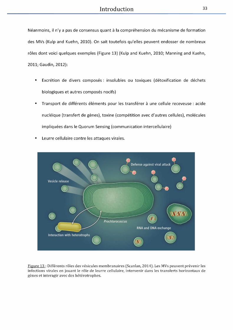
• Le rôle des virus dans les océans .............................................................................................. 41
III. Les environnements thermophiles ............................................................................................... 54
• Les sources chaudes terrestres ................................................................................................. 56
• Les sources hydrothermalesprofondes .................................................................................... 81
IV. Bibliographie – Introduction ......................................................................................................... 96
Objectifs des travaux de thèse .................................................................................................. 107

Chapitre I : Revue bibliographique « An abyssalmobilome » .................................................. 111
Chapitre II : Ménage à trois, caractérisation de MPV1 ............................................................. 124
Chapitre III : Deux nouveaux virus, MCV1 et MCV2, isolés bactéries du phylum desThermotogae ............................................................................................................................. 149
Chapitre IV : Etude du génome de Thermosipho sp. AT1244-VC14 ......................................... 192
Chapitre V : Virome : validation et normalisation d’un protocole (étude préliminaire) .......... 203
Discussion et Perspectives ......................................................................................................... 211
I. Deux nouveaux virus, MCV1 et MCV2, isolés du phylum des Thermotogae et comparaison avec
MPV1 ................................................................................................................................................... 214
II. Thermotogae et CRISPR .............................................................................................................. 217
III. Importance de la lysogénie et virome ........................................................................................ 222
IV. Vésicules membranaires et détoxification ................................................................................. 225
V. Bibliographie ............................................................................................................................... 229
Résumé

Liste des figures
Introduction générale .................................................................................................................. 1
Figure 1 : Photographie au microscope électronique à transmission du virus de la mosaïque
du tabac ........................................................................................................................................... 2
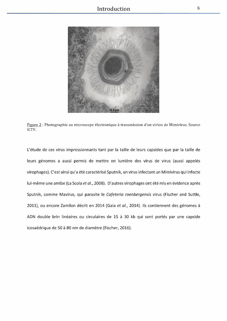
Figure 2 : Photog aphie au i os ope le t o i ue à t a s issio d’u viio de Mi ivius. 6
Figure 3 : Différents aspects morphologiques du Mamavirus et du virophage Sputnik ............. 7
Figure 4 : Co pa aiso de l’ae ph log ti ue du viva t de Woese et de la p opositio
d’u e ouvelle lassifiatio du o de du viva t pa Raoult et Fo tee. .................................. 9
Figure 5 : S h atisatio du le d’un virus tempéré voie lytique et voie lysogénique. ........ 13
Figure 6 : Mécanisme de défense microbien contre les éléments génétiques mobiles ........... 16
Figure 7 : Schématisation du mode de fonctionnement du système CRISPR-cas ..................... 17
Figure 8 : Illustration du fonctionnement du système R-M de type I ......................................... 19
Figure 9 : Illustration du mode de fonctionnement potentiel du BREX ..................................... 22
Figure 10 : Exemple de stratégie virale pour échapper au système toxine antitoxine.............. 26
Figure 11 : Relatio e t e la o e t atio viale et l’a o dae des micro-organismes
(bactéries et archées) dans les sédiments marins profonds de différents océans. ................... 29
Figure 12 : Photographie au MET de GTA .................................................................................... 32

Figure 13 : Différents rôles des vésicules membranaires ............................................................ 33
Figure 14 : Observation en MET de vésicules membranaires produites par des Thermococcales
........................................................................................................................................................ 35
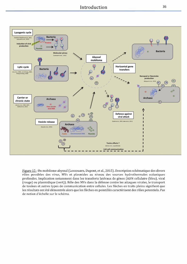
Figure 15 : Un mobilome abyssal .................................................................................................. 36
Figure 16 : Distribution globale de la diversité virale .................................................................. 39
Figure 17 : Illustration humoristique des divers impacts des virus sur les communautés
microbiennes ................................................................................................................................. 41
Figure 18 : Illustration de la stratégie Killing the winner ............................................................. 43
Figure 19 : Illustration du viral shunt ............................................................................................ 46
Figure 20 : Photographie de Emiliania huxleyi ............................................................................. 47
Figure 21 : Rôle des vius da s l’volutio et la pathogi it de leu s hôtes .......................... 48
Figure 22 : Structures similaires à des queues de caudovirus .................................................... 51
Figure 23 : Illust atio de l’utilisatio des ges « viraux » de la photosynthèse pour supporter
la p odu tio viale lo s d’u e ife tio de Prochlorococcus ..................................................... 53
Figure 24 : (a) Désert du Sahara (b) Antarctique ......................................................................... 54
Figure 25 : Photographie de geysers et source chaude .............................................................. 56
Figure 26 : Diversité des communautés microbiennes retrouvée dans des enrichissements
alis s à pa ti d’ ha tillo s de soues haudes teest es ................................................... 59
Figure 27 : Photographie de MET de bactériovirus isolés de sources chaudes terrestres ........ 63
Figure 28 : Alignement de génomes de Fusellovirus ................................................................... 76
Figure 29 : Particules de SSV1 analysées en cryo-EM ................................................................. 77
Figure 30 : S h a de l’o ga isatio des viio s de PFV .......................................................... 78
Figure 31 : Structures pyramidales ............................................................................................... 80
Figure 32: Sources hydrothermales profondes sites ................................................................... 81
Figure 33 : S h a du fo tio e e t d’u e soue h d othe ale p ofo de ...................... 83
Figure 34 : Rep se tatio de l’a o dae elative des ph la a t ie s ................................ 85
Figure 35 : Rep se tatio de l’a o dae elative des diffe ts ph la archéens ................. 86
Figure 36 : Pla e e t des The otogales da s l’ae ph log ti ue a t ie pa appo t à
l’ARN 6S ...................................................................................................................................... 87
Figure 37 : Observation au microscope électronique de virions ................................................ 92
Figure 38 : Photographie en microscopie électronique deD6E .................................................. 93
Figure 39 : Observation en MET de (a) PAV1 et (b) TPV1 ........................................................... 94
Figure 40 : Relation entre les plasmides de Thermococcales et le virus PAV1 .......................... 95
Chapitre I : Revue bibliographique « An abyssal mobilome » .................................................. 111
Figure 1 : Electron micrographs of viruses and membrane vesicles isolated from deep-sea
hydrothermal vents ..................................................................................................................... 116
Figure 2 : Multipurpose mobilome within deep-sea hydrothermal vents. .............................. 116
Chapitre II : Ménage à trois, caractérisation de MPV1 ............................................................. 124
Figure 1 : Electron micrograph of two MPV1 particles ............................................................. 128
Figure 2 : Virus–host kinetics at atmospheric pressure monitored by flow cytometry .......... 129
Figure 3 : Comparison of the MPV1 provirus in M. piezophila and viral DNA.......................... 129

Figure 4 : Comparison of the MPV1 provirus in M. piezophila and viral DNA.......................... 130
Figure 5 : MPV1 infectivity assays performed on Thermosipho sp. AT1244-VC14T and checked
by PCR experiments. .................................................................................................................... 130
Figure 6 : qPCR assays to assess changes in copy number of M. piezophila chromosomal,
proviral and plasmid DNA target genes in the presence or absence of mitomycin C induction
...................................................................................................................................................... 131
Figure S1 : Induction assays of MPV1 production by addition of several mitomycin C
concentrations ............................................................................................................................. 141
Figure S2 : RFLP analyses of M. piezophila total DNA, plasmid DNA (pMP1) and viral DNA ... 142
Figure S3 : PCR analysis of the total DNA extracted from purified MPV1 viral capsids from an
induced culture ............................................................................................................................ 143
Figure S4 Agarose gel electrophoresis of plasmid DNA extracted by alkaline lysis from
Thermosipho sp. strain AT1244-VC14, three subcultures after being infected by MPV1 virions
...................................................................................................................................................... 144
Figure S5 : QPCR assays ............................................................................................................... 145
Chapitre III : Ca a t isatio de deu ouveau vius, MCV et MCV , isols a t ies de l’o d e
des Thermotogales .................................................................................................................. 155
Figure 1 : Electron micrographs of (a) a MCV1 virion (b) two MCV2 virions ........................... 159
Figure 2 : Virus-host kinetics of M. camini 97 and M. camini 55 ............................................. 161
Figure 3 : Electron micrographs of concentrated MVs produced by M. camini 97 ................. 162
Figure 4 : Genome comparison of MPV1, MCV1 and MCV2 with GenoPlotR .......................... 167
Chapitre V : Virome : validation et o alisatio d’u p oto ole tude p li i aie ........... 203
Figure 1 : Schéma général du protocole « Filtration » (A) et du protocole « Gradient » (B) .. 207
Discussion et Perspectives ....................................................................................................... 211
Figure 1 : Comparaison de 11 génome de Thermotoga maritima – like. ................................. 213
Figure 2 : S h a du p i ipe de fotio e e t g ale d’u s st e CRISPR-cas ......... 220
Figure 3 : Observation en microscopie électronique de MVs produites par M.camini 97 ...... 226

Liste des tableaux
Introduction générale .................................................................................................................. 1
Tableau 1 : Différences entre organismes, virus et constituants cellulaires ................................ 3
Tableau 2 : Classification selon Baltimore .................................................................................... 10
Tableau 3 : Bactériovirus des sources chaudes terrestres .......................................................... 61
Tableau 4 : Archéovirus des sources chaudes terrestres. ........................................................... 70
Revue bibliographique « An abyssal mobilome » ..................................................................... 111
Tableau 1 : Bacterial and archaeal viruses isolated from deep-sea hydrothermal vents . ...... 114
Tableau 2 : Classification selon Baltimore .................................................................................. 117
Tableau 3 : Archaeal plasmids isolated from deep-sea hydrothermal vents ............................. 61
Chapitre IV : Etude du génome de Thermosipho sp. AT1244-VC14 .......................................... 194
Tableau 1 : Summary of important genes implicated in the CRISPR-cas system of Thermosipho
sp. 1244-VC14 . ............................................................................................................................ 200
Chapitre V : Etude du génome de Thermosipho sp. AT1244-VC14 ........................................... 203
Tableau 1 : Concentrations en microorganismes cibles avec lesquelles les échantillons ont été
chargés . ....................................................................................................................................... 206

Liste des abre�viations
AAF Aéro-Anaérobie Facultatif Abi Abortive infection ADN Acide DésoxyriboNucléique AN Acide Nucléique ARN Acide RiboNucléique ARNm Acide RiboNucléique messager ARNnc ARN non codant (ou antisens) ARNr Acide RiboNucléique ribosomique ARNt Acide RiboNucléique de transfert ATP Adénosine TriPhosphate BREX BacteRiophage EXclusion CRISPR-cas Clustered Regularly Interspaced Short Palindromic Repeats -crispr associted genes DMS DiMethyl Sulfur GTAs Gene Transfer Agents GTP Guanosine TriPhosphate HGT Horizontal Gene Transfer ICTV International Committee on Taxonomy of Viruses KtW Killing the Winner LUCA Last Universal Common Ancestor MEB Microscope électronique à Balayage MEF Microscopie à EpiFluorescence MET Microscope électronique à transmission MOD Matière Organique Dissoute MOP Matière Organique Particulaire Mtase MethylTransferase MVs Membrane Vesicles ORFs Open Reading Frames pb paire de bases Pgl Phage growth limitation PICIs Phage Inducible Chromosomal Islands PNK PolyNucleotide Kinase R-M Restriction Modification Rease Restriction Endonuclease TA Toxine Antitoxine UV UltraViolet vAMGs viruses-encoded Auxiliary Metabolic Genes VAPs Virus Associated Pyramids VLPs Virus-Like Particles


Introductionge�ne� rale

Introduction 1
I. Les virus
• De�couverte et De� finition :
Un peu d’historique
Les virus sont des entités biologiques particulièrement anciennes et, selon certains auteurs,
ils seraient apparus avant LUCA (Last Universal Common Ancestor) (Abrescia et al., 2012;
Forterre, 2013a, 2016). Les infections dues à des virus telles que la rage sont, elles, connues
depuis des millénaires (Lwoff, 1957). Néanmoins les agents responsables de celles-ci n’ont été
décrits qu’à la fin du XIXème siècle. En effet, même si des traitements empiriques comme la
variolisation étaientpratiqués depuis plusieurs siècles,la compréhension et la description des
virus commencent réellement avec l’étudedu virus de la mosaïque du tabac en 1892. Adolf
Mayer décrit cette maladie affectant les plants de tabac comme infectieuse et pense qu’une
bactérie en est responsable. Par la suite, Dimitri Ivanovski va parler d’agent ultrafiltrable car il
est capable de franchir les filtres de Chamberland, permettant à l’époque d’éliminer nombre
de bactéries. Ivanovski maintient pourtant l’explication bactérienne et ce n’est qu’en 1898
que Beijernick comprendra l’importance de l’observation de son collègue et proposera le
concept de « contagium vivum fluidum ».
Le virus de la mosaïque du tabac resteraun modèle d’étude puisque ce sera le premier à être
cristallisé par Wendell Stanley en 1935. L’année suivante Frederick Bawden et Norman W.
Pirie décriront une structure composée de protéines et d’acides nucléiques suite aux travaux
de leur confrère. Au début du XXème siècle, Frederick Twort et Félix D’Hérelle découvrent
indépendamment l’un de l’autre les virus infectant des bactéries.


Introduction 3
Lwoff propose une définition qui restera celle longtemps admise par la communauté
scientifique : les virus sont « des entités nucléo-protéiques infectieuses de moins de 200 nm
strictement intracellulaires potentiellement pathogènes, elles possèdent un seul type d’acide
nucléique (ADN ou ARN), se multiplient à partir de leur matériel génétique (dans leur hôte),
sont incapables de croissance ou de division et sont dépourvues de système de Lipmann »
(Tableau 1). Aujourd’hui les virus sont plus communément définis comme des parasites
intracellulaires obligatoires possédant un seul type d’acide nucléique et détournant la
machinerie cellulaire de leur hôte afin de se multiplier. Ils possèdent une coque protéique
appelée capside qui peut, ou non, être entourée par une enveloppe lipidique (Madigan and
Martinko, 2007).
Tableau1 : Diffe� rences entre organismes, virus et constituants cellulaires (Lwoff, 1957). Les crite� resde comparaisons du tableau sont le type d’acide nucle� ique, la capacite� a� re�pliquer celui-ci, la capacite�de croissance et de divisionet la pre� sence d’un syste�me de Lipmann. C’est selon ces crite� res que Lwofffait la diffe� rence entre les organismes, les virus et les constituants cellulaires (mate�riel ge�ne� tique etorganelles).

Introduction 4
• De�couvertes re�centes
La définition de ce qu’est un virus reste néanmoins sujette à débat dans la communauté
scientifique. En effet, l’absence de ribosome dans leur matériel génétique propre les exclut du
monde du vivant tel qu’il a été décrit par Carl Woese en 1990 (Figure 4a). Néanmoins,
certaines découvertes récentes remettent en question la façon dont les virus sont pris en
compte, étudiés et classifiés.
Tout d’abord, l’étudede l’abondance virale grâce à des techniques moléculaires (comme les
métagénomes) ou de microscopie à épifluorescence a permis de mettre en évidence que les
virus sont les entités biologiques les plus abondantes de la planète. Il est considéré qu’il y a,
en moyenne, dix fois plus de virus que de cellules dans l’environnement (Suttle,2007). Cette
abondance a longtemps été sous-estimée en raison des limitations techniques de l’époque,
ce qui explique en partie le fait que leur importance ait été négligée (Breitbart et al., 2007).
De plus, les virus ont longtemps été considérés comme des « pickpockets » de gènes
cellulaires, mais il s’avèreque les cellules intègrent et utilisent aussi des gènes viraux (Forterre,
2010, 2013a). En effet, 10 à 15% des génomes de bactéries ou d’archées sont constitués de
gènes provenant de virus ou de plasmides ayant été récemment intégrés dans leurs génomes
(Cortez et al., 2009). Ce constat implique un fort impact des virus sur l’évolution et la
composition, notamment des communautésmicrobiennes. Ils peuvent en effet incrémenter
de nouvelles fonctions, résistances ou propriétés à ces cellules (Weinbauer, 2004).
Introduction 5
La découverte de virus géants a aussi remis en question la façon dont étaientperçus et étudiés
les virus. Historiquement, la petite taille est un critère de sélection des virus. En effet, pour
séparer le compartimentbactérien/archéen du compartimentviral, l’utilisation des filtres de
0.22 µm est une méthode classique étant donné qu’il était considéré que les virus passent
généralement au travers de cette porosité. Néanmoins, la découverte des Mimivirus aussi
appelés « virus géants » qui font la taille de petites bactéries a entrainé la remise en question
de ces fondamentaux de la virologie. Ces virus, infectant une amibe (eucaryote unicellulaire
de la classe des rhizopodes) de l’espèce Acanthamoeba polyphaga, ont tout d’abord été
confondus avec de petites cellules à Gram négatif (La Scola et al., 2003). En effet, ils ont été
les premiers virus observables au microscope optique puisque leur capside mesure 0,8µm (La
Scola et al., 2003). Leur génome de 1,2 Mpb est deux fois plus grand que celui de certaines
bactéries et code pour plus de 1000 protéines (La Scola et al., 2003; Raoult et al., 2004). Par
ailleurs, lorsque le virus se réplique à l’intérieur de l’amibe, l’« usine virale » qu’il met en place
est aussi grosse que le noyau de l’amibe elle-même. Il peut coder pour des gènes aux fonctions
enzymatiques précédemment retrouvés uniquement chez les cellules (comme des gènes
impliqués dans la synthèse protéique) et contient de l’ARN et de l’ADN (Raoultet al., 2004). A
la lumière de ces nouvelles données, les approches méthodologiques en termesde recherche
de nouveaux virus ont évolué et permirent de découvrir de nouveaux virus géants.La plupart
d’entre eux sont des virus infectants des amibes(Fischer, 2016). Ils sont actuellement classés
en sept familles (Ascoviridae, Asfarviridae, Iridoviridae, Marseilleviridae, Mimiviridae,
Phycodnaviridae, and Poxviridae) regroupées en un ordre nommé « Megavirales » (Colson et
al., 2013). Cet ordre contient des virus ayant des génomes allant d’une centaine de paires de
bases pour les plus petits jusqu'àplus de 2,5 Mpb pour les plus grands (Pandoravirus) (Fischer,
2016).

Introduction 7
Figure 3 : Diffe� rents aspects morphologiques du Mamavirus et du virophage Sputnik (La Scola et al.,2008). (a-e) Observations re� alise�es au microscope e� lectronique a� transmission (f) observation parcoloration ne�gative au microscope e� lectronique. (a) « Mamavirus virus factory » avec des particules deMamavirus a� diffe� rentes e� tapes de maturation. Les fle� ches montrent de petits amas de particules deSputnik. (b) Sputnik dans des capsides de Mamavirus. (c) Production de particules de� fectives deMamavirus. (d-f) Mamavirus avec des morphologies anormales dues a� l’infection par Sputnik. Echelle200nm.
L’ensemble de ces découvertes remet donc fondamentalement en question la définition de
ce qu’est un virus mais aussi la façon dont ils sont considérés et classifiés. Elles ont mené à
l’élaboration de nouveaux concepts tels que celui de la « Virocell ». Ce concept a été décrit
par le Pr Patrick Forterre dans les années2010, suite aux nombreuses découvertes remettant
en question la définition des virus (Forterre, 2011, 2013b).

Introduction 8
L’une des bases de cette théorie repose sur le fait que, dans la communauté scientifique au
sens large, les virus et les virions sont souvent confondus, les virions étant la phase de vie
extracellulaire du virus (capside et acide nucléique). Par exemple, dans la définition d’un virus,
il est écrit que celui-ci ne contient qu’un seul type d’acide nucléique, or lorsque le virus se
réplique il passe par une étape d’ARN messager et certains virus comme les retrovirus sont
capables de passer de l’ARN à l’ADN (puis de nouveau à l’ARN messager).
Or, d’autres auteurs, à l'instar du Pr Jean Michel Claverie (responsable de l’équipe qui a
séquencé le génome du premier virus géant (Raoultet al., 2004)),pensent que confondre les
virus et les virions revientà confondre l’homme et ses spermatozoïdes. Ce dernier propose
alors une théorie selon laquelle le virus serait l’usine virale lors de l’infection. Néanmoins,
cette usine virale n’est pas présente partout et est notamment absente des bactériovirus par
exemple.
C’est ainsi qu’a été émis le concept de Virocell du Pr Patrick Forterre : la Virocell est la phase
cellulaire de la réplication du virus (ou organisme codant pour une capside) et le virus est alors
un processus biologique intégrant tous les aspects du cycle de reproduction virale (virion,
virocell, et éventuellement génome intégré) (Forterre, 2011, 2016). Ce concept sert de base à
la proposition d’une nouvelle classification des organismes, intégrant ainsi les virus au monde
du vivant (par opposition à la proposition communément admisede Carl Woese (Woese et
al., 1990; Woese, 1994; Raoult and Forterre, 2008) (Figure 4).


Introduction 10
un seul type d’acidenucléique paraît obsolète. Néanmoins, l’intégration des virus au monde
du vivant reste controversé (López-García,2012; López-García and Moreira, 2012).
D’autres modèles ont aussi été proposés et notamment celui d’un quatrième domaine du
vivant, en plus des bactéries, archées et eucaryotes, nommé « Akamara ».Celui-ci
regrouperait les agents infectieux acellulaires possédant un génome d’acidenucléique (Hurst,
2011). Ce nouveau domaine serait séparé en deux règnes, celui des Eurivia incluant virus et
virus satellites (codant pour une capside) et celui des Viroidia comprenant les viroïdes qui ne
codent pas pour leur capside (Hurst, 2011).
D’autre équipes ont néanmoins repris le concept de Virocell dernièrement (Rosenwasser et
al., 2016) en relevant que se placer du point de vue des virus permettait aussi de mieux
comprendre leur implication et leur importance dans les écosystèmes, notamment au niveau
océanique (cf Partie « Rôle dans les océans» p41)
• Classification
Actuellement, il existe deux grands types de classifications.
Le premier est la classification par la méthode de Baltimore, définie pour la première fois en
1971. Celle-ciclasse les virus en 7 groupes en fonction de leur type d’acidenucléique (ADN ou
ARN), du nombre de brin (simple ou doublebrin), de la polarité de ceux-ci (positif ou négatif),
et du mode de réplication (Tableau 2).


Introduction 12
nucléique (ADN, ARN, simple ou doublebrin), la présence ou non d’une enveloppe autour de
la capside, le type de symétrie de celle-ci et la taille du virion. L’ICTV, qui remet à jour cette
classification périodiquement, comprend une base de données internationale recensant la
majorité des espèces de virus décrites à ce jour. A l’heureactuelle sont connues 3704 espèces
réparties en 7 ordres et 111 familles. Les 7 ordres sont :Caudovirales, Herpesvirales,
Mononegavirales, Nidovirales, Picornavirales, Tymovirales et le dernier créé les
Ligamenvirales. Pour être acceptée, une nouvelle espèce de virus doit contenir au moins un
virus isolé parmi ses membres.
Quasiment toutes les espèces de virus sont classées au sein d'un genre mais une partie d’entre
elles restent non assignées car leur caractérisation est inadéquate. Ces deux types de
classifications ne sont pas incompatibles puisque l’ICTV reprend en partie les critères de la
classification de Baltimore tout en incluant notamment le type d’hôte infecté et la
morphologie (Abrescia et al., 2012).
• Les cycles viraux
Les virus entretiennent trois principaux types de relations avec leurs hôtes (Weinbauer, 2004;
Madigan and Martinko, 2007) :
Les virus virulents réalisent un cycle lytique en détournant à leur profit la machinerie cellulaire
de l’hôte et les virions néo synthétisés sont libérés suite à la lyse provoquée de la cellule hôte.
Les virus tempérés, qui effectuent un cycle lysogénique, persistent sous forme d’acides
nucléiques, le plus souvent intégrés au sein du génome de l’hôte (ou plus rarement libres,
sous forme extra-chromosomique), à l’état de provirus. Ils se répliquent ainsi en même temps
que le génome de la cellule hôte jusqu’à ce qu’un stress (comme des carences nutritives,
antibiotiques ou UV) ne conduise les virus à déclencher un cycle lytique.


Introduction 14
Des étapes communes lors de l’infection sont partagées par les différents virus infectant des
bactéries ou des archées quel que soit le type de cycle qu’ils mettent en œuvre (Weinbauer,
2004).
Tout d’abord, le virion s’adsorbe sur une surface cellulaire. Cette étape est dans un premier
temps réversible puis les fibres caudales virales se lient de façon définitive aux récepteurs
présents sur la membrane de la cellule. Ensuite, les enzymes virales perméabilisent la
membrane cellulaire permettant l’injection du génome viral dans la cellule.Une fois dans le
cytoplasme, le génome peut être intégré au chromosome hôte ou rester sous forme libre.
Pour permettre la néo-synthèse de virions, le génome doit être sous forme libre. Les gènes
viraux sont alors exprimés, conduisant à la réplication du génome et à la production des
protéines virales qui formeront notamment les capsides. Les génomes sont ensuite
empaquetés dans les capsides virales néo-synthétisées, puis libérés dans l’environnement par
bourgeonnement, extrusion ou suite à la lyse de la cellule hôte. La phase de vie extracellulaire
des virions se termine soit par l’infection d’un nouvel hôte soit par leur dégradation dans
l’environnement.

Introduction 15
• Course a� l’armement : syste�me de de� fense ho; tes – virus
Au cours des milliers d’années d’évolution entre les cellules et les virus, de nombreuses
techniques de résistances ont été mises au point par les cellules pour survivre à l’infection
virale. Il est d’ailleurs supposé que l’une d’entre elle ait permis le passage de l’ARN vers l’ADN
(Forterre, 2016).
Il existe divers mécanismes de défense contre les infections virales (Figure 6) qui peuvent être
classés en deux groupes :
Le premier regroupe les systèmes basés sur la reconnaissance de l’acide nucléique (AN)
exogène et sa différenciation avec les AN cellulaires. C’est, par exemple, le cas du système
CRISPR-cas, du système de restriction modification (R-M), du système de phosphorothioation
(système DND) et probablementd’un système récemment décrit, le BREX (Makarova et al.,
2013).
Le second groupe rassemble des systèmes de défenses basés sur la mort cellulaire
programmée ou la dormance des cellules infectées, comme le système d’infection abortive
(Abi) ou le système toxine-antitoxine (TA) (Makarova et al., 2013).

Introduction 16
Figure 6 : Me�canisme de de� fense microbien contre les e� le�ments ge�ne� tiques mobiles(van Houte et al.,2016). Les bacte� riovirus sont repre� sente� s en gris et les plasmides en vert. Six me�canismes sontpre�sente�s, la modification de la surface cellulaire empe;chant la fixation des virions, l’exclusion due a�la pre�sence d’un autre provirus, le syste�me de restriction modification base� sur le clivage des ADNexoge�nes non me� thyle� s, l’infection abortive menant au « suicide » de la cellule infecte�eet le syste�meCRISPR-cas comprenant une « me�moire » des infections passe�es. Ces syste�mes sont de�veloppe�s par lasuite.
Système CRISPR-cas
L’un des mécanismes de défenses contre les invasions d’AN exogènes les plus connusest le
système CRISPR-cas. Cette famille de séquences d’ADN répétées a été découvert en 1987 chez
E.coli puis ce système fut nommé CRISPR-cas (Clustered Regularly Interspaced Short
Palindromic Repeats – associated with cas genes) après la découverte de séquences similaires
chez les archées et les bactéries. Ce système permet de contrer les « attaques » d’acides
nucléiques exogènes tels que les virus ou les plasmides. Le locus de CRISPR est composé de
plusieurs répétitions de séquences courtes partiellement palindromiques non contiguës, les
repeats. Ces séquences sont séparées par des spacers correspondant à des parties d’acides
nucléiques exogènes comme par exemple des morceaux de génome viral. Si les repeats sont
très conservées dans un locus CRISPR donné et ont toujours une structure partiellement
palindromique afin d’avoir une conformation secondaire stable, les séquences spacers sont-

Introduction 17
elles hypervariables. Les gènes cas sont la partie effective du système et codent pour diverses
protéines portant des domaines fonctionnels typiques tels que nucléase, hélicase, polymérase
et protéines de liaisons aux polynucléotides. Les CRISPRpermettent à la cellule de reconnaître
l’acide nucléique exogène alors que les protéines produites par les gènes cas se chargent de
l’éliminer (Horvath and Barrangou, 2010).
Figure 7 :Sche�matisation du mode de fonctionnement du syste�me CRISPR-cas (Horvath andBarrangou, 2010). (a) Processus d’immunisation : entre�e de l’AN exoge�ne, production des prote� inesissues des ge�nes cas, clivage de l’AN exoge�ne, cre�ation d’un nouveau spacer et inte�gration de celui-ciau sein du locus CRISPR (b) Processus d’immunite� : entre�e d’un AN exoge�ne, reconnaissance de celui-ci par le crRNA pre� ce�demment transcrit et inactivation de la cible.

Introduction 18
Le mécanisme général d’action est le suivant(Figure 7) :
Processus d’immunisation
• Entrée d’acidenucléique exogène• Reconnaissance• Création d’une nouvelle séquence repeat• Sélection et création du spacer• Intégration dans le génome
Mise en place de l’immunité
• Transcription du loci CRISPRen pre-crRNA• Transformation des pre-crRNA en crRNA• crRNA guide le complexe cas vers la cible• Dégradation de l’acide nucléique cible
C’est un système très répandu car il est présent dans 87% des génomes archéens et 50% des
génomes bactériens (Sorek et al., 2008; Makarova et al., 2015). Ces systèmes CRISPR-cas sont
particulièrement sujets aux transferts latéraux de gènes, même entre espèces lointaines,
apportant un avantage évolutif certain notamment face aux infections virales potentielles
(Sorek et al., 2008).
Système Restriction – Modification (R-M) (Tock and Dryden, 2005; Makarova et al., 2013)
Un autre mécanisme particulièrement bien étudié est le système de restriction modification
(R-M). Tous les systèmes R-M fonctionnent sur le même principe : discriminer les AN
endogènes des AN exogènes afin de cliver ces derniers. Cela met en jeu deux enzymes :
- Une methyltransferase (MTase), qui méthyle des bases adénine ou cytosine de l’AN
de la cellule pour qu’il puisse être reconnu et protégé du clivage.
- Une enzyme de restriction de type endonucléase (REase) qui permet le clivage ciblé
des AN étrangers non méthylés (viraux ou plasmidiques).


Introduction 20
Système de phosphorothioation de l’ADN (DND) (Xu et al., 2010; Makarova et al., 2013)
Le principe du système DND, un peu comme le système R-M est de taguer l’ADN cellulaire, ici
via une S-modification de l’ADN (phosphorothioation : un atome de soufre remplace un atome
d’oxygène non-liant du phosphate de la liaison phospho-diester), et de cliver les AN exogènes
non marqués. Comme souvent pour les îlots de gènes de défenses, ce système contient divers
gènes arrangés en cluster. L’activité de restriction comprend au moins trois gènes et quatre
autres gènes contigus sont nécessaires pour l’activité de modification de l’ADN. Il est suggéré
que ce système DND soit prochedu système R-M de type I. En effet, la composition du cluster
de gènes et le mode de fonctionnement (destruction des AN exogènes uniquement quand les
AN cellulaires sont protégés par le marquage) sembleprochedu système R-M de type I.
Système BREX (BacteRiophage EXclusion) (Barrangou and van der Oost, 2014; Goldfarb et al.,
2015; Nunes-Alves, 2015).
Le système BREX contient des similarités avec un système plus ancien appelé Pgl (phage
growth limitation). Celui-ci est basé sur l’utilisation du gène plgZ qui est impliqué dans un
système de résistance de Streptomyces coelicolor. Dans une étude récente, réalisée sur
environ 1500 génomes de bactéries et d’archées, des gènes contenant un domaine plg ont
été retrouvés dans 10% des génomes. Or dans plus de la moitié des cas, ces gènes sont
retrouvés dans un cluster composé de 6 gènes dont 2 gènes ont une forte homologie avec le
système Pgl. Cette observation est cohérente avec des données précédemment obtenues qui
soulignaient le fait que les mécanismes de défenses contre les infections virales étaient
généralement groupés en clusters (Makarova et al., 2011). C’est ce cluster particulier de gènes
qui a été appelé système BREX. Il s’agit donc de 6 gènes permettant la résistance à une large
gamme de virus, contrairementau système Pgl qui a une gamme d’action restreinte.

Introduction 21
Cette cassette inclus une Lon-like protéase putative (serine peptidase ATP-dépendante), une
protéine contenant un domaine phosphatase alcaline, une protéine putative de liaison à
l’ARN, une ADN méthylase, une protéine à domaine ATPase et une protéine de fonction
inconnue. BREX semble être un système complexe contenant des protéines ayant de
nombreuses activités biochimiques (protéase, phosphatase et méthylase). Même si ce
système sembleprochede système Pgl puisqu’il y a partage d’une partie des gènes, on note
quelques différences notables. En effet, le système Pgl permet une première infection avec
libération de virions et permet aussi la lysogénie. Le système BREX ne permet ni la primo
infection ni la lysogénie, ce qui semble prouver que les deux systèmes n’ont pas le même
mode de fonctionnement (Figure 9). D’autre part, le système BREX semble permettre
l’adsorption du virus mais bloque la réplication du génome viral. Néanmoins, contrairementà
ce qu’il se passe pour le système restriction modification, l’ADN viral ne sembleni dégradé ni
clivé par le BREX, ce qui suggère un nouveau mécanisme de défense. Par contre, le
chromosome de la cellule est méthylé quand les cellules contiennent le système BREX,
suggérant que celui-ci est capable de promouvoir une méthylation site-spécifique du
chromosome cellulaire. Cette méthylation, probablementmédiée par le gène pglX, pourrait
permettre une sorte de reconnaissance entre les acides nucléiques endogènes et exogènes.
Aussi, si le gène pglX est inactivé, la résistance aux infections n’est plus active, suggérant une
forte importance de celui-cidans le mécanisme de défense. Le système de fonctionnement
du BREX n’est pas encore connu néanmoins il semblerait qu’il soit actif avant la phase de
réplication de l’ADN viral. Il sembleaussi que, comme pour le système CRISPR-cas, le système
BREX soit une cible de choix pour les transferts horizontaux de gènes. Pour conclure, certains
virus testés lors de l’étudeont montré une résistance au système BREX, nouvelle preuveque
la course à l’armement entre virus et cellules est soumise à une constante évolution.

Introduction 22
Figure 9 : Illustration du mode de fonctionnementhypothe� tique du BREX (Barrangou and van der Oost,2014). (a) Cycles classiques : lysoge�nique ou lytique (gaucheet droite) (b) Cas de bacte� ries posse�dantle syste�me de de� fense BREX contre un virus (gauche) tempe� re� ou (droite) virulent. Il y a alorstranscription du cluster BREX entraî;nant l’inhibition de la production virale, voire de l’inte�gration duge�nomeviral dans le cas d’un virus tempe�re� (chez Bacillus cereus).Le me�canisme d’actionn’est pasencore re�ellement connu a� ce jour.

Introduction 23
Système toxine – antitoxine (TA) (Hayes and Van Melderen, 2011; Makarova et al., 2013; Bibi-
Triki, 2014)
Il existe divers types de système TA classés selon les toxines et antitoxines qu’ils utilisent,
l’organisation des gènes impliqués et leurs modes d’actions. Il y a 6 grands types de systèmes.
Le type I met en œuvre un ARN non codant ou antisens (ARNnc) comme antitoxine et une
protéine (généralement une holin-like protéine) comme toxine. Le type II est prévalent chez
les archées et les bactéries. Il utilise des protéines comme toxine et antitoxine qui sont
généralement transcrites dans le même opéron. Pour le système de type III, l’antitoxine est
sous forme d’ARNnc qui peut directement se fixer sur la toxine. Trois autres types ont été plus
récemment proposés (IV, V et VI) basés sur des mécanismes d’actions différents de leurs
antitoxines respectives.
Quel que soit le type de système, ils sont tous basés sur une toxine intracellulaire stable qui,
si elle est exprimée au-dessus d’un certain niveau, va entraîner la mort cellulaire, et une
antitoxine instable qui peut inactiver la toxine et/ou régulerson expression pour prévenir la
mort cellulaire. Les toxines sont des protéines dirigées contre des cibles intracellulaires
spécifiques alors que les antitoxines sont des protéines ou de petits ARN non codants qui ont
pour cible la toxine. La plupart des toxines sont des endoribonucléases site-spécifiques qui
freinent la synthèse protéique en réponse à divers stress. Leur effet est souvent temporaire
et réversible ce qui leur donne une capacité d’adaptation face aux différentes situations
rencontrées. Les autres toxinespeuvent avoir des effets sur l’action d’une topoisomérase, sur
l’assemblage de la membrane cellulaire ou sur les structures du cytosquelette.

Introduction 24
Globalement, le système TA a pour objectif d’induire une entrée en dormance ou une lyse de
la cellule,grâce à une production contrôlée de toxine, pour répondre à divers stress, incluant
l’invasion par des AN exogènes.
Le système TA joue un rôle majeur dans la lutte contre l’invasion d’AN exogène mais aussi
dans la survie cellulaire, la formation de biofilmet la résistance aux antibiotiques.
Systeme Abi (Chopin et al., 2005; Fineran et al., 2009; Makarova et al., 2013)
Le système Abi (Abortive infection), aussi appelé « phage exclusion » bloquela multiplication
du virus en entraînant la mort prématurée de l’hôte lors de l’infection virale. Ce système est
caractérisé par un début d’infection virale classique, avec adsorption du virion puis injection
de l’AN viral dans la cellule.L’infection est ensuite stoppée menant à la libération d’une faible
quantité de nouveaux virions (voir aucun) dans l’environnement mais aussi à la mort
prématurée de la cellule infectée. Ce système permet de prévenir l’infection des cellules
environnanteset est ainsi considéré comme un « suicide altruiste » de la cellule infectée. Le
mécanisme moléculaire qui est mis en jeu reste assez méconnu. Il n’y a en effet qu’une
trentaine de systèmes Abi décrits, provenant majoritairement de plasmides présents dans 2
organismes modèles (Lactococcus lactis et Escherichia coli) ce qui suggère que ce système est
assez peu répandu, ne représentant qu’une fraction mineure de la diversité totale des
modules de défenses microbiens. Néanmoins il partage des similarités avec les systèmes TA
(comme par exemple la formation de pores dans la membrane) et semblesouvent associée
au système R-M, pouvant donc potentiellement interagir avec celui-ci.

Introduction 25
La réponse des virus
Ces mécanismes de défenses cellulaires partagent des similarités comme le fait qu’ils soient
des cibles prioritaires pour les transferts latéraux de gènes, ou qu’ils ciblent en général autant
les virus que les plasmides.
Un autre point commun partagé par nombre de ces systèmes est que les virus ont trouvé des
mécanismes pour les éviter d’où le nom de « course à l’armement » (Samson et al., 2013).
L’un des mécanismes les plus couramment utilisé par les virus pour échapper à ces systèmes
de défense est de créer une mutation dans leur propre génome. Par exemple pour le système
CRISPR-cas, une seule mutation dans les proto-spacers (spacers encore dans le génome viral)
permet de contrecarrer le système de défense cellulaire (Horvath and Barrangou, 2010). La
délétion d’une partie du génome viral peut aussi servir de mécanisme de défense, supprimant
ainsi la cible connue par le système de défense bactérien. Les virus peuvent aussi interférer
directement avec le système de défense en inhibant l’activité enzymatique des nucléases du
système R-M ou en interférant avec les protéines codéespar les gènes cas (Tock and Dryden,
2005; Horvath and Barrangou, 2010; Samson et al., 2013). Ils peuvent aussi détourner le
système à leur avantage en codant pour l’antitoxine du système TA (Figure 10) ou encore en
activant le système R-M pour que celui-ciméthyle aussi les AN viraux (Tock and Dryden, 2005;
Samson et al., 2013).


Introduction 27
II. Les virus de bacte� ries et d’arche�es dans les oce�ans
Alors que les bactériovirus ont été découverts au début du XXème siècle, il faudra attendre
les années 70 pour que le premier archéovirus soit mis en évidence. Lorsque celui-ci fut
identifié, le concept même d’archée n’était pas conçu (Torsvik and Dundas, 1974). Les virus
associés à des bactéries ou archées sont de loin les plus nombreux, tout comme leurs hôtes.
Cependant leur diversité morphologique est assez faible puisque 96% d’entre eux ont une
symétrie binaire (Ackermann and Prangishvili, 2012). Il s’agit de virus appartenant à l’ordre
des Caudovirales, divisé en 3 familles : les Myoviridae, les Podoviridae et les Siphoviridae. Les
4% restants sont cubiques comme les Tectiviridae, filamenteux comme les Lipothrixviridae ou
pléomorphes comme les Fuselloviridae ou les Ampullaviridae. Ces virions contiennent un ADN
doublebrin dans la majeure partie des cas, mais ils peuvent aussi porter de l’ADN simple brin,
ou de l’ARN simple ou doublebrin, même si ce dernier cas est très rare (Ackermann, 2009).
Ces données s’appliquent à l’ensemble des bactériovirus et archéovirus, comprenant ceux
présents dans les océansdont les caractéristiques vont être développées dans cette partie du
manuscrit.
• Abondance et diversite�
Oce�an viral, estimations de l’abondance et « fausses » particules virales
Les virus sont les entités biologiques les plus abondantes de la biosphère. Le nombre de virus
dans les océansest en effet estimé à 1030 et la plupart d’entre eux sont des virus de bactéries
ou d’archées (Breitbart and Rohwer, 2005; Suttle, 2005). La nouvelle ère de la virologie marine
a débuté durant les années 90 (Torrella and Morita, 1979; Bergh et al., 1989) faisant des
océansun des environnements où la quantité de données sur les communautésvirales est la
plus importante (Breitbart and Rohwer, 2005; Brum and Sullivan, 2015; Chow and Suttle,

Introduction 28
2015). On considère qu’il y a environ 10 à 15 fois plus de virus que de cellules bactériennes
dans les océanset cette forte abondance a été confirmée par les données de métagénomique
disponibles (Suttle, 2005; Ackermann, 2007; Breitbart et al., 2007; Rohwer and Thurber,
2009). A l’image des hôtes qu’ils infectent, l’abondance des divers virus est très variable. Si
l’abondance virale est estimée à 108 VLP/ml(virus-like particles) en ce qui concerne les eaux
côtières, celle-ci diminue avec la profondeur et l’éloignement par rapport aux côtes (Suttle,
2007; Danovaro et al., 2008). Ces résultats sont cohérents si on considère que l’abondance
virale est corrélée à celle des micro-organismes (Figure 11) (Danovaro et al., 2008). Il peut
d’ailleurs être considéré que les virus utilisent les stratégies « r »et « K »décrivant
habituellement des populations bactériennes. Il est alors possible de les classer en 2
groupes (Chow and Suttle, 2015) :
• Les virus les plus abondants, sont caractérisés par une forte virulence, une réplication
rapide,un burst size important et de petits génomes et suivent plutôt une stratégie
« r ».
• Les virus présents en minorité sont, eux, caractérisés par une faible virulence, un faible
burst size, de grands génomes et mettent plutôt en œuvre des cycles lysogéniques ou
des états porteurs et suivent une stratégie « K ». Cependant, ces virus représentent la
plus grandediversité génotypique (Breitbart and Rohwer, 2005).
Ce qui va caractériser le mode de vie d’un virus, son évolution et sa place dans l’écosystème
peut donc être résumé en 4 grands critères : le type de cycle (lytique, lysogénique, ou état
porteur) ; l’étendue de sa gamme d’hôte (virus spécialiste ou généraliste) ; le burst size ; et
enfin les caractéristiques propres des hôtes qu’il infecte (abondance, état métabolique…).
Certaines familles virales peuvent être caractérisées par ces stratégies (Suttle,2005).


Introduction 30
Les estimations d’abondances virales sont ou devraient être exprimées en « VLP » c’est à dire
en « Virus-like particles ».
Ces comptages étaientinitialement réalisés grâce à la MET, ce qui permettait la visualisation
des particules simplement en ayant des indications sur la taille et la morphologie des VLP.
Néanmoins, cette méthode sous-estime le nombre de particules car le seuil de détection est
assez élevé et aucune information sur l’infectiosité ne peut être obtenue. D’autre part la MET
est particulièrement chronophage et assez coûteuse (Weinbauer, 2004).
Aujourd’hui les techniques les plus utilisées pour les estimations sur échantillons
environnementaux sont la microscopie à épifluorescence et la cytométrie de flux. Ces deux
techniques sont plus rapides et plus précises que la précédente même si l’information sur la
morphologie des VLP est perdue. L’infectiosité ne peut pas non plus être déterminée. Ces deux
techniques sont basées sur le même principe, c’est à dire la coloration au SYBR Green des
acides nucléiques, ce qui entraîne divers biais. D’une part la coloration au SYBR Green ne se
fait globalement que sur des ADN double brin, sous estimant par la même occasion la
présence des virus à ARN et d’autre part, cette coloration ne discrimine pas les virions des
autres acides nucléiques non infectieux (Brum et al., 2015; Chow and Suttle, 2015). La
coloration se fait en effet indifféremment sur tout type de VLPs, qui peuvent être des GTAs
(Gene Transfert Agent : Agent de transfert de gène), des vésicules membranaires (MVs) ou
même des acides nucléiques libres agrégés sur d’autres particules. Ces colorations
généralistes, peuvent donc mener à une surestimation de l’abondance virale océanique et
sont encore aujourd’hui sujettes à débat (Forterre et al., 2013). De plus, depuis quelques
années, l’importance et l’abondance des MV et des GTA a été mise en évidence dans les
océans(Lang et al., 2012; Biller et al., 2014; Scanlan, 2014).

Introduction 31
En effet, à la lumière de récentes études, il apparaît que ces entités sont particulièrement
abondantes (ex : 3.105 à 6.106 MVs par ml d'eau de mer) et peuvent être facilement comptées
comme VLP puisque leur taille et le type d’acide nucléique qu’elles portent ne sont pas
différents de certains virus (Forterre et al., 2013; Biller et al., 2014; Scanlan, 2014).
Les GTA sont des particules ressemblant morphologiquement à de petits bactériovirus caudés,
capables de transfert de gènes (Figure 12). Néanmoins, même s’ils ressemblent à des virus, il
leur manque la caractéristique principale de ceux-ci : la capacité à coder pour leurs protéines
de capside. En effet, ils empaquettentaléatoirement des fragments du chromosome bactérien
sans pour autant contenir le matériel nécessaire à la production des protéines constituant ces
entités caudées (Lang et al., 2012). Ils n’ont donc jamais l’opportunité de transférer à une
cellule réceptrice la capacité de produire à son tour d’autres GTAs. Ils sont différents des virus
issus de transduction généralisée car, dans ce cas, le virus encapside par erreur des morceaux
de chromosome de l’hôte à la place du génome viral (faible pourcentage de particules
transductrices dans un lysat). Les GTAs sont néanmoins produits par un mécanisme contrôlé
par la cellule et sont probablementrelargués dans l’environnement par la lyse de la cellule
hôte. Ces particules sont retrouvées chez diverses bactéries et archées comme des
Proteobactéries, des Spirochètes ou encore des Methanococcales (Paul, 2008; Lang et al.,
2012) et sont sans doute particulièrement abondantes dans les océans(McDaniel et al., 2010).


Introduction 34
Récemment, la production de vésicules membranaires a aussi été décriteet étudiée chez les
archées. Le premier modèle à avoir été décrit est celui de Sulfolobus islandicus produisant une
toxine (la sulfolobicine) associée à des MVs (Prangishvili et al., 2000). D’autres MVs ont ensuite
été caractérisées chez les archées, toujours dans le phylum des Crenarchées et elles sont
visiblement pour la plupart entourées de la S-layer, membrane caractéristique entourant les
archées (Ellen et al., 2009). Des MVs ont aussi été découvertes associées à des Euryarchées,
notamment des Thermococcales (Figure 14) (Soler et al., 2008, 2011; Gorlas et al., 2015). Ces
MVs ont des caractéristiques particulières. En effet, elles peuvent produire des structures
filamenteuses en forme de colliersde perles, mais elles peuvent aussi ressembler à de petits
virions (notamment les Globulaviridae) (Soler et al., 2008). Outre leur aspect morphologique,
certaines de ces MVs peuvent aussi transporter du matériel génétique (ADN chromosomique,
plasmidique ou viral), la résistance de celui-ci aux traitements thermiques et enzymatiques
devient alors très importante. Ce type de transport pourrait donc être un mécanisme de
protection des acides nucléiques facilitant les transferts latéraux de gènes notamment en
conditions environnementales dites extrêmes (Soler et al., 2008, 2011, 2015, Gaudinet al.,
2013, 2014).


Introduction 37
Diversite� du viroplancton
La diversité génomique et morphologique virale en milieu marin est très importante même si
elle reste encore largement méconnue (Breitbart and Rohwer, 2005). La plupart des virus
répertoriés sont associés à des bactéries, ils sont caudésou non et font moins de 250nm(Sime-
Ngando, 2014). Selon diverses études, les virus caudés, de l’ordre des Caudovirales
représenteraient 10 à 40% de l’abondance virale en milieu aquatique (Sime-Ngando, 2014).
Ces estimations paraissent assez cohérentes sachant que 96% des virus infectant des bactéries
ou des archées sont des virus caudés (Ackermann and Prangishvili, 2012). Par ailleurs,
l’importance des virus de type « phagique » mais non caudés a probablement été sous-
estimée mais reste sujette à débat. Certaines études estiment qu’ils représentent 50 à 90%
du viroplancton (Brum et al., 2013). Les virus d’archées semblent avoir beaucoup plus de
diversité morphologique que les virus infectant les bactéries, en particulier en ce qui concerne
les virus infectant des archées thermophiles ou hyperthermophiles (Prangishvili et al., 2006,
2016; Lossouarn, Dupont, et al., 2015).
La plupart des populations virales retrouvées dans les océanssont assez ubiquistes. En effet,
des données obtenues lors de l’expédition Tara océan, montrent que les populations virales
sont rarement endémiques à une station (15%) mais plutôt communément observées sur plus
de 4 stations (47%) et certaines jusque dans 24 des 26 stations d’échantillonnage étudiées
(Brum et al., 2015). Ces données sur la distribution des populations virales sont confortées par
une autre étude menée sur plus de 3000 échantillons (provenant de divers écosystèmes
notamment marins) qui montre que 86% des génotypes viraux sont retrouvés dans plus d’un
échantillon et 73% dans plus de 5 échantillons différents (Figure 16) (Paez-Espino et al., 2016).
Des séquences proches ou identiques ont été retrouvées à des endroits géographiquement
éloignés ce qui implique une forte dispersion des virus dans ces environnements.
Introduction 38
Même si la plupart des populations virales sont largement distribuées, la spécificité des
communautés virales réside dans la variation de l’abondance de chacun des génotypes,
s’adaptant aux différentes conditions environnementales (Brum et al., 2015; Chow and Suttle,
2015). Il est aussi intéressant de remarquer que la diversité génomique virale des
environnements côtiers sembleplus grandeque celle des environnements bathypélagiques
(Paez-Espino et al., 2016). D’autre part, la diversité virale locale est toujours très importante
dans les océansmais pour autant la diversité globale reste proportionnellement plus faible.
Ce constat peut être expliqué par un modèle appelé le « Bank model » (Breitbart and Rohwer,
2005). Les virus seraient séparés en deux grandes catégories :les virus très actifs,
numériquement majoritaires mais ne représentant qu’unefaible partie de la diversité et les
autres virus, inactifs, fonctionnant comme une réserve de gènes. Lorsque les conditions
environnementales deviennent favorables à leurs hôtes, des virus issus de la « banque »
prolifèrent et leur population devient alors dominante. La population précédemment
majoritaire, dont les hôtes sont en déclin, voit alors son abondance diminuer, et passe donc
dans la fraction inactive constituant ainsi la « banque virale » (Breitbart Rohwer 2005).


Introduction 40
Le challenge réside donc aujourd’hui en la détection, la compréhension et la classification de
nouveaux virus (Brum and Sullivan, 2015). En effet, moins de 2200 génomes viraux (ADN
double brin et rétrovirus) sont disponibles via NCBI comparé aux plus de 45000 génomes
bactériens déposés (Reddy et al., 2014). Par ailleurs, seul un faible pourcentage de virus
infectant des populations dominantes peuvent être affiliés à des virus cultivés (dans cette
étude sur le Tara océan, moins de 1%) reflétant la pauvreté des bases de données virales en
terme de quantité et de diversité de génomes séquencés. Les virus qui ont pu être assignés
sont liés aux populations dominantes bien connues dans les océanstels que les hétérotrophes
SAR11,SAR16 ou Roseobacter ou les autotrophes tels que Prochlorococcus et Synecococcus
(Brum et al., 2015). Une étude réalisée sur des viromes issus des océansmontre que 63 à 93%
des séquences présentes dans ceux-ci n’ont pas de similarités avec des séquences référencées
dans les bases de données (Hurwitz and Sullivan, 2013). Ces données de métagénomique
permettent de faire un bond en avant concernant la quantité de données disponibles sur les
génomes viraux. Néanmoins, un des challenges restantparticulièrement complexe à résoudre
est de connecter les virus à leurs hôtes, d’où l’importance de continuer à caractériser des
systèmes hôte-virus en laboratoire.


Introduction 42
Impact sur la mortalite� microbienne et cycles bioge�ochimiques
Les virus sont un facteur de mortalité très important pour les communautés microbiennes
océaniques. En effet, en se basant sur des observations directes de cellules infectées, les virus
seraient responsables de 10 à 50% de la mortalité quotidienne de la production microbienne
hétérotrophe (Sime-Ngando, 2014). Cette observation fait des virus une cause de mortalité
équivalente à celles des organismes brouteurs tel que les flagellés ou les cilliés (Sime-Ngando
and Colombet, 2009).
L’une des conséquences de cette forte mortalité induitepar les virus est la modulation de la
composition des communautés microbiennes présentes, contrôlant ainsi la diversité et la
diversification de ces communautés(Personnic et al., 2006; Sime-Ngando, 2014). Ce constat
a mené à l’élaboration d’une théorie nommée « Killing the winner » (KtW) (Figure 18)
(Thingstad and Lignell,1997). Celle-cirepose sur divers constats. D'une part, plus un hôte est
abondant plus il a de chancede rencontrer un virus. D’autre part, les hôtes métaboliquement
actifs à division rapide sont des proies prioritaires pour les infections virales (Winter et al.,
2010; Ankrah et al., 2014; Sime-Ngando, 2014). La théorie KtW suppose donc que les
communautésles plus actives, et souvent les plus abondantes, sont des cibles prioritaires pour
les infections virales.Les virus peuvent donc contrôler les populations présentes en infectant
massivement les populations dominantes, laissant la place à des communautés moins
compétitives et minoritaires. Les virus supportent ainsi le maintien d’une diversité importante
en permettant la coexistence de diverses communautés bactériennes plus ou moins
compétitives.


Introduction 44
Cette observation entraîne la naissance d’une nouvelle théorie « Piggyback the winner» ou
sur les épaules des gagnants. Pour les auteurs de cette théorie, lorsque l’abondance
microbienne devient très importante, les virus tempérés sont privilégiés par rapport aux virus
virulents (Knowles et al., 2016). Néanmoins, ils considèrent que cette théorie n’exclut pas celle
de Killing the winner. En effet, la stratégie Killing the winner fonctionnerait jusqu’à une
certaine densité de population où « Piggyback the Winner » prendrait la relève. A haute
densité cellulaire, la dynamique lytique serait supprimée pour laisser la place à une prévalence
des virus tempérés (Knowles et al., 2016; Thingstad and Bratbak, 2016). La lysogénie peut
conférer un avantage non négligeable aux cellules. En effet lorsqu’un génome viral est intégré
au sein d’un chromosome, l’hôte peut acquérir une immunité face à ce même type de virus.
Le virus peut aussi améliorer le fitness de la cellule lui apportant de nouvelles fonctions lors
de son intégration dans le génome. Ces travaux mettent en avant une sous estimation de
l’importance et de l’impact de la lysogénie dans la dynamique des écosystèmes (Thingstad and
Bratbak, 2016).
Quel que soit le modèle proposé, il est indéniable que les virus provoquent des lyses massives
dans les océans. Lors de ces lyses de communautés dominantes, les virus entraînent le
relargage de grandes quantités de matière organique, la rendant ainsi disponible pour
d’autres organismes. Ils ont donc un impact important sur la chaîne alimentaire de
l’écosystème océanique et de la boucle microbienne (Weinbauer, 2004). Il s’agit de
l’implication des micro-organismes dans le transfert du carbone via leur utilisation de la
matière organique dissoute (MOD) issue notamment de l’excrétion phytoplanctonique
(Personnic et al., 2006). Les communautés microbiennes sont aussi intégrées à la chaîne
trophique puisqu’elles sont broutées par les eucaryotes unicellulaires planctoniques tels que
certains zooplanctons.

Introduction 45
Les virus interviennent donc dans la boucle microbienne en lysant une partie des micro-
organismes, modulant ainsi les flux de carbone de l’écosystème aquatique (Weinbauer et al.,
2011). Il apparaît donc logique que si l’abondance virale varie, cela entraîne des modifications
sur l’abondance des micro-organismes. Diverses expériences ont montré que cela entraînait
aussi des modifications sur l’activité enzymatique cellulaire, la concentration et la composition
des matières organiques dissoutes ou particulaires, la dynamique des organismes flagellés, ou
encore la production primaire (Weinbauer, 2004). Par ailleurs, il est estimé que les virus
marins sont responsables de plus d’un quart du recyclage du carbone fixé par photosynthèse,
alimentant ainsi les réseaux trophiques en rendant disponible le carbone contenu dans les
cellules lysées (Suttle,2007; Weinbauer et al., 2011). Cela entraîne par conséquent un court-
circuitage des réseaux trophiques supérieurs au profit d’une exportation vers les océans
profonds (Weinbauer, 2004; Weinbauer et al., 2011). Ce phénomène est appelé le « viral
shunt », il s’agit du relargage des produits lysés des différents niveaux trophiques en matière
organique dissoute (MOD) ou particulaire (MOP) (Figure 19). Ce relargage de matière
organique engendré par les virus permet notamment de promouvoir le croissance des
populations microbiennes hétérotrophes (Weinbauer et al., 2011; Shelford et al., 2012). Il
entraîne aussi un transfert de carbone vers les océansprofonds, jouant ainsi un rôle majeur
dans la pompebiologique.


Introduction 49
Les virus peuvent être l’intermédiaire des HGTs par différentes voies (Figure 21) (Salmond and
Fineran, 2015).
Tout d’abord, lors de la lyse virale, il y a relargage des acides nucléiques cellulaires dans
l’environnement. Les cellules présentes autour de la cellule lysée peuvent alors intégrer ces
morceaux d’acides nucléiques via le phénomène de transformation. (Figure 21 a).
Les virus sont aussi capables de transduction, qu’elle soit généralisée ou spécialisée (Figure 21
b et c). La transduction généralisée peut être réalisée par des virus virulents ou tempérés.
Lors de l’empaquetage des acides nucléiques dans la capside, le virus englobe
accidentellement de l’acide nucléique cellulaire au lieu du génome viral. Si ce virion parvient
à infecter une nouvelle cellule, il injecte alors l’acide nucléique cellulaire dans la nouvelle
cellule.Ce nouvel acide nucléique peut alors rester sous forme libre (et être perdu lors de la
génération suivante) ou être intégréau chromosome hôte par recombinaison homologue. La
transduction spécialisée est, quant à elle, le fait des virus tempérés capables de s’intégrer au
chromosome de l’hôte. Lorsque le génome viral s’excise de façon imprécise du chromosome
de l’hôte, il peut prendre une partie du chromosome de l’hôte avec son génome excisé. Si ce
virion parvient à intégrer un nouvel hôte il peut alors transmettre les parties de chromosome
de son ancien hôte vers le nouveau. Ces deux types de transductions sont connues pour
faciliter la mobilité des gènes de virulences ou de résistances aux antibiotiques notamment
dans les environnements marins (Jiang and Paul, 1998; Penadés et al., 2015). De plus, au vu
de l’abondance des cellules lysogénisées dans l’environnement, les phénomènes de
transductions sont probablement des acteurs majeurs des transferts horizontaux de gènes
dans la biosphère marine(Jiang and Paul, 1998).
Introduction 50
En effet, 10 à 15% des gènes de bactéries et archées sont des gènes d’origine virale ou
plasmidique récemment intégrés au chromosome hôte (Cortezet al., 2009).
Le cas des PICIs (Phage inducible chromosomal islands, ou îlots chromosomiques phage-
inductible) est un peu particulier car il met en jeu un virus dit « helper » (Figure 21 d). Ces îlots
chromosomiques peuvent « pirater » un virus pour promouvoir leur transfert et augmenter
drastiquement l’efficacité de leur transduction. Ce mécanisme est aussi appelé « Piratage
moléculaire »et est notamment connu chez Staphylococcus aureus avec les îlots de
pathogénicité SaPIs (Novick et al., 2010; Christie and Dokland, 2012). Cette hypothèse de
piratage moléculaire est maintenant aussi connue dans les océansprofonds (cf. Chapitre 2 de
ce manuscrit (Lossouarn, Nesbø,et al., 2015)).
Concernant les GTAs (déjà évoqués précédemment, cf partie I de l’introduction), ils peuvent
aussi médiésdes HGTs. Il est possible que ces GTAs soient une évolution de provirus mutés
devenus défectifs (Figure 21 e).
L’un des exemples les plus connus de virus apportant une nouvelle fonction aux cellules
infectées, est celui de Vibrio cholerae découvert en 1996 (Waldor and Mekalanos, 1996). Le
virus CTXΦ permet à son hôte de coder pour un facteurde virulence essentiel (une puissante
toxine) rendant Vibrio cholerae particulièrement virulent et pathogène (Waldor and
Mekalanos, 1996). Ce phénomène est appelé conversion lysogénique (Figure 21 f), il décrit
une situation où le virus apporte de nouveaux gènes bénéfiques pour l’hôte.
Dans le cas où l’hôte porte deux provirus, si ceux-ci partagent des similarités, ils peuvent créer
des réarrangements chromosomiques chez l’hôte en entraînant des inversions ou des
délétions par exemple (Figure 21 g).
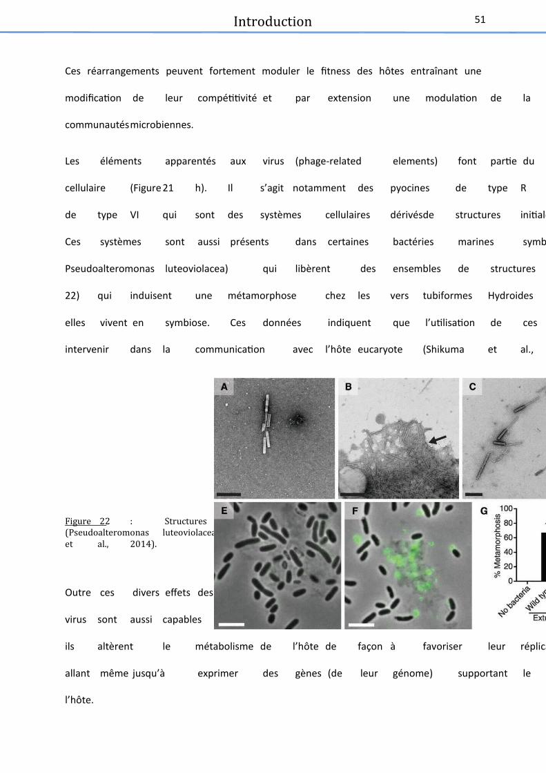
Introduction 51
Ces réarrangements peuvent fortement moduler le fitness des hôtes entraînant une
modification de leur compétitivité et par extension une modulation de la composition des
communautésmicrobiennes.
Les éléments apparentés aux virus (phage-related elements) font partie du métabolisme
cellulaire (Figure 21 h). Il s’agit notamment des pyocines de type R et du système de sécrétion
de type VI qui sont des systèmes cellulaires dérivésde structures initialement de type virales.
Ces systèmes sont aussi présents dans certaines bactéries marines symbiotiques (comme
Pseudoalteromonas luteoviolacea) qui libèrent des ensembles de structures tail-like (Figure
22) qui induisent une métamorphose chez les vers tubiformes Hydroides elegans avec lesquels
elles vivent en symbiose. Ces données indiquent que l’utilisation de ces éléments pourrait
intervenir dans la communication avec l’hôte eucaryote (Shikuma et al., 2014).
Figure 22 : Structures similaires a� des queues de caudovirus produites par une bacte� rie marine(Pseudoalteromonas luteoviolacea) et engendrant la me� tamorphose de son ho; te symbiotique (Shikumaet al., 2014).
Outre ces divers effets des virus sur le chromosome de l’hôte, lors de l’infection virale, les
virus sont aussi capables de prendre le contrôle total de leur hôte. De manière plus générale,
ils altèrent le métabolisme de l’hôte de façon à favoriser leur réplication (Ankrah et al., 2014)
allant même jusqu’à exprimer des gènes (de leur génome) supportant le métabolisme de
l’hôte.

Introduction 52
Ces gènes codant pour des protéines impliquées dans le métabolisme de l’hôte infecté sont
appelés « gènes auxiliaires » (ou « Virus-encoded auxiliary metabolic genes » soit vAMGs).
Ces vAMGs peuvent parfois être déjà présents dans le chromosome de l’hôte mais étaient
jusqu’alors inhibésou peu produits. Ces gènes sont divers, et peuvent être impliqués dans de
nombreuses fonctions de l’hôte comme la photosynthèse, la voie des pentoses phosphates,
la régulation du phosphate, le métabolisme du soufre, la synthèse des polysaccharides, le
métabolisme des sphingolipides ou le métabolisme (processing)de l’ADN et de l’ARN
(Rosenwasser et al., 2016).
Exemple : Gènes de la photosynthèse chez les virus marins
L’un des exemples les plus connusde vAMGsau sein du viroplancton est celui des cyanovirus
codant pour des gènes impliqués dans la photosynthèse (Figure 23). Découvert en 2003, ce
système montre que S-PM2 (virus infectant des cyanobactéries) contient dans son génome
deux gènes majeurs de la photosynthèse. Il s’agit de psbA et psbD, deux gènes codant pour
deux protéines (D1 et D2) ayant un rôle central dans le photosystème II. La production des
cyanobactéries est généralement limitée par la photo-inhibition, en particulier à la surface des
océans. Les protéines D1 et D2 jouent un rôle important dans la photo-inhibition puisque c’est
l’excès de protéine D1 photo-inactivée et endommagée qui l’entraîne, amenant à une
diminution de l’efficacité du photosystème. D’autre part, durant l’infection virale, le taux de
D1 cellulaire chute fortement entraînant une baisse de l’apport d’énergie dans la cellule.
Néanmoins, l’expression des protéines D1 et D2 codéespar les virus permet de maintenir un
photosystème efficace, passant outre la photo-inactivation, donnant ainsi plus d’énergie aux
virus pour qu’ils puissent se répliquer massivement et libérer un grand nombre de virions dans
l’environnement (Mann et al., 2003; Lindell et al., 2004).


Introduction
54
III. Les environnements thermophiles
Les thermophiles, organismes vivants à des températures optimales au-dessus de 45°C font
patie d’u g oupe plus aste appel e t ophiles. Cette otio est p ofo d e t
anthropomorphique puisque nous appelons « environnements extrêmes » les biotopes qui
nous seraient hostiles. Or, pour ces micro-organismes, ce sont nos conditions de vies qui sont
extrêmes. Ces organismes très divers, vivent sous au moins une condition physico-chimique
(température, pression osmotique, pression hydrostatique, pH, concentra tio e o g e…
paraissant être en hors des limites compatibles de la vie non extrêmophiles. Les
environnements qui sont considérés comme extrêmes peuvent par exemple être les zones
polaires, les étendues désertiques (chaudes ou froides), les fonds marins, les lacs de soude,
lacs acides ou hypersalés (Figure 24).
Figure 24 : (gauche) De� sert du Sahara (source : commons.wikimedia.org) (droite) Antarctique
(source : laterredufutur.com).
Il a longtemps été considéré que la vie à haute température était impossible car dénaturant
les p oti es. La haleu est d’ailleu s l’u des o e s de st ilisatio les plus a ie s et aussi
le plus pa du. Pou ta t, ’est la o ditio de ie sine qua non des micro-organismes
thermophiles et hyperthermophiles.

Introduction
55
La otio de theophile se liitait à °C jusu’e où Bacillus stearothermophilus,
apale de oissa e de à °C, tait o sid o e l’o gais e apale de oît e au
plus hautes températures (Stark and Tetrault, 1951; Prieur, 2014). Néanmoins, les travaux de
Thomas Brock et son équipe dans les années 70 ont révolutionné nos connaissances sur ces
micro-organismes. En effet, ils ont isolé les premières souches microbiennes ayant une
température optimale de croissance au-dessus de °C aps l’ ha tillo age de soues
chaudes terrestres du Parc national de Yellowstone aux Etats-Unis (Madigan and Martinko,
; P ieu , . Aujou d’hui, les the ophiles peu e t t e lass s e t ois sous
catégories : les thermophiles modérés (45 à 60°C), les thermophiles extrêmes (60 à 85°C) et
les hyperthermophiles (au-dessus de °C Mehta ad “ataaaaa, . A l’heu e
actuelle, la limite de la vie est fixée à 122°C, température à laquelle une méthanogène
piezophile décrite en 2008 peut encore proliférer sous 20 MPa de pression (Takai et al., 2008).
Depuis les t aau fo dateu s de B o k et so uipe, l’it t de la o u aut s ie tifi ue
pou les the ophiles ’a ess de g adi . E effet, outre leurs divers intérêts
biotechnologiques (le plus connu étant la Taq Polymérase de Thermus aquaticus (Chien et al.,
1976)), ces micro-organismes sont aussi étudiés afin de permettre une meilleure
compréhension des cycles biogéochimiques globaux, du rôle des micro-organismes à
l’i te fae ie/ i ale, das le ad e de e he hes su les o igies de la ie… Glo alee t
l’ tude de leu die sit et de leu ph siologie reste nécessaire car encore trop peu de
données sont disponibles sur ces organismes (Forterre, 2007; Prieur, 2014).

Introduction
56
x Les sources chaudes terrestres
Les sources chaudes terrestres sont un grand réservoir de thermophiles. Elles se trouvent
généralement dans les zones à activité volcanique, où les environnements chauds affleurent
à la surfa e. E effet, das es zo es, l’eau s’ifilt e da s la oûte teest e, se hauffe sous
l’effet d’u e ha e ag ati ue ais e out pas g âe à la p essio lithosphi ue. “ous
l’effet de la haleu , la p essio auge te faisat ejailli à la su fae l’eau ei hie e
minéraux et autres éléments (Mehta and Satyanarayana, 2013; Prieur, 2014).
Ces manifestations hydrothermales sont présentes dans divers endroits du globe mais les plus
connues restent le Parc National de Yellowstone aux USA (Figure 25), la péninsule de
Kamchatka en Russsie, le Chili, le Japon, la Nouvelle-) la de, l’Italie et l’Isla de. Elles peu e t
se manifester sous forme de solfatares, de geysers ou de sources chaudes (Mehta and
Satyanarayana, 2013).
Figure 25 : Photographie de geysers (a) en Nouvelle-Ze� lande (source : eugene.kaspersky.fr) (b) en
Islande (source : extremeiceland.is). (c) Photographie de source chaude a� Yellowstone (USA, source :
yellowstoneparknet.com).
a
c
b

Introduction
57
Les micro-organismes isole�s des sources chaudes
Les sources chaudes terrestres sont un réservoir très important de biodiversité bactérienne et
a h e e. L’appo t o stat de ut i e ts et les g adie ts ph sio-chimiques présents
laisse t e effet plae à u e g ade ai t d’esp es i o ie es. N a oi s ette
dive sit ’a t d ou e te u’assez tad ae l’a e e t des thodes d’aplifiatio
i lat des g es de l’ARN “ L pez-López et al., 2013). Les groupes de micro-organismes
majoritaires de cet écosystème sont le phylum des Crenarchaoeta pour les archées et l’o d e
des Aquificales pour les bactéries. Deux phyla plus récents ont aussi été découverts dans ces
sources, appartenant au domaine Archaea: les Korarchaeota et les Nanoarchaeota. Les
membres de ces deux phyla, tout comme les Aquificales font partie des embranchements les
plus p ofo ds de l’ae ph log ti ue du i a t L pez-López et al., 2013). La composition
des communautés de ces écosystèmes est très variable et dépend notamment du type de
source chaude étudiée.
Exemple des champs de solfatares
Les solfatares sont des fumerolles qui émettent des gaz soufrés. Ils sont retrouvés dans des
« champs de solfatares » qui comprennent des sols, boues et eaux de surfaces (riches en
soufre), chauffés par la chambre magmatique se situant en dessous, pouvant atteindre des
te p atu es de l’o d e de °C. Leu s paa t es ph sio hi i ues peu e t aie d’u
hap de solfataes à u aut e, ae des pH allat de à . L’aidit est due à la pse e
d’aide sulfu iue fo suite à l’odatio iologiue du sulfu e d’h d oge et du souf e.
Les solfatares sont généralement très riches en minéraux de fer, émettent du dioxyde de
carbone et ont des concentrations variables en dihydrogène, méthane, azote et monoxyde de
carbone (Mehta and Satyanarayana, 2013).

Introduction
58
Les communautés de a t ies et d’a h es qui colonisent ces environnements sont variées,
pouvant être acidophiles, thermophiles extrêmes ou modérées. La plupart de ces populations
sont hétérotrophes (comme Desulfurococcus mobilis ou Thermoproteus uzoniensis) ou
chimioautolithotrophes (comme Pyrobaculum islandicumou Thermoproteus neutrophilus),
utilisant souvent le soufre dans leur métabolisme énergétique. Ces solfatares peuvent
permettre la croissance de souches aérobies comme Sulfolobussolfataricus, anaérobie comme
Thermoproteux tenax ou encore des souches AAF (aéro-anaérobie facultatif) comme
Acidianus infernus (Segerer et al., 1986; Mehta and Satyanarayana, 2013). Des bactéries
comme Fervidobacterium islandicum ou Thermotoga thermarum appate a t à l’o d e des
Thermotogales colonisent aussi ces environnements mais sont moins souvent isolées de ceux-
ci que les archées (Windberger et al., 1989; Huber et al., 1990; Huber and Hannig, 2006).
Exemple : Soue haude a ide d’Ohaku-dani au Japon
Aujou d’hui eo e, la aste ajo it des i o-organismes est incultivée. Les études en
tag o i ue o t fou i de o eu g o es de sou hes ui ’o t pas e o e pu t e
ises e ultu e e lao atoie. U e app o he it essate a t ise e œu e da s une
étude récente. Les auteurs utilisent des échantillons naturels congelés et les soumettent à
di e ses o ditio s de ultu es. Cette thode a pe is de ett e e ultu e et d’isole de
nouvelles espèces issues de sources chaudes acides (68°C, pH 1,4) (Sakai and Kurosawa, 2016).
Ces espèces appartiennent au genre Sulfolobus (HS-1 et HS-3) qui est bien représenté dans les
sou es haudes. U e dizaie de ph lot pes d’a h es o t t et ou s das les
enrichissements réalisés dans cette étude, majoritairement des Sulfolobus, mais aussi des
Thermoplasma, des Metallosphaera et des Sulfurisphaera… (Figure 26).

Introduction
59
Figure 26 : Diversite� des communaute� s microbiennes retrouve�e dans des enrichissements re�alise� s a�
partir d’échantillons de sources chaudes terrestres (Sakai and Kurosawa, 2016). Les chiffres a� gauche
des figures repre�sentent la tempe�rature de culture, le pH et le pourcentage en extrait de levure(a)
0 , % b , %. Cette figure révèle notamment qu’à °C, il y aurait une prépondérance de bactéries
parmi les micro-organismes cultive� s ici.
Virus associe�s aux bacte�ries et arche�es des sources chaudes terrestres
Das es os st es où la pse e d’eu a otes se le t e t s fai le, les i us so t le
facteur principal de mortalité des communautés microbiennes (Schoenfeld et al., 2008; López-
López et al., . Ils iflue e t do eau oup la st u tu e, l’olutio et les aiatio s de
composition de ces populations. Pour autant, il y a encore trop peu de systèmes hôtes-virus
caractérisés dans ces environne e ts. E effet, u e uaa tai e d’a h o i us et u e
quinzaine de bactériovirus ont été bien décrits à ce jour. Néanmoins, leur diversité
morphologique et génomique est impressionnante en particulier concernant les archéovirus
(Rachel et al., 2002; Basta et al., 2009; Prangishvili et al., 2016).

Introduction
60
Bacte�riovirus
Il e iste aujou d’hui s st es h tes-virus thermophiles (entre 55 et 75°C) issus de sources
chaudes bien décrits dans la littérature (Uldahl and Peng, 2013; Nagayoshi et al., 2016).
N a oi s, lo s d’u e aste tude alis e e , i us o t t isols et patiellee t
d its, ais leu aat isatio ’ tat pas o pl te, ils e so t pas o sid s o e
caractérisés. Ces virus infectent des bactéries appartenant au genre Thermus, mais aussi aux
genres Geobacillus, Meiothermus et Rhodothermus (Yu et al., 2006; Uldahl and Peng,
2013)(Tableau 3).
Parmi les 14 systèmes décrits seuls certains seront détaillés et pour permettre une meilleure
lisibilité, les autres sont décrits dans le tableau ci dessous.
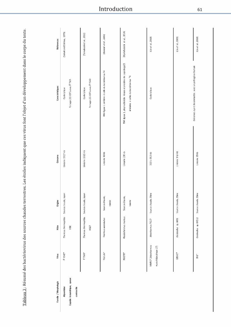

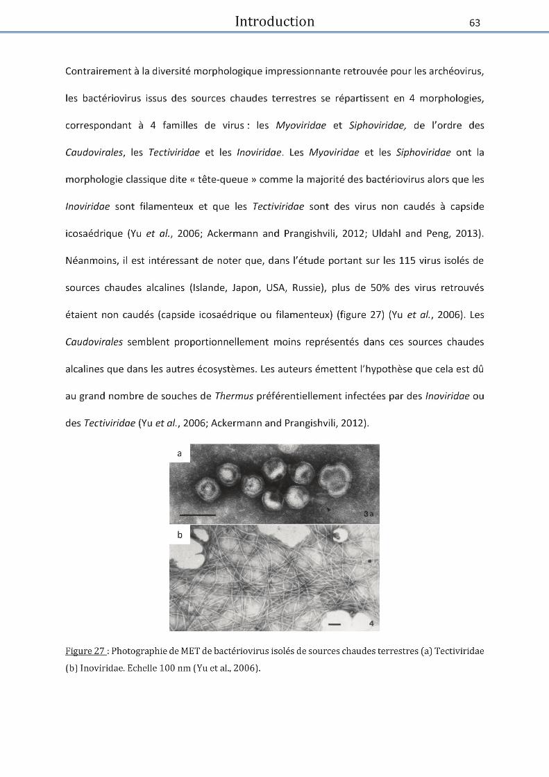

Introduction
64
Les Myoviridae se caractérisent par une capside icosaédrique à laquelle est attachée une
ueue o t atile, ils so t g ale e t i ule ts, etta t e œu e u le l ti ue.
Le premier virus thermophile infectant une souche issue de source chaude fut le myovirus
) YS40. Isolé au Japon, il infecte via un cycle lytique la souche Thermus thermophilus HB8
(Sakaki and Oshima, 1975).
) TMA est aussi un Myoviridae i ule t isol d’u e sou e haude au Japo . Il est asso i à
Thermus thermophopilus HB27, qui comme HB8 croît de façon optimale à 65°C.
) TMA et ) YS40 patage t plusieu s siilait s. Tout d’ao d, ils poss de t des o phologies
très similaires avec une capside icosaédrique de 125 nm de diamètre et une queue contractile
de 178 nm de long pour 27 nm de large. Ensuite, ils possèdent de grands génomes. En effet,
leu s ADN dou le i li ai es fo t espe tie e t , et , k . Leu taille ’est pas
leu seul poit o u puisu’ils patage t ORFs do t les t ois uats e p se te t pas
de similarités dans les bases de données) et leur organisation génomique est similaire. Il
semble aussi que ces deux virus nécessitent la présence de pili sur la souche HB8 pour
s’adso e su ette ellule et i s e leu s g o es. Ce ais e est aussi utilis pa les
Inoviridae (Stassen et al., 1994; Naryshkina et al., 2006; Tamakoshi et al., 2011).
Les bactériovirus de thermophiles sont un réservoir de nouvelles protéines et notamment
d’e z es the osta les aat de o euses appliatio s pote tielles e iote h ologie
(Uldahl and Peng, 2013).

Introduction
65
C’est das e ad e ue les myovirus TS2126 et RM378 ont été découverts (Hjörleifsdottir et
al., 2002; Blondal, Thorisdottir, et al., 2005; Hjörleifsdottir et al., 2014).
Pour ces deux virus, une RNA ligase 1 montrant des similarités avec celle du phage T4 a été
mise en évidence. Cette RNA ligase est chez le phage T4, impliquée dans le contournement
d’u ais e ati- i us is e plae pa l’h te. Ce ais e est as su le li age d’ARN
de transferts (ARNt) viraux. La ligase à pour objectif de réparer ces ARNt. Elle est associée à
u e pol u l otide kiase PNK ui effe tue des odifiatio s su l’ARNt pou le t asfoe
en substrat disponible pour la ligation. Néanmoi s, la RNA ligase de T“ ’est pas asso i
à une PNK connue. En effet, aucun ORF du génome à ADN double brin linéaire de 90 kb de
T“ ’a o t de si ilait ae la PNK de T . N a oi s il est possile u’u e PNK
différente soit présente dans so g o e. D’aut e pat, l’atiit de ette ligase s’est l e
particulièrement efficace pour effectuer des ligations inter ou intra moléculaires, sur des ARN,
ADN simple et double brin (Blondal, Thorisdottir, et al., . E e a he, la p se e d’ue
PNK et d’u e RNA ligase si ilaies à elles de T o t t ises e ide e das le g o e
à ADN double brin de 130 kb de RM378 (Blondal et al., 2003; Blondal, Hjorleifsdottir, et al.,
2005; Hjorleifsdottir et al., 2014).
GBSV1 et BV1 ont eux aussi été isolés de souches provenant de sources chaudes en Chine. Ce
sont deux myovirus infectant deux souches de Geobacillus sp. 6K51 pour GBSV1 et Geobacillus
sp. 6K512 pour BV1. Leurs hôtes ont une température de croissance optimale de 65°C. Les
virions sont dot s d’u e apside iosa d i ue de de dia t e et d’u e ueue
o t atile de à de lo g. Leu s go es so t o pos s d’ADN dou le i li ai e
de 34,6kb (GBSV1) et 35kb (BV1).

Introduction
66
Ils contiennent tous les deux 54 ORFs qui présentent de fortes similarités avec des Geobacillus
et Bacillus ainsi que les virus qui leurs sont associés. Ces souches de Geobacillus et Bacillus
sont majoritairement issues des sols mais il est à noter que plusieurs ORFs de ces virus ont des
similarités avec des gènes de Geobacillus kaustophilus, bactérie thermophile notamment
retrouvée au niveau de la fosse des Mariannes (Takami et al., 2004). Fait intéressant, 7 à 8
ORFs de ces génomes partagent de fortes similarités avec des protéines produites par Listeria
innocua. Cette bactérie est un pathogène intracellulaire opportuniste humain. Ces similarités
laisse t à pe se u’il e iste u lie olutif pote tiel e t e es i us et e pathog e “o g
and Zhang, 2008; Liu et al., 2009, 2010).
Parmi les Siphoviridae, 3 systèmes ont été décrits. Cette famille de virus se caractérise par la
p se e d’u e apside i osa d i ue à lauelle est attah e u e ueue fle i le o
contractile. Les trois siphovirus infectent des souches de Thermus.
Les deux siphovirus , P23-45 et P74-26 infectent via un cycle lytique la même souche Thermus
thermophilus HB8. Ils ont été isolés de sources chaudes acides dans la péninsule de Kamchatka
en Russie. Les virions sont similaires et morphologiquement proches de TSP4 (Thermus
siphoviridae phage puisu’ils o t u e apside iosa d i ue de de dia t e et u e
t s lo gue ueue fle ile de de lo g. Leu s apsides o tie e t des g o es d’ADN
double brin de 84 kb pour P23-45 et 83 kb pour P74-26. Ils comprennent respectivement 117
et 116 ORFs dont 95% sont similaires. Ces génomes présentent une caractéristique
inhabituelle. En effet ils contiennent, dans leurs régions intergéniques, de grandes séquences
répétées polypurine-polypyrimidine. Ces séquences sont fréquemment retrouvées chez les
eucaryotes mais rarement chez les bactéries ou archées. Elles forment des triples hélices
stales ui lo ue t la pli atio et la t as iptio , p o o ua t l’istailit du g o e i al.

Introduction
67
Les expériences réalisées in vitro montrent que ces séquences forment bien un ADN triple brin
ce qui bloque la réplication des virus. Cette conformation particulière pourrait donc être
i pli u e da s la gulatio de l’e p essio de es i us. Aut e fait it essat, u e p oti e
pote tielle de liaiso à l’ARN polymérase a été identifiée dans le génome de P23-45. Celle-ci
se ait apale d’i hi e la t a s iptio de l’h te tout e peetta t elle des ges i au
(Yu et al., 2006; Minakhin et al., 2008; Berdygulova et al., 2011).
La famille des Tectiviridae est caractérisée par des virions aux capsides icosaédriques
contenant une membrane interne lipidique. Les deux virus connus sont P23-77 et ) IN93. P23-
77 infecte Thermus thermophilus isol e d’u e soue haude e Nou elle-Zélande et ) IN93
infecte Thermus thermophilus isol e d’u e soue haude au Japo . Ces h tes se deloppe t
respectivement à 70 et 65°C. P23-77 est virulent alors que ) IN93 est tempéré comme
l’i di ue la p se e de g es i pli u s das la l sog ie das so g o e Jaati e et al.,
2008; Matsushita and Yanase, 2009). Les protéines de capsides de P23-77 ont été
particulièrement étudiées (Jaatinen et al., 2008; Jalasvuori et al., 2010; Rissanen et al., 2013).
En effet, les capsides de ces deux virus présentent des similarités avec celles du virus SH1
i fe tat l’a h e halophile Haloarcula hispanica. Trois gènes communs ont été mis en
évidence dans leurs génomes, deux codant pour les protéines de capsides mineurs et majeurs
et u oda t pou u e ATPase putatie i pli u e das l’e pauetage du génome viral.
D’aut e pat, la foe des apsides de es i us est pa tiuli e. E effet hez “H o e pou
les deux virus présentés ici, la formation de la capside se base sur une symétrie T=28 et des
capsomères de décoration en forme de tours sont présents à la surface des virions. La
conformation des protéines de capside serait basée sur de multiples feuillets béta.

Introduction
68
Cette conformation semble particulièrement bien conservée et est présente dans différents
virus P23-77 like. Elle serait ancienne, révélant un possible ancêtre commun lointain pour ces
i us. C’est su ette ase u’il a t p opos d’i lu e es deu Tectiviridae dans les
Sphaerolipoviridae o p e a t ota e t di e s i us d’a h es halophiles do t “H
(Jaatinen et al., 2008; Jalasvuori et al., 2010; Rissanen et al., 2012, 2013; Pawlowski et al.,
2014).
Pour terminer, la famille Inoviridae se caractérise par des virions ayant une structure en forme
de longs filaments sortant de leurs hôtes par extrusion sans provoquer la lyse cellulaire. Cette
famille comprend deux virus : PH75 et ) OH3 qui portent un ORF commun.

Introduction
69
Arche�ovirus
La diversité morphologique et génomique des virus infectant les archées est impressionnante
e pati ulie hez les h pe theophiles. “eize o photpes d’a h o ius ont été décrits à
ce jour (dont 4 sont uniques) alors que parmi les plus de 6000 bactériovirus connus, seuls 9
o photpes o t t d its et au u ou eau o photpe ’a t d ou e t depuis les
années 70 (Ackermann and Prangishvili, 2012; Pietilä et al., 2014; Uldahl, 2015). Par ailleurs,
la vaste majorité des gènes composant leurs génomes restent sans similarités avec les
séquences présentes dans les bases de données et/ou ont des fonctions inconnues.
Un peu plus de 80 archéovirus ont été décrits à ce jou . La ajeu e patie d’e t e eu ife te t
des Crenarchaeota hyperthermophiles appartenant aux genres Sulfolobus et Acidianus ou aux
Euryarchaeota halophiles des genres Haloarcula et Halorubrum (Uldahl, 2015). Parmi les
archaeovirus des sources chaudes, 11 morphologies différentes sont retrouvées et réparties
en différentes familles (Tableau 4). Comme pour les bactériovirus, pour une meilleure lisibilité,
seuls e tai s i us se o t d its das le o ps du te te. U ta leau su at l’e se le des
archéovirus caractérisés à ce jour est présenté ci-dessous.

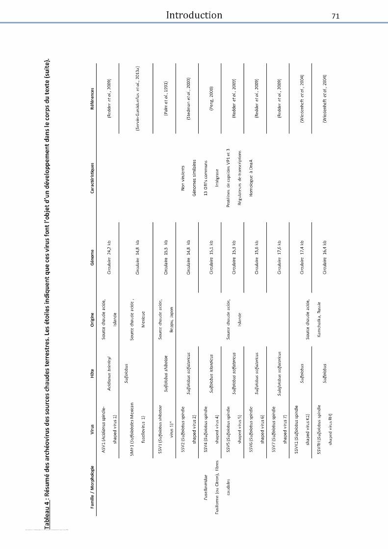


Introduction
75
Ampulaviridae
C’est u e des failles les plus to a tes d’u poit de ue o phologiue. Elle e comprend
u’u i us : ABV (Acidianus ottle shaped i us . ABV a t isol à pa ti d’u e soue haude
acide en Italie et infecte Acidianus convivator qui croît à une température optimale de 75°C.
Les virions font 230 nm de long pour 4 nm de large à la pointe la plus fine et 75 nm de large à
la ase du i io . La poite e fo e d’e to oi du i io pou ait te i pli u e da s
l’adso ptio et l’ise tio du g o e i al da s la ellule h te. L’aut e e t it du i io
comporte une vingtaine de filaments, insérés dans un disque ou un anneau, qui ne semblent
pas i pli u s da s l’adh sio à l’h te ais ui pou aie t aoi u le das la o ilit du
virion. La libération des virions ne causant pas la lyse cellulaire mais un retard de croissance,
ce virus pou ait ett e e œu e u le de tpe tat po teu . Le i io o tie t u ADN
dou le i li ai e d’e i o k . Celui-ci est composé de 57 ORFs dont seulement 3 ont
des similarités avec des séquences présentes dans les bases de données incluant une
glycosyltransferase, une thymidylate kinase et ADN polymérase de la famille B. Cette dernière
p ot i e o t e d’ to ates si ilait s ae des p oti es eu a otes ais aussi ae des
pol ases d’Euryarchaeota. Enfin, la procapside de ce virus montre une homologie de
structure avec celle de phi29, impliquant un potentiel ancêtre commun entre ces deux virus
(Häring et al., 2005; Peng et al., 2007).
Fuselloviridae
Cette famille de virus, dont les virions sont caractérisés par une forme de citron, infecte des
archées des genres Sulfolobus (SMF1, SSV1, 2, 4, 5, 6, 7 K1, RH)et Acidianus (ASV1) (Tableau
. Ils o t t isols pou o e d’e t e eu das des soues haudes e Islade, ais
certains proviennent du Japon, de Russie, du Mexique ou des USA. Ils contiennent de petits
g o es i ulaies d’e i o / k Pi a et al., 2011; Uldahl and Peng, 2013).
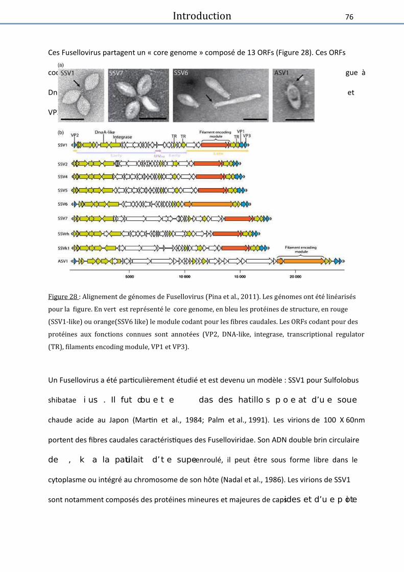
Introduction
76
Ces Fusellovirus partagent un « core genome » composé de 13 ORFs (Figure 28). Ces ORFs
codent notamment pour une intégrase, des régulateurs de transcription, un homologue à
DnaA et des protéines impliquées dans la structure des virions (protéines de capsides VP1 et
VP3) (Pina et al., 2011; Krupovic et al., 2014).
Figure 28 : Alignement de ge�nomes de Fusellovirus (Pina et al., 2011). Les ge�nomes ont e� te� line�arise� s
pour la figure. En vert est repre� sente� le core genome, en bleu les prote� ines de structure, en rouge
(SSV1-like) ou orange(SSV6 like) le module codant pour les fibres caudales. Les ORFs codant pour des
prote� ines aux fonctions connues sont annote�es (VP2, DNA-like, integrase, transcriptional regulator
(TR), filaments encoding module, VP1 et VP3).
Un Fusellovirus a été particulièrement étudié et est devenu un modèle : SSV1 pour Sulfolobus
shibatae i us . Il fut dou e t e das des hatillo s p o e at d’u e soue
chaude acide au Japon (Martin et al., 1984; Palm et al., 1991). Les virions de 100 X 60nm
portent des fibres caudales caractéristiques des Fuselloviridae. Son ADN double brin circulaire
de , k a la patiulait d’t e supe-enroulé, il peut être sous forme libre dans le
cytoplasme ou intégré au chromosome de son hôte (Nadal et al., 1986). Les virions de SSV1
sont notamment composés des protéines mineures et majeures de capsides et d’u e p oti e

Introduction
77
de liaiso à l’ADN Reite et al., ; P agish ili et al., ; Me o et al., ; Que i et
al., 2015). Une analyse en cryomicroscopie a permis de révéler en détail la structure du virion,
montrant notamment une symétrie sur 6 axes de la queue et la présence de nombreuses
aspérités à la surface de la capside (Figure 29)(Stedman et al., 2015).
Figure 29 : Particules de SSV1 analyse�es en cryo-EM (Stedman et al., 2015). (A) Particules de SSV1 en
cryo-EM. Reconstruction tridimensionnelle de SSVB vue d’ensemble C coupe transversale D vue
de dessous.
Lipothrixiviridae
La famille Lipothrixiviridae est celle qui contient le plus de membres caractérisés provenant
des sources chaudes terrestres. Les virions de cette famille ont une forme de longs filaments
(|900 à 2000nm de long) entourés par une enveloppe lipidique provenant de leur hôte. La
ajeu e patie d’e t e eu ife te t des a h es appate at au gee Acidianus (AFV1, 2, 3,
6, 7 8 et 9) mais sont aussi retrouvés chez trois virus infectant Thermoproteustenax (TTV1, 2
et 3) et un virus infectant Sulfolobus islandicus (SIFV). Ils proviennent de sources chaudes en
Italie, e Russie, e Islade et au U“A. Leu s go es so t o pos s d’ADN à dou le i
linéaires, généralement d’u e uaatai e de kilo ases e si les plus petits TTV , TTV
ne font que 15,9kb (Janekovic et al., 1983; Uldahl and Peng, 2013).


Introduction
79
Ensuite, 17 ORFs sont partagés par ces virus, incluant des ORFs codant pour des protéines de
st u tu es et d’aut es ipli u s das la pli atio de l’ADN. Ces die s l e ts so t
retrouvés dans un génome viral récemment séquencé, celui de SMR1 (Sulfolobus Mexican
udi i us . Ce go e a t ide tifi à pati d’u tag o e alis su u e souce
haude e i ai e. Il est psu u’il i fe te le ge e Sulfolobus car les séquences de ces
a h es so t p do i a tes das e tag o e. D’aut e pa t il a t lass pa i les
Rudiviridae car il possède, non seulement les séquences inversées répétées terminales, mais
aussi ORFs e o u s ae d’aut es Rudivirus. Ces ORFs communs codent notamment
pour des protéines de structures, des glycosyl-transférases et des protéines impliquées dans
la pli atio de l’ADN P agish ili et al., 1999; Vestergaard et al., 2005, 2008; Servín-
Garcidueñas et al., 2013; Uldahl and Peng, 2013).
Famille des Turriviridae
Les deux membres composant cette famille STIV1 et 2 (Sulfolobus turreted icosahedral virus
1 et 2) sont caractérisés par des virions de forme icosaédriq ue d’e i o de dia t e.
Ils so t su o t s d’u e e eloppe et pse te t de petites st u tu es ou geo a tes à la
surface de la capside. Ces structures sont potentiellement impliquées dans la reconnaissance
et/ou l’adh sio à l’h te. “TIV ife te Sulfolobus solfataricus via un cycle lytique. Sa capside
contient un ADN double brin circulaire de 17,6kb. STIV2 infecte lui Sulfolobus G4ST-2 et est
o pos d’u g o e à ADN dou le i i ulaie de , k Rie et al., 2004; Happonen et
al., 2010).
Ces deux virus semblent similaires au niveau génomique et structurel. En effet, leurs génomes
do t l’o gaisatio est se lale patage t ORFs. Ils patage t aussi u ais e de
so tie de l’h te assez patiulie etta t e œue des st u tu es paid ales, aussi appelées
VAP Vi us Asso iated Paids , t oua t l’e eloppe ellulaie.

Introduction
80
Ce ais e est aussi et ou hez u aut e i us d’a h e : SIRV2 (famille des Rudiviridae).
Un ORF est commun aux trois génomes et la protéine codée par celui-ci est responsable de la
formation de ces structures pyramidales chez STIV1 et SIRV2, elle a donc probablement la
même fonction pour STIV2 (Figure 31) (Rice et al., 2004; Brumfield et al., 2009; Happonen et
al., 2010; Snyder et al. , . D’aut es st u tu es de ce type ont aussi été retrouvées chez
une souche de Pyrobaculum oguniensis sou ise au UV sas u’il eut t possile de d fii
l’o igie ou la fo tio de elle- i. E effet ette p odu tio e se le pas d’o igie i ale
puisu’au u e ife tio i ale ’a pu t e ise e ide e hez ette sou he Figu e 1)
(Rensen et al., 2015).
Figure 31 : Structures pyramidales (I) Sulfolobus sofataricus (A) observe� en MEB (microscope
e� lectronique a� balayage) (B) en MET (Gauche) Souche non infecte�e (Droite) Souche infecte� e par STIV
(indique� par des fle� ches)(Brumfield et al. 2009). (II) Structures pyramidales observe�es chez une
souche de Pyrobaculum apre� s traitement aux UVs (sans pre�sence ave� re�e de virus). (Rensen et al. 2015)
Echelle : 1Pm.
(I) (II)
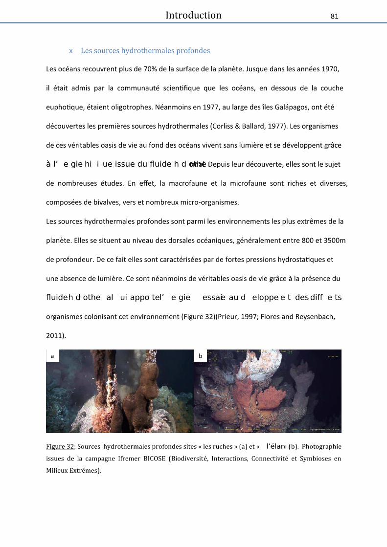
Introduction
81
x Les sources hydrothermales profondes
Les océans recouvrent plus de 70% de la surface de la planète. Jusque dans les années 1970,
il était admis par la communauté scientifique que les océans, en dessous de la couche
euphotique, étaient oligotrophes. Néanmoins en 1977, au large des îles Galápagos, ont été
découvertes les premières sources hydrothermales (Corliss & Ballard, 1977). Les organismes
de ces véritables oasis de vie au fond des océans vivent sans lumière et se développent grâce
à l’ e gie hi i ue issue du fluide h d othermal. Depuis leur découverte, elles sont le sujet
de nombreuses études. En effet, la macrofaune et la microfaune sont riches et diverses,
composées de bivalves, vers et nombreux micro-organismes.
Les sources hydrothermales profondes sont parmi les environnements les plus extrêmes de la
planète. Elles se situent au niveau des dorsales océaniques, généralement entre 800 et 3500m
de profondeur. De ce fait elles sont caractérisées par de fortes pressions hydrostatiques et
une absence de lumière. Ce sont néanmoins de véritables oasis de vie grâce à la présence du
fluide h d othe al ui appo te l’ e gie essaie au d eloppe e t des diff e ts
organismes colonisant cet environnement (Figure 32)(Prieur, 1997; Flores and Reysenbach,
2011).
Figure 32: Sources hydrothermales profondes sites « les ruches » (a) et « l’élan » (b). Photographie
issues de la campagne Ifremer BICOSE (Biodiversite� , Interactions, Connectivite� et Symbioses en
Milieux Extre;mes).
a b

Introduction
82
Leur formation sulte de l’ifilt atio de l’eau de e dans la croûte terrestre fracturée
ofo e. L’eau de e se hauffe p og essie e t au o ta t de la ha e
ag ati ue, elle s’ei hit g âe à elle-ci en divers gaz dissous, comme le méthane, le sulfure
d’h d og e, le dih d oge, le o o de de a o e et le diode de a o e. “ous l’effet
de la haleu , l’eau e o te jusu'à la su fae du pla he oa i ue e lessiat les o hes
u’elle t ae se. Elle s’ei hit alo s e l e ts die s, ota e t des io s talli ues
comme le zinc ou le fer puis rejaillit sous forme de fluide hydrothermal (Von Damm et al.,
1995; Flores and Reysenbach, 2011).
Ces fluides sont très chauds (|350°C), acides, anoxiques et enrichis en gaz dissous et en divers
éléments (cités ci-dessus). Les éléments contenus dans ces fluides précipitent au contact de
l’eau de e à °C, pou foe des he i es hydrothermales (Figure 33). Au niveau de
celles-ci se créent des gradients de température, de pH et de concentration en oxygène du
fait de la e o t e e t e l’eau de e et le fluide hydrothermal. La confrontation de ces
paramètres physico-chimiques très différents entraîne une ambiance chaotique qui ajoute au
caractère extrême de cet environnement (Prieur, 1997; Desbruyères et al., 2000;
Miroshnichenko, 2004; Flores and Reysenbach, 2011)


Introduction
84
A la base de ces écosystèmes sont retrouvés des micro-organismes chimiosynthétiques qui
so t à l’o igie de la p odu tio p iai e. Il s’agit aussi ie d’a h es ue de at ies, la
plupart sont anaérobies et peuvent être thermophiles ou hyperthermophiles. La
concentration en H2S dans ces environnements étant particulièrement haute (de 3 à 110
Mol/kg de s die ts l’o datio des o pos s souf s est la soue p i ipale d’ e gie
de ces micro-organismes chimiosynthétiques (Thornburg et al., 2010). Les Protéobactéries
représentent une grande partie de la population bactérienne colonisant ces écosystèmes
(figure 34). Elles peuvent être sulfo-oxydantes ou ferro-oxydantes, mésophiles à modérément
the ophiles. Il s’agit sou e t de sio tes appo ta t de l’e gie à leu s h tes et pa fois
participant à la détoxification des composés soufrés (Kelley et al., 2002; Pflugfelder et al.,
2005; Thornburg et al., 2010; Eichinger et al., 2014).Des Aquificales thermophiles hydrogéno-
oxydantes et anaérobies sont aussi bien représentées. Elles sont autotrophes et retrouvées
das de o eu e i o e e ts e t es et o t u e aifi atio p ofo de das l’a e
phylogénétique du vivant (Miroshnichenko, 2004; Flores et al., 2011). De nombreuses
at ies hi ioo ga oh t ot ophes peuple t e iotope. Il s’agit ota e t des
thermophiles aérobies des genres Bacillus, Geobacillus et Thermus appartenant au phylum
des Firmicutes et des Deinococcus-Thermus (Thornburg et al., 2010; Flores and Reysenbach,
2011). Enfin, les bactéries thermophiles du phylum des Thermotogae sont souvent isolées à
partir de ces écosystèmes et seront décrites plus en détail par la suite.

Introduction
85
Figure 34 : Abondance relative des phyla bacte� riens (a) au niveau des sources hydrothermales
profondes, forte pre�dominance des epsilon prote�obacte� ries, puis des alpha prote�obacte� ries (b) dans
les oce�ans profonds, pre�dominance des alpha prote�obacte� ries (Thornburg et al., 2010).
Concernant le domaine Archaea, des archées méthanogènes appartenant notamment à
l’o d e des Methanococcales ou des archées organotrophes fermentaires appartenant à
l’o d e des Thermococcales so t o u e t et ou es. D’aut es h pe theophiles so t
aussi présentes comme les Desulfurococcales qui réduisent le soufre ou les Archaeoglobales
pouvant réduire des composés soufrés ou ferreux (Figure 35) (Thornburg et al., 2010; Flores
et al., 2011).

Introduction
86
Figure 35 : Représentation de l’abondance relative des différents phyla archéens a au niveau des
sources hydrothermales profondes, (b) dans les oce�ans profonds, pre�dominance des euryarche�es non
classe� es dans les deux environnements (Thornburg et al., 2010).
Introduction
87
Les Thermotogae
Le phylum des Thermotogae regroupe plus de 70 espèces décrites, réparties au sein de 3
ordres, 4 familles et 12 genres différents (Pollo et al., 2015). Les Thermotogae sont des
bactéries très majoritairement thermophiles ou hyperthermophiles (un genre non
thermophile : les Mesotoga), anaérobies et à Gram négatif. Elles se présentent sous forme de
bâtonnets portant une « toge » caractéristique. La composition de cette toge reste peu connu,
elle contient deux protéines identifiées : la protéine d’a age OmpA1 et la porine OmpB. Ces
bactéries vivent dans des environnements chauds géothermiques ou volcaniques comme les
sources hydrothermales profondes, les sources chaudes terrestres ou les puits de pétrole. Sur
le plan métabolique, les Thermotogae peuvent fermenter de nombreux sucres et substrats
p ot i ues o ple es. Cette fee tatio s’a o pag e, pou la plupa t, de la du tio de
composés soufrés (Bonch-Osmolovskaya, 2008).
La position phylogénétique des Thermotogae, est sujette à débat (Bonch-Osmolovskaya,
2008; Zhaxybayeva et al., 2009; Gupta and Bhandari, 2011). En comparant les séquences des
gènes codant pour les ARNr 16S et les protéines ribosomales des Thermotogae, celles-ci sont
proches des Aquificales. Cette proximité contribue au fait que les Thermotogae occupent la
seconde a he la plus p ofo de das l’ae ph log ti ue des at ies Figu e 6).

Introduction
88
Figure 36 : Placement des Thermotogales (par extension des Thermotogae) dans l’arbre
phylogénétique bactérien par rapport à l’ARNr S Bonch-Osmolovskaya, 2008). Elles sont situe�es
profonds dans cette arbre, proches des Aquificales.
N a oi s, lo s d’a alses ph log ti ues su le este du go e, le app o he e t ae
les Aquificales est beaucoup moins évident. Les Thermotogae s’a e t aifeste e t plus
proches des Firmicutes, notamment des Clostridiales, mais également des Archaea . L’ tude
des génomes de différentes souches de Thermotogae (T. maritima, T. petrophila, T. lettingae,
T. ela esie sis… ) a montré que ceux-ci partagent 45% de similarité avec des Firmicutes
(notamment Thermoanaerobacterales, ordre faisant partie des Clostridiales) et 10% de
similarité avec des archées (notamment les Thermococcales) (Zhaxybayeva et al., 2009; Gupta
ad Bhadai, . L’histoie olutie des g o es de Thermotogae pourrait être le reflet
d’ao da ts t a sfe ts lat au de g es. Pa e eple, u op o putatif odat pou u e
ferrédoxine oxydoréductase a été mis en évidence chez la majeure partie des espèces décrites
de Thermotogae. Cet opéron est partagé avec Pyrococcus furiosus qui est une archée de
l’o d e des Thermococcales, occupant la même niche écologique que certaines Thermotogae,
notamment au niveau des sources hydrothermales profondes. Il a été démontré que cet
opéron a été obtenu par transfert latéral de gènes des archées vers les bactéries, ce qui
essite l’ipli atio d’ l e ts g ti ues o iles au ou s de l’ olutio de es esp es
(Zhaxybayeva et al., 2009).

Introduction
89
Ces t asfe ts latau de g es a e t do à e isage l’ipli atio pote tielle de i us
via le ph o e de t a sdu tio et d’it g atio s ue es p o iales . Cette h poth se se
trouve renforcée par la description de CRISPR (Clustered Regularly Interspaced Short
Palindromic Repeats) au sein de nombreux génomes de Thermotogae ; ces séquences
t oig e t de la ise e plae d’i u it is-à- is d’ife tio s i ales et p ou e t ue de
telles ife tio s o t au oi s d jà eu lieu au ou s de l’histoie olutie des Thermotogae
(Zhaxybayeva et al., 2009; Pollo et al., 2015). Pour autant, à ce jour, un seul virus a été décrit
chez les Thermotogae. Il s’agit de MPV isol de Marinitoga piezophila et récemment décrit
(Lossouarn, Nesbø, et al., 2015). A celui-ci se rajoutent deux mini-plasmides cryptiques, très
similaires, pQR7 et pMC24, retrouvés respectivement chez Thermotoga sp. RQ7 et
Thermotoga maritima MC24, qui sont les seuls éléments génétiques mobiles décrits au sein
de cet ordre bactérien (Bonch-Osmolovskaya, 2008).
Virus associe�s aux micro-organismes thermophiles
D’i po ta ts effo ts o t t o sa s, ota e t au lao atoi e, afi de ieu o p e d e
la diversité génétique, les capacités métaboliques et les adaptations physiologiques des
communautés microbiennes hydrothermales. Notre connaissance de la diversité virale
associée à ces micro-organismes demeure, en revanche, encore assez limitée. Alors
u’e i o esp es de at ies et a h es so t o ues pou olo ise es
environnements, seuls quelques virus ont été décrits (Williamson et al., 2008; Jebbar et al.,
2015; Lossouarn, Dupont, et al., 2015).

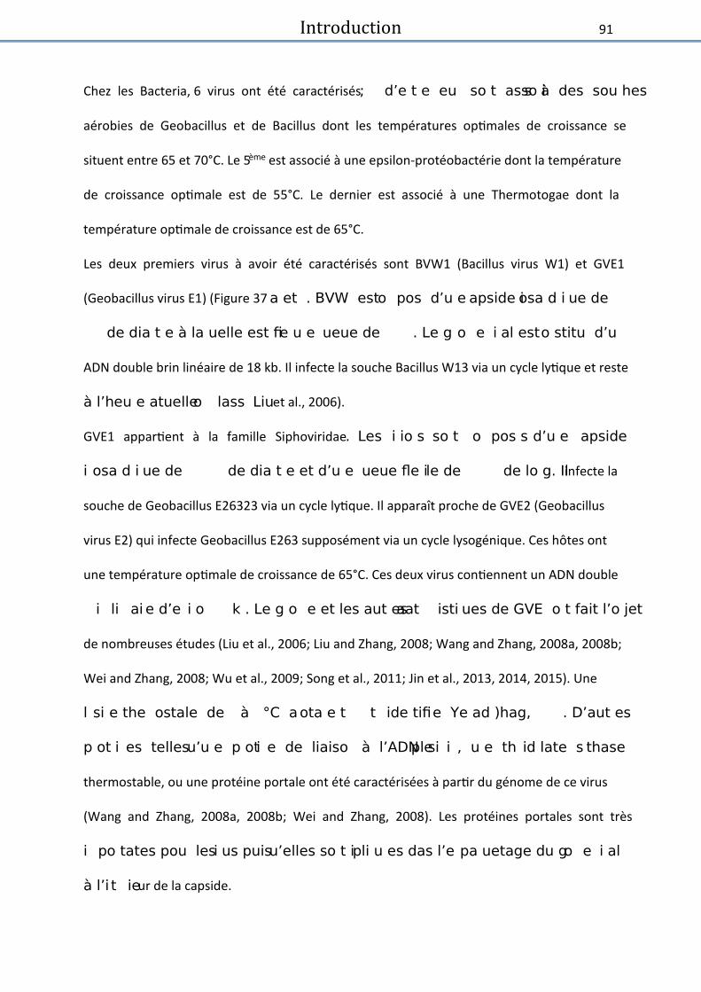
Introduction
91
Chez les Bacteria, 6 virus ont été caractérisés ; d’e t e eu so t asso is à des sou hes
aérobies de Geobacillus et de Bacillus dont les températures optimales de croissance se
situent entre 65 et 70°C. Le 5ème est associé à une epsilon-protéobactérie dont la température
de croissance optimale est de 55°C. Le dernier est associé à une Thermotogae dont la
température optimale de croissance est de 65°C.
Les deux premiers virus à avoir été caractérisés sont BVW1 (Bacillus virus W1) et GVE1
(Geobacillus virus E1) (Figure 37 a et . BVW est o pos d’u e apside iosa d i ue de
de dia t e à la uelle est fie u e ueue de . Le g o e i al est o stitu d’u
ADN double brin linéaire de 18 kb. Il infecte la souche Bacillus W13 via un cycle lytique et reste
à l’heu e atuelle o lass Liu et al., 2006).
GVE1 appartient à la famille Siphoviridae. Les i io s so t o pos s d’u e apside
i osa d i ue de de dia t e et d’u e ueue fle ile de de lo g. Il infecte la
souche de Geobacillus E26323 via un cycle lytique. Il apparaît proche de GVE2 (Geobacillus
virus E2) qui infecte Geobacillus E263 supposément via un cycle lysogénique. Ces hôtes ont
une température optimale de croissance de 65°C. Ces deux virus contiennent un ADN double
i li ai e d’e i o k . Le g o e et les aut es aat isti ues de GVE o t fait l’o jet
de nombreuses études (Liu et al., 2006; Liu and Zhang, 2008; Wang and Zhang, 2008a, 2008b;
Wei and Zhang, 2008; Wu et al., 2009; Song et al., 2011; Jin et al., 2013, 2014, 2015). Une
l si e the ostale de à °C a ota e t t ide tifie Ye ad )hag, . D’aut es
p ot i es telles u’u e p oti e de liaiso à l’ADN siple i , u e th id late s thase
thermostable, ou une protéine portale ont été caractérisées à partir du génome de ce virus
(Wang and Zhang, 2008a, 2008b; Wei and Zhang, 2008). Les protéines portales sont très
i po tates pou les i us puisu’elles so t ipli u es das l’e pa uetage du go e i al
à l’i t ieur de la capside.

Introduction
92
Comme pour beaucoup de protéines de bactériovirus thermophiles, celle-ci montre peu de
si ilait s ae les p oti es des i us de sophiles. Elle a do tout d’ao d t ise e
évidence grâce à sa structure secondaire prédite qui montrait de fortes similarités avec les
autres protéines portales connues. Sa fonction a ensuite été confirmée notamment grâce à
u a uage à l’o peetta t de oi sa positio das la st u tu e du i io Figu e 7 c)
(Wang and Zhang, 2008a).
Figure 37 : Observation au microscope e� lectronique de virions caude� s de (a) BVW1 (b) GVE1 et (c)
GVE marqués aux particules d’or Liu et al., 2006; Wang and Zhang, 2008a).
U e e dol si e, i pli u e das la lse ellulaie i duite pa l’ife tio i ale a aussi t mise
en évidence (Jin et al. , . Cette p oti e fait patie d’u s st e holie-endolysine qui
crée des pores dans la membrane cellulaire et entraîne une perte de perméabilité menant à
la l se ellulaie et do à la li atio des i io s das l’e i onnement (Jin et al., 2013).
(Liu and Zhang, 2008; Ye and Zhang, 2008)
D6E infecte une souche de Geobacillus (Figure 38 . C’est u o i us virulent dont les virions
so t o pos s d’u e apside iosa d i ue de de dia t e et d’u e ueue o t atile
de . Il o po te u g o e à ADN dou le i li ai e d’e i o k ui o tie t des
si ilait s ae d’aut es i us the ophiles et otamment GVE2. En effet, leurs modules de
transcription et de réplication sont très similaires (Wang and Zhang, 2010).

Introduction
93
Figure 38 : Photographie en microscopie e� lectronique deD6E (Wang and Zhang, 2010).
NrS-1 (Nitratiruptor sp. Siphoviridae 1) infecte une epsilon-proteobactérie du genre
Nitratiruptor qui croît à une température optimale de 55°C. Sa capside de 64 nm de diamètre
à laquelle est attachée une queue de 210 nm de long, contient un ADN double brin linéaire de
37kb. Ce virus tempéré a été classé dans la famille Siphoviridae (Yoshida-Takashima et al.,
2013).
Enfin, MPV1 (Marinitoga piezophila Virus 1) est le premier et le seul virus infectant une
Thermotogae d it à e jou . C’est u sipho ius te p i fe tat Marinitoga piezophila
KA3. Il sera décrit plus longuement dans la partie résultats (Lossouarn, Nesbø, et al., 2015).
Chez les Archaea, les deux seuls virus hyperthermophiles marins connus ont été isolés et
caractérisés au laboratoire LM2E; ils sont associés à deux souches de Thermococcales.PAV1
(Pyrococcus abyssi virus 1) et TPV1(Thermococcus prieurii virus 1) ont des virions en forme de
citron (Figure 39 . Pou es deu i us, la elatio à l’h te est du tpe tat po teu , au ou s
de laquelle les virus sont libérés par bourgeonnement. Cependant pour TPV1, il est à noter
ue la p odu tio i ale peut t e i duite au UV sas s’a opag e de la lse de so h te,
son cycle de reproduction est alors plutôt défini comme lysogénique (Geslin, Le Romancer,
Erauso, et al., 2003; Geslin et al., 2007; Gorlas et al., 2012).

Introduction
94
Figure 39 : Observation en MET de deux virus en forme de citrons (a) PAV1 et (b) TPV1 (Geslin, Le
Romancer, Gaillard, et al., 2003; Gorlas et al., 2012) Echelle (a) 100 nm (b) 120 nm.
Leurs génomes sont à ADN double brin circulaire de 18 kb pour PAV1 et 21 kb pour TPV1. Ces
génomes ne présentent que peu de similarités avec les séquences virales disponibles.
Néanmoins, deux de leurs gènes partagent une forte similarité. Ces gènes codent pour des
protéines potentiellement i pli u es da s l’adh sio et la e o aissa e ellulaie. D’aut e
pat, PAV patage plusieu s ges ae des plasides d’aut es Thermococcales, ils sont tous
regroupés et occupent la moitié du génome viral (Figure 40). Il est intéressant de noter que
les p ot i es de st u tu e du i io , do t les patag es ae TPV , se situe t das l’aut e
moitié du génome viral supposant que celui- i est le f uit d’u e e o i aiso e t e deu
éléments génétiques mobiles différents (Geslin et al., 2007; Gorlas et al., 2012; Krupovic et
al., 2013).
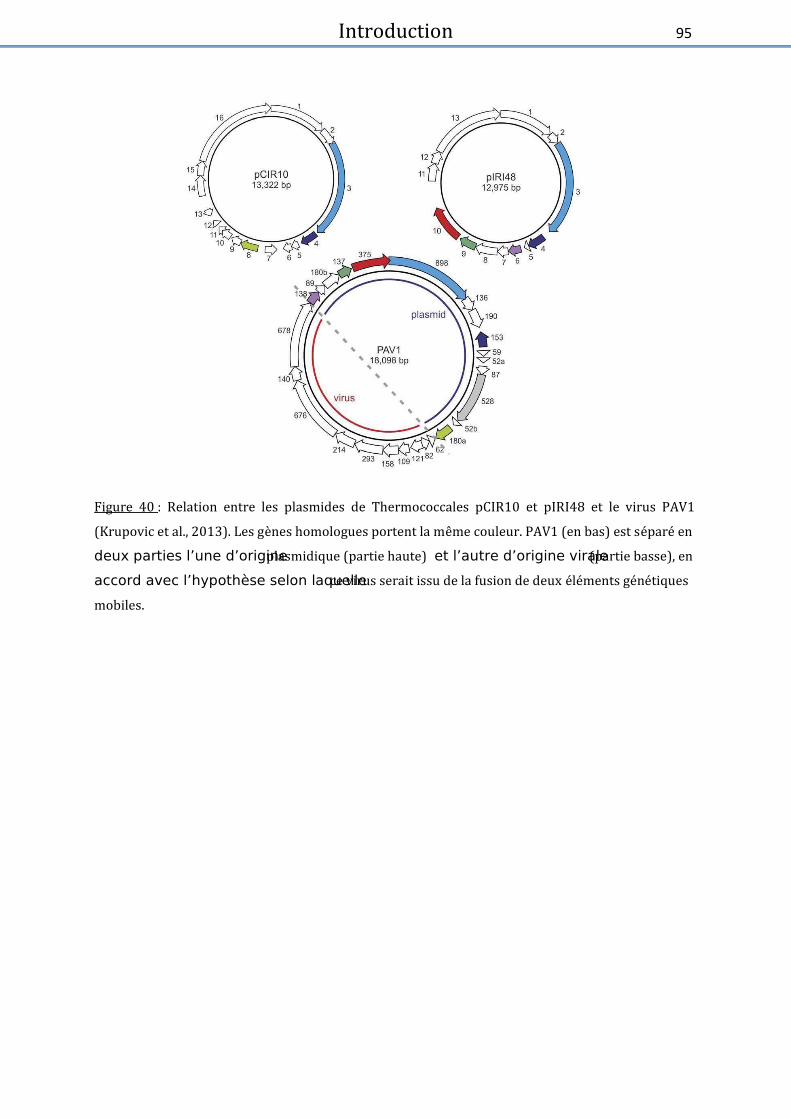
Introduction
95
Figure 40 : Relation entre les plasmides de Thermococcales pCIR10 et pIRI48 et le virus PAV1
(Krupovic et al., 2013). Les ge�nes homologues portent la me;me couleur. PAV1 (en bas) est se�pare� en
deux parties l’une d’origine plasmidique (partie haute) et l’autre d’origine virale (partie basse), en
accord avec l’hypothèse selon laquelle ce virus serait issu de la fusion de deux e� le�ments ge�ne� tiques
mobiles.

Introduction
96
IV. Bibliographie – Introduction
-A- Abrescia, N.G.A., Bamford, D.H., Grimes, J.M., and Stuart, D.I. (2012) Structure unifies the viral universe.
Annu. Rev. Biochem. 81: 795–822. Ackermann, H. (2009) Phage classification and caracterization. In, Clokie,M. and Kropinski,A. (eds),
Bacteriophages: Methods and protocols., pp. 127–140. Ackermann, H.W. (2007) 5500 Phages examined in the electron microscope. Arch. Virol. 152: 227–243. Ackermann, H.W. and Prangishvili, D. (2012) Prokaryote viruses studied by electron microscopy. Arch. Virol.
157: 1843–1849. Alcolombri, U., Ben-Dor, S., Feldmesser, E., Levin, Y., Tawfik, D.S., and Vardi, A. (2015) Identification of the
algal dimethyl sulfide–releasing enzyme: A missing link in the marine sulfur cycle. Science (80-. ). 348: 1466–1469.
Ankrah, N.Y.D., May, A.L., Middleton, J.L., Jones, D.R., Hadden, M.K., Gooding, J.R., et al. (2014) Phage infection of an environmentally relevant marine bacterium alters host metabolism and lysate composition. ISME J. 8: 1089–1100.
-B- Barrangou, R. and van der Oost, J. (2014) Bacteriophage exclusion, a new defense system. EMBO J.
e201490620. Basta, T., Garrett, R.A., and Prangishvili, D. (2009) Viruses of the Archaea. Encycl. Life Sci. Bawden, F.C. (1951) The multiplication of viruses. Sci. Prog. 39: 1–12. Berdygulova, Z., Westblade, L.F., Florens, L., Koonin, E. V, Chait, B.T., Ramanculov, E., et al. (2011) Temporal
regulation of gene expression of the Thermus thermophilus bacteriophage P23-45. J. Mol. Biol. 405: 125–142.
Bergh, O., Børsheim, K.Y., Bratbak, G., and Heldal, M. (1989) High abundance of viruses found in aquatic environments. Nature 340: 467–8.
Bibi-Triki, S. (2014) Identification et caractérisation fonctionnelle et structurale du système toxine-antitoxine HicA3-HicB3 de Yersinia pestis.
Biller, S.J., Schubotz, F., Roggensack, S.E., Thompson, A.W., Summons, R.E., and Chisholm, S.W. (2014) Bacterial vesicles in marine ecosystems. Science (80-. ). 343: 183–186.
Blondal, T., Hjorleifsdottir, S., Aevarsson, A., Fridjonsson, O.H., Skirnisdottir, S., Wheat, J.O., et al. (2005) Chaate izatio of a ′-Pol u leotide Kiase/ ′-Phosphatase from Bacteriophage RM378. J. Biol. Chem. 280: 5188–5194.
Blondal, T., Hjorleifsdottir, S.H., Fridjonsson, O.F., Ævarsson, A., Skirnisdottir, S., Hermannsdottir, A.G., et al. (2003) Discovery and characterization of a thermostable bacteriophage RNA ligase homologous to T4 RNA ligase 1. Nucleic Acids Res. 31: 7247–7254.
Blondal, T., Thorisdottir, A., Unnsteinsdottir, U., Hjorleifsdottir, S., Ævarsson, A., Ernstsson, S., et al. (2005) Isolation and characterization of a thermostable RNA ligase 1 from a Thermus scotoductus bacteriophage TS2126 with good single-stranded DNA ligation properties. Nucleic Acids Res. 33: 135–142.
Bonch-Osmolovskaya, E. (2008) Thermotogales. In, eLS. John Wiley & Sons, Ltd. Breitbart, M. (2012) Marine Viruses: Truth or Dare. Ann. Rev. Mar. Sci. 4: 425–448. Breitbart, M. and Rohwer, F. (2005) Here a virus, there a virus, everywhere the same virus? Trends
Microbiol. 13: 278–284. Breitbart, M., Thompson, L.R., Suttle, C.A., and Sullivan, M.B. (2007) Exploring the Vast Diversity of Marine
Viruses. Oceanography 135–139.
Introduction
97
Brum, J.R., Ignacio-Espinoza, J.C., Roux, S., Doulcier, G., Acinas, S.G., Alberti, A., et al. (2015) Patterns and ecological drivers of ocean viral communities. Science (80-. ). 348:.
Brum, J.R., Schenck, R.O., and Sullivan, M.B. (2013) Global morphological analysis of marine viruses shows minimal regional variation and dominance of non-tailed viruses. ISME J. 7: 1738–51.
Brum, J.R. and Sullivan, M.B. (2015) Rising to the challenge: accelerated pace of discovery transforms marine virology. Nat. Rev. Microbiol. 13: 147–159.
Brumfield, S.K., Ortmann, A.C., Ruigrok, V., Suci, P., Douglas, T., and Young, M.J. (2009) Particle assembly and ultrastructural features associated with replication of the lytic archaeal virus Sulfolobus turreted icosahedral virus. J. Virol. 83: 5964–5970.
Brüssow, H., Canchaya, C., and Hardt, W.-D. (2004) Phages and the evolution of bacterial pathogens: from genomic rearrangements to lysogenic conversion. Microbiol. Mol. Biol. Rev. 68: 560–602.
Burnet, F.M. (1945) Virus as Organism. Evolutionary and Ecological Aspects of some Human Virus Diseases.
Virus as Org. Evol. Ecol. Asp. some Hum. Virus Dis. -C-
Chien, A., Edgar, D.B., and Trela, J.M. (1976) Deoxyribonucleic acid polymerase from the extreme thermophile Thermus aquaticus. J. Bacteriol. 127: 1550–1557.
Chopin, M.-C., Chopin, A., and Bidnenko, E. (2005) Phage abortive infection in lactococci: variations on a theme. Curr. Opin. Microbiol. 8: 473–479.
Chow, C.-E.T. and Suttle, C.A. (2015) Biogeography of Viruses in the Sea. Annu. Rev. Virol. 2: 41–66. Christie, G.E. and Dokland, T. (2012) Pirates of the Caudovirales. Virology 434: 210–21. Colso , P., De La alle ie, X., Yuti, N., Asga i, “., Bigot, Y., Bideshi, D.K., et al. Megai ales , a
proposed new order for eukaryotic nucleocytoplasmic large DNA viruses. Arch. Virol. 158: 2517–21. Cortez, D., Forterre, P., and Gribaldo, S. (2009) A hidden reservoir of integrative elements is the major
source of recently acquired foreign genes and ORFans in archaeal and bacterial genomes. Genome Biol. 10: 1.
-D- Da o ao, R., Dell’A o, A., Co ialdesi, C., Magag ii, M., No le, R., Ta u i i, C., and Weinbauer, M.
(2008) Major viral impact on the functioning of benthic deep-sea ecosystems. Nature 454: 1084–1087. De Paepe, M., Leclerc, M., Tinsley, C.R., and Petit, M.-A. (2014) Bacteriophages: an underestimated role in
human and animal health? Front. Cell. Infect. Microbiol. 4: 39. Desbruyères, D., Almeida, A., Biscoito, M., Comtet, T., Khripounoff, A., Le Bris, N., et al. (2000) A review of
the distribution of hydrothermal vent communities along the northern Mid-Atlantic Ridge: dispersal vs. environmental controls. Hydrobiologia 440: 201–216.
Du k o th, D.H. Who Dis o e ed Bate iophage? . Bacteriol. Rev. 40: 739–802. -E-
Earthguide.image library, Coccolithophore – Emiliania huxleyi. Eichinger, I., Schmitz‐Esser, S., Schmid, M., Fisher, C.R., and Bright, M. (2014) Symbiont‐driven sulfur crystal
formation in a thiotrophic symbiosis from deep ‐sea hydrocarbon seeps. Environ. Microbiol. Rep. 6: 364–372.
Ellen, A.F., Albers, S.-V., Huibers, W., Pitcher, A., Hobel, C.F. V, Schwarz, H., et al. (2009) Proteomic analysis of secreted membrane vesicles of archaeal Sulfolobus species reveals the presence of endosome sorting complex components. Extremophiles 13: 67–79.
Emiliania huxleyi | Collection | Expedition Arctic.

Introduction
98
-F- Fineran, P.C., Blower, T.R., Foulds, I.J., Humphreys, D.P., Lilley, K.S., and Salmond, G.P.C. (2009) The phage
abortive infection system, ToxIN, functions as a protein –RNA toxin–antitoxin pair. Proc. Natl. Acad. Sci. 106: 894–899.
Fischer, M.G. (2016) Giant viruses come of age. Curr. Opin. Microbiol. 31: 50–57. Fischer, M.G. and Suttle, C.A. (2011) A Virophage at the Origin of Large DNA Transposons. Science. 332. Flores, G.E., Campbell, J.H., Kirshtein, J.D., Meneghin, J., Podar, M., Steinberg, J.I., et al. (2011) Microbial
community structure of hydrothermal deposits from geochemically different vent fields along the Mid-Atlantic Ridge. Environ. Microbiol. 13: 2158–2171.
Flores, G.E. and Reysenbach, A.-L. (2011) Hydrothermal environments, marine. In, Encyclopedia of Geobiology. Springer, pp. 456–467.
Forterre, P. (2010) Defining life: the virus viewpoint. Orig. Life Evol. Biosph. 40: 151–60. Forterre, P. (2013a) Les virus à nouveau sur le devant de la scène. Biol. Aujourdhui. 207: 153–168. Forterre, P. (2011) Manipulation of cellular syntheses and the nature of viruses: The virocell concept.
Comptes Rendus Chim. 14: 392–399. Fo te e, P. Mi o es de l’e fe Belin Pour. Regards, Paris. Forterre, P. (2013b) The virocell concept and environmental microbiology. ISME J. 7: 233–236. Forterre, P. (2016) To be or not to be alive: how recent discoveries challenge the traditional definitions of
viruses and life. Stud. Hist. Philos. Sci. Part C Stud. Hist. Philos. Biol. Biomed. Sci. Forterre, P., Soler, N., Krupovic, M., Marguet, E., and Ackermann, H.-W. (2013) Fake virus particles
generated by fluorescence microscopy. Trends Microbiol. 21: 1–5. -G-
Gaia, M., Benamar, S., Boughalmi, M., Pagnier, I., Croce, O., Colson, P., et al. (2014) Zamilon, a Novel Virophage with Mimiviridae Host Specificity. PLoS One 9: e94923.
Gaudin, M. (2012) Etude des vésicules membranaires produites par les Archées hyperthermophiles marines de l’ordre des Thermococcales.
Gaudin, M., Gauliard, E., Schouten, S., Houel-Renault, L., Lenormand, P., Marguet, E., and Forterre, P. (2013) Hyperthermophilic archaea produce membrane vesicles that can transfer DNA. Environ. Microbiol. Rep. 5: 109–16.
Gaudi, M., K upo i, M., Ma guet, E., Gauliad, E., C ikaite‐K upo i, V., Le Ca, E., et al. Extracellular membrane vesicles harbouring viral genomes. Environ. Microbiol. 16: 1167–1175.
Geslin, C., Gaillard, M., Flament, D., Rouault, K., Le Romancer, M., Prieur, D., and Erauso, G. (2007) Analysis of the first genome of a hyperthermophilic marine virus-like particle, PAV1, isolated from Pyrococcus abyssi. J. Bacteriol. 189: 4510–4519.
Geslin, C., Le Romancer, M., Erauso, G., Gaillard, M., Perrot, G., and Prieur, D. (2003) PAV1, the first virus-like pa tile isolated f o a h pe the ophili eu a haeote, P o o us a ssi. J Bacteriol 185: 3888–3894.
Geslin, C., Le Romancer, M., Gaillard, M., Erauso, G., and Prieur, D. (2003) Observation of virus-like particles in high temperature enrichment cultures from deep-sea hydrothermal vents. Res. Microbiol. 154: 303–307.
Geslin, C., Le Romancer, M., Gaillard, M., and Prieur, D. (2005) Diversité virale associée aux écosystèmes hydrothermaux océaniques profonds et aux sources chaudes terrestres. Virologie 9: 357–366.
Glansdorff, N. (2009) Extremophiles - volume 1 UNESCO. Gerday,C. and Glansdorff,N. (eds) Eolss, Oxford. Goldfarb, T., Sberro, H., Wei sto k, E., Cohe , O., Do o , “., Cha pak‐Aika, Y., et al. BREX is a
novel phage resistance system widespread in microbial genomes. EMBO J. 34: 169–183. Gorlas, A., Koonin, E. V, Bienvenu, N., Prieur, D., and Geslin, C. (2012) TPV1, the first virus isolated from the
hyperthermophilic genus Thermococcus. Environ. Microbiol. 14: 503–516.

Introduction
99
Gorlas, A., Marguet, E., Gill, S., Geslin, C., Guigner, J.M., Guyot, F., and Forterre, P. (2015) Sulfur vesicles from Thermococcales: A possible role in sulfur detoxifying mechanisms. Biochimie 118: 356–364.
Gupta, R.S. and Bhandari, V. (2011) Phylogeny and molecular signatures for the phylum Thermotogae and its subgroups. Antonie Van Leeuwenhoek 100: 1–34.
-H- Happonen, L.J., Redder, P., Peng, X., Reigstad, L.J., Prangishvili, D., and Butcher, S.J. (2010) Familial
relationships in hyperthermo-and acidophilic archaeal viruses. J. Virol. 84: 4747–4754. Häring, M., Rachel, R., Peng, X., Garrett, R.A., and Prangishvili, D. (2005) Viral Diversity in Hot Springs of
Pozzuoli, Italy, and Characterization of a Unique Archaeal Virus, Acidianus Bottle-Shaped Virus, from a New Family, the Ampullaviridae. J. Virol. 79: 9904–9911.
Hayes, F. and Van Melderen, L. (2011) Toxins-antitoxins: diversity, evolution and function. Crit. Rev. Biochem. Mol. Biol. 46: 386–408.
Hjorleifsdottir, S., Aevarsson, A., Hreggvidsson, G.O., Fridjonsson, O.H., and Kristjansson, J.K. (2014) Isolation, growth and genome of the Rhodothermus RM378 thermophilic bacteriophage. Extremophiles 18: 261–270.
Hjörleifsdottir, S., Hreggvidsson, G.O., Fridjonsson, O.H., Aevarsson, A., and Kristjansson, J.K. (2002) Bacteriophage RM 378 of a thermophilic host organism.
Holligan, P.M., Fernández, E., Aiken, J., Balch, W.M., Boyd, P., Burkill, P.H., et al. (1993) A biogeochemical study of the coccolithophore, Emiliania huxleyi, in the North Atlantic. Global Biogeochem. Cycles 7: 879–900.
Horvath, P. and Barrangou, R. (2010) CRISPR/Cas, the immune system of bacteria and archaea. Science (80-. ). 327: 167–170.
van Houte, S., Buckling, A., and Westra, E.R. (2016) Evolutionary Ecology of Prokaryotic Immune Mechanisms. Microbiol. Mol. Biol. Rev. 80: 745–763.
Huber, R. and Hannig, M. (2006) Thermotogales. In, Dworkin,M., Falkow,S., Rosenberg,E., Schleifer,K.-H., and Stackebrandt,E. (eds), The Prokaryotes: Volume 7: Proteobacteria: Delta, Epsilon Subclass. Springer New York, New York, NY, pp. 899–922.
Huber, R., Woese, C.R., Langworthy, T.A., Kristjansson, J.K., and Stetter, K.O. (1990) Fervidobacterium isla diu sp. o ., a e e t e el the ophili eu ate iu elo gi g to the The otogales. Arch. Microbiol. 154: 105–111.
Hurst, C.J. (2011) An introduction to viral taxonomy with emphasis on microbial and botanical hosts and the proposal of Akamara, a potential domain for the genomic acellular agents. Stud. viral Ecol. Microb. Bot. host Syst. 1: 41–65.
Hurwitz, B.L. and Sullivan, M.B. (2013) The Pacific Ocean Virome (POV): A Marine Viral Metagenomic Dataset and Associated Protein Clusters for Quantitative Viral Ecology. PLoS One 8:.
-J- Jaatinen, S.T., Happonen, L.J., Laurinmäki, P., Butcher, S.J., and Bamford, D.H. (2008) Biochemical and
structural characterisation of membrane-containing icosahedral dsDNA bacteriophages infecting thermophilic Thermus thermophilus. Virology 379: 10–19.
Jalasvuori, M., Pawlowski, A., and Bamford, J.K.H. (2010) A unique group of virus-related, genome-integrating elements found solely in the bacterial family Thermaceae and the archaeal family Halobacteriaceae. J. Bacteriol. 192: 3231–3234.
Janekovic, D., Wunderl, S., Holz, I., Zillig, W., Gierl, A., and Neumann, H. (1983) TTV1, TTV2 and TTV3, a family of viruses of the extremely thermophilic, anaerobic, sulfur reducing archaebacterium Thermoproteus tenax. Mol. Gen. Genet. MGG 192: 39–45.
Jebbar, M., Franzetti, B., Girard, E., and Oger, P. (2015) Microbial diversity and adaptation to high hydrostatic pressure in deep-sea hydrothermal vents prokaryotes. Extremophiles 19: 721–740.

Introduction
100
Jiang, S.C. and Paul, J.H. (1998) Gene transfer by transduction in the marine environment. Appl. Environ. Microbiol. 64: 2780–2787.
Jin, M., Chen, Y., Xu, C., and Zhang, X. (2014) The effect of inhibition of host MreB on the infection of thermophilic phage GVE2 in high temperature environment. Sci. Rep. 4:.
Jin, M., Xu, C., and Zhang, X. (2015) The effect of tryptophol on the bacteriophage infection in high-temperature environment. Appl. Microbiol. Biotechnol. 99: 8101–8111. Jin, M., Ye, T., and Zhang, X. (2013) Roles of bacteriophage GVE2 endolysin in host lysis at high
temperatures. Microbiology 159: 1597–1605. -K-
Kelley, D.S., Baross, J.A., and Delaney, J.R. (2002) Volcanoes, fluids, and life at mid-ocean ridge spreading centers. Annu. Rev. Earth Planet. Sci. 30: 385–491.
Klemperer, O. (1940) Electron microscopes of high magnification. Reports Prog. Phys. 7: 107. Knowles, B., Silveira, C.B., Bailey, B.A., Barott, K., Cantu, V.A., Cobián-Güemes, A.G., et al. (2016) Lytic to
temperate switching of viral communities. Nature 531: 466–470. Krupovic, M., Gonnet, M., Hania, W. Ben, Forterre, P., and Erauso, G. (2013) Insights into dynamics of mobile
genetic elements in hyperthermophilic environments from five new Thermococcus plasmids. PLoS One 8: e49044.
Krupovic, M., Quemin, E.R.J., Bamford, D.H., Forterre, P., and Prangishvili, D. (2014) Unification of the globally distributed spindle-shaped viruses of the Archaea. J. Virol. 88: 2354–2358.
Kulp, A. and Kuehn, M.J. (2010) Biological Functions and Biogenesis of Secreted Bacterial Outer Membrane Vesicles. Annu. Rev. Microbiol. 163–184.
-L- Lang, A.S., Zhaxybayeva, O., and Beatty, J.T. (2012) Gene transfer agents: phage-like elements of genetic
exchange. Nat. Rev. Microbiol. 10: 472. La Scola, B., Audic, S., Robert, C., Jungang, L., Lamballerie, X. de, Drancourt, M., et al. (2003) A Giant Virus
in Amoebae. Science (80-. ). 299: 2033. La Scola, B., Desnues, C., Pagnier, I., Robert, C., Barrassi, L., Fournous, G., et al. (2008) The virophage as a
unique parasite of the giant mimivirus. Nature 455: 100–104. Lindell, D., Sullivan, M.B., Johnson, Z.I., Tolonen, A.C., Rohwer, F., and Chisholm, S.W. (2004) Transfer of
photosynthesis genes to and from Prochlorococcus viruses. Proc. Natl. Acad. Sci. U. S. A. 101: 11013–11018.
Liu, B., Wu, S., Song, Q., Zhang, X., and Xie, L. (2006) Two Novel Bacteriophages of Thermophilic Bacteria Isolated from Deep-Sea Hydrothermal Fields. Curr. Microbiol. 53: 163–166.
Liu, B., Wu, S., and Xie, L. (2010) Complete genome sequence and proteomic analysis of a thermophilic bacteriophage BV1. Acta Oceanol. Sin. 29: 84–89.
Liu, B. and Zhang, X. (2008) Deep-sea thermophilic Geobacillus bacteriophage GVE2 transcriptional profile and proteomic characterization of virions. Appl. Microbiol. Biotechnol. 80: 697–707.
Liu, B., Zhou, F., Wu, S., Xu, Y., and Zhang, X. (2009) Genomic and proteomic characterization of a thermophilic Geobacillus bacteriophage GBSV1. Res. Microbiol. 160: 166–71.
López-García, P. (2012) The place of viruses in biology in light of the metabolism-versus-replication-first debate. Hist. Philos. Life Sci 34: 391–406.
López-García, P. and Moreira, D. (2012) Viruses in Biology. Evol. Educ. Outreach 5: 389–398. López-López, O., Cerdán, E.M., and González-Siso, I.M. (2013) Hot Spring Metagenomics. Life 3: 308–320. Lossouarn, J., Dupont, S., Gorlas, A., Mercier, C., Bienvenu, N., Marguet, E., et al. (2015) An abyssal
mobilome: Viruses, plasmids and vesicles from deep-sea hydrothermal vents. Res. Microbiol. 166: 742–752.

Introduction
101
Lossouarn, J., Nesbø, C.L., Mercier, C., )ha ae a, O., Joh so , M.“., Cha hu k, R., et al. M age à t ois : a selfish ge eti ele e t uses a i us to p opagate ithi The otogales. Environ. Microbiol. 17: 3278–3288.
Lwoff, A. (1957) The concept of virus. Microbiology 17: 239–253. -M-
Madiga, M. ad Ma tiko, J. L’esse tiel de la i ologie. I, F a e,P.E. ed , Brock Biologie des micro-organismes. Pearson Education France, pp. 232–256.
Makarova, K.S., Wolf, Y.I., Alkhnbashi, O.S., Costa, F., Shah, S.A., Saunders, S.J., et al. (2015) An updated evolutionary classification of CRISPR-Cas systems. Nat. Rev. Microbiol. 13: 722–736.
Makarova, K.S., Wolf, Y.I., and Koonin, E. V (2013) Comparative genomics of defense systems in archaea and bacteria. Nucleic Acids Res. gkt157.
Makarova, K.S., Wolf, Y.I., Van der Oost, J., and Koonin, E. V (2009) Prokaryotic homologs of Argonaute proteins are predicted to function as key components of a novel system of defense against mobile genetic elements. Biol. Direct 4: 1.
Makarova, K.S., Wolf, Y.I., Snir, S., and Koonin, E. V (2011) Defense islands in bacterial and archaeal genomes and prediction of novel defense systems. J. Bacteriol. 193: 6039–6056.
Mann, N.H., Cook, A., Millard, A., Bailey, S., and Clokie, M. (2003) Marine ecosystems: bacterial photosynthesis genes in a virus. Nature 424: 741.
Manning, A.J. and Kuehn, M.J. (2011) Contribution of bacterial outer membrane vesicles to innate bacterial defense. BMC Microbiol. 11: 258.
Martin, A., Yeats, S., Janekovic, D., Reiter, W.-D., Aicher, W., and Zillig, W. (1984) SAV 1, a temperate uv-inducible DNA virus-like particle from the archaebacterium Sulfolobus acidocaldarius isolate B12. EMBO J. 3: 2165.
Matsushita, I. and Yanase, H. (2009) The genomic structure of thermus bacteriophage {phi}IN93. J. Biochem. 146: 775–85.
McDaniel, L.D., Young, E., Delaney, J., Ruhnau, F., Ritchie, K.B., and Paul, J.H. (2010) High frequency of horizontal gene transfer in the oceans. Science (80-. ). 330: 50.
Mehta, D. and Satyanarayana, T. (2013) Diversity of Hot Environments and Thermophilic Microbes. In, Thermophilic Microbes in Environmental and Industrial Biotechnology. Springer Netherlands, Dordrecht, pp. 3–60.
Menon, S.K., Maaty, W.S., Corn, G.J., Kwok, S.C., Eilers, B.J., Kraft, P., et al. (2008) Cysteine usage in Sulfolobus spindle-shaped virus 1 and extension to hyperthermophilic viruses in general. Virology 376: 270–278.
Minakhin, L., Goel, M., Berdygulova, Z., Ramanculov, E., Florens, L., Glazko, G., et al. (2008) Genome comparison and proteomic characterization of Thermus thermophilus bacteriophages P23-45 and P74-26: siphoviruses with triplex-forming sequences and the longest known tails. J. Mol. Biol. 378: 468–480.
Miroshnichenko, M.L. (2004) Thermophilic microbial communities of deep-sea hydrothermal vents. Microbiology 73: 1–13.
-N- Nadal, M., Mirambeau, G., Forterre, P., Reiter, W.-D., and Duguet, M. (1986) Positively supercoiled DNA in
a virus-like particle of an archaebacterium. Nature 321: 256–258. Nagayoshi, Y., Kumagae, K., Mori, K., Tashiro, K., Nakamura, A., Fujino, Y., et al. (2016) Physiological
P ope ties ad Ge oe “t u tu e of the H pe theophili Filae tous Phage φOH Whih Ife ts Thermus thermophilus HB8. Front. Microbiol. 7: 50.
Introduction
102
Naryshkina, T., Liu, J., Florens, L., Swanson, S.K., Pavlov, A.R., Pavlova, N. V, et al. (2006) Thermus the ophilus Bate iophage ϕY“ Ge o e ad P oteo i Chaate izatio of Viio s. J. Mol. Biol. 364: 667–677.
Novick, R.P., Christie, G.E., and Penadés, J.R. (2010) The phage-related chromosomal islands of Gram-positive bacteria. Nat. Rev. Microbiol. 8: 541–551.
Nunes-Al es, C. Bate ial ph siolog : Puttig the BREX o phage epliatio . Nat. Rev. Microbiol. 13: 129.
-P- Paez-Espino, D., Eloe-Fadrosh, E.A., Pavlopoulos, G.A., Thomas, A.D., Huntemann, M., Mikhailova, N., et al.
U o e i g Ea th’s i o e. Nature advance on: Palm, P., Schleper, C., Grampp, B., Yeats, S., McWilliam, P., Reiter, W.-D., and Zillig, W. (1991) Complete
nucleotide sequence of the virus SSV1 of the archaebacterium Sulfolobus shibatae. Virology 185: 242–250.
Paul, J.H. (2008) Prophages in marine bacteria: dangerous molecular time bombs or the key to survival in the seas? ISME J. 2: 579–589.
Pawlowski, A., Rissanen, I., Bamford, J.K.H., Krupovic, M., and Jalasvuori, M. (2014) Gammasphaerolipovirus, a newly proposed bacteriophage genus, unifies viruses of halophilic archaea and thermophilic bacteria within the novel family Sphaerolipoviridae. Arch. Virol. 159: 1541–1554.
Penadés, J.R., Chen, J., Quiles-Puchalt, N., Carpena, N., and Novick, R.P. (2015) Bacteriophage-mediated spread of bacterial virulence genes. Curr. Opin. Microbiol. 23: 171–178.
Peng, X., Basta, T., Häring, M., Garrett, R.A., and Prangishvili, D. (2007) Genome of the Acidianus bottle-shaped virus and insights into the replication and packaging mechanisms. Virology 364: 237–243.
Personnic, S., Duhamel, S., Bettarel, Y., Sime-Ngando, T., and Jacquet, S. (2006) Les virus planctoniques : u compartiment biologique clé des milieux aquatiques. Courr. l’e viro e e t l’INRA 19–34.
Pflugfelder, B., Fisher, C.R., and Bright, M. (2005) The color of the trophosome: elemental sulfur distribution in the endosymbionts of Riftia pachyptila (Vestimentifera; Siboglinidae). Mar. Biol. 146: 895–901.
Pietilä, M.K., Demina, T.A., Atanasova, N.S., Oksanen, H.M., and Bamford, D.H. (2014) Archaeal viruses and bacteriophages: comparisons and contrasts. Trends Microbiol. 22: 334–344.
Pina, M., Bize, A., Forterre, P., and Prangishvili, D. (2011) The archeoviruses. FEMS Microbiol. Rev. 35: 1035–54.
Pollo, S.M.J., Zhaxybayeva, O., and Nesbø, C.L. (2015) Insights into thermoadaptation and the evolution of mesophily from the bacterial phylum Thermotogae. Can. J. Microbiol. 1–16.
Prangishvili, D., Arnold, H.P., Götz, D., Ziese, U., Holz, I., Kristjansson, J.K., and Zillig, W. (1999) A novel virus family, the Rudiviridae: structure, virus-host interactions and genome variability of the Sulfolobus viruses SIRV1 and SIRV2. Genetics 152: 1387–1396.
Prangishvili, D., Basta, T., Garrett, R.A., and Krupovic, M. (2016) Viruses of the Archaea. In, Encyclopedia of Life Sciences. John Wiley & Sons, Ltd.
Prangishvili, D., Holz, I., Stieger, E., Nickell, S., Kristjansson, J.K., and Zillig, W. (2000) Sulfolobicins, specific proteinaceous toxins produced by strains of the extremely thermophilic archaeal genus Sulfolobus. J. Bacteriol. 182: 2985–8.
Prangishvili, D., Stedman, K., and Zillig, W. (2001) Viruses of the extremely thermophilic archaeon Sulfolobus. Trends Microbiol. 9: 39–43.
P ieu , D. Les at ies de l’e t e, des asses à l’espa e de oek. De Boek “upieu s.a. Prieur, D. (1997) Microbiology of deep-sea hydrothermal vents. Trends Biotechnol. 15: 242–244. Prieur, D. (1977) Microbiology of deep-sea hydrothermal vents. Mar. Biotechnol. 7799: 1–3.

Introduction
103
-Q,R- Quemin, E.R.J., Pietilä, M.K., Oksanen, H.M., Forterre, P., Rijpstra, W.I.C., Schouten, S., et al. (2015)
Sulfolobus Spindle-Shaped Virus 1 Contains Glycosylated Capsid Proteins, a Cellular Chromatin Protein, and Host-Derived Lipids. J. Virol. 89: 11681–11691.
Rachel, R., Bettstetter, M., Hedlund, B.P., Häring, M., Kessler, a, Stetter, K.O., and Prangishvili, D. (2002) Remarkable morphological diversity of viruses and virus-like particles in hot terrestrial environments. Arch. Virol. 147: 2419–29.
Raoult, D., Audic, S., Robert, C., Abergel, C., Renesto, P., Ogata, H., et al. (2004) The 1.2-Megabase Genome Sequence of Mimivirus. Science. 306: 1344–1350.
Raoult, D. and Forterre, P. (2008) Redefining viruses: lessons from Mimivirus. Nat. Rev. Microbiol. 6: 315 –319.
Reddy, T.B.K., Thomas, A.D., Stamatis, D., Bertsch, J., Isbandi, M., Jansson, J., et al. (2014) The Genomes OnLine Database (GOLD) v.5: a metadata management system based on a four level (meta)genome project classification. Nucleic Acids Res.
Reiter, W.-D., Palm, P., Yeats, S., and Zillig, W. (1987) Gene expression in archaebacteria: physical mapping of constitutive and UV-inducible transcripts from the Sulfolobus virus-like particle SSV1. Mol. Gen. Genet. MGG 209: 270–275.
Rensen, E., Krupovic, M., and Prangishvili, D. (2015) Mysterious hexagonal pyramids on the surface of Pyrobaculum cells. Biochimie 118: 365–367.
Rensen, E.I., Mochizuki, T., Quemin, E., Schouten, S., Krupovic, M., and Prangishvili, D. (2016) A virus of hyperthermophilic archaea with a unique architecture among DNA viruses. Proc. Natl. Acad. Sci. 113: 2478–2483.
Rice, G., Tang, L., Stedman, K., Roberto, F., Spuhler, J., Gillitzer, E., et al. (2004) The structure of a thermophilic archaeal virus shows a double-stranded DNA viral capsid type that spans all domains of life. Proc. Natl. Acad. Sci. U. S. A. 101: 7716–7720.
Rissanen, I., Grimes, J.M., Pawlowski, A., Mäntynen, S., Harlos, K., Bamford, J.K.H., and Stuart, D.I. (2013) Bacteriophage P23-77 capsid protein structures reveal the archetype of an ancient branch from a major virus lineage. Structure 21: 718–726.
Rissanen, I., Pawlowski, A., Harlos, K., Grimes, J.M., Stuart, D.I., and Bamford, J.K.H. (2012) Crystallization and preliminary crystallographic analysis of the major capsid proteins VP16 and VP17 of bacteriophage P23-77. Acta Crystallogr. Sect. F Struct. Biol. Cryst. Commun. 68: 580–583.
Rohwer, F. and Thurber, R.V. (2009) Viruses manipulate the marine environment. Nature 459: 207–12. Rosenwasser, S., Ziv, C., Creveld, S.G. van, and Vardi, A. (2016) Virocell Metabolism: Metabolic Innovations
During Host-Virus Interactions in the Ocean. Trends Microbiol. -S-
Sakai, H.D. and Kurosawa, N. (2016) Exploration and isolation of novel thermophiles in frozen enrichment cultures derived from a terrestrial acidic hot spring. Extremophiles 20: 207–214.
Sakaki, Y. and Oshima, T. (1975) Isolation and characterization of a bacteriophage infectious to an extreme thermophile, Thermus thermophilus HB8. J. Virol. 15: 1449–1453.
Salmond, G.P.C. and Fineran, P.C. (2015) A century of the phage: past, present and future. Nat. Rev. Microbiol. 13: 777–786.
Samson, J.E., Magadán, A.H., Sabri, M., and Moineau, S. (2013) Revenge of the phages: defeating bacterial defences. Nat. Rev. Microbiol. 11: 675–687.
Scanlan, D. (2014) Bacterial vesicles in the ocean. Science (80-. ). 343: 143–144. Schoenfeld, T., Patterson, M., Richardson, P.M., Wommack, K.E., Young, M., and Mead, D. (2008) Assembly
of Viral Metagenomes from Yellowstone Hot Springs. Appl. Environ. Microbiol. 74: 4164–4174.

Introduction
104
Segerer, A., Neuner, A., Kristjansson, J.K., and Stetter, K.O. (1986) Acidianus infernus gen. nov., sp. nov., and Acidianus brierleyi comb. nov.: facultatively aerobic, extremely acidophilic thermophilic sulfur-metabolizing archaebacteria. Int. J. Syst. Evol. Microbiol. 36: 559–564.
Servín-Garcidueñas, L.E., Peng, X., Garrett, R.A., and Martínez-Romero, E. (2013) Genome sequence of a novel archaeal rudivirus recovered from a mexican hot spring. Genome Announc. 1: e00040-12.
Shelford, E.J., Middelboe, M., Møller, E.F., and Suttle, C.A. (2012) Virus-driven nitrogen cycling enhances phytoplankton growth. Aquat. Microb. Ecol. 66: 41–46.
Shikuma, N.J., Pilhofer, M., Weiss, G.L., Hadfield, M.G., Jensen, G.J., and Newman, D.K. (2014) Marine tubeworm metamorphosis induced by arrays of bacterial phage tail –like structures. Science (80-. ). 343: 529–533.
Sime-Ngando, T. (2014) Environmental bacteriophages: viruses of microbes in aquatic ecosystems. Front. Microbiol. 5: 355.
Sime-Ngando, T. and Colombet, J. (2009) Virus et prophages dans les écosystèmes aquatiques. Can. J. Microbiol. 55: 95–109.
Snyder, J.C., Brumfield, S.K., Peng, N., She, Q., and Young, M.J. (2011) Sulfolobus turreted icosahedral virus c92 protein responsible for the formation of pyramid-like cellular lysis structures. J. Virol. 85: 6287 –6292.
Soler, N., Gaudin, M., Marguet, E., and Forterre, P. (2011) Plasmids, viruses and virus-like membrane vesicles from Thermococcales. Biochem. Soc. Trans. 39: 36–44.
“ole , N., K upo i, M., Ma guet, E., ad Fo tee, P. Me ae esi les i atu al e i o e ts : a major challenge in viral ecology. ISME J. 9: 793–796.
Soler, N., Marguet, E., Verbavatz, J.M., and Forterre, P. (2008) Virus-like vesicles and extracellular DNA produced by hyperthermophilic archaea of the order Thermococcales. Res. Microbiol. 159: 390–399.
Song, Q., Ye, T., and Zhang, X. (2011) Proteins responsible for lysogeny of deep-sea thermophilic bacteriophage GVE2 at high temperature. Gene 479: 1–9.
Song, Q. and Zhang, X. (2008) Characterization of a novel non-specific nuclease from thermophilic bacteriophage GBSV1. BMC Biotechnol. 8: 1.
Sorek, R., Kunin, V., and Hugenholtz, P. (2008) CRISPR—a widespread system that provides acquired resistance against phages in bacteria and archaea. Nat. Rev. Microbiol. 6: 181–186.
Stark, E. and Tetrault, P.A. (1951) Isolation of bacterial, cell-free, starch saccharifying enzymes from the medium at 70 C. J. Bacteriol. 62: 247.
Stassen, A.P.M., Folmer, R.H.A., Hilbers, C.W., and Konings, R.N.H. (1994) Single-stranded DNA binding protein encoded by the filamentous bacteriophage M13: structural and functional characteristics. Mol. Biol. Rep. 20: 109–127.
Stedman, K.M., DeYoung, M., Saha, M., Sherman, M.B., and Morais, M.C. (2015) Structural insights into the architecture of the hyperthermophilic Fusellovirus SSV1. Virology 474: 105–109.
Sullivan, M.B., Lindell, D., Lee, J.A., Thompson, L.R., Bielawski, J.P., and Chisholm, S.W. (2006) Prevalence and evolution of core photosystem II genes in marine cyanobacterial viruses and their hosts. PLoS Biol. 4: e234.
Suttle, C.A. (2007) Marine viruses - major players in the global ecosystem. Nat. Rev. Microbiol. 5: 801–812. Suttle, C.A. (2005) Viruses in the sea. Nature 437: 356–361.
-T- Takai, K., Nakamura, K., Toki, T., Tsunogai, U., Miyazaki, M., Miyazaki, J., et al. (2008) Cell proliferation at
122°C and isotopically heavy CH4 production by a hyperthermophilic methanogen under high-pressure cultivation. Proc. Natl. Acad. Sci. 105: 10949–10954.
Introduction
105
Takami, H., Nishi, S., Lu, J., Shimamura, S., and Takaki, Y. (2004) Genomic characterization of thermophilic Geobacillus species isolated from the deepest sea mud of the Mariana Trench. Extremophiles 8: 351–356.
Tamakoshi, M., Murakami, A., Sugisawa, M., Tsuneizumi, K., Takeda, S., Saheki, T., et al. (2011) Genomic ad p oteo i ha ate izatio of the la ge Mo i idae a te iophage ϕTMA of the e t e e thermophile Thermus thermophilus. Bacteriophage 1: 152–164.
Tarasov, V.G., Gebruk, A. V, Mironov, A.N., and Moskalev, L.I. (2005) Deep-sea and shallow-water hydrothermal vent communities: Two different phenomena? Chem. Geol. 224: 5–39.
Thingstad, T.F. and Bratbak, G. (2016) Microbial oceanography: Viral strategies at sea. Nature. Thingstad, T.F. and Lignell, R. (1997) Theoretical models for the control of bacterial growth rate, abundance,
diversity and carbon demand. Aquat. Microb. Ecol. 13: 19–27. Thornburg, C.C., Zabriskie, T.M., and McPhail, K.L. (2010) Deep-Sea Hydrothermal Vents: Potential Hot Spots
for Natural Products Discovery?⊥ . J. Nat. Prod. 73: 489–499.
Tock, M.R. and Dryden, D.T.F. (2005) The biology of restriction and anti-restriction. Curr. Opin. Microbiol. 8: 466–472.
Torrella, F. and Morita, R.Y. (1979) Evidence by electron micrographs for a high incidence of bacteriophage particles in the waters of Yaquina Bay, oregon: ecological and taxonomical implications. Appl. Environ. Microbiol. 37: 774–778.
Torsvik, T. and Dundas, I.D. (1974) Bacteriophage of Halobacterium salinarium. Nature 248: 680–681. -U,V-
Uldahl, K. and Peng, X. (2013) Biology, biodiversity and application of thermophilic viruses. In, Thermophilic Microbes in Environmental and Industrial Biotechnology. Springer, pp. 271–304.
Uldahl, K.B. (2015) Hyperthermophilic archaeal viruses as novel nanoplatforms. Vestergaard, G., Häring, M., Peng, X., Rachel, R., Garrett, R.A., and Prangishvili, D. (2005) A novel rudivirus,
ARV1, of the hyperthermophilic archaeal genus Acidianus. Virology 336: 83–92. Vestergaard, G., Shah, S.A., Bize, A., Reitberger, W., Reuter, M., Phan, H., et al. (2008) SRV, a new rudiviral
isolate from Stygiolobus and the interplay of crenarchaeal rudiviruses with the host viral-defence CRISPR system. J. Bacteriol.
Von Damm, K.L., Oosting, S.E., Kozlowski, R., Buttermore, L.G., Colodner, D.C., Edmonds, H.N., et al. (1995) Evolution of East Pacific Rise hydrothermal vent fluids following a volcanic eruption. Nature 375: 47–50.
-W,X,Y,Z- Waldor, M.K. and Mekalanos, J.J. (1996) Lysogenic conversion by a filamentous phage encoding cholera
toxin. Science (80-. ). 272: 1910. Wang, Y. and Zhang, X. (2008a) Characterization of a novel portal protein from deep-sea thermophilic
bacteriophage GVE2. Gene 421: 61–66. Wang, Y. and Zhang, X. (2010) Genome Analysis of Deep-Sea Thermophilic Phage D6E. Appl. Environ.
Microbiol. 76: 7861–7866. Wang, Y. and Zhang, X. (2008b) Identification and characterization of a novel thymidylate synthase from
deep-sea thermophilic bacteriophage Geobacillus virus E2. Virus Genes 37: 218–224. Wei, D. and Zhang, X. (2008) Identification and characterization of a single-stranded DNA-binding protein
from thermophilic bacteriophage GVE2. Virus Genes 36: 273–278. Weinbauer, M.G. (2004) Ecology of prokaryotic viruses. FEMS Microbiol. Rev. 28: 127–181. Weinbauer, M.G., Chen, F., and Wilhelm, S.W. (2011) Virus-mediated redistribution and partitioning of
carbon in the global oceans. In, Microbial carbon pump in the ocean., pp. 54–56.
Introduction
106
Williamson, S.J., Cary, S.C., Williamson, K.E., Helton, R.R., Bench, S.R., Winget, D., and Wommack, K.E. (2008) Lysogenic virus host interactions predominate at deep-sea diffuse-flow hydrothermal vents. ISME J. 2: 1112–1121.
Windberger, E., Huber, R., Trincone, A., Fricke, H., and Stetter, K.O. (1989) Thermotoga thermarum sp. nov. and Thermotoga neapolitana occurring in African continental solfataric springs. Arch. Microbiol. 151: 506–512.
Winter, C., Bouvier, T., Weinbauer, M.G., and Thingstad, T.F. (2010) Trade-offs between competition and defe se spe ialists ao g u iellula plakto i o ga is s: the killi g the i e h pothesis revisited. Microbiol. Mol. Biol. Rev. 74: 42–57.
Woese, C.R. (1994) There must be a prokaryote so e he e: i o iolog ’s seah fo itself. Microbiol. Rev. 58: 1–9.
Woese, C.R., Kandler, O., and Wheelis, M.L. (1990) Towards a natural system of organisms: proposal for the domains Archaea, Bacteria, and Eucarya. Proc. Natl. Acad. Sci. 87: 4576–4579.
Wu, S., Liu, B., and Zhang, X. (2009) Identification of a tail assembly gene cluster from deep-sea thermophilic bacteriophage GVE2. Virus Genes 38: 507–514.
Xu, T., Yao, F., Zhou, X., Deng, Z., and You, D. (2010) A novel host-specific restriction system associated with DNA backbone S-modification in Salmonella. Nucleic Acids Res. 38: 7133–7141.
Ye, T. and Zhang, X. (2008) Characterization of a lysin from deep-sea thermophilic bacteriophage GVE2. Appl. Microbiol. Biotechnol. 78: 635–641.
Yoshida-Takashima, Y., Takaki, Y., Shimamura, S., Nunoura, T., and Takai, K. (2013) Genome sequence of a novel deep-sea vent epsilonproteobacterial phage provides new insight into the co-evolution of Epsilonproteobacteria and their phages. Extremophiles 17: 405–419.
Yu, M.X., Slater, M.R., and Ackermann, H.W. (2006) Isolation and characterization of Thermus bacteriophages. Arch. Virol. 151: 663–679.
Zhaxybayeva, O., Swithers, K.S., Lapierre, P., Fournier, G.P., Bickhart, D.M., DeBoy, R.T., et al. (2009) On the chimeric nature, thermophilic origin, and phylogenetic placement of the Thermotogales. Proc. Natl. Acad. Sci. U. S. A. 106: 5865–5870.


Objectifs des travaux de the�se

Objectifs
108
Nous savo s, aujou d’hui, ue les vius so t o ip se ts das tous les os st es,
compris les plus extrêmes, où ils représentent le plus grand réservoir de biodiversité non
caractérisée. Néanmoins, notre connaissance de la diversité virale associée aux bactéries et
archées issues des sources hydrothermales océaniques profondes demeure encore limitée.
Peu de do es so t dispo iles su l’ao da e viale ou su l’ipat de es vius su les
micro-organismes colonisant ces écosystèmes. En effet, seuls 8 virus infectant des archées et
at ies isoles de es e vio e e ts e t es o t t d its. Deu d’e t e eu so t
asso is à des a h es h pe the ophiles aa o ies appate at à l’o d e des
Thermococcales. Il s’agit de PAV et TPV , dits au LME. Les six autres virus sont associés
à des bactéries appartenant aux Bacillales, Epsilon-proteobacteria et Thermotogales. Une
revue présentant les éléments génétiques mobiles, virus, plasmides et vésicules, isolés de
sources hydrothermales océaniques vien t d’ t e pu li e : An abyssal mobilome: virus,
plasmid and vesicle from deep sea hydrothermal vents par J. Lossouarn, S. Dupont, A. Gorlas,
C. Mercier, N. Bienvenu, P. Forterre and C. Geslin in Research in microbiology. Celle-ci
constitue le premier chapitre du manuscrit.
Afi de pou suiv e l’effo t de aat isatio de ette dive sit viale au sei des soues
hydrothermales océaniques profondes, un criblage des bactéries du phylum des
Thermotogae avait t i iti au lao atoi e afi d’ide tifie de ouveaux éléments
génétiques mobiles. Cet ordre bactérien, composé de bactéries chimio-organotrophes,
anaérobies et majoritairement thermophiles, est largement représenté au niveau de la
biosphère profonde. Partageant la même niche écologique que les Thermococcales, les
Thermotogae ont une histoire évolutive complexe et particulière qui suggère la présence de

Objectifs
109
nombreux transferts latéraux de gènes. Ces transferts sont souvent dus à différents types
d’ l e ts g ti ues o iles.
D’aut e pat, la pse e de CRISPR et de p ovius a t ise e vide e das de o eu
génomes de Thermotogae. Malgré toutes ces preuves laissant penser à des infections virales
p se tes ou pass es, jusu’à e jou , u seul vius a t d it o e i fectant une
at ie de et o d e. C’est d’ailleu s das e o te te ue a p e i e a e de th se a
t l’o asio d’aheve la aat isatio de MPV (Marinitoga piezophila virus 1), premier
virus associé à une Thermotogae. La caractérisation de ce système très original, menée dans
le cadre de la thèse de J. Lossouarn, a ainsi pu être finalisée et publiée (Lossouarn J, Nesbø
CL, Mercier C, Zhaxybayeva O, Johnson MS, Charchuck R, Farasin J, Bienvenu N, Baudoux AC,
Michoud G, Jebbar M, and Geslin C. (2015) "Ménage à trois": a selfish genetic element uses a
virus to propagate within Thermotogales. In Environ Microbiol. doi: 10.1111/1462-
2920.12783 ). Il constitue le second chapitre de ce manuscrit.
Lors du criblage des Thermotogae, d’aut es vius avaie t té mis en évidence et notamment
deux virus infectant deux souches de Marinitoga camini. J’ai e u t avail de
caractérisation fonctionnelle et génomique de ces nouveaux virus (MCV1 et MCV2) qui fera
p o hai e e t l’o jet d’u e pu liatio . D’aut e pat, les génomes des deux souches hôtes
de es vius o t t s ue s faisa t l’o jet d’u « Genome announcement » (publication
acceptée). Ces travaux constituent le troisième chapitre du manuscrit.
E fi , lo s de l’tude de es t ois vius, u e sou he appateant au genre Thermosipho a
particulièrement attiré notre attention. En effet, pMP1 (plasmide porté par M. piezophila
KA3, hôte du virus MPV1) peut se propager au sein de cette souche de Thermosipho sp.
AT1244-VC14. Néanmoins, MCV1 et MCV2 ne sont pas capab les d’ife te ette sou he. Ces
pati ulait s o t e au s ue çage de ette sou he aisi u’à l’tude des CRISPR

Objectifs
110
présents dans son génome. Ces travau fero t l’o jet d’u e pu liatio de tpe « Genome
report » et sont présentés dans le quatrième chapitre de ce manuscrit.
E paall le, j’ai t asso ie à u p ojet desti à ett e e plae u p oto ole e vue de
p oduie u vio e de s di e ts ai s p ofo ds. E effet, il ’ a ue peu d’a alses de
métagénomique virale publiées sur des échantillons de sources hydrothermales océaniques
profondes. La plus complète, portant sur des échantillons de flux hydrothermaux diffus,
révèle que les virus hydrothermaux tempérés seraient moins diversifiés que ceux composant
la communauté virioplanctonique globale mais qu'ils présenteraient un très grand nombre
de gènes aux fonctions inconnues (Williamson et al., 2008). Dans ce contexte, le LM2E
souhaite analyser par une approche métagénomique des échantillons de sédiments
h d othe au ai s afi d’a de à u e diversité génomique de nouveaux virus
infectant des bactéries et archées. Les expériences débutées lors de ce projet, la validation
et la or alisatio d’u proto ole pour obtenir un virome (étude préliminaire), sont
pou suivis das le ad e d’u e th se alisée au LUBEM de Brest par Lisa Billard sous la
direction des Pr. C. Payan et Dr S. Vallet.

Chapitre I : Revue bibliographique
« An abyssal mobilome »

Chapitre I
112
Pre�sentation des travaux
Depuis leu dé ouve te e 1977, les soues hyd otheales p ofo des fo t l’o jet de
nombreuses études. Elles abritent une riche diversité de micro-organismes aux métabolismes
variés et au potentiel biotechnologique notable. Pourtant, les éléments génétiques mobiles
(EGMs) qui y sont associés restent encore peu décrits (Lossouarn, Dupont, et al., 2015).
Les EGMs regroupent les virus, les vésicules membranaires (MVs), les plasmides, les agents de
transferts de gènes (GTAs, gene transfer agents), les transposons et les transpovirions. Ils
ep ése te t u e foe ajeu e de l’évolutio des o u autés i o ie es ota e t
de part leur implication dans les transferts latéraux de gènes. Il se le aussi u’ils soie t
particulièrement abondants dans les environnements extrêmes comme les sources
hydrothermales profondes (Anderson et al., 2014).
L’o je tif de ette evue est de asse le les o aissa es a uises jusu’à p ése t su les
virus, vésicules membranaires, et plasmides associés aux bactéries et archées thermophiles et
hyperthermophiles colonisant les sources hydrothermales océaniques profondes.

An abyssal mobilome: viruses,plasmids and vesicles from deep-seahydrothermal vents
Julien Lossouarna, Samuel Duponta, Aurore Gorlasb, Coraline Merciera, Nadege Bienvenua,Evelyne Marguetb, Patrick Forterrec, Claire Geslina,*
a Laboratory of Microbiology of Extreme Environments (LMEE), UMR 6197/UBO/Ifremer/CNRS, IUEM, Place Nicolas Copernic, Technop^ole Brest Iroise, 29280,Plouzan!e, France
b Institute of Integrative Cellular Biology, CEA, CNRS, Universit!e Paris sud, 91405 Orsay, Francec Institut Pasteur, Laboratoire de Biologie Mol !eculaire du G "ene chez les Extr !emophiles, 25 Rue du Docteur Roux, Paris Cedex 15, France
Received 30 January 2015; accepted 9 April 2015Available online 22 April 2015
Abstract
Mobile genetic elements (MGEs) such as viruses,plasmids,vesicles,gene transfer agents (GTAs),transposons and transpovirions,whichcollectively represent the mobilome, interact with cellular organisms from all three domains of life, including those thriving in the most extremeenvironments. While efforts have been made to better understand deep-sea vent microbial ecology, our knowledge of the mobilome associatedwith prokaryotes inhabiting deep-sea hydrothermal vents remains limited. Here we focus on the abyssal mobilome by reviewing accumulatingdata on viruses,plasmids and vesicles associated with thermophilic and hyperthermophilic Bacteria and Archaea present in deep-sea hydro-thermal vents.© 2015 Institut Pasteur.Published by Elsevier Masson SAS.All rights reserved.
Keywords: Deep-sea hydrothermal vent; Bacteria; Archaea; (Hyper-)thermophiles; Mobilome
1. Introduction
Deep-sea hydrothermalventsrepresentone of the mostextreme environments on Earth.These ecosystems are char-acterized by steep physicochemical gradients, high hydrostaticpressure,high temperatures,obscurity and the prevalence ofchemosynthesis.These extreme environments are home to avastdiversity ofmesophilic and (hyper-)thermophilic pro-karyotesbelongingto the Bacteriaand Archaea[1e3].Althoughour knowledgeof deep-seahydrothermalvent
microbial communities is progressing, the impact of MGEs onmicrobial ecology and evolution remains largely overlooked inthese abyssal ecosystems [2]. MGEs such as viruses, plasmids,membrane vesicles,gene transfer agents (GTAs),transposonsand transpovirons, which collectively represent the mobilome,interact with cellular organisms from all three domains of life,including those thriving in extreme environments [4,5]. Manyreviews have highlighted how MGEs,and especially viruses,are powerfulagentsthataffectnot only the diversity andevolutionof microbial communities,but also globalbiochemical cycles in marine environments [6e12]. Evidencewas recently reviewed supporting the hypothesis thatMGEscould also play a key role in deep-sea hydrothermalvents,notably by facilitating horizontalgene transfer(HGT) [2].MGEs have potential as powerful drivers of cellular host ad-aptations to extreme marine environments [2]. Here we presentthe first review focused on deep-seahydrothermalMGEs
* Corresponding author.E-mail addresses: [email protected](J. Lossouarn),
[email protected] (S.Dupont),[email protected] (A.Gorlas), [email protected](C. Mercier),[email protected] (N. Bienvenu), [email protected](E.Marguet),[email protected](P. Forterre),[email protected] (C.Geslin).
Research in Microbiology 166 (2015) 742e752www.elsevier.com/locate/resmic
http://dx.doi.org/10.1016/j.resmic.2015.04.0010923-2508/© 2015 Institut Pasteur.Published by Elsevier Masson SAS.All rights reserved.

associated with (hyper-)thermophilic prokaryotes, collectivelydenoted as the abyssal mobilome.
2.Viruses in deep-sea hydrothermal vents
2.1. Evidence for viral activity
Only a few viral ecological studies have been performed ondeep-sea hydrothermalvents [13e17].Viral abundance andviral production were notably investigated in diffuse flowhydrothermalventfluids.In these samples,collected fromvents within the Endeavour Ridge system [13] and the EastPacific Rise [14],average VLP (virus-like particle)abun-dances were estimated at~107 VLPs per milliliter and were~10-fold higher than prokaryote abundances.In comparison,VLP abundances in productive coastal waters were estimatedat ~108 VLPs/mL and exceeded those of prokaryotes by ~15-fold [6,7].These VLPs might represent bona fide viralparti-cles (virions), but also membrane vesicles containing cellular,plasmid or viral DNA (viral membrane vesicles) (see section3).
Viralactivities occurring in these extreme ecosystems arealso highlighted by genome sequence analyses ofdeep-seahydrothermalBacteriaand Archaea[2]. The presenceofclustered,regularlyinterspacedshort palindromicrepeat(CRISPR)/CRISPR-associated (Cas)systemshas been re-ported in many thermophilic bacterialand archaealgenomes[18,19].These systemsprovideacquired,yet heritable,sequence-specific “adaptive” immunity againstvirusesandotherhorizontally acquired elements,such asconjugativeplasmids [19]. CRISPR loci consist of several non-contiguousdirectrepeatsseparated by stretchesof variable sequencescalled spacers,which correspond to fragments derived frominvading DNAs such as viruses and plasmids [18].CRISPRregions therefore actas a record of pastviralinfections thatoccurredin the history of the prokaryotes[2,19,20].
Interestingly,it was reportedthat thermophilicstrainsharbored a higher number of CRISPR lociin their genomesthan mesophilic and psychrophilic strains [2,21].This mayindicate that viral infections play a major role in the ecologyand evolution ofthermophilic communities,notably thoseinhabiting deep-sea hydrothermal vents [2].
A viral metagenomic study using CRISPRs, indicated that adiffuse flow sample collected from Hulk vent on the Juan deFuca ridge in the Pacific ocean contained a range of viruseswith the potentialfor infecting mesophilic and thermophilichosts from both the archaeal and bacterial domains [22]. As inothermarine viralmetagenomes,mostof the viral readsbelonged to the Myoviridae.Other tailed viruses frequentinmarine virome,the Podoviridae and the Siphoviridae,werealso recovered.Archaeoviralreads belonging to the Rudivir-idae, Fuselloviridae and Lipothrixviridae, which arefrequently found in hotterrestrialspring viralassemblages,were largely absentfrom this marine ventvirome [22].Theabundanceof Archaeain deep-seahydrothermalventsstrongly suggests thatarchaeoviruses were,however,presentin the marine vent virome. The problem is that little is knownaboutthe virosphere ofhydrothermalmarine environments,which is more the consequence of insufficient screening thanlow virus abundance.Indeed,to date only two viruses havebeen isolatedfrom describedmarinehyperthermophilicArchaea [23e25], in addition to proviruses and plasmids (seenext sections). Therefore, deep-sea hydrothermal systems mayplay host to novel archaeoviruses.
Lysogeny is actually presumed to be a more common viralcycle in deep-sea hydrothermal vents than those listed in otherenvironments.Comparative analysis ofa cellularand viralmetagenome obtained from a Hulk ventdiffuse flow samplerevealed higher enrichmentof proviruses in the ventcellularfraction than in a range of other aquatic and terrestrial cellularmetagenomes [26].This result complements the high propor-tion of induciblelysogenicmicroorganismspreviously
Table 1Bacterial and archaeal viruses isolated from deep-sea hydrothermal vents.
Host strains (growth temperature)Family Virions morphology Virus-hostrelationship
Genomes
Bacterioviruses referencesBVW1 [29] Bacillus sp.w13 (65!C) Unclassified Long flexible tail,300 nm
Hexagonal head,70 nm diameterLytic Double-stranded linear
DNA 18 kbGVE1 [29] Geobacillus sp.E26323 (65!C) Siphoviridae Flexible tail,180 nm
Hexagonal head,130 nm diameterLytic Double-stranded linear
DNA 41 kbGVE2 [30] Geobacillus sp.E263 (65!C) Siphoviridae Not described
Probably similar to GVE1Lytic,potentiallylysogenic
Double-stranded linearDNA 40.9 kb
D6E [31] Geobacillus sp.E263 (65!C) Myoviridae Contractile tail 60 nmHexagonal head,60 nm diameter
Lytic Double-stranded linearDNA 49.3 kb
Nsr-1 [32] Nitratiruptor sp.SB155-2 (55!C) Siphoviridae Flexible tail,210 nmHexagonal head,64 nm diameter
Lysogenic Double-stranded linearDNA 37.1 kb
MPV1 [34] Marinitoga piezophila KA3 (65 !C) Siphoviridae Flexible tail,200 nmHexagonal head,50 nm
Lysogenic Double-stranded linear/circularDNA 43.715 kb
Archeoviruses referencesPAV1 [23,24] Pyrococcus abyssi GE23
(85! C)Fuselloviridae Lemon-shaped
120 nm length,80 nm widthCarrier state Double-stranded linear
DNA 18 kbTPV1 [25] Thermococcus prieurii
(80! C)Fuselloviridae Lemon-shaped
140 nm length,80 nm widthCarrier state Double-stranded linear
DNA 21.5 kb
743J. Lossouarn et al. / Research in Microbiology 166 (2015) 742e752
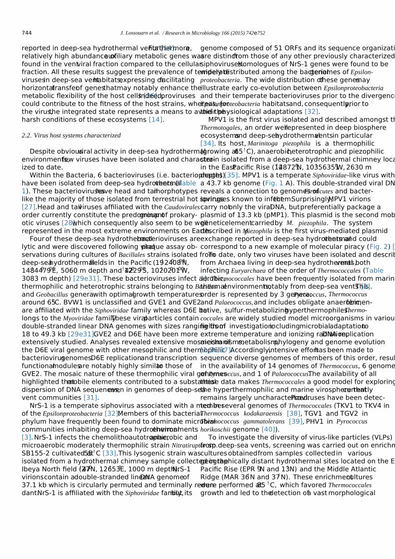
reported in deep-sea hydrothermal vents [14].Furthermore,arelatively high abundance ofauxiliary metabolic genes wasfound in the ventviral fraction compared to the cellularfraction. All these results suggest the prevalence of temperatevirusesin deep-sea venthabitats,expressing orfacilitatinghorizontaltransferof genesthatmay notably enhance themetabolic flexibility of the host cells [26].Indeed,provirusescould contribute to the fitness of the host strains, whereas, forthe virus,the integrated state represents a means to avoid theharsh conditions of these ecosystems [14].
2.2. Virus host systems characterized
Despite obviousviral activity in deep-sea hydrothermalenvironments,few viruses have been isolated and character-ized to date.
Within the Bacteria, 6 bacterioviruses (i.e. bacteriophages)have been isolated from deep-sea hydrothermalvents (Table1). These bacterioviruseshave head and tailmorphotypeslike the majority of those isolated from terrestrial hot springs[27].Head and tailviruses affiliated with the Caudoviralesorder currently constitute the predominantgroup of prokary-otic viruses [28],which consequently also seem to be wellrepresented in the most extreme environments on Earth.
Fourof these deep-sea hydrothermalbacterioviruses arelytic and were discovered following viralplaque assay ob-servations during cultures of Bacillales strains isolated fromdeep-seahydrothermalfields in the Pacific (19!2400800N,148!4407900E, 5060 m depth and 12!4202900S, 102!0200100W,3083 m depth) [29e31]. These bacterioviruses infect aerobic,thermophilic and heterotrophic strains belonging to Bacillusand Geobacillus genera,with optimalgrowth temperaturesaround 65!C. BVW1 is unclassified and GVE1 and GVE2are affiliated with the Siphoviridae family whereas D6E be-longs to the Myoviridae family.These viralparticles containdouble-stranded linear DNA genomes with sizes ranging from18 to 49.3 kb [29e31].GVE2 and D6E have been moreextensively studied. Analyses revealed extensive mosaicism ofthe D6E viral genome with other mesophilic and thermophilicbacteriovirusgenomes.D6E replicationand transcriptionfunctionalmodulesare notably highly similarto those ofGVE2. The mosaic nature of these thermophilic viral genomeshighlighted thatmobile elements contributed to a substantialdispersion of DNA sequences,even in genomes of deep-seavent communities [31].
NrS-1 is a temperate siphovirus associated with a memberof the Epsilonproteobacteria [32].Members of this bacterialphylum have frequently been found to dominate microbialcommunities inhabiting deep-sea hydrothermalenvironments[3].NrS-1 infects the chemolithoautotrophic,anaerobic andmicroaerobic moderately thermophilic strain Nitratiruptor sp.SB155-2 cultivated at55 !C [33].This lysogenic strain wasisolated from a hydrothermal chimney sample collected in theIbeya North field (27! 470N, 126!530E, 1000 m depth).NrS-1virionscontain adouble-stranded linearDNA genomeof37.1 kb which is circularly permuted and terminally redun-dant.NrS-1 is affiliated with the Siphoviridae family,but its
genome composed of 51 ORFs and its sequence organizationare distinctfrom those of any other previously characterizedsiphoviruses.Homologues of NrS-1 genes were found to bewidely distributed among the bacterialgenomes of Epsilon-proteobacteria. The wide distribution ofthese genesmayillustrate early co-evolution between Epsilonproteobacteriaand their temperate bacterioviruses prior to the divergence ofEpsilonproteobacteria habitatsand,consequently,prior totheir physiological adaptations [32].
MPV1 is the first virus isolated and described amongst theThermotogales, an order wellrepresented in deep biosphereecosystemsand deep-seahydrothermalventsin particular[34]. Its host, Marinitoga piezophila is a thermophilic(growing at65 !C), anaerobic,heterotrophic and piezophilicstrain isolated from a deep-sea hydrothermal chimney locatedin the EastPacific Rise (12!487210N, 103!563510W, 2630 mdepth) [35]. MPV1 is a temperate Siphoviridae-like virus witha 43.7 kb genome (Fig. 1 A). This double-stranded viral DNAreveals a connection to genomes ofFirmicutes and bacter-ioviruses known to infectthem.Surprisingly,MPV1 virionscarry notonly the viralDNA, butpreferentially package aplasmid of 13.3 kb (pMP1). This plasmid is the second mobilegeneticelementcarriedby M. piezophila. The systemdescribed in M.piezophila is the first virus-mediated plasmidexchange reported in deep-sea hydrothermalvents and couldcorrespond to a new example of molecular piracy (Fig. 2) [34].
To date, only two viruses have been isolated and describedfrom Archaea living in deep-sea hydrothermalvents,bothinfecting Euryarchaea of the order of Thermococcales (Table1). Thermococcales have been frequently isolated from marinethermalenvironments,notably from deep-sea vents [1].Thisorder is represented by 3 genera:Pyrococcus, Thermococcusand Palaeococcus,and includes obligate anaerobic,fermen-tative, sulfur-metabolizinghyperthermophiles.Thermo-coccales are widely studied model microorganisms in variousfields of investigation,includingmicrobialadaptationtoextreme temperature and ionizing radiation,DNA replicationmechanisms,metabolism,phylogeny and genome evolution[1,36,37].Accordingly,intensive efforthas been made tosequence diverse genomes of members of this order, resultingin the availability of 14 genomes of Thermococcus, 6 genomesof Pyrococcus, and 1 of Palaeococcus.The availability of allthese data makes Thermococcales a good model for exploringthe hyperthermophilic and marine virosphere thatcurrentlyremains largely uncharacterized.Proviruses have been detec-ted in several genomes of Thermococcales (TKV1 to TKV4 inThermococcus kodakaraensis [38], TGV1 and TGV2 inThermococcus gammatolerans [39], PHV1 in Pyrococcushorikoschii genome [40]).
To investigate the diversity of virus-like particles (VLPs)from deep-sea vents, screening was carried out on enrichmentculturesobtainedfrom samples collectedin variousgeographically distant hydrothermal sites located on the EastPacific Rise (EPR 9!N and 13!N) and the Middle AtlanticRidge (MAR 36!N and 37!N). These enrichmentcultureswere performed at85 !C, which favored Thermococcalesgrowth and led to thedetection ofa vast morphological
744 J. Lossouarn et al. / Research in Microbiology 166 (2015) 742e752

diversity of VLPs by transmission electron microscopy [41].Among the different morphotypes observed, the lemon-shapedtype prevailed,butrods and new pleomorphic morphologieswere also reported.Additionally,this study highlighted thestriking similarity between VLP morphologiesin deep-seahydrothermalventsand those from terrestrialhot environ-ments [41]. Thereafter,two viruses associatedwith
Thermococcales strains were isolated and characterized. Theyrepresentthe only two marine hyperthermophilic viruses re-ported to date.
The virus PAV1 was discovered in Pyrococcus abyssi strainGE23,isolated from a deep-sea ventsample collected in theNorth Fidji Basin (White Lady Site;16! 5900S, 173!5500W,2000 m depth),and cultivated at 85!C [23].PAV1 is lemon-
Fig. 1. Electron micrographs of viruses and membrane vesicles isolated from deep-sea hydrothermal vents, negatively stained with 2% uranyl acetate. A: MPV1, avirus isolated from Marinitoga piezophila KA3. B: PAV1, a virus isolated from Pyrococcus abyssi GE23. C: TPV1, a virus isolated from Thermococcus prieurii. D:Membrane vesicles from Thermococcus prieurii.
Fig. 2. Multipurpose mobilome within deep-sea hydrothermalvents.Possible roles of viruses (via their viral cycles:lytic,lysogenic,carrier state or chronic),vesicles and plasmids in HGT of cellular, viral or plasmid DNA (in blue, red or green, respectively); a specific role for vesicles in defense against viral attack andmany other roles in cell-cell communication. This is a schematic presentation and is not to scale. Arrows with solid lines indicate demonstrated results; arrows withdotted lines show more speculative ones. (For interpretation of the references to color in this figure legend, the reader is referred to the web version of this article.)
745J. Lossouarn et al. / Research in Microbiology 166 (2015) 742e752

shaped (120 " 80 nm) with a shorttail terminated by fibers(Fig. 1 B). PAV1 virions contain a double-stranded circularDNA of 18 kb which is also present in high copy number in aplasmid form in the hostcystoplasm.In genome size andvirion morphology,PAV1 strikinglyresemblesthe SSVfuselloviruses isolated from aerobic,acidic hotspring Cren-archaeota belonging to both Sulfolobus and Acidianus genera[23].However,none of its 25 predicted proteins,exceptone(MCP), exhibitsimilarity to SSV proteins.Instead,PAV1 isevolutionarily related to plasmidsof Thermococcales, withthree geneshaving homologuesin plasmid pTN2,two inplasmid pP12-1 and one in pRT1.In addition,PAV1 sharesone gene with provirus TKV4 of T. kodakaraensis [24,36,40].PAV1 persists in the host strain in a stable carrier state. Indeed,PAV1 virionsare spontaneously and continuously releasedfrom the hostcell withoutcausing its lysis or growth delay[23].The infectivity of PAV1 has been demonstrated by theinverted spot test method, which indicates that PAV1 particlescan infectPyrococcus glycovorans leading to growth retar-dation butdoes notinfectany ofthe Thermococcus strainstested [42].
Virus TPV1 was discovered in Thermococcusprieuriiisolated from a hydrothermal chimney sample collected fromthe East Pacific Rise (“SarahSpring”area,7!25024 S,107!47066 W,2700 m depth)and cultivated at80 !C [43].TPV1 is lemon-shaped (140 " 80 nm)with a shorttailterminated by fibers (Fig. 1 C), similarly to PAV1 [25]. TPV1contains a double-stranded circular DNA of 21.5 kb which isalso presentin high copy number in a free form in the hostcell. The TPV1 genome encompasses28 predicted genes.Proteins and transcriptional regulators predicted to be involvedin genome replication were identified.TPV1 encodes also apredicted integrase (SSV-type)of the tyrosine recombinasefamily.The only two genesthatare homologousbetweenTPV1 and PAV1 encode proteins containing a concanavalin A-like lectin/glucanase domain that might be involved in virus-hostrecognition.TPV1 shares more homologous genes withproviruses, (TKV2, TKV3, TKV4, TGV1) and plasmid (pT26-2) from Thermococcus species, than with PAV1 [25]. Infectionwith TPV1 does not cause host lysis, and viral replication canbe induced by UV irradiation.TPV1 can infect several refer-ence species belonging to the Thermococcus genus leading togrowth retardation [42].Interestingly,TPV1 shares its host
with two plasmids pTP1 (3.1 kb) and pTP2 (2.0 kb). These 3mobile elements do nothave a single gene in common andstablypropagatein infectedcells, withoutany apparentantagonistic effect on each other [25,44].
3.Plasmids in deep-sea hydrothermal vents
Plasmids,includingthoseof (hyper-)thermophiles,areabundant in the biosphere.They actively participate in HGT,which plays a majorrole in microbialplasticity,adaptationand evolution [45e47]. To date, only 6 bacterial plasmids and24 archaealplasmids have been isolated and described fromdeep-sea hydrothermal vents.
3.1. Bacterial domain
Among the few plasmids isolated from deep-sea vent bac-teria, one is associated with a mesophilic strain [48] while theother five are carried by thermophilic strains [49e53].Onlythese five bacterial plasmids are given in Table 2. The latter havegenerally not been described in detail, and most have only beenannotated and briefly mentioned in the genome description oftheir hosts. We will briefly focus on two of them.
pDF308 is a 308.54 kb megaplasmid associated with thethermophilic, anaerobic and heterotrophic strain Deferribacterdesulfuricans SSM1 cultivated at65 !C [52,54].This mega-plasmid,whose two-thirdsof the CDSshave no apparentsimilarity with any CDSs in the database,interestingly en-codes 17 copies of a gene cluster encoding two transposases tothe IS200 and IS605 family,whereas none were found in thehostchromosome [52].Very similarCRISPR/Cassystemshave also been identified on both the pDF308 and D.desul-furicans genomes [52].
pMP1 is a plasmid recently described within the Thermo-togales order [34,53]. This 13.3 kb genetic element, carried byMarinitoga piezophila (previously introduced in 1.2 section),is unrelated to the only known plasmids amongstThermoto-gales: two cryptic miniplasmidsin the genusThermotoga[55,56] and a 1724 bp miniplasmid in Mesotoga prima [57].pMP1 encodesa site-specificrecombinase XerD (primasefamily) and a protein with both a DnaB (helicase) and a DnaG(primase)domain.A large fraction ofthe encoded proteinsmightactually be involved in DNA binding,replication and
Table 2Bacterial plasmids (associated with thermophilic strains) isolated from deep-sea hydrothermal vents.
Plasmids Size (kp) Host stains (growth temperatures) Replication Informations References
Unnamed 53.6 Persephonella marina Ex-H1 (73! C) unknown DNA replication protein DnaCDNA topoisomerase IA
[49]
pDF308 308.54 Deferribacter desulfuricans SSM1 (60e65! C) unknown CRISPR/cas systemTransposases
[52]
pOCEPR01 135.3 Oceanithermus profundus 506 (60! C) unknown DNA helicase,DNA polymerase,DNA primasepolymerase,bacteriolytic enzymesCRISPR/cas system
[50]
pTHEAM01 76.6 Thermovibrio ammonificans HB-1 (75! C) unknown DNA topoisomerase IA [51]pMP1 13.3 Marinitoga piezophila KA3 (65 ! ) unknown DNA primase
Integrase/recombinase[34,53]
746 J. Lossouarn et al. / Research in Microbiology 166 (2015) 742e752

propagation of pMP1. The plasmid, which shares its host withthe temperate siphovirus MPV1 (mentioned in section 1.2), isinduced by mitomycin C,along with the provirus,and re-sponds by replicating and escaping from the bacterial hosts byusing helper viral capsids [34].
3.2. Archaeal domain
The vast majority of archaeal plasmids isolated from deep-sea hydrothermal vents are associated with hyperthermophilicstrains,with optimalgrowth temperaturesbetween 80 and95 !C, belonging to Thermococcalesand Methanoccocalesorders; these are given in Table 3.
Plasmids are common in Thermococcales strains (approx.40% were found to harborat leastone extrachromosomalelement) [1,36,58e60];however,only 16 of them have beensequenced and described.They have recently been classifiedaccording to their different types of replication proteins [60].
The pTN1 family comprises 3 smallplasmids ofalmost3 kb:pTN1 isolated from Thermococcus nautili[61],pGT5isolated from P.abyssi GE5 [62] and pTP1 recently isolatedfrom T.prieurii [44].They encode a new family of rolling-circle replicationinitiatorproteins(Rep74,Rep75 andRepTP1) which contain the three characteristic motifs of theRC-Rep superfamily II, with a single tyrosine in motif 3 [63],as wellas a fourth motifconserved with RC transposases,mainly encoded by insertion elements [61].pGT5 and pTN1were used to constructshuttle vectorsin Thermococcales:pYS2 [64]and pYS3 [65]are,respectively,available for P.abyssi and Pyrococcus furiosus and pLC70 [66] for Thermo-coccus species.
The pTP2 family could be represented by the 2.0 kbplasmid pTP2 which propagates in T. prieurii cells with pTP1,mentioned above.pTP2 is the smallest known plasmid of thehyperthermophilic archaea and is unrelated to other Thermo-coccales plasmids [44]. It encodes an RC-Rep containing two
Table 3Archaeal plasmids isolated from deep-sea hydrothermal vents.
Plasmids Family Subfamily Size (kb)Host strains (growth temperatures) Replication Informations References
pTN1 pTN1 3.4 Thermococcus nautili 30-1 (85! C) Rolling-circletype
Rep74 [61]
pGT5 pTN1 3.4 Pyrococcus abyssi GE5 (95!C) Rolling-circletype
Rep75 [62]
pTP1 pTN1 3.1 Thermococcus prieurii (80 !C) Rolling-circletype
RepTP1 [44]
pTP2 pTP2 2 Thermococcus prieurii (80 !C) Rolling-circletype
RepTP2 [44]
pRT1 pRT1 3.4 Pyrococcus sp.JT1 (95!C) Theta type Rep63 [70]pAMT11 pRT1 20.5 Thermococcus sp.AMT11 (85! C) Theta type Rep72,integrase SSV-type [71]pT26-2 pT26-2 21.6 Thermococcus sp.26/2 (85!C) Theta type ATPase (replicative helicase)
Integrase SSV-type[40]
pTBMP1 pTBMP1 54.2 Thermococcus barophilus MP (85 !C) Theta type [73]pTN2 pTN2 pTN2-like 13 Thermococcus nautili 30-1 (85! C) Theta type Helicase protein (SFI)
DNA primase-polymerase[40]
pP12-1 pTN2 pTN2-like 12.2 Pyrococcus sp.12/1 (95!C) Theta type Helicase protein (SFI)DNA primase-polymerase
[40]
pCIR10 pTN2 pTN2-like 13.3 Thermococcus sp.CIR10 (85!C) Theta type Helicase protein (SFI)DNA primase-polymerase
[40]
pIRI48 pTN2 pTN2-like 12.9 Thermococcus sp.IRI48 (85!C) Theta type Helicase protein (SFI)Primase-polymerase domain
[60]
pEXT9a pTN2 pEXT9a-like10.5 Thermococcus sp.EXT9 (85!C) Theta type Helicase protein (SFI) [60]pIRI33 pTN2 pEXT9a-like11 Thermococcus sp.IRI33 (85!C) Theta type Helicase protein (SFI) [60]pAMT7 pTN2 pEXT9a-like 8.5 Thermococcus sp.AMT7 (85!C) Theta type Helicase protein (SFI) [60]pMETVU01 pTN2 pEXT9a-like10.7 Methanocaldococcus vulcanius
M7 (80!C)Theta type Helicase protein (SFI)
Proteins homologous toproteins of pEXT9a-like plasmids
NC_013408.1[60]
pTN3 pTN3 18.3 Thermococcus nautili 30-1 (85! C) Theta type DNA replicative helicase(MCM family)Integrase SSV-type
[74]
pGS5 2.8 Archaeoglobus profundus AV18 (80!C) Rolling-circletype
RC-Rep [68]
pMETVU02 4.7 Methanocaldococcus vulcanius M7 (80 !C) Theta type Putative MCM family protein NC_013409.1pFS01 12.2 Methanocaldococcus sp.FS406-22 (90! C) Theta type Putative MCM family protein
DNA polymerase domainNC_013888.1
ECE1 58.4 Methanocaldococcus jannaschii (85 !C) Theta type Putative MCM protein and ParARestriction/modification system
[77]
ECE2 16.5 Methanocaldococcus jannaschii (85 !C) Theta type Restriction/modification system[77]pMEFER01 22.2 Methanocaldococcus fervens AG86 (80!C) Theta type Putative MCM family protein NC_013157.1pMETOK01 14.9 Methanothermococcus okinawensis
IH1t (60e65!C)Theta type Helicase and nuclease domainsNC_015632
747J. Lossouarn et al. / Research in Microbiology 166 (2015) 742e752

tyrosines in motif 3 (superfamily I),which shares significantsequence similarity with RC-Rep ofplasmid pGS5 isolatedfrom Archaeoglobus profundus AV18 [67]. Futhermore, it hasbeen shown thatpGS5 is negatively supercoiled due to thepresence of a gyrase introducing negative supercoiling [68], incontrast with other plasmids isolated from hyperthermophileswhich are relaxed or slightly positively supercoiled [69].
The pRT1 family contains pRT1,a small3.4 kb plasmidisolated from Pyrococcussp. JT1 [70] and a much largerplasmid of 20.5 kb, pAMT11, isolated from Thermococcus sp.AMT11 [71].They encode the homologous replication pro-teinsRep63 and Rep72,respectively,unrelated to RC-Repproteins,and may replicatevia a thetamechanism [72].Interestingly,pAMT11 genome revealed homology and syn-teny with the genome of TKV1 a virus-like integrated elementof T. kodakaraensis [38].
The pT26-2 family constitutesa new largefamily ofarchaealplasmids and integrated elements that probably pre-date the separation of Thermococcales and Methanococcales[40].The plasmid pT26-2,from Thermococcus sp.26-2,en-codes several proteins including an SSV-type integrase and anew DNA replicativehelicaserelated to thesuperfamilyAAA þ ATPases probably involved in theta-type replication.pT26-2 shares homologues with virus-like integrated elementsof Thermococcales (TKV2 and TKV3 present in T.kodakar-aensis genome,TGV1 presentin T. gammatolerans genomeand PHV1 presentin P. horikoschii genome)and ofMeth-anococcales (MMPV1, MMC7V1, MMC7V2, MMC6V1 inMethanococcus maripaludis strainsgenomes;MVV1 inMethanococcus voltae genome), and with plasmid pMEFER01isolated from Methanocaldococcus fervens.The pT26-2 fam-ily is identified by the presence of nine core genes [40,60], andphylogenetic analyses have shown thatthese “pT26-2 coregenes” have co-evolved with cellular hosts [40].
The pTBMP1 family includes only pTBMP1, which is thelargestplasmidof Thermococcales knownto date.This54.2 kb element,sequenced in the framework ofthe Ther-mococcus barophilus genome project[73],is notrelated tootherThermococcales plasmids.Interestingly,pTBMP1 en-codes a homologue ofarchaealreplication initiatorproteinCdc6/Orc1 and thus replicates via a theta mechanism [73].
The pTN2 family,comprising 7 plasmids from Thermo-coccales and one from Methanococcales,with sizes rangingfrom 8.5 kb to 13.3 kb, is divided into two subfamilies based onthe gene content[60].The pTN2-like subfamily comprisespTN2 isolated from T.nautili, pP12-1 from Pyrococcus sp,pCIR10 and pIRI48 both from Thermococcussp [40,60],whereas the pEXT9a-like subfamily is represented by pEXT9a,pAMT7 and pIRI33, all isolated from Thermococcus sp. [60],and pMETVU01 isolated from Methanocaldococcus vulcaniusM7. In addition to the superfamily 1 helicase [40], these plas-mids encode proteins distantly related to the archaeo-eukaryoticprimase superfamily,PriS and PriL,strongly suggesting thatthey replicate via a theta mechanism [40]. pTN2-like (except forpIRI48 which encodes only the PriS domain) and pEXT9a-likeproteins have the PriL domain in common,whereas the PriSdomain is missing in pEXT9a-like proteins [60]. The pTN2-like
plasmids also encode severalproteins thathave homologueswith the virus PAV1 [23,24].
The pTN3 family includes pTN3 (18.3 kb), isolated from T.nautili sp. 30-1 [74]. pTN3 encodes an SSV-type integrase andis closely related to the virus-like integrated elementTKV4from T.kodakaraensis [74].pTN3 and TKV4 actually repre-sentgenomes of defective viruses thatencode proteins com-mon to viruses belonging to PRD1-adenovirus viral lineages:the capsid protein which contains the double-jelly roll fold andthe putative packaging ATPase [74,75].Interestingly,pTN3,which is presentin T. nautili in both an integrated andepisomalform,is also harbored in membrane vesicles pro-duced by its host [74,76] (see next section).
Seven plasmids have been isolated from deep-sea hyper-thermophilic methanogensto date.They are almostexclu-sively associated with the Methanocaldococcusgenusandhave sizes ranging from 4.7 kb for pMETVU02, isolated fromM. vulcanius, to 58.4 kb for ECE1, isolated from Meth-anocaldococcus jannaschii [77]. Most have only beensequencedand annotated,such as pMETVU02, pFS01,pMEFER01,pMETOK01.pMETVU02 encodesa putativeMCM helicase suggesting replication via a theta mechanism.Interestingly,the plasmid pMETVU01 also isolated from M.vulcanius has homologues in the genomes ofpEXT9a-likeplasmids isolated from Thermococcales and described above.This suggestspossiblehorizontaltransfersfrom Thermo-coccales to Methanococcales [60,72].
4.Membrane vesicles (MVs) in deep-sea hydrothermalvents
4.1. General features of membrane vesicles in the threedomains of life
The production of MVs is a universalphysiologicalphe-nomenon shared by cellsfrom thethree domainsof life[74,78]. MVs were recently reported in deep-sea hydrothermalventsby studying Thermoccocalesstrains[74,79](see seebelow section 4.3).In the open ocean,MVs may reach up to6.106 to 3.105MVs per mL of seawater, and notably play a keyrole in carbon cycling [80].
MVs are spherical closed compartments between ~50 and200 nm in diameter with differing production depending onthe domain of life [81,82] (Fig.1D). In Eukarya, differenttypes of MVs have been described,exosomes,which origi-natedfrom the extrusionof intracellularmultivesicularbodiesand ectosomes(also called microparticles)directlyformed by budding from the cytoplasmic membrane [81,82].In Bacteria with double-membranes,outermembrane vesi-cles (OMVs) are formed by budding of the outer membraneand can contain notonly periplasmic and outermembraneproteins[83],butalso proteinscharacteristic ofthe innermembrane and cytoplasmic compartments [84].In Archaea,and more precisely in Thermococcales, MVs are produced bybudding of the cytoplasmic membrane,similarly to eukary-otic ectosomes,and are frequently surrounded by a glyco-protein S-layer [85].
748 J. Lossouarn et al. / Research in Microbiology 166 (2015) 742e752

4.2. Role of MVs
MVs are involved in many exchanges between cells in thethreedomainsof life, e.g., transporting toxicmolecules,quorum-sensing agents,pathogenicity factors and metabolites[74].Protection of MVs against viral infection has also beenreported [86]. Lastly, MVs can carry different types of nucleicacids (Fig.2).
Notably,some archaea produce protein orpeptide toxinsagainstcompeting species [87,88].ArchaealMVs were firstdescribed in the terrestrial and thermophilic order Sulfolobalesas a conveyorof proteinaceoustoxins[88e90].Similarly,Thermococcus MVs were recently found to display toxicitytowardscompeting Thermococcalesstrains,suggesting theexistence of “Thermococcins” (A.Gorlas etal.,unpublishedwork).
A recentstudy reports thatmarine bacterialextracellularvesicles are enriched in outermembrane components,sug-gesting that they harbor viral receptors that can act to directlybind viralparticles [84].As reported for mesophilic bacteria[86],protection of MVs againstviralinfection could also bepossible in hotdeep-sea environments.Thermococcales vi-rions were sometimes observed attached to MVs,which mayresult in less exposure to viral infection [23,79,85] and couldincrease survival of the host population.
Interestingly,MVs have been universally reported to beable to carry and transfer differenttype of nucleic acids byfusing with recipientcells.Transfers between cells of chro-mosomal,plasmid orviralDNAs packaged into MVs havebeen reported in bacteria [91e94]. All these results lead us toconsider that MVs could be involved in HGT and thus repre-sentanothertype ofMGE. The ability ofarchaealMVs tocarry and potentially transfer different types of DNA was alsorecently reported in Thermoccocalesstrainsisolated fromdeep-sea hydrothermal vents.
4.3. A focus on MVs isolated from Thermococcales andimplicated in HGT
MV productionactuallyappearsto be widespreadinThermococcales isolated from deep-sea hydrothermalvents;26 out of 34 Thermococcales strains, observed by transmissionelectron microscopy,revealed the production ofMVs [79].Some ofthese MVs were strongly associated with cellularDNA and consequently appeared as fluorescentdots by epi-fluorescence microscopy. Experiments showed that these MVscan protect exogenic DNA against DNase treatments and heatdenaturation,suggestingintravesicularlocalizationof theDNA [79].Transformation with naked DNA mightactuallynot be an option in deep-sea hydrothermal vents due to ther-modenaturation.It was therefore hypothesized thatMVs, byprotecting DNA againstthermodegradation,could play animportantrole in HGT in these deep-sea hotenvironments[79]. The production ofnanospheresand nanotubeswasrecently observed at the surface of MVs from T. kodakaraensisand T. gammatolerans. It has beenspeculatedthat thismechanism mightbe used to facilitate DNA transferfrom
MVs to cells [95].The idea thatMVs produced by Thermo-coccales can be considered as MGEs was recently strength-ened when plasmid incorporation and transfer through MVsinto a plasmid-free cellwas demonstrated using the geneti-cally tractable species T. kodakaraensis [85]. MVs containingendogenousplasmidswere also observed in Thermococcusnautili strains [36,74].MVs produced by T.nautili sp.30-1harbored the pTN3 plasmid,which correspondsto a viralgenome,carries genes from a defective virus,buthas inef-fective virion production [74].The name “viralmembranevesicles” (vMVs)wasproposed to specify these particularvesicles containing viral/plasmid genomes,considering thatvirus and plasmid spacesoften overlap [96].These resultsreinforce the hypothesisof an evolutionary connection be-tween MVsand viruses.This presumption waspreviouslymade by considering thatsome viruses produce virions thatstrikingly resemble MVs.For example,some archeovirusesand bacterioviruses produce pleomorphic virions consisting oflipid vesiclesinto which viralproteinsand correspondingnucleic acids are embedded [97].It has been postulated thatMVs produced by ancientcellsor proto cells,anteriortoLUCA, could have played a key role in the onset of some ofthe first viral lineages [98].
DespitecopiousevidencethatMVs are producedby(hyper-)thermophilic prokaryotes in deep-sea biospheres, theirabundance and functions remain largely undetermined to date.The study of MVs thus emerges as a new fundamental field ofresearch in cell and evolutionary biology. Further work is nowneeded to betterunderstand the globalimpactof MVs ondeep-sea hydrothermal vents.
5.Conclusion
MGEs may have been key players in the evolution of thebiosphere since the very beginning.The mobilome remains adriving force in the diversity and evolution of prokaryotes andeukaryotes.Although the real impact of MGEs is not clearlyknown in deep-sea hydrothermal vents, as reviewed in Fig. 2,they have the potentialfor becoming powerfulagentsthatdrive adaptation of their cellular hosts. For example, temperatevirusesand theirlysogenic hostsseek to coexistso as toenhance theirintertwined fitness when confronted with theextreme conditions ofdeep-sea vents.Evidence ofthis viacomparative analysis of cellular and viralmetagenomes [26]lies in the relative enrichmentof genesrelated to energymetabolism in the proviralfraction,by providing their hostswith new or supplementalfeatures to enable adaptation to achallenging environment.These proviruses boosthostfitnessand,in turn,enhance their own survival.
Comparative studies of MGEs from Archaea and Bacteriawill help us understand the dynamic genetic network of themicrobialcommunities in the deep biosphere.The study ofhow microbialpopulationsare modulated by these geneticinteractions will become a keystone area.
Although the ecological and evolutionary effects of MGEsare notclearly known in deep-sea vents,theirproductionprobably plays a major role in the lifestyle of extremophile
749J. Lossouarn et al. / Research in Microbiology 166 (2015) 742e752

microorganisms,making analysis ofthese processes a pre-requisite forfully understanding theirphysiology and ratio-nally exploiting theirbiotechnologicalpotential(transferoftoxins,nucleic acids,etc.).
Conflict of interest
The authors declare no conflict of interest.
Acknowledgments
This work was financially supported by the French Ministryof HigherEducation to JL,Ifremerto SD and the AgenceNationale de la Recherche, project Thermovesicles (ANR-12-BSV3-OO23-01) to CG and PF.
References
[1] Prieur D, Erauso G, Geslin C, Lucas S, Gaillard M, Bidault A, et al. Geneticelements of Thermococcales. Biochem Soc Trans 2004;32:184e7.
[2] Anderson RE, Brazelton WJ, Baross JA. Is the genetic landscape of the deepsubsurface biosphere affected by viruses? Front Microbiol 2011;2:219.
[3] Flores GE,Reysenbach A-L.Hydrothermalenvironments,Marine.In:Reitner J,ThielV, editors.Encyclopedia of earth science series:ency-clopedia of geobiology.Netherlands: Springer.; 2011. p.456e67.
[4] Forterre P. The virocell concept and environmental microbiology. ISME J2013;7:233e6.
[5] Koonin EV, Dolja VV. Virus world as an evolutionary network of viruses andCapsidless selfish elements. Microbiol Mol Biol Rev 2014;78:278e303.
[6] Suttle CA.Viruses in the sea.Nature 2005;437:356e61.[7] Suttle CA.Marine viruses - major players in the global ecosystem.Nat
Rev Microbiol 2007;5:801e12.[8] RohwerF, ThurberRV. Virusesmanipulate the marine environment.
Nature 2009;459:207e12.[9] Kristensen DM, Mushegian AR, Dolja VV, Koonin EV. New dimensions
of the virus world discovered through metagenomics.Trends Microbiol2010;18:11e9.
[10]McDaniel LD, Young E, Delaney J, Ruhnau F, Ritchie KB, Paul JH. HighFrequency of horizontal gene transfer in the oceans.Science 2010;330.50e50.
[11]Cortez D,Forterre P,Gribaldo S.A hidden reservoir of integrative ele-ments is the major source of recently acquired foreign genes and ORFansin archaeal and bacterial genomes. Genome Biol 2009;10:R65.
[12]Forterre P, Prangishvili D. The major role of viruses in cellular evolution:facts and hypotheses.Curr Opin Virol 2013;3:558e65.
[13]Ortmann AC,Suttle CA.High abundances of viruses in a deep-sea hy-drothermalventsystem indicatesviral mediated microbialmortality.Deep Sea Res Part 2005;52:1515e27.
[14]Williamson SJ,Cary SC, Williamson KE, Helton RR, Bench SR,Winget D, et al. Lysogenic virusehost interactions predominate at deep-sea diffuse-flow hydrothermal vents.ISME J 2008;2:1112e21.
[15]Yoshida-Takashima Y,Nunoura T,Kazama H,NoguchiT, Inoue K,Akashi H, et al. Spatial distribution of viruses associated with planktonicand attached microbial communities in hydrothermal environments. ApplEnviron Microbiol 2012;78:1311e20.
[16]Wommack KE,Williamson SJ,Sundbergh A,Helton RR,GlazerBT,Portune K,et al. An instrumentfor collecting discrete large-volumewater samples suitable for ecologicalstudies of microorganisms.DeepSea Res Part 2004;51:1781e92.
[17]Juniper SK, Bird DF, Summit M, Vong MP, Baker ET. Bacterial and viralabundances in hydrothermaleventplumes over northern Gorda Ridge.Deep Sea Res Part 1998;45:2739e49.
[18]Horvath P,Barrangou R.CRISPR/Cas,the immune system of bacteriaand archaea.Science 2010;327:167e70.
[19]Richter H,Randau L,Plagens A.Exploiting CRISPR/Cas: interferencemechanisms and applications.Int J Mol Sci 2013;14:14518e31.
[20]Marraffini LA, Sontheimer EJ. CRISPR interference: RNA-directed adap-tive immunity in bacteria and archaea. Nat Rev Genet 2010;11:181e90.
[21]Makarova KS,Wolf YI, Koonin EV.Potential genomic determinants ofhyperthermophily.Trends Genet 2003;19:172e6.
[22]Anderson RE,Brazelton WJ,Baross JA.Using CRISPRs as a meta-genomic tool to identify microbial hosts of a diffuse flow hydrothermalvent viral assemblage.Fems Microbiol Ecol 2011;77:120e33.
[23]Geslin C,Le Romancer M,Erauso G,Gaillard M,PerrotG, Prieur D.PAV1, the firstvirus-like particle isolated from a hyperthermophiliceuryarchaeote,“Pyrococcus abyssi”. J Bacteriol 2003;185:3888e94.
[24]Geslin C, Gaillard M, Flament D, Rouault K, Le Romancer M, Prieur D,et al. Analysis of the first genome of a hyperthermophilic marine virus-like particle,PAV1, isolatedfrom Pyrococcus abyssi. J Bacteriol2007;189:4510e9.
[25]Gorlas A,Koonin EV,Bienvenu N,Prieur D,Geslin C.TPV1, the firstvirus isolated from the hyperthermophilic genus Thermococcus. EnvironMicrobiol 2012;14:503e16.
[26]Anderson RE,Sogin ML,Baross JA.Evolutionary strategies of viruses,bacteria and archaea in hydrothermal vent ecosystems revealed throughmetagenomics.Plos One 2014;9:e109696.http://dx.doi.org/10.1371/journal.pone.0109696.
[27]Uldahl K, Peng X. Biology, biodiversity and application of thermophilicviruses.In: Satyanarayana T,Littlechild J,KawarabayasiY, editors.Thermophilic microbes in environmentaland industrialbiotechnology:biotechnologyof thermophiles.Netherlands:Springer; 2013.p.271e304.
[28]Ackermann HW,Prangishvili D.Prokaryote viruses studied by electronmicroscopy.Arch Virol 2012;157(10):1843e9.
[29]Liu B, Wu S,Song Q,Zhang X,Xie L. Two novelbacteriophages ofthermophilic bacteria isolated from deep-sea hydrothermalfields.CurrMicrobiol 2006;53:163e6.
[30]Liu B, Zhang X. Deep-seathermophilicGeobacillus bacteriophageGVE2 transcriptionalprofile and proteomic characterization of virions.Appl Microbiol Biotechnol 2008;80:697e707.
[31]Wang Y,Zhang X.Genome analysis ofdeep-sea thermophilic phageD6E. Appl Environ Microbiol 2010;76:7861e6.
[32]Yoshida-Takashima Y,TakakiY, Shimamura S,Nunoura T,TakaiK.Genome sequence of a novel deep-sea vent Epsilonproteobacterial phageprovides new insight into the co-evolution of Epsilonproteobacteria andtheir phages.Extremophiles 2013;17:405e19.
[33]Nakagawa S, Takai K, Inagaki F, Hirayama H, Nunoura T, Horikoshi K,et al.Distribution,phylogenetic diversity and physiological characteris-tics of epsilon-proteobacteria in a deep-sea hydrothermal field.EnvironMicrobiol 2005;7:1619e32.
[34]Lossouarn J,Nesbø CL, MercierC, Zhaxybayeva O,Johnson MS,Charchuck R,etal. «M!enage"a trois»:a selfish genetic elementuses avirus to propagate within Thermotogales.Environ Microbiol 2015.
[35]Alain K, Marteinsson VT, Miroshnichenko ML, Bonch-Osmolovskaya EA, Prieur D, Birrien J-L. Marinitoga piezophila sp. nov.,a rod-shaped,thermo-piezophilic bacterium isolated under high hydro-static pressure from a deep-sea hydrothermalvent.Int J SystEvolMicrobiol 2002;52:1331e9.
[36]Soler N,Gaudin M, Marguet E, Forterre P.Plasmids, viruses and virus-like membranevesiclesfrom Thermococcales. Biochem SocTrans2011;39:36e44.
[37]Leigh JA, Albers S-V, Atomi H, Allers T. Model organisms for geneticsin the domain Archaea:methanogens,halophiles,Thermococcales andSulfolobales. FEMS Microbiol Rev 2011;35:577e608.
[38]Fukui T,Atomi H,Kanai T,Matsumi R,Fujiwara S,Imanaka T.Com-plete genome sequence of the hyperthermophilic archaeon Thermococcuskodakaraensis KOD1 and comparisonwith Pyrococcus genomes.Genome Res 2005;15:352e63.
[39]Zivanovic Y, Armengaud J, Lagorce A, Leplat C, Gu!erin P, Dutertre M,etal. Genome analysis and genome-wide proteomics of Thermococcusgammatolerans, the mostradioresistantorganism known amongsttheArchaea.Genome Biol 2009;10:R70.
750 J. Lossouarn et al. / Research in Microbiology 166 (2015) 742e752

[40]Soler N,Marguet E,Cortez D,Desnoues N,Keller J,van Tilbeurgh H,etal. Two novelfamilies of plasmids from hyperthermophilic archaeaencoding new familiesof replicationproteins.NucleicAcids Res2010;38:5088e104.
[41]Geslin C, Le Romancer M, Gaillard M, Erauso G, Prieur D. Observationof virus-like particlesin high temperature enrichmentculturesfromdeep-sea hydrothermal vents.Res Microbiol 2003;154:303e7.
[42]Gorlas A, Geslin C. A simple procedure to determine the infectivity andhost range of viruses infecting anaerobic and hyperthermophilic micro-organisms.Extremophiles 2013;17:349e55.
[43]Gorlas A,Alain K, Bienvenu N,Geslin C.Thermococcus prieuriisp.nov.,a hyperthermophilic archaeon isolated from a deep-sea hydrother-mal vent.Int J Syst Evol Microbiol 2013;63:2920e6.
[44]Gorlas A,Krupovic M, Forterre P,Geslin C.Living side by side with avirus: characterizationof two novel plasmidsfrom Thermococcusprieurii, a host for the spindle-shaped virusTPV1. Appl EnvironMicrobiol 2013;79:3822e8.
[45]FrostLS, Leplae R,Summers AO,ToussaintA. Mobile genetic ele-ments:the agentsof open sourceevolution.Nat Rev Microbiol2005;3:722e32.
[46]Lipps G. In: Lipps G, editor. Plasmids: current research and future trends.Norfolk: Caister Academic press; 2008.
[47]Sobecky PA,Hazen TH.Horizontalgene transferand mobile geneticelements in marine systems.Methods Mol Biol 2009;532:435e53.
[48] Ji B, Gimenez G,Barbe V,Vacherie B,Rouy Z, AmraniA, et al.Complete genome sequence ofthe piezophilic,mesophilic,Sulfate-reducingbacterium Desulfovibrio hydrothermalis AM13T. GenomeAnnounc 2013;1(1).e00226e12.
[49]Reysenbach AN,HamamuraN, PodarM, GriffithsE, FerreiraS,Hochstein S,et al. Completeand Draftgenomesequencesof Sixmembers of the Aquificales.J Bacteriol 2009;191(6):1992e3.
[50]PatiA, Zhang X,Lapidus A,Nolan M,Lucas S,Del Rio TG, et al.Complete genome sequence ofOceanithermus profundus type strain(506T). Stand Genomic Sci 2011;4(2):210e20.
[51]GiovannelliD, Ricci J, P!erez-RodríguezI, Hugler M, O'Brien C,Keddis R,etal. Complete genome sequence of Thermovibrio ammoni-ficans HB-1T, a thermophilic,chemolithoautotrophic bacterium isolatedfrom a deep-sea hydrothermalvent. Stand Genomic Sci2012;7(1):82e90.
[52]TakakiY, ShimamuraS, NakagawaS, FukuharaY, HorikawaH,Ankai A, et al. Bacteriallifestyle in a deep-sea hydrothermalventchimney revealed by the genome sequence of the thermophilic bacteriumDeferribacter desulfuricans SSM1.DNA Res 2010;17:123e37.
[53]Lucas S,Han J, Lapidus A,Cheng J-F,Goodwin LA,Pitluck S,etal.Complete genome sequence of the thermophilic,piezophilic,heterotro-phic bacterium Marinitoga piezophila KA3. J Bacteriol2012;194:5974e5.
[54]Takai K,Kobayashi H,Nealson KH,Horikoshi K.Deferribacter desul-furicans sp. nov.,a novelsulfur-,nitrate-and arsenate-reducing ther-mophile isolated from a deep-sea hydrothermalvent.Int J Syst EvolMicrobiol 2003;53:839e46.
[55]Akimkina T, Ivanov P, Kostrov S, Sokolova T, Bonch-Osmolovskaya E,Firman K,et al.A highly conserved plasmid from the extreme thermo-phile Thermotoga maritima MC24 is a member of a family of plasmidsdistributed worldwide. Plasmid 1999;42:236e40.
[56]Harriott OT,Huber R,Stetter KO,Betts PW,Noll KM. A cryptic min-iplasmid from the hyperthermophilic bacterium Thermotoga sp.strainRQ7.J Bacteriol 1994;176:2759e62.
[57]Zhaxybayeva O,Swithers KS,FoghtJ, Green AG,Bruce D,Detter C,et al. Genome sequence ofthe mesophilic ThermotogalesbacteriumMesotoga prima MesG1.Ag.4.2revealsthe largestThermotogalesgenome to date.Genome Biol Evol 2012;4:700e8.
[58]Benbouzid-RolletN, L!opez-García P,Watrin L,Erauso G,PrieurD,Forterre P. Isolation of new plasmids from hyperthermophilic Archaea ofthe order Thermococcales.Res Microbiol 1997;148:767e75.
[59]Lepage E,Marguet E,Geslin C,Matte-Tailliez O,Zillig W,Forterre P,et al. Molecular diversity of new Thermococcales isolates from a singlearea of hydrothermal deep-sea vents as revealed by randomly amplified
polymorphic DNA fingerprinting and 16S rRNA gene sequence analysis.Appl Environ Microbiol 2004;70:1277e86.
[60]Krupovic M,Gonnet M,Hania WB,Forterre P,Erauso G.Insights intodynamics of mobile genetic elements in hyperthermophilic environmentsfrom five new Thermococcus plasmids.PloS One 2013;8:e49044.
[61]Soler N, JustomeA, Quevillon-CheruelS, Lorieux F, Le Cam E,MarguetE, etal. The rolling-circle plasmid pTN1 from the hyperther-mophilic archaeon Thermococcus nautilus. Mol Microbiol2007;66:357e70.
[62]Erauso G,Marsin S,Benbouzid-RolletN, BaucherMF, Barbeyron T,ZivanovicY, et al. Sequence ofplasmid pGT5 from thearchaeonPyrococcus abyssi: evidence forrolling-circle replication in a hyper-thermophile.J Bacteriol 1996;178:3232e7.
[63] Ilyina TV,Koonin EV.Conserved sequence motifs in the initiator pro-teins forrolling circle DNA replication encoded by diverse repliconsfrom eubacteria,eucaryotesand archaebacteria.NucleicAcids Res1992;20:3279e85.
[64]Lucas S, Toffin L, Zivanovic Y, Charlier D, Moussard H, Forterre P, et al.Construction of a shuttle vector for, and spheroplast transformation of thehyperthermophilic archaeon Pyrococcus abyssi. Appl Environ Microbiol2002;68:5528e36.
[65]Waege I, Schmid G, Thumann S, Thomm M, Hausner W. Shuttle vector-based transformation system forPyrococcus furiosus. Appl EnvironMicrobiol 2010;76:3308e13.
[66]Santangelo TJ,Cubonov!a L, Reeve JN.Shuttle vectorexpression inThermococcus kodakaraensis: contributions ofcis elements to proteinsynthesisin a hyperthermophilic archaeon.Appl Environ Microbiol2008;74:3099e104.
[67]Burggraf S,Jannasch HW,Nicolaus B,Stetter KO.Archaeoglobus pro-fundus sp.nov.,Represents a New Species within the Sulfate-reducingArchaebacteria.Syst Appl Microbiol 1990;13:24e8.
[68]L!opez-García P, Forterre P, van der Oost J, Erauso G. Plasmid pGS5 fromthe hyperthermophilic archaeon Archaeoglobus profundus is negativelysupercoiled.J Bacteriol 2000;182:4998e5000.
[69]L!opez-García P, Forterre P. DNA topology in hyperthermophilic archaea:reference states and their variation with growth phase,growth tempera-ture,and temperature stresses.Mol Microbiol 1997;23:1267e79.
[70]Ward DE,RevetIM, Nandakumar R,Tuttle JH,de Vos WM,van derOostJ, et al. Characterization ofplasmid pRT1 from Pyrococcus sp.strain JT1.J Bacteriol 2002;184:2561e6.
[71]GonnetM, Erauso G,PrieurD, Le RomancerM. pAMT11,a novelplasmid isolated from a Thermococcus sp.strain closely related to thevirus-like integrated element TKV1 of the Thermococcus kodakaraensisgenome. Res Microbiol 2011;162:132e43.
[72]Forterre P,Krupovic M,Raymann K,SolerN. Plasmidsfrom Eur-yarchaeota. Microbiol Spectr 2014. 2:PLAS-0027-2014.
[73]Vannier P, Marteinsson VT, Fridjonsson OH, Oger P, Jebbar M. Completegenome sequence of the hyperthermophilic,piezophilic,heterotrophic,and Carboxydotrophic archaeon Thermococcus barophilus MP.J Bac-teriol 2011;193:1481e2.
[74]Gaudin M,Krupovic M,Marguet E,Gauliard E,Cvirkaite-Krupovic V,Le Cam E,etal. Extracellular membrane vesicles harbouring viralge-nomes. Environ Microbiol 2014;16:1167e75.
[75]Krupovic M, Bamford DH. Archaeal proviruses TKV4 and MVV extendthe PRD1-adenovirus lineage to the phylum Euryarchaeota.Virology2008;375:292e300.
[76]Oberto J,Gaudin M,Cossu M,Gorlas A,Slesarev A,Marguet E,et al.Genome sequence of a hyperthermophilic archaeon, thermococcus nautili30e1, that produces viral vesicles. Genome Announc 2014. 2: e00243-14.
[77]Bult CJ, White O, Olsen GJ, Zhou L, Fleischmann RD, Sutton GG, et al.Completegenomesequenceof the methanogenicarchaeon,Meth-anococcus jannaschii. Science 1996;273:1058e73.
[78]Gy€orgy B, Szab!o TG, P!aszt!oi M, P!al Z, Misj!ak P, Aradi B, et al.Membrane vesicles,currentstate-of-the-art:emerging role of extracel-lular vesicles.Cell Mol Life Sci 2011;68:2667e88.
[79]Soler N,MarguetE, Verbavatz J-M,Forterre P.Virus-like vesicles andextracellular DNA produced by hyperthermophilic archaea of the orderThermococcales. Res Microbiol 2008;159:390e9.
751J. Lossouarn et al. / Research in Microbiology 166 (2015) 742e752

[80]Scanlan D.Bacterial vesicles in the ocean.Science 2014;343:143e4.[81]Deatherage BL,Cookson BT.Membrane vesicle release in bacteria,
eukaryotes,and archaea:a conserved yetunderappreciated aspectofmicrobial life.Infect Immun 2012;80:1948e57.
[82]Raposo G, StoorvogelW. Extracellularvesicles:exosomes,micro-vesicles,and friends.J Cell Biol 2013;200:373e83.
[83]Kulp A, Kuehn MJ. Biologicalfunctions and biogenesis ofsecretedbacterial outer membrane vesicles. Annu Rev Microbiol2010;64:163e84.
[84]Biller SJ, Schubotz F,Roggensack SE,Thompson AW,Summons RE,Chisholm SW. Bacterialvesiclesin Marine ecosystems.Science2014;343:183e6.
[85]Gaudin M,Gauliard E,Schouten S,Houel-RenaultL, Lenormand P,Marguet E, et al. Hyperthermophilic archaea produce membrane vesiclesthat can transfer DNA.Environ Microbiol Rep 2013;5:109e16.
[86]Manning AJ, Kuehn MJ. Contribution ofbacterialoutermembranevesicles to innate bacterial defense.BMC Microbiol 2011;11:258.
[87]Haseltine C,Hill T, Montalvo-Rodriguez R,KemperSK, Shand RF,Blum P.Secreted EuryarchaealMicrohalocins KillhyperthermophilicCrenarchaea. J Bacteriol 2001;183:287e91.
[88]Prangishvili D,Holz I, Stieger E,Nickell S,Kristjansson JK,Zillig W.Sulfolobicins,specific proteinaceous toxins produced by strains of theextremelythermophilicarchaealgenus Sulfolobus. J Bacteriol2000;182:2985e8.
[89]Ellen AF,Albers S-V,Huibers W,Pitcher A,HobelCFV, Schwarz H,et al. Proteomic analysisof secreted membrane vesiclesof archaealSulfolobus species reveals the presence ofendosome sorting complexcomponents.Extremophiles 2009;13:67e79.
[90]Ellen AF, Rohulya OV, Fusetti F, Wagner M, Albers S-V, Driessen AJM.The Sulfolobicin genesof Sulfolobus acidocaldarius encodenovelantimicrobial proteins.J Bacteriol 2011;193:4380e7.
[91]Dorward DW,Garon CF,Judd RC.Exportand intercellular transfer ofDNA via membraneblebsof Neisseria gonorrhoeae. J Bacteriol1989;171:2499e505.
[92]Velimirov B,Hagemann S.Mobilizable bacterialDNA packaged intomembranevesiclesinducesserial transduction.Mob GenetElem2011;1:80e1.
[93]Kolling GL, Matthews KR. Export of virulence genes and Shiga toxin bymembrane vesicles of Escherichia coliO157:H7.ApplEnviron Micro-biol 1999;65:1843e8.
[94]Yaron S, Kolling GL, Simon L, Matthews KR. Vesicle-mediated transferof virulence genesfrom Escherichia coli O157:H7 to otherentericbacteria.Appl Environ Microbiol 2000;66:4414e20.
[95]MarguetE, Gaudin M,Gauliard E,Fourquaux I,le Blond du Plouy S,Matsui I, et al. Membrane vesicles, nanopods and/or nanotubes producedby hyperthermophilic archaea of the genus Thermococcus. Biochem SocTrans 2013;41:436e42.
[96]SolerN, Krupovic M,MarguetE, Forterre P.Membrane vesicles innaturalenvironments:a majorchallengein viral ecology.ISME J2014;9:793e6.
[97]Pietil€a MK, AtanasovaNS, Manole V, Liljeroos L, ButcherSJ,Oksanen HM,et al. Virion architectureunifiesglobally distributedpleolipoviruses infecting halophilic archaea.J Virol 2012;86:5067e79.
[98]Forterre P,Krupovic M.The origin of virions and virocells: the escapehypothesis revisited.In: Witzany G,editor.Viruses essent.Agents life.Netherlands: Springer; 2012.p.43e60.
752 J. Lossouarn et al. / Research in Microbiology 166 (2015) 742e752


Chapitre II : Me�nage a� trois,
caracte� risation de MPV1
Chapitre II
125
Pre�sentation des travaux
Ces travaux ont consisté en la caractérisation du premier virus associé au phylum des
Thermotogae, MPV1 (Marinitoga piezophila vi us et elle d’u l e t g ti ue o ile
« pirate », le plasmide pMP1 (plasmide Marinitoga piezophila 1).
MPV1 est un siphovius te p i du ti le à la ito i e C. Les viio s so t o pos s d’u e
capside icosaédrique de 55 nm de diamètre à laquelle est attachée une longue queue flexible
de 200 nm de long pour 10 nm de large. Ce virus est intégré sous forme de provirus dans le
génome de Marinitoga piezophila KA3. Cette Thermotogae thermophile et anaérobie est
piézophile mais peut être adaptée à la pression atmosphérique.
Ce « Ménage à trois » epose su l’ite atio d’u e ellule hôte at ie e, d’u vius et
d’u plaside pMP . Ce plaside a la patiulait d’t e apale de s’is e das les
capsides virales à la place du génome viral. En effet, lors du déclenchement du cycle lytique
par induction à la mitomycine C, le nombre de copies du plasmide augmente drastiquement
(nombre de copies des génomes viraux multiplié par 3, nombre de copies du plasmide
ultipli pa . Le ais e d’e apsidatio du plaside ’est pas o u e si elui-
i patage u e sue e d’e vio p ave le go e vial pote tielleent impliqué dans
l’e apsidatio de l’ADN . D’aut e pat, le plasmide est aussi capable de se propager dans une
at ie d’u aut e gee : Thermosipho sp. AT1244-VC14. Ce ménage à trois semble être un
nouvel exemple de piratage moléculaire où le pirate (ici le plasmide) prend la place du génome
vi al das la apside afi d’assu e sa p op e p opagatio das l’e vio e e t, profitant ainsi
de la protection assurée par cette coque protéique.
Ces travaux ont été menés dans le cadre de la thèse de Julien Lossouarn (soutenue en mars
4 . Lo s de es t avau de th se, le i lage d’u e i uatai e de sou hes de
Thermotogae été réalisé menant notamment à la découverte et à la caractérisation du

Chapitre II
126
age à t ois. J’ai appo t a o ti utio à ette tude du at mon stage de master 2 et
le début de ma thèse. Mes travaux ont porté sur différentes étapes de cette caractérisation.
Dans un premier temps, la mise au point de la méthode de purification des particules virales
a été améliorée via l’utilisatio d’u g adie t liai e d’iodiaol. Pa la suite, l’tude du
contenu des particules virales par l’e t atio des aides u l i ues AN p se ts das les
capsides purifiées a été réalisée, permettant le séquençage de ceux ci. Le séquençage et les
PCR sur ces AN p se ts à l’it ieu de la apside aat v l la p se e de l’ADN vial ais
aussi de l’ADN plasmidique, un suivi en qPCR de l'ADN bactérien (gène codant pour ARNr16s),
viral (gène codant pour la capside) et plasmidique (gène hypothétique), avec et sans induction,
a t alis , afi d’ tudie l’volutio de eu -ci. E fi , des tests d’ife tio o t t alis s,
permettant de mettre en évidence la propagation du plasmide dans une souche appartenant
au genre Thermosipho.


‘Ménage à trois’: a selfish genetic element uses a virusto propagate within Thermotogales
Julien Lossouarn,1,2,3 Camilla L. Nesbø,4,5
Coraline Mercier,1,2,3 Olga Zhaxybayeva,6
Milo S. Johnson,6 Rhianna Charchuck,4
Julien Farasin,1,2,3 Nadège Bienvenu,1,2,3
Anne-Claire Baudoux,7,8,9 Grégoire Michoud,1,2,3
Mohamed Jebbar1,2,3 and Claire Geslin1,2,3*1Université de Bretagne Occidentale (UBO, UEB),Institut Universitaire Européen de la Mer (IUEM) – UMR6197, Laboratoire de Microbiologie des EnvironnementsExtrêmes (LMEE), rue Dumont d’Urville, F-29280Plouzané, France.2CNRS, IUEM – UMR 6197, Laboratoire deMicrobiologie des Environnements Extrêmes (LMEE),rue Dumont d’Urville, F-29280 Plouzané, France.3Ifremer, UMR 6197, Laboratoire de Microbiologie desEnvironnements Extrêmes (LMEE), Technopôle Pointedu diablea, F-29280 Plouzané, France.4CEES, Department of Biology, University of Oslo, Oslo0316, Norway.5Department of Biological Sciences, University ofAlberta, Edmonton, AB T6G2R3, Canada.6Department of Biological Sciences, Dartmouth College,Hanover, NH 03755, USA.7Sorbonne Universités, UPMC Univ Paris 06, Paris75005, France.8UMR 7144, Equipe DIPO, Station Biologique deRoscoff, Roscoff 29680, France.9CNRS, UMR 7144, Adaptation et Diversité en MilieuMarin, Station Biologique de Roscoff, Roscoff 29680,France.
Summary
Prokaryotic viruses play a major role in the microbialecology and evolution. However, the virosphereassociated with deep-sea hydrothermal ecosystemsremains largely unexplored. Numerous instancesof lateral gene transfer have contributed to thecomplex and incongruent evolutionary history ofThermotogales, an order well represented in deep-seahydrothermal vents. The presence of clustered regu-larly interspaced short palindromic repeats (CRISPR)
loci has been reported in all Thermotogales genomes,suggesting that these bacteria have been exposed toviral infections that could have mediated geneexchange. In this study, we isolated and characterizedthe first virus infecting bacteria from the orderThermotogales, Marinitoga piezophila virus 1 (MPV1).The host, Marinitoga piezophila is a thermophilic,anaerobic and piezophilic bacterium isolated from adeep-sea hydrothermal chimney. MPV1 is a temperateSiphoviridae-like virus with a 43.7 kb genome. Sur-prisingly, we found that MPV1 virions carry not onlythe viral DNA but preferentially package a plasmid of13.3 kb (pMP1) also carried by M. piezophila. This‘ménage à trois’ highlights potential relevance ofselfish genetic elements in facilitating lateral genetransfer in the deep-sea biosphere.
Introduction
Deep-sea hydrothermal vents are home to manythermophilic and hyperthermophilic Bacteria and Archaea,whose genetic diversity, metabolic capacity and physi-ological adaptations are being actively explored (reviewedin Flores and Reysenbach, 2011). However, the virosphereassociated with these deep-sea hydrothermal microorgan-isms remains largely unstudied. The observation of abun-dant populations of lysogens and temperate virusesindicates that virus-mediated gene exchange and recom-bination may be critical to the survival and stability ofprokaryotes within extreme environments (Williamsonet al., 2008). Additionally, metagenomic studies suggestthat a wide diversity of deep-sea hydrothermal microorgan-isms is exposed to viral infections (Williamson et al., 2008;Anderson et al., 2011a). Yet, published viromes mostlycontain reads with no homologues in public databases anddo not provide any information about the infected hosts(Anderson et al., 2011b). Therefore, to understand theimpact of viruses on the microbial diversity in the deep-seaecosystems, it remains crucial to isolate new viruses and tocharacterize host–virus model systems.
To date, only seven viruses, isolated from deep seahydrothermal vents, have been described. Two of themare lemon-shaped archaeoviruses, PAV1 and TPV1,which infect Thermococcales, obligate anaerobic sulfur-metabolizing hyperthermophiles (Geslin et al., 2003a;2007; Gorlas et al., 2012). The other five isolates are tailed
Received 29 August, 2014; revised 13 January, 2015; accepted 13January, 2015. *For correspondence. E-mail [email protected]; Tel. +33 (0)2 98 49 88 58; Fax +33 (0)2 98 49 87 05.
bs_bs_banner
Environmental Microbiology (2015) doi:10.1111/1462-2920.12783
© 2015 Society for Applied Microbiology and John Wiley & Sons Ltd
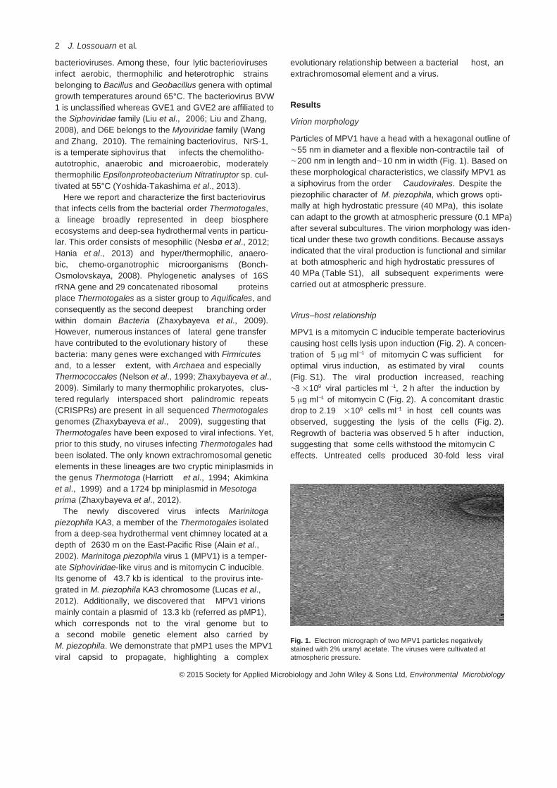
bacterioviruses. Among these, four lytic bacteriovirusesinfect aerobic, thermophilic and heterotrophic strainsbelonging to Bacillus and Geobacillus genera with optimalgrowth temperatures around 65°C. The bacteriovirus BVW1 is unclassified whereas GVE1 and GVE2 are affiliated tothe Siphoviridae family (Liu et al., 2006; Liu and Zhang,2008), and D6E belongs to the Myoviridae family (Wangand Zhang, 2010). The remaining bacteriovirus, NrS-1,is a temperate siphovirus that infects the chemolitho-autotrophic, anaerobic and microaerobic, moderatelythermophilic Epsilonproteobacterium Nitratiruptor sp. cul-tivated at 55°C (Yoshida-Takashima et al., 2013).
Here we report and characterize the first bacteriovirusthat infects cells from the bacterial order Thermotogales,a lineage broadly represented in deep biosphereecosystems and deep-sea hydrothermal vents in particu-lar. This order consists of mesophilic (Nesbø et al., 2012;Hania et al., 2013) and hyper/thermophilic, anaero-bic, chemo-organotrophic microorganisms (Bonch-Osmolovskaya, 2008). Phylogenetic analyses of 16SrRNA gene and 29 concatenated ribosomal proteinsplace Thermotogales as a sister group to Aquificales, andconsequently as the second deepest branching orderwithin domain Bacteria (Zhaxybayeva et al., 2009).However, numerous instances of lateral gene transferhave contributed to the evolutionary history of thesebacteria: many genes were exchanged with Firmicutesand, to a lesser extent, with Archaea and especiallyThermococcales (Nelson et al., 1999; Zhaxybayeva et al.,2009). Similarly to many thermophilic prokaryotes, clus-tered regularly interspaced short palindromic repeats(CRISPRs) are present in all sequenced Thermotogalesgenomes (Zhaxybayeva et al., 2009), suggesting thatThermotogales have been exposed to viral infections. Yet,prior to this study, no viruses infecting Thermotogales hadbeen isolated. The only known extrachromosomal geneticelements in these lineages are two cryptic miniplasmids inthe genus Thermotoga (Harriott et al., 1994; Akimkinaet al., 1999) and a 1724 bp miniplasmid in Mesotogaprima (Zhaxybayeva et al., 2012).
The newly discovered virus infects Marinitogapiezophila KA3, a member of the Thermotogales isolatedfrom a deep-sea hydrothermal vent chimney located at adepth of 2630 m on the East-Pacific Rise (Alain et al.,2002). Marinitoga piezophila virus 1 (MPV1) is a temper-ate Siphoviridae-like virus and is mitomycin C inducible.Its genome of 43.7 kb is identical to the provirus inte-grated in M. piezophila KA3 chromosome (Lucas et al.,2012). Additionally, we discovered that MPV1 virionsmainly contain a plasmid of 13.3 kb (referred as pMP1),which corresponds not to the viral genome but toa second mobile genetic element also carried byM. piezophila. We demonstrate that pMP1 uses the MPV1viral capsid to propagate, highlighting a complex
evolutionary relationship between a bacterial host, anextrachromosomal element and a virus.
Results
Virion morphology
Particles of MPV1 have a head with a hexagonal outline of∼55 nm in diameter and a flexible non-contractile tail of∼200 nm in length and∼10 nm in width (Fig. 1). Based onthese morphological characteristics, we classify MPV1 asa siphovirus from the order Caudovirales. Despite thepiezophilic character of M. piezophila, which grows opti-mally at high hydrostatic pressure (40 MPa), this isolatecan adapt to the growth at atmospheric pressure (0.1 MPa)after several subcultures. The virion morphology was iden-tical under these two growth conditions. Because assaysindicated that the viral production is functional and similarat both atmospheric and high hydrostatic pressures of40 MPa (Table S1), all subsequent experiments werecarried out at atmospheric pressure.
Virus–host relationship
MPV1 is a mitomycin C inducible temperate bacterioviruscausing host cells lysis upon induction (Fig. 2). A concen-tration of 5 μg ml−1 of mitomycin C was sufficient foroptimal virus induction, as estimated by viral counts(Fig. S1). The viral production increased, reaching∼3 ×10⁹ viral particles ml −1, 2 h after the induction by5 μg ml−1 of mitomycin C (Fig. 2). A concomitant drasticdrop to 2.19 ×10⁶ cells ml−1 in host cell counts wasobserved, suggesting the lysis of the cells (Fig. 2).Regrowth of bacteria was observed 5 h after induction,suggesting that some cells withstood the mitomycin Ceffects. Untreated cells produced 30-fold less viral
Fig. 1. Electron micrograph of two MPV1 particles negativelystained with 2% uranyl acetate. The viruses were cultivated atatmospheric pressure.
2 J. Lossouarn et al.
© 2015 Society for Applied Microbiology and John Wiley & Sons Ltd, Environmental Microbiology

particles than mitomycin-treated culture, with a maximalvirus abundance of 1.11×10⁸ viral particles ml−1 after 9 hof incubation (Fig. 2). This basal viral production could beexplained by a spontaneous induction of MPV1 within afraction of the growing host cells. Similar spontaneousinduction was observed during the growth of Nitratiruptorsp., the deep-sea hydrothermal isolate lysogenized by themitomycin C inducible temperate siphovirus NrS-1(Yoshida-Takashima et al., 2013).
DNA content of viral particles
The genome sequence of M. piezophila KA3 contains aprovirus sequence of ∼40 kb integrated into the strain’schromosome and a cccDNA of 13 386 bp denoted aspMARPI01 (Lucas et al., 2012). pMARPI01 has an insilico RFLP pattern identical to that of pMP1, the plasmidthat we isolated from M. piezophila KA3 by an alkalinelysis extraction (Fig. S2A). For brevity, we will refer to bothpMARPI01 and the discovered plasmid as pMP1 through-out the rest of the manuscript. Analysis of DNA extractedfrom the purified MPV1 viral capsids confirmed that theprovirus is the integrated form of the MPV1 genome.Surprisingly, RFLP analyses indicated that the viral DNAis much less frequently present in MPV1 virions(Fig. S2B). Instead, the viral capsids mainly contain pMP1and hence preferentially package the plasmid. Sequenc-ing of the DNA from the viral particles further confirmedthis hypothesis, as not only both a 43 715 bp sequencecorresponding to the provirus inserted in M. piezophilagenome (Fig. 3) and a 13 386 bp plasmid DNA
Fig. 2. Virus–host kinetics at atmospheric pressure monitored byflow cytometry. Typical growth curve of the host M. piezophila isshown as filled squares on continuous black line, and spontaneousproduction of MPV1 particles, reaching 1.11 ×10⁸ viral particles perml, is shown as open squares on dotted black line. Curves in greycorrespond to the cultures grown with the addition of 5 μg ml−1 ofmitomycin C (depicted with the arrow): filled triangles on continuousline show M. piezophila growth, whereas open triangles on dottedline refer to parallel MPV1 viral production, reaching ∼3 ×10⁹ viralparticles per ml.
Fig. 3. Comparison of the MPV1 provirus in M. piezophila and viralDNA. The regions with significant pairwise BLASTN similarityscores are connected. Informative ORF annotations are shown.The ORF highlighted in green has a homologue in the pMP1plasmid (see Fig. 4). The ORF marked with a star is disrupted inthe linearized genome packaged in the virions. The black dotsindicate the short sequences with significant sequence similarity tothe regions of the pMP1 genome. The division of the virus intofunctional modules is approximate, as it is based only on geneannotations. The figure was produced using genoPlotR (Guy et al.,2010).
Virus–plasmid interactions in Thermotogales 3
© 2015 Society for Applied Microbiology and John Wiley & Sons Ltd, Environmental Microbiology

corresponding to pMP1 (Fig. 4) were fully assembled, butalso a 20-fold higher coverage of the plasmid reads com-pared with the ‘true viral’ reads was observed. We rule outpossible extracapsid DNA contamination, as the purifiedviral suspensions from induced cultures were treated withDNase before DNA extraction. Moreover, specific poly-merase chain reaction (PCR) amplifications targeting theprovirus, the plasmid and the 16S rRNA gene in the DNAextracted from the viral capsids, resulted in no PCRproduct corresponding to the 16S rRNA gene (Fig. S3).The same results were obtained with DNA extracted froma purified viral suspension of an uninduced culture (notshown). We concluded that even without induction bothviral and plasmid DNA were incorporated into the viralcapsids.
Attempts to infect a putative sensitive host
Despite several attempts, we never succeeded in curingM. piezophila KA3 strain of MPV1 or pMP1, thus we couldnot test MPV1’s ability to re-infect its host. A purified viralsuspension was added to growing cultures of two otherpotential host cells (Thermosipho sp. strain AT1244-VC14and M. camini strain DV1142). For both cultures, no lysiswas observed under a phase-contrast microscope.Marinitoga camini strain DV1142 did not get infected by
MPV1 (data not shown). After three subcultures of theThermosipho sp. strain AT1244-VC14, no viral DNA wasdetected, but a 198 bp fragment from the pMP1 plasmidwas amplified by PCR from the total DNA (Fig. 5). Moreo-ver, the complete plasmid pMP1 was extracted by alkalinelysis from the Thermosipho sp. strain infected by MPV1(Fig. S4). These findings highlight an inter-genus plasmid(pMP1) propagation by means of a viral capsid.
Effect of the mitomycin C induction on the evolution ofthe copy numbers of chromosomal, proviral and plasmidtarget genes by qPCR analyses
The relative quantity of the chromosomal DNA inM. piezophila KA3 induced with mitomycin C was twotimes lower than in the culture not exposed to the antibi-otic (Fig. 6 and Fig. S5). This might be a result of a chro-mosomal DNA damaging effect of mitomycin C, whichinduces the lytic phase of MPV1. On the other hand, therelative concentration of viral DNA was almost threefoldhigher in mitomycin C treated cells than in untreated ones(Fig. 6 and Fig. S5). This post-induction increase of theviral DNA replication was consistent with the lysogenicrelationship between MPV1 and its host. Finally, therelative concentration of plasmid DNA was 10-fold higherin mitomycin C treated cells than in the uninduced cells(Fig. 6 and Fig. S5). The induction of the plasmid wasunexpected and could be a response, as a survival strat-egy, to escape to the host lysis.
Marpi_2120Sigma factor
Marpi_2121 Transcriptional Coactivator p15
Marpi_2122 Site-specific recombinase XerD
Marpi_2127DNA primaseMarpi_2131
Growth inhibitor
13,386 bp 26.5% GC
1000
2000
3000
400
0
5000
6000
70008000
9000
10000
11000
1200
0
13000
Fig. 4. Map of the pMP1 plasmid. Only informative ORFannotations are shown. The ORF highlighted in green has ahomologue in the MPV1 genome (see Fig. 3). The black dotsindicate the short sequences with significant similarity to the MPV1genome. The figure was created in Geneious 7 (BiomattersLimited).
Fig. 5. MPV1 infectivity assays performed on Thermosipho sp.AT1244-VC14T and checked by PCR experiments. Lanes 1, 2:PCR on total DNA of the uninfected Thermosipho strain targeting(1) MPV1 viral DNA or (2) pMP1 plasmid DNA. Lanes 3, 4: PCR ontotal DNA of the Thermosipho strain just after addition of MPV1purified capsids targeting (3) MPV1 viral DNA or (4) pMP1 plasmidDNA. Lanes 5, 6: PCR on total DNA of Thermosipho strain 22 hafter addition of MPV1 purified capsids targeting (5) MPV1 viralDNA or (6) pMP1 plasmid DNA. Lanes 7, 9: PCR on total DNA ofthe Thermosipho strain after 3 subcultures targeting (7) MPV1 viralDNA or (9) pMP1 plasmid DNA. Lanes 10, 12: Negative controlstargeting (10) MPV1 viral DNA or (12) pMP1 plasmid DNA.Lanes 11, 13: Positive controls targeting (11) MPV1 viral DNA or(13) pMP1 plasmid DNA. Lane 8: Smart Ladder 1 kb (Eurogentec).
4 J. Lossouarn et al.
© 2015 Society for Applied Microbiology and John Wiley & Sons Ltd, Environmental Microbiology

Sequence analysis of the bacterioviral DNA and theplasmid DNA
The viral DNA sequences obtained from the purifiedMPV1 capsids was identical to the provirus sequence onM. piezophila KA3’s chromosome (Fig. 3). De novoassembly of the viral reads revealed that while someof the packaged DNA molecules are circular, othersare linearized, disrupting the Open Reading Frame (ORF)corresponding to Marpi_0300 in the provirus (marked by astar in Fig. 3). The viral genome has 54 predicted ORFs,which include all genes necessary for viral particle pro-duction (Fig. 3). The gene order in the viral genome issimilar to that seen in Enterococcus viruses (Yasminet al., 2010). Based on BLAST searches, 23 of the 54predicted viral ORFs have no detectable homologuesin GenBank (Table S2). Among the 31 ORFs withhomologues in GenBank, eight ORFs have homologuesin proviruses identified by us in two Thermosipho sp.genomes (C. Nesbø, unpublished), and three ORFs havehomologues in the genomes of other Thermotogales(Table S2). Therefore, it is likely that MPV1 and closelyrelated viruses have exchanged genes with otherThermotogales in the past. However, in the availableThermotogales genomes, CRISPR arrays are > 85%identical to MPV1 DNA across the entire spacer in onlyone Marinitoga genome (Marinitoga sp. 1137, C. Nesbø,unpublished) (data not shown).
When BLAST ‘self matches’ to the M. piezophilagenome and matches to the above-mentionedThermosipho proviruses were excluded, 22 out of the 31ORFs had Firmicutes genes as their top-scoring BLASThit. Moreover, among these 31 ORFs, 12 were similar togenes in other bacterioviruses. The majority of thesebacterioviral homologues belong to siphoviruses, butsome are from the Myoviridae family of bacterioviruseswith contractile tails (Krupovic et al., 2011), exemplified bythe thermophilic Geobacillus bacteriovirus GBSV1 (Liu
et al., 2009) (Table S2). On phylogenetic trees of 11 outof 19 MPV1 ORFs with more than three GenBankhomologues, the sister taxon of an MPV1 gene was eithera Firmicute or a Firmicute bacteriovirus (Table S2). Theseanalyses suggest a close evolutionary relationship ofMPV1 to Firmicutes bacterioviruses.
Lysis in Gram-negative bacteria involves three typesof proteins: (i) endolysins (to degrade murein); (ii) holins(to increase inner membrane permeability) and (iii)spannins (to disrupt the outer membrane) (Wang et al.,2000). Based on sequence similarity and composition,we have identified candidate lysis genes in the MPV1genome. We assigned Marpi_340 a putative endolysinfunction, as similarity searches place the gene withphosphoesterases and metallophosphatase superfamily.We designated Marpi_343 and Marpi_344 as holins:Marpi_343 has significant similarity to holin homologuesin GenBank, whereas physicochemical features ofMarpi_344 (102 amino acids in length, predictedtransmembrane helix, dual start motif and a chargedC-terminal sequence) are compatible with those ofknown holins (Young, 2013). Similarity searches placedMarpi_345 within the NlpC/P60 family (secreted cysteineproteinases that degrade cell walls), making this genea candidate for encoding a protein that disrupts theouter membrane. Marpi_341 possibly encodes anothermembrane-degrading enzyme, as it exhibits a weaksimilarity to type I phosphodiesterase/nucleotide pyro-phosphatase superfamily in Heliobacterium mode-sticaldum Ice1 (a Firmicute).
pMP1 plasmid contains 13 ORFs: six have Genbankhomologues with a predicted function (Fig. 4 andTable S3), whereas remaining seven could be assigned tothe gene families in the Superfamily database (Goughet al., 2001). No known structural genes involved in virionformation were identified. Nine of the putative 13 proteinshave a predicted pI above 9.0 (Table S3). Moreover, allseven protein-coding genes with homologues in theSuperfamily database were assigned to gene familiesinvolved in DNA interactions (i.e. transcription, regulationand DNA metabolic processes; Table S3). AlthoughpMP1 was not found integrated into the sequencedM. piezophila chromosome, one of its genes (Marpi_2122) encodes a site-specific recombinase XerD (anintegrase family protein). Genes for DNA polymerasesand primases are not commonly found on plasmids;however, Marpi_2127 encodes a protein with both a DnaB(helicase) and a DnaG (primase) domain. Recently, aDNA primase/polymerase protein (PolpTN2) encoded bythe pTN2 plasmid from Thermococcus nautilus has beencharacterized (Gill et al., 2014). To summarize, a largefraction of pMP1-encoded proteins might be involved inDNA binding and possibly replication and propagation ofthe plasmid.
Fig. 6. qPCR assays to assess changes in copy number ofM. piezophila chromosomal, proviral and plasmid DNA target genesin the presence or absence of mitomycin C induction (with5 μg ml−1 of mitomycin C). Assays without mitomycin C (the blackbars) have been set to a value of 1. The light grey bars representthe relative changes in copy number after the induction.
Virus–plasmid interactions in Thermotogales 5
© 2015 Society for Applied Microbiology and John Wiley & Sons Ltd, Environmental Microbiology
Pairwise BLASTN comparison of the pMP1 and MPV1genomes revealed a 17 bp region with 100% identity[E-value =0.14; located in Marpi_300 (MPV1) andMarpi_2128 (pMP1)] and three short regions with > 80%identity (E-value < 0.05): a 28 bp region with 89% identity[located in Marpi_300 (MPV1) and Marpi_2127 (pMP1)], a33 bp region with 85% identity [located in Marpi_315(MPV1) and Marpi_2120 (pMP1)] and a 23 bp regionwith 91% identity [located in Marpi_306 (MPV1) andMarpi_2127 (pMP1)] (Figs 3 and 4, Tables S2 and S3).These matching sequence motifs could serve as recogni-tion sequences during packing of pMP1 genome into theviral particles. Interestingly, one of the pMP1 ORFs(Marpi_2120) has significant similarity to the proviral gene(Marpi_0315; green in Fig. 3) located two ORFs upstreamof a putative small terminase subunit, which is involved inpacking of viral DNA (Feiss and Rao, 2012). BothMarpi_2120 and Marpi_0315 have a DNA binding helix–turn–helix domain and show significant similarity toSigma-70 family RNA polymerase genes. Taken together,these observations strongly suggest that both Marpi_0315 and Marpi_2120 could be involved in packing DNAinto the viral particles.
Discussion
Deep-sea hydrothermal vents are harsh environments,characterized by steep physicochemical gradients, highhydrostatic pressures, high temperatures, lack of solarenergy and prevalence of microbial chemosynthesis. Thedynamic, gradient-dominated nature of the vent environ-ment provides niches for diverse communities of microor-ganisms and makes it a particularly attractive site forstudies of viral diversity and evolution (Anderson et al.,2011a). In this ecosystem, Thermotogales share the eco-logical niche with diverse prokaryotes, such as frequentlyisolated bacteria from the Firmicutes phylum and archaeafrom the Thermococcales order (Prieur et al., 2004). Thebacterial order Thermotogales is known for its complexevolutionary history shaped by large amounts of genetransfer from the above prokaryotic lineages (Nelsonet al., 1999; Zhaxybayeva et al., 2009). Viruses representone possible vector for this gene exchange, but prior tothis study no viruses infecting Thermotogales cells havebeen identified. In support of this hypothesis, the reportedgenome of the first Thermotogales bacteriovirus, MPV1,reveals a connection to genomes of Firmicutes andbacterioviruses known to infect them (Table S2). BecauseThermotogales and Firmicutes appear to share largenumber of genes (Zhaxybayeva et al., 2009), our findingsimply that some of the genes exchanges may have beenmediated by viruses. Further studies of Thermotogalesand Firmicutes viruses will be needed to explore thishypothesis.
MPV1 is a temperate virus and lysogeny is indeed acommon lifestyle in extreme environments such ashydrothermal vents (Williamson et al., 2008; Andersonet al., 2011a). From the viral point of view, lysogenycould be favored due to reduced viability of viral particlesoutside of the cell. For the cell, on another hand, carryinga virus could increase its fitness by enhancing the sur-vivability in fluctuating environmental conditions via intro-duced novel genes and/or mutations (Anderson et al.,2011a). Further analyses will be necessary to betterunderstand the MPV1 viral cycle. In particular, themechanisms of MPV1 adsorption and extrusion throughthe ‘toga’, an unusual cell envelope forming a loosesheet envelope around cells, remain unknown. The togais mainly composed of two proteins, the anchor proteinOmpA1 and the porin OmpB (Petrus et al., 2012). Porinsare known to serve as viral receptors: In E. coli, forexample, T4 viral particles use the OmpC porin as areceptor (Hashemolhosseini et al., 1994; Marti et al.,2013). Therefore, we hypothesize that OmpB may serveas MPV1 receptors in Thermotogales.
Peculiarly, MPV1 viral capsids mainly package aplasmid of 13.3 kb (pMP1), facilitating the inter-genusplasmid propagation. Thus, this system is the firstvirus-mediated plasmid exchange reported in deep-seahydrothermal vents. Although the interaction betweenMPV1 and pMP1 is not fully understood, the ratio ofpacked plasmid/viral DNA is high, which suggests thatpMP1/MPV1 represent a new example of molecularpiracy. This term has been coined to describe a biologicalphenomenon in which one replicon (the pirate) uses thestructural proteins encoded by another replicon (the viralhelper) to pack its own genome and thus allow the pirate’spropagation (Christie and Dokland, 2012). The type ofrelationship between a virus and another mobile geneticelement within a same host, the so-called ‘ménage àtrois’, has been described in organisms from all threedomains of life. A distinct plasmid (a transpoviron) discov-ered in giant eukaryoviruses infecting amoebae dependson giant viruses for its replication and propagation(Desnues et al., 2012). Analogies can be made betweenthe eukaryotic transpovirons and virus-associatedplasmids described in archaea and bacteria. For example,non-conjugative plasmids pSSVi and pSSVx exploitfuselloviruses in order to propagate within strains of thehyperthermophilic archaeon Sulfolobus (Arnold et al.,1999; Wang et al., 2007). Similar strategies weredescribed for the well-studied E. coli P4/P2 systemin which the plasmid P4 uses the capsids of thebacteriovirus helper P2 to propagate in E. coli cells (Six,1975; Lindqvist et al., 1993; Briani et al., 2001) and theStaphylococcus aureus SaPIs pathogenicity islands/bacterioviruses helper systems (Ram et al., 2012). Inthe two bacterial systems described above, the pirate
6 J. Lossouarn et al.
© 2015 Society for Applied Microbiology and John Wiley & Sons Ltd, Environmental Microbiology

element is mobilized and packed into viral-liketransducing particles assembled from proteins suppliedby a helper caudovirus. These particular relationshipsinvolve a complex web of interactions between the helperand the pirate that occur at several levels, from transcrip-tional control to macromolecular assembly (Christie andDokland, 2012).
The MPV1/pMP1 system shares many features with theE. coli and S. aureus pirates. Both pMP1 and P4 areplasmids of similar sizes and are able to persist in theirrespective host cells in a self-replicating multicopycccDNA state. Similar to P4 and SaPIs, pMP1 harboursan integrase gene, suggesting that it might recombinewithin host chromosome. Also, like the SaPIs pirates,pMP1 is induced by mitomycin C along with the provirusand responds by replicating and escaping from the bac-terial hosts by using helper viral capsids. The induction ofthe plasmid was unexpected and, to our knowledge, hasnot yet been reported in prokaryotes.
It has been demonstrated that pirates have evolveddifferent strategies of interference with their helpers tofavor the packing of their own pirate genomes into thehelper capsids (Christie and Dokland, 2012). SpecificDNA packing strategy of the pMP1 element remains tobe discovered. For example, we have not observedsmall MPV1 capsids that are likely to contain a singlecopy of the plasmid pMP1. We hypothesize that multiplecopies of pMP1 (x3) are packaged in MPV1 capsids.Indeed, the discontinuous headful packaging of multipleplasmid DNAs in viral capsid has been demonstrated(Bravo and Alonso, 1990; Coren et al., 1995; Leffers andRao, 1996). It has been shown that the frequency ofthese plasmid-containing particles is enhanced whenthere is DNA similarity between a plasmid and a virus(Bravo and Alonso, 1990). We detected three regions ofsignificant sequence similarity between pMP1 and MPV1genomes. Among these, the strongest candidate for acommon packing signal is a 28 bp region in Marpi_0300,which is disrupted in the linearized version of the MPV1genome. The pMP1 protein encoded by Marpi_2120might be involved in the binding to the putative packingsignal.
Would there be any benefit for the host from this‘ménage à trois’? Carrying a provirus often confers resist-ance against related bacterioviruses (Canchaya et al.,2003), whereas bearing the pirate that hijacks the viralcapsid could increase the survival of the bacterial hostpopulation, due to lesser exposure to the bacterioviralDNA (Christie and Dokland, 2012). Resistance of thehelper bacterioviruses against the pirate elements, whichalter the propagation capacity of the bacteriovirus for theirown survival, is expected to evolve but have not yet beenobserved. Thus, the MPV1/pMP1 system likely representsan ongoing evolutionary ‘arms race’.
Mobile genetic elements (MGEs), such as viruses,plasmids, membrane vesicles, gene transfer agents,transposons and transpovirons (all collectively referred asa mobilome) interact with cellular organisms from all threedomains of life, including those thriving in most extremeenvironments. The MPV1/pMP1 system demonstrates theexistence of an intricate network of interactions in thedeep-sea vent mobilome, and further studies are likely tounravel unknown mechanisms for the host–MGEs inter-actions. The MGEs have a potential to be powerful driversof cellular host adaptations to the extreme marine envi-ronments, and comparative studies of these elementsfrom hyper/thermophilic Archaea and Bacteria will help usunderstand the dynamics of the microbial communities inthe deep biosphere.
Experimental procedures
Screening of Thermotogales strains for mobilegenetic elements
A total of 60 strains of Thermotogales were retrieved from theUBO culture collection ‘UBOCC’ (http://www.univ-brest.fr/UBOCC) and from our laboratory culture collections. Asdescribed in Supporting information, these strains were iso-lated from different deep-sea vents in the Lau Basin, the EastPacific Rise and the Middle Atlantic Ridge and were screenedfor mobile genetic elements as previously described (Geslinet al., 2003a,b; Gorlas et al., 2012). One of them, the refer-ence strain M. piezophila KA3 (Alain et al., 2002), wasdiscovered to be the host of the virus MPV1 and theplasmid pMP1.
Culture conditions
Thermotogales strains were cultured in a modified Ravotmedium as previously described (Ravot et al., 1995; Geslinet al., 2003a,b) with minor modifications as described in Sup-porting information. Most experiments were performed withthe M. piezophila KA3 cultured at 65°C and 0.1 MPa. For thehigh hydrostatic pressure growth experiments, M. piezophilaKA3 cells were grown at 65°C and 40 MPa pressure asdescribed in Supporting information.
Viral induction assays by addition of mitomycin C
Attempts were made to increase the viral production byinduction using mitomycin C (Yoshida-Takashima et al.,2013). Several final concentrations of mitomycin C(0.1 μg ml−1, 1 μg ml−1, 5 μg ml−1 and 10 μg ml−1) were addedto cultures of M. piezophila at mid-log growth phase. After 3 hof incubation with mitomycin C, aliquots were collected induplicate to determine viral production by epifluorescencemicroscopy. A ‘mitomycin C-free’ culture was used as acontrol. Samples were fixed with 25% glutaraldehyde [0.5%final concentration, Electron microscopy (EM) grade] for15 min at 4°C, flash frozen in liquid nitrogen and storedat −80°C until analysis. The viral counts were performed
Virus–plasmid interactions in Thermotogales 7
© 2015 Society for Applied Microbiology and John Wiley & Sons Ltd, Environmental Microbiology
according to Noble and Fuhrman (1998). Viral particles col-lected on anodisc 0.02 μm filters were counted in each of 20random fields using an Olympus microscope BX60 (magnifi-cation ×100, WIB filter).
Flow cytometry counts of bacteria and viruses
Bacteria and virus abundances were monitored by flowcytometry using a FACSCanto II flow cytometer (BDBioscience) equipped with a laser with an excitation wave-length of 488 nm (15 mW). Samples were fixed as previouslydescribed. The thawed samples were diluted from 100- to5000-fold in autoclaved 0.2 μm filtered TE buffer (10:1 Tris–EDTA, pH 8.0) and stained with the nucleic acid-specific dyeSYBR Green I (Invitrogen-Molecular Probes) for 15 min atroom temperature and 10 min at 80°C for bacteria andviruses respectively. The trigger was set on green fluores-cence. The sample was delivered at a rate of 50 μl min−1 andanalysed during at least 1 min. Tris-EDTA (TE)-buffer withautoclaved 0.2 μm filtered seawater was used as a control.
Concentration and purification of MPV1
Three hundred millilitres of M. piezophila culture wereinduced by addition of mitomycin C at early log-phase(5 μg ml−1). Two hours later, debris and cells were pelleted bycentrifugation at 7500 g, 4°C for 15 min and discarded. Thesupernatant was ultracentrifuged at 80 000 g, 10°C for 2 h(Beckman Optima LE-80 K 70.1 Ti rotor). The viral pellet wasresuspended in 1 ml of buffer (10 mM Tris-HCL, 100 mMNaCl, 5 mM CaCl2, 20 mM MgCl2). The viral suspension waspurified by ultracentrifugation in a linear Iodixanol gradient[OptiPrep, 30–45% diluted in a buffer (see above)] at40 000 g and 10°C for 5 h (Beckman Optima LE-80 K SW 55rotor). Following ultracentrifugation, the opaque virus bandwas recovered and stored at 4°C until use. To exclude thepossibility of cellular nucleic acids contamination, the concen-trated viral particles suspensions were treated with DNaseand RNase (50 μg ml−1 and 100 μg ml−1 respectively). Theviral particles were examined using a JEOL JEM 100 CX IItransmission electron microscope as previously described(Geslin et al., 2003a,b; Gorlas et al., 2012).
Extraction of cellular and viral DNA
Total DNA from M. piezophila cells and viral DNA from puri-fied virions were prepared as previously described (Geslinet al., 2003a,b; Gorlas et al., 2012). Plasmid DNA wasextracted from cells in log growth phase by alkaline lysismethod (Bimboim and Doly, 1979). To confirm the nature ofthe DNA packaged in the viral capsids and to check potentialchromosomal contamination, PCR amplifications were per-formed using primer sets for 16S rRNA gene (M. piezophilachromosomal DNA marker), major capsid protein encodinggene (Marpi_326, provirus and virus marker) and hypotheti-cal protein encoding gene (Marpi_2124, pMP1 plasmidmarker). The primers and PCR protocol are described inSupporting information.
RFLP analyses
Marinitoga piezophila KA3 total DNA and plasmid DNA wererestricted by the enzymes HhaI and HindIII (Promega®);
packaged viral DNA was restricted by HhaI, in accordancewith the manufacturer’s instructions. Restriction frag-ments obtained were separated on a 0.8% agarose gelelectrophoresis.
Attempts to infect a putative sensitive host
Strains belonging to Marinitoga and Thermosipho genera,obtained from our laboratory cultures collections, werescreened for the presence of genetic elements and strainsscored negative were selected as potential hosts. As a result,Thermosipho sp. AT1244-VC14 and M. camini DV1142strains were selected. A purified MPV1 suspension wasadded in exponentially growing host (virus/host =30) cul-tured in 5 ml modified Ravot medium at 65°C. Non-infectedcultures were used as negative control. After three subcul-tures, total and plasmid DNAs were extracted from eachculture. PCR amplifications were performed on total DNA aspreviously described in Supporting information by using thespecific primers targeting the bacteriovirus MPV1 and theplasmid DNA pMP1.
QPCR assays for quantifications of the copy numbers ofchromosomal, proviral and plasmid target genes
A culture of M. piezophila KA3 was split into two aliquots. Onealiquot was induced with mitomycin C (5 μg ml−1) at early-loggrowth phase, whereas the second was not induced. Thecultures were incubated for 80 min at 65°C, and total DNAwas extracted. The DNA concentration was estimated byNanoDrop ND-1000 and diluted to a concentration of0.2 ng μL−1. Real-time PCR amplifications were performed intriplicate using Syber Green Supermix (Taq Core, Qiagen)according to the manufacturer’s instructions with modifica-tions. Primers sets are the same than those previously used;real-time PCR protocol is described in Supporting informa-tion. The copy numbers of target genes were calculated frommitomycin C treated and non-treated cultures respectively.
Sequencing and assembly of bacteriovirus andplasmid DNA
Purified viral DNA and plasmid DNA were sequenced onIon Torrent Personal Genome Machine (PGM) using the IonPGM sequencing 200 kit v.2 (LifeTechnologies) and theIon Xpress Barcode adapters 1–16 kit (LifeTechnologies).The sequences were assembled using the CLC GenomicsWorkbench 6.5.1 (http://www.clcbio.com) as well as MIRA 4(Chevreux et al., 1999). Sequence analyses of protein-coding genes of bacteriovirus and pMP1 are described inSupporting information.
Acknowledgements
This work was financially supported by the French Ministryof Higher Education to JL and the Agence Nationale de laRecherche, project Thermovésicles (ANR-12-BSV3-OO23-01) to CG, a Norwegian Research Council award (projectno. 180444/V40) to CLN, and Dartmouth’s Walter and
8 J. Lossouarn et al.
© 2015 Society for Applied Microbiology and John Wiley & Sons Ltd, Environmental Microbiology

Constance Burke Research Initiation Award and the Deanof Faculty start-up funds to OZ. We thank S. Dupont for histechnical help and G. Sinquin for assistance in electronmicroscopy.
References
Akimkina, T., Ivanov, P., Kostrov, S., Sokolova, T.,Bonch-Osmolovskaya, E.A., Firman, K., et al. (1999) Ahighly conserved plasmid from the extreme thermophileThermotoga maritima MC24 is a member of a family ofplasmids distributed worldwide. Plasmid 42: 236–240.
Alain, K., Marteinsson, V.T., Miroshnichenko, M.L.,Bonch-Osmolovskaya, E.A., Prieur, D., and Birrien, J.-L.(2002) Marinitoga piezophila sp. nov., a rod-shaped,thermo-piezophilic bacterium isolated under high hydro-static pressure from a deep-sea hydrothermal vent. Int JSyst Evol Microbiol 52: 1331–1339.
Anderson, R.E., Brazelton, W.J., and Baross, J.A. (2011a) Isthe genetic landscape of the deep subsurface biosphereaffected by viruses? Front Microbiol 2: 219. doi: 10.3389/fmicb.2011.00219.
Anderson, R.E., Brazelton, W.J., and Baross, J.A. (2011b)Using CRISPRs as a metagenomic tool to identify micro-bial hosts of a diffuse flow hydrothermal vent viral assem-blage. FEMS Microbiol Ecol 77: 120–133.
Arnold, H.P., She, Q., Phan, H., Stedman, K., Prangishvili, D.,Holz, I., et al. (1999) The genetic element pSSVx of theextremely thermophilic crenarchaeon Sulfolobus is ahybrid between a plasmid and a virus. Mol Microbiol 34:217–226.
Bimboim, H., and Doly, J. (1979) A rapid alkaline extractionprocedure for screening recombinant plasmid DNA.Nucleic Acids Res 7: 1513–1523.
Bonch-Osmolovskaya, E.A. (2008) Thermotogales. In Ency-clopedia of Life Sciences (ELS). Chichester: John Wiley &Sons, Ltd.
Bravo, A., and Alonso, J.C. (1990) The generation ofconcatemeric plasmid DNA in Bacillus subtilis as a conse-quence of bacteriophage SPP1 infection. Nucleic AcidsRes 18: 4651–4657.
Briani, F., Deho, G., Forti, F., and Ghisotti, D. (2001) Theplasmid status of satellite bacteriophage. Plasmid 45: P4.
Canchaya, C., Proux, C., Fournous, G., Bruttin, A., andBrüssow, H. (2003) Prophage genomics. Microbiol Mol BiolRev 67: 238–276.
Chevreux, B., Pfisterer, T., Drescher, B., Driesel, A.J., Müller,W.E., Wetter, T., and Suhai, S. (1999) Genome sequenceassembly using trace signals and additional sequenceinformation. In Computer Science and Biology: GermanConference on Bioinformatics (GCB) 45, pp. 45–56.
Christie, G.E., and Dokland, T. (2012) Pirates of theCaudovirales. Virology 434: 210–221.
Coren, J.S., Pierce, J.C., and Sternberg, N. (1995) Headfulpackaging revisited: the packaging of more than one DNAmolecule into a bacteriophage P1 head. J Mol Biol 249:176–184.
Desnues, C., La Scola, B., Yutin, N., Fournous, G., Robert,C., Azza, S.,et al. (2012) Provirophages and transpovironsas the diverse mobilome of giant viruses. Proc Natl AcadSci U S A 109: 18078–18083.
Feiss, M., and Rao, V.B. (2012) The bacteriophage DNApackaging machine. In Viral Molecular Machines.Rossmann, M.G., and Rao, V.B. (eds). New York City,USA: Springer, pp. 489–509.
Flores, G.E., and Reysenbach, A.-L. (2011) HydrothermalEnvironments, Marine. In Encyclopedia of Earth ScienceSeries: Encyclopedia of Geobiology. Reitner, J., and Thiel,V. (eds). Dordrecht, The Netherlands: Springer, pp. 456–467.
Geslin, C., Le Romancer, M., Erauso, G., Gaillard, M., Perrot,G., and Prieur, D. (2003a) PAV1, the first virus-likeparticle isolated from a hyperthermophilic euryarchaeote,Pyrococcus abyssi. J Bacteriol 185: 3888–3894.
Geslin, C., Le Romancer, M., Gaillard, M., Erauso, G., andPrieur, D. (2003b) Observation of virus-like particles in hightemperature enrichment cultures from deep-sea hydrother-mal vents. Res Microbiol 154: 303–307.
Geslin, C., Gaillard, M., Flament, D., Rouault, K.,Le Romancer, M., Prieur, D., et al. (2007) Analysis of thefirst genome of a hyperthermophilic marine virus-like par-ticle, PAV1, isolated from Pyrococcus abyssi. J Bacteriol189: 4510–4519.
Gill, S., Krupovic, M., Desnoues, N., Béguin, P., Sezonov, G.,and Forterre, P. (2014) A highly divergent archaeo-eukaryotic primase from the Thermococcus nautilusplasmid, pTN2. Nucleic Acids Res 42: 3707–3719.
Gorlas, A., Koonin, E.V., Bienvenu, N., Prieur, D., and Geslin,C. (2012) TPV1, the first virus isolated from thehyperthermophilic genus Thermococcus. Environ Microbiol14: 503–516.
Gough, J., Karplus, K., Hughey, R., and Chothia, C. (2001)Assignment of homology to genome sequences using alibrary of hidden Markov models that represent all proteinsof known structure. J Mol Biol 313: 903–919.
Guy, L., Kultima, J.R., and Andersson, S.G. (2010)genoPlotR: comparative gene and genome visualization inR. Bioinformatics 26: 2334–2335.
Hania, W.B., Postec, A., Aüllo, T., Ranchou-Peyruse, A.,Erauso, G., Brochier-Armanet, C., et al. (2013) Mesotogainfera sp. nov., a mesophilic member of the orderThermotogales, isolated from an underground gas storageaquifer. Int J Syst Evol Microbiol 63: 3003–3008.
Harriott, O.T., Huber, R., Stetter, K.O., Betts, P.W., andNoll, K.M. (1994) A cryptic miniplasmid from thehyperthermophilic bacterium Thermotoga sp. strain RQ7.J Bacteriol 176: 2759–2762.
Hashemolhosseini, S., Montag, D., Krämer, L., and Henning,U. (1994) Determinants of receptor specificity ofcoliphages of the T4 family: a chaperone alters the hostrange. J Mol Biol 241: 524–533.
Krupovic, M., Prangishvili, D., Hendrix, R.W., and Bamford,D.H. (2011) Genomics of bacterial and archaeal viruses:dynamics within the prokaryotic virosphere. Microbiol MolBiol Rev 75: 610–635.
Leffers, G., and Rao, V.B. (1996) A discontinuous headfulpackaging model for packaging less than headful lengthDNA molecules by bacteriophage T4. J Mol Biol 258: 839–850.
Lindqvist, B.H., Dehò, G., and Calendar, R. (1993) Mecha-nisms of genome propagation and helper exploitation bysatellite phage P4. Microbiol Rev 57: 683–702.
Virus–plasmid interactions in Thermotogales 9
© 2015 Society for Applied Microbiology and John Wiley & Sons Ltd, Environmental Microbiology

Liu, B., and Zhang, X. (2008) Deep-sea thermophilicGeobacillus bacteriophage GVE2 transcriptional profileand proteomic characterization of virions. Appl MicrobiolBiotechnol 80: 697–707.
Liu, B., Wu, S., Song, Q., Zhang, X., and Xie, L. (2006)Two novel bacteriophages of thermophilic bacteria isolatedfrom deep-sea hydrothermal fields. Curr Microbiol 53: 163–166.
Liu, B., Zhou, F., Wu, S., Xu, Y., and Zhang, X. (2009)Genomic and proteomic characterization of a thermophilicGeobacillus bacteriophage GBSV1. Res Microbiol 160:166–171.
Lucas, S., Han, J., Lapidus, A., Cheng, J.-F., Goodwin, L.A.,Pitluck, S., et al. (2012) Complete genome sequenceof the thermophilic, piezophilic, heterotrophic bacteriumMarinitoga piezophila KA3. J Bacteriol 194: 5974–5975.
Marti, R., Zurfluh, K., Hagens, S., Pianezzi, J., Klumpp, J.,and Loessner, M.J. (2013) Long tail fibres of the novelbroad-host-range T-even bacteriophage S16 specificallyrecognize Salmonella OmpC. Mol Microbiol 87: 818–834.
Nelson, K.E., Clayton, R.A., Gill, S.R., Gwinn, M.L., Dodson,R.J., Haft, D.H., et al. (1999) Evidence for lateral genetransfer between Archaea and Bacteria from genomesequence of Thermotoga maritima. Nature 399: 323–329.
Nesbø, C.L., Bradnan, D.M., Adebusuyi, A., Dlutek, M.,Petrus, A.K., Foght, J., et al. (2012) Mesotoga prima gen.nov., sp. nov., the first described mesophilic species of theThermotogales. Extremophiles 16: 387–393.
Noble, R.T., and Fuhrman, J.A. (1998) Use of SYBR Green Ifor rapid epifluorescence counts of marine viruses andbacteria. Aquat Microb Ecol 14: 113–118.
Petrus, A.K., Swithers, K.S., Ranjit, C., Wu, S., Brewer, H.M.,Gogarten, J.P., et al. (2012) Genes for the major structuralcomponents of Thermotogales species’ togas revealed byproteomic and evolutionary analyses of OmpA and OmpBhomologs. PLoS ONE 7: e40236.
Prieur, D., Erauso, G., Geslin, C., Lucas, S., Gaillard, M.,Bidault, A., et al. (2004) Genetic elements ofThermococcales. Biochem Soc Trans 32: 184–187.
Ram, G., Chen, J., Kumar, K., Ross, H.F., Ubeda, C., Damle,P.K., et al. (2012) Staphylococcal pathogenicity islandinterference with helper phage reproduction is a paradigmof molecular parasitism. Proc Natl Acad Sci U S A 109:16300–16305.
Ravot, G., Magot, M., Fardeau, M.-L., Patel, B., Prensier, G.,Egan, A., et al. (1995) Thermotoga elfii sp. nov., a novelthermophilic bacterium from an African oil-producing well.Int J Syst Bacteriol 45: 308–314.
Six, E.W. (1975) The helper dependence of satellite bacte-riophage P4: which gene functions of bacteriophage P2 areneeded by P4? Virology 67: 249–263.
Wang, I.-N., Smith, D.L., and Young, R. (2000) Holins: theprotein clocks of bacteriophage infections. Annu RevMicrobiol 54: 799–825.
Wang, Y., and Zhang, X. (2010) Genome analysis of deep-sea thermophilic phage D6E. Appl Environ Microbiol 76:7861–7866.
Wang, Y., Duan, Z., Zhu, H., Guo, X., Wang, Z., Zhou, J.,et al. (2007) A novel Sulfolobus non-conjugativeextrachromosomal genetic element capable of integration
into the host genome and spreading in the presence of afusellovirus. Virology 363: 124–133.
Williamson, S.J., Cary, S.C., Williamson, K.E., Helton, R.R.,Bench, S.R., Winget, D., and Wommack, K.E. (2008)Lysogenic virus-host interactions predominate at deep-seadiffuse-flow hydrothermal vents. Int J Syst Evol Microbiol 2:1112–1121.
Yasmin, A., Kenny, J.G., Shankar, J., Darby, A.C., Hall, N.,Edwards, C., and Horsburgh, M.J. (2010) Comparativegenomics and transduction potential of Enterococcusfaecalis temperate bacteriophages. J Bacteriol 192: 1122–1130.
Yoshida-Takashima, Y., Takaki, Y., Shimamura, S., Nunoura,T., and Takai, K. (2013) Genome sequence of a noveldeep-sea vent epsilonproteobacterial phage provides newinsight into the co-evolution of Epsilonproteobacteria andtheir phages. Extremophiles 17: 405–419.
Young, R. (2013) Phage lysis: do we have the hole story yet?Curr Opin Microbiol 16: 790–797.
Zhaxybayeva, O., Swithers, K.S., Lapierre, P., Fournier, G.P.,Bickhart, D.M., DeBoy, R.T., et al. (2009) On the chimericnature, thermophilic origin, and phylogenetic placement ofthe Thermotogales. Proc Natl Acad Sci U S A 106: 5865–5870.
Zhaxybayeva, O., Swithers, K.S., Foght, J., Green, A.G.,Bruce, D., Detter, C., et al. (2012) Genome sequence ofthe mesophilic Thermotogales bacterium Mesotoga primaMesG1. Ag. 4.2 reveals the largest Thermotogales genometo date. Genome Biol Evol 4: 812–820.
Supporting information
Additional Supporting Information may be found in the onlineversion of this article at the publisher’s web-site:
Fig. S1. Induction assays of MPV1 production by addition ofseveral mitomycin C concentrations. Several final concentra-tions of mitomycin C (0.1 μg ml−1, 1 μg ml−1, 5 μg ml−1 and10 μg ml−1) were added to cultures of M. piezophila at mid-loggrowth phase. Aliquots were collected in duplicate to deter-mine viral production by epifluorescence microscopy. Thehigh of bars correspond to the averages of the replicates.Error bars show one standard deviation.Fig. S2. RFLP analyses of M. piezophila total DNA, plasmidDNA (pMP1) and viral DNA.A. RFLP analyses of both M. piezophila total DNA andplasmid DNA (pMP1).1: DNA Smart Ladder; 2: plasmid DNArestricted by HhaI; 3: total DNA restricted by HhaI; 4: plasmidDNA restricted by HindIII; 5: total DNA restricted by HindIII.B. RFLP analyses of the total DNA extracted from the purifiedviral capsids.1: DNA Smart Ladder; 2: Packaged DNA uncut;3: Packaged DNA restricted by HhaI. The two thickest bands(of ∼10 000 bp and 2600 bp) correspond to the plasmid DNArestriction pattern (see lane 2 on the panel A). The weakerbands correspond to the viral sequence, since several ofthem are identical to those obtained after an in silico restric-tion of the M. piezophila provirus (not shown).Fig. S3. PCR analysis of the total DNA extracted from puri-fied MPV1 viral capsids from an induced culture. 1: Pack-aged DNA amplified by 16S rRNA primers; 2: PackagedDNA amplified by pMP1 plasmid primers; 3: Packaged DNA
10 J. Lossouarn et al.
© 2015 Society for Applied Microbiology and John Wiley & Sons Ltd, Environmental Microbiology

amplified by MPV1 proviral primers; 4: 16S rRNA negativecontrol; 5: 16S rRNA positive control; 6: DNA Smart Ladder.Fig. S4. Agarose gel electrophoresis of plasmid DNAextracted by alkaline lysis from Thermosipho sp. strainAT1244-VC14, three subcultures after being infectedby MPV1 virions. 1: DNA Smart Ladder; 2: plasmid DNArestricted by HhaI. The two bands indicated by arrows corre-spond to pMP1 restriction pattern obtained by HhaI (Fig. S2panel A, lane 2).Fig. S5. QPCR assays. Copy number of target genes in thepresence or absence of mitomycin C induction. The blackbars represent the number of copies of the 16S rRNA chro-
mosomal gene. The light gray bars represent the number ofcopies of the MPV1 viral gene. The dark gray bars representthe number of copies of the pMP1 plasmid gene.Table S1. Comparison of viral production at 0.1 and 40 MPamonitored by flow cytometry.Table S2. Summary of the similarity searches, protein prop-erties’ predictions and phylogenetic analyses of the MPV1provirus.Table S3. Summary of the similarity searches, protein prop-erties’ predictions and phylogenetic analyses of the pMP1plasmid.Appendix S1. Experimental procedures.
Virus–plasmid interactions in Thermotogales 11
© 2015 Society for Applied Microbiology and John Wiley & Sons Ltd, Environmental Microbiology

Supplementary experimental procedures
Screening of Thermotogales strains for mobile genetic elements. A total of 60 strains of Thermotogales were retrieved from the UBO culture collection “UBOCC” (www.univ-brest.fr/UBOCC) and from our laboratory cultures collections. These strains were isolated from deep-sea vents in the Lau Basin (latitude, 22°32'S; longitude, 176"43'W; depth, 1832 to 1887 m), the East Pacific Rise (latitude, 12°48721'N; longitude, 103°56351'W; depth, 2630 m) and the Middle Atlantic Ridge (latitude, 36°139N; longitude, 33°549W; depth 2775 m).
Culturing methods. Thermotogales strains were cultured in a modified Ravot medium which contained (per liter of distilled water) 0.2 g of NH4Cl, 0.5 g of MgCl2.6H2O, 0.1 g of CaCl2.2H2O, 0.5 g of KCl, 0.83 g of NaCH3COO.3H20, 2 g of yeast extract, 2 g of tryptone, 30 g of sea salt, 3.3 g of piperazine-N,N-bis(2-ethanesulfonic acid) (PIPES), 2 g de maltose and 0.001 g of resazurin. The pH was adjusted to 6 and the medium was sterilized by autoclaving. After the medium cooled down, the following solutions, separately sterilized by autoclaving, were added: 5 ml of a 7 % (wt/vol) KH2PO4 solution and 5 ml of 7 % (wt/vol) K2HPO4 solution. The medium was dispensed (50 ml) into 100-ml sterile vials and completed by adding 1% (wt/vol) elemental sulfur previously sterilized by steaming at 100°C for 1 h on three successive days. Anaerobiosis was obtained by applying vacuum to the medium and saturating it with N2. Finally, the medium was reduced by adding a sterile solution of Na2S.9H2O (final concentration of 0.05% [wt/vol]). The medium inoculated, to the final concentration of 1%, was incubated at 60°C, 65°C and 70°C depending to the strains. Marinitoga piezophila KA3 is piezophile as its optimum growth occurring at 40 MPa. However, after several subcultures at 0.1 MPa cells are adapted to atmospheric pressure. Most of the experiments were consequently performed with M. piezophila KA3 cultured at atmospheric pressure and 65°C with L-cystine added as electron acceptor. The high hydrostatic pressure experiments were performed in 5 ml syringes under 40 MPa pressure at 65 °C. The syringe was loaded anaerobically with 5 ml of reduced and modified Ravot medium and inoculated to a final concentration of 1% with a M. piezophila KA3 culture in late exponential phase that was grown under high pressure. The syringes were incubated at high pressure (40 MPa) and temperature (65°C) in HP/HT systems, custom-built by Top Industrie.
Primers design and PCR protocol. To confirm the nature of the DNA packaged in the viral capsids and to check potential chromosomal contamination, PCR amplifications were performed. Three primer sets were designed by using the Primer 3 software (Untergasser et al., 2007). The first set is specific to Marinitoga piezophila KA3: 16S rRNA gene (forward: 5'-ACACATGCAAGTCGAACGAG-3' and reverse: 5'- CACTGGAAACGGTGGCTAAT-3'), the second set is specific to the major capsid protein gene Marinitoga piezophila KA3 MPV1 provirus: (forward: 5'-AGGAGGACCTCAACCAACAA-3' and reverse: 5'-CAAGCACGAGATTTGAGTGG-3'). The last set is specific to “hypothetical protein” gene encoded on Marinitoga piezophila KA3 pMP1 plasmid: (forward: 5'-TATCTCGGGAACAGCTCCAA-3' and reverse: 5'-TGACAAAATGGAACATGTTTTTG- 3'). The PCR reactions were carried out in a volume of 50 µL containing 100 ng template, 0.4 µM of each primer, 0.8 µM dNTPs, 1.5 mM MgCl2, 1 x buffer and 0.024 U polymerase (Taq Core, Qiagen). The products sizes were 118 bp for 16S rRNA gene, 198 bp for MPV1 major capsid protein gene and 198 bp for pMP1 hypothetical protein gene. Construction of standard curves for Marinitoga piezophila KA3 chromosome, plasmid pMP1 and provirus MPV1 quantifications by QPCR . QPCR analyses were performed to assess the changes in the replication rates, after mitomycin C induction, of both plasmid (by

targeting a gene encoding a hypothetical protein specific to pMP1, Marpi_2124) and viral DNA (by targeting the gene encoding the MPV1 major capsid protein, Marpi_326). In the different experiments with or without induction, the replication rates of the chromosome (by targeting the 16S rRNA gene) were also monitored as a control. Primers sets are the same as those described above. The PCR products were obtained by conventional PCR and the products sizes were 118 bp for 16S rRNA gene, 198 bp for MPV1 major capsid protein gene and 198 bp for pMP1 hypothetical protein gene. The reactions were performed at 60°C in a volume of 50µL containing 100 ng of Marinitoga piezophila KA3 total DNA, 0.4 µM of each primer, 0.8 µM dNTPs, 1.5 mM MgCl2, 1 x buffer and 2 U Pfu polymerase. The PCR products were deposited on agarose gel, purified by "GeneJet Gel Extraction Kit" (ThermoScientific) and cloned in pUD plasmid used to transform recombinant E. coli DH5α strains that were cultivated in LB medium at 37°C with addition of 100 µg/mL of ampicillin. The cloned plasmids were extracted using "GeneJET Plasmid Midiprep" (ThermoScientific) kit and purified as previously described. The concentration of the three different plasmids was measured using a NanoDrop ND-1000 and the corresponding copies numbers were calculated using the following equation: DNA copy = 6.02 x 10²³ x DNA concentrations / 660 x DNA length (pUD + insert). 10-fold serial dilutions were performed for the 3 types of cloned plasmids pUD, ranging from 2 x 10⁴ to 2 x 10¹⁰ copies/µL to construct the standard curves of genes targeting the chromosome and the provirus and ranging from 2 x 10⁵ to 2 x 10¹¹ copies/µL, for the gene targeting the cccDNA. The R2 of standard curves obtained were up to 0.997 and efficiency of the reactions up to 97%. Preparation of template DNA for QPCR analysis. Two cultures of Marinitoga piezophila KA3 were established using the same inoculum. The first one was induced by addition of mitomycin C (5 µg/mL) in the early exponential growth phase whereas the second culture was not induced. The two cultures were incubated for 80 minutes after induction and then total DNA extractions were performed. The concentration of the extracted DNA was measured by using a Nanodrop ND-1000 and diluted in order to reach 0.2 ng/µL.
QPCR assays by using Sybr Green I dye . The QPCR assays were performed using a StepOnePlus Instrument (Applied Biosystem). The QPCR reactions were performed in a volume of 25µL containing 1ng template, 12.5 µL of SybrGreen Supermix (Taq Core, Qiagen) and 0.9 µM of each primers The thermal cycling protocol was as follows : 10 min at 95°C then 40 cycles of 15 s at 95°C, 1 min at 60°C and 10 s at 72°C.
Sequencing and assembly of bacteriovirus and plasmid DNA. The provirus was detected using Prophinder (Lima-Mendez et al., 2008). Homologs of the predicted proteins in both MPV1 bacteriovirus and pMP1 plasmid genomes were extracted from the RefSeq and viral RefSeq databases using BLASTP (Altschul et al ., 1997) with an E-value cutoff of 10-4. For ORFs with no or few matches in RefSeq database, the nr database was also searched. Matches spanning at least 70% of a query were retained. For datasets with > 100 hits, only one representative per genus was kept. Homologs were aligned using ClustalW 2 (Larkin et al., 2007), and phylogenetic trees were reconstructed in FastTree (Price et al., 2009) under JTT+CAT model. Trans-membrane helices, helix-turn-helix motifs, coiled coils and isoelectric point for each ORF were predicted using programs from the EMBOSS package (Rice et al., 2000). ORFs were assigned to gene families using Superfamily (Gough et al., 2001) and Conserved Domains (Marchler-Bauer et al ., 2009) databases. Predicted ORFs of the MPV1 genome were also screened for the features of the genes involved in known lysis pathways of double-stranded bacterioviral DNA of Gram-negative bacteria (Wang et al., 2000; Young, 2013). Analysis of amino acid composition and predictions of protein charge

and transmembrane helices were performed using the PEPSTATS, CHARGE and TMAP programs of the EMBOSS package (Rice et al., 2000). Protein secondary structure was predicted using JPred 3 (Cole et al., 2008).
References
Altschul SF, Madden TL, Schäffer AA, Zhang J, Zhang Z, Miller W, et al. (1997). Gapped BLAST and PSI-BLAST: a new generation of protein database search programs. Nucleic Acids Res 25: 3389–3402. Cole C, Barber JD, Barton GJ. (2008). The Jpred 3 secondary structure prediction server. Nucleic Acids Res 36: 197–201. Gough J, Karplus K, Hughey R, Chothia C. (2001). Assignment of homology to genome sequences using a library of hidden Markov models that represent all proteins of known structure. J Mol Biol 313: 903–919. Larkin M, Blackshields G, Brown N, Chenna R, McGettigan PA, McWilliam H, et al. (2007). Clustal W and Clustal X version 2.0. Bioinformatics 23: 2947–2948. Lima-Mendez G, Van Helden J, Toussaint A, Leplae R. (2008). Prophinder: a computational tool for prophage prediction in prokaryotic genomes. Bioinformatics 24: 863–865. Marchler-Bauer A, Anderson JB, Chitsaz F, Derbyshire MK, DeWeese-Scott C, Fong JH, et al. (2009). CDD: specific functional annotation with the Conserved Domain Database. Nucleic Acids Res 37: 205–210. Price MN, Dehal PS, Arkin AP. (2009). FastTree: computing large minimum evolution trees with profiles instead of a distance matrix. Mol Biol Evol 26: 1641–1650. Rice P, Longden I, Bleasby A. (2000). EMBOSS: the European molecular biology open software suite. Trends Genet 16: 276–277. Untergasser A, Nijveen H, Rao X, Bisseling T, Geurts R, Leunissen JA. (2007). Primer3Plus, an enhanced web interface to Primer3. Nucleic Acids Res 35: 71–74. Wang I-N, Smith DL, Young R. (2000). Holins: the protein clocks of bacteriophage infections. Annu Rev Microbiol 54: 799–825. Young R. (2013). Phage lysis: do we have the hole story yet? Curr Opin Microbiol 16: 790–797.

Supporting information
Figures and tables
Fig. S1. Induction assays of MPV1 production by addition of several mitomycin C
concentrations. Several final concentrations of mitomycin C (0.1 µg/mL, 1 µg/mL, 5 µg/mL
and 10 µg/mL) were added to cultures of M. piezophila at mid-log growth phase. Aliquots
were collected in duplicate to determine viral production by epifluorescence microscopy. The
high of bars correspond to the averages of the replicates. Error bars show one standard
deviation.

Fig. S2. RFLP analyses of M. piezophila total DNA, plasmid DNA (pMP1) and viral DNA.
A. RFLP analyses of both M. piezophila total DNA and plasmid DNA (pMP1).1: DNA Smart
Ladder; 2: plasmid DNA restricted by HhaI; 3: total DNA restricted by HhaI; 4: plasmid DNA
restricted by HindIII; 5: total DNA restricted by HindIII.
B. RFLP analyses of the total DNA extracted from the purified viral capsids.1: DNA Smart
Ladder; 2: Packaged DNA uncut; 3: Packaged DNA restricted by HhaI. The two thickest
bands (of ~10,000 bp and 2,600 bp) correspond to the plasmid DNA restriction pattern (see
lane 2 on the panel A). The weaker bands correspond to the viral sequence, since several of
them are identical to those obtained after an in silico restriction of the M. piezophila provirus
(not shown).

Fig. S3. PCR analysis of the total DNA extracted from purified MPV1 viral capsids from an
induced culture. 1: Packaged DNA amplified by 16S rRNA primers; 2: Packaged DNA
amplified by pMP1 plasmid primers; 3: Packaged DNA amplified by MPV1 proviral primers;
4: 16S rRNA negative control; 5: 16S rRNA positive control; 6: DNA Smart Ladder.
1 2 3 4 5 6

Fig. S4. Agarose gel electrophoresis of plasmid DNA extracted by alkaline lysis from
Thermosipho sp. strain AT1244-VC14, three subcultures after being infected by MPV1
virions. 1: DNA Smart Ladder; 2: plasmid DNA restricted by HhaI. The two bands indicated
by arrows correspond to pMP1 restriction pattern obtained by HhaI (Figure S2 panel A, lane
2).
1 2

Fig. S5. QPCR assays. Copy number of target genes in the presence or absence of mitomycin
C induction. The black bars represent the number of copies of the 16S rRNA chromosomal
gene. The light grey bars represent the number of copies of the MPV1 viral gene. The dark
grey bars represent the number of copies of the pMP1 plasmid gene.

Table S1 Comparison of viral production at 0.1 and 40 MPa monitored
by flow cytometry.
Bacterial counts (cells/mL) Viral counts (virions/mL) Time
0.1 MPa 40 MPa 0.1 MPa 40 MPa
t = 0h 2.63 x 10⁶ 1.41 x 10⁶ 9.22 x 10⁵ 5.79 x 10⁵
t = 4h 5.13 x 10⁷ 4.31 x 10⁷ 2.01 x 10⁷ 9.14 x 10⁶
t = 9h 3.63 x 10⁸ 3.74 x 10⁸ 1.10 x 10⁸ 3.39 x 10⁷

Table S2. Annotation of the MPV1 provirus.
BLAST analyses Protein predictions ORFa lengthb annotationc BlastP match in nrd phagee Thermotogaef Superfamily CD Acc.h CD name Tmap HTH CCi pI
291 1,470 Site-specific recombinases, DNA
invertase Pin homologs
WP 006569700 site-specific recombinase, DNA invertase Pin
Thermoanaerobacter siderophilus x
Tmel 0397 (Tmel 1486)
Resolvase-like cd00338 Ser_Recombinase 1 9.7682
292 162 hypothetical protein No match 4.3022
293 372 hypothetical protein YP002728383 hypothetical protein SULAZ 0390 Sulfurihydrogenibium
azorense Az-Fu1 pfam14213 DUF4325 10.3206
294 966 Histidine kinase-, DNA gyrase B-, and HSP90-like ATPase./STAS
domain.
YP004027214 ATP-binding region ATPase domain-containing protein Caldicellulo siruptor kristjanssonii
x SpoIIaa-like pfam13466 STAS_2 1 4.8478
295 1,800 T5orf172 domain. YP065302 hypothetical protein DP1566 Desulfotalea psychrophila
x smart00974 T5orf172 2 5 9.3993
296 1,557 Adenine specific DNA methylase
Mod
WP008474462 DNA methylase N-4/N-6 domain protein Nitrolancetus
hollandicus x
S-adenosyl-L-methionine-dependent
methyltransferases
pfam01555 N6_N4_Mtase 9.5555
297 621 SOS-response transcriptional repressors (RecA-mediated
autopeptidases)
WP006874092 hypothetical protein Anaerotruncus colihominis
x (Tmel 1479, H17ap60334
04802)
LexA/Signal peptidase
cd00093 HTH_XRE 1 5.8055
298 210 Predicted transcriptional regulators
WP008783175 DNA-binding protein Bifidobacterium sp. 12 1 47BFAA
x (Tmel 1478, H17ap60334
04807)
lambda repressor-like DNA-binding
domains
cd00093 HTH_XRE 1 10.8154
299 450 hypothetical protein, contains
possible packing site No match
(H17ap60334 04812)
1 9.873
300 2,046 hypothetical protein WP003364581 recombinase RecF
Clostridium botulinum x
P-loop containing nucleoside
triphosphate hydrolases
pfam13476 AAA_23 4 5.4559
301 369 Recombinational DNA repair
protein (RecE pathway) WP003407752 recombinase RecT
Clostridium butyricum x
(H17ap60334 04827, Tmel
1472) pfam03837 RecT 1 5.1063
302 729 Metal-dependent hydrolases of the
beta-lactamase superfamily I YP003781312 phage-like hydrolase
Clostridium ljungdahlii x
Metallo-hydrolase/oxid
oreductase cl00446
Lactamase_B superfamily
7.7311
303 768 phage conserved hypothetical protein, C-terminal domain
ADA79920 putative phage replication protein Lactobacillus phage phiPYB5
x
"Winged helix" DNA-
binding domaing
pfam09524 Phg_2220_C 9.4949
304 255 hypothetical protein No match 8.8039
305 1,275 Replicative DNA helicase YP001568017 replicative DNA
helicase Petrotoga mobilis x
Theba 0328, Pmob 0972,
Marpi 1890, Kole 1515, Theth 0376, Kole 1515, THA 1353, THA 826, Tlet 0531, Fnod
0667, Ferpe 0238
P-loop containing nucleoside
triphosphate hydrolases
cd00984 DnaB_C 1 6.0647
306 468 single stranded DNA-binding
protein (ssb)
WP016288767 single-stranded DNA-binding protein Lachnospiraceae bacte
rium 3- 1 x
Marpi 0443, Theba 0951,
Theth 1460, Kole 0314, CTN 0058, Pmob 0559, Tlet 0829, Tmel 0637
Nucleic acid-binding proteins
cd04496 SSB_OBF 4.4113
307 420 deoxyuridine 5''-triphosphate
nucleotidohydrolase (dut)
XP005142568: deoxyuridine 5'-triphosphate nucleotidohydrolase, mito
chondrial Melopsittacus undulatus x dUTPase-like cd07557 trimeric_dUTPase 1 9.8363

308 201 hypothetical protein No match 9.8649
309 249 hypothetical protein No match cl06547 Shugoshin_N superfamily
2 1 10.4978
310 366 Holliday junction resolvase YP003183942 endodeoxyribonuclease RusA Alicyclobacillus acidocaldarius
x
Holliday junction resolvase
RusA
pfam05866 RusA 10.1709
311 465 hypothetical protein No match 4.55 312 273 hypothetical protein No match 4.5088 313 276 hypothetical protein No match 1 5.6601
314 231 hypothetical protein No match Fzo-like
conserved regiong
1 8.979
315 483 hypothetical protein YP005093729 sigma-70 family RNA polymerase sigma factor Marinitoga
piezophila KA3 Marpi 2120 (Plasmid)
Marpi 2120, (Tmel 1467, H17ap60334
04862)
Sigma3 and sigma4
domains of RNA
polymerase sigma factors
TIGR02937 sigma70-ECF 1 9.4095
316 327 hypothetical protein No match Tnap 1136 4.303
317 1,029 putative zinc finger/helix-turn-
helix protein, YgiT family
YP003841324 helix-turn-helix domain-
containing protein Caldicellulosiruptor obsidiansis
x Pmob 1215 (best
match), Tnap 1135
lambda repressor-like DNA-binding
domains
cd00093 HTH_XRE 2 1 1 6.6932
318 75 tRNA Pro TGG
319 381 hypothetical protein
WP004096102 hypothetical protein Acetonema longum, Among Phi blast
matches: WP016077038.1 phage terminase small subunit Lactobacillus
phage AQ113
Homeodomain
-like cl17451
HTH_23 superfamily
1 9.9187
320 1,248 phage terminase, large subunit,
PBSX family
YP002804814 phage terminase large subunit, pbsx family Clostridium
botulinum x cl12054
Terminase_3 superfamily
7.066
321 1,251 hypothetical protein
WP021769687 hypothetical protein Leptotrichia sp. oral taxon 22
second hit: WP006773947 phage portal protein Clostridium hathewayi
x pfam05133 Phage_prot_Gp6 1 1 5.0359
322 1,506 hypothetical protein WP003432484 hypothetical protein
Clostridium butyricum x cl01681
DUF2115 superfamily
1 2 9.4814
323 282 hypothetical protein No match 9.4531 324 603 hypothetical protein No match (x) Prefolding 2 4.3817
325 354 hypothetical protein WP017687985 hypothetical protein Pa
enibacillus sp. PAMC 26794 x pfam02924 HDPD 4.7569
326 1,020 Phage major capsid protein E. WP004445976 capsid protein Clostridi
um botulinum x pfam03864 Phage_cap_E 8.7531
327 183 hypothetical protein No match 10.0744
328 321 hypothetical protein YP001320641 hypothetical protein Am
et 2832 Alkaliphilus metalliredigens 4.38
329 474 hypothetical protein WP006306523 hypothetical protein
Centipeda periodontii x 10.7346
330 387 hypothetical protein No match TRAP-likeg 4.4597
331 966 hypothetical protein WP002670872 hypothetical protein
Treponema denticola
(Tmel 1452, H17ap60334
04952) 1 4.3066
332 351 hypothetical protein No match (Tmel 1451, H17ap60334
04957) 4.7969
333 498 hypothetical protein No match (Tmel 1450, H17ap60334
04962) 9.0927
334 4,080 phage tail tape measure protein,
TP901 family, core region YP001285821 putative tail tape
measure protein Geobacillus virus E2 x
(Tmel 1449, H17ap60334
04967) ARM repeat pfam10145 PhageMin_Tail 9 2 7.0184
335 615 hypothetical protein No match Theba 1821 9.6954

336 339 hypothetical protein No match (Tmel 1448, H17ap60334
04972) 4.5118
337 1,131 hypothetical protein No match (Tmel 1447, H17ap60334
04977)
Concanavalin A-like
lectins/glucanasesg
4.6324
338 4,011 hypothetical protein WP016610917 phage minor structural
protein Enterococcus casseliflavus x
(Tmel 1446, H17ap60334
04982)
Fibronectin type III
cd00063 FN3 2 4.4423
339 162 hypothetical protein No match tRNA-binding
armg 1 9.0385
340 768 DNA repair exonuclease WP018671617 hypothetical protein
Brevibacillus laterosporus
(Tmel 1444) THA 1537, THA 1538
Metallo-dependent
phosphatases cd00838 MPP_superfamily 9.3745
341 261 hypothetical protein No match Bacterial
hemolysins pfam11853 DUF3373 1 6.5311
342 378 hypothetical protein No match (Tmel 1439) Duffy binding domain-likeg
9.1701
343 330 Phage holin protein (Holin LLH). WP016226830 LL-
H family phage holin Lachnospiraceae bacterium 10-1
x cl09890 Holin_LLH superfamily
1 1 6.2626
344 300 hypothetical protein No match 1 4.861
345 564 Orthopoxvirus protein of
unknown function (DUF830). YP002454578 hypothetical protein BCG9842 A0016 Bacillus cereus G9842
x Cysteine
proteinases 5.6907
a) ORF is number in the genome sequence, i.e. 291 corresponds to Maripi_0291.
b) Length in bp.
c) Annotation in genome.
d) Best BlastP match outside the Thermotogales except for the ORF with match to the plasmid ORF which had no significant matches outside the Thermotogales. We also list additional matches where these added some information to the possible function of the protein.
e) Presence in other phage.
f) Presence in other Thermotogae genomes. If this was in another predicted prophage the ORF id is listed in parenthesis.
g) Weak HMM library classifications, with an E-value greater than 0.0001.
h) CD accession.
i) Number of Coiled Coils.
j) * indicates that no phylogenetic analysis could be done as there were no matches in Genbank. ** indicates that no phylogenetic analysis could be done as there were to few hits or the sequences were too divergent to align confidently.

Table S3. Summary of the similarity searches, protein properties’ predictions and phylogenetic analyses of pMP1.
BLAST analyses Protein predictions ORFa lengthb annotationc BlastP match in nrd Superfamily CD Acc.f CD name Tmap HTH CCg pI
2120 480 Sigma-70,_region_4. YP_005096064 hypothetical protein Marpi_0315 Marinitoga piezophila KA3 (Prophage)
Sigma3 and sigma4 domains of RNA polymerase sigma factors cl17315
LuxR_C_like superfamily
0.886 9.8069
2121 204 Transcriptional_Coactivator_p15_(PC4) YP_008403878 hypothetical protein JCM7686_1010 Paracoccus aminophilus
ssDNA-binding transcriptional regulator domain cl03492
PC4 superfamily
9.6314
2122 750 Site-specific_recombinase_XerD YP_001739834 integrase family protein Thermotoga sp. RQ2
DNA breaking-rejoining enzymes cl00213
DNA_BRE_C superfamily
10.3024
2124 480 hypothetical_protein No match 4.657 2125 330 hypothetical_protein No match 5.2917 2126 294 hypothetical_protein No match 0.623 5.7185
2127 2148 DNA_primase_(bacterial_type) YP_004026647 DnaB domain containing protein Caldicellulosiruptor kristjanssonii Zinc beta-ribbon
7.0274
2128 1527 hypothetical_protein No match
Sigma2 domain of RNA polymerase sigma factorse cd01029
TOPRIM_primases
0.884 9.9326
2129 198 hypothetical_protein No match 1 10.3545 2130 231 hypothetical_protein No match 1 10.1286
2131 603 Growth_inhibitor YP_002335318 hypothetical protein THA_1536 Thermosipho africanus TCF52B
Cell growth inhibitor/plasmid maintenance toxic component
9.9314 2132 522 hypothetical_protein No match Ribbon-helix-helix pfam02452 PemK 2 10.2669
2133 552 hypothetical_protein YP_001410905 hypothetical protein Fnod_1402 Fervidobacterium nodosum Rt17-B1
2
9.467
a) ORF is number in the genome sequence, i.e. 2120 corresponds to Maripi_2120.
b) Length in bp.
c) Annotation in genome.
d) Best BlastP match.
e) Weak HMM library classifications, with an E-value greater than 0.0001.
f) CD accession.
g) Coiled Coils probablility.
h) * indicates that no phylogenetic analysis could be done as there were no matches in Genbank. ** indicates that no phylogenetic analysis could be done as there were to few hits or the sequences were too divergent to align confidently.

Chapitre III : Deux nouveaux virus,
MCV1 et MCV2, isole�s bacte� ries du
phylum des Thermotogae

Chapitre III
151
Pre�sentation des travaux
Lors du criblage des souches de Thermotogae ayant pour objectif la recherche de potentiels
éléments génétiques mobiles leur étant associés, deux nouveaux virus ont été identifiés
Th se de J. Lossoua, as . Da s le ad e de a th se, j’ai e la aractérisation
fonctionnelle et génomique de ces virus isolés de deux souches de Marinitoga camini (M.
camini), la souche DV1197 porteuse du bactériovirus MCV1 (M. camini virus 1) et la souche
DV1155 porteuse de MCV2 (M. camini vi us . Ces vius ette t e œuv e u le
lysogénique et sont intégrés au sein des chromosomes de ces deux hôtes. Les génomes des
souches de Marinitoga camini DV 97 et DV o t t s ue s, faisa t aisi l’o jet d’u
Genome Announcement en 2016 (voir p. 130).
MCV1 et MCV2 sont deux siphovirus tempérés inductibles à la mitomycine C. Ils ont des
capsides icosaédriques de 70 nm de diamètre environ et de longues queues flexibles de 260
nm pour MCV1 et 270 nm pour MCV2 pour une largeur de 10 nm.
Les g o es de es deu vius so t à ADN dou le i , d’u e lo gueu de , k pou MCV
et 50,7 kb pour MCV2. Ils contiennent respectivement 77 et 70 ORFs prédits. Ces génomes
viraux ont été comparés au génome de MPV1, qui infecte aussi une bactérie du genre
Marinitoga. Ces virus sont retrouvés dans le même t pe d’e vio e e t sou es
hydrothermales profondes) mais ont été isolés de souches provenant de lieux
géographiquement éloignés (Océans Pacifique et Atlantique). Néanmoins, les comparaisons
réalisées en t e es t ois g o es o t v l u’ils patageaie t de o euses siilait s
tat au iveau de l’o ga isatio go i ue u’au iveau des ORFs eu-mêmes, et ce malgré
la distance géographique les séparant. Ils partagent aussi des similarités avec les Firmicutes
ainsi que les virus les infectant.
Chapitre III
152
Des essais d’idu tio au tau lou ds o t aussi t alis s su la sou he po tat MCV ,
M. camini DV 97. Le ut tait d’tudie les possiilit s d’i du tio s viales via des stress
p se ts das l’e vio e e t atu el de la sou he. N aoi s, l’idu tio ’a pas e t ai
la production de particules virales mais celle de vésicules membranaires (MVs). En effet,
l’ajout de uiv e ou de ad iu , à , M a p ovo u la p odu tio de MVs de
différentes tailles apparaissant souvent avec un contenu opaque en MET. Ces essais restent
préliminaires, mais il est possible que cette production de MVs soit un mécanisme de
détoxification des métaux lourds pour la cellule.
L’e se le de es t avau fo t l’o jet d’u e pulication scientifique en préparation qui sera
prochainement soumise au journal « Environmental Microbiology».


Draft Genome Sequences of Two Marinitoga camini IsolatesProducing BacteriovirusesCoraline Mercier,a,b,c Julien Lossouarn,a,b,c* Thomas Haverkamp,d Nadège Bienvenu,a,b,c Anne Godfroy,a,b,c
Valérie Cueff-Gauchard,a,b,c Claire Geslin,a,b,c Camilla Nesbod,e
UBO, Laboratoire de Microbiologie des Environnements Extrêmes, UMR 6197, IUEM, Rue Dumont d’ Urville, Plouzané, Francea; CNRS, Laboratoire de Microbiologie desEnvironnements Extrêmes, UMR 6197, IUEM, Rue Dumont d’Urville, Plouzané, Franceb; Ifremer, Laboratoire de Microbiologie des Environnements Extrêmes, UMR 6197,CS10070, Plouzané, Francec; Centre for Ecological and Evolutionary Synthesis, University of Oslo, Oslo, Norwayd; Department of Biological Sciences, University of Alberta,Edmonton, Alberta, Canadae
* Present address: Julien Lossouarn, Micalis Institute, INRA, AgroParisTech, Université Paris-Saclay, Jouy-en-Josas, France.
Here, we present the draft genome sequences of two thermophilic Marinitoga strain members of the Thermotogales order,Marinitoga camini DV1155 and Marinitoga camini DV1197. These strains were isolated from deep-sea hydrothermal vents ofthe Mid-Atlantic Ridge.
Received16 September 2016Accepted21 September 2016Published10 November 2016
CitationMercier C, Lossouarn J, Haverkamp T, Bienvenu N, Godfroy A, Cueff-Gauchard V, Geslin C, Nesbo C. 2016. Draft genome sequences of two Marinitoga camini isolatesproducing bacterioviruses. Genome Announc 4(6):e01261-16. doi:10.1128/genomeA.01261-16.
Copyright© 2016 Mercier et al. This is an open-access article distributed under the terms of the Creative Commons Attribution 4.0 International license.
Address correspondence to Claire Geslin, [email protected].
Bacteria from the Marinitoga genus are thermophilic, anaero-bic, and organotrophic microorganisms isolated from various
hot environments such as deep-sea hydrothermal vents or costalthermal springs. Among the five Marinitoga species described (1–5), only the M. piezophila KA3 genome has been sequenced (6).Here, we present the draft genome sequences of two novel Marini-toga camini isolates, DV1155 and DV1197, both containing pro-viral sequences.
The two strains were sampled during the DIVA 2 cruise in 1994(7). M. camini DV1155 was isolated from a black smoker chimneyat the Menez Gwen site. This hydrothermal field is located on theMid-Atlantic Ridge at a depth of 840 to 870 m (8).M. caminiDV1197 was isolated from a colonization module deployed at theLucky strike hydrothermal vent field. This site is located at 1700 mof depth in the Mid-Atlantic Ridge (8).Strains DV1197 andDV1155 grow optimally at 60°C and 65°C, respectively, at atmo-spheric pressure in a modified Ravot medium (9) with elementalsulfur instead of cysteine.
Genomic DNA was extracted following the protocol of Geslinet al.(10).The purity and quantity of the DNA were measuredusing Nanodrop and Qubit instruments (Thermo, Fisher Scien-tific). Shotgun libraries were constructed using the Nextera XT kitand sequenced as one of ten pooled, barcoded libraries on a MiSeq(all from Illumina) using 500 cycles generating 2! 250 bp paired-end reads.The genomes assembled de novo by CLC GenomicsWorkbench 7.0.4,using trimming defaultsettings,automaticword size, a bubble size corresponding to the average length of theinputreads,a minimum contig length of1000 bp,and readsmapped back to the contigs.
For M. camini DV1155 this resulted in 56 contigs totaling2,435,399 bp,with an N50 of 90,885 bp,longest contig size of254.99 bp,and G" C content of 27.3%.No extrachromosomalDNA was observed but a proviral sequence of 50,700 pb was found
in the genome using Prophinder (11). For M. camini DV1197, weobtained 51 contigs totaling 2,274,557 bp with an N50of 72,669,longest contig size of 188,990 bp, and a G" C content of 27.4%. Noextrachromosomal DNA was observed but a proviral sequence of53,437 bp wasdetected usingthe samemethodsthan forM. camini DV1155.
Both draft genomes were annotated in the NCBI ProkaryoticGenome Automatic Annotation Pipeline (PGAAP [12]),whichidentified 2,315 genes and 2,256 coding sequences (CDS) forM. camini DV1155 and 2,221 genes and 2,157 CDS for M. caminiDV1197.
Accession number(s). Both whole-genome shotgun projectshave been deposited at DDBJ/EMBL/GenBank under the acces-sion no.AZAX00000000 and AZAY00000000 forM. caminiDV1155 and M.camini DV1197,respectively.The versions de-scribed in this paper are the first versions,AZAX01000000 andAZAY01000000.
ACKNOWLEDGMENTSWe thank Daniel Desbruyères, chief scientist of the French oceanographiccruise “DIVA 2” (1994),the captain and crew of the Nadir,the DSVNautile pilots, and support crew.
FUNDING INFORMATIONThis work,including the efforts ofCoraline Mercier,was funded byFrenchMinistry of Higher Education.This work, includingtheeffortsof CoralineMercier,wasfunded byBrittanyRegion.Thiswork,including the effortsof Camilla Lothe Nesbo,wasfunded byNorwegianResearchCouncil (award 180444/V40).This work,includingthe effortsof Claire Geslin,was fundedby AgenceNationale de la Recherche (ANR) (ANR-12-BSV3-OO23-01).
REFERENCES1. Wery N,Lesongeur F,Pignet P,Derennes V,Cambon-Bonavita MA,
Godfroy A,Barbier G. 2001.Marinitoga camini gen.nov.,sp.nov.,a
crossmark
Genome AnnouncementsNovember/December 2016Volume 4 Issue 6 e01261-16 genomea.asm.org1
rod-shaped bacterium belonging to the order Thermotogales,isolatedfrom a deep-sea hydrothermal vent. Int J Syst Evol Microbiol51:495–504.http://dx.doi.org/10.1099/00207713-51-2-495.
2. Alain K,Marteinsson VT,Miroshnichenko ML,Bonch-OsmolovskayaEA,Prieur D,Birrien J-L. 2002.Marinitoga piezophila sp.nov.,a rod-shaped,thermo-piezophilic bacterium isolated under high hydrostaticpressure from a deep-sea hydrothermalvent.Int J Syst EvolMicrobiol52:1331–1339. http://dx.doi.org/10.1099/00207713-52-4-1331.
3. Nunoura T,Oida H,MiyazakiM, SuzukiY, TakaiK, HorikoshiK.2007. Marinitoga okinawensis sp. nov., a novel thermophilic and anaerobicheterotroph isolated from a deep-sea hydrothermal field, Southern Oki-nawa trough.Int J Syst EvolMicrobiol57:467– 471.http://dx.doi.org/10.1099/ijs.0.64640-0.
4. Postec A, Le Breton C, Fardeau M-L, Lesongeur F, Pignet P, QuerellouJ,Ollivier B,Godfroy A. 2005.Marinitoga hydrogenitolerans sp.nov.,anovel member of the order Thermotogales isolated from a black smokerchimney on the Mid-Atlantic Ridge.Int J SystEvol Microbiol55:1217–1221. http://dx.doi.org/10.1099/ijs.0.63550-0.
5. Postec A, Ciobanu M, Birrien JL, Bienvenu N, Prieur D, Le RomancerM. 2010. Marinitoga litoralis sp. nov., a thermophilic, heterotrophic bac-terium isolated from a coastal thermal spring on Ile Saint-Paul, southernIndian Ocean. Int J Syst Evol Microbiol60:1778 –1782. http://dx.doi.org/10.1099/ijs.0.017574-0.
6. Lucas S, Han J, Lapidus A, Cheng J-F, Goodwin LA, Pitluck S, Peters L,Mikhailova N, Teshima H, Detter JC, Han C, Tapia R, Land M, HauserL, Kyrpides NC, Ivanova N, Pagani I, Vannier P, Oger P, Bartlett DH,Noll KM, Woyke T, Jebbar M. 2012. Complete genome sequence of thethermophilic, piezophilic, heterotrophic bacterium Marinitoga piezophilaKA3. J Bacteriol194:5974 –5975. http://dx.doi.org/10.1128/JB.01430-12.
7. Desbruyeres D, Alayse A-M, Antoine E, Barbier G, Barriga F, BiscoitoM, Briand P, Brulport J-P, Comtet T, Cornec L, Crassous P, Dando PR,Marie-claire F, Felbeck H, Lallier FH, Fiala-Medioni A, Gonçalves JM,Ménard F, Kerdoncuff JP, Saldanha L,Pierre-marie S. 1994. New infor-mation on the ecology of deep-sea vent communities in the Azores triplejunction area: preliminery results of the Diva 2 cruise. InterRidge News3:18 –19.
8. Colaço A,Desbruyères D,Comtet T,Alayse A. 1998.Ecology oftheMenez Gwen hydrothermal vent field (Mid-Atlantic Ridge/Azores triplejunction). Cah Biol Mar39:237–240.
9. Lossouarn J,Nesbø CL,MercierC, Zhaxybayeva O,Johnson MS,Charchuck R, Farasin J, Bienvenu N, Baudoux A-C, Michoud G, JebbarM, Geslin C. 2015. “Ménage à trois”: a selfish genetic element uses a virusto propagate within Thermotogales.Environ Microbiol17:3278 –3288.http://dx.doi.org/10.1111/1462-2920.12783.
10.Geslin C,Le Romancer M,Erauso G,Gaillard M,Perrot G,Prieur D.2003. PAV1, the first virus-like particle isolated from a hyperthermophiliceuryarchaeote,“Pyrococcus abyssi.” J Bacteriol185:3888 –3894.http://dx.doi.org/10.1128/JB.185.13.3888-3894.2003.
11. Lima-MendezG, Van Helden J,ToussaintA, LeplaeR. 2008.Prophinder: A computational tool for prophage prediction in prokaryoticgenomes.Bioinformatics24:863– 865.http://dx.doi.org/10.1093/bioinformatics/btn043.
12.Angiuoli SV, Gussman A, Klimke W, Cochrane G, Field D, Garrity GM,Kodira CD, Kyrpides N, Madupu R, Markowitz V, Tatusova T, Thom-son N, White O. 2008. Toward an online repository of standard operatingprocedures (SOPs) for (meta) genomic annotation.Omics12:137–141.http://dx.doi.org/10.1089/omi.2008.0017.
Mercier et al.
Genome Announcements2 genomea.asm.org November/December 2016Volume 4 Issue 6 e01261-16

Chapitre III
155
Functional and genomic overview of two new viruses, MCV1 and MCV2, which infect
Marinitoga bacteria isolated from deep-sea hydrothermal vents.
C. Merciera,b,c, J. Lossouarna,b,c*, C.L. Nesbød,e, T.H.A Haverkampd, A.C. Baudoux f , M.
Jebbara,b,c, N. Bienvenua,b,c, S. Thirouxa,b,c, S. Duponta,b,c, C. Geslina,b,c
a UBO, Laboratoire de Microbiologie des Environnements Extrêmes UMR6197, IUEM, Rue Dumont
d’Urville, 29280 Plouzané, France
b CNRS, Laboratoire de Microbiologie des Environnements Extrêmes UMR 6197, IUEM, Rue Dumont
d’Urville, 29280 Plouzané, France
c Ifremer, Laboratoire de Microbiologie des Environnements Extrêmes UMR 6197, CS10070, 29280
Plouzané, France
d Centre for Ecological and Evolutionary Synthesis, University of Oslo, Oslo, Norway
e University of Oslo and University of Alberta, Edmonton AB Canada
f Sorbonne Universités, UPMC Univ Pierre et Marie Curie (Paris 06), CNRS, Adaptation et Diversité
en Milieu Marin UMR7144, Station Biologique de Roscoff, 29680 Roscoff, France
Correspondence : Dr Claire Geslin
Université de Bretagne Occidentale, CNRS, Ifremer, UMR 6197-Laboratoire de
Microbiologie des Environnements Extrêmes (LM2E), Institut Universitaire Européen de
la Mer (IUEM), Technopôle Brest-Iroise, BP70, 29 280 Plouzané, France
e-mail : [email protected]
Tél : +33 (0)2 98 49 88 58
Fax : +33 (0)2 98 49 87 05
Running title: Viruses in Thermotogae
*Current address: J. Lossouarn: Micalis Institute, INRA, AgroParisTech, Université Paris-
Saclay, Jouy-en-Josas, France

Chapitre III
156
Abstract
Viruses represent a driving force in the evolution of microorganisms including those thriving
in extreme environments. However, our knowledge of the viral diversity associated to
microorganisms inhabiting the deep-sea hydrothermal vents remains limited. The phylum of
Thermotogae, including thermophilic bacteria, is well represented in this environment. Only
one virus was described in this phylum, MPV1 carried by Marinitoga piezophila. In this study,
we report on the functional and genomic characterization of two new bacterioviruses that infect
bacteria from the Marinitoga genus. Marinitoga camini virus 1 and 2 (MCV1 and MCV2) are
temperate siphoviruses with a linear dsDNA genome of 53.4 kb and 50.5 kb respectively. Here
we present a comparative genomic analysis of the MCV1 and MCV2 viral genomes with that
of MPV1. The results indicate that even if the host strains come from geographically distant
sites, their genomes share numerous similarities. Interestingly, heavy metals did not induce viral
production instead the host of MCV1 produce membrane vesicles.
This study highlights interaction of mobile genetic elements (MGE) with their hosts and the
importance of including hosts-MGEs’ relationships in ecological studies.
Key words : Hydrothermal deep-sea vent/ Marinitoga/ thermophile/ siphovirus/ membrane
vesicles

Chapitre III
157
Introduction
Deep sea hydrothermal vents are one of the most extreme environments on Earth. They are
minerals edifices with massive sulphide deposits and high concentration of various metals (e.g.
Cu, Cd, Ni, Zn, Mn, Co, Ag, Au) (Auguste et al., 2016). These ecosystems are characterized
by steep physicochemical gradients, high hydrostatic pressures, very high to low temperatures,
obscurity and prevalence of microbial chemosynthesis (Flores and Reysenbach, 2011). They
are home to a vast diversity of psychrophiles, mesophiles, hyper/thermophiles, acidophiles,
piezophiles and even moderate halophiles microorganisms belonging to the Bacteria and
Archaea domains (Jebbar et al., 2015). The virosphere associated with these deep-sea
microorganisms remains largely unexplored; to date only 8 viruses have been characterized
(Lossouarn et al., 2015a). Two of these viruses infect Thermococcales, one of the largest
archaeal orders that comprises hyperthermophilic and chemoorganoheterotrophic
microorganisms. These are non-virulent lemon-shaped archaeoviruses, and were isolated from
Pyrococcus abyssi (PAV1) and Thermococcus prieurii (TPV1) (Geslin et al., 2003a,b; Geslin
et al., 2007; Gorlas et al., 2012) . The remaining six are head and tailed bacterioviruses. Four
of these, BVW1, GVE1, GVE2 and D6E, are virulent (i.e. they replicate through a lytic cycle)
and infect thermophilic and heterotrophic bacteria belonging to the Bacillus and Geobacillus
genera (Liu et al., 2006; Liu and Zhang, 2008; Wang and Zhang, 2010). The other two are
temperate siphoviruses (i.e. they replicate through a lysogenic cycle), NrS1 infects a
thermophilic ɛ proteobacterium belonging to the Nitratiruptor genus (Yoshida-Takashima et
al., 2013) and MPV1 infects Marinitoga piezophila, a bacterium from the Petrotogales order
and Thermotogae phylum (Lossouarn et al., 2015a,b).
The phylum Thermotogae is composed of anaerobic, chemo-organotrophic, sulfur reducing
thermophilic and hyperthermophilic bacteria, as well as a newly characterized mesophilic genus
(Gupta and Bhandari, 2011; Nesbø et al., 2012; Zhaxybayeva et al., 2012; Pollo et al., 2015).

Chapitre III
158
The Thermotogae are rod-shaped and characterized by an atypical outer sheath-like envelope,
the toga (Kuwabara and Igarashi, 2012). They are found in hot ecosystems all over the world,
including hydrothermal vents, thermal springs, oil reservoirs and more recently they have also
been isolated from lower temperature hydrocarbon-impacted sites, such as polluted sediments
(Pollo et al., 2015).
In universal phylogenetic trees based on 16S rRNA sequences, the Thermotogae branch deep
close to Aquificae (Hug et al., 2016). However, this localization is only supported by a small
subset of their genes (e.g. ribosomal protein encoding genes), while the majority of their genes
place them with Firmicutes and with Archaea, especially Thermococcales (Zhaxybayeva et al.,
2009; Gupta and Bhandari, 2011; Bhandari and Gupta, 2015). Comparative genomic analyses
of Thermotogae have revealed complex evolutionary histories with extensive horizontal gene
transfer (HGT) (Zhaxybayeva et al., 2009; Nesbo et al., 2015; Pollo et al., 2015). Similarly to
many thermophilic microorganisms, clustered regularly interspaced short palindromic repeats
(CRISPRs) are present in numerous Thermotogae genome sequences (Zhaxybayeva et al.,
2009; Nesbo et al., 2015; Haverkamp et al., unpubished), suggesting that they were exposed to
viral infections during the course of their evolutionary history. However, only one virus has
been described in this phylum, MPV1 carried by Marinitoga piezophila KA3 (Alain et al.,
2002; Lossouarn et al., 2015b). MPV1 is a temperate siphovirus with a genome of 43.7 kb
matching the provirus integrated in M. piezophila KA3 chromosome (Lucas et al., 2012). This
host carries a second mobile genetic element, a plasmid of 13.3 kb (referred as pMP1).
Remarkably, pMP1 uses the MPV1 viral capsid to propagate, highlighting a complex
evolutionary relationship between a bacterial host, an extrachromosomal element and a virus
(Lossouarn et al., 2015b).
Here, we report on the functional and genomic characterization of two new bacterioviruses that
infect bacteria from the Marinitoga genus. Marinitoga camini virus 1 (MCV1) and Marinitoga
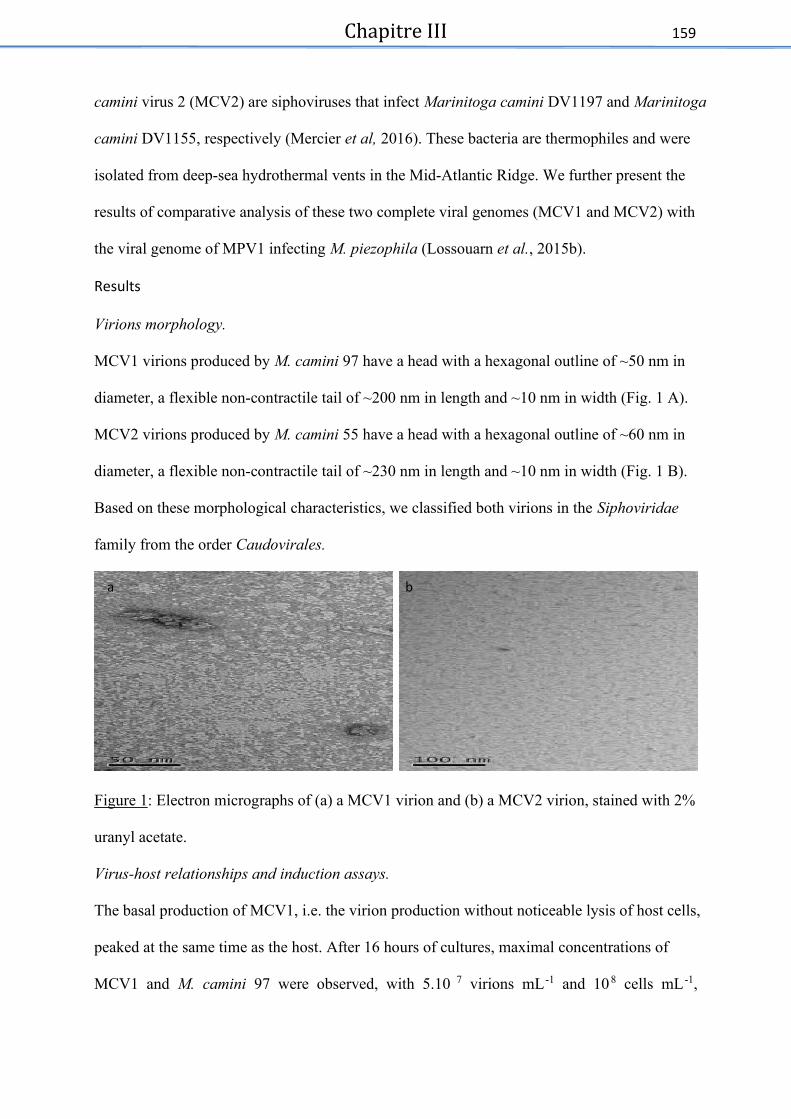
Chapitre III
159
camini virus 2 (MCV2) are siphoviruses that infect Marinitoga camini DV1197 and Marinitoga
camini DV1155, respectively (Mercier et al, 2016). These bacteria are thermophiles and were
isolated from deep-sea hydrothermal vents in the Mid-Atlantic Ridge. We further present the
results of comparative analysis of these two complete viral genomes (MCV1 and MCV2) with
the viral genome of MPV1 infecting M. piezophila (Lossouarn et al., 2015b).
Results
Virions morphology.
MCV1 virions produced by M. camini 97 have a head with a hexagonal outline of ~50 nm in
diameter, a flexible non-contractile tail of ~200 nm in length and ~10 nm in width (Fig. 1 A).
MCV2 virions produced by M. camini 55 have a head with a hexagonal outline of ~60 nm in
diameter, a flexible non-contractile tail of ~230 nm in length and ~10 nm in width (Fig. 1 B).
Based on these morphological characteristics, we classified both virions in the Siphoviridae
family from the order Caudovirales.
Figure 1: Electron micrographs of (a) a MCV1 virion and (b) a MCV2 virion, stained with 2%
uranyl acetate.
Virus-host relationships and induction assays.
The basal production of MCV1, i.e. the virion production without noticeable lysis of host cells,
peaked at the same time as the host. After 16 hours of cultures, maximal concentrations of
MCV1 and M. camini 97 were observed, with 5.10 7 virions mL -1 and 108 cells mL -1,
a b

Chapitre III
160
respectively. After induction, the viral production drastically increased, reaching a peak of ~109
virions mL -1, 4h after mitomycin addition. A concomitant drop in host abundance to ~5.10 6
cells mL-1 was observed, indicating that host cell lysis occurred upon viral production (Fig. 2.
A). Thus, induced cells produce 20-fold more virions than untreated culture. Regrowth of
bacteria was detected 8h after induction (2.10 6 to 10 7 cells mL -1) suggesting that some cells
could withstand the mitomycin C treatment.
As observed for MCV1, the spontaneous MCV2 production in untreated cells was around 5.107
viral particles mL -1 after 8 hours of growth when the cellular production reached a peak to
~7.107 cells mL -1. Four hours after induction, the viral production increased, reaching ~5.10 9
viral particles mL -1, which is 100-fold more virions than for untreated cells. A concomitant
drastic drop to ~107 cells mL-1 was observed in host cell counts, suggesting host cell lysis. Weak
regrowth of cells was observed 6 hours after induction, probably due to some mitomycin C
resistant cells (Fig. 2 B).


Chapitre III
162
In order to check the potential viral induction with environmental parameters, heavy metals
(copper, cobalt, zinc and cadmium) were added to culture. Surprisingly, viral production was
not induced but membrane vesicles (MVs) were observed. After addition of copper or cadmium
(0.5 mM) in an early log culture of M. camini 97, TEM examination revealed a production of
abundant pleomorphic and heterogenous MVs with sizes ranging from 50 to 250 nm (Fig. 3).
Double membranes were observed on the majority of MVs pointing their cellular provenance.
The high MV’s production observed (with a very low concomitant viral production) suggests
that this may be a way for cells to survive metals exposure. At high concentration of metals (up
to 1mM of copper or cadmium), cell lysis was noticed and less MVs were observed.
Figure 3: Electron micrographs of concentrated membrane vesicles, indicated with an arrow,
produced by M. camini 97 (a) after adding 0,1mM of copper (b) or 0,5mM of cadmium, stained
with 2% uranyl acetate.
Attempts to infect a putative sensitive host.
Purified MCV1 and MCV2 virions were added to strains belonging to Marinitoga and
Thermosipho genera (Marinitoga camini strain DV1075, Marinitoga okinawensis, Marinitoga
hydrogenotolerens and Thermosipho sp. strain AT1244-VC14). After the addition of virions no
lysis was observed under a phase-contrast microscope for these potential hosts. Potential
infection was checked by PCR using primers targeting the major capsid protein encoding gene.
a b

Chapitre III
163
Thus, infectivity has not been shown, neither for MCV1 nor for MCV2, although
phylogenetically related strains were tested.
High temperature stability of virions.
Viral particles stability was checked by adding virions in a culture medium, without bacterial
host, and incubated at 60°C. The results show that the stability of the virions is low; in less than
5 hours of incubation, the flow cytometry signal is lost (i.e loss of 22%.h -1). With their hosts,
the number of virions is stable and forms a stationary phase during at least 8 hours (see Fig. 2
A and B). This suggests that with hosts present there is equilibrium between the continuously
viral production and viral decay.
DNA content of MCV1 and MCV2 and sequences analysis.
The genome sequence of M. camini 97 contains a predicted provirus sequence of ~53,400 bp
integrated adjacent to the tRNA-glu gene. Analysis of DNA extracted from the purified MCV1
viral capsids confirmed that the provirus is the integrated form of the MCV1 genome (Fig. S1a).
The MCV1 viral genome, assembled as a linear dsDNA sequence of 53,412 bp, has 77 predicted
ORFs, including 21 annotated genes. These predicted genes covered 90% of the genome
sequence. The majority of the putative genes have the same orientation while 22 are encoded
on the complementary strand. The overall G + C content of the MCV1 genome (28.1%) is
similar to the overall G + C content of the host genome (27.4%).
For M. camini 55, the bacterial chromosome contains a predicted proviral sequence of
~50,300bp also integrated adjacent to the tRNA-glu gene. Sequencing of viral DNA from
purified capsids confirmed that the packaged viral DNA has the same sequence as the predicted
provirus with a similar size (Fig. S1b). MCV2 has a genome length of 50,307 bp, with 70
predicted ORFs, including 17 annotated genes. These predicted genes covered 88.6 % of the
genome sequence. The majority of the putative genes have the same orientation with 22 present

Chapitre III
164
on the complementary strand. The overall G + C content of the MCV2 genome (28.7%) is
comparable with the overall G + C content of the host genome (27.3%).
Results from BLASTN and BLASTP comparisons of the predicted ORFs and proteins from the
two viruses to each other and sequences in Genbank are listed in Supplemental Tables 2 and 3.
MCV1 and MCV2 appear to contain all genes necessary for viral particles synthesis. Several
genes involved in DNA metabolism were identified, including, a replicative DNA helicase,
single stranded DNA binding protein, recombinational DNA repair protein RecT, holliday
junction resolvase and another resolvase. For the head and tail assembly, classical proteins like
terminase large subunit, major capsid protein and tail tape measure protein (a chaperon protein
allowing the tail assembly) were identified. Finally, for the lytic and lysogenic cycles, holin
protein, anti-repressor proteins, transcriptional regulators (six total, including one from XRE
family), SOS response repressor and protease lexA were identified.
Fifty-one ORFs in each virus had a BLASTN match in pairwise comparisons (expected value
< e-0.5). Twenty-four ORFs in each of the virus genomes have no match in Genbank, outside
the Marinitoga genus, when using an expected value cut-off at 0.05 (Supplemental Table 2 and
3). The MCV1 and MCV2 genomes also contain, respectively, 21 and 17 unique ORFs not
found in the other viral genome nor in the MPV1 genome (Fig. 4, Supplemental Tables 2 and
3). The majority of the genes specific to one of the viruses encode hypothetical proteins, and
these viral specific ORFs tend to be found in clusters of three or more genes in the same relative
location in the two genomes (Fig. 4). Interestingly, one of the MCV1 specific genes, UF08_51
encoding a hypothetical protein, has no match in the MCV2 virus, but a match in the Marinitoga
sp. 55 genome (MCV2’s host) outside the provirus region (X274_0211). The viral specific
proteins with predicted functions mainly encode proteins with different DNA binding domains
(e.g. HTH- and zinc ribbons domains) and or typically mobile genes (e.g. toxin-antitoxin
genes).

Chapitre III
165
For both MCV1 and MCV2, most Thermotogae matches outside the genus Marinitoga, were
to genes from Thermosipho genomes (N=13 and N=14, respectively). When comparing the two
genomes to sequences outside the Thermotogae, 37% were affiliated with Firmicutes, 18%-
20% with Proteobacteria and 6%-7% with Archaea (only Euryarchaea).
Comparing the predicted proteins from the two viruses to viral proteins in Genbank, revealed
that 28 MCV1 ORFs and 24 MCV2 had one or more significant matches (expected value <
0.05). The majority of these bacterioviral homologues belong to siphoviruses and myoviruses
isolated from Firmicutes phylum (Bacilli and Clostridia classes) but also from Thermus-
Deinococcus and Proteobacteria phyla. Among these viruses, four were isolated from extreme
environments, Geobacillus bacteriovirus D6E and Epsilonproteobacteria bacteriovirus Nsr1
from hydrothermal deep-sea vents in the Pacific, Thermus bacteriovirus P23-77 from alkaline
hot spring in New Zealand and Thermus bacteriovirus phi OH2 isolated from a hot spring in
Japan. The other bacterioviral homologues were isolated from diverse types of samples as soil
and even from fermented foods.
Comparative genomics of 3 viruses infecting 3 strains and 2 species of Marinitoga.
A genome comparison of MCV1 and MCV2 to the previously described MPV1 bacteriovirus
that infects M. piezophila KA3 is shown in Figure 4, which shows that three genomes have
similar gene synteny, with module organization similar to that seen in various siphoviruses
(Yasmin et al. , 2010). The modules are, 5’ to 3’: replication and transcription, packing, head
and tail assembly, host lysis and finally lysogeny.
The genome sequence of MPV1 is considerably shorter (10 kb) than the two others, and has a
total of 54 predicted ORFs, compared to 77 ORFs in MCV1 and 70 ORFs in MCV2. A set of
conserved “core genes” shared by all the three viruses was identified and includes 35 genes,
among them are proteins for DNA metabolism like replicative DNA helicase and holliday
junction resolvase. Proteins involved in the head and tail assembly are also present in the three

Chapitre III
166
genomes, e.g. terminase large subunit or major capsid protein. Several hypothetical proteins are
found in all three genomes, suggesting that these proteins probably provide important functions
for these viruses (Lossouarn et al., 2015b, Supplemental Tables 2 and 3). Both MCV1 and
MCV2 have large clusters of unique genes (9 genes each) in the 3’-end of their provirus
genome, in their putative lysogeny modules. Similarly, in the lysogeny module, MPV1 has six
genes not found in MCV1 and MCV2, among them a DNA methylase and a histidine kinase.
In these three viral genomes the lysogeny modules appeared relatively more variable.


Chapitre III
168
Discussion
The Thermotogae comprise a widespread phylum of mostly thermophilic or hyperthermophilic
bacteria. Placed deep in the SSU rRNA based tree of life, their phylogeny is complex owing to
numerous HTG-events in their evolutionary history. An important HGT vector is viruses,
nevertheless, only one virus, MPV1 isolated from Marinitoga piezophila, has been
characterized from this phylum (Lossouarn et al., 2015b). The characterization of MCV1 and
MCV2, also infecting Marinitoga strains, adds to our knowledge on the viral diversity in deep-
sea hydrothermal vents.
MCV1 and MCV2 were isolated from M. camini 97 and M. camini 55, respectively, which were
obtained from deep-sea hydrothermal vents in the Mid Atlantic Ridge. M. camini 97 was
isolated from a colonization module deployed at the Lucky strike hydrothermal vent, located at
1700m of depth. M. camini 55 was extracted from a black smoker chimney at the Menez Gwen
site, located at a depth of 840-870m (Colaço et al., 1998). MPV1 was isolated from Marinitoga
piezophila KA3, a piezophlic strain isolated from a deep-sea hydrothermal chimney sample
collected at a depth of 2630 m on the East-Pacific Rise (13S N). Though these host strains come
from geographically distant hydrothermal environments, they colonize the same type of niches
with equivalent physical/chemical qualities, which allow them a similar metabolism based on
anaerobiosis, chemoorganotrophy and thermophily.
In a recent study it was shown that, for the hyperthermophilic Thermotogae, similarity of
environment is more important for gene exchanges than the geographical proximity (Nesbo et
al., 2015). The authors compared 11 Thermotoga maritima-like genomes, isolated from two
environment types (oil reservoirs and marine sites). They reported that the ‘genomes from the
same type of environment tended to be more similar, and exchanged more genes with each other
than with geographical ly close isolates from different types of environments’ (Nesbo et al.,
2015). Another recent study on Thermosipho spp. provides additional support for this

Chapitre III
169
hypothesis underlining the importance of gene flow for the maintenance of distinct lineage and
species (Haverkamp et al., unpublished). Furthermore, one likely vector for such exchanges
could be viruses.
MCV1 and MCV2 are temperate siphoviruses containing double stranded DNA similar to the
previously described MPV1 (Lossouarn et al., 2015b). A set of “core genes” shared by all three
viruses was identified and includes proteins involved in DNA metabolism, head and tail
assembly and lytic and lysogenic cycles. An ORF (UFO08_9 in MCV1 and UFO09_17 in
MCV2) is a part of the core genes and is also found in some Thermosipho spp. genomes
(putative proviruses) (Haverkamp et al., unpublished). It also shares similarities with a
Marinitoga piezophila ORF (outside proviral sequence) encoding for a sigma factor 70. This
ORF could be an example of HGT mediated by viruses.
Among the core genes, shared hypothetical proteins were also identified, suggesting that these
unknown proteins probably provide important functions for these viruses and represent a
reservoir of new genes. Some of these hypothetical genes could represent virus-encoded
auxiliary metabolic genes (VAMGs). These types of specialized viral genes, with a cellular
origin, are however, likely to move back to their hosts where they could be involved in the
microbial adaptation or evolution in these extreme environments as observed in aquatic
ecosystems. Aquatic viral populations serve as gene reservoirs that contribute to niche
partitioning of microbial species (Sime-Ngando, 2014). Moreover, some of VAMGs could be
found in viromes (for example in the Pacific Ocean Virome, POV), improving host fitness, as
genes involved in DNA repair and replication or chemotaxis and motility (Hurwitz et al., 2015).
Thereby, hypothetical proteins present in the core genes of the Marinitoga viruses, could
represent such auxiliary genes useful to help hosts fitness in extreme environmental conditions.
Interestingly, for genes with BLASTP matches in Genbank, over 60% have their top match,
outside the Thermotogae, to genes from Firmicutes and bacterioviruses associated to

Chapitre III
170
Firmicutes (Supplemental Table 2 and 3). Among those viruses, are phi OH2 and D6E infecting
Firmicutes isolated from extreme environment such as hot springs and hydrothermal vents
(Wang and Zhang, 2010; Doi et al., 2013). Thermotogae and Firmicutes appear to have
exchanged large number of genes (Zhaxybayeva et al., 2009), and our findings suggest that
mobile genetic elements may have mediated some of these genes exchanges.
It is notable that most of the genes specific to each of the three viruses are found in the lysogeny
module which contains genes expressed during the part of their lifecycle when they integrate
into, or are integrated in, the host genome. This suggests that acquiring new genes probably
adds a selective advantage for the virus, perhaps making it better at controlling or interacting
with the host genome. The impact of lysogeny on genes dispersal and cell physiology is
probably underestimated. Temperate viruses are often considered as a pool of genetic elements
implicated in microbial genomes evolution and adaptation, but their ecological implication is
so far not well understood (Paul, 2008; Knowles et al., 2016; Thingstad and Bratbak, 2016).
For viruses, being incorporated into the host genome appears to be a strategy to survive harsh
environmental conditions. Under laboratory conditions, at high temperature, the viral decay for
MCV1 virions is near 22%.h-1. Dell Anno et al. showed that in deep-sea ecosystems an
important fraction of viruses (about 25%), once they are released by cell lysis, undergo fast
decomposition (Dell’Anno et al., 2015). They observed that viral decay is faster in deep-sea
ecosystem and that the stability of virions is lower than in other marine ecosystems. In
comparison, in marine environments in the absence of new production, virions decay presents
an average half-life of 48h (Wommack and Colwell, 2000). It is therefore likely that it is helpful
for the extremophile virus to stay incorporated in the host genome rather than to shift to a lytic
cycle where virions will have a very short time to infect another host.
An interesting feature of the host-viruses kinetics for all three Marinitoga viruses are high
spontaneous viral productions (more than 10 7 virions ml -1) which may be due to the fluctuant
Chapitre III
171
and extreme physicochemical conditions they encounter. This was also described for another
bacteriovirus Nrs-1, infecting a thermophilic ɛProteobacterium from the same type of
environment (Yoshida-Takashima et al., 2013), where virions appeared to be released
continuously and spontaneously, probably resulting from stochasticity in gene expression or a
low induction of the hosts SOS response. Spontaneous provirus induction, i.e. virions’
production, could thus be advantageous for both lysogenic hosts and their proviruses. On the
one hand, a portion of non-lysogenized bacteria, potentially competitive, could be killed and on
the other hand survivors, undergoing lysogenic conversion, could be able to spread the viral
DNA. This spontaneously virions release, could also have an important ecological impact by
releasing organic matter, which may contribute to the nutrient cycling in hydrothermal deep-
sea vents.
Environmentally relevant stressors (i.e. high concentrations of heavy metals) did not induce the
production of virions but we detected an increase in membrane vesicles (MVs) production. The
production of MVs is a universal physiological phenomenon shared by cells from all three
domains of life (Kulp and Kuehn, 2010; Deatherage and Cookson, 2012). MVs are involved in
numerous physiological processes such as cell-to-cell communications, nucleic acid or toxins
transport, stress response and interactions with viruses (Soler et al., 2008; Manning and Kuehn,
2011; Gaudin et al., 2013; Biller et al., 2014). For Thermococcales it was demonstrated that
MVs could be a defense system for cells by preventing toxic accumulation of sulfur (Gorlas et
al., 2015). Similarly, several studies on eukaryotes and bacteria have shown that membrane
vesicles can play a role in detoxification (Sterling et al., 2007; Jagannadham and
Chattopadhyay, 2015; Slavic et al., 2016). The phenomenon was first observed in eukaryotic
marine organisms such as mollusks and crustaceans, which accumulate cadmium and where
excretion of cadmium is performed by MVs as a detoxifying mechanism (Sterling et al., 2007).
The observation of increased MVs production with addition of copper or cadmium suggests

Chapitre III
172
that the MVs produced by Marinitoga could be involved in detoxification. The fact that
environments where these organisms thrive, hydrothermal vents, are characterized by the
presence of high levels of heavy metals (cadmium, copper, zinc…) supports this notion.
Further studies will be needed in order to test if MVs can protect the cells against heavy metals
toxicity. Moreover, since these strains produce both MVs and viruses they could serve as
models to investigate mechanisms involved in virus/MVs/hosts’ interactions.
Globally, viruses and other mobile genetic elements (MGEs) have a strong impact on microbial
ecology, especially in oceans. The analyses presented here extend our knowledges about these
newly discovered mobile genetic elements in the deeply branching bacterial phylum
Thermotogae. This bacterial order and associated MGEs are significant for addressing long-
term evolutionary adaptation to fluctuant and extreme physicochemical conditions. Our work
confirms a strong evolutionary connection of Thermotogae’ viruses to Firmicutes and their
bacterioviruses. The impact of MGEs on their hosts is very important and it remains crucial to
study new host-MGEs’ relationships in an ecological context to well understand the reason of
this old and deep co-evolution.
Experimental procedures
Screening of Thermotogae strains for mobile genetic elements
A total of 55 strains of Thermotogae were retrieved from the UBO culture collection ‘UBOCC’
(http://www.univ-brest.fr/UBOCC) and from our laboratory culture collections. These strains
were isolated from different deep-sea vents in the Lau Basin, the East Pacific Rise and the
Middle Atlantic Ridge and were screened for mobile genetic elements as previously described
(Lossouarn et al., 2015b). Two of them, Marinitoga camini DV1197 and Marinitoga camini
DV1155, were discovered to be the hosts of viruses MCV1 and MCV2 respectively.
Culture conditions.

Chapitre III
173
Marinitoga camini DV1197 (hereafter referred to as M. camini 97) was cultured at 60°C in a
modified Ravot medium as previously described (Geslin et al., 2003a,b; Lossouarn et al.,
2015b). The medium was completed by adding 1% (wt/vol) elemental sulfur. Marinitoga
camini DV1155 (hereafter referred to as M. camini 55) was cultured in the same medium at
65°C.
Viral induction assays.
Attempts were made to increase the viral production by induction using an antibiotic treatment
that damage DNA (mitomycin C) as well as metal stresses (Llanos et al., 2000; Yoshida-
Takashima et al., 2013). A concentration of 4µg/mL of mitomycin C was used as described in
Lossouarn et al., 2015b. Induction assays by metals were done on 25 mL of M. camini 97
culture in early log-phase, by adding various metals (copper, cadmium, zinc, cobalt) with
different concentrations (1 to 0.001 mM for copper and cadmium and 0.1 to 0.001 mM for zinc
and cobalt). After 24h incubation period, debris and cells were pelleted by centrifugation at
7500g, 4°C for 20 min and discarded. Supernatants were ultracentrifuged at 80,000g, 10°C for
1h (Beckman Optima LE-80 K 70.1 Ti rotor). Pellets were resuspended in 50µL of buffer (10
mM Tris-HCL, 100 mM NaCl, 5 mM CaCl2, 20 mM MgCl2) and observed using a JEOL JEM
100 CX II® transmission electron microscope as described previously (Geslin et al., 2003a,b;
Gorlas et al., 2012).
Flow cytometry counts of bacteria and viruses.
Samples were fixed with 25% glutaraldehyde (0.5% final concentration, Electron microscopy
(EM) grade, Sigma-Aldrich) for 15 min at 4°C, flash frozen in liquid nitrogen and stored at -
80°C until analysis. The thawed samples were diluted from 100 to 10,000-fold in autoclaved
0.2 µm filtered TE buffer (10:1 Tris–EDTA, pH 8.0) and stained with the nucleic acid-specific
dye SYBR Green I (Life technologies®) for 15 min at room temperature for bacteria and 10
min at 80°C for viruses as described in Brussaard (2004). Samples were analyzed using a
Chapitre III
174
FACSCanto II flow cytometer (BD Bioscience®) equipped with a laser with an excitation
wavelength of 488 nm (15 mW). The trigger was set on green fluorescence. The samples were
delivered at a flow rate of 30 or 50 µl.min-1 and analyzed during at least 1 min. TE-buffer with
autoclaved 0.2 µm filtered seawater was used as a control.
Viral concentration and purification.
Five hundred milliliters of M. camini 97 and 55 were induced by addition of mitomycin C (4
µg/mL) in early log-phase. Three hours after induction, debris and cells were pelleted by
centrifugation at 7,500 g, 4°C for 15 min and discarded. The supernatant was ultracentrifuged
at 80,000 g, 10°C for 1 h (Beckman Optima LE-80 K 70.1 Ti rotor®). The viral pellet was
resuspended in 1 mL of buffer (10 mM Tris-HCL, 100 mM NaCl, 5 mM CaCl2, 20 mM
MgCl2). The viral suspension was purified by ultracentrifugation in a linear iodixanol gradient
[OptiPrep®, 25–40% diluted in a buffer (see above)] at 200, 000 g and 10°C for 10 h (Beckman
Optima LE-80 K SW 41 rotor®). After ultracentrifugation, an opaque virus band was recovered
and stored at 4°C until use. To exclude the possibility of cellular nucleic acids contamination,
the concentrated viral particles suspensions were treated with exo-DNase RQ1 and RNase
(Promega®, 2 U/ml each) for 45min at 37°C, followed by addition of DNAse-stop and a heat
treatment (10 min at 70°C). The viral particles were examined using a JEOL JEM 100 CX II®
transmission electron microscope as previously described (Geslin et al., 2003a,b; Gorlas et al.,
2012).
Attempts to infect a putative sensitive host.
Marinitoga and Thermosipho spp. isolates from our culture collection were screened for the
presence of virions and strains scored negatively were selected as potential hosts. As a result,
Thermosipho sp. AT1244-VC14, Marinitoga camini DV1075, Marinitoga hydrogenotolerens
and Marinitoga okinawensis were tested. Different levels of multiplicity of infection (MOI), of
purified MCV1 and MCV2, were tested (50, 30, 10, 1, 0.1, 0.001 and 0.0001 virions per cell).

Chapitre III
175
Non-infected cultures were used as negative control. After 8 subcultures, total DNA was
extracted from each subculture. PCR amplifications were performed on total DNA as described
in supporting information by using primers that target the major capsid protein encoding gene
of MCV1 and 16S rRNA gene (Table S1).
High temperature stability of virions.
Purified virions of MCV1 (3.108 virions/mL) were added in modified Ravot medium and
incubated during 22 hours at 60°C. 500µl were sampled, in triplicate, every 2 hours (0h to 8h)
and two supplementary samples were obtained at 20 and 22 hours. Each sample was flash frozen
in liquid nitrogen and fixed with glutaraldehyde and stored at -80°C, until flow cytometry
analysis as described before.
Extraction of cellular and viral DNA.
Total DNA from Thermosipho sp. AT1244-VC14, M. camini 97 and M. camini 55 cells and
viral DNA from purified virions (MCV1 and MCV2) were prepared as described previously
(Geslin et al., 2003a,b; Gorlas et al., 2012). In order to obtain viral genomes, we eliminated
extracapsid DNA contamination, by treating the purified viral suspensions from induced
cultures with DNases and RNase (DNAse RQ1 2U.ml -1, ExoIII 2U.ml-1, RNAse A 5mg.ml -1)
before DNA extraction.
Sequencing and assembly of bacterioviruses DNA.
Purified viral DNA of MCV1 and MCV2 were sequenced as barcoded libraries on a MiSeq
using 500 cycles generating 2x250 bp paired-end reads, using the Nextera XT kit for library
construction. The sequences were assembled using the CLC Genomics Workbench 6.5.1
(http://www.clcbio.com). Sequence analyses of protein-coding genes of bacterioviruses
(MCV1 and MCV2) are described in Supporting information.

Chapitre III
176
Ackowledgments
This work was financially supported by the French Ministry of Higher Education and the
Brittany region to CM and the Agence Nationale de la Recherche, project Thermovesicles
(ANR-12-BSV3-OO23-01) to CG and JL, a Norwegian Research Council award (project no.
180444/V40) to CLN. We thank G. Sinquin and P. Elies for assistance in electron microscopy.
Conflict of interest
The authors declare no conflict of interest.
References
Alain, K., Marteinsson, V.T., Miroshnichenko, M., Bonch-osmolovskaya, E., Prieur, D., and
Birrien, J. (2002) Marinitoga piezophila sp. nov., a rod-shaped, thermo-piezophilic
bacterium isolated under high hydrostatic pressure from a deep-sea hydrothermal vent. Int.
J. Syst. Evol. Microbiol. 52: 1331–1339.
Auguste, M., Mestre, N.C., Rocha, T.L., Cardoso, C., Cueff-Gauchard, V., Le Bloa, S., et al.
(2016) Development of an ecotoxicological protocol for the deep-sea fauna using the
hydrothermal vent shrimp Rimicaris exoculata. Aquat. Toxicol. 175: 277–285.
Bhandari, V. and Gupta, R.S. (2015) Erratum to: Molecular signatures for the phylum (class)
Thermotogae and a proposal for its division into three orders (Thermotogales,
Kosmotogales ord. nov. and Petrotogales ord. nov.) containing four families
(Thermotogaceae, Fervidobacteriaceae fam. nov., Kosmotogaceae fam. nov. and
Petrotogaceae fam. nov.) and a new genus Pseudothermotoga gen. nov. with five new
combinations. Antonie Van Leeuwenhoek 108: 1281.
Biller, S.J., Schubotz, F., Roggensack, S.E., Thompson, A.W., Summons, R.E., and Chisholm,
S.W. (2014) Bacterial vesicles in marine ecosystems. Science. 343: 183–186.
Brussaard, C. P. (2004). Optimization of procedures for counting viruses by flow cytometry.
Applied and Environmental Microbiology, 70: 1506-1513.

Chapitre III
177
Colaco, A., Desbruyeres, D., Comtet, T., & Alayse, A. M. (1998). Ecology of the Menez Gwen
hydrothermal vent field (Mid-Atlantic Ridge Azores Triple Junction). Cahiers de Biologie
Marine, 39: 237-240.
Deatherage, B.L. and Cookson, B.T. (2012) Membrane vesicle release in Bacteria, Eukaryotes,
and Archaea: a conserved yet underappreciated aspect of microbial life. Infect. Immun.
80: 1948–1957.
Dell’Anno, A., Corinaldesi, C., and Danovaro, R. (2015) Virus decomposition provides an
important contribution to benthic deep-sea ecosystem functioning. Proc. Natl. Acad. Sci.
112: E2014–E2019.
Doi, K., Mori, K., Martono, H., Nagayoshi, Y., Fujino, Y., Tashiro, K., et al. (2013) Draft
genome sequence of Geobacillus kaustophilus GBlys, a lysogenic strain with
bacteriophage ϕOH2. Genome Announc. 1: e00634-13.
Flores, G.E. and Reysenbach, A.-L. (2011) Hydrothermal environments, marine. In,
Encyclopedia of Geobiology. Springer, pp. 456–467.
Gaudin, M., Gauliard, E., Schouten, S., Houel-Renault, L., Lenormand, P., Marguet, E., and
Forterre, P. (2013) Hyperthermophilic archaea produce membrane vesicles that can
transfer DNA. Environ. Microbiol. Rep. 5: 109–16.
Geslin, C., Gaillard, M., Flament, D., Rouault, K., Le Romancer, M., Prieur, D., and Erauso,
G. (2007) Analysis of the first genome of a hyperthermophilic marine virus-like particle,
PAV1, isolated from Pyrococcus abyssi. J. Bacteriol. 189: 4510–4519.
Geslin, C., Le Romancer, M., Erauso, G., Gaillard, M., Perrot, G., and Prieur, D. (2003a) PAV1,
the first virus-like particle isolated from a hyperthermophilic euryarchaeote, “Pyrococcus
abyssi.” J Bacteriol 185: 3888–3894.
Geslin, C., Le Romancer, M., Gaillard, M., Erauso, G., and Prieur, D. (2003b) Observation of
virus-like particles in high temperature enrichment cultures from deep-sea hydrothermal

Chapitre III
178
vents. Res. Microbiol. 154: 303–307.
Gorlas, A., Koonin, E. V, Bienvenu, N., Prieur, D., and Geslin, C. (2012) TPV1, the first virus
isolated from the hyperthermophilic genus Thermococcus. Environ. Microbiol. 14: 503 –
516.
Gorlas, A., Marguet, E., Gill, S., Geslin, C., Guigner, J.M., Guyot, F., and Forterre, P. (2015)
Sulfur vesicles from Thermococcales: A possible role in sulfur detoxifying mechanisms.
Biochimie 118: 356–364.
Gupta, R.S. and Bhandari, V. (2011) Phylogeny and molecular signatures for the phylum
Thermotogae and its subgroups. Antonie Van Leeuwenhoek 100: 1–34.
Guy, L., Kultima, J.R., Andersson, S.G.E. (2010) genoPlotR: comparative gene and genome
visualization in R. Bioinformatics 26 : 2334-2335.
Hug, L.A., Baker, B.J., Anantharaman, K., Brown, C.T., Probst, A.J., Castelle, C.J., et al.
(2016) A new view of the tree of life. Nat. Microbiol. 1: 16048.
Hurwitz, B.L., Brum, J.R., and Sullivan, M.B. (2015) Depth-stratified functional and taxonomic
niche specialization in the “core”and “flexible”Pacific Ocean Virome. ISME J. 9: 472 –
484.
Jagannadham, M. V and Chattopadhyay, M.K. (2015) Role of outer membrane vesicles of
bacteria. Resonance 20: 711–725.
Jebbar, M., Franzetti, B., Girard, E., and Oger, P. (2015) Microbial diversity and adaptation to
high hydrostatic pressure in deep-sea hydrothermal vents prokaryotes. Extremophiles 19:
721–740.
Knowles, B., Silveira, C.B., Bailey, B.A., Barott, K., Cantu, V.A., Cobián-Güemes, A.G., et al.
(2016) Lytic to temperate switching of viral communities. Nature 531: 466–470.
Kulp, A. and Kuehn, M.J. (2010) Biological functions and biogenesis of secreted bacterial outer
membrane vesicles. Annu. Rev. Microbiol. 163–184.

Chapitre III
179
Kuwabara, T. and Igarashi, K. (2012) Microscopic studies on Thermosipho globiformans
implicate a role of the large periplasm of Thermotogales. Extremophiles 16: 863–870.
Liu, B., Wu, S., Song, Q., Zhang, X., and Xie, L. (2006) Two novel bacteriophages of
thermophilic bacteria isolated from deep-sea hydrothermal fields. Curr. Microbiol. 53:
163–166.
Liu, B. and Zhang, X. (2008) Deep-sea thermophilic Geobacillus bacteriophage GVE2
transcriptional profile and proteomic characterization of virions. Appl. Microbiol.
Biotechnol. 80: 697–707.
Llanos, J., Capasso, C., Parisi, E., Prieur, D., and Jeanthon, C. (2000) Susceptibility to heavy
metals and cadmium accumulation in aerobic and anaerobic thermophilic microorganisms
isolated from deep-sea hydrothermal vents. Curr. Microbiol. 41: 201–205.
Lossouarn, J., Dupont, S., Gorlas, A., Mercier, C., Bienvenu, N., Marguet, E., et al. (2015a) An
abyssal mobilome: Viruses, plasmids and vesicles from deep-sea hydrothermal vents. Res.
Microbiol. 166: 742–752.
Lossouarn, J., Nesbø, C.L., Mercier, C., Zhaxybayeva, O., Johnson, M.S., Charchuck, R., et al.
(2015b) “Ménage à trois”: a selfish genetic element uses a virus to propagate within
Thermotogales. Environ. Microbiol. 17: 3278–3288.
Lucas, S., Han, J., Lapidus, A., Cheng, J.-F., Goodwin, L.A., Pitluck, S., et al. (2012) Complete
genome sequence of the thermophilic, piezophilic, heterotrophic bacterium Marinitoga
piezophila KA3. J. Bacteriol. 194: 5974–5975.
Manning, A.J. and Kuehn, M.J. (2011) Contribution of bacterial outer membrane vesicles to
innate bacterial defense. BMC Microbiol. 11: 258.
Mercier, C., Lossouarn, J., Haverkamp, T., Bienvenu, N., Godfroy, A., Cueff-Gauchard, V., et
al. (2016) Draft genome sequences of two Marinitoga camini isolates producing
bacterioviruses. Genome announc. 4: 6.

Chapitre III
180
Nesbø, C.L., Bradnan, D.M., Adebusuyi, A., Dlutek, M., Petrus, A.K., Foght, J., et al. (2012)
Mesotoga prima gen. nov., sp. nov., the first described mesophilic species of the
Thermotogales. Extremophiles 16: 387–393.
Nesbo, C.L., S Swithers, K., Dahle, H., Haverkamp, T.H.A., Birkeland, N.-K., Sokolova, T., et
al. (2015) Evidence for extensive gene flow and Thermotoga subpopulations in subsurface
and marine environments. ISME J. 9: 1532–1542.
Paul, J.H. (2008) Prophages in marine bacteria: dangerous molecular time bombs or the key to
survival in the seas? ISME J. 2: 579–589.
Pollo, S.M.J., Zhaxybayeva, O., and Nesbø, C.L. (2015) Insights into thermoadaptation and the
evolution of mesophily from the bacterial phylum Thermotogae. Can. J. Microbiol. 61:
655–670.
Sime-Ngando, T. (2014) Environmental bacteriophages: viruses of microbes in aquatic
ecosystems. Front. Microbiol. 5: 355.
Slavic, K., Krishna, S., Lahree, A., Bouyer, G., Hanson, K.K., Vera, I., et al. (2016) A vacuolar
iron-transporter homologue acts as a detoxifier in Plasmodium. Nat. Commun. 7.
Soler, N., Marguet, E., Verbavatz, J.M., and Forterre, P. (2008) Virus-like vesicles and
extracellular DNA produced by hyperthermophilic archaea of the order Thermococcales.
Res. Microbiol. 159: 390–399.
Sterling, K.M., Mandal, P.K., Roggenbeck, B.A., Ahearn, S.E., Gerencser, G.A., and Ahearn,
G.A. (2007) Heavy metal detoxification in crustacean epithelial lysosomes: role of anions
in the compartmentalization process. J. Exp. Biol. 210: 3484–3493.
Thingstad, T.F. and Bratbak, G. (2016) Microbial oceanography: Viral strategies at sea. Nature
531: 454–455.
Wang, Y. and Zhang, X. (2010) Genome analysis of deep-sea thermophilic phage D6E. Appl.
Environ. Microbiol. 76: 7861–7866.
Chapitre III
181
Wommack, K.E. and Colwell, R.R. (2000) Virioplankton: Viruses in aquatic ecosystems.
Microbiol. Mol. Biol. Rev. 64: 69–114.
Yasmin, A., Kenny, J.G., Shankar, J., Darby, A.C., Hall, N., Edwards, C., and Horsburgh, M.J.
(2010) Comparative genomics and transduction potential of Enterococcus faecalis
temperate bacteriophages. J. Bacteriol. 192: 1122–1130.
Yoshida-Takashima, Y., Takaki, Y., Shimamura, S., Nunoura, T., and Takai, K. (2013) Genome
sequence of a novel deep-sea vent epsilonproteobacterial phage provides new insight into
the co-evolution of Epsilonproteobacteria and their phages. Extremophiles 17: 405–419.
Zhaxybayeva, O., Swithers, K.S., Foght, J., Green, A.G., Bruce, D., Detter, C., et al. (2012)
Genome sequence of the mesophilic Thermotogales bacterium Mesotoga prima
MesG1.Ag.4.2 reveals the largest Thermotogales genome to date. Genome Biol. Evol. 4:
700–708.
Zhaxybayeva, O., Swithers, K.S., Lapierre, P., Fournier, G.P., Bickhart, D.M., DeBoy, R.T., et
al. (2009) On the chimeric nature, thermophilic origin, and phylogenetic placement of the
Thermotogales. Proc. Natl. Acad. Sci. U. S. A. 106: 5865–5870.
Supporting Information
Figure S1: Comparison of viral genomes isolated form capsids and provirus integrated in the
host genomes. The sequences were aligned using MAUVE 2.3.1 and the comparison was
plotted using GenoplotR (Guy et al., 2010) a: MCV1 and M. camini 97 and b: MCV2 and M.
camini 55.
Table S1: Primers targeting ARNr 16S, MCV1 and MCV2 major capsid proteins.
Table S2: Annotations and comparison tables of MCV1.
Table S3: Annotations and comparison tables of MCV2.

Chapitre III
182
Supporting informations
Supplementary Material and Methods
Sequence assembly and analysis
The MCV1 and MCV2 genomes were assembled de novo by CLC Genomics Workbench 7.0.4,
using trimming default settings, automatic word size, a bubble size corresponding to the
average length of the input reads, a minimum contig length of 1000 bp, and reads mapped
back to the contigs. Coding sequences (CDS) were identified and annotated in the Geneious 9
(www.geneious.com). BLASTP searches against Genbanks none redundant database (nr) and
pairwise BLASTN searches to compare the three viruses (MPV1, MCV1 and MCV2) were also
performed in Geneious 9 (www.geneious.com). Blast searches against virus sequences in
Genbank were done at NCBI (https://blast.ncbi.nlm.nih.gov/Blast.cgi) limiting the search to
Viruses (taxid:10239). Conserved domain (CD) searches were also done at NCBI. Both Genome
sequences have been deposited at DDBJ/EMBL/GenBank under the accession no. KP836355
and KP836356 for MCV1 and MCV2, respectively.
Table S1: Primers targeting ARNr 16S and MCV1 & MCV2 major capsid proteins
Target Forward/reverse Sequence Bacterial 16S rRNA gene Forward AGAGTTTGATCATGGCTCAG Bacterial 16S rRNA gene Reverse GGTTACCTTGTTACGACT T Major Capsid Protein MCV1&MCV2
Forward AGCTCAGCCAACAAAAGCAG
Major Capsid Protein MCV1&MCV2
Reverse AGAAGGAATTTGGGCTGCTG

locus tag annotation Length Best match in MCV22 % Identity Match in MPV1%
identity2 expectedbast match outside the Marinitoga inBLASP searches of Genbank
UF08_1 hypothetical protein 169UF09_6_hypotheticalprotein 95% WP_042729055 phage replication protein O Pseudomonas fluorescens
UF08_2 hypothetical protein 70UF09_7_hypotheticalprotein 99% no match
UF08_3 replicative DNA helicase 427UF09_8_replicative DNAhelicase 98% Marpi_0305 replicative DNA helicase 66% 0 WP_031484309 replicative DNA helicase
UF08_4single-stranded DNA-bindingprotein 142
UF09_9_single-strandedDNA-binding protein 97% Marpi_0443 single stranded DNA-binding protein 57% 2,34E-52 WP_052670309 hypothetical protein
UF08_5 hypothetical protein 92UF09_13_hypotheticalprotein 99% Marpi_0308 hypothetical protein 47% 5,06E-08 WP_015616411 hypothetical protein
UF08_6 hypothetical protein 82UF09_14_hypotheticalprotein 100% Marpi_0309 hypothetical protein 53% 6,58E-15
XP_008855251 hypothetical protein ENU1_022120
UF08_7 Holliday junction resolvase RusA 122UF09_15_hypotheticalprotein 94% Marpi_0310 Holliday junction DNA helicase 72% 3,10E-58 WP_054198714 hypothetical protein
UF08_8 hypothetical protein 77UF09_16_hypotheticalprotein 99% Marpi_0314 hypothetical protein 86% 6,39E-38 no match
UF08_9 hypothetical protein 161UF09_17_hypotheticalprotein 95% Marpi_2120 DNA-directed RNA polymerase sigma-70 factor 38% 1,11E-25
WP_012057673 RNA polymerase subunit sigma-24
UF08_10 hypothetical protein 71 WP_002672686 hypothetical proteinUF08_11 hypothetical protein 337 WP_025278489 hypothetical protein
UF08_12 hypothetical protein 127UF09_19_hypotheticalprotein 100% Marpi_0319 hypothetical protein 83% 2,09E-60 WP_047809280 hypothetical protein
UF08_13 terminase large subunit 416UF09_20_terminase largesubunit 99% Marpi_0320 phage terminase, large subunit, PBSX famil 97% 0 WP_039259229 terminase
UF08_14 hypothetical protein 405UF09_21_hypotheticalprotein 99% Marpi_0321 hypothetical protein 93% 0 WP_027339946 hypothetical protein
UF08_15 hypothetical protein 63 no match
UF08_16 hypothetical protein 51
XP_006580293 PREDICTED: regulation of nuclear pre-mRNA domain-containing protein 1B-like isoform X3
UF08_17 hypothetical protein 42 WP_052353345 hypothetical protein
UF08_18 hypothetical protein 62UF09_22_hypotheticalprotein 98%
XP_008914408 hypothetical protein PPTG_24293
UF08_19 hypothetical protein 319UF09_23_hypotheticalprotein 99% Marpi_0322 hypothetical protein 88% 0 WP_038226045 hypothetical protein
UF08_20 hypothetical protein 45UF09_24_hypotheticalprotein 100% Marpi_0323 hypothetical protein 91% 1,59E-19 no match
UF08_21 Chaperone protein DnaK 200UF09_25_hypotheticalprotein 99% Marpi_0324 hypothetical protein 81% 6,53E-98
WP_063491264 DUF4355 domain-containing protein
UF08_22 hypothetical protein 118UF09_26_hypotheticalprotein 99% Marpi_0325 hypothetical protein 93% 3,54E-72 WP_013866486 hypothetical protein
UF08_23 major capsid protein 340UF09_27_major capsidprotein 99% Marpi_0326 phage capsid protein 91% 0 EGO87149 capsid protein
UF08_24 hypothetical protein 61UF09_28_hypotheticalprotein 100% Marpi_0327 hypothetical protein 80% 1,38E-24
KLD98821 hypothetical protein AF74_02140
UF08_25 hypothetical protein 107UF09_29_hypotheticalprotein 100% Marpi_0328 hypothetical protein 86% 2,41E-59 WP_012063953 hypothetical protein
UF08_26 hypothetical protein 158UF09_30_hypotheticalprotein 100% Marpi_0329 hypothetical protein 97% 3,14E-106 WP_006306523 hypothetical protein
UF08_27 hypothetical protein 127UF09_31_hypotheticalprotein 98% Marpi_0330 hypothetical protein 88% 8,57E-75
WP_010381216 MULTISPECIES: cell divisiontopological specificity factor MinE

locus tag annotation Length Best match in MCV22 % Identity Match in MPV1%
identity2 expectedbast match outside the Marinitoga inBLASP searches of Genbank
UF08_28 hypothetical protein 327UF09_32_hypotheticalprotein 70% Marpi_0331 hypothetical protein 34% 1,76E-46 WP_004102183 hypothetical protein
UF08_29 hypothetical protein 313UF09_32_hypotheticalprotein 67% Marpi_0331 hypothetical protein 65% 2,87E-150 WP_004102183 hypothetical protein
UF08_30 hypothetical protein 125UF09_33 hypotheticalprotein 39% Marpi_0332 hypothetical protein 38% 4,79E-11 WP_004102185 hypothetical protein
UF08_31 hypothetical protein 163UF09_34_hypotheticalprotein 29% Marpi_0333 hypothetical protein 25% 2,47 WP_004102187 hypothetical protein
UF08_32 tail length tape-measure protein 1 304UF09_35_tail length tapemeasure protein 89% Marpi_0334 phage tail tape measure protein, TP901 family 28% 7,57E-41
WP_012057655 phage tail tape measure protein
UF08_33 hypothetical protein 201 Marpi_0335 hypothetical protein 76% 2,06E-92 WP_021570654 hypothetical protein
UF08_34 hypothetical protein 115UF09_37_hypotheticalprotein 93% Marpi_0336 hypothetical protein 66% 6,87E-47 WP_004102189 hypothetical protein
UF08_35 hypothetical protein 393UF09_38_hypotheticalprotein 91% Marpi_0337 hypothetical protein 32% 2,68E-37 WP_004102191 hypothetical protein
UF08_36 Hypothetical protein 1 374UF09_39_phage realtedprotein tail 92% Marpi_0338 hypothetical protein 53% 2,30E-40 WP_004102192 hypothetical protein
UF08_37 hypothetical protein 79UF09_40_hypotheticalprotein 100%
KYC55724 hypothetical protein APG09_01571
UF08_38 hypothetical protein 213KKT87314 hypothetical protein UW86_C0004G0005
UF08_39 hypothetical protein 156KKT21313 hypothetical protein UW04_C0006G0016
UF08_40 hypothetical protein 254UF09_41_DNA repairexonuclease 98% Marpi_0340 DNA repair exonuclease 78% 5,56E-152 WP_004102197 DNA repair exonuclease
UF08_41 hypothetical protein 97UF09_42_hypotheticalprotein 100% Marpi_0341 hypothetical protein 41% 5,37E-07
KYC55578 hypothetical protein APG08_01587
UF08_42 hypothetical protein 127UF09_43_hypotheticalprotein 99% Marpi_0342 hypothetical protein 75% 2,64E-62 WP_012057645 hypothetical protein
UF08_43 transcriptional regulator 71KXS38990 hypothetical protein AWU54_2187
UF08_44 antirepressor protein 294 WP_020411111 antirepressor
UF08_45 Phage holin protein 100UF09_47_phage holinprotein 98% Marpi_0343 phage holin protein (Holin_LLH) 54% 4,68E-30 WP_016226830 LL-H family phage holin
UF08_46 hypothetical protein 97UF09_48_hypotheticalprotein 97% Marpi_0344 hypothetical protein 57% 2,69E-31 WP_032557479 succinate dehydrogenase
UF08_47 hypothetical protein 188UF09_49_hypotheticalprotein 99% Marpi_0345 Orthopoxvirus hypothetical protein 80% 1,06E-109
CUB50865 hypothetical protein BN2127_JRS10_00382
UF08_48 resolvase 485UF09_50_Resolvase N-terminal domain 69% Marpi_0291 site-specific recombinase DNA invertase Pin 44% 2,26E-121 WP_036226465 hypothetical protein
UF08_49 hypothetical protein 56 Marpi_0292 hypothetical protein 77% 8,09E-22WP_059109184 ribosome biogenesis GTPase Der
UF08_50 hypothetical protein 121 WP_057574369 hypothetical protein
UF08_51 hypothetical protein 153KUK80138 Uncharacterized protein XD96_1662
UF08_52 hypothetical protein 201KUK80137 Uncharacterized protein XD96_1661
UF08_53 hypothetical protein 134KUK80136 Uncharacterized protein XD96_1660
UF08_54 hypothetical protein 361KPV61867 hypothetical protein AOA66_1539

locus tag annotation Length Best match in MCV22 % Identity Match in MPV1%
identity2 expectedbast match outside the Marinitoga inBLASP searches of Genbank
UF08_55 bipolar DNA helicase HerA 553CUU36910 hypothetical protein DCOP10_11973
UF08_56 hypothetical protein 345 WP_027719510 hypothetical protein
UF08_57 hypothetical protein 119XP_010260093 PREDICTED: uncharacterizedprotein LOC104599305
UF08_58 hypothetical protein 369 CDB45357 putative uncharacterized protein
UF08_59 transcriptional regulator 202
UF09_61_SOS-responserepressor and proteaseLexA 78%
Marpi_0297 SOS response transcriptional repressor, RecA-mediated autopeptidase 45% 1,76E-52 WP_053001073 hypothetical protein
UF08_60SOS-response repressor andprotease LexA 216
UF09_61_SOS-responserepressor and proteaseLexA 99%
Marpi_0297 SOS response transcriptional repressor, RecA-mediated autopeptidase 43% 1,10E-37 WP_053001073 hypothetical protein
UF08_61 transcriptional regulator 71UF09_62_hypotheticalprotein 99% Marpi_0298 transcriptional regulator 54% 5,46E-11 KKQ85063 Helix-turn-helix domain protein
UF08_62 antirepressor protein 241 WP_015397656 anti-repressor protein
UF08_63 hypothetical protein 64KUO58776 cobalt ABC transporter ATP-binding protein
UF08_64 hypothetical protein 241 WP_045166120 haloacid dehalogenase
UF08_65transcriptional regulator, XREfamily 113
UF09_64_Transcriptionalregulator XRE family 98% WP_062805019 hypothetical protein
UF08_66 transcriptional regulator 70UF09_65_hypotheticalprotein 86% WP_057001098 hypothetical protein
UF08_67 hypothetical protein 111UF09_66_hypotheticalprotein 99% KKW41634 Foldase protein PrsA
UF08_68 hypothetical protein 232UF09_67_hypotheticalprotein 100%
XP_005178606 PREDICTED: laminin subunitalpha
UF08_69 hypothetical protein 78UF09_68_hypotheticalprotein 99% KKW21360 Alanine-tRNA ligase
UF08_70 hypothetical protein 54UF09_69_hypotheticalprotein 98%
XP_008625909 hypothetical protein YYE_04044
UF08_71 hypothetical protein 154 Marpi_0299 hypothetical protein 58% 3,30E-47 no match
UF08_72 Viral nuclease 313UF09_70_YqaJ-like viralrecombinase 100% WP_004102128 hypothetical protein
UF08_73recombinational DNA repairprotein RecT 294
UF09_1_recombinationalDNA repair protein RecT 100%
WP_004102129 DNA recombination proteinRecT
UF08_74 hypothetical protein 78UF09_2_hypotheticalprotein 100% WP_047597684 hypothetical protein
UF08_75 hypothetical protein 43UF09_3_hypotheticalprotein 100% CZZ10673 Uncharacterised protein
UF08_76 transcriptional regulator 63UF09_4_hypotheticalprotein 100%
WP_049499605 MULTISPECIES: transcriptional regulator
UF08_77 hypothetical protein 103UF09_5_hypotheticalprotein 100% WP_059175353 hypothetical protein

locus tag annotation best match outside Thermotogaetaxonomic name of best matchouside the Thermotogae
expected(outside
Thermotogae)
% indentity(outside
Thermotogae) taxonomy of best match outside Thermotogae Match in other Viruses
UF08_1 hypothetical proteinWP_042729055 phage replication proteinO Pseudomonas fluorescens 7,07E-27 47%
Bacteria; Proteobacteria; Gammaproteobacteria;Pseudomonadales; Pseudomonadaceae; Pseudomonas
AMO43596.1hypothetical protein PAEP54_00450[Pseudomonas phage YMC11/07/P54_PAE_BP]
UF08_2 hypothetical protein
UF08_3 replicative DNA helicase WP_031484309 replicative DNA helicase Desulfovibrio frigidus 5,03E-79 37%Bacteria; Proteobacteria; Deltaproteobacteria;Desulfovibrionales; Desulfovibrionaceae; Desulfovibrio
NP_795382.1 putative DNA polymerase III delta primesubunit [Streptococcus pyogenes phage 315.1]
UF08_4single-stranded DNA-binding protein
KUJ95750 Single-stranded DNA-binding protein Desulfonauticus sp. 38 4375 3,03E-28 39%
Bacteria; Proteobacteria; Deltaproteobacteria;Desulfovibrionales; Desulfohalobiaceae; Desulfonauticus
YP_009193875.1 single-stranded DNA-binding protein[Paenibacillus phage Harrison]
UF08_5 hypothetical protein WP_015616411 hypothetical protein Clostridium pasteurianum 2,01 36%Bacteria; Firmicutes; Clostridia; Clostridiales; Clostridiaceae;Clostridium
UF08_6 hypothetical proteinXP_008855251 hypothetical protein ENU1_022120 Entamoeba nuttalli P19 1,78 34%
Eukaryota; Amoebozoa; Archamoebae; Entamoebidae;Entamoeba
UF08_7Holliday junctionresolvase RusA WP_054198714 hypothetical protein Helicobacter pullorum 3,11E-09 35%
Bacteria; Proteobacteria; Epsilonproteobacteria;Campylobacterales; Helicobacteraceae; Helicobacter
ANS05815.1 holliday junction resolvase [unculturedMediterranean phage]
UF08_8 hypothetical protein
UF08_9 hypothetical proteinWP_027121895 RNA polymerase sigma factor Mycoplasma imitans 0,656 25%
Bacteria; Tenericutes; Mollicutes; Mycoplasmataceae;Mycoplasma AEW47696.1 putative tail fiber 1 [Bacillus phage BCU4]
UF08_10 hypothetical protein WP_002672686 hypothetical protein Treponema denticola 0,378 38%Bacteria; Spirochaetes; Spirochaetales; Spirochaetaceae;Treponema ABE69212.1 nucleoprotein, partial [Obodhiang virus]
UF08_11 hypothetical protein WP_025278489 hypothetical protein Barnesiella viscericola 1,01E-09 25%Bacteria; Bacteroidetes; Bacteroidia; Bacteroidales;Porphyromonadaceae; Barnesiella
AET73077.1 hypothetical protein PGAG_00188 [Phaeocystisglobosa virus 12T]
UF08_12 hypothetical protein WP_047809280 hypothetical protein Desulfosporosinus acididurans 1,95E-08 32%Bacteria; Firmicutes; Clostridia; Clostridiales; Peptococcaceae;Desulfosporosinus AKH46794.1 hypothetical protein [uncultured marine virus]
UF08_13 terminase large subunit WP_039259229 terminase Clostridium botulinum 1,83E-154 58%Bacteria; Firmicutes; Clostridia; Clostridiales; Clostridiaceae;Clostridium
YP_009216669.1 terminase large subunit (endogenousvirus) [Clostridium phage phiCT453A]
UF08_14 hypothetical protein WP_027339946 hypothetical protein Halonatronum saccharophilum 2,78E-14 25%Bacteria; Firmicutes; Clostridia; Halanaerobiales;Halobacteroidaceae; Halonatronum
UF08_15 hypothetical protein
UF08_16 hypothetical protein
XP_006580293 PREDICTED: regulation of nuclear pre-mRNA domain-containing protein 1B-like isoform X3 Glycine max 7,93 37%
Eukaryota; Viridiplantae; Streptophyta; Embryophyta;Tracheophyta; Spermatophyta; Magnoliophyta;eudicotyledons; Gunneridae; Pentapetalae; rosids; fabids;Fabales; Fabaceae; Papilionoideae; Phaseoleae; Glycine; Soja
BAQ85920.1 hypothetical protein [unculturedMediterranean phage uvMED]
UF08_17 hypothetical protein WP_052353345 hypothetical protein Methermicoccus shengliensis 1,05 41%Archaea; Euryarchaeota; Methanomicrobia;Methanosarcinales; Methermicoccaceae; Methermicoccus
UF08_18 hypothetical proteinXP_008914408 hypothetical protein PPTG_24293
Phytophthora parasitica INRA-310 1,32 32%
Eukaryota; Stramenopiles; Oomycetes; Peronosporales;Phytophthora
UF08_19 hypothetical protein WP_038226045 hypothetical protein Vibrio sp. ER1A 5,27E-19 22%Bacteria; Proteobacteria; Gammaproteobacteria; Vibrionales;Vibrionaceae; Vibrio
YP_006990482.1 head morphogenesis protein [Clostridiumphage phiMMP02]
UF08_20 hypothetical protein
UF08_21 Chaperone protein DnaKWP_063491264 DUF4355 domain-containing protein Lactobacillus plantarum 9,11E-04 32%
Bacteria; Firmicutes; Bacilli; Lactobacillales; Lactobacillaceae;Lactobacillus YP_004301280.1 gp5 [Brochothrix phage NF5]
UF08_22 hypothetical protein WP_013866486 hypothetical proteinMethanothermococcus okinawensis 5,08E-16 51%
Archaea; Euryarchaeota; Methanococci; Methanococcales;Methanococcaceae; Methanothermococcus
AMM44193.1 hypothetical protein KELLEZIO_23[Arthrobacter phage KellEzio]
UF08_23 major capsid protein EGO87149 capsid proteinClostridium botulinum C str. Stockholm 5,55E-72 40%
Bacteria; Firmicutes; Clostridia; Clostridiales; Clostridiaceae;Clostridium
YP_008858642.1 major capsid protein [Brevibacillus phageDavies]
UF08_24 hypothetical proteinKLD98821 hypothetical protein AF74_02140 Arcobacter butzleri L349 3,87 38%
Bacteria; Proteobacteria; Epsilonproteobacteria;Campylobacterales; Campylobacteraceae; Arcobacter
UF08_25 hypothetical protein WP_012063953 hypothetical protein Alkaliphilus metalliredigens 3,37E-03 37%Bacteria; Firmicutes; Clostridia; Clostridiales; Clostridiaceae;Alkaliphilus
UF08_26 hypothetical protein WP_006306523 hypothetical protein Centipeda periodontii 5,22E-06 30%Bacteria; Firmicutes; Negativicutes; Selenomonadales;Veillonellaceae; Centipeda BAN05345.1 tail protein [Nitratiruptor phage NrS-1]
UF08_27 hypothetical proteinWP_010381216 MULTISPECIES: cell division topological specificity factor MinE Pseudoalteromonas 5,85 32%
Bacteria; Proteobacteria; Gammaproteobacteria;Alteromonadales; Pseudoalteromonadaceae
AJP09134.1 hypothetical protein [Heliothis virescensascovirus 3f]
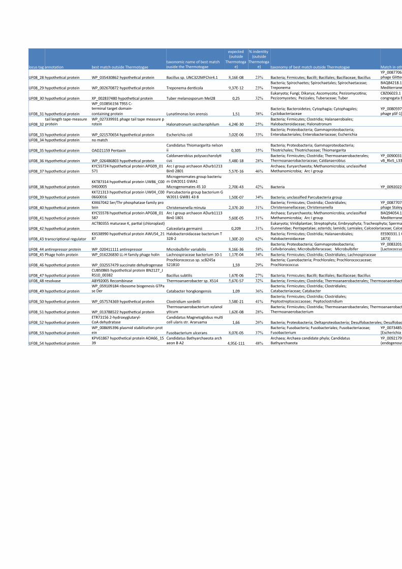
locus tag annotation best match outside Thermotogaetaxonomic name of best matchouside the Thermotogae
expected(outside
Thermotogae)
% indentity(outside
Thermotogae) taxonomy of best match outside Thermotogae Match in other Viruses
UF08_28 hypothetical protein WP_035430862 hypothetical protein Bacillus sp. UNC322MFChir4.1 8,16E-08 23% Bacteria; Firmicutes; Bacilli; Bacillales; Bacillaceae; BacillusYP_008770658.1 hypothetical protein Glittering_22 [Bacillusphage Glittering]
UF08_29 hypothetical protein WP_002670872 hypothetical protein Treponema denticola 9,37E-12 23%Bacteria; Spirochaetes; Spirochaetales; Spirochaetaceae;Treponema
BAQ84218.1 hypothetical protein [unculturedMediterranean phage uvMED]
UF08_30 hypothetical protein XP_002837480 hypothetical protein Tuber melanosporum Mel28 0,25 32%Eukaryota; Fungi; Dikarya; Ascomycota; Pezizomycotina;Pezizomycetes; Pezizales; Tuberaceae; Tuber
CBZ06023.1 hypothetical protein CcBV_2.3B [Cotesiacongregata bracovirus]
UF08_31 hypothetical protein
WP_010856156 T9SS C-terminal target domain-containing protein Lunatimonas lon arensis 1,51 38%
Bacteria; Bacteroidetes; Cytophagia; Cytophagales;Cyclobacteriaceae
YP_008059799.1 hypothetical protein pSf1_0077 [Shigellaphage pSf-1]
UF08_32tail length tape-measureprotein
WP_027339931 phage tail tape measure protein Halonatronum saccharophilum 4,24E-30 25%
Bacteria; Firmicutes; Clostridia; Halanaerobiales;Halobacteroidaceae; Halonatronum
UF08_33 hypothetical protein WP_021570654 hypothetical protein Escherichia coli 3,02E-06 33%Bacteria; Proteobacteria; Gammaproteobacteria;Enterobacteriales; Enterobacteriaceae; Escherichia
UF08_34 hypothetical protein no match
UF08_35 hypothetical protein OAD21159 PentaxinCandidatus Thiomargarita nelsonii 0,305 35%
Bacteria; Proteobacteria; Gammaproteobacteria;Thiotrichales; Thiotrichaceae; Thiomargarita
UF08_36 Hypothetical protein WP_026486803 hypothetical proteinCaldanaerobius polysaccharolyticus 5,48E-18 28%
Bacteria; Firmicutes; Clostridia; Thermoanaerobacterales;Thermoanaerobacteraceae; Caldanaerobius
YP_009003197.1 tail fiber protein [Rhizobium phagevB_RleS_L338C]
UF08_37 hypothetical proteinKYC55724 hypothetical protein APG09_01571
Arc I group archaeon ADurb1213Bin0 2801 5,57E-16 46%
Archaea; Euryarchaeota; Methanomicrobia; unclassifiedMethanomicrobia; Arc I group
UF08_38 hypothetical proteinKKT87314 hypothetical protein UW86_C0004G0005
Microgenomates group bacterium GW2011 GWA1Microgenomates 45 10 2,70E-43 42% Bacteria YP_009202238.1GepA-like protein [Bacteriophage Lily]
UF08_39 hypothetical proteinKKT21313 hypothetical protein UW04_C0006G0016
Parcubacteria group bacterium GW2011 GWB1 43 8 1,50E-07 34% Bacteria; unclassified Parcubacteria group
UF08_40 hypothetical proteinKXK67042 Ser/Thr phosphatase family protein Christensenella minuta 2,37E-20 31%
Bacteria; Firmicutes; Clostridia; Clostridiales;Christensenellaceae; Christensenella
YP_008770743.1 metal-dependent hydrolase [Bacillusphage Staley]
UF08_41 hypothetical proteinKYC55578 hypothetical protein APG08_01587
Arc I group archaeon ADurb1113Bin0 1801 5,60E-05 31%
Archaea; Euryarchaeota; Methanomicrobia; unclassifiedMethanomicrobia; Arc I group
BAQ94054.1 hypothetical protein [unculturedMediterranean phage uvMED]
UF08_42 hypothetical proteinACT80355 maturase K, partial (chloroplast)
Calceolaria germainii 0,209 31%Eukaryota; Viridiplantae; Streptophyta; Embryophyta; Tracheophyta; Spermatophyta; Magnoliophyta; eudicotyledons;Gunneridae; Pentapetalae; asterids; lamiids; Lamiales; Calceolariaceae; Calceolaria
UF08_43 transcriptional regulatorKXS38990 hypothetical protein AWU54_2187
Halobacteroidaceae bacterium T328-2 1,30E-20 62%
Bacteria; Firmicutes; Clostridia; Halanaerobiales;Halobacteroidaceae
EES90331.1 transcription regulator [Clostridium phage D-1873]
UF08_44 antirepressor protein WP_020411111 antirepressor Microbulbifer variabilis 8,16E-36 58%Bacteria; Proteobacteria; Gammaproteobacteria;Cellvibrionales; Microbulbiferaceae; Microbulbifer
YP_008320146.1 hypothetical protein BM13_04[Lactococcus
UF08_45 Phage holin protein WP_016226830 LL-H family phage holin Lachnospiraceae bacterium 10-1 1,17E-04 34% Bacteria; Firmicutes; Clostridia; Clostridiales; Lachnospiraceae
UF08_46 hypothetical protein WP_032557479 succinate dehydrogenaseProchlorococcus sp. scB245a521B10 1,59 29%
Bacteria; Cyanobacteria; Prochlorales; Prochlorococcaceae;Prochlorococcus
UF08_47 hypothetical proteinCUB50865 hypothetical protein BN2127_JRS10_00382 Bacillus subtilis 1,67E-06 27% Bacteria; Firmicutes; Bacilli; Bacillales; Bacillaceae; Bacillus
UF08_48 resolvase ABY92005 Recombinase Thermoanaerobacter sp. X514 5,67E-57 32% Bacteria; Firmicutes; Clostridia; Thermoanaerobacterales; Thermoanaerobacteraceae; Thermoanaerobacter
UF08_49 hypothetical proteinWP_059109184 ribosome biogenesis GTPase Der Catabacter hongkongensis 1,09 36%
Bacteria; Firmicutes; Clostridia; Clostridiales;Catabacteriaceae; Catabacter
UF08_50 hypothetical protein WP_057574369 hypothetical protein Clostridium sordellii 3,58E-21 41%Bacteria; Firmicutes; Clostridia; Clostridiales;Peptostreptococcaceae; Peptoclostridium
UF08_51 hypothetical protein WP_013788522 hypothetical proteinThermoanaerobacterium xylanolyticum 1,62E-08 28%
Bacteria; Firmicutes; Clostridia; Thermoanaerobacterales; Thermoanaerobacterales Family III. Incertae Sedis;Thermoanaerobacterium
UF08_52 hypothetical proteinETR73156 2-hydroxyglutaryl-CoA dehydratase
Candidatus Magnetoglobus multicell ularis str. Araruama 1,66 26% Bacteria; Proteobacteria; Deltaproteobacteria; Desulfobacterales; Desulfobacteraceae; Candidatus Magnetoglobus
UF08_53 hypothetical proteinWP_008695396 plasmid stabilization protein Fusobacterium ulcerans 8,07E-05 37%
Bacteria; Fusobacteria; Fusobacteriales; Fusobacteriaceae;Fusobacterium
YP_007348557.1 hypothetical protein phAPEC8_00189[Escherichia phage phAPEC8]
UF08_54 hypothetical proteinKPV61867 hypothetical protein AOA66_1539
Candidatus Bathyarchaeota archaeon B A2 4,95E-111 48%
Archaea; Archaea candidate phyla; CandidatusBathyarchaeota
YP_009217915.1 putative radical SAM domain protein(endogenous virus) [Clostridium phage phiCT453B]

locus tag annotation best match outside Thermotogaetaxonomic name of best matchouside the Thermotogae
expected(outside
Thermotogae)
% indentity(outside
Thermotogae) taxonomy of best match outside Thermotogae Match in other Viruses
UF08_55 bipolar DNA helicase HerACUU36910 hypothetical protein DCOP10_11973 Armatimonadetes bacterium DC 2,57E-100 35% Bacteria; Armatimonadetes
UF08_56 hypothetical protein WP_027719510 hypothetical protein Desulfovirgula thermocuniculi 7,83E-23 27%Bacteria; Firmicutes; Clostridia; Thermoanaerobacterales;Thermoanaerobacteraceae; Desulfovirgula
UF08_57 hypothetical proteinXP_010260093 PREDICTED: uncharacterized protein LOC104599305 Nelumbo nucifera 0,918 31%
Eukaryota; Viridiplantae; Streptophyta; Embryophyta; Tracheophyta; Spermatophyta; Magnoliophyta; eudicotyledons;Proteales; Nelumbonaceae; Nelumbo
UF08_58 hypothetical proteinCDB45357 putative uncharacterized protein
Phascolarctobacterium sp. CAG:207 8,84 22%
Bacteria; Firmicutes; Negativicutes; Selenomonadales;Acidaminococcaceae; Phascolarctobacterium
UF08_59 transcriptional regulator WP_058964196 transcriptional regulatorRuminococcaceae bacterium AM2 9,76E-41 40%
Bacteria; Firmicutes; Clostridia; Clostridiales;Ruminococcaceae; unclassified Ruminococcaceae YP_240035.1 ORF020 [Staphylococcus phage 47]
UF08_60SOS-response repressorand protease LexA
WP_009006200 MULTISPECIES: SOS-response transcriptional repressor Fusobacteri um 1,90E-28 36% Bacteria; Fusobacteria; Fusobacteriales; Fusobacteriaceae
YP_007010947.1 transcriptional regulator [Deep-seathermophilic phage D6E]
UF08_61 transcriptional regulatorKKQ85063 Helix-turn-helix domain protein
Parcubacteria group bacterium GW2011 GWF2 38 8 3,05E-10 42% Bacteria; unclassified Parcubacteria group
YP_009168545.1 putative transcription regulator[Lactobacillus phage LBR48]
UF08_62 antirepressor protein WP_015397656 anti-repressor protein Bartonella australis 1,53E-50 43%Bacteria; Proteobacteria; Alphaproteobacteria; Rhizobiales;Bartonellaceae; Bartonella
YP_008129893.1 anti-repressor protein [Paenibacillus phagePG1]
UF08_63 hypothetical proteinKUO58776 cobalt ABC transporter ATP-binding protein Gracilibacter sp. BRH c7a 5,48 43%
Bacteria; Firmicutes; Clostridia; Clostridiales;Gracilibacteraceae; Gracilibacter
UF08_64 hypothetical protein WP_045166120 haloacid dehalogenaseThermoanaerobacter cellulolyticus 0,502 33%
Bacteria; Firmicutes; Clostridia; Clostridiales; Anaerocellumgroup
UF08_65transcriptional regulator,XRE family WP_062805019 hypothetical protein Enterococcus pernyi 3,38E-16 43%
Bacteria; Firmicutes; Bacilli; Lactobacillales; Enterococcaceae;Enterococcus YP_009197552.1 repressor [Lactobacillus phage iLp84]
UF08_66 transcriptional regulator WP_057001098 hypothetical proteinCarnobacterium maltaromaticum 5,38E-09 38%
Bacteria; Firmicutes; Bacilli; Lactobacillales;Carnobacteriaceae; Carnobacterium YP_001468419.1 gp34 [Listeria phage A500]
UF08_67 hypothetical protein KKW41634 Foldase protein PrsACandidatus Magasanikbacteria bacterium GW2011 G WA2 56 11 2,63E-02 30% Bacteria; Candidatus Magasanikbacteria
UF08_68 hypothetical proteinXP_005178606 PREDICTED: laminin subunit alpha Musca domestica 0,612 27%
Eukaryota; Metazoa; Ecdysozoa; Arthropoda; Hexapoda; Insecta; Pterygota; Neoptera; Endopterygota; Diptera; Brachycera;Muscomorpha; Muscoidea; Muscidae; Musca
UF08_69 hypothetical protein KKW21360 Alanine-tRNA ligaseCandidatus Adlerbacteria bacterium GW2011 GWC1 5 0 9 6,25 36% Bacteria; Candidatus Adlerbacteria
UF08_70 hypothetical proteinXP_008625909 hypothetical protein YYE_04044 Plasmodium vinckei vinckei 8,07 33% Eukaryota; Alveolata; Apicomplexa; Aconoidasida; Haemosporida; Plasmodiidae; Plasmodium;
UF08_71 hypothetical proteinYP_007677603.1 hypothetical protein Romulus_115[Staphylococcus phage vB_SauM_Romulus]
UF08_72 Viral nuclease WP_015358110 phage-type endonuclease Clostridium stercorarium 2,85E-117 54%Bacteria; Firmicutes; Clostridia; Clostridiales;Ruminococcaceae; Ruminiclostridium
YP_008240349.1 phage-type endonuclease [Thermus phagephi OH2]
UF08_73recombinational DNArepair protein RecT WP_060476225 recombinase RecT Geobacillus sp. PA-3 9,59E-101 55% Bacteria; Firmicutes; Bacilli; Bacillales; Bacillaceae; Geobacillus YP_008240347.1 RecT protein [Thermus phage phi OH2]
UF08_74 hypothetical protein WP_047597684 hypothetical protein Pseudomonas sp. RIT288 0,888 38%Bacteria; Proteobacteria; Gammaproteobacteria;Pseudomonadales; Pseudomonadaceae; Pseudomonas
UF08_75 hypothetical protein CZZ10673 Uncharacterised protein Enterobacter cloacae 1,68E-02 60%Bacteria; Proteobacteria; Gammaproteobacteria; Enterobacteriales; Enterobacteriaceae; Enterobacter; Enterobactercloacae complex
UF08_76 transcriptional regulatorWP_049499605 MULTISPECIES: transcriptional regulator Streptococcus 1,94E-13 53% Bacteria; Firmicutes; Bacilli; Lactobacillales; Streptococcaceae YP_008770535.1 DNA binding protein [Bacillus phage Page]
UF08_77 hypothetical protein WP_059175353 hypothetical protein Thermodesulfovibrio aggregans 0,571 29%Bacteria; Nitrospirae; Nitrospirales; Nitrospiraceae;Thermodesulfovibrio

locus tag annotation Length (aa) Match in MCV1 % identity MCV1 ORF Match in MPV1 % identity expected bast match outside the Marinitoga in BLASP searches of Genbank taxonomic name of best match ouside Marinitoga
UF09_1 recombinational DNA repair protein RecT 294UF08_73_recombinational DNA repairprotein RecT 100% WP_004102129 DNA recombination protein RecT Thermosipho africanus
UF09_2 hypothetical protein 78 UF08_74_hypothetical protein 100% WP_047597684 hypothetical protein Pseudomonas sp. RIT288UF09_3 hypothetical protein 43 UF08_75_hypothetical protein 100% CZZ10673 Uncharacterised protein Enterobacter cloacaeUF09_4 transcriptional regulator 63 UF08_76_hypothetical protein 100% WP_008377672 transcriptional regulator Enterococcus sp. C1UF09_5 hypothetical protein 103 UF08_77_hypothetical protein 100% WP_059175353 hypothetical protein Thermodesulfovibrio aggregansUF09_6 hypothetical protein 211 UF08_1_hypothetical protein 95% WP_042729055 phage replication protein O Pseudomonas fluorescensUF09_7 hypothetical protein 70 UF08_2_hypothetical protein 99% EHY68330 hypothetical protein SEHO0A_02810 Salmonella enterica subsp. houtena e str. ATCC BAA-1581UF09_8 replicative DNA helicase 427 UF08_3_replicative DNA helicase 98% Marpi_0305 replicative DNA helicase 66% 0 WP_031484309 replicative DNA helicase Desulfovibrio frigidus
UF09_9 single-stranded DNA-binding protein 149UF08_4_single-stranded DNA-bindingprotein 97% Marpi_0443 single-stranded DNA-binding protein 59% 2,61E-51 WP_052670309 hypothetical protein Defluviitoga tunisiensis
UF09_10 hypothetical protein 183UF09_11 hypothetical protein 114 WP_044141503 helicase Bacillus pumilus
UF09_12deoxyuridine 5'-triphosphatenucleotidohydrolase 140
Marpi_0307 deoxyuridine 5'-triphosphatenucleotidohydrolase Dut 81% 1,57E-77 WP_040347745 deoxyuridine 5'-triphosphate nucleotidohydrolase Brevibacterium mcbrellneri
UF09_13 hypothetical protein 92 UF08_5_hypothetical protein 99% Marpi_0308 hypothetical protein 47% 4,64E-08 WP_015616411 hypothetical protein Clostridium pasteurianumUF09_14 hypothetical protein 82 UF08_6_hypothetical protein 100% Marpi_0309 hypothetical protein 53% 6,58E-15 XP_008855251 hypothetical protein ENU1_022120 Entamoeba nuttalli P19UF09_15 hypothetical protein 122 UF08_7_hypothetical protein 94% Marpi_0310 Holliday junction DNA helicase 69% 7,97E-57 WP_054198714 hypothetical protein Helicobacter pullorumUF09_16 hypothetical protein 77 UF08_8_hypothetical protein 99% Marpi_0314 hypothetical protein 83% 9,55E-38 WP_047264626 lipoprotein Candidatus Liberibacter africanusUF09_17 hypothetical protein 161 UF08_9_hypothetical protein 95% Marpi_0315 hypothetical protein 50% 3,15E-46 WP_012057673 RNA polymerase subunit sigma-24 Thermosipho melanesiensisUF09_18 hypothetical protein 277 WP_022851452 hypothetical protein Geovibrio sp. L21-Ace-BESUF09_19 Homeodomain-like 127 UF08_12_hypothetical protein 100% Marpi_0319 hypothetical protein 82% 8,98E-60 WP_047809280 hypothetical protein Desulfosporosinus acididurans
UF09_20 terminase large subunit 416 UF08_13_terminase large subunit 99%Marpi_0320 phage terminase, large subunit, PBSXfamily 97% 0 WP_053284682 terminase Clostridium botulinum
UF09_21 hypothetical protein 405 UF08_14_hypothetical protein 99% Marpi_0321 hypothetical protein 93% 0 WP_027339946 hypothetical protein Halonatronum saccharophilumUF09_22 hypothetical protein 62 UF08_18_hypothetical protein 98% XP_008914408 hypothetical protein PPTG_24293 Phytophthora parasitica INRA-310UF09_23 hypothetical protein 502 UF08_19_hypothetical protein 99% Marpi_0322 hypothetical protein 85% 0 WP_038226045 hypothetical protein Vibrio sp. ER1AUF09_24 hypothetical protein 94 UF08_20_hypothetical protein 100% Marpi_0323 hypothetical protein 88% 4,78E-79 WP_047441240 peptidoglycan L-alanyl-D-glutamate endopeptidase Chryseobacteriu m sp. YR561
UF09_25 hypothetical protein 200UF08_21_Protein of unknown functionDUF4355 99% Marpi_0324 hypothetical protein 82% 1,62E-97 WP_009531408 hypothetical protein Peptostreptococcaceae bacterium OBRC8
UF09_26 structural protein 118 UF08_22_hypothetical protein 99% Marpi_0325 hypothetical protein 94% 1,10E-72 WP_036595750 hypothetical protein Paenibacillus sophoraeUF09_27 major capsid protein 340 UF08_23_major capsid protein 99% Marpi_0326 Phage major capsid protein E 91% 0 WP_039218408 phage capsid protein Clostridium novyiUF09_28 hypothetical protein 61 UF08_24_hypothetical protein 100% Marpi_0327 hypothetical protein 80% 2,01E-23 KLD98821 hypothetical protein AF74_02140 Arcobacter butzleri L349UF09_29 hypothetical protein 107 UF08_25_hypothetical protein 100% Marpi_0328 hypothetical protein 86% 7,36E-58 WP_012063953 hypothetical protein Alkaliphilus metalliredigensUF09_30 hypothetical protein 158 UF08_26_hypothetical protein 100% Marpi_0329 hypothetical protein 98% 1,24E-106 WP_006306523 hypothetical protein Centipeda periodontii
UF09_31 hypothetical protein 127 UF08_27_hypothetical protein 98% Marpi_0330 hypothetical protein 89% 2,51E-74WP_053940024 5-methyltetrahydropteroyltriglutamate--homocysteine S-methyltransferase Buchnera aphidicola
UF09_32 hypothetical protein 322 UF08_29_hypothetical protein 67% Marpi_0331 hypothetical protein 88% 0 WP_012057658 hypothetical protein Thermosipho melanesiensisUF09_33 hypothetical protein 117 UF08_30_hypothetical protein 39% Marpi_0332 hypothetical protein 94% 9,10E-72 WP_004102185 hypothetical protein Thermosipho africanusUF09_34 hypothetical protein 167 UF08_31_hypothetical protein 29% Marpi_0333 hypothetical protein 70% 2,89E-78 WP_004102187 hypothetical protein Thermosipho africanus
UF09_35 tail length tape measure protein 1 516UF08_32_tail length tape-measureprotein 89%
Marpi_0334 phage tail tape measure protein,TP901 family 72% 0 WP_004102188 phage tail tape measure protein Thermosipho africanus

locus tag annotation Length (aa) Match in MCV1 % identity MCV1 ORF Match in MPV1 % identity expected bast match outside the Marinitoga in BLASP searches of GenbankUF09_36 hypothetical protein 131 YP_003169739 hypothetical proteinUF09_37 hypothetical protein 115 UF08_34_hypothetical protein 93% Marpi_0336 hypothetical protein 66% 8,29E-48 WP_004102189 hypothetical proteinUF09_38 hypothetical protein 390 UF08_35_hypothetical protein 91% Marpi_0337 hypothetical protein 31% 3,20E-36 WP_041426083 hypothetical proteinUF09_39 phage realted protein tail 1 549 UF08_36_chitin binding protein 92% Marpi_0338 hypothetical protein 65% 0 WP_004102192 hypothetical proteinUF09_40 hypothetical protein 79 UF08_37_hypothetical protein 100% KYC55724 hypothetical protein APG09_01571UF09_41 DNA repair exonuclease 254 UF08_40_hypothetical protein 98% Marpi_0340 DNA repair exonuclease 80% 1,85E-154 WP_004102197 DNA repair exonucleaseUF09_42 hypothetical protein 97 UF08_41_hypothetical protein 100% Marpi_0341 hypothetical protein 41% 1,63E-06 KYC55578 hypothetical protein APG08_01587UF09_43 hypothetical protein 127 UF08_42_hypothetical protein 99% Marpi_0342 hypothetical protein 75% 4,27E-62 WP_012057645 hypothetical proteinUF09_44 hypothetical protein 134 WP_012057691 hypothetical proteinUF09_45 hypothetical protein 64 WP_015463858 ATP synthase subunit alphaUF09_46 hypothetical protein 195 no matchUF09_47 phage holin protein 107 UF08_45_hypothetical protein 98% Marpi_0343 phage holin protein (Holin_LLH) 54% 1,07E-31 WP_016226830 LL-H family phage holinUF09_48 hypothetical protein 98 UF08_46_hypothetical protein 97% Marpi_0344 hypothetical protein 56% 3,63E-32 no match
UF09_49 hypothetical protein 194 UF08_47_hypothetical protein 99%Marpi_0345 Orthopoxvirus protein of unknownfunction (DUF830) 80% 1,25E-109 CUB50865 hypothetical protein BN2127_JRS10_00382
UF09_50 Resolvase N-terminal domain 490 UF08_48_resolvase 69%Marpi_0291 site-specific recombinase, DNAinvertase Pi 88% 0 WP_047753688 hypothetical protein
UF09_51 hypothetical protein 162 WP_002975830 hypothetical proteinUF09_52 hypothetical protein 344 no matchUF09_53 hypothetical protein 141 WP_028559036 hypothetical proteinUF09_54 hypothetical protein 265 no matchUF09_55 Chromosome segregation ATPase 583 Marpi_0295 T5orf172 domain-containing protein 67% 0 WP_011968228 hypothetical proteinUF09_56 hypothetical protein 122 WP_015336104 hypothetical proteinUF09_57 hypothetical protein 133 WP_036093225 mRNA interferase MazF2UF09_58 hypothetical protein 117 no matchUF09_59 hypothetical protein 88 WP_015898402 hypothetical proteinUF09_60 hypothetical protein 68 no match
UF09_61 SOS-response repressor and protease LexA 216UF08_60_SOS-response repressor andprotease LexA3 99%
Marpi_0297 SOS response transcriptionalrepressor, RecA-mediated autopeptidase 44% 6,09E-39 WP_053001073 hypothetical protein
UF09_62 hypothetical protein 71 UF08_61_hypothetical protein 99% Marpi_0298 transcriptional regulator 54% 3,44E-10 KKQ85063 Helix-turn-helix domain proteinUF09_63 hypothetical protein 87 WP_063517479 hypothetical protein
UF09_64 Transcriptional regulator XRE family 117UF08_65_transcriptional regulator, XREfamily 98% WP_009618532 transcriptional regulator
UF09_65 hypothetical protein 69 UF08_66_hypothetical protein 86% WP_019132685 transcriptional regulator
UF09_66 hypothetical protein 111 UF08_67_hypothetical protein 99% KKW41634 Foldase protein PrsAUF09_67 hypothetical protein 232 UF08_68_hypothetical protein 100% XP_005178606 PREDICTED: laminin subunit alphaUF09_68 hypothetical protein 78 UF08_69_hypothetical protein 99% KKW21360 Alanine-tRNA ligaseUF09_69 hypothetical protein 54 UF08_70_hypothetical protein 98% XP_008625909 hypothetical protein YYE_04044
UF09_70 YqaJ-like viral recombinase 313UF08_72_recombination-relatedprotein 100% WP_036226574 hypothetical protein

locus tag annotation best match outside Thermotogae
taxonomic name of bestmatch ouside theThermotogae
expected (outsideThermotogae)
% indentity(outside
Thermotogae)taxonomy of best match outsideThermotogae Matches in other Viruses % identity2
UF09_1 recombinational DNA repair protein RecT WP_060476225 recombinase RecT Geobacillus sp. PA-3 9,59E-101 55%Bacteria; Firmicutes; Bacilli; Bacillales;Bacillaceae; Geobacillus
YP_008240347.1 RecT protein [Thermus phagephi OH2] 55%
UF09_2 hypothetical protein WP_047597684 hypothetical protein Pseudomonas sp. RIT288 0,888 38% Bacteria; Proteobacteria; Gammaproteobacteria; Pseudomonadales; Pseudomonadaceae; PseudomonasUF09_3 hypothetical protein CZZ10673 Uncharacterised protein Enterobacter cloacae 1,68E-02 60% Bacteria; Proteobacteria; Gammaproteobacteria; Enterobacteriales; Enterobacteriaceae; Enterobacter; Enterobacter cloacae complex
UF09_4 transcriptional regulator WP_008377672 transcriptional regulator Enterococcus sp. C1 3,68E-13 52%
Bacteria; Firmicutes; Bacilli;Lactobacillales; Enterococcaceae;Enterococcus
YP_008770535.1 DNA binding protein [Bacillusphage Page] 45%
UF09_5 hypothetical protein WP_059175353 hypothetical proteinThermodesulfovibrio aggregans 1,21 29% Bacteria; Nitrospirae; Nitrospirales; Nitrospiraceae; Thermodesulfovibrio
UF09_6 hypothetical protein WP_042729055 phage replication protein O Pseudomonas fluorescens 3,02E-26 47%
Bacteria; Proteobacteria;Gammaproteobacteria;Pseudomonadales;Pseudomonadaceae; Pseudomonas
AMO43596.1 hypothetical protein PAEP54_00450[Pseudomonas phage YMC11/07/P54_PAE_BP] 38%
UF09_7 hypothetical protein EHY68330 hypothetical protein SEHO0A_02810
Salmonella enterica subsp.houtena e str. ATCC BAA-1581 2,68 28% Bacteria; Proteobacteria; Gammaproteobacteria; Enterobacteriales; Enterobacteriaceae; Salmonella
UF09_8 replicative DNA helicase WP_031484309 replicative DNA helicase Desulfovibrio frigidus 1,84E-79 37%
Bacteria; Proteobacteria;Deltaproteobacteria;Desulfovibrionales;Desulfovibrionaceae; Desulfovibrio
NP_795382.1 putative DNA polymerase III deltaprime subunit [Streptococcus pyogenes phage315.1] 33%
UF09_9 single-stranded DNA-binding protein WP_007489185 single-strand-binding family protein Flavonifractor plautii 3,80E-28 40%Bacteria; Firmicutes; Clostridia;Clostridiales; Flavonifractor
YP_009193875.1 single-stranded DNA-bindingprotein [Paenibacillus phage Harrison] 40%
UF09_10 hypothetical proteinUF09_11 hypothetical protein WP_044141503 helicase Bacillus pumilus 4,04 26% Bacteria; Firmicutes; Bacilli; Bacillales; Bacillaceae; Bacillus
UF09_12deoxyuridine 5'-triphosphatenucleotidohydrolase
WP_040347745 deoxyuridine 5'-triphosphate nucleotidohydrolase
Brevibacterium mcbrellneri 5,34E-25 42%
Bacteria; Actinobacteria;Micrococcales; Brevibacteriaceae;Brevibacterium ADZ30445.1 dUTPase [Cowpox virus] 41%
UF09_13 hypothetical protein WP_015616411 hypothetical protein Clostridium pasteurianum 2,01 36% Bacteria; Firmicutes; Clostridia; Clostridiales; Clostridiaceae; ClostridiumUF09_14 hypothetical protein XP_008855251 hypothetical protein ENU1_022120 Entamoeba nuttalli P19 1,78 34% Eukaryota; Amoebozoa; Archamoebae; Entamoebidae; Entamoeba
UF09_15 hypothetical protein WP_054198714 hypothetical protein Helicobacter pullorum 2,40E-09 35%
Bacteria; Proteobacteria;Epsilonproteobacteria;Campylobacterales;Helicobacteraceae; Helicobacter
YP_007111562.1 holliday-junction resolvase RusA[Enterobacteria phage mEp043 c-1] 28%
UF09_16 hypothetical protein WP_047264626 lipoproteinCandidatus Liberibacter africanus 6,96 35% Bacteria; Proteobacteria; Alphaproteobacteria; Rhizobiales; Rhizobiaceae; Candidatus Liberibacter
UF09_17 hypothetical protein WP_057677254 RNA polymerase sigma factor RpoS Curvibacter sp. PAE-UM 0,19 29%
Bacteria; Proteobacteria;Betaproteobacteria; Burkholderiales;Comamonadaceae; Curvibacter
AEW47696.1 putative tail fiber 1 [Bacillus phageBCU4] 28%
UF09_18 hypothetical protein WP_022851452 hypothetical protein Geovibrio sp. L21-Ace-BES 1,66E-10 25% Bacteria; Deferribacteres; Deferribacterales; Deferribacteraceae; Geovibrio
UF09_19 Homeodomain-like WP_047809280 hypothetical proteinDesulfosporosinus acididurans 2,13E-08 32%
Bacteria; Firmicutes; Clostridia;Clostridiales; Peptococcaceae;Desulfosporosinus
AKH46794.1 hypothetical protein [unculturedmarine virus] 32%
UF09_20 terminase large subunit WP_053284682 terminase Clostridium botulinum 4,35E-152 58%
Bacteria; Firmicutes; Clostridia;Clostridiales; Clostridiaceae;Clostridium
YP_009216669.1 terminase large subunit(endogenous virus) [Clostridium phage phiCT453A] 59%
UF09_21 hypothetical protein WP_027339946 hypothetical proteinHalonatronum saccharophilum 2,27E-14 25% Bacteria; Firmicutes; Clostridia; Halanaerobiales; Halobacteroidaceae; Halonatronum
UF09_22 hypothetical protein XP_008914408 hypothetical protein PPTG_24293Phytophthora parasitica INRA-310 3,3 32% Eukaryota; Stramenopiles; Oomycetes; Peronosporales; Phytophthora
UF09_23 hypothetical protein WP_038226045 hypothetical protein Vibrio sp. ER1A 1,43E-18 22%
Bacteria; Proteobacteria;Gammaproteobacteria; Vibrionales;Vibrionaceae; Vibrio
YP_006990482.1 head morphogenesis protein[Clostridium phage phiMMP02] 27%
UF09_24 hypothetical proteinWP_047441240 peptidoglycan L-alanyl-D-glutamate endopeptidase
Chryseobacterium sp. YR561 6,09 40% Bacteria; Bacteroidetes; Flavobacteriia; Flavobacteriales; Flavobacteriaceae; Chryseobacterium
UF09_25 hypothetical protein WP_009531408 hypothetical proteinPeptostreptococcaceae bacterium OBRC8 6,06E-02 27%
Bacteria; Firmicutes; Clostridia;Clostridiales; Peptostreptococcaceae YP_004301280.1 gp5 [Brochothrix phage NF5] 30%
UF09_26 structural protein WP_036595750 hypothetical protein Paenibacillus sophorae 5,79E-14 38%Bacteria; Firmicutes; Bacilli; Bacillales;Paenibacillaceae; Paenibacillus
AMM44193.1 hypothetical protein KELLEZIO_23[Arthrobacter phage KellEzio] 35%
UF09_27 major capsid protein WP_039218408 phage capsid protein Clostridium novyi 8,00E-72 40%
Bacteria; Firmicutes; Clostridia;Clostridiales; Clostridiaceae;Clostridium
YP_008858642.1 major capsid protein [Brevibacillusphage Davies]
32%
UF09_28 hypothetical protein KLD98821 hypothetical protein AF74_02140 Arcobacter butzleri L349 4,77 38% Bacteria; Proteobacteria; Epsilonproteobacteria; Campylobacterales; Campylobacteraceae; Arcobacter
UF09_29 hypothetical protein WP_012063953 hypothetical proteinAlkaliphilus metalliredigens 3,24E-03 37% Bacteria; Firmicutes; Clostridia; Clostridiales; Clostridiaceae; Alkaliphilus
UF09_30 hypothetical protein WP_006306523 hypothetical protein Centipeda periodontii 3,58E-04 29%
Bacteria; Firmicutes; Negativicutes;Selenomonadales; Veillonellaceae;Veillonella BAN05345.1 tail protein [Nitratiruptor phage NrS-1] 36%
UF09_31 hypothetical protein
WP_053940024 5-methyltetrahydropteroyltriglutamate--homocysteine S-methyltrans ferase Buchnera aphidicola 3,45 24%
Bacteria; Proteobacteria;Gammaproteobacteria;Enterobacteriales;Enterobacteriaceae; Buchnera
AJP09134.1 hypothetical protein [Heliothis virescensascovirus 3f] 23%
UF09_32 hypothetical protein WP_002670872 hypothetical protein Treponema denticola 5,23E-14 25%
Bacteria; Spirochaetes;Spirochaetales; Spirochaetaceae;Treponema
YP_009188581.1 putative major tail protein[Gordonia phage GMA3] 26%
UF09_33 hypothetical protein WP_032077236 metal-dependent RNase Clostridium drakei 3,25 31% Bacteria; Firmicutes; Clostridia; Clostridiales; Clostridiaceae; ClostridiumUF09_34 hypothetical protein WP_053069208 hypothetical protein Bizionia psychrotolerans 3,65 24% Bacteria; Bacteroidetes; Flavobacteriia; Flavobacteriales; Flavobacteriaceae; Bizionia
UF09_35 tail length tape measure protein WP_027339931 phage tail tape measure proteinHalonatronum saccharophilum 3,01E-45 28%
Bacteria; Firmicutes; Clostridia;Halanaerobiales; Halobacteroidaceae;Halonatronum YP_008318494.1 tail protein [Shigella phage SfII] 20%

locus tag annotation best match outside Thermotogae
taxonomic name of bestmatch ouside theThermotogae
expected (outsideThermotogae)
% indentity(outside
Thermotogae)taxonomy of best match outsideThermotogae Matches in other Viruses % identity2
UF09_36 hypothetical protein YP_003169739 hypothetical proteinThermus thermophilus bacteriophage P23-77 1,56E-08 49%
Viruses; Virus families not assigned toan order; Sphaerolipoviridae;Gammasphaerolipovirus
YP_003169739.1 hypothetical protein [Thermusthermophilus bacteriophage P23-77]
UF09_37 hypothetical protein no match
UF09_38 hypothetical protein WP_027339930 hypothetical proteinHalonatronum saccharophilum 1,05 52% Bacteria; Firmicutes; Clostridia; Halanaerobiales; Halobacteroidaceae; Halonatronum
UF09_39 phage realted protein tail WP_034858455 phage minor structural protein Enterococcus casseliflavus 1,01E-49 32%
Bacteria; Firmicutes; Bacilli;Lactobacillales; Enterococcaceae;Enterococcus
BAR29161.1 hypothetical protein [unculturedMediterranean phage uvMED]
UF09_40 hypothetical protein KYC55724 hypothetical protein APG09_01571Arc I group archaeon ADurb1213 Bin0 2801 5,57E-16 46% Archaea; Euryarchaeota; Methanomicrobia; unclassified Methanomicrobia; Arc I group
UF09_41 DNA repair exonuclease KXK67042 Ser/Thr phosphatase family protein Christensenella minuta 6,93E-21 31%
Bacteria; Firmicutes; Clostridia;Clostridiales; Christensenellaceae;Christensenella
YP_008770743.1 metal-dependent hydrolase[Bacillus phage Staley]
UF09_42 hypothetical protein KYC55578 hypothetical protein APG08_01587Arc I group archaeon ADurb1113 Bin0 1801 5,23E-04 31%
Archaea; Euryarchaeota;Methanomicrobia; unclassifiedMethanomicrobia; Arc I group
BAQ94054.1 hypothetical protein [unculturedMediterranean phage uvMED]
UF09_43 hypothetical protein ACT80355 maturase K, partial (chloroplast) Calceolaria germainii 0,211 31% Eukaryota; Viridiplantae; Streptophyta; Embryophyta; Tracheophyta; Spermatophyta; Magnoliophyta; eudicotyledons; Gunneridae; Pentapetalae; asterids; lamiids; Lamiales; Calceolariaceae; CalceolariaUF09_44 hypothetical protein no matchUF09_45 hypothetical protein WP_015463858 ATP synthase subunit alpha Psychromonas sp. CNPT3 1,44E-02 39% Bacteria; Proteobacteria; Gammaproteobacteria; Alteromonadales; Psychromonadaceae; PsychromonasUF09_46 hypothetical protein
UF09_47 phage holin protein WP_016226830 LL-H family phage holinLachnospiraceae bacterium 10-1 6,60E-06 34% Bacteria; Firmicutes; Clostridia; Clostridiales; Lachnospiraceae
UF09_48 hypothetical protein
UF09_49 hypothetical proteinCUB50865 hypothetical protein BN2127_JRS10_00382 Bacillus subtilis 1,48E-06 27% Bacteria; Firmicutes; Bacilli; Bacillales; Bacillaceae; Bacillus
UF09_50 Resolvase N-terminal domain ABY92005 RecombinaseThermoanaerobacter sp. X514 1,22E-40 30%
Bacteria; Firmicutes; Clostridia;Thermoanaerobacterales;Thermoanaerobacteraceae;Thermoanaerobacter
YP_009217897.1 serine recombinase (endogenousvirus) [Clostridium phage phiCT453B]
UF09_51 hypothetical protein WP_002975830 hypothetical protein Leptospira vanthielii 2,32E-02 34% Bacteria; Spirochaetes; Leptospirales; Leptospiraceae; Leptospira
UF09_52 hypothetical protein
UF09_53 hypothetical protein WP_028559036 hypothetical protein Paenibacillus pinihumi 0,41 28%Bacteria; Firmicutes; Bacilli; Bacillales;Paenibacillaceae; Paenibacillus
AEX62303.1 vacuolar sorting protein [MoumouvirusMonve]
UF09_54 hypothetical protein
UF09_55 Chromosome segregation ATPase WP_011968228 hypothetical protein Clostridium beijerinckii 1,09E-148 56%
Bacteria; Firmicutes; Clostridia;Clostridiales; Clostridiaceae;Clostridium
EES90330.1 phage protein [Clostridium phage D-1873]
UF09_56 hypothetical protein WP_015336104 hypothetical proteinDesulfovibrio hydrothermalis 3,40E-02 51%
Bacteria; Proteobacteria;Deltaproteobacteria;Desulfovibrionales;Desulfovibrionaceae; Desulfovibrio AIO08090.1 polyprotein [Hepatitis C virus]
UF09_57 hypothetical protein WP_036093225 mRNA interferase MazF2 Leptospira weilii 9,29E-04 32% Bacteria; Spirochaetes; Leptospirales; Leptospiraceae; Leptospira
UF09_58 hypothetical protein
UF09_59 hypothetical protein WP_015898402 hypothetical proteinAcidobacterium capsulatum 1,39E-09 37%
Bacteria; Acidobacteria;Acidobacteriales; Acidobacteriaceae;Acidobacterium
YP_004539068.1 unnamed protein product[Mycobacterium phage Faith1]
UF09_60 hypothetical protein
UF09_61 SOS-response repressor and protease LexAWP_009006200 MULTISPECIES: SOS-response transcriptional repressor Fusobacteri um 4,77E-29 36%
Bacteria; Fusobacteria;Fusobacteriales; Fusobacteriaceae
YP_007010947.1 transcriptional regulator [Deep-seathermophilic phage D6E]
UF09_62 hypothetical protein KKQ85063 Helix-turn-helix domain proteinParcubacteria group bacterium GW2011 GWF2 38 8 1,91E-09 42%
Bacteria; unclassified Parcubacteriagroup
YP_009168545.1 putative transcription regulator[Lactobacillus phage LBR48]
UF09_63 hypothetical protein WP_063517479 hypothetical protein Vibrio parahaemolyticus 0,526 40% Bacteria; Proteobacteria; Gammaproteobacteria; Vibrionales; Vibrionaceae; Vibrio
UF09_64 Transcriptional regulator XRE family WP_009618532 transcriptional regulator Desulfosporosinus sp. OT 5,58E-13 44%
Bacteria; Firmicutes; Clostridia;Clostridiales; Peptococcaceae;Desulfosporosinus
ALV83461.1 transcriptional regulator [Bacillus phagevB_BceS-MY192]
UF09_65 hypothetical protein WP_019132685 transcriptional regulator Peptoniphilus obesi 9,79E-13 52%
Bacteria; Firmicutes; Tissierellia;Tissierellales; Peptoniphilaceae;Peptoniphilus
WP_044984172.1 transcriptional regulator, partial[Thermoanaerobacterium phage THSA-485A]
UF09_66 hypothetical protein KKW41634 Foldase protein PrsA
Candidatus Magasanikbacteria bacterium GW2011 GWA2 56 11 2,09E-02 30% Bacteria; Candidatus Magasanikbacteria
UF09_67 hypothetical protein XP_005178606 PREDICTED: laminin subunit alpha Musca domestica 0,65 27% Eukaryota; Metazoa; Ecdysozoa; Arthropoda; Hexapoda; Insecta; Pterygota; Neoptera; Endopterygota; Diptera; Brachycera; Muscomorpha; Muscoidea; Muscidae; Musca
UF09_68 hypothetical protein KKW21360 Alanine-tRNA ligase
Candidatus Adlerbacteria bacterium GW2011 GWC1 50 9 2,41 38% Bacteria; Candidatus Adlerbacteria
UF09_69 hypothetical protein XP_008625909 hypothetical protein YYE_04044Plasmodium vinckei vinckei 8,07 33% Eukaryota; Alveolata; Apicomplexa; Aconoidasida; Haemosporida; Plasmodiidae; Plasmodium; Plasmodium (Vinckeia)
UF09_70 YqaJ-like viral recombinase WP_015358110 phage-type endonuclease Clostridium stercorarium 2,85E-117 54%
Bacteria; Firmicutes; Clostridia;Clostridiales; Ruminococcaceae;Ruminiclostridium
P_008240349.1 phage-type endonuclease [Thermusphage phi OH2]

Chapitre IV : Etude du ge�nome de
Thermosipho sp. AT1244-VC14

Chapitre IV
194
Pre�sentation des travaux
Lo s de la aat isatio des t ois i us p se t s i dessus, des tests d ife tiosits o t t
réalisés sur la souche Thermosipho sp. AT1244-VC14. Cette souche est la seule qui permet la
propagation du plasmide pMP1. Néanmoins, celle- i est pas se sile à l ife tio pa les
différents virus de Marinitoga caractérisés (MPV1, MCV1 et MCV2).
C est da s e ad e ue le s ue çage et ltude du go e de ette sou he o t t alis s.
Cette tude a ota e t pe is de ett e e ide e la p se e d u système de défense
bactérien : le système CRISPR-as. Il s agit d u syste C‘I“P‘-cas de classe 1 Type III-A.
L e se le des g es essaies au o fo tio e e t du syst e so t p se ts, elui-ci
paraît donc fonctionnel.
Les spacers du système ont aussi été étudiés et ceux-ci ne montrent aucune homologie de
séquence avec pMP1 ou MPV1. Néanmoins, deux spacers ont des similarités avec les virus
MCV1 et MCV2 (>85%). Ceci pourrait expliquer pourquoi ces deux virus sont incapables
d i fe te ette sou he. D aut e pat, la pse e de es spae s pouait sig ifie ue
Thermosipho sp. AT1244-VC14 a déjà été en contact avec MCV1, MCV2, ou un virus proche de
ceux- i. Ces sultats fe o t l o jet d u Ge oe epo t atuelle e t e p paatio .

Chapitre IV
195
Draft Genome Sequences of a thermophilic and anaerobic bacterium, Thermosipho sp.
AT1244-VC14, isolated from a deep sea hydrothermal vent.
Coraline Merciera,b,c, Julien Lossouarna,b,c, Camilla Nesbød,e, Thomas H.A Haverkamp d,
Nadège Bienvenu a,b,c, Claire Geslin a,b,c.
a UBL, Laboratoire de Microbiologie des Environnements Extr e �mes UMR6197, IUEM, Rue Dumont d ’Urville, 29280 Plouzane �,
France
b CNRS, Laboratoire de Microbiologie des Environnements Extre �mes UMR 6197, IUEM, Rue Dumont d’Urville, 29280 Plouzane �,
France
c Ifremer, Laboratoire de Microbiologie des Environnements Extre �mes UMR 6197, CS10070, 29280 Plouzane �, France
d Centre for Ecological and Evolutionary Synthesis, University of Oslo, Oslo, Norway
e Department of Biological Sciences, University of Alberta, Edmonton, Alberta, Canada
*Present address: Micalis Institute, INRA, AgroParisTech, Universite � Paris-Saclay, Jouy-en-Josas, France
Running title: Draft genome of a Thermosipho isolate
# Address correspondence to: Claire Geslin ([email protected])

Chapitre IV
196
Abstract
Here, we present the draft genome sequences of a thermophilic Thermosipho strain member
of the Thermotogales order. This strain was isolated from an active chimney in a deep sea
hydrothermal vent, Rainbow site, located on the Mid Atlantic Ridge. This genome notably
contains a complete and expected functional CRISPR-cas system, classified as Classe I, Type III-
A. Spacers were also found in 4 loci, no match with Marinitoga piezophila mobile genetic
elements were found whereas two spacers match with two new viruses of Marinitoga camini.
It suggests that this strain has already been infected by those viruses or other closely related
viruses.
Introduction
The bacterial order Thermotogales, consists of mesophilic (Nesbø et al., 2012; Ben Hania et
al., 2013) and hyper/thermophilic, anaerobic, chemo-organotrophic microorganisms (Bonch-
osmolovskaya, 2008). These bacteria are rod-shaped characterized by an outer sheath-like
envelope, the toga. This lineage is broadly represented in deep biosphere ecosystems and
deep-sea hydrothermal vents in particular. Phylogenetic analyses of 16S rRNA gene and 29
concatenated ribosomal proteins place Thermotogales as a sister group to Aquificales, and
consequently as one of the deepest branching order within domain Bacteria (Zhaxybayeva et
al., 2009; Hug et al., 2016). Genomes of this bacterial order contain numerous evidence of
lateral gene transfer and notably, CRISPR-cas systems are present in all Thermotogales
genome sequences (Nesbø et al., 2009; Zhaxybayeva et al., 2009). CRISPR-cas is a bacterial
and archaeal defense system against invading mobile genetic elements, with a wide
distribution in the prokaryotic domain. Indeed, approximatively 47% of analyzed prokaryotes

Chapitre IV
197
encode CRISPR cas loci distributed in 87% of archaeal genomes and 50% of bacterial genomes
(not necessarily complete loci) (Makarova et al., 2015).
At the time of writing, over 80 completed and ongoing Thermotogae genome projects are
counted, comprised 10 of the 12 described Thermotogae genera (Pollo et al., 2015). Among
the eight Thermosipho species described (T. africanus, T. activus, T.affectus, T. atlanticus, T.
geolei, T. globiformans, T. japonicus and T. melanesiensis), only two complete genome
sequences are currently available (T. africanus and T. melanesiensis).
Thermosipho sp. AT1244-VC14 (hereafter referred to as Thermosipho sp. 1244) was isolated
from the hydrothermal site Rainbow (Portugal, Azores) in the Mid-Atlantic Ridge at a depth of
2275m. It was sampled from an active chimney in 2001 during the ATOS cruise. This rod-
shaped anaerobic thermophilic chemoorganotrophic bacterium grows optimally at 60°C
under atmospheric pressure and anaerobic conditions in a rich medium. Here we describe the
genome of Thermosipho sp. 1244 and study his CRISPR-cas system. The aim of this study is to
extend our knowledge about this genus and its relationships with mobile genetic elements.
Results and discussion
The chromosome consists in 1,818,555 bp in 34 contigs, with the longest contig size of 301,822
bp, and a G+C content of 31.2 %. No extrachromosomal element has been detected. On the
basis of the 16S rRNA gene sequence extracted from this genome, the closest strain of
Thermosipho sp. 1244 is Thermosipho affectus strain ik275mar (Podosokorskaya et al.,
2011)(99% identity, 99% query coverage, e-value 0).

Chapitre IV
198
RAST server counted 1,851 coding DNA sequences (CDSs), 42 tRNAs and 5 rRNAs. The genome
comprises 70.1% of assigned putative functions, while the rest was annotated as encoding
hypothetical proteins. Several genes were identified and associated with resistance to toxic
compounds, as well as stress response to heat shock, or oxidation, which is coherent with the
environmental origin of the strain.
Clustered regularly interspaced short palindromic repeats (CRISPRs) are present in all
sequenced Thermotogales genomes (Zhaxybayeva et al., 2009), suggesting that
Thermotogales have been exposed to numerous viral infections. In this context, a transposase
and CRISPR loci associated with cas genes were found in Thermosipho sp. 1244 genome.
Twenty-eight different CRISPRs associated genes were annotated, including cas 1, cas 2, cas
3, cas 4, cas 6 and cas 10 genes. This system seems to be functional indeed all essential genes
were found in the genome (Makarova et al., 2015).
According to the Makarova classification (Makarova et al., 2015), this CRISPR cas system could
be classified as a Class 1, characterized by the presence of a multi subunit CRISPR-RNA-effector
complex (crRNA-effector complex). It also could be a good candidate for the Type III (like other
sequenced and studied Thermosipho genomes, Haverkamp et al in prep.) because it contains
Cas10 Large Subunit (LS) involved in the target cleavage of the system and also protein
containing CARF domains which could be implicated in the regulation of the system.
Supporting the hypothesis of Type III classification, several protein sequences involved in the
multiprotein cr-RNA effector complex were founded. Notably numerous Csm and Cmr which
are found in Type III A and B CRISPR cas systems. Concerning the subtypes, it could be classified
in the Type III-A, supported by the presence of cas1, cas2, and cas6 genes (absent from III-B).
Important genes with their putative functions are summarized in the table 1.
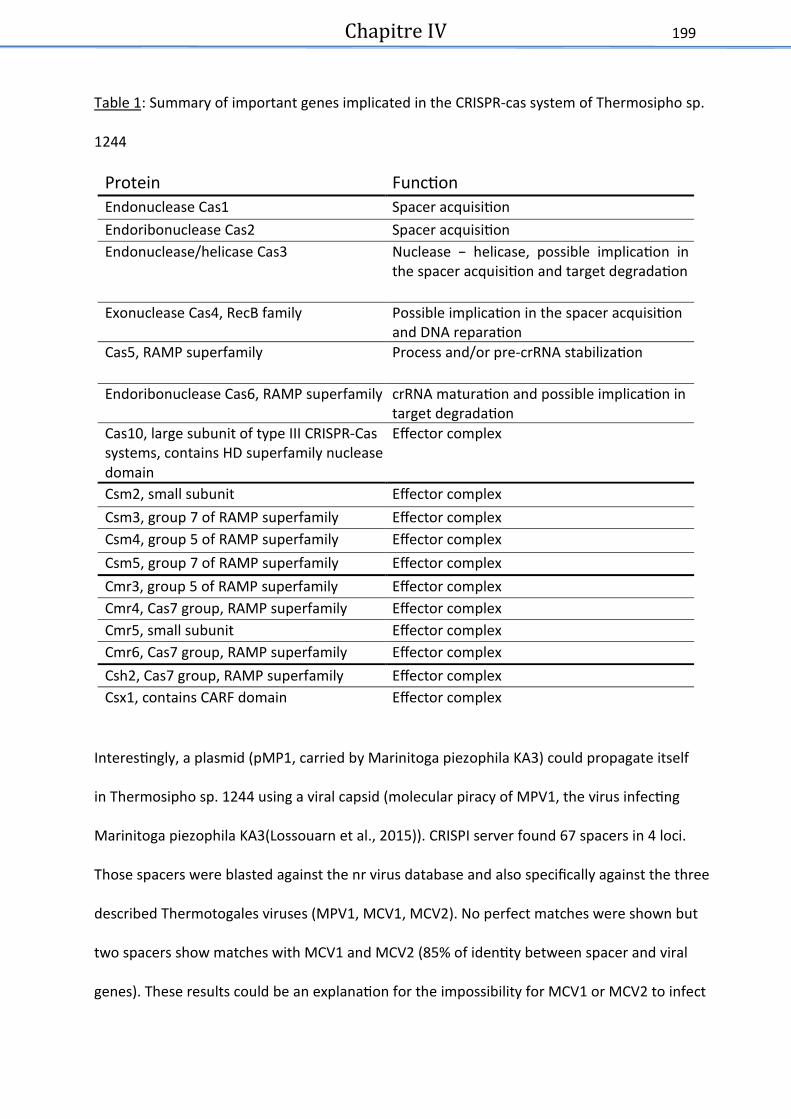
Chapitre IV
199
Table 1: Summary of important genes implicated in the CRISPR-cas system of Thermosipho sp.
1244
Protein Function Endonuclease Cas1 Spacer acquisition Endoribonuclease Cas2 Spacer acquisition Endonuclease/helicase Cas3 Nuclease – helicase, possible implication in
the spacer acquisition and target degradation
Exonuclease Cas4, RecB family Possible implication in the spacer acquisition and DNA reparation
Cas5, RAMP superfamily Process and/or pre-crRNA stabilization
Endoribonuclease Cas6, RAMP superfamily crRNA maturation and possible implication in target degradation
Cas10, large subunit of type III CRISPR-Cas systems, contains HD superfamily nuclease domain
Effector complex
Csm2, small subunit Effector complex Csm3, group 7 of RAMP superfamily Effector complex Csm4, group 5 of RAMP superfamily Effector complex Csm5, group 7 of RAMP superfamily Effector complex Cmr3, group 5 of RAMP superfamily Effector complex Cmr4, Cas7 group, RAMP superfamily Effector complex Cmr5, small subunit Effector complex Cmr6, Cas7 group, RAMP superfamily Effector complex Csh2, Cas7 group, RAMP superfamily Effector complex Csx1, contains CARF domain Effector complex
Interestingly, a plasmid (pMP1, carried by Marinitoga piezophila KA3) could propagate itself
in Thermosipho sp. 1244 using a viral capsid (molecular piracy of MPV1, the virus infecting
Marinitoga piezophila KA3(Lossouarn et al., 2015)). CRISPI server found 67 spacers in 4 loci.
Those spacers were blasted against the nr virus database and also specifically against the three
described Thermotogales viruses (MPV1, MCV1, MCV2). No perfect matches were shown but
two spacers show matches with MCV1 and MCV2 (85% of identity between spacer and viral
genes). These results could be an explanation for the impossibility for MCV1 or MCV2 to infect

Chapitre IV
200
this strain. It also shows that this Thermosipho strain has already been infected by MCV1,
MCV2 or a similar virus. No matches were found corresponding to MPV1 or pMP1 which is
consistent with the possibility for pMP1 to propagate itself in this Thermosipho strain.
Nucleotide sequence accession numbers.
Genome has been deposited at DDBJ/EMBL/ GenBank under the accession number X.
Materials and methods
Culture conditions
Thermosipho sp. 1244 was cultured at 60°C in a modified Ravot medium as previously
described (Geslin et al., 2003; Lossouarn et al., 2015). The medium was completed by adding
1% (wt/vol) elemental sulfur.
Genome sequence and annotation
The genome was sequenced by using a combination of the Illumina and Ion Torrent platforms
with a genome coverage of 30 X.
The annotation was done with RAST server (Overbeek et al., 2014) and CRISPR cas system was
studied by CRISPI web server (Rousseau et al., 2009).
Acknowledements
We thak the hief s ie tist of the F eh o eaog aphial uise ATO“ , Pie e-Marie
Sarradin, the captain and crew of the NO L Atalante and the DSV Nautile pilots and support
crew.
Funding informations
This work was financially supported by the French Ministry of Higher Education and the
Brittany region to CM and the Agence Nationale de la Recherche, project Thermovesicles
(ANR-12-BSV3-OO23-01) to CG, a Norwegian Research Council award (project no.
180444/V40) to CLN.

Chapitre IV
201
References
Bonch-osmolovskaya, E. (2008) Thermotogales: Phylogeny and Taxonomy of the Order.
Encycl. Life Sci. 1–7.
Geslin, C., Le Romancer, M., Erauso, G., Gaillard, M., Perrot, G., and Prieur, D. (2003) PAV1,
the first virus-like patile isolated f o a hype theophili eu ya haeote, Py o o us
ayssi. J Bacteriol 185: 3888–3894.
Ben Hania, W., Postec, A., Aüllo, T., Ranchou-Peyruse, A., Erauso, G., Brochier-Armanet, C., et
al. (2013) Mesotoga infera sp. nov., a mesophilic member of the order Thermotogales,
isolated from an underground gas storage aquifer. Int. J. Syst. Evol. Microbiol. 63: 3003–
3008.
Hug, L.A., Baker, B.J., Anantharaman, K., Brown, C.T., Probst, A.J., Castelle, C.J., et al. (2016) A
new view of the tree of life. Nat. Microbiol. 1: 16048.
Lossouarn, J., Nesbø, C.L., Mercier, C., Zhaxybayeva, O., Johnson, M.S., Charchuck, R., et al.
5 M age à t ois : a selfish ge eti ele e t uses a i us to p opagate ithi
Thermotogales. Environ. Microbiol. 17: 3278–3288.
Makarova, K.S., Wolf, Y.I., Alkhnbashi, O.S., Costa, F., Shah, S.A., Saunders, S.J., et al. (2015)
An updated evolutionary classification of CRISPR-Cas systems. Nat. Rev. Microbiol. 13:
722–736.
Nesbø, C.L., Bapteste, E., Curtis, B., Dahle, H., Lopez, P., Macleod, D., et al. (2009) The genome
of Thermosipho africanus TCF52B: lateral genetic connections to the Firmicutes and
Archaea. J. Bacteriol. 191: 1974–1978.
Nesbø, C.L., Bradnan, D.M., Adebusuyi, A., Dlutek, M., Petrus, A.K., Foght, J., et al. (2012)
Mesotoga prima gen. nov., sp. nov., the first described mesophilic species of the
Thermotogales. Extremophiles 16: 387–393.

Chapitre IV
202
Overbeek, R., Olson, R., Pusch, G.D., Olsen, G.J., Davis, J.J., Disz, T., et al. (2014) The SEED and
the Rapid Annotation of microbial genomes using Subsystems Technology (RAST). Nucleic
Acids Res. 42: D206–D214.
Podosokorskaya, O.A., Kublanov, I. V., Reysenbach, A.L., Kolganova, T. V., and Bonch-
Osmolovskaya, E.A. (2011) Thermosipho affectus sp. nov., a thermophilic, anaerobic,
cellulolytic bacterium isolated from a Mid-Atlantic Ridge hydrothermal vent. Int. J. Syst.
Evol. Microbiol. 61: 1160–1164.
Pollo, S.M.J., Zhaxybayeva, O., and Nesbø, C.L. (2015) Insights into thermoadaptation and the
evolution of mesophily from the bacterial phylum Thermotogae. Can. J. Microbiol. 1–16.
Rousseau, C., Gonnet, M., Le Romancer, M., and Nicolas, J. (2009) CRISPI: a CRISPR interactive
database. Bioinformatics 25: 3317–3318.
Zhaxybayeva, O., Swithers, K.S., Lapierre, P., Fournier, G.P., Bickhart, D.M., DeBoy, R.T., et al.
(2009) On the chimeric nature, thermophilic origin, and phylogenetic placement of the
Thermotogales. Proc. Natl. Acad. Sci. U. S. A. 106: 5865–5870.

Chapitre V : Virome : validation et
normalisation d’un protocole (étude
pre� liminaire)

Chapitre V
204
x Pre�sentation des travaux
Les familles virales sont aussi nombreuses que diversifiées et ne disposent pas de gènes
a ueu s o u s o e l’ARN S pa e e ple , e ui les e d pati uli e e t
o ple es à tudie das leu e se le. N a oi s, g âe à l’esso des ouvelles te h iues
de séquençage haut débit, il est maintenant possible de produire et séquencer des viromes
e se le de g es viau d’u e vio e e t.
Peu de viromes sont disponibles dans les environnements marins profonds, où la diversité
virale reste encore faiblement caractérisée (Williamson et al., 2008; Anderson et al., 2014;
Brum and Sullivan, 2015). Ces premières études montrent que la diversité virale semble moins
i po tate ue das d’aut es os st es o e la olo e d’eau pa e eple. Pa ailleu s,
de nombreux gènes viraux découverts dans ces viromes ne montrent aucune similarité dans
les bases de données, mettant en évidence le manque de connaissances sur ces
e vi o e e ts ais aussi le fait u’ils soie t u se voi i po tat de ouveau g es à
explorer (Williamson et al., 2008; Anderson et al., 2014).
C’est da s e o te te u’u p ojet t asve se a t is e plae e t e le LM E et le LUBEM
(Laboratoire Universitaire de Biodiversité et Ecologie Microbienne) de Brest pour développer
u p oto ole pe etta t d’o te i des vi o es p ove at de dive s e vi o e e ts.
L’o je tif au LM E ta t de p oduie u vio e p ove at de s die ts h d othe au
p ofo ds afi d’e i hi les o aissa es sur ces environnements peu explorés. Au LUBEM,
l’o je tif est d’tudie les vius p se ts da s les e pe to atio s de patie ts atteits de
u ovis idose pou ieu o p e d e l’effet des o u aut s viales p se tes chez les
patients mais aussi leur impact sur les communautés microbiennes.

Chapitre V
205
Des cohortes de patients sains et de patients atteints de la mucoviscidose seront analysées
afi d’ tudie les vaiatio s de populatio s viales, d’obtenir une vision globale de la présence
de celles-ci dans l’ os st e o ho-pulmonaire humain.
Les thématiques de travail du LM2E et du LUBEM sont différentes tout comme les échantillons
à traiter. Néanmoins, la volonté commune d’o te i de ouvelles do es su des
o pati e ts viau p ove at d’environnements complexes a mené à la réalisation de ce
projet, permettant le partage des idées et des méthodologies. La base de ce projet était de
tester deux protocoles différents (basés sur les méthodes de purification virale utilisées dans
les deux laboratoires), l’u tat as su la filt atio et l’aut e su la pu ifiatio via un
g adie t li ai e d’iodiaol. Le ut ta t d’ li i e la ajo it des cellules tout en
conservant les virus présents.
L’o je tif de e t avail tait de valide et de o alise u potocole qui servira de base pour
l’o te tio de vio es.
x Origine des e�chantillons teste�s
Les sédiments hydrothermaux étudiés proviennent du bassin de Guaymas dans le Golfe du
Mexique, au large de la Californie. Ils ont été échantillonnés dans le cadre de la campagne
océanique IFREMER « BIG » (Biodiversité à Guaymas) en Juillet 2010 et conservés à -80°C. Afin
d’avoi suffisa e t de at iel à tudie , hatillo s o t t eg oup s. Ils p oviee t
de 4 « horizons » différents, à partir de carottes échantillonnées à 4 profondeurs différentes
dans la couche sédimentaire (0 à 2 cm : 1 échantillon, 2 à 4 cm : 2 échantillons, 6 à 8 cm : 1
échantillon et de 8 à 10 cm : 1 échantillon). Le pool contient donc 30 g de sédiments en
suspension dans du tampon (10 mM Tris-HCl, 100 mM NaCl, 5 mM CaCl2, pH 8), pour un
volume total de 90 ml (soit une co e t atio d’e vio , g. l-1).

Chapitre V
206
Les expectorations choisies proviennent de patients des 2 sexes et de tous âges (de 18 mois
à 80 ans), par conséquent le pool étudié représente davantage un intérêt technique du point
de vue de la at i e diffiile a t s u ueuse u’u i t t e ologie viale. Du fait de
la viscosité des expectorations, celles-ci doivent être préalablement fluidifiées volume à
volume avec du Sputagest® Mast Diagnostic (contenant du dithiothréitol) pour pouvoir
p leve des volues o e ts. Ce pool d’e pe to atio s est conservés à -80°C.
x Micro-organismes et virus contro; les
Les échantillons ont ensuite été artificiellement chargés en particules virales et micro-
o gais es o us et tits afi de les suiv e e tat ue sig atu es et de vifie l’effiait
des p oto oles tests. Il s’agit ota e t de levu es Candida albicans), de bactéries
(Pseudomonas aeruginosa), de virus à ADN (le siphovirus MPV1 et un adénovirus) et à ARN
(rhinovirus).
Tableau 1 : Concentrations en microorganismes cibles avec lesquelles les e� chantillons ont e� te� charge� s
(en UFC/ml (unite� formant colonie) ou TCID50/mL (tissue culture infective dose 50))

Chapitre V
208
Les défis majeurs résident en la conservation et la détection des virus, faiblement abondants
et fragiles. Les virus à ARN, les plus fragiles, sont ceux qui présentent le plus grand défi
te h i ue. Ces t avau auuels j’ai pu patiipe du at a th se, lo s de l’ead e e t du
stage de Master 1 de Julia Sanchez, sont maintenant réalisés dans le cadre de la thèse de Lisa
Billard au LUBEM (résultats présentés ci-dessous, Projet Virome porté par le Pr C. Payan et le
Dr S. Vallet, LUBEM).
x Premier re�sultats
Un premier essai de séquençage haut débit (HiSeq 2X125 bp, GATC, 5 milions de reads) a été
réalisé su des hatillo s d’e pe to atio s a tifiielle e t hag s e vius et i o-
organismes contrôles (cités ci-dessus . Il s’agit d’u e p e i e tape alis e su des
échantillons bruts, non purifiés, mais chargés artificiellement avec les contrôles. Ainsi, les
extractions et rétro-transcriptions ont été menées sur ces échantillons bruts chargés afin
d’o se ve les sultats d’u sue çage su ha tillo o t ait . Ce séquençage permettra
par la suite de comparer ses résultats aux échantillons ayant subits les protocoles de
purifications, pour observer leur efficacité. Ces premiers résultats de séquençages hauts
d its o t o t u’e vio , % des eads o te us o t des siilait s ave les ases de
données virales. Ces résultats concordent avec ceux obtenus dans la littérature (Losada et al.,
2015; Wang et al., 2016). Les quelques reads pour lesquels une assignation taxonomique a été
possile o espo de t à des vius à ADN dou le i , au u vius à ARN ’a pu t e d te t
hormis des rétrovirus probablement intégrés au génome humain.

Chapitre V
209
Il s’agit e ajo it de at iovius appartenant à la famille Siphoviridae infectant des
bactéries habituellement retrouvées dans le système respiratoire des patients atteints de
mucoviscidose, mais aussi de quelques virus humains (Cystic Fibrosis Foundation, Patient
Registry, Annual Data Report 2012).
x Avancement des travaux
Après ce premier essai, le protocole a évolué et la méthode retenue se déroule en 5 étapes.
Tout d’a o d, les hatillo s so t so i u s pou d tahe les patiules viales des at i es.
Ensuite ceux-ci sont centrifugés et filt s filt es , afi d’ li i e la ajo it des i o-
organismes et particules présents. La thode as e su l’utilisatio de g adie ts, ’a pas t
retenue car sa ise e œuv e est d liate et les p eie s sultats de suivi e PCR ’ taie t
pas concluant. Un traitement aux nucléases est aussi réalisé pour éliminer les acides
u l i ues li es. E fi , ap s l’e t atio , u e t o-transcription et une amplification
aléatoire (par SIA : sequence independant amplification) sont réalisées permettant une
meilleure détection des différents virus présents (Données communiquées et issues du travail
de master 2 et de thèse de Lisa Billard).
L’o je tif est ai te at de valide le p oto ole de pu ifiatio e tudiat les s ue es
issues d’hatillo s naturels (non chargés en micro-organismes et virus contrôles) pour, par
la suite, appliquer ce protocole aux collections de sécrétions respiratoires de patients atteints
de u ovis idose. Ces tudes pe ett o t d’avoi u e eilleu e visio des populatios virales
p se tes das le i o iote des patie ts et de o p e d e l’i pat des ite atio s
potentielles entre les micro-organismes et les virus présents au sein de ce microbiote.
Les avancées de ce travail seront mises à profit, par le LM2E, afin de réaliser de nouveaux
vi o es à pati d’ ha tillo s h d othe au o ai ues p ofo ds.

Chapitre V
210
x Bibliographie
Anderson, R.E., Sogin, M.L., and Baross, J.A. (2014) Evolutionary strategies of viruses, bacteria and archaea in hydrothermal vent ecosystems revealed through metagenomics. PLoS One 9: e109696.
Brum, J.R. and Sullivan, M.B. (2015) Rising to the challenge: accelerated pace of discovery transforms marine virology. Nat. Rev. Microbiol. 13: 147–159.
Losada, P.M., Chouvarine, P., Schulz, A., Hedtfeld, S., Mielke, S., Dorda, M., et al. (2015) The microbial metagenome of cystic fibrosis lower airways. J. Cyst. Fibros. 14: S66.
Wang, Y., Zhu, N., Li, Y., Lu, R., Wang, H., Liu, G., et al. (2016) Metagenomic analysis of viral genetic diversity in respiratory samples from children with severe acute respiratory infection in China. Clin. Microbiol. Infect. 22: 458-e1.
Williamson, S.J., Rusch, D.B., Yooseph, S., Halpern, A.L., Heidelberg, K.B., Glass, J.I., et al. (2008) The Sorcerer II Global Ocean Sampling Expedition: Metagenomic Characterization of Viruses within Aquatic Microbial Samples. PLoS One 3: e1456.

Discussion et Perspectives

Discussion et perspectives
212
Les virus sont les entités biologiques les plus abondantes de la planète (Suttle, 2005). Cette
abondance est particulièrement étudiée dans les océans où ils sont un important réservoir de
ouveau ges. E effet, il est esti ue lit es d’eau de e o tie e t
génotypes viraux et que 1 kilogramme de sédiment marin en comprendrait 1 million.
N a oi s, aujou d’hui eo e, u e g ade pa tie de la dive sit viale este ie plo e
(Breitbart and Rohwer, 2005).
Par ailleurs, même si la diversité locale des génotypes viraux est très importante, il apparaît
que celle- i est oi s lev e à l’ helle glo ale (Breitbart and Rohwer, 2005; Chow and Suttle,
2015). Diverses études en métagénomique montrent en effet que beaucoup de génotypes
viraux sont ubi uistes, et ouv s das diff e ts t pes d’ os st es et lieu
géographiquement éloignés, et ceci via le transfert des particules virales ou des provirus. Ce
ui vaie e t e les os st es est l’ao da e de haue g otpe, elle-ci étant
dépendante des conditions environnementales et des hôtes présents (Breitbart and Rohwer,
2005; Brum et al., 2015; Paez-Espino et al., 2016).
Chez les bactéries et notamment chez les Thermotogae, il a été montré que les génomes
p ove a t d’u e t pe d’e vio e e t avaie t te da e à hage plus de ges et à
o t e plus de siilait s ue des go es issus d’e vio e e ts g og aphiue e t
proches mais aux conditions physico chimiques différentes (Nesbo et al., 2015). En effet, dans
cette étude, des souches hyperthermophiles provenant de puits de pétroles et de sites marins
ota e t de sou es h d othe ales situ s das l’oa Pa ifi ue ou Atlatiue o t t
comparées. Les résultats présentés montrent que les génomes provenant du même type
d’e vio e e t puits de p t oles ou sites h d otheau marins) ont tendance à montrer
plus de similarité et à échanger plus de gènes que ceux qui sont géographiquement proches
ais issus d’e vio e e ts diff e ts Figu e .

Discussion et perspectives
213
Figure 1 : Comparaison de 11 ge�nome de Thermotoga maritima – like. Les ge�nomes sont classe�s par
rapport a� la position ge�ographique du lieu ou� ils ont e� te� isole� s (Oce�an Altlantique ou Pacifique) et par
rapport à l’environnement où ils ont été prélevés Puits de pe� trole ou site hydrothermale) (Nesbo et
al., 2015).
Il semble donc que, pour les virus comme pour leurs hôtes, la distance géographique ne soit
pas un facteur limitant les échanges. Le même constat a été réalisé lors de nos travaux
concernant la comparaison des trois génomes viraux issus de différentes souches de
Marinitoga.
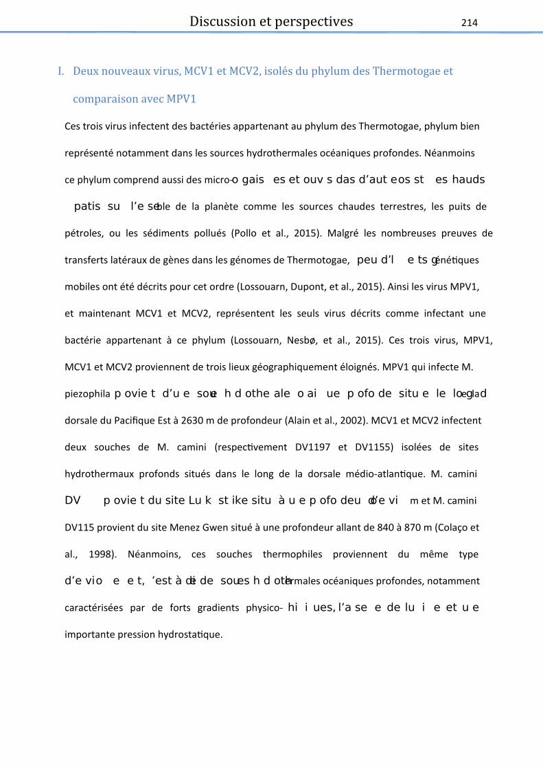
Discussion et perspectives
214
I. Deux nouveaux virus, MCV1 et MCV2, isole� s du phylum des Thermotogae et
comparaison avec MPV1
Ces trois virus infectent des bactéries appartenant au phylum des Thermotogae, phylum bien
représenté notamment dans les sources hydrothermales océaniques profondes. Néanmoins
ce phylum comprend aussi des micro-o gais es et ouv s das d’aut e os st es hauds
patis su l’e seble de la planète comme les sources chaudes terrestres, les puits de
pétroles, ou les sédiments pollués (Pollo et al., 2015). Malgré les nombreuses preuves de
transferts latéraux de gènes dans les génomes de Thermotogae, peu d’l e ts génétiques
mobiles ont été décrits pour cet ordre (Lossouarn, Dupont, et al., 2015). Ainsi les virus MPV1,
et maintenant MCV1 et MCV2, représentent les seuls virus décrits comme infectant une
bactérie appartenant à ce phylum (Lossouarn, Nesbø, et al., 2015). Ces trois virus, MPV1,
MCV1 et MCV2 proviennent de trois lieux géographiquement éloignés. MPV1 qui infecte M.
piezophila p ovie t d’u e soue h d othe ale o ai ue p ofo de situ e le lo g de la
dorsale du Pacifique Est à 2630 m de profondeur (Alain et al., 2002). MCV1 et MCV2 infectent
deux souches de M. camini (respectivement DV1197 et DV1155) isolées de sites
hydrothermaux profonds situés dans le long de la dorsale médio-atlantique. M. camini
DV p ovie t du site Lu k st ike situ à u e p ofo deu d’e vio m et M. camini
DV115 provient du site Menez Gwen situé à une profondeur allant de 840 à 870 m (Colaço et
al., 1998). Néanmoins, ces souches thermophiles proviennent du même type
d’e vio e e t, ’est à die de soues h d othermales océaniques profondes, notamment
caractérisées par de forts gradients physico- hi i ues, l’a se e de lu i e et u e
importante pression hydrostatique.

Discussion et perspectives
215
Ainsi, ces trois virus, malgré leur éloignement géographique, présentent de nombreuses
similarités comme décrit précédemment dans la partie résultats (Chapitre 3) de ce manuscrit.
En se basant sur des critères morphologiques, ils ont été classés dans la famille Siphoviridae
appate at à l’o d e ts padu Caudovirales. Ce sont trois virus tempérés mettant un
œuv e u le lsog i ue. Ils patage t aussi de o euses siila it s g o i ues. Ils
o tie e t tous u ADN dou le i li ai e d’u e taille allat de , à , k do t
l’o gaisatio e odules est se lale, de ’ e ’ : réplication et transcription,
e apsidatio , asselage des viio s, lse de l’hôte et lsog ie. U « core genome »,
composé de 35 ORFs, a été identifié au sein des trois génomes. La majeure partie de ces ORFs
codent pour des protéines hypothétiques mais certains ont pu être annotés. Parmi ces
protéines est retrouvée une protéine de structure : la protéine majeure de capside. Celle-ci
se le t s o se v e puisu’elle est siilaie à plus de % da s les t ois go es. U e
autre protéine impliquée dans la fabrication des virions a aussi été identifiée avec une forte
si ilait % , il s’agit de la g a de sous-unité de la terminase. Les terminases virales sont
des e z es ui e o aisse t l’aide u l i ue vial et pe ette t so e apsidatio das
les nouveaux virions (Feiss and Catalano, 2005) . La petite sous u it de la te i ase ’a pu
t e ide tifie da s au u des go es, il est possile u’elle soit pse te ais trop
différente de celles retrouvées dans les bases de données pour être identifiée. Des protéines
impliquées dans le métabolisme des acides nucléiques font aussi partie de ce « core
genome », notamment une hélicase. Une résolvase de jonctions de Holliday de la famille
RusA a été identifiée dans les trois génomes viraux. Ces enzymes sont notamment impliquées
dans le processus de recombinaison homologue où elles clivent les jonctions de Holliday liant
uat e i s d’ADN (Macmaster et al., 2006). Une protéie de liaiso à l’ADN siple bri a
t ise e vide e sas pou auta t ue l’o puisse ide tifie sa fotio .
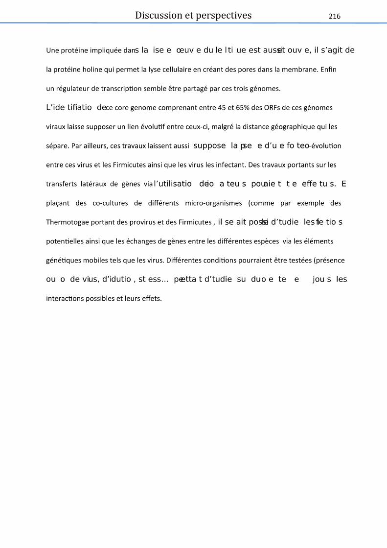
Discussion et perspectives
216
Une protéine impliquée dans la ise e œuv e du le l ti ue est aussi et ouv e, il s’agit de
la protéine holine qui permet la lyse cellulaire en créant des pores dans la membrane. Enfin
un régulateur de transcription semble être partagé par ces trois génomes.
L’ide tifiatio de ce core genome comprenant entre 45 et 65% des ORFs de ces génomes
viraux laisse supposer un lien évolutif entre ceux-ci, malgré la distance géographique qui les
sépare. Par ailleurs, ces travaux laissent aussi suppose la pse e d’u e fo te o-évolution
entre ces virus et les Firmicutes ainsi que les virus les infectant. Des travaux portants sur les
transferts latéraux de gènes via l’utilisatio de io a teu s pouaie t t e effe tu s. E
plaçant des co-cultures de différents micro-organismes (comme par exemple des
Thermotogae portant des provirus et des Firmicutes , il se ait possile d’tudie les ife tio s
potentielles ainsi que les échanges de gènes entre les différentes espèces via les éléments
génétiques mobiles tels que les virus. Différentes conditions pourraient être testées (présence
ou o de vius, d’idutio , st ess… peetta t d’tudie su du o e te e jou s les
interactions possibles et leurs effets.

Discussion et perspectives
217
II. Thermotogae et CRISPR
Les Thermotogae ont été la cible de nombreux transferts latéraux de gènes au cours de leur
évolution. Leur phylogénie particulière et la composition de leurs génomes en portent de
nombreuses traces. En effet, ces bactéries thermophiles sont proches des Aquificales selon les
tudes se asat su l’ARN S et les protéines ribosomales. Néanmoins, elles présentent de
nombreuses similarités avec les Firmicutes mais aussi, dans une moindre mesure, avec les
archées (Zhaxybayeva et al., 2009; Gupta and Bhandari, 2011). Il est connu que les infections
virales sont des vecteu s ajeu s des t asfe ts latau de g es. D’ailleu s, lo s de l’tude
des génomes viraux présentée das e aus it, d’i po ta tes siilait s ave des go es
d’aut es ph la o t t ide tifi es et notamment avec des Firmicutes et les virus qui leurs sont
associés. Parmi les gènes présentant des similarités avec ceux retrouvés dans les bases de
données et après exclusion des « self-matchs » avec des Thermotogae, plus de 60% des ORFs
de ces génomes ont montré une similarité importante avec les Firmicutes ou leurs virus. Ces
résultats mettent en évidence une possible co-évolution entre les Firmicutes et les
Thermotogae. D’aut e pat, ela i pli ue aussi ue les vi us po ts pa es at ies
pourraient être en grande partie responsable des nombreux transferts horizontaux de gènes
o stat s au sei des a t ies de l’o d e des Thermotogae. De manière générale, il semble
que, dans les sources hydrothermales profondes les transferts latéraux de gènes soient plus
o eu ue das d’aut es e vio e ents marins. En effet, presque 60% des génomes
s ue s issus de et os st e h d othe al po te t des p ovius. D’aut e pat, de
nombreux gènes impliqués dans les transferts latéraux de gènes (comme les intégrases) sont
retrouvés dans les métagénomes issus de ces environnements extrêmes (Anderson et al.,
2014; Anantharaman et al., 2016).

Discussion et perspectives
218
M e si au u vius ’avait t is e vide e avat MPV , des p euves d’ife tions passées
avaient été détectées. En effet, des systèmes CRISPR-cas ont été retrouvés dans tous les
génomes de Thermotogae séquencés à ce jour (Nesbø et al., 2009; Zhaxybayeva et al., 2009;
Pollo et al., 2015).
Le système CRISPR- as est u s st e de d fe se des at ies et a h es o te l’i vasio
par des éléments génétiques mobiles et notamment les virus (Horvath and Barrangou, 2010).
Ces systèmes sont très répandus au sein des génomes bactériens en général, puisque 50%
d’e t e eu e o tie e t, et e o e plus da s les go es d’a h es ui so t % à po te
un système CRISPR-cas (Makarova et al., 2015). Il a aussi été décrit que ce système de défense
était particulièrement important pour les thermophiles qui contiennent proportionnellement
plus souvent des systèmes CRISPR-cas que les mésophiles (Sorek et al., 2008; Chellapandi and
Ranjani, 2015). Ils sont caractérisés par la présence de courtes séquences répétées entre
les uelles so t is s des spae s. Ces spae s so t des paties d’ADN e oges pe ettat
la reconnaissance des éléments génétiques mobiles envahissant la cellule. Ces séquences sont
associées à des gènes cas CRISPR asso iated se uee ui pe ette t l’iativatio des
acides nucléiques exogènes. Ils sont aussi utilisés pour classer les différents types de systèmes
en classes, types et sous types (Horvath and Barrangou, 2010; Chellapandi and Ranjani, 2015;
Makarova et al., 2015). Les types I et III sont les plus répandus. Chez les thermophiles les sous-
types les plus représentés sont les I-A, I-B, III-A et III-B (Lange et al., 2013; Makarova et al.,
2015).
Lors de la caractérisation de MPV1, virus isolé de M. piezophila, il a été montré que le plasmide
pirate pMP1, également porté par M. piezophila, était capable de se propager dans la souche
AT1244-VC14 appartenant au genre Thermosipho (Lossouarn, Nesbø, et al., 2015).

Discussion et perspectives
219
Ce o stat est patiuli e e t i t essat sahat ue des tests d’ife tio pa pMP et
MPV1 ont été menés sur diverses souches de Marinitoga, plus proches phylogénétiquement
de leur hôte naturel, mais ceci sans succès. Le plasmide pMP1 est donc capable de se propager
das u e sou he d’u gee diff e t de so hôte iitial Thermosipho sp. AT1244-VC14. Cette
souche a été isolée de la dorsale médio- atla ti ue à de p ofo deu au iveau d’u e
source hydrothermale active. Les résultats montrant la capacité de propagation de pMP1 dans
cette souche Thermosipho sp. AT1244-VC14 (contrairement aux Marinitoga testées) ont
conduit au séquençage de celle-ci. Cette étude a permis de mettre en évidence la présence
d’u s st e CRISPR-cas, comme pour toutes les autres souches séquencées de
Thermotogae. Ce s st e paaît fo tio el puisu’il o tie t tous les ges essaies à sa
ise e œuv e.
Le processus de fonctionnement du système CRISPR-cas se déroule en deux grandes étapes
Figu e . La p ei e est l’a uisitio du spae das lauelle so t ipli u es les p oti es
as et as . Lo su’u aide u l i ue e og e e t e da s la ellule, il a e o aissa e
d’u otif PAM p otospae adjae t motif) qui est adjacent au protospacer (spacer encore
das l’aide u l i ue e og e . Cette e o aissa e pe et de live le p otospae pou
e suite l’i t g e e t e s ue es p tes du lo us CRISPR. La se o de tape est
l’i te f e e ave l’aide nucléique exogène. Lorsque ce dernier est reconnu par le système
de défense, il y a transcription de la séquence CRISPR correspondante. Le produit généré
correspond à un long transcrit précurseur appelé pre-crRNA qui est ensuite modifié pour
donner le crRNA CRISPR RNA . L’e do u lase as est ota e t i pli u e das e
p o essus. Le RNA atu e s’asso ie e suite à dive ses protéines cas (notamment les
protéines issues des gènes cas10, csm et cmr pour le type III) pour donner un complexe stable.

Discussion et perspectives
220
C’est e o ple e ui va se fie à l’aide u l i ue e og e pou pe ett e sa d g adatio
(Horvath and Barrangou, 2010; van der Oost et al., 2014; Rollie et al., 2015).
Figure 2 : Schéma du principe de fonctionnement générale d’un système CR)SPR-cas. La premie� re
partie du schéma représente l’acquisition d’un nouveau spacer et la seconde partie représente
l’interférence avec un acide nucléique exogène. Les gènes cas et leurs produits protéiques sont colorés
selon leurs fonctions (jaune) acquisition du spacer, (rose) mise en place (« processing ») du crRNA,
(bleu) assemblage et stabilite� du complexe effecteur, (violet) de�gradation de la cible. Les e� le�ments gris
sont des produits qui ne sont pas issus des ge�nes cas. Le ge�nome viral est vert et le protospacer est
symbolise� par un rectangle vert sur celui-ci.
–
‘ ’ ‘ ’
CRISPR–
CRISPR locus
Acquisition
Expression
Interference
Leader
cas genes
Spacer acquisition
crRNA processing
crRNP assemblyand surveillance
SpacerRepeat
Transcription
Repair and/or recombination enzymes
Target degradation
Cas6or
?
Pre-crRNA
RNase III
RNase
– ––

Discussion et perspectives
221
Le système présent dans la souche Thermosipho sp. AT1244-VC14 peut être classé comme
système de Classe 1, type III-A (Makarova et al., 2015). Le génome contient en effet le gène
signature de la classe III : cas10 (grande sous unité). Le séquençage a aussi révélé la présence
des deux gènes signatures des types III-A et III-B, respectivement csm2 et cmr5. Les gènes
codant pour les protéines cas5 et plusieurs protéines du groupe cas7 (comme cmr4 ou cmr6)
ont aussi été identifiés et font partie des éléments retrouvés dans tous les CRISPR-cas III-A et
III-B. Néanmoins la présence des gènes cas1, cas2 et cas6, classe ce système dans le type III-A
car les autres systèmes de types III ne contiennent généralement pas ces gènes (Makarova et
al., 2015).
Les spacers contenus dans ce système ont été étudiés notamment grâce au server CRISPI
(Rousseau et al., 2009). Au u at h ’a t ide tifi pou MPV et pMP , e ui est oh ent
avec la possibilit d’i fe tio de la sou he pa es l e ts g ti ues o iles. Pa o t e,
matchs au dessus de 85% de similarités ont été retrouvés pour MCV1 et MCV2. Ce constat est
oh e t ave l’i possiilit pou es vi us d’i fe te Thermosipho sp. AT1244-VC14.
Toutefois, nous ne pouvons conclure que la non-infection soit le seul fait de la présence de
ces séquences spacers. Ceci met aussi en évidence que cette souche de Thermosipho a sans
doute déjà été en contact avec ces virus ou des virus proches de ceux-ci.

Discussion et perspectives
222
III. Importance de la lysoge�nie et virome
L’i po ta e de la lsog ie da s les oas a lo gte ps t sous-estimée du fait des moyens
de détection utilisés et ui o sistaie t g ale e t e l’utilisatio de plage de lses. Par
ailleurs, le faible nombre de bactéries et archées cultivables (moins de 1% de la diversité
esti e et l’ase e de g es a ueu s au sei des g o es viau o e l’ARN S pa
e e ple e de t l’e plo atio de ette dive sit o ple e. N a oi s, la apide volution
des méthodes de séquençage a pe is d’o te i de plus e plus de go es de at ies et
d’a h es ais aussi de ouveau tag o es viau appels vio es. M e s’il reste des
d fis ajeu s das l’e ploitatio de eu -ci, ils permettent une meilleure compréhension et
une vision plus globale de la répartition des virus dans les environnements (Brum and Sullivan,
2015; Chow and Suttle, 2015; Paez-Espino et al., 2016).
Il y a encore peu de viromes disponibles au niveau des environnements marins profonds.
N a oi s, uel ues tudes ajeu es o t pe is de ett e e vide e l’ao da e et la
diversité des génomes viraux dans ces environnements (Williamson, Rusch, et al., 2008;
Anderson et al., 2014). Les études menées sur des métagénomes par Williamson, Anderson et
leurs collabo ateu s o t aussi peis de ett e e vide e l’i po ta e de la lsog ie da s
les sources hydrothermales océaniques profondes. En effet, il apparaît que ce cycle viral serait
plus padu da s e tpe d’ os st e h d othe al ai ue das la olo e d’eau. Aisi
dans ces conditions extrêmes les virus auraient tendance à rester intégrés de façon stable
dans les génomes cellulaires (Williamson, Cary, et al., 2008; Williamson, Rusch, et al., 2008;
Anderson et al., 2013, 2014). La prépondérance de virus tempérés au niveau des sources
hydrothermales, notamment révélée par des études métagénomiques, peut être considérée
comme une stratégie de survie face aux conditions environnementales extrêmes et variables.

Discussion et perspectives
223
Sachant que la dégradation des virions en environnement marin profond (viral decay) est
importante (pour MCV1, survie de moins de 5 heures hors de son hôte), il paraît cohérent que
les virus ont plus de ha es de su vie à l’it ieu de leu hôte plutôt u’à l’e t ieu . E
restant intégrés dans les génomes cellulaires les virus se pr u isse t de la possiilit d’ t e
d g ad s s’ils e t ouve t pas u aut e hôte à ife te apide e t. Ils pouaie t ai si t e
aussi protégés du broutage et des conditions environnementales difficiles (Paul, 2008). Cette
relation de type lysogénique peut être vue comme symbioti ue puisu’elle appo te aussi des
fi es à l’hôte ui po te le p ovius. E effet, l’adaptatio de la ellule à so
environnement (fitness) peut en être améliorée et des échanges de gènes menant à une
eilleu e o p titivit das l’e vi o e e t peuvent être mis en oeuvre. Le fait de porter
u p ovius o f e aussi à la ellule u tpe d’i u it o t e les aut es vius p o hes de
celui qui est intégré dans son génome (Paul, 2008; Breitbart, 2012). Les virus découverts dans
ces métagénomes portent aussi souvent des gènes appelés AMG (Auxiliary Metabolic Genes,
soit g es au iliaies du taolis e , e ui o fo te l’h poth se d’u fi e ip o ue
entre les deux entités (Anderson et al., 2014; Rosenwasser et al., 2016). Ces gènes sont
impliqués dans le métabolisme énergétique, notamment ceux du soufre et du méthane mais
aussi dans la biosynthèse des acides aminés et la réplication/réparation/recombinaison de
l’ADN (Anderson et al., 2014; Anantharaman et al., 2016).
C’est da s e o te te u’u p ojet t asve se a t is e plae e t e le LM E et le LUBEM
(Laboratoire Universitaire de de Biodiversité et Ecologie Microbienne) de Brest pour
d veloppe u p oto ole pe ettat d’o te i des vi o es p ove at de dive s
e vi o e e ts. Au LM E, l’o je tif est de p oduie u vio e à pati de s di e ts
hydrothermaux profonds pour élargir les connaissances sur les communautés virales
présentes dans ces environnements peu explorés.

Discussion et perspectives
224
Au, LUBEM l’o je tif est d’tudie les o u aut s viales p se tes da s le i o iote de
patie ts atteits de u ovis idose afi d’o te i u e eilleu e o p he sio de l’i pat
de ces communautés sur les patients ainsi que sur leur microbiote.
J’ai pati ip , via l’e ad e e t du stage de aste de Julia Sa hez, à la ise e plae d’u
premier protocole. Ces travaux portés, par le Pr Payan et le Dr S. Vallet se poursuivent
maintenant dans le cadre de la thèse de Lisa Billard au LUBEM. Le protocole pour produire un
vi o e est e passe d’t e is au poit. Il pe ett a aisi d’o te i des hatillo s où la
majorité des cellules sont éliminées tout en conservant au maximum les particules virales
présentes. Le séquençage des échantillons permettra de mettre en évidence les virus présents
dans les divers échantillons traités et ainsi améliorer notre compréhension de leur impact dans
les environnements étudiés.
C’est das ette d a i ue u’au LM E, de ouveaux échantillons hydrothermaux marins
profonds vont être étudiés via une approche de métagénomique. En effet, afin de prospecter
la biodiversité des écosystèmes extrêmes, des membres du laboratoire embarquent
régulièrement à bord de navires océanographiques pour des campagnes à la mer. Les
échantillons issus de ces campagnes océanographiques permettront donc de réaliser de
nouveaux viromes, en bénéficiant du travail mené en amont. Cette approche permettra
d’ te d e nos connaissances sur le mobilome abyssal, qui reste encore à ce jour assez
o u. Aisi, e tpe d’tude pe et d’avoi a s à u e visio glo ale des l e ts
génétiques mobiles p se ts da s l’e vio e e t. N a oi s, il este p i o dial d’
associer des travaux de caractérisation de nouveaux modèles hôtes virus. Ces travaux
permettront de faire le lien entre les hôtes et les EGMs qui leurs sont associés et de fournir de
ouveau od les pe etta t d’e i hi les ases de do es e s ue es o ues.

Discussion et perspectives
225
IV. Ve�sicules membranaires et de� toxification
Les v siules e a ai es MVs fo t pa tie d’u plus g a d e sele appel o ilo e
(comprenant les virus, les plasmides, les MVs, les GTAs, les t a sposo s… . Elles sont
nombreuses notamment dans les océans où leur abondance a récemment été estimée à
105/106 MVs/ l d’eau de e (Scanlan, 2014). La production de vésicules membranaires est
un mécanisme universel de communication cellulaire, connu chez les eucaryotes et les
at ies sophiles ôle das le t a sfe t de to ies, d’age ts du uo u se sig, de
fateu s de pathogi it , t asfe ts d’ARN et d’ADN, ôle dans la défense contre les attaques
vi ales…, mais très peu documenté chez les bactéries thermophiles et les archées (Scanlan,
2014; Jagannadham and Chattopadhyay, 2015; Lossouarn, Dupont, et al., 2015).
C’est lo s de dive s essais d’idu tio viale au tau lou ds u’a t d ouve te la
production de MVs chez la souche Marinitoga camini DV1197, hôte du virus MCV1.
I itiale e t, l’o je tif tait d’iduie la p odu tio viale g âe à des fateu s de st ess
e vi o e e tau st ess pouva t t e atu elle e t p se ts da s l’e vio e e t où la
souche a été isolée). En effet, les inductions étaient, jusu’alo s, alis es à l’aide d’u
antibiotique, la mitomycine C. Hors, cet antibiotique ne représente en aucun cas un stress
e vi o e e tal. C’est à pati de e o stat ue ous avo s essa d’iduie la p oduction
vi ale à l’aide de diff e ts tau lou ds atu elle e t p se ts das les sou es
hydrothermales profondes. Le cadmium, le zinc, le cuivre et cobalt ont été testés à diverses
o e t atio s de l’o d e du mM) sur les cultures en début de phase exponentielle. Les
échantillons induits ont ensuite été concentrés et observés en MET (Figure . C’est alo s u’a
été découvert non pas une induction de la production virale mais une forte production de MVs
e atio à l’ajout de uiv e et de ad iu .

Discussion et perspectives
226
Pour que cette production soit induite, il a fallu utiliser des concentrations en métaux
beaucoup plus importantes (0.5 mM) que celles généralement mesurées dans
l’e vio e e t de l’o d e du PM, (Charlou et al., 2000)). Néanmoins, ces concentrations
atu elles e tau peuve t vaie d’u site à l’aut e ais aussi da s le teps, et les fateu s
de stress déclenchant ces réactions de défense peuvent donc être multiples.
Figure 3 : Observation en microscopie e� lectronique de MVs produites par M.camini DV1197 suite a�
l’ajout de métaux lourds. Concentration de MVs suite à l’ajout de a , mM de cadmium (b) 0,1 mM
de cuivre.
L’u des ôles de es MVs est d’e te ou de suest e des ol ules to iues. Elles so t,
par exemple, impliquées dans un mécanisme de détoxification du fer chez divers eucaryotes,
notamment des plantes ou des protozoaires en séquestrant celui-ci dans de petits
compartiments intracellulaires (Slavic et al., 2016). Chez les bactéries, elles peuvent être
impliquées dans la résistance aux antibiotiques ou aux désinfectants (Jagannadham and
Chattopadhyay, 2015). Dans les océans, elles peuvent participer à la détoxification de divers
métaux lourds tels que le cadmium chez des crustacés ou des mollusques (Sterling et al.,
2007).
a b

Discussion et perspectives
227
Au niveau des sources hydrothermales profondes, il a été montré que divers symbiontes
pouvaie t, g âe à la p odu tio de MVs, p otge leu s hôtes de l’a uulatio to iue de
composés soufrés tout en utilisant celles-i o e sto kage d’e gie (Pflugfelder et al.,
2005; Eichinger et al., 2014). Les MVs contenant du soufre (aussi appelées globules de soufre)
est un phénomène décrit de longue date et bien connu chez les bactéries (Dahl and Prange,
2006). Ce type de MVs a aussi été retrouvé hez des a h es de l’o d e des Thermococcales
provenant de sources hydrothermales profondes, environnement riche en composés soufrés
et en divers composés toxiques (Gorlas et al., 2015). Dans ce cas aussi, les MVs seraient
impliquées dans un mécanisme de détoxification.
C’est da s e o te te ue l’h poth se selon laquelle ces vésicules produites en réaction à
l’ajout de tau se aie t u s st e de d to ifiatio a t ise. D’aut es tests o t peis
de ette e vide e u e p odu tio de MVs hez des sou hes d’a h es tha og es
(Post-doctorat de Samuel Dupo t, Th se de Saah Thiou . D’aut e pat, il se le ait ue
lorsque cette production de MVs est déclenchée, tant chez les Thermotogae que chez les
archées méthanogènes, celle-ci inhibe la production de particules virales. En effet, lorsque de
nombreuses MVs so t o se v es e MET suite à l’ajout des tau , t s peu, voie aucune
pati ule viale ’est d te te. Il est do possile ue es p odu tio s soie t gul es via un
effet potentiellement antagoniste.
Afin de poursuivre l’effo t de aat isation de nouveaux éléments génétiques mobiles
(EGM) issus des sources hydrothermales profondes au laboratoire de nouvelles études vont
être réalisées notamment via la thèse de Sarah Thiroux débutée en octobre 2016. Celles-ci
porteront sur la production de MVs ainsi que leur potentielle implication dans la détoxification
des tau . Il s’agia ota e t d’e p ie es pa spe t o t ie de asse (ICP-MS)
pe etta t l’aal se du o te u des v siules e tau lou ds.

Discussion et perspectives
228
Ces expériences permettront de vérifier la présence des métaux au sein des MVs et ainsi de
valider leur implication dans un mécanisme de détoxification. La présence d’aides u l i ues
dans les vésicules sera aussi recherchée afi d’ tudie leu pote tielle ipli atio das les
t asfe ts lat au de ges et do l’ volutio des o u aut s i o ie es ui les
produisent. Enfin, des analyses en microscopies seront réalisées. Celles-ci auront pour objectif
de o aît e l’iflue e des MVs su les ife tio s viales et les iteractions possibles entre
ces différents éléments.
Par ce nouveau projet nos connaissances sur la diversité moléculaire des éléments génétiques
mobiles (EGMs : virus, vésicules, plasmides) seront améliorées. Cela permettra également de
comprendre leurs impacts sur les communautés microbiennes (adaptation, détoxication,
évolution) mais aussi plus globalement leur influence sur les écosystèmes chauds où encore
t op peu de does so t dispo iles à l’heu e atuelle. E fi, ces études pourront aussi
mettre en évidence le potentiel biotechnologique de ces EGMs (transfert des toxines, de
tau lou ds, d’aides u l i ues... .

Discussion et perspectives
229
V. Bibliographie
Alain, K., Marteinsson, V.T., Miroshnichenko, M., Bonch-osmolovskaya, E., Prieur, D., and Birrien, J. (2002) Marinitoga piezophila sp. nov., a rod-shaped, thermo-piezophilic bacterium isolated under high hydrostatic pressure from a deep-sea hydrothermal vent. Int. J. Syst. Evol. Microbiol. 52: 1331–1339.
Anantharaman, K., Breier, J.A., and Dick, G.J. (2016) Metagenomic resolution of microbial functions in deep-sea hydrothermal plumes across the Eastern Lau Spreading Center. ISME J. 10: 225–239.
Anderson, R.E., Brazelton, W.J., and Baross, J.A. (2013) The deep viriosphere: assessing the viral impact on microbial community dynamics in the deep subsurface. Rev. Mineral. Geochemistry 75: 649–675.
Anderson, R.E., Sogin, M.L., and Baross, J.A. (2014) Evolutionary strategies of viruses, bacteria and archaea in hydrothermal vent ecosystems revealed through metagenomics. PLoS One 9: e109696.
Breitbart, M. (2012) Marine Viruses: Truth or Dare. Ann. Rev. Mar. Sci. 4: 425–448.
Breitbart, M. and Rohwer, F. (2005) Here a virus, there a virus, everywhere the same virus? Trends Microbiol. 13: 278–284.
Brum, J.R., Ignacio-Espinoza, J.C., Roux, S., Doulcier, G., Acinas, S.G., Alberti, A., et al. (2015) Patterns and ecological drivers of ocean viral communities. Science (80-. ). 348:.
Brum, J.R. and Sullivan, M.B. (2015) Rising to the challenge: accelerated pace of discovery transforms marine virology. Nat. Rev. Microbiol. 13: 147–159.
Charlou, J.L., Donval, J.P., Douville, E., Jean-Baptiste, P., Radford-Knoery, J., Fouquet, Y., et al. (2000) Compared geochemical signatures and the evolution of Menez Gwen
° ’ N and Lucky Strike ° ’ N hydrothermal fluids, south of the Azores Triple Junction on the Mid-Atlantic Ridge. Chem. Geol. 171: 49–75.
Chellapandi, P. and Ranjani, J. (2015) Knowledge-based discovery for designing CRISPR-CAS systems against invading mobilomes in thermophiles. Syst. Synth. Biol. 9: 97 –106.
Chow, C.-E.T. and Suttle, C.A. (2015) Biogeography of Viruses in the Sea. Annu. Rev. Virol. 2: 41–66.
Colaço, A., Desbruye�res, D., Comtet, T., and Alayse, A. (1998) Ecology of the Menez Gwen hydrothermal vent field ( Mid-Atlantic Ridge / Azores Triple Junction ). Cah. Biol. Mar. 39: 237–240.
Dahl, C. and Prange, A. (2006) Bacterial sulfur globules: occurrence, structure and metabolism. In, Inclusions in prokaryotes. Springer, pp. 21–51.
Eichinger, I., Schmitz ‐Esser, S., Schmid, M., Fisher, C.R., and Bright, M. (2014) Symbiont ‐driven sulfur crystal formation in a thiotrophic symbiosis from deep‐sea hydrocarbon seeps. Environ. Microbiol. Rep. 6: 364–372.

Discussion et perspectives
230
Feiss, M. and Catalano, C.E. (2005) Bacteriophage lambda terminase and the mechanism of viral DNA packaging. In, Viral Genome Packaging Machines: Genetics, Structure, and Mechanism. Springer, pp. 5–39.
Gorlas, A., Marguet, E., Gill, S., Geslin, C., Guigner, J.M., Guyot, F., and Forterre, P. (2015) Sulfur vesicles from Thermococcales: A possible role in sulfur detoxifying mechanisms. Biochimie 118: 356–364.
Gupta, R.S. and Bhandari, V. (2011) Phylogeny and molecular signatures for the phylum Thermotogae and its subgroups. Antonie Van Leeuwenhoek 100: 1–34.
Horvath, P. and Barrangou, R. (2010) CRISPR/Cas, the immune system of bacteria and archaea. Science (80-. ). 327: 167–170.
Jagannadham, M. V and Chattopadhyay, M.K. (2015) Role of outer membrane vesicles of bacteria. Resonance 20: 711–725.
Lange, S.J., Alkhnbashi, O.S., Rose, D., Will, S., and Backofen, R. (2013) CRISPRmap: an automated classification of repeat conservation in prokaryotic adaptive immune systems. Nucleic Acids Res. gkt606.
Lossouarn, J., Dupont, S., Gorlas, A., Mercier, C., Bienvenu, N., Marguet, E., et al. (2015) An abyssal mobilome: Viruses, plasmids and vesicles from deep-sea hydrothermal vents. Res. Microbiol. 166: 742–752.
Lossouarn, J., Nesbø, C.L., Mercier, C., Zhaxybayeva, O., Johnson, M.S., Charchuck, R., et al. Ménage à trois : a selfish genetic element uses a virus to propagate within
Thermotogales. Environ. Microbiol. 17: 3278–3288.
Macmaster, R., Sedelnikova, S., Baker, P.J., Bolt, E.L., Lloyd, R.G., and Rafferty, J.B. (2006) RusA Holliday junction resolvase: DNA complex structure--insights into selectivity and specificity. Nucleic Acids Res. 34: 5577–84.
Makarova, K.S., Wolf, Y.I., Alkhnbashi, O.S., Costa, F., Shah, S.A., Saunders, S.J., et al. (2015) An updated evolutionary classification of CRISPR-Cas systems. Nat. Rev. Microbiol. 13: 722–736.
Nesbø, C.L., Bapteste, E., Curtis, B., Dahle, H., Lopez, P., Macleod, D., et al. (2009) The genome of Thermosipho africanus TCF52B: lateral genetic connections to the Firmicutes and Archaea. J. Bacteriol. 191: 1974–1978.
Nesbo, C.L., S Swithers, K., Dahle, H., Haverkamp, T.H.A., Birkeland, N.-K., Sokolova, T., et al. (2015) Evidence for extensive gene flow and Thermotoga subpopulations in subsurface and marine environments. ISME J. 9: 1532–1542.
van der Oost, J., Westra, E.R., Jackson, R.N., and Wiedenheft, B. (2014) Unravelling the structural and mechanistic basis of CRISPR-Cas systems. Nat. Rev. Microbiol. 12: 479–492.
Paez-Espino, D., Eloe-Fadrosh, E.A., Pavlopoulos, G.A., Thomas, A.D., Huntemann, M., Mikhailova, N., et al. Uncovering Earth’s virome. Nature advance on:

Discussion et perspectives
231
Paul, J.H. (2008) Prophages in marine bacteria: dangerous molecular time bombs or the key to survival in the seas? ISME J. 2: 579–589.
Pflugfelder, B., Fisher, C.R., and Bright, M. (2005) The color of the trophosome: elemental sulfur distribution in the endosymbionts of Riftia pachyptila (Vestimentifera; Siboglinidae). Mar. Biol. 146: 895–901.
Pollo, S.M.J., Zhaxybayeva, O., and Nesbø, C.L. (2015) Insights into thermoadaptation and the evolution of mesophily from the bacterial phylum Thermotogae. Can. J. Microbiol. 1–16.
Rollie, C., Schneider, S., Brinkmann, A.S., Bolt, E.L., and White, M.F. (2015) Intrinsic sequence specificity of the Cas1 integrase directs new spacer acquisition. Elife 4: e08716.
Rosenwasser, S., Ziv, C., Creveld, S.G. van, and Vardi, A. (2016) Virocell Metabolism: Metabolic Innovations During Host-Virus Interactions in the Ocean. Trends Microbiol.
Rousseau, C., Gonnet, M., Le Romancer, M., and Nicolas, J. (2009) CRISPI: a CRISPR interactive database. Bioinformatics 25: 3317–3318.
Scanlan, D. (2014) Bacterial vesicles in the ocean. Science (80-. ). 343: 143–144.
Slavic, K., Krishna, S., Lahree, A., Bouyer, G., Hanson, K.K., Vera, I., et al. (2016) A vacuolar iron-transporter homologue acts as a detoxifier in Plasmodium. Nat. Commun. 7:.
Sorek, R., Kunin, V., and Hugenholtz, P. (2008) CRISPR —a widespread system that provides acquired resistance against phages in bacteria and archaea. Nat. Rev. Microbiol. 6: 181–186.
Sterling, K.M., Mandal, P.K., Roggenbeck, B.A., Ahearn, S.E., Gerencser, G.A., and Ahearn, G.A. (2007) Heavy metal detoxification in crustacean epithelial lysosomes: role of anions in the compartmentalization process. J. Exp. Biol. 210: 3484–3493.
Suttle, C.A. (2005) Viruses in the sea. Nature 437: 356 –361.
Williamson, S.J., Cary, S.C., Williamson, K.E., Helton, R.R., Bench, S.R., Winget, D., and Wommack, K.E. (2008) Lysogenic virus host interactions predominate at deep-sea diffuse-flow hydrothermal vents. ISME J. 2: 1112–1121.
Williamson, S.J., Rusch, D.B., Yooseph, S., Halpern, A.L., Heidelberg, K.B., Glass, J.I., et al. (2008) The Sorcerer II Global Ocean Sampling Expedition: Metagenomic Characterization of Viruses within Aquatic Microbial Samples. PLoS One 3: e1456.
Zhaxybayeva, O., Swithers, K.S., Lapierre, P., Fournier, G.P., Bickhart, D.M., DeBoy, R.T., et al. (2009) On the chimeric nature, thermophilic origin, and phylogenetic placement of the Thermotogales. Proc. Natl. Acad. Sci. U. S. A. 106: 5865–5870.


De nouveaux systèmes hôtes-virus associés aux sources hydrothermales profondes
Coraline MERCIER
Résumé : Nos connaissances sur la diversité virale associée aux micro-organismes présents dans les sources hydrothermales océaniques profondes restent encore limitées. Seules quelques études concernant l’abondance virale et l’impact de ceux-ci sur la mortalité microbienne dans ces écosystèmes sont disponibles. En effet, seuls 6 bactériovirus et 2 archéovirus provenant de ces écosystèmes ont été caractérisés à l’heure actuelle. Les deux archéovirus infectent des archées anaérobies hyperthermophiles appartenant à l’ordre des Thermococcales et ont été décrits au laboratoire. Afin d’étendre nos connaissances sur la diversité virale associée aux micro-organismes colonisant ces environnements, il a été décidé d’élargir les recherches à l’ordre bactérien des Thermotogales. Cet ordre bactérien est composé de bactéries chimio-organotrophes anaérobies en majorité thermophiles ou hyperthermophiles. De nombreux transferts latéraux de gènes ont contribué à l’histoire évolutive des Thermotogales supposant une forte implication des virus dans celle-ci. Ces travaux de thèse ont permis la caractérisation fonctionnelle et génomique de deux nouveaux siphovirus, MCV1 et MCV2, infectant deux souches de Marinitoga camini. Ces souches ont été isolées de deux sites hydrothermaux profonds (Menez Gwen et Lucky Strike) au niveau de la dorsale médio-atlantique. Ces virus mettent en œuvre un cycle lysogénique avec une production basale sans induction relativement haute (>107 virions/ml). Une comparaison de ces deux génomes viraux à celui de MPV1, virus précédemment isolé de Marinitoga piezophila, a été réalisée, révélant la présence de nombreuses similarités. Un core genome de 35 ORFs partagé par ces trois génomes a été identifié, incluant des protéines impliquées dans le métabolisme de l’ADN, l’assemblage des virions et le cycle lysogénique. Des protéines hypothétiques ont aussi été identifiées parmi ces gènes communs, elles portent donc probablement des fonctions importantes pour ces bactériovirus. Par ailleurs, 60% des gènes de ces virus ayant une correspondance dans les bases de données, après exclusion des Thermotogales, partagent des similarités avec les Firmicutes et les bactériovirus qui leurs sont associés. Le génome d’une autre Thermotogales, Thermosipho sp. AT1244-VC14 a été étudié ainsi que son système CRISPR-cas. Ces résultats indiquent que cette souche, qui porte un système CRISPR-cas qui semble complet et fonctionnel, a probablement déjà été infectée par MCV1, MCV2 ou un virus similaire. Ces travaux permettent d’étendre nos connaissances sur les virus portés par les bactéries du phylum Thermotogae, encore peu décrits à ce jour. Les éléments génétiques mobiles associés à ce phylum sont particulièrement intéressants car ils ont probablement eu un impact important dans l’évolution de ces communautés microbiennes ainsi que dans leur adaptation aux conditions physico-chimiques extrêmes et fluctuantes présentes dans les écosystèmes qu’elles colonisent. Mots clés : bactériovirus, Thermotogales, sources hydrothermales profondes, éléments génétiques mobiles, océans profonds.
New host-virus systems from deep sea hydrothermal vents
Abstract : Our knowledge of the viral diversity associated to microorganisms inhabiting the deep-sea hydrothermal vents is still limited. Only a few studies have focused on viral abundance and impact on microbial mortality within these ecosystems. A limited number of viruses (6 bacterioviruses and 2 archaeoviruses) were isolated from these environments and characterized. Two viruses associated to hyperthermophilic anaerobic Archaea, from the Thermococcales order, have been described in our laboratory. In order to deepen our knowledge on the viral diversity of these extreme environments, we have extended our investigation to the bacterial order of Thermotogales. This order is composed of anaerobic chemoorganotrophic bacteria that are, for the most part, hyper/thermophilic. Numerous lateral gene transfers have contributed to the evolutionary history of the Thermotogales, implying the potential involvement of viruses. Here, we will report the characterization of two new siphoviruses MCV1 and MCV2 that infect two strains of Marinitoga camini. Those bacterial strains were isolated from two deep-sea hydrothermal vents sites (Menez Gwen and Lucky strike) in the Mid Atlantic Ridge. These viruses are temperate with a high basal production of virions (>107 virions/mL). Comparative genomics with MPV1, a virus isolated from M. piezophila, was performed and show that those bacterioviruses share numerous similarities. A set of “core genes” shared by all these three viruses was identified and includes proteins involved in DNA metabolism, head and tail assembly and lysogenic cycle. Shared hypothetical proteins were also identified, suggesting that these unknown proteins probably provide important functions for these viruses. Interestingly, for genes with blastp matches in Genbank, over 60% have their top matches, outside the Thermotoga, to genes from Firmicutes and bacterioviruses associated to Firmicutes. We also analyzed the genome of Thermosipho sp. 1244 and studied his CRISPR-cas system. Our results indicated that this Thermosipho strain, with a complete and functional CRISPR-cas system, had already been infected by MCV1, MCV2 or a similar virus. The analyses presented here extend our knowledges about these newly discovered viruses in the deeply branching bacterial phylum Thermotogae. This bacterial order and associated mobile genetic elements are significant for addressing long-term evolutionary adaptation to fluctuant and extreme physicochemical conditions. Key words: bacterioviruses, Thermotogales, deep sea, hydrothermal vents, mobile genetic elements.
Copyright © 2022 FDOKUMEN


![[tel-01057231, v1] Facteurs associés au mauvais contrôle ...](https://static.fdokumen.com/doc/165x107/63280d54cedd78c2b50de16b/tel-01057231-v1-facteurs-associes-au-mauvais-controle-.jpg)