147-bai-tap-tu-luan-ve-dong-dien-khong-doi-2014-2015-co-dap-so thuvienvatly com 86d4b 40788
Upload
independentCategory
view
2download
0
1408–1417 Nucleic Acids Research, 2002, Vol. 30, No. 6 © 2002 Oxford University Press
DAP-like kinase interacts with the rat homolog ofSchizosaccharomyces pombe CDC5 protein, a factorinvolved in pre-mRNA splicing and required for G2/Mphase transitionHarry Engemann, Volker Heinzel, Grit Page, Ute Preuss and Karl Heinz Scheidtmann*
Institute of Genetics, University of Bonn, Roemerstrasse 164, D-53117 Bonn, Germany
Received October 1, 2001; Revised January 7, 2002; Accepted January 18, 2002
ABSTRACT
DAP-like kinase (Dlk, also termed ZIP kinase) is aleucine zipper-containing serine/threonine-specificprotein kinase with as yet unknown biological func-tion(s). Interaction partners so far identified areeither transcription factors or proteins that cansupport or counteract apoptosis. Thus, Dlk might beinvolved in regulating transcription or, more generally,survival or apoptosis. Here we report on a new inter-action partner, the rat homolog of Schizosaccharo-myces pombe CDC5 protein, a presumptivetranscription and splicing factor involved in the G2/Mtransition. In vitro, rat CDC5 forms complexes with, butis not phosphorylated by, Dlk. Rather, it was phos-phorylated by an associated kinase which wasidentified as CK2. The interaction domain of Dlk wasmapped to the leucine zipper, while that of CDC5was mapped to the C-terminal region betweenresidues 500 and 802. In vivo, both proteinsco-localize perfectly in distinct speckle-like structuresin the nucleus, some of which overlap withpromyelocytic leukemia protein. Interestingly,splicing factor SC35, which also resides in speckles,was partially displaced upon overexpression ofeither CDC5 or Dlk, perhaps due to phosphorylationby Dlk. Together with previous data, these resultssuggest that Dlk might play a role in coordinatingspecific transcription and splicing events.
INTRODUCTION
Protein phosphorylation is perhaps the widest used regulatorytool of eukaryotic organisms and almost every process fromDNA replication to metabolic reactions is regulated by revers-ible phosphorylation. To deal with the specific demands ofsuch diverse processes a huge number of kinases exists.However, as most of the novel enzymes were discovered byvirtue of their sequence homology or as interaction partners intwo-hybrid screens, their biological role is not readily evident.
We and others have recently identified a new kinase, Dlk, forDAP-like kinase (1), or ZIP kinase, for zipper-interactingprotein kinase (2). This kinase belongs to the novel subfamilyof DAP kinases (death-associated protein kinases) (3,4) whichexhibit a high degree of sequence identity in their catalyticdomains but differ greatly in their non-catalytic parts. Due tothis relationship we will use the term ‘Dlk’ throughout thispaper. All the DAP-related kinases have been implicated inapoptosis, but a direct role in this process was only demon-strated for DAP kinase itself, which is involved in interferonγ- and TNFα/Fas-induced cell death (5,6).
Dlk, which normally resides in the nucleus in associationwith nuclear speckles, does not induce apoptosis to a significantextent (1). However, C-terminal truncation or co-expressionwith pro-apoptotic protein Par-4 results in relocation of Dlk tothe actin cytoskeleton and efficient induction of apoptosis(7,8). On the other hand, the function of nuclear Dlk remainsobscure. A unique property of Dlk not shared by the otherDAP-related kinases is its leucine zipper at the C-terminus, whichenables it to interact with the transcription factors ATF-4 (2,8)and AATF (apoptosis-antagonizing transcription factor) (9).In vitro, Dlk phosphorylates the myosin light chain (MLC) andcore histones H3, H4 and H2A (1). These latter findings pointto a role in transcription, perhaps chromatin remodeling and/orsplicing. To search for interaction partners that might serve asregulators or targets of Dlk and that might illuminate itsfunction we employed the two-hybrid system (8). One of theputative interaction partners was the rat homolog of theSchizosaccharomyces pombe CDC5 protein, which isdescribed in more detail in this paper.
To avoid confusion, we want to point out here that the CDC5genes of S.pombe and Saccharomyces cerevisiae encodeproteins with completely different functions. The S.cerevisiaeCDC5 gene encodes a polo-like kinase (plo1 in S.pombe)(10,11) which plays a crucial role in regulating diverse mitoticprocesses, such as centrosome maturation, spindle formationand the metaphase/anaphase transition (reviewed in 12,13). Onthe other hand, the CDC5 gene of S.pombe encodes a putativetranscription and splicing factor that is involved in regulatingthe G2/M transition (14,15). The S.cerevisiae homolog of thelatter is Cef1 (16). The S.pombe CDC5 homolog is structurallyand functionally highly conserved throughout evolution and
*To whom correspondence should be addressed. Tel: +49 228 734 258; Fax: +49 228 734 263; Email: [email protected]
Nucleic Acids Research, 2002, Vol. 30, No. 6 1409
the homologs from Arabidopsis thaliana (17), Drosophilamelanogaster and even human are capable of complementingthe gene defect in yeast (18).
The CDC5 protein is a nuclear phosphoprotein which, inmammalian species, consists of 802 amino acids. It contains,as characteristic elements, two so-called myb repeats and onemyb-like repeat (18,19). In the transcription factor Myb theseelements form helix–turn–helix structures and represent theDNA-binding domain. This relationship to Myb proteinssuggested that CDC5 might also be a transcription factor. Insupport of this assumption, CDC5 can bind to DNA and, whenfused to the Gal4 DNA-binding domain (BD), it can act as atransactivator of Gal4-responsive reporter genes (20). Moreconvincingly, a specific target sequence of CDC5 (GATT-TAACATAA) was elucidated and this sequence elementrenders a reporter gene CDC5-responsive (21). However,CDC5 was also shown to be involved and, in fact, to be essen-tial for pre-mRNA splicing in both yeast and mammaliansplicing systems (16,22–24). Thus, CDC5 might be involvedin both transcription and splicing and might actually provide alink between the two processes.
In this paper we describe the identification of rat CDC5 as aninteraction partner of Dlk. Both proteins co-localize perfectly inspeckle-like structures which partially overlap with promyelocyticleukemia (PML) protein. Interestingly, overexpression of CDC5or Dlk resulted in partial displacement of splicing factor SC35from nuclear speckles, perhaps resulting from competition for thesame binding partners or from phosphorylation of SC35 by Dlk.
MATERIALS AND METHODS
Cell culture and transfection
Rat embryo fibroblasts, line REF52.2, were used for expres-sion studies. Cells were grown as monolayers in Dulbecco’sminimal essential medium (Gibco BRL, Karlsruhe, Germany)supplemented with 10% fetal bovine serum (BiochromeSeromed, Berlin, Germany) and antibiotics. Transfectionswere performed with Lipofectamine (Gibco BRL) according tothe manufacturer’s protocol. Insect SF9 (Spodopterafrugiperda) cells were used for the baculovirus expressionsystem and were grown in serum-free SF900 mediumcontaining 50 µg/ml gentamycin (Gibco BRL) at 27°C.
Two-hybrid assays
The two-hybrid screen was performed with the conventionaltwo-hybrid system (25) with yeast strain Y190 and the Gal4BD or transactivation domain (AD) proteins as fusion partners.Thus, a Gal4 BD–Dlk fusion protein was employed as bait anda cDNA library from SV40-transformed rat cells SV52 fusedto the Gal4 AD was used as prey (8,9). To determine the inter-action domains, deletion mutants of Dlk or CDC5 wereexpressed as Gal4 BD or Gal4 AD fusion proteins, respectively,upon transformation of yeast Y190 or AH109 cells. A func-tional interaction between the hybrid proteins was indicated byactivation of the Gal4-responsive reporter gene His3 or Ade2,respectively, and β-galactosidase. A detailed description hasbeen given previously (8).
Isolation of the complete CDC5 cDNA and construction ofexpression plasmids
For generation of the complete coding sequence of rat CDC5the Long Expand PCR System (Roche Molecular Biochem-icals, Mannheim, Germany) was employed using a cDNAlibrary from 208F rat fibroblasts as template. The forwardprimer (5′-CCCGGGTACCAAGATGCCCCGGATTATGA-3′)and reverse primer (5′-CCCGGGATCCTCTGTGCTTCA-GAACTTTG-3′) were derived from the 5′- and 3′-termini of thepublished sequence of rat CDC5 (italic, GenBank accessionno. AF000578.2), extended by KpnI and BamHI restrictionsites, respectively. The PCR product was completelysequenced and cloned into the respective expression vectors:pEGFP, pDsRED, pGAD424 (Clontech, Heidelberg,Germany), pCMV-Tag2 (Stratagene Europe) or pVL1392HIS(1). CDC5 deletion mutants were generated as described previ-ously for Dlk (7). Where necessary, restriction site linkers forcloning were provided by PCR. Molecular biological tech-niques followed standard protocols (26,27).
In vitro translation
For in vitro translation the coding sequence of CDC5 wasexcised by partial restriction with EcoRI/BamHI and the frag-ment of 2436 bp was cloned into the EcoRI and BamHI sites ofpBluescript SK+. In vitro translation was performed in theTNT-T7/T3 Coupled Reticulocyte Transcription/TranslationSystem (Promega, Heidelberg, Germany), as recommended bythe manufacturer.
Generation of recombinant baculoviruses and purificationof His-tagged proteins by affinity chromatography
The generation and propagation of recombinant baculovirushas been described (1). For expression and purification of His-tagged Dlk, CDC5 or AATF-protein, SF9 cells were infected withthe respective recombinant viruses for 3 days and sequentiallyextracted as described (1), with the following modifications: cellswere lysed with isotonic lysis buffer [10 mM NaPO4, 140 mMNaCl, 3 mM MgCl2, 5 mM β-mercaptoethanol, proteaseinhibitors (Roche) and 0.5% Nonidet P-40, pH 9.0] and thechromatin digested with 0.1 mg/ml DNase I at 30°C for15 min. The nuclei were pelleted by low speed centrifugationand the supernatant containing soluble proteins was discarded.The nuclear pellet was resuspended in buffer J (10 mM TrispH 7.5, 140 mM NaCl, 1% Nonidet P-40, 1% Na deoxycholate,0.1% SDS, 5 mM β-mercaptoethanol, 0.01% w/v aprotinin),diluted 3-fold with PBS, pH 9, and incubated with Ni–NTA–agarose (Qiagen, Hilden, Germany) without further centrifugation.The beads were extensively washed with PBS and IMAC-50(20 mM Tris–HCl pH 8, 0.5 M NaCl, 10% glycerol, 1 mMPMSF, 5 mM β-mercaptoethanol and 50 mM imidazole) andeither used as such for affinity chromatography or the boundproteins were eluted with 500 mM imidazole, 500 mM NaCl,1 mM DDT, 10% glycerol and aprotinin and stored at –70°C.For binding studies, protein bound to Ni–NTA–agarose wasincubated with in vitro translated and [35S]methionine-labeledCDC5 at 4°C for 2 h. The beads were further washed and boundproteins were eluted, analyzed by SDS–PAGE and visualized byfluorography.
1410 Nucleic Acids Research, 2002, Vol. 30, No. 6
Kinase assays
Kinase assays were performed with purified His-tagged Dlkand purified substrates essentially as described (1). Reactionswere stopped with 2× SDS–PAGE sample buffer containing10 mM EDTA and subjected to SDS–PAGE and auto-radiography. Purified His-tagged ASF/SF2 or GST–SC35were kindly provided by Dr S. Stamm (Institute ofBiochemistry, Erlangen, Germany) or by Dr R. Lührmann (MaxPlanck Institute of Biophysical Chemistry, Göttingen, Germany),respectively.
Fluorescence microscopy analyses
Antibodies. Anti-PML monoclonal antibody (mAb) 5E10 (28)and mAb104, directed against splicing factors, were kindlyprovided by Dr S. Stamm; polyclonal anti-U5-116 kDantiserum, directed against a member of the snRNP family(29), was a gift from Dr R. Lührmann; anti-Sm antibodies werea kind gift from Dr B. Wirth (Institute of Human Genetics,Bonn, Germany); anti-SC35 Mab was purchased from BDBiosciences (Heidelberg, Germany), anti-SF2/ASF and anti-PCNA were from Santa Cruz and anti-FLAG M-2 antibodyfrom Stratagene. Secondary Cy3-conjugated antibodies werefrom Dianova (Hamburg, Germany).
Immunofluorescence. Cells were fixed, permeabilized andstained with primary and secondary antibodies as described(7,8). Briefly, cells were washed in PBS, fixed in 3% para-formaldehyde in PBS for 15 min, permeabilized in 0.2% TritonX-100 in PBS and then incubated in blocking solution(1% BSA in PBS) for 30 min. Incubations with the primaryand secondary antibodies were performed for 60 or 30 min,respectively, at appropriate dilutions in PBS, at room temperature.Between each step cells were washed two or three times inPBS. After final washing the cells were briefly rinsed in water,after which the coverslips were mounted in Permafluormounting medium (Immunotech). The immunostained cellswere examined with an Axiophot fluorescence microscope(Zeiss) with a 63× oil immersion objective.
For visualization of GFP or dsRED fusion proteins cellswere fixed as described above, without permeabilization. Forstaining of nuclei with 4,6-diamidino-2-phenylindole (DAPI)cells were fixed and permeabilized as above and then stainedwith DAPI at a final concentration of 1 µg/ml for 15 min atroom temperature. Final washing steps and mounting wasperformed as described above.
RESULTS
Using the conventional yeast two-hybrid system with Gal4–Dlkas bait, we isolated several cDNAs that coded for putativeinteraction partners of Dlk (8,9). One of these, a clone of795 bp, was identical to the 3′-portion of the rat homolog of theS.pombe CDC5 gene, with an open reading frame of 264codons. This clone was apparently incomplete since the humanand mouse ‘CDC5-like’ cDNAs were reported to be 3.4 kb,coding for proteins of 802 amino acids (18,19). In contrast, therat CDC5-like cDNA was initially reported to be 1907 bp,coding for a polypeptide of 523 residues (30), while an updatedversion comprised 2847 bp with an open reading frame of 802amino acids (GenBank accession no. AF000578.2). Since the
human CDC5-like protein seems to play similar roles in theG2→M transition and can complement mutant CDC5 inS.pombe, it was regarded as a true homolog and designatedhCDC5 (20). Accordingly, we will refer to the rat homolog asrCDC5 or, for simplicity, CDC5. (Please note the differencebetween S.pombe and S.cerevisiae CDC5; see Introduction.)
To obtain the complete coding region of rCDC5 weperformed PCR using rCDC5-specific oligonucleotides asprimers and a cDNA library from 208F rat fibroblasts astemplate. The forward primer was derived from the 5′-portionof the published sequence just upstream of the start codon andthe reverse primer from the 3′-portion just downstream of thestop codon (see Materials and Methods), omitting the untranslatedregions. This reaction revealed a product of 2440 bp, aspredicted. Sequence analysis revealed 100% identity with therCDC5 cDNA in the database with a continuous open readingframe of 802 codons. The sequence showed 98% identity withhuman CDC5 (18,19) with only 15 amino acid exchanges, sixof which are conservative, while the others are not expected tocause significant alterations in structure or function.
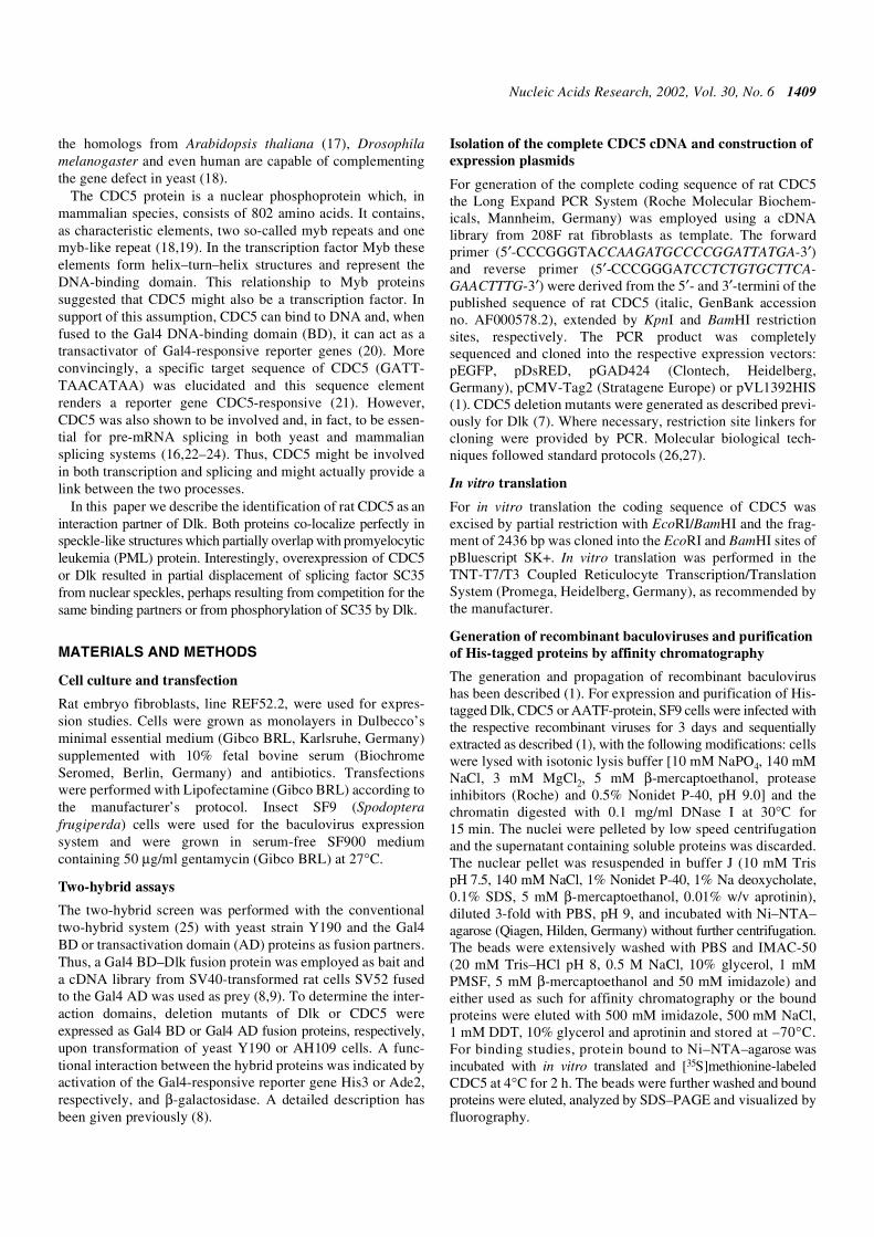
To confirm the interaction between CDC5 and Dlk, asrevealed by the two-hybrid system, CDC5 was translatedin vitro in a coupled transcription/translation system andallowed to bind to affinity purified His-tagged Dlk. In vitrotranslation revealed a product of 105 kDa, as predicted (Fig. 1A,lane 1). This protein bound specifically to His–Dlk (lane 3) butnot to control beads saturated with proteins from controlextracts (lane 2).
CDC5 is not a substrate of Dlk in vitro
CDC5 is a phosphoprotein and was shown to be a substrate ofCDKs (31,32). Additionally, CDC5 contains consensusphosphorylation sites for CK2, PKA, PKC and MAP kinase(19). To see whether CDC5 is also phosphorylated by its inter-action partner Dlk, it was expressed as a His-tagged protein inthe baculovirus system, purified by affinity chromatographyon Ni–NTA–agarose (Fig. 1B, lane 4) and subjected to in vitrophosphorylation using purified His–Dlk (Fig. 1B, lane 5) askinase. As shown in Figure 1B (lane 3), CDC5 was efficientlyphosphorylated in the absence of Dlk as well as in its presence(lane 2). These results suggested that CDC5 was phosphory-lated by a co-purified kinase but not, or only weakly, by Dlk.In addition to CDC5 and Dlk there was one major phosphory-lated protein band at 28 kDa. This was not seen when purifiedDlk was employed in an autophosphorylation reaction (lane 1).On the other hand, exogenously added substrates (MLC andhistones) were only phosphorylated by Dlk (Fig. 1B, lanes 1and 2) and not by the CDC5-associated kinase (lane 3).
To obtain clues about the nature of the co-purified kinase weemployed some commonly used substrates, myelin basicprotein (MBP) or a mixture of MLC and histones, in the kinasereactions. As shown in Figure 1C (lanes 1 and 2), MBP wasefficiently phosphorylated by the CDC5-associated kinasewhile MLC was not and the histones only weakly, thus clearlydistinguishing it from Dlk, which phosphorylates MBP(Fig. 1C, lane 6), but also MLC and core histones H3, H4 andH2B (Fig. 1B, lane 1). Since protein kinase CK2 is often foundas a contaminating kinase, we employed GTP as the phosphatedonor, which is diagnostic for CK2. Additionally, weemployed β-casein as substrate. The phosphorylation patternsof purified CDC5 with its associated proteins was identical
Nucleic Acids Research, 2002, Vol. 30, No. 6 1411
with GTP (Fig. 1C, lanes 3 and 4), although phosphorylation ofMBP was somewhat weaker. Additionally, β-casein wasefficiently phosphorylated (lane 5). In contrast, Dlk was notable to use GTP (lane 8) and β-casein was only weaklyphosphorylated (Fig. 1C, lane 7).
We next sought to distinguish CDC5 phosphorylation byCK2 from that by Dlk by employing a CK2-specific inhibitor,
heparin. Heparin (10 nM) efficiently blocked phosphorylationof CDC5 both with ATP or GTP as phosphate donor and β-caseinas substrate. However, Dlk, which was not inhibited underthese conditions, was still incapable of phosphorylating CDC5(data not shown). Likewise, when we tried to dissociate mostof the CK2 activity from CDC5, the latter was still notphosphorylated by Dlk (not shown). These data show thatrecombinant CDC5 protein purified from SF9 cells is associatedwith and efficiently phosphorylated by CK2. The phosphorylated28 kDa band presumably represents the regulatory β subunit ofinsect CK2, which is phosphorylated by the catalytic α subunit(33). On the other hand, CDC5 does not seem to be a substrateof Dlk, at least not in vitro.
Mapping of the interaction domains
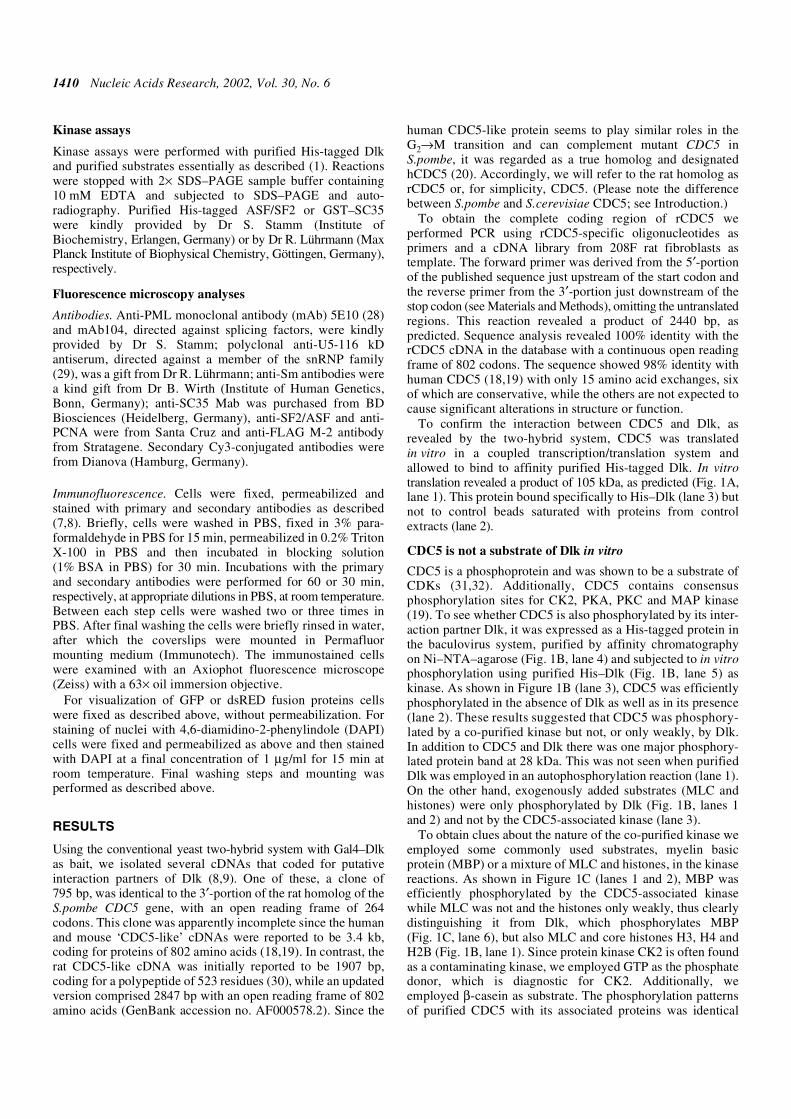
Having confirmed the interaction between Dlk and CDC5in vitro, we wanted to determine the interaction domainsbetween the two proteins. For this purpose we employed aseries of deletion mutants of both CDC5 and Dlk in the yeasttwo-hybrid interaction assay. CDC5 deletion mutants wereconstructed by PCR, providing suitable linkers for cloning, andfused to the Gal4 AD, as shown in Figure 2A (right). The Dlkmutants fused to the Gal4 BD have been described previously(8) (see Fig. 2B, right). These constructs were co-expressed invarious combinations in yeast cells and the transformed cellswere scored for adenine autotrophy and β-galactosidaseactivity. Appropriate controls were included. Mapping theinteraction domain in Dlk (shown in Fig. 2B, left) revealed thatfull-length (wt) CDC5 interacted with full-length Dlk (wt) butnot with Dlk∆C1, lacking the leucine zipper, or any of theshorter deletion mutants (∆C2 and ∆C3), indicating that theleucine zipper of Dlk was required. On the other hand, Dlk (wt)interacted with full-length CDC5 and with deletion mutantCDC5(101–802), lacking the N-terminal 100 residues, but notwith any of the mutants lacking 302 or more residues from theC-terminus [mutants CDC5(1–500), CDC5(1–377) andCDC5(1–104); Fig. 2A, left]. Considering that the originaltwo-hybrid clone comprised the coding region from codon 427to 690, these results placed the interaction domain of CDC5with Dlk between residues 501 and 690. To confirm thisconclusion we generated additional deletion constructscomprising residues 483–659 and 483–802. Both constructsexhibited interaction with Dlk, although interaction of thelarger one was somewhat stronger (Fig. 2A, left), suggestingthat residues between 500 and 659 and between 660 and 802contribute to the interaction.
Subcellular localization of CDC5 and interaction with Dlkin vivo
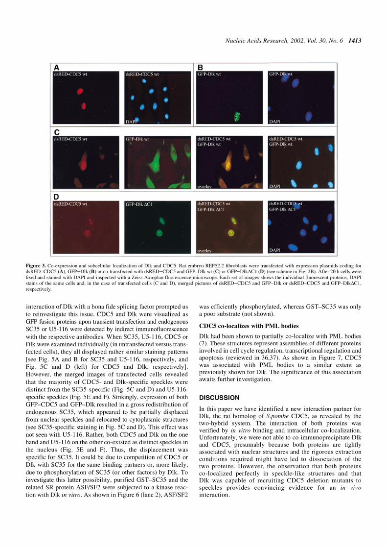
We next investigated a possible interaction of the two proteinsin vivo by co-expression and fluorescence co-localization. Dlkwas expressed as a GFP-tagged fusion protein and CDC5 as adsRED- or FLAG-tagged fusion protein. For fluorescencestudies, both constructs were transfected at the minimalamounts required for detection to minimize artefacts of over-expression. CDC5 alone usually exhibited a punctuate nuclearstaining pattern (Fig. 3A, C or D, left), in agreement withprevious reports (22). This staining pattern resembled that ofDlk (Fig. 3B) (1). Indeed, upon co-transfection both proteinsco-localized perfectly in nuclear dots (Fig. 3C). This strictco-localization of CDC5 was not seen with mutant Dlk∆C1,
Figure 1. CDC5 binds to Dlk but is not a substrate of Dlk in vitro. (A) CDC5was synthesized and labeled with [35S]methionine in vitro and employed in abinding reaction using baculovirus-expressed His–Dlk or proteins fromuninfected cells adsorbed to Ni–NTA–agarose as matrix. Bound proteins wereeluted and analyzed by SDS–PAGE on a 10% polyacrylamide gel and fluor-ography, as described in the Materials and Methods. Lane 1, input CDC5;lanes 2 and 3, CDC5 bound to control beads or His–Dlk, respectively. Theinput represented 1/10 of the translation product used for binding. (B) In vitrophosphorylation of CDC5. His–CDC5 and His–Dlk were isolated from recom-binant baculovirus-infected SF9 cells by affinity purification on Ni–NTA–agarose.Purified proteins were separated on a 10% SDS–polyacrylamide gel andstained with Coomassie blue (lanes 4 and 5) or subjected to in vitro phosphory-lation individually (lanes 1 and 3) or in combination (lane 2). A mixture ofMLC and histones was included as exogenous substrates. Phosphorylatedproducts were analyzed by SDS–PAGE (13.5% polyacrylamide) andautoradiography, as outlined under Materials and Methods, with exposure for4 h. (C) CDC5 is phosphorylated by associated CK2. Purified CDC5 or Dlkwere subjected to kinase reactions in the presence of ATP (lanes 1, 2 and 5–7)or GTP (lanes 3, 4 and 8). MBP (lanes 1, 3, 6 and 8), a MLC/histone mix (lanes2 and 4) or dephosphorylated β-casein (lanes 5 and 7) were included assubstrates. SDS–PAGE analyses were on a 13.5% (lanes 1–5) or 10% poly-acrylamide gel (lanes 6–8). Autoradiography was for 12 h. The 28 kDa bandmost likely represents the β-subunit of CK2 (33).
1412 Nucleic Acids Research, 2002, Vol. 30, No. 6
lacking the leucine zipper. In this case, Dlk∆C1 generallydisplayed a more diffuse nuclear distribution, as seen previ-ously (7), while CDC5 still occurred in speckles (Fig. 3D).This result corroborated the two-hybrid data that the inter-action of Dlk with CDC5 is mediated by its leucine zipper. Onthe other hand, the association of CDC5 with specklesapparently did not depend on Dlk.
Mapping of the nuclear localization signal
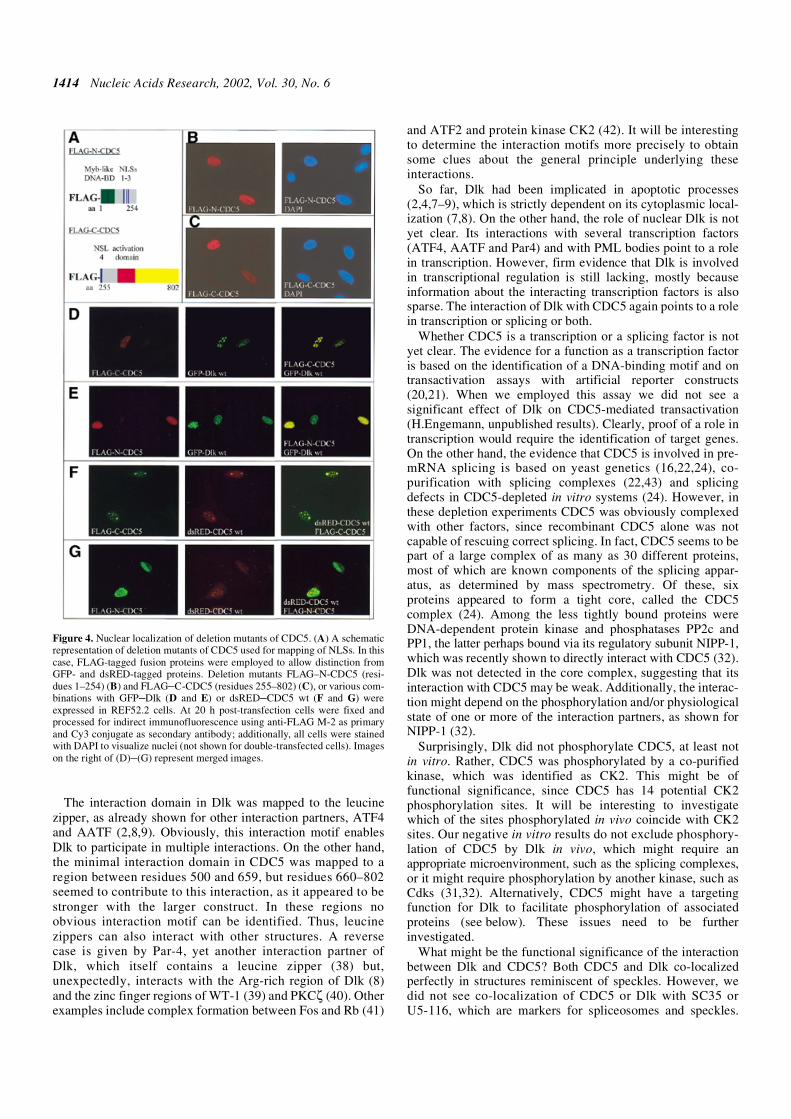
CDC5 contains four putative nuclear localization signals(NLSs) between amino acids 165 and 271. Deletion mutantsencompasing residues 1–254 (FLAG–N-CDC5) and 255–802(FLAG–C-CDC5) were constructed, thereby separating NLSs1–3 from NLS 4 (see scheme in Fig. 4A). The deletionconstructs were expressed as FLAG-tagged proteins and theirsubcellular localization was examined by indirect immuno-fluorescence analyses. As shown in Figure 4B and C, respect-ively, both the N-terminal (FLAG–N-CDC5) and C-terminalconstructs (FLAG–C-CDC5) were efficiently translocated tothe nucleus, indicating that one or more of the putative NLSs1–3 on one side and NLS 4 on the other are sufficient to directnuclear transport. However, both deletion proteinsdisplayed a diffuse rather than a speckled distribution,suggesting that full-length CDC5 is required for associationwith speckles.
To see how Dlk might behave in this setting, the CDC5constructs were co-expressed with wild-type Dlk or mutantDlk∆C1. Interestingly, upon co-expression of wild-type Dlk,the C-terminal fragment (Fig. 4D), but not the N-terminal frag-ment (Fig. 4E), of CDC5 again appeared in speckles. This wasnot seen with mutant Dlk∆C1, as expected (data not shown).Taken together, these results indicate that Dlk can target CDC5to nuclear speckles.
It had been suggested that CDC5 forms homodimers, asdeduced from the symmetry of the DNA-binding motif (21).This issue was investigated by employing the deletion mutantsdescribed above. If CDC5 is indeed able to form homodimersthen co-expression of the truncation mutants with full-lengthCDC5 should result in targeting of the truncated proteins tospeckles. This was indeed the case for the C-terminal (Fig. 4F)but not for the N-terminal fragment (Fig. 4G), thus showingthat CDC5 forms dimers and that the interaction occurs withinthe C-terminal portion.
Overexpression of CDC5 and Dlk results in displacementof SC35 from nuclear speckles
Human CDC5 has been reported to co-localize with splicingfactor SC35 (22), a member of the SR protein family that iscommonly used as a marker for spliceosomes and speckles(34,35). In contrast, no such co-localization with SC35 orU5-116 protein had been observed for Dlk (7). However, the
Figure 2. Mapping of the interaction domains of Dlk and CDC5 by two-hybrid assay. Full-length and deletion constructs of CDC5 (A) and Dlk (B) fused to theGal4 AD or BD domains, respectively, (see schemes on the right) were transfected in various combinations into yeast AH109, as indicated. Shown are yeast coloniesgrown on adenine-deficient agar plates as a result of a functional interaction of the Dlk and CDC5 proteins. (A) Full-length Gal4 BD–Dlk was co-transfected withGal4 AD–CDC5 variants. Co-transfection of Gal4 BD with Gal4 AD–CDC5 wt served as a negative control. (B) Interaction of full-length Gal4 AD–CDC5 withGal4 BD–Dlk variants. pBD + pAD served as a negative and pBDp53 + pADLT as a positive control. In the schematics of CDC5, the myb repeats are shown ingreen, the activation domain (20) in red, the putative NLSs in blue and the C-terminal part containing the interaction domain in yellow. The schematics of the Dlkvariants indicate the locations of the catalytic domain and the leucine zipper (in light gray), the non-catalytic domain in dark gray and the putative NLSs in blue;the C-terminal most NLS is the functional one (7).
Nucleic Acids Research, 2002, Vol. 30, No. 6 1413
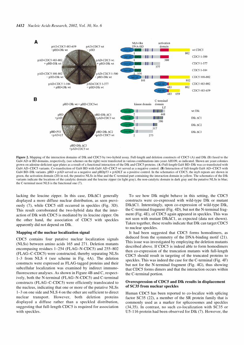
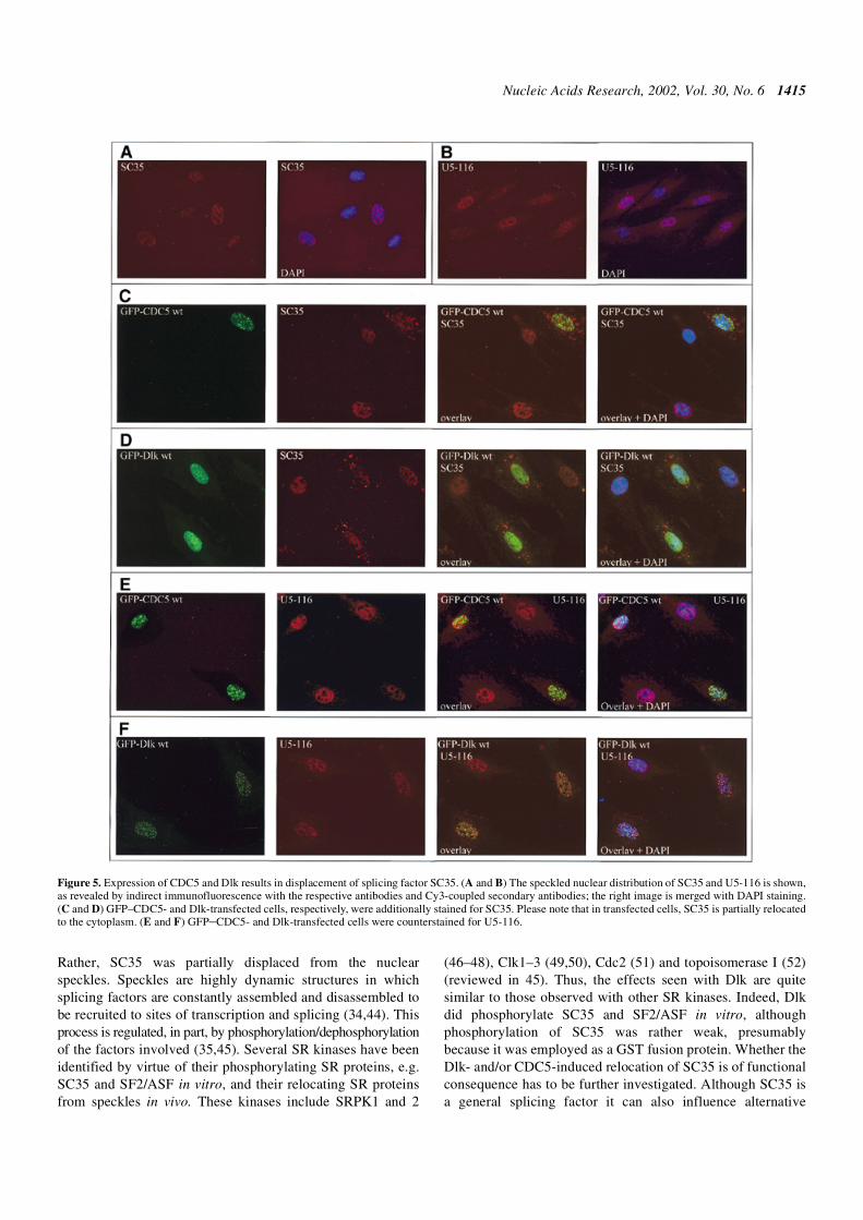
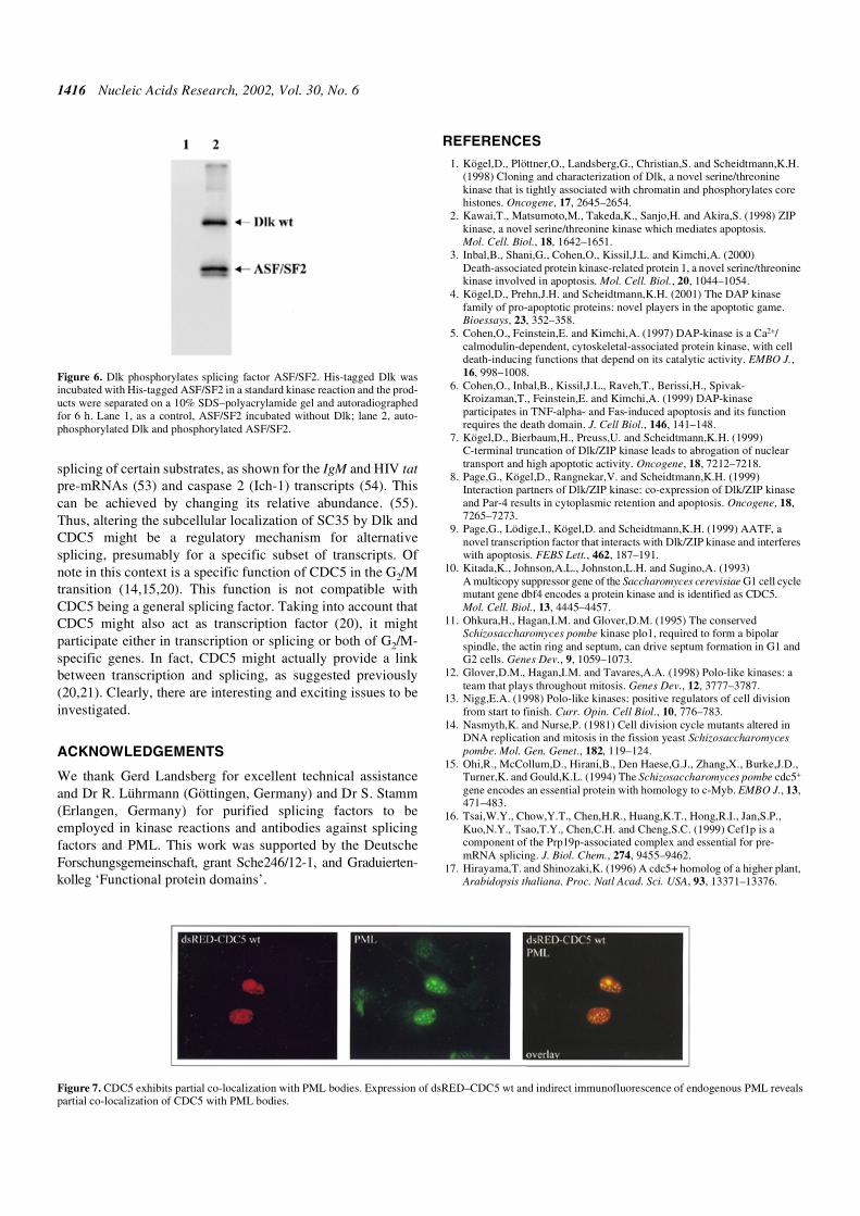
interaction of Dlk with a bona fide splicing factor prompted usto reinvestigate this issue. CDC5 and Dlk were visualized asGFP fusion proteins upon transient transfection and endogenousSC35 or U5-116 were detected by indirect immunofluorescencewith the respective antibodies. When SC35, U5-116, CDC5 orDlk were examined individually (in untransfected versus trans-fected cells), they all displayed rather similar staining patterns[see Fig. 5A and B for SC35 and U5-116, respectively, andFig. 5C and D (left) for CDC5 and Dlk, respectively].However, the merged images of transfected cells revealedthat the majority of CDC5- and Dlk-specific speckles weredistinct from the SC35-specific (Fig. 5C and D) and U5-116-specific speckles (Fig. 5E and F). Strikingly, expression of bothGFP–CDC5 and GFP–Dlk resulted in a gross redistribution ofendogenous SC35, which appeared to be partially displacedfrom nuclear speckles and relocated to cytoplasmic structures(see SC35-specific staining in Fig. 5C and D). This effect wasnot seen with U5-116. Rather, both CDC5 and Dlk on the onehand and U5-116 on the other co-existed as distinct speckles inthe nucleus (Fig. 5E and F). Thus, the displacement wasspecific for SC35. It could be due to competition of CDC5 orDlk with SC35 for the same binding partners or, more likely,due to phosphorylation of SC35 (or other factors) by Dlk. Toinvestigate this latter possibility, purified GST–SC35 and therelated SR protein ASF/SF2 were subjected to a kinase reac-tion with Dlk in vitro. As shown in Figure 6 (lane 2), ASF/SF2
was efficiently phosphorylated, whereas GST–SC35 was onlya poor substrate (not shown).
CDC5 co-localizes with PML bodies
Dlk had been shown to partially co-localize with PML bodies(7). These structures represent assemblies of different proteinsinvolved in cell cycle regulation, transcriptional regulation andapoptosis (reviewed in 36,37). As shown in Figure 7, CDC5was associated with PML bodies to a similar extent aspreviously shown for Dlk. The significance of this associationawaits further investigation.
DISCUSSION
In this paper we have identified a new interaction partner forDlk, the rat homolog of S.pombe CDC5, as revealed by thetwo-hybrid system. The interaction of both proteins wasverified by in vitro binding and intracellular co-localization.Unfortunately, we were not able to co-immunoprecipitate Dlkand CDC5, presumably because both proteins are tightlyassociated with nuclear structures and the rigorous extractionconditions required might have led to dissociation of thetwo proteins. However, the observation that both proteinsco-localized perfectly in speckle-like structures and thatDlk was capable of recruiting CDC5 deletion mutants tospeckles provides convincing evidence for an in vivointeraction.
Figure 3. Co-expression and subcellular localization of Dlk and CDC5. Rat embryo REF52.2 fibroblasts were transfected with expression plasmids coding fordsRED–CDC5 (A), GFP–Dlk (B) or co-transfected with dsRED–CDC5 and GFP–Dlk wt (C) or GFP–Dlk∆C1 (D) (see scheme in Fig. 2B). After 20 h cells werefixed and stained with DAPI and inspected with a Zeiss Axioplan fluorescence microscope. Each set of images shows the individual fluorescent proteins, DAPIstains of the same cells and, in the case of transfected cells (C and D), merged pictures of dsRED–CDC5 and GFP–Dlk or dsRED–CDC5 and GFP–Dlk∆C1,respectively.
1414 Nucleic Acids Research, 2002, Vol. 30, No. 6
The interaction domain in Dlk was mapped to the leucinezipper, as already shown for other interaction partners, ATF4and AATF (2,8,9). Obviously, this interaction motif enablesDlk to participate in multiple interactions. On the other hand,the minimal interaction domain in CDC5 was mapped to aregion between residues 500 and 659, but residues 660–802seemed to contribute to this interaction, as it appeared to bestronger with the larger construct. In these regions noobvious interaction motif can be identified. Thus, leucinezippers can also interact with other structures. A reversecase is given by Par-4, yet another interaction partner ofDlk, which itself contains a leucine zipper (38) but,unexpectedly, interacts with the Arg-rich region of Dlk (8)and the zinc finger regions of WT-1 (39) and PKCζ (40). Otherexamples include complex formation between Fos and Rb (41)
and ATF2 and protein kinase CK2 (42). It will be interestingto determine the interaction motifs more precisely to obtainsome clues about the general principle underlying theseinteractions.
So far, Dlk had been implicated in apoptotic processes(2,4,7–9), which is strictly dependent on its cytoplasmic local-ization (7,8). On the other hand, the role of nuclear Dlk is notyet clear. Its interactions with several transcription factors(ATF4, AATF and Par4) and with PML bodies point to a rolein transcription. However, firm evidence that Dlk is involvedin transcriptional regulation is still lacking, mostly becauseinformation about the interacting transcription factors is alsosparse. The interaction of Dlk with CDC5 again points to a rolein transcription or splicing or both.
Whether CDC5 is a transcription or a splicing factor is notyet clear. The evidence for a function as a transcription factoris based on the identification of a DNA-binding motif and ontransactivation assays with artificial reporter constructs(20,21). When we employed this assay we did not see asignificant effect of Dlk on CDC5-mediated transactivation(H.Engemann, unpublished results). Clearly, proof of a role intranscription would require the identification of target genes.On the other hand, the evidence that CDC5 is involved in pre-mRNA splicing is based on yeast genetics (16,22,24), co-purification with splicing complexes (22,43) and splicingdefects in CDC5-depleted in vitro systems (24). However, inthese depletion experiments CDC5 was obviously complexedwith other factors, since recombinant CDC5 alone was notcapable of rescuing correct splicing. In fact, CDC5 seems to bepart of a large complex of as many as 30 different proteins,most of which are known components of the splicing appar-atus, as determined by mass spectrometry. Of these, sixproteins appeared to form a tight core, called the CDC5complex (24). Among the less tightly bound proteins wereDNA-dependent protein kinase and phosphatases PP2c andPP1, the latter perhaps bound via its regulatory subunit NIPP-1,which was recently shown to directly interact with CDC5 (32).Dlk was not detected in the core complex, suggesting that itsinteraction with CDC5 may be weak. Additionally, the interac-tion might depend on the phosphorylation and/or physiologicalstate of one or more of the interaction partners, as shown forNIPP-1 (32).
Surprisingly, Dlk did not phosphorylate CDC5, at least notin vitro. Rather, CDC5 was phosphorylated by a co-purifiedkinase, which was identified as CK2. This might be offunctional significance, since CDC5 has 14 potential CK2phosphorylation sites. It will be interesting to investigatewhich of the sites phosphorylated in vivo coincide with CK2sites. Our negative in vitro results do not exclude phosphory-lation of CDC5 by Dlk in vivo, which might require anappropriate microenvironment, such as the splicing complexes,or it might require phosphorylation by another kinase, such asCdks (31,32). Alternatively, CDC5 might have a targetingfunction for Dlk to facilitate phosphorylation of associatedproteins (see below). These issues need to be furtherinvestigated.
What might be the functional significance of the interactionbetween Dlk and CDC5? Both CDC5 and Dlk co-localizedperfectly in structures reminiscent of speckles. However, wedid not see co-localization of CDC5 or Dlk with SC35 orU5-116, which are markers for spliceosomes and speckles.
Figure 4. Nuclear localization of deletion mutants of CDC5. (A) A schematicrepresentation of deletion mutants of CDC5 used for mapping of NLSs. In thiscase, FLAG-tagged fusion proteins were employed to allow distinction fromGFP- and dsRED-tagged proteins. Deletion mutants FLAG–N-CDC5 (resi-dues 1–254) (B) and FLAG–C-CDC5 (residues 255–802) (C), or various com-binations with GFP–Dlk (D and E) or dsRED–CDC5 wt (F and G) wereexpressed in REF52.2 cells. At 20 h post-transfection cells were fixed andprocessed for indirect immunofluorescence using anti-FLAG M-2 as primaryand Cy3 conjugate as secondary antibody; additionally, all cells were stainedwith DAPI to visualize nuclei (not shown for double-transfected cells). Imageson the right of (D)–(G) represent merged images.
Nucleic Acids Research, 2002, Vol. 30, No. 6 1415
Rather, SC35 was partially displaced from the nuclearspeckles. Speckles are highly dynamic structures in whichsplicing factors are constantly assembled and disassembled tobe recruited to sites of transcription and splicing (34,44). Thisprocess is regulated, in part, by phosphorylation/dephosphorylationof the factors involved (35,45). Several SR kinases have beenidentified by virtue of their phosphorylating SR proteins, e.g.SC35 and SF2/ASF in vitro, and their relocating SR proteinsfrom speckles in vivo. These kinases include SRPK1 and 2
(46–48), Clk1–3 (49,50), Cdc2 (51) and topoisomerase I (52)(reviewed in 45). Thus, the effects seen with Dlk are quitesimilar to those observed with other SR kinases. Indeed, Dlkdid phosphorylate SC35 and SF2/ASF in vitro, althoughphosphorylation of SC35 was rather weak, presumablybecause it was employed as a GST fusion protein. Whether theDlk- and/or CDC5-induced relocation of SC35 is of functionalconsequence has to be further investigated. Although SC35 isa general splicing factor it can also influence alternative
Figure 5. Expression of CDC5 and Dlk results in displacement of splicing factor SC35. (A and B) The speckled nuclear distribution of SC35 and U5-116 is shown,as revealed by indirect immunofluorescence with the respective antibodies and Cy3-coupled secondary antibodies; the right image is merged with DAPI staining.(C and D) GFP–CDC5- and Dlk-transfected cells, respectively, were additionally stained for SC35. Please note that in transfected cells, SC35 is partially relocatedto the cytoplasm. (E and F) GFP–CDC5- and Dlk-transfected cells were counterstained for U5-116.
1416 Nucleic Acids Research, 2002, Vol. 30, No. 6
splicing of certain substrates, as shown for the IgM and HIV tatpre-mRNAs (53) and caspase 2 (Ich-1) transcripts (54). Thiscan be achieved by changing its relative abundance. (55).Thus, altering the subcellular localization of SC35 by Dlk andCDC5 might be a regulatory mechanism for alternativesplicing, presumably for a specific subset of transcripts. Ofnote in this context is a specific function of CDC5 in the G2/Mtransition (14,15,20). This function is not compatible withCDC5 being a general splicing factor. Taking into account thatCDC5 might also act as transcription factor (20), it mightparticipate either in transcription or splicing or both of G2/M-specific genes. In fact, CDC5 might actually provide a linkbetween transcription and splicing, as suggested previously(20,21). Clearly, there are interesting and exciting issues to beinvestigated.
ACKNOWLEDGEMENTS
We thank Gerd Landsberg for excellent technical assistanceand Dr R. Lührmann (Göttingen, Germany) and Dr S. Stamm(Erlangen, Germany) for purified splicing factors to beemployed in kinase reactions and antibodies against splicingfactors and PML. This work was supported by the DeutscheForschungsgemeinschaft, grant Sche246/12-1, and Graduierten-kolleg ‘Functional protein domains’.
REFERENCES
1. Kögel,D., Plöttner,O., Landsberg,G., Christian,S. and Scheidtmann,K.H.(1998) Cloning and characterization of Dlk, a novel serine/threoninekinase that is tightly associated with chromatin and phosphorylates corehistones. Oncogene, 17, 2645–2654.
2. Kawai,T., Matsumoto,M., Takeda,K., Sanjo,H. and Akira,S. (1998) ZIPkinase, a novel serine/threonine kinase which mediates apoptosis.Mol. Cell. Biol., 18, 1642–1651.
3. Inbal,B., Shani,G., Cohen,O., Kissil,J.L. and Kimchi,A. (2000)Death-associated protein kinase-related protein 1, a novel serine/threoninekinase involved in apoptosis. Mol. Cell. Biol., 20, 1044–1054.
4. Kögel,D., Prehn,J.H. and Scheidtmann,K.H. (2001) The DAP kinasefamily of pro-apoptotic proteins: novel players in the apoptotic game.Bioessays, 23, 352–358.
5. Cohen,O., Feinstein,E. and Kimchi,A. (1997) DAP-kinase is a Ca2+/calmodulin-dependent, cytoskeletal-associated protein kinase, with celldeath-inducing functions that depend on its catalytic activity. EMBO J.,16, 998–1008.
6. Cohen,O., Inbal,B., Kissil,J.L., Raveh,T., Berissi,H., Spivak-Kroizaman,T., Feinstein,E. and Kimchi,A. (1999) DAP-kinaseparticipates in TNF-alpha- and Fas-induced apoptosis and its functionrequires the death domain. J. Cell Biol., 146, 141–148.
7. Kögel,D., Bierbaum,H., Preuss,U. and Scheidtmann,K.H. (1999)C-terminal truncation of Dlk/ZIP kinase leads to abrogation of nucleartransport and high apoptotic activity. Oncogene, 18, 7212–7218.
8. Page,G., Kögel,D., Rangnekar,V. and Scheidtmann,K.H. (1999)Interaction partners of Dlk/ZIP kinase: co-expression of Dlk/ZIP kinaseand Par-4 results in cytoplasmic retention and apoptosis. Oncogene, 18,7265–7273.
9. Page,G., Lödige,I., Kögel,D. and Scheidtmann,K.H. (1999) AATF, anovel transcription factor that interacts with Dlk/ZIP kinase and interfereswith apoptosis. FEBS Lett., 462, 187–191.
10. Kitada,K., Johnson,A.L., Johnston,L.H. and Sugino,A. (1993)A multicopy suppressor gene of the Saccharomyces cerevisiae G1 cell cyclemutant gene dbf4 encodes a protein kinase and is identified as CDC5.Mol. Cell. Biol., 13, 4445–4457.
11. Ohkura,H., Hagan,I.M. and Glover,D.M. (1995) The conservedSchizosaccharomyces pombe kinase plo1, required to form a bipolarspindle, the actin ring and septum, can drive septum formation in G1 andG2 cells. Genes Dev., 9, 1059–1073.
12. Glover,D.M., Hagan,I.M. and Tavares,A.A. (1998) Polo-like kinases: ateam that plays throughout mitosis. Genes Dev., 12, 3777–3787.
13. Nigg,E.A. (1998) Polo-like kinases: positive regulators of cell divisionfrom start to finish. Curr. Opin. Cell Biol., 10, 776–783.
14. Nasmyth,K. and Nurse,P. (1981) Cell division cycle mutants altered inDNA replication and mitosis in the fission yeast Schizosaccharomycespombe. Mol. Gen. Genet., 182, 119–124.
15. Ohi,R., McCollum,D., Hirani,B., Den Haese,G.J., Zhang,X., Burke,J.D.,Turner,K. and Gould,K.L. (1994) The Schizosaccharomyces pombe cdc5+
gene encodes an essential protein with homology to c-Myb. EMBO J., 13,471–483.
16. Tsai,W.Y., Chow,Y.T., Chen,H.R., Huang,K.T., Hong,R.I., Jan,S.P.,Kuo,N.Y., Tsao,T.Y., Chen,C.H. and Cheng,S.C. (1999) Cef1p is acomponent of the Prp19p-associated complex and essential for pre-mRNA splicing. J. Biol. Chem., 274, 9455–9462.
17. Hirayama,T. and Shinozaki,K. (1996) A cdc5+ homolog of a higher plant,Arabidopsis thaliana. Proc. Natl Acad. Sci. USA, 93, 13371–13376.
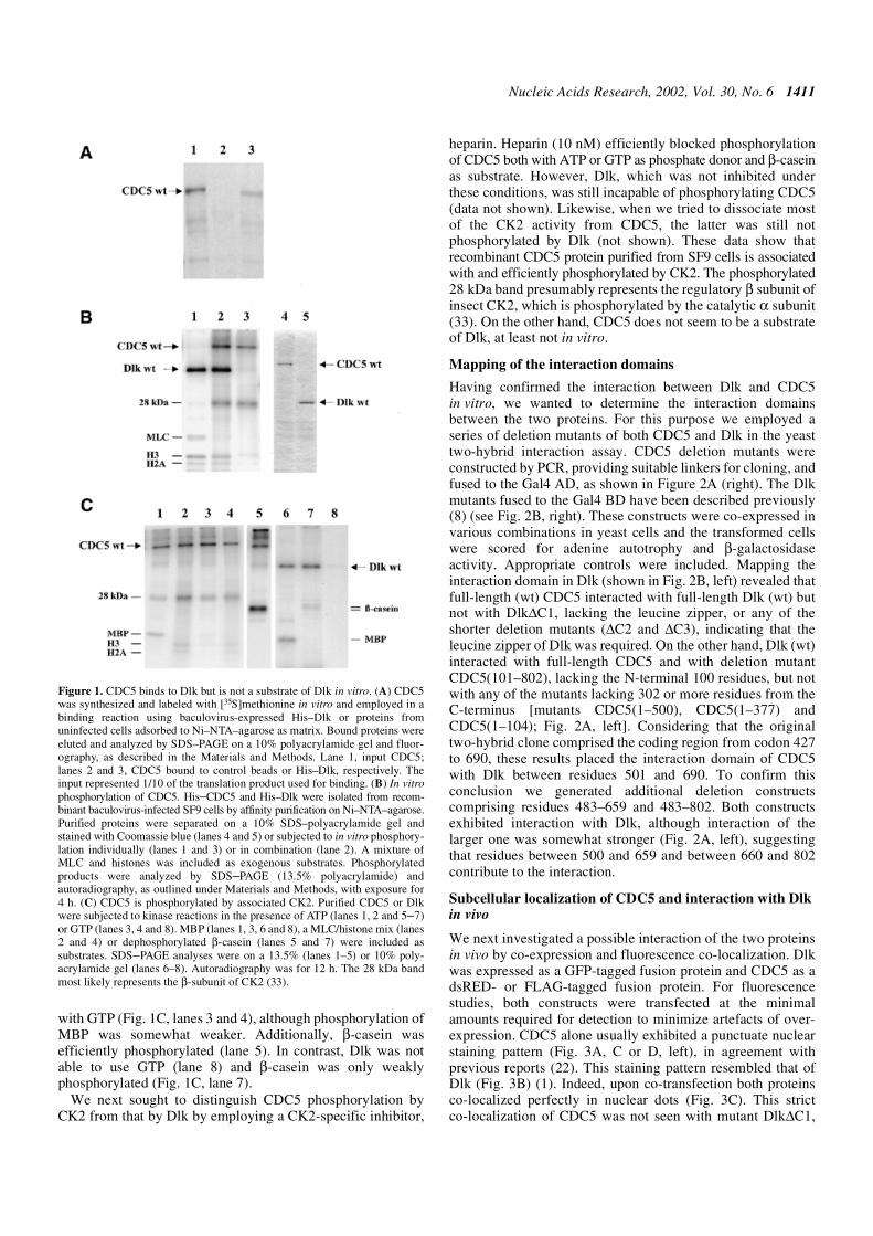
Figure 7. CDC5 exhibits partial co-localization with PML bodies. Expression of dsRED–CDC5 wt and indirect immunofluorescence of endogenous PML revealspartial co-localization of CDC5 with PML bodies.
Figure 6. Dlk phosphorylates splicing factor ASF/SF2. His-tagged Dlk wasincubated with His-tagged ASF/SF2 in a standard kinase reaction and the prod-ucts were separated on a 10% SDS–polyacrylamide gel and autoradiographedfor 6 h. Lane 1, as a control, ASF/SF2 incubated without Dlk; lane 2, auto-phosphorylated Dlk and phosphorylated ASF/SF2.

Nucleic Acids Research, 2002, Vol. 30, No. 6 1417
18. Ohi,R., Feoktistova,A., McCann,S., Valentine,V., Look,A.T., Lipsick,J.S.and Gould,K.L. (1998) Myb-related Schizosaccharomyces pombe cdc5pis structurally and functionally conserved in eukaryotes. Mol. Cell. Biol.,18, 4097–4108.
19. Bernstein,H.S. and Coughlin,S.R. (1997) Pombe Cdc5-related protein.A putative human transcription factor implicated in mitogen-activatedsignaling. J. Biol. Chem., 272, 5833–5837.
20. Bernstein,H.S. and Coughlin,S.R. (1998) A mammalian homolog offission yeast Cdc5 regulates G2 progression and mitotic entry.J. Biol. Chem., 273, 4666–4671.
21. Lei,X.H., Shen,X., Xu,X.Q. and Bernstein,H.S. (2000) Human Cdc5, aregulator of mitotic entry, can act as a site-specific DNA binding protein.J. Cell Sci., 113, 4523–4531.
22. Burns,C.G., Ohi,R., Krainer,A.R. and Gould,K.L. (1999) Evidence thatMyb-related CDC5 proteins are required for pre-mRNA splicing. Proc.Natl Acad. Sci. USA, 96, 13789–13794.
23. McDonald,W.H., Ohi,R., Smelkova,N., Frendewey,D. and Gould,K.L.(1999) Myb-related fission yeast cdc5p is a component of a 40SsnRNP-containing complex and is essential for pre-mRNA splicing.Mol. Cell. Biol., 19, 5352–5362.
24. Ajuh,P., Kuster,B., Panov,K., Zomerdijk,J.C., Mann,M. and Lamond,A.I.(2000) Functional analysis of the human CDC5L complex andidentification of its components by mass spectrometry. EMBO J.,19, 6569–6581.
25. Chien,C.T., Bartel,P.L., Sternglanz,R. and Fields,S. (1991) The two-hybrid system: a method to identify and clone genes for proteins thatinteract with a protein of interest. Proc. Natl Acad. Sci. USA, 88,9578–9582.
26. Ausubel,F.M., Brent,R., Kingston,R.E., Moore,D.D., Seidmann,J.G.,Smith,J.A. and Struhl,K. (1987) Current Protocols in Molecular Biology.John Wiley & Sons, New York, NY.
27. Sambrook,J., Fritsch,E.F. and Maniatis,T. (1989) Molecular Cloning:A Laboratory Manual, 2nd Edn. Cold Spring Habor Laboratory Press,Cold Spring Habor, NY.
28. Stuurman,N., de Graaf,A., Floore,A., Josso,A., Humbel,B., de Jong,L.and van Driel,R. (1992) A monoclonal antibody recognizing nuclearmatrix-associated nuclear bodies. J. Cell Sci., 101, 773–784.
29. Fabrizio,P., Laggerbauer,B., Lauber,J., Lane,W.S. and Luhrmann,R.(1997) An evolutionarily conserved U5 snRNP-specific protein is aGTP-binding factor closely related to the ribosomal translocase EF-2.EMBO J., 16, 4092–4106.
30. Too,C.K. (1997) Differential expression of elongation factor-2, alpha4phosphoprotein and Cdc5-like protein in prolactin-dependent/independentrat lymphoid cells. Mol. Cell. Endocrinol., 131, 221–232.
31. Stukenberg,P.T., Lustig,K.D., McGarry,T.J., King,R.W., Kuang,J. andKirschner,M.W. (1997) Systematic identification of mitoticphosphoproteins. Curr. Biol., 7, 338–348.
32. Boudrez,A., Beullens,M., Groenen,P., Van Eynde,A., Vulsteke,V.,Jagiello,I., Murray,M., Krainer,A.R., Stalmans,W. and Bollen,M. (2000)NIPP1-mediated interaction of protein phosphatase-1 with CDC5L, aregulator of pre-mRNA splicing and mitotic entry. J. Biol. Chem., 275,25411–25417.
33. Saxena,A., Padmanabha,R. and Glover,C.V. (1987) Isolation andsequencing of cDNA clones encoding alpha and beta subunits ofDrosophila melanogaster casein kinase II. Mol. Cell. Biol., 7, 3409–3417.
34. Huang,S. and Spector,D.L. (1996) Dynamic organization of pre-mRNAsplicing factors. J. Cell. Biochem., 62, 191–197.
35. Misteli,T. and Spector,D.L. (1997) Protein phosphorylation and thenuclear organization of pre-mRNA splicing. Trends Cell Biol., 7,135–138.
36. Seeler,J.S. and Dejean,A. (1999) The PML nuclear bodies: actors orextras? Curr. Opin. Genet. Dev., 9, 362–367.
37. Zhong,S., Salomoni,P. and Pandolfi,P.P. (2000) The transcriptional roleof PML and the nuclear body. Nature Cell Biol., 2, E85–E90.
38. Rangnekar,V.M. (1998) Apoptosis mediated by a novel leucine zipperprotein Par-4. Apoptosis, 3, 61–66.
39. Johnstone,R.W., See,R.H., Sells,S.F., Wang,J., Muthukkumar,S.,Englert,C., Haber,D.A., Licht,J.D., Sugrue,S.P., Roberts,T. et al. (1996)A novel repressor, par-4, modulates transcription and growth suppressionfunctions of the Wilms’ tumor suppressor WT1. Mol. Cell. Biol., 16,6945–6956.
40. Diaz-Meco,M.T., Municio,M.M., Frutos,S., Sanchez,P., Lozano,J.,Sanz,L. and Moscat,J. (1996) The product of par-4, a gene induced duringapoptosis, interacts selectively with the atypical isoforms of protein kinase C.Cell, 86, 777–786.
41. Nead,M.A., Baglia,L.A., Antinore,M.J., Ludlow,J.W. and McCance,D.J.(1998) Rb binds c-Jun and activates transcription. EMBO J., 17,2342–2352.
42. Yamaguchi,Y., Wada,T., Suzuki,F., Takagi,T., Hasegawa,J. andHanda,H. (1998) Casein kinase II interacts with the bZIP domains ofseveral transcription factors. Nucleic Acids Res., 26, 3854–3861.
43. Neubauer,G., King,A., Rappsilber,J., Calvio,C., Watson,M., Ajuh,P.,Sleeman,J., Lamond,A. and Mann,M. (1998) Mass spectrometry andEST-database searching allows characterization of the multi-proteinspliceosome complex. Nature Genet., 20, 46–50.
44. Phair,R.D. and Misteli,T. (2000) High mobility of proteins in themammalian cell nucleus. Nature, 404, 604–609.
45. Misteli,T. (1999) RNA splicing: what has phosphorylation got to do withit? Curr. Biol., 9, R198–R200.
46. Gui,J.F., Tronchere,H., Chandler,S.D. and Fu,X.D. (1994) Purificationand characterization of a kinase specific for the serine- and arginine-richpre-mRNA splicing factors. Proc. Natl Acad. Sci. USA, 91, 10824–10828.
47. Kuroyanagi,N., Onogi,H., Wakabayashi,T. and Hagiwara,M. (1998)Novel SR-protein-specific kinase, SRPK2, disassembles nuclear speckles.Biochem. Biophys. Res. Commun., 242, 357–364.
48. Wang,H.Y., Lin,W., Dyck,J.A., Yeakley,J.M., Songyang,Z., Cantley,L.C.and Fu,X.D. (1998) SRPK2: a differentially expressed SR protein-specifickinase involved in mediating the interaction and localization of pre-mRNA splicing factors in mammalian cells. J. Cell Biol., 140, 737–750.
49. Colwill,K., Feng,L.L., Yeakley,J.M., Gish,G.D., Caceres,J.F., Pawson,T.and Fu,X.D. (1996) SRPK1 and Clk/Sty protein kinases show distinctsubstrate specificities for serine/arginine-rich splicing factors. J. Biol.Chem., 271, 24569–24575.
50. Nayler,O., Stamm,S. and Ullrich,A. (1997) Characterization andcomparison of four serine- and arginine-rich (SR) protein kinases.Biochem. J., 326, 693–700.
51. Okamoto,Y., Onogi,H., Honda,R., Yasuda,H., Wakabayashi,T.,Nimura,Y. and Hagiwara,M. (1998) cdc2 kinase-mediatedphosphorylation of splicing factor SF2/ASF. Biochem. Biophys. Res.Commun., 249, 872–878.
52. Rossi,F., Labourier,E., Forne,T., Divita,G., Derancourt,J., Riou,J.F.,Antoine,E., Cathala,G., Brunel,C. and Tazi,J. (1996) Specificphosphorylation of SR proteins by mammalian DNA topoisomerase I.Nature, 381, 80–82.
53. Mayeda,A., Screaton,G.R., Chandler,S.D., Fu,X.D. and Krainer,A.R.(1999) Substrate specificities of SR proteins in constitutive splicing aredetermined by their RNA recognition motifs and composite pre-mRNAexonic elements. Mol. Cell. Biol., 19, 1853–1863.
54. Jiang,Z.H., Zhang,W.J., Rao,Y. and Wu,J.Y. (1998) Regulation of Ich-1pre-mRNA alternative splicing and apoptosis by mammalian splicingfactors. Proc. Natl Acad. Sci. USA, 95, 9155–9160.
55. Duncan,P.I., Stojdl,D.F., Marius,R.M., Scheit,K.H. and Bell,J.C. (1998)The Clk2 and Clk3 dual-specificity protein kinases regulate theintranuclear distribution of SR proteins and influence pre-mRNA splicing.Exp. Cell Res., 241, 300–308
Copyright © 2022 FDOKUMEN