Cymbopogon citratus as source of new and safe anti-inflammatory drugs: Bio-guided assay using...
31
Accepted Manuscript Title: Cymbopogon citratus as source of new and safe anti-inflammatory drugs: bio-guided assay using lipopolysaccharide-stimulated macrophages Authors: Vera Francisco, Artur Figueirinha, Bruno Miguel Neves, Carmen Garc´ ıa-Rodr´ ıguez, Maria Celeste Lopes, Maria Teresa Cruz, Maria Teresa Batista PII: S0378-8741(10)00803-2 DOI: doi:10.1016/j.jep.2010.11.018 Reference: JEP 6437 To appear in: Journal of Ethnopharmacology Received date: 23-6-2010 Revised date: 30-10-2010 Accepted date: 3-11-2010 Please cite this article as: Francisco, V., Figueirinha, A., Neves, B.M., Garc´ ıa-Rodr´ ıguez, C., Lopes, M.C., Cruz, M.T., Batista, M.T., Cymbopogon citratus as source of new and safe anti-inflammatory drugs: bio-guided assay using lipopolysaccharide-stimulated macrophages, Journal of Ethnopharmacology (2010), doi:10.1016/j.jep.2010.11.018 This is a PDF file of an unedited manuscript that has been accepted for publication. As a service to our customers we are providing this early version of the manuscript. The manuscript will undergo copyediting, typesetting, and review of the resulting proof before it is published in its final form. Please note that during the production process errors may be discovered which could affect the content, and all legal disclaimers that apply to the journal pertain.
-
Upload
independent -
Category
Documents
-
view
3 -
download
0
Transcript of Cymbopogon citratus as source of new and safe anti-inflammatory drugs: Bio-guided assay using...

Accepted Manuscript
Title: Cymbopogon citratus as source of new and safeanti-inflammatory drugs: bio-guided assay usinglipopolysaccharide-stimulated macrophages
Authors: Vera Francisco, Artur Figueirinha, Bruno MiguelNeves, Carmen Garcı́a-Rodrı́guez, Maria Celeste Lopes,Maria Teresa Cruz, Maria Teresa Batista
PII: S0378-8741(10)00803-2DOI: doi:10.1016/j.jep.2010.11.018Reference: JEP 6437
To appear in: Journal of Ethnopharmacology
Received date: 23-6-2010Revised date: 30-10-2010Accepted date: 3-11-2010
Please cite this article as: Francisco, V., Figueirinha, A., Neves, B.M., Garcı́a-Rodrı́guez,C., Lopes, M.C., Cruz, M.T., Batista, M.T., Cymbopogon citratus as source of new andsafe anti-inflammatory drugs: bio-guided assay using lipopolysaccharide-stimulatedmacrophages, Journal of Ethnopharmacology (2010), doi:10.1016/j.jep.2010.11.018
This is a PDF file of an unedited manuscript that has been accepted for publication.As a service to our customers we are providing this early version of the manuscript.The manuscript will undergo copyediting, typesetting, and review of the resulting proofbefore it is published in its final form. Please note that during the production processerrors may be discovered which could affect the content, and all legal disclaimers thatapply to the journal pertain.

Page 1 of 30
Accep
ted
Man
uscr
ipt
1
Graphical abstract
In this report it was demonstrated that a lipid- and essential oil-free infusion of Cymbopogon
citratus leaves (Cy), as well its polyphenols, have anti-inflammatory properties through inhibition of pro-
inflammatory signaling pathways and nitric oxide production in lipopolysaccharide-stimulated
macrophages. These evidences support the use of Cymbopogon citratus in traditional medicine and
indicate that it could be a natural source of new and safe anti-inflammatory drugs.
Title
Cymbopogon citratus as source of new and safe anti-inflammatory drugs: bio-guided assay using
lipopolysaccharide-stimulated macrophages
Authors' names:
Vera Francisco a, b, Artur Figueirinha a, c, Bruno Miguel Neves b, d, Carmen García-Rodríguez e, Maria
Celeste Lopes b, d, Maria Teresa Cruz b, d & Maria Teresa Batista a, d
Affiliations/Addresses:a Centro de Estudos Farmacêuticos - Faculdade de Farmácia, Universidade de Coimbra, Pólo das Ciências
da Saúde, Azinhaga de Santa Comba, 3000-548 Coimbra, Portugal
b Centro de Neurociências e Biologia Celular, Universidade de Coimbra, Azinhaga de Santa Comba, 3004-
517 Coimbra, Portugal

Page 2 of 30
Accep
ted
Man
uscr
ipt
2
c Departamento de Ambiente, Instituto Politécnico de Viseu, Campus Politécnico de Repeses, 3504-510
Viseu, Portugal
d Faculdade de Farmácia, Universidade de Coimbra, Pólo das Ciências da Saúde, Azinhaga de Santa
Comba, 3000-548 Coimbra, Portugal
e Instituto de Biología y Genética Molecular, CSIC-Universidad de Valladolid, C/ Sanz y Forés s/n, lab
E10, 47003 Valladolid, Spain
Corresponding author:
M. T. Batista
Centro de Estudos Farmacêuticos
Universidade de Coimbra
Pólo das Ciências da Saúde
Azinhaga de Santa Comba
3000-548 Coimbra, Portugal
Phone: +351 239 488497
Fax: +351 239 488503
E-mail address: [email protected]
Abstract
Ethnopharmacological relevance: Aqueous extracts of Cymbopogon citratus (Cy) leaves are used in
traditional medicine for the treatment of inflammatory conditions, however little is known about their
mechanism of action.
Aim of the study: The aim of this study is to explore the anti-inflammatory properties of Cymbopogon
citratus leaves and their polyphenol-rich fractions (PFs), as well its mechanism of action in murine
macrophages.
Materials and Methods: A lipid- and essential oil-free infusion of Cy leaves was prepared (Cy extract)
and fractionated by column chromatography. Anti-inflammatory properties of Cy extract (1.115 mg/ml)
and its PFs, namely phenolic acids (530 µg/ml), flavonoids (97.5 µg/ml) and tannins (78 µg/ml), were
investigated using lipopolysaccharide (LPS)-stimulated Raw 264.7 macrophages as in vitro model. As
inflammatory parameters, nitric oxide (NO) production was evaluated by Griess reaction, as well as
effects on cyclooxygenase (COX-2), inducible NO synthase (iNOS) expression and on intracellular
signaling pathways activation, which were analyzed by Western blot using specific antibodies.

Page 3 of 30
Accep
ted
Man
uscr
ipt
3
Results: Cy extract inhibited iNOS expression, NO production and various LPS-induced pathways like
p38 mitogen-activated protein kinase (MAPK), c-jun NH2-terminal kinase (JNK) 1/2 and the transcription
nuclear factor (NF)-κB. The extracellular signal-regulated kinase (ERK) 1/2 and the phosphatidylinositol-
3-kinase (PI3K)/Akt activation were not affected by Cy extract. Both phenolic acid- and tannin-rich
fractions significantly inhibited NF-κB activation, iNOS expression and NO production but none of the
PFs modulated MAPKs or PI3K/Akt activation. Neither Cy extract or PFs affected LPS-induced COX-2
expression but LPS-induced PGE2 production is inhibited by Cy extract and by phenolic acid-rich
fraction.
Conclusions: Our data provide evidence that support the usage of Cymbopogon citratus leaves extract in
traditional medicine, and suggest that Cy, in particular its polyphenolic compounds, could constitute a
natural source of a new and safe anti-inflammatory drugs.
Keywords: Cymbopogon citratus, Poaceae-Gramineae, polyphenol, inflammation, nitric oxide, mitogen-
activated protein kinases, nuclear factor-κB
Abbreviations: Cy, Cymbopogon citratus; COX, cyclooxygenase; ERK, extracellular signal-regulated
kinase; FF, flavonoid-rich fraction; FSDC, fetal skin-derived dendritic cell; HPLC, high-performance
liquid chromatography; Iκκ, IκB kinase; IκB, inhibitory protein κB; JNK, c-jun NH2-terminal kinase;
LPS, lipopolysaccharide; MAPK, mitogen-activated protein kinase; MTT, 3-(4,5-dimethylthiazol-2-yl)-
2,5-diphenyl tetrazolium bromide; NF-κB, nuclear factor-κB; NO, nitric oxide; NOS, nitric oxide
synthase; PAF, phenolic acid-rich fraction; PFs, polyphenol-rich fractions; PGE2, prostaglandin E2; PI3K,
phosphatidylinositol-3-kinase; SNAP, S-nitroso-N-acetylpenicillamine; TF, tannin-rich fraction; TLC,
thin layer chromatography;

Page 4 of 30
Accep
ted
Man
uscr
ipt
4
1. Introduction
Chronic inflammation is one of the leading causes of mortality in the western world and is associated
with several pathologies like cancer (Porta et al., 2009), rheumatoid arthritis, diabesity (Schmidt and
Duncan, 2003), cardiovascular and neurodegenerative diseases (Hunter and Doddi, 2010; Whitney et al.,
2009). However, the current anti-inflammatory drugs have several limitations such as lack of
responsiveness, side effects, delivery problems and cost of manufacture. Therefore, there is an urgent
need to find new anti-inflammatory agents with selective pharmacology and less toxicity. Plant extracts
have been used for centuries in traditional medicine to alleviate inflammatory diseases, however, and for
some of them, little is known about their mechanisms of action. The understanding of molecular
mechanisms behind the healing properties of natural products is crucial to find compounds that could be
useful as templates to new therapeutic molecules. Indeed, most of the drugs actually available are derived
from natural products (Newman and Cragg, 2007), therefore, the knowledge of phytochemicals molecular
mechanisms became a good strategy in the search for new anti-inflammatory compounds.
In the inflammatory process, macrophages have a key role in providing an immediate defense against
foreign agents. Upon activation with an inflammatory stimulus, such as lipopolysaccharide (LPS),
macrophages produce a variety of pro-inflammatory mediators, including prostaglandin E2 (PGE2) and
nitric oxide (NO) (Geller and Billiar, 1998). PGE2 is synthesized by the rate limiting enzyme
cyclooxygenase (COX), while NO is synthesized by nitric oxide synthase (NOS). Cyclooxygenase exists
as two major isoforms (COX-1 and COX-2) and one variant (COX-3). While COX-1 is constitutively
expressed in many tissues, COX-2 is an inducible enzyme expressed in the inflammatory-related cells,
like macrophages, which produces large amounts of prostaglandins. In addition, LPS-activated
macrophages also express transcriptionally inducible NOS (iNOS) that produces high amounts of NO
from L-arginine. To date, three isoforms of NOS have been identified: endothelial NOS (eNOS), neuronal
NOS (nNOS) and iNOS. The high-output of NO by iNOS contributes to the pathogenesis of septic shock
and inflammatory diseases (Guzik et al., 2003; Zamora et al., 2000). Therefore, the selective inhibition of
COX-2 and iNOS in macrophages is a useful strategy to screen new anti-inflammatory drugs.
The expression of pro-inflammatory molecules is tightly regulated by several transcription factors
and signaling pathways. Among these pathways, mitogen-activated proteins kinases (MAPKs) are
signaling molecules that play critical roles in the regulation of cell growth, differentiation, cell
survival/apoptosis and cellular response to cytokines and stress. The MAPKs pathways include p38
MAPK (Han et al., 1994), c-jun NH2-terminal kinase (JNK) and extracellular signal-regulated kinase
(ERK) (Davis, 1994), and they are involved on LPS-induced COX-2 and iNOS expression in
macrophages (Chen et al., 1999; Chen and Wang, 1999; Tsatsanis et al., 2006). Accordingly, it has been
demonstrated that MAPKs inhibitors suppress the expression of iNOS gene (Chen et al., 1999). Besides,
the iNOS expression could also be modulated by phosphatidylinositol-3-kinase (PI3K)/Akt pathway (Salh
et al., 1998), a serine/threonine kinase activated in response to certain growth factors and cytokines that
provides a strong cell survival signal (Crawley et al., 1996; Gold et al., 1994). MAPKs and Akt also play

Page 5 of 30
Accep
ted
Man
uscr
ipt
5
a critical role in the activation of nuclear factor (NF)-κB (Carter et al., 1999; Nakano et al., 1998). The
NF-κB transcription factor regulates the expression of many genes involved in immune and inflammatory
responses, including iNOS and COX-2 (Geller and Billiar, 1998). Many stimuli like LPS, cytokines and
oxidants activate NF-κB through several signaling pathways that lead to the phosphorylation of inhibitory
protein κB (IκB) by IκB kinase (Iκκ), which is a marker for ubiquitination and subsequent degradation by
proteasome. IκB degradation unmasks the nuclear localization motif of NF-κB, which is rapidly
translocated to the nucleus, where it activates the transcription of target genes. Therefore, the involvement
of MAPKs, Akt and NF-κB in the regulation of inflammatory mediator’s synthesis makes them potential
targets for novel anti-inflammatory therapeutics.
Cymbopogon citratus (DC) Stapf (Cy), Poaceae-Gramineae, commonly known as lemongrass, is a
spontaneous perennial grass, largely distributed around the world, especially in tropical and subtropical
countries. Its leaf essential oil citral is used in the food, perfumery, soap, cosmetic, pharmaceutical and
insecticide industries (Negrelle and Gomes, 2007). Aqueous extracts of dried leaves are used in
traditional medicine for the treatment of inflammation, digestive disorders, diabetes, nervous disorders,
and fever, as well as other health problems (Carbajal et al., 1989; Lorenzetti et al., 1991). However, the
mechanism of action of Cy is poorly explored and characterized, namely the mechanism responsible for
its anti-inflammatory effects. We have previously demonstrated that Cy leaves extract has potent
antioxidant activity that is related to its polyphenolic content (Figueirinha et al., 2008). In addition, we
verified that this extract and its polyphenolic fractions inhibit LPS-induced NO production and iNOS
expression in fetal skin-derived dendritic cell line (FSDC) (Figueirinha et al., 2010), reinforcing the
potential use of Cy extract as source of a new anti-inflammatory drug.
Thus, this study aimed to explore the anti-inflammatory properties of Cymbopogon citratus extract
by addressing its molecular mechanism of action. For that, we evaluated the effect of a lipid- and essential
oil-free infusion (extract) obtained from Cy leaves and its polyphenol-rich fractions in COX-2 and iNOS
expression, NO production and activation of MAPKs, Akt and NF-κB signaling pathways in vitro. As an
in vitro model of inflammation, we used the mouse macrophage cell line, Raw 264.7, stimulated with
LPS from E. coli.

Page 6 of 30
Accep
ted
Man
uscr
ipt
6
2. Materials and Methods
2.1. Materials
LPS from Escherichia coli (serotype 026:B6) and the iNOS inhibitor, aminoguanidine, were
obtained from Sigma Chemical Co. (St. Louis, MO, USA). Iscove´s Modified Dulbecco´s Medium,
dexamethasone and wortmannin were from Sigma–Aldrich Química (Madrid, Spain). Fetal calf serum
was purchased from Gibco (Paisley, UK). The protease and phosphatase inhibitor cocktails were obtained
from Roche (Basel, Switzerland). SB203580, U0126, SP600125 and BAY 11-7082 were from
Calbiochem (San Diego, CA, USA). Acrylamide was from Promega (Madison, WI, USA) and the
polyvinylidene difluoride membranes were from Millipore Corporation (Bedford, MA). Antibodies
against phospho-p44/p42 MAPK (ERK1/2), phospho-p38 MAPK, phospho-SAPK/JNK 1/2, phospho-Akt
(Ser473) and IκB-α were from Cell Signaling Technologies (Danvers, MA, USA). The pan anti-JNK
antibody was from R&D Systems (Minneapolis, MN, USA), the pan anti-p38 MAPK and Akt were from
Cell Signaling Technologies (Danvers, MA, USA. The anti-actin and pan anti-ERK 1/2 antibodies were
purchased from Millipore (Bedford, MA, USA). The alkaline phosphatase-linked secondary antibodies
and the enhanced chemifluorescence reagent were obtained from GE Healthcare (Chalfont St. Giles, UK).
All other reagents were from Sigma Chemical Co. (Saint Louis, MO) or from Merck (Darmstadt,
Germany).
2.2. Plant material and extract preparation
Dry leaves of Cymbopogon citratus (Cy) Stapf. were purchased from ERVITAL® in July 2004
and kept at -20ºC until be used. The plant was cultivated in the region of Mezio, Castro D´Aire
(Portugal). A voucher specimen was deposited in the herbarium of the University of Coimbra, Faculty of
Pharmacy and J. Paiva (Botany Department, University of Coimbra, Portugal) confirmed the identity of
the plant. An infusion was prepared from the powdered plant material (1:30 w/v), treated with n-hexane
to remove lipids and essential oils and then freeze-dried (Cy extract). A yield of 16.6±1.2 g / 100 g of dry
plant was obtained.
2.3. Extract fractionation
Cy extract was fractionated as previously described (Figueirinha et al., 2008) (Fig. 1). Briefly,
the extract was treated with water and fractionated on a reverse phase semipreparative column
Lichroprep® RP-18 (310 x 25 mm, particle sizes 40-63 μm), Merck (Darmstadt, Germany), eluted with
water giving fraction F1 and with aqueous methanol solutions (fractions F2-F7). Dry residue of F7 was
recovered in 50% aqueous ethanol and fractionated by gel chromatography on a Sephadex® LH-20
(Sigma-Aldrich – Amersham, Sweden) column (85 x 2.5 cm) using ethanol as mobile phase. All the

Page 7 of 30
Accep
ted
Man
uscr
ipt
7
fractionation process described above was monitored by high-performance liquid chromatography
(HPLC) and thin layer chromatography (TLC) for polyphenols, providing three major fractions: tannin-
rich fraction (TF; yield of 3.5% (w/w) of Cy extract) corresponding to F6, flavonoid-rich fraction (FF;
yield of 4.4%(w/w) of Cy extract) corresponding to sub-fraction F7a, and phenolic acid-rich fraction
(PAF; yield of 23.8% (w/w) of Cy extract) corresponding to F2 and sub-fraction F7b, as described in
Figueirinha et al. (2010). The fractions were then taken to dryness under reduced pressure (40 ºC). The
Cy extract and the polyphenol-rich fractions were weighted in sterilized and humidity-controlled
conditions, and then solubilized in endotoxin-free water.
Fig. 1. Fractionation scheme of Cymbopogon citratus (Cy) extract. Aqueous solution was fractionated
on a reverse phase semi-preparative Lichroprep® RP-18 (310 x 25 mm, particle sizes 40-63 μm) and
Sephadex® LH-20 (85 x 2.5 cm) columns, providing three major fractions: phenolic acid-rich fraction
(PAF), flavonoid-rich fraction (FF), and tannin-rich fraction (TF).
2.4. Cell culture
Raw 264.7, a mouse leukaemic monocyte macrophage cell line from American Type Culture
Collection, and kindly supplied by Dr. Otília Vieira (Centro de Neurociências e Biologia Celular,
Universidade de Coimbra, Coimbra, Portugal), were cultured in Iscove´s Modified Dulbecco’s Eagle
Medium supplemented with 10% non-inactivated fetal bovine serum, 100 U/ml penicillin, and 100 μg/ml
streptomycin at 37 °C in a humidified atmosphere of 95% air and 5% CO2. Along the experiments, cells
were monitored by microscope observation in order to detect any morphological change.

Page 8 of 30
Accep
ted
Man
uscr
ipt
8
2.5. Determination of cell viability by MTT assay
Assessment of metabolically active cells was performed using 3-(4,5-dimethylthiazol-2-yl)-2,5-
diphenyl tetrazolium bromide (MTT) reduction colorimetric assay as previously reported (Mosmann,
1983). Raw 264.7 cells (6x10⁵ cells/well) were plated and allowed to stabilize for 12 h. Following this
period, cells were either maintained in culture medium (control) or pre-incubated with Cy extract, its
polyphenolic fractions or with inhibitors for 1 h, and later activated with 1 µg/ml LPS for 24 h. After the
treatments, a MTT solution (5 mg/ml in phosphate buffered saline) was added and cells incubated at 37
ºC for 15 min, in a humidified atmosphere of 95% air and 5% CO2. Supernatants were then removed and
dark blue crystals of formazan solubilized with acidic isopropanol (0.04 N HCl in isopropanol).
Quantification of formazan was performed using an ELISA automatic microplate reader (SLT, Austria) at
570 nm, with a reference wavelength of 620 nm.
2.6. Measurement of nitrite production by Griess reagent
The production of nitric oxide (NO) was measured by the accumulation of nitrite in the culture
supernatants, using a colorimetric reaction with the Griess reagent (Green et al., 1982). Briefly, 170 µl of
culture supernatants were diluted with equal volumes of the Griess reagent [0.1% (w/v) N-(1-naphthyl)-
ethylenediamine dihydrochloride and 1% (w/v) sulphanilamide containing 5% (w/v) H3PO4] and
maintained during 30 min, in the dark. The absorbance at 550 nm was measured in an automated plate
reader (SLT, Austria). Culture medium was used as blank and nitrite concentration was determined from
a regression analysis using serial dilutions of sodium nitrite as standard.
2.7. Determination of nitric oxide scavenging activity using S-nitroso-N-acetylpenicillamine (SNAP) as
NO donor
The nitric oxide scavenging activity was evaluated by incubating 1.115 mg/ml Cy extract, 530
μg/ml phenolic acid-rich fraction (PAF), 97.5 μg/ml flavonoid-rich fraction (FF), or 78 μg/ml tannin-rich
fraction (TF) with 200 µM of NO donor SNAP, in culture medium during 3 h. After this period the nitrite
levels in the medium were quantified by Griess method, as described above.
2.8. Measurement of prostaglandin E2 (PGE2) by enzyme immunoassay (EIA)
To analyse the production of PGE2, Raw 264.7 cells (6x10⁵ cells/well) were plated and allowed
to stabilize for 12 h. Following this period, cells were either maintained in culture medium (control) or
pre-incubated with Cy extract or with its polyphenolic fractions, and later activated with 1 µg/ml LPS for
24 h. After the treatments, the supernatants were collected and frozen at -80ºC until the assay was

Page 9 of 30
Accep
ted
Man
uscr
ipt
9
performed. The PGE2 levels of diluted supernatants were quantified using an enzyme immunoassay (EIA)
commercial kit from Cayman (Ann Arbor, MI, USA), following the manufacturer instructions.
2.9. Western blot analysis
To prepare total cell lysates for Western blot analysis, Raw 264.7 cells (24x105 cells/well) were
plated and allowed to stabilize for 12 h. Following this period, cells were either maintained in culture
medium (control) or pre-incubated with Cy extract and its polyphenolic fractions for 1 h and then 1 µg/ml
LPS was added for the indicated time. Cells were lysed with RIPA buffer (50 mM Tris-HCl, pH 8.0, 1%
Nonidet P-40, 150 mM NaCl, 0.5% sodium deoxycholate, 0.1% sodium dodecyl sulfate and 2 mM
ethylenediamine tetraacetic acid) freshly supplemented with 1 mM dithiothreitol, protease and
phosphatase inhibitor cocktails and sonicated (four times for 4 s at 40 μm peak to peak) in Vibra Cell
sonicator (Sonics & Material INC.) to decrease viscosity. The nuclei and the insoluble cell debris were
removed by centrifugation at 4ºC, at 12,000g for 10 min. The postnuclear extracts were collected and
used as total cell lysates. Protein concentration was determined by the bicinchoninic acid protein assay
and cell lysates were denaturated in sample buffer (0.125 mM Tris pH 6.8, 2% (w/v) sodium dodecyl
sulfate, 100 mM dithiothreitol, 10% glycerol and bromophenol blue).
Western blot analysis was performed to evaluate the levels of iNOS and COX-2, and the
activation of MAPKs, Akt and NF-κB signaling pathways. Briefly, equivalent amounts of protein were
separated by 10% (v/v) SDS-PAGE followed by Western blotting. To examine the different proteins
studied, the blots were incubated overnight at 4 ºC with the respective primary antibodies: COX-2
(1:10000), iNOS (1:7500), phospho-p38 MAPK (1:1000), phospho-JNK1/2 (1:1000), phospho-ERK 1/2
(1:1000), phospho-Akt (1:500) and total IκB (1:1000). Protein detection was performed using the
enhanced chemifluorescence system and the membranes were scanned for blue excited fluorescence on
the Storm 860 (GE Healthcare). The generated signals were analyzed using the software ImageQuant
TL®. To demonstrate equivalent protein loading, membranes were stripped and reprobed with antibodies
against the total form of MAPKs and Akt or with anti-actin antibody.
2.10. Statistical analysis
Results are expressed as mean±SEM of the indicated number of experiments. Statistical analysis
comparing a treatment condition to control was performed between two groups and analyzed using two-
sided unpaired t-test. When comparing the effect of different treatments to LPS-stimulated cells, a
multiple group comparison was performed and one-way ANOVA followed by Dunnett's test was used.
The statistical tests were applied using GraphPad Prism, version 5.02 (GraphPad Software, San Diego,
CA, USA). The significance level was #p <0.05, ##p <0.01 and ##p <0.001, when compared to control and
*p <0.05, **p <0.01 and ***p <0.001, when compared to LPS.

Page 10 of 30
Accep
ted
Man
uscr
ipt
10
3. Results
3.1. Evaluation of the anti-inflammatory properties and molecular targets of lipid- and essential oil-
free Cymbopogon citratus leaves infusion (Cy extract)
Some studies have been conducted with citral, the main volatile compound of the essential oil of
Cymbopogon citratus (Cy) (Cheel et al., 2005; Lee et al., 2008), however little is known about the
properties and mechanisms of action of the fixed compounds, namely polyphenols. Therefore, in the
present study we analyzed their anti-inflammatory potential and evaluated some molecular targets of
these extracts in LPS-stimulated Raw 264.7 cells. The Cy extract concentration used for this study was
selected based on our previous results, obtained in dendritic cells (Figueirinha et al., 2010), and also on
the absence of macrophages toxicity (table 1).
Table 1. Effect of Cy extract, polyphenol-rich fractions and signaling pathways inhibitors on
macrophage cell viability. Raw 264.7 cells were treated with the indicated compounds for 24 h, and the
cell viability was assessed as described in materials and methods. The results are expressed as percentage
of control (non-treated cells) and each value represents the mean±SEM from at least 3 independent
experiments. Statistical analysis was performed using one-way ANOVA followed by Dunnett's test.
ConditionCell viability (% of control)
mean±SEM
Control 100
Cy extract (1.115 mg/ml) 122.40±3.71
PAF (530 μg/ml) 82.24±3.95
FF (97.5 μg/ml) 84.69±4.09
TF (78 μg/ml) 89.33±5.80
LPS (1 µg/ml) 103.10±5.46
LPS + Cy (1.115 mg/ml) 112.00±5.59
LPS + PAF (530 μg/ml) 97.00±7.15
LPS + FF (97.5 μg/ml) 92.12±4.69
LPS + TF (78 μg/ml) 93.74±5.73
LPS + Dexamethasone (20 µM) 96.97±12.21
LPS + SB203580 (20 µM) 93.96±9.08
LPS + SP600125 (20 µM) 95.58±8.35

Page 11 of 30
Accep
ted
Man
uscr
ipt
11
LPS + U0126 (10 µM) 89.68±7.77
LPS + Wortmannin (500 nM) 86.06±11.37
LPS + BAY 11-7083 (250 nM) 101.20±5.88
LPS + Aminoguanidine (50 µM) 95.18±8.21
3.1.1. Cy extract does not affect LPS-induced COX-2 expression but inhibits the PGE2 production
We analyzed the effect of Cy extract on LPS-induced COX-2 expression after 24 h of murine
macrophages stimulation by Western blot using a specific anti-COX-2 antibody (Fig. 2A). In non-
stimulated Raw 264.7 cells (control), COX-2 protein was almost undetectable, but after LPS treatment the
expression strongly increased to 10573±1544% of control (p<0.001). The LPS-induced COX-2
expression was not significantly inhibited by Cy extract (7824±1489% of control) while the extract alone
was able to induce the expression of COX-2 (3226±579% of control).
Instead Cy did not inhibit the LPS-induced COX-2 expression, the enzyme activity could be
compromised. Therefore, we next investigated the effect of Cy extract on a product of COX-2 activity,
PGE2, by enzyme immunoassay (EIA). As shown in Fig. 2B, the cell treatment with LPS induced a great
increase in PGE2 production, consistent with the results obtained for COX-2 expression, which is
inhibited by macrophage pre-treatment with Cy (42.40% of inhibition). The Cy alone increased the LPS-
induced PGE2 production comparing to untreated Raw 264.7 cells (from 0.71±0.16 of control to
7.56±0.29 of Cy). Taken together, these results indicated that Cy extract did not inhibit the LPS-induced
COX-2 activity, but modulates its activity, exhibiting anti-inflammatory properties, while the extract
alone increased the COX-2 expression and the PGE2 production.

Page 12 of 30
Accep
ted
Man
uscr
ipt
12
Fig 2. Lack of effect of Cymbopogon citratus (Cy) extract on LPS-induced COX-2 expression and
inhibition of LPS-induced PGE2 production in murine macrophages. (A) Raw 264.7 cells (24x105
cells) were maintained in culture medium (control), or pre-incubated with 1.115 mg/ml Cy extract for 1 h
and then treated with 1 µg/ml LPS for 24 h. COX-2 expression was analyzed by Western blot using a
specific anti-COX-2 antibody. An anti-actin antibody was used to confirm equal protein loading. The blot
shown is representative of 3 blots yielding similar results. Results were expressed as percentage of COX-
2 protein levels relatively to control. (B) Raw 264.7 cells (6x10⁵ cells) were maintained in culture
medium (control), or pre-incubated with 1.115 mg/ml Cy extract for 1 h and then treated with 1 µg/ml
LPS for 24 h. PGE2 levels were evaluated in the culture supernatants by enzyme immunoassay (EIA), as
described in material and methods, and the results expressed as percentage of LPS. Each value represents
the mean±SEM from 2-3 independent experiments (###p<0.001, compared to control; **p<0.01, compared
to LPS).
3.1.2. Cy extract inhibits LPS-induced iNOS expression and nitrite production
We also investigated the effect of Cy extract on the production of the pro-inflammatory mediator
NO, found in inflammatory disorders (Guzik et al., 2003). First, the effect of Cy in iNOS expression
triggered by LPS was verified by Western blot (Fig. 3A). In untreated cells (control), iNOS protein
expression is not detected but after treatment with LPS for 24 h, iNOS expression is strongly increased
(1841±121.4% of control), as described earlier (Thiemermann, 1997). Pre-treatment of cells with Cy
extract reduced the LPS-induced expression by 28.95% while extract alone slightly increased the iNOS
expression (491.7±53.67% of control).

Page 13 of 30
Accep
ted
Man
uscr
ipt
13
Secondly, the effect on NO production was analyzed by measuring accumulation of nitrite in the
culture medium. As shown in Fig. 3B, untreated Raw 264.7 cells produced low levels of nitrites
(2.115±0.7590 μM), consistent with the data obtained for iNOS expression in resting conditions. After
cell activation with LPS for 24 h, the nitrite production increased to 46.67±2.623 μM, while macrophage
pre-treatment with Cy strongly decreased the LPS-induced nitrite production (64.07% of inhibition). The
Cy alone slightly increased nitrite production (10.59±1.691 µM). To evaluate Cy anti-inflammatory
potency, a comparison with the known anti-inflammatory compound dexamethasone was performed. A
decrease on LPS-induced NO production by Cy extract was verified in a magnitude similar to that
observed for 20 µM dexamethasone (64.07% and 79.56%, respectively). We also analyzed the NO
scavenging capacity of Cy extract, using SNAP as NO donor, and we found that Cy extract was no NO
scavenging properties (data not shown). Taken together, these results suggest that Cy extract exhibit anti-
inflammatory properties by inhibiting LPS-induced NO production while the extract slightly promoted
NO production.

Page 14 of 30
Accep
ted
Man
uscr
ipt
14
Fig 3. Inhibitory effect of Cymbopogon citratus (Cy) extract on LPS-induced iNOS protein
expression and nitrite production in murine macrophages. (A) Raw 264.7 cells (24x105 cells) were
maintained in culture medium (control), or pre-incubated with 1.115 mg/ml Cy extract for 1 h and then
treated with 1 µg/ml LPS for 24 h. iNOS expression was analyzed by Western blot using a specific anti-
iNOS antibody and an anti-actin antibody was used to confirm equal protein loading. The blot shown is
representative of 3 blots yielding similar results. Results were expressed as percentage of iNOS protein
levels relatively to control. (B) Raw 264.7 cells (6x10⁵ cells) were maintained in culture medium
(control), or pre-incubated with 1.115 mg/ml Cy extract or 20 μM dexamethasone for 1 h and then treated
with 1 µg/ml LPS for 24 h. Nitrite levels in the culture supernatants were evaluated by the Griess reaction
as described in material and methods. Nitrite concentration was determined from a sodium nitrite standard
curve and the results are expressed as concentration (μM) of nitrite in culture medium. Each value
represents the mean±SEM from at least 3 experiments (###p<0.001, compared to control; *p <0.05,
***p<0.001, compared to LPS).
At last, the signaling pathways involved in the modulation of NO production were investigated
using specific inhibitors. The concentrations of these inhibitors were chosen based on the absence of
citotoxicity to macrophages (table 1). As shown in Fig. 4, the LPS-induced nitrite production was
inhibited by SB203580 (53.59% of inhibition), a specific inhibitor of p38 MAPK, by SP600125 (65.40%
of inhibition), a selective and reversible JNK inhibitor, by BAY 11-7082 (67.80% of inhibition), a NF-κB
inhibitor, and by aminoguanidine (79.42% of inhibition), an inhibitor of iNOS. Both ERK 1/2 inhibitor
(U0126) and PI3K/Akt inhibitor (wortmannin) were without effect on nitrite production.

Page 15 of 30
Accep
ted
Man
uscr
ipt
15
Fig 4. Evaluation of signaling pathways involved in the modulation of nitrite production on LPS-
stimulated macrophages. Raw 264.7 cells (6x10⁵ cells) were maintained in culture medium (control), or
pre-incubated with the indicated inhibitors (20 μM SB203580 as p38 MAPK inhibitor, 20 μM SP600125
as JNK inhibitor, 10 μM U0126 as ERK inhibitor, 500 nM Wortmannin as PI3K/Akt inhibitor, 250 nM
BAY 11-7082 as NF-κB inhibitor and 500 μM Aminoguanidine as iNOS inhibitor) and then 1 µg/ml LPS
was added for 24 h. Nitrite levels in the culture supernatants were evaluated by the Griess reaction as
described in materials and methods. Nitrite concentration was determined from a sodium nitrite standard
curve and the results are expressed as concentration (μM) of nitrite in culture medium. Each value
represents the mean±SEM from at least 3 experiments (###p<0.001, compared to control; ***p<0.001,
compared to LPS).
3.1.3. Cy extract inhibits LPS-induced activation of p38 MAPK, JNK 1/2 and NF-κB
Our results demonstrated that LPS-induced NO production in macrophages was inhibited by Cy
extract and regulated by p38 MAPK, JNK 1/2 and NF-κB signaling pathways but not by ERK 1/2 or
PI3K/Akt. Therefore, we next evaluated the effect of Cy extract on the activation of those pathways by
Western blot using phospho-specific antibodies. As shown in Fig. 5, LPS stimulation for 30 min induced
the phosphorylation of Akt and all MAPKs, namely p38 MAPK, JNK 1/2 and ERK 1/2, as described
previously (Rao, 2001). Pre-treatment with 1.115 mg/ml Cy extract inhibited the LPS-induced

Page 16 of 30
Accep
ted
Man
uscr
ipt
16
phosphorylation of p38 MAPK and JNK 1/2 but had no effect in the activation of ERK 1/2 and Akt
pathways. When added to control cells, Cy alone stimulated both MAPKs and Akt signaling pathways.
Since NF-κB transcription factor is a crucial player in the inflammatory process by controlling
the expression of several pro-inflammatory genes, such as iNOS, an investigation of how NF-κB
activation is affected by the Cy extract in LPS-activated macrophages was carried out measuring IκBα
proteolytic degradation by Western blot. After 15 min of macrophages stimulation with LPS, we observed
that IκBα was almost completely degradated (Fig. 5E). Pre-treatment of 1 h with 1.115 mg/ml Cy extract
partially prevented the IκBα degradation induced by LPS and therefore the NF-κB activation. Taken
together these data suggest that Cy extract selectively inhibits different LPS-induced pro-inflammatory
signaling cascades.

Page 17 of 30
Accep
ted
Man
uscr
ipt
17
Fig 5. Inhibitory effect of Cymbopogon citratus (Cy) extract on the LPS-activation of p38 MAPK,
JNK 1/2 and NF-κB signaling pathways. Raw 264.7 cells (24x105 cells) were maintained in culture
medium (control), or pre-incubated with 1.115 mg/ml Cy extract for 1 h and then treated with 1 μg/ml

Page 18 of 30
Accep
ted
Man
uscr
ipt
18
LPS for 30 min to see the effect on MAPKs and Akt phosphorylation or for 15 min to see the effect on
IκBα degradation. Total cell extracts were analyzed by Western blot using antibodies against (A)
phospho-p38 MAPK, p38 MAPK, (B) phospho-JNK 1/2, JNK 1/2, (C) phospho-ERK 1/2, ERK 1/2, (D)
phospho-Akt, Akt, (E) IκBα and actin. Each blot shown is representative of 3 blots yielding similar
results.
3.2. Contribution of polyphenolic fractions, namely phenolic acid-, flavonoid- and tannin-rich
fractions of Cymbopogon citratus leaves infusion to the Cy extract activity
Cy polyphenolic fractions inhibited the LPS-induced NO production and iNOS expression in
dendritic cells (Figueirinha et al., 2010). So, we next evaluated the contribution of each polyphenolic
fraction, namely phenolic acids (PAF) flavonoids (FF) and tannins (TF), to the effect of Cy extract in
LPS-stimulated macrophages. The concentrations of the fractions used in this work were selected based
on the absence of citotoxicity (table 1) and their ratios in the Cy extract after the fractionation: PAF
(23.8%), FF (4.4%) and TF (3.5%).
3.2.1. Cy polyphenolic fractions do not affect COX-2 expression, but PAF inhibits PGE2 production
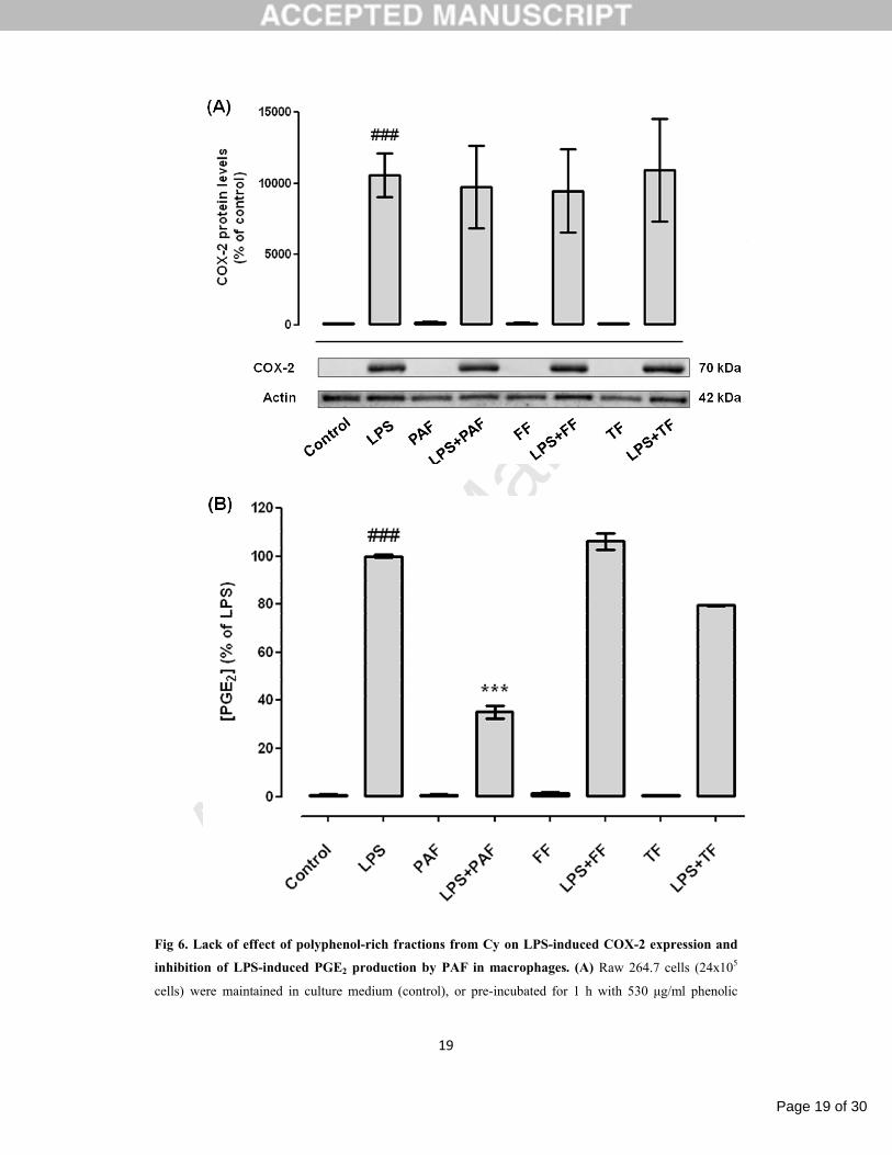
First, we tested the effect of polyphenol-rich fractions in the LPS-induced COX-2 expression in
Raw 264.7 macrophages. Similarly to the Cy extract, none of the fractions tested, PAF (530 μg/ml), FF
(97.5 μg/ml) and TF (78 μg/ml), affected the macrophage COX-2 expression elicited by LPS (Fig. 6A).
Since Cy extract inhibited the PGE2 production in LPS-stimulated macrophages, we next
investigated the contribution of polyphenolic fractions to this activity. As shown in figure 6B, the LPS-
induced PGE2 production is strongly inhibited by PAF (35.17±2.47% of LPS), but not significantly
affected by FF or TF (106.20±3.50% and 79.54±0.36% of LPS, respectively). These results indicated that
PAF is partially responsible for the anti-inflammatory properties of Cy extract by inhibition of PGE2
production.
The effect of Cy fractions on COX-2 expression and PGE2 production in non-stimulated cells
was also tested and none of the treatments interfered neither with the COX-2 expression either with PGE2
production.
Page 19 of 30
Accep
ted
Man
uscr
ipt
19
Fig 6. Lack of effect of polyphenol-rich fractions from Cy on LPS-induced COX-2 expression and
inhibition of LPS-induced PGE2 production by PAF in macrophages. (A) Raw 264.7 cells (24x105
cells) were maintained in culture medium (control), or pre-incubated for 1 h with 530 μg/ml phenolic

Page 20 of 30
Accep
ted
Man
uscr
ipt
20
acid-rich fraction (PAF), or 97.5 μg/ml flavonoid-rich fraction (FF), or 78 μg/ml tannin-rich fraction (TF)
and then treated with 1 µg/ml LPS for 24 h. Total cell extracts were analyzed by Western blot using a
specific anti-COX-2 antibody and an anti-actin antibody was used to confirm equal protein loading. The
blot shown is representative of 3 blots yielding similar results. Results were expressed as percentage of
COX-2 protein levels relatively to control. (B) Raw 264.7 cells (6x10⁵ cells) were treated as above. PGE2
levels were evaluated in the culture supernatants by enzyme immunoassay (EIA), as described in material
and methods, and the results expressed as percentage of LPS. Each value represents the mean±SEM from
2-3 independent experiments (###p<0.001, compared to control; *** p<0.001, compared to LPS).
3.2.2. Polyphenol-rich fractions inhibit LPS-induced iNOS expression and NO production
Since Cy extract inhibited iNOS expression and NO production in LPS-stimulated Raw 264.7
macrophages, the contribution of polyphenol-rich fractions to this activity was investigated. All fractions
drastically decreased the expression of iNOS (Fig. 7A) and this inhibition was higher than that observed
for the whole extract. PAF inhibited the LPS-induced iNOS expression by 75.37%, FF by 75.73% and TF
by 86.34%, while Cy extract inhibited the iNOS expression by 28.95% (Fig. 3A). In addition, PAF and
TF fractions significantly inhibited the LPS-induced nitrite production by 50.63% and 41.59%,
respectively (Fig. 7B). Similarly to Cy extract, none of the fractions exhibit NO scavenging properties
(data not shown). From these results, we can conclude that these fractions highly contribute to the anti-
inflammatory properties of Cy extract. To note that, the fractions alone did not increase iNOS expression
nor NO production, suggesting that polyphenolic compounds are not responsible for the slight pro-
inflammatory properties observed with the Cy extract.

Page 21 of 30
Accep
ted
Man
uscr
ipt
21
Fig 7. Inhibitory effect of polyphenol-rich fractions from Cy on LPS-induced iNOS expression and
nitrite production. (A) Raw 264.7 cells (24x105 cells) were maintained in culture medium (control), or
pre-incubated for 1 h with 530 μg/ml phenolic acid-rich fraction (PAF), or 97.5 μg/ml flavonoid-rich
fraction (FF), or 78 μg/ml tannin-rich fraction (TF) and then treated with 1 µg/ml LPS for 24 h. Total cell

Page 22 of 30
Accep
ted
Man
uscr
ipt
22
extracts were analyzed by Western blot using an anti-iNOS antibody and an anti-actin antibody was used
to confirm equal protein loading. The blot shown is representative of 3 blots yielding similar results.
Results were expressed as percentage of iNOS protein levels relatively to control. (B) Raw 264.7 cells
(6x10⁵ cells) were treated as above. Nitrite levels in the culture supernatants were evaluated by the Griess
reaction as described in material and methods. Nitrite concentration was determined from a sodium nitrite
standard curve and the results are expressed as concentration (μM) of nitrite in culture medium. Each
value represents the mean±SEM from at least 3 experiments (###p<0.001, compared to control;
***p<0.001, compared to LPS).
3.2.3. Polyphenol-rich fractions inhibit LPS-mediated NF-κB activation but not MAPKs or PI3K/Akt
signaling pathways
As Cy extract inhibited the LPS-induced p38 MAPK and JNK 1/2 activation, the contribution of
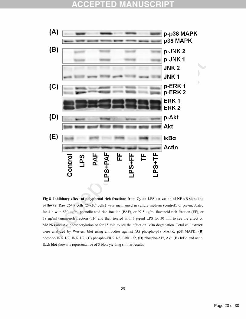
polyphenol-rich fractions to the signaling pathways modulated by Cy was analyzed. As shown in Fig. 8,
the polyphenol-rich fractions did not interfere significantly with the LPS-induced activation of MAPKs
and Akt pathways, but inhibited the LPS-induced IκBα degradation. Overall, these results indicate that
the polyphenolic fractions of Cymbopogon citratus are not responsible for the modulation of p38 MAPK
and JNK 1/2; however they seem to be involved in the inhibition of LPS-induced NF-κB activation.
Page 23 of 30
Accep
ted
Man
uscr
ipt
23
Fig 8. Inhibitory effect of polyphenol-rich fractions from Cy on LPS-activation of NF-κB signaling
pathway. Raw 264.7 cells (24x105 cells) were maintained in culture medium (control), or pre-incubated
for 1 h with 530 μg/ml phenolic acid-rich fraction (PAF), or 97.5 μg/ml flavonoid-rich fraction (FF), or
78 μg/ml tannin-rich fraction (TF) and then treated with 1 μg/ml LPS for 30 min to see the effect on
MAPKs and Akt phosphorylation or for 15 min to see the effect on IκBα degradation. Total cell extracts
were analyzed by Western blot using antibodies against (A) phospho-p38 MAPK, p38 MAPK, (B)
phospho-JNK 1/2, JNK 1/2, (C) phospho-ERK 1/2, ERK 1/2, (D) phospho-Akt, Akt, (E) IκBα and actin.
Each blot shown is representative of 3 blots yielding similar results.

Page 24 of 30
Accep
ted
Man
uscr
ipt
24
4. Discussion
In the course of screening anti-inflammatory compounds derived from plants, we previously
demonstrated that Cymbopogon citratus (Cy) has strong antioxidant properties due to the presence of
polyphenols (Figueirinha et al., 2008) and that Cy extract inhibits NO production and iNOS expression in
dendritic cells (Figueirinha et al., 2010), suggesting an anti-inflammatory activity for this plant. The
present study demonstrates that Cy extract, used in traditional medicine to treat inflammation and other
health problems (Carbajal et al., 1989; Lorenzetti et al., 1991), has anti-inflammatory properties due to
the selective inhibition of NO production through the pro-inflammatory signaling cascades p38 MAPK,
JNK 1/2 and NF-κB, in murine macrophages.
Using LPS-stimulated macrophages as in vitro model, we demonstrated that Cy extract inhibited
iNOS expression and NO production. Using pharmacological signaling pathways inhibitors, it was
observed that the LPS-induced NO production is mainly controlled by p38 MAPK, JNK 1/2 and NF-κB
pathways. Accordingly, previous studies demonstrated that JNK 1/2 (Zhou et al., 2008) and p38 MAPK,
but not ERK 1/2 (Chen and Wang, 1999), modulated iNOS expression and NO production in LPS-
stimulated Raw 264.7 macrophages. In addition, activated MAPKs and PI3K/Akt were also implicated in
NF-κB activation (Carter et al., 1999; Nakano et al., 1998), being NF-κB one of the critical transcription
factors that controls iNOS gene expression in macrophages (Geller and Billiar, 1998). Cy extract also
inhibited p38 MAPK, JNK 1/2 and NF-κB signaling pathways. Therefore, and since the signaling
pathways involved in NO production are the same that Cy extract inhibited, the inhibition of NF-κB, p38
MAPK and JNK 1/2 pathways by Cy extract is probably responsible for its inhibitory effect on NO
production. It was also observed that the iNOS inhibitor aminoguanidine almost abolished the nitrite
production induced by LPS, indicating that in Raw 264.7 macrophages stimulated with LPS, the iNOS
protein is the main, if not the only, NO producer. The effect of Cy extract on NO production was quite
similar to that of the iNOS inhibitor aminoguanidine, emphasizing its potent anti-inflammatory capacity
and indicating that Cy extract inhibited NO production in part by inhibiting the iNOS expression.
However, taking into account the higher effect in NO production relatively to the effect on iNOS
expression, the Cy extract may also affect the levels of NO by other mechanisms. It was previously
demonstrated that Cy extract had strong antioxidant properties (Cheel et al., 2005; Orrego et al., 2009),
however we observed that Cy extract did not possess NO scavenging activity. Therefore, probably it
affected the NO levels by other mechanisms than its antioxidant properties.
Cy extract has a high content in polyphenolic compounds (Figueirinha et al., 2008) that are
secondary metabolites of plants with many healthy effects, including anti-inflammatory properties
(Gonzalez-Gallego et al., 2007). Besides its antioxidant properties, recent data suggest that polyphenols
could have other anti-inflammatory action mechanisms, namely, inhibition of iNOS, COX-2, MAPKs and
NF-κB pathways and that the inhibitory mechanisms of polyphenols are not only signal specific, but also
cell type dependent (Santangelo et al., 2007). Analyzing the effect of the polyphenol-rich fractions on
iNOS expression and NO production we conclude that PAF and TF are the fractions responsible for the

Page 25 of 30
Accep
ted
Man
uscr
ipt
25
inhibitory effect on NO production observed with the Cy extract. Probably, these fractions have a
synergistic effect since the Cy extract has a little more activity than each fraction. Moreover, the
polyphenolic fractions have a stronger inhibitory effect on iNOS expression. All the fractions inhibited
iNOS expression while only PAF and TF inhibited NO production, suggesting that polyphenolic fractions
modulate not only the iNOS expression but also its activity. Accordingly, recent studies demonstrate that
polyphenols could modify the iNOS activity by modulating the availability of L-arginine, the rate-
limiting substrate of iNOS (Mori and Gotoh, 2000). We also previously demonstrated that Cy extract and
its polyphenols have iNOS and NO inhibitory properties in dendritic cells (Figueirinha et al., 2010).
However, the polyphenol-rich fractions have different inhibitory capacity in dendritic and macrophage
cells, indicating that the action of Cy polyphenols might be cell specific.
Many evidences reported that MAPKs signaling cascades might be differentially involved in the
macrophage response to anti-inflammatory compounds (Choi et al., 2008; Lee et al., 2010; Park et al.,
2008; Zhou et al., 2008). In order to explore the mechanisms underlying the inhibitory effect of
polyphenol-rich fractions on NO production, phosphorylation levels of p38 MAPK, JNK 1/2, ERK 1/2
and Akt were analyzed by Western blot in LPS-stimulated Raw 264.7 macrophages. In contrast to Cy
extract, none of the polyphenolic fractions inhibited MAPKs or PI3K/Akt pathways indicating that
polyphenols are not involved in the inhibition of these pathways, being the compounds responsible for
these effects eliminated during the fractionating procedure. Furthermore, we also observed that
polyphenolic fractions inhibited the LPS-induced IκB degradation, suggesting that the inhibitory effect of
the fractions on LPS-induced iNOS expression is due to inhibition of NF-κB activation, as described for
other compounds (Cheng et al., 2001; Pan et al., 2000). Since NF-κB has an important role in
inflammation and its inhibition is one of the main strategies to alleviate chronic inflammation, we can
conclude that Cy extract, in particular their polyphenol-rich fractions, are a promising source of new anti-
inflammatory drugs. In agreement, we are actually conducting more detailed work to better understand
the modulation of NF-κB by Cy extract and to identify the compounds responsible for this effect, using
bioguided assays.
In the present study it was showed that Cy extract inhibited PGE2 production, being phenolic
acid-rich fraction (PAF) responsible by this activity. However, neither Cy extract nor its polyphenolic
fractions seemed to inhibit the LPS-induced COX-2 expression. Current treatment of inflammation is
mainly based in non-steroid anti-inflammatory drugs (NSAIDs) that act by inhibiting COX-2. However,
recent investigation points out that COX-2 specific inhibitors are associated with adverse renal and
cardiovascular effects (Harirforoosh and Jamali, 2009; Ritter et al., 2009). Since FF and TF did not
inhibit COX-2 expression neither its activity, they could be used as anti-inflammatory agents avoiding the
secondary effects associated with COX-2 inhibition.
Intriguingly, Cy extract alone has a stimulatory effect, slightly increasing iNOS expression and
NO production. This effect was due to the intrinsic properties of the extract and not due to the presence of
endotoxins, since we obtained the same increase on NO production after application of the Cy extract in a
endotoxin removal column (data not shown). However, in the LPS-stimulated macrophages the anti-

Page 26 of 30
Accep
ted
Man
uscr
ipt
26
inflammatory properties of Cy overlay its pro-inflammatory activity, occurring inhibition of iNOS
expression and NO production, as well as inhibition of p38 MAPK, JNK 1/2 and NF-κB activation, all
these events being straightly connected with inflammation. In addition, the polyphenol-rich fractions did
not show stimulatory effects and did not interfere with signaling pathways, indicating that the compounds
responsible for the pro-inflammatory properties of the Cy extract are not the polyphenols contained in
those fractions.
In conclusion, this paper demonstrates that a lipid- and essential oil-free infusion of
Cymbopogon citratus leaves strongly inhibited the iNOS expression, NO production, p38 MAPK, JNK
1/2 and NF-κB signaling pathways in murine macrophages (Fig. 9), being the phenolic acids (PAF) and
tannins (TF) responsible for its anti-inflammatory properties through inhibition of transcription factor NF-
κB, iNOS expression and NO production. Taken together, these results provide evidence to understand
the therapeutical effects of Cy extract, and suggest that its polyphenols might be a potential natural source
of new anti-inflammatory drugs for the treatment of inflammatory disorders. However, further work is
required to identify which compound(s) are responsible for the anti-inflammatory properties of
Cymbopogon citratus, as well as the cellular and molecular mechanisms underlying these properties.
Fig 9. Schematic model for the anti-inflammatory mechanism of lipid- and essential oil-free
infusion of Cymbopogon citratus leaves (Cy extract) and its polyphenol-rich fractions on LPS-
stimulated murine macrophages.

Page 27 of 30
Accep
ted
Man
uscr
ipt
27
Acknowledgements
We thank Dr. O. Vieira (Centro de Neurociências e Biologia Celular, Universidade de Coimbra,
Coimbra, Portugal) for the kind gift of the mouse macrophage-like cell line Raw 264.7, to Ervital®,
Portugal, for the provision of the plant and to J. Paiva for the plant classification. This work was
supported by FEDER/COMPETE (FCOMP-01-0124-FEDER-011096), by FEDER, by Foundation for
Science and Technology (FCT) (PTDC/SAU-FCF/105429/2008 project and PhD fellowships-
SFRH/BD/46281/2008 and SFRH/BD/30563/2006) and by a project from the Spanish Ministry of
Science SAF06/08031.
Conflict of interest
None of the authors has any conflict of interest.

Page 28 of 30
Accep
ted
Man
uscr
ipt
28
References
Carbajal, D., Casaco, A., Arruzazabala, L., Gonzalez, R., Tolon, Z., 1989. Pharmacological study of
Cymbopogon citratus leaves. Journal of Ethnopharmacology. 25, 103-107.
Carter, A.B., Knudtson, K.L., Monick, M.M., Hunninghake, G.W., 1999. The p38 mitogen-activated
protein kinase is required for NF-kappaB-dependent gene expression. The role of TATA-binding
protein (TBP). The Journal of Biological Chemistry. 274, 30858-30863.
Cheel, J., Theoduloz, C., Rodriguez, J., Schmeda-Hirschmann, G., 2005. Free radical scavengers and
antioxidants from Lemongrass (Cymbopogon citratus (DC.) Stapf.). Journal of Agricultural and
Food Chemistry. 53, 2511-2517.
Chen, C., Chen, Y.H., Lin, W.W., 1999. Involvement of p38 mitogen-activated protein kinase in
lipopolysaccharide-induced iNOS and COX-2 expression in J774 macrophages. Immunology. 97,
124-129.
Chen, C.C., Wang, J.K., 1999. p38 but not p44/42 mitogen-activated protein kinase is required for nitric
oxide synthase induction mediated by lipopolysaccharide in RAW 264.7 macrophages. Molecular
Pharmacology. 55, 481-488.
Cheng, Z., Lin, C., Hwang, T., Teng, C., 2001. Broussochalcone A, a potent antioxidant and effective
suppressor of inducible nitric oxide synthase in lipopolysaccharide-activated macrophages.
Biochemical Pharmacology. 61, 939-946.
Choi, H.J., Eun, J.S., Park, Y.R., Kim, D.K., Li, R., Moon, W.S., Park, J.M., Kim, H.S., Cho, N.P., Cho,
S.D., Soh, Y., 2008. Ikarisoside A inhibits inducible nitric oxide synthase in lipopolysaccharide-
stimulated RAW 264.7 cells via p38 kinase and nuclear factor-kappaB signaling pathways.
European Journal of Pharmacology. 601, 171-178.
Crawley, J.B., Williams, L.M., Mander, T., Brennan, F.M., Foxwell, B.M., 1996. Interleukin-10
stimulation of phosphatidylinositol 3-kinase and p70 S6 kinase is required for the proliferative but
not the antiinflammatory effects of the cytokine. The Journal of Biological Chemistry. 271, 16357-
16362.
Davis, R.J., 1994. MAPKs: new JNK expands the group. Trends in Biochemical Sciences. 19, 470-473.
Figueirinha A., Paranhos A., Pérez-Alonso J.J., Santos-Buelga C., Batista M.T., 2008. Cymbopogon
citratus leaves: Characterisation of flavonoids by HPLC–PDA–ESI/MS/MS and an approach to
their potential as a source of bioactive polyphenols. Food Chemistry. 110, 718-728.
Figueirinha, A., Cruz, M.T., Francisco, V., Lopes, M.C., Batista, M.T., 2010. Anti-Inflammatory Activity
of Cymbopogon citratus Leaf Infusion in Lipopolysaccharide-Stimulated Dendritic Cells:
Contribution of the Polyphenols. Journal of Medicinal Food. 13, 681-90.
Geller, D.A., Billiar, T.R., 1998. Molecular biology of nitric oxide synthases. Cancer Metastasis Reviews.
17, 7-23.

Page 29 of 30
Accep
ted
Man
uscr
ipt
29
Gold, M.R., Duronio, V., Saxena, S.P., Schrader, J.W., Aebersold, R., 1994. Multiple cytokines activate
phosphatidylinositol 3-kinase in hemopoietic cells. Association of the enzyme with various
tyrosine-phosphorylated proteins. The Journal of Biological Chemistry. 269, 5403-5412.
Gonzalez-Gallego, J., Sanchez-Campos, S., Tunon, M.J., 2007. Anti-inflammatory properties of dietary
flavonoids. Nutricion Hospitalaria. 22, 287-293.
Green, L.C., Wagner, D.A., Glogowski, J., Skipper, P.L., Wishnok, J.S., Tannenbaum, S.R., 1982.
Analysis of nitrate, nitrite, and [15N]nitrate in biological fluids. Analytical Biochemistry. 126,
131-138.
Guzik, T.J., Korbut, R., Adamek-Guzik, T., 2003. Nitric oxide and superoxide in inflammation and
immune regulation. Journal of Physiology and Pharmacology. 54, 469-487.
Han, J., Lee, J.D., Bibbs, L., Ulevitch, R.J., 1994. A MAP kinase targeted by endotoxin and
hyperosmolarity in mammalian cells. Science. 265, 808-811.
Harirforoosh, S., Jamali, F., 2009. Renal adverse effects of nonsteroidal anti-inflammatory drugs. Expert
Opinion on Drug Safety. 8, 669-681.
Hunter, J.D., Doddi, M., 2010. Sepsis and the heart. British Journal of Anaesthesia. 104, 3-11.
Lee, H.J., Jeong, H.S., Kim, D.J., Noh, Y.H., Yuk, D.Y., Hong, J.T., 2008. Inhibitory effect of citral on
NO production by suppression of iNOS expression and NF-kappa B activation in RAW264.7 cells.
Archives of Pharmacal Research. 31, 342-349.
Lee, H.J., Lim, H.J., Lee da, Y., Jung, H., Kim, M.R., Moon, D.C., Kim, K.I., Lee, M.S., Ryu, J.H., 2010.
Carabrol suppresses LPS-induced nitric oxide synthase expression by inactivation of p38 and JNK
via inhibition of I-kappaBalpha degradation in RAW 264.7 cells. Biochemical and Biophysical
Research Communications. 391, 1400-1404.
Lorenzetti, B.B., Souza, G.E., Sarti, S.J., Santos Filho, D., Ferreira, S.H., 1991. Myrcene mimics the
peripheral analgesic activity of lemongrass tea. Journal of Ethnopharmacology. 34, 43-48.
Mori, M., Gotoh, T., 2000. Regulation of nitric oxide production by arginine metabolic enzymes.
Biochemical and Biophysical Research Communications. 275, 715-719.
Mosmann, T., 1983. Rapid colorimetric assay for cellular growth and survival: application to proliferation
and cytotoxicity assays. Journal of Immunological Methods. 65, 55-63.
Nakano, H., Shindo, M., Sakon, S., Nishinaka, S., Mihara, M., Yagita, H., Okumura, K., 1998.
Differential regulation of IkappaB kinase alpha and beta by two upstream kinases, NF-kappaB-
inducing kinase and mitogen-activated protein kinase/ERK kinase kinase-1. Proceedings of the
National Academy of Sciences of the United States of America. 95, 3537-3542.
Negrelle, R.R.B., Gomes, E.C., 2007. Cymbopogon citratus (DC.) Stapf: chemical composition and
biological activities. Revista Brasileira de Plantas Medicinais. 9, 80-92
Newman, D.J., Cragg, G.M., 2007. Natural products as sources of new drugs over the last 25 years.
Journal of Natural Products. 70, 461-477.
Orrego, R., Leiva, E., Cheel, J., 2009. Inhibitory effect of three C-glycosylflavonoids from Cymbopogon
citratus (Lemongrass) on human low density lipoprotein oxidation. Molecules. 14, 3906-3913.

Page 30 of 30
Accep
ted
Man
uscr
ipt
30
Pan, M.H., Lin-Shiau, S.Y., Lin, J.K., 2000. Comparative studies on the suppression of nitric oxide
synthase by curcumin and its hydrogenated metabolites through down-regulation of IkappaB
kinase and NFkappaB activation in macrophages. Biochemical Pharmacology. 60, 1665-1676.
Park, H.J., Lee, H.J., Choi, M.S., Son, D.J., Song, H.S., Song, M.J., Lee, J.M., Han, S.B., Kim, Y., Hong,
J.T., 2008. JNK pathway is involved in the inhibition of inflammatory target gene expression and
NF-kappaB activation by melittin. Jorrnal of Inflammation (London, England). 5, 7.
Porta, C., Larghi, P., Rimoldi, M., Totaro, M.G., Allavena, P., Mantovani, A., Sica, A., 2009. Cellular
and molecular pathways linking inflammation and cancer. Immunobiology. 214, 761-777.
Rao, K.M., 2001. MAP kinase activation in macrophages. Journal of Leukocyte Biology. 69, 3-10.
Ritter, J.M., Harding, I., Warren, J.B., 2009. Precaution, cyclooxygenase inhibition, and cardiovascular
risk. Trends in Pharmacological Sciences. 30, 503-508.
Salh, B., Wagey, R., Marotta, A., Tao, J.S., Pelech, S., 1998. Activation of phosphatidylinositol 3-kinase,
protein kinase B, and p70 S6 kinases in lipopolysaccharide-stimulated Raw 264.7 cells:
differential effects of rapamycin, Ly294002, and wortmannin on nitric oxide production. Journal
of Immunology. 161, 6947-6954.
Santangelo, C., Vari, R., Scazzocchio, B., Di Benedetto, R., Filesi, C., Masella, R., 2007. Polyphenols,
intracellular signalling and inflammation. Annalli dell´Istituto Superiore di Sanita. 43, 394-405.
Schmidt, M.I., Duncan, B.B., 2003. Diabesity: an inflammatory metabolic condition. Clinical Chemistry
and Laboratory Medicine. 41, 1120-1130.
Thiemermann, C., 1997. Nitric oxide and septic shock. General Pharmacology. 29, 159-166.
Tsatsanis, C., Androulidaki, A., Venihaki, M., Margioris, A.N., 2006. Signalling networks regulating
cyclooxygenase-2. The International Journal of Biochemistry & Cell Biology. 38, 1654-1661.
Whitney, N.P., Eidem, T.M., Peng, H., Huang, Y., Zheng, J.C., 2009. Inflammation mediates varying
effects in neurogenesis: relevance to the pathogenesis of brain injury and neurodegenerative
disorders. Journal of Neurochemistry. 108, 1343-1359.
Zamora, R., Vodovotz, Y., Billiar, T.R., 2000. Inducible nitric oxide synthase and inflammatory diseases.
Molecular Medicine. 6, 347-373.
Zhou, H.Y., Shin, E.M., Guo, L.Y., Youn, U.J., Bae, K., Kang, S.S., Zou, L.B., Kim, Y.S., 2008. Anti-
inflammatory activity of 4-methoxyhonokiol is a function of the inhibition of iNOS and COX-2
expression in RAW 264.7 macrophages via NF-kappaB, JNK and p38 MAPK inactivation.
European Journal of Pharmacology. 586, 340-349.