CYCLE S0F DIGE IGENETIC TICTRE MA T0DES A Thesis ...
327
STUDIES0NLIFE -CYCLE S0F DIGE IGENETIC TICTRE MA T0DES A Thesis presented for the Degree of Doctor of Philosophy by 11. T. Diaz Diaz, Licenciado en Biologia VOLT) D1- TEXT The University of Leeds. November, 1976.
-
Upload
khangminh22 -
Category
Documents
-
view
3 -
download
0
Transcript of CYCLE S0F DIGE IGENETIC TICTRE MA T0DES A Thesis ...

STUDIES0NLIFE -CYCLE S0F
DIGE IGENETIC TICTRE MA T0DES
A Thesis presented for the Degree of Doctor of Philosophy
by
11. T. Diaz Diaz, Licenciado en Biologia
VOLT) D1- TEXT
The University of Leeds.
November, 1976.

i
ABSTRACT
30,530 specimens of freshwater molluscs belonging to eleven genera and
eighteen species including gastropods and bivalves, have been examined for
infection with larval trematodes. Thirty species of cercariae have been
found. Of these two are described as 'Monostome', three 'Gymnocephalous',
one 'Pleurolophocercous', eleven 'Furcocercous', one 'Microcercous', one
'Cystocercous', five 'Echinostome', and six Xiphidiocercaria'. These
specimens were collected on different dates from at different places in
West Yorkshire.
Eighteen of these cercariae are comsidered to be new to science, one is
described for the first time in Great Britain and the remaining eleven
have already been described in this country by previous authors.
A short review of each group of cercariae found during this study is
presented.
The life cycles of eight species have been completed under laboratory
conditions. The species involved are:
Echinonarv-, hium recurvaturm (Linstow, 1873)
Hvnoderaeum conoideum (Bloch, 1782)
Notocotylus imbricatus (Looss, 1893) Szidat, 1935
Notocotylus attenuatus (Rudolphi, 1809)
Plagiorchis farnleyensis n. sp.
Plagiorchis kirkstallensis n, sp.
Sphaeridiotrema wintersettensis n. sp.
Dolichosaccus rastellus (Olsson, 1876)
The life cycle of the latter species is the first record from
Great Britain.

ii
ACK130WLEDGEi' OTS
I would like to thank Professor R. MoN. Alexander for the use of the
laboratory facilities in the Department of Zoology.
My thanks are due to Dr. G. A. Boxshall of the British Museum for the
revision of the manuscript.
Iy thanks are also due to dir. A. 0. Holliday for the preparation of
the photo6raphs and IIr. S. Pickersgill for his involvement in the animal
husbandry.
It is a pleasure to acknowledge Dr. R. Wynne Owen for his patient advice
and assistance throughout this study especially during the preparation of
the manuscript.
My final thanks must go to the University de Oriente, Venezuela for
the provision of a Research Studentship.

iii
CONTENTS PAGE
INTRODUCTION 1
. MATERIALS MD IMTHODS 7
LIST OF FR&S -TATER MOLLUSCS EM, 1INED AND LOCALITES
FROM WICH TIM Y Wem; COLLECTED 12
CýRCARIAE ANI) MIR LIFE CYCLES 19
Monostome cercariae 19
Cercaria of Notocotylus imbricatus 21
Description 21
Redia 23
Encystment 24
Metacercaria 25
Infection of the final host 25
The egg 27
The adult 27
Discussion 2e
Table 1 33
Table 2 34
Cercaria of Notocotylus attenuatus 35
Description 35
Redia 37
Encystment 38
1vietacercariae 38
Infection of final host 39
The egg 40
The adult 40
Discussion 41
Table 3 46
Gymnocephalous cercariae 47
Cercaria of SPhaeridiotrema wintersettensis 51

iv
Description
Redia
Iietacercaria and location in the snail host
Netacercariae
Infection of final host
Sp; haeridiotrema wintersettensis n. sp.
Description
Egg and Miracidium
Discussion
Table 4
Cercaria pymnocephalous II
Description
Redia
Encystment
Discussion
Cercaria Kynnocebhalous III
Description
Redia
Encystment
Discussion
Pleurolophocercous cercariae
Cercaria pleurolophocera I
Description
Redia
Infection of second intermediate host
Discussion
Echinostome cercariae
Cercaria of Hypoderaeum conoideum
Description
Redia
Location of metacercariae in the second
intermediate host 0
51
52
53
54
55
56
56
58
60
67
69
69
70
70
71
76
76
77
78
78
80
83
83
84
84
86
90-
95
95
96
97 m

t
V
Netacercariae 99
Infection of the final host 100
Adult 101
Egg and Niracidium 101
Discussion 103
Table 5 107
Table 6 108
Cercaria of liIchinoparvnhiuT, recurvatum 109
Description 109
Redia 110
Metacercaria and location in the snail host 111
Retacercariae 112
Infection of the final host 113
Table 7 114
Echinoparvrphium recurvatum 115
Description 115
Eggs 117
Discussion 117-
Cercaria ech, ostoma III 122
Description 122
Redia 123
Infection of the second intermediate host 124
7-day-old Metacercaria 124
Discussion 125
Cercaria ma. r; nacauda I 127
Description 127
Redia 128
hetacercaria 128
Attempts to infect vertebrate 129
Discussion 130
Cercaria mapnacauda II 133
Description 133

vi
Redia 134
Infection of the second intermediate host 135
iietacercaria of 5 hours development 135
Metacercaria of 14 days' development 135
Metacercaria of 20 days' development 136
Discussion 137
Xiphidiocercous cercariae 140
Cercaria microcotylea I 148
Description 148
Sporocyst 149
Encystment 149
Experiments on the infection of the secondary
intermediate hosts 149
Discussion 150
Cercaria torda 152-
Measurements of Cercaria tarda from Bit nia 154
tentaculata
Specific diagnosis 154-
Cercaria xirhidiocercaria IX 155
Description 155
Sporocyst 156
Encystment and feeding experiments 157
Twelve-day-old metacercariae 157
Discussion 158
Cercaria Dolichosaccus rastellus 160
Description 160
Sporocyst 161
Dolichosaccus rastellus 162
Penetration and encystment 163
: etacercariae 165
incystment and migration 166
Further development in the tadpole 166

vii
Discussion 171
Table 8 176
Cercaria of Plegiorchis farnleyensis 177
Description 177
Sporocyst 178
Penetration and Encystment 179
Metacercaria 181
Infection of the final host 181
Plamiorchis farnleyensis 183
Description 183
egg and "MMiiracidium 186
Attempts to infect the snail host 187
Discussion 187
Table 9 195
Cercaria of Plapiorch_is kirkstallensis 196
Description 196
Sporocyst 197
Penetration and Encystment 198
Ketacercaria 199
Infection of the final host 200
Plegiorchis kirkstallensis 201
Description 201
Egg and Yiracidium 204
Experimental infection of the first
intermediate host 205
Discussion 206
Table 10 214
Microcercous cercariae 215
Cercaria microcercous I 218'-
Description 218
Sporocyst 219

viii
The metacercarial cyst 219
15-day-old metacercaria- 220
Experimental infection 221
The Juvenile Form 222
Discussion 223-
Cystocercous cercariae 227
Cercaria macrocerca I 230
Description 230
The sporocyst 231
Metacercariae 232
Discussion 233
Furcocercous cercariae 237
Cercaria Apa. temon Kracilis minor 243
Measurements of the Cercaria of Apatemon gracilis 244
Mrffor from Plenorbis vortex
Specific diagnosis 244-
Cercaria furcocercaria IV 245
Description 245
Sporocyst 247
Discussion 247
Cercaria Bilharzielae rolonicee 249
Measurements of the Cercaria of Bilharzielae 250
polonicae from Planorbis planorbis
Specific diagnosis 250
Cercaria letifera 251
Measurements of Cercaria letifera from
Lymnaea perep: er 252
Specific diagnosis 252
Cercaria raracauda 253
Nleasurerlents of Cercaria naracauda from ý, 55
LLyrnnaea staLnalis
Specific diagnosis 255

ix
SIT MARY
EF R NC ,S
Cercaria Cot-ylurus brevis
Measurements of the cercaria of Cotylurus brevis
from Lymnaea pereer
Specific diagnosis
Cercaria furcocercaria VII
Description
Sporocyst
Discussion
Cercaria of Diplostomum nhoxini
? ieasurements of the Cercaria of Diplostomum
phoxini from Lymnaea Pereper
Specific diagnosis
Cercaria furcocercaria II
Description
Sporocyst
Discussion
Cerc^ria, vivax I
Description
Sporocyst
Discussion
Cercaria vivax II
Description
Sporocyst
Discussion
256-
257
257-
258
258
259
260
262 -
263
263
264
264
266
267
269
269
270
271'
273
273
274
275
276
278

aI; INTRODUCTION
Thomas (1883) and Leuckart(1882) independently established and published
the first trematode life cycle, that of the liver fluke Fasciola hepatica from
Great Britain and Germany respectively. Since that time no more accounts of
freshwater larval trematodes were published by British workers until 1923,
when Hesse, from Scotland, reported two cercariae emerging from Lymnaea nereper.
One was a furcocercous cercaria and the other a xiphidiocercous cercaria'.
Hesse gave a description of the cercariae and of some aspects of their biology
but did not name them. In the same year, 1923, Veversfed ducklings with the
hepatopancreas of L. nere? er infested with a tetracotyle stage that he thought to
belong to Tetracotyle typica (Diesing, 1835), and the hepatopancreas of L. stagnalis
naturally infected with echinostome cercariae that he identified as C. echinata
(Siebold, 1837). Twenty days after feeding he obtained adults of Hyrnoderaeum
conoideum (Bloch, 1782) and immature forms of Echinostoma revolutum (Frolich,
1802), but the tetracotyle cysts failed to develop. He stated that H. conoideum
must have developed from other cercariae also encysted in L. perejer.
In 1926 Brown investigated larval trematodes in Birmingham and Leeds.
As results of these studies, nine species of cercariae from L. perep; er and L.
starnalis were added to the British fauna, of which Cercaria granulosa, C.
eguisninosa, C. pucilis, C. pseudarYnataý C. lentosoma, C. macrosoma, C. micromorrha
were new to science, while the remaining two species, C. echinata and C. fissicfluda
(La Val, 1855), had been previously described. He attempted to investigate
their life cycle of some of these forms, but without conducting experiments.
Brown (1927) obtained a xiphidiocercaria with eyespots emerging from Pisidium
amnicum and Sphaerium corneum from a Yorkshire river (R. Wharf e), and he
experimentally infected larvae of the may-fly, Ephemera danica, which were fed
to a trout. Juvenile forms of Crenidostomum farionis (Muller, 1784) were
recovered from its pyloric caeca.
and grayling from the River Wharfe.
Adults of C. farionis were found in trout
Brown (1931) described five additional
cercariae from Cheshire; two were echinostome Cercaria oscillatoria n. sp. "

02 and C. limbifera (Seifert, 1926) and three were new species of furcocercariae
(. echinomorpha, C. chronatonhora and C. pygocytophora). Two years later
Brown (1933) described the life cycle of Lecithodendrium chilostomum (1ehlis, 1831)
and also briefly described the life cycle of Dicrocoelium dendriticum (Rudolphi,
1819).
Wright (1927) added additional information on the precercarial, cercarial
and metacercarial stages of Fasciola hepatica. He also described two
xiphidiocercariae. Cercaria cambrensis I and C. cambrensis II, from L. truncatula
and L. Pere, -er respectively. Harper (1929) published a brief account of some
observations on the life cycles of Notocotylus seineti (Fuhrman, 1919) and
Echinoparyphium recurvatum (Linstow, 1873). He also described four new species
of xiphidiocercariae and gave some information on their biology, penetration
and encystment in a variety of invertebrates. The same author (1931) described
three species of furcocercariae and traced the development of two of these
species into the corresponding tetracotyle stages, one of which was new species.
He also established the life cycle of Strigea tarda (Steenstrups), from sporocyst
to adult form.
In 1930 Matheson reported an outbreak of dermatitis caused by Echistosomatid
cercaria which he identified as Cercaria elvae (Miller, 1926). Taylor and Baylis
(1930) identified Natheson's material as C. ocellata (La Val, 1885). The same
authors (Taylor and Baylis) described some experiments with C. ocellata and
another additional furcocercaria that they referred to as "Cercaria X".
Rees (1932) published the most outstanding contribution to the study of
larval trematodes of freshwater molluscs in Britain. She reported ten different
species of cercariae from several species of Lymnaea of which Cercaria "Y", and
Cercaria "Z" and Cercaria cambrensis III were new species. She gave details
of the incidence of infection of the first intermediate hosts by the cercariae,
together with the occurrence, structure and some aspects of their life cycle.
She also added descriptions of various structural details to those previously Ws described forhCercaria monostomi (Linst. ), C. Fasciola hepatica (L. ), C. cambrengia
I (Wright), C. limbifera (Seifert), C. acellata (La Val., ), C. macrosoma(Brown)

03 and Cercaria "X" (Baylis). Subsequently Rees published a series of interesting
papers (1932,1940,1952,1955,1957) dealing with several aspects of the
biology and life cycles of larval trematodes of British freshwater molluscs. and until 1960
After the Rees (1932) paper', only isolated accounts of British freshwater
larval trematodes have been published, Vickers (1940), Dawes (1952),
Erasmus (1957,1958a, 10,60) and Thomas (1958).
Iles (1959) found Cercaria pseudocellata (Szidat, 1942) under which she
put as synorym$, Cercaria elvae (faller, 1926; Matheson, 1930),. 2. ocellata
(Vogel, 1930) and C. ocellata (Taylor and Baylis, 1930; Rees, 1932). She also
reported seven different species of furcocercariae four of which were new and
the remaining three had previously been described. One year later Iles gave cycle
a detailed study of the life9tof Anetemon racilis minor (Yamaguti, 1933) and
some observations on the life cycles of C. pnracauda and C. tetraglandis Iles
(1959).
Nasir ýý957-19ex
fined over 3,000 specimens of L. stapnalis from Edgbaston
Pool, in Birmingham and found eight different species of cercariae - three
echindstome j. nudicaudatum n. sp., E. pinnicaudatum n. sp., and H. conoideum
(Bloch)) two furcocercariae, one strigeid (C. Cotylurus brevis Dubois and
Raush, 1950) and three stylet cercariae (C. seud'maata (Brown), Pla'iorchis
(Iwlultiglandularis) megalorchis (Rees, 1952) and C. edabastonensis n. sp. ).
He described the life cycle of Echinostoma nudicaudatum and E. pinnicaudatum
and also established the identity of the previously known C. helvetic8 XXXIV
Dubois (1934) with the strigeid adult Cotylurus brevis. Later (1962) he reported
the presence of two giant-tailed echinostome cercariae found at the same place
(Edgbaston Pool) which he described and figured as new species. Nasir and
Erasmus (1964) gave a key for the identification of 81 of the species of cercariae
from British freshwater molluscs then known.
Khan (1960 (a) and (b); 1961 (a), (b), (c) and (d); 1962(a), (b) and (c),
during a survey of larval tramatodes carried out in London and some parts of
Essex, Middlesex, Surrey and Hertfordshire, reported 35 different cercarial-

04 species of which 23 were new. This list comprised 7 echinostome, 3 gymnocephalous,
17 furcocercariae, 6 xiphidiocercariae and 2 cercariae. He completed the life
cycle of three of them - C. londonensis, C. essexensis and C. bushiensis -
which developed into adults of the genera Echinostoma, Hypoderaeum and Cyathocotyle
respectively.
Berrie (1960) elucidated the life cycle of DiDlostomum snathaceum (Rud. )
and D. nhoxini (Faust) from two Dinlostomulum larvae in the eyes of Gasterosteus
aculeatus. The same author (1960) studied the variation of D. rhoxini in
different definitive laboratory hosts.
Probert (1965(a) and (b); 1966) studied the larval trematodes infecting
the freshwater molluscs of a lake in Breconshire (Llangorse Lake) and reported
twenty-one species of cercariae, twelve of which were considered to be new.
At the same time he investigated the incidence of larval trematodes from the
same lake. Williams (1966) during studies of the incidence of larval trematodes
in L. pereger from a pond in MLiingavie near Glasgow, Scotland recorded four
species of furcocercariae, one echinostome and a monostome cercaria all of which
except the cercaria of Diplostomum asteroste3äilliams (1966) was new. ' He
studied the development of Cotylurus cornutus (Rudolphi, 1809) in ducks and gave
brief accounts of the life cycle of all cercariae encountered except that of
Haplometra cylindracea.
Pike (1967) studied some stylet cercariae and a microphallid type in freshwater
molluscs from Wentloog level near Cardiff, South Wales. He redescribed C. helvetica
XXXIII Dubois (1931) and the cercaria of Sphaerostoma bramae (Mull) and their
metacercariae. He also recorded a further six species of cercariae - C. p rvus
(Khan (19614) and C. tarda Khan (1961J) on which he gave additional information
together with a description of their metacercariae and four new species -
C. wentlogensis, C. runniensis, C. octoglandulata and a Nlicrophallid cercaria sp.
One year later Pike (1968a) found the cercaria of Psilotrema olipoon (Linstow,
1887). He experimentally obtained the adult from ducklings and discussed the
taxonomy of this fluke. He also described two new species of Gymnocephalous
cercariae - C. frondicola and C. g-ranocutis. In the same year (1968b) he studied

05 the distribution and incidence of larval trematodes in the freshwater fauna
from the Wentloog level, and gave some information on the distribution of cercarial
stages in the host fauna and some indication of their specificity as well as
the percentage throughout the year of each host species infected with these
larvae. In 1969 Pike gave an account of the life cycles of two notocotylid
trematodes - Notocotylus triserialis Dies, 1839 (= N. attenuatus, Rud. 1809)
and N. irlbricatus (Looss, 1893) Szidat, 1935.
In Europe the most outstanding contributions to the study of trematode
larvae were undertaken by Nitzsch (1816), Ercolani (1881,1882), Siebold (1854),
Filippi (1854,1857), La Valette St. George (1855), Noulinie (1856) and Luhe
(1909). Several years later, additional interesting accounts were added to
this field by Mathias (1922,1924,1925,1930,1935), Dubois (1928,1929,1931),
Dollfus (1938 and 1960), Szidat (1924,1928,1931,1933,1935), Wesenberg-Lund
(1934) and Wikgren (1956).
In North America; a great amount of work on larval trematodes has been
done mainly by Cort (1915,1917,1918), Faust (1917,1918,1919), H. M. Miller
(1926), E. L. Miller (1936), Brooks (1930,1943,1948) and Cable (19356 1938).
The study of freshwater larval trematodes in Japan was initiated by Osafane
in 1898 and continued by Senoo (1903), Fujita (1906), Kobayashi (1911,1922),
Tanabe (1922,1948), Takahashi (1927), Komiya (1938,1939,1941), Yamaguti
(1938,1940,1942,1943) and many other's.
In Africa, Cawston (1915,1917,1922), Porter (1921,1938), Fain (1953)
and Vercammen-Grendjean (1960) have made significant contributions to knowledge
in this field.
Sewell (1922) published a comprehensive account of Indian larval trematodes.
Other contributions of recent years have been made by Singh (1952,1955).
A, garwall (1956), Premvati (1965), Parade (1967), Srivastava (1968) and Tapar (1970).
In many countries the study of freshwater larval trematodes has also been
carried out by many workers. The major works have been those of Looss (1856,
1899) and Leiper (1915, in Egypt, Jo)uiston, 1938, Johnston and Angel (1940)

D6 and Johnston and Simpson (1944) in Australia, McLeod (1936,1940) in Canada,
Tubangui (1928,1932) and Velasquez (1961,1963,1973) in Phillippines,
Lie (1963,1964,1965) in Malaya, Zajicek (1963) and Zdatska (1963) in Czechoslo-
vakia, Nakagawa (1951), Faust (1922,1924, Tsuchimochi (1926) and Komiya (1941,
1952) in China, Uribe (1925) and Nasir and Diaz (1967,1968(a) and (b), 1973)
in Venezuela.

07 MATMIALS AND I THODS
(i) The host molluscs
During the period from 1972 to 1976 more than 30,530 freshwater molluscs
representing 11 genera and 18 species were examined. The molluscs, which
included both bivalves and gastropods, were collected from a variety of
freshwater bodies (lakes, reservoirs, canals and rivers). They were caught
by fishing net, by hand and, in some inaccessible localities, by the use of a
grappling hook tied to a piece of rope which was thrown out into the lake or
canal then hauled back dragging with it submerged vegetation or broken branches
from which the molluscs were removed. The species Bithynia tentaculata (Linn. ),
Dreissena rolvmorDha (Pallas) and Acroloxus lacustris (Linn. ) were principally
obtained in this manner.
Once brought back to the laboratory the specimens were washed in tap
water and placed in aquaria measuring about 30 by 45 cm containing aerated
water and various aquatic plants obtained from the same locality as the
molluscs. The aquaria were kept at a temperature of 13 to 150C in an aquarium
room. The molluscs were fed a varied diet comprising fresh and dry lettuce
leaves, carrots and cabbage. The water in each aquarium was changed once or
twice per week. In these conditions some of the molluscs survived for more than
8 months.
Some species such as LimnaeaQereper (Mull. ), L. stapnalis (Linn. ),
Planorbis corneus (Linn. ), Bithynia tentaculata, PotamopyrTuus ienk1nsi (Smith)
and Srhaerium corneum (Linn. ) were reared in the laboratory for experimental
purposes. The eggs of these species, with the exception of P. jenkinsi and
S. corneum were collected at various localities and kept in aquaria in suitable
conditions. P. enkinsi and S. corneum were collected in great numbers and
kept in aquaria as describedabove. After 25 to 30 days they were removed
and transferred to other vessels where they were left for a variable period
of time after which the small P. . enkinsi could be found at the surface and the
small S. corneum could be found at the bottom of the vessel. In both cases
ground snail shell and pieces of chalk were added as a source of calcium

08 (calcium carbonate). However, some of the mollusc shells were very thin
and the animals soon died. The survival rate of molluscs reared in this way
was about 30 to 40, although this was greater than that of the other species
raised in the laboratory.
(ii) Cercariae
In order to determine the presence of cercarial infections the molluscs
were placed individually in glass vessels measuring about 5 by 7 cm which were
half full of tap water. After 4,8,12 and 24 h they were examined for
cercariae under a stereo dissecting microscope. Those giving a positive
result (cercariae present) were placed in holding aquaria; those giving a
negative result were discarded. The infected molluscs were examined again to
confirm that they continued to release the same species of cercaria as originally
determined so that possible errors caused by the presence of double infections
could be eliminated. Recently released cercäriae were studied in vivo in tapwater
and 0.75% saline solution between slide and coverslip.
The excretory system of the cercariae was studied in three ways. The
first method was to study the cercariae on a slide under a coverslip in 0.85%
saline solution adding drops of the saline from time to time to ensure that the
specimen did not dry out. It was found that the high saline concentration
induces an increase in the activity of the flame cells which facilitated
observation of these cells.
The second method consisted of placing the vessel containing the cercariae
in the freezing compartment of a refrigerator (about - 4°C) for 15 to 20 minutes.
The low temperature causes a decrease in the activity of the cercariae which sink
to the bottom of the vessel. When the cercariae were mounted on a slide their
movements were still very slow and the flame cells displayed little activity.
As the te, aperature was raised by the heat of the microscope lamp the cercariae
resumed their normal activity and the increase in activity of the flame cells
made them more easily visible.
The third method was to use a--m \ºXt ure - of intravital'stains, normally
1% malachite green, 1 methyl blue, 1' neutral red and 1% bismark brown. This

09
combination of stains also caused a considerable increase in the activity of
the flame cells and has the added advantage of displaying certain internal
structures of the cercaria.
The study of the penetration gland-cells of the cercariae was carried out
using cercariae extracted from dissected snails as well as recently emitted
cercariae. In this study staining with 1 neutral red and 1% bismark brown
gave the best results. 1% malachite green was used to help determine the
arrangement of spines on the body of the cercariae and to display the 'collar of
spines' of the echinostome cercariae.
Measurements (in mm) of the cercariae were taken from 25 to 30 specimens
fixed in 10 formalin. Drawings were made freehand and the dimensions of
structural features were arranged proportionally to the mean measurements of
the specimens.
(iii) Sporocysts, Rediae and R7etacercariae
Sporocysts, rediae and metacercariae were studied in vivo on a slide in
tap water and 0.75% saline solution. One per cent methyl blue, 1% malachite
green and 1% neutral red were used as intravital stains to highlight certain
structures. Measurements of sporocysts and rediae were taken from 15 to 20
specimens immediately after fixation in 101% formalin. Measurements (in mm)
of similar numbers of metacercariae were taken on live specimens. Drawings
were made freehand as described for the cercariae.
(iv) Life Cycle Studies.
A number of different species of animal was used in experimental
studies on the life cycles of the parasites. These animals were used both,
as secondary intermediate hosts and as final hosts. Among the secondary
intermediate hosts used were various aquatic larvae of insects belonging to
the orders Trichoptera, Odonata, Ephemeroptera, Coleoptera and Diptera. Frog
and toad tadpoles and freshwater fishes were also used. The first four groups
of insect larvae were collected from Durkar (near Wakefield) from a semi-
permanent freshwater pool. None of these larvae ever harboured a trematode
infection. The larvae of Chironornus were obtained partly from Durkar and

10 partly from commercial sources. Of the latter 50ö were examined for the
presence of trematode infections but none was ever found. The remaining
5CF% were used in two groups, one to infect the final hosts and the other as a
control. The larvae of Aedes aepynti, fishes of the species Lebistes reticulatus
(Peters) and Tilapia mosambica, tadpoles and young adults of Bufo bufo (common
toad and tadpoles, young adults and adults of Xenopus laevis (South African
clawed toad) were reared in the Zoology Department at Leeds University. Other
fishes were obtained from commercial sources and from Mr. S. Axford of the
Yorkshire River Authority.
The definitive experimental hosts - Mus musculus, Rattus norveicus and
Columba livia - were supplied by the Zoology Department. The ducklings, chicks
and canaries used in these experiments were purchased commercially. The control
and experimentally infected animals were kept in suitable cages in the animal
house of Zoology Department and fed on commercial prepared food.
The faeces were examined by sedimentation, and the resultant sediment
was examined beneath low-power and high-power microscopes. The faeces of all
these animals were examined for at least 3 consecutive days prior to the beginning
of the experiments in order to avoid possible errors from natural infections.
No natural trematode infections of experimental hosts were found.
All birds were force-fed by means of a_medicinal pipette while the mammals
were slightly anesthetized with ether before force-feeding, and the cysts were
introduced using a hypodermic syringe with a plastic tube on the end of the needle.
The age of all experimental animals used during this investigation ranged
between 9 to 169 days old, except that the age of the canaries was unknown.
(v) Adults
To recover both the preadults and the adults of the parasites the different
hosts were killed with chloroform and the alimentary canal was dissected out.
It was placed into suitable saline solution in a petri dish and washed several
times before being examined under dissecting microscope. The trematodes were
fixed either by dropping them into warm (600 to 700 C) Bouin solution or by

11 flattening, them between slide and coverslip. For the latter method the
specimens were heat-killed in saline and fixed either in alcohol-formalin-
acetic acid (AFA) or Bouin at normal temperature under slight coverslip
pressure. The fixed specimens were measured and studied anatomically.
both before and after staining with celestin blue, borax carmine and alum
carmine.
Some adults were fixed in Bouin solution and used for histological studies.
Serial sections were cut at 5- ,pm and stained with haematoxylin and Eosin.
The sections were then mounted in Canada balsam or Xam. The parasites were
studied in vivo, fixed and as serial sections. Measurements were taken from
fixed specimens and are based on 20 to 70 individuals. Drawings were made with
the aid of a camera lucida and certain morphological features were added
subsequently.
(vi) E. cr, -s and Miracidia
Parasite eggs were collected from the faeces of the host and from petri
dishes in which the alimentary canal was washed, or sometimes by teasing apart
the sexually mature worms. The e,, gs were kept in glass vessels in the
laboratory immersed in tap water, distilled water or 0.85% physiological saline.
The vessels were maintained at a temperature of 19 to 22°C and were constantly
aerated to permit the development of the eggs. Of the three media distilled'
water gave the best results because in both tap water and saline cultures protozoal
growth had an adverse effect on the trematode eggs.
Miracidia were studied alive in 0.755 saline solution. Drawings were
made with the aid of a camera lucida and additional details were incorporated
freehand.
Measurements were made with the aid of an ocular micrometer, the mean is ý»re-n,
vto-rvna. Lt jT followed bb, minima and maxima inýýarenthesesi

LIST OF FtRý, Slf i ', ilATER MOLLUSCS EXLA'aNE'D AND LOCALITIES 12
FROM WHICH THEY WERE COLLECTED
Since I arrived in Leeds I have visited 34 different places to collect
freshwater molluscs. In 21 of these 34 collecting sites I have obtained
molluscs.
The gastropod and bivalve molluscs have been identified with the aid
of keys developed by Ellis (1926), Janus (1965) and Macan and Cooper (1969),
and texts of Macan (1959) and Mellanby (1963) dealing with British freshwater
invertebrate animals.
The number and species of molluscs examined, localities, dates and cercarial
infections are listed below.
Species of molluscs examined
Class GASTROPODA
Sub-class PROSOBRAICHIA.
Order Mesogastropoda
Family Viviparidae
Genus Viviparus
Viviparus vivioarus (Linn. )
Family Hydrobiidae
Genus Potomopyrgus
Potomoovrgus jenkinsi (Smith)
Genus Bithynia
Bithynia tentacula. ta (Linn. )
Bit nisi leachi (Sheppar)
Sub-class PULNCIIATA
Order Basommatophora
Family Lymnaeidae
Genus Lymnaea
Lynnaea auricularia (Linn. )
Lyrnnaea rereger (Dull. )
Lymnaea stagnalis (Linn. )

13 Family Physidae
Genus Phyla
Physa fontinalis (Linn. )
Family Planorbidae
Genus Planorbis
Planorbis carinatus (Mull. )
Planorbis corneus (Linn. )
Planorbis crista (Linn. )
Planorbisplanorbis (Linn. )
Planorbis vortex (Linn. )
Family Ancylidae
Genus Acroloxus
Acroloxus lacustris (Linn. )
Freshwater Bivalve Molluscs
Class LAIMELLT&"W CHIATA
Order Eulamellibranchiata
Family I4. argaritiferidae
Genus Anodonta
Anodonta c nea (Linn.
Family Sphaeriidae
Genus Sphaeriun
Sphaerium corneum (Linn. )
Genus Pisidium
Pisidium amnicum (Mull. )
Family Dreissenidae
Genus Dreissena
Dreissena nolymorpha (Pallas)

1 Location and number of molluscs species examined and the
cercarial and metacercarial infections observed
Viviioarus vivinarus (Linn. )
550
Kirkstall Power Station (Leeds-Liverpool Canal)
October, November 1972; January, February, August, October 1973;
April, June, July 1974; Nanuary, February, November 1975;
January, February 1976.
Infections: 1chinostomes (cysts)
Potamopyrgus jenkinsiý (Smith)
7000
Kirkstall Power Station (Leeds-Liverpool Canal); Gledhow Valley Road; 11 Cwthorne Park.
October, November 1972; January, February, August, October 1973;
April, June, July 1974; January, February, November 1975; January,
February 1976.
Infections: Echinostomes (cysts); Xicrocercous (cysts).
Bit is tentaculata (Linn. )
2500
Kirkstall Power Station (Leeds-Liverpool Canal);
Newmillerdam Lake; Wintersett Lake; West Bretton Lake;
Walton Park; Gledhow Valley Road.
October, November 1972; January, February, March, April, May;
August; October 1973; January, April, June, July 1974;
January, February, April, July, August, September, November 1975;
January, February, 1976.
Infections: Cercaria Notocotylus imbricatus; C. pleurolopbocerca I
C. Sphaeridiotrema wintersettensis; C. ymnoceohalous II;
C. iymnocenhalous III; C. vivax I; C. vivax II; C. rnicrocotvlea I;
C. tarda; C. microcercous I; C. mamacauda II.

15 Bit is leachi (Sheppar)
59
Newmillerdam Lake.
October 1972; January, March 1973; July 1974; April, August; September,
November 1975; January, February 1976.
Infections: C. snhaeridiotrema wintersettensis.
Limnaea auricularia (Linn. )
600
Kirkstall Power Station (Leeds-Liverpool Canal); Wintersett Lake:
Newmillerdam Lake, KirLinöton; Riffa Beck, Pool-in Wharfedale.
November 1972; January, May 1973; January 1974; January, April 1975.
Infections: Cercaria xirhidiocercaria IX.
Limnaea perep"er (Mull.
6002
Kirkstall Power Station (Leeds-Liverpool Canal); Wintersett Lake; Newmillerdam
Lake; Riffa Beck Pool-in-Wharfedale; Ilkley Moor; Durkar; Gledhow Vall&y
Road; Kirýington; Walton Park; West Bretton Lake; Rawdon; Enroy; Roundhay
Park; Eccup Reservoir; Ferrybridge; Otley; Carnforth.
October; November 1972; January, February, May, August, October 1973;
January, April, June, July, November 1974; January, February, March, April,
May 1975; January, February 1976.
Infections: Cercaria Pla'iorchis Kirkstallensis; C. plapiorchis farnlevensis;
C. Notocotylus attenuatus; C. Hynoderneurn conoideum; C. echinorsrynhium
recurvrtum; C. furcocercaria VII; C. furcocercaria IV; C. furcocercaria II;
C. Diplostbmum phoxini; C. nnracauc'a; C. Cotylurus brevis; C. etifera;
C. Dolichosaccus rastellus; C. Apatemon gracilis minor.
Limn? ea stag alis (Linn. )
4058
Kirkstall Fower Station (Leeds-Liverpool Canal); Wintersett Lake; Newmillerdam
Lake: Walton Park; New Farnley; Gledhow Valley Road; Cowthorne Park; Enroy;

16 Eccup Reservoir; Otley.
October, November 1972; January, February, March, April, May, August 1973;
January, February, April, May, July 1974; January, February, March, April,
May 1975; January, February 1976.
Infections: C_, Plagiorchis kirkstallensis; C. Pla&iorchis farnlevensis;
C. Dolichosaccus rastellus; C. Hyroderaeum conoideum; C. echinostoma III;
C. Ana. temon gracilis minor; C. paracauda; C. xiy phidiocercsria IX.
Ph sa fontinalis (Linn.
1979
Kirkstall Power Station (Leeds-Liverpool Canal).
October, November 1972; January, February, August, October, 1973.
April, June, July 1974; January, February, November 1975; January,
February 1976.
Infections: Echinostomes (cysts); Microcercous (cysts)
Planorbis carinatus (Mull. )
458
Kirkstall Power Station (Leeds-Liverpool Canal); West Bretton Lake;
Enroy; Cowthorne Park; Walton Park.
October, November 1972; January, February, March, April, August 1973; January,
April, June, July 1974; January, Febrriiary, November 1975; January, February
1976.
Infections: i: chinostomes (cysts); Microcercous (cysts).
Planorbis corneus (Linn. )
530
Kirkstall Power Station (Leeds-Liverpool Canal); West Bretton Lake;
Walton Park.
October, November 1972; January, March, April 1973; April, June, July 1974;
January, February 1975; January, February 1976.
Infections: E'chinostomes (cysts).

Planorbis crista (Linn. )
2500
Durkar, Enroy.
17
February, April 1973; January 1974.
Infections: Echinostomes (cysts)
Planorbis planorbis (Linn. )
472
Kirkstall Power Station (Leeds-Liverpool Canal); Walton Park; New Farnley;
Cowthorne Park; -West Bretton Lake; Enroy; Wintersett Lake.
October, November, 1972; January, March, April 1973; April, June
1974; January, February 1975; January, February 1976.
Infections: C. Bilharzielae nolonicae; C. magnacauda I.
Plar_orbis vortex (Linn. )
420
Kirkstall Power Station (Leeds-Liverpool Canal); Cowthorne Park; West-
Bretton Lake; Wintersett Lake.
October, November 1972; February, kIarch, June, July 1973; April,
June, July 1974; January, February 1975; January, February 1976.
Infections: C. Apatemon gracilis minor.
Acroloxus lacustris (Linn. )
190
Kirkstall Power Station (Leeds-Liverpool Canal); Tüewmillerdam Lake;
Riffa Beck, Pool-in-Wharfedale.
November, October 1972; January, February, August, October, 1973.
April, June, July 1974; January, April, September, December 1975.
January, February 1976.
Infections: Echinostomes (cysts).
Anodonta cygnea (Linn. )
480
Wintersett Lake; Newmillerdam Lake; Harewood.

October, November 1972; January, March, April, June, July, August 1973;
January, February, April, June, July 1974; January 1975.
Infections: None.
Sphaerium corneum (Linn. )
1200
Kirkstall Power Station (Leeds-Liverpool Canal); Wintersett Lake;
Newmillerdam Lake.
October, November 1972; January, March, April, August 1973;
January, April, June, July 1974; January, February, May 1975.
Infections: Cerceria macrocerca I
Pisidium aranicum (Dull. )
32
Kirkstall Power Station (Leeds-Liverpool Canal); Wintersett Lake.
October, November, 1972; January, April, May, August 1973; January,
April, June, July 1974; January, February, May 1975.
Infections: None.
Dreissena nolimorpha (Pallas)
1500
Newmillerdarn Lake; Wintersett Lake.
February, May, August 1973; January 1974; January 1975.
Infections: None.

CERCARIAi"; AND T1iEIR LIFE. CYCLES
Monostore cercariae
Luhe (1909) defined Monostome cercariae as cercariae in which the
acetabulum was absent, eyespots were present, a long slender undivided tail was
without setae and which developed in rediae and encysted in the open. Faust
(1917b) divided the Mlonostome cercariae according to the size of the body and
the number of eyespots into two sub-groups; Binoculate which were characterized
by having two lateral eyespots and Trioculate possessing a third additional
median eyespot. Sewell (1922), basing his interpretation on the meaning of
"Monostome" and using it in its widest sense, divided Morostome cercariae into
six sub-groups, comprising all the forms without an acetabulum: - (1) Pleurolopho-
cerca Cercaria pleurolonhocerca Sons. as the type of this group); (2) Urbanensis
(. urbanensis Cort. ); (3) Ephermera (. erhemera Nitzsch. ); (4) Lophocerca
(included C. indicae IX, XIII, XXXIX and LV. ); (5) Lophoides (C. indicae xXVII. )
and (6) Ubiquita (C. indicae LII and LICI. ). At present only the Urbanensis
and Ephemera groups are considered to be true Monostome ceroariae and are very
closely related to the Binoculate and Trioculate Groups of Faust (1917).
According to Dubois (1929) Cercaria urbanensis is a trioculate form and
therefore should belong to the Ephemera group.
Cercaria Pleurolophocerca Sons. was the type of the first sub-group and is
characterized by having a well-developed acetabulum (Langeron, 1920). The
Lophocerca was removed by Luhe (1909) from the Nonostomes because it possessed
a divided tail and a fin-fold on the body. Miller (1926) placed it in
the Apharyngeal brevifurcate monostome cercariae. Dubois (1929) pointed out
that the absence of the acetabulum in the members of this group is not sufficient
reason to keep them in the Monostome group because its organization and evolution
are quite different. The members of the Lophoides group are also Furcocercariae
characterized by the possession of a penetration organ and excretory system
similar to that of the Schistosome cercariae. The Ubiquita group includes
cercariae with a stylet, without an oesophagus or -intestinal caeca and with
three to six pairs of penetration gland-cells. Dubois (1929), Wesenberg-Lund
(1934) and Nasir, Ramana and Diaz (1969) considered that the members of this

20 group belong to the true Xiphidiocercaria. Therefore this group should be
transferred to the1Xiphidiocercous cercariae.
Rothschild (1938d) redefined the characters of Notocotyloidea cercariae,
and divided them into two süb-groups, the IJotocotylidae and the Pronocephalidae.
The Notocotylidae comprises all the cercaria without aural lappets or collar
arranged into three groups according to the structure of the anterior portion
of the excretory bladder; 1. Honostomi group in which the anterior transverse
portion of the main excretory vessel is situated posterior to the median eyespot,
2. Imbricata group in which the anterior transverse portion of the excretory
vesicle forms a loop between the lateral eyespots and passes anterior to the
median eyespot, 3. Yenchingensis group, the cercariae of which are characterized
by having an unpaired finger-like diverticulum on the anterior transverse portion
of the bladder, extending anteriorly generally a little to one side of the median
eyespot. The Fronocephalidae, includes all the cercariae with aural lappets or
a collar and comprises a single group - the Indicae XI group. In this group
the transverse excretory canal is situated posterior to the median eyespot.
Dubois (1951) divided monostome cercariae into two groups, the Triserialis
sub-group which develop in pulmonate gastropods and the Imbricata sub-group
which develop in prosobranchiate gastropods.
I have adopted Rothschild's (193e4 classification of the Nonostome cercaria
and the species described below represent the Monostomi and Yenchingensis
groups respectively.

21 _.. Gercaria of Notocotylus imbricatus
The cercaria of Notocotylus, imbricatus (Looss, 1893) Szidat, 1935.
The cercaria of N. imbricatus was found during May and July, 1973 and 1974
emerging from Bithynia tentaculata obtained from Newmillerdam Lake. It was
found again in August 1975 in the same host collected at Kirkstall Power
Station (Leeds-Liverpool Canal) but since then it-has been found only in
the first locality. The infection rate was higher at N'ewmillerdam than at
Kirkstall Power Station, being 9.30/'u"- 14% at the former site and 1.4iß at
the latter.
The cercariae emerge in large numbers throughout the day, particularly
in the afternoon. They are very active swimmers and do not appear to be reacting
directionally to any particular stimulus. The body is bent ventrally and
contracted, and the tail lashes violently, as in the cercaria of N. attenuatus.
After a short period of swimming (4 to 12 min. ) they encyst on any available
substrate.
Description (Platel-4 Figs. - 3)
Body elongate oval in outline, concave ventrally and very contractile.
Cuticle thick, with fine granular contents, bearing 14-16 hair-like projections
on papillae arranged in a single semi-circular row at anterior end of body.
Approximately 10 pairs of long hair-like projections present on either side
of body just posterior to this row, extending almost to posterior extremity
of body. Dense groups of brown pigment granules scattered irregularly over
body giving a characteristic brown colouration. Tail non-spinose, subterminal, w
with a prominent pointed tip and capable of great contraction and extension.
Cuticle of tail granular, armed on each side with 3 hair-like projections on
papillae. Tail composed of two types of transversely elongate cells
(1) small cells arranged in a linear series on each side, connected to inner
wall of tail and extending along its entire length, (2) large cells concentrated
in central core of tail but not reaching to its distal tip. Both cell types
nucleate, with fine granular cytoplasm. Bright pigment spots present, in tail,
more numerous posteriorly. Longitudinal muscles of tail formed by two bands each

22 consisting of'numerous'fibres extending from base to tip. A pair of small
locomotory pockets situated at posterior end on either side of tail insertion
and connected with gland cells like those described in the cercaria of Notocotylus
attenuatus.
Oral sucker rounded, muscular and subterminal. Mouth situated almost
eentrally on oral sucker, bordered with a single row of 10-11 setae on papillae.
Narrow oesophagus divided into two long caeca immediately posterior to transverse
canal. Pharynx absent. Intestinal caeca extending posteriorly and terminating
antero-lateral to bladder. Anterior margin of body with 3 apertures each side
of oral sucker, leading into 3 ducts but no gland-cells observed. Cystogenous
gland-cells numerous throughout body except in region surrounding mouth opening.
No genital primordium or nervous system observed. Three eyespots located
slightly anterior to oesophageal bifurcation. Lateral eyespots almost circular
and larger than median eyespot, which is transversely elongate and with both a
lower concentration of pigment and a central area devoid of pigment; eyespots
lie immediately posterior to oral sucker.
Excretory bladder small and almost oblong in shape, opening by small
dorsal excretory pore located at junction of body and tail. Long primary
excretory ducts arise antero-laterally and pass anteriorly external and parallel
to the caeca on each side of body; these ducts curving inwards between posterior
margin of median eyespot and oesophageal bifurcation and uniting to form a
transverse commissure.
Finger-like diverticulum originating from transverse o mmissure and
extending forwards almost to middle of oral sucker. Main excretory ducts,
including diverticulum, filled with numerous refractile granules. Eighteen
pairs of flame cells present, arranged in groups of three. Flame cells only
clearly observable after extrusion of cystogenous gland-cell contents although
their capillaries could not be traced. Caudal excretory duct passing into
tail and extending almost to middle of tail where a small lateral excretory
pore is situated.

23 Redia (Plate 1 Fig9.4-5
The rediae are found in, large numbers in the hepatopancreas of
Bithynia tentaculata. They are elongate and sac-shaped, sometimes broadest
in the middle according to the degree of contraction of the body. The body
cuticle is thick, collar and locomotory appendages are absent and inconspicuous
birth pore is located on one side slightly anterior to the posterior margin
of the pharynx. The anterior end of the body is occupied by a well-developed
and highly protrusible muscular pharynx which leads back into the long wide
intestine which extends nearly to the posterior end of the body and is filled
with brownish-yellow granular material. The redia contains 3-6 fully
developed cercariae, developing cercariae and some germ balls. In some rediae
a small daughter redia may be present. Only 2 flame cells were seen near the
anterior of the body and the rest of the protonephridial system could not be
elucidated. The daughter rediae are. very active and are capable of independent
contraction and elongation. They each possess a small pharynx and a relatively
long intestine, and the body contains a few germ balls. No birth pore could
be detected.
The adult form-of this cercaria was obtained experimentally in ducklings
and chickens as described below (p. 27 ).
Measurements of the cercaria and redia of Idotocotvlus imbricatus from
Bithynia tentaculata
Body 0.221 (0.212 - 0.239) long x 0.116 (0.114 - 0.121) wide
Tail 0.327 (0.300 - 0.364) long x 0.028 (0.026 - 0.030) wide
Oral sucker 0.026 (0.026 - 0.026) long x 0.026 (0.026 - 0.026) wide
Oesophagus 0.019 (0.015 - 0.022) long
Eyespots 0.013 (0.011 - 0.015) long x 0.013 (0.011 - 0.015) wide
Redia 0.424 (0.238 - 0.602) long x 0.196 (0.168 - 0.224) wide
Pharynx 0.062 (0.049 - 0.076) long x 0.058 (0.049 - 0.060) wide

24 Encystment
Encystment has been observed occurring on aquatic vegetation and on
mollusc shells. The gastropods Lymnaea pereger, L. auricularia, L. stagnalis,
Planorbis corneus, P. nlanorbis, P sa fontinalis and the bivalve 8ohaerium
cornevm.
In the laboratory however the most frequent sites are the bottom and sides
of the container.
The cercaria may swim around for a short period of time
but when it comes into contact with any substrate it attaches itself by means
of the oral sucker and caudal pockets which excrete a sticky substance aiding
adhesion. The body of the cercaria contracts several times then the cystogenous
material is secreted over the whole body to form a transparent layer around
it which soon hardens to form a compact cyst wall. The tail remains attached
to the external surface of the cyst wall for a certain time during which it
lashes violently. When it separates it continues to make swimming movements
for about 20 minutes until it sinks to the bottom and finally disintegrates.
The encysted metacercaria undergoes a series of rotating movements within the
cyst which become less frequent with age. The cyst is fully-formed between
2 and 3.5 minutes after the beginning of encystment.
Exposure to warm temperature and the use of combinations of two intravital
stains for example 1% neutral red and 1% bismark brown, considerably increased
the rate of the encystment process. When several cercariae were mounted on a
microscope slide and placed near the lamp for approximately 5 seconds encystment
was completed in about 45 seconds. After adding two to three drops of the
mixture of stains to cercariae mounted on a microscope slide encystment was
completed in 30 to 40 seconds.
Several newly-formed encysted metacercariae which had been mechanically
removed from their cysts were able to crawl in a leech-like manner by means
of the oral sucker and caudal pockets, but re-encystment was never observed.
The adhesive substance secreted by the gland cells associated with the
caudal pockets plays an important role in the encystment process. On several

25 occasions when cercariae were seen attached to a surface the contents of, these
glands were apparently not excluded and the cercariae did not encyst.
When the contents of the caudal pocket gland-cells were secreted the cercariae
were observed to encyst on the substrate on every occasion.
Netacercaria (Plate I Fig. 6
The cysts are subspherical and the main cyst wall comprises two layers,
an outer fibrous layer and inner thick layer.
The enclosed cercarial body is folded within the cyst. In one-week-old
metacercariae no increase in size was apparent but the median eyespot had dis-
appeared and the pigment of the lateral eyespots had become more dispersed.
The main excretory ducts were wider and the refractile granules which had
decreased in number, were seen moving freely within them. The oral sucker
had become less prominent. In two-and-half-week-old metacercariae the lateral
eyespots had completely dispppeared and no trace of the caudal pockets was
observed. Other metacercarial structures remained at the same level of
development as in the cercaria. The activity of the coiled metacercaria then
gradually diminished and it lay almost motionless. No genital rudiments or
ventral glands were seen in 60-day-old metacercariae. Mortality of about 10; 1b
of these 60-day-old metacercariae was recorded. No intermediate host is
required and encystment takes place in the open or upon any substrate.
Measurements of 7 day-old metacercariae of Notocotylus imbricatus
experimentally obtained from several mollusc shells.
Cyst 0.121 (0.117 - 0.125) long x 0.118 (0.117 - 0.121) wide
Outer wall 0.017 (0.015 - 0.019) thick
Inner wall 0.009 (0.007 - 0.011) thick
Oral sucker 0.024 (0.022 - 0.026) long x 0.028 (0.026
- 0.030) wide
Infection of the final host
Eight naturally infected Bithynia tentaculata were placed in a vessel
together with 42 empty shells of large Lymnaeaperefi. er (previously boiled for
about 25 minutes, to avoid any natural encysted stages) for 24 h. Zany cercariae
lb-

26
settled on the shell and the sides and bottom of the container and the resulting
cysts were mechanically removed as far as possible without damage to the
metacercarial body. It was found that newly-formed cysts are very fragile
and it is difficult to remove them without damage to the young metacercariae
inside. Old cysts are firmly attached to the substrate but usually remain
whole when scraped off and the contained metacercariae survive unharmed.
1-, 10-, 25- and 60-day old cysts were used for infection experiments which
were fed to 6 ducklings, 6 chickens, 4 pigeons, 4 mice and 4- rats. An
additional member of each of these species was kept as a control. 4+'hen
examined 10-14 days after the comimencement of the experiments all the
controls were negative for helminth infection. Faecal samples of all
experimental animals were examined for the presence of trematode eggs daily.
The data of Table 1 show that the encysted metacercaria develop into
adult flukes in the caeca of ducklings and chickens but not in pigeons,
rats and mice.
Immature trematodes were found after 10 days in both a chicken and
duckling, indicating that metacercarial cysts are infective one day after
formation. Notocotylid eggs were first seen in the faeces of a chicken
(No. 2) after 12 days, and when killed this bird was found to contain 47
juvenile and 8 adult flukes. Notocotylid eggs were not seen in faecal
samples from a duck until the 14th day.
In both bird species the duration of infection is relatively short -
possibly longer in the duck than the chicken, the percentage recovery is also
greater in the former and together these observations suggest that the
duckling is the slightly more suitable host.

27
The egg (Plate 4 Fib 20-22)
Twenty five sexually mature worms were removed from the caeca of
experimentally infected laboratory hosts and placed in tapwater to
allow the gravid worms to extrude the eggs. The worms were removed and
the eg. s were incubated in dishes of aer ted water at 19 to 220C for
five weeks. No development occurred inside the eggs which were observed
every other day during incubation and none of them hatched. Several
similar experiments were performed but the results were all negative.
Additional freshly extruded eggs were exposed for 24 hr to 7 laboratory
raised Bithynia tentaculata. The snails were kept in an aquarium and
observed periodically for 36 days after exposure. After 45 days the
snails were killed and examined but no precercarial stages were found.
The adult Notocotylus imbricatus obtained from experimentally
infected ducks and chickens in the present study agree in all essential
points with that described by Pike (1969), with only the following
slight. differences in N. imbricatus of Pike (1969), there are 15 to 16
ventral glands on each lateral row against 14 to 17 in my material,
testes with 4 to 7 lobes on the lateral margins against 6 to 8 and
ovary with 3 to 6 lobes against 3 to 5.
Mature eggs with 2 or 3 filaments on one pole, without filaments on
either pole or with an additional internal filament were observed.
(See Fig. 20-22). This variation in the number of egg-filaments is the
first record for this species.
The life cycle of the present study is similar to that reported by
Pike (1969) for N. imbricatus, but a comparison in certain aspects cannot
be made owing to lack of information as stated in page 41 .

28
Measurements of adults of ivotocotylus imbricatus experimentally
obtained from ducklin. Fs and chickens
Body
Oral sucker
Oesophagus
light testis
Left testis
Ovary
Mehlis' complex
Cirrus sac
Metraterm
Eggs
Polar filaments
Discussion
1.705 (1.104 - 2.130) long x 0.681 (0.578 - 0.786) wide
0.095 (0.084 - 0.126) long x 0.122 (0.105
0.049 (0.019 - 0.095) long x 0.016 (0.011
0.283 (0.168 - 0.420) long x 0.127 (0.098
0.290 (0.210 - 0.392) long x 0.136 (0.084
0.140 (0.093 - 0.154) long x 0.172 (0.112
0.093 (0.070 - 0.126) long x 0.139 (0.112
0.429 (0.280 - 0.602) long x 0.045 40.028
0.276 (0.140 - 0.350) long
0.022 (0.019 - 0.022) long x 0.010 (0.007
0.068 (0.038 - 0.106) long
0.137) wide
0.019) wide
0.168) wide
0.16zß) wide
0.210) wide
0.182) wide
0.056) wide
0.011) wide
The life cycle of Notocotylus imbricatus was first completed by äzidat
(1935) in Germany, working with monostome cercariae released from Bithvnia
tentaculata. She regarded these cercariae as belonging to the species Cercaria
irabricata from Paludina imoura t= Bithynia tentaculeta) named by Looss in 1893,
but Rothschild (1940) suggested that this designation must be regarded as Domen
nudum as Looss 's description was inadequate. However, Rothschild did accept
as valid a cercaria reported from KelPnia tuberculata by Looss (1896) and named
Cercaria imbricata.
Rothschild (1950) went on to propose that the cercaria found by Szidat

29 (1935) differed from that described as Cercaria imbricata by Looss (1896)
and in fact that the two cercariae belonged to different monostome sub-groups.
While Cercaria imbricata (Looss, 1896) was shown by Rothschild (1938) to belong to
the Imbricata sub-group, the cercaria of the present study together with Cercaria
fennica Wikgren 1956 (= Cercaria imbricata) Odening (1963), C. imbricata of Donges
(1962), C. imbricata of Palm (1967), C. imbricata of Pike (1969) and C. imbricata
of Odening (1963) all belong to the Yenchingensis sub-group. Both Cercaria
helvetica I Dubois, 1929 (= Cercaria imbricata) Doubois (1951) and C. imbricata
of Wesenberg-Lund (1934) were described only briefly, without reference to
details of the protonephridial system and it is not possible to assign them to
a cercarial sub-group. Cercariae of the two sub-groups, Imbricata and
Yenchingensis are very difficult to separate on morphological criteria and
their differentiation depends upon the form of the anterior excretory duct,
particularly of the transverse commissure. In cercariae of the Yenchingensis
sub-group an unpaired finger-like divertiQulum extends forwards from the
transverse commissure; no such structure is present in cercaria of the
imbricata sub-group.
It is widely accepted, as suggested by various authors including Szidat
(1936) and Odening (1968), that the larval stages of trematodes show a high
level of specificity concerning the molluscan first intermediate host.
Odening (1968) pointed out that only one monostome cercaria has been found
in Europe from Bithynia tentaculata-Cercaria imbbicata- and all the experimental
investigations of Szidat (1935), Donges (1962) and Odening (1968) show that this
is the cercaria of Notocot, Tlus imbricatus. It therefore appears highly probable
that the cercaria found by Looss (1893) in Paludina impura (= Bithynia tentaculata),
in Germany was that of Notocotylus imbricatus. The true position of the cercaria
found by the same author (1896) in Melania tuberculata in Egypt remains problematical
The cercaria described in the present study exhibits a high degree. of
specificity in relation to its primary intermediate host. The molluscan fauna
of the habitat from which the infected snails were collected comprised
Lymnaea pereger, L. stagnalis, L. auricularia, Planorbis planorbis, P. carinatus,

30 Physa fontinalis, Bithynia tentaculata, Viviparus viviparus, Potomonyrgus
jenkinsi, Acrolexus lacustris, Anodonta cygnea, Sphaerium corneuin, Pisidium
amniculum. Only B. tentaculata was found to harbour Cercaria imbricata.
The only monostome cercariae belonging to the Yenchingensis sub-group
previously recorded in Britain are Cercaria unistoma Llewellyn (1957),
C. middlesexensis Khan (1; 61c) and Cercaria imbricata as described by Pike (1969).
The first two of these cercariae are very similar to the present cercaria in the
following points:
(1) Molluscan first intermediate host of same species Bit nia tentaculata),
(2) Similarity of cercariae emergence and behaviour patterns.
(3) Similarity of morphology and dimensions (see Table 2 ).
It is therefore proposed that Cercaria unistoma and C. middlesexensis be
regarded as synonyms of C. imbricata Looss (1893). The C. imbricata of Pike
(1969) also obtained from Bithynia tentaculata, shows differences from these
cercariae and from that the present account. These differences are given below.
Present study C. imbricata of Pike (1969)
Redia Redia
Birth Pore present Birth pore absent.
Rediae containing both.
daughter rediae and cercariae.
Cercaria
Anterior end of body bearing
14-16 hair-like projections on
Daughter rediae contain only
cercariae.
Cercaria
Three pair hair-like processas near
oral sucker. Additional structures
papillae. Additiona11Y, 10 pairs of
hair-like projections on either side
body absent. `
of the body.
Each side of tail armed with three Tail without hair-like processes.
hair-like projections on papillae.
Mouth opening bordered with a single
row of 10-11 setae on papillae.
Mouth opening lacking these structures

31 Pigment granules scattered
irregularly over the body
Tail without glandular cells.
Undivided caudal excretory duct.
Pigment granules arranged in six
longitudinal rows along the body.
Six pairs of elongate cells (glandular
cells) located along the tail.
Caudal excretory duct divided.
These differences shown by C. imbricata of Pike (1969) and C. imbricata
of the present study may be considered of sufficient weight for the two to be
regarded as separate species. However, the most convincing evidence that
C. imbricata of Pike (1969) and of the present study are conspecific is that
they both develop into adults of the same species - Notocotylus imbricatus.
Pike (1969) fed 150 metacercariae aged 1 to 21 days to a duckling and
after 17 days recovered approximately 40 mature and immature specimens of
Notocotylus imbricatus from the intestinal caeca. He believed that this
variation in the degree of sexual maturity may be related to differences
in the ages of the metacercariae fed and stated that in this species at
least there may be some development of the metacercaria within the cyst.
Pike's statement is probably right when he said that these diffirences in
the attainment of sexual maturity are accounted for by the age of the cysts
since he fed a single duckling with metacercariae of mixed age. The
age-range of metacercariae used in the present study was 1-60 day-old and
the number of birds, including ducklings and chickens, was 12 (see Table 1 ).
The minimum length of time required to reach sexual maturity in the two species
was slightly different - 12 days in chickens and 14 days in ducklings. However,
results similar to those of Pike (infection with mature and immature forms)
were obtained by the present writer in both chickens and ducks even when they
were fed cysts of uniform age. No growth or development was observed by the
writer in metacercarial cysts after 1 day. According to Herber (1942), Erkina
(1954), Odening (1966) and Acholonu and Olsen (1967) the metacercariae of
notocotylid trematodes are infective immediately after their formation.

32 The occurrence of immature forms along with mature specimens of the same
age is difficult to explain and may be related to some factor of the host
parasite relationship requiring further work to elucidate.

Table 1 33
Experimental infection of domestic birds with a pproximatel y 140
Metacercariae of Notocotylus imbricatus (Looss, 1893) Szidat, 1935.
Experimental Age of Host autop- Numbe r and Degree of development definitive Cysts sied after - ö of trema- of Trematodes
host (Days) (Days) todes Immature Mature recov ered
Chicken 1 1 10 52 (32%) 52 0 Chicken 2 10 12 55 (32%) 47 8
Chicken 3 25 15 43 (27%) 12 31
Chicken 4 25 16 28 (18%) 8 20
Chicken 5 60 18 0 0 0 Chicken 6 60 22 0 0 0
Duckling 1 1 10 74 (46%) 74 0
Duckling 2 10 14 68 (42%) 17 51
Duckling 3 25 15 85 (53%) 13 72
Duckling 4 25 16 58 (37/",; ) 8 50
Duckling 5 60 21 22 (14%) 0 22
Duckling 6 60 24 0 0 0
Pigeon 1 1 9 0 0 0
Pigeon 2 10 9 0 0 0
Pigeon 3 25 18 ,0 0 0
Pigeon 4 60 22 0 0 0
Mouse 1 1 9 0 0 0
House 2 10 9 0 0 0
Mouse 3 25 18 0 0 0
Mouse 4 60 18 0 0 0 Rat 1 1 9 0 0 0
Rat 2 10 9 0 0 0 Rat 3 25 18 0 0 0
Rat 4 60 18 0 0 0

34 Table '2
Comparative measurements of Cercaria imbricta (present study, C. unistoma
(Llewellyn (1957)) and C. middlesexensis(Khan (1960)), in m. m.
C. unistoma Llewellyn (1957)
Cercaria
Body 0.221 (0.21 - 0.27) long x 0.123 (0.10 - 0.14) wide
Tail 0.216 (0.20 - 0.26) long x 0.042 (0.04 - 0.05) wide
Oral sucker 0.031 (0.03 - 0.04) long x 0.031 (0.03 - 0.035) wide
Redia
Body 0.760 (0.51 - 0.105) long x 0.218 (0.18 - 0.27) wide
Pharynx 0.073 (0.07 - 0.08) long x 0.069 (0.060 - 0.07) wide
C. middlesexensis Than (1960)
Cercaria
Body 0.284 (0.270- 0.316) long x 0.132 (0.116 - 0.166) wide
Tail 0.421 (0.383 - 0.463) long x 0.03 (0.026
- 0.033) wide
Oral sucker 0.031 (0.026 - 0.033) diameter
Redia
'Body 0.675 - 1.5 x 0.135 - 0.195
Pharynx 0.056 - 0.083 in diameter
-C. imbricata (Present study)
Cercaria
Body 0.221 (0.212 - 0.239) long x 0.116 (0.114 - 0.121) wide
Tail 0.327 (0.300 - 0.364) long x 0.028 (0.026 - 0.030) wide
Oral sucker 0.026 (0.026 - 0.026) long x 0.026 (0.026 - 0.026) wide
Redia
Body 0.424 (0.238 -
'0.602) long x 0.196 (0.168 - 0.224) wide
Pharynx 0.062 (0.049 - 0.076) long x 0.058 (0.049 - 0.060) wide

35 Cercaria of Notocotylus attenuatus
Some specimens of Lymnaea pere: er collected at Gledhow Valley Road, Kirkstall
Power Station (Leeds-Liverpool Canal), West Bretton Lake, Cawthorne, and
Riffa Beck at Pool in October 1972, April 1973 and March, April and May 1975
were releasing large monostome cercariae. The incidence of infection at Pool
and West Bretton Lake was very high (between 21 and 40iß) whereas at the
remaining localities it was relatively low (between 4.1 and e lo).
The cercariae were liberated throughout the day, in greatest numbers
between 11: 00 h. and 14: 00 h. After emergence the cercariae swim very rapidly
by violent beating of the tail and during this period the body is bent ventrally
and contracted. They swim for a short period (up to about 2 to 3 min. ) and,
after coming into contact with any substratum cease swimming, attach by means
of the oral sucker and locomotory pockets and encyst.
Description (Plate 5 Figs. 1- 5)
Body elongate and somewhat cylindrical when extended. Body surface
completely covered with minute spines. Cuticle thick, provided with hair-
like projections and papillae arranged regularly around periphery of body.
Dark brown pigment granules scattered irregularly throughout body and connected
together by fine strands of pigment giving the body a dark-brown appearance.
Tail non-spinose, slender, subterminal, granually tapering distally and very
contractile; about twice of length of body. Tail cuticle thin and containing
fine granules. Longitudinal and circular muscles extending along its entire
length, the former originating at base of tail and comprising bands of 15-17
muscle fibers each. Seven pairs of elongate glandular cells with fine
granular cytoplasm arranged regularly along length of tail and situated laterally.
Two locomotory pockets located ventrally at postero-lateral margin of body;
each containing a group of numerous glandular cells capable of secreting sticky
material to facilitate adhesion of these structures to substratum. Locomotory
pockets invaginated during swimming and projected as two conical projections
during creeping movements.

36 Oral sucker circular, subterminal and slightly protrusible, with 7 to 8
papillae on setae. situated around mouth opening; border of sucker armed with a
row of closely set minute spines. Mouth slightly subterminal, opening on oral
sucker and communicating with a narrow and relatively long thin-walled oesophagus.
Pharynx absent. Oesophagus divided posterior to median eyespot; intestinal
caeca extending to near posterior end of body before bending laterally and
terminating anterior to loconiotory pockets.
Three small apertures present antero-lateral to oral sucker, leading into
three duct-like extensions, but no gland cells observed. Body filled with
cystogenous gland-cells from lateral region of oral sucker to posterior end of
body; each cell containing numerous short rods, each with very fine grey-green
granular contents. No trace of genital primordium observed.
Nervous system comprising a transverse commissure immediately anterior to
oesophageal bifurcation and two cerebral ganglia each giving rise to 2 antero-
and 2 postero-lateral nerves. The 2 postero-lateral nerves extending
posteriorly and internal to main excretory ducts as far as anterior level of
locomotory pockets where they became obscured by dense cystogenous gland-cells.
Anterior nerves passing to antero-lateral margin of oral sucker. Between
posterior margin of oral sucker and oesophageal bifurcation 3 pigmented eyespots
located, each one formed of clusters of dark-brown granules. In cercariae
obtained by dissection of snail hosts only 2 eyespots present. Median eyespot
fully formed only in well-developed cercariae when it is similar in size to
the lateral ones and with a rounded central region devoid of pigment. On one
occasion a well-developed cercaria with only 2 eyespots was seen.
Protonephridial system consisting of a short broad excretory bladder
with a deep median constriction opening to exterior via pore located on dorsal
surface at base of bladder. when fully expanded bladder appears spherical.
Primary excretory ducts originating from anterior part of bladder, passing
anteriorly and uniting by a transverse commissure just anterior to oesophageal
bifurcation. A large number of refractile granules occupying whole excretory
circuit, sometimes observed moving freely inside bladder. Flame cell pattern

37 could not be elucidated due to the presence of cystogenous gland-cells. Only
16 pairs of flame cells were seen on each side of the body, but this probably
an underestimate. Caudal excretory duct arising from posterior end of
bladder, extending posteriorly and opening in proximal half of tail by a small
lateral secondary excretory pore. It was not possible to trace further details
of the excretory system.
Redia (Plate 5 Fig.,,
This cercaria develops in rediae found in the hepatopancreas of
Lymnaea pereper.. They are sausage-shaped, attenuated posteriorly, very
motile and capable of extension and contraction mainly in the posterior
part. The cuticle of the body is thick and is furnished with 10-12 short
hair-like projections on papillae at the anterior end of the body but both
a collar and locomotory appendages are absent. The birth pore is conspicuous
in some rediae while in others it can hardly be seen. It is located on one
side of the body a little posterior to the level of the pharynx. The anterior
end of the body on either side of the pharynx is occupied by numerous gland-
cells with fine granular contents.
The pharynx is almost circular in outline; behind it lies a short oesophagus
which widens out into a long saccate intestine extending about two thirds of
the length of the body in some rediae and occasionally almost reaching the
posterior end. It contains a transparent fluid and some granular material,
probably digested snail tissue. The body cavity contains cercariae at different
stages of development of which only 3-5 were mature. The germ balls are
usually found at the posterior extremity.
The protonephridial system comprises a wide tubular excretory bladder
from which one anterior and two posterior collecting excretory ducts arise.
The anterior collecting duct passes anteriorly to the level of postero-lateral
margin of the pharynx, where it receives a capillary from a flame cell. The
two posterior collecting ducts pass backwards and each ends in a flame cell,
near the distal part of the intestine. The excretory bladder contains

38 dispersed refractile granular material. On two occasions rediae were seen free
on the bottom of the vessel in which infected snails were isolated.
Measurements of the cercaria and redia of Notocotylus attenuatus from
L= naea ere , er
Body
Tail
Oral sucker
Oesophagus
Eyespots
Redia
Pharynx (of redia)
Encsstment
0.428 (0.392 - 0.504) long x 0.154 (0.126
- 0.182) wide
0.798 (0.756 - 0.682) long x 0.056 (0.056 - 0.056) wide
0.042 (0.041 - 0.045) long x 0.042 (0.041 - 0.04-5) wide
0.026 (0.021 - 0.037) long
0.015 (0.015 - 0.019) long x 0.015 (0.015 - 0.019) wide
0.818 (0.700 - 1.180) long x 0.222 (0.210 - 0.252) wide
0.076 (0.068 - 0. C87) long x 0.076 (0.068 - 0.083) wide
The cercariae encyst on various substrates soon after being discharged by
the snail host and the encystment metacercariae are found on vegetation, stones,
shells of several species of snails (including its on host species) and in the
laboratory on the bottom and wall of the container and microscope slides.
After a brief period of swimming (2 to 4 min. ) the cercariae settle down
and encyst; the whole process of encystment is similar to that in the cercaria
of Notocotylus imbricatus but requires more time (4-8 min. ).
Metacercaria (Plate 5 Fig. 7
The cysts are subspherical. There are two cyst walls; the outer is
fragile and easily ruptured and the inner is rather thick and difficult to
break without causing injury to the worm. In the freshly formed cyst the
body of the metacercaria does not differ essentially from that of the cercaria.
It is rolled up within the cyst displaying constant rotating movements, and the
three eye-spots still persist. As the metacercaria ages the cyst becomes
brown in colour but there is no apparent sign of growth. The pigment of the
eyespots begins to be dissipated with the median eyespot the first to disappear.

39 After 56 days there is still no growth and the encysted metacercariae do
not exhibit any kind of movement except when gentle pressure is applied with
a coverslip or on some occasions when subject to strong light. The cyst wall
is by this time more opaque, the lateral eyespots have been lost and the refractile
granules of the cercaria have decreased in number. The mortality rate of the
metacercariae at"this stage is about 25%.
Twelve naturally infected snails were placed in vessels containing water.
Cercariae emerged in great numbers and, on coming into contact with the walls
or bottom of the vessel, or with the lettuce leaves placed in it as food for
the snails, began to encyst. In a few hours large numbers of cysts had formed
adhering to the container or to the lettuce leaves. The cercariae rarely
failed to encyst and it was difficult to find dead non-encysted specimens.
Measurements of 3 day-old metacercariae of Notocotylus attenuatus
experimentally obtained from the shells of several species of snails.
Cyst 0.167 (0.155 - 0.171) long x 0.166 (0.163 - 0.172) wide
Outer wall 0.047 (0.030 - 0.060) thick
Inner wall 0.015 (0.014 - 0.019) thick
Oral sucker 0.044 (0.041 - 0.049) long x 0.042 (0.038 - 0.049) wide
Infection of the final host
The experimentally obtained cysts used in these experiments were 1,3,
25 and 37 days old and were fed to 6 ducklings, 6 chickens, 4 pigeons, 4 rats
and 6 mice. Two additional representatives of each species were kept as
control but none of these was infected when examined after 4 days.
The results (see table 3) indicate that pigeons, rats and mice are not
susceptible to infection by this species as no trenatodes were found at autopsy
following the ingestion of netacercariae.
On the other hand both chickens and ducklings are readily infectable
and in them the worms reach maturity within 8 days. Metacercariae as young
as 24 h are infective to both these species. Ducklings appear to be the more
suitable hosts since the percentage recovery of metacercariae as adults worms

40 after 4-6 days was 36.6% compared with 14.2'1'o in chickens. Further, worms
appeared to survive in larger numbers up to and beyond 16 days in the former
hosts.
All the worms in both host species were recovered from the caeca.
The egg., (PIaTe7 Figs 12-15)
Numerous eggs were obtained from gravid worms in experimental infections
of laboratory hosts. Batches were isolated in small vessels containing either
pond water or tap water and maintained at room temperature (19 - 22°C) for
3 weeks. The eggs were examined periodically during incubation but no
development was observed in either series. Other eggs of the same batch were
placed immediately after collection with 12 laboratory raised Lymnaea Pere, -, er
most of which were seen ingesting eggs. The snails were kept in an aquarium
and examined 2 and 3 weeks later but were found to be uninfected. Several
attempts made to observe hatching were unsuccessful. Attempts to infect snails
by ingestion of eggs also give negative results.
The adult Notocotylus triseriales (= N. attenuatus) Pike (1969), experimentally
obtained from a duck are morphologically identical with adult N. P. ttenuptus
experimentally obtained in this study. There was some variations in the number
of testes lobes and the number of ventral glands. These variations are given
below.
N. triseriales (= N. attenuatus) of Pike (1969) Present study
Ventral glands
Lateral rows 14 to 15 14 to 18
Median row 14 to 15 (one specimen only with 12) 13 to 15
Testes
Lobes 4 to 86 to 12
These minute discrepancies observed between N. triseriales (= 14. attenuatus)
of Pike (1969) and N. attenuatus of the present study cannot be regarded as a
sufficient basis for specific distinction and can be interpreted only as intra-
specific variation.

41 The life cycle described during the present study is very similar to that
reported by Pike (1969), but since Pike used only a single duckling and 2-week
old metacercariae some comparison cannot be made; these include (1) the time
required for metacercariae to become infective (2) viability of the metacercariae
(3) time required for attainment of sexual maturity in the final host and
(4) longivity of the adult phase.
The finding of mature eggs with 1,2 or 3 filaments at one pole and an
additional internal filament constituted the first record for this species
(see Fi¬, s. /2. /, . This variation in the number of filaments has also been
reported in other members of the family notocotylidae by (Wu, 1953; L'rkina, 1958; '
Ameel, 1962).
The morphology of adult specimens obtained during the present work was
similar to that described by Pike (1969).
Measurements of adult Notocotylus att,, nuatus obtained from laboratory birds.
Body 2.665 (1.444 - 4.103) long x 0.717 (0.552 - 0.999) wide
Oral sucker 0.128 (0.098 - 0.182) long x 0.136 (0.098 - 0.210) wide
Oesophagus 0.096 (0.056 - 0.140) long x 0.025 (0.014 - 0.056) wide
Right testis 0.409 (0.238 - 0.602) long x 0.199 (0.140 - 0.308) wide
Left testis 0.411 (0.252 - 0.602) long x 0.194 (0.140 - 0.322) wide
Ovary 0.202 (0.140 - 0.294) long x 0.220 (0.140
- 0.294) wide
I"Iehlis' complex 0.115 (0.056 - 0.182) long x 0.194 (0.140
- 0.238) wide
Cirrus sac 0.764 (0.490 - 1.218) long x 0.100 (0.070 - 0.168) wide
Metraterm 0.458 (0.322 - 0.686) long
Eggs 0.022 (0.019 - 0.023) long x 0.013 (0.011 - 0.015) wide
Polar filaments 0.160 90.114 - 0.228) long
Discussion
The genus Notocotvlus was founded by Diesing (1839) to contain Catatroris
verrucosa (Frolich, 1789) and gave Notocotylus triserialis Diesing, 1839
(= Pd. attenuatus) (Rudolphi, 1804) Kossack, 1911 as type species. Dubois (1951),
in his revision of the genus Notocotylus, stated that the original material of'

42 N. attenuatus (Rud., 1809) examined by Kossack (1911) must have been destroyed
during the 1939-49 world war and our knowledge about N. attenuatus is therefore
limited to Rudolphi's diagnosis which was very brief and without illustration.
Therefore Dubois considered Pd. attenuatus (Rud., 1809) as a species ineu irenda
and placed N. triserialis (Dies, 1839) as the type species. The present
author does not agree with Dubois's opinion and supports the contentions
of Yamaguti (1958) and Beverley-Burton (1961) that the name No"tocotylus attenuatus
should be retained.
Joyeux (1922) found an unusual monostome cercaria from Planorbis rotunda
in France characterized by having a very small tail, lacking eyespots and encysting
without leaving the snail. He experimentally infected ducklings and obtained
adult trematodes which he identified as Notocotylus attenuatus Rudolphi, 1809.
Mathias (1930) described a monostome cercaria from Lymnae8 limosa also in France.
Netacercariae were on several occasions fed to ducklings and the flukes obtained
were regarded as Notocotylus attenuatus. Mathias's cerceria had a long tail,
always longer than the body; three eyespots and it encysted on any substratum
after leaving the snail host. Mathias examined Joyeux's adult specimens and
found marked differences in the size of the body, vagina and cirrus pouch. Both
he and Joyeux concluded that the two sets of specimens belonged to different
species. Dubois (1951), after ibxamining the specimens of Joyeux suggested
that they were identical to Catatronis verrucosa (Frolich, 1789). Odening
(1966) found a monostome cercaria in Sepmentina nitida and Qyraulus albus
similar to that found'by Joyeux (1922) from France. He experimentally obtained
adult parasites and recorded them as Catatrotiis verrucosa (Frolich, 1789) thus
corroborating Dubois's opinion.
Szidat and Szidat (1933) found two monostome cercariae in Germany emerging
fron Lymnaea Falustris and Planorbis corneus. The first was named Cerceria va a
n. sp., and its adult form obtained experimentally was Notocotylus attenuatus
Rud. 1809. The second was C. ephemera Nitzch, 1807 and the corresponding adult
trematodes were named Notocotylus thienemenni n. sp. "According to the rules of
Zoological nomenclature the latter parasite should be named Notocotylus erhemera

43 (Nitzch, 1807). Kupriyanova-Shahmatova (1959) working in the USSR, found the
same cercariae that Szidat and Szidat (1933) had found in Germany and emerging
from the same hosts, and on the basis of experimental evidence considered that
Notocotylus ýhienemanni was a synonym of N. attenuatus. But Zdarska (1964),
who recorded the cercaria of N. attenuatus from several species of Lymnaeid
snails and C. ephemera from a Planorbid also differentiated between the two
species. Yamaguti (1938) reported a monostome cercaria from Bulimus striatulus
japonicus in Okayama, Japan. He fed ducklings with cysts obtained experimentally
from the snails B. s. japonicus and Semisulcospira libertina and recovered adults
trematodes which he identified as Notocotvlus attenuatus. According to Yamaguti
the main excretory ducts in the cercaria unite at the level of the intestinal
bifurcation to form the transverse commissure which gives rise to a branch
(finger-like diverticulum) extending to the equatorial level of the oral
sucker. He thereby placed this species in the Yenchingensis group. The body
length is less than half that of the cercaria of N. attenuatus and the first
intermediate host belongs to the same genus Bulimus (= Bithynia). Undoubtedly
Yamaguti was working with the Cercaria of Notocotylus imbricatus and not with
that of N. attenuatus.
The number and arrangement of glands in the three ventral rows have been
widely utilized as taxonomic criteria within the genus Notocotylus. Baylis
-(1928) considered that the number of these glands is not very constant within
the species of this genus, and the number of them increased with the length
and age of the worms. Similar observations were given by Duthoit (1931) and
Noble (1933). Baylis used this feature to separate N. chionis Baylis (1928)
from its most closely related species (N. attenuatus, N. ae, -yntiacus and N. ibbus)
but later (1936) stated that the number of these glands does not vary according
to the length or age of the worm. Although he believed that this character
was not reliable in distinguishing existing species in this genus he again used
it for specific identification of N. ralli Baylis (1936). Szidat and Szidat
(1933) Herber (1942) and Pike (1969) shared the opinion that the ventral gland
relationships could be utilized as a taxonomic criterion. William (1963) after

44 comparing mature and immature specimens of Notocotylus stagnicolae Herber,
1942, experimentally recovered from chickens, showed some differences in the
number and arrangement of ventral glands. The pattern in 8- and 10-day-old
worms varied from that of mature 12-day-old worms. In the former each lateral
row consisted of 14 to 16 glands (usually 15), the median row consisted of 13
to 14 glands (usually 14) whereas the latter characteristically have the
arrangement 16-15-16. Therefore he concluded that only sexually mature
worms should be used in distinguishing closely related species.
In the present material the ventral gland relationships in adult worms
experimentally obtained from ducklings and chickens showed great variation.
In 52 worms studied the number of glands in the lateral rows varied from 14 to
18 (usually 15) and in the median row 13 to 15 (usually 15). This variation was
more markedly in the glands of the median row. In most of the worms there is
one gland (sometimes two) in the median rows anterior to the most anterior glands
in the lateral rows (Figs. 16,17,19,23). Less frequently in the most anterior
gland in the median row is absent.
Herber (1942) during studies of life cycles of two species of notocotylid
trematodes, Quingueserialis guingueserialis (Barker and Langhlin, 1911) and
Notocotylus stapnicolae n. sp. pointed out the great difficulty of distinguishing
the adult species of the subfamily Notocotylinae and specially of the genus
Notocotylus in which the worms are morphologically very similar and there are
few good characters for differentiation. He suggested that the lobed lateral
border of each te3tis, the number of uterine loops and the position of the
vitellaria can be used as good diagnostic characters.
In the specimens of the present study great variability was found in the
number of testis lobes (6 to 12) and in the anterior limits of the vitellaria;
in some specimens the vitelline follicles extended to the midline of the body
while in others they did not. Additional variation was noted in the shape
of the ovary and in the number of lobes (Fig. 11). In my opinion these
features suggested by Herber (1942) do not enable separation between these

45 closely related trematodes and I consider these morphological variations
as intraspecific until more data regarding life cycles are available for the
species of the genus Notocotylus.
In Britain Wright and Bennet (1964) recovered adults of Notocotylus
p,, ttenuatus by feeding duckling and chickens with cysts obtained experimentally
from Lymnaea pere, Ter, but they did not give any description of the larval stages
or the adults, they merely pointed out the presence, in some cercariae, of 2
eyespots and an anterior ring-shaped diverticulum but they did not state whether
the diverticulum arose laterally or centrally from the transverse commissure.
They also described variation in the number of ventral and lateral glands in
the adults. Williams (1966) obtained the cercaria of N. attenuatus from
L. pereger and its adult form from ducklings in Glasgow, Scotland, but no
description of the cercaria and adult was given. Pike (1969) working in
South Wales reported the life cycle of Notocotylus triserialis Diesing 1839
(= N. attenuatus).

46 Table 3
Experimental infection of mammals and birds with Notocotylus
(Rud, 1809), Kossack. 1211
experimental Number of Age of Host autop- Number of Degree of development Definitive Cysts fed cysts sied after - trematodes of Trematodes
Host to host (Days) (Days) recovered Immature Mature
Chicken 1 165 1 4 43 43 0 Chicken 2 185 3 6 62 62 0
Chicken 3 168 25 8 37 11 26
Chicken 4 175 25 16 7 0 7 Chicken 5 185 37 39 0 0 -0 Chicken 6 170 37 39 0 0 ,A Pigeon 1 160 1 4 0 0
Pigeon 2 170 1 6 0 0 0 Pigeon 3 160 3 8 0 0 0 Pigeon 4 150 25 39 0 0 0
Rat 1 160 1 4 0 0 0 Rat 2 155 3 6 0 0 Ö
Rat 3 155 25 8 0 0 q Rat 4 158 37 39 0 0 0 Mouse 1 170 1 4 0 0 0 Mouse 2 138 3 6 0 0 0
Mouse 3 145 25 8 0 0 0
Mouse 4 152 37 16 0 0 0_
Mouse 5 150 37 39 0 t0 0 Duckling 1 175 1 4 94 94 d
Duckling 2 175 1 4 83 83 0 Duckling 3 180 3 6 88 88 0 Duckling 4 170 25 8 76 8 68 Duckling 5 165 25 16 78 0 78 Duckling 6 180 37 39 12 0 12
Duckling 7 178 37 39 4 0 4

47 Gymnocephalous cercariae
Luhe (1909) created a super-group "Leptocercous cercariae" for distome
cercariae with undivided tails which were narrower than the bodies at their
insertion, even when the tails were contracted. He included in this super group
the "Gymnocephalous cercariae" which were unarmed, the "Echinostomes" with a
cephalic collar of spines and the "Xiphidiocercariae" with a stylet embedded
in the oral sucker. Luhe defined "Gymnocephalous cercariae" as distome
cercariae having no collar spines or stylet, with an undivided tail, and
developing in rediae.
Sewell (1922) considered that "Gymnocephalous cercariae are not a natural
group and subdivided them to be four sub-groups; the "parapleurolophocerca",
'Agilis", "Reflexae" and the "Isopori". The major characters of the cercariae
contained in these subgroups are given below.
(1) Parapleurolophocerca.
1. Small cercariae, body length about 0.175-0.263 mm.
2. Tail longer than the body and furnished with a finfold.
3. A pair of pigmented eyespots is usually present.
4. The anterior region of the surface of the body is entirely or
partially armed with spines.
5. Oral sucker protractile, a well-developed pharynx, oesophagus
and caeca may be present or absent.
6. Acetabulum may be present, rudimentary or absent.
7. Penetration gland cells with ducts opening on the anterior end
of the body.
8. Large excretory bladder with thick wall reniform or globular in
outline.
9. Development in elongated, saccular rediae, provided with an
intestine and a birth pore but devoid of locomotory appendiges.
These cercariae leave the rediae and complete their development
in the tissues of the mollusc. host.
(2) Agilis.
1. These cercariae look like Echinostome cercariae but have no collar I

as of spines; large size, body length about 0.4 to 0.6 mm. in
oval or pyriform shape.
2. Cystogenous cells containing rod-like. granules.
3. Oral sucker situated anteriorly. The digestive system comprises
a short prepharynx and a well-developed pharynx. An oesophagus
and intestinal caeca may be present or absent. -
4. Excretory bladder composed of anterior and posterior chambers.
Bain excretory ducts arise fron the anterior chamber forming
a loop at either side of the pharynx and contain refractile
granules.
5. Development in rediae with conspicuous collar, locomotory
appendages, birth pore, muscular pharynx, and long intestine.
(3) Reflexae - the members of this subgroup are close to those of the
Agilis group but they have a finfold in the distal part
of the tail. Oesophagus and intestinal caeca present.
(4) Isopori;
1. Medium-sized cercariae with proportionally very large tails.
2. A pair of pigmented eyespots.
3. A complex stylet embedded in the oral sucker. An acetabulum is
small and situated near the mid-line of the body.
4. Prominent pharynx and a rhabdocoel gut, penetration gland cells
present, ducts open at the base of the stylet.
Excretory bladder is oval or elongate in outline. Its wall
consists of a single layer of large granular cells. The main
excretory duct arises anteriorly from the excretory bladder and
passes to the level of the acetabulum, then divides into anterior
and posterior collecting ducts. Flare cell formula- 2x4x2
= 16.
6. ° Development occurs in elongated sausage-shape rediae without
locomotory appendages. Well-developed pharynx and a short
int stine.

49 Wesenberg-Lund (1934) did not consider that Sewell's classification of
Gymnocephous cercariae into four different groups could be used to separate
the European gymnocephalic cercariae since the features shown by the Indian
and North American cercariae were not shared by the previously described forms.
Porter (1938) found five gymnocephalous cercariae in'South Africa two of
which were placed in the Agilis group of Sewell (1922). The rest she could not
classify in any of the groups mentioned above, mainly because of the presence
of an oesophagus and intestinal caeca. She erected a new sub-group
"Paragilis" to include those three species which she could not place in the
Agilis group, and also suggested that Cercaria fusiformis O'Roke (1917) and
Cercaria a; ilis Filippi as described by Sonsino (1892) should be included in
this new subgroup "Paragilis".
Fain (1953) in view of the unstable classification of the Gymnocephalous
group pointed out that it is not possible to give a reliable system, and he
therefore, gave a temporary list of the groups of Gymnocephalous cercariae
which can be recognised at present, as follows:
1. Pleurolophocerca and Parapleurolophocerca group.
2. Agilis group.
3. Paragilis group.
4. Reflexae group.
5. Megalura group.
6. Gymnocephale s. str. group.
The Megalura group was first placed in the Gymnocephalous group by
Cort (1915) to include three cercariae which exhibited a close relationship
namely, Cercaria distomata Sbasino, C. megalura Cort and Cercaria sp. Lutz.
Sewell (1922) considered that this group of cercariae is more closely related
to the Echinostome cercariae. Fain (1953) noted the importance of Gymnocephale
group and pointed out that it includes the larvae of Fasciola hepatica and
F. gigýantica.
Cable (1938) considered that the basis for the division of the Gymnocephalous
cercariae, especially in the A¬; ilis and Reflexae group; is still uncertain

50
because the presence or absence of collar spines alone is not sufficient to
separate these Echinostome-like forms from the true echinostomes and he
redefined the above two groups to accommodate Cercarise ornatosoma Cable (1935b).
Nasir and Diaz (1968) confirmed the observation of Cable (1938) in their study
of the life cycle of Echinochasmus zubedakhaname, the larval stage of which
is absolutely devoid of collar spines whereas these are invariably present
through metacercaria to adult. The same situation occurred in Stepha-nonrora
raradenticulatiNasir and Rodriguez (1969). On the contrary, the cercaria,
metacercaria and adult of Ste'hanovrora denticulata (Rudolphi, 1802) Nasir and
Scorza (1968) all have a constant number of collar spines.
I have followed the classification scheme of Sewell (1922) as modified
by Cable (1938).
The gymnocephalous cercariae, the cercaria of Snhaeridiotrema wintersettensis,
C. i ymnocenhalous II and C. i-mnocenhalous III found during the present survey
are described below belong to the Agilis division.

51 Cercaria of Sphaeridiotrema wintersettensis
In July 1973,250 specimens of Bithynia tentaculata were collected at
Newmillerdam Lake; 30 were infected with gymnocephalous cercariae. * These
cercariae at first appeared to be conspecific but detailed anatomical studies
revealed that 2 different species were present. These are named Cercaria of
S. wintersettensis and C. ymnocephalous II. Seventeen of the infected snails
carried C. ewmnocephalous II and 13 carried the cercaria of S. wintersettensis.
No snails carried both species.
The cercariae are discharged throughout the whole 24 hr__, but in greatest
numbers in the early morning. Initially they swim relatively rapidly but they
eventually settle and begin creeping rapidly ober the substratum.
Descriftion (Plate 9; FiO. 1-4 )
Body flattened, oval and nonslinose. Cuticle thick, with fine granular
contents bearing papillae with hair-like processes; 12 small papillae located
at anterior end and 10 large papillae on each side. Tail subterminal.
Cuticle thin, provided with small papillae with hair-like processes laterally
along its entire length. Longitudinal muscles of tail formed by two bands each
comprising 10-12 strands and with both longitudinal and circular muscles
extending from the base to the tip. Proximal part of the tail occupied by a
mass of cells containing a clear fluid giving them the appearance of vesicles;
two rows of such cells, each cell having a prominent nucleus and fine granular
cytoplasma, extending almost to tip of tail, becoming more abundant in distal
two-thirds.
Oral sucker terminal or subterminal. Eleven papillae with setae situated
around mouth opening. Posterior margin of oral sucker provided with a trilobed
cephalic gland. Prepharynx present. Pharynx muscular, opening into oesophagus;
narrow intestinal caeca extending to anterior region of excretory bladder.
Lumen of oesophagus and caeca filled with rather thick granular contents.
Acetabulum situated slightly posterior to centre of body, protusible and
surrounded with a very thick outer membrane lined with circular muscles;
*Note added after preparation of account. Cercaria bf Snhaeridiotrema y ntersettensis also recorded in Bithynia leachi (1 snail out of 59 examined)

52 its border provided with a row of very minute spines set close together.
Penetration gland-cells arranged in two rows of 10 cells each, situated
between the anterior half of the primary excretory ducts and the oesophagus.
Each cell with coarsely granular cytoplasm and a small round nucleus; ducts
opening anteriorly on the body.
Numerous large cystogenous gland-cells with rhabditiform contents located
beneath the cuticle and distributed throughout the body.
Rudiments of genital system present, consisting of 3 masses of undifferenti-
ated cells; one small mass located anterior of the acetabulum probably representing
the cirrus pouch and two large masses located behind acetabulum probably represent-
ing testes.
Excretory bladder situated at posterior end of body; transversely elongated.
Primary excretory duct on each side dilated containing 12-16 large refractile
granules and passing anteriorly to the anterior level of pharynx, then posteriorly,
as secondary ducts, following a sinuous course as far as the level of the
anterior margin of the acetabulum, where the division into anterior and
posterior collecting ducts occurs. Each collecting duct has 3 groups of
2 flame cells. Caudal excretory duct branching at the middle of the tail and
opening laterally. Flame cell formula: 2 Z2 +2+ 2) + (2 +2+ 2)/ = 24.
This cercaria developed into an adult trematode belonging to the genus
Sphaeridiotrema; -Further details are given below.
Redia (plate 9; Fig. 5)
Rediae were observed in different stages of development they showed very
little motility. The anterior margin of the body is notched; the collar is
mndivided and a birth pore present, just behind the collar. A pair of
locomotory appendages is located almost at the end of the body. The pharyhx
I is oval leading to a wide intestine which extends posteriorly for about two-
thirds of the body length. The intestine lumen containing a brown-yellow
granular material and the body cavity of the rediae is occupied by numerous
germ balls and partly developed cercariae of which only 2-4 specimens were

53 mature in each redia.
Measurements of the cercaria and redia of Sphaeridiotrema wintersettensis from
Bithynia tentaculata
Body 0.206 (0.190 - 0.220) long x 0.117 (0.106 - 0.144) wide
Tail 0.344 (0.334 - 0.353) long x 0.041 (0.041 - 0.041) wide
Oral sucker 0.039 (0.038 - 0.041) long x 0.039 (0.038 - 0.041) wide
Acetabulum 0.039 (0.038 - 0.092) long x 0.047 (0.045 - 0.049) wide
Prepharynx 0.016 (0.015 - 0.019) long
Pharynx 0.022 (0.019 - 0.026) long x 0.023 (0.019 - 0.022) wide
Oesophagus 0.022 (0.015 - 0.026) long
Preacetabular extent 0.109 (0.098 - 0.117) long
Postacetabular extent 0.050 (0.057 - 0.060) long
Redia 0.928 (0.490 - 1.190) long x 0.226 (0.140 - 0.266) wide
Pharynx 0.073 (0.064 - 0.095) long x 0.070 (0.060 - 0.102) wide
Nietacercaria and Location in the snail host
After leaving the snail host (Bithynia tentaculata) the cercariae of
S. wintersettensis re-entered either the same snail or a different individual
of the same or another species and encysted on the inner surface of the shell.
The metacercarial cysts of this trematode were first encountered on the
inner surface of the shell of a B. tentaculata which was infected with precercarial
stages (rediae) of Cercaria pleurolotphacerca I. Cysts were also found
on a snail individual infected with the cercariae of S. wintersettensis.
Previous workers ýSzidat, 1937; Burns, 1961) have shown that the cercariae
of Sphaeridiotrema
first intermediate host.
encyst on the inner surface of the shell of its
A variety of freshwater molluscs - Lymnflea stpgnalis,
L., rere,; er, Physa fontinalis, HýTdrobia jenkinsi, Pla, norbis corneus, B. tenteculata
and Ancylus lacustris:. (all obtained from Kirkstall Power station where the
cercaria of S. wintersettensis did not occur) were utilized as experimental
second intermediate hosts. The snails were kept in small containers together-

54 with naturally infected snails which were releasing a large number of cercariae
of S. wintersettensis. After the cercariae had been swimming for a brief
period of time they attached themselves to the shell or body surface of the
snails and began to crawl over the surface. On reaching the preferred site
between the mantle and the shell the tail was shed and they proceeded to encyst.
The actual formation of the cysts could not be seen-under the microscope.
After removal of the shells from the experimentally infected snails a
number of cysts could be seen adhering to the inner surfaces of the whorls,
columella, sutures and, less frequently, the aperture. On several occasions
a'single cyst or group of two to three cysts was found attached to the external
wall of the hepatopancreas. None was ever found within the tissues of the snails.
The newly formed cysts were not firmly attached and were therefore easy
to remove. The older cysts, however, were covered by successive layers of
calcareous material produced by the snail which made it very much more difficult
to remove them without injuring the metacercariae.. After the cysts had been
extracted from these depressions the empty holes resembled a honeycomb. The
number of cysts per snail varied between 8 and 68.
The species P. fontinalis and H. ienkinsi proved to be the most suitable
experimental hosts for this species of cercaria, and A. lacustris was also
highly suitable. On several occasions a small number of L. nereger, L. sta. -Malis
and A. lacustris were collected from the habitat where the cercariae of
S. wintersettensis occurred, but they were not infected with it. Only B.
tentaculata from that locality was found to be naturally infected.
Metacercaria (Plate 10; Fia. 6-8)
Each cyst is spherical to oval in outline with an active metacercaria
lying within it. The wall is thin in recently formed cysts and the metacercaria
is morphologically similar to the cercaria. In seventeen-day-old cysts the
walls are thicker and clearly composed of two layers; an outer and an inner
layer. In these cysts metacercariae are more active and lie folded on their
ventral surfaces. The oral sucker and pharynx are larger than in the cercaria
the intestinal caeca are wider, slightly septate and their granular contents have

55 become more distinct. The cystogenous gland-cells have completely disappeared,
whereas two to three penetration gland-cells are still visible. The
excretory bladder is unchanged but the main excretory ducts have expanded.
The excretory refractile granules are almost the same size and move more freely
within the ducts. Only 12 flame cells could be seen. The genital system
is represented by two masses of undifferentiated cells, situated either one
behind the other or overlapping, and probably representing the testes. The
most prominent feature of the metacercariae is the acetabulum with its central
projection.
Twenty five to thirty days after encystment some of the cysts became brown
4
or orange-yellow in colour and the part of the shell where they were situated
turned opaque white. At this stage the cephalic gland had increased in size
and its content has become very much coarsely granular. It gave off two ducts
from each entere-lateral margin which opened at the anterior edge of the oral
sucker. A small cirrus sac was located near the oesophageal bifurcation.
No seminal vesicle or other part of the male system was observed. In some of
the cysts the activity of the metacercariae had increased.
Measurements of 17 day-old metacercariae of Sphaeridiotrena wintersettensis
experimentally obtained from a variety of freshwater molluscs.
Cyst 0.133 (0.130 - 0.138) long x 0.133 (0.130 - 0.133) wide
Outer wall 0.005 (0.004 - 0.006) thick
Oral sucker 0.032 (0.031 - 0.033) long x 0.049 (0.048 - 0.052) wide
Pharynx 0.026 (0.025 - 0.027) long x 0.024 (0.022 - 0.027) wide
Acetabulum 0.052 (0.052 - 0.054) long x 0.054 ( 0.051 - 0.055) wide
Infection of final hosts
Chickens, ducklings and canaries obtained from cbnmercial suppliers, and
pigeons bred in the laboratory, were used as potential host animals (see Table
4 ). Faecal samples were examined every 12 h. as described above (see p 10
and the birds were fed on the appropriate commercial pellets.

56 Details of these are given in Table 4 It is clear that no birds became
infected after the ingestion of 2-4 day-old metacercariae. Trematodes were
found only in ducklings and canaries and the observations suggest that the
latter species is the more suitable since trematodes were found in the faeces
of ducklings after only 3 days. Even in the canary it is possible that the
duration of infection is short - in canaries 4 and 8, although trematode eggs
similar to that of S. wintersettensis were seen in the faeces of the birds,
no trematodes could be found. All specimens of S. wintersettensis recovered,
whether from the small intestine or the faeces, had attained sexual maturity
and the earliest time of appearance of eggs in the host faeces was 62 h.
Hetacarcariae retain their viability for at least 107 days.
Sphaeridiotrema wintersettensis n. sp.
Description (plate 11-- Ficy 9-11)
Body extremely small, oval, slightly tapered anteriorly and rounded
posteriorly, with maximum width at equatorial level of acetabulum; Cuticle
thick; anterior part of body to level of pharynx covered with small spines.
Oral sucker terminal, almost circular. Mouth situated terminally or
slightly subterminally, opening directly into very short prepharynx, followed
by a well-developed muscular pharynx. Short, wide oesophagus divided at
anterior level of acetabulum; intestinal caeca extend to near posterior end
of body.
Acetabulum almost spherical, very muscular, highly protrusible, larger
than oral sucker and lying either in centre of body or slightly anterior to
it. Its surface is covered with numerous spines.
Testes transversely elongated, occupying the greater part of posterior end
of body and completely overlapping each other dorsoventrally. Dorsal testis
larger than ventral testis.
Vasa efferentia extending anteriorly from the anterior margins of testes
and uniting to form a very short vas deferens just before entering posterior
end of cirrus sac. Seminal vesicle bipartite, with distal portion larger than

57 than proximal. Pars prostatica weakly developed and extending as a short
ejaculatory duct. A few prostatic cells occupy the space around anterior
portion of seminal vesicle, pars prostatica and ejaculatory duct and some extend
along length of cirrus. Cirrus muscular, non-spinose and usually everted from
genital pore. Cirrus sac variable in position, usually overlapping oesophagus,
oval to flask-shaped, slightly broadening at its equatorial level, and thick
walled. Genital pore slightly lateral of mid line either to left or right
of oesophageal bifurcation.
Ovary with smooth margins, usually oval in shape but occasionally
transversely elongated. Submedian, usually lying in front of dorsal testis.
Oviduct extending from mid-ventral surface of ovary for a short distance,
receiving duct from seminal receptable which is large and oval or pyriform
in outline. Laurer's canal apparently absent. Oviduct enlarges to form ootype
surrounded by inconspicuous Mehlis' gland cells. A relatively short uterus
originating from distal part of ootype and passing anteriorly to acetabulum,
ending in a short muscular metraterm which opens at genital pore.
Vitellaria well-developed, formed by a few large follicles lying along
intestinal caeca and extending from both sides of oesophageal bifurcation almost
to posterior extremity of ventral testis. In some specimens follicles fusing
at level of oesophageal bifurcation. Lateral vitelline ducts unite to form a
large vitelline reservoir lying between anterior edge of dorsal testis and ovary,
usually overlapping seminal receptable. A common vitelline duct was not seen.
Each individual with only a single egg, very large in proportion to the
size of the worm.
Protonephridial system comprising an excretory bladder similar to that
of the cercaria and metacercaria and opening to exterior via a terminal pore.
From bladder the main excretory ducts arise at the level of posterior testis.
Further details of this system have not been traced.

58 Measurements of adult Srhaeridiotrema wintersettensis experimentally obtained
from canaries and ducks.
Body .
0.321 (0.266 - 0.376) long x 0.168 (0.155 - 0.212) wide
Oral Sucker 0.050 (0.045 - 0.057) long x 0.055 (0.049 - 0.060) wide
Prepharynx
Pharynx 0.033 (0.030 - 0.038) long x 0.030 ( 0.026 - 0.034) wide
Acetabulum 0.073 (0.060 - 0.083) long x 0.077 (0.064 - 0.091) wide
Anterior testis 0.048 (0.038 - 0.057) long x 0.052 (0.041 - 0.064) wide
Posterior testis 0.058 (0.049 - 0.076) long x 0.056 (0.053 - 0.079) wide
Ovary 0.040 (0.034 - 0.049) long x 0.034 (0.030 - 0.041) wide
Cirrus sac 0.049 (0.038 - 0.060) long x 0.026 (0.022 - 0.030) wide
Preacetabular extent 0.125 (0.098 - 0.155) long
Postacetabular extent 0.126 (0.106 - 0.140) long
Posttesticular extent 0.013 (0.003 - 0.019) long
Lggs 0.682 (0.079 - 0.083) long x 0.060 (0.057 - 0.060) wide
Egg and P; iracidiur (PlaTe 12 Figs-12-17)
Live mature worms, removed from the small intestines of the experimentally
infected canaries, were placed in small dishes containing tap water to allow
them to discharge eggs but it was not possible to obtain eggs by this method.
Some eggs were recovered from the faeces of the experimental birds. These
eggs were washed several times and incubated in distilled water in order to study
the miracidium.
The eggs (Fig. 12) are very large, ovoid and thin-shelled with a slight
thickening at its posterior end. They are golden yellow in colour and are
not embryonated when first laid.
Approximately 27 eggs were incubated at room temperature (19 to 22°C) and
development was followed from 1 to 18 days in several individuals. Fourteen
to eighteen days were required for full developient of the miracidium but no
hatching was observed.

59 First da-v. In a freshly deposited egg the ovum is located some
distance posterior to the operculum and above the midline of the egg, with
a large nucleus and rather homogeneous cytoplasm. A considerable number of
vitelline cells are also present.
Third day. Segmentation has taken place and the ovum has increased in S
size. The embryo is at the morula stage, comprising many blastomeres of
different sizes. The vitelline cells have begun to collapse and decrease
progressively in number. Further development occurred very rapidly with a
great number of cell divisions.
Fifth day. At this stage a small rounded miracidium (Fig. 13) is
already formed. It is surrounded by several fat-like globules of various sizes
and 2 to 3 vitelline cells. Two flame cells can be distinguished - one located
near the anterior end and the other just in front of the midline of the body
but no trace of their excretory ducts can be seen. An apical gland is represented
by a somewhat rounded structure, with granular contents. Several polynucleated
germ cells occupy the posterior half of the body. The cuticle of the miracidium
is granular and covered by rather short cilia except on the apical papilla.
Seventh day. The young miracidium (Fig. 14) has increased a little in
size and both the body and apical gland have changed in shape from rounded to
elongate. Two eyespots formed of brown granular pigment have appeared and are
fused toge)iher, each with a lens on the posterior side. They are located
one third of the length along the body. The two flame cells lie just behind
the midline of body and the number of germ cells has increased whereas the
number of fat-like globules and vitelline cells has decreased.
Ninth dhy. The miracidium now occupies almost all the available space
within the egg capsule (Fig. 15). The apical gland has greatly increased in
size, almost extending to behind the eyespots, and with coarsely granular
still remaining concentrated at the anterior end. No trace of the vitelline
gland was observed. The fat-like globules have further decreased in size and
in number. A group of nucleated cells located near the anterior end probably
represent the geria cells. Eyespots are each oval in shape. No excretory ducts

60 or pores were visible.
Fourteenth day. The miracidium has increased considerably in length
and its posterior end is curved to one side (Fig. 16). The granular cuticle
of the body is slightly thicker and is covered with long cilia. There are 2
pairs of sensory bristles on each side of the apical papilla and the apical
gland now extends almost as far as the midline of the body. Part of its
granular contents can be seen in its lumen together with four nuclei with central
nucleoli and its duct opens at the apex of the apical papilla. The nervous
system is represented by an oblong mass of numerous, small nuclei, located at
the centre of the body between the penetration gland-cells. These latter cells
have hyaline contents and each possesses 2 vacuole-like structures extending
half the length of the body. Their ducts open at the base of the apical papilla.
The protonephridial ducts are now clearly visible and open via dorsal lateral
excretory pores situated in the posterior part of-the body. Four large germ
cells with central nuclei and granular cytoplasm are situated at the posterior
end of the body. An additional group of small cells is located behind the
flame cells. Two fat-like globules are present.
Eighteenth day. The miracidium (Fig. 17) has not developed any additional
structures but the fat-like globules have gradually decreased in size. There
are now 3 pairs of vacuole-like structures within the penetration gland-cells
and the hyaline contents of these cells have become finely granular. The 2
eyespots have become L-shaped. Several additional germ cells have appeared.
The miracidium becomes fully developed between 14 to 18 days after the start
of incubation but although observations were continued for a further 18 days
tree-swimming miracidia were never seen.
Experimental attempts to infect the snail host were not performed because
of the paucity of eggs.
Discussion
The genus Sphaeridiotrerna was erected by Odhner (1913) to include the
species S. Plobulus which was first described by Rudolphi (1814) under the name
Dis tomum globulun.

61 Only two additional species have been described, namely SDhaeridiotrema
s inoacelebulum Burns (1961) and Sphaeridiotrema macrocotyla (Macy and Bell,
1968). The latter species was first described as a new genus, Astacatrematula,
which the authors separated from Sphaeridiotrema by the location of the genital
pore in relation to the acetabulum, by the position of testes and by the sac-
like structure of the acetabulum. These two genera were synonymised by
Yamaguti (1971) who placed Astacatrematula macrocotyla in the genus Sphaeridiotrema
Odhner (1913).
Burns (1961) elucidated the life cycle of S. soinoacetabulum by feeding
ducks with metacercariae obtained from experimentally infected snails 16 days
after exposure to gymnocephalous cercaria. The adult trematodes were recovered
from the caeca of the host 9 days after feeding. According to Burnsy the
tail of the cercaria of S. sDinoacetsbullum exhibits the following characteristic -
"margins clear, suggesting short lateral fins". If this is the true structure of
the tail then this cercaria will be readily distinguishable. It differs from
the Cercaria of S. wintersettensis. in the cystogenous gland-cells contents,
in the number of flame cells and in the absence of both refractile granules
and main excretory ducts. But these two cercariae are similar in their mode
of encystment and in the location of the cysts within the snail host.
Sphaeridiotrena wintersettensis and S. spinoscetabulum are characterized by
the possession of spines around the opening of the acetabulum, a condition
not found in the other two species of the genus. The adult of S-eheeridiotrema
wintersettensis can be readily distinguished from that of S. sninoacetabulum
by the larger size of certain body structures such as the oral sucker and
acetabulum whereas its ovary and testes are two to three times smaller than
those of S. sninoacetabulum. Additionally, the length of the egg of S.
spinoecetabulum is less than half the diameter of the acetabulum, whereas in
Sphaeridiotrema wintersettensis egg length and the diameter of the acetabulum
are almost the same.
As stat4d above the adults of S. spinoacetabulum were obtained fron the
caeca of ducklings 9 days after feeding whereas adults of Sphaeridiotreý,! a

62 wintersettensis were found in the small intestine of canaries and ducklings
after 62 and 72 h. respectively. In the latter host on only one occasion were
a few mature worms recovered from the intestine, and it is probable that after
g days all the worms had been passed out with the faeces.
Szidat (1937) experimentally connected Cercaria helvetica XVII Dubois, 1929,
with Sphaeridiotrema plobulus (Rudolphi, 1814). Probert (1965h, during studies
of trematode larvae in Breconshire (Llangorse Lake) in gales, found a gymnocephalous
. cercaria from Bithynia tentaculata and without any experimental evidence,
thought it to be the cercaria of S. -lobulus. In the original description
(Dubois, 1929) the presence of an undulating membrane along the length of
the tail was noted. Probert however, considered that "the fin-fold of S.
helvetica XVII had been mistaken for the much folded cuticle of the tail by
Dubois", and established that the cercaria which he had found differed only
in the presence of body 'hair' and in the shape of the bladder. The latter
two diagnostic characters were used by Probert (1965) to separate C. cystopensta
from C. helvetica XVIII Dubois, 1929. Probert is apparently dealing with another
species, distinct from Srohaeridiotrema g-lobulus.
The cercaria of S. plobulus had been the subject of much confusion.
According to Dubois (1929) and Szidat (1937) this cercaria is characterized by
h&ving its main excretory ducts filled with refractile granules. Macy and Ford
(1964) found a gymnocephalous cercaria emerging from Flumenicola virens in Oregon,
U. S. A. which they reported as the true cercaria of S. globulus. They also
found cysts in the same snail host, which when fed to Peking ducklings, gave
rise to adult worms, identified as S. globulus, in the caeca 2 days after
infection. They stated that their cercaria differs in the relative sizes
of the oral sucker (551L1. in diameter) and 79JAwin diameter) from that described
by Szidat (1937) as C. helvetica Dubois, 1929, in which these two organs have
the same diameter (30V4 . They also pointed out that refractile granules
were not present in the main excretory ducts in their cercaria.
Srhaeridiotrema Rlobulus is very closely related to S. spinoacetebulum
and according to Burns (1961) the measurements and relative position of the

63
major organs in S. sroinoacetabulum agree with those given by Braun (1902) and
Odhner (1913) for S. Flobulus. Burns (1961) also stated that his specimens
were very similar to those found by Price (1934) in U. S. A., but neither of
these two authors was able to find any cuticular spines around the opening of
the acetabulum or a Laurer's canal. Price (1934) reported that the uterus of
S. globulus contained 60 eggs. This is a significant difference between
S. globules and the adult of Sphaeridiotrema wintersettensis, the most striking
feature of which is the possession of only one egg in its uterus.
Francalanci and Manfredini (1969) reported S. globulus from Italy in
ducks, guinea fowls and turkeys. The relative sizes of body and pharynx
in this species are almost three times greater, the oral sucker is about 4 times
longer and the acetabulum is about seven times longer than in Sphaeridiotrema
wintersettensis.
The cercaria of Sohaeridiotrema macrocotyla Macy and Bell (1968) is rather
inadequately described. Some details of the protonephridial system, such as
anterior excretory loops, division of secondary ducts and number and arrangement
of flame cells are not given. Therefore comparison from this point of view
cannot be made. On the other hand, the cercaria of Srhaeridiotrema wintersettensis
differs from this species in the following respects:
S. wintersettensis
1. Few refractile granules in the
S. m? crocotyla
1. Hain excretory ducts filled with
main excretory ducts.
2. Rhabditiforxn contents of
cystogenous gland-cells.
3. Papillae on setae surrounding
cuticle of body and tail, and
opening of mouth.
4. Thick layer of circular muscles
surrounding outer margin of
acetabulum and minute spines
present around inner margin of
acetabulum.
numerous refractile granules.
2. Cystogenous gland-cells with
granular contents.
3. Body, tail and opening mouth devoid
of such structures.
4. Conditions lacking, in the acetabulum
of S. macrocotyla.

64 The life cycle of Sphaeridiotrema wintersettensis differs from that of
S. macrocotyla in the following points:
1. The cercarial stages of S. macrocotyla encyst on the gills and
sternites of the crayfish Astracus trowbridgi. They also encyst in the open
(in water) but not in snails. The cercariae of Sphaeridiotrema wintersettensis
do not encyst in the open but in snails.
2. Sphaeridiotrema macrocotvla matures in chickens but not in ducks while
Sphaeridiotrers. wintersettensis matures in ducks but not in chickens.
3. Macy and Bell (1968) stated that S. macrocotyla reached sexual
maturity 3 days after feeding but, according to the published description of
the infection experiments, a one day-old chick killed and examined 2 days after
infection with about 100 14 day-old cysts harboured only immature worms and
mature worms were obtained 5 days postinfection. On the other hand
Sphaeridiotrema wintersettensis attained sexual maturity in canaries 62 hr.
after infection.
The adult of Sphaeridiotrerna wintersettensis can be separated by the following
characters from the adult of S. macrocotyla:
S. wintersettensis S. macrocotyla
Body 0.266-0.376 x 0.155-0.212 mm 0.650-0.850 x 0.350-0.510 mm
Dorsal testis 0.049 -0.076 x 0.053-0.079 mm 0.088-0.189 x 0.094-0.198 mm
Ventral testis 0.038-0.057 x, 0.041-0.064 mm 0.111-0.159 x 0.72-0.210 mm
Acetabulum 0.060-0.083 x 0.064-0.091 mit 0.180-0.236 x 0.177-0.219 mm
Ovary 0.034-0.049 x 0.030-0.041 mm 0.066-0.135 x 0.054-0.084 mm
Cirrus sac 0.038-0.060 x 0.022-0.030 mit 0.135-0.285 x 0.033-0.090 mm
Bipartite seminal vesicle
Seminal receptacle present
Globular seminal vesicle
Posterior third of the uterus
enlarged to serve as a seminal
receptacle.
Pike (1968a)completed the life cycle of Psilotrema olipoon in Britain.
His cercariae have , a. body with spines, the main excretory ducts are filled with

65 numerous small refractile granules and there are 6 pairs of penetration gland-
cells and 16 flame cells on each side of the body. In contrast C. of
S. wintersettensis is not spinose, it has 12-16 refractile granules and there
are 10 pairs of penetration gland-cells and 12 flame cells on either side
of body. These characters serve to discriminate between the two species.
The cercaria of Ribeiroia marin and that of Psilostomirn ondratee can be
distinguished from the cercaria of S. wintersettensis, by the occurrence in the
former two species of an oesophagus with a pair of lateral diverticula, by
the number of flame cells and by the utilization of fishes as the secondary
intermediate hosts.
The cercaria of Psilostomum reflexae differs from the present species in
the possession of an undulating membrane on the tail.
Cercaria nuda Komiya, 1952, C. miyapiensis Komiya, 1967 and C. cystogenata
Probert, 19656can all be separated from the present cercaria in having two
different kinds of cystotenous gland-cells.
Cercaria macarapenensis Nasir and Acuna, 1966 and C. sanlorenzensis
Nasir and Acuna, 1964 both possess 24 flame cells as in the cercaria of Sphaeridio-
trema wintersettensis but the intestinal caeca are very short, not extending
beyond the acetabulum.
Cercaria sensa Komiya, 1952, is provided with approximately 25 cephalic
gland-cells (= probably these are penetration gland-cells) while in the present
cercaria there are only 20 penetration gland-cells. C. sensa also differs in
the absence of a caudal excretory duct.
Cerceria sucrensis Tdasir and Acuna, 1965, can readily be separated from the
present form, having a finfold on the posterior third of the tail.
Cercaria albinea Khan, 1960 and C. densacutis Khan, 196Ck are similar to
the cercaria of S. wintersettensis in the possession of cystogenous gland-cells
with rhabditiform contents, in having 24 flame cells and in the extent of the
intestinal caeca. However, these 2 forms differ from the present species in
having an accessory excretory bladder lying antero-dorsal to the main excretory
bladder and in lacking both cephalic glands and papillae with hair-like processes.

66 Cercaria p rrnnocerhalous II and C. E- mnocerhalous III differ from the
cercaria of S. wintersettensis in the size of the body, in the number of
penetration gland-cells and, in the case of C. pymnocenhalous III in the number
of flame cells and the possession of two types of cystogenous gland-cells. -
As a result of these comparative studies the present material is
considered to represent a new species and named Sphneridiotrema wintersettensis.

67 Table 4
Experimental infection of birds fed with 40 to 50 day old metacercariae
of Sphaeridiotrema wintersettensis n. sp.
Experimental Age o f Eggs detected Host autopsied Number of definitive Cysts in faeces after - after - Trematodes host (Days ) (Tiours) (days) recovered
Chicken 1 2 to 4 - 7 0
Chicken 2 2 to 4 - 7 0
Chicken 3 25 to 35- - 4 0
Chicken 4 25 to 35 - 5 0
Chicken 5 62 to 68 - 5 0
Chicken 6 62 to 68 - 4 0
Chicken 7 62 to 68 - 4 0
Chicken 8 107 - 4 0
Pigeon 1 2 to 4 - 7 0
Pigeon 2 2 to 4 - 7 0
Pigeon 3 25 to 35 - 4 0
Pigeon 4 25 to 35 - 5 0
Pigeon 5 62 to 68 - 5 0
Pigeon 6 107 - 4 0
Duckling 1 2 to 4 - 7 0
Duckling 2 2 to 4 - 7 0
Duckling 3 25 to 35 72 3 0
Duckling 4 25 to 35 96 4 3 (F)
Duckling 5 62 to 68 - 4 0
Duckling 6 107 - 6 0
Duckling 7 107 72 3 7(2 S. I 5 F)
Can. ary1 2 to 4- 7 0
Canary 2 2 to 4- 7 0
Canary 3 25 to 35 72 3 8 (S. I)
Canary 4 25 to 35 72 3 0
Canary 5 62 to 68 72 3 12 (S. I. )

Canary 6 62 to "68 72 37 (S. I. ) '6'8'
Canary 7 107 62 21 1o (s. I. )
Canary 8 107 62 23- 0
Canary 9 107 62 21 6 (s. I. )
F= faeces
S. I. = small intestine
= no eggs in faeces

69 Cercaria gymnocenhalous II
These cercariae were released from Bit a tentaculata collected from
Newmillerdam Lake in July 1973, (see p. 51 ).
After emerging from the host the cercariae at first swim vigorously by
means of lashing movements of the tail, similar to those ofech 'nosfovne
cercariae. After a few hours some cercariae can be seen at the bottom of
the vessel creeping by means of their suckers. Sixteen to twenty hours after
emergence they aggregate at the bottom exhibiting slow movements until their
death.
The cercariae emerge during the whole day, with one maximum occurring
early in the morning and another in the late evening.
Description (Plate 13 Figs. 1.4)
Body flat and ovoid; cuticle thick, nonspinose, with fine granular
contents and covered on both sides with papillae bearing hair-like processes;
number of papillae difficult to count. Tail subterminal, without spines and
bearing 5 pairs of papillae with hair-like processes located posterior to
branching of the caudal excretory duct on both sides. Tail possesses
longitudinal and circular muscles and contains 2 masses of nucleate cells with
fine granular cytoplasm, one proximally and the other distally.
Oral sucker terminal; its posterior margin occupied by a trilobed cephalic
gland filled with a very fine granular substance. Posterior border of
cephalic gland clearly visible behind posterior margin of the oral sucker
when pressure applied to coverslip. Prepharynx present, pharynx muscular.
Oesophagus slender, relatively long and divided to form intestinal caeca extending
almost to posterior end of body. Both oesophagus and caeca with granular
contents. Acetabulum very muscular, located slightly posterior to middle of
body and surrounded by muscle bands. Thirteen pairs of penetration gland-cells
situated lateral to oesophagus; each cell with coarse granular contents and a
spherical nucleus; their ducts could not be traced. Numerous cystogenous gland-
cells present beneath body cuticle under almost whole body surface; Each cell
with Rhabditiform contents. Genital primordium represented by mass of

70 undifferentiated between posterior margin of acetabulum and excretory bladder.
Protonephridial system comprising a V-shaped excretory bladder, with
primary excretory ducts arising from the anterolateral arms. Each extends
anteriorly around acetabulum, expanding to accommodate 15-18 refractile
granules and continuing forwards to level of pharynx where duct is constricted;
it then loops and turns posteriorly bifurcating behind acetabulum into anterior
and posterior collecting ducts. Each collecting duct with two groups of three
flame cells. Flame cell formula 2 f3 + 3) + (3 + 37 = 24. Caudal excretory
duct divided ig2osterior half of tail, into two, branches, each opening laterally.
Redia (Plate 1-5; Fig. 5)
The rediae were present in the hepatopancheas of the infected snails.
They are elongate and sac-like in form, narrowest anteriorly and widest at
short distance in front of the posterior extremity. The pharynx is muscular,
well developed and continuing into a long, slender gut which extends almost as
far as the locomotory appendages. Its cavity is filled with a brown
granular material. The collar is undivided and the birth pore which is
protruded in some specimens is situated behind it, to one side of the body.
A pair of locomotory appendages is situated at the posterior end. The redia
usually contains 8-12 cercariae at different stages of development as well
as numerous germinal masses which vary in size.
Encystment
The cercariae have never been seen encysted free in the water, on the
external or internal surface of mollusc shells or on vegetation.
The further development of this cercaria is not known.

71 IWIeasurements of Cercaria ymnocerhalous II from Bith is tentaculata
Body 0.237 (0.228 - 0.247) long; x 0.157 (0.152 - 0.167) wide
mail 0.400 (0.387 - 0.414) long x 0.053 (0.049 - 0.057) wide
Oral sucker 0.044 (0.041 - 0.045) long x 0.044 (0.041 - 0.045) wide
Acetabulum 0.059 (0.057 - 0.060) long x 0.070 (0.064 - 0.076) wide
Prepharynx 0.0027 (0.003 - 0.003) long
Pharynx 0.025 (0.022 - 0.026) long x 0.022 (0.022 - .
0.022) wide
Oesophagus
Preacetabular extent 0.126 (0.121 - 0.129) long
Postacetabular extent 0.055 (0.053 - 0.057) long
Redia 1.393 (1.183 - 1.656) long x 0.273 (0.210 - 0.341) wide
Pharynx 0.109 (0.095 - 0.129) long x 0.112 (0.083 - 0.136) wide
Discussion
The freshwater Gymnocephalous cercariae similar to
Cercaria nnocephalous II are:
Cercaria of Fasciola hepatica Linnaeus, 1758 (Rees, 1932)
Cercaria of Guaicaipuria pseudoconcilia (Nasir, Diaz and Lemus de
guevara, 1969) Nasir, Diaz and Marcano, 1971 Cercaria
pseudoconcilia Nasir, Diaz and Lemus de Guevara, 19639
Cercaria of Psilotrema o1i oo (Linstow, 1887) Odhner, 1913 (Pike, 1968 .
Cercaria of Ribeiroia marini (Faust and Hoffman, 1934) Basch and
Sturrock, 1962 Zyn. Cercaria III Marir', 1928,11= Cercaria
zn rini Faust and Hoffman, 193
Cercaria of SDhaeridiotrema globulus (Rudolphi, 1819) Odhner (1913), as
described by Szidat (1937), Probert, 1966.
Cercaria of Guicaipuria raranseudoconcilia Nasir and Silva, 1972.
Cercaria of Stephanoprora paradenticulata Nasir and Rodriguez, 1969.
Cercaria of Sohaeridiotrema sninoacetabulum Burns, 1961.
Cercaria of Sphaeridiotrema macrocotyla Macy and Bell, 1968,
Cercaria of Echinochasmus perfoliatus Komiya, 1951.

72 Cercaria of Echinochasmus iaponicus Ito, 1962.
Cercaria of Rebeiroia C= Psilostomurn) ondratae (Mcmullen, 1933) (as
described by Beaver, 1939] "
Cercaria of Psilostomum reflexae Feldman, 1941.
Cercaria gigantica Cobbold, 1855 Cercaria pignentosa Cawston, 1919
(Faust, 1920; Porter, 19381f.
Cercaria albinea Khan, 19601o.
Cercaria circumstricta Faust, 1922.
Cercaria concilia Nasir, Diaz and Lemus de Guevara, 1969.
Cercaria cvsto"eý nata Probert, 1965.
Cercaria densacutis Khan, 1960; ',
Cercaria dollfusi Fain, 1953.
Cercaria edmonddartevellei Vercammen-Grandjean, 1960.
Cercaria frondicola Pike, 1968A.
Cercaria granocutis Pike, 19681.
Cercaria lileta Fain, 1953.
Cercaria llangorsensis Pr Bert, 19656.
Cercaria miva; iensis F. omiya, 1967.
Cercaria nuda Komiya, 1952.
Cercaria paraudoi Nasir, Diaz and Hamana, 1969.
Cercaria rauldebrauweri Vercammen-Grandjean, 1960.
Cercaria sensa Komiya, 1952.
Cercaria sudanensis No. 3 Archibald and Marshall, 1931.
Cercaria indicae XLI Sewell, 1922.
Cercaria helvetica XIX Dubois, 1929.
Cercaria comnlicata Faust, 1930.
Cercaria tuberculata Filippi (1854) as described by W'esenberg-Lund, 1934.
Cercaria sanlorenzensis Nasir and Acuna, 1964.
Cercaria macarapanensis Nasir and Acuna, 1968.
Cercaria barceloica Nasir, 1971.
Cercaria asap uensis Nasir, iamana and Diaz, 1969.

73 Cercaria nliepuicauda Nasir and Diaz, 1973.
Cercaria penthesilia Faust, 1921.
Cercaria sucrensis Nasir and Acuna, 1965.
Cercaria sudanensis No. 4 Archibald and Karshall, 1931.
Cercaria ornaticauda Brooks, 1943.
Cercaria stenophysaeNasir and Diaz, 1973.
Cercaria ar46 iani Nasir and Diaz, 1973.
Cercaria laurotravassosi Nasir and Diaz, 1973.
Cercaria. redicystica Tubangui, 1928.
Cercaria synphoriani Fain, 1953.
Cercaria ituriensis Fain, 1953.
Cercaria neerlandica I Honer, 1963.
Cercaria grandis Thesenberg-Lund, 1934.
Cercaria broederstroomae Porter, 1938.
Cercaria semirobusta Faust, 1924b.
Cercaria conpolae Porter, 1938.
Cercaria santacruzia Nasir, 1973.
Of the above mentioned cercariae, only Cercaria'albinea, C. densacutis,
C. cystogenata, C. sensa, C. miyagiensis, C. macarananensis, C. sanlorenzensis
and C. sucrensis have the same number of 24 flame cells as C. Kymnocerhalous II.
In 6ercaria sucrensis, the posterior third of the tail is provided with
a thin membrane and intestinal caeca do not reach the equatorial level of
the acetabulum. C. sanlorenzensis and C. macarap nensis are, like
C. mnocephalous II characterised by the rhabditiform contents of their
cystogenous gland-cells, but the intestinal caeca of the first two do not
extend posterior to the acetabulum.
Cercaria cystogensta, C. nuda and C. miyagiensis can be distinguished
from the present form by their possession of 2 types of cystogenous gland-cells;
one with rhabditiform content and the other with granular contents. C. sensa
differs from C. F-irmnocephalous II in the absence of a caudal excretory duct and
in the presence of sensory hair-like structures surrounding the mouth opening,

74 a spinose border to the acetabulum and approximately. 25 cephalic glands,
between the pharynx and acetabulum.
Cercaria albinea can be separated by the presence of an accessory excretory
bladder and a well-differentiated genital primordium, and by the arrangement
of the flame cells. C. densacutis differs by having an accessory bladder,
in the arrangement of the flame cells in the number of excretory granules
in the primary ducts. In addition both species lackpapillae with hair-like
processes on the body and tail.
Archibald and Marshall (1931) described a gymnocephalous cercaria from
Bullinus contortus in the Sudan and named it cercaria sudanensis No. 3. In
this cercaria'i the posterior margin of the oral sucker is occupied by two
cephalic glands. Probert (19654 reported C. llangorsensis from Bithynia
tentaculata in Britain. His form has oral refractive structures at
the posterior border of the oral sucker, one on either side of the pharynx.
These structures are similar to those described by Archibald and Marshall
in C. sudanensis No. 3. Pike (196$Ä described Cercaria of Psilotrema oligoon,
C. frondicola and C. granocutis from B. tentaculata in Britain. In his
cercariae he observed two small transparent crescent-shaped structures
lying against the posterior margin of the oral sucker. These structures
are probably the borders of the cephalic glands which I have described in
the cercaria of S. wintersettensis, C. pwmnocerhalous II and C. pymnocephalous III.
The appearance of these glands in the latter cercariae is similar when pressure
is exerted upon a living specimen between slide and coverslip, but there are
3 lobes in the gland as compared to two in Probert's (1965) and Pike's (194ß
specimens.
Cercaria sudanensis No. 3 can be easily separated from C. rmnocehalous II,
in having a different number of flame cells, a tripartite excretory bladder,
and an unbranched caudal excretory duct which almost reaches the posterior end
of the tail. In C. llenp7orsensis the main excretory ducts are filled with
several hundred refractile excretory granules, the flame cell formula is:

75 2 Z3 +3+3+3,7 = 30, the excretory bladder is composed of two distinct
chambers and spines are present anteriorly on the body. The cercaria of
Psilotrema olipoon, C. frondicola and C. granocutis differs from C. rymnocerhalous
II in the following characters; the number of refractile granules in the primary
excretory ducts, the number of penetration glands, the absence of apapillae
with hair-like structures on the body and tail, the possession of a spinose
body and the number of flame cells. The preparation of C. sensa and C.
grranocephalous II has already been discussed above.

76 Cercaria -rmnocephalous III
This cercaria was found emerging from Bithvnia tentaculata obtained from
Newmillerdam Lake on 11.9.73. Of 85 snails collected only one was infected
with C. gymnocephalous III.
The cercariae are normally discharged in small numbers during the early
hours of light each day but occasionally the period of release is late in
the day. They swim vigorously for a period of between 20 minutes and one hour,
but soon sink to the bottom where they begin crawling, using their suckers.
The cercariae have never been observed to congregate on the bottom of the
container.
Description (Plate '14; Figs., 1-.; 5)
Body oval and spinose from anterior end to posterior level of pharynx.
Cuticle very thick, with coarsely granular contents; provided with small spines
and a pair of papillae with hair-like processes on each side of oral sucker.
Tail subterminal, nonspinose, bearing a pair of papillae with hair-like processes,
located posterior to bifurcation of caudal excretory duct. Tail with
longitudinal and circular muscles along its length; the former arranged in two
bands of 10-12 fibres each and the latter very numerous and difficult to count;
central area of tail occupied by numerous cells with a central nucleus and granular
cytoplasm which are more abundant in the anterior half; a characteristic small hole
situated almost at distal tip of tail and visible only when larva is observed
in ventral view.
Oral sucker terminal, with a row of 7-9 papillae with petae surrounding
the mouth opening; posterior wall occupied by a trilobed cephalic gland as in
the cercaria of S. wintersettensis and C. rymnocephalous II. Prepharynx relatively
long. Pharynx muscular, followed by oesophagous branching immediately anterior
to the acetabulum into two wide caeca extending almost to posterior end of body.
Both oesophagus and caeca with granular contents.
Acetabulum circular, located behind middle of body and with its border
surrounded by circular muscles as in the cercaria of S. wintersettensis and
ý. -yrrnocephalousl l Two Croups of six lobed penetration gland cells present

77 between pharynx and acetabulum. Each gland-cell with coarsely granular contents
and 2 rounded nuclei. Ducts of ceIJ pass to anterior end of body and open to
exterior.
Cystogenous gland-cells of two call types: (1) a very small oval
cell type with very fine granular contents distributed over whole ventral
surface of body and (2) a large cell type with rhabditiform; contents
found towards dorsal surface. Some of large cells with'a visible central
nucleus.
Protone'phridial system formed by an elongate excretory bladder divided
into two by a deep latero-median construction, with two long narrow primary
excretory ducts arising from its anterior margin and passing towards the
acetabulum, where they dilate considerably and include numerous refractile
granules also found in the bladder. Ducts continue anteriorly almost to posterior
level of oral sucker where they reflex as narrow secondary ducts, each containing
eleven ciliated patches, and pass posteriorly into anterior and posterior
collecting ducts near the excretory bladder. Anterior collecting duct receiving
eight groups of two flame cells and posterior duct receiving one group of two
flame cells. Caudal excretory duct divided into two short ducts with lateral
opernings positioned two thirds along length of tail. Flame cell formula:
2J 2+2+2+2+2+2+2+2+ (2)7=36.
Redia (plate 14; Fig. 6)
The liver of the infected snail was full of numerous redia. Body elongate
and capable of certain degree of movement. Mouth subterminal, leading into a
well developed muscular pharynx, followed by a long intestine which extends
almost two thirds of the length of the redia and has brown-yellow granular
contents.
Collar undivided. No birth pore was observed. Two prominent locomotor
appendages situated at the posterior end. Lach redia contained several
cercariae in different stages of development together with a considerable
number of germ cells. Only 3-5 cercariae were fully mature.
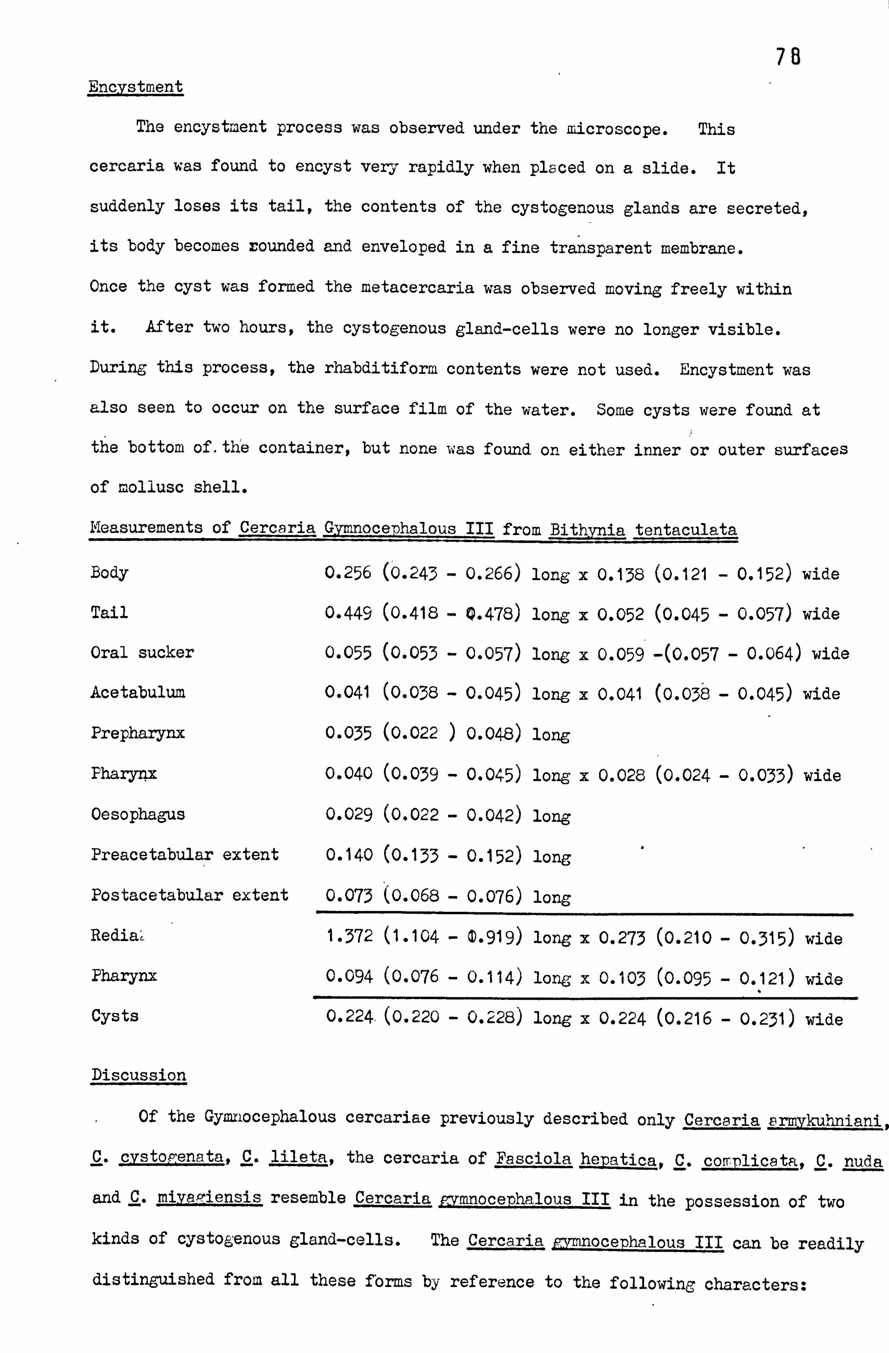
78 Encystment
The encystnent process was observed under the microscope. This
cercaria was found to encyst very rapidly when placed on a slide. It
suddenly loses its tail, the contents of the cystogenous glands are secreted,
its body becomes rounded and enveloped in a fine transparent membrane.
Once the cyst was formed the metacercaria was observed moving freely within
it. After two hours, the cystogenous gland-cells were no longer visible.
During this process, the rhabditiform contents were not used. Encystment was
also seen to occur on the surface film of the water. Some cysts were found at
the bottom of. the container, but none was found on either inner or outer surfaces
of mollusc shell.
Measurements of Cercaria Gymnocephalous III from Bithynia tentaculata
Body 0.256 (0.243 - 0.266) long x 0.138 (0.121 - 0.152) wide
Tail 0.449 (0.418 - 0.478) long x 0.052 (0.045 - 0.057) wide
Oral sucker 0.055 (0.053 - 0.057) long x 0.059 -(0.057 - 0.064) wide
Acetabulum 0.041 (0.038 - 0.045) long x 0.041 (0.038
- 0.045) wide
Prepharynx 0.035 (0.022 ) 0.048) long
Pharynx 0.040 (0.039 - 0.045) long x 0.028 (0.024 - 0.033) wide
Oesophagus 0.029 (0.022 - 0.042) long
Preacetabular extent 0.140 (0.133 - 0.152) long
Postacetabular extent 0.073 (0.068 - 0.076) long
Redia4 1.372 (1.104 - 0.919) long x 0.273 (0.210
- 0.315) wide
Pharynx 0.094 (0.076 - 0.114) long x 0.103 (0.095
- 0.121) wide
Cysts 0.224. (0.220 - 0.228) long x 0.224 (0.216
- 0.231) wide
Discussion
Of the Gymnocephalous cercariae previously described only Cercaria armykuhniani,
C. cystopenata, C. lileta, the cercaria of Fasciola hejatica, C. complicata, C. nuda
and C. miyagiensis resemble Cercaria cymnocerhalous III in the possession of two
kinds of cystogenous gland-cells. The Cercaria grmnocephalous III can be readily
distinguished from all these forms by reference to the following characters:

79 1. Cercaria armikuhniai and C. lileta differ from C. pymnocephalous III
by having two oesophageal diverticula.
2. In C. nuda, C. miyagiensis and C. complicata, the excretory bladder
is composed of two chambers and the acetabulum is smaller than the oral sucker
whereas in C. 'rmnocephalous III the excretory bladder is single chambered and
the acetabulum is larger than the oral sucker.
3. Cercaria cystogenata is characterized. by having a nonspinose body, no
wore than 25 refractile granules (average 18) in each of the primary excretory
ducts and the flame cell formula of 2(2 +, 2 +2±2+2+ 2) = 24, where
C. gvmnocenhalous III possesses a spinose body, very numerous (> 100) refractile
granules in the primary excretory duct and the flame cell formula is
2Z(2+2+2+2+2+2+2+2+ (2,7=36.
4. The cercaria of Fasciola hepatica has 10 flame cells on each side
of the body, the caudal excretory duct bifurcates at about one third of the
tail length and the secondary ducts form posterior excretory loops at the
posterior end of the intestinal caeca. Cercaria mrmn. ocephalous III has'36
flame cells, the caudal excretory duct forks at about two thirds of the length
of the tail and the secondary ducts do not form posterior excretory loops.
Nasir and Diaz (1968) and Nasir and Rodriguez (1969) described Cercaria
poracea in Poinacea glauca and the cercaria of Stephanoprora paradenticulata
in Marisa cornuarietis and P. lauca, both from Venezuela. These two species
are, with Cercaria m_mnoceDhalous III, the only gymnocephalous cercariae with
36 flame cells. The 3 species are probably closely related but they can be
easily distinguished on the basis of the presence of different types of
cystogenous gland-cells, number of refractile granules and details of the
digestive system. In C. nomacea the oesophagus and caeca are absent, whereas
in the cercaria of Stephanoprora denticulata they are present but do not
extend posteriorly to the equatorial level of the acetabulum.
Cercaria pymnocephalous III is clearly different from C. rmnocephalous I
and C. gymnocerhalous II in body size, in the nature of the cystogenous gland-
cells and the flame cell formula.

80 Pleurolophocercous cercariae
Sewell (1922) created the Pleurolophocercous group to accommodate the
monostomes Cercaria indicae VII, C. indicae VIII, C. indicae III, C. lophocerca
Fil, C. lophocerca Lebour (nec. Fil. ) and C. pleurolophocerca Sonsino, which
he named as the type species of this group. Sewell (1922) found that Cercaria
indicae XXXI and C. indicae L were strikingly similar in all anatomical features
to the members of his Pleurolophocercous group except that they possessed a
well-developed acetabulum. He created the Parapleurolophocercous group to
accommodate these two problematic species. Sewell placed the Pleurolophocercous
group within the large N: onostome group of Luhe (1909) and suggested that it was
related to the Gymnocephalous cercariae.
Langeron (192k) redescribed Cercaria pleurolorhocerä Sonsino and noted
the presence of a well-developed acetabulum. Dubois (1929) found Cercaria
lophocerca (Fil. ) and in it described a very small acetabulum. Dubois
considered that the presence of this organ in C. pleurolophocerca and C.
lonh ocerea has great importance; it is responsible for the separation of the
whole Pleurolophocercous group from the monostomes and it also illustrates the
phenomenon of acetabulum reduction referred to by Odhner (1911). Vogel (1934)
redefined the Pleurolophocercous group because he considered that, as
constituted by Sewell (1922), it was not based on valid criteria and Wesenberg-
Lund (1934) removed it from the Monostome cercariae. Porter (1938) found
Cercaria britsiae in South Africa; although this species is very closely related
to members of the Parapleurolophocercous group it possessed a well-developed
digestive system. Porter therefore suggested that the Parapleurolophocercous
group of Sewell should be revised to include those forms with intestinal caeca.
Cable (1938) improved Vogel's definition and pointed out that the Pleurolophocercoue
and Parapleurolophocercous groups (Sewell, 1922) are closely related, stating
"It is indeed possible that if a natural classification is to be offered,
certain members of each group should be combined". Rothschild (1938a) considered
that there were insufficient grounds for separating the Pleurolophocercous and
Parapleurolophocercous groups and combined them, giving the following diagnosis:

81 1. A pair of pigmented eyespots is generally present, but may be lacking
altogether.
2. The anterior end of the body, or the whole body, is armed with backwardly
directed spines.
3. The anterior sucker is modified to form a protusible penetration organ.
4. The acetabulum may be absent or only poorly developed.
5. A pharynx is always present, but the alimentary canal is absent or
only poorly developed.
6. Penetration glands, with ducts opening anteriorly, are present.
7. The bladder is reniform or roughly globular, with thick walls. The
lateral excretory ducts enter the bladder antero-laterally.
8. The tail is longer than the body, and powerful, and is provided with
cuticular fin-folds or a cuticular sheath.
9. The cercariae are phototropic. They swim by short dashes, followed
in each case by a pause, during which the body hangs below the tail in the water.
10. Development occurs in rediae with an intestine and birth pore but
no locomotory appendages. The cercariae leave the rediae at an early stage
and continue development within the tissues of the first intermediate host.
11. First intermediate host: fresh water, brackish water or marine
gastropods. Sedond intermediate host: generally a fish, but exceptionally
an amphibian. Encystment may occur in the first intermediate host.
Martin (1950) sub-divided the Pleurolophocercous group according to the
arrangement of the tail fin-folds into four groups. The type species of each
group was as follows: group A. Ruharlorchis californiensis and Parasitictodora
hancocki cercariae; group B. Cercaria sp Rothschild; group C. another
Cercaria sp Rothschild; and group D. Hanlorchis cercariae. Ulman (1954)
also sub-divided the group on the basis of the tail fin-folds calling it the
Velocercariae for those cercariae characterized by having the dorsal fin which
was longer than the ventral and may run on around the tip of the tail. Vercammen-
Grandjean (1960) considered the Pleurolophocercous and Parapleurolophocercous
groups to be synonymous and regarded them as intermediate between the

Echinostomidae and the Gymnocephala. Dawes (1946) placed these two groups 2
in the Gynnocephalous cercariae.
I consider the classification scheme proposed by Rothchild (1938) to be
the most natural and I have adopted it in the present account.
Only one member of the Pleurolophocercous group was found in the present
study.

83 Cercaria pleurolophocera I
During the summers of 1973,1974 and 1975 various collections were carried
out in areas where the gastropod Bithynia tentaculata occurs; but only at
Newmillerdam Lake and Kirkstall Power Station (Leeds-Liverpool Canal) were some
snails infected with pleurolophocercous cercariae. The rate of infection
varied between 2 and 5ä.
The cercariae are emitted throughout the day, with the highest
productivity in the morning and decreasing towards the late evening. Periods
of vigorous swimming alternate with short rest periods. After being emitted
they have a relatively long liftspan of between 24 and 35 hours.
Description (Plate 15; Fig. l'-3. and 6)
Body elongate, with on each side a row of 7 hair-like projections. Oral
sucker well-developed and Subterminal; provided on a protusible region with
five rows of spines apparently lying on dorsal wall; the number of spines in
each row from anterior is 15,11-12,11-12,7-8 and 7-8 respectively.
Tail subterminal, containing both longitudinal and circular muscle fibres.
Two well-developed fin-folds; one a lateral membrane originating at union
between body and tail, gradually widening to a maximum width near middle of
tail, then decreasing considerably towards distal end where it becomes difficult
to perceive; the second a dorsoventral membrane extending from middle of tail
to its posterior extremity.
Two eyespots containing dark brown pigments, one on each side of body,
between posterior margin of oral sucker and pharynx. Prepharynx followed by
spherical, muscular pharynx, No other digestive structures
present.
Twelve penetration gland-cells, arranged in 3 transverse rows (3,4 and 5
cells per row respectively) and situated between posterior end of pharynx and
anterior end of rudimentary acetabulum; each gland-cell with a coarsely granular
content and a central spherical nucleus; ducts extend anteriorly, passing between
eyespots and continuing towards region of oral sucker where they open to the

84 exterior in 4 groups of 3. Acetabulun not well-developed and appearing only
as an undifferentiated mass of cells in newly emerged cercariae; its orifice
becomes visible after 24 hours. No rudiments of genital organs present.
Cystogenous gland-cells with a coarsely granular content located in
posterior 3-fifths of the body except in region occupied by excretory bladder.
Excretory bladder of triangular form and with its walls composed of large
anucleate cells with granular cytoplasm. Principal primary ducts open
anteriorly from anterolateral margin of excretory bladder extending a little
behind level of pharynx, where they receive anterior and posterior collecting
ducts from the flame cells. Flame cell formula: 2 J2 + 2) + (2 +2+2+
2+2+2+2+ 2)J = 40. Caudal excretory duct extending to end of tail.
Redia (Plate 15 ; Fig. 4
The rediae extracted from infected snails were not very numerous and almost
all were at the same stage of development. T4e pharynx is very small in
proportion to the length of the body; at each side of its aperture there is
a pair of hair-like projections. The intestine is small and contains a brown
substance similar to material of the hepatopancreas of the snail. The birth
pore is situated to one side behind the pharynx, and a neck and locomotory
appendages are absent. The body of the redia is full of numerous germinal
masses and cercariae in different stages of development including 3 to 6 fully-
formed cercariae. Apparently most cercariae leave the rediae at a very early
stage, completing their development in the tissues of the snail. The excretory
system of the redia consists of numerous flame cells forming 2 rows, one on each
side of the body. The number and arrangement of these cells is difficult to
determine, but there appear to be at least 24 on each side. A single duct can
be seen opening outwards through a small eyfetory pore situated towards one side
in the anterior part of the body.
Infection of second intermediate host
To determine the possible secondary host of Cercaria bleurolothocerca I,
different species of freshwater fishes - 12 guppies, 8 fllapia and 5 chub were

85 isolated with infected snails in glass vessels containing tap water.
After 24 hours, a specimen of each fish species was examined for the
presence of metacercaria cysts. They were found only in the chub.
The remaining tilapia and guppies gave negative results on dissection,
while the chub, which were examined alive under a dissecting microscope, showed
cysts at the base of the tail, under the epidermis and scales. These fishwere
maintained in aquaria to permit the development of the metacercariae.
The cysts were extracted from the musculature of the base of the tail and
from the connective tissue around the bases of the fins. They displayed the
following characteristics: - oval form with a fine wall within which the
metacercariae is contained, showing rotating movements. The penetration ducts
and glands are still visible but their contents have dwindled, presumably having
been used in the process of penetration. The excretory bladder is similar
in size and shape to that of the cercaria but the rest of the system is difficult
to discern, although some flame cells are visible in the postacetabular region.
The eyespots are still visible but there is no evidence of the oesophagus and
intestinal caeca. The acstabulum shows no additional development over that
of the cercaria.
The remainder of the infected fishes survived for only a few days so further
details of the life cycle could not be investigated.
Measurements of Cercaria nleurolobhocerca I from Bithynia
tentaculata
Body 0.147 (0.140 - 0.152) long x 0.044 (0.041 - 0.049) wide
Tail 0.293 (0.273 = 0.307) long x 0.025 (0.022 - 0.026) wide
Oral sucker 0.026 (0.022 - 0.026)long x 0.022 (0.022 - 0.026) wide
Acetabulum 0.013 (0.011 - 0.015) long x 0.016 (0.015 - 0.019) wide
Prepharynx 0.016 (0.015 - 0.019) long
Pharynx 0.011 (0.011 - 0.011) long x 0.010 (0.007 - 0.011) wide
Oesophagus
Preacetabular extent 0.083 (0.079 - 0.087) long
Postacetabulur extent 0.048 (0.041 - 0.057) long

86 Redia 0.808 (0.560 - 1.442) long x 0.098 (0.084 - 0.112) wide
pharynx 0.021 (0.019 - 0.022) long x 0.022 (0.019 - 0.022) wide
Cyst 0.112 (0.106 - 0.121) long x 0.071 (0.069 - 0.079) wide
Oral sucker 0.023 (0.022 - 0.026) long x 0.024 (0.022 - 0.026) wide
Acetabulum 0.013 (0.011 - 0.015) long x 0.012 (0.011 - 0.015) wide
Prepharynx 0.012 (0.011 - 0.015 long
pharynx 0.011 (0.011 - 0.011) long x 0.012 (0.011 - 0.015) long
Preacetabular extent 0.056 (0.053 - 0.060) long
Discussion
All previously described cercariae of the group Pleurophocerca Sewell,
1922 (-= Parapl. eurolophocerca Sewell, 1922) are listed below.
Cercaria of Aponhallus brevis Ransom, 1920 (Miller, 1941,1946).
Cercaria of A. donicus (Skrjabin and Lindtrop, 1919) as described by
Niemi and Macy, 1974.
Cercaria of A. muehlingi (Jagerskiold, 1899) Luhe, 1909 (Odening, 1970).
Cercaria of A. venustus (Ransom, 1920) as described by Cameron, 1937.
Cercaria of Ascocotyle pachycvstis Schroeder and Leigh, 1965.
Cercaria of Clonorchis sinensis (Cobbold, 1875) Looss, 1907 (IKomiya and
Tajimi, 1940).
Cercaria of Euhaplorchis californiensis Martin, 1950.
Cercaria of Euryhelmis sauamula (Rudolphi, 1819) Poche, 1926 (Anderson
and Pratt, 1965).
Cercaria of Hatil orchis taichui (Nisnigori, 1924) Witenberg, 1930 (Martin, 1958)
Cercaria of H. vokoMawai (Katsuta, 1932 ) Chen, 1936 (Martin, 1958).
Cercaria of Heterophyid sp. Hopkins, 1940.
Cercaria of Metagonirus yokoaawai (Katsurada, 1913) Katsurada, 1912, (in
Gupta and Tane ja, i97Oa).
Cercaria of Metorchis coniunctus (Cobbold, 1860) as described by Cameron, 1944.
Cercaria of Opisthorchis felineus (Rivolta, 1884) Blanchard, 1895 syb"
0. tenuicollis (Rudolpri, 1819) of Ejsmont, 1937 in Faust and Russel, 1958).

87 Cercaria of Q. viverrini (Poirier, 1886) Stiles and Hassall, 1896,
(Wykoff, Harinasuta, Juttijudada, and Winn, 1965) (Ito;. Papasarathorn
and Tonkoom, 1962).
Cercaria of Phocitremoides ovale (Maxon and Pequegnat, 1949) Martin, 1958
(Cerca.. xia 'Pleurolophocercous II' Maxon and Pequegnat, 1949).
Cercaria of Stellantchasmus falcatus Onji and Nishio, 1942 (Martin 1958).
Cercaria Nr. 2 Petersen, 1931.
Cercaria atomica Fain, 1953.
Cercaria ato: nica kivuensis Vercammen-Grandjean, 1960.
Cercaria bhimtalensis Malaki and Singh, 1962.
Cercaria bithyniella, Fain 1953.
Cercaria bristiae Porter, 1938.
Cercaria chromophila Faust, 1922.
Cercaria constrictovesica Cable and Wheeler, 1939.
Cercaria flavidusi Preivati, 19(65.
Cercaria -; omtiensis Preavati, 1965.
Cercaria indicae VIII Sewell, 1922.
Cercaria indicae XXXI Sewell, 1922.
Cercaria indicae L Sewell, 1922.
Cercaria lorhocerca Filippi,. 1857 (Dubois, 1929, Khan, 196Cb r: eyer, 1964,
Probert, 1965, Wesenberg-Lund, 1934).
Cercaria melanoides Forter, 1938.
Cercaria onacocorpa Cable and Wheeler, 1939.
Cercaria nicobaricae Sewell, 1931.
Cercaria uarvomelaniae Tubangui, 1928.
Cercaria ninjorensis Gupta and Taneja, 1970'
Cercaria pleurolonhocerca Sonsino, 1892 (syn. C. indicae VII
Sewell, 1922 (Langeron, 1924);
Cercaria pleuroloparaDleuriforrnis Nasir and Diaz, 1973.
Cercaria nlotiopsis Johnston and Simpson, 1939.
Cercaria of Parasitictora hancocki (Maxon and Pequ qnat, 1949)

Be Martin, 1950 Cercaria 'Pleuroloahocercous I' Maxon and Pequegnat, 1949).
Cercaria of Procerovum calderoni (Africa and Garcia, 1935), Price, 1949
(Velasquez, 1973).
Cercaria guadripteviria Sinitsin, 1911.
Cercaria semicarinata Cable and Wheeler, 1939.
Cercaria sioidea Fain, 1953.
Cercaria srinostoma Cable, 1937.
Cercaria vogeli Cable, 1935 (Cable, 1938; Cable and Wheeler, 1939).
Of these 46 species only Clororchis sinensis, EurT helmis saua. mula, C.
atomica and C. pleurolonarapleuriformis are equipped with 12 penetration gland-
cells but these four species are easily distinguished by their flame cell formulae
from C. nleurolorhocerca I 2Z2 +2) +(2+2+2+2+2+2+2+2).? =40',
being 2 f(3 + 3) + (3 +3+ 3)7 = 30 in C. sinensis, 2 J2 +2+3+ 25 = 18
in E. squamula, 2 Z(-5 + 6) + (6 +6+ 6)7 = 58 in Co atomics. In C.
pleurolonaranleuriformis the total numbers of flame cells is 36, but their
connections with their respective capillaries have not been determined.
The cercaria of Parasitictora hancocki (Martin, 1950) was originally
described by Maxon and Peque, ýnat (1949) who referred to it as 'Pleurolophocercous I'
without giving it a specific name. Later its life cycle was clarified by
Martin (1950). In the original description of Maxon and Pequegnat there are
stated to be 12 penetration gland ducts, which divide into four groups of 3;
the glands are situated in the medial and postero-lateral region of the body
but their number was not determined. Martin (1950) did not agree with Maxon
and Pequelnats' observations and gave the number of penetration gland ducts
as 14 (groups of 3,4,4 and 3), corresponding to the 14 glands situated in
the medial region of the body, partially surrounding the rudimentary acetabulum.
In Cercaria nleulorolophocerca I described above there are 12 ducts of penetration
glands divided into 4 groups of 3 and connected to 12 glands arranged in 3 transverse
rows, comprising respectively 3,4 and 5 glands, between the pharynx and the
rudimentary acetabulum.

89 Dunagan (1960), in his key for the identification of cercariae of the
superfamily Opisthorchioidea, classified Ceraaria guadripterygia Sinitsin, 1911
amongst those with a lateral and dorso-ventral fin fold, a rudimentary
acebabulum and possibly 12 penetration gland-cells characteristics which coincide
with those of Cercaria pleurolophocerca I described by the writer. However,
C. Quadripteryiia parasitizes a marine mollusc Hvdrobia ventrosa)in the Black
Sea, and some doubt exists concerning the number of its penetration gland-cells,
the existence of a pharynx and the number and arrangement of its flame cells.
In C. rleurolorhocerca I the primary host is a fresh water mollusc, Bitlwnia
tentaculata, there is a definitive number of 12 penetration gland-cells, a
pharynx is present and the number and arrangement of flame cells is expressed by
the formula 2 f2+2) + (2+2+2+2+2+2+2+2)=40.
Llewelyn (1957), Khan (1960) and Probert (1965) recorded Cercaria lonnocerca,
Filippi, 1859 from Bithynia tentaculata, this being the only representative
of the group Pleurolophocerca, Sewell, 1922 (=, parapleurolophocerca, Scwe11 1922
in this country. C. lophocerca has already been distinguished from
C. pleurolophocerca I described above on the basis of the number of penetration
glandiccells.: J oreover, other characteristics of C. vleurolophocerca I, such
as the flame cell formula, extension of the lateral membrane along the tail
and presence of a short oesophagus make the distinction between these two
forms more convincing.
. Consequently this cercaria is recorded as new and is named Cercarie la hoce l'ca z.
17

90 Echinostome Cercariae
The members of this group of cercariae are easily identified by the
presence of a cephalic collar provided with spines around its margin. Luhe
(1909) placed this group in the Leptocercous cercariae and divided it
according to the length of the gut in the redia and presence or absence of
a fin-fold on the tail. He also described Cercaria echinatoides, C. coronata,
C. echinata and C. snin ifera. Cort (1915) suggested that Echinostome
cercariae cannot be sub-divided into smaller natural groups. Sewell (1922)
in view of the marked differences in the excretory system and the presence
or absence of a fin-fold on the tail shown by these cercariae, divided Echinostome
cercariae into four sub-groups three of which are named after Luhe's species.
The sub-groups are:
(1) Sub-group Coronata: without a caudal fin-fold.
In the members of this group the main collecting
ducts arise from the bladder, pass forwards to the
level of pharynx (the portion in front of the
acetabulum is wide and'contains numerous refractile
granules) where they form loops and continue back
as far as the level of acetabulum where they divide
into anterior and posterior collecting tubes. The
caudal excretory canal passes backwards through the
greater part of the length of the tail and opens
on the surface about at its proximal end.
(2) Sub-group Echinatoides: with a caudal fin-fold, and an excretory system
similar to that of the Coronata.
(3) Sub-group Echinata: without a caudal fin-fold.
The main collecting ducts arise from a common
secondary bladder and each passes forwards to
the side of the pharynx. The portion of the duct
in front of the acetabulum is wide and contains
numerous refractile granules. At the side of the

91 pharynx the tubes form loops and continue back
to the posterior end of the body. At the side of
the bladder the main duct apparently divides
into anterior and posterior collecting tubes.
The caudal excretory canal opens via short
later al branches near the base of the tail.
(4) Sub-group Megalura: Without a caudal fin-fold.
A glandular adhesive-organ is located at the distal
end of the tail. The main excretory ducts arise from
the small excretory bladder and runs anteriorly to
the level of the pharynx. Here they turn back as
far as the acetabulum where they divide into anterior
and posterior collecting ducts. The in
excretory ducts do not contain refractile granules.
No caudal excretory canal is present.,
Sewell (1922) separated Cercaria agi. lis Filippi (1859) and Cercaria reflexae
Cort (1915) from the Echinostome group and created two new groups, Agilis and
Reflexae, to include these echinostome-like cercariae. The two groups were
separable on the basis of the presence of a caudal fin-fold in the Reflexae
and its absence in Agilis.
Faust (1924a) introduced ten groups distinguished by the nature of their
excretory system, and placed the Agilis and Reflexae groups of Sewell (1922)
in the Echinostone cercariae. Miller (1936) classified the Echinostome
cercariae on the basis of presence or absence of collar spines and caudal fin-
fold, but did not include any of the members of Megalura group of Sewell (1922)
as he did not consider that there was sufficient evidence to relate them
to the Echinostome cercariae. Miller also added Cercaria a; rilis and C. reflexae
to this group, because they had already been shown to be true echinostome types.
Dubois (1929), Wesenberg-Lund (1934) and Tiller (1936) all suggested that
the Echinostome cercariae cannot be reliably subdivided into smaller groups.
Porter (1938) found certain cercariae which were closely related to the

92
Echinatoides, Coronata and Echinata groups (Sewell, 1922) and on the basis of
morphological differences she created three new sub-groups named Sub-echinatoides,
Echinocrenata and Cucumeriformes groups. Khan (196C1 stated "Echinata,
Echinatoides and Coronata groups of Sewell (1922) seem to be unnatural and
should remain a single group until more suitable characters for their
sub-division are discovered".
Jain and Yadav (1968) on the basis of the arrangement of the caudal excretory
duct attempted to divide the Echinostome cercariae into three groups defined
as follows:
1. Echinosolenata group
The caudal excretory duct is single, median and unbranched through-
out its entire course.
2. Echinobisolenata
The caudal excretory duct forks into two branches.
3. Echinopolysolenata
The caudal excretory duct forks into three branches, two
lateral and one median which runs backwards in the tail.
Of these three new groups only the Echinobisolenata appears to be
represented in Britain. However such a division is obviously not practicable
for those cercariae (for example some Magnacauda) in which the caudal excretory
duct is not present.
The use of the flame cell formulae, type of excretory system, presence or
absence of a caudal fin-fold and number of collar spines as diagnostic characters
in Echinostome cercariae classification has been proved to be of little value by
several workers. For example, the cercariae of Echinostoma nudicaudatum
Nasir (1960) is characterized by the absence of a caudal fin-fold and the
presence of an excretory system of the Echinata type, while the cercaria of
Echinostoma pinnicaudatum Nasir (1961) possesses a caudal fin-fold and the
excretory system is the Echinata type. However, despite these structural
differences in the cercariae, the adults are both referred to the-same genus.
A second example concerns the genus Hypoderaeum. In the cercaria of

93 Hypoderaeum essexensis Khan (19601 the fin-fold is lacking and the excretory system
is of the Coronata type whereas the cercaria of Hyroderaeum conoideum (present
study) lacks a caudal fin-fold and possesses an excretory system of the
Echinata type. Again the adult forms belong to the same genus despite large
differences in cercarial structure.
The number of flame cells in Echinostome cercariae is difficult to
determine as the body is opaque owing to the presence of numerous cystogenous
gland-cells, penetration gland-cells, the nature of the digestive system and
the presence of refractile granules in the main excretory ducts. Mathias (1925)
described the cercaria of Rypoderaeum conoideurn with 50-54 spines, but did not
give the number of flame cells; Dubois (1929) gave 49 (47-53) spines and 48
" flame cells; Rees (1932) 43,44 and 45 spines in the metacercariae, but no
flame cells; Wesenberg-Lund (1934) 50 spines and 13 flame cells; Wikgren (1956)
25-26 spines on the ventral side and 7 spines on each 'lateral row and 16 flame
cells in all; Meyer (1964) 49 spines and 48 flame cells; in the present
study 47-52 spines and at least 40 flame cells.
The variation in number of collar spines and flame cells in Echinostome
cercariae makes their identification difficult and the taxonomic significance
of such characters can be only determined after the life cycles of the different
species have been elucidated and the correspondence of cercaria and adult
established.
The Magnacauda sub-group was erected by Byrd and Reider (1940) to accommodate
a large-tailed echinostome cercaria. In these forms a small number of collar
spines may be present or absent and if absent in the cercaria the spines are
developed after encystment. These cercariae have experimentally been shown
to develop into adults which belong to the genera Petasiger Dietz, 1909 and
Stephanoprora Odhner, 1902. The excretory system of these cercariae exhibits
a greater affinity to the Echinata type, but in the species hitherto described
the caudal excretory duct is either absent or non-functional (lacks an excretory
pore). They develop in rediae with divided or undivided collars.
I agree with Byrd and Reider (1940), Llewellyn (1957) and Khan (19601

94 that in view of the marked differences from the other members of the
Echinostome cercariae, the tßagnacauda sub-group should be treated separately.
During the course of this investigation, 3 cercariae of'the Echinata
sub-group and 2 of the Magnacauda have been found.

95 Cercaria of Hvpoderaeun conoideum (Block, 1782) Dietz, 1909
This cercaria was obtained in autumn 1972 from Lvrnaea stagnalis collected
at Gledhow Valley Road ind in winter 1972 from both L. pereger and L. stagnalis
collected at Kirkstall Power Station (Leeds-Liverpool Canal). The rate of
infection in L. pereger was 12.5% and in L. Stagnalis between 4.1 and 8.3%
depending on the locality.
The cercariae are discharged intermittently throughout the day. They swim
actively by vigorous movements of the tail and during swimming the body is bent
ventrally and contracted. They typically swim near the bottom of the vessel
and when swimming ceases they sink gradually. Touching any surface initiates
rapid creeping movements executed by the alternate use of oral sucker and
acetabulum. The cercariae survive for 24 - 32 hours.
Description (Plate i6-, Figs. i-4, and 6)"
Body elongate, pyriform in outline and capable of considerable deformation.
,ý
Body surface armed with small spines arranged in transverse rows. Cuticle
thick, bearing spines on anterior and lateral margins as far as level of
acetabulum and on posterior margin of body. Approximately 10-12 pairs of
long hair-like projections present on each side of body. Collar well-developed,
bearing 47-52 inconspicuous spines including 4 corner spines (2 oral and 2 aboral).
Remaining spines arranged in a single row. Tail larger than body inserted
subterminally. Both longitudinal and circular muscles extending length of tail,
similar to those described in C. echinostoma III. Core of tail containing
small elongate cellular bodies with fine granular contents; these bodies more
abundant proximally cuticle of tail provided with numerous hair-like projections
laterally; finfold and spines absent.
Oral sucker circular, slightly smaller than acetabulum, and situated
terminally or subterminally. Digestive system comprising mouth, prepharynx,
an elongate okal pharynx and a large slender oesophagus. Oesophagus divided
just anterior to acetabulum into intestinal coaca extending to posterior end
of body. Lumen of oesophagus and intestinal caeca containing granular material.
Acetabulum highly protrusible, situated posterior to middle of body; its

96 surface provided with one row of spines.
Anterior margin of body with apertures of 6 narrow ducts which extend
to posterior margin, of oral sucker before becoming too difficult to trace.
Ducts apparently connected to 6 penetration gland-cells located lateral
to Oesophagus; each gland-cell with coarse granular cytoplasm and without
a nucleus.
Cystogenous gland-cells with coarse granular contents packing body from
pharynx to posterior end. Genital primordium represented by two clusters
of undifferentiated cells, one anterior and one posterior to acetabulum.
Excretory bladder consisting of two short, wide chambers, a large basal
chamber opening to exterior via a dorsal pore and a small anterior chamber;
chambers connected by a narrow canal. Large primary excretory ducts-arising
from anterior chamber; containing numerous refractile granules. Ducts
initially narrow but widening at level of acetabulum, continuing as far as
pharynx and forming anterior loops lateral to posterior margin of oral sucker
before passing posteriorly as secondary ducts. Secondary ducts following a
slightly sinuous course almost to posterior end of body, forming posterior loops
at this level, then passing anteriorly to posterior level of acetabulum where
bifurcation into anterior and posterior collecting ducts located. Secondary
ducts each containing 11-12 ciliary patches along their lengths. At least 20
flame cells present each side of body but capillaries very difficult to observe.
Caudal excretory duct arising from base of posterior chamber and giving off two
lateral branches in proximal part of tail. Flame cell formula not known.
Redia (Plate 16 Fig. 5
This cercaria developed in rediae in the hepatopancreas of the snail
hosts. The redia is elongate and very active. The body cuticle is thick
and filled with fine granular material; its whole surface containing scattered
yellowish brown pigment. The collar is conspicuous just behind the pharynx and
the birth pore is found immediately posterior to it.
The pharynx is well developed, muscular and protrusible. It leads into
a short sac-like intestine which extends for about a third of the body length

97
and is filled with dark brown material. There are two prominent locomotory
appendages located near the posterior end. The body cavity contains 7-8
cercariae at different stages of development of which 2 or 3 are fully
developed at any one time. Germ balls are also abundant and vary in size.
This cercaria'was found experimentally to develop into adults of
Hypoderaeura conoideum. Details of subsequent life cycle stages are given
elsewhere (page 100 )"
Measurements of the cercaria of Hyroderaeum conoideum from Lynnaea Pere--er
and L. staP°nalis
Body 0.342 (0.304 - 0.399) long x 0.112 (0.106 - 0.136) wide
Tail 0.414 (0.410 - 0.418) long x 0.039 (0.038 - 0.041) wide
Oral sucker 0.041 (0.041 - 0.041) long x 0.041 (0.041 - 0.041) wide
Acetabulum 0.053 (0.049 - 0.057) long x 0.053 (0.049 - 0.057) wide
Prepharynx 0.019 (0.007 - 0.026) long
Pharynx 0.025 (0.022 - 0.030) long x 0.019 (0.019 - 0.019) wide
Oesophagus 0.091 (0.053 - 0.063) long
Preacetabul; ar extent 0.182 (0.133 - 0.220) long
Postacetabular extent 0.076 (0.068 - 0.087) long
Redia 0.917 (0.545 - 1.400) long x 0.231 (0.196 - 0.280) wide
Pharynx 0.074 (0.053 - 0.102) long x 0.078 (0.064 - 0.106) wide
Location of metacercariae in the second intermediate host
Freshwater snails of the species Lymnaea stap-nalis, L. nereger, Planorbis
corneus, PhJsa fontinalis and PotomonyrEus ijenk_insi were collected from the
Leeds-Liverpool Canal (Kirkstall Power Station) and were isolated in glass
dishes containing tapwater. After 24 hours it was observed that some specimens
of L. Derep, er were shedding large numbers of cercariae which were subsequently
identified as the cercariae of Hypoderaeum conoideum (see page 95 ).
Whilst examining specimens of L. Pere°er for the occurrence of precercüriül
stages (in this case rediae) of H. conoidetun in the digestive gland, I noted
the prtisence of many cysts in the digestive gland, kidney, mantle wall and

98- some possible loose in the mantle cavity. These cysts possessed a collar
with 47 to 52 spines, main excretory tubules with refractile granules, a long
oesophagus and cystogenous gland-cells with coarsely granular contents. These
characters are similar to those of the cercariae of Hrpod_eraeum conoideum,
indicating that the cercariae penetrate snails and became encysted metacercariae.
Similar cysts-were also found in P. corneus, P. fontinalis, Potomonyrrus enkinsi,
L. stamelis and B. tentaculata.
Laboratory reared specimens of P. corneus, P. fontinalis, L. rerecer and
L. sta*nalis were exposed to H. conoideum cercariae. The cercariae swim actively
after being released by the primary intermediate host and do not appear to
be attracted to either L. nererer or other snail species. Upon making contact,
apparently accidentally, with their snail host or other snails of different
species they commence crawling about with the aid of their suckers. Some were
observed passing between the inner surface of the shell and the host tissue
where they disappeared. Several minutes latter they had apparently lost
their tails since some detached tails were seen at the bottom of the container.
Six hours later the majority of the cercariae had disappeared with only a few
still freely swimming. Twenty four hours after penetration the snails were,
carefully examined to determine the location of the cysts. All the snails
were infected and the cysts were found in the same organs as in the wild snails.
These cysts showed the same characters as those found in the naturally infected
snails. The precise site of entry into the second intermediate host has
not been determined, but it is believed that the cercariae penetrate anywhere
on the body of the snails and then migrate to encyst in the preferred site.
The number of cysts in each snail varied from 12 to 187, being more numerous in
L. perep; er and P. fontinalis than in P. corneus and P. ienkinsi.
on one occasion three cysts were found at the bottom of a container containing
6 L. pereger which had been brought from Kirkstall and isolated for experimental
infection by cercariae. It was believed initially that these cercariae had
encysted in the open but daily examination for five days failed to detect the
presence of any free-swimming cercariae. Five of the snails were killed and

99 examined but no precercarial stages were found although 2 snails were found to
be harbouring cysts of this species. Approximately 150 recently-emitted
cercariae were pipetted in about 100 cc water into three small glass dishes.
They were examined many times during the next 24 hour period but no cysts were
seen. It is probable that some of the cercariae were not able to penetrate
the tissues of the snails and encysted in the mantle epithelium, under the shell.
Later they may have been dislodged by the scraping action of the shell against
the mantle as the body withdrew into the shell. This is probably how some
free cysts observed on one occasion originated. The cercaria of H. conoideum
were never found to encyst in the open; they normally left their primary
intermediate host and re-entered either the same snail individual or another
specimen of the same or a different species.
Mietacercariae (Plate 17, Fig. 7)
The cysts are nearly spherical. The cyst wall is smooth,. transparent and
comprising two layers - an inner layer which is tough, difficult to remove
without injuring the encysted metacercaria and both narrower and more opaque
than the outer layer which is almost hyaline and easily reptured.
In cysts aged 24 h the encysted metacercariae lie bent back upon themselves
and are usually in constant motion. The metacercariae are morphologically
similar to the cercariae and the main differences noted were a slight increase
in size of the main excretory ducts and the fact that the contents of the
cystogenous gland-cells where less dense.
In a 15 day-old cyst the metacercaria is almost inactive and markedly
more opaque. Its length has increased and the anterior and posterior ends
of the body which is still folded double almost touch each other. . The
body spines and collar spines are more conspicuous, the former being more
prominent anteriorly but the cystogenous and penetration gland-cells are no
longer visible. The caeca are slightly longer, the excretory granules less
numerous and there has been slight growth of both the sucker and pharynx.

100 Measurements of 15 day-old metacercariae of HYpoderaeum conoideum experimentally
obtained from laboratory reared snails.
Cyst 0.163 (0.152 - 0.174) long x 0.153 (0.149 - 0.171) wide
Outer wall 0.024 (0.019 - 0.030) thick
Inner wall 0.009 (0.007 - 0.013) thick
Oral sucker 0.047 (0.045 - 0.049) long x 0.047 (0.045 - 0.049) wide
Pharynx 0.033 (0.030 - 0.038) long x 0.029 (0.026 - 0.034) wide
Acetabulum 0.066 (0.064 - 0.068) long x 0.066 (0.064 - 0.072) wide
Infection of the final host
The metacercariae used in the feeding experiments were 2,5,15,66 and
120 days old and they were obtained from Lymnaea Lereger, L. stagnalis and
Physä fontinalis which werd experimentally infected under laboratory conditions.
Nice, rats and pigeon were reared in the animal house of the Zoology Department.
Faecal samples were examined daily as described above (see p. 10 ). The
number of metacercariae fed to each host was 80.
All the attempts to infect mice and rats were unsuccessful (see Table 5 ).
None of the birds that had been fed with 2 to 5 day-old metacercariae
produced any worms. However trematode eggs similar to those of H. conoideum
appeared in the faeces of chicken 3, pigeon 3 and duckling 3 on the 12th day
following the ingestion of 15 day old metacercariae. They were killed and
examined on the same day and both immature and mature worms were recovered from
the duodenum. In another series of experiments (chicken 4, pigeon 4 and duckling
4) all rematodes harboured were mature on the 15th after infection and were
located in the first third of the small intestine.
The administration of cysts aged 66 and 120 days showed that the capacity
to develop in the final was retained to some extend in cysts of these ages.
In pigeon and duckling a few trematodes persisted for 28 days.
These feeding experiments demonstrated that mice and rats are probably
unsuitable definitive hosts for this species. The number of worms experimentally
obtained from chicken, pigeons and ducklings was low compared to the number of

101 metacercariae administered. Metacercariae between 2 and 5 days old did not
appear to have become infective but had done so by the 12th. Metacercariae
viability is retained in some individuals for at least 120 days.
Rees (1932) has adequately described the adult Hypoderaeum conoideum.
VIy observations agree largely with those of Rees; the only point of difference
is that I have found a large receptaculun semiriis uterinum as an enlargement
of the ootype and did see in any specimen (82 examined) a receptaculun seminis.
Adult as described by Rees.
Measurements of poderaeum conoideum from experimental
infections of laborato ry birds
Body 6.520 (5.049 - 7.811) long x 1.194 (0.841 - 1.315) wide
Oral sucker 0.177 (0.168 - 0.210) long x 0.201 (0.168 - 0.280) wide
Prepharynx 0.015 (0.007 - 0.030) long
Pharynx 0.162 (0.112 - 0.189) long x 0.139 (0.112, - 0.168) wide
Acetabulum 0.697 (0.404 - 0.812) long x 0.736 (0.532 - 0.868) wide
Anterior testis 0.714 (0.490 - 0.882) long x 0.378 (0.280 - 0.462) wide
Poaterior testis 0.751 (0.560 - 0.924) long x 0.350 (0.280 - 0.490) wide
Ovary 0.246 (0.140 - 0.294) long x 0.293 (0.182 - 0.364) wide
Cirrus Sac 0.657 (0.476 - 0.938) long x 0.203 (0.140 - 0.294) 'Wide
Preacetabular.
extent 0.709 (0.553 - 1.053) long
Postacetabular extent 5.566 (3.997 - 6.316) long
Eggs 0.090 (0.087 - 0.098) long x 0.060 (0.049 - 0.068) wide
Collar 0.388 (0.350 - 0.420) long
Lateral spines 0.022 (0.020 - 0.026) long x 0.007 (0.007
- 0.008) wide
Dorsal spines 0.013 (0.013 - 0.015) long x 0.007 (0. C07 - 0.007) wide
Corner spines 0.026 (0.024 - 0.028) long x 0.010 (0.009
- 0.011) wide
Egg and Miracidiunn (Plate 18, Figs. 12-13)
The mature worms were kept in small containers in 0.75% normal saline.
The number of eggs passed was very small and most of the eggs used in these

102 experiments were obtained from the faeces of experimentally infected hosts.
The eggs are yellowish, oval in outline, thin-walled, operculated and
not embryonated. The newly laid eggs comprise a rounded ovum with-rather
central nucleus and clear undivided cytoplasm. The ovum is surrounded by
numerous vitelline cells.
The eggs were incubated in distilled water and tapwater at room temperature
(19 to 220C). Development was observed periodically, and after 16 to 18
days the miracidia are fully developed. A few miracidia were able to hatch
from the eggs as several empty shells were found at the bottom of the container
but no free swimming miracidia were seen. Some of them died without hatching.
On three occasions hatching was induced by applying slight pressure thereby
forcing the operculum open but in the majority of cases the miracidia were
crushed by this process. The miracidia only escaped from the egg with great
difficulty because of the small size of the operculum. The water, the miracidia
became more active and the cilia commenced very rapid beating. The free
miracidia usually exhibited rotating movements along their longitudinal axis
or swam in small circles. After 2 to 5 minutes they become rather inactive,
shed their cilia and become rounded. This process was observed twice and
the miracidia remained alive for 10 to 15 minutes before starting to
disintegrate. The flame cells of the disintegrating miracidia continued beating
for between 2 and 5 minutes. Natural, un-induced hatching was not observed.
No attempts were made to infect snails because no free swimming miracidia were
available.
The fully developed miracidium is slightly longer than the egg and usually
has its posterior end curved to one side. The miracidium occupied more than
half of the space within the egg, the remainder of the space generally being
filled by two or three large vacuoles and a small amount of granular material,
probably derived from the vitelline glands.
Its body is elongate or oval and somewhat pointed anteriorly. Its anterior
end has a protrusible apical papilla. The surface of the miracidium is
ciliated except the extreme anterior end which is naked. There is an

103 apical gland which is flask-shaped in outline, extending some distance
anterior of the eyespots and filled with rather coarsely granular contents.
Its duct opens by a-very small aperture located at the anterior tip of the
apical papilla. There are two L-shaped eyespots formed of dark brown
pigment granules. Just posterior to the eyespots, there is a rounded mass
of nucleated cells which represents the nervous system.
The protonephridial system comprises two large flame cells which are
rather variable in position but are usually situated behind midline of the
body. Their excretory ducts follow a. sinuous course forming one or two
loops and then each opens separately via a small lateral excretory pore situated
near the posterior end of the body. No penetration gland cells were observed.
The germ cells are situated at the posterior end of the body; - they are
nucleated and contain a fine granular cytoplasm.
Discussion
This is a widely distributed species of trematode. It was first described
by Bloch (1782) under the nahe of Cucullamus conoides. Dietz (1910) established
the genus Hypoderaeum and included the trematode as Hyroderaeum conoideum.
The cercaria of H. conoideum was first described by Mathias (1925) from
Lymnaea sta, Analis and Radix ovata in France. Dubois (1929) reported it from
Lymnaea ralustris and L. stapnalis in Switzerland and provided further details
of the protonephrideal system, which had been inadequately described by IV. athias.
Wesenberg-Lund (1934) found the same cercaria in R. ovata from Denmark.
It has subsequently been recorded from Finland (wikgren, 1956), Switzerland
(Meyer, 1964), Western Kazakhstan USSR (Smirnova and Ibrasheva, 1967) and (1 757-195E3,
Britain (Nasir, ýdilliams, 1966).
A great deal of intraspecific variation is apparent if the published
descriptions of this cercaria from different localities are compared. A
comparison of certain characters, including numbers of collar spines and
flame cells, is presented in Table 6. Wesenberg-Lund (19: 4) described
six small apertures located on the anterior margin of the mouth and regarded

104 them as the penetration gland openings but the actual glands and their ducts
were not observed. In the present specimens these 6 apertures were observed
to lead into 6 narrow ducts which extended as far as the posterior margin of
the oral sucker. The penetration gland-cells were also visible lying lateral
to the oesophagus.
Observations on the life cycle of this fluke were first presented by
Railliet (1893) who found the larval stages in several species of Lymnaea,
Planorbis and Paludina. Vevers (1923) fed ducklings with both the liver of
L. nereger infected with Tetracotyle stages which he believed to belong to
Tetracotyle tYnica Diesing, 1853 and the liver of L. sts nalis naturally
infected with echinostome cercariae which he identified as C. echinata
(Siebold, 1835) the larval stage of Echinostoma revolutum (Fro-lieh, 1802).
Twenty days postinfection adult forms of H. conoideum and immature forms
of E. revolutum were recovered from the small intestine and cloaca respectively.
No other trematodes were obtained. He stated that "the specimens of H. conoideum
recovered from ducks must have developed from some other cercariae also
encysted in Lymnaea nererer, but not detected at the time of the examination
of the snails". Vevers gave a brief description of the immature forms of
E. revolutum but not of H. conoideum. The life cycle was first completed by
Mathias (1925) who obtained the adult parasites by feeding ducks with
experimentally encysted metacercariae collected from L. limosa and P. corneus.
Mathias also infected species of Lymnaea with miracidia reared experimentally
from eggs.
Rees (1932) found encysted echinostome metacercariae in the mantle cavity
and digestive gland of L. rereper in South Wales. The cysts were characterised
by having 43-45 collar spines and she, therefore, considered that they belonged
to cercaria "Z" which had previously been described from the same locality
(Rees, 19322) and which also carried with 43-45 collar spines. Duckling were
fed with the cysts from L. ereG"er and adults forms of H. conoideum were obtained
from the small intestine. Rees concluded that the cysts used in the feeding
experiments were those of the cercaria of H. conoideu_a and not those of

105 cercaria "Z". No free swimming cercariae of H. conoideum were found. Rees
gave a full account of the morphological characters of this species.
Subsequently the life cycle of H. conoideum has been completed by several Ym aravn and
authors e. g. Hsu and Chow (1938) in China anddteter (1973) in India,
Williams (1966) reported an incidence rate of 3.621/1, o for the cercaria of
H. conoideum in Lymnaea rere7er in a pond near Glasgow. These cercariae
encysted in young L. _ere er and when cysts were fed to day-old ducklings,
adult parasites were recovered about 18 days later. Williams neither described
nor figured any life cycle stages, giving only the diameter of the cysts.
The variation in number of collar spines in the cercarial and metacercarial
stages is reflected by similar variation between adults of H. conoideum. In
most specimens there were 47 or 48 spines but a few had 52 spines. In Rees'
specimens the number varied from 43-45 as compared to the range of 47 to 53
given by Mathias (1925) for H. conoideum. Beverley-Burton (1961) found
H. conoideum in several species of ducks near Ipswich, Suffolk. Her specimens
had 47-49 spines and she suggested that the known range for H. conoideum
should be extended from 45-53 (as stated by Skrjabin, 1956) to 43-53. She
compared her specimens of H. conoideum with those of H. ¬nidini Bashkirova,
1941 from Anas sp. and with those of H. skriabini Oschmarin, 1947 from Aythya
ferina and concluded that "An examination of the specimens of these two forms
would possibly result in their being synonymised with H. conoideum".
Yamaguti (1971) listed 13 species belonging to the genus Hvpoderaeum, of
which H. essexensis Khan (1962 (obtained experimentally in duckling, pigeon
and chicken from Great Britain) and H. dir eri Lie (1964) (obtained experimentally
from duck and goslings from Malaysia) are characterized by having 49 and 49-56
collar spines respectively. The precercarial, cercarial and adult stages are
very similar to those found in the present study. The number of collar spines
(which falls within the range of spines for H., conoideum) could be interpreted
as instraspecific variation and it would be possible to mistakenly regard them
as conspecific. Fortunately, life cycle data are available for these two

106 species, and show that all three are separate species. It is apparent that
the lack of life cycle data could lead to the erroneous designation of
synonyms.
The adult of Hypoderaeum conoideums obtained experimentally from chicken,
pigeon and duckling agrees in all essential features. to the description given
by Rees (1932) except that she reported the presence of a large elongated
"receptaculum seminis" often filled with spermatozoa, which arises from the
oviduct and opens into the ootype. However, in none of my specimens was
this structure present. In my specimens the ootype enlarges to form a wide
sperm-filled "receptaculum seminis uterinum", which is a part of the uterus,
but a true receptaculum seminis is absent. Rees must have interpreted this
enlargement of the cotype as a true receptaculum seminis. The ovicapt is
also present in my specimens and may have been overlooked by Rees.
aimod. eraeum conoideum (Bloch, 1782) has also been recorded in Britain
by Owen (1951) in the domestic ducks, Cairina moschata and AnasT)latyrnyncha dom.,
and by Soliman (1955) in the Khaki Campbell duck.

107
Table 5 r°,
Experimental infection of mammals and birds with metacercariae ýf Hrt oderaeum conoideum (Eloch, 1782 )
Experimental Age of Eggs Host Number of Degree of development
Location in host definitive cysts detected autopsied trematodes of tremat s' host (Days) in faeces after - recovered Irmature' Mature
(+ or -) (Days) (Days)
Mouse 1 2
Mouse 2 5
Mouse 3 15
Mouse 4 66
Mouse 66 Rat l 2 Rat 2 5
Rat 3 15
Rat 4 66 Rat 5 66 Chicken 1 2
Chicken 2 5
Chicken 3 15 Chicken 4 15 Chicken 5 66 Chicken 6 120 Chicken 7 120 Pigeon 1 2 Pigeon 2 5
Pigeon 3' 15 Pigeon 4+ 15 Pigeon 5 66
Pigeon 6 120
Pigeon 7 120
Duckling 1 2 Duckling 2 5
Duckling 3 15
Duckling 15 Duckling 5
Duckling 6
Duckling 7
12+
15ý
15+ 28-
12+
15+
15+ 28+
12+
i5+ 15+ 15+ 28+
12
12
15 15 15 12 12
15 15 15 12 12 12
15 15 15 28 12 12 12
15 15 15 28 12 12 12
15
0 0 0 0 0 0 0 0 0 0 0
20
13 0 5 0 0 0
16 10
0 7 1 0 0
21
14
0 0 ,
0 0
0 0.
p' 0 0
0. 0
0 0
p. :0 ý:
p 0 5 '` 15
13
o 5
p 0 p
£ ö 0 =. 4 12
10
1 0 0
} 14 1 14
X10
gi p' 9
_, ý
i, H. , j"
-
g ¢:.,
Duodenum Jejunum
Jejunum
Duodenum and jejunum Jejunum
Duodenum Jejunum
Duodenum Jejunum Duodenum and jejunum Duodenum and jejunum Jejunum

9 a) Id ., l 0 0 0 U
4) Co d)
w 0
co 9-4
co, U
N U.
a) 10'
4-1 0
U)O
.rj
U UI
rý a)
c Hb
O
U) "H r-1 rR a a, 4
a)
a) rcl
93 0
co e 0 U
co Pl 0 0
äS ö
P4 +) s4 0 (1) O"H 9., -P . P a ý aa. a i+
m
40 ä,
ý ärd Pi Co 40Ea C)
rßl OO -N Cl) rl+3 0o C) "ri cd Q) :: S Fa 0 Paco 'd a
P, 0 0 H
0
+p aý m cn
ä ca ä ä
4)
G) U) N
P4
aý r 0 Id 'd N "rl "r-(
W -P 'd N CO r-4 4.3 co Ell (D 0
EO (1) Id co co r-i r-I 10 (D
" r-1 U +) a) 4-3 co 0
Z U co a) 0 N N
U-'
i H rý
cd a OD
cu 0) 9i 'd 'd 0 " I " U "H rd U ) U)
" 4-1 U) ca Cl) I . 4 I CO
El- 0 U , CU co -p r-I 0 U 4-1 ä U 0
Ea ^ s~ 3 r1 0
co + 0 N H I Cd o + n 1-4 U P<1 T- N (1)
t! 1 + ý. -p 4-4 Ea 01, ca U N ý H Z CI tc1 ý - " rj I I . I N U P, C` 0 (31, Oll it 0 Öý K)
z U) - L, - Lf. N', r t Iý1 rn T" ... co
m Ö i O
%-0 v C\j N w m C\j rn U r 0 kO I C\J
ý N
1 cu ON 40
(IN 4-3 ti r1
rj N cd U) , 5., " 0 4) bO to Ri 0 r1 N 4 p U) . N 4) 11 Ul ; ý4 co (D (D a) co P-1 !Z 0 o Z
108

109 Cercaria of EchinopPryphium recurvatum (Linstow, 1873) Dietz, 1909
The cercaria of Echinoparyphium recurvatum has only been found twice,
present survey, parasitising Lymnaea pereper. During spring and summer 1975,
between 6.2 and 26 of L. pere? 'er collected from Kirkstall Power Station
(Leeds-Liverpool Canal) and Riffa Beck Pool-in-Wharfedale respectively,
harboured this cercaria.
The cercariae emerge throughout the day in a moderate number, but
the greatest number are shed between 9 and 12 in the morning. They swim
actively and may be seen swimming most commonly in the lower part of the
container. During the swimming periods and the creeping movements the
cercariae behave like those of Hyroderaeum conoideum. They remain alive
for about 24 hours.
Description (Plate 20, Figs. 1-5)
Body oval to elongate. Entire body surface provided with prominent,
closely-set, backwardly-directed spines arranged in transverse rows and
decreasing in size posteriorly. Cuticle thick, furnished with numerous short
hair-like projections on papillae arranged fairly regularly around body but
denser at anterior end. Collar well-developed, provided with 44-45 spines,
including a group of 4 corner spines on each side of pharynx and 37 spines
arranged in a single row. Tail subterminal on ventral aspect of body;
capable of great extension and contraction. Tail provided with 12-14 long
hair-like projections but surface non-spinose and without fin-fold.
Oral sucker terminal, smaller than acetabulum and protrusible. Mouth
subterminal, opening into prepharynx, followed by well-developed, muscular k
pharynx and long
oesophagus. Oesophagus divided just anterior to acetabulum. Long intestinal
caeca extending to posterior extremity of body, terminating at level of base
of bladder. Lumen of oesophagus and caeca containing a single row of short,
broad cells with rounded nuclei and coarse granular cytoplasm.
Acetabulum protrusible, well developed, almost circular in outline located
slightly behind middle of body and surrounded by a muscular fold.

110 Penetration gland-cells inconspicuous, arranged in a linear row each side
of oesophagus. Six pairs of gland-cells present, each with rather coarse,
granular cytoplasm and devoid of a nucleus.: Ducts narrow, opening on antero-
lateral region of mouth. Many cystogenous gland-cells with coarsely granular
contents scattered throughout body from posterior margin of pharynx to posterior
end of body. Genital primoralium comprising an undifferentiated mass of cells
located immediately anterior to acetabulum and joined by a column of cells to
a similar mass of cells lying between acetabulum and bladder. Nervous system
clearly visible and consisting of two cerebral ganglia connected by a dorsal
comraissure. Anterior pair of nerves from ganglia passing lateral to
oral sucker; postero-lateral nerves large and extending almost to distal tip
of intestinal caeca..
Excretory bladder two-chambered, a large round basal chamber, which opens
dorsally through excretory pore located at its base and small anterior latero-
transversally chamber; chambers connected by a narrow canal. Two wide,
slightly coiled primary excretory ducts arising separately from anterior part
of anterior chamber and passing to level of acetabulum, there becoming dilated
to accommodate numerous refractile granules. Small refractile granules
frequently present in lower parts of ducts. Primary ducts continuing to level
of oral sucker, forming anterior loops then passing posteriorly as narrow
secondary ducts containing 17-18 ciliated patches. At equatorial level of
posterior chamber secondary ducts divided into anterior and posterior collecting
ducts. Anterior collecting duct receiving eleven groups of two flame cells
and posterior collecting duct receiving two groups of 2 flame cells. Caudal
excretory duct divided into two short ducts opening laterally one third along
length of tail. Flame cell formula: 2 J2 +2+2+ 2` +2+2+2+2+2+2+
2)+(2+2y= 52.
Redia (Plate 20 Fig-6)
The digestive gland of Lymnaea pere4-. pr was found to be packed with numerous
elongate rediae of various sizes. The redia are very active, especially the
small ones. The body cuticle is thick and in mature rediae it is covered with

111 small dark-green pigment spots whereas the younger rediae are almost
colourless. The collar is well-developed and located between the pharynx
and the birth pore the latter of which is protusible and lies on one side.
There is a prominent muscular pharynx which is capable of protrusion.
This is followed by a short intestine filled with brown material. A pair
of locomotory appendages is situated near to the posterior extremity. Eight
to nine mature cercariae were present at the same time, together with a small
number of immature cercariae. The rest of the body was filled with numerous
germ balls which were more abundant at the posterior end.
This cercaria has been experimentally linked to the adult of Echinoparyphium
recuvaturn"
Measurements of the cercaria of Echinoparynhium recurvatum from Lymnaea rereper
Body 0.373 (0.296 - 0.426) long x 0.104 (0.095 - 0.144) wide
Tail 0.423 (0.395 - 0.444) long x 0.039 (0.038 - 0.045) wide
Oral sucker 0.042 (0.038 - 0.053) long x 0.041 (0.034
- 0.049) wide
Acetabulum 0.056 (0.049 - 0.065) long x 0.051 (0.049 - 0.053) wide
Prepharynx 0.016 (0.015 - 0.019) long
Pharynx 0.024 (0.023 - 0.027) long x 0.020 (0.015 - 0.023) wide
Oesophagus 0.047 (0.106 - 0.171) long
Preacetabular extent 0.209 (0.175 - 0.258) long
Postacetabular extent 0.095 (0.057 - 0.118) long
Redia 2.577 (2.232 - 2.893) long x 0.449 (0.342 - 0.579) wide
Pharynx 0.120 (0.114 - 0.113) long x 0.119 (0.106 - 0.129) wide
Metacercaria and Location in the snail host
As soon as the cercariae emerged from the snail host, they began swimming
movements which continues until they made contact with a snail of any species
avilable. They crawled about on the surface of the snail using the oral sucker
and acetabulum until penetration was effected and encystment took place.
The snail Lynnaea Pereger is utilized by the cercaria of Echinoparyphium
recurvatum as both primary and secondary intermediate host. All attempts to

112 infect individuals of this species which had been reared in the laboratory
were successful. Additional laboratory-bred snails belonging to the
species L. stagnalis, P sa fontinalis. Planorbis carinatus and P. corneus
were exposed to these cercariae and became infected. Collections of all
these snail specimens, together with otomopyrgus ienkinsi, from the
localities where the cercaria of E. recurvetum occurs, exhibited natural
infections. It is therefore evident that the cercaria of E. recurvatum
has a low hest specificity as it can use any of the snails investigated as
secondary intermediate host.
The encysted metacercariae in both naturally and experimentally infected
snails were usually located in the mantle cavity and digestive gland but
were occasionally found in the wall of oesophagus and intestine.
The metacercaria (Plate 20, Fig. 7)
The cyst is almost circular in outline, and contains a relatively inactive
metacercaria which is tightly coiled and occupies the whole of the lumen of
the cyst. The cyst wall is transparent and consists of an outer hyaline
layer and thick inner layer. After 15 days the oral sucker, pharynx, and
acetabulum had slightly increased in size and although the collar spines had
become more conspicuous they were difficult to count on the coiled body of
the metacercaria. The penetration and cystogenous gland-cells had completely
disappeared and the intestinal caeca had become indistinct. The most
prominent features of the metacercariae were the main excretory ducts, their
contents and the beating cilia in the secondary ducts. No trace of an
excretory bladder was found. The size of the cysts remained essentially
unchanged during development although in a few cysts the outer layer showed
a slight increase in thickness, probably due to a host response.

113 Measurements of 15 day-old metacercariae of Echinonarythium recurvatum
from exnerimentally infected snails
Cyst
Oral sucker
Pharynx
Acetabulum
0.159 (0.155 - 0.163) long x 0.160 (0.159 - 0.163) wide
0.043 (0.038 - 0.049) long x 0.043 (0.041 - 0.045) wide
0.024 (0.022 - 0.026) long x 0.018 (0.015 - 0.022) wide
0.052 (0.049 - 0.057) long x 0.048 (0.045 - 0.049) wide
Infection of the final host
Cysts between 2 and 30 days old were obtained from experimentally
infected Lymnaea -pereger reared in the laboratory and were were fed in a
humber of 60 to each experimental bird hosts (see Table 7 ). All the birds
were purchased comercially except the pigeons which were laboratory-bred.
1 chicken, 2 pigeons and 2 ducklings were each fed forty 2 to 4 day cysts.
Faecal examinations were made daily for 20 days, but no trenatode eggs appeared.
The birds were then killed and were found not to be infected with trematodes.
These experiments suggest that 2 to 4 day metacercariae were not sufficiently
developed and therefore were not infective. Subsequent experiments were
performed in which 7 pigeons, 3 chickens and 3 ducklings were fed metacercariae
between 25 and 30 days old. Mature E. recurvatum were found in the small
intestine of 4 pigeons, 1 chicken and 3 ducklings between 10 and 19 days later.
In view of the results of these feeding experiments, several conclusions
can be drawn.
1. Adult worms were obtained in all three host species when fed with infective
metacercariae. This indicated that E. recurvatum has a low host specificity,
since it was able to attain sexually maturity in all the experimental
hosts tried in the laboratory.
2. Ducklings proved to be the most appropriate experimental hosts since,
when fed with infective metacercariae, none of them failed to yield
ixdult worms.
3. Chickens proved to be the least suitable experimental hosts. Only in
chicken 3, did mature trematodes develop.

114 4. The number of worms obtained from the feeding experiments was low
(not exceeding 20%) in comparison to the number of cysts introduced.
5. The rate of development was similar in all the birds, with eggs first
appearing in the faeces 10 days after having been fed infective
metacercariae.
6. Although no feeding experiments were designed to determine the
longevity of the worms in experimental hosts, it seems likely that
they do not remain in these experimental hosts for a long period
of time.
Table 7
Experimental infection with metacercariae of Echinoparvrhium
recurvatum (Linstow, 1873) experimentally obtained
from L . ymnaea ; ere ; er.
Experimental Age of Eggs detected Host autopsied Number of definitive cysts in faeces (+ after - trematodes
host (Days) or -) after (Days) recovered 10 days
Pigeon 1 2 - 20 0
Pigeon 2 4 - 20 0
Pigeon 3 25 + 10 8
Pigeon 4 30 - 15 0
Pigeon 5 25 + 18 12
Pigeon 6 30 - 15 0
Pigeon 7 27 - 15 0
Pigeon 8 27 + 15 3 Pigeon 9 27 + 19 1
Chicken 1 2 - 20 0
Chicken 2 25 - 10 0
Chicken 3 25 + 10 5
Chicken 4 30 - 15 0
Chicken 5 27 - 15 0
Duckling 1 2 - 2Q 0
Duckling 2 4 - 20 0
Duckling 3 25 + 10 7
Duckling 4 30, + 15 3
Duckling 5 27 + 19 11

Echinoparyphium recce 115 Descrintion (Plate21"22, Fi3.8.. 12 )
Body elongate, gradually tapering anteriorly and slightly rounded
posteriorly, with the, naxiinum width at the level of acetabulum. Cuticle
covered with relatively large spines between level of pharynx and of ovary.
Both dorsal and ventral body surfaces provided with transverse rows of scale
like-spines extending to posterior end of acetabulum. Cephalic collar well-
developed and armed with 45 spines arranged in a double uninterrupted row,
including a group of 4 corner spines on each side of pharynx.
Oral sucker small, spherical usually terminal or slightly subterminal.
Mouth subterminal, leading back into small prepharynx which is easily visible
in well-extended specimens; pharynx muscular, oval in shape. Oesophagus very
long and gradually enlarging near the anterior border of cirrus sac where it
branches into two long slender caeca extending almost to posterior end of body.
Acetabulum very muscular, slightly protrusible, and with its inner
margin serrated.
Testes oval to elongate in shape, with smooth margins and lying in tandem
in middle of posterior half of body.
Vasa efferential arising from antero-lateral borders of testes and passing
anteriorly for a considerable distance. At the posterior margin of acetabulum
the right vas efferens bends to run alongside and both enter separately into
posterior end of cirrus sac where they join to form a swollen seminal vesicle,
filling most of the s; ace within the cirrus sac. No well-developed pars
prosta: tica and a long ejaculatory duct present. Several prostatic cells
surround anterior portion of seminal vesicle, ejaculatory duct and cirrus.
This latter organ not armed with spines but with a thick cuticula; lining
and markedly protusible. Cirrus sac thick-walled, oval to flask-shaped
in outline and variable in position; usually extending from anterior region
of acetabulum to common genital pore lying at about level of oesophageal
bifurcation slightly to one side of body.
Ovary pretesticular, rounded and lying in middle of body, with a broad
ovicapt originating from its postero-ventral area and soon given rise to oviduct.

116
Laurer's canal relatively long and slender arising near proximal end of oviduct
looping on itself and ascending to posterior margin of ovary where it opens
onto dorsal body surface. Oviduct extending dorsally and diagonally, passing
below ovary then turning to right receiving common viteline duct just before
ootype expansion. Ootype surrounded by a well-devebped Mehlis' complex
lying between ovary and anterior testis. It extends backwards a short distance
and then enlarges into a broad and coiled receptaculum seminis uterinum (proximal
part of uterus) filled with spermatozoa; a true receptaculum seminis is absent.
Uterus extending anteriorly forming several intracaecal uterine coils and
continuing to posterior level of acetabulum where it forms the muscular
metraterm opening at common genital pore.
Vitellaria well-developed and comprising large follicles lying in lateral
body area, and extending from just behind acetabulum to posterior end of body;
lateral fields joinint; behind testes and overlapping intestinal caeca and
excretory bladder, lateral vitelline ducts unite in front of anterior testis
and ovary to form vitelline reservoir; short common vitelline duct arising from
vitelline reservoir and jointing oviduct just before the latter becomes the
ootype. Eggs iew but relatively large.
Nervous system consists of a dorsal commissure lying at the level of pre-
pharynx and anterior, and posterior pairs of nerves. Anterior nerves pass
anteriorly around mergin of oral sucker. Posterior nerves pass posteriorly to
posterior margin of acetabulum where they are difficult to observe due to
presence of vitelline follicles.
Protonephridial system consisting of a tubular, Y-shaped excretory
bladder opening by an excretory pore situated at its base; main stem of
bladder extending to posterior margin of posterior testis where it divides
into two main excretory ducts which pass anteriorly to level of pharynx.
Flame cell formula not determined.

117 Measurements of adults of Echinonaryphium recurvetum obtained from
experimental infections of laboratory birds.
Body 2.863 (2.282 - 3.164) long x 0.531 (0.490
- 0.644) wide
Oral sucker 0.116 (0.106 - 0.125) long x 0.115 (0.106 - 0.126) wide
Prepharynx 0.011 (0.003 - 0.030) long
Pharynx 0.094 (0.087 - 0.106) long x 0.080 (0.072 - 0.102) wide
Acetabulum 0.357 (0.258 - 0.391) long x 0.354 (0.285 - 0.380) wide
Anterior testis 0.345 (0.285 - 0.395) long x 0.215 (0.163
- 0.254) wide
Posterior testis 0.342 (0.304 - 0.368) long x 0.209 (0.159 - 0.250) wide
Ovary 0.156 (0.121 - 0.171) long x 0.141 (0.102 - 0.174) wide
Preacetabular extent 0.714 (0.588 - 0.798) long
Postacetabular extent 1.859 (1.400 - 2.240) long
Posttesticular extent 0.494 (0.418 - 0.684) long
Eggs 0.098 (0.091 - 0.114) long x 0.058 (0.057 - 0.064) wide
Corner spines 0.048 (0.045-- 0.053) long x 0.012 (0.011 - 0.015) wide
Lateral spines 0.040 (0.034 - 0.045) long x 0.010 (0.008 -. 0.011) wide
Dorsal spines 0.039 (0.038 - 0.041) long x 0.012 (0.010 - 0.011) wide
(PlaTe 22 F gs. 1314)
Since few eggs are produced by this species and are difficult to find
in the faeces of the host specimens for examination were obtained by teasing
mature worms apart in water.
The eggs are unembryonated when laid, golden. yellow in colour, thin-shelled
and -operculate (see fiEs. 13-14 )
Attempts to incubate eggs to obtain miracidia failed owing to fungal
and bacterial contamination
Discussion
This form was first described under the binomen Distomum recurvatum
by von Linstow (1873) using material collected from the small intestine of
Fuligula manila. Dietz (1909) reviewed the family Echinostomidae (Poche, 1925)

118 and erected several new genera, including Echinonaryphium. Luhe (1909)
proposed the new combination Echinoraryphium recurvatum (von Linstow, 1873).
The life cycle of cercaria of Echinoraryphiun recurvatum has been elucidated
by Mathias (1926) after feeding encysted stages of an echinostome cercaria from
Planorbis planorbis to ducks. Unfortunately his descriptions of the larval
stages and rediae were not adequate. Harper (1929) experimentally completed
the life cycle of this species but again the cercaria was not adequately described.
Azim (1930) obtained the adult parasites after feeding dogs, white rats and
wild rats with metacercariae from several species of Bulinus but he did not
study the cercaria in detail. Rasin (1933) gave a complete account of the
life cycle. " Since then this species has been recorded by many workers; -
Wesenberg-Lund (1934), Dinulesco (1939), Kuntz (1953), Senger (1954), Llewelyn
(1957), Khan (1960), Probert (1966), Williams (1966), Butenko (1967), Bisseru
(1967) and Murar (1973).
In Britain this cercaria has been found to parasitize two different
species of gastropod. Harper (1929) in Dundee and North Fifeshire, and Than
(1960) in Bushy Park, London, recorded it from the prosobranch Valvata
nisc inalis. Llewelyn (1957) in Rumney, South Wales, Probert (1966) in Langorse
Lake, South Wales and Williams (1966) in Tiilgavie near Glasgow, Scotland,
recorded it from the pulmonate Lvmnaearemreger. The cercaria described in
the present study was also obtained from L. Pere-er. Of the above-mentioned
authors only Harper gave a description of the cercaria.
The shape, number of collar spines, spination of the body and digestive
system agree with the description of the cercaria of Echinoparvnhium recurvatum
given by Harper (1929). The body measurements cannot be compared as he
did not state if his measurements were taken from living or fixed specimens.
In Harper'scspecimens the secondary excretory ducts form a posterior loop
at the posterior end of the body and end at the region of the oral sucker.
In my specimens there are no posterior loops and the secondary ducts divide
into anterior and posterior collecting ducts just anterior to posterior end
of body. The absence of these loops has been reported by Wesenberg-Lund (1934),
Kuntz (1953) and Khan (196Cc. Harper also stated that the caudal excretory

119 duct extended about three-quarters the length of the tail before dividing.
In the present study it is a short duct which branches at about one-third of
the tail length. Wesenberg-Lund also described a similar duct whereas in
the specimens of Kuntz and Llewellyn the caudal excretory duct was represented
by a single short undivided tubule. Kuntz gave the first account of the
protonephridial system and affirmed that it was non-functional in the developed
cercaria, only becoming visible when the liquid contained inside the bladder
is forced into it. Harper mentioned the presence of ciliary patches within
the lower part of the secondary excretory ducts, but he did not determine their
number. Kuntz and Llewellyn observed 11 and 13 respectively whereas I have
observed 17-18 within these ducts. Kuntz reported the flame cell formula to
be 2Z3+3+3+3)+(3+3+3+3f = 54, but he was not able to
determine accurately where the capillaries drain the flame cells. In the
present specimens the flame cells are arranged in groups of two making a
total of 52. Harper described only 16 flame cells on each side of the body;
Llewellyn, Khan, Probert and Williams did not mention them.
There are clearly some substantial differences between the descriptions
given by different authors of the cercaria of Ecäinonaryphium recurvatum,
particularly concerning the protonephridial system. Some of these discrepancies
are probably due to variation within this highly cosmopolitan species. Others
are possibly accountable for by the different interpretations of different
authors working on material which is notoriously difficult to study. By
no means all of these corkers have demonstrated that the cercariae under
examination will develop into adults of the well-defined species Echinonerrnhiu, rn
recurvatum, and the actual extent of variability of cercarial structures within
this species must therefore remain confused.
It is also evident that there is great variability in body size, in
internal organs and even some variation in the extent of the vitellaria of
E. recurvatum. Tubangui (1932) found E. recurvatun in domestic ducks in the
Phillippines. His specimens differed in the size of its various organs and
its eggs from those described by von Linstow (1873) and Dietz (1910). Yamaguti

120 (1938) reported E. recurvatum in Japan from Scolopax rusticola, and he
considered his specimens to be larger than others previously described.
Later Yamaguti (1971) gave a range for the body size of E. recurvatum of 1.9 -
7.3 x 0.4 - 0.85 mm. The specimens obtained experimentally in the present
study fall within this size range and in addition the anterior extension of
vitelline follicles originated either a short distance behind or immediately
behind the acetabulum.
Verma (1936) described a new variety, Echinoparyphium recurvatum. var.
indiana in a common snipe and brahmany duck from India. He distinguished his
variety from typical E. recurvatum mainly by its much larger cirrus sac which
measured 0.42 mm instead of 0.24 - 0.25 mm as in typical E. recurvatum).
Verma also stated that the cirrus sac reached to near the posterior margin of
the testes. In typical specimens of E. recurvatum, the posterior end of the
cirrus sac usually extended to about the middle of the acetabulum. The
position of the cirrus sac anterior or antero-dorsal to the acetabulum is a
diagnostic character of the genus. Verma did not give complete details
of this species because he had found another similar species in one of the
snipes, bearing 47 spines instead of 45. It is clear that the great extension
would probably be to the anterior margin of the anterior testis. Even so
it is likely that Verma was dealing with more than one echinostome species
rather than E. recurvatum alone.
Yamaguti (1939) reported Echinorar rnhium recurvatum var. vanelli
from the small intestine of Vanellus vanellus in Japan. He stated that it
differed from typical E. recurvatum (von Linstow, 1873) because in a few
specimens the vitelline follicles reached as far as the posterior end of
the acetabulum while in von Linstow's specimens they never reached that level.
As already pointed out the character of the vitellaria is variable within the
present species and such minor discrepancies probably represent intraspecific
variation and should not be used for diagnosing new varieties. Subsequently
E. r. var. vanelli should be considered a synonym of E. recurv,; tum von Linstow
(1873).

121 Many authors, including Tubangui, 1932; Beaver, 1937; Dollfus, 1953;
Yamaguti, 1958; Nasir, 1960; Lie, 1963 and Nasir and Diaz, 19E& have
expressed the opinion that the number size and arrangement of the collar
spines, are the most important characters in distinguishing between closely
related forms of echinostome. According to Skrjabin (1956), the subspecies
EchinoDarvrhium recurvatum var. cerci described by Oschmarin (1956) from
Cercus melanoleucos is also adorned with 45 spines. According to Yamaguti
(1971), the body measurements given by Oschmarin fall within the range for
E. recurvatum. In the writer's opinion, the creation of a subspecies based
on a single specimen of a very variable species is unsound. Therefore this
subspecies E. recurvatum var cerci Oschmarin (1956) is considered as a pynonym
of E. recurvetum von Linstow (1873).
The validity of these three varieties has previously been questioned
by Beverly-Burton (1960).
Echinop ryphium recurvatum (von Linstow, 1873) has also been reported
from Britain by Owen (1951) in the domestic ducks, Cairina moschata and
Arras platyrhyncha dom., Soliman (1955) in the Khaki Campbell duck and Muscovy
ducks and Beverly-Burton (196,, ) in Anns p. platyrhyncha and Ayrth a. fuli ula.

122 Cercaria echinostoma III
Only one out of a sample of 47 Lymnaea stagnalis taken from Wintersett
Lake during the summer of 1973 was infested with Cercaria echinostoma III.
The cercariae are shed in moderate numbers throughout the day. They
swim vigorously, lashing the tail violently and they swim near the bottom,
around the edge, of the vessel. When swimming ceases they sink to the bottom
and crawl rapidly using their suckers, before resuming swimming. The cercariae
after 24 hours are found at the bottom and show only very slow movements of
the body and tail until they die.
Description (Plate "23 , Figs . 1-3 and 5)
Body elongate, oval, tapering at anterior end, with a constriction behind
the collar, and very contractile. Body surface covered with small spines;
cuticle thick, devoid of spines and composed of fine granular material.
Collar well-developed and armed with rows of 38 spines; of these 6 form
corner spines, 3 oral and'3 aboral on each side of pharynx, 2 groups of 6 lateral
spines arranged in rows, and 14 dorsal spines present in 2 alternating rows.
Tail larger than body, attached slightly subterminally, capable of great degree
of contraction and very muscular, with two bands of well-developed longitudinal
muscles arising from proximal part and passing almost entire length of tail,
each band comprising 8-12 muscle-fibres. Circular muscles arranged in pairs
orientated transversely and forming a slight ridge; each ridge formed by
7-8 pairs of muscle-fibres, extending almost its whole length. Tail non-
spinose and without fin-fold.
Oral sucker smaller than acetabulum, situated slightly subterminally.
Mouth circular; prepharynx followed directly by a relatively small muscular
pharynx, leading into long oesophagus. Oesophagus divided just anterior to
acetabulum, forming two prominent caeca extending to posterior end of body.
Both oesophagus and caeca filled with granular material and additionally
containing small elongate vesicle-like structures of varying shape.
Acetabulum protrusible, well-developed and nearly circular in outline;
located posterior to middle of body and encircled by a muscular ring.

123 Anterior margin of body with 5 pairs of small apertures, each with
internal duct-like extensions - presumably ducts of penetration gland cells.
Region of body between posterior level of pharynx and posterior end of body
occupied by excretory bladder surrounded by large cystogenous gland-cells
with coarse, granular contents. Two groups of refractile. granules located
either side of pharynx. No trace of genital primordium observed.
Excretory bladder divided into two chambers; a large rounded posterior
chamber at base of tail opening to exterior by a small dorsal excretory pore
anterior to base of tail and a small transversely elongate anterior chamber.
The two primary excretory ducts arising from anterior chamber. Ducts passing
anteriorly, dilated considerably lateral to'acetabulum to enclose numerous
refractile granules, then continuing to posterior level of oral sucker, there
forming a loop. Secondary excretory ducts passing posteriorly to equatorial
level of posterior chamber before dividing into anterior and posterior collecting
ducts; secondary ducts containing ciliary patches along entire length.
Connection of flame cells with capillaries not discerned owing to density of
cystogenous gland-cells. Fifty-one flame cells observed on each side. Caudal
excretory duct forks into two branches about one third along length of tail.
Flame cell formula not known.
Redia (Plate 23 Fig. 5
The rediae were numerous in the digestive gland of L aea stagnalis.
They are elongate and narrower at their. anterior and posterior extremities.
The body of the redia exhibits some movement as well as contractions.
Anteriorly there is a well-developed collar and immediately behind the collar
on one side the birth pore is located. The large pharynx is very muscular
and protrusible, communicating with a short sac-like intestine which extends
only a short distance behind the level of the collar and which contains granular
material, probably from the digestive gland of the host. The locomotory
appendages are situated two-thirds of the length along the body. The body of
the redia contains numerous germinal masses and cercariae at different stages
of development, including 8-12 fully mature cercariae. .

124 Infection of the second interr-ediato host
Li order to determine the second intermediate host of Ceresria echino°toma
III, various species of freshwater snails Bit nia tentaculata, Physa fontirelis,
Lymnnen stalnRlin, L. rere; -er, P1Pnorbis corneus, P. nla norbis), tadpoles of
Bufo ufp, guppies and sticklebacks were exposed for 24 hr. in groups of five
to between 50 and 80 cercariae in a glass vessel containing tap water. The
sticklebacks were collected from a source where neither L. stpgnalis nor
Cercriria echinostone III has been found, the guppies and the tadpoles were
reared in the laboratory.
Penetration and encystwent were effected in all the gastropods and the
cysts were found in tho kidney. The number of cysts found in F. fontinalis
was much greater than in the other species sugg-eating that it was possibly the
most suitable secondary intermediate host. Three tail-less and moribund
cercaria were recovered from the abdominal cavity of the tadpoles but no
encystod metacercariae were found. In the guppies and sticklebacks no traces
either of cysts or of corcariae were seen.
In further experiments approximately twelve 15-day-old metacercarial
cysts from . E. fontinnlis were fed to three 7-day-old chicks, four 45-day-old
nice, piGeonu approximately 6 months old and 2 canaries, all laboratory
brod except the canaries were purchased commercially. The faeces of these
animals were exarinod daily for trexatodes eggs. After 14 days 2 treiatodes
egos were found in the faeces of one canary. All the experimental rnirals
were then killed and examined but no trematodes were found.
The single snail infected with C. echinostone III died soon after the
experiments described above. Further collections were made during 1973,1974,1975
and the beginning of 1976, but no other snails infected with this species were
found and it was not possible to elucidate the ccnplete life cycle.
7-dhv-old 1-. ct'cNrcnria (Plate 23 Fig. 6)
The cysts i: ro rounded and composed of two layers, an outer fine and
transparent layer and an inner opaque layer. The netacercaria is folded,
occupyinj the entire cyst. The oral sucker, acetabulum and pharynx are all

125 larger than in the cercaria. The spines become more clearly visible, the
primary excretory ducts and refractile granules appear to be more prominent and
the ciliary patches are more active than in the cercaria. The oesophagus
and caeca contain less material than the cercaria. No trace of cytogenous
gland-cells was observed.
Measurements of Cercaria echinostoma III from Lvmnaea sta nalis
Body 0.550 (0.518 - 0.588) long x 0.196 (0.182 - 0.210) wide
Tail 0.740 (0.700 - 0.774) long x 0.074 (0.070 - 0.084) wide
Oral sucker 0.048 (0.045 - 0.049) long x 0.053 (0.049
- 0.060) wide
Acetabulum 0.082 (0.079 - 0.087) long x 0.085 (0.079 - 0.087) wide
Prepharynx 0.015 (0.015 - 0.019) long
Pharynx 0.023 (0.023 - 0.023) long x 0.020 (0.019 - 0.022) wide
Oesophagus 0.198 (0.182 - 0.297) long x 0.019 (0.015 - 0.022) wide
Preacetabular extent 0.300 (0.285 - 0.323) long
Postacetabular extent 0.176 (0.155 - 0.190) long
Redia 2.182 (1.709 - 2.630) long x 0.451 (0.394 - 0.473) wide
Pharynx 0.164 (0.114 - 0.235) long x 0.147 (0.087 - 0.228) wide
Discussion
Cercaria echinostome III is closely related to the cercaria of Echinostoma
nudicaudatum Nasir, 1960 and C. deficininnatum Khan, 1960a. These two forms
are inseparable from C. echinostoma III in certain characters, such as contents
and distribution of the cystogenous gland-cells, localization of the refractile
granules in the main excretory ducts, extension and division of the caudal
excretory duct, digestive system and development within a rediae with a short
sac-like intestine. However, inthe cercaria of E. nudicaudatum and C. deficininn-
atum there are anterior and posterior excretory loops, 37 collar spines, including
sets of 5 corner spines, the body and tail are provided with long hair-like
projections and the number of flame cells is smaller.
Brown (1926) reported several species of echinostome cercaria from Birmingham
and Leeds, namely Cercaria echinata (Siebold, 1837) in Lymnaea stacmalis;
C. 'ranulosa in L. pereger and C. iQuinosninosa in L. stagnalis. The former

126 can be eliminated as according to Brown it has a larger body, oral sucker and
acetabulum, 37 collar spines, including 4 corner spines on each side of the
pharynx, and there are anterior and posterior excretory loops.
Referring to Brovm's description of the two latter species certain
differences between each of them and C. echinostoma III can be recognized.
The main ones are described below. Cercaria granulosa has a smaller body
and tail, no caudal excretory duct, only 24 flame cells and possesses of 39
feebly developed spines including 4 corner spines. C. eauinosrinosa has a
smaller body, oral sucker and acetabulum; the tail is less than the half as
long as that of C. echinostoma III. It possesses 37 collar spines.
This species is regarded as new and named Cercaria echinostoma III.,

127 Cercaria ma. gnacauda I
In spring 1973 a collection of 45 specimens of Planorbis ilanorbis was
I/
made from Walton Park. Nine were infected by an echinostome cercaria with
a large tail. Further collections of P. planorbis were made during 1974 and
1975 but none were found producing this cercaria.
The cercariae are released throughout the day, at a rate of about 35 to
40 per day. The swimming movements of the cercariae are characteristically
in the form of a figure "8" or 'S".
Description (Plate 24 Figs. j- 4)
Body completely spinose, with orange pigment distributed posterior to
oral sucker. Two papillae each with a hair-like"projection situated
at anterior end, one on either side of oral sucker. Collar of 19 spines
comprisingil in continuous series dorsal and lateral to oral sucker and 4 corner
spines set on each side ventral to oral sucker. Tail almost 9 times length
of body, covered with spines and containing many dark granules and brilliant
green pigment. Circular and longitudinal muscles (24 - 26) extend along
entire tail.
Oral sucker subterminal. Prepharynx prominent. Pharynx muscular and
elongate, with orange pigment in luven. Oesophagus divides in front of
acetabulum to form 2 intestinal caeca which extend to posterior limit of body.
Oesophagus and caeca full of granular material.
Acetabulum situated just posterior to middle of body, unarmed. Cystogenous
gland-cells throughout body but fewer anterior to posterior limit of pharynx,
contents green and rhabdiliforci. No penetration gland-cells or ducts
visible. Rudiments of gonads present as two masses of indifferenriated
cells, one anterior and one posterior to ventral sucker.
Excretory bladder 3-chambered in form of 1 accessory chamber anterior and
1 posterior (in tail) to principal chamber and communicating with it via short
narrow ducts. Principal chamber with excretory pore slightly anterior to body-
tail junction. On each side of the body a primary excretory duct arises
from anterior accessory chamber and follows a convoluted course forwards to

128
level of pharynxp between the acetabulum and pharynx this duct becomes dilated
and is filled with many layered refractile granules. Each duct loops back
at pharynx level and gives rise to a secondary duct which continues posteriorly
to the level of excretory bladder and which is ciliated between the level of
acetabulum and bladder; the secondary duct loops forwards again as a tertiary
duct and gives rise at level of acetabulum to antero- and postero-lateral canals,
each connected to the ducts of flame cells. Excretory bladder of tail with
short blind-ending canal. Flame cell formula: 2Z2 +2+ 2) + (2 +2+ 2)= 24.
Redia (plate 24, Fig. 6)
The rediae show active bending movements after being extracted from the
interstitial spaces of the host hepatopancreas. The mouth is terminal and
the muscular pharynx is well-developed. The elongate pack-shaped intestine
with dark brown granular contents extends to the region of the locomotory
appendages. The neck is undivided and the birth pore is situated immediately
behind it. There are between 7 and 10 cercariae present in various stages
of development. The 12 flame cells are arranged in triads, but other details
of the protonephridial system were not visible.
Metacercaria (Plate 24, Fig. 5)
Twenty five guppies raised in the Laboratory were used as experimental
secondary hosts for Cercaria ma. pnacauda I. On 3 occasions fish were exposed
to cercariae while being observed under the binocular microscope, but at no
time were cercariae seen to enter either the mouth or the branchial cavity
through the opercular slit. Nevertheless, metacercarial cysts were found
in the branchial filaments of several fish exposed to cercariae for 24 hours.
The cysts are ovoid and each is enveloped by a thin-wall. Each
metacercaria is rolled up and occupies the ma, jo ity of the space within the cyst
displaying the following characteristics (Plate 24; Eig. 5)
The oral sucker is transversely elongate, slightly smaller than the acetabulum.
The number and arrangement of collar spines are identical with those of the cercaria.
Orange pigment is distributed throughout almost the whole body and the digestive
system is more clearly visible and with darker granular content than in the

129 cercaria. The primary excretory ducts are dilated and in living specimens
contain refractile granules moving freely within. The excretory bladder
is a simple chamber containing refractile Granules with 2 pairs of flame cells
visible on each side of the bladder.
Attempts to infect Vertebrate host
Brown (1931) reported tha. occurrence of encysted metacercariae of Cercaria
oscillatoria in the same host individuals of the gastropod Planorbis-nlanorbis_
which were giving rise to cercariae. Similarly, Johnston and Angel (1941)
encountered the supposed metacercariae of Petasiger australis in two species of
the gastropod genus Amerianna maintained under experimental conditions in the
laboratory. These authors suggested, however, that metacercarial infection
of snails was not a normal event in this species.
Khan (1960) and Nasir (1962) exposed individuals of several snail species,
including Planorbis planorbis and P. carinatus to a number of different
"magnacauda""cercariae but did not subsequently find any of the corresponding
metacercariae in the snails. Khan came to the conclusion that the involvement
of snails in this was was "highly improbable".
Several species of fresh water gastropods - Planorbis planorbis, P.
carinatus, P. vortex, P. corneus, Physa fontinalis, Lymnaeapereper and L.
stagnalis - some reared from eggs deposited in laboratory aquaria and others
brought from different localities where the cercariae described above were known
tot to occur - were placed in vessels of water with snails emitting Cercaria
mz m_acauda I. After 24,48 and 72 hours, the molluscs were examined, but
no metacercariae were found.
Two of the snails used in the above experiments were isolated (still
emitting cercariae) in small vessels, one without water and the other
containing damp cotton wool to which was added more water from time to time to
avoid desiccation. The snail in the waterless vessel was dissected after
24 hr, and the other after 72 hr; both were free of metacercariae.
These results corroborate those obtained by Khan (19601 and Nasir (1962).
Groups of approximately 25 cysts aged 1Ö-25 days and extracted from the

130
branchial filaments of experimentally infected guppies were orally
introduced with a fine pipette to the following animals: 2 &day-old Gallus
domesticus, 3 12-day-old Anas platyrhyncha platyrhyncha, 2 60-day-old Columbia
livia, 2 canaries, 4 21-day-old Mus muscülus, 3.60-day-old Rattus norve, picus.
The faeces of the animals were examined for digenean eggs daily for
15 days. None was found.
After 15 days the animals were killed and a search made for flukes. None
was found.
Measurements of Cerc, iria margnacauda I from Planorbis planorbis
Body 0.205 (0.193 - 0.209) long x 0.088 ( 0.076 - 0.099) wide
Tail 1.755 (1.680 - 1.890) long x 0.122 (0.098 - 0.140) wide
Oral sucker 0.036 (0.034 - 0.038) long x 0.036 (0.034 - 0.041) wide
Acetabulum 0.042 (0.038 - 0.045) long x 0.054 (0.053 - 0.057) wide
Prepharynx 0.009 (0.007 - 0.011) long ,
Pharynx 0.017 (0.015- 0.019) long x 0.014 (0.011 - 0.015) wide
Oesophagus
Preacetabular extent 0.111 (0.102 - 0.112) long
Postacetabular extent 0.057 (0.053 - 0.060) long
Redia 0.775 (0.630 - 1.050) long x 0.218 (0.182 - 0.238) wide
Pharynx 0.052 (0.049 - 0.057) long x 0.050 (0.045 - 0.053) wide
Discussion
Byrd and Reiber (1940) created a new sub-group "Nagnacauda" to
accommodate long-tailed echinostome cercariae which seemed to be different
from all other members of the echinostome group. They are:
Cercaria mapnacauda OQoke, 1917, from Helisoma trivolvis.
Cercaria caudadena Faust, 1921, from Pianorbis pfeifferi.
Cercaria cita, (reported by Killer in 1925 but not described until 1929),
from several species of Planorbis.
Cercaria oscillatoria Brovm, 1931, from Planorbis carinatus.

131 Cercaria raucispina Faust and Hoffman, 1934 (syn. Cercaria IV i1arin, 1928),
/ from Biomphalaria labrata.
Cercaria of Petasiger nitidus as described by Beaver, 1939, from Helisoma
antrosum percarinatus and H. camnanulatum smithii (Brooks, 1945-1948,
obtained these cercariae in several species of the genus Helisoma.
Cercaria oederatoca. uda Byrd and Reiber, 1940, from Helisoma trivolvis.
Cercaria gigantura Johnston and Angel, 1941 (the supposed larva of Petäsiger
australis), from Amerianna pyramid_ata.
Cercaria pi antura var. Lrandis Johnston and Simpson, 1944, from Amerianna
nyramidata.
Cercaria chandleri Abdel-MZalek, 1952, from Helisoma corpulento Z= larva
of Pete-er chandleri Abdel- ". alek, 195g.
Cercaria amelli Hedrick, 1943, from Arunicola limosa.
Cercaria limosae Hedrick, 1943, from A. limosa.
Cercaria illecebrosa, Lee and Seo, 1959, from A. lir. osa.
Cercaria ia hamntonensis Khan, 1960f from Plenorbis planorbis.
Cercaria thamesensis Khan, 196Cä from P. planorbis.
Cercaria reynoldsi Etges, 1961, from Helisoma ancens.
Cercaria rashidi Nasir, 1962, from P. carinatus.
Cercaria titfordensis Nasir, 1962, from P. carinatus.
Cercaria rithonensis tiukherjee, 1563, from Gyraulus convexiusculus.
Cercaria of Stephanoprora denticulata (Rudolphi, 1802) Odhner, 1910
(Nasir and Scorza, 1968), from Biomphalaria 1abrata.
Cercaria of Petasit-er novemdecim Lutz (1928 (Nasir, Gonzalez and Diaz,
1972) from Biomnhalaria gltºbrata.
Cercaria p rrponhspiralis Nasir and Diaz, 1973, from Ptrrgonhorus of. spiralis.
Cercaria amarillis Nasir and Diaz, 1973, from Armigenus kuhnienus.
Of the above only C. paucisnina and C. rithorensis resemble C. mnp%n^cuada I
in possessing 19 collar spines and cystogenous gland-cells with rhabditiform
contents. Cercaria r ucisrina and C. rithorensis are distinguishable from
C. mri nacauda I , because in them the intestinal caeca do not extend behind the

1 JG
acetabulum, while in C. magnacauda I .
they reach the posterior end of the b i�ý.
Unfortunately, however, information is lacking on structural details of the
protonephridial system in C. paucisnina and C. rithorensis except for the
occurrence in them of anterior loops in the ducts resembling those of
C. manacauda I.
The cercaria of Petasi; er nitidus and P. novemdecim, as well as
Cercaria gipantura var. grandis and Cercaria amarillis are all distinguishable
from C. magnacauda I on the basis of their flame cell formulae.
Various species of cercariae belonging to the sub-group '"Xagnacauda"
have been recorded from several species of Planorbis in England by Brown
(1932) Khan (196Ca) and Nasir (1962). They are C. oscillatoria from Cheshire,
C. htmptonensis from the river Thames near Hampton Court, C. thamensis from the
river Thames near Bushy Park, C. titfordensis from Titford Pool, in Birmingham
and C. rashidi from Edgbaston Fool in Birmingham.
In Cercaria oscillatoria the total number of collar spines is not known
and the number and arrangement of flame cells is also very poorly documented.
Thus a comparison cannot be made using these criteria but certain characteristics,
particularly the occurrence of 5 angled collar spines at each side of the pharynx
and encystment in a mollusc host, serve to distinguish this species from
C. mpgn. acauda I described above.
CercFria hamptonensis, C. thamensis, C. titfordensis and C. rashidi are
indistinguishable from C. ma cauda I regarding the rhabditiform cystogenous
gland structure, extensions of the intestinal caeca to the posterior end of
the body, 4 angled spines on either side of the pharynx and in the use of several
species of Planorbis as first intermediate hosts. Nevertheless diagnostic
characteristics based on the total number of collar spines and the flame cell
formula aid in separating C. magnacauda I from the above-mentioned cercariae.
Thus C. hamptonensis and C. thamensis possess 20 collar spines, while C. m&pnacauda
I is characterized by 19. Both C. titfordensis and C. rashidi possess a total
of 36 flame-cells in triads as opposed to a total of 28 in pairs in C. ma, onacauda I.
As a result of the comparisons described above C. magnacauda I is considered
to be a species not previously described.

133 Cercaria marnacauda II
Infected snails Bithynia tentaculata were collected from Newmillerdani Lake
at intervals during 1973 and 1974. "While B. tentaculata occurs at numerous
locations Cercaria ma. -, macauda II was found only at : dewmillerdam. The rate of
infection was between 3 and 5iý.
Cercariae emerge from their hosts throughout the whole 24 hour period, with
the ereatest numbers occurring in the early hours of the morning. They each
possess a short tiil and are not easily seen by the naked eye; other members
of the sub-Croup "ilagnacauda", on the other hand, possess a tail which is
relatively very large, making them readily visible to the naked eye.
The cercaria swims actively, describing a figure of "g", instead of an
"S"-shame as in other echinostome cercäriae.
Descriv)tion (Plate 25 Fig, 1-3)
Body lacking a collar of spines; cuticle not srinose but with hair-like
projections of varying length distributed as follows: 7 on papillae anterior
to oral sucker, 1 pair without papillae laterally at level of prepharynx, 5 pairs
on papillae laterally between pharynx and posterior margin of acetabulum; region
between mid-level of oral sucker and posterior margin of pharynx provided with
many very short hair-like projections (see fig. 1). Body cuticle thick,
appearing coarsely granular. Spineless tail 1.5 times as long as body;
posterior end of tail with 3 pairs of large setae without papillae; tail musculature
consisting of 16 - 20 bands of longitudinal muscles extending from proximal to
distal end; tail contains numerous pigmented bodies with brown granules giving
it a characteristic appearance. At base of tail are groups of anucleate cells
with a fine, granular cytoplasm.
Pre-pharynx relatively large; pharynx muscular, and shorter than prepharynx.
Oesophagus long dividing anterior to ventral sucker. Intestinal caeca extend
almost to end of body. Walls of oesophagus and caeca very thin, with no visible
contents, and their outlines difficult to observe.
Oral sucker terminal, its orifice edged with a circle of papillae without
setae. Anterior to orifice is a semicircle of 10 small spines (N. B. not collar

134 spines). Acetabulum situated in posterior third of the body, its orifice edged
with a row of 17-19 spines. Cystogenous gland-cells with rhabditiform content
more abundant at lateral margin of body. At anterior margin of body are 4
apertures, apparently the external apertures of penetration gland ducts,
connected at some point, to the gland-cells but not visible owing to the darkness
of the body. Genital primorolia "C"-shaped, located to one side of acetabulum
and consisting of masses of undifferentiated cells.
Excretory bladder 2-chambered; anterior chamber transversely elongate
and communicating with the posterior (tail) chamber by a short neck. Posterior
chamber adopts different shapes depending upon state of contraction and dilation
of anterior chamber. Caudal excretory duct, consisting of a fine, blind tube,
is connected to posterior tail chamber. Excretory pore pituated at base of
anterior chamber. Anterior excretory ducts arise from anterior chamber
on both sides of the body (see fig-2 ); they are dilated at level of
acetabulum, and they continue towards the oral sucker, where they become narrower.
At this level each duct forms a loop and continues as a pecondary excretory
duct towards the acetabulum. It bifurcates posterior to acetabulum forming
anterior and posterior collecting ducts, receiving capillaries of flame cells.
Each primary excretory duct contain 20-27 irregular-shaped refractile granules
which tend to be smaller in anterior part of duct. Flame cell formula:
3f(-3+3)+(3)]=18. 4
Redia (plate 25 Fig. 6
Rediae are situated in the digestive gland of Bithynia tentaculata.
The body of the mature redia contains brown granules in addition to small
globules with very fine granular eontents. The mouth is terminal, opening
posteriorly to a well-developed almost spherical pharynx followed by a large
sack-shaped intestine extending to the anterior margin of the locomotory
appendages and full of a brown, granular material. The birth pore is
lateral, just behind the neck, which is undivided, and the body cavity contains
5 to 12 cercariae in different stages of development. The locomotory apperiddges
are situated in the posterior half of the body. The flame cells are obscured by

135 the body contents, immature rediae not visible even in the colourless body
of young rediae.
Infection of second intermediate host
Since it has been shown by. Beaver (1939), Abdel-Malek (1953), Nasir and
Scorza (1968) and Nasir, Gonzalez and Diaz (1972), among others that encystment
of some magnacauda cercariae occurs in fish, some species of freshwater fish
were exposed to infections as described below:
19 guppies and 14 3-spined sticklebacks were placed in water containing
snails from which C. megnacauda II were being released. Observations using a
low power binocular microscope demonstrated that the fish. were attracted to
the cercariae and actively ingested them. The fish were killed for examination
at intervals of 1,3 and 5 hr. following initial exposure and encysted
metacercariae were found on the branchial filaments of all fish. All of
the gills were involved and representative number on'the 1st, 2nd, 3rd and 4th
fills of 3 guppies are: 7,9,7,9; 8,9,12,6; 20,11,28,12. In no
case were the bodies or tails of cercariae found in any part of the alimentary
canal.
Infections with metacercariae cysts were also produced experimentally
in minnows, bullheads and stone loaches.
Metacercaria of 5 hours development (Plate 25, Fig. +)
The cysts are ovoid in form (comprising a thin wall containing the
metacercaria which is very motile. The content of the cystogenic gland-cells
remains almost intact, giving the impression that they had not been used during
the process of encystment. The 10 small spines on the oral sucker (not the
collar spines) become more easily visible. The ventral sucker almost touches
the excretory bladder which now is joined by a single chamber with some refractile
granules moving freely in its lumen. The intestinal caeca could not be
distinguished. Collar spines are still not discernible.
Metacercaria of 14 days' development
The cysts still retain their ovoid form. In the metacercaria there is a
considerable increase in size of certain organs, including the oral sucker,

136
acetabulum and pharynx. The rudiments of the genitalia are now apparent as
2 cell masses, one above the other, posterior to the acetabulum. The cystogenous
gland-cells have a diminished content and the rhaditiform bodies have become
shorter. There is a prominent outfolding or swelling of the ventral body wall,
sometimes located in the mid-line of the body, between the pharynx and the
ventral sucker, sometimes displaced to either side. This outfolding was not
observed in the cercariae or in 5-day-old metacercariae. The number of
refractile granules in the primary excretory ducts becomes reduced apparently by
the coalescing of 6 or 7 small granules.
A collar of 18 spines is present, including 2 angular ones on each side
which are clearly separated from the rest of the group. No spines were visible
in the region dorsal to the oral sucker, possibly because they were-not yet
developed.
Metacercaria of 20 days development (Plate 25 Fig. 5
There is a well developed collar of 22 spines including 2 angular
spines on each side. The cystogenous gland-cells}aye almost disappeared
leaving only small granules which are apparently remnants of the rhabditiform
bodies dispersed in some parts of the body. The refractile granules are
closer together, concentrated around the periphery of the acetabulum. The
metacercariae make only very slow movements.
Three hundred and thirty cysts of between 3 and 32 days development extracted
from the branchial filaments of various freshwater fishes (guppies, bullheads,
minnows and stone loaches) infected experimentally with cerdaria magnacauda II
were administered at the rate of 15 per host via a pipette to 4 pigeons,
6 ducklings, 6 chickens, 3 canaries and 3 mice. In no case was infection
observed.

137 Measurements of Cercaria maMacauda II from Bithynia tentaculata
Body 0.132 (0.125 - 0.163) long x 0.059 (0.049 - 0.068) wide
Tail 0.161 (0.155 - 0.224) long x 0.035 (0.030 - 0.053) wide
Oral sucker 0.023 (0.022 - 0.026) long x 0.023 ( 0.022 - 0.026) wide
Acetabulum 0.026 (0.022 - 0.026) long x 0.024 (0.022 - 0.026) wide
Prepharynx 0.017 (0.015 - 0.019) long
Pharynx 0.012 (0.011 - 0.015) long x 0.011 (0.011 - 0.011) wide
Oesophagus 0.032 (0.026 - 0.041) long
Preacetabular extent 0L088 (0.083 - 0.121) long
Postacetabular extent 0.016 (0.015 - 0.022) long
Redia 0.610 (0.490 - 0.700) long x 0.246 (0.182 - 0.280) wide
Phazynz 0.102 (0.095 - 0.114) long x 0.108 . (0.098 - 0.114) wide
Discussion
All described cercariae of the spineless magnacauda group of echinostomes
are listed below:
Cercaria oernatocauda Byrd and Reiber, 1940, Reelfoot Lake, U. S. A.
Cercaria ameeli Hedrick, 1943, Michigan, U. S. A.
Cercaria limosae Hedrick, 194:, Michigan, U. S. A.
Cercaria pyrgoDhsairalis Nasir and Diaz, 1973, Venezuela.
Cercaria oematocauda is inseparable from C. maenacauda II using as
criteria the number and arrangement of the flame cells, the size of the pharynx
and the presence of refractile granules in the primary excretory ducts.
However, the length of the tail of the present specimens is 1.5 times the length
of the body whereas in C. oematocauda, the tail is 4 times longer than the body.
In C. maana. cauda II the intestinal caeca extend to the posterior end of the
body whereas in C. oematocauda they reach only to the mid-level of the acetabulum.
In addition the accessory excretory bladder which is located in the anterior part
of the tail in C. megnacauda II, is not present in C. oematocauda. Finally
in C. magnacauda II both suckers are adorned in spines and the genital primordia
almost completely surround the acetabulum while in C. oematocauda neither of
these characters is displayed.

13B In Cercaria ameeli and C. limosae the primary excretory ducts do not contain
refractile granules and the flame cell formula is 2 L(2 +2+2+2+ 2)7 = 20.
In contract the present specimens contain 20 to 27 refractile granules in the
primary excretory duct and the flame cell formula is 2 (3 + 3) + Of = 18.
The tail of Cercaria illecebrosa is 12 times longer than the body and
this character can be utilized to separate this species from C. magnacauda II.
But the existence of certain similarities between them merits detailed comparison.
Both cercariae have collar spines without discernible angular spines, and
the collar spines become visible only after a certain period of encystment in
the secondary host. The metacercaria of C. illecebrosa is characterized
by the possession of 22 collar spines including 2 angular ones to each side
of the pharynx.
In C. illecebrosa the primary excretory ducts do not contain refractile
granules but they do so in the metacercaria, whereas refractile granules are
present in each primary duct of C. magnacauda II.
The two species possess a row of 10 spines on the internal dorsal region
of the oral sucker. They also have the same number of flame'cells (18),
but other details of the protonephridial system such as the secundary
excretory ducts, their division and their ciliation are not known. Finally
C. illecebrosa has 9 prominent penetration gland-cells but such structures
were not visible in the present specimens.
Cercaria pyrgophspiralis differs from C. ma, -nacauda II in the length of
the tail, number of spines in both suckers, number and arrangement of flame cells
and division of the secondary excretory ducts.
The Magnacauda cercariae, recorded in Britain are listed above (Page 13 0-131)
All except C. oscillatoria differ from C. map-nacauda II in the definitive number
of collar spines and angulars, _number of flame cells and length of tail.
In the latter species details of collar spines and flame cell systems are not
known but encystment occurs in gastropods of the same species as those in which
the cercariae are produced.

139 Cercaria manacauda II can be separated from C. marnacauda I encountered
during the present investigation by the following characters.
I. Cerceria me nncauda_IIhas no collar spines, although they appear
in metacercariae of 18 to 20 days development, but their number
and arrangement are different.
II.. In the metacercaria of Cercariae magnacauda II,
the number of spines is 22 including 2 angulars on each
side whereas in the netacercariae of Cercaria magnacauda I
there are 19 spines including 4 angulars on each side.
III. The flame cell formula in C. marnacauda II is
2 Lr3 + 3) + (3)7 = 18 while in C. ma,; nacauda I it is
2 f(2 +2 +-2 +2+ 2) + (2 + 2)7 = 28.
IV. In C. mggnacauda II the tail is 1.5 times longer than
the body whereas in C. maenacaud. a I it is 9 times longer.
The differences described above between Cercnria ma n. acauda II
and all other known magnacauda forms are considered of sufficient
magnitude to warrant the establishment of a new species here named
Cercaria mriMnacauda II.

140, Xiphidiocercous cercariae
The cercariae referred to the Xiphidiocercariae (Diesing, 1855) were
subdivided by Luhe (1909) into four groups; Cercaria microcotylae,
C. virgulae, C. ornatae and. C. armatae. They are defined as distome
cercariae and are characterised by a slender tail, a stylet and penetration
gland cells but lack eye spots.
Although later workers have found a similar stylet structure in some
of the Uicrocercous and Macrocercous cercariae, the stylet in the
Xiphidiocercaria" is however different from those in the Macrocercous and
Microcercous cercariae. The stylets of the latter two groups are more complex,
ending with several points or possessing lateral wings. The stylets
of Xiphidiocercaria usually have a pointed anterior end and some may have
shoulders their walls may be thin or thick and a basal bulb may or may not be
present.
The absence of eyespots in the Xiphidiocercaria is not a consistent feature.
Several stylet cercariae have been described as having a pair of eye spots.
These species are parasites of freshwater gastropods and clams (family
Sphaeridae) and develop in rediae. UC-w ly, %(1957) created anew subgroup named
Cercaria ophthalmicae for these forms.
An additional group 'Spelotrema' of certain marine cercariae was added
to the Xiphidiocercariae by Lehour (1911).
Luhe (1909) classified the Xiphidiocercariae on the basic similarities to
four groups given below but only relatively small numbers of cercariae fall
precisely into any of these groups.
1. Microcotylae - small-sized body (not exceeding 0.2 mm)
acetabulum smaller than oral sucker
2 to 4 penetration gland cells
small, bicornuate excretory bladder
development occurs in the sporocyst or rediae
2. Virgulae - presence of virgula organ-in the oral sucker
three to six penetration gland cells
V-shaped excretory bladder

i
1 41 3. Ornatae - slender tail with a fin-fold
4 to 6 penetration gland cells
oval or rectangular shaped excretory bladder with
wide cornua
4. Armatae -' body length over 0.25 mm
straight and slender tail without a fin-fold, not
longer than the body
acetabulum smaller than -oral sucker and situated behind
the mid-line of the body
Y-shaped excretory bladder
Cort (1915) created a new group 'Cercaria polyadena' belonging to the
Xiphidiocercaria for the'two North American species Cercaria polyadena and
Cercaria isocotylea because they share the following distinct characters:
1. Development in a gastropod, elongated sac-shaped sparocysts,
2. Tail slender and less than the body length except when very much
extended,
3. Acetabulum behind the mid-line of the body and smaller than the oral
sucker,
4. Stylet about 0.030 mm in length, pix times as long as broad and with
thickening one-third of the distance from the point to the base,
5. Penetration gland cells, six or more, one each side between the
acetabulum and the pharynx
6. Excretory bladder bicornuate
7. Very short prepharynx present, oesophagus when developed, of short
to medium length, intestinal caeca when present reaching to the
posterior end of the body
Cort also placed the two European species - Cercaria 1'm4 naea ovatae
von Linstow and Cercaria secunda Ssinitzin in*the'Cercaria polyadena group.
Later, Sewell (1922) considered that the body size of the Polyadenous
cercariae (Cort, 1915) was not sufficiently different from that of the
Xiphidiocercaria armatae (Lune, 1909) and he suggested the Polyadenous cercariae

142
should belong to a subgroup 5f the Arnatae group. Sewell (1922) also introduced
another subgroup 'Daswan' for the two species C. indicae X and C. indicae LIII
and consequently the Armatae were divided into two subgroups - Polyadenous cercaria
Cort, 1915 and the Daswan cercariae Sewell, 1922.
Faust (1924a, b) reclassified the Xiphidiocercaria according to the flame
cell formula and added two more new subgroups to the Armatae group, first
Tenuispina, for Cercaria tenuispina Luhe, the larva of 0pisthoplyphe ranae
(Frohlich, 1719) Looss, 1907 and Cercaria stylobuccalis Faust, 1922 and second
Hemilophura for C. hemilonhura Cort, 1914.
Porter (1938) using Faust's work (1929) as a basis grouped several South
African cercariae of the Armatae group according to their family, Dicrd, ^coeliidae
Looss (1907), PlagiorchAdae Luhe (1901) and Brachycoeliidae Johnston (1912).
Brooks (1943) subdivided the Polyadena group into a subgroup Conniae
including Cercaria conniae and several other Xiphidiocercariae-which have
in common a javelin-shaped stylet without a basal bulb.
Vercammen-Grandjean (1960) classified the Xiphidiocercaria using as criteria
the excretory system together with other characters as the presence of virgula
organ, fin-fold and development of the intestinal caeca. . He divided it into
three main groups; Micronephridiae, Heteronephrijae and Orthonephridiae.
Grabda-Kazubska (1972) prepared a new definition of the group Xiphidiocercaria
Armatae according to the detailed morphology of the cercariae. She considered
that the only significant feature differentiating between the groups Armatae
and Ornatae is a small fin-fold at the end of the tail, therefore she included
the group 'Prima' of the Xiphidiocercaria Ornatae -
in the Xiphidiocercariae Armatae. She created four types in the group Armatae
as follows:
1. Cercariae of the Haern toloechus type
(1) Body length not exceeding 100-200)4m
(2) Tail shorter than the body with the dorso-ventral fin-fold at
the distal apex
(3) Caudal pockets poorly developed or absent

143 (4) Ventral sucker smaller than oral sucker, situated somewhat
posteriorly to middle of body
(5ý Stylet slender, complete, with slightly marked transverse
thickening.
(6) 4 'to 6 penetration gland cells with differentiated cytoplasm
(7) Alimentary system weakly developed, usually the part anterior
to the bifurcation of the intestinal coeca may be seen.
(8) Primordia of the reproductive system little differentiated
(9) Small excretory vesicle situated behind the ventral sucker
(10) Lateral branches of excretory vesicles are somewhat longer than
the median stem. Main collecting ducts enter the branches
of the vesicle terminally.
(11) Sporocysts small, oval or rounded, contain few cercariae
(12) Cercariae penetrate larval mosquitoes, damselflies and
dragonflies.
2. Cercariae of the Pl8 iorchis type
(1) Small to medium in size, usually 200-300 um long
(2) Tail shorter than the body, without a fin fold.
(3) Caudal pockets well developed with spines inside.
(4) Ventral sucker usually smaller than oral; situated in the
middle of the body.
(5) Stylet complete with conspicuous transverse thickening at the
base of the blade, sometimes dilated posteriorly.
(6) Large penetration glands clibarly differentiated into two
groups by their chemical composition and appearance.
(7) Number of glands is constant in a species, mostly 6 to 8 pairs.
(8) Cystogenous glands fill almost the whole body.
(9) Alimentary tract weakly developed, usually it is apparent
only as far as the bifurcation of intestinal. cAeca.
(10) Primordia of the reproductive system are composed of only
slightly differentiated groups of cells situated around the
A

144 ventral sucker,
(11) Small excretory vesicle with branches shorter or equal to the
median stem part; situated behind the ventral sucker,
(12) Main collecting- ducts'enter lateral branches of the vesicle
terminally or somewhat subterminally,
(13) Cercariae develop in elongated sporocysts usually in great numbers,
(14) They emerge from the snail during the whole day,
(15) They penetrate larval insects,
3. Cercariae of the Oristhiorlyphe type
(1) Cercaria of medium or large size, 200-500 }zm,
(2) Tail shorter than the body or equal to it,
(3) Caudal pockets usually well developed with spines inside,
(4) Suckers almost equal in size,
(5) Stylet simple, without transverse thickening, incomplete
posteriorly,
(6) Penetration glands, 4 to 12 pairs, not apparently differentiated,
(7) Number of glands not constant, may vary within the species or
even on opposite sides of the same specimen,
(8) Cystogenous glands usually concentrated in the mid-region of
the body,
(9) Intestine and primordia of the genital system usually well
developed,
(10) Excretory vesicle with short or fairly long lateral branches,
Sometimes the lateral branches of the vesicle extend anterior to
the posterior border of the ventral sucker,
(11) Jain collecting ducts enter the lateral branches terminally
or subteraiinally,
(12) Cercariae develop in sac-like, elongated or rounded sporocysts,
(13) Usually they emerge from the snail in the early morning.
(14) Most part of the free-living time they spent in a resting
position on the ground or motionless in the water.

145 (15) Vibration of the water stimulates them to swimming.
(16) They penetrate tadpoles and adult amphibians.
4. Cercariae of the Ochetosoma type
(1) The size and the proportions of the body and suckers,
structure of stylet and penetration glands are similar to
those in the Onisthioglyrnhe type. The biological features
are also similar but differ from the Opisthiorlyphe type by
the shape and dimensions of the excretory vesicle.
(2) The lateral branches of the vesicle are very long extending
beyond the anterior border of the ventral sucker and sometimes
almost touching in front of it.
(3) Main collecting ducts enter these branches in their midway
along their length.
(4) Cercariae develop in sac-like sporocysts, penetrate tadpoles
and adult amphibians.

146 Cercariae Microcotylea was created by Luhe (1909) to accommodate very
small Xiphidiocercariae with the following characters -
(1) Body length less than 0.2 mm
(2) A long undivided tail
(3) Acetabulum smaller than the oral sucker situated posterior to the
mid-line of the body.
(4) 2 to 4 penetration gland-cells situated anteriorly or laterally to
the acetabulum
(5) An undeveloped alimentary system, usually represented by a short
prepharynx and small pharynx
(6) Small bicornual excretory bladder
(7) Development in oval or rounded sporocysts
Cort (1915) improved on the characterization given by Luhe (1909). Sewell
(1922) divided the Cercariae microcotylae into four further subgroups:
(1) Cellulosa - with only one pair of penetration gland cells on each
side; triradiate excretory bladder and flame cell formula
2 a1 + 1) + (1 + ij7=8;
digestive system comprising prepharynx, pharynx,
short intestinal caeca present or absent;
development in oval or rounded sporocysts.
(2) Pusilla -3 to 4 pairs of penetration gland-cells;
digestive system similar to cellulosa group;
flame cell formula 2 Li +1 +1 )+ (1 +1 +1y=12;
development in small rounded or elongated sporocyst
(3) Vesiculosa - the members of this group are closely similar to those
of the Pusilla group. The main difference between
them concerns the number and arrangement of the penetration
bland cells.
In the vesiculosa the 4 pairs of penetration gland-cells
are arranged in two separate groups, three cells situated
laterally to the acetabulum and another single cell

147 located in front of the acetabulum.
excretory bladder spherical or pyriform in outline.
{4) Parapusilla - Their characteristics resemble the Pusilla group except
.4 for the presence of a complete digestive system and a
more advanced development of the genital system.
Porter (1938) followed a pattern based on the development of the digestive
system similar to that which Sewell (1922) used to separate the groups of Pusilla
and Parapusilla and she added two new subgroups, Paracellulosa and Paravesiculosa
which have more fully developed digestive systems than the Cellulosa and Vesiculosa
groups.
Dubois (1929) created a new subgroup "Helvetica" in the Ilicrocotylae group.
The members of this group have a more complex excretory system than others with
the flame cell formula: 2 52 +2+ 2) + (2 +2+ 2y = 24.
Wikgren (1956) found two Microcotylae cercariae, Cercaria cordifornis
Wesenberg-Lund (1934)and Cercaria fennica II. The former belongs to the subgroup
Pusilla (Sewell, 1922) but he could not place the second one in any of the
existing subgroups. It possessed a single pair of penetration gland-cells
and he consequently created a new subgroup named ttIonoadena" for Cerceria
lonhocauda Faust 1930 and C. fennica II.
Fillippi (1857) described Cercaria virgula which is characterized by
having bilateral, glandular, flask-like organs located in the region of the
oral sucker. Luhe (1909) created the Virgulae group to accommodate those
Xiphidiocercariae with this organ and Sewell (1922) subdivided the group into
two subgroups "Virgula" and'Paravirgula" according to the development of the
digestive system.
I have adopted Luhe's (1909) classification of the Xiphidiocercaria.
Members of Microcotylae, Virgulae and Armatae groups have been found in this
study.

148 Cerceria microcotylea I
Bithynia tentaculata, collected at Kirkstall Power Station (Leeds-
Liverpool canal), Wintersett Lake and Newmillerdam Lake in August, November
and December, 1973,1974 and 1975 were found to be infected with a very small
nicrocotylea xiphidiocercaria. The percentage infected in each locality was
very similar varying from 2.2 to 2.6$.
The cercariae emerge from the snail throughout the hours of day light.
After swimming for some time they sink to the bottom of the container where they
aggregate in large numbers and crawl actively using their suckers. They
remain alive in the container for about 18-24 hr.
Description. Body elongate and very contractile; surface covered with small
spines and bearing 6 papillae with hair-like structures on each side. Tail
subterminal, its lumen occupied by numerous small granules and some irregular
bodies with fine granular contents distributed along its length. Tail bearing,
on each side of apex, a papilla with a hair-like structure similar to those found
on the body cuticle. (see Plate 26, Fig. 't. )
Oral sucker large, almost circular and occupying anterior part of body.
Stylet small, with basal bulb, lateral knob-like projections and pointed terminal
portion. Acetabulum rounded, spinose, smaller than oral sucker and situated
a little posterior to mid-point of body. ' Cystogenous gland-cells numerous,
with thick granular contents and distributed throughout the body with
concentrations around the acetabulum and posterior half of oral sucker.
Mouth situated at centre of oral sucker; prepharynx absent; pharynx
isodianaetric. Oesophagus long, dividing into short caeca just anterior to
acetabulum; caeca extending to just beyond anterior margin of acetabulum.
Penetration gland-cells in 2 groups of 3 trilobed cells, one group on each
side of body, situated antero-lateral to acetabulum. Anterior pair with thick
granular cytoplasm, middle and posterior pairs with fine granular contents.
Ducts opening on anterior margin of body. (see Plate 26, Fig. 3).
Genital primordia represented by 2 masses of undifferentiated cells located
posterolateral. to acetabulum.
149 Excretory bladder V-shaped and thick s. alled. Principal excretory ducts
originating from apex of each branch of bladder; following a sinuous course and
dividing into anterior and posterior. collecting ducts each with 3 pairs of flame
cells. Excretory pore situated at base of excretory bladder. Flame cell
formula 2 ZT2 +2+ 2) + (2 +2+ 2)7 =24.
Sporocyst (Plate ' 26, ' Fig. 5)
Development of the sporocysts occurs in the digestive gland of the
molluscan host. They are oval or rounded in form, thin-walled, and with numerous
granules distributed throughout the body. They contain between 4 and 8 cercariae
at different sta es of development, only 4-5 of which may be well developed.
Gern balls of different sizes were not abundant. The birth pore and flame
cells were not observed.
Encystment.
Encystment was observed under the microscope. When the cercariae were
placed (in 0.75%, saline solution) on a slide under a coverslip the tail and stylet
were detached, the body became rounded and almost all the cystogenous gland-cell
contents were secreted. The tail-less cercaria is surrounded by a fine,
transparent membrane and moves continually within it. On some occasions encystment
took place before the tail and stylet were discarded. These organs were subsequently; j
discarded and remained within the cyst. In a recently formed cyst the membrane
ruptures easily allowing the metacercaria to escape.
In the fully formed cysts almost all the structural details of the nietacercaria
are similar to those of the cercaria, with only the cysto�enous gland-cells
displaying marked differences. (see Plate 26, Fig. 6).
Experiments on the infection of secondary intermediate hosts.
Two attempts were made to follow the course of infection in 14 tadpoles of
Bufo bufo (common toad) and in 17 Trichopteran larvae after a 12 hour exposure to e. c.,
50 cercariae per host. The experimental hosts were divided into two groups. One
group was dissected and examined 12 hours after exposure, when approximately 12-20
tail-less cercariae were found in the body cavity of each host examined. The
second group was examined after 72 hours but no trace either of cercariae or
metacercariae was found.

150, Measurements of Cercaria microcotylea I from Bithynia tentaculata
Body 0.163 (0.136 - 0.182) long x 0.075 (0.060 - 0.098) wide
Tail 0.131 (0.125 - 0.136) long x 0.019 (0.019 - 0.019) wide
Oral sucker 0.038 (0.034 - 0.041) long x 0.039 (0.038 - 0.045) wide
Acetabulum 0.023 (0.022 - 0.026) long x 0.023 (0.022 - 0.026) wide
Pharynx 0.010 (0.009 - 0.010) long x 0.010 (0.009 - 0.010) wide
Preacetubular extent 0.099 (0.091 - 0.110) long
Postacetabular extent 0.042 (0.030 - 0.049)long
Stylet *(overall) 0.015 (0.015 - 0.015) long x 0.005 (0.004 - 0.006) wide
Stylet (shoulder) 0.004 (0.004 - 0.004) wide
Sporocyst 0.219 (0.159 - 0.304) long x 0.121 (0.114 - 0.136) wide
Cyst 0.090 (0.085 - 0.097) long x 0.081 (0.078 - 0.085) wide
Discussion
Below are listed all those xiphidiocercaria of the subgroup Microcotylea
which, like C. microcotylea I, are characterized by the possession of 3 pairs
of penetration gland-cells.
Cercaria indicae e XL Sewell, 1922
Ccrc indicae XVI Sewell, 1922
Cercaria indicae XLVI Sewell, 1922
Cercaria indicae XSX Sewell, 1922
Cercaria indicae XVIII Sewell, 1922
Cercaria indicae V Sewell, 1922
Cercaria helnetica XII,; Dubois, 1929 (as described by Llewelyn, 1957)
Cercaria cordifornis Wesenberg-Lund, 1934
Crem; n. inicobarica IV Sewell, 193i
Ceresria ltuurensis Porter, 1938
Cs r hartebeestia Porter, 1938
Cercaria elizabethal Porter, 1938
Cercaria stonei Porter, 1938
Cercaria nvmnhal Porter, 1938

151 Cercaria veta Porter, 1938
Cercaria globeläania Porter, 1938
Cercaria uniblotuzana Porter, 1938
C. rr ria diponiostomae Ito, 1962.
Some of these species can be immediately separated from C. microtylea I
because they have a different flame seel formula. They are C. indicae V,
C.,, nicobarica IV, C. hartebeestia, C. ruunensis, C. elizabethal, C. stonei,
C. n, mý phal, C. veta, C. globelaaria, C. 'umnlotuzana' and C. digoniostomae.
Cercaria indicae XVIII, C. indicae XIX, C. indicae XLVI, C. indiose XL and
Q. cordiforin s lack an oesophagus and intestinal caeca, and the number of flame
cells is not known. The former structures are invariably present in
C. microcotylea I and-they are of sufficient diagnostic significance to justify
the separation of C. yjicrocatylea I from the other species. Additional
differences are the size of the body, tail and suckers, and in the form of
the genital primordia.
Cercaria indicae XVI resembles C. microcotylea I in the possession of a
complete digestive system but differs from it in the following characters;
size of body and tail, the absence of body spines, the number of flame cells
and the form and contents of the penetration glands.
Cercaria helvetica XII also has 24 flame cells and is therefore closely
related to the present species. However, C. helvetica XII differs in the absence
of an oesophagus, intestinal caeca, spines in the ventral sucker and papillae
with hair-like structures from the body and tail, and in the size of the body,
tail and suckers.
Cercaria narvus Khan, 1962; C. minuta Probert, 1965 and C. Harper, 1929
are the only representatives of the sub-group TMIicrocotylea recorded in Britain.
All of these cercariae can be readily separated from the present species by
the number of penetration gland-cells.
As a result of these comparative studies the present cercaria is considered
to represent a new species.

152 . __ .'"
Cercaria tarda Khan, 19614
Bithynia tentaculata'from Kirkstall Power Station (Leeds-Liverpool canal),
rewmillerdam Lake and intersett Lake were found releasing Cercaria tarda Khan, 19614.
An infection rate of 3.3ö (25 out of 750) was recorded for B. tentaculata collected
from Kirkstall Power Station on 15/11/1972. Similar infection rates were found
in collections made in the latter two localities on 15/10/1973 and 27/6/1974
respectively.
This cercaria was described in detail by Khan (1961J) in B. tentaculata,
collected at Bushy Park, London.
Cercaria tarda encountered in the present investigation agrees with the
original description of Khan (19614) in the following characters:
1. Same first intermediate host (B. tentaculnta)
2. Number, position and contents of the penetration glands
3. Excretory system
4. Distribution of oil globules in the body
5. Size and form of stylet
6. Form and position of genital rudiments
7. Spinose body
There are certain differences in the measurements recorded and, these could
either be due to intraspecific variation or could result from differences in
interpretation by different authors. In Khan's original description (1961),
there is no pre-pharynx. This structure is in fact present but is only visible
when the cercaria is completely extended. The intestinal caeca, nd slightly
anterior to the ventral sucker in Khan's specimens, while in the present specimens
the caeca terminate anterolateral to this sucker. Khan does not mention the
presence of spines on the ventral sucker or on the tail but they are numerous
on these 2 organs. Khan found an infection rate of 0.6% in B. tentaculata;
whereas in the 3 localities sampled in the present study it varied from 3.3 to
4.6%. Khan also suggested that C. tarda emerges prolifically during the night
and that they are very poor swimmers but in my observations the cercariae are
emitted in large numbers during; almost all of the day and also at night, and
they swim vigorously.

153 Pike (1967) found Cercaria tarda in the same primary host as Khan (1961)
and the present author in Wentloog Level near Cardiff, South Wales. He also
gave details of the encystment of cercaria and described the metacercaria.
On certain points my observations coincide with Pike's. They are; the
presence of hair-like processes on the body, a longer oesophagus, the active
swimming movements of the cercariae and the number of mature cercariea present
within sporocysts. On other points imy observations differ-the pale granular
contents of the Ist and 2nd pair of penetration glands and the positioning of
the flame cells between the secondary ducts and not outside them. (see Plate 27, Figl11
3-4). rike (1; 67) experimented with several possible second intermediate hosts
and found that in the nymphs of Trichoptera (Caddis) were the most suitable for
encystment. I only used a single species of t'richoptera and found. approximately
60 - 80 cysts per individual. All the details of the metacercaria studied by
me are in complete accord with the data given by Pike on all essential points.
Three attempts were made to complete the life-cycle of C. tarda by feeding
7-15 day-old metacercarial cysts to laboratory raised Columba livia, Gallus
domesticus, Mus musculus and Rattus norvegicus, but in none of the hosts were
any parasites obtained.
One of the characters which is most remarkable in the present species
is the "virgula" organ, which regularly changes shape (Fi. ýi-ö>). In
recently emitted cercariae it is reniform, while in those examined 24 hours
after emergence the virgula changed from a kidney shape to almost oval, showing
its 2 ducts which open out into the anterior part of the oral sucker, where
their secretion is discharged in droplets. The "virgula", when it is full (of
a moderately thick granular substance) occupies almost all the central part of
the oral sucker. 24 hours after emergence this organ diminishes in size.
There are 2 other species which have been described from Britain -
Cerceria 1Fhtinensis Probert, 1965 and Cercaria octoglandula Pike, 1967, both
in Bithynia tentaculata in South Wales. These 2 forms are very similar to
C. tarda in the possession of 4 pairs of penetration glands; but they differ
in the number of flame cells; 20 in C. lehtinensis and 36 in C. octoglend. ula.

15 4 4 Measurements of Cercaria tarda from Bit is tentaculata
Body 0.164 (0.159 - 0.171) long x 0.060 (0.057 - 0.068) wide
Tail
Oral sucker
Acetabulum
Pharynx
Oesophagus
Preace, tabular extent
Postacetabular extent
Stylet (overall)
(lStylet (shoulder)
Sporocyst
Cyst
Specific diagnosis
0.106 (Oa02 - 0.110) long x 0.019 (0.015 - 0.022) wide
0.036 (0.034 - 0.038) long x 0.027 (0.026
- 0.030) wide
0.024 (0.022 - 0.026)-long x 0.023 (0.019
- 0.023) wide
0.014 (0.011 - 0.015) long x 0.012 (0.011
- 0.015) wide
0.025 (0.023 - 0.030) long
0.071 (0.068 - 0.076) long
0.064, (0.057 - 0.068) long
0.020 (0.019 - 0.021) long x 0.007 (0.007 - 0.007) wide
0.005 (0.005 - 0.006) wide
0.159 (0.125 - 0.190) long x 0.086 (0.076 - 0.326) wide
0.105 (0.102-0.106) long x 0.104 (0.102 - 0.106) wide
Virgulate xiphidiocercaria. Four pairs of latero-posterior penetration
gland cells with coarsely granular contents. Body and tail spinose.
Body with hair-like processes on papillae. Acetabulum spinose.
Oesophagus long; caeca weakly developed extending to the equatorial level
of acetabulum. Virgula variable tn shape. Excretory bladder large,
epithelial. Flame cell formula: 222 +2+ 2) + (2 +2+ 2)7 = 24.

155 Cercaria Xirhidocercaria IX
This cercaria was found in Lymnaea stagnalis from Wintersett Lake and in
L. auricularia from Kirklington. The collections were made in June-July 1974
and the infection rate varied between 5.4 and 12j15'.
The cercariae are discharged throughout the day. They are very mobile
and swim vigorously, with short periods of rest during which they sink to the
bottom of the container and crawl using their suckers. They survive for
between 20 and 24 hours.
Description (Plate 28, Figs. 1-4)
Body somewhat oval in shape being wider at level of acetabulum,
show great powers of extension and contraction and with whole surface armed with
small spines. Body cuticle supplied with 14 to 16 pairs of long hair-like
projections on both sides. Posterior part of body folded to form a depression
which includes very thick-walled caudal pocket and armed with numerous long
spines. Tail inserted subterminally in a depression of body. Two short
hair-like projections on papillae located on left side of tail. No spines
observed. Cavity of tail containing many cellular bodies with clear central
nuclei which are more abundant proximally. Stylet javelin-shaped, embedded
in dorsal anterior lip of oral sucker; thick walled, with reinforced shoulders,
with pointed apex and without basal bulb.
Oral sucker almost circular in outline and larger than acetabulum, situated
slightly subterminal. Digestive system comprising mouth, a very short
prepharynx (only seen when the cercaria is fully extended), a well developed
pharynx, a slender oesophagus which divides into caeca a short distance anterior
to acetabulum caeca extending as far as posterior border of acetabulum.
Acetabulum central, isodiametric, its opening provided with large spines
directed inwards. Numerous cystogenous gland-cells scattered throughout the
body beneath cuticle but absent from region between oral sucker and Oesophageal
bifurcation and from region occupied by posterior part of bladder; each gland
with granular content. Most of body filled by globular refractile bodies of
different sizes: the small ones, 0.003 (0.003 - 0.004)long x 0.003 (0.003 - 0.004)

156 wide arranged in small groups and sparsely distributed, medium 0.009 (0.009 -
0.009) long x 0.008 (0.007 - 0.009) wide scattered mainly on suckers, and large
0.013 (0.012 - 0.015) long x 0.012 (0.009 - 0.015) wide extending along lateral
edges of body; some also in region of pharynx and oesophagus.
Fourteen penetration gland-cells with coarse granular contents, arranged
in 2 groups of 7- situated antero-lateral to acetabuluni on either side of
Oesophagus. Ducts passing anteriorly, lateral to pharynx and on each side of
oral sucker to open near tip of stylet.
Nervous system comprising an elon;, ate commissure at level of pharynx and
2 cerebral ganglia from which arise 4 , nerve cords, 2 anteriorly and 2 posteriorly.
Anterior nerves passing lateral to oral sucker and ending at its equatorial
level; posterior nerves only traceable for a short distance at level of
'Oesophagus owing to the presence of numerous cystogenous gland-cells and
(refractile globules. Genital rudiments represented by a large C-shaped mass
of cells around acetabulum.
Protonephridial system consisting of a large Y-shaped excretory bladder,
opening by small excretory pore located at base. Bladder wall thick and
composed of two layers of small epithelial cells. Main excretory ducts
arising subterminally and passing anteriorly with slightly sinuous course
as far as equatorial level of acetabulum, where they form a loop before continuing
towards bifurcation at anterior level of acetabulum. Anterior and posterior
collecting ducts both receiving three groups of 3 flame cells on each side;
making a total of 36 flame cells. Caudal excretory duct arising from posterior
end of bladder and passing backwards into tail to open at its tip. Flame cell
formula: 2 Lr3 +3+ 3) + (3 +3+ 3y = 36.
Sporocyst. This cercaria develops in oval or sausage-shape sporocysts, wide
in the digestive gland of Lymnaea auricularia and L. stagnalis. The sporocyst
wall is thick and has a granular content. Each contains numerous germ balls
and some cercariae. The number of mature cercariae per sporocyst varied
between 5 and 7. A few sporocysts were found with germ balls only. No
birth pore was observed. (see Figs. 5-6).

157 Encystment and feeding experiments
Thirty four chironomid larvae were exposed to approximately five hundred
cercariae for 4 hours. The cercariae penetrated and encysted in these larvae.
12-day old metacercariae were fed to each of three 6 day old chickens, two
pigeons (approximately 5 months old) and four mice (two months old). The
chickens were purchased commercially and-both pigeons and mice were raised
in the animal house of the Zoology department. The faeces of these animals
were examined daily but no trematodes eggs were found. On the 10th day after
feeding the animals were autopsidd but no parasites were found. Similar
experiments were carried out using 14-20 day-old metacercariae, but the results
were still negative.
Twelve-day-old Metacercariae (Plate 28, Fig. 7)
The cysts are spherical, with thick transparent walls. Some of the structures
of the metacercaria are clearly visible, such as the oral sucker, acetabulum
and Pharynx. In some cysts the metacercaria may be seen folded in a U-shape
within the cyst, or as in Fig. .7 About 7C of the cysts contain a stylet
and in 20 of the metacercariae the stylet remains in its original position on
the oral sucker. The penetration gland-cells are still present but their
contents, are darker and more finely granular. The excretory bladder does
not contain dark ., refractile granules characteristic of some cercariae of the
Plagiorchis and Opisthioglyphe type. During the 12 days of development the
number of retractile globules in the body of the metacercaria decreased
considerably but the number of flame cells remained constant at 36.
Measurements of Cercaria airhidmocercaria IX from Lymnaea stagnalis and
L., auricularia
Body 0.223 (0.209 - 0.231) long x 0.121 (0.117 - 0.125) wide
Tail 0.136 (0.125 - 0.152) long x 0.023 (0.022 - 0.026) wide
Oral sucker 0.049 (0.045 - 0.053) long x 0.050 (0.049 - 0.053) wide
Acetabulum 0.035 (0.034 - 0.038) long x 0.035 (0.034 - 0.038) wide
Pharynx 0.015 (0.015 - 0.015) long x 0.015 (0.015 - 0.015) wide

158 Preacetabular extent 0.104 (0.102 - 0.110) long
Postacetabular extent 0.083 (0.076 - 0.091) long
Stylet (overall) 0.031 (0.031 - 0.033) long x 0.005 (0.005 - 0.006) wide
Stylet (shöulder) 0.007 (0.007 - 0.007) wide
'Stylet (base) 0.006 (0.006 - 0.006) wide
Cyst 0.197 (0.190 - 0.209) long x 0.178 (0.171 - 0.190) wide
Oral sucker 0.071 (0.068 - 0.076) long x 0.076 (0.072 - 0.079) wide
Acetabulum 0.045 (0.041 - 0.049) long x 0.047 (0.045 - 0.049) wide
Pharynx 0.026 (0.026 - 0.026) long x 0.025 (0.022 - 0.026) wide
Discussion
Among the Armatae group, Cercaria xiphidiocercaria IX is closely A[
related too following species:
Cercaria of Plariorchis muris (Tai. -abe, 1922) as described by
Yamaguti, 1943.
Cercaria peregri Khan, 1961.
Cercaria microcaeca Probert, 1965.
Cercaria helvetica XXXIII (Dubois, 1931)"as described by Pike, 1967.
Cercaria elbensis Komiya, 1938,
Cercaria lagae Brooks, 1943.
Cercaria conniae Brooks, 1943.
Cercaria arRenti Brooks, 1943.
Cercaria acrodonta Faust, 1922
Cercaria longicornua Llewelyn, 1957.
Cercaria dartevelli Fain, 1953.
Cercaria brachystyla Byrd and Reiber, 1940
Cercaria A. (Tsuchimochi, 1962) as described by Ito, 19
Xinhidiocercaria I (Ginetzinskaga, 1959) as described by
Smirnova and Ibrasheva, 1967.
Cercaria sp. III Vietnam Odening, 1971
Cercaria_helvetica XXXIII, C. legea and C. lonaicornua may be readily
separated frombxiphidiocercaria IX. by the extension of the arms of the excretory
bladder in the first 3 species. In C. helvetica XY, XIII and C. lon°icornua the

159 arms reach as far as the middle of the body while in C. lagae they almost
surround the acetabulum.
In Cercaria A. C. Pereari, C. microcaeca, C. acrödonta and Xiphidiocercaria I,
the intestinal caeca are very short, terminating less than half-way between
pharynx and acetabulum. Cercaria nolfi, C. dartevelli, C. brachystyla, the
cercaria of P. muris and Cercaria sp. III Vietnam are characterized by having
long intestinal caeca, more or less reaching the. posterior end of the body;
additionally, in C. dartevelli and C. brachystyla he acetabulum is larger
than the oral sucker and in C. nolfi and C. sp. III Vietnam the division of
the main excretory ducts into anterior and posterior collecting ducts takes
place at the posterior level of the acetabulum. In the cercaria of P. muris
the main excretory ducts arise terminally from the arms of the bladder and the
contents of the penetration gland-cells are finely granular.
Cercaria elbensis, C. conniae and C. argenti are more similar to
C. xiphidiocercaria in having intestinal caeca which extend just to the
posterior limit of the acetabulum. However in C. conniae and C. arrenti the
body and tail are larger than C. xiphidiocercaria IX the main excretory
ducts divide posterior to the acetabulum and in C. conniae the oesophagus branches
considerably anterior to the acetabulum. In C. argenti the oesophagus and caeca
are hidden by rows of small glands and by the cystogenous glands which are very
abundant throughout the body. The number of flame cells in these two species
is not known so comparison of this character is not possible. C. elbensis has
a similar number of penetration gland-cells and flame cells but its stylet is
very much smaller, the disposition and size of the penetration glands are
different, also there are no refractile globules present.
On the basis of the above comparison this species is regarded as new and
named CeTC ä7'Iä xi phidioceTcaTja IX

160 Cercaria of Dolichosaccus rastellus (Olsson, 1876) 1Travassos, 1930.
This cercaria was found on two occasions in LYmnaea stapnalis and L. Lererer
collected from Wintersett Lake in June and July, 1974. The infection rate in
these two species of snails was almost similar and varied from 4 to 6.6%.
The cercariae emerge in low numbers throughout the day. Their swimming
movements are strikingly similar to those shown by cercaria of P. kirkstallensis.
hen they come into contact with any object they begin creeping rapidly over its
surface. During this period the tail lashes around very actively; when past
the object they continue creeping over the bottom of the container or resume
swimming. They remained alive for about 20-24 hours.
Description (Plate 29, Figs. 1-5
Body elongate and somewhat oval in shape. Body surface completely covered
with numerous small spines arranged in transverse rows. Spines larger laterally
than on dorsal and ventral surfaces and decreasing in size posteriorly. Cuticle
provided with 15-17 pairs of long hair-like projections arranged regularly
around body. Caudal pocket subterminal at posterior end, covered by thickened
cuticle and bearing 35-40 long inwardly-directed spines on each side. Tail
shorter than body, markedly subterminal and with slightly crenate margin. Two
well-marked bands of longitudinal muscles originating proximally and running
its entire length, parallel to caudal excretory duct; each band containing 8-9
muscle fibres. Inner walls of tail lines with anucleate cellular bodies. A
small elongate collular body present at end of tail containing three oval
nuclei and very fine granular cytoplasm. No spines or other structures observed.
Stylet lying on dorsal wall of oral sucker; its shoulders and base re-inforced
with lateral projections. Anterior tip of stylet pointed; some granular
material present between shoulders and between lateral projections of stylet.
Oral sucker terminal, almost circular in outline and slightly larger than
acetabulum. Mouth opening subterminally on oral sucker then leading into relatively
long prepharynx and a rounded and muscular pharynx. Oesophagus provided with
thicker walls than rest of digestive system; divided near to acetabulum into two
long intestinal caeca lined by a conspicuous epithelial layer. Several vesicle-like

161 structures present in lumen of caeca which terminate at posterior extremity
of body.
Acetabulum, muscular, isodiametric and situated in middle of body;
moderately protrusible and unarmed. Most of body filled with cystogenous
gland-cells with coarsely granular contents; these cells more abundant in
preacetabular region. A few small rounded refractile globules dispersed
in body.
Penetration gland-cells lying antero-lateral to acetabulum and arranged
in two groups of eight cells; each cell with a small nucleus and coarse,
granular cytoplasm. Ducts passing forwards to region of oral sucker, then
bending medially before opening to exterior in two groups of 4. Nervous system
consisting of a transverse commissure lying dorsal to the pharynx and connected
to 2 cerebral ganglia. Two short anterior nerves and 2 posterior nerves
arising from each ganglia. Genital primordium represented by a large mass
of cells lying immediately anterior to acetabulum.
Protonephridial system comprising a large Y-shaped excretory bladder with
two wide cornua extending to posterior margin of acetabulum. Cornua wall
thick and its outer surface lined with conspicuous epithelial cells; each
cornua giving rise to a short main excretory duct on postero-distal margin.
Main excretory duct following a short sinuous course to anterior level of
cornua where divided into anterior and posterior collecting ducts. Caudal
excretory duct long, passing posteriorly along tail and terminating at its tip.
Both bladder and caudal excretory duct contain granular material. Flame cell
formula: 2/ -(
3+3+3 )+( 3+3+3 f3 6.
Sporocyst (Plate 29, Fig-7
The digestive gland of each infected snail was packed with numerous
sporocysts. "Each is elongate and sausage-like and enveloped by a .
thick
external wall, brownish-yellow in colour within which they were observed moving
freely. The true internal wall of the sporocyst is thin and colourless and
its lumen contains numerous germ balls of different sizes together with cercariae

16.2 at various stages of development; the number of well-developed cercariae per
sporocyst varied between 5 and 9. No birth pore was present.
Measurements of the Cercaria and sporocyst of Dolichosaccus rastellus
from Lynnaea rerep'er and L. stagnalis
Body 0.318 (0.311 - 0.330) long x 0.115 (0.106 - 0.117) wide
Tail 0.280 (0.269 - 0.288) long x 0.035 (0.030 - 0.041) wide
Oral sucker 0.059 (0.057 - 0.060) long x 0.059 (0.057 - 0.064) wide
Acetabulum 0.051 (0.045 - 0.057) long x 0.049 (0.045 - 0.057) wide
Prepharynx 0.015 (0.011 - 0.019) long
Pharynx 0.022 (0.019 - 0.022) long x 0.022 (0.022 - 0.022) wide
Oesophagus 0.021 (0.019 - 0.026) long x 0.007 (0.007 - 0.007) wide
Preacetabular extent 0.147 (0.140 - 0.152) long
Postacetabular extent 0.117 (0.114 - 0.125) long
Stylet (overall) 0.031 (0.031 - 0.031) long x 0.006 (0.006
- 0.006) wide-
Stylet (Shoulder) 0.008 (0.006 - 0.009) wide
Sporocyst 1.489 (1.036 - 1.960) long x 0.313 (0.253 - 0.364) wide
The cercaria found in this study has been experimentally linked with
adults of the species Dolichosaccus rast. ellus(OiSSOn, 1876) Travassos, 1930.
Details of the life cycle of this species are given below.
Dolichosaccus rastellus
During a short survey of the parasites of amphibia in Yorkshire intended
to reveal the natural definitive hosts of certain Plagiorchidid trematodes,
twelve specimens of Bufo bufo were collected from Wintersett. These specimens
were found to be infected with digenetic trematodes identifiable as
Dolichossccus rastellus.
Snails of different species - (45 Lymnaea staenalis, 17 L. lgabra, 50 . L.
nereger and 48 Bit is tentaculata) were collected from the saue locality
(Wintersett) and placed in separate glass receptacles half filled with tapwater.

163, After 12 h. 2 and 3 L. pereper and L. ste nalis were respectively emitting
the cercaria of Dolichosaccus rastellus. None of the other snails were
releasing any cercariae, even after 72 h.
Tadpoles of B. bufo which had been reared in the aquarium from eggs
collected in the vicinity of New Farnley were exposed to a large number of
the cercariae of D. rastellus. Penetration was observed to occur through
the buccal opening, nostrils, spiracle and, less frequently, through the skin
of the tadpole. Five tadpoles were dissected after 24 h. and cysts were found
in the lung rudiments, small intestine wall and in the body tissues. After
a further 3 days another five tadpoles were examined and several young specimens
of D. rastellus were recovered from the small intestine of each.
The remaining tadpoles were maintained in aquaria at 13-15°C. through
metamorphosis into the adult stage and were examined at various intervals for
the presence of developing parasites. Observations were made after 3,6,1&
21,42,50,104,106,112,120,141 and 164 days from the original exposure
of the tadpoles to the cercariae and are described below (page16 6 )"
Penetration and Encystment
When the cercariae come into contact with tadpoles they attach themselves
to the body of the host and commence creeping movements by the alternate use
of their two suckers until they locate a nostril, the buccal cavity or spiracle.
During this process the tail may be held almost parallel to the body or
occasionally undergoes a very short period of lashing from side to side.
After locating one of these openings, the cercariae pass into the buccal
cavity then via the glottis and reach the lung rudiments where encystment
occurs.
The discovery of cysts attached to the lumenal surface of the small
intestine wall 24 h. exposure to the cercariae suggests that they are swallowed
by the tadpoles and pass directly to the small intestine where they encyst.
On the few occasions when penetration took place through the skin of the tadpole,
encystment was observed with the aid of a steoreomicroscope. When the cercaria

164 reaches the body of the tadpole it creeps about with its body extending and
contracting and some of the cercaria lose their tail before attempting penetration,
but usually this organ is lost during or when this process is completed. After
creeping the cercaria adheres with the oral sucker and penetrate through the skin
using both the stylet and the secretion of the penetration gland-cells.
Having penetrated the skin the cercaria usually moves deep through the tissue
of the host and then encysts. However few cysts were occasionally found just
underneath the skin.
In further experiments, 20 tadpoles and 20 young adults of Xenotus lpevis
bred in the animal house at Leeds University were exposed to a large number of
cercariae. After 24 h 10 tadpoles and 10 adults were dissected. Seven alive
encysted rneracercariae were found in the lungs of one of the adults but in none
of the tadpoles. The rest experimental host were examined 72 to 96 h later but
no metacercariae were found.
Similar experiments were carried out in order to study an indirect route
of infection involving caddis-flies and Chironomus larvae which serve as second
intermediate hosts. Larvae of caddis-flies an Chironomus were isolated in groups
of 5 in glass dishes containing approximately one hundred cercariae each. After
2h two representatives of each species were removed and examined and 5 to 12
cysts were found on the surface of the ; "ialphigian tubes of both species of insect
larvae one or two cysts were also found in the tissue of Chironomus larvae.
Thirty cysts between 1hh and 12 days were fed by stomach tube to each of 10
tadpoles, 10 young adults and 5 mature adults of Xenopus laevis. After 72 h,
5 tadpoles, 5 young adults and 5 mature adults were killed and examined but
no parasites were found either in the lungs or the intestines. The rest
of these experimental hosts were killed and examined 7 days later and were
also not infected by Dolichosaccus rastellus. In additional experiments 3
pigeons, 3 chickens and 3 mice were fed with caddis-flies and Chironomus containing
15 day old metacercariae. After 8 days all the experimental hosts gave the
same negative results.

165 Metacercariae (Plate 30, Fig. 8-9)
The metacercariae of D. rastellus occurred in the lung rudiments, small
intestines and less frequently in the body tissues of tadpoles of Bufo bufo
as well as on the surface of malphigian tubes of caddis-flies and Chironomus
larvae. In the latter host species, a very small number of cysts was also
found within the tissues.
One day-old metacercariae which had been removed from both toads and
chironomids did not show any marked difference in their morpology from
that of the cercariae and no growth had occurred compared with the cercariae
stage. The excretory bladder was filled with dark coloured excretory granules.
The cyst wall was thin and hyaline and the metacercariae were easy to remove
mechanically.
In 10-day-old cysts from caddis-flies 0.231 (0.224 - 0.239) long x
0.230 (0.224 - 0.239) wide the metacercariae had undergone appreciable
development. Their bodies were longer and lay folded double within the cyst.
The oral sucker and acetabulum had reached a size of 0.057 (0.049 - 0.068) long x
0.085 (0.076 - 0.095) wide and 0.046 (0.038 - 0.057) long x 0.075 (0.072 - 0.076)
wide; the pharynx and oesophagus had increased respectively to 0.034 (0.030 -
0.038) long x 0.044 (0.038 - 0.057) wide and 0.035 (0.027 - 0.045) long whereas
the prepharynx remained unchanged. The intestinal caeca were slightly larger
and their cavities were now more clearly visible. The penetration and
cystogenous gland-cells had disappeared but the openings and ducts of the
penetration gland-cells were still present. The single genital primordium
had increased in size but no other genital primordia were yet visible. The
spines of the body and caudal pocket were more prominent. The excretory
bladder was still filled with refractile granules but they were a little less
numerous, and some of them could be seen moving freely within the cyst.
A total of 33 flame cells was seen.
Ten day old encypted metacercariae removed from the small intestine of a
tadpole were more developed than those from Caddis flies. The most prominent
feature of the metacercaria was the voluminous excretory bladder. Its arms

166 and stem had increased in size and the former reached as far as the equatorial
level of the acetabulum. The main excretory ducts remained in their subterminal
position but the excretory granules were no longer present. The number of flame
cells (36) was the same as that of the cercariae.
Several papillae had appeared on the surface of the oral sucker. The
intestinal caeca had increased greatly in width and were filled with granular
dark-brown contents. In the preacetabular region of the body, the spines were
more conspicuous whereas the caudal pocket and its spines had been lost.
The testes and ovary rudiments were visible, lying in the posterior third
of the body and slightly to the left side near the posterior edge of acetabulum,
respectively. It is probable that these very small organs in the encysted
metacercariae were obscured by the intestinal caeca or excretory bladder and
had been overlooked.
Encystment and Migration
The time of encystment both in the lung rudiments and in the small
intestine seemed to be the same, because only empty cysts and empty cyst
together with young worms were found in both sites after 72 h exposure. The
route of migration to the small intestine from the lung rudiments was not
identified, however, two young flukes were found in the stomach and one in the
upper portion of the small intestine 72 h post infections. They were probably
in the process of migrating to the small intestine. It is, therefore, reasonable
to assume that following eneystment the worms started their migration to the
small intestine via the bronchus and glottis to the buccal cavity and then
moved along the oesophagus and through the stomach, finally reaching the small
intestine where maturation occurred.
Further development in the tadnole(see Plate 319 Figs. 10-22)
The growth and development of D. rastellus in tadpoles and young adults
of B. bufo were studied using stained whole mounts. The different stages
of organogenesis, gameto&enesis and oviposition were also determined. Details
are given inTable 8''- . The length of time required for the worms to become
167 fully grown and to produce eggs was between 127 and 164 days after infection.
Adult worms from naturally infected B. bufo were almost all of the same size
and were at a similar stage of sexual maturity. Over the period of these
observations a considerable number of tadpoles metamorphosed whereas. others
remained as tadpoles. It was noted that the rate of mortality was slightly
higher in metamorphosed frogs than in tadpoles and that mortality was lower
in uninfected specimens.
Further, uninfected tadpoles metamorphosed more rapidly than infected ones.
The mature worms recovered from the young experimentally infected toads were
a little longer than those found in naturally infected adult toads. Eighteen
to twenty four specimens were studied in each of the stages described below.
Three days post infection `
At this age, the young worms showed a slight decrease in the relative
size of the body, suckers and pharynx as compared to the cercariae except for
the prepharynx which had increased slightly in relative size. The genital
primordia and excretory bladder were better developed than in the cercaria;
the genital primordia were represented by a large cluster of undifferentiated
cells located in the area of acetabulum together with the testes rudiments
separated from this mass of cells and lying diagonally in the pösterior third
of the body. The penetration gland-cells had been lost although some of their
ducts were still visible and the refractile excretory granules were no longer
present. The nervous system was well-developed. The peripharyngeal glands
had begun to appear and the body and cuticle spines were more distinct.
Six days post-infection
The body, suckers and pharynx of the young worms showed a greater degree
of development than in the previous stage. The intestinal caeca had increased
in breadth. The differentiation of the genital primordia had begun with the
cirrus sac, metraterm, Mehlis' complex, oviduct and ovary becoming distinct
and the testes had doubling in size. In some specimens the testes were
contiguous. The cuticle of the body was slightly thicker.

168 Eighteen to twenty one days post-infection
Although the size of the body had increased there was little or no
growth of the suckers, pharynx, testes, ovary and cirrus rudiments. The
slender seminal vesicle had begun to form and displayed a small constriction
of a short distance from its anterior end. The developing genital pores were
surrounded by an elongate mass of tissue, probably the future genital atrium.
Forty two to fifty days post-infection
By this stage greater development had occurred in the genital system.
The cirrus sac and all its constituent parts were clearly identifiable. The
metraterm wall had increased in thickness and its glands were distinct. Several
loops of the tLterus had appeared. While the ootype, Nehlis' complex and oviduct
were completely formed and the ovicapt was slightly developed, the Laurer's
canal, vitelline cells, ducts and reservoir and vasa efferentia were not yet
evident. - Early stages of spermatogenesis and ovogenesis were observed.
One hundred-and-four to one hundred-and-twenty days post-infection
At this stage of development the body had lengthened considerably.
There was however, little change in the genital organs although Laurer's
canal and the vasa efferentia were now evident. The seminal vesicle was filled
with spermatozoa and the ootype had become enlarged. Additional loops
of the uterus had started to appear at the posttesticular region of the body
but only a few could be seen lateral to the testes.
One hundred-and-twenty-seven days post-infection
The vitelline cells were more numerous and had differentiated into
follicles which joined at the posttesticular region and extended into the
area between the pharynx and the acetabulum. The genital atrium was fully
formed and a long common vitelline duct, vitelline reservoir and lateral
vitelline ducts were now present. At about this age egg-production commenced
as a single was present in the uterus of one specimen.

169 One hundred-and-forty-one days post-infection
All the worms recovered at this stage were larger than at 127 days but
their reproductive organs were either the same size or were smaller, except
for the ovary which was slightly larger. No eggs were present in the uterus.
One-hundred-and-sixty-four days rost-infection
At this stage, 24 worms were recovered from toads, of which 21 were developed.
All the organ systems were fully recognizable and eggs were present in the uteri
of these 21 worms. The immature worms were at approximately the same degree
of development as those of 141 days after infection.
In all the worms examined, no seminal receptacle was detected. As shown
in table no further growth occurs following the attainment of sexual maturity.
Dolichosaccus rastellus was described from Rana temporaria in Great Britain
by Prudhoe (1970). While in most respects the specimens of Prudhoe and of
the present writer are closely similar certain differences are evident,
particularly in the male and female reproductive systems, and in the protonephridial
system. A description of each of these systems in the writer's specimens
is given below.
, ale reproductive sy sp tem, ,, (see- Plate 33, Fig. 25)
Vasa efferentia arising from. anterior surface of each testis and passing
directly to the cirrus sac which they enter independently. Male and female
genital pores open into a large genital atrium lined with thick cuticle
situated anterior to acetabulum and displaced to the left of the medial line.
Female reproductive system (see Plate 33, Fig. 22)
Ovary rounded, smooth, submedian and lying to right of midline just behind
acetabulum, usually slightly overlapping it; short ovicapt (sometimes difficult
to see) arising from dorso-lateral surface of ovary and expanding into a muscular
oviduct which gives off a wide Laurer's canal which soon tapers markedly anteriorly
and opens dorsally near the anterior edge of the anterior testis; oviduct

17 0. extends anteriorly and is joined dorsally by the common vitelline duct;
it then enlarges forming a thick-walled muscular ootype surrounded by closely
aggregated cells of the Mehlis complex. Seminal receptacle absent. Uterus
arising from ootype and describing several loops between acetabulum and testes,
continuing anteriorly along lateral side of acetabulum and finally terminating
in a rather long metraterm lined by a thick cuticle and provided with circular
muscles. It is surrounded by numerous gland cells; proximal portion of uterus
acts as receptaculum seminis.
Lateral vitelline ducts arise on each side of body and extend towards
anterior margin of anterior testis, where they join the common vitelline reservoir,
from which the common vitelline duct arises. The latter unites with oviduct
just before it turns "into the ootype.
Protonephridial system (see Plate 33, Fig. 27)
Protonephridial system consists of a well-developed tubular Y-shaped
excretory bladder lined with thick cuticle and opening to the exterior through
an excretory pore which lies subterminally. The bladder extends to the posterior
level of the ovary where it divides into two short broad cornua.
A main excretory duct arises subterminally from each cornua and forks into
anterior and posterior collecting ducts just posterior to acetabulum.
Flame cell formula: 2 L3 +3+ 3) + (3 +3+ 3J = 36.

171 Measurements of Dolichosaccus rastellus naturally infecting Bufo bufo
from Wintersett Lake (25 specimens measured)
Body 1.696 (1.428-1.901) long x 0.592 (0.518 - 0.714) wide
Oral sucker 0.179 (0.160 - 0.201) long x 0.183 (0.160 - 0.198) wide
Pharynx 0.093 (0.080 - 0.103) long x 0.110 (0.106
- 0.0122) wide
Acetabulum 0.143' (0-118 - 0.171) long x 0.133 (0.110
- 0.152) wide
Anterior testis 0.189 (0.179 - 0.209) long x 0.217 (0.213 - 0.251) wide
Posterior testis 0.198 (0.175 - 0.232) long x 0.250 (0.217 - 0.293) wide
Ovary 0.180 (0.125 - 0.228) ling x 0.174 (0.149 - 0.213) wide
Cirrus sac 0.427 (0.334 - 0.471) long x 0.081 (0.068 - 0.091) wide
Preacetabular extent 0.511 (0.420 - 0.602) long
Postacetabular extent 1.068 (0.910 - 1.190) long
Posttesticular extent 0.429 (0.350 - 0.546) long
Eggs 0.043 (0.041 - 0.046) long x 0.024 (0.022 - 0.027) wide
Discussion
Joyeux and Baer (1927) obtained adult parasites which they identified
as Opisthoglvphe rastellus (Olsson, 1876) from adult Rana esculenta which had been
fed on tadpoles of Alytes obstetricans previously experimentally infected with
a xiphidocercatia obtained from Lymnaea stagnalis and L. euricularia. They
stated that the cercaria used for these experiments closely resembled
C. lirnnaeae ovatae, described in Linstow (1884).
The present cercaria differs slightly from that described by Joyeux and
Baer (1927) in the relative sizes of oral sucker and acetabulum, but differs
markedly in body-length, tail-length and size of pharynx. However, most
of these differences coild be interpreted as being due to either differences
in the state of contraction of the measured specimens or pressure on the
specimens from the coverslip. The length of the stylet is the same in both
cercariae. The posterior part of the body was devoid of spines in the specimens
of Joyeux & Baer (1927) whereas spines are present on this part of the present
cercaria although they are less prominent than on the anterior part. Joyeux

172 and Baer did not give any details of either flame cells or the caudal excretory
duct.
The number of penetration gland-cells in C. j. ova tae has been misinterpreted
by various authors. Spesenberg-Lund (1934) suggested that there are 5 impaired
glands but he figured 5 pairs anterior to the acetabulum (P1. XIII, fig. 5).
Joyeux and Baer (1958) observed 4 pairs and Meyer (1964) described 5 pairs.
Unfortunately, I have been unable to obtain the original description of
C. 1. ovatee (V. Linstow, 1884). In the present cercaria 8 pairs of penetration
gland'cells were seen.
Grabda-Kazubska (1969) elucidated the life cycle of Opistho-lyphe ranae
and 0. rastellus. Both xiphidocercariae are very similar in structure and
body proportions, differing only in the following characters: number of
penetration gland-cells (6-8 pairs in 0. ranae and 6 pairs in 0. rastellus),
anterior collecting ducts (wider and less twisted in 0. rastellus), genital
primordium (better developed in 0. ranae), stylet (25-281A%Iong in 0. ranae,
30-31t1m in 0. rastellus) and there is a thickening at the base-of the stylet
in 0. rastellus which is absent from 0. ranae. The cercaria described by
Grabda-Kazubska (1969) as that of 0. rastellus bears a closer resemblance to
the present cercaria than does the cercaria of 0. ranae, although the dimensions
of body structures are markedly different except for those of the stylet which
coincide exactly. The cercaria found in this study has 8 pairs of penetration
gland-cells.
According to Joyeux and Baer (1927) and Grabda-Kazubska (1969) Amphibia
act as the second intermediate hosts for Onisthoglyphe rastellus (= Dolichosaccus
rastellus). Linstow (18$4) however, observed the encystment of Cercaria
lymnaea ovatae in both larval Trichoptera (Limnophilus sp. and Anabolia sp. )
and Ephemerida (Ephemera sp. ) and ti'esenberg-Lund (1934) infected Corethra
larvae with C. j. ovatae from Lymnnea stagnalis, L. ovata and Planorbis nlenorbis.
Joyeux and Baer (1927) pointed out that the habitat where L. auricularia was
found contained a large number of invertebrate forms, including insect larvae,
Hernobdella spl, Gammarus sp., and Pisidium sp., but none of these animals

- 173
contained any cysts of C. I. ovatae. They also reported that several attempts
to infect these invertebrates-in the laboratory were. unsuccessful. In the
present study Trichoptera larvae and Chironomus sp. were experimentally
infected with C. I. ovatae.
The mode of penetration of C.,. ovatae was not described by Joyeux and Baer
(1927) but the encysted metacercariae were localized in the body cavity,
musculature and in the branchial sac. According to Grabda-Kazubska (1969)
the cercariae passed into the buccal cavity and branchial cavities through the
buccal opening and spiracles and they encysted almost exclusively in the buccal
cavity and adjoining areas. They were rarely found in the anterior part of the
body and never in the muscles of the dorsal part of the body. In my observations
the cercariae passed into the buccal cavity through the mouth opening, nostrils
and spiracles. Then they migrated to the lung rudiments where they encysted.
Less frequently cysts were found in the body tissues or attached to the wall
of the small intestine.
The times of encystment and migration to the intestine of the frog by the
metacercariae of D. rastellus given by Grabda-Kazubska (1969) are very similar
to those found in the present study, being 2-3 days and 3 days respectively.
One of the most important differences between the life cycles of D. ra. stellus
given by both Joyeux and Baer (1927) and Grabda-Kazubska (1969) and the present
material relates to the length of time taken to reach sexual maturity in the
frog. According to the above authors egg-production began 8-15 and 21 days
postinfection, while in the present study the beginning of egg-production was
first detected 127 days post infection and specimens with the uterus filled
with eggs were not recovered until 164 days post infection. Undoubtedly in
my specimens commencement of egg-production was retarded by unknown factors.
After comparing the adults of D. rastellus obtained in the present
experiments with those described by Grabda-Yazubska, no morphological differences
in the internal organs or dimensions of general body structures were found.
On the other hand the material described by Joyeux and Baer differed markedly
in the presence of a receptaculum seminis, in the dimensions of the body,

174 pharynx and oral sucker, and in that the acetabuluim and cirrus sac are about
twice as long as in the present specimens.
Johnston (1912) founded the genus Dolichosaccus, and described three new
species - D. trypherus, D. ischyrus and D. diamesus from Australian frogs.
In the former species he described a sac-like- structure in fixed and stained
whole mounts that he named "the fertilization-space" which would normally be
called the "receptaculum seminis". After studying living worms Johnston stated
that the ova are fertilized in this space. In trematodes fertilization is known
to take place in or near the ootype and the spermatozoa are stored in the seminal
receptacle. No other case has been described in the literature in which the
ova pass into the seminal receptacle to be fertilized. Johnston also stated
"If this sac does not represent the receptaculum seminis, then no such structure
is present in the species of the genus". Consequently he characterized the
genus by "receptaculum seminis doubtful". yamaguti (1958) gave as a diagnostic
feature of the genus "Receptaculum seminis comparatively large" and may have
based this on Johnston's description of a "fertilisation space". The same
author in 1971 gave a similar diagnosis "Seminal receptacle present".
Perkins (1928) in a re-examination of the sub-family Telorchinae Looss,
1899, stated that the worm described by Looss (1906) under the name Onisthoglyphe
renae Fro' ich (1791) was in fact 0. rastellus (Olsson, 1876). He also contended
that it did not belong to genus Opisthoplyphe and established a new genus,
Lecithopyge, to contain it. Perkings (1928) named three varieties of
Lecithopyge rastellus - L. rastellus rastellus L. r. cylindriformis and L. r. subul-
atus. Travassos (1930b) placed this genus in the synonymy of the genus
Dolichosaccus Johnston, 1912. Prudhoe (1970) during a study of the parasites
of 6mphibia in the helminthological collection of the British Museum (I'atural
History) examined the type specimens of the sub-species of Lecithopyge Perkins,
1928 and considered L. r. rastellus to be Dolichosaccus rastellus (Olsson, 1876)
and re-described it. Prudhoe also described Dolichosaccus (-L. ) novaezealandiae
n. sp., from Leioxpelma archeryi and L. hochstetteri.

175 The specimens of D. rastellus obtained naturally and experimentally in the
present study conform closely to the re-description of this species given by
Prudhoe (1970) except that a seminal receptacle is absent in the present
specimens. All specimens recovered both naturally and experimentally, were
carefully examined, but this organ was not detected. Therefore it is apparent
to the author that the genus Dolichosaccus contains species characterized either
by the presence or by the absence of the seminal receptacle.

7 76
Co
E
y o I I 1 1 1
OI UO I NI
O OO
U
Ö Ö 14
UN %0 te 14 0
`' ý i -I a
N a irS.
a -+ O Ö 0 0 0 Ö dÖ a'
4
15, (9k)
t'
e 1.
e CO ýO an
vi 4.3 OH ö ö O ö ö 0 öb
Cd '4 i-I .o 0
" " Z O O 0 o 0 0 0 " , -1
am� d
++ tlý'1 O
rplý O
A fV M
N
oD
1
rý d m a. ) ý.. 1 , .I
nod o ö ö ö ö d ö 0"
14 N
N N O' O CO Ü Ö r2 MM M
itl - Ö Ö Ö Ö d Ö Ö Ö Ö
04-3 U
Ö 00 .
N
4ý L N
NÖ V
e" " r0
' NN M M
ß. xaY O O O O OO O 0
-- . np
N %Q %(DýI M Ö M
%0 Co M ý
N N
M pO e7 r1 O I r lO r l r M f
O ÖO OO OÖ ÖO OÖ oO Oo ýNÖ IN 1M c N1 Ný
ö O ý N
. p O O o O I . -I rl rl oO od OO OÖ ÖO Ö0 0O 0" O'
i' NýI Vn
O Ö .tO ÖÖ
ýO ÖO
NN e 1n O'
Ö ýlpý lý e O (ýI O 0,
O . -I 1-i r4 N
OÖ ÖO ÖÖ ÖO OO ÖO ÖÖ ÖÖ ä
O (ýIMn
i-INý xnMln 0,
N0 ll.
Klo, 1
0 1 L1
NÖ MNan
OÖ ÖÖ ÖÖ ýy
OO ÖO . p
O p sO
OO
OO O O OO oÖ ÖO oO ; OO OÖ
Ö 0% NN ÖO
Co Ö tNn ÖO i rl
Iss cV lt%
ý in r4 ý
ý. V% N ß
i ri r + -i r+ "
n '1 03 o c3 ÖÖ ÖÖ ÖÖ ÖÖ '. ÖÖ 0 Ö d c; 53.4
Vl W
ein I
r-1
SN 1
ri ri
6N
, r- ö
11 N1
öö INI 1NI
* 1M1 IN1
ä 43 o oO lo J '$ öö öö ö0 öö 0 (51 2ö öö öö v% N Ö
HO In N Ö
0 0, ss pý
Li ll
EO ß
M '-1 %0 t"
In N
-1 O -1 rý r 1 lr M
+I of : +ri
o0 1NI
oO 1N1
OO INI
Oo INI
OO INI
OO 1N1
OO 1NI
Oo IN1 al +"
O
ýj lc. ' OÖ
O' Q' ÖÖ
O' 'O ÖÖ
CO 0N0 Ö M
ýNp e L(y1
O O O rl Z
Oo OO OO ÖO oO OO ÖO o6 M tý OD (b
Os pl 2r s
l ýLýp pr pTý 4 -4 NmN 3
O O O O --I r r! 0O
1 1N oO 1N1
oo 1N1
OO 1 I N
oo 1N 1
ÖO 1m
ÖÖ 1 1M
ÖÖ 1N 1
n In ý-I MM
ö ý6 ' O
t a0 CO ö
l n r s
i 14 cp ýp ýp
o O OO
e ö0 0 ot o0 0ö ö0 öö 00 0ö öo 'S '6ý öo '6 lS ö ö öö s ri
S `ý` d
Nd 1
" öNo
11
'° öNo
11 OHO 1 dö 1NI
öö 1N1
öö 1N1
öo N1
ä s s ay sý o äs ý Oo 0 0i oG oÖ oo 0o oo c3 G
t, a0
O
g .A
14 O pNý
O MN
N rl -1
lf'. ao
-I
Co
-I -1
Co 0 r4 nl
rN
" e p ' r
v ý. W C)
OO 1N1
cl
O0 1NI
0O 1N1
OO 1N
Oo 1N1
0o 1N 1
OO 1N
O0 1N1
Ob le r4 0
M M O O I r rl º 1 e-d oO O oo OO OO oO Oo O0 "O O N
O Oý r1 or fß
N '0 rý N }rl
Co r4
ON '8 2
cy }a0 Lll
N Co s 1 N "O OD :} "O
Ný O
Ng 1 NÖ ÖÖ
AN ÖÖ
N rl O
dNd rl
Ö
d N ý NÖ
d N N ýp rl r"i
c rß-1 Il
N ý MI Ir, } , 00 IM'1
c p I
O, OO oO Oo oO OO O oll oO OO
0 is md
M 1
N l0n rl (ý N 1 I
} "-1
, p
0 N. 43 43 Y
vl
P4 14
03
Y
V
11 14 0 a d a
4.1 a
H 1-1
bh PO ýP
Ii
ud uC
ä
0 N
41 u
a 0
d E4 H
I'
C
ti u
m m d
0
M

Cercaria of Plagiorchis farnleyensis 177
This cercaria was recovered from 3 out of 45 Lymnaea starnalis
collected at New Farnley in 1974. Although the cercariae were emitted
in fairly large numbers throughout the day the greatest numbers were
released during the late afternoon and at night. They are not active
swimmers and spend long periods crawling on the bottom of the container.
During this time most specimens lose their tails although some resume swimming
activity, ' mostly near the bottom but sometimes hear the surface. Maximum
survival time was 16 to 18 hr.
Description (Plate 34 , Figs. 1-5)
Body oval and capable of great contraction. Body cuticle spinose and
bearing 10 - 12 pairs of hair-like projections on papillae on each side, the
first 4 pairs set closer together. Conspicuous caudal pocket at posterior end
provided with thick spines; body cuticle bordering caudal pocket thickened,
forming 2 prominent expansions. Tail subterminal, non-spinose and without
papillae; numerous cellular bodies with hyaline cytoplasm present in tail
cavity, mainly along lateral margins. Stylet javelin-shaped, re-inforced along
its length, with well-marked shoulders and slightly curved tip and without
lateral projections and basal bulb.
Oral sucker terminal or just subterminal. Short thin-walled prepharynx
leading into well defined muscular pharynx. Long, narrow oesophagus divided
anterior to acetabulum into intestinal caeca; caeca extending two-thirds length
of body and with very finely granular contents.
Acetabulum subspherical and located just posterior to middle of body.
Cystogenous gland-cells located mainly laterally and around acetabulum; cells
poorly developed and with granular cytoplasm. Body containing numerous
small refractile globules, more abundant anteriorly and in region postero-
lateral to acetabulum.
Two groups of eight penetration gland-cells situated lateral to acetabulum
in middle of body. Each cell with a bilobed margin, a small rounded nucleus
and with coarse granular contents exhibiting Brownian movement. Ducts

178
passing forwards around margin of oral sucker and opening on anterior margin
of body.
Nervous system comprising a transverse commissure lying over prepharynx
and onnected to 2 cerebral ganglia. Two short anterior nerves and 2 posterior
nerves arising from each ganglion. Genital primordium identical to that of
C. xiphidocercaria IX.
Excretory bladder Y-shaped, opening to exterior via a pore at base of
posterior chamber; bladder walls thick, consisting of elongate epithelial
cells with fine granular cytoplasm. Two wide lateral cornuae arising from
anterior chamber of bladder and reaching almost to posterior margin of acetabulum.
Main excretory ducts arising apically from each cornua, divided into anterior
and posterior collecting ducts anterior to acetabulum. Both collecting ducts
receiving 6 groups of 3 flame cells; caudal excretory duct absent. Flame
cell formula: 2LT3+3+ 3) + (3 +3+ 3)7 = 36.
, 9) Sporocyst (Plate 34, Fig.
The sporocysts are found in the digestive gland of Lymnaea stagnalis.
They are small, elongate and have a thick body wall which contains dispersed
yellowish brown pigment. The anterior end of the sporocyst contains a mass
of cells and its lumen is completely full of germ balls and cercariae at various
stages of development. The mature cercariae are always found at the posterior
end. There is no birth pore.
Measurements of the Cercaria and sporocyst of P1agionchis farnleyensis
from Lvmneea starýnalis
Body 0.266 (0.254 - 0.296) long x 0.124 (0.106 - 0.136) wide
Tail 0.201 (0.190 - 0.212) long x 0.029 (0.026 - 0.030) wide
Oral sucker 0.052 (0.049 - 0.053) long x 0.054 (0.049 - 0.057) wide
Acetabulum 0.030 (0.026 - 0.034) long x 0.032 (0.030 - 0.034) wide
Prepharynx 0.013 (0.007 - 0.016) long
Pharynx 0.022 (0.021 - 0.022) long x 0.023 (0.021
- 0.024) wide
Oesophagus 0.034 (0.030 - 0.040) long

179 Preacetabular extent 0.137 (0.125 - 0.152) long
Postacetabular extent 0.099 (0.091 - 0.106) long
Stylet (overall) 0.030-(0.030 - 0.031) long x 0.006 (0.006 - 0.007) wide
Stylet (shoulder) 0.009 (0.009 - 0.010) wide
Sporocyst 0.524 (0.238 - 0.840) long x 0.126 (0.070 - 0'. 168) wide
Penetration and Encystment
The larvae of various freshwater insects (caddis-flies, dragon-flies and
damsel-flies) were collected from a pond in New Farnley. These larvae were
found to harbour cysts of digenetic trematodes all of which displayed similar
characters including a stylet or its remains lying within them and a y-shaped
excretory bladder containing refractile granules which give it a dark colour.
Snails from this locality, namely Lymnaea sta; nalis, L. rereger and
Planorbis corneus, were isolated in glass dishes three-quarters filled with
tap-water. After 24 hr. specimens of L. stap. nalis were emitting cercariae but
not the other species. . The cercariae were identified as xiphidiocercaria of
the Armatae group, bearing a stylet in the oral sucker and possessing 8 pairs
of penetration gland-cells with coarsely granular contents, cystogenous gland-
cells with granular contents, a Y-shaped
excretory bladder and 18 flame cells
on each side of the body.
Various other different equatic invertebrates, namely LýYtiscus marginalis,
may-flies, Tubifex sp., Corizids., Glossiphonia sp., Asellus sp. and Gammarus
pulex, were collected from a small pool at t? uurkar in order to test their
suitability as second intermediate hosts. None of these invertebrates from
Durkar ever harboured any trematode infections. Additional specimens of these
species were kept in individual containers and exposed to a known number of
cercariae for 12 hr. Penetration and encystment were successfully induced
in all experimental species except Tubifex sp., Corixids., and Glossirhonia sp.
The most heavily infected organisms were D. marginalis and Asellus sp.
The metacercariae obtained from different invertebrates had all achieved the
same stage of development.

180 Studies were made of penetration and encystment of the cercariae were made
in larvae of Chinonomus plumosus, Aedes Aeti and Corynoneura sp. The
former were purchased commercially, the larvae of A. ae ypti were reared under
laboratory conditions and Corynoneura sp. were collected from a pool in
the University Campus. Many larvae of both C. plumosus and Corynoneura sp.,
were examined before the start of the experiments but no prior infection by
trematodes was detected.
Three heavily infected L. stagnalis were isolated in small containers of
water and after a considerable number of cercariae had been discharged the
snails were removed. Cercariae penetration and encystment were observed under
the microscope and were found to be similar in all three insect species. The
following description of these two processes is based on observations made on
the larvae of Corynoneura sp.
After emission the majority of the cercariae swim for a short period of
time, often sinking to the bottom of the container. They commence crawling by
means of their suckers and most of them lose their tails while continuing to
crawl about. On coming into contact with the insect larvae they attach to them
with their suckers and begin moving over their surface. The contact between
swimming or tail-less cercariae and the insect larvae appears to occur by chance
as the cercariae do not exhibit an obvious tactic response to the presence of
insect larvae. On several occasions cercariae were seen creeping over the body
of the insect without penetrating it and some resumed swimming. After a period
of crawling the cercariae shed their tails and penetrate through the cuticle
using both the stylet and the secretion of the penetration gland-cells. The
process of penetration can apparently be detected by the insect larvae which
react by swimming a short distance, flexing their bodies and then resuming
swimming. After penetration the cercariae move about within the host's tissues
before encysting. During encystment the body of the cercariae is elongated
and the contents of the cystogenous gland-cells are poured out to form the
metacercarial cyst wall. The stylet either remains embedded in the oral sucker
or free within the cyst; rarely it is shed before encystment takes place.

181 The exact position of penetration was usually the intersegmental articulation
and both penetration and encystment were completed within 4 to 10 minutes.
Encysted metacercariae were found throughout the body cavity, with the
greatest number in the ist, '2nd and 3rd thoracic segments and less frequently
in the head and false legs.
Netacercaria (Plate34 Fics. 7-ß)
The cysts are spherical. The wall of the newly formed cyst is thin and
the metacercaria can be seen through it. The metacercaria occupies almost the
whole cyst and makes slow movements. In 8 day-old metacercariae the walls are
thicker, the dark refractile globules in the body more compact and the excretory
bladder is larger and packed with dark refractile excretory granules. The
oral sucker, pharynx and acetabulum have undergone a certain amount of development.
The intestinal caeca could not be traced and the penetration gland-cells were
difficult to observe as they had lost much of their granular contents during
the penetration process. The stylet is often retained in the oral sucker but
may sometimes be seen free within the cyst.
Measurements of 8 day-old metacercariae of P. farnlevensis, experimentally
obtained from C. plurmosus, A. aeypti and Corynoneura sp.
Cyst 0.204 (0.190 - 0.209) long x 0.197 (0.190 - 0.209) wide
Oral sucker 0.070 (0.066 - 0.072) Long x 0.080 (0.075 - 0.087) wide
Acetabulum 0.043 (0.042 - 0.049) long x 0.047 (0.045 - 0.052) wide
pharynx 0.027 (0.025 - 0.028) long x 0.034 (0.030 - 0.037) wide
Infection of the final host
Twenty metacercariae of varying ages obtained from different naturally
infected freshwater insect larvae were fed to each of 5 experimental mouse hosts,
(Nos. 1 to 5 in Table 9 ). Daily examinations of their faeces for 7 days
before beginning feeding experiments proved to be negative for trematode eggs.
After feeding the faeces of the mice were again examined daily and on the 6th
day tresiatode eggs appeared. All the mice were dissected the same day and all

182 were infected. A total of 44 sexually mature worms (14,5,7,6 and 12
respectively), belonging to the genus Plagiorchis were recovered from the
small intestines.
A second series of feeding experiments was performed using metacercariae from
C. plumosus, A. aeFypti and Corynoneura sp., infected experimentally with
xiphidiocercariae from L ea stagnalis from the pond.
Seven mice, 5 rats, 4 pigeons and 4 chickens were fed with 60 cysts
which varied from 2 to 12 days old (see Table 9 ). The mice, rats and
pigeons were bred in the laboratory; the chickens were purchased commercially.
Faecal samples were examined daily.
On the 6th day after feeding, e, 6gs appeared in the faeces of mice 8,9,
10,12; rats 2,3,4,5; pigeon 2 and dickens 2 and 3. On the 6 and 7th
days after feeding mice 8 and 9, rats 2 and 3, pigeon 2 and chickens 2 and 3
were all killed and examined. The sexually mature parasites removed from the
small intestines of the hosts were similar to the plagiorchids from the first
experiments with naturally infected insect larvae. Mice 6 and 7, rat 1, pigeon
1 and chicken 1 were killed on the . 2nd and 4th day after infection and the
intestines examined but no flukes were found. It would appear that the
metacercariae are not fully developed at 2 to 4 days of age and were probably
not infective for the definitive experimental hosts. The faeces of pigeons
3 and 4 and chicken 4 which were examined daily after infection did not
contain trematode eggs. These host individuals were not infected with
trematodes when autopsied after 7 or 8 days.
The faeces of the remaining hosts were examined every other day after
infection. Eggs were seen on each occasion but after the 40th day their number
gradually diminished in mice and rats, more rapidly in the latter. Tlice 10
and 11 and rats 4 and 5 were killed and examinedoon the 50th day. A total
of 12 and 10 mature flukes were taken from mice 10 and 11 respectively. A
single mature fluke was recovered from rat 4, whereas rat 5 was found to be
uninfected. Mouse 12 continued to pass eggs until the 55th day; it was killed
and examined on the same day and no flukes were found.

183 The results of these experiments indicate that 6 days are required for
the metacercariae to become infective in the second intermediate host and
the same length of time is required for them to reach sexual maturity in
the final experimental host. Although mature flukes were obtained experimentally
from a pigeon and chickens, it is probable that only a few of the metacercariae
reach sexual� maturity in these hosts. In addition the infection lasted
for a very short period which suggests that they are not suitable experimental
hosts for this trematode species. However, the number of flukes reaching
sexual maturity in mice and rats was higher and the infection endured for
longer, at least 50 to 55 days after feeding. This indicates that they are
more suitable experimental hosts.
The mature trematodes recovered from the'small intestine of mice, rats,
pigeons and chickens were carefully compared with those obtained from mice fed
with naturally infected metacercariae and were found to be morphologically
indistinguishable.
Plagiorchis farnleyensis (Plates35-36, Figs 10-13 and 15-17)
Description
Body flattened, oval in outline, narrower anteriorly and with the maximum
width at middle of body. Cuticle thick, densely covered with spines which
are larger and more prominent in the preacetabular region than those on the
posterior third of the body. Numerous unicellular gland cells containing
coarse granules are situated both sides of oral sucker, pharynx, oesophagus,
and some are present between the oesophageal bifurcation and acetabulum.
Mouth elongate and opening on oral sucker. It leads into a short, broad,
thin-walled prepharynx. Pharynx is very muscular, oval to spherical in outline
with four lobes in front. There is an extremely short oesophagus which is
rarely visible in whole mounts but clearly visible in live specimens; it
bifurcates some distance in front of the acetabulum. The intestinal caeca
follow a slightly undulating course and are charactistically curved at the
level of pharynx. They extend near to posterior end of body.

164 Acetabulum spherical and almost equal in size to the oral sucker.
Testes with smooth margins and situated in oblique tandem in middle of
posterior half of body. A vas efferens arises anterodorsally from each
testis and proceeds forward as a narrow tube, both uniting to form a very short
vas deferens which enters the cirrus sac. Within the sac vas deferens expands
to form a seminal vesicle which is divided by a deep constriction into a
proximal part and distal spherical part. Seminal vesicle opens into a pars
prostatica which continues as a short tube, the ejaculatory duct; both
surrounded by prostatic cells which occupy almost all the space available around
these organs. Ejaculatory duct leads into the cirrus which is eversible,
highly muscular and unarmed. Genital pore situated to left of anterior edge
of acetabulum. Cirrus sac elongate, clavate, very muscular and thick-walled,
extending obliquely along right edge of acetabulum, reaching almost to middle
of body.
Ovary pretesticular, transversally elongate, somewhat ellipsoidal and
usually displaced to the right. Oviduct originates from the mid-ventral surface
of the ovary and turns posteriorly before enlarging to form the seminal receptacle
which is oval and contains spermatozoa. Laurer's canal rung from ventral surface
of seminal receptacle and passes anteriorly to open on dorsal body surface.
The oviduct extends beyond entrance of Laurer's canal for a short distance and
leads into ootype which is surrounded by cells of Mehlis' complex. The ootype
passes dorsally and becomes continuous with the uterus which turns dorsally
towards posterior end of body forming first the descending limb passing between
testes then bending anteriorly and continuing forwards as the ascending limb
to posterior end of cirrus sac. Here it joins the prominent muscular metraterm
which is surrounded by glandular cells and curves slightly around acetabulum
on the left side. Both male and female pores open into a common genital
atrium. Eggs numerous.
Vitellaria consist of numerous follicles lying on both sides of intestinal
caeca, extending from a part posterior to the middle of oral'sucker and extending
185 to posterior end of the body where they join and become much denser.
Vitelline follicles unite mid-ventrally anterior to acetabulum forming
a transversal commissure the latter may be absent in some specimens. Median
vitelline duct arises from dorsal side of oviduct, runs posteriorly and expands
to form a large vitelline reservoir which receives two separate lateral vitelline
ducts.
Protonephridial system remains similar to that of the cercaria except that
both excretory bladder and its ducts have markedly increased in size; Y-shaped
excretory bladder opening dorsally through a priminent excretory pore surrounded
by gland cells. A main excretory duct arises from each cornua and passes
sinuously to level of postero-lateral margin of acetabulum, where it branches
into anterior and posterior collecting ducts; both receive capillaries from
3 groups of 3 flame cells.
= 36.
The flame cell formula is 2 ý3 +3+ 3) + (3 +3+ 3)7
Measurements of Plagiorchis farnlevensis obtained experimentally from
mammals and birds.
Body 1.004 (0.889 - 1.134) long x 0.525 (0.476 - 0.560) wide
Oral sucker 0.154 (0.140 - 0.168) long x 0.152 (0.140 - 0.154) wide
Prepharynx 0.019 (0.015 - 0.022) long x 0.038 (0.026 - 0.049) wide
Pharynx 0.072 (0.068 - 0.083) long x 0.072 (0.060 - 0.079) wide
Acetabulum 0.138 (0.126 - 0.140) long x 0.148 (0.140 - 0.154) wide
Anterior 'testes 0.169 (0.114 - 0.216) long x 0.223 (0.190 - 0.250) wide
Posterior testis 0.153 (0.121 - 0.178) long x 0.216 (0.113 - 0.262) wide
Ovary 0.112 (0.087 - 0.152) long x 0.134 (0.114 - 0.163) wide
Cirrus sac 0.351 (0.296 - 0.395) long x 0.065 (0.038 - 0.076) wide
Preacetabular extent 0.297 (0.259 - 0.322) long
Postacetabular extent 0.130 (0.112 - 0.140) long
Eggs 0.031 (0.026 - 0.034) long x 0.016 (0.015 - 0.019) wide

186 Egg and Miracidium(Plate 35, Figs -'14s, 146)
Several hundred eggs were collected from mature flukes recovered from
different experimental hosts. The mature flukes were washed several times,
then placed in shallow dishes containing either tap or distilled water to
stimulate the discharge of their eggs (it was found that fungal growth was much
reduced in distilled water. Eggs were also removed mechanically from the uterus.
In newly formed eggs the shell is very thin and transparent, and has a
bright green-colour. The shell rapidly becomes thick, slightly opaque and
yellowish or golden yellow in colour. The eggs are oval, operculated and
unembrionated. Freshly deposited eggs have a large ovum, usually located
at the antopercular end, with clear cytoplasm and surrounded by 6 to 7 vitelline
cells. Several intra and extrauterine eggs displayed a binucleated zygote,
but no indication of cytoplasmic division was observed.
Approximately 5O of the eggs collected from the mature flukes were used
in attempts to infect the snail host. The remaining eggs were incubated at
room temperature (19 to 22°C) in order to obtain the miracidium larva.
After 24 hours incubation the eggs do not differ essentially fron newly
extruded eggs and showed little development. After about 52 hr. the young
embryos were at the morula stage, represented by 6 to 7 blastomeres, and the
vitelline cells had already disintegrated into sa11 and large globules surrounding
the embryo. As development proceeded the globules were distributed throughout
the egg capsule but were more noticeable at the anterior and posterior ends.
The number of cells increased rapidly after five days incubation by which time
the embryo had grown considerably and was represented by a mass of cells that
occupied almost all the space within the egg capsule. The vitelline globules
had decreased in size and in number.
On the 6th day certain structures such as the apical gland, penetration
gland-cells and beating flame cells were evident and the miracidium commenced
making slow contractions. The entire development of the miracidium in both
tap water and distilled water took place in 6 to 7 days incubation. Observations
were continued for 7 weeks until the movements of the miracidium were no longer

167 visible, but hatching did not take place. Several attempts to obtain living
miracidia by mechanical hatching were made by applying slight coverslip
pressure but in all cases without success.
The fully developed miracidium is oval to pear-shaped with an attenuated
anterior end occupied by the apical papilla. Externally the miracidium is
covered with rather long cilia evenly distributed over the body, but absent
from the anterior end. Thereýis a large sac-like apical gland filled with
coarsely granular material and with its duct opening at the anterior tip of the
apical papilla. Two large penetration gland-cells with fine granular cytoplasm
are situated at either side of the body, slightly behind the mid-line. Their
ducts open on each side of the base of the apical papilla. The nervous system
comprises a small mass of nucleated cells located almost behind the apical
gland. The protonephridial system consists of a single flame cell located
in the posterior third of the body. A duct from the flame cell empties via a
small lateral excretory pore. A small mass of germ cells is situated in the
posterior half of the body.
Attempts to infect the snail host
Laboratory raised snails, Lymnaea pereMer, L. stagnalis, Physa fontinalis
and Bithynia tentaculata, were placed in containers with fully embrionated
eggs, and they were observed eating the eggs. Both empty egg cases and
normal eggs were found in the faeces of the snails but no cercariae were
produced during the 3 months of observation. During this period a number of
snails was examined microscopically after crushing between two slides but
no precercarial stages were seen.
Discussion
The genus Pla'iorchis was erected by Luhe (1899) and comprises more than
100 species recorded from birds, mammals, reptiles and amphibians. Massino
(1929) presented a key for 25 species of Plagiorchis then Schulz and Skworzow
(1931) reviewed the genus and divided it into two subgenera, Plagiorchis and
Multirla. ndularis, on the basis of the presence or absence of a vitelline

188 commissure anterior to the acetabulum. In the former the vitellaria fill
the lateral fields of the body, extending from posterior of the body to the
anterior of the acetabulum; in the latter the vitelline follicles extend-to
the anterior level of the acetabulum where they form an anterior vitelline
commissure.
Some authors accepted the subdivision for example; Olsen (1937), Rees (1952),
Skrjabin and Antipin (1958), Dollfus (1960), Vercamnen-Grandjean (1960),
Groschaft and Tenora (1974), but others did not, for example, Fedorava (1954),
Yamaguti (1958), Najarian (1961) and. Velasquez (1964).
Skrjabin and Antipin ('1958) thought that this subdivision of the genus
Plapiorchis was necessary but suggested that in a few cases some members of a
species possessed a vitelline commissure while other members of the same species
did not. Groschaft and Tenora (1974) also regarded this character as of diagnostic
importance in the division of the genus, but Najarian (1961) considered that
the division was not very satisfactory since the follicles of the vitelline
glands in trematodes are not equally distributed ventrally or dorsally, especially
in the members of the genus Plagiorchis. Blankespoor (1974) in the study of
Plagiochis noblei demonstrated a great deal of variability of vitelline glands
presumably induced by different final hosts. According to his observations,
in worms recovered from the robin (Turdus migratorius), the anterior limit of
the vitelline glands showed great variation in its position from the acetabulum
to the level of the oral sucker. Worms with a vitelline commissure posterior
to the acetabulum were recovered from a Bora (Porzana carolina) and other birds,
whereas specimens with a well developed commissure anterior to the acetabulum
were obtained from a single robin. He discovered that much greater variability
was displayed by the posterior limits of vitelline follicles in worms recovered
from both avian and mammal hosts. Blankespoor infected 17 different hosts
including birds and mammals and after the examination of more than 1500 specimens
of P. noblei concluded that the only two apparently stable diagnostic features
were the size-ratio of suckers and egg size, while the body and most of the
internal organs of the worms showed great variability.

189 Styszynka-Jurewicz (1962), in a study of Plagiorchis elepans, reported that
the degree of development and the extent of vitellaria depends on the age of
the worms while Krasnolobova (1971a, b), during a study of the development of
P. laricola, pointed out that the size of testes and ovary depends on the age
of the worms or may be influenced by seasonal factors. Similar observations
were made by Groschaft and Tenora (1974) in their study of morphological
variability in the species P. vespertilionis and P. koreanus. They also
found that the development of these worms may slow down under unfavourable
conditions.
During the course of this investigation I have established the life cycles
of two trematodes belonging to the genus Plagiorchis, namely P. kirkstallensis
and P. farnlevensis. In the former species no commissure was found in any
of the specimens examined but in the latter species, although a conspicuous
anterior commissure was usually present, in three specimens the commissure was
lacking. I agree with the interpretation of Fedora (1954), Yamaguti (1958)
and others, and I have therefore not distinguished between the two nominal
subgenera of the genus Plagiorchis Luhe (1899).
In P. fernleyensis and P. kirkstallensis some variability was observed -
for example in the location of the cirrus sac and ovary with relation to the
acetabulum, and in the shape of testes but this may represent distortion caused
by fixation and pressure of the coverslip rather than true intraspecific
variation.
Specific determination in the genus F1egiorchis is very difficult
since the adult forms are all very similar. Therefore, in the writer's
opinion, the larval stage could become of great importance in distinguishing
between the species although some of the cercariae are inadequately described
and comparison between them is almost impossible. Additionally relatively
few plagiorchid life cycles have been described in detail. The cercaria of
Plagiorchis farnlevensis shows close similarities to the following:

190 Cercaria of Plariorchis dilimanensis Velasquez, 1964.
Cercaria of Plagiorchis (Multiglandularis) megalorchis Rees, 1952.
Cercaria of Plagiorchis muris (Tanabe, 1922) as described by Yamaguti, 1943.
Cercaria of Pla, oiorchis iaenschi Johnston and Angel, 1951.
Cercaria of Plagiorchis reterborensis Kavelaars and Bourn, 1968.
Cercaria of Plagiorchis cirratus (Rudoiphi, 1802) as described by
Buttner and Vacher, 1960.
Cercaria of Plagiorchis elegans (Rudolphi, 1802) as described by
Stycznska-Jurewicz, 1962.
Cercaria of P1ax-iorchis laricola (Skrjabin, 1924) as described by
Krasnolobova, 1971.
Cercaria of Pleaiorchis vesnertilionis nararchis Macy, 1960.
Cercaria of P1,. riorchis proximus (Barker, 1915) as described by McMullen, 1937a.
Cercaria of Plegiorchis (Plapiorchis) berghei Vercammen Grandjean, 1960.
Cercaria of Plariorchis (Plariorchis) laurenti Vercammen Grandjean, 1960.
Cercaria of Plagiorchis noblei (Park, 1936) as described by Williams, 1963.
Cercaria of Plagiorchis maculosus (Rudolphi, 1802) as described by Angel, 1959"
Cercaria of Plagiorchis microcanthos (Macy, 1931) as-described by McMullen, 1937+
Cercaria of Plagiorchis fastuosus (Szidat, 1924) as described by Krasnolobova, 1973.
Cercaria of Plagiorchis goodmani Najarian, 1961.
The cercariae of Planiorchis muris (Tanabe, 1922) as described by Yamag'uti
(1943), of P. proximus (Barker, 1.915) as described by McMullen (1937, of P.
goodmani Najarian, 1961 and of P. elegans (Rudolphi, 1802) as described by
Stycznska-Juvewicz (1962) are all known to encyst precociously in the sporocyst.
The first species has 7 pairs of penetration glands and the stylet is larger
and the caeca longer than in the present species. The cercaria of P. proximus
has a smaller body and tail than the cercaria of P. farnleyensis. McKullen
(1937) did not give any details of the penetration gland-cells but according
to the original author (Cort, 1914) they are arranged in 2 groups of 10 to 12
cells each. The cercaria of P. +oodmani has a smaller stylet than the present
species, it has very short caeca and its main excretory ducts arise subterminally

191 from the branches of the bladder. The cercaria of P. elegans can be readily
separated from the present species as it possesses only 6 pairs of penetration
gland-cells. The cercariae of P. cirratus (Rudolphi, 1802) as described by
Buttner & Vacher (1960), of P. ber ei Vercammen-Grandjean, 1960 and of
P. laurentis Vercammen-Grandjean, 1960 differ from the cercaria of P. farnleyensis
in the possession of a total of 12 penetration gland-cells, bad, in the former,
in the length of the intestinal caeca which. reach the posterior end of the body.
The cercaria of P. laurenti also differs from the present species in having a
very much smaller stylet.
The cercaria of P. noblei closely resembles the cercaria of P. farnleyensis
in the number of penetration gland-cells and in body proportions, although the
body length is different. However in the former species there are no refractile
globules and only a single pair of hair-like projections on papillae. Also the
structure of the excretory bladder is different. It is not possible to compare
the digestive systems of the two forms as the description for the cercaria of P.
nobles is incomplete.
The dimensions of the tail, oral sucker, stylet and pharynx of the cercaria
of P. farnleyensis agree with those given by Naveläars & Bourn (1968) for
the cercaria of P. peterborensis. However, the stylet of the former species
has a larger body and 'shoulders' than the latter. The description of the
cercaria of P. peterborensis is very incomplete and omits many characters of
taxonomic importance so it is not possible to give a complete comparison
between the two species.
The cercariae of P. fastuosus (5zidat, 1924) as described by Krasnolobova
(1973) and of P. laricola (Skrjabin, 1924) as described by Krasnolobova (19711
all have 8 pairs of penetration glands and the main excretory ducts divide
posterior to the acetabulum. The last species has only 7 cells on one side
and 8 on the other. Asymmetry of this nature has been observed before
(Dobrovolsky, 1967) in other cercariae belonging to the families Plagiorchidae
and Telorchiidae. The dimensions of the body, tail and oral sucker of the
cercaria of P. laricola are greater than in the cercaria of P. farnleyensis and
the intestinal caeca of the former extend to the posterior end of the body.

192 In the cercaria of P. fastuosus the caeca extend only to the level of the
acetabulum and the caudal duct bifurcates near the end of the tail.
In the cercaria of P. jaenschi Johnston and Angel, 1952, the caeca reach
to the posterior end of the body, the oesophagus is very short, the body cuticle
is devoided of papillae and there are at least 10 pairs of penetration gland-cells
or probably more. The cercariae of P. mep"alorchis Rees, 1952, of P. microcanthus
(Macy, 1931) as described by P: c%iullen (1937 and of P. verpertilionis narorchis
Macy, 1960 have smaller bodies and tails than the cercaria of P. farnleyensis.
These three species also differ in that the main excretory ducts divide posterior
to the acetabalum and the caeca extend to the posterior end of the body (except
in P. verpertilionis rarorchis for which details of the caeca are not available).
The cercaria of P. megalorchis can also be separated from the present species
by the finely granular nature of the penetration gland-cell contents.
The cercariae of P. dilimanensis Velasquez, 1964 and of P. maculosus
(Rudolphi, 1802) as described by Angel (1958) have smaller bodies and stylets
than the cercaria of P. farnleyensis and their caeca reach the posterior extremity
of the body. The body cuticle of both species is devoid of papillae.
Cercaria cambrensis III Rees, 1932, and C. edgbastonensis Nasir, 1960
both differ from the cercaria of P. farnlevensis in the size of the stylet.
They also differ in that C. carnbrensis III has only 18 flame cells and encysts
both in snails and in open water and C. edgbestonensis encysts in open water.
The cercaria of P. farnleyensis and the cercaria of P. kirkstallensis both
found in the present study, can be distinguished from C. xijhidocercaria IX
by the number of penetration gland-cells, size of body, tail and refractile
globules and site'of origin of the main excretory ducts, Additionally in the
cercaria of P. kirkstallensis the acetabulum and tail are larger, the stylet
has lateral projections, the caudal excretory duct is present and the cystogenous
gland cells are more numerous.
The adult of P. farnleyensis is most closely related to P. p; oodmeni,
P. (DI) meealorchis, P. dilimanensis, P. eleg; ans, P. jaenschi, P. peterborensis
and P. muris. Plagiorchis p, oodmani and P M) megalorchis can be readily separated

13 ' from P. farnlevensis by their lack of a common genital atrium, so that the9 male
and female genital pores open separately. They can also be distinguished from
the pr©sent species by their possession of larger bodies, suckers, ovaries and
testes. In P. (t_7) meggalorchis the whole descending limb of the uterus acts
as a receptaculum seminis, while a true seminal receptacle is lacking.
Plagiorchis dilimanensis and. P. ele--ans differ from P. farnleyensis in
having a longer body, smaller acetabula and in the size of their cirrus sacs.
The cirrus sac is smaller in P. dilimanensis than in P. farnleyensis and in
P. elegans it is larger. In both species the length of testes only slightly
different from that of the present species. However, they are much narrower
and are of a different shape.
In PlPpiorchis . jaenschi the vitellaria are less developed than in P.
farnle-vensis, they extend from the level of the oesophagus to the end of the
intestinal caeca and the two fields remain separated. According to Johnston
and Angel (1951) "some scattered follicles may occur in the oesophageal region
between the lateral fields". The lateral posterior fields behind the testes
do not join in this species whereas in P. farnlevensis they fuse and fill almost
the whole of the post-testicular region, and the anterior vitelline commissure
(present in the majority of the specimens) is strongly marked. P. iaenschi
is also distinguishable from P. farnleyensis by the smaller size of its testes,
by its larger cirrus, and by the structure of the metraterm which is thin-walled
and nonglandular.
According to Kavelaars and Bourns (1968), when 4 or 6 day-old metacercariae
obtained experimentally from Aedes aegypti were fed to mice, 29 immature worms
of P. peterborensis were collected 7 days post-infection. The adult worms were
obtained 14 days after feeding. In P. farnlesensis mature specimens were secured
from mice fed with 7 day-old metacercariae, 6 days after feeding, using the
same second intermediate host A. ae rpti. This biological difference together
with the larger size of its suckers, pharynx, eggs and the possession of non-
glandular metraterm clearly separates these two species.
The life cycle and morphology of P. muris (Mcr ulleh, 1937 resemble
P. farnlevensis more closely than the other members of Plariorchis.

Nevertheless it can be separated from P. farnleyensis by the following
characters. The body length of P. muris is more than two and half times larger
and its acetabulum and ovary are almost twice as large as in P. farnleyensis.
The oral sucker, pharynx and cirrus sac of P. farnleyensis are also smaller
in size. The present species can further be distinguished from P. muris
by having a glandular metraterm.
Several representatives of the genus Plaiorchis have been recorded in
Great Britain by Nicoll (1923) and Baylis (1939) namely Pl8Eiorchis maculosus
(Rudolphi, 1802), P. cirratus (Rudolphi, 1802), P. elerans (Rudolphi, 1802),
P. nanus (Rudolphi, 1802), P. trisnLulare (Diesing, 1850), P. vitellatus
(Linstow, 1875), P. permitus (Braun, 1901) and P. notabilis Nicoll, 1909.
Foggie (1937) described an outbreak caused by a trematode in Northern
Ireland which was provisionally identified as Pla ior this laricola Skrjabin
(1924). Rees (1952) found numerous trematodes belonging to the genus
Plagiorchis infecting turkey poults from Radnorshire, and she initially regarded
them as identical to Foggie's specimens. After comparative studies Rees, using
Olsen's key, found that the trematodes were different from P. laricola
and from other species described at that time and she, therefore, named it
Plagiorchis (Multiglandularis) mefalorchis. Fahrar, 1954, described P. lutrae
from Lutra lutra in Scotland. Fahgy and Rayski (1963) reported P. (Multiglendularis)
muris from sheep in Scotland. Of these twelve species recorded from Great Britain
only P. notabilis is closely related to P. farnleyensis.
A complete comparison of the P. farnleyensis with P. notabilis is difficult
since the anatomical studies and measurements of the latter species were based
on a single specimen taken from Anthus obscurus. However, it can be separated
from P. farnlevensis by the presence in it of a short and stout cirrus eac
which does not reach beyond posterior level of the acetabulum by the posterior
separation of the vitelline follicles and by its body length which is almost
twice that of P. farnleyensis.

195 Table 9
Experimental infection of mammals and birds with the metacercariae of
Plagiorchis farnleyensis n. sp.
Experimental Number of Age of definitive cysts fed cysts
host to host (Days)
Mouse 1 20 Unknown
Mouse 2 20 Unknown
Mouse 3 20 Unknown
Mouse 4 20 iizknown
Mouse 5 20 Unknown
Mouse 6 60 2
Mouse 7 60 4 Mouse 8 60 7
Mouse 9 60 12
Mouse 10 60 12 Mouse 11 60 12
Mouse 12 60 12 Rat 1 60 2
Rat 2 60 7
Rat 3 60 12 Rat 4 60 12
Rat 5 60 12
Pigeon 1 60 2
Pigeon 2 60 12
Pigeon 3 60 12
Pigeon 4 60 12
Chicken 1 60 2 Chicken 2 60 12
Chicken 3 60 12
Chicken 4 60 12
Eggs detected Host autop- Number of in faeces sied after - Trematodes (+ or -) (Days) recovered (Days)
6+ 6 14 6+ 6 5 6+ 6 7 6+ 6 6 6+ 6 12
- 4 0
_ 6 0 6+ 6 42
7+ 7 39 50+ 50 12
50+ 50 10
55- 55 0 - 4 0 6+ 6 37 7+ 7 35
50+ 50 1
50- 50 0 - 4 0 6+ 6 5
- 7 0
- 8 0
- 4 0 6+ 6 8
7+ 7 3
- 7 0
-= no eggs in faeces
+= eggs in faeces

196 Cercaria of Plegiorchis kirkstallensis
This cercaria was only found in the Leeds-Liverpool Canal at Kirkstall Power
Station in January and, November 1975. An infection-rate of between 1.6 and
2.5% was recorded for Lymnea stagnalis, the host of this form.
The cercariae emerge from the host in small numbers throughout the day.
Soon after emerging they swim up and down before settling on the bottom, ventral
side up. During this resting period the body contracts and the tail is stretched
and contracted several times, moving rapidly from one side to the other; then
swimming is again resumed. Occasionally the cercariae were seen crawling.
They remained alive for 24 hours.
Description (Plate37 , Figs. 1.4)
Body elongate-oval, totally covered with spines and with 8 or 9 long
hair-like structures present on both sides. Base of body occupied by a
prominent caudal pocket and provided with long and strong spines. Tail
non-spinose, subterminal and capable of considerable elongation and contraction;
its margin slightly crenated. Tail provided with numerous cellular bodies each
with hyaline protoplasm and without a nucleus. These cells mainly arranged
linearly internal to the tail wall although several cells irregularly dispersed
in central part. Tail formed by numerous transverse and longitudinal muscles,
the latter formed of two bands on either side of caudal excretory duct. Stylet
javelin-shaped with large shoulders and a pair of lateral projections near its
base; connected to base are several very fine muscle fibres which probably
control its movements.
Anterior extremity of body occupied by a well-developed oral sucker
situated slightly subterminally. Mouth opening almost in middle of oral sucker
and passing into a short prepharynx, followed by a large circular pharynx.
Oesophagus rather short and divided at considerable distance in front of
acetabulum into 2 intestinal caeca which extend almost to posterior end of body.
Acetabulum situated in middle of body, t smaller than oral sucker and.
moderately protusible. Cystogenous gland-cells very numerous with granular
contents and extending from mid-level of pharynx almost to posterior end of body.

197
No refractile globules observed.
Sixteen penetration gland-cells with coarse, granular contents and a
small rounded nucleus arranged in two groups on each side of body; an inner
group composed of three cells located anterior to acetabulum and an outer group
of five cells situated antero-lateral to acetabulum. Cell ducts slightly
sinuous and passing forwards to mid-level of oral sucker then turning towards
stylet to open at its base.
Nervous system well-developed, formed by two cerebral ganglia connected
across lower part of pharynx by a conspicuous commissure. Four lateral nerves
present, two anterior reaching level of oral sucker and two posterior extending
to base of bladder. Genital rudiments consisting of two masses of
undifferentiated cells; one C-shaped mass lying lateral to acetabulum and
another between excretory bladder and acetabulum.
Excretory bladder Y-shaped, lined with several layers of epithelial cells.
Main excretory ducts arising terminally from antero-lateral margins of bladder
and following a convoluted course to about mid-level of acetabulum where
divided into anterior and posterior collecting ducts. Anterior collecting ducts
passing anteriorly and medially following a sinuous course; receiving
capillaries from 9 flame cells. Posterior collecting ducts bending backwards
towards posterior end of body, also receiving capillaries from 9 flame cells.
Long and wide caudal excretory duct arising from base of bladder dilated
and occupying almost whole of caudal pocket, then passing posteriorly to open
proximally at tip of tail. Flame cell formula:
Sporocyst (Plate 37, Fig. 5)
2ß3+3+3) + (3+3+3)=36.
The sporocysts are small, oval or sac-like in shape, and they are situated
in the digestive gland of Lvmnaea stapnalis. The sporocyst wall is thick and
has granular contents. It encloses several mature cercariae and germ balls
of different sizes. No birth pore is present.
This cercaria has been shown experimentally to develop into an adult
belonging to the genus Pl'giorchis. Details of the other stages are given
below (Page 19-9).

1i tJ
Measurements of the cercaria and sporocyst of P1ariorchis kirkstallensis from
Lymnaea staenalis
Body 0.275 (0.266 - 0.285) long x 0.118 (0.098 - 0.129) wide
Tail 0.216 (0.201 - 0.228) long x 0.027 (0.027 - 0.030) wide
Oral sucker 0.052 (0.049 - 0.053) long x 0.053 (0.049 - 0.057) wide
Acetabulum 0.041 (0.038 - 0.046) long x 0.036 (0.034 - 0.038) wide
Prepharynx 0.005 (0.004 - 0.008) long
Pharynx 0.020 (0.019 - 0.023) long x 0.020 (0.019 - 0.023) wide
Oesophagus
Preacetabular extent 0.130 (0.122 - 0.141) long
Postacetabular extent 0.110 (0.095 - 0.122) long
Stylet (overall) 0.032 (0.028 - 0.034) long x 0.005 (0.004
- 0.009) wide
Stylet (shoulder) 0.008 (0.008 - 0.010) wide
Stylet (base) 0.008 (0.006 - 0.011) wide
Sporocyst 1.183 (0.518 - 1.820) long x 0.196 (0.140
- 0.280) wide
Penetration and Encystment
Larvae of several freshwater insects (Chirononus piumosus, Phi anea sp.,
Leptocerus sp., Dytiscus marfinalis and CoenaRrion sp. ), were collected from
Kirkstall Power Station (Leeds-Liverpool Canal) and were found to be infected
with metacercariae of P. kirkstallensis. Because of the difficulties of
obtaining naturally uninfected larvae, a series of experiments to study
penetration and encystment was performed using only the larvae of Chironomus
plumosus and Aedes ae pti.
The larvae of C. nlumosus were purchased commercially. They were carefully
examined alive for the presence of natural infections and no metacercarial stages
were found. Fifty per cent of these larvae were used as experimental controls.
The larvae of A. ae, ypti were reared in the laboratory.
Penetration and encystment were observed in both insect species but only
that in Chironorus larvae is described below.

199
A large number of recently emerged cercariae was placed together with the
insect larvae into small vessels containing a small amount of water. The
cercariae did not exhibit any obvious directional response to the. presence
of the insect larvae but as soon as they came into contact with the host they
attached themselves by means of their suckers and started crawling over its body
surface, searching with the stylet for a suitable penetration site. The
cetcariae penetrated the host cuticle with the aid both of their stylet and of
the cystolytic contents of the penetration gland-cells which appeared to
dissolve the cuticle. The insect larvae reacted to penetration by performing
vigorous movements. The tail of the cercaria lashed violently during the
initial phase of penetration but usually became detached before penetration
was completed. On several occasions, however, cercariae were seen bearing
their tails after penetration. Very few cercariae either resumed swimming
after the initial attachment to the host or failed to penetrate and encyst
within it. The cercariae encysted soon after effecting entry into the host,
although some specimens migrated a considerable distance within the body cavity
before encystment occurred. The stylet was sometimes shed during or after
encystment but sometimes it persisted either embedded in the oral sucker or
free within the cyst. After entry into the host the body of the tail-less
cercaria underwent a series of contracting movements and the contents of the
cystogenous gland-cells were completely discharged to form a thin transparent
wall around the parasite.
The cercariae encysted primarily in the muscle fibres and in the fat bodies.
Less frequently cysts were found in the posterior false legs. The most
common sites of entry were the intersegmental articulations of the thorax
and abdomen. Several cercariae were seen to pass into the anal aperture,
penetrate the wall of the rectum and encyst in the body cavity. The entire
process of penetration and encystment was completed in between 4 and 16 minutes.
Metacercaria (Plate 38 Fig. 7
In newly formed cysts the metacercariae exhibited rapidly rotating movements.
The oral sucker, acetabulum, pharynx, and the rest of the digestive system were

, 200
essentially unchanged from the previous larval stage. The contents of the
cystogenous gland-cells were absent but those of the penetration gland-cells
were still present. The stylet in some specimens lay between the metacercaria
and the cyst wall, but in the majority it could be seen embedded in the oral
sucker. The caudal pockets were still visible. No excretory granules were
evident within the lumen of the excretory bladder. In 8 day-old cysts the oral
sucker, acetabulum and pharynx had undergone a little development. The genital
primordium was prepresented by an undifferentiated mass of cells lying on the
right side at the equatorial acetabular level. The penetration gland-cells
had lost most of their contents, but were still clearly recognizable. The
outer cyst wall had accummulated yellow pigment around its margin and the bladder
had increased in size and contained numerous highly refractile granules which gave
it a characteristic dark colour.
Measurements of 8 day-old metacercariae of P. kirkstallensis experimentally
obtained from C. rlurosus and A. aepypti
Cysts 0.168 (0.167 - 0.171) long x 0.168 (0.167 - 0.171) wide
Oral sucker 0.058 (0.049 - 0.068) long x 0.073 (0.068 - 0.076) wide
Pharynx 0.032 (0.022 - 0.038) long x O. 023 (0.022 - 0.026) wide
Acetabulum 0.041 (0.041 - 0.041) long x 0.053 (0.053 - 0.053)- wide
Infection of the final host
NIetacercariae aged 2,4,8 and 16 days which had been reared experimentally
in larmae of Chironomus plumosus and Aedes ae, pypti were fed using; a stomach tube
to 23 animals, including both mammals and birds, (see Table 10 ).
Two additional representatives of each species used in these experiments
were kept as controls; none carried any helminth infection. No pigeon,
chicken or duckling became infected and it appears likely that none of these
is a suitable host.
Mice 1 and 2 and rat 1 probably failed to become infected because the
metacercariae used were not sufficiently developed to establish themselves
in the definitive hosts. The data available indicate that the metacercariae
I
require at least 8 days in which to develop before they become infective.

201
Mice 5,6 and 7 and rats 4 and 5, were each fed twenty five 16 day-old
metacercariae to determine the duration of infection in the host. Faecal
examination was conducted daily and eggs were first seen in the faeces of both
rats and mice on the 8th day after which faecal examination was continued
every other day. From the 30th day egg-production decreased gradually in
both species and rat 4 and mouse 5 were killed and examined on the 32nd day;
they were found to contain 5 and 12 sexually mature flukes respectively. On
the 38th day, faecal examination did not reveal the presence of eggs in rat 5
although eggs were present in the faeces of mice 6 and 7. Rat 5 and mouse 6
were killed and examined; only 9 flukes were found in mouse 6 and none was
found in rat 5. On the 42nd day after infection no eggs were seen in the
faeces of mouse 7. It was autopsied on the same day and no flukes were found.
The results of these experiments show that the flukes reach maturity in
8 days post-infection. They did not establish themselves in the three
experimental avian hosts but they reached maturity and survived in rats
and mice for about 38 and 42 days respectively.
The latter hosts have proved to be the most suitable laboratory host for
the development of Plariorchis kirkstallensis.
The flukes recovered from the small intestine of the rats were morphologically
indistinguishable from those obtained from the mice.
Plegiorchis kirkstallensis (Plate3,8-39, Figs. 8-10 and 11,12 and 14)
Description
Body elongated, flattened dorso-ventrally and tapering behind acetabulum.
Cuticle thin and provided with minute spines, more numerous anteriorly but
granually diminishing in size and number posteriorly, being absent on the
posterior end of the body.
Oral sucker very muscular, subterminal, with the mouth directed ventro-
anteriorly and followed by a short wide prepharynx. Pharynx spherical, with
4 lobes in front. The very short oesophagus is hardly discernible in some
specimens; it bifurcates into caeca a considerable distance in front of the
acetabulum. Caeca extending posteriorly and increasing in size, terminating

202 at the posterior extremity of the body.
Acetabulum very muscular, smaller than the oral sucker, inner margin is
bordered by 9 small papillae.
Testes oval in form, with smooth edges, lying diagonally in middle of
body, separated by the uterus. Vasa efferentia arising from anterior margin
of each testis and passing forwards to enter cirrus sac separately. There is
no vas deferens. Within cirrus sac, the ducts are enlarged to form bipartite
seminal vesicle, comprising a proximal cylindrical part and distal spherical
part. Seminal vesicle followed by pars prostatica, surrounded by prostatic
cells occupying almost entire volume of cirrus sac and leading into a sinuous
ejaculatory duct; covered with unarmed cuticle. Genital pore lies on left
side of body anterior to acetabulum. Cirrus sac elongated, cylindrical,
muscular and thick-walled; bent ventrally and usually extending from anterior
border of ovary to genital pore.
Ovary oval or almost spherical, lying to right of the midline, behind
acetabulum and from-anterior margin of the anterior testis. Oviduct emerges
from median lateral edge of ovary, turns dorsally t. nd forms a loop before joining
with seminal receptacle. Laurer's canal arises from lateral margin of seminal
receptable; it extends posteriorly, then anteriorly beside seminal receptacle
and finally opens to exterior in the midline near posterior margin of ovary.
Oviduct continues posteriorly receiving the median vitelline duct and joins
ootype which is surrounded by Mehlis's complex. From ootype the descending
limb of uterus extends posteriorly describing a sinous course between the testes
and almost reaching posterior end of body; ascending limb passes anteriorly
close to anterior margin of anterior testis where it becomes the metraterm.
This is surrounded by unicellular gland-cells, is provided with circular fibres,
lines by thick cuticle and numerous gland cells and it extends to the genital
pore.
Both male and female genital pores open into a shallow common genital atrium.
Eggs abundant. Vitellaria consisting of numerous follicles distributed along
both sides of body from slightly posterior to intestinal bifurcation to posterior

2 03
end of body. The vitelline follicles fuse post-testicularly. The transverse
vitelline ducts extending in direction of middorsal line where they fuse to
form vitelline reservoir lying behind ovary.
A large tubular excretory vesicle present, capable of marked expansion
and opening to exterior via an excretory pore located ventrally near posterior
end of body. Main excretory canal extends anteriorly between testes and divides
into two ducts just behind ovary. A coiled secondary duct arises from each
excretory duct and passes anteriorly to level of the posterior margin of
acetabulum where it divides into anterior and posterior collecting ducts. The
flame cell formula is the same as that of the cercaria -2 Lr3 +3+ 3) +
(3+3+3)/= 36.
Measurements of P. kirkstallensis obtained experimentally from rats and mice
Body 2.404 (2.340 - 2.643) long x 0.630 (0.591 - 0.736) wide
Oral sucker 0.218 (0.209 - 0.235) long x 0.213 (0.208
- 0.235) wide
Prepharynx 0.038 (0.019 - 0.058) long x 0.070 (0.062 - 0.081) wide
Pharynx 0.119 (0.110 - 0.127) long x 0.119 (0.104 - 0.138) wide
Oesophagus 0.011 (0.007 - 0.017) long
Acetabulum 0.163 (0.151 - 0.172) long x 0.162 (0.151 - 0.171) wide
Anterior testis 0.257 (0.239 - 0.296) long x 0.168 (0.155 - 0.195) wide
Posterior testis 0.282 (0.264 - 0.309) long x (0.148 - 0.182) wide
Ovary 0.195 (0.159 - 0.231) long x 0.135 (0.121 - 0.152) wide
Cirrus sac 0.508 (0.395 - 0.627) long x 0.065 (0.062 - 0.072) wide
Preacetabular extent 0.602 (0.567 - 0.671) long
Postacetabular extent 1.611 (1.582 - 1.799) long
Posttesticular extent 0.714 (0.637 - 0.892) long
Begs 0.046 (0.043 - 0.049) long x 0.023 (0.022 - 0.024) wide

204 Egr, and Niracidium (Plate ; 39, Fies. 13a, b and c)
Studies on the eggs and miracidia were carried out both on extra-uterine
eggs recovered from the faeces of experimentally infected mice and rats and on
those collected from sexually mature worms which were allowed to oviposit in
tapwater or in distilled water.
The eggs are yellowish-brown in colour, oval, operculated, and they
have a small thickened projection or knob at the posterior end. The shell is
thin and semi-transparent which facilitates the observation of development.
The eggs are unembrionated when laid.
Newly extruded eggs include 4 to 5 vitelline cells with clear nuclei
and granular cytoplasm. The ovum is located at one side (usually to the left)
and slightly posterior to the operculuin and is easily distinguishable from
the vitelline cells.
Approximately 150 eggs were placed in water in each of 4 small glass
containers which were sealed with a vaselined glass cover. Water was added
from time to time to prevent desiccation due to evaporation and the eggs were
incubated at room temperature (19 to 22°C).
After 38 h. incubation the early embryo is in the morula stage and
comprises between 4 and 6 blastomeres. The vitelline cells begin to coalesce
and disintegrate. Between 2 and 3 days after incubation the number of embryonic
cells increases and the vitelline cells are completely disintegrated. Their
contents of the latter together with small vacuoles (probably lipoid in nature),
may be seen surrounding the embryo which has increased in size to occupy much
of the space within the egg capsule. The entire development of the miracidium
is completed in 4 to 5 days. At this time the vitelline material is less dense
and large vacuoles are formed, probably by the fusion of smaller ones. The
miracidium is very active contracting and extending its body and often knocking
the operculum. During three weeks observations no miracidium succeeded in
emerging from its egg. In this period its movements decreased considerably
and could only be induced by using intense light or slight coverslip pressure.

12 05 Several eggs which had been kept under similar laboratory conditions failed to
produce a miracidium. It is probable that the eggs had not been fertilized
before being laid.
The mature miracidium is ovate and somewhat pear-shaped in outline. The
surface of the body is covered with cilia except for its anterior tip which is
occupied by a protrusible apical papilla. The apical gland is saccular, has
coarse granular contents and its duct opens at the anterior tip of the body.
There are two large, symetrically-placed penetration gland-cells with fine
granular cytoplasm situated slightly posterior to the equator of the body
and opening at the anterior end at each side of the apex of the apical gland.
The pronephridial system comprises 2 flame cells situated two thirds of the
distance along the body together with their capillary ducts opening via lateral
excretory pores located near the posterior end of the body. The nervous system
is represented by an oval mass of cells just behind the apical gland. The germ
cells are found at the posterior end of the body; they have a central nucleus
and fine granular contents, but are not numerous.
jxperimental infection of the first intermediate host
Since attempts to induce the hatching of eggs proved to be unsuccessful
several freshwater snails, L2 Lzrmnaea stacralis and 15 L. erePrr reared
under laboratory conditions, were placed for 24 hours together with hundreds
of incubated and non-incubated eggs in small vessels containing tap-water.
The majority of the snails were seen feeding upon the eggs. The faeces of both
L. stagnalis and L. pererer were examined and a few normal eggs, e, gs with fully
formed miracidia and empty shells were found. The presence of eä`, 3hells in
the faeces suggested that some eggs had hatched in the digestive system of the
snails. However, examination of the stomach, intestine and surrounding tissue
of some of the snails which had ingested eggs did not reveal any mother sporocysts
or other precercarial stages. The remaining snails were kept under favourable
laboratory conditions. The snails were examined every. other day for 5 weeks, but
no cercaria were seen emerging from them. Later dissection of these snails
again failed to reveal any larval trematodes.

206 Discussion
The absence of a seminal receptacle is one of the characteristics of the
genus P1ap-iorchis according to Luhe (1899) and Looss (1899). However, Park
(1936) in his description of P. noblei from gelaius phoeniceus californicus
in the U. S. A. recorded the presence of a seminal receptacle in young; specimens.
He was unable to trace this organ in old specimens and thus placed the
trematodes-in the genus P1agiorchis. Park suggested that the study of serial
section was absolutely necessary for the generic diagnosis of Plagiorchis.
Williams (1963) found a seminal receptacle in adults of P. noblei although
he pointed out that this organ was difficult to observe in some specimens.
Olsen (1937) set up a new genus Play, -iorchoides to contain all forms having a
seminal receptacle and nominated P. noblei as the type species. Also on the
basis of the presence of seminal receptacle Mehra (1937) created a new genus, '
Neolepoderma, and included P. noblei Park, 1936 in it. The validity of the
genus Plagiorchoides was accepted by Park (1939), Tubangui (1940) and Skrjabin
and Antipin (1958). Yamaguti (1958) reviewed the genus Pla&iiorchis and included
within his diagnosis trematodes without seminal receptacles or with very small
ones and he regarded Neolepoderma Mehra, 1937 and Plagiorchoides Olsen, 1937, as
as synonymous with P1apiorchis Luhe, 1899.
Several members of Plagiorchis have now been described with seminal
receptacles. Stossich (1904) redescribed P. vesp ertilionis (Muller, 1784)
reporting the presence of a seminal receptacle while Travasso; (1927)
indicated the presence of a"vesicule seminale femelle" in PL Luehi, Dallfus
(1925) translated the original description of P. muris (Tanabe, 192$)
and pointed out the presence of a small "receptaculum seminis" and Yamaguti (1954)
found a small seminal receptacle in P. maculosus (Pudolphi, 1802). However,
Angel (1958) was not able to see one either in whole mounts or serial sections
and she therefore stated that "the presence or absence of a receptaculum
seminis seems thus to be characteristic of doubtful value". A seminal receptacle
is also present in P. rhinoloDhi Park (1939),. L. potamonides, Tubangui (1946)

2 07 P. goodmani Yajarian (1961), P. dilima. neusis Velasquez (1964) and P. neterborensis
Karelaars and Bourns (1968).
Several authors (Park, 1936; Williams, 1963; Kavelaars and Bourns, 1968)
have expressed the opinion that the demonstration, of a seminal receptacle
required careful examination of serial sections, that it may be present only
during the early stages of development and that it may be absent in some members
of the genus. However, in my opinion, this organ seems to be a constant character
within any orte species of the genus Plagiorchis since all the specimens of both
P. farnleyensis and P. K. irkstallensis which were studied alive clearly sh9wed
the presence of a seminal receptacle filled with spermatozoa.
I agree with the opinion of Yamaguti (1958), Najarian (1-61), Velasquez
(1969) and Kavelaars and Bourns (1968) who suggest that the presence or absence
of a, seminal receptacle may be used as specific character within the genus
Pla ; iorchis but not a generic one.
Of the Plagiorchis-type cercariae for which the life cycle has already
been elucidated, the cercaria of P. kirkstallensis is closely related to those
'Us eA .. _0'4 . page190.
Only the cercariae of P. poodmani, P. dilimanensis, P. megalorchis and
P. noblei have eight pairs of penetration gland cells and 36 flame cells.
However, the cercaria of P. kirkstallensis differs from the cercariae of P.
roodmR and of P. dilirn nensis in the size and shape of the stylet. Further,
in the cercaria of P. rood! nani the caeca do not extend to the acetabulum, the
tail is spinose, the main excretory duct arises subterminally from the branches
of the bladder and the cercaria encysts precociously in the sporocysts,
while the cercaria of P. dilimanensis differs from the cercaria of P.
kirkstallensis in having a smaller body, prepharynx, acetabulum and tail.
The cercaria of P. meralorchis is separable from the cercaria of P.
kirkstallensis since in the former the penetration gland-cells have fine granular
contents and the base of the stylet is not reinforced. Although Rees (1952)
did not give the measurements of fixed specimens, Khan (19614; found the same
species emerging from L aea stagnalis in sand pits at South Ockendon in London,

208
and provided such information. According to Khan's measurements the cercaria
of P. megalorchis differs from the present species in having a smaller body,
tail, oral sucker and acetabulum. In the cercaria of P. noblei there are no
published details of the caudal pocket, caudal excretory duct, genital rudiments,
oesophagus and caeca, so a comparison of these features is not possible, but
the cercaria of P. kirkstallensis is distinguished from it in having a larger
body and acetabulum, and in possessing lateral projections at the base of the
stylet and long-hair like projections on the body-cuticle.
The cercaria of P. muris, was originally described by Tanabe (1922) as
having four pairs of penetration gland-cells but later McMullen (1937 suggested
there are 7 or 8 pairs. Yama. guti (1943) gave some additional details of the
morphology of this species and stated that there are seven pairs of
penetration gland-cells with finely granular contents. Furthermore in the
cercaria of P. M) muris there is no caudal excretory duct, the body is filled
with numerous refractile globules and it is known to encyst in the sporocyst.
The cercaria of P. laricola has 7 to 8 pairs of penetration gland-cells,
but differs from the cercaria of P. kirkstallensis in its larger body,
oral sucker and tail; in addition the main excretory ducts divide behind
the acetabulum.
It is difficult to distinguish between the cercaria of P. peterborensis
and the cercaria of P. kirkstallensis because for the former species there are
no published details of the number of penetration gland-cells, flame cells,
cystogenous gland-cells or of the morphology of oesophagus or caeca, but never-
theless this form differs from the cercaria of P. kirkstallensis in having a
smaller body and in lacking both caudal excretory duct and lateral projections
at the base of the stylet.
The cercaria of P. proximus and the cercaria of P. aenschi differ from the
cercaria of P. kirkstallensis in the number of penetration gland cells. Moreover
the former has a smaller body and tail, °and encysts in snails. In the cercaria
of P. taenschi the division of the main excretory duct takes place behind the
acetabulum and its stylet lacks basal lateral'projections.

209 ý1ý
Angel (1958) found variations in the length of the stylet of the cercaria
of P. maculosus according to whether the measurements were made in living
or formalin-fixed specimens as follows: - living 24.3 to 28.9A1" fixed.
21.5 to 24 to 24. " She stated "Precise length of the stylet should not be
used as a diagnosis character for Plapiorchis maculosus". Even so both
measurements given by Angel are smaller than for the cercaria of P. kirkstallensis.
Further differences are that the body, oral sucker and acetabulum are smaller
in the cercaria of P. maculosus than in the present species but full details of
the penetration gland-cells and the flame cell formula are not available for
P. maculosus.
The cercaria of E. microcanthos the body, tail, oral sucker and sporocysts
are considerably smaller than in the cercaria of P. kirkstallensis, and the body
is filled with numerous refractile granules. These differences are believed
to be large enough to separate the two species.
Macy (1960) stated that certain details of the structure and size of the
oesophagus and caeca and the number of penetration gland-cells and flame cells
could not be seen in the cercaria of P. vespetilionis parorchis owing to
the presence of numerous refractile granules and glandular cells occupying
the postacetabular region of the body. However, the separation of this species
from the cercaria of P. kirkstallensis can be made on the basis of measurements
of certain structures such as body, tail, pharynx and acetabulum, which are
considerably smaller in the cercaria of P. vespertilionis parorchis, as well as
on the absence of hair-like projections and on the presence of numerous
refractile globules.
" The cercariae of P. berghei, P. laurenti, P. elegans and P. cirratus can each
be easily separated from the cercaria of P. kirkstaTnsis in having six pairs
of penetration gland-cells on each side, anterior to the acetabulum. Other
characters which can be used to distinguish between these species include the
very much smaller stylets of P. berghei and P. laurenti (27 x 21 x
respectively), encystment occurring in the sporocysts in P. elegans
3ßrnd
and the
division of the primary excretory ducts being located posterior to the acetabulum

210
in P. cirratus.
The cercaria of P. fastuosus is characterized by the possession of four
pairs of penetration gland-cells and intestinal caeca which are restricted to
the region anterior to the acetabulum. This differs from the cercaria of
P. kirkstallensis in which there are eight pairs of epenetration gland-cells
and the caeca extend to near the posterior end of the body.
The life cycles of many xiphidiocercariae belonging to the Armatae group
have not been yet elucidated. Some of these share with the cercaria of
P. kirkstallensis the possession of eight pairs of penetration glands and
36 flame cells, namely: Cercaria uuttera Fain (1953); C. holthauseni Rankin (1939);
C. helvetica IV Dubois (1928); C. helvetica 7CCX Dubois (1928); C. blukwa Fain
(1953); C. baldai Nasir (1964) and Q. urceus Nasir and Acuna (1966). However,
the cercaria of P. kirkstallensis differs from C. baldai, C. holthauseni and
C. urceus in the size and shape of the stylet; from C. ? uttera, C. blukwe,
C. helvetica IV and C. helvetica XXX in the nature of the contents of the
penetration glands and the size of the stylet except that in C. helvetica XXX
the dimensions of this structure are-more or less the same as in the cercaria
of P. kirkstallensis. Cerceria helvetica XXX, possesses a long oesophagus which
divides immediately anterior to the acetabulum whereas in the cercaria of
P. kirkstallensis there is a short oesophagus which bifurcates a considerable
distance anterior to the acetabulum.
The cercaria of P. kirkstallensis resembles C. cembrensis III Rees (1932)
from Wales and C. edgbastonensis Nasir (1960) from Birmingham in having the same
number of penetration gland-cells but the encystment of these two latter forms
take place in water whereas the cercaria of P. kirkstallensis encyst in several
lerva. e of freshwater artropods and it has never been found to encyst in open
water. Additionally C. cembrensis III, has only 18 flame cells and in C.
edgbastonensis the intestinal caeca do not reach the acetabulum.
The cercaria of P. kirkstallensis c1Dsely resembles the ccrcaria of
P. farnleyensis in morphological features and approximate measurements of body
structures. However the following differences between the cercariae have been

211
noted: -
1. Pharynx length in P. kirkstallensis almost twice that of P. farnleyensis.
2. Acetabulum of P. kirkstallensis larger than that of P. farnleyensis.
3. Caeca of P. kirkstallensis reach posterior end of the body; in
P. farnlevensis they extend just behind of the acetabulum.
4. Refractile globules of P. kirkstallensis are absent; in P. farnleyensis
they are present (numerous).
5. Cystogenous gland-cells of P. kirkstallensis are numerous extending from
mid-level of pharynx almost to posterior end of the body; in
P. farnleyensis they are located mainly laterally and around acetabulum.
6. Sporocyst of P. kirkstallensis larger than that of P. farnleyensis.
The adult of P. kirkstallensis is closely similar to that of P. muris,
P. iaenschi, P. peterborensis, P. 11) cirratus, P. elegans, P. vespertilionis,
P. proximus, P. `P) ber ai, P (P) laurenti, P. noblei, P. msculosus,
P. dilimanensis, and P. goodmani.
1Plagiorchis kirkstallensis may be separated from P. -oodmani by the following
characters: (1) a genital atrium is present, (2) the testes are half the size
of those of P. goodmani. P. kirkstallensis differs from P. proximus, P. eleg-ans,
in P. dilimanensis and P.. jaenschiJthat the pharynx, acetabulum, testes and ovary
are about twice or more than twice the size of these in the latter species
except that in P. aenschi the acetabulum is about equal in size. The eggs of
P. kirkstallensis are distinctly larger than in those other species. P.
kirkstallensis differs from P (P) beraihsi and P (P) laurenti in its possession
of a much smaller oesophagus, by the presence of a glandular metraterm, by the
absence of a vas deferens and finally because these two species are apparently
restricted to amphibians.
P1agiorchis vesperti''''lionis parorchis and P. microcanthos may be
distinguished from P. kirkstallensis by the absence in them of a seminal receptacle,
by the much smaller size of their eggs and by greater extent of their body spines.
Plagiorchis (M) cirratus differs from P. kirkstallensis by the absence in
the former of prepharynx, oesophagus and seminal receptacle and in that the

212
vitelline follicles form an anterior commissure between the acetabulum and
oesophageal bifurcation.
According to Yamaguti (1954) a small seminal receptacle was present in the
single specimen of P. maculosus described from Sturnia nhilinpinensis but Angel
(1958) could not find this organ either in whole mounts or in serial sections.
Nevertheless this species can be separated from P. kirkstallensis by the
possession of suckers about the same size, or oral sucker slightly larger than
acetabulum in the former species compared to the much larger size of oral sucker
than the acetabulum in the latter. The egg sizes also differ (. maculosus -
0.030 x 0.019 mm, P. kirkstallensis - 0.046 x 0.023 mm).
Biological differences similar to those used in the separation of
P. farnleyensis from P. peterborensis may also be utilized to distinguish between
the latter species and P. kirkstallensis. In P. Deterborensis using the same
secondary and final experimental hosts maturity is attained 14 days after feeding,
instead of 8 days as in P. kirkstallensis. The adult forms of P. kirkstallensis
and P. peterborensis can be distinguished in the following respects: in P.
Deterborensis the posterior extremity of the cirrus sac touches the anterior
border of the anterior testis and the ovary is situated just behind the right
side of the acetabulum, sometimes overlapping it, whereas in P. kirkstallensis
the ovary lies farther back from the acetabulum and the posterior extremity
of the cirrus sac never touch the anterior testis.
One of the important diagnostic features used by Kavelaars and Bourns
(1968) to distinguish between P. peterborensis and P. muris was the size of the
eggs. Egg dimensions in P. peterborensis are 0.038 - 0.041 mm x 0.020 - 0.024 mm
compared with 0.038 mm x 0.019 mm for P. muris. According to the original
description (Tanabe, 1922) the size of the eggs in P. muris is 0.030 mm x 0.020-
0.023 mm. Both species differ from P. kirkstallensis in which egg size is
0.046 mx0.023 mm. The larger body and pharynx, the length of the cirrus
sac, , the. absence of anterior commissure ändthe presence of a glandular metraterm
additionally serve to separate P. kirkstallensis'from P. muris.

213
Plagiorchis nobles resembles P. kirkstallensis more closely than those
other species in its morphology and in the relative size of body and internal
organs. Williams (1963) failed to infect mice which were fed with 3-6 and
6-8 day-old metacercariae obtained experimentally from Aedes aeprnti. He
obtained similar negative results after feeding a large brown bat,
Eptesicus fuscus fuscus, with 6-12 day-old metacercariae from A. aenypti.
His experiments clearly indicated that mammals are not suitable experimental
hosts for P. noblei. The adult forms of P. noblei were experimentally
obtained from chickens fed with 4-6 day-old metacercariae from A. eegypti.
However mice and rats proved to be suitable experimental hosts for
P. kirkstallensis while chickens, ducklings and pigeons were unsuitable.
The egg size can also be used to distinguish between these two related species.
In P. nobles the eggs are 0.035 - 0.040 mm x 0.019 - 0.020 mm. as compared to
the dimensions given above for the eggs of P. kirkstallensis.
The adult forms of P. kirkstallensis and P. farnleyensis differ markedly
in the size of bqy, prepharynx, testes, ovary, cirrus sac and eggs. The
measurements of the eggs are as follows: P. kirkstallensis 0.046 mm x 0.023
and P. farnleyensis 0.030 mm x 0.017 mm.
The life cycles of these two species are very similar to each other in
that both P. kirkstallensis and P. farnleyensis have been cultured in vivo
using the same first, second and final experimental hosts. The latter species
has less rigid host specificity and can develop in both mammals and birds while
P. kirkstallensis has only been experimentally shown to develop in mammalian
hosts.
Another interesting character which separates those two species is the
protonephridial system of the miracidium in P. kirkstallensis which comprises
two flame cells and one in P. farnleyensis.

0 214 Table 10
Experimental infection of mammals and birds with the metacercariae
of Plagiorchis kirkstallensis n. sp. from Chironoirus plumosus
and Aedes aepti (25: 30 cysts administered to each host)
Experimental Age of Eggs detected Host autopsied Number of definitive cysts in'faeces' after - trematodes
host (Days) (+ or -) (Days) recovered (Days)
Mouse 1 2 - 2 0
Mouse 2 4 - 4 0
Mouse 3 8 8+ 8 16
Mouse 4 8 8+ 8 14
Mouse 5 16 32+ 32 12
Mouse 6 16 38+ 38 9
Mouse 7 16 42- 42 0
Rat 1 2 - 2 0
Rat 2 8 8+ 8 12
Rat 3 16 8+ 18 10 Rat 4 16 32+ 32 5
Rat 5 16 38- 38 0
Pigeon 1 2 - 2 0
Pigeon 2 10 - 8 0
Pigeon 3 16 - 8 0
Chicken 1 2 - 2 0
Chicken 2 8 - 4 0
Chicken 3 16 - 8 0
Chicken 4 16 - 8 0
Duckling 1 2 - 2 0
Duckling 2 8 - 4 0
Duckling 3 16 - 8 0
Duckling 4 16 - 8 0

21 5 Kicrocercous Cercariae
Luhe (1909) defined the members of microcercous group as Distome cercariae
with stumpy tails. Dollfus (1914) created a new group, Cotylocercous cercariae,
which he considered to be a natural sub-division and distinguished between it
and the other members of this group. He included the marine cercariae,
Cercaria pa. chyicerca Diesing (1858), C. brachyura Lespes (1857), C. cotylura
Pagenstecher (1862), C. linearis Lespes (1857) and C. buccini Lebouur (1912)
but excluded C. micrura Filippi (1857) as it is a freshwater form. He
described this group as follows:
Cercariae developing in simple sporocysts in marine gastropods; oral
sucker provided with a stylet, penetration gland cells occupying the greater
part of anterior region of body; excretory bladder large, not bifurcate and
wall formed by a single layer of granular cells; tail a very short cup-like
organ with thick walls, provided with large cells and acting as a sucker.
Cort (1915) considered that C. micrura could not be excluded from the
tylocercous group Dollfus (1914) on the sole basis of its occurrence in freshwater
molluscs. Sewell (1922) created the Linearis sub-group and included in it
Cercaria rachyicerca Diesing, 1858 (= C. brachyura Lespes, 1857), C. linearis
Lespes, 1857, C. buccini Lebour, 1912, and C. indicae XF. XVIII, On the basis
of certain morphological charactexc, i. e. the presence of the oesophagus,
intestinal caeca and stylet, he referred C. mim a Filippi, 1857, C. rachycerca
Pelseneer (1906) nee Diesing (1858) and C. cotvlura Pagenstecher (1862)'to the
other sub-group.
Cable (1938) propounded a new classification of the Microcercous cercariae
recognizing five groups based on the description of the larval stages and life
cycles studies, and he created a new group, Cotylomicrocercous, to include
I Cotylocercous cercariae. Cable described these groups as follows:
Group I. Cotylomicrocercous cercariae (Cotylocercous Dullfus, 1914 in part)
Tail modified to form a sucker-like organ of adhesion.
1. Linearis Sub-group (Sewell, 1922 in part)
Stylet simple or cleft; preacetabular penetration gland

215
cells present; excretory vesicle large, non-bifurcated with a
thick wall composed of a single layer of granular cells,
glandular in appearance. Each ascending excretory ducts divided
to form anterior and posterior cellecting ducts, or reflexed
anterior to acetabulum with recurrent duct receiving all of the
flame cell capillaries. Develops in sporocysts or possibly
very simple rediae in marine or freshwater gastropods. Adult
stages members of the family Allocreadiidae Stossich, 1904.
2. Cotylura Sub-group
Stylet absent. Develop in simple sporocysts in marine molluscs
only, otherwise similar to the members of the Linearis sub-group.
Group II. Sulcatomicrocercous cercariae.
Tail small, triangular, grooved ventrally. Ventral aperture
near posterior end of the body receiving the ducts of a cluster of
unicellular glands. Stylet present, simple. Numerous penetration
gland cells present. Excretory bladder heart-shaped, thick-walled;
ascending excretor ducts divided, forming flame cells capillaries.
Develop in rediae in freshwater snails. Adult stages members of
the family Troglotre, iatidae Odhner, 1914.
Group III. Chaetomicrocercous cercariae.
Tail knob-like, spinose with a group of large spines at the tip.
No ventral aperture with gland cells as in the Suleatomicrocercous
group. Stylet simple. Numerous penetration gland cells present.
Excretory bladder oval-shaped, with thick walls. Develop in rediae
in freshwater gastropods. Adult stages are mammalian lung flukes
and members of the family Troglotremidae Odhner, 1914.
Group IV. Obscuromicrocercous cercariae (Sewell's Helicis Group of
Cercariae in part)
Tail very small, cap-like, often indistinctly separated from the
body. Stylet absent, pharynx. present. Excretory bladder small,
thin-walled, usually extending into the tail with paired openings at

217
its tip. Ascending excretory canals extend anteriorly as far as
pharynx, then posteriorly almost to the end of the body, and anteriorly
again before receiving the collecting ducts. Develop in branched
or vermiform sporocysts which sometimes have hard cuticular spines,
in terrestrial molluscs. Adults of the family Brachylaimidae Joyeux
and Foley, 1930.
Group V. Ephemeroemicrocercous cercariae
Tail a knob of vesicular cells disappearing in the latter stages of
cercarial development. Pharynx and stylet absent. Develop and
encyst in sporocysts without escaping from them. Parasites of
freshwater bivalve molluscs. Adult stages members of the family
Gorgoderidae Looss, 1901.
Dobrovolny (1939a) gave a list of freshwater and marine Cotylocercous
cercariae and regarded the Microcercous cercariae as an artificial group.
Dollfus (1960) presented a complete list of freshwater and marine Cotylocercous
cercariae together with their geographical distributions, a few remarks concerning
some of the preyiously described forms and a list of their hosts where their
encystment or penetration is known. Dollfus also pointed out that the
Cotylocercous cercariae which are developed in the sporocysts do not belong to
the same major group as the Allocreadioidea, which develop in radiae and do not
have Cotylocercous cercariae.
I have followed Cable's (1938) classification of the Microcercous cercaria
and the cercaria found in the present study belong to the group Cotylomicrocercous,
Linearis sub-group.

218 Cercaria microcercous I
Infected dithvnia tents inst. were obtained during 1973,1974 and 1975
from 3 localities - Kirkstall Power Station (Leeds-Liverpool Canal), Newmillerdam
Lake and Wintersett Lakes. The incidence of infection was greatest at Kirkstall
where it varied from 5-10/'o in late spring to 20-40; in late summer. At the
other 2 localities infections of only approximately 3-5% were recorded.
Cercaria are released throughout the day in small numbers. They do not
swim actively and are found adhering by their tail to the substrate with their
bodies held vertically. The oral sucker and tail are used for locomotion in
a leech-like manner.
Description (Plate 40 , Fi¬s. 1-4)'
Body elongate, covered with spines. Anteriorly provided with a row of 7
setate papillae and, containing refractile globules more numerous anteriorly
than posteriorly:
Tail bell-shaped with a posterior invagination forming a muscular structure
resembling a sucker which is protrusible and can be used as an adhesive organ.
Central part of tail occupied by gland-cells and finely granular contents which
secrete a viscous substance presumably involved in adhesion of cercaria to
substrate.
Oral sucker muscular, subterminal. Stylet simple and located in dorsal
wall of oral sucker. Mouth subterminal, large prepharynx muscular pharynx.
Oesophagus curved. Intestinal caeca terminating close to posterior margin of
acetabulum and containing dense granular material.
Acetabulum behind mid-line of body; aperture surrounded by 3 irregular
rows of small spines.
Penetration gland-cells arranged in 2 groups of 6 cells on each side of
acetabulum and each possessing a central spherical nucleus and coarsely granular
cytoplasm; their ducts opening on anterior margin of body. Cystogenous
gland-cells arranged mainly in 2 large regular columns on each side of body
and with finely granular contents. Rudiments of. genitalia comprise 2 cell-
masses located immediately anterior and posterior to acetabulum.

219
Excretory bladder usually rectangular, sometimes oval, its walls covered
internally with several layers of granular cells with coarsely granular cytoplasma;
primary excretory ducts open from bladder anteriorly and follow a sinuous course,
each giving rise to anterior and posterior collecting ducts just lateral to
bifurcations of oesophagus. Anterior collecting ducts divide into 2 branches
each receiving 2 flame cells; posterior duct bends posteriorly dividing at
level of acetabulum with 2 branches, each receiving 2 flame cells. Flame cell
formula: 2Lr2 + 2) + (2+ 2)7 = 16. Excretory bladder opens to exterior through
a pore at its base.
Sporoovst (Plate 40 Fig. 5
Infection occurred in the hepatopancreas of Bithynia tentacu1 to where the
sporocysts were very numerous. They are elongate and have a thick
wall full of dark brown granules. A birth pore was not observed. The proto-
nephridial system consists of a principal excretory duct from which 6
capillaries originate, each with one large mushroom-shaped flame-cell. Each
cell contains numerous cilia which remain active for more than 5 hours, in
saline. The sporocyst is full of cercariae as well as numerous irregular germ
balls.
The metacercarial cyst (Plate 40' Fig. 6')
A small number of cercariae encyst within the sporocysts without emerging
but the majority leave the primary host, penetrate and encyst in a variety of
invertebrates and vertebrates.
All the mollusc species exposed experimentally to the cercariae became
infected; they were Physa fontinalis, Planorbis vortex, P. carinatus, P. corneus
and L�_�mnaea perezer. The freshwater oligochaete Chaetop-ester sp. common
in the Kirkstall locality where infected B. tentaculata were found, were
penetrated by some cercariae which formed metacercarial systs in the body cavity.
Additionally metacercariae cysts identical to those in molluscs and in
Chaeton; aster sp. occurred in the body cavity, some free and some adhering to
the gut, wall, of several stone loach (Noemacheilus barbatulus) caught in the

220 canal at Kirkstall. It is not known how the cercariae arrived in the body
cavity of this fish but it is considered possible that they were swallowed and
subsequently penetrated the gut wall.
15-day-old metacercaria
A number of 15-day old cysts were obtained from Physa fontinalis exposed
to the cercaria under laboratory conditions. They are oval in form and possess
a thin, transparent and flexible wall which allows some expansion of the
metacercaria. The stylet moves freely within the interior of the cyst.
The digestive system of the metacercaria is clearly visible and there is a
considerable increase in the granular contents of the caeca. The genital
rudiments exhibit no sign of development but the acetabulum is markedly larger
than in the cercaria. The contents of the penetration gland-cells remain the
same whereas the cysto; enous gland-cells are completely empty. The excretory
bladder is smaller than in the younger specimens and is full of irregular
refractile bodies. The contents of the glandular cells of the bladder were
totally discharged into the bladder lumen. The flame cell number remains
constant. No tail is present in any of the metacercariae.
Measurements of Cercaria us
microcerco, I from Bithvnia tentaculata
Body 0.265 (0.216 - 0.296) long x 0.083 (0.079
- 0.087) wide
Tail 0.037 (0.034 - 0.038) long x 0.029 (0.026 - 0.030) wide
Oral sucker 0.040 (0.038 - 0.041) long x 0.040 (0.038 - 0.045) wide
Acetabulum 0.047 (0.041 - 0.049) long x 0.047 (0.042
- 0.049) wide
Prepharynx 0.032 (0.019 - 0.041) long
Pharynx 0.018 (0.015 - 0.019) long x . 0.020 (0.019
- 0.022) wide
Oesophagus 0.038 ( 0.034 - 0.041) long
Preacetabular extent 0.134 (0.117 - 0.152) long
Postacetabular extent 0.079 (0.064 - 0.095) long
Stylet (overall) 0.009 (0.007 - 0.010) long x 0.003 (0.003 - 0.003) wide
Stylet (shoulder) O. OC3 (0.003 - 0.003) wide
Sporocyst 0.996 (0.657 - 1.578) long x 0.236 (0.184
- 0.263) wide
Cysts 0.188 (0.169 - 0.213) long x 0.159 (0.153
- 0.172) wide

221 Experimental infections
A series of experiments was carried out in order to establish the life
cycle of C. microcercous I. The cysts used in these experiments came from
snails infected both naturally and exporimentally. Some of the experimental
animals were fed isolated cysts, others were fed the whole mollusc after
removal of the shell.
Experiment 1.
Eighty ten-day-old cysts were introduced orally into each of
4 Rattus norvegicus
5 Mus musculus
3 Gallus domesticus
4 Columbia livia
Faeces were examined for the presence of trematodes eggs for 7 days.
None were present. These animals were killed and examined but none carried
trematode infections.
Experiment 2.
Eight snails containing 28 day-old cysts were fed to each of
4 Tilania mossambica
3 Salmo trutta
9 Leb___ istes reticulatus
4 cottus Eo bio
Faeces were examined as in experiment 1 and the animals were killed
and examined after 9 days but no trematodes were found.
Experiment 3.
Six snails containing metacercarial systs between 20 and 45 days old
were fed to each of
3 Barbus conchonius
4 Noemacheilus barbatulus
4 Helostora temrninchi
3 Leuczscus S unlius) cephalus
4 Phoxinus nhoxinus

222 4R tilus talus
Two specimens of each species were killed and examined after 8 days, when
25 immature parasites were recovered - 18 from L. () cephalus, 2 from P. nhoxinus
and 5 from R. '18ilus. The remaining fishes were examined between 2 and 7
days later and 7 additional immature parasites were recovered from
L. (S) cephalus.
Experiment 4.
Twenty-two snails carrying netacercarial cysts aged between 10 and 75 days
were fed to
7 Leuciscus (Squalius) cephalus
The faeces of these fishes were examined 15 and 42 days after feeding,
but no trematode eggs were seen. The animals were killed and examined after
days and from 2 of them 2 and 3 immature parasites were obtained respectively.
The stage of development of the immature parasites was similar to those recovered
from experiment 3.
It may be concluded that Leucis cus saualius) cephalus constitutes a possible
final host.
The Juvenile form (Plate, ' 40, Fig. 7)
In 15 day-old immature parasites from chub a.. m&rked increase in the size
of the suckers over that of the cercaria was observed, with the acetabulum
growing more rapidly to occupy almost the whole of the middle of the body.
The prepharynx shrinks, becoming almost invisible while the pharynx and
oesophagus became larger and strongly muscular. The caeca show a small
extension relative to the acetabulum. The penetration gland persist just
to each side of the oesophagus and they contain pale yellow granules. The
cuticle of the body is thicker and its spines disappear.
There are 2 small ovoid testes situated postero-laterally to the acetabulum
and a short efferent duct opens postero-dorsally from each one. The ovary
is small and is located on the mid-line of the body, behind the acetabulum.
The oviduct originates from the central margin of the ovary and passes into

223
the Mehlis'complex. The currus sac is small extracaecal and situated at-the
anterc-lateral level of the acetabulum. It is represented by aggregations
of poorly differentiated cells. The rest of the reproductive system is not
clearly defined. The measurements of the immature parasites are:
Body 0.358 (0.307 - 0.418) long x, 0.161 (0.155 - 0.177) wide
Oral sucker 0.066 (0.045 - 0.037) long x 0.072 (0.049 - 0.091) wide
Acetabulum 0.113 (0.102 - 0.125) long x 0.116 (0.102 - 0.136) wide
Pharynx 0.034 (0.026 - 0.041) long x 0.032 (0.026 - 0.041) wide
Oesophagus 0.036 (0.030 - 0.045) long
Anterior testes 0.032 (0.015 - 0.057) long x 0.031 (0.015 - 0.045) wide
Posterior testes 0.033 (0.019 - 0.041) long x 0.028 (0.011
- 0.041) wide
Ovary 0.017 (0.011 - 0.022) long x 0.017 (0.011 - 0.022) wide
Preacetabular extent 0.148 (0.125 - 0.178) long
Postacetabular extent 0.103 (0.079 - 0.140) long
Cirrus sac x. 037 (0.034 - 0.041) long x 0.015 (0.011 - 0.019) wide
It is clear that these trematodes are members of the genus ; -, haerostoma
(Sub-family Sphaerostomatinab of the family Onecoelidae Ozaki, 1921 ,.
The comparison of these specimens with other species of this genus was
not possible because certain characters of taxonomic importance such as the
genital system were not fully developed.
Discussion
It appears that the metacercaria of this trematode possesses a wide range
of potential second intermediate hosts, cysts having been found in gastropods,
an oligochaete annelid and a fish. Cable (1938) reported that cercaria Cºt,
trichoderma will penetrate and encyst in a wide variety of invertebrates
forms including ChaetoF,, v=ster sp. and Tubifex sp. (Oligocha6ta; Euplanrria
dorotocenhaln (Platyhelminthes). and even other cercariae of its own species
and of C. kentuckiensis Cable (1935a). The cercaria of Plapioporus lepomis
Dobrovolny (1935penetrated and encysted in Hyalella knicherbocheri;, many
species of Chironomidae; Hofydr s sp. (Artlropods, Insecta) and they penetrated h

224 h
but did not develop in simuliid larvae, Ostracods and Daphnia (ArtZropoda,
Crustacea) Dobrovolny (1934-
Unfortunately sexually mature specimens were not recovered during the
infection experiments described above but it seems likely that Cyprinid fishes
may prove to be the definitive hosts. Srhaerostoma bramae has been recorded
from freshwater fishes on numerous occasions in Britain, see Chappell and Owen
(1969) and Kennedy (1974) but it is clear that the cercaria described in the
present account differs from that of S. bramae in several important features.
A comparison of Cercaria microcercous I with other microcercous cercariae
is given below:
Microcercous cercaria recorded from freshwater molluscs are:
Cercaria micrura Filippi (1857) in Bit nia ent¢ lataL larva of
Sphnerostoma bramae Huller (1776) according to Sinitsin (1905j.
Cerc, ria myzura Pagenscher (1881) in Tderitina (Theodoxia) fluvialitis
L= larva of Allocreadium anpistico[Le Hausmann (1896) according to Mathias
(1937)"
Cercaria of P1a"ioporus silicus Sinitsin (1931) in Goniobasisplecifera
Cercaria of Plarionorus virens Sinitsin (1931) in Flurninicola virens
Cercafia of Plagiö orus sinitsini Mueller (1934) according to
Dobrovalny (1939) in Goniobasis livescens.
Cercaria of P1arionorus lent' is Dobrovolriy (1939) in Goniobasis livescens.
Microcercous cercaria B. Kobayashi (1922) in Melania libertina.
Microcercous cercaria D. Kobayashi (1922) in Tlelania sp.
Cercaria incerta (Kobayashi, 1922) Faust, (1924) as described by
Ito (1953) in Sirtisulcosnira sp.
Cercaria trichoderma Cable (1935b in Goniobasis semicarinata
Cercaria trichocerhE. la Cable (1939) in Goniobasis denygis.
Cercaria abbrevistyla Cable (1939) in Goniobasis deny, -is
Cercnria dioctorenalis Dobrovolny (193°j, in Goniobasis livescens
Cercaria indicae XXXVIII Sewell (1922) in Paludomus. transchorica
Cercaria olea Bal. o-zet (1933) in Amnicola durotetiana
p

Cercaria trioctorenalis Dobrovolny (1939) in Goniovansis livesc ý5
Cercaria inaurata Bafozet (1953) in Nelanonsis al5-: erica
Cercaria triZlundularis Probert (1965) in Bithynia tentaculata
Of these C. micrura (Cercaria of Srohaerostoma bramae) Cercaria of
P. sinitini, C. abbrevistyla, C. dioctorenalis, cercaria of P. lencMis,
C. kolea, C. trichoderma and C. incerta possess 16 flame cells as in C. microcercous
I but only C. abbrevisty5rla possesses 6 pairs of penetration gland-cells.
C. microcercous I also resembles C. ebbrevistyla in the size of the tail and
in the oral and ventral suckers but differs from it in the following respects:
1. The body of C. microcercous I measures 0.256 by 0.081 mm whereas that
of C. abbrevistyla measures 0.205 by 0.115 mm.
2. The intestinal caeca of C. rnicrocercous I ,
Just reach the posterior
level of the acetabulum and they contain granular material whereas in
C. abbrevist\rla they extend as far as the posterior part of the body and are
empty.
3. The aperture of the acetabulum is surrounded by 3 rows of spines and
the aperture of the oral sucker is unarmed in C. microcercous I but both suckers
are provided with 5 rows of spines in C. sbbrevistyla.
4. In C. Microcercous I the excretory bladder is located a considerable
distance from the posterior part of the acetabulum, whereas in C. Pbbrevistvla
the anterior part of the bladder is displaced to one side and reaches the
equatorial level of the acetabulum.
5. There are 7 setate papillae on the anterior part of the body in
C. microcercovs I as compared with C. sbbrevist_r1a which has delicate "articular
hairs" scattered over the entire body according to Cable (1939).
These morphological and physiological differences serve to distinguish
between these two species. However, several characters, such as the structure
of the excretory ducts and the formation of the cyst while known for C. rdcrocercous
I have not yet been described for C. abbrevistvla.
The cercaria of S. bramae (FIL. ) as described by Luhe (1909), Wesenberg-Lund
(1934), Wikgren (1956) and 'Pike (1967) is characterized by having 4 pairs of

226
p-netration gland-cells, so that C. ciicrocercous I can be readily separated
from this species by the presence of 6 pairs of penetration gland-cells.
Cercaria microcauda Llewellyn (1957) and C. trip1 ndularis Probert (1965)
have been described from South Wales. While these two forms are closely
related to C. microcercous I, they can be distinguished from it by having
4 and 3 pairs of penetration gland-cells respectively.

227 Cystocercous Cercariae
Luhe (1909) described the members of this group as cercariae with a caudal
appendage which is much longer than the body, an anterior end which forms a
chamber into which the body of the cercariae may be retracted; without
eyespots, and either with or without a stylet. He attributed to this group
Cercaria mirabilis Brain (1891) and C. c_ystonhora Wagener (1860), and the
macrocercous forms (the larval stages of Gorgoderinae Looss, 1899) which
were characterized by having a rather cylindrical unforked tail, piercing spines,
and the development occurring in Srhaerium.
Sewell (1922) has stated that the Cystocercous cercariae group is not a
natural group and has divided it into three sub-groups. The first sub-group
is the Gorgodera group (type, Cercaria macrocera Filippi (1854) together with
C. gor"odera cygnoides and C. Gor"odera varsoviensis Sinitsin (1905) to which he
assigned the cercariae of the members of the genus Gorgodera Looss, 1899. He
characterized this group as follows:
1. Tail cylindrical and tapering, without any marked swelling in its
basal portion.
2. Anterior end of the tail forming a chamber with a length of one-tenth
or less of the total tail-length into which the body of the worm may
be retrated.
3. Stylet present, bearing several points.
4. Acetabulum considerably larger than the oral sucker; four to nine
penetration gland cells.
5. Excretory bladder narrow, elongated and extending nearly to the
posterior margin of the acetabulum.
6. Developed in sporocysts with complicated excretory systems.
7. Encysted in aquatic insect larvae
The second sub-group is the Gorg"oderina group. This group comprises the
cercariae of the members of the genus Gorgoderina Looss, 1902. It is defined
as follows:
1. Anterior end of the tail forming a cavity which may enclose the body

228
of the cercaria, behind this is a marked pear-shaped swelling containing
a number, of spherical cells. Its posterior portion is cylindrical
and tapering and the dilated anterior part is approximately one-third
of the total length.
2. Stylet present, bearing several spinose points.
Acetabulum equal or slightly larger than the oral sucker, several
penetration gland cells.
3. Developing in sporocysts.
The third sub-group is the Appendiculata group. Sewell created this group to
accommodate Cercaria aprendiculata Pelseneer (1906),. g. vaullegardi Pelseneer (1906)
and C. indicae XXV Sewell (1922) which are all very closely related, he also
considered C. cvstophora Wagener (1866) to be closely related to them. This
group is defined as follows:
1. Body somewhat elongate, colourless and transparent; sucker isodiametric
2. Mouth leads back to a pharynx which is followed by a triclad gut.
3. Excretory bladder elongated nearly reaching the acetabulum.
4. Tail complex, consisting of two parts, a distal flattened or
cylindrical process, and a proximal rounded or oval, and much swollen
portion that forms a cyst containing a long slender filament.
5. Develop usually in rediae which in turn arise*rom sporocysts. The
redia has a pharynx and gut but no locomotory appendages.
Sewell separated Cercaria mirabilis Braun (1891) from Cystocercous cercariae
of Luhe (1909) and erected a separate division named the PMiirabilis group, which
he included in the Furcocercous cercariae. Wesenberg-Lund (1934) agreed with
Sewell's inclusion of the Mirabilis group in the Furcocercous cercariae.
Wesenberg-Lund divided Cystocercous cercariae into two groups: (1) Macrocercaria
which included Gorgodera and Gorgoderina groups (Sewell, 1922) and (2) Cyrstropha
group. He included all the European Macrocercariae in the Gorgodera and
Gorgoderina groups which he separated on the basis of the size of the chamber
of the tail. The Cyrstropha group is very closely related to the Appendiculata
group of Sewell (1922). In the members of this group the tail is divided into

229
ssveral parts provided with various appendages and development occurs in sporocysts
or rediae. He listed twelve forms, both marine and freshwater, in this group.
r-liller (1936) recognised three groups of Cystocercous cercariae;
Cystocercous, Cystophorous and l'lacrocercous (Gorgoderine) cercariae. Apparently
Miller did not accept the allocation of Mirabilis group in the Furcocercous
cercariae since he placed it among the Cystocercous cercariae and did not subscribe
to Sewell's division of the Macrocercous cercariae into Gorgodera and Gorgoderina
groups because experimental evidence for such a division was lacking. He also
gave a complete list of all the Cystocercous cercariae in the three groups known
at that time.
All the gorgori4i4 cercariae exhibit many similarities varying mainly in
the structure of the tail, the presence or absence of the stylet, the
excretory system and encystment behaviour.
I have followed the classification scheme of Sewell (1922). The cercaria
found in this study is a Cystocercous type and falls into Gorgoderina group
Sewell (1922).

230 Cercaria macrocerca I
In the spring and summer of 1973,1974 and 1975,830 specimens of
Sphaeriura corneum were collected from Kirkstall Power Station (Leeds-
Liverpool Canal). About 15% of the bivalves examined were infected
with a macrocercous cercaria but no other trematode infection was observed.
The cercariae are expelled-from the exhalant siphon of the host.
They are poor swimmers and are generally found on the bottom of the vessel
containing them. The body of the cercaria remains for most of the time in
the chamber of the tail but occasionally emerges, when, with the aid of its
suckers, the cercaria creeps over the surface of the fessel. The life span
of the cercaria with its body retracted into the chamber is between 24 and 28
hours while those which prematurely emerge from the chamber survive for only
3 to 8 hours.
The cercariae are emitted at certain intervals during the day and night.
The number produced during the day varied between 12 and 30 whereas during
the night production was much greater, between 150 and 270. In a few cases
the bivalve host emitted cercariae during the night but not during the day,
or vice versa.
Description (Plates41-k2, Figs. 1-7 and 13)
Body elongate, cuticle thick and granular and bearing on each side 23-25 hair
like projections on papillae. Stylet, embedded in anterior part of mouth
aperture; rounded posteriorly but terminating anteriorly in a point with two
shoulder-like lateral projections; no posterior basal bulb.
Tail divided into 3 regions. (1) A proximal region or chamber, which
contains the cercarial body. (2) A wide middle region, formed of parenchymal
cells which are enlarged anteriorly and possess a central spherical nucleus and
hyaline or lightly granular cytoplasm, the central part of this med-region
occupied by a mass of spherical, nucleate cells. (3) A distal thin and elongate
region, formed of longitudinal and circular muscles; longitudinal in 3 bands -
a central band comprising 8-9 fibres and 2 lateral bands each containing 6-7
muscle fibres. The tail possess numerous cells varying in size and form with

231 1 or 2 nuclei and with granular cytoplasm. 'Oral
sucker, subterminal, with papillated surface, some papillae possessing
a process, others not. Mouth large and funnel-shaped. Dorsal part of mouth
cavity containing globular structures coloured opaque green. Oesophagus long,
thick-walled and dividing anterior to acetabulum to form 2 intestinal caeca
extending almost to posterior end of body.
Acetabulum, just posterior to mid-length of body; its orifice occupied
by structures similar to those found in oral cavity; its pariphery bearing a
row of 6 papillae each with 4-5 smaller projections.
Penetration gland-cells with coarsely granular contents and each with a
central nucleus; arranged in 2 groups of 6 glands on each side of body, one
group anterior to bifurcation of the oesophagus and the other behind this but
in front of acetabulum; on each side 6 ducts arise from the posterior group of
gland-cells and pass forwards receiving connections from anterior group of
gland cells to open at anterior end of body on either side of stylet.
Genital primordium well developed, ovary immediately behind and to left
of acetabulum; testes situated laterally, that on right level with ovary, that
on left behind ovary. Additionally a mass of cells situated at posterior
margin of acetabulum. Cytogenous gland-cells extend around excretory bladder,
forming a conspicuous cylinder of cells with coarsely granular contents.
Excretory bladder with 2 principal ducts arising anteriorly and dividing
at each side of region occupied by the first pair of penetration glands
where the anterior and posterior ducts originate, anterior ducts each receives
3 groups of flame cells at preacetabular level and posterior ducts each
receives 4 groups of 3 cells each at postacetabular level; excretory pore
situated at posterior end of bladder. Flame cell formula 253 +3+ 3) +
(3+3+3+3)7 = 42.
The sporocyst
The mother sporocysts (Plate 4Z Fig, 1O) are situated between
gill lamellae. They are elongated and irregular in form. Their walls are
thick and contain numerous daughter sporocysts at different stages of development.
232
The daughter sporocysts (Plate 42 Fig. 12 ) are found attached to
ctenidial filaments within interlamellar spaces. They are long and tubular
in form, with a thick-wall and granular contents, and showing very slow movements.
Birth pore could not be seen. The contained cercaria are 'in an advanced state
of development. Small germ balls are also present. The excretory system
is visible only in very young sporocysts apparently only recently released from
the mother sporocyst. It comprises a single bladder-like principal duct from
which a lateral duct originates. The duct extends towards one end of the
sporocyst body and then descends almost to the level of the bladder. Along
its length are found 3 groups of giant flame cells, each group containing
3 cells.
Metacercaria (Plate 42 Figs. 9-9. )
Twelve nymphs of dragon-flies (Odonata, Qnisoptera) and twenty-two
caddis-flies (Trichoptera), were collected from ponds where Sphaerium corneum
did not occur and were placed in small vessels together with sphaerids which
were emitting macrocercous cercariae.
The slow tail movements of the cercariae did not appear to constitute
a strong attraction for the odonatan nymphs but six nymphs were observed
ingesting cercaria at different times. These insects were isolated and
examined 24 hours later but encysted metacercariae were not found. Trichopteran
nymphs were never observed ingesting cercariae. All the remaining nymphs
exposed to infection were examined after 72 hours when 2 dragon-fly larvae
were found with 2 and 2 cysts respectively, situated in the head region, and
a single caddis-fly contained 1 cyst lodged in the fat body.
The metacercariae obtained from the caddis-fly was surrounded by thin
wall of parasite origin and an external fibrous wall of host origin. The
stylet lay discharged intact in the anterior of the cyst. The content of the
penetration gland-cells have decreased while that of the cystogenous gland-
cells remained almost the same. The testes, ovary and excretory bladder
had all increased slightly in size but the remaining structures showed no

233
significant development changes. The measurements of cysts are: 0.297 (0.288 -
0.304) long x 0.328 (0.304 - 0.361) wide; oral sucker 0.069 (0.064 - 0.072)
long x 0.065 (0.060 - 0.072) wide: acetabulum 0.081 (0.076 - 0.087) long x
0.072 (0.068 - 0.083) wide.
Further life cycle details of this cercariae were not realised.
Measurements of Sporocyst and Cercaria macrocerca I from Srhaerium corneum
Body 0.469 (0.452 - 0.494) long x 0.122 (0.106 - 0.140) wide
Oral sucker 0.085 (0.079 - 0.091) long x 0.060 (0.053 - 0.064) wide
Acetabulum 0.079 (0.076 - 0.083) long x 0.083 (0.076 - 0.087) wide
Oesophagus 0.090 (0.083 - 0.095) long
Preacetabular extent 0.205 (0.174 - 0.235) Long
Postacetabular extent 0.172 (0.159 - 0.182) long
Tail proximal region 0.421 (0.364 - 0.462) long x 0.291 (0.224 - 0.350) wide
Tail middle region 0.193 (0.168 - 0.210) long x 0.219 (0.196 - 0.266) wide
Tail distal region 0.913 (0.784 - 1.092) long x 0.123 (0.098 - 0.154) wide
Stylet (overall) 0.028 (0.027 - 0.030) long x 0.007 (0.006 - 0.007) wide
Stylet (shoulder) 0.005 (0.004 - 0.006) wide
Kother sporocyst 1.073 (0.700 - 1.680) long x 0.313 (0.182 - 0.532) wide
Your; daughter sporocyst 0.260 (0.114 - 0.361) long x 0.103 (0.060 - 0.133) wide
Daughter sporocyst 1.211 (1.050 - 1.330) long x 0.355 (0.280 - 0.448) wide
Discussion
The Cystocercous cercariae that have been described up to now are as follows:
Cerc-c)ria mncrocerca Filippi, 1854 in part:
= C. macrocerca Thiry, 1859.
C. of Gorg, odera pa-enstecheri Sinitgin, 1905
C. of Gor oderina vitelliloba (Olsson, 1676) Looss, 1902.
Cercaria macrocerca Wagener, 1857.
= C. of Distomura cypnoides (Zeder, 1800) Looss, 1899
C. of Gorgodera loossi Sinitsin, 1909.
Cercaria of Gorgodera varsoviensis Sinitsin, 1905.

% 2,34 Cercaria of Phyllodistomum staffordi Schell, 1967.
Cercaria of Phyllodistomum folium (von Olfers, 1816) Braun, 1899 =
C. duplicatum von Baer, 1827 in part = C. duplicatum Reuse,. 1903.
Cercaria of Gorgodera amplicava Krull, 1935
Cercaria of Phyllodistomum caudatim steelman, 1938 =. g. raicauda
Steelman, 1938 (according to Beilfuss (1954)).
Cercaria of Phyllodistomum lohrenzi Lowen, 1935 = C. coelocerca
Steelman, 1938 (according to Beilfuss (1954)).
Cercaria of Phyllodistornum solidum Rankin, 1937 = C. conica Goodchild,
1935 (according to Goodchild (1940)).
Cercaria of Phyllodistomum simile Thomas, 1958.
Cercaria sphaerocerca Miller, 1935.
Cercaria mitocerca Killer, 1935.
Cercaria donecerca Goodchild, 19394.
Cercaria steelmani Baker, 1943.
Cercaria filicauda Fischthal, 1951.
Cercaria pyriformis Fischthal, 1951.
Cercaria micromyae Fischthal, 1951.
Cercaria catakonki Fischthal, 1951
Cercaria honeyi Fischthal, 1951.
Cercaria eriensis Coil, 1953
Cercaria lampsilae Coil, 1954.
Cercaria pyriformoides Coil, 1954.
Cercaria anodontae Coil, 1954.
Cercaria wabahhensis Coil, 1955.
Cercaria tiogae Fischthal, 1953.
Cercaria rabbi Dunagan, 1957.
Cercaria papillostoma Coil, 1960.
Cercaria ral-ohaudya Vercammen-Grenjean, 1960.
Cercaria latigazica Nasir., Diaz, Hamana and Guevara, 1969.
Cercaria yacalicola Nasir., Diaz, Hamana and Guevara, 1969.

235
Cercaria duplicatum, C. mitocerca, C. filicauda, C. pyriformis, C. micro yae,
C. catakonki, C. honeyi, C. tiogae, C. anodontae, C. nyriformoides are readily
separated from Cercaria rn crocerca I by the absence in them-of a stylet.
Cercaria eriensis,: C. lamrnsilae and C. latigazica are distinguishable from
C. macrocerca I by the lack in them of a chamber in the anterior end-of the'tail
in which the cercaria is found. The cercaria of P. simile, C. m crocerca.
C. coelocerca, C. donecerca, the cercaria of P. stnffordi, C. snhaerocerca,
the cercaria of G. varsoviensis, the cercaria of G. amplicava, the cercaria of
G. attenuata, the cercaria of P. solidum, C. wabashensis, C. nanillostoma,
C. rabbi and C. ralrhaudya differ from C. macrocerca I in that their penetration
gland-cells are arranged in only 1 group on each side of the body.
Cercaria raicauda, C. steelrnani and C. yacalicola together with C.
macrocerca I are the only Gorgoderid cercariae whose penetration gland-cells
are arranged in 2 groups on each side of the body.
In both C. raicauda and C. steelmani the proximal part of the tail bears
a pair of lateral wings. The penetration gland-cells in the former are
arranged in groups of 6 and 4 on each side and the latter of groups of 8 and 4
on each side making a total of 20 in both. In C. macrocerca I there are
groups of 6 and 6 at each side making a total of 24. The flame cells of
C. steelmani are arranged presumably in 21 pairs in groups of 3 as in C.
macrocerca I but details of the primary excretory-ducts, their division in
relation to the acetabulum and their connection with their respective capillaries
are not known. In C. raicauda there are 25 pairs of flame cells but like
C. steelmani further details are not available. In C. yacalicola the
penetration gland-cells are in 2 groups of 5 and 3 at each side making
a total of 16. The flame cell formula of C. yacalicola is 2 f3 +3+ 3) +
(3+3+3+3+3+3+3y=60 compared with 2Z3+3+3)+(3+3+3+ 3)42
in the present specimens. In addition to this C. yycalicola parasites a gastropod
(univalve) mollusc Pomacea 1ý auca (L. ) and C. macrocerca I parasitises a bivalve.
The cercaria of Phyllodistomum simile and C. macrocerca are the only 2
forms of the 'macrocercous' group previously described from Britain. They

236
have already been distinguished above from C. macrocerca I on the basis of
the number and arrangement of penetration gland-cells. In the two former
species these cells are arranged in a group of 6 on each side of the body
and situated posterior to the bifurcation of the oesophagus and anterior
to the acetabulum whereas in the present specimens the gland-cells are
arranged in 2 groups of 6 and 6 at each side of the body in tandem formation.
Another character used to distinguish these two species is the flame cell formula.
In C. macrocerca it is 2 J3 + 3) + (3 +3+3+ 3)7 = 36 in the cercaria of
Phyllodistomurn simile 2 Lr4 + 4) + (4 +4+4+ 4)7= 48. These 2 species clearly
have different formulae from that of C. macrocerca I (2 Z3 +3+ 3) + (3 +3+
3±3)%=42.
After comparison the present cercaria is regarded as a new species and
named Cercaria macrocerca I. I

237
Furcocercous cercariae
The members of this large and complex group comprise all those cercariae
with forked tails. Luhe (1901) described the group as distome cercariae with
a long bifurcated tail into which the body cannot be retracted. Development
occurs in sporocysts which are capable of independent movement.
Cort (1917) divided the Furcocercariae into three; his system of
classification was based on the presence or absence of pharynx and eyespots
and the length of the tail furcae in relation to the overall tail length.
The members of Groups I and II were separated from those of Group III by the
absence in the former two of a pharynx and their possession of shorter furcae;
the members of group II were separated from those of Group I by the absence
in the former of eyespots.
Sewell (1922) followed Cort's scheme, expanding the group to include all
known distomatous furcocercariae. The monostomes were divided into two
different groups, the Lophocerca and Lophdides, although Sewell recognized a
relationship between monostome and distome furcocercariae. He divided the
distome furcocercariae into three groups; each group was divided into
series and each series was again divided into several subgroups.
An outline of his classification is given below
A. NTonostomes
B. Distomes
Group 1. Apharyngeal brevifurcate
Series 1) Hollow piercing spines
Sub-Group (a) Japonicum - Flame cell formula
2x 3(+1) x 1= 6(+2)
it it (b) Spindalis - Flame cell formula
2x4(+1)x1=8(+2)
of it (c) Douthitti - Flame cell formula
2x 5(+1) x1 10 (+2)

-238
Series 2) No hollow piercing spines
Sub-group a) Ocellata - Flame cell formula
2x4(+1)x1 =8(+1)
It to b) Gigas - Flame cell formula
2x 10 (+1) x1= 20(+2)
Group 2.
Series 1) Pharyngeal longifurcate
Sub-group a) Pahila - Flame cell formula 2x 5(+1) x= 10(+2)
it " b) Emarginate - Flame cell formula
2x5 1+2) x 1= 10(+2)
it c) Dusra - Flame cell formula 2x 10(+2) x1= 20(+4)
Series 2) Pharyngeal longifurcate
Sub-group a) Vivax - Flame cell formula 2 x12 (+3) x1= 24(+6)
b) Tetis - Flame cell formula 2x5 (+2) x1= 10 (+4)
Sewell (1922) removed Cercaria mirabilis Braun (1891) from the Cystocercous
cercariae group of Luhe (1909) and created a separate group called Mirabilis,
to include it and other allied forms, and placed it in the Furcocercous cercariae.
He stated that the "Cystocercous character of the tail may be merely an example
of convergence and not one of true relationship".
Puller (1926) considered the possession or lack of an acetabulum to be
relatively unimportant and that the presence or absence of a pharynx was of
greater taxonomic significance in the establishment of a system of classification.
He divided the Furcocercous cercariae into two main groups, Pharyngeal and
Apharyngeal. Each of these groups was further'divided into brevifurcate
and longifurcate, according to the relative length of the furcae in relation
to the length of the tail stem. These four groups were again sub-divided into
distomes and monostomes.

239
An outline of his scheme of classification is given below.
Apharyngeal cercariae
(1) Apharyngeal Brevifurcate Distome
Eight sub-groups separated on the basis of flame cell pattern,
penetration gland-cells, presence or absence of pigmented eyespots and furcal
fin-folds.
Group A- Flame cell formula 2x 3(+1) =8
Variable numbers of penetration gland-cells
Group B- Flame cell formula 2x4 (+1) = 10
Five pairs of penetration gland-cells.
Group C- Flame cell formula 2x 5 (+1) = 12
Group D- Flame cell formula 2x 6 (+1) = 14
Forms similar to Schistosome cercariae but with more
{-
elongate bodies, pigmented eyespots and furcal fin-folds.
Group E- With a posterior mucin gland and numerous penetration gland-cells.
Group F- With a large number of small penetration gland-cells, in
two differentiated sets. The posterior mucin gland and head
gland are lacking.
Group G- Five penetration gland-cells. Numerous posterior gland-cells.
Group H- Seven pairs of penetration gland-cells. Large number of
flame cells.
(2) Apharyngeal Brevifurcate Monostome
Cercariae with a fin-fold on the body, lacking both pharynx and
intestinal caeca and with no flame cells in the tail stem.
(3) Apharyngeal Longifurcate Distome
(4) Apharyngeal Longifurcate Monostome
Resembling the pharyngeal longifurcate more closely than other
apharyngeal larvae, either distome or monostome. Poorly described and do not
seem to form a homogenous group.

240
Pharyngeal cercariae
(1) Pharyngeal Brevifurcate Distome
With a pharyngeal sphincter. Excretory system not known and
development may occur in rediae.
(2) Pharyngeal Brevifurcate Monostome
No representatives.
(3) Pharyngeal Longifurcate Distome
A number of poorly described forms only in a few of which have the
number of flame cells and of penetration gland-cells been elucidated.
(4) Pharyngeal Longifurcate Monostome
Characterized by having a complex excretory system formed by 4
ascending main excretory ducts, one pair median in position and the other
lateral, united anteriorly by a commissure. Comprising three sub-groups:
Sub-group (a) Vivax - Characterized by a rudimentary acetabulum and furcal
fin-folds.
(b)-Tetis - With acetabulum absent and no fin-folds on furcae.
(c) Rhabdocaeca - similar to distome cercariae, excretory system
not the Vivax type and with 8 pairs of flame cells
in the body and 2 pairs in the tail stem.
Wesenberg-Lund (1934) established the Strigea and Proalaria groups which
are pharyngeal longifurcate distome cercariae and included certain furco-
cercariae which are not Bucephalids, Cystocercous or Lophocercariae. The
members of these two groups have a complicated life cycle using three or
possibly four different hosts. The cercariae of the former group are
characterized by having preacetabular penetration gland-cells and by the
excretory system developing anterior or posterior commissures. The second
intermediate host is preferably a snail, leech or vertebrate in which a cyst
(tetracotyle) is formed with a definitive wall. Only after passing into a third
host is development completed into adult trematodes of the genus Strip ea or
related genera. In the representatives of the Proalaria group the penetration
gland-cells are situated behind the acetabulum and the excretory system develops

2 4.1
a posterior coissure. The second intermediate host is generally a fish or,
more rarely an amphibian in which they form a Diplostomulum stage located in the
eyes or nervous system. They develop into the genus Proalaria or related genera.
Members of both groups usually have their adult form in the digestive system
of birds.
Erasmus (1954) subdivided the Pharyngeal longifurcate distome cercariae
into three groups on the basis of the relative positions of the penetration
gland cells and the acetabulum, that is whether the cells are situated anterior
or anterior and posterior or posterior to the acetabulum. Each group was
then subdivided according to the total number of these cells.
Since Sewell's (1922) Group 3 of Pharyngeal longifurcate monostome cercariae
was sub-divided into Vivax and Tetis sub-groups, several new sub-groups have
been added. Faust (1924 added a third group, the Leptoderma (for C. leptoderma
Faust (1922) which I+Iiller placed into the Vivax group (Sewell's Group 3).
Killer (1923) also created the Rhabdocaeca group. Szidat (1933) erected two
new groups, the Vivipara and Tauiana, to account for the Cercnria of Linstonwiella
viv Szidat (1933) and C. tauiana Faust (1930) respectively. Cable (193
could not place his larva, Cercaria kentuckiensis Cable (1935a), in any of the
groups already established and he therefore redefined and simplified Group 3
of Sewell including the Leptoderma and Vivipara groups in the Vivax group.
Dubois (1938) classified the Cyathocotylid cercariae into five groups Vivax,
Vivipara, Tetis, Leptoderma and Tauiana. Anderson (1944) recognized only two
sub-groups of Vivax furco-cercariae (1) the Vivax sub-group which includes
those with flame cells in the tail stem and (2) the Tauiana sub-group whose
9 members lack such flame cells. Maxon and FequTat (1949) suggested the
combination of Vivat and Tetis group into one, the Vivax sub-group. Goodman
(1951) gave a key to the Cyathocotylid cercariae and considered that these
sub-groups should be maintained "until life history studies show that the
cercariae of different groups develop into adults of the same genus". Dubois
(1951), on the basis of the flame cell pattern, created a new sub-group Novena.
In view of the fact that several species of Cyathocotylid cercariae, like Cercaria

242
daplicata Premvati (1955), C. vivacis Iles (1959), C" Pflpillosoma
Khan (196,8, Cercaria of Cyathocotyle bushiensis an (1962), and C. hirsuticauda
Probert (1966) have characters which overlap to such an extent that these forms
do not fit conveniently into any of these sub-groups, NTasir, Hamana and Diaz
(1969) recommended that these classifications should be discarded in favour
of only one group, Vivat Sewell, 1922. "
The system of Miller (1926) has been generally accepted (with minor modificat-
ions) and I have adopted this system (as given above) in the following account.
I have followed Nasir, Eamana and Diaz (1969) in regarding all the
Pharyngeal longifurcate monostome cercaria as members of a single heterogenous
group, the Vivax group of Sewell (1922).
Representatives of Apharyngeal brevifurcate distoine, Pharyngeal longifurcate
monostome and Pharyngeal longifurcate distome cercariae have been found in
this investigation.

2t 3
Cercaria Anatemon trracilis minor (Yamagguti, 1933) Iles, 1959
Five per cent of the Lymnaea pereger Eollected at Gledhow Valley Road,
Leeds in October 1973 were releasing the cercaria of Apateman cracilis minor.
One per cent, of a sample of Planorbis vortex from West Bretton Lake were infected
with this form in June 1974 constituting the first record of this species
parasitizing P. vortex in Britain.
This cercaria was first described in Britain by Iles (1959) in L. pereger
from Roath Park Lake, Cardiff. Iles also obtained the adult parasite after
feeding 104 to 234-day-old metacercaria from various leeches Er obdella sp,
Glossiphonia sp and Proclepsis sp. ) to laboratory-reared 1-to 13 day-old
Chaki Campbell ducklings (Anas boschas). Khan (1962) reported it in
L. ep reger from South Ockenden (Essex), from St. Albans and from the river
Thames near Bushy Park. Probert (1966) recorded it from the same host and
from L. nal ustris, in Llangorse Lake, Breconshire.
After detailed comparative studies Iles (1959) suggested that the
cercaria described by Komiya (1938) from L. ovata and L. palustris in
Germany as Cercaria hamburgensis was identical to the specimens she
identified as the cercaria of A. g. minor. Iles also considered that "If on Drown (tc3i)
re-examination of C. Py ocytonnoraia posterior excretory commissure is found,
it may prove to bp synonymous with the cercaria of A. g. minor. " Without
studying new live specimens these two taxa must remain distinct, saparated by
the absence of a posterior excretory commissure in C. pygocytophora.
The structure features and dimensions of the cercariae found during the
present study are similar to those described by previous authors but some
small differences were observed and are listed below:
1. the division of the intestinal caeca by several septa in the
cercaria of the present study whereas in the cercaria of
Aratemon -racilis minor, the caeca are smooth and without any division.
2. The openings of the caudal excretory ducts are at the furcal tips
in the cercaria of the present study whereas in the cercaria of
A. g. minor described by Iles (1959) and Komiya (1938) they open

244
mid-way along the furcae, in Iles's specimens at the anterior
margin while in Yomiya's at the posterior margin. (see Plate 43, Fig-3)
3. In the cercaria of the present study a single caudal body lies
between the first and second pairs of such bodies whereas in
Iles's specimens the single body is situated between the third
and fourth pairs. In Komiya's specimens the arrangement of
these caudal bodies is regular, the two members of the pair are
opposite to each otherl (see Plate 43 , Fig. 1) .
According to Iles (1959) the cercaria of A. g. minor usually emerges in the
early afternoon and sometimes overnight. In my observations the cercariae
were emitted both during the day and night although maximum production occurred
early in the morning.
This cercaria has also been reported in Radix ovata from Switzerland
(Meyer, 1964) and in Galba palustris from Kazakhstan, USSR (Smirnova and
Ibrasheva, 1967).
Measurements of the Cercaria of Apatemon gracilis minor and Planorbis vortex
Body 0.127 (0.117 - 0.140) long x 0.054 (0.045 - 0.072) wide
Tail 0.141 (0.133 - 0.152) long x 0.042 (0.038 - 0.045) wide
Furcae 0.156 (0.142 - 0.161) long x 0.017 (0.015 - 0.019) wide
Anterior organ 0.036 (0.034 - 0.041) long x 0.026 (0.022 - 0.030) wide
Acetabulum 0.030 (0.026 - 0.034) long x 0.030 (0.026 - 0.034) wide
Pharynx 0.012 (0.011 - 0.015) long x 0.011 (0.011 - 0.011) wide
Preacetabular Extent 0.057 (0.053 - 0.068) long
Postacetabular extent 0.046 (0.034 - 0.053) long
Sporocyst 1.547 (0.910 - 2.100) long x 0.105 (0.098 - 0.112) wide
Specific diagnosis
Pharyngeal longifurcate distomate furcocercaria. Four pairs of
posterior penetration gland cells. Posterior excretory commissure complete,
anterior incomplete. Body, tail stem and furcae spinose with long hair-like
projections on papillae. Acetabulum with three rows of spines. Caeca divided
by several septa. Flame cell formula: 2 LT1 + 1) + (1 +1+1+1+ (1J = 14.
Eight pairs of caudal bodies.

245 Cerearia furcocercaria IV
In the spring of 1973 195 juveniles of LYmnaea nerepter were collected
from Durkar (S. Yorkshire) in one of a number of semi-permanent pools
formed during wet weather. Two L. pereper were releasing small furcocercous
cercariae.
The cercariae escape in large numbers during daylight and, swimming
actively, soon become widely distributed in the container. When at rest
the body hangs downward suspended in the water with the furcae bent downward;
then the cercar, ae sink and may move with the aid of the suckers along the
bottom of the container.
Description (Plate 44', Figs. 1-4)
Body oval, entirely spinose; cuticle provided with a row of 9 papillae
with hair-like structures on each side of body. Oral cap formed by
approximately 7 to 9 alternating rows of spines; spines around oral cavity
thicker and more closely set; penetration spines absent.
Tail subterminal, about as long as body. Cuticle spinose, bearing
6 pairs of hair-like structures. Central core of tail occupied by numerous
cell bodies arranged in 2 rows, each cell with a clear nucleus and finely granular
cytoplasm; a few similar bodies scattered in furcae. Tail containing 12
caudal bodies with spherical nuclei and arranged regularly in 6 pairs. Each
caudal body attached to caudal excretory duct and connected to inner wall of
tail by fine connective tissue. Two bands of longitudinal muscles present;
each band constituted by 15-16 muscle fibres, extending from proximal part
of tail to two-thirds along length of furcae. Furcae longer than tail
and bearing pairs of hair-like structures.
Anterior organ clearly divided into 2 regions - an anterior thin-walled
and a posterior thick-walled region. Head gland not observed. Small
mouth situated subterminally, opening into very small prepharynx from which
arises a muscular pharynx followed by a short oesophagus. Oesophagus divided
anterior to acetabulum into 2 wide intestinal caeca each dubdivided by six
or seven very prominent septa and extending half way to postacetabular region.

246
Both oesophagus and caeca filled with a finely granular material.
Acetabulum well-developed, located anterior to centre of body, with
three rows of small spines around its aperture. This organ capable of
protrusion.
Penetration gland-cells comprising two pairs of cells on each side
of oesophagus; internal pair with fine granular and external pair with
coarsely granular cytoplasm, ducts of cells continued anteriorly to open
through kilow conical spines located either side of anterior organ.
Nucleus of cells oval.
Immediately behind postero-lateral region of anterior organ are two
pairs of accessory gland-cells (similar to the escape gland cells described by
Cort (1914)) with very fine granular contents. In cercariae extracted from
sporocysts or in recently emitted cercariae, these cells are more clearly
visible but after about 15 hours they become difficult to observe. No
eyespots present. Globular particles, scattered throughout body, their
central portion occupied by rounded structures, probably nuclei. These bodies
not a prominent character of the cercaria. Genital primordium represented by
a single mass of undifferentiated cells lying above anterior part of excretory
bladder.
Excretory bladder elongate, with three main constrictions giving it a
marked trilobed appearance. A pair of primary excretory ducts originate
laterally and follow a slightly sinuous course forward to anterior level
of acetabulum, where they fuse to form the anterior excretory commissure.
At level of posterior margin of acetabulum each primary duct gives rise to a
short secondary duct, which soon divides into anterior and posterior excretory
ducts each connected to two pairs of three flame cells - one group of posterior
cells located lateral to bladder and the other at the base of the tail. Caudal
excretory duct divided into two branches one running along each furca for about
half its length where it opens to the exterior through an excretory pore.
Flame cell formula: 2 53 + 3) + (3 + (3)J = 24.
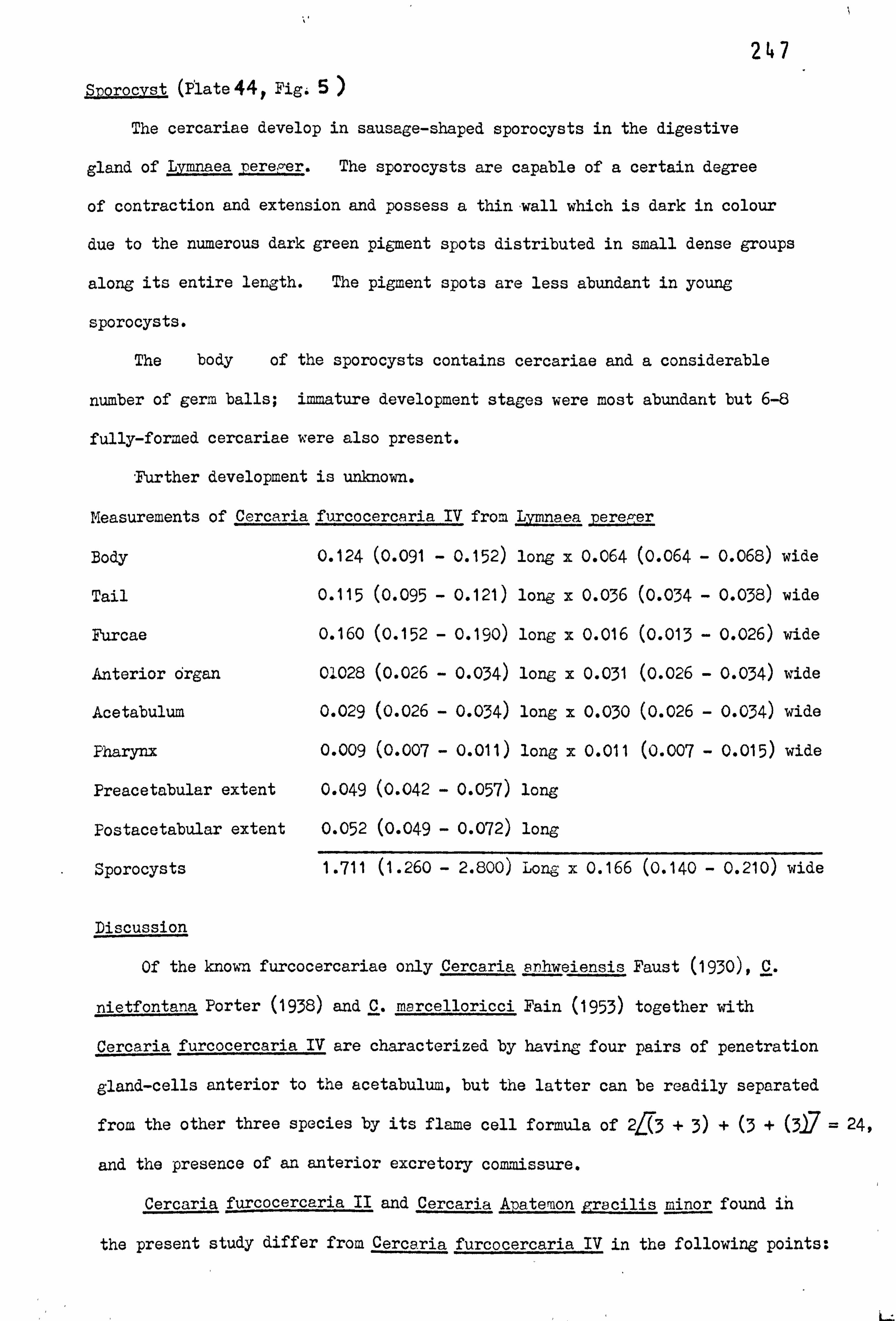
247 Sporocvst (Plate 4 4y Fig: 5
The cercariae develop in sausage-shaped sporocysts in the digestive
gland of Lvmnaeapere, °er. The sporocysts are capable of a certain degree
of contraction and extension and possess a thin wall which is dark in colour
due to the numerous dark green pigment spots distributed in small dense groups
along its entire length. The pigment spots are less abundant in young
sporocysts.
The body of the sporocysts contains cercariae and a considerable
number of germ balls; immature development stages were most abundant but 6-8
fully-formed cercariae were also present.
Further development is unknown.
Measurements of Cercaria furcocercaria IV from Lymna. ea pere? ýer
Body 0.124 (0.091 - 0.152) long x 0.064 (0.064 - 0.068) wide
Tail 0.115 (0.095 - 0.121) long x 0.036 (0.034
- 0.038) wide
Furcae 0.160 (0.152 - 0.190) long x 0.016 (0.013 - 0.026) wide
Anterior organ 01028 (0.026 - 0.034) long x 0.031 (0.026 - 0.034) wide
Acetabulum 0.029 (0.026 - 0.034) long x 0.030 (0.026 - 0.034) wide
Pharynx 0.009 (0.007 - 0.011) long x 0.011 (0.007 - 0.015) wide
Preacetabular extent 0.049 (0.042 - 0.057) long
Postacetabular extent 0.052 (0.049 - 0.072) long
Sporocysts 1.711 (1.260 - 2.800) Long x 0.166 (0.140
- 0.210) wide
Discussion
Of the known furcocercariae only Cercaria enhweiensis Faust (1930), C.
nietfontana Porter (1938) and C. marcelloricci Fain (1953) together with
Cercaria furcocercaria IV are characterized by having four pairs of penetration
gland-cells anterior to the acetabulum, but the latter can be readily separated
from the other three species by its flame cell formula of 2f(3 + 3) + (3 + Of = 24,
and the presence of an anterior excretory commissure.
Cercaria furcocercaria II and Cercaria Apatemon pracilis minor found in
the present study differ from Cercaria furcocercaria IV in the following points:
i--

248
1. Penetration gland-cells positioned posterior to acetabulum in
Cercaria Apatemon jracilis minor and C. furcocercaria II,
anterior in C. furcocercaria IV.
2.14 and 10 flame cells in Cercaria Apatemon -ra cilis minor and
C. furcocercaria II, respectively, 24 flame cells in
C. furcocercaria IV.
3. Posterior excretory commissure present in Cercaria Ana. temon Eracilis
minor and C. furcocercaria II, absent in C. furcocercaria IV.
Brown (1931) described Cercaria _jygo rtobhora from Planorbis carinatus
in Cheshire. His form can be distinguished from the present one by its
possession of the following characters: -
1. Penetration gland-cells entirely posterior to acetabulum.
2.24 flame cells.
3. Incomplete anterior excretory commissure.
0

249
Cercaria Bilharzielae polonicae
Kowalewski, 1695 (Szidat, 1929b).
This cercaria was only found once - in Planorbis planorbis from
Wintersett Lake during the summer of 1973. The infection rate was 10.6%.
Ercolani (1881) recorded a cercaria emerging from P. corneus in Italy, as Cercaria ocellata ýLa Valette St. George (1855). Later Szidat (1929b) considered that the
cercaria reported by Ercolani was the same species which he had shown to be
the larval stage of Bilharziellae polonicae. Szidat found it in the same first
intermediate host in Germany. Since 1929 this cercaria has been reported by
many authors from other parts of Europe, USSR and Africa including Brumpt (1932),
Wesenberg-Lund (1934), Porter (1938), Vergun (1956), Wisniewski (1958), Zd&rska
(1963), Butenko (1967), Aristanov (1968)and Khalifa (1972). All these -.
workers have found this cercaria to parasitise species of the Planorbidae.
Iles (1959) reported it for first time in Britain from P. corneus
in Roath Park Lake, Cardiff, Khan (19614 recorded it from the same host
species in Lake Meadows (Essex), London, and stated that the morphology and
measurements completely agreed with those given by Iles. My specimens are
morphologically similar to Iles' specimens and my measurements taken from
fixed specimens are close to hers from living individuals except that the
tail and furcae are larger in my specimens.
Iles figured the ducts of penetration gland-cells opening at both sides
of the anterior part of the mouth, whereas in my specimens they open through
five hollow conical spines on the antero-lateral margin of the body. This
has also been shown by Khalifa (1972). The proximal part of the tail of Iles's
specimens is wider than in mine, with a cup-shaped depression. This cup-shaped
depression is normally seen in my specimens when they are resting. It was also
observed by Khan (1962.
According to Khalifa (1972), Iles's specimens differ from his in having
3 ciliated patches, rather than 2 in the primary excretory ducts and the
posterior 3 flame cells arising separately. Although in my specimens there
are two ciliated patches as in those described by Khalifa and Iles, their location

250
is different. In my specimens they are situated at the beginning of the
posterior excretory ducts whereas 12aalifa and Iles described them from the
confoluted portion of the primary duct. The arrangement of flame cells
in the post-acetabular region is similar to that described by Iles.
Khalifa (1972) reported for first time the presence of a cephalic gland
and an island of Cort. In all my specimens I have observed a cephalic gland
but not an island of Cort. The pattern of hair-like processes on papillae
is similar to that in Khalifa's specimens but they are more numerous in my
specimens. (see Plate 45, Figs. 1 and 3).
Measurements of the Cercaria of Bilharzielae polonicae from Planorbis nlanorbis
Body 0.302 (0.292 - 0.307) long x 0.069 (0.064 - 0.076) wide
Tail 0.505 (0.494 - 0.513) long x 0.048 (0.045 - 0.053) wide
Furcae 0.226 (0.220 - 0.226) long x 0.025 (0.02o - 0.032) wide
Anterior organ 0.102 (0.087 - 0.110) long x 0.053 (0.049 - 0.057) wide
Acetabulum 0.030 (0.030 - 0.030) long x 0.023 (0.022 - 0.026) wide
Preacetabular extent 0.181 (0.174 - 0.186) long
Postacetabular extent 0.095 (0.091 - 0.098) long
Sporocyst 2.294 (1.680 - 2.800) long x 0.150 (0.126 - 0.168) wide
Specific diagnosis
A pharyngeal, brevifurcate, distome cercaria. Five pairs of penetration
gland cells, 2 anterior (fine granular contents) and 3 posterior (coarsely
granular contents) to acetabulum. Cephalic gland present. Body, tail and
furcae spinose. No excretory coinmissure. Two pigmented eye spots. Flame
cell formula 2 ff1 +1+ 1) +0+1+1+ (15 = 14.
Iles (1959) reported that the cercaria of B. rolonicae emerged between
17.00 and 18.00 hr. In the present study emergence was recorded throughout
the day but the behaviour of the cercaiae was similar to that observed by Iles
(1959). The present account constitutes the first record of the cercaria of
B. polonicae in Planorbis planorbis in Britain.

251 Cercaria letifera Fuhrmann, 1916
This cercaria has been found on two occasions in Lymnaea pererer from
Wintersett Lake, In August 1973,15 of the snails were parasitised, while
in October 1974, the percentage of infestation was 25w.
After comparison of the present cercaria with other pharyngeal lpngifurcate
cercariae which also possess two pairs of penetration gland-cells anterior to
the acetabulum and 16 flame cells it has been concluded that it is identical
with Cercaria letifera, originally described by Fuhrman in 1916 from Switzerland.
This species has subsequently been recorded by Dubois (1929) from Neuchatel,
Switzerland, Wesenberg-Lund, (1934) from Denmark, Fain (1953) from Belgian Congo,
Niewiadoma'sa (1960) from Poland, Khan (1962) from Great Britain, Meyer (1964)
from Switzerland and Smirnova and Ibrasyeva (1967) from Kazakhstan, USSR.
Cercaria letifera has always been found parasitising species of Lymnaea,
except by Fain (1953) who reported it from Biomrhalaria alexandrina 1anpanyicensis
collected from the Belgian Congo.
Of the published descriptions, this form is extremely similar to that given
by Khan (1962), except that in his specimens the branches of the caudal excretory
duct open at the middle of the furca whereas in the present specimens they open
at the tip of the furca. This features has also been reported by other
authors. In Khan's specimens there are fewer long hair-like processes on
the body and furcae and more on the tail, but they are not on papillae as in
the present material. Also in my specimens the oesophagus and caeca are filled
by a fine granular substance which Khan did not mention see Plate 46, -Figs. 1&3
The emergence and behaviour of these cercariae are identical to those
observed by Khan. This is the second record for C. letifera in Great Britain.
Further details of the life cycle and the corresponding adult form are both
unknown.
r

252
Measurements of Cercaria letifera from Lvmnaea pereger
Body 0.188 (0.171 - 0.201) long x 0.040 (0.038 - 0.041) wide
Tail 0.232 (0.193 - 0.247) long x 0.034 (0.030 - 0.038) wide
Furca 0.203 (0.185 - 0.212) long x 0.017 (0.015
- 0.022) wide
Anterior organ 0.042 (0.038 - 0.045) long x 0.021 (0.022 - 0.030) wide
Pharynx 0.015 (0.015) Long x 0.011 (0.011) wide
Acetabulum 0.023 (0.022 - 0.026) long x 0.023 (0.022 - 0.026) wide
Preacetabular extent -0.109 (0.091 - 0.121) long
Postacetabular extent 0.054 (0.045 - 0.057) long _
Sporocyst 2.030 (1.400 - 3.080) long x 0.116 (0.098 - 0.140) wide
Specific diagnosis
Pharyngeal longifurcate distome furcocercaria. Two pairs of
penetration gland cells anterior to acetabulum. Flame cell formula
2+1+1+ 1) + (1 +1+ (2)7= 16. Two rows of penetration spines. , fl
Body, tail and furcae spinose, with hair-like processes on papillae. Acetabulum
bearing a row of 35-37 spines. No excretory commissures. No eye-spots.
Five pairs of caudal bodies.

253
Cercaria paracauda Iles, 1959.
This cercaria was recorded from 6 different localities in the
Yorkshire area, namely Newmillerdam Lake, Kirkstall Power Station
(Leeds-Liverpool canal), Wintersett Lake, Walton Park, New Farnley and
Enroy. The collections of these snails were carried out in Spring, Summer
and Autumn, 1973,1974 and 1975. L. stagnalis obtained from all of these
sites harboured Ceresria 2aracauda, and an infection rate of 4.5-37.7% was
observed. Lvrnraea poreper was found to be infected by this cercaria in
the first two localities only and the incidence of infection varied between
0.6 - 2.0`j''j.
Iles (1959) described Cercaria paracauda from L. Dere er in Roath
Park Lake, Cardiff. Since then it has been found only by Khan (1962) in
Bushy Park, Epping Forest and Richmond Park, London, and by Probert (1966)
from Llangorse Lake, South Wales. The two latter authors also found
L. neretg: er infected with this cercaria. Only Than reported a rate of
infection (between 10 and13o) and it was similar to that reported here for
L. stagnalis, although it was considerably greater than that of L. pereger.
It is interesting to note that the maximum infection rate recorded was 37.7%
from Kirkstall Power Station where L. pereger is more abundant than L. sta, -nalis.
Furthermore both species of L. ymnasa were found in all three localities from
where Iles, Khan, and Probert reported C. p racauda, but L. stagnalis was not
infected with this cercaria.
The specimens I have found agree in most essential characters with those
described by Iles (1959) although detailed study has revealed some differences
between the two series of specimens. It is difficult to assess whether the
taxonomic significance of these differences is sufficient to justify specific
recognition. The characters which the two cercariae have in common are:
1.2 pairs of postacetabular penetration gland-cells.
2.16 flame Cells.
3. Numerous caudal bodies.
4.6 rows of spines on oral cap.

254
5.3 rows of spines on acetabulum.
6. The presence of yellow pigment granules in the area of pharynx.
7. The position of the bifurcation of the oesophagus.
8. Emergence and behaviour.
The differences between the two cercariae are tabulated below:
1. Borders of penetration I. Borders of glands deeply trilobed.
gland-cells smooth
2. Division of primary excretory
duct immediately posterior
to acetabulurn.
(see Plate 47, Fig. 2)
2. Division of primary excretory duct
antero-lateral to excretory bladder.
(see Plate 47, Fig. '3)
3. Caudal excretory ducts opening
midway along furcae.
4. Two ciliated patches in primary
excretory ducts.
5. Three rows of penetration spines
comprise 5,7 and 5 spines
respectively (total 17).
3. Caudal excretory ducts opening at
tips of furcae.
4. Three ciliated patches in primary
excretory ducts.
5" Three rows of penetration spines
comprise 4,6 and 4 spines
respectively (total 14)
6. Cephalic gland not recorded. 6. Prominent Y-shaped cephalic gland
present in postero-medial portion of
anterior organ. (see Plate 47, Fig; 3)
Despite these differences between the present material and that of Iles.
(1959) 1 have referred this material to Cercaria roaracauda Iles, 1959 on the
basis of the characters they have in common. This constitutes the first
record of C. rpracauda from the host species, L. stanalis in Britain.

255
Measurements of Cercaria paracauda from Lymnaea stagnalis
Body 0.183 (0.171 - 0.197) long x 0.041 (0.038
- 0.049) wide
Tail 0.228 (0.224 - 0.231) long x 0.037 (0.034
- 0.045) wide
Furcae 0.217 (0.214 - 0.024) long x 0.02o (0.015 - 0.022) wide
Anterior organ 0.050 (0.053 - 0.057) long x 0.026 (0.022
- 0.030) wide
Prepharynx 0.009 (0.007 - 0.011) long
Pharynx 0.014 (0.011 - 0.015) long x 0.011 (0.011 - 0.011) wide
Oesophagus 0.016 (0.007 - 0.022) long
Acetabulum 0.026 (0.026 - 0.026) long x 0.027 (0.026
- 0.030) wide
Preacetabular extent 0.093 (0.076 - 0.114) long
Postacetabular extent. 0.059 (0.057 - 0.064) long
Sporocyst 3.016 (1.841 - 4.734) long x 0.146 (0.105 - 0.184) wide
Specific diagnosis
Pharyngeal longifurcate distome furcocercaria. Two pairs of post-
acetabular penetration gland cells. Three rows of penetration spines.
Six rows of spines in oral cap. Three rows of spines on acetabulum.
Head gland. Eyespots absent. Many caudal bodies. No excretory commissure.
Body, tail and furca spinose. Yellow pigment granules. Flame cell formula:
2 J1 +1+ 1) + (1 +1+1+ (2)7 = 16. Cuticle of body, tail and furcae
armed with hair-like processes on papillae. Ten to eleven transverse rows
of spines extending from thick-walled part of the anterior organ to almost
posterior end of acetabulun.

256
Cercaria Cotylurus brevis (Dubois and Rausch, 1950) Nasir,
1960 (= Cercaria helvetica XXXIV Dubois, 1934)
The first infected Lymnaea perec; er were found during October and November,
1973 in collections from Gledhow Valley Road and Durkar. The infection-rate
was about 15% at Gledhow and 322% at Durkar. The cercaria was obtained again 9.
in January 1974, from the same host in Kir1ington where the infection rate was
10; x.
The cercaria of Cotvlurus brevis was originally described as Cercaria
KýcXIhelveticaois, 1934, in Lymnaea stapnalis from Lake Neuchatel in
Switzerland. Nasir (1960), during studies of trematode larvae from Edgbaston
pool, Birmingham, found a fork-tailed cercaria emerging from L. sta, --nalis,
which by controlled feeding experiments using laboratory-bred second intermediate
and definitive hosts was proved to be the larval stage of C. brevis.
C. brevis was originally described by Dubois and Rausch (1950) from the Pochard
Duck (Atha affines) from North America. The cercaria has also been found by
Meyer (1964) in Zurich, Switzerland, from Radix ovata and Butenko (1967) reported
it in L. staff lis from Kazakhstan, USSR.
The cercaria described by Nasir (1960) as that of C. brevis is similar to
the present material in all morphological details and also in its behaviour
and emergence patterns. It differs only in certain dimensions of the oesophagus
and acetabulum which are more close than those given by Dubois in C. helvetica
XXXIV. Also Nasir described globular bodies of variable size in the tail
of the cercaria, about which he commented "They are not the true caudal bodies
or glands found in some other strigeid cercariae", but detailed microscopical
study undertaken by myself revealed the presence of 16 caudal bodies arranged
in 9 pairs at either side of the caudal excretory duct. He described and
figured fine hair-like structures on body, tail, and furcae, but only one on
a papilla on each side of the body approximately at the level of the excretory
bladder. In my specimens all these structures on both body and furcae are on
papillae and are more numerous on the body and less numerous on the tail than
in Nasir's description. (see PlAte 489i Fig. 1)

f 257
According to Nasir (1960) L. stagnalis and L. pereger are among the
commonest species of gastropods in Edgbaston pool, but he did not report that
any specimens of L. pere! zer were infected with cercaria of C. brevis. In
the present study this cercaria has been recorded from L. pereger collected
from three different localities, and although L. stagnalis occurred in
one locality it was never found to be parasitized with the cercaria of C. brevis.
This is the first record of this cercaria from L. pereger in Britain.
Measurements of the cercaria of Cotylurus brevis from Lymnaea pereper
Body 0.190 (0.182 - 0.197) long x 0.049 (0.049 - 0.057) wide
Tail 0.195 (0.193 - 0.197) long x 0.038 (0.038
- 0.037) wide
Anterior organ 0.039 (0.038 - 0.041) long x 0.028 (0.026
- 0.030) wide
Acetabulum 0.027 (0.026 - 0.030) long x 0.027 (0.026
- 0.030) wide
Prepharynx 0.009 (0.007 - 0.011) long
Pharynx 0.014 (0'. 013 - 0.015) long x 0.013 (0.012 - 0.015) wide
Oesophagus 0.011 (0.009 - 0.013) long
Preacetabular extent 0.011 (0.095 - 0.114) long
Postacetabular extent 0.051 (0.045 - 0.057) long
Furcae 0.173 (0.171 - 0.178) long x 0.017 (0.015
- 0.019) wide
Sporocyst
Specific di&mosis
5.940 (4.866 - 7.233) long x 0.300 (0.158 - 0.526) wide
Pharyngeal longifurcate distome cercaria. Two pairs of penetration gland
cells anterior to acetabulun. Two rows of penetration spines. Three to
four rows of spines on acetabulum. Body and furcae with hair-like structures
on papillae. Anterior commissure incomplete. Flame cell formula 2 Z2 +2) +
(2 +2+ (2)7 = 20. Unpigmented eye spots. Nine pairs of caudal bodies.

258 Cercaria furcocercaria VII
About 15 of the Lymnaea peregei' collected from Wintersett Lake on
2 occasions during the summer of 1973 were found to harbour Cercaria
furcocercaria VII.
The cercariae emerge in large numbers throughout the day, mainly early
morning and late evening and after leaving the snail they swim actively for
a short period followed by a period of rest. At rest the cercariae hang with
their bodies downwards and with the furcae spread apart but their tips bent
downwards. They remain alive for about 24 hours.
Description (Plate 49 Fig*. 1- 3)
Body elongate, slightly narrower at its extremities and completely covered
with spines, anterior tip occupied by a group of 19 penetration spines arranged
in 3 rows, one behind the other, first row consisting of 6 spines, second row
seven and third row six. Oral cap with 5-6 rows of spines extending back to
anterior level of thick-walled part of anterior organ. Cuticle with 6 papillae
with hair-like structures on each side of body.
Tail stem almöst as long as body and furcae, and slightly subterminal;
eight to ten hair-like processes on each side. Two bands of longitudinal muscles
extending from base of tail to mid-way along furcae. Core of tail containing
twelve caudal bodies arranged in six pairs on either side of excretory duct,
but attachment to this duct was not observed. Furcae spinose, without finfolds
and equipped with 8- hair-like processes. Small cell bodies irregularly
dispersed along entire length of furcae and a few similar bodies also present
in tail.
Anterior organ oval, protrusible; anterior part thin-walled and
posterior part more muscular with numerous transverse muscle fibres.
Cephalic gland absent.
Digestive system well developed; mouth slightly subterminal, prepharynx
short and muscular and followed by oesophagus. Oesophagus divided a considerable
distance from anterior part of acetabulum into two thin and slightly septate
(each with 5-6 septa) intestinal caeca extending almost to posterior extremity

259
of body.
Acetabulum located behind middle of body and armed with four rows of minute
spines set very close together. Body cavity containing numerous small refractile
granules similar to those found in wall of sporocyst. Two oval unpigmented eye
spots located antero-lateral to acetabulum but difficult to observe.
Three pairs of penetration gland cells present antero-lateral to acetabulum;
cells rather small with coarsely granular contents and a circular central nucleus.
Ducts following a slightly undulating course and each opening via a separate
hollow conical spine, at anterior margin of body.
Excretory bladder 2-chambered; accessory anterior chamber transversely
elongate. Principal posterior chamber more or less oval and communicating with
the former by a short neck. From each side of accessory chamber two primary
excretory ducts extend forward to'-anterior margin of acetabulum where they join
to form the anterior excretory commissure. At this point a secondary duct
arises on each side, in turn dividing into anterior and posterior collecting
ducts. One group of 2 flame cells on each side connected to anterior collecting
duct and 4 groups of 2 to posterior collecting duct, one of these groups located
in proximal region of tail. Caudal excretory duct forked distally with each
branch passing into a furca to open by a small excretory pore just proximal to
midpoint of furca. Flame cell formula: 2/(2) + (2 + 2+ 2+ (2)7 = 20.
Sporocyst (Plate 49 Fie-4)
The digestive gland of Lvaea rereger was filled with tangled sporocysts.
They are thread-like in shape and vary in length as they perform contracting
and elongating movements. The anterior end contains oval structures with fine
granular contents. The body is constricted at irregular intervals, forming
swollen areas which are full of cercariae and the conspicuous birth pore is
located subterminally at one side of the anterior end. The body wall is thick,
with granular contents and small refractile tranules are distributed irregularly
over its surface. There are large numbers of cercariao in all stages of
development, together with several germ balls.
Further developmental details are unknown.

260
Measurements of Cercaria furcocercaria VII from Lymnaea n
Body 0.175 (0.171 - 0.190) long x 0.050 (0.045 - 0.053) wide
Tail 0.173 (0.171 - 0.182) long x 0.032 (0.030 - 0.034) wide
Furcae 0.169 (0.155 - 0.178) long x 0.014 (0.011 - 0.015) wide
Anterior organ 0.035 (0.034 - 0.038) long x 0.026 (0.026 - 0.026) wide
Acetabulun '0.029(0.026 - 0.030) Long x 0.027 (0.026*- 0.034) wide
Prepharynx 0.007 (0.003 - 0.007) long
Oesophagus 0.011 (0.007 - 0.015) long
Preacetabular extent 0.101 (0.095 - 0.0114 ) long
Postacetabular extent 0.045 (0.041 - 0.049) long
Sporocyst 2.761 (0.526 - 3.419) long x 0.161 (0.078 - 0.210) wide
Discussion
Among the species of pharyngeal longifurcate distoie cercariae,
Cercaria linearis Wesenberg-Lung (1934); C. m ga1esia Porter (1938);
C. Fare. lineeris Goodman (1951); C. rodhaini Fain (1953); C. pseudolinearis
Khan (1962) and C. cornuarietis Nasir, Hamana and Diaz (1969) resemble
C. furcocercous VII in having three pairs of penetration gland-cells in
front of the acetabulum. Of them only C. rodra. ini and C. pseudolinearis
also possess 20 flame cells. The former is easyily distinguished from the
present form bý the possession of a rudimentary acetabulum and very short
intestinal caeca and by the absence of an anterior excretory commissure.
Cercaria pseudolinearis is the form appearing most closely related to
C. furcocercaria VII. However there are certain characters of diagnostic
significance which serve to separate these two species. C. rseudolinearis has 3
rows of spines around the orifice of the acetabulum and 2 rows of penetration
spines in the anterior part of the body compared with 4 and 3 rows respectively
in C. furcocercaria VII. In C. rseudolinearis the primary excretory
ducts divide behind the acetabulum and the anterior collecting ducts receive
2 groups of flame cells, whereas in C. furcocercaria VII the primary ducts
divide anterior to the acetabulum and the anterior collecting ducts each receive
only a single group of flame cells. Finally in C. Pseudoiinearis there are

261
14 caudal bodies compared with 12 in C. furcocercaria VII. In Britain,
in addition to C. -, seudolinearis, four other species with the same number of
penetration gland-cells have previosly been described from freshwater
gastropods. They are Cercaria F. 2 Harper (1931); C. valvata Teal (1959);
C. planorbida Iles (1959) and C. roathensis Erasmus (1960). Cercaria F. 2 is
readily eliminated as it possesses in addition to other distinguishing features,
pigmented eye spots. C. roathensis differs in having a posterior excretory
commissure and 2 incomplete commissures anterior to the acetabulum.
C. planorbida also differs from C. furcocercaria VII in lacking an anteiior
excretory comtaissure and in having a total of 14 flame cells. C. valvata can
be differentiated by the apparent absence of eyespots and an anterior excretory
commissure, and by the disposition of penetration gland-cells behind the
acetabulum.

262
Cercaria of Diplostomum nhoxini (Faust, 1918) Arvy and
Buttner, 1954.
Lymnaea ep reger, collected in May and June, 1974 and 1975, from
Pool-in-4Jharfedale were found to be infected with this cercaria. The
infection rate varied between 11.4 and 22.7.
This cercaria was originally described by Arvy and Buttner (1954)
from Lvmnaea auricularia in France. They obtained the adult parasite
by feeding domestic ducks Albas Boschas and Cairina moschata var. domestica)
with minnows Phoxinus laevis) which had been experimentally exposed to cercariae
infection. Rees (1955 and 1957) found this cercaria in L. pereper var ovata
in Wales and also elucidated its life cycle, conducting experiments similar
'to those of Arvy and Buttner. Berrie (1960) reported it from Scotland in
L. rereger but did not give either a description or figures.
The cercaria found in the present study is very similar both in
morphology and in its measurements to cercaria of D. phoxini as described by
Arvy and Buttner (1954) and by Rees (1957). The description given by the
latter author is more complete and the present specimens agree with it although
the following minor differences were observed:
1. Fees stated that the ducts of penetration gland-cells open by two
pores on each side of the mouth whereas in my specimens these ducts open
throug2/hollow conical spines located on either side at the anterior end of
the body. (see Plate 50, Fig. 2)
2. There is a cephalic gland situated at the posterior part of the
anterior organ, whereas in Rees's specimens the presence of this gland was
not indicated. (see, Plate 50, Fig. 3 )
3. In my specimens the acetabulum is surrounded by a muscular ring which
was not referred to by Rees. (see Plate 50, Fig. 3)
4. Rees described the anterior and posterior excretory ducts as receiving
3 flame cells each on either side of the body. In all my specimens only one
group of 2 flame cells is connected to the anterior collecting ducts and two
groups of 2 connected to the posterior ones on each side of the body.

263 5. Rees figured (but did not describe) the branches of the
caudal excretory duct entering into the furcae and opening midway along
the posterior margin of the furcae. In my specimens these, branches open
one-third of the length along each of the furcae, one opening on the anterior
margin of one furca (the right), the other on the posterior margin of the
second (left) furca. (see Plate 50, Fig. 4)
It is probable that the presence of a cephalic gland and a muscular ring
around the acetabulum were overlooked by Rees, while the other differences
could merely represent different interpretations of the same structures by
different authors.
The emergence and behaviour of the cercaria described above are identical
to those described by Rees for the cercaria of D. phoxini.
Measurements of the Cercaria of Diplostomum phoxini from Lymnaea sere er
Body 0.122 (0.110 - 0.133) long x 0.035 (0.034 - 0.038) wide
Tail 0.218 (0.209 - 0.224) long x 0.039 (0.034
- 0.046) wide
Furcae 0.168 (0.160 - 0.175) long x 0.013 (0.011 - 0.015) wide
Anterior organ 0.044 (0.042 - 0.046) long x 0.026 (0.023
- 0.027) wide
Acetabulum 0.019 (0.019 - 0.023) long x 0.021 (0.019
- 0.023) wide
Pharynx 0.008 (0.008 - 0.010) long x 0.009 (0.008
- 0.011) wide
Oesophagus 0.007 (0.004 - 0.011) long
Preacetabular extent 0.064 (0.053 - 0.072) long
Postacetabular extent 0.040 (0.038 - 0.53) long
Sporocyst 5.485 (2.630 - 9.205) long x 0.338 (0.210 - 0.473) wide
Specific diagnosis
Pharyngeal longifurcate distome furcocercaria. Two pairs of posterior
penetration gland cells. Two rows of spines on acetabulum. No eye-spots.
`fail with eight pairs of hair-like processes. - Furcae spinose with four pairs
of heir-like processes on papillae. Cephalic gland present. Acetabulum
surrounded by a muscular ring. Flame'cell formula 2 j2) + (2 +2+ (217 = 16.
Branches of caudal excretory duct opening on either anterior or posterior
margin of furcae.

264 Cercaria furcocercaria II
On April 4,1975, a sample of 40 specimens of Lymnaea ereCer was taken
from Riffa Beck at Fool-in-Wharfedale and 6 were found to be producing large
numbersof a forked-tailed cercaria.
These cercariae are shed in great numbers during daylight. Generally
they are found in the lower half of the container, and after a certain
period of time they may be seen congregated at the bottom. They swim
vigorously and have regular periods of rest during which the body hangs beneath
the tail and they sink gradually. They remain alive for more than 24 hours.
Description (Plate 51 Fib. 1-4)
Body elongate, cylindrical in shape; covered irregularly with soines
from anterior of body to region of acetabulum. One papilla with a hair-like
structure located on each side of body at equatorial level of anterior organ.
Oral cap provided with several regular rows of 6-7 spines, extending back
about half the length of anterior organ. " Two rows of penetration spines
situated anterior to mouth, first row with 6 spines and second with 7 spines.
Tail terminal, spinose, with 8 long hair-like structures on each side
and containing well-developed longitudinal and circular muscles. A few
small cell bodies dispersed throughout tail. Six prominent caudal bodies
present on each side of excretory duct and arranged regularly in pair;, first
pair smaller and in a different position from the rest. Furcae spinose, without
finfolds.
Anterior organ protrusible and retractile, its wall distinctly divided
into an anterior portion and a thicker muscular poste rio'part. Cephalic
gland absent. Mouth apening about anterior. end of anterior organ, continuing
into a small prepharynx then passing into small but well-developed circular
pharynx. Very short oesophagus present, but difficult to observe.
Intestinal caeca not well-developed, being represented by two swollen
caeca joined at their posterior'tips. ,

265 Acetabulum much smaller than anterior organ and situated posterior to
middle of body, moderately protrusible, its aperture area adorned with
irregularly distributed spines. A few additional spines present scattered
behind aperture area.
Two prominent unpigmented eyespots anterior to acetabulum. Each
eyespot full of small elongate structures with very fine granules and with
a short anteriorly-directed duct. No connection with the nervous system
could be seen.
Nervous system consisting of oesophageal ganglion and a wide commissure
which gives off two anterior and two posterior nerves; anterior nerves
passing forwards lateral to anterior organ, posterior nerves longer and running
backwards to almost half way between posterior margin of acetabulum and posterior
end of body.
Eight penetration gland-cells with coarsely granular contents arranged
in linear formation of one, two and one on each side of body, and occupying
almost whole postacetabular space. Anterior and median cells pyriform in
shape, posterior cells longer and more rectangular in form. Nuclei of these
cells well-marked, their ducts opening on antero-lateral margins of body.
Rudiments of genital primordium situated anterior to excretory
bladder and represented by a rather elongate mass of undifferentiated cells.
Excretory bladder elongate, with three main constrictions giving it a
marked trilobed appearance. Primary excretory ducts leaving antero-lateral
margins of bladder and extending to posterior margin of acetabulum where they
fuse to form the posterior excretory commissure. Immediately behind acetabulum
a short blind-ending duct arising from primary excretory duct. Primary
ducts forming coiled secondary ducts posterior to acetabulum which bend
posteriorly and fork into anterior and posterior collecting ducts. Anterior
collecting duct receiving capillaries from 2 flame cells and posterior from
3; 2 located in postacetabular region and 1 in proximal part of tail.
Each of tail capillaries with an unusual swollen section, filled with a very

266
finely-granular material. Caudal excretory duct arising from posterior end
of bladder and passing along tail to distal end, divided into branches running
into each of the two furcae. Branches opening to exterior via small excretory
pores. Flame cell formula: 21(1 + 1) + (i +1+ (1)7 = 10.
Sporocyst' (Plate 51, Fibs :. 5-7)
Development occurs in elongate thread-like sporocysts, living in the
digestive gland of Lvmnaea pereper. The body wall is thick, well-developed,
and lined by Parenchymal cells which are more abundant at the anterior end.
The conspicuous birth pore is situated near the anterior end and the body cavity
is full of numerous cercariae in different stages of development together with
several germ balls.
The mother sporocyst is elongate and it is difficult to separate as a single
specimen from the digestive gland tissue. Its body is filled by young daughter
sporocysts of different sizes and there are a great number of flame cells
dispersed along the body. (see Fig. 7)
The daughter sporocysts within the mother sporocyst are quite active
and may be seen in constant movement. The lumen of each contains hundreds
of germ balls, and small groups of dark green refractile granules are
present on the surface of the body.
Further development is unknown.

267
Measurements of Cercaria furcocercaria II from -L=naea rereper
Body 0.162 (0.141 - 0.171) long x 0.038 (0.034 - 0.042) wide
Tail 0.141 (0.129 - 0.144) long x 0.041 (0.034 - 0.049) wide
Furcae 0.146 (0.139 - 0.152) long x 0.012 (0.011 - 0.015) wide
Anterior organ 0.038 (0.038 - 0.042) long x 0.024 (0.023 - 0.024) wide
Acetabulum 0.017 (0.015 - 0.019) long x 0.017 (0.015 - 0.018) wide
Pharynx 0.008 (0.008 - 0.011) long x 0.008 (0.008 - 0.011) wide
Caeca 0.020 (0.019 - 0.023) long x 0.018 (0.015 - 0.020) wide
Preacetabular extent 0.089 (0.080 - 0.099) long
Postacetabular extent 0.055 (0.046 - 0.065) long
Un-pigmented eyespots 0.009 (0.008 - 0.011) long x 0.009 (0.008 - 0. D1t) wide
Mother sporocyst 21.653 (19.199 - 24.196) long x 0.359 (0.316 - 0.395) wide
Young daughter sporocyst 0.793 (0.140 - 1.0,60) long x 0.068 (0.056 - 0.084) wide
Daughter sporocyst 5.184 (2.104 - 7.890) long x 0.157 (0.132 - 0.210) wide
Discussion
Nineteen species of apharyngeal, longifurcate, distome furcocercariae
characterized by having four pairs of postacetabular penetration gland cells,
as in Cercaria furcocercaria Ii, have been described in the literature. They
are:
Cercaria pygocrtorhora Brown (1931).
Cercaria stenhensi Brooks (1949).
Cercaria coperata Olivier (1942).
Cercaria rracillima Faust (1917)
Cercaria burti Killer (1923) as described by Cort and Brooks (1928).
Cercaria apatenonracilis minor (Yamaguti, 1923) as described by
Iles (1959).
Cercaria angelae Johnston and Simpson (1944).
Cercaria pseudoburti Ranking, (1939).
Cercaria hamburgensis Komiya (1952).
Ceresria of Strigea elegans Chandler and Raush (1947) as described
by Pearson (1959).

268 Cercaria okoboyensis Brooks (194$).
Cercaria wansoni Fain (1953).
Cercaria rafula Fain (1953).
Cercaria helvetica XXXI Dubois (1929).
Cercaria burti var. icnusae Giovannola (1937).
Cercaria lesson Johnston and Bechwith (1947).
Cercaria Anatemon gracilis ; racilis Szidat. (1929a, 1931).
Cercaria longiremis Wesenberg-Lund (1934).
Cercaria of Apatemon cobitidis cobitidis (Linstow, 1890) as described
by Butenko (1967).
The cercaria of Striea elee: ans and of Apatemon cobitidis cobitidis,
Cercaria rafula, C. lon, aire. mis and C. okoboyensis are the only
species previously described having the same number of flame cells as
C. furcocercaria II. Of these cercaria of S. eleaans, C. lon. iremis and C.
rafula may all be separated from C. furcocercaria II by the absence of a
posterior excretory commissure. Cercaria kobovensis and the cercaria of
A. c. cobitidis can be differentiated from C. furcocercaria II by their
having more than twelve caudal bodies in the tail.
The cercaria of A. a. minor (Yamaguti, 1933), Iles (1959) encountered
during this investigation is readily distinguished from C. furcocercaria II
by differences in the complex protonephridial system, the number and arrangement
of caudal bodies and the digestive system.

269 Cercaria vivace I
During the summer of 1973,125 specimens of Bithynia tentaculata were
collected from Newmillerdaa Lake and 2 of these yielded a furcocercaria belonging
to the Vivax sub-group.
Cercariae are emitted during both the morning and the afternoon but none
during darkness. The swimming movements of the cercariae are followed by a
long period of rest unless disturbed when they start to swim again either upwards
or more usually downwards. When the cercariae are resting some can be seen
sinking to the bottom, they live for a maximum of about 24 hours.
Description (Plate 52, Pigs. 1-3 and 5)
Somewhat pyriform in shape, being widest in the middle of the body; body
surface covered with fine spines; cuticle of body and tail provided with a row
of numerous papillae with hair-like structures, 3 similar papillae located
laterally at base of furcae and one pair of papillae near distal tips of furcae.
Tail attached to the posterior end of the body subterminally and possessing two
bands of longitudinal muscles extending from anterior part of tail to tips
of the furcae, together with many circular muscles. Fwcae covered with spines
and provided on distal half with prominent finfolds which show fine striations
resembling hair-like fin-rays. Caudal bodies absent.
Three rows of penetration spines present in area of mouth; preoral region
with 6-8 rows of hook-like spines. Anterior organ terminal and resembling
an oral sucker more than a true anterior organ. Mouth subterminal opening into
a short prepharynx, leading to a well-developed pharynx. Pharynx circular
in shape, followed by relatively short oesophagus with its anterior end surrounded
by a well-marked muscular wall. Oesophagus divides slightly anterior to mid-
line into broad intestinal caeca which continue back to posterior end of body.
Acetabulum absent.
Numerous refractile globules arranged in groups scattered throughout body.
Four penetration gland-cells with finely-Granular contents located on
either side of oesophagus; their ducts pass anteriorly to dorso-lateral part
of anterior organ to open through hollow spines situated each side of anterior
tip of body.

270
Cystogenous gland-cells absent. Rudiments of genital system represented
by rounded undifferentiated cells situated anterior to anterior chamber of
excretory bladder.
Excretory bladder 2-chambered with a large transversely-elongated anterior
chamber and posterior small rounded chamber. Four excretory ducts arise from
anterior chamber - two lateral and two median. Median ducts pass anteriorly
to about the mid-level of body where they unite to form a single median canal
continuing forwards to level of oesophageal bifurcation. Here canal joins
a transverse vessel which fuses with the 2 laterals. On each side, at level
of oesophageal bifurcation, a short blind pouch arises from the transverse duct
and at the left and right extremities secondary excretory ducts arise and pass
backwards to level of genital primordium where they divide into anterior
and posterior' excretory ducts found. These each receive 2 groups of 2 flame
cells. Caudal excretory duct passes into tail from base of posterior chamber
and divides at distal end of tail, each branch opening at tip of furca.
Flame cell formula: J2 + 2) + (2 + 2)7 = 16.
Sporocyst `(Plate 52, Fig. 4)
The cercariae develop in elongate sausage-shaped sporocysts in the
digestive gland of Bithynia tentaculata. They are small and are capable
of limited movement. The whole surface of the body contains numerous
refractile globules of different sizes arranged in small groups. Lumen
of sporocyst completely full of cercariae, partially or fully differentiated,
together with a considerable number of germ balls. The young sporocysts are
more active than mature sporocyst and germ balls may be seen moving freely in
the former.
Further development is unknown. '
Measurements of Cercaria vivax I from Bithynia tentaculata
Body 0.131 (0.117 - 0.140) long x 0.060 (0.057 - 0.064) wide
Tail 0.094 (0.087 - 0.098)' long x 0.031 (0.030 - 0.034) wide
Furcae 0.095 (0.093 - 0.0,08) long x 0.016 (0.011 - 0.020) wide
Anterior organ 0.026 (0.026 - 0.026) long x 0.026 (0.026 - 0.026) wide

271
Pharynx 0.015 (0.015 - 0.015) long x 0.015 (0.015 - 0.015) wide
Oesophagus 0.012 (0.007 - 0.015) long
Sporocysts 1.176 (0.0840 - 1.680) long x 0.142 (0.140
- 0.154) tide
Discussion
The furcocercariae of the subgroup Vivax Sewell, 1922, emend. Dubois, 1951
which, like Cerceria vivax I and Cercaria vivax II, are characterized by having
furcal finfolds are:
Cercaria vivax Sonsino (1892), Langeron (1924), Weserberg-Lund (1934)
L= larva of Prohemistornum vivax (Sonsino, 1892),
Axin (1933), 0. vivax Sonsino (1894), Langeron (1924) _
larva of Szidatia joyeuxi (Hughes, 1929), Dubois (1938),
J oux and Baer (1941), Baho7. et (1953)7
Cercaria indicae XV Sewell (1922).
Cercaria ind. icae LVIII Sewell (1922).
Cercaria dorsocauda Tubangui (1928).
Cercaria kentuckiensis (Cable, 19351 as described by Cable (1938)
larva of Mesostephanus kentuckiensis (Cable, 191,54
Mayer (1960)17.
Cercaria tatei Johnston and Angel (1940).
Cercaria of Prohemistomum industrius (Tubangui, 1922) as described by
Tang (1941).
An unnamed cercaria described by Maxon and Pequegnat (1949).
Cercaria kasen i Fain (1953).
Cercaria vivacis Iles (1951).
Cercariararillosoma Khan (1962k.
Cercaria of Prohemistomum chandleri Vernberg (1952).
Cerceria of Prohemistomum expeditum Balozet (1953).
Cercaria of Szidatia , ioyeuxi (Joyeux and Baer, -1941) as described
by Balozet (1953).
Cercaria hirsuticzuda Probert (1966).

Cercaria ariforrnis Khan (1962). 272
Cercaria sDatula Probert (1966)
Cercpria laevissimus Nasir, Acuna and Guevara (1066).
Of the cercariae listed below only Cercaria kasenyi, an unnamed cercaria 9
described by Maxon and Pequ4nat (1949), C. hirsuticauda and C. ariformis
are provided with 24 flame cells like C. vivax I.
Cercaria knsenyi and the cercaria described by Maxon and Pequegnat (1949)
can readily be separated from C. vivax I because they possess flame cells in
the tail.
Cercaria hirsi4t'cauda and C. ariformis are more closely allied to C. vivax I.
However there are several features which set the two former species apart:
1. They lack papillae with hair-like process.
2. The arrangement of flame cells and the division of the secondary
excretory ducts.
3. The absence of refractile globules in the body.
4. The absence of blind pouches on the primary excretory ducts.
Also in C. hirsuticauda there are no penetration gland-cells, and fin-folds
are present along the full length of the furcae. In C. ariformis, the
penetration gland-cells are numerous, and are apparently arranged in two groups,
one behind the anterior organ and the other in the inter-caecal region, but
unfortunately the number of these glands is not recorded.
Other cercariae belonging to the sub-group Vivat Sewell, 1922, which have
been described in Britain from Bit is tentaculata, are listed below:
Cercaria vivacis Iles (1959), from Roath Park, Cardiff.
Cercaria pi illosoma IQian (196 `, from Bushy Park, London.
The cercaria of Cyathocotyle bushiensis Khan (196.21, from Bushy Park, London.
Cercnria spatula Probert (1966), from Liar orse Lake, Breconshire.
Of these cercariae, only the cercaria of C. bushiensis has 16 flame cells,
as C. vivax I, but there are no fin-folds on the tail furcae.
As a result of the comparison made above, this species is regarded as new,
and is named Cercaria vivax I.

273 Cercaria vivax II
This cercaria was found only once - in 1 Bithymia tentaculata out of 175
collected from Nekwmillerdam Lake in July, 1973.
Repeated collections were carried out during 1974,1975 and early 1976
but no additional snails infected with C. vivax II were found.
The production of cercariae is small and occurs only during the day time.
The swimming and behaviour are almost exactly like Cercaria vivax I but the
resting period is shorter. The cercariae were observed to live for about 36 hr.
Description (Plate 53)Figs. 1-3 and 5)
Body oval, entirely covered by minute spines which are also present on
furcae. Furcae bearing papillae with hair-like structures along their margins;
cuticle of tail possessing a row of long hair-like structures on both sides.
Tail slightly subterminal, provided with well-developed circular and
longitudinal muscles, the latter passing from anterior end of tail to tips
of furcae; circular muscles extending only to bases of furcae. Tail also
containing numerous cell bodies distributed irregularly, furcae bearing small
finfolds restricted to their tips; caudal bodies absent.
Two thirds of anterior organ covered by a row of 5-7 spines; penetration
spines absent. Anterior organ isodiametric and terminal with mouth opening
located near anterior end. Prepharynx short ending in a circular, muscular
pharynx; oesophagus much shorter than pharynx and divided into two wide intestinak
caeca which extend to about the level of the excretory bladder. Whole digestive
system filled with granular material. Acetabulum absent. Refractile globules
scattered through body.
Penetration gland-cells arranged in two groups of 8, one group on each side
of pharynx; cell contents finely granular; duct openings at anterior end of
body. Nuclei of these cells not observed. Cystogenous gland-cells absent.
Rudiments of genital system present.
Excretory bladder consisting of an oval chamber situated at posterior
end of body with main lateral and median excretory ducts arising from antero-

274 lateral region and passing forwards to level of mid-line of body where median
ducts fuse to form median canal. At level of oesophageal bifurcation median
canal joins transverse canal which passes laterally to join the external duct
on each side. Transverse canal gives rise on each side to a short blind-ending
pouch. Secondary ducts arise from main lateral ducts about the middle of the
boc: y, dividing into antero and postero lateral collecting ducts. Antero-lateral
collecting ducts with two groups of 2 flame cells, postero-lateral collecting
ducts with 3 groups of 2 cells and continuing into tail where they each receive
1 group of 2 flame cells. Each flame cell in tail ending in a swollen structure
with fine granular contents. Caudal excretory duct ending at tip of each furca.
Flame cell formula: 2LT2 + 2) + (2 +2+2 +(2)7= 24.
Snorocyst (Plate 53, Fig. i)
The sporocysts were located in the hepatopencreas of Bit is tentaculata.
Body elongate with thick walls which are thicker at the extremities than over
the rest of the body. Mobility of sporocyst low. Body containing numerous
cercariae, germ balls and refractile globules with 8-10 of the cercariae mature.
The protonephridial system has been observed only in part. There are five
parallel rows of flame cells extending from one end of the body to the other.
The rest of this system was difficult to observe owing to the presence of numerous
cercariae, germ balls and pigments.
Further development unknown.
Measurements of Cercaria Vivax II from Bithynia tentaculata
Body 0.135 (0.121 - 0.140) long x 0.078 (0.072 - 0.083) wide
Tail 0.193 (0.190 - 0.205) long x 0.038 (0.038 - 0.041) wide
Furcae 0.162 (0.159 - 0.167) long x 0.018 (0.015 - 0.019) wide
Anterior Organ 0.027 (0.026 - 0.030) lone x 1.027 (0.026 - 0.030) wide
pharynx 0.017 (0.016 - 0.019) long x 0.017 (0.015 - 0.019) wide
Oesophagus 0.006 (0.006 - 0.007) long
Sporocysts 2.367 (1.841 - 3.156)long x 0.165 (0.131 - 0.210) wide

275
Discussion
Of all other Vivax-type cercariae described only the cercaria of Proso-
stephanusindustrius Tang (1941), C. Aapillosoma Khan (1962) and C. spatula
Probert (1966) are characterized by possessing fin-folds limited to the tips
of the furcae as C. vivax II.
The first two of the above species are readily separated by. having a
different number of flame cells, from Cercaria vivax II, respectively, while
C. spatula has the same number.
In C. spatula the penetration gland-cells are arranged in two groups -
an anterior group of 4 small cells with clear nuclei on each side of the
anterior organ and a posterior group of 4 or more cells located on each side
of the anteriorregion of the caeca. In C. vivax II these cells are arranged
in one group of 8 on each side, postero-lateral to the pharynx, and they do not
possess nuclei. In C. spatula the capillaries of the posterior collecting
ducts in the tail reach almost half-way along its length whereas in C. vivax II
they reach for only one-third of the length and they, each end in a swollen
structure with granular contents. In C. vivax II there is a blind-ending
duct on each side of the main excretory ducts, at the level of the oesophageal
bifurcation. These are absent from C. sDatula. In C. vivax II the body and
tail are devoid of papillae with hair-like structures which are present on
these organs in C. spatula. Finally C. spatula differs from C. vivax II in
having a larger body, tail and furcae, and its anterior organ is almost
twice as big as in C. vivax II.
The present species is considered to be new and named Cercnria viv<ax II.

276 Summary
1. A survey has been conducted of the freshwater larval trematode fauna
in West Yorkshire.
2. Fifty two collections of freshwater molluscs were made at 21 different
places among which the 5 most important are: -
(1) Kirkstall Power Station (Leeds-Liverpool Canal)
(2) Wintersett Lake (Wakefield)
(3) Newmillerdam Lake (Wakefield)
(4) Gledhow Valley Road (Leeds)
(5) Pool-in-ytharfedale
At least four different kinds of cercariae have been found at each of the
above sites. of
3. Thirty species of cercariae, emerging from one or more' Bithynia tentaculeta,
B. leachi, Lyrnaea pereP"er, L. stapnalis, L. auricularia, Planorbis vortex,
P. planorbis and Snhaerium corneum were found.
4. Eighteen new species have been fully described and their relationship to
previously described cercariae is discussed, they are listed below: -
(i) Three Gymnocephalous cercariae (Cercaria of Sphaeridi_otrema wir1tersettensis,
C. mnocephalous II and C. gymnocephalous III).
(jj) One Pleurolophocercous cercaria Cercaria ipleurolophocerca I).
Five Furcocercariae Cercaria iurcocercaria II, C. furcocercaria IV,
C. furcocercaria VII. C. vivex I and C. vivax II).
(vj) One Microcercous cercaria Cercaria microcercous I).
(v) One Cystocercous cercaria Cercaria macrocera I).
(Vi) Three t; chinostome cercariae (Cercaria echinostorna III. C. ma macauda I
and C. inagnacauda II).
(VII) Four Xiphidiocercariae (Cercaria microcotylea I. Cercaria of P1Ppiaphis
farnleve nsis, Cercaria of P. kirkstallensis and C. xiphidiocercaria IX).
5. Cercaria of Dolichosaccus rastellus is described for the first time in
Great Britain.

27.7
6. Eleven previously described cercariae for Great Britain were found.
They are: Cercaria paracauda Iles, 1959
Cercaria of Apatemon -racilis minor of Iles (1959)
Cercaria of Bilharzielae polonicae of Iles (1959)
Cercaria tarda Khan, 19614
Cercaria letifera of Khan (1962
Cercaria of Diplostomum phoxini of Rees (1955 and 1957)
Cercaria of Cotylurus brevis of Nasir (1960)
Cercaria of Echinoparyphium recurvatum of Harper (1929)
Cercaria of Hyroderaeum conoideum of Nasir (1951-SB)
Cercaria of Notocotylus imbricatus of Pike (1969)
Cercaria of Notocotyylus triserialis (= N. ettenuatus) of Pike (1969).
7. All the previously described cercariae from Great Britain are compared
with the descriptions of previous authors. Small morphological and
dimensional differences are noted and their possible significance discussed.
8. New first intermediate hosts for some of the previously described cercariae
have been recorded.
9. Studies of the life cycles of Plagiorchis farnleyensis n. sp., P. kirkstallen-
sis n. sp. and Sphaeridiotrema wintersettensis n. sp. are described.
10. The life cycle of Dolichosaccus rastellus is described from Great Britain
for the first time.
11. Some additional information is provided on the morphology and life-cycles
of Echiparyrhium recurvatum, Hypoderaeum cono ideum, Notocotylus imbricatus
and N. atte nuatus.

278
ABDM-M LEX, E. T. (1952). Life history of Petasiger chandleri n. sp. (Trematoda:
chinostomatidae) from the pied-billed grebe Podilymbus rodicens nodicn-ns.
J. -Parasit, 33 suppl: 39.
ABDEL-Mk. L. Ki-E. T. (1953). Life history of Petasiger chandleri, from the pied-
billed grebe Podilymbus podicens podicecs, with some comments on other
species of Petasir; er. Ibid., 152-158.
ACHOLONU, A. D. cC 0. \i., OLSELT (1967). Studies on the life history of two
v/AS I I. Notocotylus (Treinatoda). Proc. helminth. SocX, 34 : 43-50.
AGARWAL, S. M. (1956). On a new species of Xiphidiocercaria from Lymnaea
accuminnta. Proc. natn. Acad. Sci. India. Section B. 26: 141-144.
ANDERSON, D. J. (1944). Studies on Cercaria Szidati sp. nov., a new furcocercous
cercaria of the Vivax type. J. Parasit., 30: 264-268.
ANDLRSON, G. A. & PRATT I. (1965). Cercaria and first intermediate host of
ý. uryhelmis squamula. J. Parasit. 51: 13-15.
ANGEL, M. (1958). An account of Plagiorchis maculosus (Rud. ), its synonymy
and its life history in South Australia. Trans. Roy. Soc. S. Aus r, _82:
265-281.
ARCHIBALD, R. G. & A. MARSHAIN, (1931). A study of three non-furcocercous cercariae
obtained from Bulimus contortus in the Sudan. Parasitology, : 271-281.
ARISTANOV, E. (1968) Furkocerkarii iz presnovodpyh molljuskov del'ty Amu-
Dar'i. II. Vest. leningr. Univ., Ser. biol., 22: 7-14.
ARVY, L. & BUTTIJt A. (1954). Donnees sur is cycle evolutif de Dinlostomulum
*hoxini (Faust, 1918), (Trematoda: Diplostomidae) C. R. Acad. Sci., 212:
1085-1087.
AZINI, M. A. (1930). On the identification and life history of Echinostomum
recurvatum von Listow, 1873. Ann. trop. Mad. parasit. 24: 189-192.
AZIM, M. A. (1933). On Prehemistomum vivax (Sonsino, 1892) and its development
from Cercaria, vivax Sonsino, 1892. Z. ParasitKde., ý: 432-436.
BAER, K. E. (1827). Beitrage zur kenntniss der niedern Thiere. Nova Acta Acad.
P+at. Curios. j: 523-762; 881-882.

BAKER, J. R. (1943). Cercaria Steelmani, A new macrocercous form.
Trans. Amer. Micro. Soc. 62: 280-285.
BALOZET, L. (1953). Trematodes Larvaires de L'afrique du nord cercaires
cotylicerques. Arch. Institut. Pasteur. d'Algerie j1' . 75-83
BARKER, F. D. (1915). Parasites of the American Muskrat (fibre zibethicus).
J. Parasit. 1: 184-197,4 fig., 2 pl.
BARKER, F. D. & J. W., LANGHIN (1911). A new species of Trematode from the
Muskat, Fiber Zibethicus. Trans. Amer. k1icr. Soc., 30: 261-274.1pl.
BASCH, P. F. & R. F. STURROCK (1962). The life hi tort' of Ribeiroia merini
(Faust and Hoffman, 1934) comb. n. (Trematoda: Cathaemasiidae).
J. Parasit., : 1180-1184.
BASIiKIROVA E. Y. (1941). In Skrjabin, 1956: Trematodes of animals and man, 12 .
BAYLIS, H. A. (1928). On a new species of Notocotylus. Ann. M. ag. Nat. Hist. 2: 582- 596.
BAYLIS, H. A. (1939). A larval trematodes (Diplostomulum volvens) in the lens
of the eye of a rainbow trout. Proc. Linn. Soc. Lond. 2,130.
BEAVER, P. C. (1937:,. Experimental studies on Echinostoma revolutum (Froel)
a fluke from Birds and Dammals. Ill. Biol. Monogr. 1j3- 1-96.
BL Vet, P. C. (1939). The morphology and life history of Petasi, er nitidus
Linton (Trematoda: Echinostomatidae). J. Parasit. ._
269-276.
HEILFUSS, E. R. (1954). The life histories of Phyllodistomum lohrenzi Loewen, 1935,
and P. sandatum Steelman, 1938. (Trematoda: Goroderinae).
J. Parasit., 4,0 (5 sect. 2): 44.
BERRIE, A. D. (1960). The influence of varicus definitive hosts on the
development of Diplostomum : thoxini (Strigeida, Trematoda). J. Helminth.,
34: 205-210.
BEVERLY-BURTON, 11. (1961). Studies on the trematoda of British freshwater
birds. Proc. Zool. Soc. Lond., 137: 13-39.
BISSERU, B. (1967). Stages in the development of larval echinostomes recovered
from schistosome transmitting molluscs in Central Africa. J. Helminth.
41 : 69-108.
BLMNCHARD, R. (1895). Animaux parasites. Bull. Soc. Zool. France, 20: 217.

BLAIR SPQOR, H. D. (1974). Host-induced variation in P1aaiorchis noblei Park,
1936 (Plagiorchiidae: Trercatoda). The American Midland Naturalist,
92,1.,: 415-433.
BLOCH, M. E. (1782). Abhandlung von der Erzeugung der Eingeweidewurmer.
Berlin. 54 pp; 10 pl.
BRAUN, M. (1891). Verzeichniss von ßingeweidewurmern aus Mecklenburg.
Arch. Ver. Frde. Naturg. Meckl. pp. 97-117.
BRAUN, 111. (1899) Uber Clinostomum Leidy. Zool. Anz., 22: 489-493.
BRAUIT, M. (1901). Zur Revision der Trematoden der Vogel. I.
Centralbl. Zbl. Bakt. (1 Abt. ), 22: 560-568.
BRAUN, M. (1902). Fascioliden der Vogel, Zoology, Jahrbucher. System.,
Bd XVI. Heft 1,6-5-1902, p. 1-162, p1. I-VIII, fig. 1-99.
BROOKS, F. G. (1930). Two stylet cercariae from Douglas Lake, Michigan.
Proc. Okl. Acad. Sci. 10: 13-16.
BROOKS, F. G. (1943). Larval trematodes of Northwest Iowa. 1. Nine new
Xiphidiocercariae. J. parasit. 22: 330-339.
BROOKS, F. G. (1948). Larval treraatode parasites of Carrol Lake Snails.
Report to the biological division of Wisconsin conservation department,
and Dept. Zool. Wisconsin University, 1948,1-72.
B. 110`0, F. J. (1926). Some freshwater larval trematodes with contributions
to their life histories. Parasitology, 18: 21-34.
BROWN, F. J. (1927). On Creridostomum farionis O. F. I: u11 (= Stenhanonhiala
1 ureata Zeder), a distome parasite of the trout and ýayling. I. The
life history. Parasitology, j j: 86-99.
BRUTO, F. J. (1931). Some freshwater larval trematodes from Cheshire.
Ibid., 22: 88-98.
BI3tT'IPT, E. (1931). Cercaria ocellata determinant la dermatite des nageurs,
provient dune bilharzie de canards. C. R. Acad. Sci. Paris, 193: 612.
BURNS, W. C. (1961). Six virgulate xiphidiocercariae from Oregon including
redescription of A1lassopononorus vespertilionis and Acanthatriurl ore-onense.
J. Parasit., 47: 919-925.

BUTE'liKO, Yu. V. (1967). Infection with trematode larvae of molluscs from
waters in South Kazakhstan. Trudy Inst. Zool. Alma-Ata, 27: 22-52.
RUTTiTl, A. & C. VACHýR (1960). Recherches sur le developement et 1'identification
de nlagiorchis (Multi landularis) cirratus (Rudolphi, 1802). I. Etude du
cycle evolutif. Annls. Parasit. hum. comp. 3: 268-281.
BYRD, E. E. & R. J. REIB1R (1940). Larval flukes from Tennessee III. Studies on
Cercariae from Helisoma trivolvis Say. with description of new species.
Report Reelfoot Lake Biol. Station. 4: 132-156.
CABIN, R. N. (1935a). Cercaria kentuckiensis n. sp. first representative of
the Vivax group known to occur in the United States. J -. Parasit ..
21: 441.
CABLE, R. M. (1935b). Three new species of larval trematodes from Kentucky.
J. Farasitol. 21: 436.
CABLE, R. M. (1937). Cercariae of Indiana. A preliminary note on larval
trematodes fron McCormicks' Creek with a description of three new species.
J. Indiana Acad. Sci. 47: 227-228. tir,
CABLE, R. M. (1938). Studies on larval trematodes from Kentucky with a
summary of known related species. An . NLidl. Nat., 12: 440-464.
CABLE, R. M. (1939). Two new species of Cotylomicrocercous cercariae from
Indiana. Trans. Amer. Micro. Soc., 58: 62-66.
CABLE, R. M. & N. C. WHEELER (1939). Notes on three new species of Cercariate
belonging to the pleurolophocerca group. J. parasit., 25: 94-97.
CAMERON, T. W. (1937). Studies on the HeterorhyM Trematode Apophallus venustus
(Ransom, 1920) in Canada. Part II. Life history and Beonomics.
Can. J. Res. Sect. D., 15: 38-51.
CAryi 0N, T. W. (1944). The' morphology, Taxonomy and life history of Metorchis
conZunctus (COBBOLD, 1660). Can. J. Res. Sect. D. 22: 6-16.
CAWSTON', F. G. (1915). Bilharziosis. Lancet. 2: 1427.
CAWSTON, F. G. (1917). The cercariae'of Natal. Jour Parasit. ,: 131-135.
CAWSTON, F. G. (1919). Further observations in regard to South African
cercariae. Idled. J. South Africa. 1144: 401-402.

CAWSTON, F. G. (1922). The experimental infestation of Physopsis africana.
Ann. Trop. N. ed. Parasit., 16: 207-211; 3 text-figs.
CHEN, H. T. (1936). A study of the Haplorchinae (Loose, 1699) Poche, 1926
(Trematoda: Heterophydiae). Parasitology, 28: 40-55.
COIL, W. H. (1953). A new gorgoderid cercaria parasitic in Lannsilis-
ventricosa. Barnes, J. Parasit. M: 183-186.
COIL, W. H. (1954). Contributions to the life cycles of gorgoderid trematodes.
Amer. Midl. Natl. 52: 481-500.
COIL, W. H. (1955). Cercaria wabashensis a new macrocercous cercaria
(Gorgoderinae) from Wt1i. Indiana. Proc. helm. Soc. Wash., 22: 64-66.
COIL, W. H. (1960). Studies on two gorgoderid cercariae from Oklahoma
with the description of a new species. Proceedings of the Helminthological
Society of Washin, ton, 27 : 39-41.
CORT, W. W. (1914). Larval trematodes from North American freshwater snails.
J. Parasit. 1: 65-84.
CORT, W. W. (1915). Some North American larval trematodes. Ill. Biol. Monogr.
1: 1-86.
CORT, W. W. (1917). Homologies of the excretory system of the fork-tailed
cercariae. J. Parasit..., 4: 48-57.
CORT, W. W. (1918). A new cercariaeum from North America. J. Parasit. 5: 86-91.
CORT, W. W. & S. T. BROOKS (1928). Studies on the holostome cercariae from
Douglas Lake, Michigan, (Studies on the trematodes family Strigeidae
(Holostomidae), No. VIII). Trans. Amer. I'licr. Soc. 4Z: 179-221.
AAWWS, B. (1946). The trematoda. With special reference to British and other
European forms. Cambridge Univ. -Press, 644 p.
DAWES, B. (1952). Trematode life cycle enacted in a London pond.
Nature, Lond., Q: 72-73., -
DIESING, C. M. (1839)" Nene Gattungen von Binenvrurmm nebst einem Wachtrage
zur Monographie der Amphistomen. Annln Wiener Mus Naturg. Wien. 2: 221-242.
DIESING, C. M. (1850). Systema helminthum, I (Vindobonae, 679 pp).
DIESING, K. M. (1855). Revision der cercarien. S. B. Akad. Wiss. Wien. 15: 377-400.

DI£SING, K. (1858). Revision der Myzhelminthen, Abtheilung Trematoden.
S. B. Akad. Wiss. Wien. 32: 307-390.
DIESING, K. (1858). Berichtigungen und Zusatze zur Revision der Cercarien.
S. B. Akad. Wiss. Wien. 31: 239-290.
DIZTZ, E. (1909). Die Echinostomiden der Vogel. Zool. Anz. 34: 180-192.
DIETZ, R. (1910). Die Echinostomiden der Vogel. Zool. Jb. 12: 265-512.
DINUL1SCO, G. (1939>. Echinobarynhium recurvatum Linstow. Conditions de
son developpement larvaire chez Paludina vivit)ara L. Trav: Stat.
Zool. Wimereux, j: 215-224.
DOBROVOLNY, C. G. (1939a). Life history of Placionorus sinitsini Mueller
and embryology of new cotylocercous cercariae (Trematoda). Trans.
Amer. Nlcr. Soc. 58: 121-155.
DOBROVOLSKI, A. A. (1967). The life cycle of Telorchis assula (Dujardin, 1845).
Dollfus (1957) (Trematoda: Telorchiidae). Mater. nauch. Konf. vses.
Obshch. Gel'mint. Year. 1966, part 5. pp. 132-141. Lin Russia
DOLL US, R. P. (1914). Cercgria path cy erca Diesing et les cercaires a que-
uedite'en moignon' Z. Congr. internat. Zool. Monaco. pp. 683-685.
DCLLFUS, R. P. (1925). Distomiens parasites de Muridae du genre nus.
Ann. Parasitol..: 185-205.
DOLLFUS, R. P. (1938). Cycle evolutif d'un Trenatode du genre Cnitocaacur
W. Nicoll. Progenese de la larva metacercaire chez des Amphipodes.
Comptes rendus acad. Sciences, Paris, t. CCXVII., No. 8, Seance du 22-8-1938.
parn 30-8-1938, p. 431-433. Errata in No. 11, p. 480.
DOLLFU3, R. Ph. (1953). Sülla forma adulta di un Echinostomide (Trematoda:
Digenea) ottenuta sperimentalmente nel ratto biano di laboratorio.
Acad. Naz. Dei. Lincei., 13-14,1,5.
DOLLF7US, R. Ph. (1960). Rccherches Experimentales sur Nicolla Gallica
(R. -ph., Dollfus, 1941) R. -ph. Dollfus 1958, Sa cercaire Cotylicerque et
sa metacercr. ire progenetique observations dur la famille des Coitocaecidae
Y. Ozaki 1928, s. f. Coitocaecinae F. Podie 1926. Trematoda Podocotyloidae
et sur les cercaires Cotylicerques D'eau douck et marines. Ann. Parasit.
Hum. Comp., , 2: 65-115.

DONGES, J. (1962). Entwicklungsgeschichtliche und morphologische
Untersuchungen an Notocotyliden (Trematoda). Z: . Parasit Kde. 22: 43-67.
DUBOIS, G. (1929). Les cercaires de la region de Neuchatel. Bull. Soc.
Neuchatel. Sei. Nat., : 1-177.
DUBOIS, G. (1931). Etude dune nouvelle espece de xiphidiocercaires,
Cercaria helvetica XXXIII nov. spec. du groupe des Cercariae armatae et
de sou systeme excretem. Bull. Soc. nneuchatel Sci. nat. x:. 45-49.
DUBOIS, G. (1934). Contribution a letude des cercaires de la region de
Neuchatel, Suivie dune note sur les cercaires du lac Noir (Zermatt).
Rev. suisse Zoologie, 411 .,: 73-84.
DUBOIS, G. (1938). Monographie des Strigeida (Trematoda) Mem. Soc.
Neuchatel Sei. Nat. 6: 1-535.
DUBOIS, G. (1951). Nouvelle cle de determination des groupes systematiques
et des genres de Strigeida (Trematoda) Rev. Buis. Zool., : 639-691.
DUBOIS, G. (1962). Les Strigeida (Trematoda) de la collection E. van den Brock.
Bulletin de la Societe Neuchateloise des Sciences Naturelles, 85: 109-120.
English & German summaries, pp. 119-126
DUBOIS, G. & R. RAUSCH (1950). A contribution to the study of North American
. trigeids (Trematoda). Amer. Midl. Nat. 43: 1-31.
DUiAGAN, T. T. (1957). Larval trematodes (Gorgoderidde) From Central Texas.
Proceedings of the Helminthological Society of Tashington, 23 : 87-93.
DUNAGAN, T. T. (1960). Cercariae belonging to the Opisthorchioidea.
Proc. helminth. Soc. Wash., 21: 44-52.
DUTHOIT, C. M. G. (1931). On a new species of the Trematode genus Notocotylus.
Ann. Mag. Nat. Bist. : L: 290-7,93.,
EJSNONT, L. (1937). Opisthorchis*tenuicollis (= 0. felineus) en pologne. Cas
observes chez 1'homme. Ann. Parasit. hum. comp. 15: 507-517.

ERASMIUS, D. A. (1954). Studies on tie morphology, physiology and life-histories
of certain parasitic platyhelminthes. Part I: The structure and life-
history of a strigeid trematode. Part II: Physiology of certain stages
of the life-history of Taemia risiformie Ph. D. University of W=ales.
ERASYLUS, D. A. (1957). Studies on the morphology, biology and development of a
strigeid Cercaria Cercaria X Eaylis 1930). Parasitology, 48. ,:
312-335.
E'RASMUS, D. A. (1958a). Notes on the use of electronic flash in photoiicrography.
Nature. Lond., 181: 1290.
ERASN. US, D. A. (1960). A new Furcocercaria Cerceria rosthensis from British
fresh waters. J. Helminth., 34: 85-92.
ERCOLATI, G. (1881). Dell' adattamento della specie all'ambiente, nuove
ricerche sulla storia genetica dei Trematodi. I. I-Tem. R. Accad. Bologna.
4th Ser. 2: 237-334.
ERCOLANI, G. (1882). "Dell" adattamento della specie all'ambiente.
Nuove ricnerche sulla storia genetica dei Trematodi. Ibid. 2: 43-111.
ERKINA, N. G. (1954). Der Entwicklungscyclus des Trematoden Notocotvlus chionis,
eines Parasiten von Wasservogeln. (Russisch. ). C. R. Acad. Sei.
U. R. S. S., N. S. 97: 559-560.
FAHMY, M. A. M. (1954). On some Helminth Parasites of the Otter, Lutra lutra.
J. Helminth. 28' . 189-204.
FAHMY, M. A. M. & C. RAYSbI (1963). Unusual Host for two trematodes in Scotland.
J. Helminth. 37 : 287-290.
FAIN, A. (1953). Contribution a l'etude des formes larvaires des trematodes au
Congo Beige et specialment de la larva de Schistosoma mänsoni.
I'Iem. Inst. Royal. Colon. Beige. 22: 1-312.
FAUST, E. C. (1917b). Life history studies on Montana trematodes. Ill.
Biol. iIonogr. 4: 1-121.
FAUST, E. C. (1917). Notes on the cercariae of Bitter Root Valley, Montana.
J. Parasit. . ': 105-123.
FAUST, E. C. (1918). Studies on Illinois cercariae. J. Parasit. 4: 93-110.
FAUST, B. C. (1919). The excretory system in Digenea. I, II & III. Biol. Bull.
vol. xxvi no. 5", pp. 315-344.

FAUST, J. C. (1920). A survey of Cawston's species of South African cercariae.
Parasitology 12: 212-216.
FAUST, E. C. (1921). Larval flukes from Georgia. Trans. Am. micro. Soc.
40: 49-58.
FAUST, E. C. (1922). Phases in the life history of a holostome. Cyathocotyle
orientalis, nov. spec., with notes on the excretory system of the larvae.
J. Parasit. 8: 78-85-
FAUST, E. C. (1922). Notes on larval flukes from China. Parasitology, vl4: 248-267.
FAUST, E. C. (1924a). What constitutes a justifiable basis for systematic
grouping of larval trematodes. J. Parasit. 11: 112-115.
FAUST, E. C. (1924b). Notes on larval flukes fron China II. Studies on sorge
larval flukes fron the Central and South Provinces of China. Amer. J.
Hyg. 4: 241-301.
FAUST, E. C. (1929). Human Helminthology. Lea and Febiger Philadelphia, 616 pp.
FAUäT, L. C. (1930). Larval flukes associated with Cercariae of Clonorchis
sienensis in Bithyrbid- Snails in China and Adjacent Territory.
Parasitology, 22: 145-155.
FAUST, E. C. & W. A. HOFF1, W1 (1934). Studies on Schistosomiasis mansoni in
Puerto Rico III. Biological studies 1. The extra-mammalian phases of
the life cycle. Puerto Rico. J. Publ. Health. and Trop. Med. 10: 1-47.
FAUST, E. C. & P. F., RUSSEL (1958). Craig and Faust's Clinical parasitology.
Lea and Febiger, Philadelphia, 1078 p.
FEDORAVA, O. Z. (1954). On the genus P1 iorchis. In Skrjabin, K. I.,
Trematodes of animals and man. 1958. Moskva, 14: 934.
FELD2'JAN, S. I. (1941). Studies on the morphology and biology of a psilostome
fluke. J. Parasit. 27: 525-533.
FILIPPI, F. De (1854). Memoire pour senoir a 1'histore genetique des
Trematodes. Mem. R. Acad. Torine. 2nd Ser. 15: 331-358.
FILIPPI, F. De. (1657). Ann. Mag. Nat. Hist. 2nd. Ser. pp. 127-132.
(Cited from Dawes B. 1946).
FILIPPI, F. De (1849). Troisieme memoire, etc. Mem. R. Acead. Torino, 2nd
Ser. 18: 201-232.

FISCHTHAI, J. H., (1951). Rhopalocercariae in the Trenatode subfamily
Go. goderinae. Baer. NIdl. at. 46: 395-443.
FISCHTF-IAL, J. H. (1953). Cercnria ttorae, a new rhopalocercous cercaria from
the clam Alasmidonta varicosa (Dennarck). J. Parasit. 34: (4 Oect. 2): 31.
FOGGIE, A. (1937). An outbreak of parasitic necrosis in turkeys caused by
Plogiorchis laricola (Skrjabin. ) J. Helminth., 15: 35-36.
FR&NCALANCI, G. and L. MAITMI DINI (1969). La distoinatosi intestinale dei Volstili
domestici da Srhperidiotrema globulus (Rudolphi, 1814).
Clin. Vet., 92: (9): 308-314.
FROLICH, J. t:. (1789). Beschreibungen einiger neuer Eingeweide Wurmer.
Naturforscher, Part 24. pp. 101-162. (Brachycoeliurnhsalarsndrae and
Cstatroris verrucosa founded).
F: RGLICH, J. A. (1791). Beitrage zur Naturgeschicht der Einge Weide. Wurmer.
Der Naturforscher, Halle, 25: 52-113. .
FROLICH, J. A. (1802). Beitrage zur Naturgeschichte der Eingeweidewurmer.
Naturforscher. St., 2q: 5-96.
FUERDWNN, 0. (1916). Notes helminthologiques suisses. I. Sur une nouvelle
espece de cercaire a queue fornchue. Rev. suisse Zool. ? d: 389-393.
FUJITA, T. (1906). Studies on two species of cercariae in a pelecypoda,
Venerupis sernidecussata Dobutsugaka-Zasshi, -18 (214); 1-7 (in Japanese).
GINLTZINäKAGA, T. A. (1959). Zur Fauna des Rybinsker Staubeckens (Text auf. russisch)
Ekolog. Parasit. 1959: 96-149.
GIOVANNOLA, D. A. (1937). Su due Nuove cercariae"Studiate in Sardegna. Riv. Parasit.
1: 31-37.
GOODCHILD, C. G. (1939a). Cercaria donecerca n. sp (Gorgoderid cercaria) from
Muculium nart=, eium (Say), 1822. - J. Parasit., 25: 133-136.
GOODCHILD, C. G. (1939b). CercariaCconica n. sp. from the clam Pisidiuri nbditum
Haldeman. Tr. Am. Micr. Soc. X8: 179-184. ° ,
GOODCHILD, C. G. (1940). The-life'History of"Ph_�1lodistomun soliduri Rankin, 1937,
(Trematoda: Gorgoderidae), J. Parasit. 26 (Supple. -): 36.
GGCD11411 J. D. (1951) " Studies on Trematode Cercariae-at"-it, elfod{ Lake, Tennessee.
J. Tenn. Acad. Sei. 26: 55-72.

GRABDA-K, AZUBSKk, B. (1969). Studies on abbreviation of the life cycle in
Oristhioglythe ranae (Frolich, 1791) and 0. rastellus (Olsson, 1876)
(Trematoda: Plagiorchiidae). Acta. Parasit. pol. 16: 249-269.
GROSCiJAFT, J. and F. TENORA (1974). Some Remarks on the Morphological
Variability of the species PlP iorchis vespertilionis (Muller, 1780)
and Pl iorchis koreanus Ogata, 1938 (Trematoda: Plagiorchiidae)
Parasiting Bats. Acta Universitatis Agriculturae, Facultas Agronomica,
Bino. 22(1): 115-130. %
GUPTA, -, N. K. and S. K. TANEJA (1970a). Two monostome cercariae from the snail,
Melanoides tuberculatus of Chandigrah. Res. Bull (N. S. ) Panjab Univ., 20:
33-38.
GUPTA, N. K. and S. K. TANEJA (1970b). Two furcocercous cercariae from freshwater
snails of Chandigarh. Res. Bull. (N. S. ) Panjab Univ., 20: 223-228.
HARPER, W. F. (1929). On the structure and life histories of British freshwater
larval trematodes. Parasitology 21: 184-219.
HARPER, W. F. (1931). On the structure and life histories of British freshwater
furcocercariae. Ibidem. 23: 310-324.
HAUST, NN (L. ) 1896). Ueber trematoden der Susswasserfische (Vorlaufige
Mitteilung). Centralbi. fur Bakt. Parasitenkunde und Infek., Abt. 1.
Bd XIX, Nr. 11,21-3-1896. p. 389-392.
E DRICK, L. R. (1943). Two new large-tailed cercariae (Fsilostomidae)
from Northern Michi¬an. J. Parasit., ': 182-186.
HERBER, E. C. (1942). Life history studies on two trematodes of the sub-family
Notocotylinae. J. Parasit., 28: 179-196.
HESSE, A. J. (1923). A description of "two cercariae from Lymnaea Pere, -er in
Scotland. J. Helminth., 1: 227-236.
HONE, IA. R. (1963). Freshwater larval trematodes in the Netherland: a
synecological study of their occurrence. Thesis (Utrecht). 116 p.
HOPKINai S. H. (1940). Some cercariae from Texas. 'J. - Parasit. 24: (Suppl. ):
24 (Abstract).
HSU, H. F. and C. Y. CHOW (1938). Studies on helminths of fowls. II. Some

trematodes of fowls in Tsingkiangpu, Kiangsu, China. Chin. Med. J. Suppl.
II. p. 449.
HUGHES, R. C. (1929). Studies on the Trematode family Strigeid (Rolostomidae)
No. XVII. Tetracotyle f1pbelliformis Faust. Pap. Mich. Acad. Sci. 10:
495-508.
ILES, C. (1959). The larval trenatodes of certain freshwater molluscs.
I. The Furcocercariae. Parasitology, A2: 478-504.
ILK'S, C. (1960). The larval trematodes of certain freshwater molluscs.
II. Experimental studies on the life cycle of two species of furcocercariae.
ibidem. 50,: 401-417.
ITO, J. (1953). Redescription of Cercaria incerta Faust 1924, a cot3lomicro-
cercous cercaria in snail host Semisulcospire spp. in Japan. (Treiºatoda)
Jap. J. Red. Sci. Biol. 6: 289-297.,
ITO, J. (1962). Studies on cercariae from freshwater snails in Thailand.
Jap. J. IfCJ. Soc. ". Biol. 15: '249-270.
ITO, J., PAPASALMORN, T., & B. TONGKOCM (1962). Studies on cercariae from
freshwater snails in Thailand. Jap. J. Med. Sci. and Biol., 15: 249-270.
JAGERSICIOLD, L. A. (1899). Distomum 1 ncma Creplin, ein Genitalnapftragendes
Distomum. Bergen Mus. Arab., Bd. 2, S. 1-16.
JAIN, G. P. & D. C. YADAV (1968). The excretory system in. ichinostome Cercariae
(Trematoda). Rev. Biol. Trop., 15 ,:
185-193.
JOHNSTON, S. J. (1912). On some trematode parasites of Australian frogs.
Proc. Linn. Soc. N. S. W. 37: \ : 285-362.
JOHNSTON, T. H. (1938). Larval trematodes fron Australian terrestrial and fresh-
water molluscs. Part III. Leucochloridium australiense, n. sp.
Trans. Roy. Soc. S. flust.. 62: 25-33.
JOUNSTON, T. H. et. L. M. ANGEL (1940). Larval treiatodes from Australian
freshwater molluscs. Part VII. Trans., Roy. Soc. S. Aust-. 64: 331-339
JOHNSTON, T. 11. & L. M. ANGEL (1941). Life history of the trecaatodc, Peta sipnr
australis n. sp. Trans. Roy. Soc. S. Aust'.. 65: 285-291.

JOHNSTON, T. II. and L. N. ANGEL (1951). Life history of Pla'iorchis atnschi a
new trematode from Australian water rat. Trans. Roy. Soc. $. Aust.
74 .: 49-58.
JOHNSTON, T. H. & A. C. BLCKWITH (1947). "'Jarval tra atodes from Australian
freshwater molluscs Part XII. Trans. Roy. Soc. S. Aust. j: 324-333-
JOHNSON, T. 11. & S. R. SDIP30IN (1939). The diplostomulum stage of Cercaria
murrayensis. Trans. Roy. Soc. S..? ust.. - 63: 230-237.
JOHNSTON, T. H. & E. R. SIU SGN (1944). Larval trematodes from Australian
freshwater molluscs. Part IX. Trans. Roy. Soc. S. i Aust. -.
68: 125-132.
JOYEUX, C. (1922). Recherchen sur les Notocotyles. Bull. Soc. Path.
exot. 15: 331-343.
JOYLUX, C.... & J. G. By' (1927). Recherches sur le cycle evolutif du trematode
Oristhioýrlvphe rastellus (Olsson, 1876). Bull. biol. France Belgique, 61: 35'1373
JOYEUX, C ..
&J. G. BIER (1941). Le cycle evolutif de Szidatia JovenYi (Hughesj1929),
Trematoda Strigeida. Arch. Inst. Pasteur. Tunis. 30 ,: 279-286.
JOYEUX, C. J. G., BLM (1958). Quelques nodalifes du cycle evolutif d'OristhioFjvýhe
ranae (Frolich) (Trematoda). Bull. Soc. neuchatel. Sci. nat. (3), 81: 85-111"
JOYEUX, C. and H. F0L Y (1930). Les helminthes de I. eriones shawi Rozet dans le
nord. de 1'Algerie. Bull. Soc. Zool. Fr. : 353-374.
KAVELAARS, B. J. & T. K. R. BOURNS (1968). Plv7iorchis reterborensis sp. n.
Trena. toda. Plariorchiidae), a parasite of Lyrinaea stsrnalis annressn, reared
in the laboratory House. Mus musculus. Can. J. Zool. 46: 135-140.
KHALIFA, R. (1972). Studies on Schistosomatidae Looss. 1899. (Trematoda) of
aquatic birds of Poland. I. On the life cycle of Bilh7rziella nol onica
Yowalewski, 1895. With a discussion of the subfamily Bilharziellinae
Price, 1929. Acta. Parasitologica Polonica. Vol. 20. fasc. 27, pp. 343-365.
KHAN, D. (1960a). Studies on larval trematodes infecting freshwater snails
in London (U. K. ) and some adjoining areas. Part I. 1chinostome cercariae.
J. Helminth., 34: 277-304.
KHAN, D. (1960b). Studies on larval trematodes infecting freshwater snails in

London (U. K. ) and some adjoining areas. Part II. Gymnocephaloua cercariae.
ibidem, fig: 305-318"
KHAN, D. (1961a). The life history of Echinostorna londonensis n. sp. the adult
of CercRria londonensis Khan, 1960. ibidem, M: 119-132.
KHAN, D. (1961b). Studies on larval trematodes infecting freshwater snails
in London (U. K. ) and some adjoining areas. Part III. Lophocercous
cercariae. ibidem,, ,:
133-142.
LTAN, D. (1961c). Studies on larval trematodes infecting freshwater snails
in London (U. K. ) and some adjoining areas. Part IV. Schistomatid eorcariae.
ibidem. M: 275-284.
KHAN, D. (1961d). Studies on larval trematodes infecting freshwater snails
in London (U. K. ) and some adjoining areas. Xiphidio-cercariae. Z. Parasit y'de.
21: 71-87.
KHAN, D. (1962a). Studies on larval trematodes infecting freshwater snails
in London (U. K. ) and some adjoining areas. Part VI. The Cercaria
bushiensis n. sp. (2ýyathocotyle bushiensis n. sp. ). J. Helminth, 1: 67-94.
KHAN, D. (1962b). The life history of Hypoderseuun essexensis n. sp., the adult
of Cercaria essexensis, Khan 1960. ibidem. 36: 95-106.
KHAN, D. (1962c). Two new species of cercariae from London (U. K. ). Z. parasitz e
21: 195-201.
KOBAYASHI, H. (1911). On the first intermediate host of lung fluke.
Saikiriaku Zasshi (188): 1-4 (in Japanese).
KOBAYASHI, H. (1922). A review of Japanese cercariae. litt. %ed. Akad. Keijo,
27 pp.
KOMIYA, Y. (1939). Die Entwicklung des Exkretion-ssysten einiger Trematoden
larven aus Alster und Elbe, -nebst
Bemerkungen uber ihren Entwicklungszyklus.
Z. Para: itKae. 10: 340-385. '
K0iIYA, Y. (1541). Cercaria shin haiensis, h. sp. -and its life cycle, with

special reference to its excretory system (Cercariae from Chinese fresh
watet .)J. Shanghai, Sei. Inst., 1: 109-120.
KONIYA, Y. (1951). On the metacercaria from the body cavity of Ephemera op.
naiads. Presumably a larval form of Prostro'^oniurrs sp. Jap. : Iod. J.
4, : 131-137.
iii Im, Y (1952). in The excretory system of digenetic trecl. tode3,1961.
In the commemoration of Dr. Yoshitaka Komiya at the 10 anniversary a3 a
chief of department of Parasitology, hational Institute of Health.
Tokyo, Japan. 455-460.
KOMIYA, Y. (1967). Cercaria miyapiensis n. sp. (Trewtoda) fron Parafossnrulus
riancnouri=s_in 11iagi Prefecture. Jap. J. med. Sei. Biol. 20: 421-424.
KOMIYA, Y. & T. TAJIr"S (1940). in The excretory system of digenetic treiatodes
1961. In the co.,: mernoration of Dr. Yoshiteka Koriya at the 10th
anniversary as a chief of department of Parasitology, National Institute
of Health. Yokyo, Japan, 91-113.
KOSSACK, w. (1911). Uber Monostoiiden. Zool. Jb. Abt. Syst., 31: 491-590.
KO-vU WSII, M. (1895). Studya he1mintologiezu&. III. Bilhnrzin nolonica
sp. nov. Rozpr. Akad. Umiejet., 31: 41-70.
KFASNOLOBOVA, T. A. (1971a). Biologiceskije. osobennosti treaatod roda F1r. friorchis
(Luhe, 1699) Plariorchiidae, iksperixnentalnoje izucenije ziznernoeocikla
trematody P1pgiorchis laricola (Skrjsbin, 1924). Tr. Gelm: Lab., 11 t 43-57.
KRASNOLOBOVA, T. A. "(1971b). Bioloüicekije osobennosti tre : tod coda P1'i iorcrjs.
Razvitije trematody P. 1 ricola v okoncatelnych chozjajevach. Tr. c elm.
Lab., 2 2 ': 92-118.
I NCLOBCVA, T. A. (1973). Levelopmental cycle of P1uriorchis frlstuosu9
Szidat, 1924. Folia parasitoloý; ica, 20 : 272.
KRULL, W. H. (1935). A note on the life history of Echirostonr co9litu1
Barker &nd beaver 1915 (Trematodes: E hinostonatidae).
Proc. helninth. Soc. Wash. 2: 76.
KUiL°At, P. S. & C. T. PATER (1973). Studies on the co=on trematodes of ducks
in Kerala. IV. Life history of echinostoznes.
Indian Veterinary Journal 50 : 1152.

icUNrz, R. E. (1953). Demonstration of the spreading factor in the cercariae
of Schistosorna mansoni. nxpt. Parasitol., 2: 397-402.
1,. VPRIYAi OVA-SRARMIATVVA, R. A. (1959). r. zperimental evidence of the specific
identity of Notocotylus attenuatus (Rud. 1609) and ivotocotti'lus thiene, anni
L. und U. Szidat, 1933. Rabot. Gel'mintol. 80 let. Skrjab. n, pp. 165-187.
Referat Zhur. Biol. 1960. No. 20854.
LAL, M. B. (1959). A new leech-infesting cercaria from Duddingston Loch,
Edinburgh. iVature, Lond., 183: 697-696.
LA'GERON, Z. (1924). Recherches sur les cercaires de piscines de Gafsa et
enquete our le bilharziose tunisienne. Arch. Inst. Pasteur Tunis.
13: 19-67.
LA VAL TTt: Di ST. G: ýORGL, A. De (1e55). Symbo13e ad trematodum dvolutionis
historian. Berolini 1655,1-38 p.
LLBUUR, M. V. (1911). A review of the British marine cercariae.
Parasitology, 4: 416-456.
LEJ, H. F. & B. S. S LO (1959). A new large-tailed echinostomo cercaria
from Amnicola limosa (SAY) in the Douglas Lake region of Mchitan.
Trans. Amer. micr. Soc. 78: 215-219.
LLIPER, R. T. (1915). Report on the results of the Bilharzia mission in
Egypt. 1915. J. Roy. Army £ed. Corps, . 25: 1-267.
L6SPyS, C. (1857). Observations cur quelques cercaires parasites do mollusqucs
marin. Ann. sci. nat. 4th Ser. Zool.,: 113-117.
LEUCXART, R. (1881). Zur Entwicklungsgeschichte des Leberegole. Zool.
Anz. 4th Year, pp. 641-646.
y'UCKART, R. (1882). Zool. Anz. 5th year. pp. 524-528; also Arch.
Naturgesch. 48th year, 1: 80-119.
LIE, K. J. (1963). The life history of Echinosto is malmvtxnun. Leiper, 1911
(Trexatoda: Echinosto tidae). Trop. Georgr. 2ted. , 15: 17-24.

LIE, K. J. (1964). Studies on Echinostomatidae (Trematoda) in Malaya.
VI. The life history of Hynoderaeum dinreri sp. n. Trop. Georgr.
Med. 16: 61-71.
LIE, K. J. (1965). Studies on :' chinostoiatidae (Trenatoda) in Malaya.
IX. The Mehlis' gland complex in echinostomes. J. Parasit.,
789-792.
LIA;, K. J. & T. UiATH RY (1965). Studies on ichinostomatidae (Trematoda)
in Malaya. X. The life history of 4chinoraryphium dunni sp. n.
J. Parasit. 51 : 793-799.
LI1STOW, O. V. (1875). Beobactungen an neun und bekannten Hel . nthen.
Arch. f. INaturg., 41. J., V. 1: 183-207.
LINSTOW, O. V. (1a87). Helminthologische Untersuchungen. Zool. Jb. Syst.
j: 97-114.
LINSTOW, O. V. (1890). Ueber den Bau and die Entwicklung des Distomum
cylindraceun. Zed. Archiv. f. Miicr. Anat., 36: 171-191.
LITVISh0, N. T. (1963). K vyjavleniju promezutocnyh hozjaev vozbuditelja
bilharcielleza domasnih utok na Ukraine. k 65-letiju F. I. Skrjabina.
Ploskva: Izd Akad. Nauk SSSR, 210, -220.
ULWELLYii, C. (1957). The morphology, biology and incidence of the larval
Digenea parasitic in certain freshwater mollusks. Ph. D. Thesis,
Univ. Wales.
LOOSS, A. (1893). pur Frage nach der Tatur des Korperparenchynms bei den
Trematoden, nebst Bemerkungen uber einige andere, zur Zeit noch offene
Fragen. Ber. Sachs. Ges (Akad. ) Wiss., 45: 10-34.
L00SS, A. (1896). Recherches. sur la faune parasitaire de 1'Egypt. I.
Them. Inst. Lgypt, 1: 296 pp.
LOOSS, A. (1898). Quelques observations a propos de la note - Forme nuovede
entozoa d'±gitto do M. le docteur Sonsino. Central bl. f. Bakteriol. u.
Parasitenk. UC IV. 453-461.
LOOSS, A. (1899)" Weitere Beitrage zur kenntniss der Trematoden -
Fauna Aegyptens, Zugleich Versuch einer naturlichen Gliederung des Genus
Distomura Retzius. Zool. Jb. Syst. 12: 521-784.

LOOSS, A. (1901). Ueber Tre2atoden aus Seeschildkroten der egyptischen Kusten,
(Notizen zur Helminthologie Egyptens 4). Centr. Ba'. ci. eriol, Parasitenk.
(1 Abt. ) 30: 555-569,618-625.
LOOSS, A. (1902). Ueber neue und bekannte Trematoden aus Seeschildkroten.
Nebst irorterun, en zur Systematik und Nomenclatur. Zool. Jahrb. Jena.
Abt. Syst. 16: 411-894.
LOOSS, A. (1906). Centralbl. f. Bakt. I. Abt. Orig. 43: 604 (Cited from
Perkins, M. 1928).
LOOSS, A. (1907). Notizen zur Helminthologic Aegyptens. VIII. Uber einige
neue Trematoden der a&yptischen Fauna. Zbl. Bakteriol. Parasite - Infek-
tionskrankh., Abt. II., 43: 478-490.
LUNG, M. (1909). Trenatodes. In Die Susswasserfauna Deutschlands.
' Heft 17,217 pp.
LUTZ, A. (1928). ßstudios de zoologia y parasitologia venezolana.
133 pp. Rio de Janeiro.
NIcL OD J. A. (1936). '2rans. Roy. Soc. Can. . ý: 39-48.
(Cited from HcLeod, J. A., 10-340).
McL&OD, J. A. (1940). Studies on cercarial der: uatitis and the trematode
family Schistosociatidae in Manitoba. Canad. J. Res., (Sect. C. ) 18:
1-28.
NIc EOD, J. A. & G. D. LITTLL (1942). Continued studies on cercarial dermatitis
and the trenatode family Sehistosomatidae in Manitoba. Part T. Can. J.
Res. Section D. Zool. Sci. 20: 17U-181
NIc1N ''N, D. B. (1937a). The life histories of three treniatodes parasitic
in birds and mammals, belonging to the &enus P1a, f"iorchis. J. Parasit.
23: 235-243.
11601L N, D. B. (1937b). A discussion of the taxonomy of the family Plagiorchiidae
Luhe, 1901, and related trematodes. J. Parasit. 23: 244-258.
I'1ichULUJOI, D. (1938). Notes bn Norpholo8y and Life cycle of Four North American
Cercariae. Livro. Jubil. Prof. L. Travassos. 299-306p.

HcMNLLEN, D. B. (1934). The life cycle of the turtle trematodes, Cerorchis
medius. J. Parasit. 20: 249-250.
I~': ACY, R. W. (1931). New bat trematodes of the genera P1aciorchis limatulum
and dicrocoelium. J. Parasit .. 18: 23-33.
i ACY, ital. (1960). The life cycle of Pla; iorchis vespertilionis "perorchis
n. ssp. (Trematoda: Plagiorchiidae) and observations on the effect of
light on the emergence of the cercariae. J. Parasit. 46: "
337-345.
MACY, R. W. & J. D. BELL (1968). The life cycle of Astacatre)natula macrocotyla
gen. et. sp. n. (Trematoda: Psilostomidae) from Oregon. J. parasit.,
54: 319-323.
NACY, R. W. & J. R. FORD (1964). The philostome trematode Sphaeridiotrema
crlobulus (Rud. ) in Oregon. J. Parasit., 500: 93.
r, L'1IAKI, A. & K. S. SINGH (1962). Parasitological survey of Kumaun region.
Part XVI. Three cercariae from Kumaun. Indian J. Helmtnth. 133-153.
NARfN, R. A. (1928). Studies on Schistosomiasis (A. rn ni) in Puerto Rico.
III. Cercariae from Planorbis f; "uadeloupensis. Puerto Rico Rev. Publ.
Health and Trop. Iced. 2: 397-40?.
MARTIN, W. E. (1950). iuhärlorc_-iscglfor i sA n. g., n. sp., Heterophyidae,
Trematoda, with notes on its life-cycle. Trans. Amer. Micro. Soc.
6g : 194-209.
MARTIN, W. E. (1958). The life histories of some Hawaiian heterophyid trematodes.
J. Parasit., 44: 305-323.
KASSINC, B. G. (1929). Die Trematoden der Gattung Pla iorchis Luhe, 1889, der
Vogel Russlands. Beitrag zur kenntuis der Helminthen fauna Russlands.
Zentralbi. Bakt. 2 abt. 78: 125-142.
iýiATri:: SCN, C. (1930). Notes on Cerceria elvae Miller as the probable cause of
an outbreak of dermatitis at Cardiff. Trans. Roy. Soc. trop. Med. Hyg.
2-3: 421.
MATHTAS, P. (1922). Cycle evolutif d'un trematode holostomide (Strip-ea tarda
Steenstrup). C. R. Acad. Sci., Paris. 1,: 599-602.
MATHIAS, P. (1924). Cycle evolutif d'un trematode echinostome (Hypoderaeum

conoideum Bloch) C. R. Soc. Biol., Paris. 0: 13-15.
DIATHIAS, P. (1925). Recnerches experimentales sur la Cycle volutif de Quelques
Trematodes. Bull. Biol. France et Belge. 1-125.
P, ATHIAS, L. P. (1926). Sur le cycle evolutif d'un trematode de la famille
echinoztomatidae Dietz. (Echinonaryphium recurv9. tum). C. R. acad. Sei.
Paris. 183: 90-92.
MMATHIAS, P. (1930). Sur Cerceria ocellata LA VALETTE - Paris Ann. Paras. hum.
& Comp., 8(2): 151-160.
IAIkTRIAS, P. (1935). Cycle evolutif d'un trematode holostomide ( nocotyle
p; ravierin. sp. ). C. R. Acad. Sei. Paris. 200: 1786-1788.
NýAXON, M . G. & W. i;. PP(ULGNAT (1949). Cercariae from upper Newport Bay.
J. Ent. Zoo. 41: 30-55.
MEI , H. R. (1937). Certain new and already known distomes of the family
Lepodermatidae Odhner (Trematoda) with a discussion on the classification
of the family. Z., Parasitkde, 1: 4-29-469.
MEYER, N. C. (1954). The larger animal parasites of the fresh-water fishes
of Maine. Fishery Research and Management Division Bulletin. Haine
Department of Inland Fisheries and Game 1,92.
NLY±tR, D. G. (1960). On the life history of Iliesosteihanus kentuckiensis
(Cable, 1935) n. comb. (Trematoda: Cyathocotylidae). J. Parasit...,
466: 819-832..
I ; YLR, P. O. (1964). Die Trematoden larven aus deal Gebiet von Zurich.
Vjschr. naturf. Ges. Zurich., 109: 277-372.
TILLER, H. M. (1923). Notes on some furcocercous larval trematodes. J. Parasit.
10: 35-46.
MILLER, l. li. (1925). The larval trematode infestation of the freshwater
mollusks of San Juan Island, Puget Sound. Washington Univ. Studies Sc.
Ser., j: 9-22.
1411) ER, H. &. (1926). Comparative studies on furcocercous cercariae III.
Ill. biol. monogr. 10: 1-112.
KILLER, E. L. (1935). Studies on North American cercariae. J. Parasit. 21: 244-254.
r

NILI R, L. L. (1936). Studies on North American cercariae. Ill. Biol.
Monogr. 14: 1-125.
KILLER, H. J. (1941). The life history of Apophallus ransom, 1920.
J. Parasitol. Suppl., 27: 7.
[MILLER, X. J. (1946). The cercaria of ApoDhallus brevis. Can. J. Res. 24' : 27-29.
MoULINL , J. J. (1856). De la reproduction chez les trematodes endo-parasites.
Elem. Inst. nat. genevois. : 7-278,
1., Vla. RJr` ;, R. P. (1963). Studies on two new cercariae fron Bareilly,
India. Indian. J. Helminth. , 11 ,,: 77-61.
hULL-M, O. F. (1776). Zoologicae danicae prodromus seu animalium Daniae et
Norvegiae indigenarum characteres, nomina et synoriyna imprimis popularium
(Havn. ). (Planarians and trematodes separated as 'Planaria' and 'Fasciola'. )
NAJARIAN, H. H. (1961). The life Cycle of Plariorchis modmani N. Comb. (Trematoda:
Plagiorchiidae). J. Parasit . 47: 625-634.
NAKAGAWA, K. (1915). On the cercariae infecting fresh-water snails from
Shinchiku. Taiwan Igakkai Zasshi (Jour. Formosa_ Med. Assn. ) no. 148.
NASIR, P. (1957-19'58). The trematodes parasites of Lymnaea sta nalis L.
fron L: dgbaston Pool, with descriptions of the complete life cycles of
three of them. Ph. D. Thesis University of Birmingham.
NASIR, P. (1960). Studies on the life of i. chinostoma nudicaudatuiin n. 5o.
(Echinostomatidae: Tre . toda) . J. Parasit. .. 46: 833-847.
NASIR, P. (1961). Observations on the life cycle of Lchinostoma lpinnicEucatum PCoc
(Lchinostomatidae: Trematoda). A helminth. Soc. Wash., 28: 207-212.
NASIR, P. (1962). Two new species of giant-tailed echinostome cercariae from
Planorbis carinatus (Mull). Trans. An. microsc. Soc. 1 81: 131-137.
1cASIlR, P. (1964). Studies on freshwater larval trematodes. Part V. A new
polyadenous Xiphidiocercaria, C. büldai from Venezuela. Proc. helmirth.
Soc. wash., 3: 28-30.
NASIR, F. (1964). Studies on freshwater larval trematodes. IV. A new
virgulate hiphidio' cercaria, and another new longifurcate, pharyr eate,
distome, furcocercaria from Venezuela. Zool. Anz., 173: 220-226.

NASIR, P. (1971). Freshwater larval trematodes XXVIII. Three new species
of cercariae. Proc. helminth. Soc. Wash., 38: 206-210.
NASIR, P. (1973). Freshwater larval trematodes. XXXIII Biology of
cercariae and description of cercaria santacruzia n. sp.
R. ivista di Parassitologia J4, : 169-180.
NASIR, P. & C. A. ACUNA (1964). Studies on Freshwater larval trematodes.
Part VII. Observations on a new Gymnocephalic Cercaria, C. sanlorenzensis Wash.
from Venezula. Proc. Relminth. Soc. 431 : 267-290.
IUSIR, P. & C. A. ACUI& (1965). Studies on freshwater larval Trematodes,
Part IX. A new species of a Gymnocephalica cercaria, Cercaria sucrensis, from
Venezuela. Proc. nelminth. Soc. Wash. ý2: 152-153.
IWASIR, P. & A. ACUNA (1966). Studies on freshwater larval trematodes. X. cercarý3ý
A new species of a gymn. ocephalic, Cercaria macarapanensis, from Venezuela.
Zool. 176: 296-302.
U SIR, P., ACUNA, A. C. & S. C., GUEVARA (1966). Studies on freshwater larval
trematodes. Part . ̀III. Two new species of Cercariae infecting
Pecbbychilus laevissimus Sowerby in Venezuela. Zool. Anz. 177: 133-138.
NASIR, P. & h. T. DIAZ (1967). Studies on freshwater larval trewatodes
XV. An-unusual xiplzidiocercaria, Cercaria rifanoi, from Venezuela.
Proc. Helninth. Soc. Wash., 34: 129-130.
NASIR, P. & M. T. DIAZ (1966L. Studies on freshwater larval trematodes.
XVII. The life cycle of Echinochasmus zubeda'Ychaname sp. n.
Z. 30: 126-133.
NASIR, P. & R. T. DIAZ (19684. Studies on freshwater larval trematodes.
XVI. Five new species of Cercariae from Venezuela. Proc. helminth. Soc.
Wash., 32: 67-74.
NVASIR, P. & M. T. DIAZ (1973). Freshwater larval trematodes. XXXII. Twenty
new species of Venezuelan cercariae. Rivista di Parassitologia 34: 1-44.
NASIR, P., DIAZ, 1T. T., 4; L. J. RAh1AI: A (1969). Studies on freshwater larval
trematodes. XXV. Two new species of echinostome cercariae. Proc.
helrsinth. Soc. Wash., 6: 175-177.

NA, SIA, P., DIAZ, M. T. ,&D. `LIUS de GUZVARA (1969). Studies on freshwater
larval tremaitodes. XIX. Two new species of eymnoeephalic cercariae
from Venezuela. Zool. Ate., 181: 427-434.
N SIR, P., DIAZ, M. T., & D. iARCA1o (1971). Freshwater larval trematodes.
XXVI. Life cycle of Guaicaipuria t udoconcilia (Nasir, Diaz and Lemus
de Guevara, 1969) comb. n. gen. n., subfam. n. Proc. helminth. Soc. Wash.,
38: 190-193.
Nd3IR, P. & D. A. E1tA$MJS (1964). A key to the Cercariae fron British Freshwater
holluscs. J. Helminth., 38 .:
245-268.
NASIR, P., HAiU1A, L. J. & h. T. DIAL (1969). Studies on freshwater larval
treniatodes. XXIII. Additional five new species of Venezuelan cercariae.
Proc. helm. Soc. Wash., 6: 231-239. ',
NASIR, P., Güi'ZAUZ, G. & M. T. DIAZ (1972). Freshwater larval trematodes
XXX. Life cycle of Petasiper novemdecim. Lutz, 1928. Proc. helmi. nth.
Soc. 'Wash. 162-168.
NASIR, P. & L. RODRIGUyEZ (1969). Studies on freshwater larval trematodes.
XX. The life cycle of Sterhanoprora naradenticulata. n. sp. (Trenatoda:
hchinostomatidae). Zool. Anz., 182: 230-244.
NASIR, P. & J. V. SCORZA (1968). Studies on freshwater larval trematodes.
XVIII. The life cycle of Sterhanoprora denticulata (Rudolphi, 1802)
Od'nner, 1910 (Trernatoda: Digenea: Echinostomatidae). Z. Parasitj{de.
3: 134-148.
NASIR, P. de J. SILVA (1972). Studies on freshwater larval trematodes.
XXIX. Life cycle of Guaicainuria psranseudoconcilia sp. n.
Proc. helminth. Soc. wash., : 142-146.
NICOLL, W. (1909). Studies on the structure and classification of the digenetic
trematodes. quart. J. Micr. Sci. 53: 391-487.
NICOLL, W. (1923). A reference list of the trematode parasites of British
Birds. Parasitology, 15: 151-202.
NICOLL, W. (1926). A Reference list of the trematode parasites of British
Amphibia. Parasitotoýj, 1& 14-20. 4

NIJMI, D. R. & R. W. MACY (1974). The life cycle and infectivity to man of
Aponhallus donicus (Skrjabin and Lindtrop, 1919) (Trematoda: Heterophyidae)
in Oregon. Prot. halminti or.. Wash. 42 . 223-229.
NIEWIADO1NL'1KA, K. (1960). On two cercaria of the genas Tvlodelnhys Dies:
T. excarata, (Rud. ) and T. clarata (Nord. ) Diplostomatida. Acta. Parasit.
Pol. 8: 427-437.
NISHIGO1I, Ia.. (1924). On a new species of fluke, Stamnosoma formosanum, and
its life history. Jour. ted. Ass. Formosa, no. 234 (Japanese text).
NITZSCH, CH. L. (1816). Beitrag zur Infusorienkunde oder Naturbeschreibung; der
Lerkarien und Bazillarien. Neue Schrift. d. nat. Ges. Halle, 1.
NOBLi , A. E. (1933). Two new trematodes from the American coot. Trans. Amer.
micr. Soc. ý2: 353-360.
N0BLw, A. E. (1936). New Avian trematodes of the genus T-Teodinlostomum.
J. parasit. 22 : 247-254.
OD±NING, K. (1960). Studien an Treratoden aus Schlangen. Vogeln und
Sangetieren. Aber. dtsch. Akad. Wiss. Berlin. 2.436-445.
ODLl3ING, K. (1963). Das c. xkretionssystem monostomer cercarien (Trematoda:
Notocotylidae) aus Gewassern von Berlin und Umgebung. Linnologica (Berlin),
365-373.
OticýIJfl G, X. (10,66). Physidae und planorbidae als wirte in den lebenszyklen
einheimischer notocotylidae. (Trematoda: Paramphistoraida). Z.
Parasi tKde.. 7: 210-239.
OD HING, K. (1963). Anige Trematoden aus Froschen und Schildkroten in Vietnam
und Kuba. Loot. Anz., 181: 289-302.
OD DING, K. (1970). Der entwicklungszklus von Aporhatlus muehling (Trematoda:
Opisthorchiida: Heterophyidae) in Berlin. Z. Parasitkde, Z: 194-210.
0DtflTh2, T. (1902). Trematoden aus Reptilien ubst allgemeinen systematischen
Bemerkungen. Ofversi; ht Kongl. Vetensk- kad. Forh. Stockh. 1: 19-45.
ODiii\f R, T. (1910). Nordafrikanische Trematoden groBtenteils vom weiBen Nil.
Res. Send. Zoo1. Exp. grpt and white Nile, 1901. No. 23A, 1-170.

ODHN R, T. (1911). Zum naturlichen System der digenen Trematoden IV.
Zool. Anz.. ýS: 513-531.
ODHTh R, T. (1913). Noch einmal die Homologien der'Weiblichen Genitalwege der
monogenen Trematoden. Zool. Anz., 41: 558-559.
ODHNER, T. (1914). Die Verwandtschaftsbeziehungen der Trematodejattung
Pasa on us. Bin. Zool. Bidrag. Upsala and Stockholm, III, 231-246.
figs. 1-5 daps texte.
OLIVER, L. (1940). Four new species of strigeid cercariae from Northern
Michigan and the metacercariae of one of them. Trans. Amer. r: icr.
soc., 61: 168-179.
OLSLN, O. W. (1937). A new species of bladder fluke, Gor oderina tanneri
(Gorgoderidae: Trematoda) from Rana 'Pretiosa. J. Parasit. 2,: 499-503.
OLSSON, P. (1876). Bidrag till Skandinaviens helminth fauna.
Kß; 1. Svenska vetensk.. cad. Handle. Stockholm, 14: 35p.
Oi3I, K. & T. 14ISHIO (1924). On the intestinal distomes. Tiba Igaklcai
Zassi, 2: 351-399.
ONO, S. (1935). Studies on the trematodes invading Lvr! n^p--, snails as the
first intermediate hosts, found in the vicinity of ,: ukden. II on the encystation
and development of chinostomatidae. J. Jap. Soc. Vet. Sci., 1A: 232-246.
Japanese with nglish summary).
O'ROKE, b. C. (1917). Larval Tre . toles fron Kansas Freshwater Snails.
Kansas Univ. Sci. Bull. 10: 161-160.
OSAPAPTL, K. (1898). Studies on the life history of Clonorchis sinensis.
Okayama Igakkai Zasshi (107), 347-356; (108) 8-12 (in Japanese).
OSCH1 GRIN, P. G. (1956). In Skrjabin (1956): Trematodes of animals and man, 12.
OWEN, R. W. (1951). The helminth parasites of domesticated birds in Kid Wales.
J. Helminth., 5 105-130.
PAG Z1STiCH R, H. A. (1862). Untersuchungen uber niedere Seethiere aus Cette.
Zeit. f. Wiss. Zool. 12: 263-311.

H. A. (1681). Allgemeine Zoologie oder Grundsatze des thierschen
Baues und Lebens. Part IV., 959 pp.
PALM, V. (1967'). Die Lerkarienfauna der Susswesserschnecken aus de. Gebiet
von laeinmachnow bei Potsdam. Teil III. Ib. onootome Zerkarien (Notocotylidae),
Gymnozephale Zerkarien und Zerkariaeen. Angen. Parasit., .a:
7-21.
PANDL, K. C. (1c67). On a Xiphidiocercaria, Cerccria diaLlandula. ta. n. sp. from
Lucknow Uttar Pradesh, India. Proc. Rata. Acad. Sci. India. Seat. B.
37, - : 401-4U5.
PARK, J. T. (1936). Two new trematodes, Sterrnurus ma; ýnatestis and Tubilovesicula
california (ilemiuridae) from littoral fishes of Dillon's Beach, California.
Trans. Amer. P1icr. Soc. : 477-4b2.
PARK, J. T. (1939). Trematodes from mals and Aves. II. Two new trematodes
of plagiorchi: dae: PlP6-iorchoide5 rhinolophj
n. sp. and Pl=+f?: i. orchis orientalis n. sp. from Tyosen (Korea). Kei2yo
J. Zed. 10: 1-6.
PLAaSUN, J. C. (1959). Observations on the morpholoa and life cycle of StriFea
ele, oans Chandler and Rausch, 1947. (2rematoda: Strigeidae). J. Parasit ..
45: 155-174.
F. ýLS1I i, P. (1906). Trematodes parasites de mollusques marins. Bull.
Sci. fr. Be1g. 40: 161-186.
PERKINS, fei. (1928). A review of the Telorchiinae, a group of distomid
treiatodes. Parasitology, 20: 336-356.
PPT2RSLN, B. (1931). Cerkarien aer Niederelbe. Zool. Anz. U: 13-27.
PIK,;, A. (1967). Some Stylet cercariae and a tkiicrophalloid type in British
freshwater molluscs. Parasit0o9y. 57,: 729-754.
PILL, A. d. (1968a). Observations on the life cycle of Psilotrenna olipoon
(Linstow, 1867) Odhner. 1913. and on larval stases of other psilostome
trematodes. Parc. sitology, 58: 171-183.
Pte, A. uo'. (1968b). The distribution and incidence of larval treaka. toäes
in the freshwater fauna of the Wentloog level, South Wales.
J. Zool. Lond., 155: 293-309.

PIKE, A. W. (1969). Observations on the life cycles of Notocotylus triserialis
Diesing, 1839 and N. imbricatus (Looss, 1393) sensu Szidat, 1935-
J. Helrainth.. t3 ': 145-165.
PORTER, A. (1921). The life histories of some treratodes occurring
in South Africa. So. Afr. Jour. Sci. 18: 156-163.
PCRTIR, A. (1938). The larval Trernatodes found in Certain South African
Mollusca, with special Reference to Schistosomiasis (Bilbarziasis).
Publ. South African Inst. Med. Res. 8: 1-492.
PREMVATI, . (1955). Cerc, ýria dunlicata n. sp. from the snail Melýnnoid. es
tuberculatis (Muller). J. Zool. Soc., India. Z: 13-24.
PR&MVATI, . (1965). Three new species of monostome cercariae from the snail
r9ýlä. ý_oides. Proc. Natn. Acad. Sci. India. Section B 26 : 75-84.
PRICE, E. W. (1934). Losses among; wild ducks due to infestation with
Sphaeridiotrema globulus (Rud. ) (Trematoda: Psilostociaae).
Proc. helminth. äoc. Wash. 1: 631-634.
PBOBI2T, A. J. (1965a). Studies on the larval treraatodes infecting; the
fresh-water molluscs of Llangorse Lake, South Wales Part I. The Xiphidio-
and Nicrocercous cerccriae. J. Helminth., 2: 35-52.
PROB RT, A. J. (1965b)'. Studies on the larval. trernatodes infecting the
fresh-water molluscs of Llangorse Lake, South Wales. Part II. The
Gymnocephalous Cercariae. Ibidem., : 53-66.
FRO&: RT, A. J. (1966). Studies on the incidence of larval trematodes infecting
the freshwater molluscs of Llangorse Lake, South Wales. J. Helminth.,
115-130.
PRUDHOE;, S. (1970). Trenatode genus Dolichosaccus Johnston 1912, with the
descriptions of two species. An. Inst. Biol. Univ. Peal. Antun. Mexico.
41, Ser. Zoologia. (1): 135-144-

RAILLIkT, A. (1893). Traite de zoologie medicale et ag-ricole, 2 ed. (fast. 1),
736 p. 366.8 vo. Paris.
RAtv'i'. IN, J. (1937). An ecological study of parasites of some North Carolina
Salamanders. Ecol. i4onogr. 6: 171-269.
RAUT IJ , J. S., Jr. (1939). The life cycle of the frog bladder fluke,
GorF; oderina a. ttenuata Stafford, 1902 (Trenn toga: Gorgoderidae).
Amer. xiid3.. Nat. 21: 476-488.
RANSOM, B. H. (1920). Synopsis of the trernatode family Heterophyidae with
descriptions of a new genus and five new species. Proc. U. S. Nat.
1'ius. L VII, 527-573, text - figs. 1-23.
R. S, G. (1932). An investigation into the occurrence, structure and life
history of the trematode parasites of four species of Limnrea.
Proc. Zool. Soc. Lond. =, 1-32.
REFS, F. G. (1940). Studies on the germ cell cycle of the d. ig,. netic
trematode, Parorchis acanthi s Nicoll. Part II. Structure of
miracidium and germinal development in the larval stages. Parasitology,
3: 372-391.
RES, F. G. (1952). The structure of the adult and the larval stages of
Pla iorchis (Multi ,; lsndularis) rnep. 5)lorchis N. T1on. from the Turkey and
demonstration of the life history. Parasitology, . 42: 92-113.
R 5, F. G. (1955). The adult and diplostomulum stage (Ditlostoüulum phoxini)
(Faust) of Diplostorum nelratoides Dubois and an experimental demonstration
of part of the life cycle. ibiden, : 295-312.
RLES, F. G. (1957). Cerc2ria diplostomi rhoxini (Faust). A furcocercaria which
develops into Dinlostomulum nhoxini in the brain of the minnow.
ibiden, 47: 126-137.
RUSS, H. (105,03). Die Cercarie und Spcrocyst des Distomurn dur]. icatui Baer.
Ztschr. Wissensch. Zool. 74: 455-477.
ROTHSCHILD, X- (1938). Further observations on the effect of tre Tatode
parasites on Perin is ulva (Penn&nt, 1777). Novit. Zool. A-1: 84-102.

ROTHSCJ. ILD, M. (1938c). Cercaria sinitsini n. sp., a Cystophorous cercaria
from Peringia. ulvae (Pennant, 1777). Novit. Zool. 41: 42-57.
ROTHSCHILD, M. (1938d). Notes on the classification of cercariae of
the superfa#. ily, Notocotyloidae. (Trematoda), with special reference
to the excretory system. Novit. Zool. 41: 75-83.
ROTHSCHILD, M. (1940). Cercaria imbricata Looss, 1696 nee 1893.
A note on nomenclature. Novit. Zool., 42: 215-216.
RUDGLPHI, C. A. (1809). ntozoorum sive vermium intestinalium
historic naturilis, 2. (Amstelod. ).
RUDOLPHS, C. A. (1814). Erster Nachtrag zu meiner NaturgescIndchte der
Eine weidewurmer. Ges. naturf. Frde Berlin Mag. neuest.
Entdeck. 6: 83-113.
SCRLLL, S. (1967). The life history of . rhyllod. istomurn Steffordi Pearse, 1924
(Trematoda: Gorgoderidae, Looss, 1901). J. Parasit.,. Z: 569-576.
SCdRU DLR, R. L. & W. H. LEIGH (1965). The life history of Ascocotyle
fachvcystis n. sp., a trenatode (Digenea: Heterophyidae) fron the
raccoon in South Florida. J. Parasit.,,: 594-599.
SCHULZ, R. E. & A. A. SIC1c7. ; Gýrü931). Plaýiorchis axvicolae n. sp. " from
w äteT-Tat . Z,. Para. sit kde.. .:
765-774.
S IFIRT, R. (1926). Cercaria lirnbifera eine neue echinostome Cercariae.
Zool. Anz. 67: 112.
SLN00, H. (1903). Studies on the cercaria (1). Dobutsugaker Zasshi, 15:
1-5 (in Japanese).
&OWLLL, R. B. S. (1922). Cercariae Indicae. Indian J. Died. Res. 10: 1-370.
SLW; ýLL, R. B. S. (1931). Cercariae Nicobaricae. Indian. J. ted. Res., 18: 785-806.
SI. iýB0LD, C. T. (1835). V. Helminthologische Beitrage. Arch. Iaturgescii. 1: 45-83.
SIEBOLD, C. T. (1654). U ber die Band und Blasenwurmer, nebst eine einleitung
Uber die zntstehung der Eingexeidewurmer, Leipzig.
SINGH, R. N. (1952). Studies on strigeid cercariae from Northern India.
Part II. A new species of Strigeid cercaria, Cercaris srhericnuds from
Indoplanorbi_s exustu: 3. Proceeding of the Natural Academy of Science,
India, Section B. 22, ,:
12-21.
SINGH, R. N. (1955). Two new species of strigeid cercariae from Northern India.
Proc. Nat. Acad. Sci. India. Section B., 25 15-22.
SINIT5L: I, D. F. (1905). Data on the natural history of trematodes. Distomes
of fishes and frogs in the vicinity of Warsaw. 210 pp. Warsaw.
(English transl. from the Russian).
SINITSIN, D. F. (1909). Studien uber die Phylogenie der Trematoden.
2. Bucephalus v. Baer und Cercaria ocellata de la Val. Z. wiss. Zool.
fig,: 299-325.
SINITSIN, D. F. (1931). Studien uber die Phylogenie der Trematoden.
IV (The life histories of Plagiorparus siliculus and P. virgins, with
special reference to the origin of the Digenea). Z. wiss. Zool. 138: 409-456.
SKRJABI1, K. I. (1924). Nierentrematoden der Vogel Russlands. Centr. Bakteriol.
Parasitenk. (2 Abt. ) 62: 80-90.
SIRJABIN, K. I. (1956). Trematodes of Animals and t: an. Principles of
Trematolo, y, koscow, 12: 932 (Russian text).
SKRJABIN, K. I. & B. N. A1NTIPIiV (1958). Superfamily Plagiorchioidea Dollfus 1930.
In Skrjabin, K. I., 1958, Trematodes of animals and man, Principles of
trematodology, 14: 934 pp., 249 figs., Akad. Nauk SSSR ed., Moscou
(em russo; cf. pp. 75-631, figs. 15-177).
ST-MiCVA, V. A. & S. I. IBRASLEVA (1967). Trematodes from freshwater molluscs
in West Kazakhstan. Trudy Inst. Zool., Alma-Ata, 27: 53-87. (In Russian).
SOLIIWI, K. W. (1955). Observation on some Helminth parasites from ducks in
Southern England. J. Helminth, 29' .
17-26.
SCNSITtiO, P. (1892). Studi sui parassiti di molluschi di aqua dolce nei dintorni
di cairo in Lgitto, Festschr. 70. Geburst. R. Leuckarts, p. 134-146.
SUNSINO, P. (1894). Aggiunta alla precendente rota sullo sviluppo della
Jelharzia haematobia. Atti Soc. Tosc. Sc. Nat. Proc. Verb. 9: 10-14.
SRIVA$T'AVA, C. B. (1966). On three new trematodes from freshwater eels
(Trematoda: Opecoelidae). Zool. Anz., 180 : 321-328.
STEELMAN, G. M. (1938). A description of Cercaria raiacauda n. sp.
Amer. Midi. Nat. 20: 613-618.
6TCSSICH, X. (1904). Alcuni Distomi della collezione elcintologica del
Xuseo di Napoli. Arm. üus. Zool. Napoli, No. 23. Pp. 14.
ST(JNY-UM, H. W. (1932). : orne larval treratodes fror the coast in the region
of Roscoff, Finstere. Parasit., _L:
321-343.
STYCZYNSKA-J1YxEWICZ, i. (1962). Behaviour of cercariae of Onisthoplvphe ranae
Daj. as an adaptation to the behaviour of tadpoles in the oxygen conditions
of small water bodies. Polskie Archwm. Hydrobiol. 10: 194-214.
SZIDAT, L. (1924). Beitrage zur Lntwicklungsgeschichte der Holostomiden.
Zool. Anz. 58: 299-314.
SZIDAT, L. (1928). Ueber ein I4lassenfischsterben im Kurischen Haff. Schriften
der Phys. -okon. Ges., Konigsb. ý5: Z45- Z4. '- '. r
SZIDAT, L. (1929a). Beitrage zur kenntnis der Gattung Strirea (Abild).
1. Allgemeiner Teil: Untersuchungen uber die iýiorphologie, physiologie
and Gntwicklungs. eschichte der iolostomiden nebst Bemerkungen uber die
Ietanorphose der Treinatoden und die Fhylogenie derselben. Z. Farasit(de..
1: 612-687.
SZIDAT, L. (1929b). Beitrage zur kenntniss der Gattung Strigea (Abild. )
II. Spezieller Teil: Revision der Gattung Stripea nebst Beschreibung
einer Anzahl neuer Gattung und Arten. L. parasitkde. 1
688-764.
SZIDAT, L. (1931). Beitrage zur }ntwicklungschicht der Holostomiden.
IV. Die carcariae des Entenparasiten Aratemon Stripea) pracilis
Rud. und ihre Entwicklung im Blutgegassystem des Zwischenwirtes (Herpobdella
"atocria Car. ). Z. Parasit. kde. : 160-172.
SZIDAT, L. (1933). Ueber drei neue raonostome Gabelschwanzcercarien der
ostpreus sischen fauna. Z. parasit Kde., . 5: 433-459.
SZIDAT, L. (1935). Warum wrift der storch sein Jungen aus dem Nest?
±in Beitrag zur Brutbiologie und Gekalogie des Wissen Storches (Ciconia
ciconia L. ) Sowie zur kenntnis seiner Parasitenfauna. J. Orithl., Pl: 76-87.
SZIDAT, L. (1936). Parasiten aus Seeschwalben. I. Uber neue Cyathocotyliden
aus dein Darm von Sterna hirundo L. und Sterna nardisea. Z. parasit. kde"
8: 285-316.

SZIDAT, L. (1937). Uber die entwicklungsgeschicht von Snhaeridiotremalobulus
Rud. 1814 und die Stellung der Psilostornidae Odhner im naturlichen system.
Z... Parasitkde, , Q: 529-542.
SZIDAT, L. (1942). "Alas its C. ocellata La Vallette? " Dtsch. tropenmed. Z.
46: 481-497 & 509-524
SZIDAT, L. & U. SZIDAT (1933). Beitrage zur kenntnis der Trematoden der
monostomi. den-gattung Notocotylus Dies. Zentralb. f. Rankt. Orig., 129:
411-422.
TAKAHASHI, SO (1927). Studies on the life cycles of Echinostoma macrorchis,
and chinostoma cinetorchis, especially on their intermediate hosts.
Ibid., 20,, -,
712-723 (in Japanese).
TAKABEf H. (1922). Studies on the life-history of Digenea. On a new trematode,
Leroy muris n. sp. Onayasma Ida KKai Zassi, , 366) 47-58
TANABE,, H. (1948). On a cause of "'koganbyo" (Schistome dermatitis along the
Lake Shinji in Japan). Yonago Igaku Zasshi, 1. : 2-3 (in Japanese).
TANG, C. C. (1941). Morphology and life history of Prosostephanus incaustrius
(Tubangui 1922) Lutz, 1935 (Trematoda: Cyathocotylidae). Peking nat.
Hist. Bull. 1ý: 299-316 _
TAPAR, G. S. (1970). Studies on the life histories of trematodes parasites.
II. Some new and little known cercariae from Lucknow and its environments.
Indian J. lelmi. nth. 21, : 119-146.
TAYLOR, E. L. & H. A. BAYLIS (1930). Observations and experiments on a dermatitis-
producing cercaria and on another cercaria from Limnaea sta; nalis in Great
Britain. Trans. Roy. Soc. trop. Med. Hyg. 24: 219-243.
THIRY, L. (1859). Beitrage zur kenntniss der Cercaria macrocerca Filippi.
Zeitsch. f. wissen-sch. Zool. 10: 271-277.
THW AS, A. P. (1831). Report of experiments on the development of thQ liver
fluke. J. Roy. Agric. Soc. &&: 1-30; also J. joy. Nicr. Soc. 1: 740-741.

THoms, A. P. (1883). The life history of the liver fluke. Quart. J. ', Iicr.
Soc. : 90-113.
THCiAS, J. D. (1958). Studies on the structure, life history and ecology
of the trematode Phyrllodisto=nu'n simile L ybelin, 1926 (Gorgo-deridae:
Gorgoderinae) from the urinary bladder of brown trout, Salmo trutta L.
Proc. Zool. Soc. London, 130: 397-435.
TRAVAS3O3, L. (1927). Nouvelle espece de trematodes. Conpt. Rend. Soc. "
Biol. Paris, U: 1096.
T'RAVASSOS, L. (1930). Pequizas helminthologicas realisadas em : iaiiiburgo
i. Qenero Haplometra, Looss 1899. Men. Inst. Osvaldo Cruz, Rio de Janeiro,
2: 103-168.
T AVASSOS, L. (1930b). Pesquizas helminthologicas realisadas em xlarburgo.
IV. Notas sobre o genero OtAsthioý1yphe Looss, 1899, e generos proximos.
Item. Inst. Osw. Cruz. 24: 1-18.
TSUCHI140CHI, K. (1926). On larval flukes infesting Limnaea in Formosa
(Japanese text with English summary). Taiwan Ig. Z. 257: 733-754.
TUBA, NGUI, ri. A. (1922). Two new intestinal trematodes from the dog in China.
Proc. U. S. nat. mus. 60: 1-12.
TUBANGUI, I. I. A. (1928). Larval trenatodes from Philippine snails. Philipp.
J. Sci. 36: 37-54.
TUBANGUI, M. A. (1932). Observations on the life history of Echino, arvphium
murinum (Tubangui, 1931) and Echinostoma revolutum (Froel. 1802).
(Trematoda). Philippi, J. Sci. 47: 497-513.
TUBANGUI, M. A. (1946). Plaý-: iorchoides notamonides (Plagiorchiidae), a new
trenatode found in experimental rats. J. Parasit. : 1512-153
TUBANGUI, M. A. (1947). A summary of the parasitic worms reported from the
Philippines. Phil. J. Sci. 76: 225-322.
ULLYANt H. (1954). Observations on a new cercaria developing in Melanopsis
praemorsa in Israel. Parasitology, 44: 1-15

VvLASQUEZ, C. C. (1961). Further studies on Transversotre_na larvei,
Velasgaez with Observations on the life cycle. (Digenea:
Transversotrematidae). J. Parasit. 47: 64-70.
V LA. SQULZ, C. C. (1963). Patterns of penetration and development of two
xiphidiocercariae from the Philippines. International Congress of
Zoology (16th) trashington, August 20-27,1963. Proceedings. Vol. 1: 136.
u-LASQ, UEZ, C. C. (1964). Cbservations on the life history of Pla'iorchis
dilimanensis sp. n. (Trematoda: Digenea). J. Parasit. 50: 557-563.
V IIASQULZ, C. C. (1969). Life history of Paranonostomum rhilin"oinensis
sp. n. (Trematoda: Digenea: Notocotylidae). J. Parasit., .,,,: 289-292.
VLLASQ12Z, C. C. (1973). Life cycle of Procerovum calderoni (Africa and Garcia,
1935). Price 1940. (Trematoda: Digenea: Heteropb yidae).
J _.
Parasit. ab): 813-816.
URCAMEN-GRANDJEAN, P. H. (1960). Les Trematodes du lac kivu. Sud. Kus. Roy.
Afrique centr. A.. nal. (n. s. ) 5: 1-170.
VERGUN, G. I. (1956). . faune lielinok trematod nekotoryn molljuskov. v. rajone
stectuego teenija r. Severnogo. Donca. Problemi. Parazytologii, 2: 30-32.
VARNA, S. C. (1936). Notes on treratode parasites of Indian birds.
Allanabad. Univ. Stud. 12: 147-188.
VEVe S, G. K. (1923). Observations on the life history of Hynodereeum conoideum
(Bloch) and Echinostomura revolutum (Proel. ), trematode parasites of the
domestic duck. Ann. App. Biol. 10: 134-136.
VICTM, G. C. (1940). On the anatomy of C. mpcrocerca from Sphaeriun corheum.
Quart. J. micr. Sei., 82: 311-326.
VOGEL, H. (1930). Hautveranderungen Durch Cercaria ocellata. Deriiatol.
Wochenschr. 90j 577
VOC1L, H. (1934). ' Der Entwicklungszyklus von Cnistrorchis felinens (Riv. ).
nebst. Bemerkungen uber die Systematik und Epidemiologie. Zoologica,
Stuttgart, Z: 1-103.
WAGEN, ýR, G. R. (1857).. Beitrage zur k; ntwicklungs - Geschichte. der k. 'ingeweidewurne.
Natuurk. Verhandel. Holland. 141a. atsch. Wetensch. Haarlem, 2 s. 13: 1-112.
a

Y1AGENJi', G. R. (1860). Veber Gvrodactylus ele sns von Nordmann. Arch.
Anat. Flys. wiss. Ned. pp. 768-797.
WAGE1\ER, G. R. (1866). Veber Redien und Sporocysten Filippi. Arch. Anat.
Physiol. pp. 145-150.
WLSE1JB ? G-LUND, C. (1934). Contributions to the development of the rematoda
Digenea. Part II. The biology of the freshwater cercariae in Danish
freshwaters. PMTem. Acad. Roy. Sc. et Let. Denemark, Sect. Sc., 9 s, 5: 1-223.
yiIKGREN, B. J. (1956). Studies on Finnish larval flukes with a list of known
Finnish adult flukes. Acta Zool. fenn. 91: 1-106.
'vi'ILLI. Mi S. J. (1936). Hunan infestation with intestinal parasites in summer
country, Alabama. A progress report (Abstract of paper presented at
the 13th Annual Meeting of Alabama Polytechnic Institute, March, 1936).
Journal of the Llabaia of Science, 8: 24-25.
WILLIAMS, R. R. (1963). Life history studies on four digenetic trematodes
that utilize Lymnaea (Stagnicola) refle (Say) as their first intermediate
host in a temporary pona habitat. Ph. D. Thesis, The Ohio State Univ.
1963.
4TILLI. S, M. O. (196E1., On some larval trernatodes from Lynn-ea perexrer
(Muller) from Scotland. J. Helrninth., 40: 245-252.
idILLIA: IS, M. O. (1966h . Studies on the morphology and life cycle of
Diplostornum (Di, lostomum) Fasterostei (Strigeida: Trematoda)
Parasitology, ; 6,, : 693-706.
WJTSNIEWSKI, W. L. (1958). The development cycle of Bunodera luciorercae
(O. F. Muller). Acta. parasit. polon. 6: 289-307.
WITEIQZRG, G. (1930). Corrections to my paper 'Studies on the trematode
family Reterophyidae'. Ann. fiag. a)rat. Hist. (10), 5-: 412-414.
WRIGHT, W. R. (1927). Studies on larval trematodes from North Wales.
Ann. Trop. Med. Parasit., 21: 57-60.1
WRIGHT, C. A. & LS. BENNETT, (1964). The life cycle of Notocotylus attenuatus
Parasitology. , 54, : 14.

Fl(KOFF, D. E., HARINASUTA, C., JUTTIJUDADA P. & N. M. Wfl11 (1965). 0nisthorchis
vivernini in Thailand - the life cycle and comparison with 0. felinens.
J. Parasit.; 51 : 207-214.
YANAGUTI, S. (1933). Studies on the helminth fauna of Japan. I. Trematodes
of birds, reptiles and manmals. Jap. J. Zool..: 1-134.
Y UGUTI, S. (1938). Studies on the helminth fauna of Japan. Part 24.
Trematodes of fishes V. Jap. J. Zool. 8: 15-74.
YANAGUTI, S. (1939). Studies on the helminth fauna of Ja. pan Part 25.
Trematodes of birds IV. Jap. J. Zool. 8: 129-210.
YAMAGUTI, S. (1940). A Japanese bibliography of trematoda. Nippon
Kisechugakkai. pp. 94 (in Japanese).
YAt'1. GUTI, S. (1942). Entwicklungsgeschichte von Echino ustobi Yamaguti,
1939. Ibid., 12 ._.:
277-283.
YYC"ZAGUTI, S. (1943). On the morphology of the larval forma of
parap7onimus westermani, with special reference to the excretory
system. Jap. J. Zool. 10: 461-469.
YAY, L&GUTI, S. (1954). Acta Ned. Okayama, 8, pp. 329-340. Parasitic
worms mainly from Celebes. Part 4. Trema. todes of reptiles and birds.
Y, *ULGUTI, S. (1958). System,: Helminthun. Vol. 1,1576 p. Interscience
publishers. New York. 1958.
YADaGUYI, S. (1971). Synopsis of digenetic trematodes of vertebrates.
Vol. I. Keigaku publ., Tokyo.
ZAJICLK, D. (1963). Cerkarie a dalsi vyvojova stadia notolic u. plzu z
nekterych rybnichich jiznich cech. Ceskoslovenska parasitologie, 10:
187-206.
rDARSKA, Z. (1964). Zur Entwicklung und zur selbstandigen Artenzugehorigkeit
der Trematode Notocotylus ephemera (Nitzsch, 1807) - (Syn. N. thienengnni
SZIDAT L. et SZIL`AT U., 1933) (Tschechisch m. dtsch. Res. )Cs? ºcaParasit,
11: 309-318.

ZDARSIc, Z. (1964). Contributions to the knowledge of metabolic and
morphological changes in the netacercaria of Fchinostoma revolutum.
Vest. csl. Spol. Zool. 28: 285-289.
ZDARSKA., Z. (1963). Larvalni stada motolic z vodnich plzu na uzemi CSSR.
Cslka Parasit., 10; 207-262.
ZDUN W. (1959). Cercariae from Coretus corneus (L. ) in the environments
of Warszawa. Acta parasit. pol., 3: 95-115.
a
i

Baylis, H. A. (1928) A new species of a Notocotylus(Trematoda), with some remarks on the genus. Ann. Mag. Nat. Hist. 2: 582-585.
Baylis, H. A. (1936. ) A new species of Notocotylus(Trematoda) from the water-rail. Ann. Mag. Nat. Hist. s. 10.17,474-477"
Baylis, H. A. (1939) Further records of parasitic worms from British
vertebrates. '. Ann. Mag. Nat. Hist. (11), L: 473-498. Beaver, P. C. (1939) Morphology and life history of Psilostomum ondatrae
Price, 1931. J. PäsiT? ýý-: 383-393- Berrie, A. D. (1960) Two Diplostomulum larvae(Strigeida, Trematoda) in
the eyes of Sticklebacks(Gasterosteus aculeatus L. ) J. Helminth. 34 211-216.
Burns, W. C. (1961) The life history of Snhaeridiotrema spinoacetabulum
, sp. n. (Trematoda: Psilostomidae) from the ceca of ducks.
J. PATa. sºr. 47 : 933-938 . Chappel, L. H. & R. ß'1. Owen(1969) A reference list of-parasite species
recorded in freshwater fish from Great Britain and Ireland. J. nat. Hist., x: 197-216.
Dobrovolny, C. G. (1939b) The life history of Plagioporus letomis, a new trematode from fishes. J. Pai $i7 c: "_ : 461-470. ,
Dollfus, R. P. (1960) Recherches sur le developpement et ltidentification de Plagiorchis(Multiglandularis) cirratus(Rudolphi, 1802), II. Description et identification. Ann. de parasitol. Humaine et Comparee, x: 282-291.
Dubois, G. (1928) Etude des cercaires et la region de Neuchatel. Bull. Soc. neuch. Sc. Nat. 52, n. s. v. 1(1927): 14-32-
Dubois, G. (1951) Etude des trematodes nord-americains de la collection E. L. Schiller et revision du genre Notocotylus Diesing 1931. Bull. Soc. Neuch. Sc. Nat. 74: 41-76.
Ellis, A. E. (1926) British snails: a guide to non-marine gastropoda of Great Britain-and Ireland, pliocene to recent. With illustration
by R. A. Ellis., sm. 8°. Oxford.
Etges, F. J. (1961). Cercaria reynoldsi n. sp. (Trematoda, Echinosto: natoidae from Helms ance s(Menke) in Mountain. Lake, Virginia. Trans. Amer. Microsc. Soc., 80: 221-226.
Grabda-Kazubska, B. (1972) Main morphological characters in Xiphidiocer- cariae armatae Luhe, 1909 and their taxonomic importance. Parasit.. '` : SchrRcihe-. -= , 21: 49-5ý0
. Janus, H. (1965) The young specialist looks at molluscs, 180pp. Burke. '
London. Kennedy, C. R. (1974) A checklist of British and'Irish freshwater fish
parasites with notes on their distribution., J. Fish. Biol., 6: 613-6: +4.

Khan, D. (1962d) Studies on larval trematodes infecting freshwater snails
in London(U. K. ) and some adjoining areas. Part V. Pharyngeal,
Longifurcate, Distome, Furcocercariae. J. Helminth. 36 'i: 59-66.
Komiya, Y. (1951) The cercaria of Echinochasmus perfoliatus Ratz and its
excretory system. Jap. Med J. 4__': 239-244- Kuntz, R. E. (1953) Development of the cercaria of Echinoparyphium recur-
vatum(Linstow, 1873) Luhe, 1909, with emphasis on excretory system.
U. S. Naval Med. Research Unit (3), Cairo, Egypt, Research Project
No. NMM005050" 11.01,7 processed 1., 2 pls.
Linstow, C. V. (1873) Einige neue Distomen und Bemerkungen uber die weib-
lichen Sexualorgane der Trematoden. Arch. Naturg. 39(1): 95-108.
Luhe, M. (1899) Zur kenntnis einiger Distomen. Zool. Anz. 221 524-539.
Macan, T. T. (1959) A guide to freshwater invertebrates animals, ll8pp.
Longmans, London.
Macan, T. T. & R. D., Cooper(1969) A key to the British Fresh- and Brackish-water Gastropods with notes on their ecology. Freshwater
Biological. Association Scientific Publication No. 13.44pp" Mathias, P. (1930) Sur le cycle evolutif d'un trematode de la famille
des Notocotylidae Luhe(Notocotylus attenuatus Rud. ) C. R. Acad. Sc. '
191: 75-77.
Mathias, P. (1936) Sur le cycle evolutif d'un trematode digenetigue,
Allocreadium angusticolle(Hausmann). Comp. Rend. Soc. Biol. 122: ., '
1175-1176.
Nellanby, H. (1963) Animal life in Fresh Water, 6th-edition. Methuen,
London.
Miller, H. M. (1929) A large-tailed Echinostome cercaria from North
America. Trans. Amer. Micr. Soc. 48: 310-313.
Murar, B. (1973) Nalez vyvinovych stadii Echinoparyphium recurvatum (Trematoda, Echinostomatidae) pri Planorbis planorbis. Biologia
Bratralava, 28, _.,:
689-690.
Nasir, P. (1960) Trematode parasites of snails from Edgbaston Pool: the
life history of the strigeid Cotylurus brevis Dubois & Rausch, 1950.
Parasitology, 50: 551-575"
Nasir, P. (1960) A new stylet cercaria, C. edgbastonensis, from Lymnaea
stagnalie (L. ). Proc. Helminthol. Soc. Wash., 27: 124-126.
Nasir, P. &.. A. C., Acuna(1966) Studies on freshwater trematodes, Part XIII.
Some new species of cerc. ariae from Venezuela. Proc. Tielminthol.
Soc. Wash., 3., ;;: 96-100.
Nasir, P., Diaz, M. T., Hamana Salazar, L. J. & S., -Guevara(1969) Studies
on freshwater-larval trematodes. XXI. Two new species of Macrocer-.. l
cous cercariae. Proc. helminthol. Soc. Wash., _.: 102-106.
pdening, K. (1968) Status und Entwicklung von Notocotylus imbricatus U. Szidat (Trematoda). Folia Parasitologica(Praha) 15: 323-330"ß, r,
Odening, K6(1971) Einige SuBwassercercarien aus Nordvietnam. Parasit:.
SchrAei he. , 21: 187-199.4

Olsen, 0. W. (193'1) A systematic study of the trematodes subfamily Plagiorchiinae Pratt, 1902. Trans. Am. Micr. Soc. ý6: '`, : 311-339.
Park, J. T. (1936) New trematodes from birds,. Plagiorchis noblei sp. nov. (Plagiorchiidae) and Galactosomum humbargari sp. nov. (Heterophyidae). Trans. Am. Micr. Soc. ý5, '., : 360-365.
Probert, A. J. (1966) Studies on larval trematodes infecting the freshwa- ter molluscs of Liangorse Lake, South Wales. Part III. The Furco- cercariae. J. Helminth. XL, ..: 91-114.
Rankin, J. S. (1939) Ecological studies on larval trematodea from western Massachusetts. J. Par. 25: 309-328.
Rankin, J. S. (1939) Cercaria pseudoburti n. sp. A strigeid cercaria from
western Massachusetts. J. Pärasi7 2 : 87-91"
Rasin, K. (1933) Echinoparyphium recurvatum(Linstow, 1873). a jeho vyvoj. Biolog. spisy vysoke skoly zverolekarske, Brno, 12"
_ : 1-99.
Rees, F. G. (1932) On the anatomy on the trematode Hypoderaeum conoldeum
Bloch, 1782, together with attempts at elucidatixag. ihe. life cycles
of two other digenetic trematodes. Proc. Zool. Soc. London. II. 817-826.
Senger, C. M. (1954) Notes on the growth, development and survival of two
Echinostome trematodes. ExpL. --- - Parasitý: -_ - 3-: 49-496.
Stossich, 1,1. (19C4) Una nuova specie del genere Pla, iorchis Luhe.
Ann. Museo. Del. R. Univ. Napoli(N. S. ),
Szidat, L., and U. Szidat (1933) Beitrage zur Kenntnis der Tre-
matoden der Nonostomidengattung Notocotylus Diesing.
Zentrbl. Bakt., 1 Abt., Orig., 129: 411-422. Szidat, U. (1935)" Weitere Beitrage zur Kenntuisder Trematoden der
Honostomidengattung tlotocotylus Diesing. " Zentbl. Bakt. ParasitKde, 133,265-270.
Szidat, U. (1936) Beitrage zur Kenntnis der Trematodengattung
Notocotylus Diesing. III. Notocotylus linearis (Rud., 1819)
n. sp., aus den Blinddarmen des Kiebitz (Vanellus vanellus L. )
Zentrbl. Bakt., 1 Abt., Orig., 136: 231-235. _ Tubangui, M. A. (1932) Trematodes parasites of Philippine vertebrates. V.
Flukes from birds. Philipp. J. Sci. _j
T,. '1: 369-404.
Uribe, C. (1925). Notes on Venezuelan Xiphidiocercariae. J. QaTisiT1l: 125-134.
Vernberg. W. B. (1952) Studies on the trematode family Cyathocotylidae
Poche, 1926, with the description of a new species of Holostephanus
from fish and the life history of Frohemistomum chandleri sp. nov. J.? a: -rasit 38 : 327-340" I
Yamaguti, S. (1935) Plagiorchis maculosus(Rud., 1802) aus der Japanischen
Schwalbe, Hirnndb-"rustica gutturalis(Scopoli). Z. Farasit. 7: 513-514.1
Yamaguti, S-. (1938) Zur Entwicklungsgeschichte von Notocotylus ettenuatus (Rud., 1809) und N. maghiovatus Yamaguti, 19314., Z. ParasitKae,; 10: 288-292.
Yamaguti, S. (1943) Cercaria of Plagiorchis muris(Tanabe, 1922). Annot. Zool. Japan, 22y 11.
_A: 1-3.
_ __ý__