SPEC-PPL-CW-01-07-Specification-Irrigation-Capital-Works ...
Lasers in Surgery and Medicine 11:287-296 (1991)
CW Laser Ablation Velocities as a Function of Absorption in an
Experimental One-Dimensional Tissue Model
Geert H.M. Gijsbers, MSC, Frank M. Selten, MSC, and Martin J.C. van Gemert, PhD
Laser Center, Academic Medical Center, 1 105 AZ Amsterdam, The Netherlands
One-dimensional continuous-wave laser tissue ablation models predict that the steady-state ablation velocity is independent of the optical absorption of the tissue and proportional to laser power density. This theoretical result was tested in an experi- mental model that approximates the one-dimensional situation. The model consists of 3 to 4 mm-diameter polyacrylamide rods irradiated at one end with an argon laser beam with uniform power density. It was found that ablation velocity is indeed in- dependent of the light absorption coefficient in the material and proportional to the incident power density. Analysis of the rela- tion between the experimental velocities and the power density showed that approximately 40% of the incident laser light is re- flected or attenuated in the escaping vapors. Furthermore, com- pared to the actual absorbed laser power, heat loss caused by thermal radiation and convection from the side surface of the rod is considerable during ablation, especially at lower power densi- ties. This heat loss is not present in a true one-dimensional geom- etry and creates therefore a marked difference between the rod and one-dimensional geometry. Computations show that in the pre-ablation stage the heat losses from the rod can be neglected, compared to the absorbed energy. In this case the rod is a very good approximation of the true one-dimensional situation.
Key words: tissue ablation, steady state, experimental model, polyacrylamide
INTRODUCTION
To date, tissue ablation is one of the main applications of continuous-wave lasers in laser surgery and laser angioplasty. From a physical standpoint the phenomena during laser tissue ab- lation are still not well understood although a proper understanding of these phenomena will help to improve clinical applications. Recently, several theoretical models [ 1-31 have been pub- lished that describe continuous-wave laser abla- tion of biological tissue but they still lack rigorous experimental verification. McKenzie [1,21 as- sumes a one-dimensional geometry in which a very broad laser beam is incident on a large piece of tissue, so that side effects can be neglected and all ablational phenomena are only dependent on
the tissue depth. Although he accounts for a vac- uolization zone and carbonization he assumes that the major process of ablation is caused by vaporization of the tissue water at 100°C. Partovi et al. [31 describe the situation of a cylindrical laser beam incident on tissue. They also assume that ablation is due to vaporization of a single substance at a single vaporization temperature. Several years before, Dabby and Paek [41 mod- elled one-dimensional vaporization of a solid ma- terial by a penetrating laser beam. They allowed the temperatures below the surface to become
Accepted for publication December 19, 1990. Address reprint requests to G. Gijsbers, Laser Center, Aca- demic Medical Center, Meibergdreef 9, 1105 AZ Amsterdam, The Netherlands.
0 1991 Wiley-Liss, Inc.
288 Gijsbers et al. higher than the vaporization temperature so as to supply a heat flow from within the material to the surface, where heat is generated by absorption of the laser energy. This flow is necessary to provide the energy for the phase change at the ablating surface. This model has later been used 151 to ex- plain the explosive nature of ablation of metals by a high-intensity laser beam.
All authors assume ablation through the va- porization of a single substance, characterized by a single vaporization temperature T, and a single latent heat of vaporization L,. All models arrive at the same expression for the one-dimensional ablation velocity in the steady-state situation where an equilibrium has been established be- tween the input of heat due to absorption of laser light and the loss of heat due to the removal of tissue. This theoretical one-dimensional steady- state ablation velocity is independent of the opti- cal absorption coefficient, but only dependent on the thermal properties of the material and the incident laser power density.
Rastegar et al. [61 proposed an experimental geometry to test the theoretical results of one- dimensional models. The model consists of a thin rod of material which is axially irradiated with a homogeneous laser beam profile on one of the rod ends. It was assumed that the loss of heat from the cylindrical wall is negligible compared to the heat production in the rod so that thermal phe- nomena are virtually only dependent on the depth along the axis of the rod.
The aim of this study is twofold. First, to test the theoretical result that steady-state one- dimensional ablation velocities are independent of the light absorption. Second, to evaluate whether the thin rod adequately represents the theoretical one-dimensional situation. To this end we determined heat losses from the thin rods dur- ing ablation by using the measured ablation ve- locities and in the pre-ablational stage by compu- tations.
THEORY
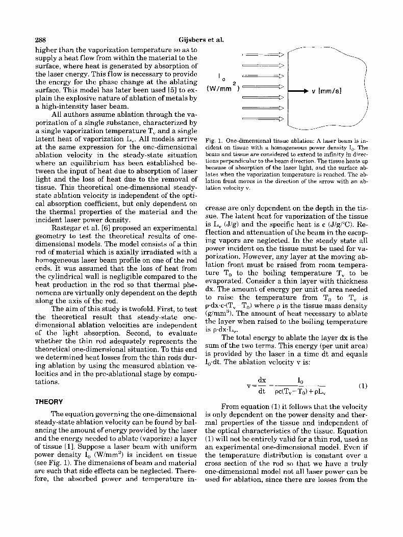
The equation governing the one-dimensional steady-state ablation velocity can be found by bal- ancing the amount of energy provided by the laser and the energy needed to ablate (vaporize) a layer of tissue [l]. Suppose a laser beam with uniform power density I, (W/mm2) is incident on tissue (see Fig. 1). The dimensions of beam and material are such that side effects can be neglected. There- fore, the absorbed power and temperature in-
==L - Fig. 1. One-dimensional tissue ablation: A laser beam is in- cident on tissue with a homogeneous power density I,. The beam and tissue are considered to extend to infinity in direc- tions perpendicular to the beam direction. The tissue heats up because of absorption of the laser light, and the surface ab- lates when the vaporization temperature is reached. The ab- lation front moves in the direction of the arrow with an ab- lation velocity v.
crease are only dependent on the depth in the tis- sue. The latent heat for vaporization of the tissue is L, (J/g) and the specific heat is c (J/g/"C). Re- flection and attenuation of the beam in the escap- ing vapors are neglected. In the steady state all power incident on the tissue must be used for va- porization. However, any layer at the moving ab- lation front must be raised from room tempera- ture To to the boiling temperature T, to be evaporated. Consider a thin layer with thickness dx. The amount of energy per unit of area needed to raise the temperature from To to T, is p.dxdT,-T,) where p is the tissue mass density (g/mm3). The amount of heat necessary to ablate the layer when raised to the boiling temperature is p.dx-L,.
The total energy to ablate the layer dx is the sum of the two terms. This energy (per unit area) is provided by the laser in a time dt and equals I,-dt. The ablation velocity v is:
From equation (1) it follows that the velocity is only dependent on the power density and ther- mal properties of the tissue and independent of the optical characteristics of the tissue. Equation (1) will not be entirely valid for a thin rod, used as an experimental one-dimensional model. Even if the temperature distribution is constant over a cross section of the rod so that we have a truly one-dimensional model not all laser power can be used for ablation, since there are losses from the

CW Laser Ablation Velocities 289 rod sides. Most of the laser power absorbed in the rod is used for vaporization; this is accounted for by the term pL, in equation (1). We assume that ablation is caused by complete vaporization. How- ever, part of the absorbed laser power is lost by radiation and convection from the rod side. Fur- thermore, some laser power is lost by reflection on the rod and by attenuation in the vapors pro- duced by the ablation process. Hence, from energy conservation, P, the total power available t o va- porize a rod with radius r, is equal to:
P = .rrr2.1, (1-R) - q (2)
where IL is the incident laser power density (W/ mm'), R is the fraction of the laser power lost by reflection and attenuation, and q is the rate of heat loss (watts) by radiation and convection from the total rod surface. Note that R and q are the losses during ablation in the steady-state situa- tion! Applying the same arguments with which equation (1) has been derived, the ablation veloc- ity of the rod must be equal to
E, = pc(T,-To) + pL, (J/mm3) ( 3 4
Q = q/nr2 (W/mm2) (3b)
where E, is the energy needed to vaporize a unit of volume of the tissue and Q is the rate of heat loss from the rode side divided by the cross-sec- tional area of the rod. If R and Q can be considered to be constants then a plot of ablation velocity vs. laser power density will yield a straight line, which, in retrospect, is the case (see Fig. 7).
Fitting measured ablation velocities as a function of power densities to a line of the form v = b-I, + a will yield R and Q from a = - Q/E, and b = (l-R)/E,. If E, is known then the reflection R can be derived from the fitted slope, b, of the line:
R = 1 - E;b (4)
Q can be determined from the point where the fitted line will intersect the power density axis at threshold density Ith:
Q = (1-R) Ith (5)
This analysis yields heat power losses from the rod, during ablation, from experimental data
(see below). If Q is small relative to the absorbed power density I, (1-R) then the situation in the rod can be compared to the one-dimensional mod- els mentioned in the Introduction.
To assess the deviation of the thermal behav- ior of a rod in the pre-ablational stage from the true, infinite, one-dimensional situation, the heat loss from a rod at the time just before ablation starts, is computed. This is the moment when the surface of the rod on which the laser beam is in- cident reaches the vaporization temperature T,. The method is described in Appendix A.
MATERIALS AND METHODS
Polyacrylamide gel [7] has been used as an experimental model for biological tissue. The gel resembles tissue in that it contains a large amount of water in a matrix of organic molecules. As is the case for tissue, the gel does not melt before ablation and the material is ablated by complete vaporization. The use of polyacrylamide ensures reproducibility of the samples and en- ables molding the material in the desired rod ge- ometry. The absorption can be controlled by add- ing different concentrations of dye.
The food coloring dye E l l 0 (Eurocert Sunset Yellow, Morton Chemical, The Netherlands) was used. The dye absorbs in the blue-green region. The absorption coefficient, as used in Beer's Law, is proportional to the concentration of the dye.
Just before polymerization, the liquid mix- ture of acrylamide, bis-acrylamide, dye, and poly- merizing agents was put into glass tubes of an inner diameter of 3 or 4 mm and a length of 6 cm. After polymerization the polyacrylamide rods were pushed out of the tubes by using a Perspex piston. The polyacrylamide content was 15 g in 100 ml material. The remaining components were dye (up to 0.5 g per 100 ml at the highest absorp- tion), some small fractions of polymerizing agents, and water. The water content varied somewhat with dye concentration but was always close to 85%. Apart from being colored by the dye, the rods were completely clear; i.e., there was no observable light scattering.
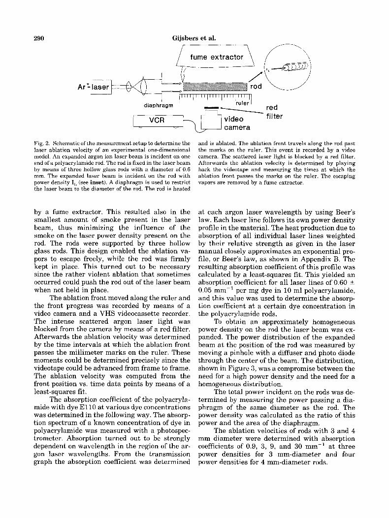
The measurement setup can be found in Fig- ure 2. The rod was kept horizontal and was irra- diated by an expanded argon laser beam (Spectra Physics model 2030) emitting at all lines to obtain sufficient power to vary the power density inci- dent on the rod over a range which is as large as possible. The possible toxic fumes were removed
290 Gijsbers et al.
fume extractor
Fig. 2. Schematic of the measurement setup to determine the laser ablation velocity of an experimental one-dimensional model. An expanded argon ion laser beam is incident on one end of a polyacrylamide rod. The rod is fixed in the laser beam by means of three hollow glass rods with a diameter of 0.6 mm. The expanded laser beam is incident on the rod with power density I, (see inset). A diaphragm is used to restrict the laser beam to the diameter of the rod. The rod is heated
by a fume extractor. This resulted also in the smallest amount of smoke present in the laser beam, thus minimizing the influence of the smoke on the laser power density present on the rod. The rods were supported by three hollow glass rods. This design enabled the ablation va- pors to escape freely, while the rod was firmly kept in place. This turned out to be necessary since the rather violent ablation that sometimes occurred could push the rod out of the laser beam when not held in place.
The ablation front moved along the ruler and the front progress was recorded by means of a video camera and a VHS videocassette recorder. The intense scattered argon laser light was blocked from the camera by means of a red filter. Afterwards the ablation velocity was determined by the time intervals at which the ablation front passes the millimeter marks on the ruler. These moments could be determined precisely since the videotape could be advanced from frame to frame. The ablation velocity was computed from the front position vs. time data points by means of a least-squares fit.
The absorption coefficient of the polyacryla- mide with dye E l l 0 at various dye concentrations was determined in the following way. The absorp- tion spectrum of a known concentration of dye in polyacrylamide was measured with a photospec- trometer. Absorption turned out to be strongly dependent on wavelength in the region of the ar- gon laser wavelengths. From the transmission graph the absorption coefficient was determined
W
and is ablated. The ablation front travels along the rod past the marks on the ruler. This event is recorded by a video camera. The scattered laser light is blocked by a red filter. Afterwards the ablation velocity is determined by playing back the videotape and measuring the times at which the ablation front passes the marks on the ruler. The escaping vapors are removed by a fume extractor.
at each argon laser wavelength by using Beer's law. Each laser line follows its own power density profile in the material. The heat production due to absorption of all individual laser lines weighted by their relative strength as given in the laser manual closely approximates an exponential pro- file, or Beer's law, as shown in Appendix B. The resulting absorption coefficient of this profile was calculated by a least-squares fit. This yielded an absorption Coefficient for all laser lines of 0.60 2 0.05 mm-' per mg dye in 10 ml polyacrylamide, and this value was used to determine the absorp- tion coefficient at a certain dye concentration in the polyacrylamide rods.
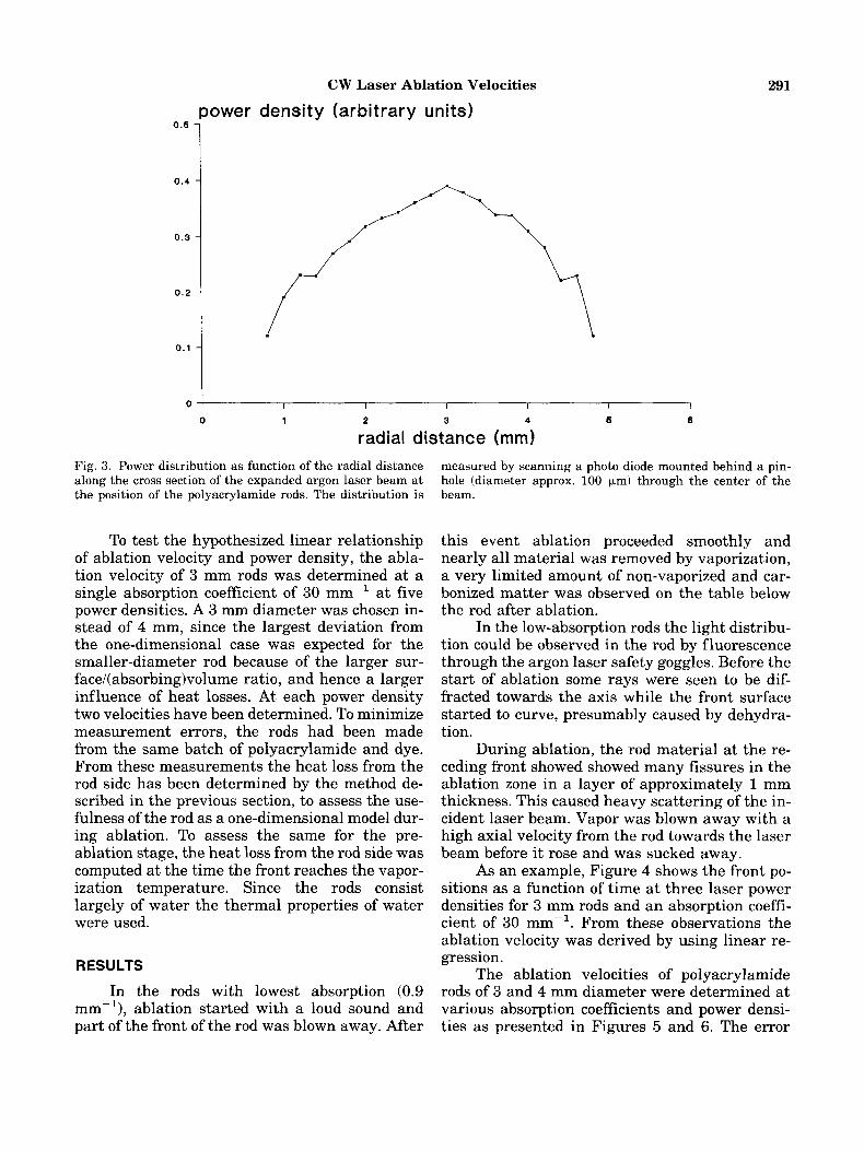
To obtain an approximately homogeneous power density on the rod the laser beam was ex- panded. The power distribution of the expanded beam at the position of the rod was measured by moving a pinhole with a diffuser and photo diode through the center of the beam. The distribution, shown in Figure 3, was a compromise between the need for a high power density and the need for a homogeneous distribution.
The total power incident on the rods was de- termined by measuring the power passing a dia- phragm of the same diameter as the rod. The power density was calculated as the ratio of this power and the area of the diaphragm.
The ablation velocities of rods with 3 and 4 mm diameter were determined with absorption coefficients of 0.9, 3, 9, and 30 mm-' at three power densities for 3 mm-diameter and four power densities for 4 mm-diameter rods.
CW Laser Ablation Velocities 291
I I I I I I
0 1 2 3 4 6 8
radial distance (mm) Fig. 3. Power distribution as function of the radial distance along the cross section of the expanded argon laser beam a t the position of the polyacrylamide rods. The distribution is
To test the hypothesized linear relationship of ablation velocity and power density, the abla- tion velocity of 3 mm rods was determined at a single absorption coefficient of 30 mm-' at five power densities. A 3 mm diameter was chosen in- stead of 4 mm, since the largest deviation from the one-dimensional case was expected for the smaller-diameter rod because of the larger sur- face/(absorbing)volume ratio, and hence a larger influence of heat losses. At each power density two velocities have been determined. To minimize measurement errors, the rods had been made from the same batch of polyacrylamide and dye. From these measurements the heat loss from the rod side has been determined by the method de- scribed in the previous section, t o assess the use- fulness of the rod as a one-dimensional model dur- ing ablation. To assess the same for the pre- ablation stage, the heat loss from the rod side was computed at the time the front reaches the vapor- ization temperature. Since the rods consist largely of water the thermal properties of water were used.
RESULTS
In the rods with lowest absorption (0.9 mm-'1, ablation started with a loud sound and part of the front of the rod was blown away. After
measured by scanning a photo diode mounted behind a pin- hole (diameter approx. 100 pm) through the center of the beam.
this event ablation proceeded smoothly and nearly all material was removed by vaporization, a very limited amount of non-vaporized and car- bonized matter was observed on the table below the rod after ablation.
In the low-absorption rods the light distribu- tion could be observed in the rod by fluorescence through the argon laser safety goggles. Before the start of ablation some rays were seen to be dif- fracted towards the axis while the front surface started to curve, presumably caused by dehydra- tion.
During ablation, the rod material at the re- ceding front showed showed many fissures in the ablation zone in a layer of approximately 1 mm thickness. This caused heavy scattering of the in- cident laser beam. Vapor was blown away with a high axial velocity from the rod towards the laser beam before it rose and was sucked away.
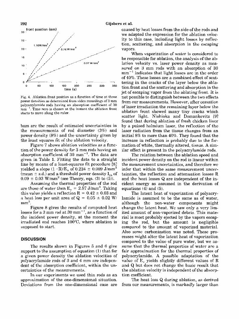
As an example, Figure 4 shows the front po- sitions as a function of time at three laser power densities for 3 mm rods and an absorption coeffi- cient of 30 mm-l. From these observations the ablation velocity was derived by using linear re- gression.
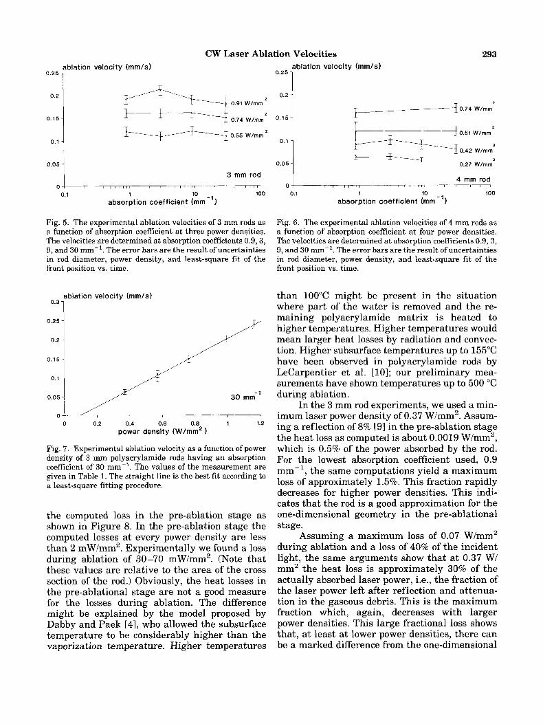
The ablation velocities of polyacrylamide rods of 3 and 4 mm diameter were determined at various absorption coefficients and power densi- ties as presented in Figures 5 and 6. The error
292 Gijsbers et al. front position (mm)
30 1 / /
I // /
, 0 50 100 150 200 250 300 350
time (s)
Fig. 4. Ablation front position as a function of time at three power densities as determined from video recordings of 3 mm polyacrylamide rods having an absorption coefficient of 30 rnm-'. Time zero is chosen at the instant the ablation front starts to move along the ruler.
bars are the result of estimated uncertainties in the measurements of rod diameter (3%) and power density (8%) and the uncertainty given by the least squares fit of the ablation velocity.
Figure 7 shows ablation velocities as a func- tion of the power density for 3 mm rods having an absorption coefficient of 30 mm-l. The data are given in Table 1. Fitting the data to a straight line by means of a least-squares fit procedure 181 yielded a slope (l-R)/Ev of 0.224 * 0.009 J/mm3 (mean 2 s.d.) and a threshold power density Ith of 0.09 * 0.03 W/mm2 (see Theory, eqs. (3) to ( 5 ) ) .
Assuming the thermal properties of the rod are those of water then E, = 2.57 J/mm3. Taking this value yields a reflection R = 0.42 * 0.03 and a heat loss per unit area of Q = 0.05 2 0.02 W/ mm2.
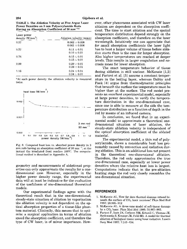
Figure 8 gives the results of computed heat losses for a 3 mm rod at 30 mm-l, as a function of the incident power density, at the moment the irradiated end reaches 100°C, where ablation is supposed to start.
DISCUSSION
The results shown in Figures 5 and 6 give support to the assumption of equation (1) that for a given power density the ablation velocities of polyacrylamide rods of 3 and 4 mm are indepen- dent of the absorption coefficient, within the un- certainties of the measurements.
In our experiments we used thin rods as an approximation of the one-dimensional situation. Deviations from the one-dimensional case are
caused by heat losses from the side of the rods and we adapted the expression for the ablation veloc- ity to this case, including light losses by reflec- tion, scattering, and absorption in the escaping vapors.
When vaporization of water is considered to be responsible for ablation, the analysis of the ab- lation velocity vs. laser power density as mea- sured on 3 mm rods with an absorption of 30 mm-' indicates that light losses are in the order of 40%. These losses are a combined effect of scat- tering in the cracks of the layer below the abla- tion front and the scattering and absorption in the jet of escaping vapor from the ablating front. It is not possible to distinguish between the two effects from our measurements. However, after cessation of laser irradiation the remaining layer below the ablation front showed many tiny cracks which scatter light. Nishioka and Domankevitz C91 found that during ablation of fresh chicken liver by a pulsed holmium laser, the reflection of the laser radiation from the tissue changes from an initial 8% to more than 40%. They found that the increase in reflection is probably due to the for- mation of white, thermally altered, tissue. A sim- ilar effect is present in the polyacrylamide rods.
The relation between the ablation speed and incident power density on the rod is linear within the measurement uncertainties, and therefore we infer that within the same measurement uncer- tainties, the reflection and attenuation losses R and the heat losses Q are independent of the in- cident energy as assumed in the derivation of equations (4) and (5) .
The latent heat of vaporization of polyacry- lamide is assumed to be the same as of water, although the non-water components might change the latent heat. We saw only a very lim- ited amount of non-vaporized debris. This mate- rial is most probably ejected by the vapors escap- ing the rod, but the amount is negligible compared to the amount of vaporized material. Also some carbonization was noted. These pro- cesses might alter the latent heat of vaporization compared to the value of pure water, but we as- sume that the thermal properties of water are a fair approximation for the thermal properties of polyacrylamide. A possible adaptation of the value of E, yields slightly different values of R and Q but does not change the basic result that the ablation velocity is independent of the absorp- tion coefficient.
The heat loss Q during ablation, as derived from our measurements, is markedly larger than
CW Laser Ablation Velocities
0.15-
0.1 -
0.05 -
293
4 rnm rod
ablation velocity (mm/s) 0.25 1
ablation velocity (mm/s) 0.25 1
0.2
0.15
0.1
3 rnm rod I
0.1 1 10 100
absorption coefficient (mm
Fig. 5. The experimental ablation velocities of 3 mm rods as a function of absorption coefficient at three power densities. The velocities are determined at absorption coefficients 0.9,3, 9, and 30 mm-'. The error bars are the result of uncertainties in rod diameter, power density, and least-square tit of the front position vs. time.
ablation velocity (mm/s)
0.3 1 0.25 -
0.2 -
0.15-
0.1
0.05 -
/
30 mrn-'
, 0 0.2 0.4 0.6 0.8 1 1.2
power density (W/mm2
Fig. 7. Experimental ablation velocity as a function of power density of 3 mm polyacrylamide rods having an absorption coefficient of 30 mm-'. The values of the measurement are given in Table 1. The straight line is the best fit according to a least-square fitting procedure.
the computed loss in the pre-ablation stage as shown in Figure 8. In the pre-ablation stage the computed losses at every power density are less than 2 mW/mm2. Experimentally we found a loss during ablation of 30-70 mW/mm2. (Note that these values are relative to the area of the cross section of t.he rod.) Obviously, the heat losses in the pre-ablational stage are not a good measure for the losses during ablation. The difference might be explained by the model proposed by Dabby and Paek [41, who allowed the subsurface temperature to be considerably higher than the vaporization temperature. Higher temperatures
0.2 I F- -3 0.74 Wlmm'
2
0.51 Wlmm
0.42 Wlmm
0.27 Wlmm
2
2 -- Fig. 6. The experimental ablation velocities of 4 mm rods as a function of absorption coefficient at four power densities. The velocities are determined at absorption coefficients 0.9,3, 9, and 30 mm-'. The error bars are the result of uncertainties in rod diameter, power density, and least-square fit of the front position vs. time.
than 100°C might be present in the situation where part of the water is removed and the re- maining polyacrylamide matrix is heated to higher temperatures. Higher temperatures would mean larger heat losses by radiation and convec- tion. Higher subsurface temperatures up to 155°C have been observed in polyacrylamide rods by LeCarpentier et al. [lo]; our preliminary mea- surements have shown temperatures up to 500 "C during ablation.
In the 3 mm rod experiments, we used a min- imum laser power density of 0.37 W/mm2. Assum- ing a reflection of 8% [91 in the pre-ablation stage the heat loss as computed is about 0.0019 W/mm2, which is 0.5% of the power absorbed by the rod. For the lowest absorption coefficient used, 0.9 mm-', the same computations yield a maximum loss of approximately 1.5%. This fraction rapidly decreases for higher power densities. This indi- cates that the rod is a good approximation for the one-dimensional geometry in the pre-ablational stage.
Assuming a maximum loss of 0.07 W/mm2 during ablation and a loss of 40% of the incident light, the same arguments show that at 0.37 W/ mm2 the heat loss is approximately 30% of the actually absorbed laser power, i.e., the fraction of the laser power left after reflection and attenua- tion in the gaseous debris. This is the maximum fraction which, again, decreases with larger power densities. This large fractional loss shows that, at least a t lower power densities, there can be a marked difference from the one-dimensional
294 Gijsbers et al. TABLE 1. The Ablation Velocity at Five Argon Laser Power Densities on 3 mm Polyacrylamide Rods Having an Absorption Coefficient of 30 mrn-'*
Laser power density (Wimrn') Ablation velocity (mm/s) 0.37 0.062 2 0.006
0.063 t 0.006 0.57 0.11 t 0.01
0.10 2 0.01 0.76 0.15 t 0.01
0.15 t 0.01 0.99 0.20 -+ 0.01
0.20 t 0.01 1.16 0.24 t 0.01
0.24 t 0.01
*At each power density the ablation velocity is measured twice.
2 heat loss (W/mm )
1 1 0.002
0.001 1 3 mm rod 30 m m - l
0.000 , , I , , , , , , 0 0.1 0.2 0.3 0.4 0.5 0.6 0.7 0.8 0.9 1 1.1 1.2
power density (W/mm '1
Fig. 8. Computed heat loss vs. absorbed power density in 3 mm rods having an absorption coefficient of 30 mm-', at the instant the irradiated front reaches 100°C. The computa- tional method is described in Appendix A.
geometry and measurements of ablational prop- erties can only approximate the results for a one- dimensional case. However, especially in the higher power density range, the experimental data will at least be indicative for the evaluation of the usefulness of one-dimensional theoretical models.
Our experimental findings agree with the theoretical result that in the one-dimensional, steady-state situation of ablation by vaporization the ablation velocity is not dependent on the op- tical absorption properties of a tissue-like phan- tom material. Clinically, this means that to opti- mize a surgical application in terms of ablation speed the absorption coefficient, and therefore the type of CW laser, is of minor importance. How-
ever, other phenomena associated with CW laser ablation are dependent on the absorption coeffi- cient. The time to start ablation and the spatial temperature distribution depend strongly on the absorption coefficient, and therefore on the laser wavelength. Intuitively one can appreciate that for small absorption coefficients the laser light has to heat a larger volume of tissue before abla- tion starts than is the case for larger absorption. Also higher temperatures are reached at deeper levels. This results in larger coagulation and ne- crosis zones for lower absorption.
The exact temperature behavior of tissue during ablation is still unclear. McKenzie [l,2] and Partovi et al. [3] assume a constant temper- ature in the boiling layer, whereas Dabby and Paek [41 argue from thermodynamic principles that beneath the surface the temperature must be higher than at the surface. The rod model pre- sents an excellent experimental model, especially at large power densities, to verify the tempera- ture distribution in the one-dimensional case, since one is able to measure at the side the tem- perature distribution as a function of depth in the rod by means of an infrared camera.
In conclusion, we found that in an experi- mental model to approximate a theoretical one- dimensional situation of laser ablation, the steady-state ablation velocity is independent of the optical absorption coefficient of the ablated material.
The experimental model, a thin rod of poly- acrylamide, shows a considerable heat loss pre- sumably caused by convection and radiation dur- ing ablation. This is an additional loss not present in the theoretical one-dimensional ablation. Therefore, the rod only approximates the true one-dimensional case, especially at lower power densities where the relative heat loss is largest. Computations indicate that in the pre-ablation heating stage the rod very closely resembles the one-dimensional situation.
REFERENCES
1. McKenzie AL. How far does thermal damage extend be- neath the surface of CO, laser incisions? Phys Med Biol 1983; 28:905-912.
2. McKenzie AL. A three-zone model of soft-tissue damage by a GO, laser. Phys Med Biol 1986; 31:967-983.
3. Partovi F, Izatt JA, Cothren RM, Kittrell C, Thomas JE, Strikwerda S, Kramer JR, Feld MS. A model for thermal ablation of biological tissue using laser radiation. Lasers Surg Med 1987; 7:141-154.
CW Laser Ablation Velocities 295 4.
5.
6.
7.
8.
9.
10.
11.
12.
Dabby FW, Paek UC. High-intensity laser-induced va- porization and explosion of solid material. IEEE J Quant Elec 1972; QE-8:106-111. Gagliano FP, Paek UC. Observation of laser-induced ex- plosion of solid materials and correlation with theory.
Rastegar S, van Gemert MJC, Welch AJ. Technique for measurement of one-dimensional instantaneous ablation velocity. Lasers Surg Med 1988; 8:533-535. Bini MG, Ignesti A, Millanta L, Olmi R, Rubino N, Vanni R. The polyacrylamide as a phantom material for electro- magnetic hyperthermia studies. IEEE Trans Biomed Eng
Bevington PR. “Data Reduction and Error Analysis for the Physical Sciences.” New York: McGraw-Hill Book Company, 1969:92-118. Nishioka NS, Domankevitz Y. Reflectance during pulsed holmium laser irradiation of tissue. Lasers Surg Med 1989; 9:375-381. LeCarpentier GL, Motamedi M, Rastegar S, Welch AJ. Simultaneous analysis of thermal and mechanical events during CW laser ablation of biological media. “Thermal and Optical Interactions With Biological and Related Composite Materials. Bellingham: SPIE, 1989, Vol 1064,
Eckert ERG, Drake RM. “Heat and Mass Transfer.” New York: McGraw-Hill Book Company, 1959:311-319. Carslaw HS, Jaeger JC. “Conduction of Heat in Solids.” 2nd Edition. Oxford, UK: Clarendon Press, 1986: 133- 134.
Appl Opt 1974; 13:274-279.
1984; BME-31:317-322.
pp 107-113.
2 r
--.[a.E.(T4-T$) + 4.35.10-6.r-1’4.(T-T0)5/4] (A.1)
p = mass density (g/mm3) c = specific heat (J/g/”C)
K = thermal conductivity (J/mm/s/”C) pa = absorption coefficient (mm-l) IL = incident laser power density (W/mm2) R = reflection coefficient
x,t = position (mm) and time (s) The heat flux at x = 0 is given by
aT K--= u.E.(T4-T;) + 4.35.10-6.r-1’4.(T-To)5’4 (A.2)
at
which is one of the boundary conditions.
are: The initial and other boundary conditions
t = O : T(x) = TO (A.3)
X = W : T = TO (A.4)
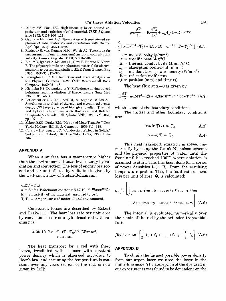
This heat transport equation is solved nu- merically by using the Crank-Nicholson scheme and the physical properties of water until the front x=O has reached 100°C where ablation is assumed t o start. This has been done for a series of power densities IL(l -R)* From the resulting temperature profiles T(x), the total rate of heat loss per unit of area, Q, is calculated:
APPENDIX A
When a surface has a temperature higher than the environment it loses heat energy by ra- diation and convection. The loss of energy per sec- ond and Per unit of area by radiation is given by the well-known law of Stefan-Boltzmann:
vE(T4 - T4,) u = Stefan-Boltzmann constant: 5.67.10-14 W/mm2/”C4 E = emissivity of the material, assumed to be 1 T, To = temperatures of material and environment.
Q=-. 1 2Tr,(u.E.(T4(x)-T;) + 4.35.10-6.r-”4,(T(x)-T~)5/4)dX r r 2
+ .irr2.(u.E.(T4(0)-T$) + 4.35.10-6.r-1’4~(T(0)-T~)5’4) (A.5) 1 L” Convection losses are described by Eckert
and Drake [ll]. The heat loss rate per unit area by convection in air of a cylindrical rod with ra- dius r is: rule:
The integral is evaluated numerically over the z-axis of the rod by the extended trapezoidal
4.35.1 0-6.r-1/4- (T - (W/mm2) r in mm
The heat transport for a rod with these losses, irradiated with a laser with constant power density which is absorbed according to Beer’s law, and assuming the temperature is con- stant over any cross section of the rod, is now given by [121:
APPEND,X
To obtain the largest possible power density from our argon laser we used the laser in the multi-line mode. The absorption of the dye used in our experiments was found to be dependent on the

296 Gijsbers et al. wavelength in the spectral region of the argon laser lines. Still, to use the concept of a single absorption coefficient, whether the absorbed laser power in the polyacrylamide with dye followed an exponential profile or Beer's law was evaluated in the following manner:
Every single line of the argon laser follows an exponential decrease of fluence rate according to Beer's law:
Qi(x) = Ii.e-FL1x (A.7) Qi : fluence rate at depth x (W/mm2) Ii :incident power density of argon line with
pi : absorption coefficient of the dye at wave- wavelength Xi (W/mm2)
length Xi (rnm-')
The absorbed power of the laser line per unit
Qi(x) = pi-Ii-e-plx (W/mm3) (A.8)
The total absorbed power per unit volume at a certain depth of all laser lines, Q, is the sum of the Qi's for all laser lines:
volume at a certain depth x, Qi(x), is equal to
(A.9) 1
The different pi's for all argon laser lines of a concentration of 1 mg dye in 10 ml polyacryla- mide have been determined by means of a photo- spectrometer, and the sum of equation A.9 was calculated for x=O to x=10 mm with the Ii's taken as the relative powers as given in the argon laser's manual.
The result, normalized to unity at x=O, is given in Figure A.l. The dots are the sum of the individual absorbed powers per volume at a cer- tain depth; the line is a fit of an exponential func-
relative absorbed power per unit volume 1 1
1 -----l
0 1 2 3 4 5 8 7 8 9 10
depth (mml
0 L , , ,
Fig. A . l . The relative absorbed power per unit volume as a function of depth in 1 mg dye E l l 0 per 10 ml polyacrylamide, which is the sum of the absorbed powers of the individual argon laser lines. The dots are the calculations of Q(x) =
C,k,.I,.e-piX, normalized to unity at x = 0 , where p1 are the absorption coefficients of the individual argon laser lines as determined by means of a photospectrometer and I, are the relative powers in the multi-line argon laser beam as given in the laser manual. The drawn line is a least-squares exponen- tial fit to the dots which is Q(z) = e-px with p = 0.60 mm- '.
tion Q(x) = ePpx with p = 0.60 mm-l. It is obvi- ous that the total absorbed power as a function of depth closely follows an exponential decrease or Beer's law with an overall absorption coefficient p. The value of p is the result of a least-squares fit to the calculated Q(x). When taking into account the uncertainty in the measured individual ab- sorption coefficients pi, the least-square fit yields a value for the overall absorption coefficient p of 0.60 * 0.05 mm-' at 1 mg dye per 10 ml poly- acrylamide. It was found that the absorption co- efficient was proportional to the concentration of dye. In this way the absorption coefficient of the polyacrylamide could be varied by changing the concentration of dye.
Copyright © 2022 FDOKUMEN