CT scan Positive Finding Pattern of Head Injury at the University of Gondar Hospital; North West...
11
CC-BY-NC 2014 mjmbr Malaysian Journal of Medical and Biological Research www.jmbr-my.weebly.com A SIAN B USINESS C ONSORTIUM ISSN: 2313-0008 (Print); 2313-0016 (Online)
-
Upload
abcjournals -
Category
Documents
-
view
3 -
download
0
Transcript of CT scan Positive Finding Pattern of Head Injury at the University of Gondar Hospital; North West...

CC-BY-NC
2014
mjmbr
Malaysian Journal of Medical and Biological Research www.jmbr-my.weebly.com
A S I A N B U S I N E S S C O N S O R T I U M
ISSN: 2313-0008 (Print); 2313-0016 (Online)

Malaysian Journal of Medical and Biological Research, Volume 1, No 2 (2014)
Copyright © CC-BY-NC 2014, Asian Business Consortium | MJMBR Page 64
CT scan Positive Finding Pattern of Head
Injury at the University of Gondar Hospital;
North West Ethiopia
Zerubabel Tegegne Desita1, Wossen Mulugeta
2
1Assistant Professor of Radiology, University of Gondar, Ethiopia 2Assistant Professor of Ophthalmology, University of Gondar, Ethiopia
ABSTRACT
Background: Head injuries rank high among morbidities due to trauma. Computerized tomography is an important modality in the investigation of these cases. However, there is no literature on the importance of computerized tomography in the diagnosis of head injury in Ethiopia. This study therefore is aimed to document the computerized tomographic features of patients with head injury managed at the University of Gondar Teaching Hospital. Materials and Methods: A cross sectional study involving 96 patients with head injury who had CT scan of the head in the UOG hospital over a 12-month period. Results: Most of the patients were male (74%). Majority (58%) were in the age range of 20 to 40 years with a mean age of 31yrs. The most common abnormal findings were skull fracture (52%) and intracerebral hemorrhage and contusions (51%). It is followed by subdural hemorrhage (33%) soft tissue swelling 32% and epidural hemorrhage 10%. Conclusion: Skull fracture and intra cerebral hemorrhage were the most common abnormal findings. This study has demonstrated the importance of CT scan in the evaluation of head injury by giving visibility of intracranial post traumatic injuries in a high proportion of patients which would be difficult to reach in to diagnosis clinically or using skull radiography alone. This obviously will have a significant role in improving patient management. Taking this in to account expansion of CT scan service for moderate to severe head injury patients is recommended in Ethiopia.
Key words: CT scan, CT positive findings, Head injury, Intracranial injury, Skull fracture
INTRODUCTION
Head injury can be defined as any alteration in mental or physical functioning related to a blow to the head. It ranks high among morbidities due to trauma (Adeyekun, 2013). The severity of head injuries is most commonly measured with Glasgow Coma Scale (GCS) score. Traditionally, a score of 13-15 indicates mild injury, of 9-12 moderate injury, and a score of 8 or less severe injury (Shoar and Saadat). Direct and counter coupe injuries can result in an intracerebral focal contusion or hemorrhage as well as an extra cerebral
12/26/2014 Source of Support: Nil, Conflict of Interest: The authors declare that they have no competing interests
How to Cite: Desita ZT and Mulugeta W. 2014. CT scan Positive Finding Pattern of Head Injury at the University
of Gondar Hospital; North West Ethiopia Malaysian Journal of Medical and Biological Research, 1, 64-72.
This article is is licensed under a Creative Commons Attribution-NonCommercial 4.0 International License. Attribution-NonCommercial (CC BY-NC) license lets others remix, tweak, and build upon work non-commercially,
and although the new works must also acknowledge & be non-commercial.

Malaysian Journal of Medical and Biological Research, Volume 1, No 2 (2014)
Copyright © CC-BY-NC 2014, Asian Business Consortium | MJMBR Page 65
hemorrhage. Brain edema, diffuse axonal injury and skull fractures may also occur alongside other injuries. More severe and diffuse axonal injury has been found to correlate with vegetative states and the acute onset of coma following injury (Gupta etal, 2011). Poly trauma due to road traffic accidents (RTA) is a leading cause of head injury in teenagers and young adults in the developed world (Lewis, 2012). Head injuries cause immediate death in 25% of acute trauma victims. More than half of the cases of head trauma are caused by RTA, leading to 70% of all deaths due to brain injury (Gupta etel, 2011; Mohammad etel, 2005). Amongst the severely injured patients, majority survives with severe disability and few continue to be in a vegetative state. Increasing age is associated with poorer outcome in patients with head injury. Assaults, falls, sports, blast injuries are other major risk factors (Peshawar, 2011). Presentations vary from normal to confusion and deep coma. Other clinical features include abnormal papillary reactivity, CN VI & VII palsies, anosmia, and CSF leak (2, Peshawar, 2011). Patients who have intracranial lesions which require urgent operative intervention need urgent diagnosis and intervention for better outcome (Peshawar, 2011). In studies done in some parts of Africa, results show that head injury ranks high from traumatic causes of morbidity and also like in studies from the developed world these researches show that younger and male population is more affected with head injury (Adeyekun, 2013). In Ethiopia as to the knowledge of the author no population or hospital based study is done on the pattern of head injury. On studies done in the capital Addis Ababa at TAH to assess cause of hospital admissions; gastrointestinal and neuro-surgical patients constituted 75.8% of all emergency admissions. Among patients with trauma, isolated head injury was major (59.2%) cause of death. Neurosurgical emergencies had the highest mortality rate (36.8%) (Biluts etel, 2009). Irrespective of the highest rate of occurrence and fatality, there is no proper investigation and management of head injury in the country especially out of the capital due to lack of CT scan and trained personnel. In part of Ethiopia ;Gondar, where the study is done ; trauma constituted about 46% of surgical patients at University of Gondar Hospital (UOGH), which shows a significant burden to the institution (Osman etel, 2003). Head injury is the second cause of surgical admission in UOGH next to intestinal obstruction and accounts 35% of all neurosurgical admissions. There is high mortality rate in post traumatic neurosurgical patients (36.6%) compared to elective patients (4.8%) (Ghebrat, 1997). Computed tomography (CT) has become the diagnostic modality of choice for head trauma due to its accuracy, reliability, safety, and wide availability in most part of the world (Adeyekun, 2013; Shoar and Saadat). The changes in microcirculation, impaired auto-regulation, cerebral edema, and axonal injury start as soon as head injury occurs and manifest as clinical, biochemical, and radiological changes. Proper therapeutic management of brain injury is based on correct diagnosis and appreciation of the temporal course of the disease process. CT scan detects and precisely localizes the intracranial hematomas, brain contusions, edema and foreign bodies (Pearce, 2012; Lee, 2005; Trauma etel, 2009). Magnetic resonance imaging (MRI) is considered more sensitive than CT for subtle abnormalities. Because CT does not always explain the posttraumatic neurologic examination, MRI is being performed with increasing frequency. Although MRI at a later stage may be of significant prognostic value, the role of early MRI within 48 hours is minimal (Trauma etel, 2009; James, 2010). Skull radiography is the widely available method of imaging for head injury before the era of CT with limited importance in detecting skull fracture but not helpful in evaluation of intracranial structures (Tasker, 2005).
Malaysian Journal of Medical and Biological Research, Volume 1, No 2 (2014)
Copyright © CC-BY-NC 2014, Asian Business Consortium | MJMBR Page 66
Not all patients with head injury should have CT scan examination. Patients with moderate and severe head injury with GCS 9-12 and below 9 respectively should have a CT scan examination. Patients with mild head injury should have CT scan based on criteria set by additional clinical findings in addition to GCS of 13-14. This includes clinical signs of skull fracture, amnesia, vomiting, posttraumatic seizure, Age ≥60 y, neurologic deficit and use of anticoagulant therapy by the patient (Shoar and Saadat; Mohammad etel, 2005). This study was conducted to determine pattern of head injury up on CT scan findings so that the local pattern of head injury can be known and evidence based plan can be implemented in the diagnosis and management of head injury locally and nationally.
METHODS
Study area: The study is conducted in the university of Gondar hospital which is located in south north part of Ethiopia 727 km from Addis Ababa .The hospital is a referral and teaching hospital giving a service to urban and rural population of the surrounding to over 5 million populations. The university is one of the centers of excellence in Ethiopia especially in the health sector under the collage of medical and health sciences. The radiology department is one of the many departments in the collage and gives radiologic medical service besides its academic activities being equipped with high-tech radiologic machines including a 4 slice CT scan machine on which the study is done and has radiologists including the one who conducts this research. Study design: This descriptive cross-sectional study included 96 patients with head injury who had post traumatic findings on CT of the head in the University of Gondar hospital, from September 2011 to September 2012 after ethical permission is obtained from the hospital administration. All consecutive patients who had post traumatic findings on pre contrast CT scans of the head are included in the study irrespective of age and sex. The CT findings were grouped on basis of the principal findings. A 4 slice bright speed CT scan machine made by GE is used and the results are evaluated and recorded by a radiologist. The data is analyzed with SPSS version 20. Cranial Computed Tomography Scan Technique and Interpretation of Image All patients underwent pre contrast CT scan from skull base to the vertex and sequential axial slices of 5mm with 1.25mm reconstructed image were obtained. Detailed radiological evaluation was done and findings were recorded. Pneumocranium was assessed by the presence of intracranial air on the CT scan. Intra-parenchymal contusions may be hemorrhagic or non-hemorrhagic. Diffuse axonal injury was categorized as parenchyma injury with hemorrhage the former seen as multiple small foci of hyper density. Intracerebral hematomas were homogeneously hyper dense with a density of blood (40-60 HU). Parenchymal contusion and laceration was seen as area of hyper density and hypo density with irregular discontinuous outline. These all are categorized together in this study. The radiological appearance of a typical epidural hematoma was biconvex, lentiform, crescentic or irregular and was heterogeneous in attenuation, containing areas of hyper dense blood clot and dense serum. The typical appearance of subdural hematoma is hyper dense crescent-shaped extra-axial collection with a convex lateral border and concave medial border overlying the cerebral convexity. It appears hyper, iso and hypo dense in acute sub acute and chronic subtypes.

Malaysian Journal of Medical and Biological Research, Volume 1, No 2 (2014)
Copyright © CC-BY-NC 2014, Asian Business Consortium | MJMBR Page 67
On CT scan, hyper density of acute hemorrhage was visualized in the sulci overlying the cerebral convexities, within the sylvian fissures, basal cisterns, and inter-hemispheric fissure which is called subarachnoid hemorrhage. Similar hyper density of blood is seen with in the ventricles in ventricular hemorrhage. Diffuse brain edema is considered when there is compression of the lateral and third ventricles and perimesencephalic cistern with prominent cerebral gyri together with homogenously decreased attenuation with grey-white matter interface. Significant midline shift is considered when shift is more than 5mm from midline. Depressed fractures of skull vault were considered to be present when at least one skull bone showed inward displacement, while other fractures categorized as non depressed when no displacement of bone fragment. Indirect signs of basal skull fractures are not taken as evidence of fracture as both infectious and other inflammatory causes can’t be excluded. Significant midline shift is considered when a shift by more than 5mm measured from the midline. Multifocal involvement is considered in any two different cranial structures injury; scalp, skull or intracranial structures. Ethical clearance Ethical clearance was obtained from the UOG hospital administration to use the patients’ radiologic and epidemiological data for this specific research without mentioning patients’ identity at any of the course of the study. Verbal consent is obtained from patients or their relatives when the patients are not clinically fit to do so.
RESULT
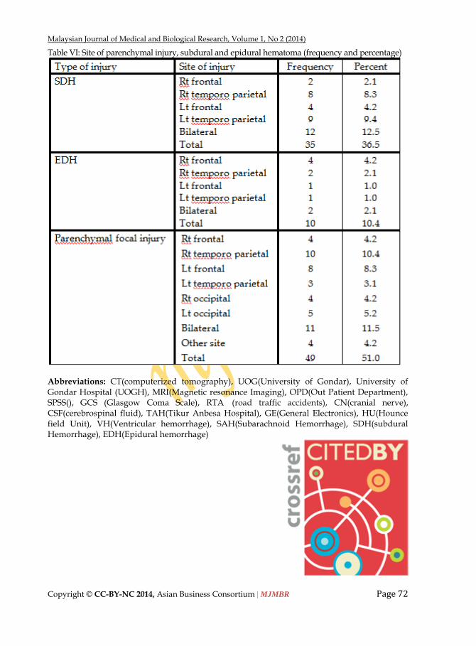
Most of the patients 74% were male. Majority 58% were in the age range of 20 to 40 years and only 3% were above the age of 50 with a mean age of 31yrs (tables 1-2). Skull fracture was found on 52%, of which 32 % was depressed skull fracture and 8% had pneumocephalus. Soft tissue swelling was found in 32% of patient (table3). Among those with soft tissue swelling 93% had skull fracture while 33% of patients with no soft tissue swelling had skull fracture (table4). Epidural hematoma is detected in 10% while subdural hematoma is diagnosed in36% of which acute and sub acute subdural hematoma was 33% and the rest 3% was chronic (table3). Only 3% of patients had both epidural and subdural hematoma simultaneously and also only 11% and 6% had subdural and epidural hematoma together with skull fracture respectively making the proportion of epidural hematoma associated with skull fracture 60 %( table5). Subdural hematoma was maximally present in the temporo-parietal region (49%), followed by frontal region (17%) while epidural hematoma is common in the frontal region (50%) followed by temporo parietal region 30(table6). More than half (51%) had parenchymal hemorrhagic or contusion, and 4% had diffuse brain edema while the rest 45% had no detected brain parenchymal injury (table3). One third of (33%) patients had brain parenchymal focal injury together with skull fracture (table4). Parenchymal hemorrhage was equivalent in both sides 18; 36.8% right and 16; 32.6% left. The right temporo-parietal and the left frontal lobes are the commonest site of cerebral hemorrhage and contusion 10; 20.4% and 8; 16.3 % respectively. Bilateral cerebral involvement is seen in 11cases; 22.4% (table6). Ventricular and subarachnoid hemorrhage was found to be 4%; each 2%. There was focal brain edema in 52.0% and significant midline shift was found in 62.5%. Overall multifocal involvement was found in 63.5 % (table3).

Malaysian Journal of Medical and Biological Research, Volume 1, No 2 (2014)
Copyright © CC-BY-NC 2014, Asian Business Consortium | MJMBR Page 68
DISCUSSION
The neuroradiology of head trauma has undergone dramatic changes since the advent of computed tomography, which has helped significantly to modify the management of head trauma. Using CT scan, this research has confirmed that young males are the most common victim of head injury and a wide variety of CT abnormalities are identified in head trauma victims at the setup the study conducted. The proportion of intracranial injuries not detectable on skull x ray associated with head trauma is found to be high signifying the high necessity of CT in the setup where the research is done. The results are consistent with previous studies that have shown that the incidence head trauma was seen in the most productive years of life. A study done in India showed 71% of the patients belonged to the 20 to 50 years age group (Gupta etal, 2011). In the present study 58% were in the age range of 20 to 40 years. Males are found to be predominantly affected accounting 74% similar to in previous studies the male to female ratio was 4:1 (Gupta etal, 2011). Most likely the reason is that males in this age group are more involved to traffic and outdoor activities than females and extreme ages in developing countries and hence most susceptible (Gupta etal, 2011). Skull fracture was found on 52%, of which 32 % was depressed skull fracture and 4.2% had pneumocephalus. Almost similar finding is seen in other researches where skull fracture is seen 62%, and pneumocranium 12% (Adeyekun, 2013; Gupta etal, 2011). Soft tissue swelling was found in 32% of patients in this study lower than seen in other study which is up to 86% being the commonest change seen in head injury (Mohammad etel, 2005). This variation can be explained by the difference in the mechanism of injury and the criteria for CT scanning in head injury in different areas based on the economic status and health care service of countries. In this study epidural hematoma is detected only in 10% while subdural hematoma in36% most being acute and sub acute similar with results on a study done in India where epidural hematoma is 10.9% and acute subdural hematoma was present in 16.4% (Mohammad etel, 2005), while subdural hematoma is higher than epidural in other studies (Gupta etal, 2011; Lewis, 2012). In the present study subdural hematoma was maximally present in the temporo-parietal region (49%), followed by frontal region (17%) while epidural hematoma is common in the frontal region (50%) followed by temporoparietal region 30%. Previous studies stated that epidural hematoma was most common (65%) in temporo-parietal region (Gupta etal, 2011). In another study done in India the commonest site for subdural and epidural hematoma was found to be in the frontal region (Mohammad
etel, 2005). Most epidural hematoma is found associated with skull fracture (60%) which is less than seen in other studies: Samudrala et al stated that epidural hematomas are associated with skull fracture in more than 90% of patients (Gupta etal, 2011). In the present study, the intracerebral hematoma and cerebral contusion together was found in 51%. As repeated scanning was not possible due to cost and accessibility of CT, cerebral contusion is merged with parenchymal hemorrhage as the former can become evident on follow up scan not seen on the first early scanning hence affecting its frequency. In a study done in India cerebral hemorrhage was found in 46.3% patients. Cortical contusions were the second most common primary traumatic neuronal injury with initially subtle or normal CT scans in 20% of the cases, and delayed hemorrhages evident on follow up scan (Gupta etal, 2011). Parenchymal hemorrhage was equivalent in both sides 18; 36.8% right and 16; 32.6% left. The right temporoparietal and the left frontal lobes are the commonest site of cerebral

Malaysian Journal of Medical and Biological Research, Volume 1, No 2 (2014)
Copyright © CC-BY-NC 2014, Asian Business Consortium | MJMBR Page 69
hemorrhage and contusion 10; 20.4% and 8; 16.3 % respectively. Bilateral cerebral involvement is seen in 11; 22.4%. In other studies the frontal and temporoparietal regions are also common sites; 52.5% (n-93) in the frontal region and 26% (n-38) in the temporal-parietal region (Gupta etal, 2011; Peshawar, 2011). The incidence of traumatic subarachnoid hemorrhage, reported by different authors, ranged from 12% to 44% (Gupta etal, 2011). In this research subarachnoid and ventricular hemorrhage is found to be less frequent 4%.Midline shift is seen in 31.2% of the cases in this study while it is 24.5% in other studies (Gupta etal, 2011). CT by its ability to accurately differentiate the various forms of gross neuro-pathological lesions, it has led to prompt and non invasive diagnosis. Evidence of brain parenchymal damage on CT is predictive of poor functional outcome. Because of the availability of CT, it was possible to diagnose those intracranial post traumatic changes which are difficult to reach in to conclusion with clinical and skull x ray evidences alone. This improves the management of such patients by large scale. Due to the cost and lack of adequate availability of the CT scan service repeat follow up scans were not frequently done and not included in the study to assess late complications and post treatment outcomes.
CONCLUSION
CT scan has shown significant proportion of head injury patients had damage to the brain and other part of head which are hardly visible with skull x ray films. As these lesions are also difficult to differentiate clinically among each other; it would be difficult to intervene surgically for those patients who need it. This shows that the presence of CT scan has made proper management of such patients possible. This research consolidates the essence of expanding CT scan imaging service in Ethiopia as part of improving the health care standard of the country. The mode of injury and treatment outcome in relation to the CT scan diagnosis is not studied in this research and is recommended to be studied in another study.
REFERENCE
Adeyekun AA, Obi-Egbedi-Ejakpovi EB. Computerized topographic patterns in patients with head injury at the university of Benin teaching hospital. Niger J Clin Pract. 2013 Jan-Mar; 16(Adeyekun, 2013):19-22.
Bruce Lee* and Andrew Newberg†. Neuroimaging in Traumatic Brain Imaging NeuroRx. 2005 April; 2(Shoar and Saadat): 372–383. PMCID: PMC1064998
Ghebrat K. Pattern of surgical admissions in Gondar Teaching Hospital, Ethiopia. East Afr Med J. 1997 Dec; 74(12):812-5
Gupta Prashant K1, Krishna Atul2, Dwivedi Amit N1, Gupta Kumkum3, Bala Madhu2 , Garg Gouri1 , Agarwal Shivani1. CT scan Findings and Outcomes of Head Injury Patients: A Cross Sectional Study. Available in www.jpmsonline.com/issue/Volume 1, Issue 3 (October-December, 2011)
Hagos Biluts, Abebe Bekele, Berhanu Kottiso, Fikre Enqueselassie, Tadios Munie. In-patient surgical mortality in Tikur Anbessa hospital: a five-year review. Ethiopian Medical Journal 2009 Vol. 47 No. 2 pp. 135-142
J Trauma. Manolakaki D, Velmahos GC, Spaniolas K, de Moya M, Alam HB. Early magnetic resonance imaging is unnecessary in patients with traumatic brain injury. Available from www.ncbi.nlm.nih.gov/pumed. 2009 Apr.
James M. Provenzale. Imaging of Traumatic Brain Injury: A Review of the Recent Medical Literature. AJR, January 2010, Volume 194, Number 1
Khalid Khanzada Peshawar. Pattern of head injury in patients admitted to Neurosurgery Dept. Govt. PGMI/Lady Reading Hospital. KJMS July-December, 2011, Vol. 3, No. 2 -79.

Malaysian Journal of Medical and Biological Research, Volume 1, No 2 (2014)
Copyright © CC-BY-NC 2014, Asian Business Consortium | MJMBR Page 70
Mark S. Pearce,1 Jane A. Salotti,1 Nicola L. CT Scans in Young People in Great Britain, Radiology Research and Practice. Volume 2012 (2012), Article ID 594278.
Mensur Osman, Yigzaw Kebede, Sissay Anberbir. Magnitude and pattern of injuries in north Gondar administrative zone, northwest Ethiopia. Ethiop Med J. 2003 Jul ;41 (Gupta etal, 2011):213-20 15227886 Cit:2
Mohammad Z, Shameem R, Munawwar H, V.K. Srivastava. A Study of the Pattern of Head Injury in District Aligarh. U.P. India. JIAFM, 2005: 27 (Shoar and Saadat). ISSN 0971-0973103
R C Tasker. Skull x rays, CT scans, and making a decision in head injury. Arch Dis Child 2005; 90:774-775 doi:10.1136/adc.2004.067546
Ricki Lewis. CT Scans for Head Injury More Likely for White Children. Available from www.medscap.com, 2012 Aug 08.
Saeed Shoar and Soheil Saadat. Sina Trauma Research Center Tehran University of Medical Sciences Iran .CT Scanning in Minor Head Injury. Available in www.intechopen.com
Table I: Age
Table II: Sex and Age in Category
Table IV: Skull fractures cross tabulation with brain parenchymal and soft tissue injury

Malaysian Journal of Medical and Biological Research, Volume 1, No 2 (2014)
Copyright © CC-BY-NC 2014, Asian Business Consortium | MJMBR Page 71
Table III: Types of injury and subtypes (frequency and percentage)
Table V Skull fracture cross tabulation with epidural and subdural hematoma
Malaysian Journal of Medical and Biological Research, Volume 1, No 2 (2014)
Copyright © CC-BY-NC 2014, Asian Business Consortium | MJMBR Page 72
Table VI: Site of parenchymal injury, subdural and epidural hematoma (frequency and percentage)
Abbreviations: CT(computerized tomography), UOG(University of Gondar), University of Gondar Hospital (UOGH), MRI(Magnetic resonance Imaging), OPD(Out Patient Department), SPSS(), GCS (Glasgow Coma Scale), RTA (road traffic accidents), CN(cranial nerve), CSF(cerebrospinal fluid), TAH(Tikur Anbesa Hospital), GE(General Electronics), HU(Hounce field Unit), VH(Ventricular hemorrhage), SAH(Subarachnoid Hemorrhage), SDH(subdural Hemorrhage), EDH(Epidural hemorrhage)

Malaysian Journal of Medical and Biological Research, Volume 1, No 2 (2014)
Copyright © CC-BY-NC 2014, Asian Business Consortium | MJMBR Page 73
Pantidalam, Kuala Lampur, Malaysia
Rd 4, Shyamoli, Dhaka-1207, Bangladesh
3900 Woodhue Place, Alexandria, VA 22309, USA
www.abcreorg.weebly.com / www.abcjournals.net
Asian Business Consortium (ABC) is a multi-disciplinary research, training, publishing, digital library supporting and service house. Though founded in 2010 as the Business and Computing organization of Asia, it was reconstituted as the ABC in 2011. It has been working for creating and nurturing talents in USA, Malaysia and Bangladesh since its inception. As ABC is going global, it intends to open chapters in Australia, Germany, Japan, Pakistan, and other Asian countries in near future. The objectives of consortium are solely centered round the welfare and humane attitude of the founders who enthusiastically took up this noble cause and materialized it with a view to promote research and educational activities for the encouragement of scholars to develop their knowledge, to publish their analysis oriented scientific researches in international Journals, books, the task of organizing workshops, seminars, conferences, training, personality development programs and allied services. In addition to research activities, ABC provides a good number of scholarships to the poor and meritorious students at various levels of education throughout the world. It plays an important role in the field of research by funding research projects and publishing the research papers. This consortium will unquestionably become the mouth-piece of the dark horses and unacknowledged scholar whose endowed and commendable contributions shall be provided an outlet keeping in mind the greater good of the larger society of the world. ABC runs the following international referred journals for creating a platform to share the thoughts of professionals, scholars and academicians throughout the world.
ABC Publications (ABC Journals)
Asian Accounting and Auditing Advancement (4A Journal)
Asian Business Review (ABR)
Asian Journal of Applied Sciences and Engineering (AJASE)
Global Disclosure of Economics and Business (GDEB)
ABC Journal of Advanced Research (ABC-JAR)
International Journal of Reciprocal Symmetry and Theoretical Physics (IJRSTP)
American Journal of Trade and Policy (AJTP)
Asian Journal of Humanity, Art and Literature (AJHAL)
Malaysian Journal of Medical and Biological Research (MJMBR)
Asia Pacific Journal of Energy and Environment (APJEE)
Engineering International (EI)
ABC Research Alert (Online)
Each journal home page provides specific information for potential authors and subscribers. Open access policy, the quick review process, rich editorial boards and quality publications have already made ABC Journals unique. ABC Journals are published under the direct supervisions of renowned academicians of the world. Collaboration in Conference: ABC considers high-quality conference papers for publication. Please contact us for detailed information. Collaboration in Publishing: If you like to start writing a book, propose a new journal or advertise in ABC journals, please feel free to contact us.