Crystal Structures of Trypanosoma cruzi UDP-Galactopyranose Mutase Implicate Flexibility of the...
16
Crystal Structures of Trypanosoma cruzi UDP-Galactopyranose Mutase Implicate Flexibility of the Histidine Loop in Enzyme Activation Richa Dhatwalia, †,∥ Harkewal Singh, †,∥ Michelle Oppenheimer, ‡ Pablo Sobrado, ‡ and John J. Tanner* ,†,§ † Department of Chemistry, University of MissouriColumbia, Columbia, Missouri 65211, United States ‡ Department of Biochemistry, Virginia Tech, Blacksburg, Virginia 24061, United States § Department of Biochemistry, University of MissouriColumbia, Columbia, Missouri 65211, United States * S Supporting Information ABSTRACT: Chagas disease is a neglected tropical disease caused by the protozoan parasite Trypanosoma cruzi. Here we report crystal structures of the galactofuranose biosynthetic enzyme UDP-galactopyr- anose mutase (UGM) from T. cruzi, which are the first structures of this enzyme from a protozoan parasite. UGM is an attractive target for drug design because galactofuranose is absent in humans but is an essential component of key glycoproteins and glycolipids in trypanosomatids. Analysis of the enzyme−UDP noncovalent interactions and sequence alignments suggests that substrate recognition is exquisitely conserved among eukaryotic UGMs and distinct from that of bacterial UGMs. This observation has implications for inhibitor design. Activation of the enzyme via reduction of the FAD induces profound conformational changes, including a 2.3 Å movement of the histidine loop (Gly60-Gly61-His62), rotation and protonation of the imidazole of His62, and cooperative movement of residues located on the si face of the FAD. Interestingly, these changes are substantially different from those described for Aspergillus f umigatus UGM, which is 45% identical to T. cruzi UGM. The importance of Gly61 and His62 for enzymatic activity was studied with the site-directed mutant enzymes G61A, G61P, and H62A. These mutations lower the catalytic efficiency by factors of 10−50, primarily by decreasing k cat . Considered together, the structural, kinetic, and sequence data suggest that the middle Gly of the histidine loop imparts flexibility that is essential for activation of eukaryotic UGMs. Our results provide new information about UGM biochemistry and suggest a unified strategy for designing inhibitors of UGMs from the eukaryotic pathogens. C hagas disease (aka American trypanosomiasis) is a major global health concern. 1 The disease is caused by the protozoan parasite Trypanosoma cruzi and spread by triatomine insects, which carry the parasite in their gut. Chagas disease is one of several neglected tropical diseases, i.e., chronic infectious diseases that are prevalent in poor countries and under- emphasized by society relative to the number of people affected. The disease is endemic in Latin America, and at least 10 million people worldwide are estimated to be infected with T. cruzi. 1 Infection mainly occurs through contact with the feces of triatomine bugs, but transmission also occurs via blood transfusions, organ transplants, ingestion of contaminated food, and mother-to-child. 1 It is estimated that 30% of Chagas patients will develop heart damage in the late chronic stage of the disease, leading to death caused by arrhythmia in early adulthood. 1 More than 10 000 deaths occur annually from Chagas disease, and the burden of disease is the highest for any parasitic disease in the Western hemisphere. 1,2 Chagas is unusual among neglected diseases in that it is spreading to nonendemic areas, including the United States, Canada, and Europe. 2,3 The two drugs used for treatment, benznidazole and nifurtimox, have signi ficant side effects and uncertain efficacy, 4−6 and there is no vaccine available. The prevalence of the disease, the spread to new regions of the globe, and the lack of adequate medications emphasize the need for new drugs to treat Chagas disease. The flavoenzyme UDP-galactopyranose mutase (UGM) has received attention recently as a drug design target for neglected tropical diseases. 7−9 UGM plays a central role in the biosynthesis of galactofuranose (Galf) by catalyzing the conversion of UDP-galactopyranose (UDP-Galp) to UDP- galactofuranose (UDP-Galf) (Figure 1). Galf has never been found in humans but is an essential component of the cell wall and extracellular matrix of many pathogenic bacteria, fungi, and protozoa. 8,9 UGM and Galf are widely distributed in pathogenic protozoa. 7,8 In particular, Galf is present in glycoinositolphos- pholipids and glycosylphosphatidylinositol anchor proteins of T. cruzi. 10,11 In the related parasite, Leishmania major, which causes leishmaniasis, Galf is present in the membrane anchor of Received: April 17, 2012 Revised: May 29, 2012 Published: May 30, 2012 Article pubs.acs.org/biochemistry © 2012 American Chemical Society 4968 dx.doi.org/10.1021/bi300498c | Biochemistry 2012, 51, 4968−4979
-
Upload
independent -
Category
Documents
-
view
1 -
download
0
Transcript of Crystal Structures of Trypanosoma cruzi UDP-Galactopyranose Mutase Implicate Flexibility of the...
Crystal Structures of Trypanosoma cruzi UDP-GalactopyranoseMutase Implicate Flexibility of the Histidine Loop in EnzymeActivationRicha Dhatwalia,†,∥ Harkewal Singh,†,∥ Michelle Oppenheimer,‡ Pablo Sobrado,‡ and John J. Tanner*,†,§
†Department of Chemistry, University of MissouriColumbia, Columbia, Missouri 65211, United States‡Department of Biochemistry, Virginia Tech, Blacksburg, Virginia 24061, United States§Department of Biochemistry, University of MissouriColumbia, Columbia, Missouri 65211, United States
*S Supporting Information
ABSTRACT: Chagas disease is a neglected tropical disease caused bythe protozoan parasite Trypanosoma cruzi. Here we report crystalstructures of the galactofuranose biosynthetic enzyme UDP-galactopyr-anose mutase (UGM) from T. cruzi, which are the first structures of thisenzyme from a protozoan parasite. UGM is an attractive target for drugdesign because galactofuranose is absent in humans but is an essentialcomponent of key glycoproteins and glycolipids in trypanosomatids.Analysis of the enzyme−UDP noncovalent interactions and sequencealignments suggests that substrate recognition is exquisitely conservedamong eukaryotic UGMs and distinct from that of bacterial UGMs. Thisobservation has implications for inhibitor design. Activation of the enzyme via reduction of the FAD induces profoundconformational changes, including a 2.3 Å movement of the histidine loop (Gly60-Gly61-His62), rotation and protonation of theimidazole of His62, and cooperative movement of residues located on the si face of the FAD. Interestingly, these changes aresubstantially different from those described for Aspergillus fumigatus UGM, which is 45% identical to T. cruzi UGM. Theimportance of Gly61 and His62 for enzymatic activity was studied with the site-directed mutant enzymes G61A, G61P, andH62A. These mutations lower the catalytic efficiency by factors of 10−50, primarily by decreasing kcat. Considered together, thestructural, kinetic, and sequence data suggest that the middle Gly of the histidine loop imparts flexibility that is essential foractivation of eukaryotic UGMs. Our results provide new information about UGM biochemistry and suggest a unified strategy fordesigning inhibitors of UGMs from the eukaryotic pathogens.
Chagas disease (aka American trypanosomiasis) is a majorglobal health concern.1 The disease is caused by the
protozoan parasite Trypanosoma cruzi and spread by triatomineinsects, which carry the parasite in their gut. Chagas disease isone of several neglected tropical diseases, i.e., chronic infectiousdiseases that are prevalent in poor countries and under-emphasized by society relative to the number of peopleaffected. The disease is endemic in Latin America, and at least10 million people worldwide are estimated to be infected withT. cruzi.1 Infection mainly occurs through contact with the fecesof triatomine bugs, but transmission also occurs via bloodtransfusions, organ transplants, ingestion of contaminated food,and mother-to-child.1 It is estimated that 30% of Chagaspatients will develop heart damage in the late chronic stage ofthe disease, leading to death caused by arrhythmia in earlyadulthood.1 More than 10 000 deaths occur annually fromChagas disease, and the burden of disease is the highest for anyparasitic disease in the Western hemisphere.1,2 Chagas isunusual among neglected diseases in that it is spreading tononendemic areas, including the United States, Canada, andEurope.2,3 The two drugs used for treatment, benznidazole andnifurtimox, have significant side effects and uncertain
efficacy,4−6 and there is no vaccine available. The prevalenceof the disease, the spread to new regions of the globe, and thelack of adequate medications emphasize the need for new drugsto treat Chagas disease.The flavoenzyme UDP-galactopyranose mutase (UGM) has
received attention recently as a drug design target for neglectedtropical diseases.7−9 UGM plays a central role in thebiosynthesis of galactofuranose (Galf) by catalyzing theconversion of UDP-galactopyranose (UDP-Galp) to UDP-galactofuranose (UDP-Galf) (Figure 1). Galf has never beenfound in humans but is an essential component of the cell walland extracellular matrix of many pathogenic bacteria, fungi, andprotozoa.8,9
UGM and Galf are widely distributed in pathogenicprotozoa.7,8 In particular, Galf is present in glycoinositolphos-pholipids and glycosylphosphatidylinositol anchor proteins ofT. cruzi.10,11 In the related parasite, Leishmania major, whichcauses leishmaniasis, Galf is present in the membrane anchor of
Received: April 17, 2012Revised: May 29, 2012Published: May 30, 2012
Article
pubs.acs.org/biochemistry
© 2012 American Chemical Society 4968 dx.doi.org/10.1021/bi300498c | Biochemistry 2012, 51, 4968−4979

the lipophosphoglycan and in glycoinositolphospholipids.12
These glycoconjugates are highly expressed throughout the lifecycle of these parasites and are important for their survival andproliferation.12−14 Galf-containing glycoconjugates are thoughtto be involved in the mechanism of myocardiac invasion by T.cruzi.15 In Leishmania, lipophosphoglycans are essential for thebinding and detachment of the parasite to the midgut of thevector insect and thus for transmission of the parasite to thehuman host.16,17 Furthermore, studies with lipophosphoglycandeletion mutants in L. major demonstrated that theseglycosylated structures are involved in resistance to oxidativestress and evasion of the human immune system.16,17
Moreover, a UGM deletion mutant of L. major exhibitsattenuated virulence.7 In summary, Galf-containing moleculesof protozoan parasites function in host-specific cell recognition,
growth, and pathogenesis. Since UGM is essential for thebiosynthesis of Galf, inhibition of the enzyme is an attractiveapproach for finding new drugs for Chagas disease andleishmaniasis.The potential for inhibitor design and the uniqueness of the
chemical mechanism of UGM have motivated structural studiesof the enzyme. Several crystal structures of bacterial UGMshave been determined.18−23 These structures revealed theessential UGM fold and provided insight into several aspects ofUGM biochemistry, including the structural basis of substraterecognition and the catalytic mechanism. Eukaryotic UGMshave received less attention. We recently reported crystalstructures and small-angle X-ray scattering analysis of UGMfrom the pathogenic fungus Aspergillus fumigatus (AfUGM),which was the first structural data for any eukaryotic UGM.24
Shortly thereafter, Sanders’ group published structures ofAfUGM based on a different (space group P1) crystal form.25
Our analysis of the data showed that AfUGM has several extrasecondary and tertiary structural elements that are not found inbacterial UGMs yet are important for substrate recognition andtetramerization.24 The AfUGM structures also revealed largeconformational changes that accompany substrate binding,which is highly relevant for inhibitor design.24
Figure 1. Reaction catalyzed by UGM.
Table 1. X-ray Diffraction Data Collection and Refinementa
oxidized reduced
space group P6522 P6522unit cell parameters (Å) a = 143.4, c = 354.2 a = 143.8, c = 354.4wavelength (Å) 0.9791 0.9795resolution (Å) 19.88−2.25 (2.33−2.25) 47.08−2.25 (2.37−2.25)observations 523 354 841 035unique reflections 101 725 102 978Rmerge(I)
b 0.105 (0.542) 0.089 (0.526)Rmeas(I)
b 0.105 (0.647) 0.095 (0.566)Rpim(I)
b 0.045 (0.278) 0.032 (0.204)mean I/σ 14.1 (2.2) 17.6 (3.7)completeness (%) 99.2 (100.0) 99.9 (99.8)multiplicity 5.1 (5.2) 8.2 (7.3)no. of protein residues 939 938no. of protein atoms 7495 7482no. of FAD atoms 106 106no. of UDP atoms 50 50no. of water molecules 381 246Rcryst 0.183 (0.246) 0.180 (0.238)Rfree
c 0.212 (0.298) 0.209 (0.283)rmsd bond lengths (Å)d 0.007 0.007rmsd bond angles (deg)d 1.12 1.11Ramachandran plote
favored (no. residues) 912 914allowed (no. residues) 21 18outliers (no. residues) 0 0
average B-factor (Å2)protein 30 32FAD 23 24UDP 21 25water 32 33
coordinate error (Å)f 0.32 0.32PDB code 4DSG 4DSHaValues for the outer resolution shell of data are given in parentheses. bDefinitions of Rmerge, Rmeas, and Rpim can be found in Weiss.43 cA common setof test reflections (5%) was used for refinement of both structures. dCompared to the parameters of Engh and Huber.44 eThe Ramachandran plotwas generated with RAMPAGE.45 fMaximum likelihood-based coordinate error estimate from PHENIX.
Biochemistry Article
dx.doi.org/10.1021/bi300498c | Biochemistry 2012, 51, 4968−49794969
As part of our ongoing studies of UGM from eukaryoticpathogens, we report crystal structures of oxidized and reducedT. cruzi UGM (TcUGM) complexed with the inhibitor UDP.Analysis of the UDP binding site suggests a common strategyfor designing inhibitors of UGMs from eukaryotic pathogens,including T. cruzi, L. major, and A. fumigatus. Comparison ofthe structures of oxidized and reduced TcUGM revealsprofound conformational changes induced by reduction ofthe FAD cofactor, which provides new information about themolecular mechanism of enzyme activation by reducing agents.These results provide a foundation for inhibitor design andinsight into UGM biochemistry.
■ EXPERIMENTAL PROCEDURESCrystallization. TcUGM was expressed and purified as
described previously.26 Crystallization studies used TcUGM at7−8 mg/mL in 150 mM NaCl buffered at pH 7.5 with either 50mM phosphate or 50 mM HEPES. Prior to crystallization, theenzyme was incubated with 10 mM UDP for 30 min.Crystallization experiments were performed at 20 °C usingsitting drop vapor diffusion with the drops formed by mixing1.5 μL each of the protein and reservoir solutions. Severalcommercially available crystallization screens were used toidentify initial crystallization conditions. Promising conditionswere obtained with reservoirs containing ammonium sulfateand HEPES buffer. Small yellow crystals appeared after 2weeks. Larger crystals were obtained within a week viamicroseeding. The optimized crystallization reservoir contains1.0 M ammonium sulfate, 0.5% poly(ethylene glycol) 8000, and0.1 M HEPES at pH 7.5. The crystals were cryoprotected in 1.2M ammonium sulfate, 0.1 M HEPES pH 7.5, and 25% glycerolbefore plunging into liquid N2.The space group is P6522 with unit cell dimensions of a =
143 Å and c = 354 Å. Based on the method of Matthews27 andassuming ∼50% solvent content, the asymmetric unit ispredicted to contain four protein molecules, which impliesVM of 2.4 Å3/Da (48% solvent). However, molecularreplacement calculations show that the asymmetric unitcontains just two protein molecules (vide inf ra), whichcorresponds to 74% solvent and VM of 4.8 Å3/Da.Crystals of the TcUGM−UDP complex with the FAD in the
reduced state were prepared by soaking the aforementionedcrystals in 1.2 M ammonium sulfate, 0.1 M HEPES pH 7.5, 60mM sodium dithionite, and 25% ethylene glycol. Once thecrystals turned from yellow to colorless, they were flash-cooledby plunging into liquid N2.X-ray Diffraction Data Collection, Phasing, and
Refinement. Diffraction data were collected at the AdvancedPhoton Source. The data set for oxidized TcUGM−UDP wascollected at beamline 19-ID and processed using HKL3000.28
The data set for reduced TcUGM−UDP was collected atbeamline 24-ID-C, and the data were integrated using XDS29
and scaled with SCALA30 via CCP4i.31 Data processingstatistics are listed in Table 1.The phase problem for oxidized TcUGM−UDP was solved
using molecular replacement as implemented in MOLREP.32
The search model was derived from the structure of AfUGM(PDB code 3UTE24). Chainsaw was used to create a model inwhich all the side chains were pruned to the β-carbon atom.The calculations produced a solution having two molecules inthe asymmetric unit with R-factor of 0.6 and score of 0.3. Themodel was manually edited and built using COOT33 andrefined using PHENIX.34 An advanced model of oxidized
TcUGM-UDP was used as the starting point for refinement ofreduced TcUGM−UDP. Refinement statistics are listed inTable 1. We note that, for both the oxidized and reducedTcUGM−UDP structures, the two molecules in the crystallo-graphic asymmetric unit are identical within experimental error.
Mutagenesis and Kinetics. The site directed mutantenzymes G61A, G61P, and H62A were created using theQuikChange Site-Directed Mutagenesis Kit (Stratagene)following the protocol supplied by the manufacturer. All themutants were expressed and purified following the procedurespreviously described for the wild-type enzyme.26
The activities of the TcUGM mutant enzymes G61A, G61P,and H62A were determined using steady-state kinetics analysisas described previously.26,35 In these experiments, the rate ofconversion of UDP-Galf to UDP-Galp was measured at 37 °Cand pH 7.5 in the presence of 20 mM dithionite. The reversereaction was studied because the equilibrium between UDP-Galp and UDP-Galf favors the former by the ratio of 13:1.Synthesis of UDP-Galf was performed as described previ-ously.26,36
■ RESULTSOverall Fold and Oligomeric State. The structures of
oxidized and reduced TcUGM complexed with the inhibitorUDP were determined at 2.25 Å resolution (Table 1). Theseare the first structures of UGM from a parasitic pathogen andthe second structure of a eukaryotic UGM.TcUGM has a mixed α/β-fold that comprises three domains
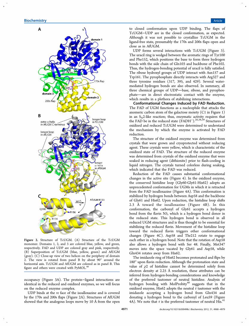
(Figure 2A). Domain 1 is the largest and consists of threesections of the polypeptide chain (residues 4−86, 199−291,and 397−475). This domain functions primarily in bindingFAD and has a Rossmann fold as its core. Domain 2 is a bundleof α-helices and participates in substrate binding (residues101−198). Domain 3 features a twisted, 7-stranded β-sheet thatsits atop a 15-residue α-helix (residues 87−100, 292−396).This domain also contributes to substrate binding.AfUGM is the closest structural neighbor of TcUGM in the
Protein Data Bank (PDB). The two enzymes are 45% identicalin sequence, and the two structures superimpose with a root-mean-square deviation of 1.1 Å (Figure 2B). Although theyshare a common overall fold, several local variations betweenthe two structures are evident (Figure 2B). For example, a loopof domain 1 is longer in TcUGM, whereas a loop in domain 3is longer in AfUGM. The significance of these differences is notobvious. In contrast, other differences are substantive in thatthey appear to dictate the oligomeric state formed in solution(see Discussion). These include the longer α-helix in domain 2of AfUGM and the additional α-helix at the C-terminus ofAfUGM (Figure 2B).The crystal structure is consistent with TcUGM being
monomeric in solution. Analysis of protein−protein interfacesin the crystal lattice using the PDBePISA37 revealed nooligomers predicted to be stable in solution. Furthermore,neither the AfUGM tetramer nor its constituent dimers arepresent in the TcUGM crystal lattice. Moreover, none of thedimeric assemblies of bacterial UGMs are observed in thelattice. It is concluded that TcUGM forms a monomer insolution, which is consistent with recent size exclusionchromatography data.26
Binding of UDP. The structures of TcUGM complexedwith UDP were determined from crystals that had been grownin the presence of the inhibitor. Electron density maps clearlyindicated that UDP is bound in the active site with full
Biochemistry Article
dx.doi.org/10.1021/bi300498c | Biochemistry 2012, 51, 4968−49794970
occupancy (Figure 3A). The protein−ligand interactions areidentical in the reduced and oxidized enzymes, so we will focuson the reduced enzyme complex.UDP binds at the re face of the isoalloxazine and is covered
by the 170s and 200s flaps (Figure 2A). Structures of AfUGMshowed that the analogous loops move by 10 Å from the open
to closed conformation upon UDP binding. The flaps ofTcUGM−UDP are in the closed conformation, as expected.Although it was not possible to crystallize TcUGM in theligand-free state, presumably the 170s and 200s flaps open andclose as in AfUGM.UDP forms several interactions with TcUGM (Figure 3).
The uracil ring is wedged between the aromatic rings of Tyr100and Phe152, which positions the base to form three hydrogenbonds with the side chain of Gln103 and backbone of Phe102.Thus, the hydrogen-bonding potential of uracil is fully satisfied.The ribose hydroxyl groups of UDP interact with Asn157 andTrp161. The pyrophosphate directly interacts with Arg327 andthree tyrosine residues (317, 395, and 429). Several water-mediated hydrogen bonds are also observed. In summary, allthree chemical groups of UDPbase, ribose, and pyrophos-phateare in direct electrostatic contact with the enzyme,which results in a plethora of stabilizing interactions.
Conformational Changes Induced by FAD Reduction.The FAD of UGM functions as a nucleophile that attacks theanomeric carbon atom of the galactose moiety (C1 in Figure 1)in an SN2-like reaction; thus, enzymatic activity requires thatthe FAD be in the reduced state (FADH−).26,38,39 Structures ofoxidized and reduced TcUGM were determined to understandthe mechanism by which the enzyme is activated by FADreduction.The structure of the oxidized enzyme was determined from
crystals that were grown and cryoprotected without reducingagent. These crystals were yellow, which is characteristic of theoxidized state of FAD. The structure of the reduced enzymewas determined from crystals of the oxidized enzyme that weresoaked in reducing agent (dithionite) prior to flash-cooling inliquid nitrogen. The crystals turned colorless during soaking,which indicated that the FAD was reduced.Reduction of the FAD causes substantial conformational
changes in the active site (Figure 4). In the oxidized enzyme,the conserved histidine loop (Gly60-Gly61-His62) adopts anunprecedented conformation for UGMs in which it is retractedfrom the FAD isoalloxazine (Figure 4A). This conformation isstabilized by hydrogen bonds between Asp58 and the backboneof Gly61 and His62. Upon reduction, the histidine loop shifts2.3 Å toward the isoalloxazine (Figure 4B). In thisconformation, the carbonyl of Gly61 accepts a hydrogenbond from the flavin N5, which is a hydrogen bond donor inthe reduced state. This hydrogen bond is observed in allreduced UGM structures and is thus thought to be essential forstabilizing the reduced flavin. Movement of the histidine looptoward the reduced flavin triggers other conformationalchanges (Figure 4C). Asp58 and Thr212 rotate to engageeach other in a hydrogen bond. Note that the rotation of Asp58also allows a hydrogen bond with Ser 48. Finally, Met347moves into the space vacated by Gly61 and Asp58, whileGln434 rotates away from His62.The imidazole ring of His62 becomes protonated and flips by
180° upon flavin reduction. Although the protonation state andvalue of χ2 of histidine cannot be determined solely fromelectron density at 2.25 Å resolution, these attributes can beinferred from hydrogen-bonding considerations and knowledgeof the preferred tautomer of neutral histidine. Analysis ofhydrogen bonding with MolProbity40 suggests that in theoxidized enzyme, His62 adopts the neutral τ tautomer with theimidazole accepting a hydrogen bond from Gln434 anddonating a hydrogen bond to the carbonyl of Leu59 (Figure4A). We note that τ is the preferred tautomer of neutral His.41
Figure 2. Structure of TcUGM. (A) Structure of the TcUGMmonomer. Domains 1, 2, and 3 are colored blue, yellow, and green,respectively. FAD and UDP are colored gray and pink, respectively.(B) Superposition of TcUGM (blue, yellow, green) and AfUGM(gray). (C) Close-up view of two helices on the periphery of domain2. The view is rotated from panel B by about 90° around thehorizontal axis. TcUGM and AfUGM are colored as in panel B. Thisfigure and others were created with PyMOL.46
Biochemistry Article
dx.doi.org/10.1021/bi300498c | Biochemistry 2012, 51, 4968−49794971

In the reduced enzyme, on the other hand, hydrogen bonding ismaximized with the imidazole protonated and flipped by 180°to allow hydrogen bonds with the carbonyl of Gly60 and theFAD 2′-hydroxyl (Figure 4B). Note that the assignedprotonation states of His62 are consistent with the fact thatthe oxidized FAD is uncharged and the reduced FAD is anionic(FADH−).Reduction also affects the conformation of the isoalloxazine
(Figure 5). In the oxidized state, the electron density maps areconsistent with a planar isoalloxazine (Figure 5A). In thereduced enzyme, the FAD exhibits a butterfly like conformationin which the pyrimidine ring bends 7° out of the plane suchthat the si face is concave (Figure 5B). This conformation isidentical to that of reduced AfUGM.Site-Directed Mutagenesis of the Histidine Loop. The
importance of the histidine loop for catalytic activity wasinvestigated using site-directed mutagenesis. Gly61 and His62were targeted for mutagenesis because they form hydrogenbonds to FADH− and exhibit large conformational changesupon flavin reduction. Gly61 is conserved among eukaryoticUGMs but appears as Ala or Pro in bacterial UGMs. Therefore,the mutant enzymes G61A and G61P were created. His62 isuniversally conserved among UGMs, and the H62A mutantenzyme was created. (For reference, Gly60 is also conserved inall UGMs.)
Mutation of the histidine loop of TcUGM is highlydetrimental to activity (Figure S1 of Supporting Informationand Table 2). Mutation of Gly61 to Ala reduces kcat by a factorof 70 but reduces Km by only a factor of 3. As a result, thecatalytic efficiency of G61A is only 4% of that of TcUGM.Similarly, mutation of Gly61 to Pro substantially decreases kcat(factor of 16) but has less effect on Km. The catalytic efficiencyof this mutant enzyme is 10% of that of TcUGM. Mutation ofHis62 to Ala has a profound effect on kcat; this mutationdecreases kcat by over 300. The catalytic efficiency of H62A isjust 2% compared to that of TcUGM. These results suggestthat Gly and His at positions 61 and 62 are important forefficient catalysis by TcUGM. Furthermore, the histidine loopsequences found in bacterial UGMs (GAH, GPH) are poorlytolerated by TcUGM.
■ DISCUSSION
Inhibitor design is aided by knowledge of substrate recognition,and substrate recognition appears to be exquisitely conservedamong eukaryotic UGMs. Comparison of the structures ofTcUGM and AfUGM complexed with UDP show that theUDP binding sites are identical (Figure 6). All residues thatcontact UDP, either directly or via water molecules, are presentin both enzymes. Furthermore, the conformations of theseresidues, as well as their interactions with UDP, are identical in
Figure 3. Electron density and interactions for the UDP bound to reduced TcUGM. (A) Stereographic view of the TcUGM active site. The cagerepresents a simulated annealing σA-weighted F0 − Fc omit map contoured at 3.0σ. (B) Schematic diagram of protein−UDP interactions in TcUGM.Backbone interactions are indicated by N in parentheses.
Biochemistry Article
dx.doi.org/10.1021/bi300498c | Biochemistry 2012, 51, 4968−49794972

the two structures. The structural similarity extends even to thewater molecules that mediate protein−inhibitor interactions(Figure 6). Moreover, all residues that contact UDP or UDP-Galp in the TcUGM and AfUGM crystal structures are alsopresent in many other eukaryotic UGMs, including L. major
UGM (Figure 7, triangles). Thus, it is likely that the AfUGMand TcUGM structures are representative of eukaryotic UGMswith regard to substrate binding.This analysis suggests a unified strategy for designing
inhibitors of UGMs from T. cruzi, L. major, and A. fumigatus,
Figure 4. Structural changes induced by FAD reduction (stereographic views). (A) Electron density for the histidine loop region of oxidizedTcUGM. The cage represents a simulated annealing σA-weighted F0 − Fc omit map contoured at 3σ. (B) Electron density for the histidine loopregion of reduced TcUGM. The cage represents a simulated annealing σA-weighted F0 − Fc omit map contoured at 3σ. (C) Superposition of oxidized(yellow) and reduced (gray) TcUGM. Red and black dashes represent hydrogen bonds for oxidized and reduced TcUGM, respectively.
Biochemistry Article
dx.doi.org/10.1021/bi300498c | Biochemistry 2012, 51, 4968−49794973

which are three important eukaryotic pathogens. For example, acompound that binds in the active site of any of these enzymesis predicted to also inhibit the other ones. Therefore, screeningefforts could be focused on one enzyme. Our results alsosuggest that testing known inhibitors of bacterial UGMs maynot be an optimal strategy for identifying inhibitors ofeukaryotic UGMs. We previously showed that the substratesite of eukaryotic UGMs differs substantially from that ofbacterial UGMs, particularly in the region around the UMPmoiety (see Figure S2 of Dhatwalia et al.24). Thus, compoundsthat target the UMP site of bacterial UGMs will not likely havehigh affinity for eukaryotic UGMs. In summary, the very high
structural similarity of the substrate binding sites of TcUGMand AfUGM raises the possibility of finding a single compoundthat inhibits multiple eukaryotic UGMs. This observationsimplifies inhibitor discovery.The structures also provide insight into the surprising lack of
conservation of the oligomeric state by eukaryotic UGMs.TcUGM is the first example of a monomeric UGM. As wereported previously,24 AfUGM forms a dimer-of-dimerstetramer having 222 point group symmetry (Figure S2).Three conformational differences between the two enzymesaccount for the difference in oligomeric state. First, AfUGM hasan additional 7-residue α-helix at the C-terminus (Figure 2B),and this helix packs against domain 1 of another protomer inthe tetramer (Figure S2). The absence of this helix in TcUGMobviously precludes formation of this critical interface. Thesecond point of departure occurs in the two parallel helices ofdomain 2 (Figure 2C). These helices form an intersubunit 4-helix bundle across one of the 2-fold axes in the AfUGMtetramer (Figure S2B). The 4-helix bundle in AfUGM has atightly packed hydrophobic interior. In contrast, TcUGM haslong charged and polar side chains on these helices, whichwould inhibit formation of the hydrophobic bundle (Figure2C). In particular, if a theoretical tetramer is built fromTcUGM monomers, Arg186, Arg114, and Gln190 form stericclashes with the symmetry related helices. The potential forthese clashes presumably prevents TcUGM from forming thisinterface. Finally, TcUGM is unable to form the fourintersubunit hydrogen bonds that are present in the centroidof the AfUGM tetramer (Figure S2A, inset). These hydrogenbonds involve Arg133, which is located at the C-terminus ofone of the helices of the 4-helix bundle (Figure 2C). Becausethe helix is a full turn shorter in TcUGM, these intersubunitinteractions cannot be formed. These conformational differ-ences account for the difference in the oligomeric states ofTcUGM and AfUGM.The TcUGM structures provide new information about
conformational changes associated with activation of theenzyme via reduction of the FAD (Figures 4 and 5). Reductionof the FAD causes several concerted changes in the protein.The histidine loop moves by 2.3 Å (Figure 4C). His62, a
Figure 5. Electron density for the isoalloxazine rings of (A) oxidizedand (B) reduced TcUGM. The cages represent simulated annealingσA-weighted F0 − Fc omit maps contoured at 3σ. The horizontal lineassists in seeing the 7° butterfly like bend angle of the reducedcofactor.
Table 2. Steady-State Kinetic Constants for TcUGM andTcUGM Mutant Enzymes
kcat (s−1) Km (μM)
kcat/Km(s−1 M−1) kcat/Km (%)
TcUGMa 13.4 ± 0.3 140 ± 10 93 ± 6 100 ± 6G61A 0.198 ± 0.011 50 ± 10 4 ± 1 4 ± 1G61P 0.83 ± 0.06 90 ± 20 9 ± 2 10 ± 2H62A 0.041 ± 0.001 24 ± 4 1.8 ± 0.2 2 ± 0.2
aData from Oppenheimer et al.26
Figure 6. Superposition of the UDP binding sites of TcUGM and AfUGM (stereographic view). TcUGM is shown with the protein in gray andUDP in pink. AfUGM is colored yellow with blue waters. Note that the two structures are essentially identical, particularly in the region around theuridine.
Biochemistry Article
dx.doi.org/10.1021/bi300498c | Biochemistry 2012, 51, 4968−49794974
universally conserved residue in UGMs, flips by 180° andbecomes protonated. The protonation of His62 may helpstabilize the negative charge of the reduced FAD. The sidechains of Asp58 and Thr212 rotate by 180°. Met 347 andGln434 move by 1.6 Å. It is notable that all of these residues arelocated on the side of the FAD that is opposite to the substrate-binding site. Thus, two critical aspects of function are delegated
to distinct regions of the protein: maintaining the redox state isthe responsibility of residues on the si face FAD, while substratebinding is performed by residues on the re side.These conformational changes are consistent with the
generally accepted chemical mechanism of UGM. Theprevailing mechanism is an SN2-type displacement in whichthe N5 atom of the reduced FAD functions as the nucleophile
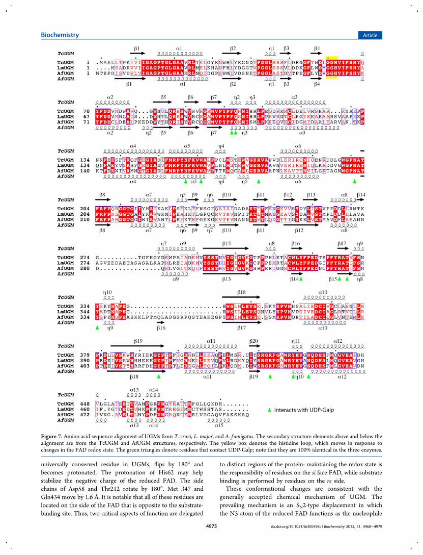
Figure 7. Amino acid sequence alignment of UGMs from T. cruzi, L. major, and A. fumigatus. The secondary structure elements above and below thealignment are from the TcUGM and AfUGM structures, respectively. The yellow box denotes the histidine loop, which moves in response tochanges in the FAD redox state. The green triangles denote residues that contact UDP-Galp; note that they are 100% identical in the three enzymes.
Biochemistry Article
dx.doi.org/10.1021/bi300498c | Biochemistry 2012, 51, 4968−49794975

that attacks the anomeric carbon of galactose to form a covalentintermediate and displace UDP.38,39 This mechanism wasrecently validated for TcUGM.26 Activity thus requires that theFAD be reduced. The coordinated movements of the histidineloop, Asp58, Thr212, and the FAD isoalloxazine in TcUGMhave two salient effects. First, a hydrogen bond is createdbetween the N5 atom of the reduced flavin and the carbonyloxygen atom of the residue preceding the conserved histidine
(Gly61). This interaction is seen in all reduced UGMs and istherefore thought to be essential for stabilizing the reducedflavin. Second, curvature is induced in the flavin isoalloxazinesuch that the si face is concave. Bending of the isoalloxazine inthis direction is consistent with the FAD functioning as anucleophile.19
Curiously, the conformational changes observed for TcUGMare different from those described for the close homologue
Figure 8. Summary of conformational changes induced by flavin reduction in TcUGM and AfUGM (stereographic views). (A) Oxidized TcUGM(PDB code 4DSG). (B) Oxidized AfUGM crystallized in space groups P6522 (cyan, PDB code 3UTE) and P1 (yellow, PDB code 3UKH). (C)Superposition of reduced TcUGM (gray, 4DSH) and reduced AfUGM (green, 3UTF).
Biochemistry Article
dx.doi.org/10.1021/bi300498c | Biochemistry 2012, 51, 4968−49794976

AfUGM (Figure 8). We previously reported structures ofoxidized and reduced AfUGM based on a P6522 crystal form.
24
Sanders’ group subsequently reported P1 structures ofAfUGM.25 The structures of the reduced active sites are nearlyidentical in TcUGM and AfUGM (Figure 8C). However, inoxidized AfUGM, the His residue of the histidine loop issubstantially displaced toward the pyrimidine portion of theisoalloxazine (Figure 8B), whereas in oxidized TcUGM the Hisresidue is near the middle ring of the isoalloxazine (Figure 8A).Furthermore, in oxidized AfUGM, the carbonyl bond vector ofthe middle Gly residue is directed away from the isoalloxazine(Figure 8B, Gly62), whereas the corresponding carbonyl ofoxidized TcUGM points toward the isoalloxazine (Figure 8A,Gly61). Reduction of the FAD in AfUGM causes a 6−8 Åmovement of the imidazole ring of the conserved histidine(Figure 8). As described above, the movement of the histidineloop in TcUGM is more subtle. It should be noted thatelucidation of redox-linked conformational changes for AfUGMis complicated by the adventitious binding of sulfate ions to theoxidized active site of the P6522 crystal form (PDB code3UTE) and weak electron density for the histidine loop andbound UDP in the oxidized P1 form (3UKH). Nevertheless,taken together, the AfUGM and TcUGM structures implicateflexibility of the histidine loop in the mechanism of enzymeactivation.The large conformational changes in the histidine loop
observed in TcUGM and AfUGM appear to be unique toeukaryotic UGMs. Comparison of oxidized and reducedbacterial UGM structures shows that flavin reduction inducesbending of the isoalloxazine but no substantial changes in theprotein conformation.19,21,22 Analysis of amino acid sequenceconservation provides a rationale for why the histidine loop isstatic in bacterial UGMs and dynamic in eukaryotic UGMs.The histidine loop sequence of GGH is conserved amongeukaryotic UGMs (Figure 7). In contrast, bacterial UGMs haveeither Ala or Pro in place of the second Gly. The extra Gly inthe histidine loop most likely accounts for the increasedflexibility of the loop in AfUGM and TcUGM.This additional flexibility of the histidine loop appears to be
important for function in TcUGM. Our mutagenesis data showthat transplanting the bacterial sequences of GAH and GPHinto TcUGM substantially decreases catalytic efficiency (Table2). It is possible that the additional flexibility afforded by theextra Gly residue in TcUGM is needed to establish optimalhydrogen bonding between the histidine loop and FADH−.Although structures of the TcUGM histidine loop mutantenzymes are not available, it is possible that the loop-flavinhydrogen bonding is suboptimal in these impaired enzymes,which likely decreases the nucleophilic character of the reducedflavin. This idea is consistent with the markedly decreased kcatvalues of the histidine loop mutants.Why eukaryotic UGMs undergo such large conformational
changes upon activation is an open question. One possibility isthat these conformational changes control the access of O2 andreductant to the flavin. In the reduced enzyme, positioning thehistidine loop close to the isoalloxazine N5−C4a edge helpsprotect the C4a atom of the reduced FAD from attack by O2(Figure 4B). Retraction of the loop upon oxidation perhapsallows open space for reducing agents to gain access to theoxidized flavin (Figure 4A). Regulation of function is anotherpossibility. Conformational changes that are linked to the flavinredox state often serve a regulatory purpose.42 Whether
eukaryotic UGMs are subject to some sort of redox-linkedregulation remains to be determined.
■ ASSOCIATED CONTENT*S Supporting InformationFigures showing steady-state kinetic profiles and the AfUGMtetramer. This material is available free of charge via theInternet at http://pubs.acs.org.Accession CodesAtomic coordinates and structure factors been deposited in theProtein Data Bank under the following accession codes: 4DSG,oxidized TcUGM-UDP; 4DSH, reduced TcUGM-UDP.
■ AUTHOR INFORMATIONCorresponding Author*E-mail [email protected]; phone 573-884-1280; fax 573-882-2754.Author Contributions∥These authors contributed equally to this work.FundingThis research was supported by NIH grant R01 GM094469 (toP.S. and J.J.T.). M.O. was supported by American HeartAssociation predoctoral fellowship 10PRE4160020. This workis based upon research conducted at the Advanced PhotonSource on the Northeastern Collaborative Access Teambeamlines, which are supported by award RR-15301 from theNational Center for Research Resources at the NationalInstitutes of Health. Use of the Advanced Photon Source wassupported by the U.S. Department of Energy, Office of Science,Office of Basic Energy Sciences, under Contract DE-AC02-06CH11357.NotesThe authors declare no competing financial interest.
■ ACKNOWLEDGMENTSWe thank Dr. Jonathan Schuermann and Dr. Norma Duke forhelp with X-ray diffraction data collection and processing.
■ ABBREVIATIONSUGM, UDP-galactopyranose mutase; UDP-Galp, UDP-galac-topyranose; UDP-Galf , UDP-galactofuranose; TcUGM, UDP-galactopyranose mutase from Trypanosoma cruzi; AfUGM,UDP-galactopyranose mutase from Aspergillus fumigatus; PDB,Protein Data Bank.
■ REFERENCES(1) Savioli, L., and Daumerie, D. (2010) First WHO report onneglected tropical diseases: working to overcome the global impact ofneglected tropical diseases (Crompton, D. W. T., Ed.) World HealthOrganization, Geneva.(2) Bern, C., and Montgomery, S. P. (2009) An estimate of theburden of Chagas disease in the United States. Clin. Infect. Dis. 49,e52−54.(3) Tanowitz, H. B., Weiss, L. M., and Montgomery, S. P. (2011)Chagas disease has now gone global. PLoS Negl. Trop. Dis. 5, e1136.(4) Bern, C., Montgomery, S. P., Herwaldt, B. L., Rassi, A., Jr., Marin-Neto, J. A., Dantas, R. O., Maguire, J. H., Acquatella, H., Morillo, C.,Kirchhoff, L. V., Gilman, R. H., Reyes, P. A., Salvatella, R., and Moore,A. C. (2007) Evaluation and treatment of chagas disease in the UnitedStates: a systematic review. JAMA, J. Am. Med. Assoc. 298, 2171−2181.(5) Teixeira, A. R., Cordoba, J. C., Souto Maior, I., and Solorzano, E.(1990) Chagas’ disease: lymphoma growth in rabbits treated withBenznidazole. Am. J. Trop. Med. Hyg. 43, 146−158.
Biochemistry Article
dx.doi.org/10.1021/bi300498c | Biochemistry 2012, 51, 4968−49794977

(6) Issa, V. S., and Bocchi, E. A. (2010) Antitrypanosomal agents:treatment or threat? Lancet 376, 768 ; author reply 768−769.(7) Beverley, S. M., Owens, K. L., Showalter, M., Griffith, C. L.,Doering, T. L., Jones, V. C., and McNeil, M. R. (2005) EukaryoticUDP-galactopyranose mutase (GLF gene) in microbial and metazoalpathogens. Eukaryot. Cell. 4, 1147−1154.(8) Tefsen, B., Ram, A. F., van Die, I., and Routier, F. H. (2011)Galactofuranose in eukaryotes: aspects of biosynthesis and functionalimpact. Glycobiology,in press.(9) Oppenheimer, M., Valenciano, A. L., and Sobrado, P. (2011)Biosynthesis of galactofuranose in kinetoplastids: novel therapeutictargets for treating leishmaniasis and chagas’ disease. Enzyme Res. 2011,1−13.(10) Almeida, I. C., Ferguson, M. A., Schenkman, S., and Travassos,L. R. (1994) GPI-anchored glycoconjugates from Trypanosoma cruzitrypomastigotes are recognized by lytic anti-alpha-galactosyl antibodiesisolated from patients with chronic Chagas’ disease. Braz. J. Med. Biol.Res. 27, 443−447.(11) Ralton, J. E., Milne, K. G., Guther, M. L., Field, R. A., andFerguson, M. A. (1993) The mechanism of inhibition ofglycosylphosphatidylinositol anchor biosynthesis in Trypanosomabrucei by mannosamine. J. Biol. Chem. 268, 24183−24189.(12) Ferguson, M. A. (1997) The surface glycoconjugates oftrypanosomatid parasites. Philos. Trans. R. Soc., B 352, 1295−1302.(13) Turnock, D. C., and Ferguson, M. A. (2007) Sugar nucleotidepools of Trypanosoma brucei, Trypanosoma cruzi, and Leishmaniamajor. Eukaryotic Cell 6, 1450−1463.(14) MacRae, J. I., Obado, S. O., Turnock, D. C., Roper, J. R.,Kierans, M., Kelly, J. M., and Ferguson, M. A. (2006) The suppressionof galactose metabolism in Trypanosoma cruzi epimastigotes causeschanges in cell surface molecular architecture and cell morphology.Mol. Biochem. Parasitol. 147, 126−136.(15) Turner, C. W., Lima, M. F., and Villalta, F. (2002) Trypanosomacruzi uses a 45-kDa mucin for adhesion to mammalian cells. Biochem.Biophys. Res. Commun. 290, 29−34.(16) Spath, G. F., Garraway, L. A., Turco, S. J., and Beverley, S. M.(2003) The role(s) of lipophosphoglycan (LPG) in the establishmentof Leishmania major infections in mammalian hosts. Proc. Natl. Acad.Sci. U. S. A. 100, 9536−9541.(17) Zhang, K., Barron, T., Turco, S. J., and Beverley, S. M. (2004)The LPG1 gene family of Leishmania major. Mol. Biochem. Parasitol.136, 11−23.(18) Sanders, D. A., Staines, A. G., McMahon, S. A., McNeil, M. R.,Whitfield, C., and Naismith, J. H. (2001) UDP-galactopyranosemutase has a novel structure and mechanism. Nat. Struct. Biol. 8, 858−863.(19) Beis, K., Srikannathasan, V., Liu, H., Fullerton, S. W., Bamford,V. A., Sanders, D. A., Whitfield, C., McNeil, M. R., and Naismith, J. H.(2005) Crystal structures of Mycobacteria tuberculosis and Klebsiellapneumoniae UDP-galactopyranose mutase in the oxidised state andKlebsiella pneumoniae UDP-galactopyranose mutase in the (active)reduced state. J. Mol. Biol. 348, 971−982.(20) Gruber, T. D., Borrok, M. J., Westler, W. M., Forest, K. T., andKiessling, L. L. (2009) Ligand binding and substrate discrimination byUDP-galactopyranose mutase. J. Mol. Biol. 391, 327−340.(21) Gruber, T. D., Westler, W. M., Kiessling, L. L., and Forest, K. T.(2009) X-ray crystallography reveals a reduced substrate complex ofUDP-galactopyranose mutase poised for covalent catalysis by flavin.Biochemistry 48, 9171−9173.(22) Partha, S. K., van Straaten, K. E., and Sanders, D. A. (2009)Structural basis of substrate binding to UDP-galactopyranose mutase:crystal structures in the reduced and oxidized state complexed withUDP-galactopyranose and UDP. J. Mol. Biol. 394, 864−877.(23) Partha, S. K., Sadeghi-Khomami, A., Slowski, K., Kotake, T.,Thomas, N. R., Jakeman, D. L., and Sanders, D. A. (2010)Chemoenzymatic synthesis, inhibition studies, and X-ray crystallo-graphic analysis of the phosphono analog of UDP-Galp as an inhibitorand mechanistic probe for UDP-galactopyranose mutase. J. Mol. Biol.403, 578−590.
(24) Dhatwalia, R., Singh, H., Oppenheimer, M., Karr, D. B., Nix, J.C., Sobrado, P., and Tanner, J. J. (2012) Crystal structures and small-angle X-ray scattering analysis of UDP-galactopyranose mutase fromthe pathogenic fungus Aspergillus fumigatus. J. Biol. Chem. 287, 9041−9051.(25) van Straaten, K. E., Routier, F. H., and Sanders, D. A. (2012)Structural insight into the unique substrate binding mechanism andflavin redox state of UDP-galactopyranose mutase from Aspergillusfumigatus. J. Biol. Chem. 287, 10780−10790.(26) Oppenheimer, M., Valenciano, A. L., Kizjakina, K., Qi, J., andSobrado, P. (2012) Chemical Mechanism of UDP-GalactopyranoseMutase from Trypanosoma cruzi: A Potential Drug Target againstChagas’ Disease. PLoS One 7, e32918.(27) Matthews, B. W. (1968) Solvent content of protein crystals. J.Mol. Biol. 33, 491−497.(28) Otwinowski, Z., and Minor, W. (1997) Processing of X-raydiffraction data collected in oscillation mode. Methods Enzymol. 276,307−326.(29) Kabsch, W. (2010) XDS. Acta Crystallogr., Sect. D: Biol.Crystallogr. 66, 125−132.(30) Evans, P. (2006) Scaling and assessment of data quality. ActaCrystallogr., Sect. D: Biol. Crystallogr. 62, 72−82.(31) Potterton, E., Briggs, P., Turkenburg, M., and Dodson, E.(2003) A graphical user interface to the CCP4 program suite. ActaCrystallogr., Sect. D: Biol. Crystallogr. 59, 1131−1137.(32) Vagin, A., and Teplyakov, A. (1997) MOLREP: an automatedprogram for molecular replacement. J. Appl. Crystallogr. 30, 1022−1025.(33) Emsley, P., and Cowtan, K. (2004) Coot: model-building toolsfor molecular graphics. Acta Crystallogr., Sect. D: Biol. Crystallogr. 60,2126−2132.(34) Adams, P. D., Afonine, P. V., Bunkoczi, G., Chen, V. B., Davis, I.W., Echols, N., Headd, J. J., Hung, L. W., Kapral, G. J., Grosse-Kunstleve, R. W., McCoy, A. J., Moriarty, N. W., Oeffner, R., Read, R.J., Richardson, D. C., Richardson, J. S., Terwilliger, T. C., and Zwart, P.H. (2010) PHENIX: a comprehensive Python-based system formacromolecular structure solution. Acta Crystallogr., Sect. D: Biol.Crystallogr. 66, 213−221.(35) Oppenheimer, M., Poulin, M. B., Lowary, T. L., Helm, R. F., andSobrado, P. (2010) Characterization of recombinant UDP-galactopyr-anose mutase from Aspergillus fumigatus. Arch. Biochem. Biophys. 502,31−38.(36) Poulin, M. B., and Lowary, T. L. (2010) Methods to study thebiosynthesis of bacterial furanosides. Methods Enzymol. 478, 389−411.(37) Krissinel, E., and Henrick, K. (2007) Inference of macro-molecular assemblies from crystalline state. J. Mol. Biol. 372, 774−797.(38) Soltero-Higgin, M., Carlson, E. E., Gruber, T. D., and Kiessling,L. L. (2004) A unique catalytic mechanism for UDP-galactopyranosemutase. Nat. Struct. Mol. Biol. 11, 539−543.(39) Sun, H. G., Ruszczycky, M. W., Chang, W. C., Thibodeaux, C. J.,and Liu, H. W. (2011) Nucleophilic participation of the reduced flavincoenzyme in the mechanism of UDP-Galactopyranose mutase. J. Biol.Chem.,.(40) Chen, V. B., Arendall, W. B., III, Headd, J. J., Keedy, D. A.,Immormino, R. M., Kapral, G. J., Murray, L. W., Richardson, J. S., andRichardson, D. C. (2010) MolProbity: all-atom structure validation formacromolecular crystallography. Acta Crystallogr., Sect. D: Biol.Crystallogr. 66, 12−21.(41) Li, S., and Hong, M. (2011) Protonation, tautomerization, androtameric structure of histidine: a comprehensive study by magic-angle-spinning solid-state NMR. J. Am. Chem. Soc. 133, 1534−1544.(42) Becker, D. F., Zhu, W., and Moxley, M. A. (2011) Flavin redoxswitching of protein functions. Antioxid. Redox Signaling 14, 1079−1091.(43) Weiss, M. (2001) Global indicators of X-ray data quality. J. Appl.Crystallogr. 34, 130−135.(44) Engh, R. A., and Huber, R. (1991) Accurate bond and angleparameters for x-ray protein structure refinement. Acta Crystallogr.A47, 392−400.
Biochemistry Article
dx.doi.org/10.1021/bi300498c | Biochemistry 2012, 51, 4968−49794978

(45) Lovell, S. C., Davis, I. W., Arendall, W. B., III, de Bakker, P. I.,Word, J. M., Prisant, M. G., Richardson, J. S., and Richardson, D. C.(2003) Structure validation by Calpha geometry: phi,psi and Cbetadeviation. Proteins 50, 437−450.(46) DeLano, W. L. (2002) The PyMOL User’s Manual, DeLanoScientific, Palo Alto, CA.
Biochemistry Article
dx.doi.org/10.1021/bi300498c | Biochemistry 2012, 51, 4968−49794979

Supporting Information
Crystal Structures of Trypanosoma cruzi UDP-Galactopyranose
Mutase Implicate Flexibility of the Histidine Loop
in Enzyme Activation
This research was supported by NIH grant R01 GM094468 (to P.S. and J.J.T).
Richa Dhatwalia,†,|| Harkewal Singh,†,|| Michelle Oppenheimer,# Pablo Sobrado,#
and John J. Tanner†,‡,*
†Department of Chemistry, University of Missouri-Columbia, Columbia, MO 65211, USA
#Department of Biochemistry, Virginia Tech, Blacksburg, VA 24061, USA
‡Department of Biochemistry, University of Missouri-Columbia, Columbia, MO 65211,
USA
||These authors contributed equally to this work.
Corresponding author
Department of Chemistry, University of Missouri-Columbia, Columbia, MO 65211, USA;
email: [email protected]; phone: 573-884-1280; fax: 573-882-2754

S-2
Table of Contents Figure S1. Steady-state kinetic characterization of TcUGM and histidine loop mutants. S-3
Figure S2. Structure of the AfUGM tetramer. S-4

S-3
Figure S1. Steady-state kinetic characterization of TcUGM and the TcUGM histidine loop mutants G61A, G61P, and H62A. The activity was determined by monitoring the formation UDP-Galp from UDP-Galf. The data were fit to the Michaelis-Menten equation.

S-4
Figure S2. Structure of the AfUGM tetramer. (A) The tetramer is viewed down the R molecular 2-fold axis. Each chain has a different color. Inset: Intersubunit hydrogen bonds formed by Arg133 at the intersection of molecular 2-fold axes. (B) The tetramer is viewed down the Q-axis.