EF-P dependent pauses integrate proximal and distal signals during translation
Upload
independentCategory
view
0download
0
Inhibition of Hb S Polymerization In Vitro by a Novel 15-mer EFHelix β73 His-Containing Peptide
Mohammed GK Akbar1, Yutaka Tamura3, Min Ding1, Hua Ding2, Michael. M. Rosenblatt2,Konda S. Reddy4, Saul Surrey5, and Kazuhiko Adachi11 The Children's Hospital of Philadelphia, Division of Hematology and University of Pennsylvania Schoolof Medicine, Philadelphia, PA. 19104
2 The Children's Hospital of Philadelphia, Joseph Stokes Jr. Research Institute Protein Core Facility,Philadelphia, PA. 19104
3 Department of Bioinformatics, Chiba University Graduate School of Medicine, Chiba 260−8670, Japan
4 Department of Biophysics, University of Pennsylvania, Philadelphia PA. 19104
5 Cardeza Foundation for Hematologic Research, Department of Medicine, Jefferson Medical College,Thomas Jefferson University, Philadelphia, PA.19104
AbstractOur mutational studies on HbS showed that the HbS β73His variant (β6Val and β73His) promotedpolymerization, while HbS β73Leu (β6Val and β73Leu) inhibited polymerization. Based on theseresults, we speculated that EF-helix peptides containing β73His interact with β4Thr in HbS andcompete with HbS, resulting in inhibition of HbS polymerization. We, therefore, studied inhibitoryeffects of 15-, 11-, 7- and 3-mer EF-helix peptides containing β73His on HbS polymerization. Thedelay time prior to HbS polymerization increased only in the presence of the 15-mer His peptide; thehigher the amount, the longer the delay time. DIC image analysis also showed fiber elongation ratefor HbS polymers decreased with increasing concentration of the 15-mer His peptide. In contrast,the same 15-mer-peptide containing β73Leu instead of His and peptides shorter than 11 amino acidscontaining β73His including His alone showed little effect on kinetics of polymerization andelongation of polymers. Analysis by protein-chip arrays showed that only the 15-mer β73His peptideinteracted with HbS. CD spectra of the 15-mer β73His peptide did not show a specific helicalstructure, however, computer docking analysis suggested a lower energy for interaction of HbS withthe 15-mer β73His peptide compared to peptides containing other amino acids at this position. Theseresults suggest that the 15-mer β73His peptide interacts with HbS via the β4Thr in the βS-globinchain in HbS. This interaction may influence hydrogen bond interaction between β73Asp and β4Thrin HbS polymers and interfere in hydrophobic interactions of β6 Val leading to inhibition of HbSpolymerization.
Hb S is a naturally occurring mutation of human tetrameric hemoglobin in which the β subunitshave a hydrophobic Val in place of a negatively charged Glu at the β6 position. Theconsequence of this mutation is that solubility of Hb S decreases when oxy Hb S loses oxygen.When deoxy Hb S becomes oversaturated deoxy Hb S assembles into long, multi-strandedfibers under physiological conditions (1,2). Fiber formation is characterized by a delay timeprior to polymerization, which is explained by homogeneous and heterogeneous nucleation.During oversaturated Hb S conditions deoxy Hb S monomers form very small polymers by
Please address all correspondence to: Kazuhiko Adachi, Ph.D. Division of Hematology The Children's Hospital of Philadelphia 34th St.& Civic Center Blvd. Philadelphia, PA 19104 Tel: 215−590−3576 ; Fax: 215−590−4834 e-mail: [email protected].
NIH Public AccessAuthor ManuscriptBiochemistry. Author manuscript; available in PMC 2008 December 4.
Published in final edited form as:Biochemistry. 2006 July 11; 45(27): 8358–8367. doi:10.1021/bi0604734.
NIH
-PA Author Manuscript
NIH
-PA Author Manuscript
NIH
-PA Author Manuscript
homogeneous nucleation, and these polymers grow by the end addition of hemoglobinmolecules in solution. The surface of these growing fibers also can serve as heterogeneousnucleation sites for further growth of additional polymers (1,3). The polymer then assemblesinto 14-stranded fibers, which finally form a viscous gel. Intracellular polymers or fibers causereduction in red blood cell deformability (sickling), leading to obstruction of flow in themicrocirculation, thus creating vasooclusion and a wide array of physiological problemsincluding episodes of painful crises (1,4).
Computational refinements of x-ray-determined crystal structures clarified the details of manyof the axial and lateral contacts in Hb S polymers (5). These results and properties of the βS
chain and of βS peptides (6,7) show a slight hinge-like motion of the A helix in the βS-globinsubunits that makes intermolecular contact with the adjacent Hb S tetramer. Crystal structuralanalysis of Hb S in the deoxy form also showed not only β6Val in a largely hydrophobicacceptor pocket, but other contact sites involving a hydrogen bond between β4Thr and β73Aspin Hb S which play a critical role in protein interactions. The β4 and β73 positions are locatednear β6Val and the EF helix acceptor sites in the β subunit, respectively, which are criticallateral contact regions in Hb S polymerization (1,5,8). In addition, recent advances inunderstanding the molecular and cellular pathophysiology of sickle cell disease, coupled withnew insights into the developmental regulation of human globin-gene expression as well ascharacterization of Hb S polymerization, have provided the scientific impetus and clinicalrationale to attempt augmentation of the production of Hb F (4). Furthermore, rational drugdesign (also known as structure-based drug design) through the use of computer modeling andresults of x-ray diffraction is an emerging approach that is revolutionizing the practice of drugdiscovery. The first anti-sickling molecules designed from receptor-based molecular modeling,the substituted benzaldehyde BW12C (now referred to as 12C79), were described by Beddellet al (9). This drug has reached clinical trials, but analysis of the covalent complex of 12C79and Hb A showed that this agent binds to the N-terminal amino group rather than the directinteraction sites of Hb S polymers including the β6Val donor site, β85Phe and β88Leuhydrophobic acceptor sites or the β4Thr-β73Asp hydrogen bond.
Our mutational studies on Hb S polymerization showed that the Hb S β73His variant (β6Valand β73His) promoted polymerization compared to deoxy Hb S, while Hb S β73Leu (β6Valand β73Leu) inhibited polymerization like naturally occurring deoxy Hb C-Harlem (β6Val andβ73Asn) (10). These results suggest that the β73 position (Asp in Hb S) serves as a unique siteto promote or inhibit polymerization by amino acid replacement (10). Kinetics ofpolymerization, solubility and minimum concentration required for polymerization of the HbS β73 variants were affected by β73 amino acid (inhibition of polymerization: His < <Asp<<Asn < Leu). Inhibition of Hb S β73Leu polymerization compared to Hb S may be causedby weakening of the hydrogen bond interaction between the β4Thr hydroxyl group and theβ73 amino acid like Hb C-Harlem (10). Furthermore, kinetics of polymerization of 1:1 Hb S/Hb A β73His mixtures were enhanced like Hb S β73His compared to AS mixtures. Theseresults suggest that the Hb A β73His variant promotes Hb S polymerization almost asefficiently as Hb S β73His, and that β73His in Hb A and Hb S variants strengthens the hydrogenbond with β4Thr, which facilitates formation of domains and 14-stranded fibers. In addition,our results suggested that altering hydrogen bond formation between β73Asp and β4Thrproduces Hb S molecules with different properties, and that decreasing hydrogen bondformation inhibited polymerization without changing significantly tetramer solubility (11).Based on these results, we hypothesized that EF-helix peptides containing β73His wouldinteract with β4Thr in Hb S and disturb protein–protein interactions of Hb S tetramers, resultingin inhibition of Hb S polymerization. We, therefore, synthesized several EF-helix peptidescontaining β73His of different length, and then evaluated their effects on Hb S polymerization.We also studied effects of these peptides on domain formation, elongation of Hb S polymersusing differential interference contrast (DIC) microscopy and performed kinetics,
Akbar et al. Page 2
Biochemistry. Author manuscript; available in PMC 2008 December 4.
NIH
-PA Author Manuscript
NIH
-PA Author Manuscript
NIH
-PA Author Manuscript
thermodynamic and computer docking studies, comparing and contrasting results of the β73Hispeptide to those of the β73Leu peptide.
Materials and MethodsHb S was purified from AS blood as previously described (12). Hemoglobin concentrationswere determined spectrophotometically using a millimolar extinction coefficient of mE 555 =50 for deoxyhemoglobin and mE 579= 53.6 for carbonmonoxy-hemoglobin (on a tetramerbasis). Sample purity was assessed by cellulose acetate electrophoresis and HPLC (13).Hemoglobin solutions were concentrated using a Centricon centrifugal concentrator with amembrane cutoff of 30,000 Da (Centricon-30, Amicon, Inc). Oxy-Hb S, which was stored asCO Hb S, was prepared by first blowing oxygen across the surface of the CO-Hb solution ina rotary evaporator under a 150 W floodlight bulb in an ice bath for about one hour. EF-helixpeptides [15mer (Lys-Lys-Val-Leu-Gly-Ala-Phe-Ser-His-Gly-Leu-Ala-His-Leu-Asp), 11-mer (Gly-Ala-Phe-Ser-His-Gly-Leu-Ala-His-Leu-Asp), 7-mer (Ala-Phe-Ser-His-Gly-Leu-Ala) and 3-mer (Ser-His-Gly) as well as 15-mer β73Leu peptide (Lys-Lys-Val-Leu-Gly-Ala-Phe-Ser-Leu-Gly-Leu-Ala-His-Leu-Asp)] were synthesized (Syn Pep Corporation, Dublin,CA), solubilized in 0.5% (v/v) acetonitrile, purified by HPLC (∼98% purity) and molecularweights confirmed by Electrospray ionization mass spectrometry (ESI-MS).
Kinetics of polymerization and solubility of Hb S in the presence and absence of the peptideswere evaluated in 1.0 M and 1.8 M buffers pH 7.3 at 30°C, with solubility determined bycentrifugation after completion of polymerization (12). The time courses of Hb S polymerelongation in the presence and absence of peptide in 1.0 M phosphate buffer were analyzed byDIC microscopy using an Olympus microscope equipped with optics employing a 40x oil (1.00NA) immersion lens (10). For microscopy, approximately one μl of solution was pipetted to aglass slide which then was sealed with a 18 mm square cover slip using Mount-Quick solution(Daido Sangyo Co., LTD, Japan) (10). Polymer formation of deoxygenated Hb S in thepresence and absence of peptides was induced by temperature-jump from 0°C on ice to roomtemperature (∼ 22° C). DIC polymer images were captured from a 150 micron square areathrough a CCD (charge-coupled devise) camera (Cohu Camera, Cohu Inc, San Diego, CA),transferred to a PC via an image grabber board (Scion AG5, Scicon Corp, Frederich, MD) andprocessed by an automated image analysis system (Universal Image Co, Downing town, PA).Still images were sent to a computer for every second of video footage. In order to measurethe length of Hb S polymer fibers, images were taken of a hemocytometer with 50 μm divisions(American Optical Corp., Buffalo, NY) at 400x magnification. Using the line measurementtool in Universal Image analysis software, we determined the length of a 50-μm division to be206 pixels, which is the smallest division of an image. The length of one pixel was determinedto be 0.243 μm; and, images of Hb S fibers then were measured in pixels and converted toμm. Total length of fibers in a single domain as a function of time was measured by countingthe number of pixels in each image using NIH Image Analysis (v 1.63) and Universal Imageanalysis software.
Peptide binding to Hb S was assessed using SELDI (surface-enhanced laser desorption/ionization) mass spectrometry as follows (14). Desalted Hb S in the oxy form (8 pM) wasbound to a RS 100 chip [Ciphergen Biosystems Inc, Fremont, CA] surface in coupling buffer(50 mM sodium carbonate, pH 9.0) at room temperature. The unbound parts on a chip surfacethen were blocked using 1M Tris buffer, pH 8.0. The β73His peptides including 15-, 11-, 7-and 3-mers and 15-mer Leu peptide (4 and 8 pM) in PBS buffer containing 0.5% (v/v)acetonitrile (pH 7.1) were incubated with the chips containing immobilized Hb S for 2.5 hrsat room temperature. Unbound peptides were removed using a PBS buffer wash followed bya second wash using PBS buffer containing 0.5 M NaCl. Ionization of the chip surface wasenhanced using alpha hydroxyl 4-cinnamic acid after binding of peptides to Hb S. Mass of
Akbar et al. Page 3
Biochemistry. Author manuscript; available in PMC 2008 December 4.
NIH
-PA Author Manuscript
NIH
-PA Author Manuscript
NIH
-PA Author Manuscript
bound peptide then was measured using the mass spectrometer-based SELDI Protein Chipanalysis system [Ciphergen Biosystems Inc, Fremont, CA].
Circular Dichroism (CD) spectra of the 15-mer peptides solubilized in 0.5 % (v/v) acetonitrilewere analyzed at room temperature using an Aviv model 62 DS instrument employing a 1mmlight path cuvette equipped with a thermoelectric module.
Three-dimensional structures of the 15-mer EF helix peptides containing β73His or β73Leuwere constructed using MOE (Ver. 2004.03, CCG Inc., Montreal, Canada), while structure ofthe sickle β-globin chain (βS) was from 2HBS which is deposited in the Brookhaven ProteinDatabank. Docking simulations of the 15-mer peptides containing β73His or β73Leu with β4Thr in the βS-globin chain were performed using BioMedCAChe (Ver. 6, FUJITSU, Tokyo,Japan), and results displayed using MolFeat (Ver. 2, FiatLux, Tokyo, Japan). Algorithmparameters for docking simulations using BioMedCAChe were as follows: (1) Pop Size 100,Crossover Rate 0.8, (2) Elitism 20, (3) Max Generation 30000, (4) Mutation Rate 0.2 and (5)Convergence (kcal) 1.0.
ResultsKinetics of polymerization of Hb S and solubility in the presence and absence of the β73His-or β73Leu-containing peptides
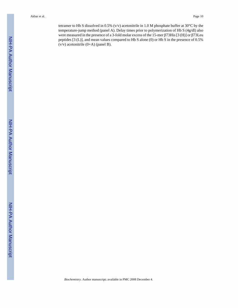
Polymerization and solubility of Hb S in the presence of the 15-mer β73His peptide werestudied in 1.0 M phosphate buffer. Deoxy Hb S (4 g/dl) polymerized in 1.0 M phosphate buffer,pH 7.4 at 30°C with a 275-second delay time (Fig. 1-A). We reported previously that Hb Sβ73His and β73Leu variants polymerized with delay times in 1.0 M phosphate buffer withsignificantly shorter and longer delay times, respectively, than Hb S (10). In contrast, the delaytime for Hb S polymerization was much longer (∼450 sec) in the presence of a 3-fold molarexcess (e.g., 3 moles of peptide per mole of hemoglobin tetramer) of the 15-mer β73His peptideto Hb S solubilized in 0.5%(v/v) acetonitrile in 1.0 M phosphate buffer at 30°C (line B in Fig.1-A). Acetonitrile at the same final concentration in the absence of peptide had no effect onpolymerization of Hb S. In addition, the 15-mer β73Leu peptide at the same concentration asthe 15-mer β73His peptide only slightly increased the delay time (e.g., from 280 sec. to 311sec.). Delay time for Hb S (4 g/dl) polymerization in the presence and absence of the 15-merβ73His or β73Leu peptide are shown in Fig. 1-B. Delay times in the presence of a 3-fold molarexcess of the 15-mer β73His peptide to Hb S increased 1.15- and 1.5-fold for the β73Leu andβ73His peptides, respectively, compared to Hb S alone. In addition, 5-fold molar excessadditions of 3-, 7- and 11-mer His-containing peptides showed no significant difference fromresults of Hb S without peptides and His alone. These results indicate that the 15-mer β73Hispeptide specifically inhibits nucleation prior to polymerization of Hb S. The length of the delaytime in 1.8 M phosphate buffer increased with increasing amounts of the 15-mer β73His peptide(Fig. 2). These results indicate that the apparent association constant of this 15-mer His peptidewith deoxy Hb S is in the range of 30 μM under these experimental conditions.
Solubility of Hb S in 1.0 M phosphate buffer at 30°C in the presence and absence of the 15-mer β73His or β73Leu peptides was measured after polymerization, and compared to that ofHb S (Fig. 3). Solubility of Hb S increased with increasing 15-mer β73His peptide. In thepresence of a 5-fold molar excess of the 15-mer β73 His peptide to Hb S the solubility of HbS increased 1.17-fold compared to Hb S. In contrast, a 5-fold molar excess of the15-merβ73Leu peptide had no effect on Hb S solubility. These results indicate that the criticalconcentration for Hb S polymerization increased in the presence of the 15-mer β73His peptide.
Akbar et al. Page 4
Biochemistry. Author manuscript; available in PMC 2008 December 4.
NIH
-PA Author Manuscript
NIH
-PA Author Manuscript
NIH
-PA Author Manuscript
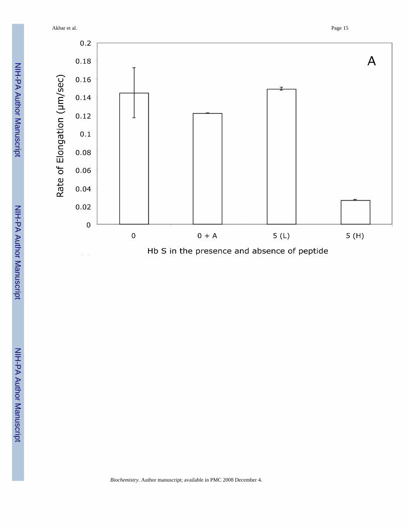
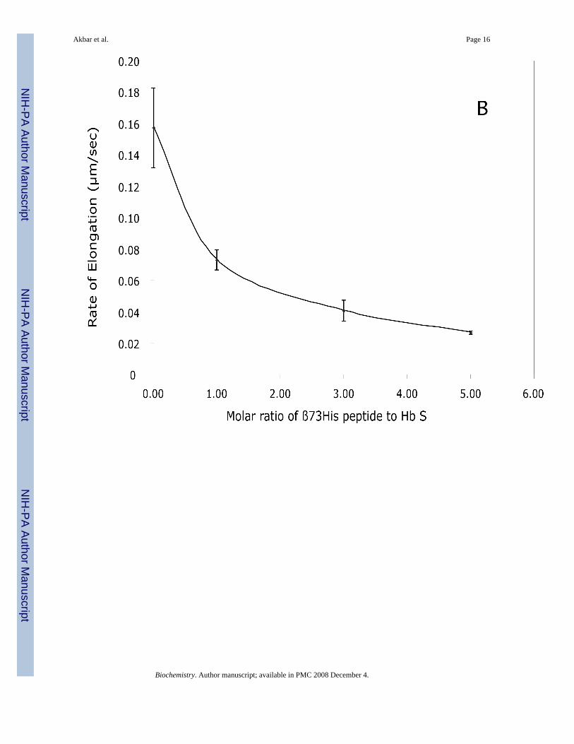
DIC image analysis of Hb S polymerization in the presence of the β73His peptidesWe reported previously that Hb S fibers were observed by DIC analysis using ∼4 g/dl of deoxyHb S in 1.0 M phosphate buffer employing the temperature-jump method. These images weresimilar to those using 0.1 M phosphate buffer (10). DIC image analysis showed that deoxy HbS fibers formed from many domains and elongated from each domain after a few minute delaytime, which depended on Hb S concentration. In the presence of a 5-fold molar excess of the15-mer β73His peptide to Hb S, deoxy Hb S (4 g/dl) showed fiber formation from manydomains (Fig. 4-B), but the domain size and final length of Hb S fibers were different fromthose of Hb S alone (Fig. 4-A). Growth rates of Hb S fibers (4 g/dl) in the presence and absenceof the 15-mer β73His peptide in 1.0 M phosphate buffer were measured by calculation of thelength of longest fibers in a single domain as a function of time (Fig. 5). Elongation rates in asingle domain are initially high and then become low, corresponding to the initial and finalstages of Hb S polymer growth, respectively (Fig. 5-A). Fiber elongation rate of Hb S in thepresence of peptide was much lower than that of Hb S alone (Fig. 5-B). Elongation rate of HbS fibers in the presence of a 5-fold molar excess of the 15-mer β73His peptide to Hb S was ∼6-fold lower compared to those of Hb S alone or in the presence of the β73Leu peptide (Fig.6-A). Effect of the 15-mer β73His peptide on elongation rate of Hb S fibers depends on peptideconcentration; the higher the concentration, the lower the elongation rate (Fig. 6-B). It also isnoteworthy that Hb S elongation rate in the presence of a 5-fold molar excess of peptidescontaining β73 His to Hb S which were smaller than 11 amino acids including 7- and 3-mersas well as His alone was not significantly affected (Fig. 6-C). These results indicate that the15-mer β73His peptide specifically inhibits not only nucleation but also elongation of Hb Spolymers.
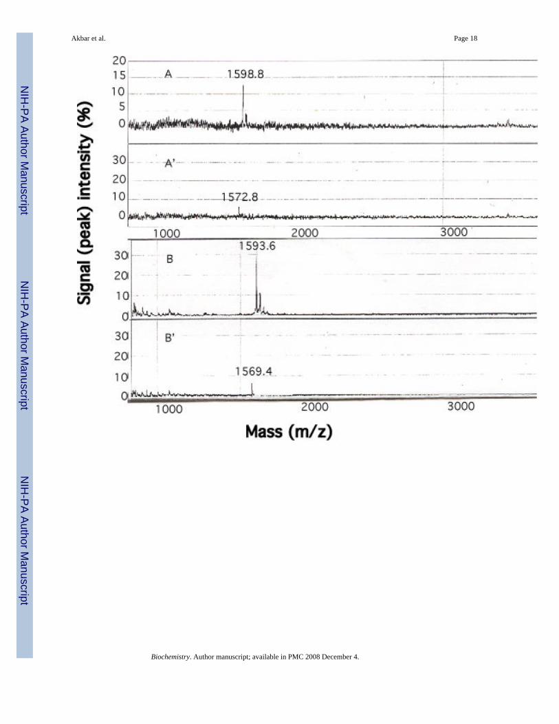
Peptide binding to Hb SMass spectrometric-based SELDI- Protein Chip technology with solid-state, time-of-flight(TOF) was employed to detect interactions between the β73His peptides and Hb S (Fig. 7). Amass signal for chip-bound 15-mer β73His peptide at 1598 m/z was detected when the peptidewas incubated at 4 pM with the chip containing bound Hb S in the oxy form (Fig. 7, panel A).Signal intensity doubled at 8 pM 15-mer β73His peptide (Fig. 7, panel B), while the 15-merβ73Leu peptide (1572 m/z) (Fig. 7, panel A’, B’) and 11-mer β73 His peptides (1128 m/z)showed little or no binding under the same conditions (Fig. 7, panel C and C’). In addition,signal intensities for the β73Leu peptide and the 11-, 7- and 3-mer peptides did not increasegoing from 4 to 8 pM peptide concentration (data not shown). Furthermore, there was no signalfor the 15-mer β73His peptide following incubation with chip-bound lysozyme instead of HbS. These results indicate that the 15-mer β73His peptide interacts selectively in a concentration-dependent manner with oxy Hb S prior to deoxy Hb S polymerization.
CD spectra of the β73His and β73 Leu peptidesHelix content of the 15 mer-β73His and β73Leu peptides solubilized in 0.5% (v/v) acetonitrilewas assessed by Circular Dichroism (CD) between 190 and 260 nm at room temperature usingan Aviv model 62 DS instrument equipped with a thermoelectric module (Fig. 8). The shapesof the two spectra were similar and did not show any specific helical structure, indicatingrandom coil structures for both peptides in this solution.
Computer docking analysis of β73His and Leu peptides with the βS-globin chain in Hb SResults from DIC images and SELDI-TOF MS suggested that the 15-mer β73His peptideselectively interacted with Hb S, presumably via the β4Thr in the βS-globin chain in Hb S priorto Hb S polymerization. We therefore speculated the random coil structure of these peptidesmight assume some structure after interaction with a specific area of β4Thr in Hb S. We nextevaluated docking energies of the 15mer β73His and β73Leu peptides with Hb S using
Akbar et al. Page 5
Biochemistry. Author manuscript; available in PMC 2008 December 4.
NIH
-PA Author Manuscript
NIH
-PA Author Manuscript
NIH
-PA Author Manuscript
conformation analysis simulation (Fig. 9). Our results indicate that His in the 15-mer β73Hispeptide is much closer to β4Thr in the βS-globin chain than Leu in the 15-mer β73Leu peptide[compare panels (a) and (b) in Fig. 9]. Docking energy differences between the 15-mer β73Hispeptide and Hb S (−181.162 kcal/mol) versus the β73Leu peptide and Hb S (−138.835 kcal/mol) probably arise from differences in interaction between the two different β73 amino acidsand β4Thr in Hb S. It is also noteworthy that the order of energy required for interaction ofamino acids at β73 in helical 15mer EF helix peptides with β4Thr in the βS chain increase inthe order of His << Asp < Asn < Leu, which is consistent with ease of polymerization of HbS with the β73 variants. These results suggest that the 15-mer β73His peptide selectivelyinteracts with Hb S when the peptide length is >15 amino acids, presumably via the β4Thr inthe βS-globin chain in Hb S. Furthermore we propose that this interaction preferentiallyinfluences the hydrogen bond interaction between β73Asp and β4Thr in Hb S polymers andmay induce structural changes in the 15-mer His peptide after its interaction with oxy Hb Sleading to inhibition of deoxy Hb S polymerization. This interaction also may interfere withhydrophobic interactions of β6 Val during Hb S polymerization.
DiscussionOur results show that the 15-mer EF helix peptide containing β73His inhibits Hb Spolymerization while the 15-mer β73Leu peptide and His-containing peptides smaller than 11amino acids have no effect. This can be explained from our previous results of polymerizationof recombinant Hb S β73His and Hb S β73Leu as well as our findings using S/Aβ73His andS/Aβ73 mixtures compared to AS mixtures (10). Namely, β73His in deoxy Hb S promoteswhile the β73Leu inhibits Hb S polymerization. These findings indicate the β73-β4 hydrogenbond in Hb S polymers influences rates of nucleation and polymerization of Hb S. This supportsour hypotheses regarding the basis for inhibition of polymerization by specific interaction ofEF-helix peptides containing β73His with Hb S. In fact, using various engineered Hb S variants,we showed that changing the amino acid side chains at the β73 position at Hb S polymerinteraction sites alters contact energy to stabilize polymers (11). This energy change affectsvibrational entropy of Hb S molecules upon polymer assembly (11). Mutation at Hb S polymercontact regions generally affects kinetic and thermodynamic properties of Hb Spolymerization.
Furthermore, polymerization of mixtures of Hb S and the recombinant variant Hb Aβ73Leuproduced morphological changes in polymer domains that are characteristic of polymerformation. We speculated non-productive binding of S-Aβ73Leu hybrids to the ends ofgrowing Hb S polymers. This “cap” then would prohibit growth of polymers and producesmaller domains than those of Hb S, AS or FS mixtures. In fact, Hb Aβ73Leu is the first Hbvariant with anti-polymerization properties that exceed those of Hb F. We also speculate thatinteractions of deoxy Hb S with specific peptides like the 15-mer β73His peptide at the EFhelix of β chains may play a capping-like role similar to Hb SAβ73Leu hybrids in Hb S fibers,even though inhibition by the peptide is less than that of Hb S-Aβ73Leu hybrids. An interestingaspect of this study is that the 15-mer β73His peptide showed not only inhibition of the delaytime and enhancement in solubility but also resulted in a delay of elongation rates of deoxy HbS fibers. DIC microscopic results further indicate inhibition of elongation of domains andformation of smaller domains in the presence of the β73 His peptides. Furthermore, the β73His peptide can bind to both the deoxy and oxy forms of Hb S. Even though interactions of theβ73 His peptide with oxy Hb S presumably around β4 Thr at the A-helix appear weakenedupon deoxygenation, deoxy Hb S still interacted with the β73 His peptide possibly influencingthe critical concentration for polymerization and inhibiting the rate of nucleation. Thisinteraction may not only influence hydrogen bond interaction between β73Asp and β4Thr information of deoxy Hb S nuclei and polymers but also may interfere with hydrophobicinteractions of β6 Val leading to inhibition of Hb S polymerization, since β4Thr is so close to
Akbar et al. Page 6
Biochemistry. Author manuscript; available in PMC 2008 December 4.
NIH
-PA Author Manuscript
NIH
-PA Author Manuscript
NIH
-PA Author Manuscript
β6 Val. In addition, the 15mer peptides contain two His residues which might form a salt bridgewith β7Glu (7). Formation of this salt bridge may disrupt the first turn of the α helix in βS
chains and could therefore impact on Hb S polymerization. These results suggest that the 15-mer His peptide not only inhibits homogeneous but also heterogeneous nucleation byinteraction of the peptides with Hb S resulting in a delay in the assembly of monomers inaddition to a delay in the elongation rate of fibers. We speculate that peptides larger than 15-mers may require specific interaction with β4 Thr in the A-helix of βS chains in order to inhibitHb S polymerization. In fact, it was reported that the βS (1−55) peptide showed a higher contentof β-sheet and a lower amount of α helix compared to the βA (1−55) peptide, while there wasvery little secondary structural differences comparing shorter βS versus βA peptides (1−30)(6). Therefore, peptides longer than 15 amino acids may be required additional structuralconformation to promote specific interactions involving His in the peptide with β4 Thr in theA-helix of βS chains in order to inhibit Hb S fiber growth like Hb S-Aβ73Leu hybrids.
Several small molecules, including phenylalanine and tryptophan derivatives, also weredesigned to interact with Hb S at the β6 donor and its acceptor pocket (15-17). However, noneshowed much promise as a therapeutic agent (18), partly because of the large dose of drugrequired to interact with circulating hemoglobin and the structural change of the EF helix inthe β6Val donor area that occurs during oxygenation and deoxygenation. Furthermore,bioavailability of the drugs was low due to serum protein binding. Our results on the inhibitoryeffects of the 15-mer β73His peptide were more compelling than that of single amino acids orpeptides shorter than 11amino acids. Furthermore, results of energy calculations usingcomputer simulations as well as results of polymerization of Hb Sβ73 variants and mixturesof Hb S with Hb A containing the β73 variants suggest potential design of more efficientinhibitory peptides. It may be possible to express soluble peptides in erythroid cells that inhibitHb S polymerization more than the 15-mer β73His peptide like Hb Aβ73Leu or Hb F afteradditional consideration of the specificity of the A-helix of βS chains in the deoxy form. Suchpeptides would not require consideration of partner chains to form hemoglobin tetramers orheme insertion into globin chains, and could possibly be introduced or expressed in anerythroid-specific fashion for gene therapy of sickle cell disease. In addition, it appears thatneither Hb F nor its hybrids with Hb S enter the polymer, so Hb F and its variants would seemto represent the limit of maximum inhibition. Therefore, Hb F plus such peptides might exhibitanti-sickling effects exceeding those of Hb F alone. This has important implications for genetherapy for sickle cell disease, since anti-polymerization induced by these peptides and Hb Fmay possibly be accomplished by completely different mechanisms; inhibition by the formervia direct interaction with Hb S, and the latter by reduction in Hb S concentration via formationof hybrids which are excluded from Hb S polymers (19). We are screening for such peptidesand will evaluate their anti-polymerization properties under near physiological conditions.
AcknowledgementsThis research was supported in part by grants from the National Institutes of Health (HL 58879, 69256, 70596 and38632) and by the Cardeza Foundation for Hematological Research, Jefferson Medical College.
AbbreviationsHb S, sickle hemoglobin; Hb A, normal hemoglobin A; Hb F, fetal hemoglobin; DIC,Differential Interference Contrast.
References1. Eaton WA, Hofrichter J. Sickle cell hemoglobin polymerization. Adv Protein Chem 1990;40:63–279.
[PubMed: 2195851]2. Bunn, HF.; Forget, BG. Hemoglobin: Molecular and Clinical Aspects. Sanders; Philadelphia.: 1996.
Akbar et al. Page 7
Biochemistry. Author manuscript; available in PMC 2008 December 4.
NIH
-PA Author Manuscript
NIH
-PA Author Manuscript
NIH
-PA Author Manuscript
3. Ferrone FA, Hofrichter J, Eaton WA. Kinetics of sickle hemoglobin polymerization. II. A doublenucleation mechanism. J Mol Biol 1985;183:611–631. [PubMed: 4020873]
4. Embury, SH.; Hebbel, RP.; Mohandus, N.; Steinberg, MH. SICKLE CELL DISEASE: Basic principlesand clinical practice. Raven Press; New York: 1994.
5. Harrington DJ, Adachi K, Royer WE Jr. The high resolution crystal structure of deoxyhemoglobin S.J Mol Biol 1997;272:398–407. [PubMed: 9325099]
6. Fronticelli C, Gold R. Conformational relevance of the beta6Glu replaced by Val mutation in the betasubunits and in the beta(1−55) and beta(1−30) peptides of hemoglobin S. J Biol Chem 1976;251:4968–4972. [PubMed: 956170]
7. Scholberg HP, Fronticelli C, Bucci E. Conformational changes in the hemoglobin S system as seen byproton binding. J Biol Chem 1980;255:8592–8598. [PubMed: 7410379]
8. Padlan EA, Love WE. Refined crystal structure of deoxyhemoglobin S. II. Molecular interactions inthe crystal. J Biol Chem 1985;260:8280–8291. [PubMed: 2409085]
9. Beddell CR, Goodford PJ, Norrington FE, Wilkinson S, Wootton R. Compounds designed to fit a siteof known structure in human haemoglobin. Br J Pharmacol 1976;57:201–209. [PubMed: 938794]
10. Adachi K, Ding M, Wehrli S, Reddy KS, Surrey S, Horiuchi K. Effects of different beta73 aminoacids on formation of 14-stranded fibers of Hb S versus double-stranded crystals of Hb C-Harlem.Biochemistry 2003;42:4476–4484. [PubMed: 12693943]
11. Ivanova M, Jasuja R, Krasnosselskaia L, Josephs R, Wang Z, Ding M, Horiuchi K, Adachi K, FerroneFA. Flexibility and nucleation in sickle hemoglobin. J Mol Biol 2001;314:851–861. [PubMed:11734002]
12. Adachi K, Asakura T. Nucleation-controlled aggregation of deoxyhemoglobin S. Possible differencein the size of nuclei in different phosphate concentrations. J Biol Chem 1979;254:7765–7771.[PubMed: 468786]
13. Haewon, CK.; Adachi, K.; Schwartz, E. Separation of hemoglobin.. In: Beutler, E.; Lichtman, MA.;Coller, b. S.; Kippes, TJ., editors. Williams Hematology. Fifth Edition. McGraw-Hill; New york:1995. p. L35-L42.
14. Tolson J, Bogumil R, Brunst E, Beck H, Elsner R, Humeny A, Kratzin H, Deeg M, Kuczyk M, MuellerGA, Mueller CA, Flad T. Serum protein profiling by SELDI mass spectrometry: detection of multiplevariants of serum amyloid alpha in renal cancer patients. Lab Invest 2004;84:845–856. [PubMed:15107802]
15. Noguchi CT, Torchia DA, Schechter AN. Determination of sickle hemoglobin polymer in SS and ASerythrocytes. Blood Cells 1982;8:225–235. [PubMed: 7159747]
16. Noguchi CT, Ackerman S, DiMaio J, Schiller PW, Schechter AN. The effect of phenylalaninederivatives on the solubility of deoxyhemoglobin S. A model class of gelation inhibitors. MolPharmacol 1983;23:100–103. [PubMed: 6865893]
17. Kumpati J. Effect of phenylalanine- or tryptophan-loaded liposomes on the rheological properties ofAA and SS erythrocytes. Biochem Med Metab Biol 1987;38:246–251. [PubMed: 3675924]
18. Orringer, EP.; Abraham, DJ.; Parker, JC. SICKLE CELL DISEASE: Basic principles and clinicalpractice.. In: Embury, SH.; Hebbel, RP.; Mohandas, N.; Steinberg, MH., editors. Development ofDrug Therapy. Raven Press; New York: 1994. p. 861-872.
19. Rotter MA, Dragos D, Aprelev A, Adachi K, Ferrone FA. Molecular crowding limits the role of fetalhemoglobin in therapy for sickle cell disease. J. Mol. Biol 2005;347:1015–1023. [PubMed:15784260]
Akbar et al. Page 8
Biochemistry. Author manuscript; available in PMC 2008 December 4.
NIH
-PA Author Manuscript
NIH
-PA Author Manuscript
NIH
-PA Author Manuscript
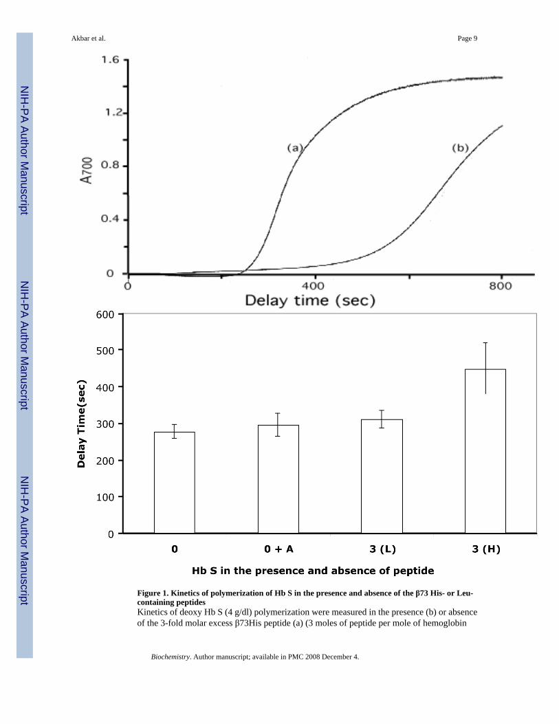
Figure 1. Kinetics of polymerization of Hb S in the presence and absence of the β73 His- or Leu-containing peptidesKinetics of deoxy Hb S (4 g/dl) polymerization were measured in the presence (b) or absenceof the 3-fold molar excess β73His peptide (a) (3 moles of peptide per mole of hemoglobin
Akbar et al. Page 9
Biochemistry. Author manuscript; available in PMC 2008 December 4.
NIH
-PA Author Manuscript
NIH
-PA Author Manuscript
NIH
-PA Author Manuscript
tetramer to Hb S dissolved in 0.5% (v/v) acetonitrile in 1.0 M phosphate buffer at 30°C by thetemperature-jump method (panel A). Delay times prior to polymerization of Hb S (4g/dl) alsowere measured in the presence of a 3-fold molar excess of the 15-mer β73His [3 (H)] or β73Leupeptides [3 (L)], and mean values compared to Hb S alone (0) or Hb S in the presence of 0.5%(v/v) acetonitrile (0+A) (panel B).
Akbar et al. Page 10
Biochemistry. Author manuscript; available in PMC 2008 December 4.
NIH
-PA Author Manuscript
NIH
-PA Author Manuscript
NIH
-PA Author Manuscript
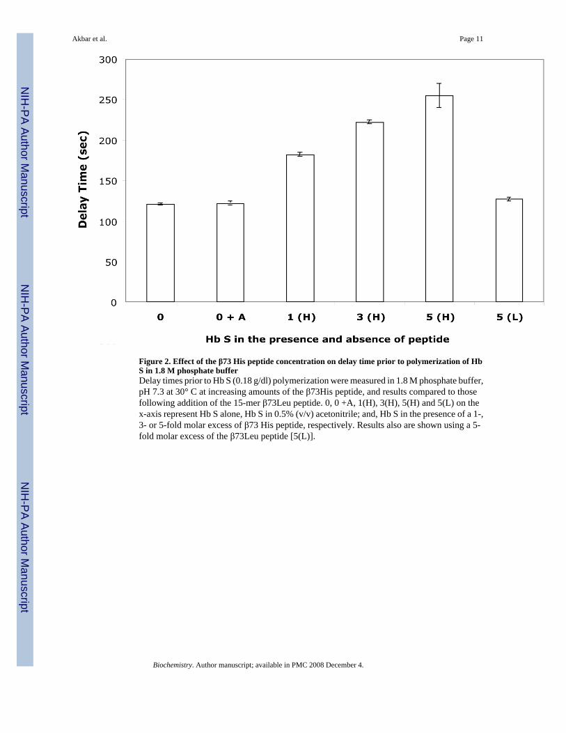
Figure 2. Effect of the β73 His peptide concentration on delay time prior to polymerization of HbS in 1.8 M phosphate bufferDelay times prior to Hb S (0.18 g/dl) polymerization were measured in 1.8 M phosphate buffer,pH 7.3 at 30° C at increasing amounts of the β73His peptide, and results compared to thosefollowing addition of the 15-mer β73Leu peptide. 0, 0 +A, 1(H), 3(H), 5(H) and 5(L) on thex-axis represent Hb S alone, Hb S in 0.5% (v/v) acetonitrile; and, Hb S in the presence of a 1-,3- or 5-fold molar excess of β73 His peptide, respectively. Results also are shown using a 5-fold molar excess of the β73Leu peptide [5(L)].
Akbar et al. Page 11
Biochemistry. Author manuscript; available in PMC 2008 December 4.
NIH
-PA Author Manuscript
NIH
-PA Author Manuscript
NIH
-PA Author Manuscript
Figure 3. Effect of the β73His peptide on Hb S solubility in 1.0 M phosphate bufferSolubility of Hb S in 1.0 M phosphate buffer was measured in the presence of varying amountsof the 15-mer β73His peptide [1-(1H), 3-(3H) and 5-(5 H) fold molar ratio] in the presence of0.5% (v/v) acetonitrile, and results compared to those using the 15-mer β73Leu peptide [5-foldmolar excess, 5(L)] in the presence of 0.5% (v/v) acetonitrile. Solubility following completionof polymerization was assessed after centrifugation. Solubilities of Hb S alone (0) and in thepresence of 0.5% (v/v) acetonitrile (0 +A) also were measured with values representing themean of two measurements (maximum different ranges of the two values are within 3.3%).
Akbar et al. Page 12
Biochemistry. Author manuscript; available in PMC 2008 December 4.
NIH
-PA Author Manuscript
NIH
-PA Author Manuscript
NIH
-PA Author Manuscript
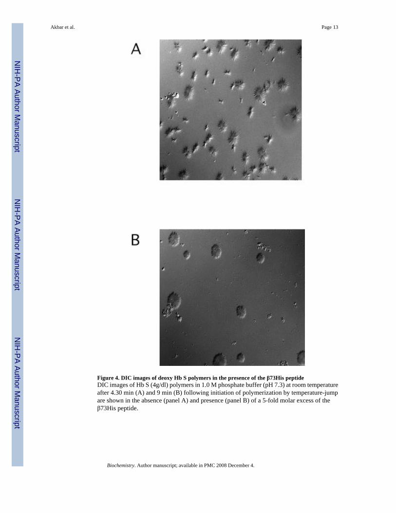
Figure 4. DIC images of deoxy Hb S polymers in the presence of the β73His peptideDIC images of Hb S (4g/dl) polymers in 1.0 M phosphate buffer (pH 7.3) at room temperatureafter 4.30 min (A) and 9 min (B) following initiation of polymerization by temperature-jumpare shown in the absence (panel A) and presence (panel B) of a 5-fold molar excess of theβ73His peptide.
Akbar et al. Page 13
Biochemistry. Author manuscript; available in PMC 2008 December 4.
NIH
-PA Author Manuscript
NIH
-PA Author Manuscript
NIH
-PA Author Manuscript
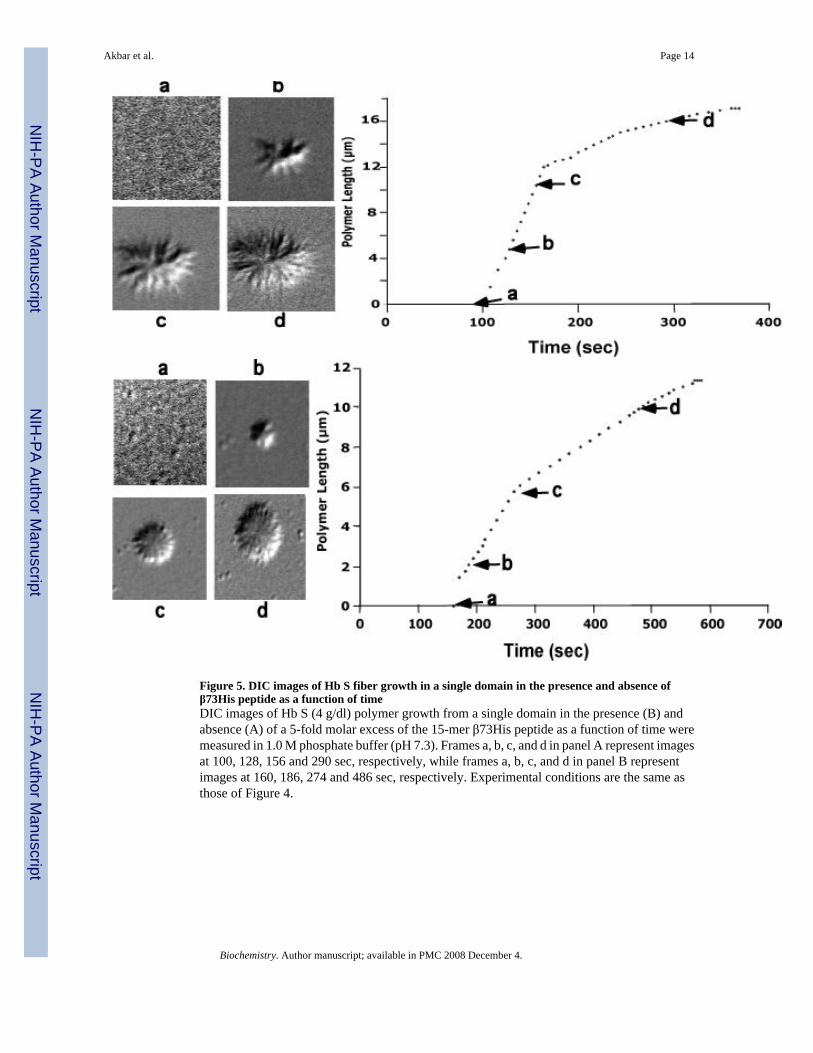
Figure 5. DIC images of Hb S fiber growth in a single domain in the presence and absence ofβ73His peptide as a function of timeDIC images of Hb S (4 g/dl) polymer growth from a single domain in the presence (B) andabsence (A) of a 5-fold molar excess of the 15-mer β73His peptide as a function of time weremeasured in 1.0 M phosphate buffer (pH 7.3). Frames a, b, c, and d in panel A represent imagesat 100, 128, 156 and 290 sec, respectively, while frames a, b, c, and d in panel B representimages at 160, 186, 274 and 486 sec, respectively. Experimental conditions are the same asthose of Figure 4.
Akbar et al. Page 14
Biochemistry. Author manuscript; available in PMC 2008 December 4.
NIH
-PA Author Manuscript
NIH
-PA Author Manuscript
NIH
-PA Author Manuscript
Akbar et al. Page 15
Biochemistry. Author manuscript; available in PMC 2008 December 4.
NIH
-PA Author Manuscript
NIH
-PA Author Manuscript
NIH
-PA Author Manuscript
Akbar et al. Page 16
Biochemistry. Author manuscript; available in PMC 2008 December 4.
NIH
-PA Author Manuscript
NIH
-PA Author Manuscript
NIH
-PA Author Manuscript
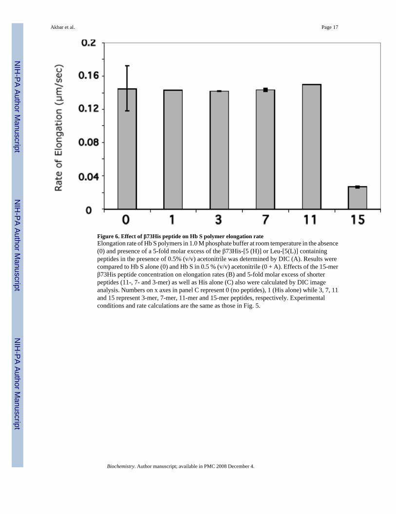
Figure 6. Effect of β73His peptide on Hb S polymer elongation rateElongation rate of Hb S polymers in 1.0 M phosphate buffer at room temperature in the absence(0) and presence of a 5-fold molar excess of the β73His-[5 (H)] or Leu-[5(L)] containingpeptides in the presence of 0.5% (v/v) acetonitrile was determined by DIC (A). Results werecompared to Hb S alone (0) and Hb S in 0.5 % (v/v) acetonitrile (0 + A). Effects of the 15-merβ73His peptide concentration on elongation rates (B) and 5-fold molar excess of shorterpeptides (11-, 7- and 3-mer) as well as His alone (C) also were calculated by DIC imageanalysis. Numbers on x axes in panel C represent 0 (no peptides), 1 (His alone) while 3, 7, 11and 15 represent 3-mer, 7-mer, 11-mer and 15-mer peptides, respectively. Experimentalconditions and rate calculations are the same as those in Fig. 5.
Akbar et al. Page 17
Biochemistry. Author manuscript; available in PMC 2008 December 4.
NIH
-PA Author Manuscript
NIH
-PA Author Manuscript
NIH
-PA Author Manuscript
Akbar et al. Page 18
Biochemistry. Author manuscript; available in PMC 2008 December 4.
NIH
-PA Author Manuscript
NIH
-PA Author Manuscript
NIH
-PA Author Manuscript
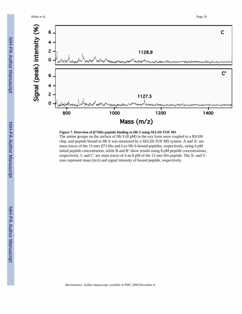
Figure 7. Detection of β73His peptide binding to Hb S using SELDI-TOF MSThe amine groups on the surface of Hb S (8 pM) in the oxy form were coupled to a RS100chip, and peptide bound to Hb S was measured by a SELDI-TOF MS system. A and A’ aremass traces of the 15-mer β73 His and Leu Hb S-bound peptides, respectively, using 4 pMinitial peptide concentration, while B and B’ show results using 8 pM peptide concentrations,respectively. C and C’ are mass traces of 4 an 8 pM of the 11-mer His peptide. The X- and Y-axes represent mass (m/z) and signal intensity of bound peptide, respectively.
Akbar et al. Page 19
Biochemistry. Author manuscript; available in PMC 2008 December 4.
NIH
-PA Author Manuscript
NIH
-PA Author Manuscript
NIH
-PA Author Manuscript
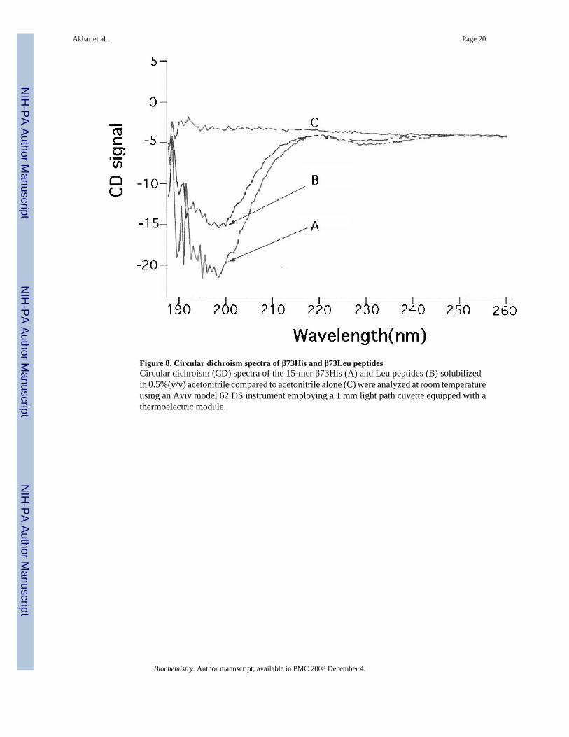
Figure 8. Circular dichroism spectra of β73His and β73Leu peptidesCircular dichroism (CD) spectra of the 15-mer β73His (A) and Leu peptides (B) solubilizedin 0.5%(v/v) acetonitrile compared to acetonitrile alone (C) were analyzed at room temperatureusing an Aviv model 62 DS instrument employing a 1 mm light path cuvette equipped with athermoelectric module.
Akbar et al. Page 20
Biochemistry. Author manuscript; available in PMC 2008 December 4.
NIH
-PA Author Manuscript
NIH
-PA Author Manuscript
NIH
-PA Author Manuscript
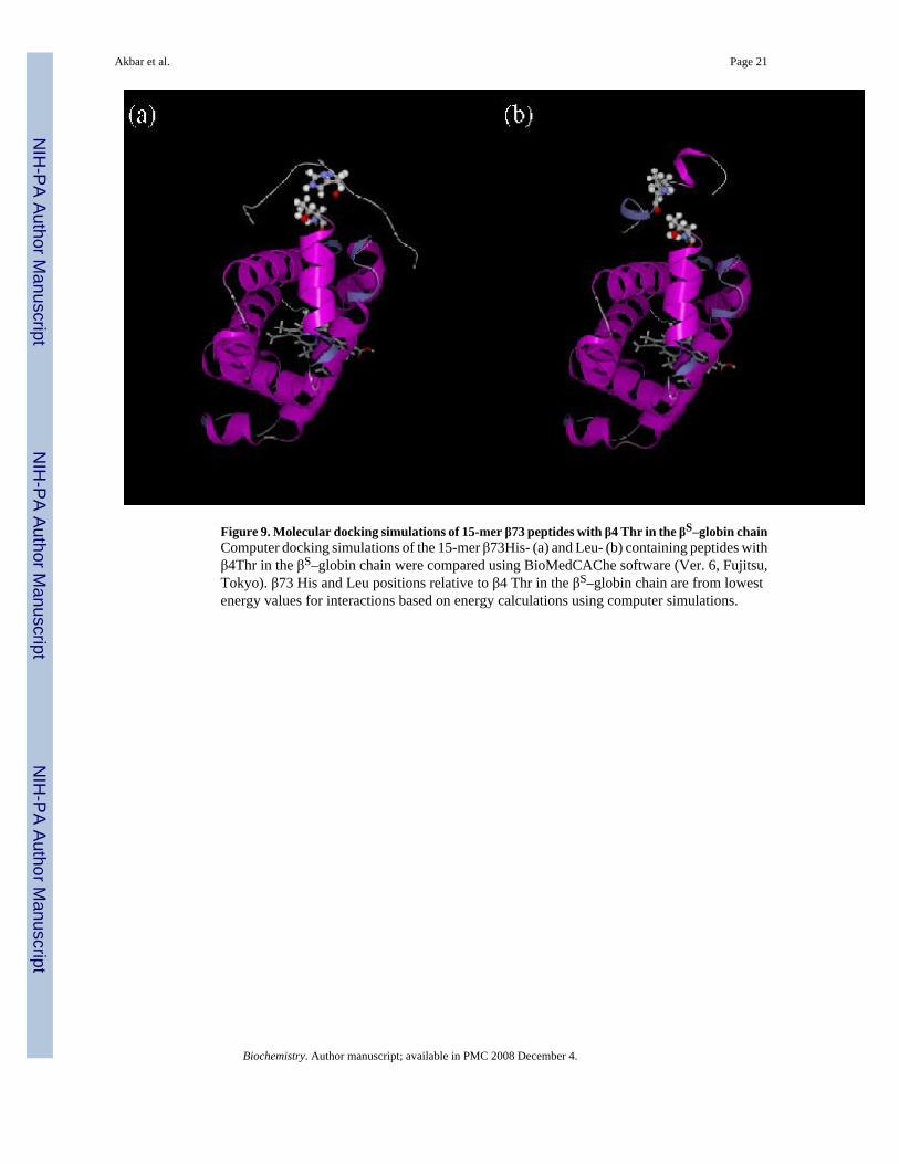
Figure 9. Molecular docking simulations of 15-mer β73 peptides with β4 Thr in the βS–globin chainComputer docking simulations of the 15-mer β73His- (a) and Leu- (b) containing peptides withβ4Thr in the βS–globin chain were compared using BioMedCAChe software (Ver. 6, Fujitsu,Tokyo). β73 His and Leu positions relative to β4 Thr in the βS–globin chain are from lowestenergy values for interactions based on energy calculations using computer simulations.
Akbar et al. Page 21
Biochemistry. Author manuscript; available in PMC 2008 December 4.
NIH
-PA Author Manuscript
NIH
-PA Author Manuscript
NIH
-PA Author Manuscript
Copyright © 2022 FDOKUMEN