A novel nickel-chelated surfactant for affinity-based aqueous two-phase micellar extraction of...
7
Journal of Chromatography A, 1320 (2013) 118–124 Contents lists available at ScienceDirect Journal of Chromatography A j our nal homep age: www.elsevier.com/locate/chroma A novel nickel-chelated surfactant for affinity-based aqueous two-phase micellar extraction of histidine-rich protein Shuo Wang a , Neng Xiong a , Xiao-Yan Dong a,b , Yan Sun a,b,∗ a Department of Biochemical Engineering and Key Laboratory of Systems Bioengineering of the Ministry of Education, School of Chemical Engineering and Technology, Tianjin University, Tianjin 300072, China b Collaborative Innovation Center of Chemical Science and Engineering (Tianjin), Tianjin 300072, China a r t i c l e i n f o Article history: Received 28 June 2013 Received in revised form 18 September 2013 Accepted 23 October 2013 Available online 30 October 2013 Keywords: Aqueous two-phase micellar system Nickel-chelated surfactant Triton X-114 Cloud point Affinity partitioning Green fluorescent protein a b s t r a c t Aqueous two-phase micellar systems (ATPMSs) composed of nonionic surfactants are considered promis- ing for the separation and purification of proteins. To improve the specificity of ATPMSs, a novel nickel-chelated surfactant was prepared by successive modifications of Triton X-114 (TX). Character- izations by Fourier transformation infrared spectroscopy demonstrated the successful synthesis of the nickel-chelated surfactant (TX-Ni). The cloud point, critical micelle concentration (CMC), molecular inter- action parameter and micelle size were measured for the mixed surfactant system of TX-Ni and TX to achieve a full understanding of their aggregation behaviors. The results showed that mixed micelles were formed, and the cloud point increased with the mole fraction of TX-Ni because TX-Ni had a more hydrophilic head group than TX. Moreover, the reduction of micelle size revealed by light scattering exper- iments indicated that the insertion of TX-Ni inhibited the micellar growth due to the increased steric and electrostatic repulsion. Finally, the efficiency of TX-Ni as an affinity surfactant was demonstrated by the affinity partitioning of histidine-tagged enhanced green fluorescent protein with an over 20-fold increase of the partition coefficient (from 0.60 to 12.42). This affinity-based ATPMS is thus considered promising for providing a versatile platform for the separation of histidine-rich proteins. © 2013 Elsevier B.V. All rights reserved. 1. Introduction Aqueous two-phase systems (ATPSs) are cost-effective and environment-friendly platforms for the separation of proteins. In recent years, ATPSs have attracted considerable interest and are regarded as an alternative to traditional separation techniques owing to their advantages, including scale-up potential, contin- uous operation, ease of process integration and biocompatibility [1–5]. Apart from conventional ATPSs which are composed of two incompatible polymers or one polymer and a salt [1], many new ATPSs have been developed, one of which is the aqueous two-phase micellar system (ATPMS) formed by nonionic surfactants [6]. When heated above a threshold temperature, which is known as cloud point, the homogenous aqueous solution of nonionic surfactant becomes turbid and then separates into two immiscible phases. Usually one phase is rich in micelles and the other is poor. Due to the different physicochemical properties of the two phases, proteins ∗ Corresponding author at: Department of Biochemical Engineering and Key Lab- oratory of Systems Bioengineering of the Ministry of Education, School of Chemical Engineering and Technology, Tianjin University, Tianjin 300072, China. Tel.: +86 22 27404981; fax: +86 22 27404981. E-mail address: [email protected] (Y. Sun). partition unevenly in this system [6]. Because both the micelle-rich phase and the micelle-poor phase are mainly composed of water and most nonionic surfactants are mild to proteins [7], ATPMSs provide a gentle and friendly environment for biomolecules. Com- pared with ATPSs composed of polymers, ATPMSs have some merits [8,9]: (1) micelles are labile entities formed by noncovalent binding of individual surfactant molecules, and as a result, the micelle shape and size are adjustable. (2) Generally, commercial nonionic surfac- tants are easily available and cheap. (3) More importantly, nonionic surfactants have been widely utilized in industries for a long time and the basic data are relatively abundant, which is beneficial for industrial applications. ATPMSs were originally used for the concentration of zinc ions [10]. In the 1980s, Bordier investigated their application in the sep- aration of membrane proteins [11]. Since then ATPMSs have been successfully applied to the extraction and purification of a variety of proteins and related biochemicals [6,12]. Particularly, ATPMSs are most suitable for the isolation of hydrophobic membrane pro- teins. In this case, the separation is governed by solubilization and hydrophobic effects, which determine the partitioning extent of proteins into the micelle-rich phase. Although the contributions of excluded-volume effects [13] and electrostatic interactions [14] have extended the applications of this method to the extraction of hydrophilic proteins, its low selectivity remains to be an obstacle 0021-9673/$ – see front matter © 2013 Elsevier B.V. All rights reserved. http://dx.doi.org/10.1016/j.chroma.2013.10.074
-
Upload
independent -
Category
Documents
-
view
0 -
download
0
Transcript of A novel nickel-chelated surfactant for affinity-based aqueous two-phase micellar extraction of...
At
Sa
Tb
a
ARR1AA
KANTCAG
1
errou[iAmhpbUd
oET
0h
Journal of Chromatography A, 1320 (2013) 118– 124
Contents lists available at ScienceDirect
Journal of Chromatography A
j our nal homep age: www.elsev ier .com/ locate /chroma
novel nickel-chelated surfactant for affinity-based aqueouswo-phase micellar extraction of histidine-rich protein
huo Wanga, Neng Xionga, Xiao-Yan Donga,b, Yan Suna,b,∗
Department of Biochemical Engineering and Key Laboratory of Systems Bioengineering of the Ministry of Education, School of Chemical Engineering andechnology, Tianjin University, Tianjin 300072, ChinaCollaborative Innovation Center of Chemical Science and Engineering (Tianjin), Tianjin 300072, China
r t i c l e i n f o
rticle history:eceived 28 June 2013eceived in revised form8 September 2013ccepted 23 October 2013vailable online 30 October 2013
eywords:queous two-phase micellar system
a b s t r a c t
Aqueous two-phase micellar systems (ATPMSs) composed of nonionic surfactants are considered promis-ing for the separation and purification of proteins. To improve the specificity of ATPMSs, a novelnickel-chelated surfactant was prepared by successive modifications of Triton X-114 (TX). Character-izations by Fourier transformation infrared spectroscopy demonstrated the successful synthesis of thenickel-chelated surfactant (TX-Ni). The cloud point, critical micelle concentration (CMC), molecular inter-action parameter and micelle size were measured for the mixed surfactant system of TX-Ni and TX toachieve a full understanding of their aggregation behaviors. The results showed that mixed micelleswere formed, and the cloud point increased with the mole fraction of TX-Ni because TX-Ni had a more
ickel-chelated surfactantriton X-114loud pointffinity partitioningreen fluorescent protein
hydrophilic head group than TX. Moreover, the reduction of micelle size revealed by light scattering exper-iments indicated that the insertion of TX-Ni inhibited the micellar growth due to the increased steric andelectrostatic repulsion. Finally, the efficiency of TX-Ni as an affinity surfactant was demonstrated by theaffinity partitioning of histidine-tagged enhanced green fluorescent protein with an over 20-fold increaseof the partition coefficient (from 0.60 to 12.42). This affinity-based ATPMS is thus considered promisingfor providing a versatile platform for the separation of histidine-rich proteins.
. Introduction
Aqueous two-phase systems (ATPSs) are cost-effective andnvironment-friendly platforms for the separation of proteins. Inecent years, ATPSs have attracted considerable interest and areegarded as an alternative to traditional separation techniqueswing to their advantages, including scale-up potential, contin-ous operation, ease of process integration and biocompatibility1–5]. Apart from conventional ATPSs which are composed of twoncompatible polymers or one polymer and a salt [1], many newTPSs have been developed, one of which is the aqueous two-phaseicellar system (ATPMS) formed by nonionic surfactants [6]. When
eated above a threshold temperature, which is known as cloudoint, the homogenous aqueous solution of nonionic surfactant
ecomes turbid and then separates into two immiscible phases.sually one phase is rich in micelles and the other is poor. Due to theifferent physicochemical properties of the two phases, proteins∗ Corresponding author at: Department of Biochemical Engineering and Key Lab-ratory of Systems Bioengineering of the Ministry of Education, School of Chemicalngineering and Technology, Tianjin University, Tianjin 300072, China.el.: +86 22 27404981; fax: +86 22 27404981.
E-mail address: [email protected] (Y. Sun).
021-9673/$ – see front matter © 2013 Elsevier B.V. All rights reserved.ttp://dx.doi.org/10.1016/j.chroma.2013.10.074
© 2013 Elsevier B.V. All rights reserved.
partition unevenly in this system [6]. Because both the micelle-richphase and the micelle-poor phase are mainly composed of waterand most nonionic surfactants are mild to proteins [7], ATPMSsprovide a gentle and friendly environment for biomolecules. Com-pared with ATPSs composed of polymers, ATPMSs have some merits[8,9]: (1) micelles are labile entities formed by noncovalent bindingof individual surfactant molecules, and as a result, the micelle shapeand size are adjustable. (2) Generally, commercial nonionic surfac-tants are easily available and cheap. (3) More importantly, nonionicsurfactants have been widely utilized in industries for a long timeand the basic data are relatively abundant, which is beneficial forindustrial applications.
ATPMSs were originally used for the concentration of zinc ions[10]. In the 1980s, Bordier investigated their application in the sep-aration of membrane proteins [11]. Since then ATPMSs have beensuccessfully applied to the extraction and purification of a varietyof proteins and related biochemicals [6,12]. Particularly, ATPMSsare most suitable for the isolation of hydrophobic membrane pro-teins. In this case, the separation is governed by solubilization andhydrophobic effects, which determine the partitioning extent of
proteins into the micelle-rich phase. Although the contributionsof excluded-volume effects [13] and electrostatic interactions [14]have extended the applications of this method to the extraction ofhydrophilic proteins, its low selectivity remains to be an obstacle
S. Wang et al. / J. Chromatogr.
piHlrin
sfcfcwh
2
2
t(aGg
rdgThpE
2
diwTTwsoefipfair
Fig. 1. Synthesis route for the nickel-chelated surfactant (TX-Ni).
recluding its wide utilizations. Generally, the use of affinity ligandss an effective way to improve the specificity of ATPMSs [3,15–20].owever, in spite of the fact that immobilized metal-ion affinity
igands have been successfully applied in polymer ATPS [21,22],everse micelle extraction [23,24], as well as the most widely usedmmobilized metal affinity chromatography (IMAC) [25], there areo reports on their incorporation into ATPMSs.
In this study, Triton X-114 (TX) was functionalized by succes-ive modifications of epichlorohydrin and iminodiacetic acid (IDA)ollowed by chelating with nickel ions to prepare a novel metal-helated surfactant, TX-Ni. Then, an affinity-based ATPMS wasormed by the mixture of TX-Ni and TX. The system was extensivelyharacterized and the effectiveness of this affinity-based ATPMSas demonstrated by the affinity partitioning and separation ofistidine-tagged enhanced green fluorescent protein (EGFP).
. Materials and methods
.1. Materials
TX was obtained from Fluka (Bucks, Switzerland). Acetoni-rile (≥99.9%), pyrene (≥99.0%) and chicken egg white lysozyme≥90%) were purchased from Sigma–Aldrich (MO, USA). IDA (>97%)nd benzyltriethylammonium chloride (BTAC, >99%) were fromuangfu (Tianjin, China). Other chemicals were all of analyticalrade from local sources. All materials were used as received.
Polyhistidine-tagged EGFP was expressed by cultivation of theecombinant Escherichia coli harboring the vector pET28a-EGFP asescribed previously [26]. The cells were harvested by centrifu-ation and disrupted by sonication in Tris–HCl buffer (20 mmol/Lris, 100 mmol/L NaCl, pH 8.0). After centrifugation of the cellomogenate at 13,000 × g for 15 min at 4 ◦C, the supernatant wasurified by IMAC and gel filtration chromatography to prepare pureGFP [23].
.2. Synthesis of nickel-chelated surfactant
The synthesis of TX-Ni was performed according to the proce-ures described in literatures [27,28] and the synthesis route is
llustrated in Fig. 1. In a typical procedure, 5.4 g of TX (0.01 mol)as added to 10 mL of benzene with 1 g of NaOH and 0.5 g of BTAC.
he mixture was vigorously stirred under N2 at 50 ◦C for 30 min.hen, 4 mL of epichlorohydrin (0.05 mol) was added and the stirringas continued for 4 h. The reaction mixture was filtered to remove
olids and excess organic solvents were removed by vacuum evap-ration. This reaction led to the activation of TX to the intermediatepoxide (1 in Fig. 1), which was obtained as yellow oil. In theollowing reaction, 1 was mixed with 20 mL of 2 mol/L disodiumminodiacetate solution (pH 11) prepared by slowly adding NaOHellets to 6.6 g of IDA in water. The mixture was stirred at 50 ◦C
or 24 h. Separation of the IDA coupled TX (TX-IDA, 2 in Fig. 1) waschieved by extraction with chloroform [17]. Thereafter, TX-IDAn the organic phase was recovered and dried by vacuum evapo-ation, and then mixed with 20 mL of 0.5 mol/L NiSO4 solution toA 1320 (2013) 118– 124 119
chelate Ni(II) ions. Separation of TX-Ni (3 in Fig. 1) was the same asthat of TX-IDA. Finally, the product was purified by redissolving inethyl acetate and then reprecipitating with n-hexane. After suctionfiltration, residue organic solvents were removed by drying undervacuum at 80 ◦C overnight.
2.3. Characterization of TX and TX-Ni
Fourier transform infrared (FTIR) analysis was performed on aTensor 27 FTIR spectrometer (Bruker Optics, Billerica, MA). Potas-sium bromide (KBr) was used to collect the background. Sampleswere mixed with KBr in an appropriate portion before spectrumcollection. Twenty scans were recorded at 4 cm−1 resolution withtwo levels of zero filling.
The purity of TX-Ni was determined by reversed-phase highperformance liquid chromatography (RP-HPLC) using a C18Symmetry® column (5 �m, 150 mm × 4.6 mm; Waters, MA, USA).Samples were eluted with a 20:80 water–acetonitrile mobile phasecontaining 0.1% trifluoroacetic acid at 0.5 mL/min [29]. The analysiswas performed on an Agilent 1100 system (Agilent Technologies,USA) equipped with a UV detector set at 277 nm.
2.4. Phase diagram determination
Phase diagrams for the aqueous solutions of TX-Ni and TX weremeasured by the cloud-point method [13]. Briefly, surfactant solu-tions of known concentrations were prepared and then put intoa programmable temperature controller (PolyScience 9512, Poly-Science, USA) with a temperature stability of ±0.01 ◦C (readoutaccuracy: ±0.25 ◦C). At beginning, each sample was cooled to 20 ◦Cwhich was low enough to make the solution exhibit a single andclear phase. The temperature was then raised at 0.2 ◦C/min untilthe solution became turbid, indicating the onset of phase separa-tion, at a temperature Tu. As soon as clouding was observed, thetemperature was lowered at 0.2 ◦C/min until the solution becameclear again at a temperature Td. The cloud point temperature wastaken to be the average of Tu and Td. The procedure was repeatedthree times for each data point to ensure reproducibility.
2.5. Determination of critical micelle concentration
The critical micelle concentrations (CMCs) of TX-Ni, TX and theirmixtures were determined by the pyrene 1:3 ratio method [30].A number of surfactant solutions containing 2 �mol/L of pyrenewere measured on a Perkin Elmer LS55 Fluorescence Spectrometer(Waltham, MA, USA). Pyrene was excited at 335 nm. The emissionintensities of the peaks located near 373 nm (I1) and 384 nm (I3)were recorded. The solution composition was expressed by themole fraction of TX-Ni (˛), defined as:
= [TX-Ni][TX-Ni] + [TX]
(1)
where [TX-Ni] and [TX] are the molar concentrations of TX-Ni andTX in the solution, respectively. All measurements were carried outat 20.0 ± 0.1 ◦C.
2.6. Light scattering
Static light scattering (SLS) experiments were performedwith a DAWN EOS multiangle light scattering instrument (WyattTechnology, USA). All measurements were conducted in theangular range 30◦ < � < 150◦ at a wavelength of � = 690 nm. The

1 atogr. A 1320 (2013) 118– 124
ac
qcsr�TiC
oHTS
D
wtc
fir
2
pN0eptWc
mamEUtrlD
i
K
wr
a
V
wp
20 S. Wang et al. / J. Chrom
pparent weight-average molecular weight (Mapp) of micelles wasalculated with the Zimm equation [31]:
Kc
�R�(q)≈ 1
MappP(q)+ 2A2c (2)
Here, K = 4�2n02(dn/dc)2/�0
4NA is the scattering constant, = ((4�n)/�0)sin(�/2) is the scattering vector, c is the micelle con-entration, �R�(q) is the excess Rayleigh ratio, P(q) is the particlecattering factor, and A2 is the second virial coefficient (n0, theefractive index of water; dn/dc, the refractive index increment;0, the wavelength of light in vacuum; NA, Avogadro’s number).he refractive index increment was measured with an Optilab DSPnterferometric refractometer (Wyatt Technology, Santa Barbara,A).
Dynamic light scattering (DLS) measurements were carried outn a Zetasizer Nano ZS (Malvern, UK) equipped with a 633 nme–Ne laser. Scattered light was detected at the fixed angle of 173◦.he apparent hydrodynamic diameter (DH) was obtained via thetokes–Einstein equation:
H = kBT
3��Dapp(3)
here kB is the Boltzmann constant, T is the absolute tempera-ure, � is the viscosity of water and Dapp is the apparent diffusionoefficient.
Surfactant solutions for light scattering experiments wereltered through 0.22 �m membranes to remove particulate mate-ials. All measurements were performed at 20.0 ± 0.1 ◦C.
.7. Protein partitioning experiments
All samples for the partitioning experiment were prepared atH 8.0 using Tris–HCl buffer solutions (20 mmol/L Tris, 100 mmol/LaCl). Equal volumes (1.5 mL) of 0.08 g/mL surfactant solution and.2 mg/mL protein solution were mixed and equilibrated at 4 ◦C toxhibit a clear and homogeneous phase. Then, the samples werelaced in a water bath to undergo phase separation. The operatingemperature was set at the cloud point of the surfactant solution.
hen the partitioning equilibrium was reached after 2 h, the twooexisting phases were separated carefully.
EGFP concentration in each phase was determined by fluoro-etric assay with excitation and emission wavenumbers of 488 nm
nd 509 nm, respectively [26]. The concentration of lysozyme waseasured by RP-HPLC on an Agilent 1100 system using a ZORBAX
clipse XDB-C8 column (4.6 mm × 150 mm; Agilent Technologies,SA). The mobile phase consisted of a gradient of water and ace-
onitrile both containing 0.1% trifluoroacetic acid was run at a flowate of 1 mL/min. After a 5-min equilibration period at 70% water, ainear gradient from 70% to 50% water in 25 min was implemented.etection was carried out with a UV detector at 215 nm.
The partitioning behavior of proteins was quantitatively studiedn terms of the partition coefficient KP, defined as:
P = [P]r
[P]p(4)
here [P]r and [P]p are the protein concentrations in the micelle-ich and the micelle-poor phases, respectively.
The volume ratio (VR) between the two phases was calculateds:
Vr
R =Vp(5)
here Vr and Vp are the volumes of micelle-rich and micelle-poorhases, respectively.
Fig. 2. Coexistence curves for the surfactant/water systems at different mole frac-tions of TX-Ni (˛): (�) = 0, (�) = 0.05 and (�) = 0.1. The solid lines are drawn forguiding the eyes.
The extraction yield (E) was calculated according to the follow-ing equation:
E = [P]rVr
[P]iVi× 100% (6)
where [P]i and Vi are the initial protein concentration and solutionvolume prior to phase separation, respectively.
The separation factor (S) was calculated as:
S = KEGFP
KLYZ(7)
where KEGFP and KLYZ are the partition coefficients of EGFP andlysozyme, respectively.
Triplicate partitioning or separation experiments were per-formed and the average values with standard deviations werepresented.
3. Results and discussion
3.1. Characterization of the nickel-chelated surfactant
TX was converted into a nickel-chelated surfactant via the cou-pling of IDA. The successful synthesis of TX-Ni was confirmed byFTIR (Fig. S1 in Supplementary Material). According to the liter-ature [32], the strong absorption at 1588 cm−1 was attributed tothe asymmetric stretch of the nickel-complexed carboxylate groupwhile the uncomplexed symmetric carboxylate peak appeared at1410 cm−1. The peak at 1727 cm−1 was assigned to the protonatedcarboxylic acid groups. The yield of TX-Ni was 55.1% with a purityof 95.3% determined by HPLC.
The retention time of TX-Ni was shorter than TX (Fig. S2 inSupplementary Material), indicating that the Ni(II)-IDA moietyincreased the hydrophilicity of TX. Taking into account that TX isa mixture of polyoxyethylene ethers, the product was not furtherpurified and its properties were considered as average ones.
3.2. Phase diagram
Aqueous solutions of nonionic surfactants will become turbidwhen heated above the cloud point. This phenomenon is attributedto the efficient dehydration of the hydrophilic portion of micellesat higher temperature, which increases the attractive interactions
among micelles and thus facilitates the formation of large aggre-gates [33]. It can be seen from Fig. 2 that the cloud point increasedwith the mole fraction of TX-Ni. Besides, no phase separation wasobserved for TX-Ni even if its solution was heated to boiling. These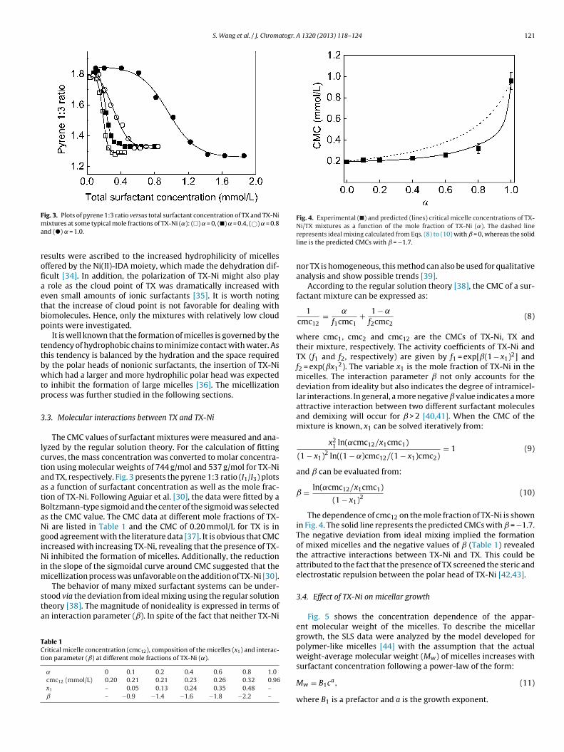
S. Wang et al. / J. Chromatogr. A 1320 (2013) 118– 124 121
Fma
rofiaetbp
ttbwtp
3
lctaatBaNgiNim
sta
TCt
Fig. 4. Experimental (�) and predicted (lines) critical micelle concentrations of TX-
ig. 3. Plots of pyrene 1:3 ratio versus total surfactant concentration of TX and TX-Niixtures at some typical mole fractions of TX-Ni (˛): (�) = 0, (�) = 0.4, (©) = 0.8nd (�) = 1.0.
esults were ascribed to the increased hydrophilicity of micellesffered by the Ni(II)-IDA moiety, which made the dehydration dif-cult [34]. In addition, the polarization of TX-Ni might also play
role as the cloud point of TX was dramatically increased withven small amounts of ionic surfactants [35]. It is worth notinghat the increase of cloud point is not favorable for dealing withiomolecules. Hence, only the mixtures with relatively low cloudoints were investigated.
It is well known that the formation of micelles is governed by theendency of hydrophobic chains to minimize contact with water. Ashis tendency is balanced by the hydration and the space requiredy the polar heads of nonionic surfactants, the insertion of TX-Nihich had a larger and more hydrophilic polar head was expected
o inhibit the formation of large micelles [36]. The micellizationrocess was further studied in the following sections.
.3. Molecular interactions between TX and TX-Ni
The CMC values of surfactant mixtures were measured and ana-yzed by the regular solution theory. For the calculation of fittingurves, the mass concentration was converted to molar concentra-ion using molecular weights of 744 g/mol and 537 g/mol for TX-Nind TX, respectively. Fig. 3 presents the pyrene 1:3 ratio (I1/I3) plotss a function of surfactant concentration as well as the mole frac-ion of TX-Ni. Following Aguiar et al. [30], the data were fitted by aoltzmann-type sigmoid and the center of the sigmoid was selecteds the CMC value. The CMC data at different mole fractions of TX-i are listed in Table 1 and the CMC of 0.20 mmol/L for TX is inood agreement with the literature data [37]. It is obvious that CMCncreased with increasing TX-Ni, revealing that the presence of TX-i inhibited the formation of micelles. Additionally, the reduction
n the slope of the sigmoidal curve around CMC suggested that theicellization process was unfavorable on the addition of TX-Ni [30].The behavior of many mixed surfactant systems can be under-
tood via the deviation from ideal mixing using the regular solutionheory [38]. The magnitude of nonideality is expressed in terms ofn interaction parameter (ˇ). In spite of the fact that neither TX-Ni
able 1ritical micelle concentration (cmc12), composition of the micelles (x1) and interac-ion parameter (ˇ) at different mole fractions of TX-Ni (˛).
0 0.1 0.2 0.4 0.6 0.8 1.0cmc12 (mmol/L) 0.20 0.21 0.21 0.23 0.26 0.32 0.96x1 – 0.05 0.13 0.24 0.35 0.48 –ˇ – −0.9 −1.4 −1.6 −1.8 −2.2 –
Ni/TX mixtures as a function of the mole fraction of TX-Ni (˛). The dashed linerepresents ideal mixing calculated from Eqs. (8) to (10) with = 0, whereas the solidline is the predicted CMCs with = −1.7.
nor TX is homogeneous, this method can also be used for qualitativeanalysis and show possible trends [39].
According to the regular solution theory [38], the CMC of a sur-factant mixture can be expressed as:
1cmc12
= ˛
f1cmc1+ 1 − ˛
f2cmc2(8)
where cmc1, cmc2 and cmc12 are the CMCs of TX-Ni, TX andtheir mixture, respectively. The activity coefficients of TX-Ni andTX (f1 and f2, respectively) are given by f1 = exp[ˇ(1 − x1)2] andf2 = exp(ˇx1
2). The variable x1 is the mole fraction of TX-Ni in themicelles. The interaction parameter not only accounts for thedeviation from ideality but also indicates the degree of intramicel-lar interactions. In general, a more negative value indicates a moreattractive interaction between two different surfactant moleculesand demixing will occur for > 2 [40,41]. When the CMC of themixture is known, x1 can be solved iteratively from:
x21 ln(˛cmc12/x1cmc1)
(1 − x1)2 ln((1 − ˛)cmc12/(1 − x1)cmc2)= 1 (9)
and can be evaluated from:
= ln(˛cmc12/x1cmc1)
(1 − x1)2(10)
The dependence of cmc12 on the mole fraction of TX-Ni is shownin Fig. 4. The solid line represents the predicted CMCs with = −1.7.The negative deviation from ideal mixing implied the formationof mixed micelles and the negative values of (Table 1) revealedthe attractive interactions between TX-Ni and TX. This could beattributed to the fact that the presence of TX screened the steric andelectrostatic repulsion between the polar head of TX-Ni [42,43].
3.4. Effect of TX-Ni on micellar growth
Fig. 5 shows the concentration dependence of the appar-ent molecular weight of the micelles. To describe the micellargrowth, the SLS data were analyzed by the model developed forpolymer-like micelles [44] with the assumption that the actualweight-average molecular weight (Mw) of micelles increases withsurfactant concentration following a power-law of the form:
Mw = B1ca, (11)
where B1 is a prefactor and a is the growth exponent.
122 S. Wang et al. / J. Chromatogr. A 1320 (2013) 118– 124
F(d
b
M
we
S
b
X
etv
mTditaAvatiT
Imw
TBE
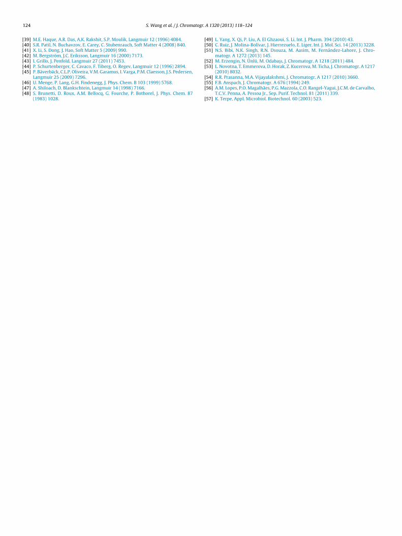
Fig. 6. (a) Concentration dependence of the hydrodynamic diameter of micelles(DH): (�) = 0, (�) = 0.05 and (�) = 0.1. (b) The intensity distribution of DH at = 0
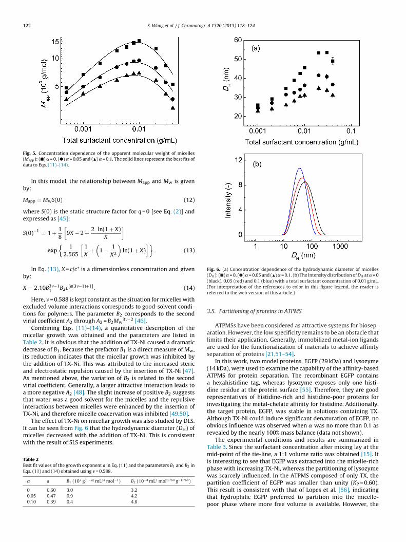
ig. 5. Concentration dependence of the apparent molecular weight of micellesMapp): (�) = 0, (�) = 0.05 and (�) = 0.1. The solid lines represent the best fits ofata to Eqs. (11)–(14).
In this model, the relationship between Mapp and Mw is giveny:
app = MwS(0) (12)
here S(0) is the static structure factor for q = 0 [see Eq. (2)] andxpressed as [45]:
(0)−1 = 1 + 18
[9X − 2 + 2 ln(1 + X)
X
]
exp{
12.565
[1X
+(
1 − 1X2
)ln(1 + X)
]}. (13)
In Eq. (13), X = c/c* is a dimensionless concentration and giveny:
= 2.10B3�−11 B2c[a(3�−1)+1]. (14)
Here, � = 0.588 is kept constant as the situation for micelles withxcluded volume interactions corresponds to good-solvent condi-ions for polymers. The parameter B2 corresponds to the secondirial coefficient A2 through A2 = B2Mw
3�−2 [46].Combining Eqs. (11)–(14), a quantitative description of the
icellar growth was obtained and the parameters are listed inable 2. It is obvious that the addition of TX-Ni caused a dramaticecrease of B1. Because the prefactor B1 is a direct measure of Mw,
ts reduction indicates that the micellar growth was inhibited byhe addition of TX-Ni. This was attributed to the increased stericnd electrostatic repulsion caused by the insertion of TX-Ni [47].s mentioned above, the variation of B2 is related to the secondirial coefficient. Generally, a larger attractive interaction leads to
more negative A2 [48]. The slight increase of positive B2 suggestshat water was a good solvent for the micelles and the repulsiventeractions between micelles were enhanced by the insertion ofX-Ni, and therefore micelle coacervation was inhibited [49,50].
The effect of TX-Ni on micellar growth was also studied by DLS.t can be seen from Fig. 6 that the hydrodynamic diameter (DH) of
icelles decreased with the addition of TX-Ni. This is consistentith the result of SLS experiments.
able 2est fit values of the growth exponent a in Eq. (11) and the parameters B1 and B2 inqs. (11) and (14) obtained using � = 0.588.
a B1 (107 g(1 − a) mL3a mol−1) B2 (10−4 mL3 mol0.764 g−1.764)
0 0.60 3.0 3.20.05 0.47 0.9 4.20.10 0.39 0.4 4.8
(black), 0.05 (red) and 0.1 (blue) with a total surfactant concentration of 0.01 g/mL.(For interpretation of the references to color in this figure legend, the reader isreferred to the web version of this article.)
3.5. Partitioning of proteins in ATPMS
ATPMSs have been considered as attractive systems for biosep-aration. However, the low specificity remains to be an obstacle thatlimits their application. Generally, immobilized metal-ion ligandsare used for the functionalization of materials to achieve affinityseparation of proteins [21,51–54].
In this work, two model proteins, EGFP (29 kDa) and lysozyme(14 kDa), were used to examine the capability of the affinity-basedATPMS for protein separation. The recombinant EGFP containsa hexahistidine tag, whereas lysozyme exposes only one histi-dine residue at the protein surface [55]. Therefore, they are goodrepresentatives of histidine-rich and histidine-poor proteins forinvestigating the metal-chelate affinity for histidine. Additionally,the target protein, EGFP, was stable in solutions containing TX.Although TX-Ni could induce significant denaturation of EGFP, noobvious influence was observed when was no more than 0.1 asrevealed by the nearly 100% mass balance (data not shown).
The experimental conditions and results are summarized inTable 3. Since the surfactant concentration after mixing lay at themid-point of the tie-line, a 1:1 volume ratio was obtained [15]. Itis interesting to see that EGFP was extracted into the micelle-richphase with increasing TX-Ni, whereas the partitioning of lysozymewas scarcely influenced. In the ATPMS composed of only TX, the
partition coefficient of EGFP was smaller than unity (KP = 0.60).This result is consistent with that of Lopes et al. [56], indicatingthat hydrophilic EGFP preferred to partition into the micelle-poor phase where more free volume is available. However, the
S. Wang et al. / J. Chromatogr. A 1320 (2013) 118– 124 123
Table 3Partitioning experiments of EGFP and lysozyme in ATPMSs.
Temperature (◦C) EGFP Lysozyme
VR KP E (%) VR KP E (%)
0 27.56 1.02 ± 0.03 0.60 ± 0.01 36.8 ± 0.9 1.06 ± 0.05 0.84 ± 0.03 47.0 ± 0.40.001 27.78 1.05 ± 0.03 1.57 ± 0.06 60.4 ± 2.1 1.05 ± 0.02 0.84 ± 0.01 46.2 ± 0.10.005 28.10 1.00 ± 0.03 2.80 ± 0.04 70.8 ± 0.6 1.02 ± 0.01 0.86 ± 0.02 45.5 ± 1.40.01 28.46 1.03 ± 0.02 3.61 ± 0.07 75.3 ± 0.7 1.03 ± 0.01 0.84 ± 0.02 46.6 ± 0.90.02 29.42 1.02 ± 0.04 6.57 ± 0.04
0.05 31.38 1.04 ± 0.01 9.92 ± 0.19
0.1 34.63 1.03 ± 0.02 12.42 ± 0.20
Table 4Affinity separation of EGFP and lysozyme by the affinity-based ATPMS.a
Protein KP (–) E (%) S (–)
EGFP 11.88 ± 0.19 83.8 ± 1.2 13.2Lysozyme 0.90 ± 0.07 45.4 ± 1.9 –
a Initial conditions were surfactant solution: [TX] + [TX-Ni] = 0.08 g/mL, = 0.1,1.5 mL; protein solution: 0.2 mg/mL EGFP and 0.2 mg/mL lysozyme, 1.5 mL. Aftert
paapectlpyKiaAmdfwi
4
bbpobTttatwtaolfsth
[[[[
[[
[
[[[[[[[[[[[[[[
[[[
[
[[36] V.C. Santos, F.A. Hasmann, A. Converti, A. Pessoa Jr., Biochem. Eng. J. 56 (2011)
75.
horough mixing at 4 ◦C, the homogeneous mixture was placed at 34.63 ◦C for 2 h.
resence of TX-Ni changed the partitioning behavior of EGFP andn over 20-fold increase (from 0.60 to 12.42) of KEGFP was obtainedt = 0.1. Because the surfactant concentration in each coexistinghase was almost the same for all cases, as it was approximatelyqual to the intersection of the operating tie-line and the fittedoexistence curve [15], the increase of KEGFP was mainly caused byhe affinity interaction between histidine residues and TX-Ni. As forysozyme, in all cases, it was prone to partition into the micelle-poorhase due to excluded-volume interactions [13] and the extractionields remained nearly the same. It is worth noting that, at = 0,EGFP was smaller than KLYZ. This is because the excluded-volume
nteraction increases with molecular weight [13]. Finally, the sep-ration of EGFP and lysozyme was performed in the affinity-basedTPMS. As listed in Table 4, EGFP was selectively extracted to theicelle-rich phase with a separation factor of 13.2. These results
emonstrated the selective separation of histidine-rich proteinsrom histidine-poor proteins. Considering that poly-histidine is aidely used fusion tag to facilitate protein purification [57], TX-Ni
s promising to extend the application of ATPMSs.
. Conclusions
A novel Ni(II)-chelated surfactant TX-Ni was prepared from TXy successive modifications with epichlorohydrin and IDA followedy chelating with nickel ions. Although TX is thermosensitive, nohase separation behavior was observed for the aqueous solutionf TX-Ni. Therefore, TX-Ni was mixed with TX to form an affinity-ased ATPMS. As TX-Ni had a more hydrophilic head group thanX, the efficient dehydration of mixed micelles took place at higheremperature, and the cloud point thus increased with the mole frac-ion of TX-Ni. Moreover, the insertion of TX-Ni increased the stericnd electrostatic repulsion between the head groups of surfac-ants, and therefore the micellization process and micellar growthere inhibited. In the ATPMS formed by TX-Ni and TX, EGFP parti-
ioned into the micelle-rich phase where it was normally excludednd an over 20-fold increase (from 0.60 to 12.42) of KEGFP wasbtained. The selective separation of EGFP from a mixture withysozyme demonstrated the efficiency of TX-Ni as an affinity sur-actant. The application of the Ni(II)-chelated ATPMS in protein
eparation remains to be further studied, but this study revealshe potential of this affinity-based ATPMS for the separation ofistidine-rich proteins.[
[
83.0 ± 1.8 1.03 ± 0.03 0.85 ± 0.02 46.1 ± 0.486.5 ± 0.8 1.02 ± 0.03 0.87 ± 0.01 46.2 ± 0.887.6 ± 1.0 1.02 ± 0.01 0.86 ± 0.01 46.2 ± 1.2
Acknowledgements
This work was supported by the Natural Science Foundationof China (Nos. 21076149 and 21236005) and the Natural ScienceFoundation of Tianjin from Tianjin Municipal Science and Technol-ogy Commission (Contract no. 13JCZDJC27700).
Appendix A. Supplementary data
Supplementary data associated with this article can be found,in the online version, at http://dx.doi.org/10.1016/j.chroma.2013.10.074.
References
[1] J.A. Asenjo, B.A. Andrews, J. Chromatogr. A 1218 (2011) 8826.[2] J.A. Asenjo, B.A. Andrews, J. Chromatogr. A 1238 (2012) 1.[3] F. Ruiz-Ruiz, J. Benavides, O. Aguilar, M. Rito-Palomares, J. Chromatogr. A 1244
(2012) 1.[4] P.A.J. Rosa, A.M. Azevedo, S. Sommerfeld, W. Bäcker, M.R. Aires-Barros, Bio-
technol. Adv. 29 (2011) 559.[5] P.A.J. Rosa, I.F. Ferreira, A.M. Azevedo, M.R. Aires-Barros, J. Chromatogr. A 1217
(2010) 2296.[6] P.G. Mazzola, A.M. Lopes, F.A. Hasmann, A.F. Jozala, T.C.V. Penna, P.O. Magalhaes,
C.O. Rangel-Yagui, A. Pessoa, J. Chem. Technol. Biotechnol. 83 (2008) 143.[7] D. Otzen, Biochim. Biophys. Acta, Proteins Proteomics 1814 (2011) 562.[8] C.L. Liu, Y.J. Nikas, D. Blankschtein, Biotechnol. Bioeng. 52 (1996) 185.[9] Z.L. Wang, Appl. Microbiol. Biotechnol. 75 (2007) 1.10] H. Watanabe, H. Tanaka, Talanta 25 (1978) 585.11] C. Bordier, J. Biol. Chem. 256 (1981) 1604.12] Y. Ali Sarafraz, TrAC Trends Anal. Chem. 30 (2011) 918.13] Y.J. Nikas, C.-L. Liu, T. Srivastava, N.L. Abbott, D. Blankschtein, Macromolecules
25 (1992) 4797.14] D.T. Kamei, D.I.C. Wang, D. Blankschtein, Langmuir 18 (2002) 3047.15] H. Lam, M. Kavoosi, C.A. Haynes, D.I.C. Wang, D. Blankschtein, Biotechnol. Bio-
eng. 89 (2005) 381.16] P.G. Mazzola, H. Lam, M. Kavoosi, C.A. Haynes, A. Pessoa, T.C.V. Penna, D.I.C.
Wang, D. Blankschtein, Biotechnol. Bioeng. 93 (2006) 998.17] S. Fernandes, R. Hatti-Kaul, B. Mattiasson, Biotechnol. Bioeng. 79 (2002) 472.18] N. Garg, I. Galaev, B. Mattiasson, Biotechnol. Appl. Biochem. 20 (1994) 199.19] T. Saitoh, W.L. Hinze, Talanta 42 (1995) 119.20] A.M. Lopes, A. Pessoa, C.D. Rangel-Yagui, Quim. Nova 31 (2008) 998.21] J. Lu, D.-Q. Lin, S.-J. Yao, Ind. Eng. Chem. Res. 45 (2006) 1774.22] U. Sivars, J. Abramson, S. Iwata, F. Tjerneld, J. Chromatogr. B 743 (2000) 307.23] X.-Y. Dong, X.-D. Feng, Y. Sun, Biotechnol. Prog. 26 (2010) 1088.24] X.-Y. Dong, Y. Meng, X.-D. Feng, Y. Sun, Biotechnol. Prog. 26 (2010) 150.25] R. Cheung, J. Wong, T. Ng, Appl. Microbiol. Biotechnol. 96 (2012) 1411.26] X.-Y. Dong, L.-J. Chen, Y. Sun, J. Chromatogr. A 1216 (2009) 5207.27] H.-C. Kang, B. Lee, J. Yoon, M. Yoon, J. Am. Oil Chem. Soc. 78 (2001) 423.28] A. Aserin, N. Garti, Y. Sasson, Ind. Eng. Chem. Prod. Res. Dev. 23 (1984) 452.29] T. Takasu, A. Iles, K. Hasebe, Anal. Bioanal. Chem. 372 (2002) 554.30] J. Aguiar, P. Carpena, J.A. Molina-Bolı′var, C.C. Ruiz, J. Colloid Interface Sci. 258
(2003) 116.31] B.H. Zimm, J. Chem. Phys. 16 (1948) 1093.32] M. Liley, T.A. Keller, C. Duschl, H. Vogel, Langmuir 13 (1997) 4190.33] P. Mukherjee, S.K. Padhan, S. Dash, S. Patel, B.K. Mishra, Adv. Colloid Interface
Sci. 162 (2011) 59.34] S.B. Sulthana, P.V.C. Rao, S.G.T. Bhat, T.Y. Nakano, G. Sugihara, A.K. Rakshit,
Langmuir 16 (2000) 980.35] T. Gu, P.A. Galera-Gómez, Colloids Surf. A 104 (1995) 307.
37] A. Sánchez-Ferrer, M. Pérez-Gilabert, E. Núnez, R. Bru, F. García-Carmona, J.Chromatogr. A 668 (1994) 75.
38] P.M. Holland, D.N. Rubingh, J. Phys. Chem. 87 (1983) 1984.
1 atogr.
[[[[[[[
[[[
[[[
[[
24 S. Wang et al. / J. Chrom
39] M.E. Haque, A.R. Das, A.K. Rakshit, S.P. Moulik, Langmuir 12 (1996) 4084.40] S.R. Patil, N. Buchavzov, E. Carey, C. Stubenrauch, Soft Matter 4 (2008) 840.41] X. Li, S. Dong, J. Hao, Soft Matter 5 (2009) 990.42] M. Bergström, J.C. Eriksson, Langmuir 16 (2000) 7173.43] I. Grillo, J. Penfold, Langmuir 27 (2011) 7453.44] P. Schurtenberger, C. Cavaco, F. Tiberg, O. Regev, Langmuir 12 (1996) 2894.45] P. Bäverbäck, C.L.P. Oliveira, V.M. Garamus, I. Varga, P.M. Claesson, J.S. Pedersen,
Langmuir 25 (2009) 7296.46] U. Menge, P. Lang, G.H. Findenegg, J. Phys. Chem. B 103 (1999) 5768.47] A. Shiloach, D. Blankschtein, Langmuir 14 (1998) 7166.48] S. Brunetti, D. Roux, A.M. Bellocq, G. Fourche, P. Bothorel, J. Phys. Chem. 87
(1983) 1028.
[[[
[
A 1320 (2013) 118– 124
49] L. Yang, X. Qi, P. Liu, A. El Ghzaoui, S. Li, Int. J. Pharm. 394 (2010) 43.50] C. Ruiz, J. Molina-Bolívar, J. Hierrezuelo, E. Liger, Int. J. Mol. Sci. 14 (2013) 3228.51] N.S. Bibi, N.K. Singh, R.N. Dsouza, M. Aasim, M. Fernández-Lahore, J. Chro-
matogr. A 1272 (2013) 145.52] M. Erzengin, N. Ünlü, M. Odabas ı, J. Chromatogr. A 1218 (2011) 484.53] L. Novotna, T. Emmerova, D. Horak, Z. Kucerova, M. Ticha, J. Chromatogr. A 1217
(2010) 8032.
54] R.R. Prasanna, M.A. Vijayalakshmi, J. Chromatogr. A 1217 (2010) 3660.55] F.B. Anspach, J. Chromatogr. A 676 (1994) 249.56] A.M. Lopes, P.O. Magalhães, P.G. Mazzola, C.O. Rangel-Yagui, J.C.M. de Carvalho,T.C.V. Penna, A. Pessoa Jr., Sep. Purif. Technol. 81 (2011) 339.57] K. Terpe, Appl. Microbiol. Biotechnol. 60 (2003) 523.