Crystal structures of CaSiO3 polymorphs control growth and osteogenic differentiation of human...
10
Biomaterials Science PAPER Cite this: DOI: 10.1039/c3bm60034c Received 7th February 2013, Accepted 21st June 2013 DOI: 10.1039/c3bm60034c www.rsc.org/biomaterialsscience Crystal structures of CaSiO 3 polymorphs control growth and osteogenic differentiation of human mesenchymal stem cells on bioceramic surfaces† Nianli Zhang, a,b James A. Molenda, c Steven Mankoci, d Xianfeng Zhou, d William L. Murphy a,c,e,f and Nita Sahai* d The repair and replacement of damaged or diseased human bone tissue requires a stable interface between the orthopedic implant and living tissue. The ideal material should be both osteoconductive (promote bonding to bone) and osteoinductive (induce osteogenic differentiation of cells and generate new bone). Partially resorbable bioceramic materials with both properties are developed by expensive trial-and-error methods. Structure–reactivity relationships for predicting the osteoinductive properties of ceramics would significantly increase the efficiency of developing materials for bone tissue engineering. Here we propose the novel hypothesis that the crystal structure of a bioceramic controls the release rates, subsequent surface modifications due to precipitation of new phases, and thus, the concentrations of soluble factors, and ultimately, the attachment, viability and osteogenic differentiation of human Mesenchymal Stem Cells (hMSCs). To illustrate our hypothesis, we used two CaSiO 3 polymorphs, pseudo- wollastonite (psw, β-CaSiO 3 ) and wollastonite (wol, α-CaSiO 3 ) as scaffolds for hMSC culture. Polymorphs are materials which have identical chemical composition and stoichiometry, but different crystal struc- tures. We combined the results of detailed surface characterizations, including environmental Scanning Electron Microscopy (SEM) back-scattered imaging, and spot-analysis and 2D elemental mapping by SEM-Energy Dispersive X-ray (SEM-EDX), High Resolution Transmission Electron Microscopy (HRTEM) and surface roughness analysis; culture medium solution analyses; and molecular/genetic assays from cell culture. Our results confirmed the hypothesis that the psw polymorph, which has a strained silicate ring structure, is more osteoinductive than the wol polymorph, which has a more stable, open silicate chain structure. The observations could be attributed to easier dissolution (resorption) of psw compared to wol, which resulted in concentration profiles that were more osteoinductive for the former. Thus, we showed that crystal structure is a fundamental parameter to be considered in the intelligent design of pro-osteogenic, partially resorbable bioceramics. Introduction An important goal in biomaterial design is to develop osteo- inductive resorbable scaffolds and bone filler materials. The fundamental concept is that when seeded with osteoblasts, hMSCS, genes or other stimuli, such materials will promote osteogenic differentiation, new bone generation, and osseo- integration, and thus minimize the possibility of failure at the implant/bone interface. Several material properties affect the behavior of cells seeded on scaffold and filler materials, and ultimately, the osteoconductivity and osteoinductivity of the material. These properties include chemical composition, surface texture, elastic modulus, porosity, permeability, and elastic modulus, as well as soluble factors added to the material. 1–5 Certain conceptual criteria have been developed for rationalizing the osteoconductivity of silicate biomaterials, 6–8 but general principles for predicting osteo- inductivity do not yet exist. In detail, silicate glasses and glass ceramics within only specific ranges of chemical compositions are osteoconductive. † Electronic supplementary information (ESI) available. See DOI: 10.1039/ c3bm60034c a Materials Science Program, University of Wisconsin, Madison, WI 53706, USA b Department of Biologic and Materials Science, 1011 N. University Avenue, School of Dentistry, University of Michigan, Ann Arbor, MI 48109, USA c Department of Biomedical Engineering, University of Wisconsin, Madison, WI 53706, USA d Department of Polymer Science, 170 University Avenue, Akron, OH 44325, USA. E-mail: [email protected], [email protected]; Tel: +1 330-972-5795 e Department of Orthopedics and Rehabilitation, University of Wisconsin, Madison, WI 53706, USA f Department of Pharmacology, University of Wisconsin, Madison, WI 53706, USA This journal is © The Royal Society of Chemistry 2013 Biomater. Sci. Published on 18 July 2013. Downloaded on 18/07/2013 19:21:54. View Article Online View Journal
-
Upload
independent -
Category
Documents
-
view
5 -
download
0
Transcript of Crystal structures of CaSiO3 polymorphs control growth and osteogenic differentiation of human...
BiomaterialsScience
PAPER
Cite this: DOI: 10.1039/c3bm60034c
Received 7th February 2013,Accepted 21st June 2013
DOI: 10.1039/c3bm60034c
www.rsc.org/biomaterialsscience
Crystal structures of CaSiO3 polymorphs control growthand osteogenic differentiation of human mesenchymalstem cells on bioceramic surfaces†
Nianli Zhang,a,b James A. Molenda,c Steven Mankoci,d Xianfeng Zhou,d
William L. Murphya,c,e,f and Nita Sahai*d
The repair and replacement of damaged or diseased human bone tissue requires a stable interface
between the orthopedic implant and living tissue. The ideal material should be both osteoconductive
(promote bonding to bone) and osteoinductive (induce osteogenic differentiation of cells and generate
new bone). Partially resorbable bioceramic materials with both properties are developed by expensive
trial-and-error methods. Structure–reactivity relationships for predicting the osteoinductive properties of
ceramics would significantly increase the efficiency of developing materials for bone tissue engineering.
Here we propose the novel hypothesis that the crystal structure of a bioceramic controls the release rates,
subsequent surface modifications due to precipitation of new phases, and thus, the concentrations of
soluble factors, and ultimately, the attachment, viability and osteogenic differentiation of human
Mesenchymal Stem Cells (hMSCs). To illustrate our hypothesis, we used two CaSiO3 polymorphs, pseudo-
wollastonite (psw, β-CaSiO3) and wollastonite (wol, α-CaSiO3) as scaffolds for hMSC culture. Polymorphs
are materials which have identical chemical composition and stoichiometry, but different crystal struc-
tures. We combined the results of detailed surface characterizations, including environmental Scanning
Electron Microscopy (SEM) back-scattered imaging, and spot-analysis and 2D elemental mapping by
SEM-Energy Dispersive X-ray (SEM-EDX), High Resolution Transmission Electron Microscopy (HRTEM) and
surface roughness analysis; culture medium solution analyses; and molecular/genetic assays from cell
culture. Our results confirmed the hypothesis that the psw polymorph, which has a strained silicate ring
structure, is more osteoinductive than the wol polymorph, which has a more stable, open silicate chain
structure. The observations could be attributed to easier dissolution (resorption) of psw compared to
wol, which resulted in concentration profiles that were more osteoinductive for the former. Thus, we
showed that crystal structure is a fundamental parameter to be considered in the intelligent design of
pro-osteogenic, partially resorbable bioceramics.
Introduction
An important goal in biomaterial design is to develop osteo-inductive resorbable scaffolds and bone filler materials. The
fundamental concept is that when seeded with osteoblasts,hMSCS, genes or other stimuli, such materials will promoteosteogenic differentiation, new bone generation, and osseo-integration, and thus minimize the possibility of failure at theimplant/bone interface. Several material properties affect thebehavior of cells seeded on scaffold and filler materials, andultimately, the osteoconductivity and osteoinductivity of thematerial. These properties include chemical composition,surface texture, elastic modulus, porosity, permeability,and elastic modulus, as well as soluble factors added tothe material.1–5 Certain conceptual criteria have beendeveloped for rationalizing the osteoconductivity of silicatebiomaterials,6–8 but general principles for predicting osteo-inductivity do not yet exist.
In detail, silicate glasses and glass ceramics within onlyspecific ranges of chemical compositions are osteoconductive.
†Electronic supplementary information (ESI) available. See DOI: 10.1039/c3bm60034c
aMaterials Science Program, University of Wisconsin, Madison, WI 53706, USAbDepartment of Biologic and Materials Science, 1011 N. University Avenue, School of
Dentistry, University of Michigan, Ann Arbor, MI 48109, USAcDepartment of Biomedical Engineering, University of Wisconsin, Madison,
WI 53706, USAdDepartment of Polymer Science, 170 University Avenue, Akron, OH 44325, USA.
E-mail: [email protected], [email protected]; Tel: +1 330-972-5795eDepartment of Orthopedics and Rehabilitation, University of Wisconsin, Madison,
WI 53706, USAfDepartment of Pharmacology, University of Wisconsin, Madison, WI 53706, USA
This journal is © The Royal Society of Chemistry 2013 Biomater. Sci.
Publ
ishe
d on
18
July
201
3. D
ownl
oade
d on
18/
07/2
013
19:2
1:54
.
View Article OnlineView Journal

Soluble Ca, Si (as H4SiO4) and other ions, if present, arereleased by partial resorption (dissolution) of the silicate bio-material. These ions combine with the Ca and PO4 ionspresent in the blood or cell culture medium and are reprecipi-tated as an interfacial hydroxyapatite (HAP) layer at thescaffold implant/living bone interface. This HAP layer favorsimplant–bone bonding (osteoconductivity) of bioactivecalcium silicate (±Na2O–MgO–P2O5) glasses, glass ceramicsand ceramics.9–13 The concepts of silicate glass network con-nectivity and “three-ring” structures consisting of silicate(SiO4) tetrahedra have only recently provided a structuralexplanation for the mechanism of osteoconductivity ofbioactive calcium silicate glasses and ceramics.6–8,14,15
A chemical–structural conceptual basis for predicting theosteoinductivity of silicate glasses and ceramics, however, isless clear and is an area of active research.16–18
Progress is hindered, in part, because well-constrainedexperimental studies are rare, and the effects of chemical com-position, crystal structure, crystallinity, and surface texture areoften convoluted. Using pseudowollastonite (psw, β-CaSiO3) ofdifferent surface textures, thus keeping the crystal structureand chemical composition constant, we have previously shownthat (a) crystallinity affects the osteoinductivity of CaSiO3 bio-ceramics, but (b) surface roughness differences within a factorof two do not influence osteoinductivity of CaSiO3.
19 We nowpropose here that, all other factors being equal, the crystallo-graphic structure of bioceramics is a critical factor in control-ling their resorption (dissolution) rate, the release rates ofosteoinductive soluble factors from the bioceramics, the sub-sequent surface modifications resulting from the potential pre-cipitation of new phases, and ultimately, the activities of cellsseeded on the bioceramic surfaces including cell adhesion,viability, and osteogenic differentiation.
In order to test this hypothesis, we devised a novelapproach by examining psw and wollastonite (wol, α-CaSiO3)as model substrates for our study, because these two ceramicsare polymorphs, having identical chemical compositionand stoichiometry, but different crystallographic structures.This choice of bioceramics permits us to isolate the effects ofcrystal structure on osteoconductivity and osteoinducti-vity.10,20–22,23–27 We chose CaSiO3 because it is already knownto be osteoconductive and two well-defined polymorphs areknown to exist, thus providing an ideal model system to testour hypothesis.
Pseudowollastonite and wollastonite differ in their mannerof polymerization of the silicate tetrahedra. Psw is the hightemperature polymorph and its crystal structure consists ofthree silicate tetrahedra, which are covalently bonded viacorner-sharing oxygens to form “3-rings” (note that “3” refersto the number of silicate tetrahedra and not to the number ofatoms in the ring; Fig. 1 and ESI Fig. 1a,b†). The low tempera-ture polymorph is wol, composed of corner-sharing silicate tetra-hedra linked to form chains. The negative-charge associatedwith the silicate rings and chains, respectively, is electrostati-cally balanced by Ca2+ ions in both polymorphs. The highstrain associated with the Si–O–Si bond angles in the “3-rings”
of psw makes these bonds much more susceptible to hydro-lysis compared to the more open, and hence, more stableSi–O–Si bonds in the wol chains. The effect of these crystallo-graphic bonding differences in the two polymorphs results ingreater psw solubility and resorption (dissolution) rate com-pared to wol.28 These differences in crystal structure-controlledresorption (dissolution) should result in different con-centration profiles over time, which reflect a combination ofdifferent rates of released soluble Si and Ca and potentiallydifferent subsequent surface modifications from precipitationof new phases. We suggest that these differences should, ulti-mately, influence the attachment, viability, and osteogenicdifferentiation of hMSCs seeded at the bioceramic surfaces(Fig. 1).
Calcium silicate has been clinically used as dental root-endmaterials, and is being investigated for coatings on orthopedicimplants.29,30 Our study of the polymorph crystal structureeffect on soluble factor release and subsequent cell activitiesprovides a new way to design biomaterials with an optimalclinical outcome. Another important contribution of our workis to use an interdisciplinary approach combining the detailedcharacterization of the pellet surfaces, cell culture mediumsolutions and molecular/genetic assays of cell activities at mul-tiple time-points up to 28 days. By combining the results ofthese different analytical techniques, we were able to identifythe effects of soluble factor concentration and release rates,and thus of the crystal polymorph structure, on hMSC activi-ties, such as adhesion, viability, proliferation, and osteogenicdifferentiation.
Materials and methodsPellet synthesis and characterization
Pseudowollastonite and wollastonite substrates were syn-thesized in the form of dense, disc-shaped pellets (12 mm
Fig. 1 Schematic representation of the central hypothesis illustrated in ourstudy by using two CaSiO3 polymorphs: bioceramic crystal structure (top panels)controls the solubility and dissolution rate, which control the levels and time-dependent trends of the soluble factors, Si and Ca (middle panels) and, ulti-mately, the activities of hMSCs seeded on the bioceramics (bottom panels).Legend for atoms in top panels: O = red, Si = blue, Ca = green.
Paper Biomaterials Science
Biomater. Sci. This journal is © The Royal Society of Chemistry 2013
Publ
ishe
d on
18
July
201
3. D
ownl
oade
d on
18/
07/2
013
19:2
1:54
. View Article Online
diameter × 1 mm thick) by pressing 0.4 g of psw (Sigma, cat.#372668) or wol (Nyco®, NYAD 5000) ceramic powder (both ofsimilar particle sizes∼ 1–2 μm), in a hardened steel die, fol-lowed by sintering at 900 °C for 10 hours, at a heating rate of50 °C per min starting from room temperature (Lindberg BlueM tube furnace).19 After sintering, pellets were naturally cooleddown at room temperature.
In order to ensure that sintering did not alter the crystalphase of the polymorphs, the crystallinity and purity of bothphases were checked by X-Ray Diffraction (XRD) using aScintag Pad V diffractometer with Cu Kα for both kinds ofpellets both before and after sintering (ESI Fig. 2†). Mineralparticles from both pellets after sintering were also character-ized by High Resolution Transmission Electron Microscopy(HRTEM, Philips CM200 UT) (ESI Fig. 1c,d†). Detailed samplepreparation procedures have been published elsewhere.19
A white light interferometer (Zygo New View) was used tomeasure the surface roughness (Ra), the average vertical devi-ation of the surface profile from the mean line over an area of2.81 mm × 2.10 mm of the scaffold pellets. Values of Ra were0.74 μm and 0.76 μm, respectively, for psw and wol before reac-tion, and ∼0.81 μm up to 28 days cell culture.
A Hitachi S-3400 Variable Pressure Scanning ElectronMicroscope (SEM) combined with Energy Dispersive X-Ray(EDX) was used to characterize pellet surfaces both before andafter cell culture in terms of surface morphology and chemicalcomposition. Details of sample preparations and instrumentsettings were published in our previous study.19 Briefly,pellet surfaces before cell culture were carbon-coated andSEM images revealed particle sizes of ∼10–20 μm for both pswand wol pellets (ESI Fig. 1e,f†). Pellet surfaces after cellculture were prepared by first lysing the cells, and then flush-ing the surfaces using a 0.2% Triton X-100 solution.The pellets were then dried under ambient conditions andcarbon-coated.
Si, Ca, and P analysis in culture media. In order to monitorthe soluble Ca, Si, and P in growth medium or osteogenicinduction medium, solution samples were collected every twodays from well-plates containing pellets seeded with hMSCs orcontrol pellets without hMSCs. The medium samples werediluted 10 times and acidified by high purity HNO3 (15–16 M)until the pH dropped below 2. Using standard procedures,the samples were stored at 4 °C until they were analyzed byInductively Coupled Plasma-Optical Emission Spectroscopy(ICP-OES, Varian Vista-MPX). Standard deviations of four repli-cates per condition were calculated to calculate the error bars.
Cell culture
Human Mesenchymal Stem Cells (hMSCs, Cambrex®) atpassage 5 were used for cell growth and osteogenic differen-tiation studies on the psw and wol surfaces. For the cellgrowth study, cells were seeded at a density of 10 000 cells cm−2,and cultured in growth medium composed of 10.08 g L−1
Alpha Modification of Eagle’s Medium (AMEM), 1% penicillin/streptomycin as antibiotics and 10% Fetal Bovine Serum(FBS). For the hMSC osteogenic differentiation study, the
seeding density was 30 000 cells cm−2 in osteogenic inductionmedium composed of growth medium + 0.28 mM 2-phosphateascorbic acid + 100 nM dexamethasone + 10 mM glycerol2-phosphate.
In order to sterilize psw and wol substrates, pellets werefirst washed with anhydrous ethanol, and then incubated over-night at a temperature of 180 °C. The pellets were placed intwenty-four well culture plates (1 pellet per well), which werepre-coated with 1% BSA (Bovine Serum Albumin) overnight at4 °C to prevent cell attachment and growth on the cultureplate surfaces, thus allowing cells to grow only on the pelletsurfaces. The hMSCs were incubated for 24 hours in GM toallow cell adhesion on the pellet surface after cell seeding. Allcultures were maintained at 37 °C in a humidified incubatorwith 5% CO2. Media were changed every two days. The “day 0”time point in the following figures means 24 hours after hMSCseeding. Pellets in growth medium and osteogenic inductionmedium without cells served as negative controls. All thequantitative assays were carried out in quadruplicate. Theentire set of experiments described below was conductedtwice.
Cell adhesion, morphology, and viability in growth medium
To prepare the samples for LIVE/DEAD fluorescence imaging,the pellet surfaces were washed twice with Phosphate BufferedSaline (PBS). Fluorescent agents, calcein AM (calcein acetoxy-methyl ester) and EthD-1 (ethidium homodimer) at concen-trations of 2 μM and 4 μM, respectively, were added to thepellet sample and incubated for 40 min at room temperature.This assay identifies esterase activity in live cells via green fluore-scence emission from calcein AM and nuclear permeability indead cells via red fluorescence emission from EthD-1. Celladhesion, morphology, and viability were then investigated byepifluorescence microscopy at days 0, 8, and 16.
Cell growth
Cells were seeded on the psw and wol pellet surfaces in quad-ruplicate at a low seeding density of 10 000 cells cm−2 and cul-tured in growth medium. Two assays were carried out toinvestigate cell growth on psw and wol substrates. Total DNAcontent was measured at days 0, 8, 12, and 20 using aCyQuANT® Assay Kit (Invitrogen). Total live cell number onsubstrates was determined using CellTiter Blue® reagent(Promega) at days 0, 2, 8, 12, 20, and 28. The underlying prin-ciple of this assay is that living cells are able to convert a redoxdye named resazurin into a fluorescent end product, resorufin.Detailed procedures of this assay are listed on the manufac-turer’s website and in our previous publication.19 Fluorescenceis measured with an excitation wavelength of 560 nm and anemission wavelength of 590 nm.
Osteoblastic cell differentiation in osteogenic inductionmedium
Analyses of alkaline phosphatase (ALP) enzymatic activity andthe expression of three osteogenic genes, osteocalcin (OCN),osteopotin (OPN), and Runx2 or core binding factor alpha-1
Biomaterials Science Paper
This journal is © The Royal Society of Chemistry 2013 Biomater. Sci.
Publ
ishe
d on
18
July
201
3. D
ownl
oade
d on
18/
07/2
013
19:2
1:54
. View Article Online

(cbfa-1), are markers for different stages of bone formation.These analyses were combined with detailed characterizationsof scaffold pellet surfaces to investigate osteogenic differen-tiation of the hMSCs.
The activity of ALP was measured at days 0, 8, 12, and 16using standard procedures published previously.19 ALP con-centrations in Fig. 2i were normalized by total DNA content asdescribed above, and expressed as μg ALP per ng DNA aftersubtraction of the values obtained for cell-free controls, whichcontain hMSCs but no bioceramic pellet.
For RT-PCR, experiments were carried out in quadruplicate,at days 8 and 16, using osteogenic induction medium. Detailedprotocols for RT-PCR experiments are available in our previouspaper.19 Briefly, total RNA was extracted using an RNeasy®Mini kit (Qiagen) according to the manufacturer’s instruc-tions. For every sample, 50 ng of RNA was reverse transcribedinto cDNA (complementary DNA) with Illustra™ Ready-to-GoRT-PCR beads. After first-strand cDNA synthesis, gene-specific
primers (Table 1) were added into the sample for thermalcycling. A sample without gene-specific primers was used asthe negative control. Electrophoresis was conducted at 100 Vuntil the dye front has traveled approximately two-thirds of thegel length. Images were taken by placing the gel in a UV lightbox.
Fig. 2 Effects of psw and wol scaffolds on hMSC activities. (a, h) Effects on attachment, growth, and proliferation. The hMSCs were seeded at a density of 10 000cells cm−2 in growth medium; (f ) effects on hMSC osteogenic induction as indicated by alkaline phosphatase (ALP) enzymatic activity normalized by the totalnumber of cells (DNA). The hMSCs were seeded at a density of 30 000 cells cm−2 in osteogenic induction medium. The expression of ALP on psw at day 0 was belowthe detection limit. (a–f ) LIVE (green)/DEAD (red) fluorescence microscopy images of hMSCs on psw (a, b, c) and wol (d, e, f ) surfaces at days 0 (a, d), 8 (b, e) and 16(c, f ). (g) Total DNA and (h) total live cell number over time in growth medium. Results are reported after subtraction of cell-free blanks. Asterisks indicate statisticallysignificant differences (p < 0.05).
Table 1 Primer sequences used for the RT-PCR
Gene Sequence 5′ to 3′
Osteocalcin Forward GGCAGCGAGGTAGTGAAGAGReverse CTGGAGAGGAGCAGAACTGG
Osteopontin Forward TGAAACGAGTCAGCTGGATGReverse TGAAATTCATGGCTGTGGAA
Runx2 or Cbfα-1 Forward CACCGAGACCAACAGAGTCAReverse TGCTTGCAGCCTTAAACTGA
β-Actin Forward GGACTTCGAGCAAGAGATGGReverse AGCACTGTGTTGGCGTACAG
Paper Biomaterials Science
Biomater. Sci. This journal is © The Royal Society of Chemistry 2013
Publ
ishe
d on
18
July
201
3. D
ownl
oade
d on
18/
07/2
013
19:2
1:54
. View Article Online

Results
Surface characterization of psw and wol pellets revealed theirsimilar crystallinity and surface texture (ESI Fig. 1c–f†), andsurface roughness (0.74 and 0.76 μm, respectively). Surfaceroughness variations within a factor of two between otherwiseidentical surfaces do not affect the osteoconductivity and pro-osteoinductivity of bioceramic surfaces.19
hMSC attachment, viability, and growth
The adhesion and viability of hMSCs at psw and wol surfaceswere examined in growth medium up to 28 days (Fig. 2). Onday 0, we observed a spherical cell morphology on psw, nocell–cell contacts, and several dead cells, indicating minimaladhesion, and suggest cytotoxicity of psw at this time point. Incontrast, the cells were spindle-shaped on wol, no dead cellswere observed, and the number of live cells was greater thanon psw, indicating better initial attachment and viability andlower cytotoxicity. At day 8, hMSCs attained a spindle-morphology on psw, but still with minimal cell–cell contact,while on wol the spindle-shaped hMSCs began to aggregateand form clusters. Cells on both substrates reached confluenceby day 16. Fluorescence images taken at day 20 and 28 (notshown) also showed that cells had reached confluence. Thesequalitative results are consistent with quantitative total DNA(Fig. 2g) and live cell number (viability) assays (Fig. 2h), whichshowed that cell number was initially lower on psw than onwol, but became statistically greater at days 20 and 28.
Osteogenic differentiation of hMSCs
Osteoblastic differentiation of hMSCs at psw and wol scaffoldsurfaces in osteogenic induction medium was monitored for16 days (Fig. 2i). The upregulation of the enzyme, alkalinephosphatase (ALP), is a non-specific, but widely accepted indi-cation of osteoblastic differentiation. The expression of ALP onpsw peaked at day 8, but an obvious peak on wol was notobserved (Fig. 2i). Alkaline phosphatase activity on psw wasgreater than that on wol at days 0, 8 and 12, but was lower byday 16. Thus, the upregulation of ALP was greater and earlieron psw than on wol. The osteoblastic differentiation of hMSCon both psw and wol was also supported by the expressions ofOCN, OPN, and cbfα-1 genes as determined by our RT-PCRanalyses (Fig. 3).
Detailed surface characterizations of psw and wol scaffoldswere conducted on day 12 of reaction in osteogenic inductionmedium (Fig. 4 and 5). In detail, we observed a layer of veryfine-grained calcium phosphate background precipitate identi-fied as hydroxyapatite and nodular amorphous silica on thesurfaces of both cell-free controls (Fig. 4a,d, blue arrow) andcell-culture samples of psw and wol (Fig. 4b,e, blue arrow;Fig. 4c,f, green arrow). Polycrystalline aggregates, which were∼20–50 μm across and 30 μm high, were seen only on the pswcontrol (Fig. 4a, red arrow) and sample (Fig. 4b,c, red arrow),but not on the wol control and sample. Elemental mapping ofSi, Ca and P on the reacted surfaces revealed very interestingpatterns. The polycrystalline aggregates were deficient in Si
and enriched in Ca, but not in P (Fig. 5a,b). Similar crystalswere identified as calcite (CaCO3) using Kikuchi analysis inour previous work19 and are identified here also as calcite crys-tals. On the psw sample, some of the smaller, Si-deficientareas were co-localized with Ca and the P enriched areas,which were ∼5–2 μm in diameter (Fig. 5b). These regions werespatially highly correlated with the nodular amorphous pre-cipitates seen in the corresponding SEM images (Fig. 4c, greenarrow). The nodules were identified as CaP or bone nodules,consistent with our previous study (ESI Fig. 3†).19 This obser-vation is important because bone nodule formation is the phe-notype in cell culture corresponding to induction of boneformation in vivo. Significantly, the bone nodules were notseen on psw control surfaces without hMSCs indicating thattheir formation on psw samples is associated only with hMSCactivity.
Release of soluble factors
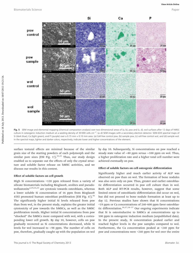
In order to determine the effects of the polymorphs’ crystalstructures on soluble factor concentrations and to relate thesefactors to hMSC activities (adhesion, viability, growth andosteoinduction), we determined the concentrations of Si, Ca,and P in both growth medium and osteogenic inductionmedium (Fig. 6). The trends and absolute concentrations forSi and Ca levels over time were similar in both culture media,suggesting that the same reaction mechanisms controlled Siand Ca concentrations in both media. Silicon concentrationspeaked at higher levels and earlier time points for psw thanfor wol (Fig. 5a,b).
This result is consistent with our expectation that the three-ring silicate structure of the psw polymorph will have a highersolubility and dissolution (resorption) rate versus the chain sili-cate structure of the wol polymorph and potentially differentsurface precipitates forming. The eventual attainment ofsteady-state Si values on both surfaces indicated that the Si
Fig. 3 Osteogenic gene expressions determined by RT-PCR on psw and wol atdifferent time points in osteogenic induction medium and at a seeding densityof 30 000 cells cm−2. Genes reported are osteocalcin (OCN), osteopontin (OPN),core-binding factor alpha-1 (Cbfα-1), and β-actin as a house-keeping gene. Fourreplicates were processed for each condition. The result above is from one of therepresentative images.
Biomaterials Science Paper
This journal is © The Royal Society of Chemistry 2013 Biomater. Sci.
Publ
ishe
d on
18
July
201
3. D
ownl
oade
d on
18/
07/2
013
19:2
1:54
. View Article Online

release rate by dissolution of the underlying substrate becameequal to the Si removal rate by amorphous silica precipitation.The higher solubility and faster dissolution rate of psw relativeto wol also released a higher Ca concentration and at a fasterrate (Fig. 4c,d). The released Ca was removed from solution byfirst combining it rapidly with phosphate from the culturemedium to form the background, fine-grained, osteoconduc-tive layer of HAP. The amount of Ca removed is dictated by thesolubility limit of HAP. Excess soluble Ca released from thepsw sample combined with carbonate from the culturemedium to form large, polycrystalline aggregates of metastablecalcite. Such behaviour of the dissolution products of CaSiO3
bioceramic has been reported previously.31 These calcite crys-tals were not seen on wol (Fig. 4a,b), because of the slower dis-solution rate of wol related to its more stable chain-silicatestructure, such that smaller Ca levels were released. Thus, onlythe background CaP or HAP osteoconductive layer was formed.Finally, we note that the source of phosphate for the formationof the background, osteoconductive HAP layer is the soluble Pwhich was already present in the starting culture media.Trends in P concentrations with time were closely related to
those of Ca, suggesting that P levels are indirectly affected bythe crystal structure of the two polymorphs.
Discussion
In general, cells respond to both the substrate physical pro-perties of and soluble factors released by a resorbable scaffold.The hypothesis for the present study is that, if all other factorsare equal, then the intrinsic crystal structure of biomaterialscontrols their solubility, dissolution rate, and subsequentsurface modifications by precipitation, which will be reflectedin the soluble factor concentrations and trends over time, ulti-mately influencing hMSC initial attachment and viability,growth, proliferation, and osteogenic differentiation. In ourstudy, (1) the chemical composition and stoichiometry of bothpolymorphs are identical; (2) the surface roughness for bothsurfaces did not change significantly before and after cellculture experiments, and we have shown previously that thevariation of surface roughness within a factor of 2 does notcause significant changes in cell activities; and (3) other
Fig. 4 SEM image and SEM-EDX spot analysis of psw and wol surfaces after 12 days of hMSC culture in osteogenic induction medium at a seeding density of30 000 cells cm−2. Cell-free controls were also studied. Images were obtained by SEM with an environmental secondary electron detector set at 75° angle to thepellet surfaces and using different magnifications. (a–c) psw and (d–f ) wollastonite. (a) Cell-free psw control, (d) cell-free wol control, (b, c) psw samples with hMSCs,and (e, f ) wol samples with hMSCs. Blue arrow: fine-grained, background, osteoconductive layer of HAP; green arrow: amorphous silica; red arrow: calcite; yellowarrow: amorphous Ca–PO4 bone nodule. (g–i) SEM-EDX spot analysis of polycrystalline aggregates of calcite (g), amorphous CaP bone nodule (h), and amorphoussilica (i). Note that bone nodules were observed only on psw samples.
Paper Biomaterials Science
Biomater. Sci. This journal is © The Royal Society of Chemistry 2013
Publ
ishe
d on
18
July
201
3. D
ownl
oade
d on
18/
07/2
013
19:2
1:54
. View Article Online
surface textural effects are minimal because of the similargrain size of the starting powders of each polymorph and thesimilar pore sizes (ESI Fig. 1†).32,33 Thus, our study designenabled us to separate out the effects of only the crystal struc-ture and soluble factor release on hMSC activities, and wediscuss our results in this context.
Effect of soluble factors on cell growth
High Si concentrations >120 ppm released from a variety ofsilicate biomaterials including Bioglass®, zeolites and pseudo-wollastonite2,19,34,35 are cytotoxic towards osteoblasts, whereasa lower soluble Si concentration of 16 ppm from Bioglass®45S5 promoted human osteoblast proliferation (ESI Fig. 4†).16
The significantly higher initial Si levels released from pswthan from wol, in the present study, explains the greater initialcytotoxicity of psw towards the hMSCs, as well as the hMSCproliferation trends. Higher initial Si concentrations from psw“shocked” the hMSCs more compared with wol, with a corres-ponding lower cell growth. By day 10, however, cells on pswgradually recovered as Si concentrations decreased, while Silevels for wol increased to ∼98 ppm. The number of cells onpsw, therefore, gradually caught up with the population on wol
by day 10. Subsequently, Si concentrations on psw reached asteady state value of ∼80 ppm versus ∼100 ppm on wol. Thus,a higher proliferation rate and a higher total cell number wereachieved eventually on psw.
Effect of soluble factors on cell osteogenic differentiation
Significantly higher and much earlier activity of ALP wasobserved on psw than on wol. The formation of bone noduleswas also seen only on psw. Thus, greater and earlier osteoblas-tic differentiation occurred in psw cell culture than in wol.Both ALP and RT-PCR results, however, suggest that somelimited extent of osteoblastic differentiation did occur on wol,but did not proceed to bone nodule formation at least up today 12. Previous studies have shown that Si concentrations<19 ppm or Ca concentrations of 240–600 ppm favor osteoblas-tic differentiation.16,17,34–37 Our ongoing experiments indicatethat Si is osteoinductive to hMSCs at concentrations up to100 ppm in osteogenic induction medium (unpublished data).In the present study, Si concentration peaked earlier andreached higher levels in the psw samples compared to wol.Furthermore, the Ca concentration peaked at >240 ppm forpsw and concentrations were <240 ppm for wol over the entire
Fig. 5 SEM image and elemental mapping (chemical composition analysis) over two-dimensional areas of (a, b), psw and (c, d), wol surfaces after 12 days of hMSCculture in osteogenic induction medium at a seeding density of 30 000 cells cm−2. (a–d) SEM images with a secondary electron detector. SEM-EDX spectral maps ofSi (dark blue), Ca (light green), and P (purple) over a 0.15 mm × 0.10 mm area. (a) Cell-free control psw, (b) sample psw, (c) cell-free control wol, and (d) sample wol.In the spectral maps, lighter and darker colors, respectively, indicate lower and higher concentrations of the element.
Biomaterials Science Paper
This journal is © The Royal Society of Chemistry 2013 Biomater. Sci.
Publ
ishe
d on
18
July
201
3. D
ownl
oade
d on
18/
07/2
013
19:2
1:54
. View Article Online
period of the experiment. Thus, we have shown that the crystalstructure of a bioceramic controls the concentrations ofsoluble factors, which ultimately influence the osteoinductivityof the ceramic. Secondly, not just the concentrations but alsothe time-release profile (release rates) rates are significant forinfluencing osteoinductivity. Finally, we note that, when com-paring the results of different studies in the literature for theeffects of soluble factors, it is critical to compare only thosestudies in which the results are for the same type of cells orcell lines.
We did not observe a close correlation of ALP expressionwith total dissolved P concentrations in our study. Performinga detailed discussion of the pro-osteoinductive effects ofsoluble P is complicated for several reasons. In one study,38 forexample, a bioactive glass scaffold with the lowest inorganic Pcontent was reported to promote osteoblast differentiation,but the effect was convoluted with the highest soluble Ca con-centration. Furthermore, inorganic phosphate concentrationsfrom 128 to 320 ppm have also been reported to induce theexpression of the OPN gene (a marker for osteoinduction)when low-passage mouse osteoblastic cells MC3T3-E were cul-tured with a sodium phosphate supplement up to 21 days.39
These inorganic P supplements, however, did not promote theexpression of another osteogenic gene, osteocalcin, addingeven more complexity to the effect of P on osteoblastic differ-entiation. Finally, the total P concentrations measured hereincluded inorganic and organic P, which changed constantly
due to hydrolysis of osteogenic induction supplements (e.g.,β-glycerol phosphate) by alkaline phosphatase.
Conclusions
Calcium silicate is a promising biomaterial for bone regener-ation because it is bioactive and can release soluble factorsthat enhance cell activity and new bone formation. We studiedthe crystal structure effects of two CaSiO3 polymorphs via sub-strate dissolution and surface precipitation on the levels ofsoluble factors and the subsequent hMSC adhesion, growth,proliferation, and differentiation. The initial hMSC adhesionand viability on psw was lower than that on wol, because ofthe initially higher, cytotoxic Si levels obtained from psw dueto its unstable “three-ring silicate” crystal structure comparedto the “chain-silicate” structure of wol. Subsequently, a smallerinhibition of osteogenic differentiation by lower steady-state Silevels and a greater pro-osteoinductivity by higher Ca steady-state levels from psw resulted in earlier osteogenic differen-tiation on psw than on wol. Our results support the hypothesisthat, all other factors being equal, the crystal structure ofresorbable bioceramics can help predict the soluble factorlevels and, thus, the behavior of seeded hMSCs on scaffold sur-faces. Accounting for the effects of crystal structure contributestowards developing a rational approach to discovering novel,pro-osteoinductive, bioceramics for bone-tissue engineering.
Acknowledgements
The authors are grateful for financial support from the NSFCAREER award (EAR 0346689), NSF DMR (0906817), WisconsinAlumni Research Foundation (WARF) awards and start-upfunds from the University of Akron to Nita Sahai; a BaileyFellowship and summer research support to Nianli Zhangfrom the Department of Geoscience, University of Wisconsin–Madison; and AO Research Foundation Fund F-07-65M to BillMurphy. The authors are also thankful for useful discussionswith members of the Sahai research group. N.S. is grateful tothe late Prof. Michel Anseau, University of Mons-Hainuat, forhis encouragement of this research at its inception.
Notes and references
1 P. Ducheyne and Q. Qiu, Bioactive ceramics: the effect ofsurface reactivity on bone formation and bone cell func-tion, Biomaterials, 1999, 20(23–24), 2287–2303.
2 R. C. Bielby, I. S. Christodoulou, R. S. Pryce,W. J. P. Radford, L. L. Hench and J. M. Polak, Time- andconcentration-dependent effects of dissolution products of58S sol-gel bioactive glass on proliferation and differen-tiation of murine and human osteoblasts, Tissue Eng.,2004, 10(7–8), 1018–1026.
Fig. 6 Soluble factor analyses in growth medium (GM) and osteogenic induc-tion medium (IM) from psw and wol cell culture samples. (a, b) Si, (c, d) Ca and(e, f ) P. (a, c, e) Concentrations of Si, Ca, and P from GM at a cell seeding densityof 10 000 cells cm−2. (b, d, f ) Concentrations of Si, Ca, and P from IM at a cellseeding density of 30 000 cells cm−2.
Paper Biomaterials Science
Biomater. Sci. This journal is © The Royal Society of Chemistry 2013
Publ
ishe
d on
18
July
201
3. D
ownl
oade
d on
18/
07/2
013
19:2
1:54
. View Article Online

3 V. Karageorgiou and D. Kaplan, Porosity of 3D biomaterialscaffolds and osteogenesis, Biomaterials, 2005, 26(27),5474–5491.
4 G. Stylios, T. Y. Wan and P. Giannoudis, Present status andfuture potential of enhancing bone healing using nano-technology, Injury, 2007, 38, S63–S74.
5 F. Rosso, A. Giordano, M. Barbarisi and A. Barbarisi, Fromcell-ECM interactions to tissue engineering, J. Cell. Physiol.,2004, 199(2), 174–180.
6 N. Sahai and J. A. Tossell, Molecular orbital study of apatitenucleation at silica bioceramic surfaces, J. Phys. Chem. B,2000, 104, 4322–4341.
7 N. Sahai and R. M. Anseau, Cyclic silicate active site andepitaxial apatite nucleation on pseudowollastonite biocera-mic-bone interfaces, Biomaterials, 2005, 26, 5763–5370.
8 M. Cerruti and N. Sahai, Silicate biomaterials for ortho-paedic and dental implants, in Medical mineralogy andgeochemistry, ed. N. Sahai and M. Schoonen, Reviewsin Mineralogy and Geochemistry, 2006, vol. 64,pp. 283–313.
9 P. N. De Aza, Z. B. Luklinska and M. Anseau, Bioactivity ofdiopside ceramic in human parotid saliva, J. Biomed. Mater.Res., B, 2005, 73(1), 54–60.
10 N. P. De Aza, B. Z. Luklinska, R. M. Anseau, F. Guitian andS. De Aza, Morphological studies of pseudowollastonitefor biomedical application, J. Microscopy, 1996, 182(Pt. 1),24–31.
11 K. L. Lin, W. Y. Zhai, S. Y. Ni, J. Chang, Y. Zeng andW. J. Qian, Study of the mechanical property and in vitrobiocompatibility of CaSiO3 ceramics, Ceram. Int., 2005,31(2), 323–326.
12 S. Y. Ni and J. Chang, In vitro degradation, bioactivity, andcytocompatibility of calcium silicate, dimagnesium silicate,and tricalcium phosphate bioceramics, J. Biomater. Appl.,2009, 24(2), 139–158.
13 T. Nonami and S. Tsutsumi, Study of diopside ceramics forbiomaterials, J. Mater. Sci.: Mater. Med., 1999, 10(8), 475–479.
14 R. Hill, An alternative view of the degradation of bioglass,J. Mater. Sci. Lett., 1996, 15(13), 1122–1125.
15 A. Tilocca, Structural models of bioactive glasses frommolecular dynamics simulations, Proc. R. Soc. London, Ser.A, 2009, 465(2104), 1003–1027.
16 I. D. Xynos, A. J. Edgar, L. D. K. Buttery, L. L. Hench andJ. M. Polak, Ionic products of bioactive glass dissolutionincrease proliferation of human osteoblasts and induceinsulin-like growth factor II mRNA expression and proteinsynthesis, Biochem. Biophys. Res. Commun., 2000, 276(2),461–465.
17 I. D. Xynos, M. V. J. Hukkanen, J. J. Batten, L. D. Buttery,L. L. Hench and J. M. Polak, Bioglass (R) 45S5 stimulatesosteoblast turnover and enhances bone formation in vitro:implications and applications for bone tissue engineering,Calcif. Tissue Int., 2000, 67(4), 321–329.
18 M. Bohner, Silicon-substituted calcium phosphates –
a critical view, Biomaterials, 2009, 30(32), 6403–6406.
19 N. L. Zhang, J. A. Molenda, J. H. Fournelle, W. L. Murphyand N. Sahai, Effects of pseudowollastonite (CaSiO3) bio-ceramic on in vitro activity of human mesenchymal stemcells, Biomaterials, 2010, 31(30), 7653–7665.
20 N. P. De Aza, B. Z. Luklinska, R. M. Anseau, F. Guitian andS. De Aza, Transmission electron microscopy of the inter-face between bone and pseudowollastonite implant,J. Microscopy, 2001, 201(pt. 1), 33–43.
21 P. N. De Aza, Z. B. Luklinska, M. R. Anseau, F. Guitian andS. De Aza, Bioactivity of pseudowollastonite in humansaliva, J. Dentistry, 1999, 27(2), 107–113.
22 P. N. de Aza, Z. B. Luklinska, A. Martinez, M. R. Anseauand F. Guitian, Morphological and structural study ofpseudowollastonite implants in bone, J. Microscopy, 2000,197, 60–67.
23 X. Y. Liu, C. X. Ding and P. K. Chu, Mechanism of apatiteformation on wollastonite coatings in simulated bodyfluids, Biomaterials, 2004, 25(10), 1755–1761.
24 X. Y. Liu, C. X. Ding and Z. Y. Wang, Apatite formed on thesurface of plasma-sprayed wollastonite coating immersedin simulated body fluid, Biomaterials, 2001, 22(14), 2007–2012.
25 S. Y. Ni, K. L. Lin, J. Chang and L. Chou, Beta-CaSiO3/beta-Ca-3(PO4)(2) composite materials for hard tissue repair:in vitro studies, J. Biomed. Mater. Res., Part A, 2008, 85(1),72–82.
26 J. Wei, F. P. Chen, J. W. Shin, H. Hong, C. L. Dai, J. C. Suand C. S. Liu, Preparation and characterization of bioactivemesoporous wollastonite – polycaprolactone compositescaffold, Biomaterials, 2009, 30(6), 1080–1088.
27 C. T. Wu, Y. Ramaswamy, P. Boughton and H. Zreiqat,Improvement of mechanical and biological properties ofporous CaSiO3 scaffolds by poly(D,L-lactic acid) modifi-cation, Acta Biomater., 2008, 4(2), 343–353.
28 H. W. Casey, R. H. Westrich, F. J. Banfield, G. Ferruzzi andW. G. Arnold, Leaching and reconstruction at the surfacesof dissolving chain-silicate minerals, Nature, 1993, 366,253–256.
29 M. G. Gandolfi, P. Taddei, F. Siboni, E. Modena, G. Ciapettiand C. Prati, Development of the foremost light-curablecalcium-silicate MTA cement as root-end in oral surgery.Chemical-physical properties, bioactivity and biologicalbehavior, Dental Mater., 2011, 27(7), E134–E157.
30 S. Xu, K. Lin, Z. Wang, J. Chang, L. Wang, J. Lu andC. Ning, Reconstruction of calvarial defect of rabbits usingporous calcium silicate bioactive ceramics, Biomaterials,2008, 29(17), 2588–2596.
31 Y. Iimori, Y. Kameshima, K. Okada and S. Hayashi,Comparative study of apatite formation on CaSiO3
ceramics in simulated body fluids with different carbonateconcentrations, J. Mater. Sci. Mater. Med., 2005, 16(1),73–79.
32 T. P. Kunzler, T. Drobek, M. Schuler and N. D. Spencer, Sys-tematic study of osteoblast and fibroblast response toroughness by means of surface-morphology gradients,Biomaterials, 2007, 28(13), 2175–2182.
Biomaterials Science Paper
This journal is © The Royal Society of Chemistry 2013 Biomater. Sci.
Publ
ishe
d on
18
July
201
3. D
ownl
oade
d on
18/
07/2
013
19:2
1:54
. View Article Online

33 S. K. Nishimoto, M. Nishimoto, S. W. Park, K. M. Lee,H. S. Kim, J. T. Koh, J. L. Ong, Y. X. Liu and Y. Z. Yang, Theeffect of titanium surface roughening on protein absorp-tion, cell attachment, and cell spreading, Int. J. Oral Maxillo-fac. Implants, 2008, 23(4), 675–680.
34 I. Christodoulou, L. D. K. Buttery, P. Saravanapavan,G. P. Tai, L. L. Hench and J. M. Polak, Dose- and time-dependent effect of bioactive gel-glass ionic-dissolutionproducts on human fetal osteoblast-specific geneexpression, J. Biomed. Mater. Res., Part B, 2005, 74(1), 529–537.
35 P. E. Keeting, M. J. Oursler, K. E. Wiegand, S. K. Bonde,T. C. Spelsberg and B. L. Riggs, Zeolite-a increasesproliferation, differentiation, and transforming growth-factor-betaproduction in normal adult human osteoblast-like cells-in vitro, J. Bone Miner. Res., 1992, 7(11), 1281–1289.
36 O. Tsigkou, J. R. Jones, J. M. Polak and M. M. Stevens,Differentiation of fetal osteoblasts and formation of miner-alized bone nodules by 45S5 Bioglass (R) conditionedmedium in the absence of osteogenic supplements, Bio-materials, 2009, 30(21), 3542–3550.
37 S. Maeno, Y. Niki, H. Matsumoto, H. Morioka, T. Yatabe,A. Funayama, Y. Toyama, T. Taguchi and J. Tanaka, Theeffect of calcium ion concentration on osteoblast viability,proliferation and differentiation in monolayer and 3Dculture, Biomaterials, 2005, 26(23), 4847–4855.
38 S. Lossdorfer, Z. Schwartz, C. H. Lohmann, D. C.Greenspan, D. M. Ranly and B. D. Boyan, Osteoblast res-ponse to bioactive glasses in vitro correlates with inorganicphosphate content, Biomaterials, 2004, 25(13), 2547–2555.
39 G. R. Beck, B. Zerler and E. Moran, Phosphate is a specificsignal for induction of osteopontin gene expression, Proc.Natl. Acad. Sci. U. S. A., 2000, 97(15), 8352–8357.
Paper Biomaterials Science
Biomater. Sci. This journal is © The Royal Society of Chemistry 2013
Publ
ishe
d on
18
July
201
3. D
ownl
oade
d on
18/
07/2
013
19:2
1:54
. View Article Online