Indapamide Polymorphs Pseudopolymorphs XRPD Solid state characterization
9
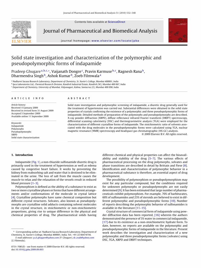
Journal of Pharmaceutical and Biomedical Analysis 51 (2010) 532–540 Contents lists available at ScienceDirect Journal of Pharmaceutical and Biomedical Analysis journal homepage: www.elsevier.com/locate/jpba Solid state investigation and characterization of the polymorphic and pseudopolymorphic forms of indapamide Pradeep Ghugare a,b,c,∗ , Vaijanath Dongre c , Pravin Karmuse b,c , Ragnesh Rana b , Dharmendra Singh b , Ashok Kumar b , Zoeb Filmwala a a Nadkarni Sacasa Research Laboratory, Department of Chemistry, St. Xavier’s College, Mumbai 400001, India b Ipca Laboratories Ltd., Chemical Research Division, Kandivli Industrial Estate, Kandivli (W), Mumbai 400 067, India c Department of Chemistry, University of Mumbai, Vidyanagari, Kalina, Santacruz (E), Mumbai 400 098, India article info Article history: Received 15 January 2009 Received in revised form 31 August 2009 Accepted 5 September 2009 Available online 11 September 2009 Keywords: Indapamide Polymorphs Pseudopolymorphs XRPD Solid state characterization abstract Solid state investigation and polymorphic screening of indapamide, a diuretic drug generally used for the treatment of hypertension was carried out. Substantial differences were obtained in the solid state properties of crystals confirming the existence of a polymorphic and three pseudopolymorphic forms of indapamide. Detailed methods of preparation of the polymorphs and pseudopolymorphs are described. X-ray powder diffraction (XRPD), diffuse reflectance infrared Fourier transform (DRIFT) spectroscopy, differential scanning calorimetry (DSC) and thermogravimetric analysis (TGA) were employed for the characterization of different crystalline forms of indapamide. The stoichiometric ratio of solvents asso- ciated with the drug molecules in the pseudopolymorphic forms were calculated using TGA, nuclear magnetic resonance (NMR) spectroscopy and headspace gas chromatographic (HS-GC) analysis. © 2009 Elsevier B.V. All rights reserved. 1. Introduction Indapamide (Fig. 1), a non-thiazide sulfonamide diuretic drug is primarily used in the treatment of hypertension as well as edema caused by congestive heart failure. It works by preventing the kidney from reabsorbing salt and water that is destined to be elim- inated in the urine. The loss of salt from the muscle causes the muscle to relax and the relaxation of the vessels result in reduced blood pressure [1–3]. Polymorphism is defined as the ability of a substance to exist as two or more crystalline phases or forms that have different arrange- ments and/or conformations of the molecule in crystal lattice [4]. Crystalline polymorphs have same chemical composition but different crystal structures. Solvates, also known as pseudopoly- morphs are crystalline solid adducts containing solvent molecules in the crystal structure, in stoichiometric or non-stoichiometric proportions, giving rise to unique difference in the physical and chemical properties of drug. The pharmaceutical solids having ∗ Corresponding author at: Nadkarni Sacasa Research Laboratory, Department of Chemistry, St. Xavier’s College, Mumbai 400001, India. Tel.: +91 22 66474756. E-mail addresses: p d [email protected] (P. Ghugare), dr [email protected] (Z. Filmwala). different chemical and physical properties can affect the bioavail- ability and stability of the drug [5–7]. The various effects of pharmaceutical processing on the drug polymorphs, solvates and phase transitions are described in detail by Brittain and Fiese [8]. Identification and characterization of polymorphic behavior in a pharmaceutical substance is therefore, an essential aspect of drug development. The possibility of polymorphism or pseudopolymorphism may exist for any particular compound, but the conditions required for unknown polymorphs or pseudopolymorphs are not easily determined [9]. It has been estimated that large number of pharma- ceuticals exhibit polymorphism. For example, 70% of barbiturates, 60% of sulfonamides and 23% of steroids are believed to exist in dif- ferent polymorphic and pseudopolymorphic forms [10]. Number of reports describing the polymorphic behavior of sulfonamides is available in the literature [11–15]. Crystal structure of commercial form of indapamide using pow- der diffraction data has been reported, [16] wherein the authors demonstrated the presence of 3% water in commercial indapamide, [17] due to its existence as a non-stoichiometric hydrate form. To date, however, no reports are available on the polymorphic and pseudopolymorphic forms of indapamide in the literature. Present work describes the investigation and characterization of a new polymorphic and three pseudopolymorphic forms (solvates) using DSC, TGA, XRPD and DRIFT techniques. 0731-7085/$ – see front matter © 2009 Elsevier B.V. All rights reserved. doi:10.1016/j.jpba.2009.09.003
-
Upload
independent -
Category
Documents
-
view
1 -
download
0
Transcript of Indapamide Polymorphs Pseudopolymorphs XRPD Solid state characterization
Sp
PDa
b
c
a
ARRAA
KIPPXS
1
pckimb
tm[dmipc
C
(
0d
Journal of Pharmaceutical and Biomedical Analysis 51 (2010) 532–540
Contents lists available at ScienceDirect
Journal of Pharmaceutical and Biomedical Analysis
journa l homepage: www.e lsev ier .com/ locate / jpba
olid state investigation and characterization of the polymorphic andseudopolymorphic forms of indapamide
radeep Ghugarea,b,c,∗, Vaijanath Dongrec, Pravin Karmuseb,c, Ragnesh Ranab,harmendra Singhb, Ashok Kumarb, Zoeb Filmwalaa
Nadkarni Sacasa Research Laboratory, Department of Chemistry, St. Xavier’s College, Mumbai 400001, IndiaIpca Laboratories Ltd., Chemical Research Division, Kandivli Industrial Estate, Kandivli (W), Mumbai 400 067, IndiaDepartment of Chemistry, University of Mumbai, Vidyanagari, Kalina, Santacruz (E), Mumbai 400 098, India
r t i c l e i n f o
rticle history:eceived 15 January 2009eceived in revised form 31 August 2009ccepted 5 September 2009
a b s t r a c t
Solid state investigation and polymorphic screening of indapamide, a diuretic drug generally used forthe treatment of hypertension was carried out. Substantial differences were obtained in the solid stateproperties of crystals confirming the existence of a polymorphic and three pseudopolymorphic forms ofindapamide. Detailed methods of preparation of the polymorphs and pseudopolymorphs are described.
vailable online 11 September 2009
eywords:ndapamideolymorphsseudopolymorphsRPD
X-ray powder diffraction (XRPD), diffuse reflectance infrared Fourier transform (DRIFT) spectroscopy,differential scanning calorimetry (DSC) and thermogravimetric analysis (TGA) were employed for thecharacterization of different crystalline forms of indapamide. The stoichiometric ratio of solvents asso-ciated with the drug molecules in the pseudopolymorphic forms were calculated using TGA, nuclearmagnetic resonance (NMR) spectroscopy and headspace gas chromatographic (HS-GC) analysis.
© 2009 Elsevier B.V. All rights reserved.
olid state characterization. Introduction
Indapamide (Fig. 1), a non-thiazide sulfonamide diuretic drug isrimarily used in the treatment of hypertension as well as edemaaused by congestive heart failure. It works by preventing theidney from reabsorbing salt and water that is destined to be elim-nated in the urine. The loss of salt from the muscle causes the
uscle to relax and the relaxation of the vessels result in reducedlood pressure [1–3].
Polymorphism is defined as the ability of a substance to exist aswo or more crystalline phases or forms that have different arrange-
ents and/or conformations of the molecule in crystal lattice4]. Crystalline polymorphs have same chemical composition but
ifferent crystal structures. Solvates, also known as pseudopoly-orphs are crystalline solid adducts containing solvent moleculesn the crystal structure, in stoichiometric or non-stoichiometricroportions, giving rise to unique difference in the physical andhemical properties of drug. The pharmaceutical solids having
∗ Corresponding author at: Nadkarni Sacasa Research Laboratory, Department ofhemistry, St. Xavier’s College, Mumbai 400001, India. Tel.: +91 22 66474756.
E-mail addresses: p d [email protected] (P. Ghugare), dr [email protected]. Filmwala).
731-7085/$ – see front matter © 2009 Elsevier B.V. All rights reserved.oi:10.1016/j.jpba.2009.09.003
different chemical and physical properties can affect the bioavail-ability and stability of the drug [5–7]. The various effects ofpharmaceutical processing on the drug polymorphs, solvates andphase transitions are described in detail by Brittain and Fiese [8].Identification and characterization of polymorphic behavior in apharmaceutical substance is therefore, an essential aspect of drugdevelopment.
The possibility of polymorphism or pseudopolymorphism mayexist for any particular compound, but the conditions requiredfor unknown polymorphs or pseudopolymorphs are not easilydetermined [9]. It has been estimated that large number of pharma-ceuticals exhibit polymorphism. For example, 70% of barbiturates,60% of sulfonamides and 23% of steroids are believed to exist in dif-ferent polymorphic and pseudopolymorphic forms [10]. Numberof reports describing the polymorphic behavior of sulfonamides isavailable in the literature [11–15].
Crystal structure of commercial form of indapamide using pow-der diffraction data has been reported, [16] wherein the authorsdemonstrated the presence of 3% water in commercial indapamide,[17] due to its existence as a non-stoichiometric hydrate form. To
date, however, no reports are available on the polymorphic andpseudopolymorphic forms of indapamide in the literature. Presentwork describes the investigation and characterization of a newpolymorphic and three pseudopolymorphic forms (solvates) usingDSC, TGA, XRPD and DRIFT techniques.
P. Ghugare et al. / Journal of Pharmaceutical and
2
2
Rguf(SeWp
2
asuoaXa
2
e6wmitwtb
2
isoawttpuTaI
Fig. 1. Chemical structure of indapamide.
. Experimental
.1. Materials and reagents
The indapamide bulk drug sample was obtained from Chemicalesearch Division, Ipca laboratories Ltd., Mumbai, India. Laboratoryrade solvents used for crystallization, analytical grade solventssed for HS-GC and KBr used for DRIFT analysis were purchasedrom Merck KGaA, Darmstadt, Germany. Analytical grade N,O-bis-trimethylsilyl) trifluro acetamide (BSTFA) was purchased frompectrochem, Mumbai, India. Dimethyl sulfoxide-d6 and tetram-thylsilane for NMR were from Aldrich Chemical Co. Milwaukee,I, USA. Hydranal Composite5 used for Karl fisher titrimetry was
urchased from Riedel de Haen, Seelze, Germany.
.2. Methods
Indapamide is soluble in acetonitrile, ethyl acetate, glacial aceticcid, methanol, ethanol and other alcoholic solvents. It is verylightly soluble in ether and chloroform, while practically insol-ble in water. Various crystallization experiments were carriedut using individual as well as combinations of solvents at normalnd elevated temperature. Polymorphic screening of samples usingRPD and TGA was carried out to check formation of polymorphicnd pseudopolymorphic forms.
.2.1. Preparation of anhydrous form (form-I)A saturated suspension of indapamide prepared by adding
xcess of indapamide in glacial acetic acid was maintained at5–70 ◦C with constant stirring for 2 h. Small portion of the mixtureas removed after regular time interval of 30 min and dried. Theelting point of the dried compound was measured. Initially, melt-
ng point of the compound was in the range of 168–175 ◦C. After 2 h,he melting temperature shifted to the range of 185–193 ◦C, heatingas then stopped and the suspension was allowed to cool to room
emperature. The solid obtained was separated from the solutiony vacuum filtration and dried at 50–55 ◦C for 2–3 h under vacuum.
.2.2. Preparation of solvatesThe saturated solutions of indapamide in acetone were prepared
n three different 250 ml flasks and maintained at 35 ◦C with con-tant stirring. In order to prepare different solvates equal volumef carbon tetrachloride, cyclohexane and diethyl ether were slowlydded to the respective flask. After complete addition, the solutionsere kept at 35 ◦C for about half an hour and then cooled to room
emperature. The precipitates formed were filtered by vacuum fil-ration followed by washing with excess amount of respective
◦
recipitating solvent. The solids obtained were dried at 50–55 Cnder vacuum. Formation of solvates was checked by TGA analysis.he samples precipitated using carbon tetrachloride, cyclohexanend diethyl ether were labeled as solvate-I, solvate-II and solvate-II, respectively.Biomedical Analysis 51 (2010) 532–540 533
2.3. Stability studies
In order to test ability to uptake water, samples of form-I,solvate-I, II and III were stored at room temperature in open air(about 55% RH) and in a desiccator for 1 year. Samples includingthe commercial form were also subjected for manual grinding usingmortar and pestle to study physical stability and solid–solid transi-tion. The resulting samples were analyzed for solid state transitionby DRIFT, water content by TGA and Karl Fischer titrimetry (KFT).
2.4. Instrumentation
2.4.1. Scanning electron microscopy (SEM)The SEM images were obtained on a JSM-6360A system (JEOL,
Tokyo, Japan), using an acceleration potential of 10 kV. The sampleswere sputter coated with platinum to eliminate charging effects.
2.4.2. Diffuse reflectance infrared Fourier transform (DRIFT)spectroscopy
The DRIFT spectra were recorded in the solid state as KBrpowder dispersion using a Spectrum One FT-IR spectrometer(PerkinElmer, Beaconsfield, UK) equipped with diffuse reflectancesampling accessory. The spectrum for each sample was recordedwith an average of 16 co-added scans in transmission mode over aspectral region of 450–4000 cm−1 with a resolution of 4 cm−1.
2.4.3. X-ray powder diffraction (XRPD)XRPD patterns of samples were recorded at room temperature
on an X’Pert PRO diffractometer (PANalytical, Almelo, The Nether-lands) with Cu K� radiation, (� = 1.5406 Å) passing through nickelfilter, divergence slit (10 mm), antiscattering slit (10 mm) and sollerslit (0.02 rad). The X-ray generator was set at a voltage of 45 kVand current of 40 mA. The diffractometer was calibrated for accu-racy of peak positions with silicon powder (ASTM-692). Sampleswere subjected to XRPD analysis in continuous mode with a stepsize of 0.008◦ 2� and step time of 15 s over an angular range of3–40◦ 2�. To minimize the preferred orientations, the samples wereprepared by back loading technique using the PW1770/10 samplepreparation kit. The sample holder was rotated in a plane paral-lel to its surface at the speed of 30 rpm during the measurements.Obtained diffractograms were analyzed with X’Pert HighScore Plusdiffraction software (version 2.1b).
2.4.4. Thermal analysis (DSC and TGA)DSC thermograms were recorded on a Q-100 instrument (TA
Instruments, New Castle, DE, USA). Samples weighing 2–3 mg wereheated in crimped aluminum pans with pierced lead from 30 to210 ◦C at the rate of 10 ◦C/min. Nitrogen was used as a purging gasunder ambient flow rate.
The mass loss of the sample as a function of temperature wasdetermined using a TGA Q-500 instrument (TA Instruments, NewCastle, DE, USA). The samples were placed in open platinum cru-cibles and heated at the rate of 25 ◦C/min in the range of 30–400 ◦Cunder a nitrogen purge (20 ml/min). The DSC and TGA data wasprocessed using Universal Analysis 2000 software (version 4.3A).
2.4.5. Nuclear magnetic resonance (solution-state)NMR spectra were obtained on a 400 MHz instrument (Bruker,
Faellanden, Switzerland). The spectra were processed with XWINNMR software (version 3.1). The samples were prepared by dissolv-ing 5 mg of each form in 600 �l of DMSO-d6 spiked with 0.03% of
tetramethylsilane as reference (ıH = 0 ppm).2.4.6. Headspace gas chromatography (HS-GC)A 6890 series gas chromatographic system (Agilent, Wilming-
ton, DE, USA) equipped with flame ionization detector and Gerstel
5 al and
Md
(mcdawwitatihosFts
34 P. Ghugare et al. / Journal of Pharmaceutic
PS2 headspace was used for HS-GC analysis. Chromatographicata were collected and processed using ChemStation software.
An Rtx-624 (30.0 mm × 0.53 mm i.d.; 3.0 �) capillary columnRestek, Bellefonte, PA, USA) was used for chromatographic deter-
ination of acetic acid and DB-5 (30.0 mm × 0.53 mm i.d.; 5.0 �)apillary column (J & W Scientific, Folsom, CA, USA) was used for theetermination of other solvents. Nitrogen was used as a carrier gast a flow rate of 2.0 ml/min. Injection was carried out in split mode,ith a split ratio of 50:1. The injector and detector temperatureere set at 200 and 240 ◦C, respectively. The oven temperature was
nitially set at 50 ◦C for 10 min, and then it was raised by 10 ◦C/mino 220 ◦C and left constant for 10 min. The headspace oven temper-ture was set at 90 ◦C with thermostating time 20 min. The needleemperature was 100 ◦C. Pressurization time was 3.0 min and thenjection volume was 0.5 ml. Vial pressure was set at 15 psi and theeadspace carrier was regulated at 25 ml/min. Standard solutions
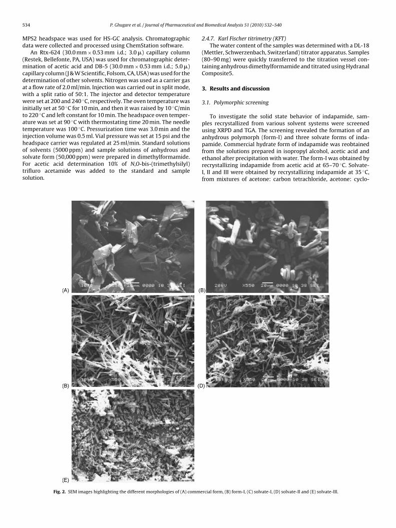
f solvents (5000 ppm) and sample solutions of anhydrous andolvate form (50,000 ppm) were prepared in dimethylformamide.or acetic acid determination 10% of N,O-bis-(trimethylsilyl)rifluro acetamide was added to the standard and sampleolution.Fig. 2. SEM images highlighting the different morphologies of (A) comme
Biomedical Analysis 51 (2010) 532–540
2.4.7. Karl Fischer titrimetry (KFT)The water content of the samples was determined with a DL-18
(Mettler, Schwerzenbach, Switzerland) titrator apparatus. Samples(80–90 mg) were quickly transferred to the titration vessel con-taining anhydrous dimethylformamide and titrated using HydranalComposite5.
3. Results and discussion
3.1. Polymorphic screening
To investigate the solid state behavior of indapamide, sam-ples recrystallized from various solvent systems were screenedusing XRPD and TGA. The screening revealed the formation of ananhydrous polymorph (form-I) and three solvate forms of inda-pamide. Commercial hydrate form of indapamide was reobtained
from the solutions prepared in isopropyl alcohol, acetic acid andethanol after precipitation with water. The form-I was obtained byrecrystallizing indapamide from acetic acid at 65–70 ◦C. Solvate-I, II and III were obtained by recrystallizing indapamide at 35 ◦C,from mixtures of acetone: carbon tetrachloride, acetone: cyclo-rcial form, (B) form-I, (C) solvate-I, (D) solvate-II and (E) solvate-III.
P. Ghugare et al. / Journal of Pharmaceutical and Biomedical Analysis 51 (2010) 532–540 535
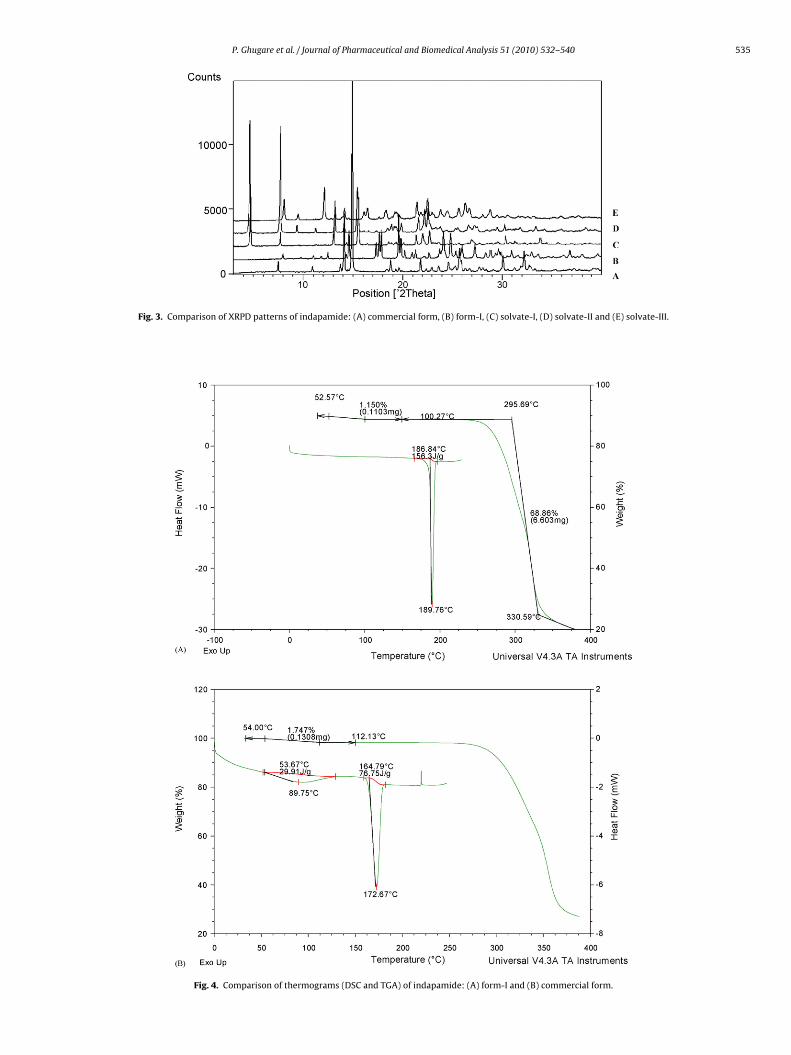
Fig. 3. Comparison of XRPD patterns of indapamide: (A) commercial
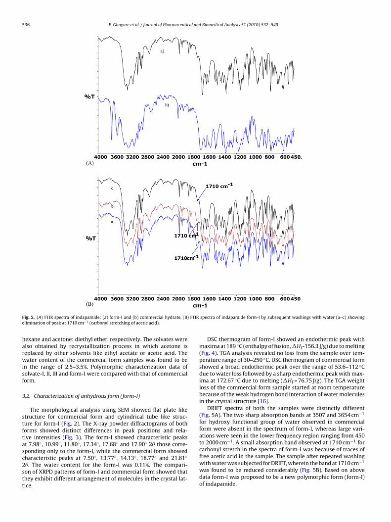
Fig. 4. Comparison of thermograms (DSC and TGA) of i
form, (B) form-I, (C) solvate-I, (D) solvate-II and (E) solvate-III.
ndapamide: (A) form-I and (B) commercial form.
536 P. Ghugare et al. / Journal of Pharmaceutical and Biomedical Analysis 51 (2010) 532–540
F FTIRe
harwisf
3
stftasc2stt
ig. 5. (A) FTIR spectra of indapamide: (a) form-I and (b) commercial hydrate. (B)limination of peak at 1710 cm−1 (carbonyl stretching of acetic acid).
exane and acetone: diethyl ether, respectively. The solvates werelso obtained by recrystallization process in which acetone iseplaced by other solvents like ethyl acetate or acetic acid. Theater content of the commercial form samples was found to be
n the range of 2.5–3.5%. Polymorphic characterization data ofolvate-I, II, III and form-I were compared with that of commercialorm.
.2. Characterization of anhydrous form (form-I)
The morphological analysis using SEM showed flat plate liketructure for commercial form and cylindrical tube like struc-ure for form-I (Fig. 2). The X-ray powder diffractograms of bothorms showed distinct differences in peak positions and rela-ive intensities (Fig. 3). The form-I showed characteristic peakst 7.98◦, 10.99◦, 11.80◦, 17.34◦, 17.68◦ and 17.90◦ 2� those corre-ponding only to the form-I, while the commercial form showed
haracteristic peaks at 7.50◦, 13.77◦, 14.13◦, 18.77◦ and 21.81◦�. The water content for the form-I was 0.11%. The compari-on of XRPD patterns of form-I and commercial form showed thathey exhibit different arrangement of molecules in the crystal lat-ice.
spectra of indapamide form-I by subsequent washings with water (a–c) showing
DSC thermogram of form-I showed an endothermic peak withmaxima at 189 ◦C (enthalpy of fusion, �Hf-156.3 J/g) due to melting(Fig. 4). TGA analysis revealed no loss from the sample over tem-perature range of 30–250 ◦C. DSC thermogram of commercial formshowed a broad endothermic peak over the range of 53.6–112 ◦Cdue to water loss followed by a sharp endothermic peak with max-ima at 172.67 ◦C due to melting (�Hf = 76.75 J/g). The TGA weightloss of the commercial form sample started at room temperaturebecause of the weak hydrogen bond interaction of water moleculesin the crystal structure [16].
DRIFT spectra of both the samples were distinctly different(Fig. 5A). The two sharp absorption bands at 3507 and 3654 cm−1
for hydroxy functional group of water observed in commercialform were absent in the spectrum of form-I, whereas large vari-ations were seen in the lower frequency region ranging from 450to 2000 cm−1. A small absorption band observed at 1710 cm−1 forcarbonyl stretch in the spectra of form-I was because of traces of
free acetic acid in the sample. The sample after repeated washingwith water was subjected for DRIFT, wherein the band at 1710 cm−1was found to be reduced considerably (Fig. 5B). Based on abovedata form-I was proposed to be a new polymorphic form (form-I)of indapamide.
P. Ghugare et al. / Journal of Pharmaceutical and Biomedical Analysis 51 (2010) 532–540 537
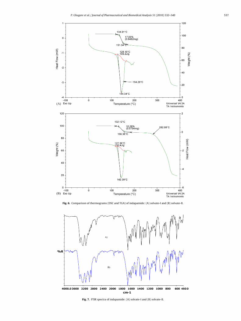
Fig. 6. Comparison of thermograms (DSC and TGA) o
Fig. 7. FTIR spectra of indapamide: (
f indapamide: (A) solvate-I and (B) solvate-II.
A) solvate-I and (B) solvate-II.
538 P. Ghugare et al. / Journal of Pharmaceutical and Biomedical Analysis 51 (2010) 532–540
(DSC a
3
sIdls(baawvis(
t1
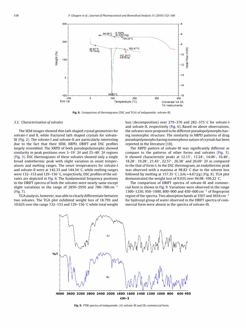
Fig. 8. Comparison of thermograms
.3. Characterization of solvates
The SEM images showed thin lath shaped crystal geometries forolvate-I and II, while fractured lath shaped crystals for solvate-II (Fig. 2). The solvate-I and solvate-II are particularly interestingue to the fact that their SEM, XRPD, DRIFT and DSC profiles
argely resembled. The XRPD of both pseudopolymorphs showedimilarity in peak positions over 3–19◦ 2� and 25–40◦ 2� regionsFig. 3). DSC thermograms of these solvates showed only a singleroad endothermic peak with slight variation in onset temper-tures and melting ranges. The onset temperatures for solvate-Ind solvate-II were at 142.33 and 144.34 ◦C, while melting rangesere 132–153 and 129–156 ◦C, respectively. DSC profiles of the sol-
ates are depicted in Fig. 6. The fundamental frequency positionsn the DRIFT spectra of both the solvates were nearly same exceptlight variations in the range of 2850–2950 and 780–788 cm−1
Fig. 7).TGA analysis, however, was able to clearly differentiate between
wo solvates. The TGA plot exhibited weight loss of 18.79% and0.62% over the range 132–153 and 129–156 ◦C while total weight
Fig. 9. FTIR spectra of indapamide: (A) so
nd TGA) of indapamide: solvate-III.
loss (decomposition) over 279–370 and 282–375 ◦C for solvate-Iand solvate-II, respectively (Fig. 6). Based on above observations,the solvates were proposed to be different pseudopolymorphs hav-ing isomorphic structure. The similarity in XRPD patterns of drugpseudopolymorphs having isomorphous nature of crystals has beenreported in the literature [18].
The XRPD pattern of solvate-III was significantly different ascompare to the patterns of other forms and solvates (Fig. 3).It showed characteristic peaks at 12.13◦, 13.24◦, 14.06◦, 16.40◦,18.28◦, 19.28◦, 21.43◦, 22.57◦, 26.38◦ and 26.69◦ 2� as comparedto the that of form-I. In the DSC thermogram, an endothermic peakwas observed with a maxima at 98.82 ◦C due to the solvent lossfollowed by melting at 157.35 ◦C (�Hf = 4.67 J/g) (Fig. 8). TGA plotdemonstrated the weight loss of 9.03% over 94.98–109.22 ◦C.
The comparison of DRIFT spectra of solvate-III and commer-cial form is shown in Fig. 9. Variations were observed in the range
1300–1250, 950–1000, 800–900 and 450–600 cm−1 of fingerprintregion of the spectra. Two absorption bands at 3507 and 3654 cm−1for hydroxyl group of water observed in the DRIFT spectra of com-mercial form were absent in the spectra of solvate-III.
lvate-III and (B) commercial form.

P. Ghugare et al. / Journal of Pharmaceutical and Biomedical Analysis 51 (2010) 532–540 539
Fig. 10. 1H NMR of indapamide (liquid state): (A) form
Fig. 11. 13C NMR of indapamid
-I, (B) solvate-I, (C) solvate-II and (D) solvate-III.
e (liquid state): solvate-I.

540 P. Ghugare et al. / Journal of Pharmaceutical and
Table 1Confirmation of stoichiometry of solvates (A) theoretical weight percentage of thesolvents and (B) observed weight loss of solvates in TGA.
Solvents of crystallization Theoretical weight % (w/w)
Mole ratio (indapamide: solvent)
1:1 2:1 1:2
(A)Cyclohexane 18.71 10.32a 31.46Diethyl ether 16.85 9.20a 28.70Carbon tetrachloride 29.48 17.23a 45.62Acetone 13.71 7.36 24.07
Pseudo polymorphs Observed weight loss in TGA (w/w)
(B)Solvate-I 17.03Solvate-II 10.28Solvate-III 9.10
a Calculated weight loss values matching with the observed values.
Table 2HS-GC results of different forms of indapamide.
Sample name Carbontetrachloride (%)
Cyclohexane(%)
Diethylether (%)
Acetic acid (%)
Form-I – – – 0.21%Solvate-I 18.11 – – –
3
ewtcci
tNtsifiStpoIppi
ysbt
[[[
[
Solvate-II – 10.51 – –Solvate-III – – 9.98 –
.4. Solvent of crystallization and stoichiometry
The calculated weight percentage of solvent by consideringither of the solvents used for preparation, were shown in Table 1ith various possible stoichiometric ratios. The comparison of
hese values with observed weight loss in TGA revealed that therystals of indapamide in solvate-I, II and III were associated witharbon tetrachloride, cyclohexane and diethyl ether, respectivelyn 2:1 proportion.
To confirm the identity and proportions of solvent of crystalliza-ion, all the solvates were subjected for 1H NMR analysis. The 1HMR of form-I and solvate-I of indapamide showed total signals for
en protons of indapamide (Fig. 10). However, 1H NMR of solvate-IIhowed an extra singlet slightly merged with the –CH3 protons ofndapamide at ı 1.31 ppm integrating six protons. This signal con-rmed association of half mole of cyclohexane in solvate-II crystals.imilarly 1H NMR of solvate-III showed an extra triplet integratingwo proton at ı 1.06 ppm and an extra quartet integrating threeroton at ı 3.36 ppm. These signals can be accounted for half molef diethyl ether present in the solvate crystals. 13C NMR of solvate-showed a signal for carbon at ı 95.9 ppm which confirmed theresence of carbon tetrachloride in the sample (Fig. 11). The peakositions of solvents in 1H NMR and 13C NMR was confirmed by
ndependent experiments of individual solvents.
Stoichiometry is further confirmed with the use of HS-GC anal-sis. The results are shown in Table 2. The standard samples ofolvents injected for retention time confirmation showed that car-on tetrachloride and cyclohexane are eluting at same retentionime at 17.20 min. Both the solvates showed a major peak for sol-
[[[[[
Biomedical Analysis 51 (2010) 532–540
vents at same retention time. Based on the knowledge of solventsused for the preparation of solvates, it was easily concluded that thesolvents associated with solvate-I and II were carbon tetrachlorideand cyclohexane, respectively. Also the identities of solvents asso-ciated with the solvates were already confirmed by NMR analysis.
4. Physical stability and water content variation
The stability and water content variation of samples wereinvestigated at various experimental conditions as described inSection 2.3. All forms were found to be stable and did not showany solid state transition under the experimental conditions. Thewater content variations were also not significant in all but forthe commercial form samples. The water content variation andhydration dehydration studies of commercial form were elabo-rately described by Smrkolj and Meden [16].
5. Conclusion
Based on crystallization experiments and solid state characteri-zation it was concluded that besides the commercial hydrate form,indapamide also exhibit an anhydrous and three solvate forms.The solvates showed stoichiometric ratio of 2:1 with the solventslike diethyl ether, carbon tetrachloride and cyclohexane. Of par-ticular interest, the carbon tetrachloride and cyclohexane solvateswere characterized which are having same diffraction propertiesbut different solvent of crystallization.
Acknowledgements
The authors thank the management of Ipca Laboratories for pro-viding necessary facilities and to Mr. Ganesh Mahale, Mr. NikhileshArya, Mr. Brejesh Sharma and Mr. Sanket Ingle for providing tech-nical assistance. One of the authors Mr. P. D. G. is grateful to Dr.Sanjay M. Nandavadekar for constant encouragement.
References
[1] J.G. Hardman, L.E. Limbird, A.G. Gilman, The Pharmacological Basis of Thera-peutics, tenth ed., McGraw Hill, New York, 2001, pp. 773–774.
[2] D.A. Abraham, Burger’s Medicinal Chemistry and Drug Discovery, vol. 3, sixthed., Wiley, New York, 2003, pp. 83–84.
[3] J.B. Taylor, D.J. Triggle, Comprehensive Medicinal Chemistry-II, vol. 6, Elsevier,UK, 2007, pp. 708–709.
[4] H. Britain, Physical Characterization of Pharmaceutical Solids, Marcel Dekker,New York, 1995.
[5] J.K. Haleblian, W. Mc Crone, J. Pharm. Sci. 58 (1969) 911–929.[6] S.R. Bryn, Solid State Chemistry of Drugs, second ed., SSCI Inc., Indiana, 1999.[7] S.R. Vippagunta, H.G. Brittain, D.J.W. Grant, Adv. Drug Deliv. Rev. 48 (2001)
3–26.[8] H.G. Brittain, Polymorphism in Pharmaceutical Solids, vol. 95, Marcel Dekker,
New York, 1999, pp. 331–361.[9] Bernstein, Am. Crystallographic Assoc. Trans. 39 (2004) 14–23.10] J.K. Haleblian, J. Pharm. Sci. 64 (1975) 1269–1288.11] S.S. Yang, J.K. Guillory, J. Pharm. Sci. 61 (1972) 26–40.12] C. Baraldia, M.C. Gamberinia, A. Tintib, F. Palazzolia, V. Feriolia, J. Mol. Struct.
918 (2009) 88–96.13] R.K.R. Jetti, R. Boese, J.A.R.P. Sarma, L.S. Reddy, P. Vishweshwar, G.R. Desiraju,
Angewandte Chemie 42 (2003) 1963–1967.
14] M.F. Haddow, T. Gelbrich, U.J. Griesser, Acta Cryst. C64 (2008) o309–o312.15] U.H. Patel, B.H. Patel, B.N. Patel, Cryst. Res. Technol. 36 (2001) 1445–1450.16] M. Smrkolj, A. Meden, Pharmazie 61 (2006) 999–1004.17] http://online.pheur.org/entry.htm.18] A. Othman, J.S.O. Evans, I.R. Evans, R.K. Harris, P. Hodgkinson, J. Pharm. Sci. 96(2007) 1380–1397.