Crystal structure of a fungal elicitor secreted by Phytophthora cryptogea, a member of a novel class...
11
Crystal structure of a fungal elicitor secreted by Phytophthora cryptogea, a member of a novel class of plant necrotic proteins Guillaume Boissy 1 , Eric de La Fortelle 2 , Richard Kahn 3 , Jean-Claude Huet 1 , Gérard Bricogne 2,4 , Jean-Claude Pernollet 1 and Simone Brunie 1 * Background: Elicitins form a novel class of plant necrotic proteins which are secreted by Phytophthora and Pythium fungi, parasites of many economically important crops. These proteins induce leaf necrosis in infected plants and elicit an incompatible hypersensitive-like reaction, leading to the development of a systemic acquired resistance against a range of fungal and bacterial plant pathogens. No crystal structures of this class of protein are available. The crystal structure determination of b-cryptogein (CRY), secreted by Phytophthora cryptogea, was undertaken to identify structural features important for the necrotic activity of elicitins. Results: The structure of CRY was determined using the multiwavelength anomalous diffraction technique and refined to 2.2 Å resolution. The overall structure has a novel fold consisting of six a helices and a beak-like motif, whose sequence is highly conserved within the family, composed of an antiparallel two- stranded b sheet and an Ω loop. This motif is assumed to be a major recognition site for a putative receptor and/or ligand. Two other distinct binding sites seem to be correlated to the level of necrotic activity of elicitins. Conclusions: The determination of the crystal structure of a member of the elicitin family may make it possible to separate the activity that causes leaf necrosis from that inducing systemic acquired resistance to pathogens, making it feasible to engineer a non-toxic elicitin that only elicits plant defences. Such studies should aid the development of non-toxic agricultural pest control. Introduction Most microorganisms that attempt to establish themselves as parasites on plants are defeated by the defences mar- shalled by the prospective host during the invasion process. These defence mechanisms are stimulated by molecules of microbial origin, generically termed elicitors, which induce one or more reaction cascades, usually resulting in a necrotic hypersensitive response [1]. This may lead to the development of systemic acquired resistance in which host tissues remote from the inoculation site become resistant to attack by microorganisms, including pathogens other than the one that was responsible for the initial, unsuccessful, attack [2,3]. Among plant pathogens, the fungal genus Phytophthora includes many species that are parasites of a diverse range of species, including many economically important crops. With the exception of Phytophthora parasitica var. nicotianae, all the Phytophthora species studied to date in culture secrete large amounts of one or several elicitin isoforms that act as toxins in order to weaken the host [4,5]. Recently, elicitin isoforms were also observed to be secreted from an Oomycete fungus belonging to the genus Pythium [6]. Elic- itins are a novel class of toxic protein elicitors that cause remote leaf necrosis and induce, in some plant species, systemic resistance against fungal and bacterial pathogens [2,3]. They are secreted from the pathogen in a process that includes the removal of a signal peptide, enter the plant through the root system and proceed to the leaves where they induce necrosis without requiring further biochemical alterations within this plant to be active [5]. No homology with any other protein sequence has been detected for the 12 Phytophthora and Pythium elicitins sequenced to date, but they show more than 60 % sequence identity with each other. This strongly suggests that these 10 kDa proteins constitute a new family. Elicitins from dif- ferent fungal species, when applied to tobacco plants at equal doses, exhibit different levels of necrotic activity, but induce protection in the plants at the same level [3]. Using these differences in necrotic activity on tobacco, the elicitin family has been classified into acidic a-elicitins and basic b-elicitins, the latter being much more necrogenic than the former. This classification is roughly independent of the ability of the elicitins to activate plant defence mecha- nisms. Highly necrotic b-elicitins cause visible leaf necro- sis when applied at approximately 100 pmoles per plant, whereas a 50–100-fold increase in concentration is required Addresses: 1 Unité de Recherche Biochimie & Structure des Protéines, INRA, 78352 Jouy-en- Josas, France, 2 MRC-LMB, Hills Road, Cambridge CB2 2QH, UK, 3 IBS, 41 Av. des Martyrs, 38027 Grenoble, Cedex 1, France and 4 L.U.R.E, Bât. 209d, 91405 Orsay Cedex, France. *Corresponding author. E-mail: [email protected] Key words: elicitin, multiwavelength anomalous diffraction (MAD), plant pathogen, toxin, X-ray crystallography Received: 2 August 1996 Revisions requested: 29 August 1996 Revisions received: 3 October 1996 Accepted: 8 October 1996 Structure 15 December 1996, 4:1429–1439 © Current Biology Ltd ISSN 0969-2126 Research Article 1429
-
Upload
nationalagriculturalresearchinra -
Category
Documents
-
view
1 -
download
0
Transcript of Crystal structure of a fungal elicitor secreted by Phytophthora cryptogea, a member of a novel class...

Crystal structure of a fungal elicitor secreted by Phytophthoracryptogea, a member of a novel class of plant necrotic proteinsGuillaume Boissy1, Eric de La Fortelle2, Richard Kahn3, Jean-Claude Huet1,Gérard Bricogne2,4, Jean-Claude Pernollet1 and Simone Brunie1*
Background: Elicitins form a novel class of plant necrotic proteins which aresecreted by Phytophthora and Pythium fungi, parasites of many economicallyimportant crops. These proteins induce leaf necrosis in infected plants and elicitan incompatible hypersensitive-like reaction, leading to the development of asystemic acquired resistance against a range of fungal and bacterial plantpathogens. No crystal structures of this class of protein are available. The crystalstructure determination of b-cryptogein (CRY), secreted by Phytophthoracryptogea, was undertaken to identify structural features important for thenecrotic activity of elicitins.
Results: The structure of CRY was determined using the multiwavelengthanomalous diffraction technique and refined to 2.2 Å resolution. The overallstructure has a novel fold consisting of six a helices and a beak-like motif, whosesequence is highly conserved within the family, composed of an antiparallel two-stranded b sheet and an Ω loop. This motif is assumed to be a major recognitionsite for a putative receptor and/or ligand. Two other distinct binding sites seemto be correlated to the level of necrotic activity of elicitins.
Conclusions: The determination of the crystal structure of a member of theelicitin family may make it possible to separate the activity that causes leafnecrosis from that inducing systemic acquired resistance to pathogens, making itfeasible to engineer a non-toxic elicitin that only elicits plant defences. Suchstudies should aid the development of non-toxic agricultural pest control.
IntroductionMost microorganisms that attempt to establish themselvesas parasites on plants are defeated by the defences mar-shalled by the prospective host during the invasion process.These defence mechanisms are stimulated by molecules ofmicrobial origin, generically termed elicitors, which induceone or more reaction cascades, usually resulting in anecrotic hypersensitive response [1]. This may lead to thedevelopment of systemic acquired resistance in which hosttissues remote from the inoculation site become resistant toattack by microorganisms, including pathogens other thanthe one that was responsible for the initial, unsuccessful,attack [2,3].
Among plant pathogens, the fungal genus Phytophthoraincludes many species that are parasites of a diverse rangeof species, including many economically important crops.With the exception of Phytophthora parasitica var. nicotianae,all the Phytophthora species studied to date in culturesecrete large amounts of one or several elicitin isoforms thatact as toxins in order to weaken the host [4,5]. Recently,elicitin isoforms were also observed to be secreted from anOomycete fungus belonging to the genus Pythium [6]. Elic-itins are a novel class of toxic protein elicitors that cause
remote leaf necrosis and induce, in some plant species, systemic resistance against fungal and bacterial pathogens[2,3]. They are secreted from the pathogen in a process thatincludes the removal of a signal peptide, enter the plantthrough the root system and proceed to the leaves wherethey induce necrosis without requiring further biochemicalalterations within this plant to be active [5].
No homology with any other protein sequence has beendetected for the 12 Phytophthora and Pythium elicitinssequenced to date, but they show more than 60% sequenceidentity with each other. This strongly suggests that these10kDa proteins constitute a new family. Elicitins from dif-ferent fungal species, when applied to tobacco plants atequal doses, exhibit different levels of necrotic activity, butinduce protection in the plants at the same level [3]. Usingthese differences in necrotic activity on tobacco, the elicitinfamily has been classified into acidic a-elicitins and basicb-elicitins, the latter being much more necrogenic than theformer. This classification is roughly independent of theability of the elicitins to activate plant defence mecha-nisms. Highly necrotic b-elicitins cause visible leaf necro-sis when applied at approximately 100 pmoles per plant,whereas a 50–100-fold increase in concentration is required
Addresses: 1Unité de Recherche Biochimie &Structure des Protéines, INRA, 78352 Jouy-en-Josas, France, 2MRC-LMB, Hills Road, CambridgeCB2 2QH, UK, 3IBS, 41 Av. des Martyrs, 38027Grenoble, Cedex 1, France and 4L.U.R.E, Bât.209d, 91405 Orsay Cedex, France.
*Corresponding author.E-mail: [email protected]
Key words: elicitin, multiwavelength anomalousdiffraction (MAD), plant pathogen, toxin, X-raycrystallography
Received: 2 August 1996Revisions requested: 29 August 1996Revisions received: 3 October 1996Accepted: 8 October 1996
Structure 15 December 1996, 4:1429–1439
© Current Biology Ltd ISSN 0969-2126
Research Article 1429

to elicit the same reaction using one of the weakly-necrotica-elicitins. These differences in biological activity can becorrelated with the sequence differences and the physico-chemical features of elicitins [5–12]. By comparing thesequences of various elicitins and relating them to the corresponding necrotic indices in tobacco plants, it is possi-ble to identify potential necrotic-modulating residues atpositions 2, 13, 72 and 94. The nature of the sidechain ofresidue 13 has the most consistent correlation with severityof the necrotic effects. All the highly necrotic b-elicitinshave a hydrophilic sidechain at position 13, usually a lysine,whereas the less necrotic a-elicitins have a hydrophobicvaline at this position. To evaluate the contribution ofresidue 13 to the necrotic activity of elicitins, a recombinantelicitin has been produced in E. coli using a synthetic geneencoding b-cryptogein (CRY), a basic elicitin secreted byPhytophthora cryptogea. Replacement of lysine 13 by a valineusing site-directed mutagenesis experiments led to a dra-matic alteration of the toxicity of the mutated recombinantprotein [13], reducing it to the toxicity level of an a-elicitin.In spite of the identification of many cellular events thatoccur in response to the action of elicitins in the plant, todate nothing is known about the action of elicitins at themolecular level. However, specific high affinity bindingsites for CRY have been observed in the tobacco plantplasma membrane [14].
In this paper, we describe the first example of a three-dimensional structure of an elicitin. The X-ray crystalstructure of wild-type CRY has been determined at 2.2 Åresolution. It reveals three distinct sites on the surface ofthe protein that are likely to be involved in the toxicfunction of elicitins. A rational approach to mutagenesisto map the functional sites of elicitins, is now possible
using a gene encoding for CRY overexpressed in Pichiapastoris [15].
Results and discussionStructure determinationCRY crystallizes in the space group P4122 with cell dimen-sions a=b=46.5Å, c=134.9Å [16]. An extensive search forheavy-atom derivatives yielded only one, a poorly phasingplatinum derivative. We therefore solved the crystal struc-ture of CRY by the multiwavelength anomalous diffraction(MAD) phasing method. Four data sets were collected inthe vicinity of the platinum LIII absorption edge (Table 1)on the D2AM beam line at the European SynchrotronRadiation facility (ESRF, Grenoble, France). As previ-ously reported [17], a vast improvement of the phases wasobtained by the introduction of a native data set. Therefinement of heavy-atom parameters and the calculationof phase probability distributions were performed usingthe program SHARP [18]. The use of this new programwas a decisive factor in obtaining an interpretable elec-tron-density map. The data structure in SHARP allows thenative dataset to be on an equal footing with those of anyderivative; incorporating native measurements on top of aMAD dataset was simple and required no special proce-dures such as phase combination or use of negative occu-pancies. The extra isomorphous information introducedinto the phasing process using this method was especiallyuseful for centric reflections (27% of the data to 3.0Å),which are devoid of anomalous signal, and also for verylow-resolution reflections (below 16.5Å) for which anom-alous differences were not measured.
The electron-density was so well defined (Fig. 1a) that the atomic model could be built for the whole molecule
1430 Structure 1996, Vol 4 No 12
Table 1
Summary of data collection.
K2PtCl4 derivative*
Native1 Native2 remote1 edge peak remote2l1 l2 l3 l4
X-ray source LURE ESRF ESRF ESRF ESRF ESRFTemperature (°) 4 C 100 K 100 K 100 K 100 K 100 KWavelength (Å) 0.91 1.0500 1.0766 1.0726 1.0722 1.0696Unit cell size (a = b, c) (Å) 46.5, 134.9 46.4, 132.8 46.1, 132.3dmin
‡ (Å) 2.2 2.9 2.9 2.9 2.9 2.9Number of observations 35101 13922 19956 22323 26399 24029Number of unique reflections 7864 3404 3074 3169 3264 3246
completeness (%) 97.2 98.2 88.0 90.3 92.6 92.4Multiplicity 3.1 4.2 6.5 7.0 8.1 7.4Number of unique Bijvoet pairs - - 2144 2208 2302 2279I > 3s (%) 92.9 91.8 96.2 96.5 96.5 96.5Rsym
† (%) 3.2 5.9 4.8 4.9 5.0 5.0in highest resolution shell 15.0 9.1 9.0 11.6 7.7 7.1
*The crystal was soaked in the dark for 6h in the sitting drop where ithad grown, reaching a final concentration of 50mM K2PtCl4. Thesoaked crystal was much more sensitive to radiation damage.
†Rsym = ΣhΣi | Ii(h) – < I (h) > | /ΣhΣi Ii (h) , where Ii (h) is the ith observationof reflection h and < I(h) > the average intensity obtained from thesame reflection observed i times. ‡dmin is the minimum Bragg spacing.

without ambiguity. The refined model gave a crystallo-graphic R factor of 21.8% for the 6987 reflections withF>2sF between 7Å and 2.2Å. A total of 66 water mol-ecules was included in the final model, for which the statis-tics are summarized in Table 2. A portion of the 2Fo–Fcelectron-density map, calculated after completion of therefinement, is shown in Figure 1b. The Ramachandran plot[19] indicates that 97.8% of the residues are in the mostfavoured regions with no outlier residues in the disallowedregion. Only two residues are in the ‘generously allowed’region, and both are well defined by the electron-density.
Overall structureCRY is a globular protein of approximate dimensions39×26×25Å. The details of the extent of the secondarystructure are given in Figures 2 and 3. The fold is mainlya-helical, comprising six a helices (a1–a6). In agreementwith results from circular dichroism experiments [8],a helices account for 56 % of the structure. All the helicesare concentrated on one face of the molecule, strengthen-ing the tertiary structure acting as a belt. In contrast, the
other face of the molecule has an unusual shape, with twoprotruding loops; one is an Ω loop and the other contains a b sheet. The overall fold of CRY shows no resemblanceto any reported protein topology. Indeed, a search usingthe programs DALI [20], DEJAVU [21] and VAST [22],which allow a systematic search for homologous three-dimensional structural motifs in the Brookhaven ProteinData Bank [23], revealed no similar topology.
The common coreThe tertiary structure of CRY is rigidly held in place bythree disulfide bridges. Corresponding disulfide bridgeshave previously been established for the a-elicitin secretedby Phytophthora capsici by NMR studies [24]. In CRY, Cys3–Cys71 links the N-terminal polypeptide to the b sheet,Cys27–Cys56 joins helices a2 and a4, and Cys51–Cys95maintains position of the C-terminal helix a6 relative tohelix a3. In addition, a salt bridge (Asp21–Lys62) binds theshort connecting loop between helices a1 and a2 to helixa4. The hydrophobic core of the molecule is mainly occu-pied by a cluster of three methionines, Met35, Met50 and
Research Article Structure of a fungal elicitor Boissy et al. 1431
Figure 1
Stereoviews of the original and final electron-density maps of CRY. (a) The ‘MAD+native’(original) electron-density map calculated at3.0 Å resolution using solvent-flattened experi-mental phases with residues of the Ω loop ofthe final refined model superimposed in thicklines. (b) The 2Fo–Fc calculated phased mapgenerated from the refined coordinates at2.2 Å resolution showing the same region ofthe molecule as (a). Both maps are contouredat the 1.5 s level.
Y 33
K 39
A 40
P 42
Y 47 M 50
K 39 K 39
Y 33
K 39
A 40
P 42
Y 47 M 50
K 39 K 39
K 39
A 40
P 42
Y 47 M 50
Y 33
K 39
A 40
P 42
Y 47 M 50
Y 33
(a)
(b)

Met59, the disulfide bridge Cys27–Cys56, the sidechains ofIle60 and Ile63 and the aromatic rings of Tyr87 and Phe91.The three disulfide bonds, the salt bridge and the 39 aminoacids that define this hydrophobic core are invariant in theknown Phytophthora elicitins. Therefore one can expect thatall the members of the elicitin family will share a verysimilar tertiary structure.
The beak-like motifThe loop from Tyr33 to Pro42 is an Ω loop, the distancebetween the two Ca (6.9Å) being in the range defined byLeszczynski and Rose [25]. Although only a few electrosta-tic interactions stabilize the Ω loop, its conformation is surprisingly well defined and is shaped according to theposition of the cumbersome, hydrophobic, phenolic ring ofthe invariant Tyr33. This Ω loop was already well definedin the initial ‘MAD+native’ electron-density map (Fig. 1a).The hydroxyl atom of Tyr33 interacts with the carbonyloxygen of Ala40, and due to the presence of a reverse turnbetween Met35 and Thr37, the carbonyl oxygen of Ser34 ishydrogen bonded to the backbone nitrogen of Ala38. Inaddition, the stacking of the sidechains of Tyr33, Pro42 andTyr47 enhances the rigidity of the whole loop. The loop
created by the antiparallel two-stranded b sheet (b1–b2)connects the two helices a4 and a5 and contains a reverseturn from Pro76 to Ser78. The Ω loop and the b sheet areinterestingly shaped in a beak-like arrangement, with thetwo reverse turns facing each other. We have dubbed thisstructural feature the beak-like motif. It is remarkable thatthis motif is essentially made of residues that are invariantin the elicitin family, with most of the variable residuesbelonging to the helices on the opposite face of the mol-ecule (Fig. 2a). Studies on Ω loops have shown that theyare often important for protein function and stability [26].In the CRY structure, it is clear that the conserved beak-like motif is essential for the structural stability and foldingof the protein. However, the interaction between the Ωloop and b sheet is weak as only two hydrogen bonds areinvolved in their contact (Thr77Og and Ser78Og arerespectively hydrogen bonded to the carbonyl oxygens ofLeu36 and Ala38).
Proposed binding sitesGiven the unusual biological properties of the elicitins,induction of plant-cell necrosis and plant systemic resis-tance to an array of pathogens, it seems clear that the elic-itin proteins must interact with one or more plant receptors.A receptor-like binding factor on the tobacco plant plasmamembrane is one of the possible candidates for a CRYreceptor, as suggested by Wendehenne et al. [14]. The Ωloop and the b sheet both exhibit stable conformations, a feature which should be common to all of the elicitinsbecause of their high degree of sequence identity. It istempting to imagine that the conserved structural beak-likemotif may be important in protein function. As the inter-action between the Ω loop and b sheet is weak, when theprotein interacts with a receptor and/or a ligand, it seemsplausible that the beak-like motif may open; the Ω loopmight act as a hinge upon binding. If so, this motif mightbe a major site for receptor/ligand recognition and animportant element in the function of protein.
As previously noted [14], binding seen to plasma mem-brane receptors may be abrogated out by the modifiedcharge density induced by changes on either the pH on theionic strength of the solution in which the experiment isperformed. This observation might explain the fact thatacidic a-elicitins and basic b-elicitins show clearly distinctlevels of toxicity. There are few variable amino acids of theelicitin family, four of which (in position 2, 13, 72 and 94)seem to be correlated to the level of toxicity of the proteins(Fig. 4). Residue 13, which is lysine in highly necroticb-elicitins and valine in the less necrotic a-elicitins, seemsto be particularly important in determining the level of tox-icity [13]. In the CRY structure, Lys13 lies in the middle ofhelix a1, well exposed to the solvent, in a region mainlycomposed of variable residues. The charge distribution ofthe electrostatic potential calculated on the solvent-accessi-ble surface, (Fig. 5), highlights distinct patches of positive,
1432 Structure 1996, Vol 4 No 12
Table 2
Summary of refinement statistics of final model with 4 °Cnative data (Native1).
Refinement data
Resolution range (Å) 7.0–2.2
No of reflectionsTotal 7583Used for refinement (scut-off = 2) 6987Rfree calculation 596
No of non hydrogen atomsProtein 717Water 66
Validity of the model
R factor* (%) 21.8Rfree
† (%) 27.9Average temperature factors (Å2)
Mainchain 35.8Sidechains 37.1Solvent molecules 47.2
Rms deviations from idealityBond lengths (Å) 0.013Bond angles (°) 1.50
Ramachandran plot [19]Residues in most favoured regions (%) 97.8Residues in additional allowed regions (%) 2.2
Overall G factor‡ 0.02
*R factor = Σ | Fobs – Fcalc | / Σ | Fobs |, where Fobs and Fcalc are theobserved and calculated structure factor amplitudes. A small fraction(7.5 %) of reflections were randomly selected and used to calculatethe †Rfree. ‡G factor is the overall measure of structure quality fromPROCHECK [45].

negative and polar regions of the surface of the molecule.From the electrostatic potential analysis (Fig. 5), it is clearthat replacing the Lys13 with a valine as seen in a-elicitinswill render this region of the molecule uncharged. Lys13 islocated in the middle of the first helix in a position wherethe Lys→Val substitution would not be expected to causea conformational change. Therefore, the dramatic alter-ation of toxicity produced by site directed mutagenesis of this residue is likely to be related to the change in theelectrostatic potential of this area.
The calculated solvent-accessible surface shows a largedepression centered around the conserved residue Tyr87.The depression, bordered by the Ω loop, the helix a3, theb sheet and the C-terminal helix, is predominantly sur-rounded by invariant residues except for the three residuesAla2, Asp72 and Lys94. The depression, as depicted inFigure 5a, contains a positively charged region on one sidecorresponding to Lys94 and a negatively charged region onthe other side, corresponding to Asp72. The negativecharge of Asp72 is partially neutralized by the charge of theN-terminal amino group. In the less toxic a-elicitins, Lys94
and Ala2 are replaced by threonines and Asp72 is replacedby a glutamic acid. Although the negative charge of residue72 is maintained as the sidechains of Glu72 and Thr2 areclose, steric hindrance between them, in a-elicitins, mayaffect the conformation of this region. It is clear that thecharge-distribution around the depression would be differ-ent in a-elicitins from that observed in b-elicitins.
Tyr87, whose side chain plunges into the bottom of thedepression, could have a special role in ligand and/orreceptor binding. Buried in the core of the molecule, thehydroxyl atom is free from interactions with its neighbours,allowing the phenolic ring to move freely, perhaps openinga deeper cavity. It is noteworthy that tyrosines have beenshown to limit the access to active sites in several cases[27]. However, at present there is no evidence of an enzy-matic function for the elicitins.
Thus, in addition to the beak-like motif, there are two otherregions around residues 13 and 87 for which the charge-distribution is directly correlated to the level of the biologi-cal activity of elicitins. In b-elicitins, the surface of these
Research Article Structure of a fungal elicitor Boissy et al. 1433
Figure 2
The secondary structure of CRY. (a) The sec-ondary structure elements, assigned byPROCHECK [45], are aligned with thesequence of CRY. Coloring scheme is derivedfrom the alignment of 12 sequences of elic-itins [5–12]. Invariant residues are shown inblue and variable residues in pink. Twostretches of invariant residues are located onthe Ω loop and the antiparallel two-strandedb sheet (b1–b2). The toxicity-related residue13 is indicated in yellow and the threeresidues (2, 72 and 94) likely to be correlatedto the level of toxicity are colored in green.(b) A stereo view of the Ca trace of CRY withevery tenth residue numbered and the N andC termini are labeled. (Figure generated usingthe program MOLSCRIPT [47].)
20 20 20
10 10 10
60 60 60
30 30
70
30
70 70
50 50 50
90 90 90
80 80
N
80
NN
40 40 40
CCC
TACTATQQTAAYKTLVSILSDASFNQCSTDSGYSMLTAKALPTTAQYKLMCA ^ ^ ^ ^ ^
STACNTMIKKIVTLNPPNCDLTVPTSGLVLNVYSYANGFSNKCSSL ^ ^ ^ ^
(a)
(b)
60
10 20 30 40 50
70 80 90
α1
α4 β1 β2 α5 α6
α2 α3Ω loop

two regions is highly charged. In a-elicitins, the surface isless charged in the depression region around residue 87 andmore hydrophobic in the region around residue 13. Thesetwo regions of the molecule are good candidates for regionsthat will prove to be important in receptor-binding.
Molecular environment in the crystalAlthough the model shows good geometry and is well-defined by the electron-density map, the overall B factor israther high (37.3Å2). This is consistent with the estimatedaverage temperature factor for the crystal (BWilson =38Å2).The Rfree [28] decreased continuously during the course ofthe refinement, confirming the validity of the results. Twocharacteristic features of the crystals should be underlined.First, the mosaicity of the crystal was relatively high, both atambient and cryogenic temperatures. Second, the observedstrong thermal diffuse scattering was not lowered by the useof frozen crystals. These two phenomena give rise to thehypothesis of a strong static disorder embedded within theentire crystal lattice. This is consistent with both theunusual high solvent content (66%) and the limited numberof contacts between symmetry-related molecules. With anapplied cut-off of 3.5Å and 4.0Å respectively, direct inter-molecular contacts amount to only 6 hydrogen bonds and 63van der Waals contacts. The lack of intermolecular contactsis therefore in agreement with the observed temperaturefactor. The analysis of the crystal packing shows half-mooncrossing channels, each 43Å long by 15Å wide, one parallelto the a, the other to the b axis. A main intermolecularcontact is observed in the region around Asn70. A dyad axisis responsible for a hydrogen-bond pair between the amidegroup of Asn70 and its twofold symmetry-related amidegroup. Large loose water molecule lattices, which represent
1434 Structure 1996, Vol 4 No 12
Figure 4
A ribbon diagram of the molecule showing the key residue Lys13 andthree putative activity-modulating residues (Ala2, Asp72 and Lys94) inball-and-stick representation. Helices are shown in blue, b strands andreverse turns in red, and coil loops in green. Disulfide bridges are rep-resented in light green (ball-and-stick). The conserved Tyr87 residue islabeled. (Figure generated using the programs MOLSCRIPT [47] andRASTER3D [48].)
Figure 3
A stereo diagram of the overall fold of CRY.Secondary structure elements are as follows :a1, residues 5–19; a2, 22–31; a3, 44–52;a4, 54–66; b1, 72–74; b2, 81–83; a5,84–89; and a6, 91–96. The Ω loop whichincludes residues 33 to 42 connects helicesa2 and a3. Helices are shown as blue cylin-ders and b strands as yellow arrows. Coil ele-ments are represented in green. The tworeverse turns and the helical turn that con-nects helices a5 and a6 are colored red, .The three disulfide bridges (3–71, 27–56,51–95) and the salt bridge (21–62) areshown in yellow (ball-and-stick). The N and Ctermini are also marked. (Figure generatedusing the program Insight II Version 2.2[Biosym Technologies].)

a minor contribution to the stabilization of the crystalpacking, are observed between symmetry-related mol-ecules, except for helix a1 which is in van der Waals contactwith its symmetry-related counterpart. This loose molecularpacking may partially explain our difficulty in obtaininguseful heavy-atom derivatives. Such a pattern acts as a flowcell where a dialysis phenomenon elutes those elementswhich are not bound with sufficient affinity. In the case ofthe platinum derivative, only two residues, Gln26 of helixa2 and Thr54 of helix a4, are involved in anion binding.The weakness of these stabilizing interactions can be corre-lated with the poor occupancy factor (0.4) and the high tem-perature factor (60Å2) of the anion site.
Analysis of the tyrosine residuesA study of the exposure to the solvent of the strictly con-served tyrosine residues was previously performed by UVdifference spectroscopy and the pK’s of the phenolicgroups were determined [12]. It is interesting to comparethese results to the CRY structure reported here. In theearlier study it was shown that Tyr12 is buried in thea-elicitins, and exposed to the solvent in the b-elicitins. Inthe CRY structure, Tyr12 is indeed exposed to the solventand the shift of its pK towards 11.5 is explained by thehydrogen bond between its hydroxyl and the carbonyl
oxygen of Thr74, which links the first helix to the b sheet.Tyr33 is found buried with a hydrogen bond between itshydroxyl and the carbonyl oxygen of Ala40. Consistentwith this, Tyr33 was previously observed to be inaccessibleto solvent and not titratable. Spectroscopic studies havealso shown that Tyr47 is located at the surface of the mol-ecule, as we confirm here. The stacking of the aromaticring of Tyr47 on Pro42 would partly explain its pK shift toover pH 12. The UV data indicate a normal pK for Tyr85,which is indeed accessible to the solvent. Tyr87 lies at thebottom of a depression and, although its free hydroxyl isburied into the core of the molecule, its phenolic ring is partly exposed to the solvent. Difference spectroscopydemonstrated that this tyrosine is indeed exposed and its pK is only slightly raised (by 0.5 pH unit). In sum, the observed stacking and hydrogen bonds formed by the tyrosines residues explain the fact that the maximalabsorbance wavelength of elicitins is 277 nm instead of the275nm seen for a free tyrosine. The agreement betweenthe model and the previous data suggests that there is nostructural alteration due to crystal packing.
Biological implicationsElicitins are 10kDa proteins secreted by Phytophthoraand Pythium fungi that are presumably involved in
Research Article Structure of a fungal elicitor Boissy et al. 1435
Figure 5
Four views of the electrostatic potentialmapped onto the solvent-accessible surface.The views (a)–(d) represent 90° incrementalrotations around the vertical axis. The positivepotential is shown in blue and the negativepotential in red. In agreement with the basicisoelectric point (pI ~ 10), the overall surfaceis mainly positively charged. The few discretepatches of charged residues are labeled. It isnoticeable that Lys13 is isolated among alarge surface of uncharged variable residues.The depression centred around Tyr87 isdepicted in (a), on the left part of (b) and onthe right part of (d). The protuberant beak-likemotif is clearly visible on the left of (c). Thecharges of the residues involved in the twosalt bridges (1–72 and 21–62) are partiallyneutralized. (Figure generated using theprogram GRASP [49].)
fungal invasion of plants. They also trigger a defencemechanism in the potential host, however, inducing leafnecrosis and an incompatible hypersensitive-like reac-tion that leads to resistance against fungal and bacterialplant pathogens. Although many of the cellular eventsthat form the plant’s response to elicitins have beendescribed, the molecular mechanism of elicitins action ispresently unknown. Potential high-affinity binding sitesfor b-cryptogein (CRY), an elicitin secreted by Phytoph-thora cryptogea [14], have been detected in the plasmamembrane from tobacco cells.
The present study of the crystal structure of CRY at2.2Å resolution provides the first structural informationon the elicitins. The polypeptide chain fold is a novelfolding type. The structure reveals a new beak-like motifthat is highly conserved in all elicitins. This motif is likelyto be a major recognition site for a putative receptorand/or ligand. Two other regions appear to be involvedin receptor or ligand binding, and their composition cor-relates with the level of necrotic-activity.
The knowledge of the elicitin structure will allow theengineering of non-toxic elicitins that can elicit plantdefences without creating damage. It now becomes possi-ble to imagine that elicitins could be used as the basis of ageneral method to protect plants against a range of phy-topathogenic microorganisms. One may hope to designan elicitin gene modified for its ability to successfullyinduce plant resistance against pathogens and transfer itto diverse plant genomes. Knowledge of the necroticmotifs, on the other hand, may allow the production ofnew herbicides. Both transgenic plants and toxic chemi-cals would be helpful not only to support progress in theunderstanding of the plant defence systems, but also toprovide agriculture with new protective means to preventpathogens from causing crop losses, without damagingthe environment.
Materials and methodsCrystallization and heavy-atom derivatives searchCRY was crystallized in concentrated sodium chloride as previouslydescribed by Guilloteau et al. [16]. Use of macroseeding proceduresmade it possible to systematically grow well shaped and sufficientlylarge crystals overnight through the pH range 4.0 to 10.0, while eliminat-ing twinning which is frequent in this crystalline form. A supersaturatedsolution was prepared by the hanging drop vapor diffusion method froma stock solution of 75mgml–1 protein, 0.02% NaN3 and 10mM NaClstock solution mixed with reservoir solution in proportion to a 1/3:2/3 or1/4:3/4 volume ratio. Four microliters of this mixture were deposited on asiliconized coverslip and left to equilibrate with the 4.85M NaCl precipi-tation buffer. The buffer used was 50mM sodium acetate pH4.0, 50mMTris-HCl pH7.50 or 50mM 3-[cyclohexylamino]-1-propanesulfonic acid(CAPS) pH10.0. Washed microsized crystals were then seeded into the resultant pre-equilibrated solution and grew within a few hours. Thecrystals belong to tetragonal space group P4122 with unit cell parame-ters a=b=46.5Å, c=134.9Å, with one molecule per asymmetric unit.High resolution data were collected at 4 °C with synchrotron radiation(l =0.9Å) to a minimum Bragg spacing of 2.2Å at station DW32 of the
LURE (Orsay, France) using a wiggler beam line. The diffraction imageswere indexed and integrated using MOSFLM [29]. The complete inten-sity set was scaled, merged, reduced to unique reflections and truncatedwith programs from the CCP4 suite [30]. No significant degradation wasobserved after X-ray exposure. This has allowed us to record a 97.2%complete data set from a single crystal for which all data were usable(< I /sigI>=4.6 for the last resolution shell).
In the course of the extensive heavy-atom derivatives search, only a tetrachloroplatinate (K2PtCl4) derivative was obtained that yielded to apoor phasing power. In addition to the loose crystal packing, this may berelated to the absence of four types of amino acids in the sequence ofCRY (there are no His, Glu, Arg or Trp residues) and to the high ionicstrength in which crystals are obtained. More favourable crystallizationprecipitating reagents led to a new crystalline form in PEG8000 19%(w/v), 200mM LiCl, 0.02% NaN3 and 100mM Tris-HCl pH7.0. Datawere collected using a Xentronics imaging detector and indexed by XDS [31] in a triclinic space group P1 (a=69.1Å, b=76.4Å, c=87.2Å,a =90°2, b =110°3, g =95°6). The self-rotation function showed a 422 pseudosymmetry and precession photographs failed to find anycrystallographic symmetry. The calculated specific protein volumeVm =2.6Å3 Da–1 corresponding to a solvent content of 53% was consis-tent with 16 molecules in the asymmetric unit. The difficulties associatedto a triclinic space group with high pseudosymmetry led us to leave thiscrystalline form and concentrate our efforts on the platinum derivative.
1436 Structure 1996, Vol 4 No 12
Figure 6
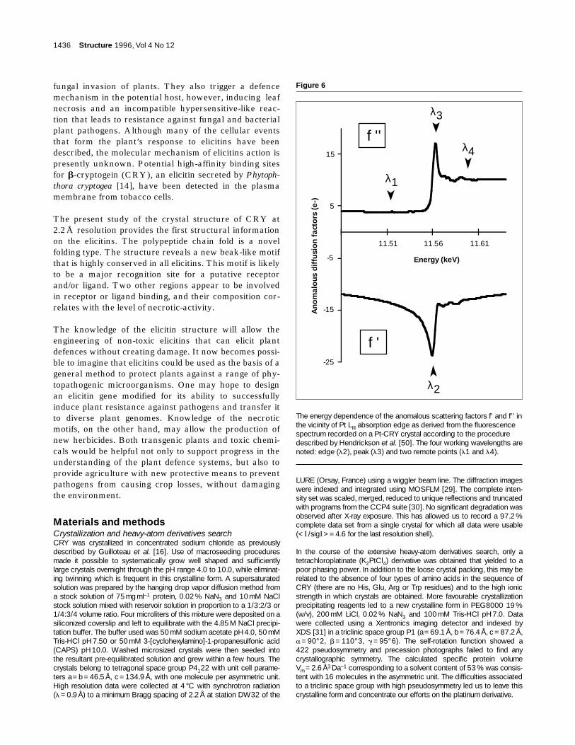
The energy dependence of the anomalous scattering factors f′ and f′′ inthe vicinity of Pt LIII absorption edge as derived from the fluorescencespectrum recorded on a Pt-CRY crystal according to the proceduredescribed by Hendrickson et al. [50]. The four working wavelengths arenoted: edge (l2), peak (l3) and two remote points (l1 and l4).
An
om
alo
us
dif
fusi
on
fac
tors
(e-
)
-25
-15
-5
5
15
11.51 11.56 11.61
Energy (keV)
λ1
λ2
λ3
λ4f ''
f '

MAD experimentMAD diffraction data were collected at four wavelengths on D2AMbeam line at the European Synchrotron Radiation Facility (ESRF) inGrenoble (France). Heavy-atom soaking was done by mixing 2.7 ml of aK2PtCl4 stock solution to a drop surrounding a crystal. Because there isno known stabilizing solution, the crystal was withdrawn and immediatelytransferred to and preserved in a previously removed crystal drop inorder to roughly wash it from concentrated platinum (50 mM). An equalamount of a cryoprotectant solution of 30 % (w/v) glycerol in 4.85 MNaCl, 50mM Tris-HCl was then added a few seconds before the crystalwas harvested with a fine dental-floss loop and flash-cooled to 100 K[32] in a stream of nitrogen gas from a Cryostream (Oxford Cryosys-tems, Oxford, UK). The X-ray optics were set up to optimize the platinumLIII absorption edge signal by measuring the fluorescence of platinum foilusing a photomultiplier placed at a right angle to the beam, in its polar-ization plane. A similar fluorescence spectrum, recorded with the frozencrystal, was used to select the wavelengths in the absorption edge(l2 =1.0726Å, maximum of |f′|), at the peak (l3 =1.0722Å, maximum off′′) and at two remote points (l1 =1.0766Å, l4 =1.0696Å) on bothsides from the peak as shown in Figure 6. The crystal was approximatelyaligned along the elongation axis c* so that Bijvoet pairs could be almostsimultaneously recorded. Oscillation diffraction patterns were recordedon a detector designed at the ESRF from a Thomson image intensifiercoupled to a Princeton CCD camera [33]. The speed of the CCD detec-tor combined with the brightness of the beam line have allowed thecomplete data collection (420 frames) in less than six hours. Reflectionswere indexed using profile-fitting as implemented in a local version ofXDS [34] modified by W. Kabsch and intensities were scaled at eachwavelength. Concomitant image scaling at each wavelength was alsoperformed. The CCP4 suite of programs was then used to merge and to process these intensities; anomalous differences were calculated bythe CCP4 program AGROVATA. A native data set was collected at thesame time on a frozen crystal (a=b=46.4Å, c=132.8Å). Data statis-tics for the whole data reduction procedure are presented in Table 1.
Maximum-likelihood parameter refinement and phasingThe MAD data sets were subjected to an anisotropic scaling usingSCALEIT (CCP4) with the l1 wavelength as a reference. The anom-alous components of each reflection were extracted to form eitherBijvoet differences (Fli(h+) – Fli(h–)) at each wavelength i or dispersivedifferences (Fli(h) – Flj(h)) at pairs (i, j) of wavelengths and were input
as coefficients to calculate Patterson map syntheses. Table 3 presentsthe results obtained from the inspection of Harker sections of thesePatterson maps. They were calculated for the best expected signals, onthe one hand with Bijvoet differences at the peak absorption, and onthe other hand with dispersive differences between the ‘edge’ and‘remote’ wavelengths. Automatic interpretation of heavy-atom Harkerand cross vector positions with RSPS [35] was coherent with a singleplatinum site. No additional less occupied sites were later found.
The refinement of parameters for heavy-atoms, relative scales betweendata sets, and lack-of-isomorphism (l.o.i.), was carried out using SHARP(for Statistical Heavy-Atom Refinement and Phasing) [18]. The mainadvantages of SHARP over existing programs are: a full two-dimensionalintegration (i.e. integration over both modules and phase of the nativestructure factor) to compute the likelihood function; a genuine likelihoodmaximization (rather than phase-integrated least-squares), which enablesthe parameters of the error model to be refined along with all the otherconventional parameters; and a hierarchical data structure that enablesany type of dataset (MIR, MAD, or both combined) to be described. Thenative dataset, if present, is described as any other derivative, but with noheavy atom specified. In this way, the l.o.i. parameters are kept at zerovalues for the native or any derivative dataset which is therefore set asthe reference for isomorphism. User-friendliness, online documentationand graphics are provided through a GUI (Graphical User Interface).Owing to the new radial integration (two-dimensional integration) men-tioned above, there is no obligation in SHARP to define a ‘native’ dataset.
In the present case, the MAD experiment was described straightfor-wardly as derivative data at four different wavelengths, the only parame-ters varying being f′, f′′, the scale parameters and the l.o.i. parameters. Inthis case, l.o.i. does not describe physical lack-of-isomorphism, but theeffect of an inaccurate heavy-atom model, and of inadequacies in theestimation of experimental standard deviations. Since the single Pt siteof low occupancy had a low phasing power, it was necessary to use asolvent-flattening procedure to produce interpretable electron-densitymaps, and thus to evaluate the success of the phasing procedure.
The electron-density maps obtained using the phase probability distribu-tions produced by SHARP, after solvent-flattening by program SOLOMON[36] using the MAD dataset only, were far more interpretable than thoseobtained from phase distributions produced by other programs. Addition of
Research Article Structure of a fungal elicitor Boissy et al. 1437
Table 3
Relative peak heights in anomalous and isomorphous Patterson syntheses.
Harker unique Isomorphous* MAD dataset*vectors difference
(12–3.5 Å)
u v w Anomalous Bijvoet Anomalous dispersive Isomorphous Anomalousdifferences differences differences differences
(8–3 Å) (10–4 Å) (12–3.5 Å) (10–3 Å)
2x 2y 1/2 3.64 (6) 4.97 (5) 3.30 (9) 6.1 (4) 7.4 (3)x + y y – x 3/4 5.44 (3) 5.59 (4) 5.32 (3) 7.7 (2) 7.2 (4)2x 0 2z 2.90 (11) 2.90 (24) 3.23 (10) 5.3 (6) 5.0 (8)0 2y 2z + 1/2 2.96 (9) 3.62 (10) 2.18 (27) 2.8 (29) 3.1 (17)
x – y y – x 2z + 1/4 9.33 (2) 11.05 (2) 8.06 (2) 14.6 (1) 15.2 (1)x + y x + y 2z + 3/4 2.51 (16) 4.31 (7) 2.75 (12) 4.0 (12) 5.6 (7)
Unique Harker vectors of the unique Pt heavy-atom site are shown forthe MAD dataset. They were calculated using programs from theCCP4 suite [30] or the routine MADMRG of the program HEAVYv4[51], which reduces MAD data to isomorphous plus anomalous differ-ences. The signal-to-noise ratio of these Patterson syntheses was opti-mized by screening the resolution range and the cut-off conditions. Themost informative differences expected for each kind of signal, as calcu-
lated using programs from the CCP4 suite, were chosen:| FNative2(h) – Fl1(h) | (isomorphous differences), | Fl3(h+) – Fl3(h–) |(anomalous Bijvoet differences) and | Fl2(h) – Fl1(h) | (anomalous dis-persive differences). The program HEAVYv4 produced only two kindsof differences, isomorphous and anomalous, and enhanced the signal-to-noise ratio of the Patterson map. *Peak heights are given as rootmean squares and their sorting numbers are shown in parentheses.

a native dataset (Native2, Table 1) in the calculation has been instrumentalin phasing the 3286 reflections with an average figure of merit of 0.46 overthe resolution shell 32.0–2.9Å. Solvent-flattening iterations performed withthe SOLOMON program described above raised the figure of merit to0.81 (Table 4). This resulted in a continuous and completely interpretableelectron-density map.
Model building and refinementTwo uncertainties were associated with the crystals: the solvent contentand the space group (P4122 or its enantiomorph P4322). The equilib-rium gradient method [37] gave inconclusive results for the number ofmolecules in the asymmetric unit. Self-rotation maps were free of addi-tional pseudo twofold axis and were consistent with one molecule perasymmetric unit, although this case corresponds to an unusually high Vm value of 3.7Å3 Da–1 [38]. A perfectly interpretable map has beenobtained using this hypothesis (66% solvent content), confirming thatthe asymmetric unit contains one CRY molecule. The space group wasclearly defined as P4122 by the right-handedness of a helices.
The 2.9Å MAD-phased electron-density map was skeletonized usingBONES [39] allowing a fast Ca tracing using the XFIT graphic tools ofthe program XTALVIEW [40]. The model building was made easier byviewing the symmetrical traces of the backbone after transformation intoa PDB type file using the program MAPMAN from the RAVE package[41]. The total lack of ambiguity combined with the perfect electron-density continuity have allowed the rapid construction of the 98 aminoacids corresponding to the whole CRY sequence with TURBO-FRODO[42]. The identification of the three disulfide bridges was helpful in con-necting the secondary structural elements, especially Cys3–Cys71 andCys51–Cys95 which were useful in the recognition of the N- and C-ter-minal helices. The starting model showed no residues in the disallowedregion of the Ramachandran plot. The first round of refinement by energyminimization using X-PLOR [43] allowed the crystallographic R factor todrop from 46.4% to 30.5% in the range from 10.0 to 2.9 Å resolution.After a few energy minimization and model rebuilding steps we decidedto continue the refinement process using high resolution data (Native1,Table 1). Because of the weak non-isomorphism induced by the freezingprocess the model was refitted into the 4° C unit cell using the molecularreplacement program AMoRe [44]. During the course of the refinement,minor modifications were made to the initial model, such as the flippingof Pro76 and Ser34 or the building of the last C-terminal residue forwhich the corresponding initial MAD electron-density map was missing.A series of simulated annealing omit maps excluding atoms located in a8Å sphere surrounding the doubtful residues have allowed them to becorrected. To refine the model, a slow-cool procedure was used with theheat stage being performed at 3000 K. After each refinement cycle the
model was checked against 3Fo–2Fc maps and manually adjusted toimprove the geometry. Water molecules were added very early onbecause some of them were already clearly visible on the 3 Å resolutionMAD map. Both a clear density bubble within a 2.5 s contoured Fo–FcFourier difference map and connections less than 3.4 Å to a possiblehydrogen donor or acceptor were strictly required to assign a peak to awater molecule. The crystallographic R factor for the final CRY modelcomprising 66 water molecules is 21.8 % between 7Å and 2.2Å. Asubset of 7.5% of all reflections was excluded from the refinement andwas used to calculate a Rfree =27.9%. Root mean square (rms) devia-tions from the stereochemical standards bond lengths and bond anglesare 0.013Å and 1.50° respectively. The stereochemistry of the modelwas analysed using PROCHECK [45]. The secondary structures wereassigned by the criteria of Kabsch and Sander [46], as implemented inPROCHECK.
Accession numbersThe atomic coordinates of b-cryptogein have been deposited in theBrookhaven Protein Data Bank (entry code 1BEO).
AcknowledgementsThe authors gratefully acknowledge Michel Roth for having providedaccess to the D2AM beamline of ESRF (Grenoble, France), Jean-LucFerrer and Eric Fanchon for help and advice with MAD data collection,Valérie Perez for fungus culture and CRY purification, Michael O’Donohuefor critical comments on this manuscript, the staff of LURE (Orsay, France)for making station DW32 available to us and Fred Saul for assistance indata collection.
References1. Dixon, R.A. & Lamb, C.J. (1990). Molecular communication in interac-
tions between plants and microbial pathogens. Annu. Rev. PlantPhysiol. Plant Mol. Biol. 41, 339–367.
2. Ricci, P., et al., & Pernollet, J.-C. (1989). Structure and activity of pro-teins from pathogenic fungi Phytophthora eliciting necrosis andacquired resistance in tobacco. Eur. J. Biochem. 183, 555–563.
3. Kamoun, S., Young, M., Glascock, C.B. & Tyler, B.M. (1993). Extracel-lular protein elicitors from Phytophthora: host-specificity and inductionof resistance to bacterial and fungal phytopathogens. Mol. PlantMicrobe Interact. 6, 15–25.
4. Kamoun, S., Young, M., Förster, H., Coffey, M.D. & Tyler, B.M.(1994). Potential role of elicitins in the interaction between Phytoph-thora species and tobacco. Appl. Environ. Microbiol. 60,1593–1598.
5. Pernollet, J.-C., Sallantin, M., Sallé-Tourne, M. & Huet, J.-C. (1993).Elicitin isoforms from seven Phytophthora species: comparison of theirphysico-chemical properties and toxicity to tobacco and other plantspecies. Physiol. Mol. Plant Pathol. 42, 53–67.
1438 Structure 1996, Vol 4 No 12
Table 4
MAD phasing statistics for CRY-Pt crystal.
Observed MAD difference ratios* Anomalous scattering factors†
remote1 edge peak remote2 f′ (e–) f′′ (e–)
Remote1 (l1) 0.027 0.016 0.014 0.013 –13.7 9.7Edge (l2) 0.030 0.015 0.015 –20.2 14.2Peak (l3) 0.034 0.011 –17.3 19.2Remote2 (l4) 0.031 –13.7 17.1
Riso (%)‡ 11.5Rdisp (%)§ 1.4 1.3 1.0
*Observed MAD difference ratios rms(s |F |) / rms(|F |) [50], where s |F |is either the Bijvoet difference at one wavelength (diagonal elements) or the dispersive difference between two wavelengths (off-diagonalelements). These statistics are calculated on fully observed reflections(F(h+) and F(h–) at all four wavelengths), e.g. 2101 reflections out of3286 between 32.6 and 2.9Å. †Values of anomalous scattering factors
as refined by SHARP [18], that were used for phasing.‡Riso =Σh |Fl1(h)–FNative2(h) | /Σh |FNative2(h) |§Rdisp =Σh |Fli(h)–Fl1(h) | /Σh |Fl1(h) | is similar to Riso with l1 acting likethe reference set. Mean figure of merit (FOM)=0.46 including reflectionswith FOM=0; Pt occupancy factor=0.42.

6. Huet, J.-C., Le Caer, J.-P., Nespoulous, C. & Pernollet, J.-C. (1995).The relationships between the toxicity and the primary and secondarystructures of elicitin-like protein elicitors secreted by the phytopatho-genic fungus Pythium vexans. Mol. Plant Microbe Interact. 8,302–310.
7. Huet, J.-C. & Pernollet, J.-C. (1989). Amino acid sequence of cin-namomin, a new member of the elicitin family, and its comparison tocryptogein and capsicein. FEBS Lett. 257, 302–306.
8. Nespoulous, C., Huet, J.-C. & Pernollet, J.-C. (1992). Structure–func-tion relationships of a and b elicitins. Signal proteins involved in theplant–Phytophthora interaction. Planta 186, 551–557.
9. Huet, J.-C. & Pernollet, J.-C. (1993). Sequences of acidic and basicelicitin isoforms secreted by Phytophthora megasperma megasperma.Phytochemistry 33, 797–805.
10. Huet, J.-C., Mansion, M. & Pernollet, J.-C. (1993). Amino acidsequence of the a elicitin secreted by Phytophthora cactorum. Phyto-chemistry 34, 1261–1264.
11. Huet, J.-C., Sallé-Tourne, M. & Pernollet, J.-C. (1994). Amino acidsequence and toxicity of the a elicitin co-secreted with ubiquitin byPhytophthora infestans. Mol. Plant Microbe Interact. 7, 302–304.
12. Nespoulous, C. & Pernollet, J.-C. (1994). Local structural differencesbetween a and b elicitins shown by CD and ultraviolet differencespectroscopy. Int. J. Pept. Protein Res. 43, 154–159.
13. O’Donohue, M.J., Gousseau, H., Huet, J.-C., Sallantin, M., Tepfer, D.A.& Pernollet, J.-C. (1995). Chemical synthesis, expression and mutage-nesis of a gene coding for b-cryptogein, a Phytophthora cryptogeaelicitin. Plant Mol. Biol. 27, 577–586.
14. Wendehenne, D., Binet, M.-N., Blein, J.-P., Ricci, P. & Pugin, A.(1995). Evidence for specific, high-affinity binding sites for a proteina-ceous elicitor in tobacco plasma membrane. FEBS Lett. 374,203–207.
15. O’Donohue, M., Boissy, G., Huet, J.C., Nespoulous, C., Brunie, S. &Pernollet, J.C. (1996) Overexpression in Pichia pastoris and crystal-lization of an elicitor protein secreted by the phytopathogenic fungusPhytophthora cryptogea. Protein Expr. Purif. 8, 254–261.
16. Guilloteau, J.-P., Nespoulous, C., Huet, J.-C., Beauvais, F., Pernollet, J.-C. & Brunie, S. (1993). Crystallization and preliminary X-ray diffractionstudies of b-cryptogein, a toxic elicitin secreted by the phytopatho-genic fungus Phytophthora cryptogea. J. Mol. Biol. 229, 564–565.
17. Newman, M., Strzelecka, T., Dorner, L.F., Schildkraut, I. & Aggarwal,A.K. (1994). Structure of restriction endonuclease BamHI phased at1.95 Å resolution by MAD analysis. Structure 2, 439–452.
18. de La Fortelle, E. & Bricogne, G. (1996). Maximum likelihood heavy-atom parameter refinement in the MIR and MAD methods. In Methodsin Enzymology. (Carter, C.W. & Sweet, R.M., eds), pp. 472–294, Aca-demic Press, UK.
19. Ramachandran, G.N. & Sasisekharan, V. (1968). Conformation ofpolypeptides and proteins. Adv. Protein Chem. 23, 283–438.
20. Holm, L. & Sander, C. (1993). Protein structure comparison by align-ment of distance matrices. J. Mol. Biol. 233, 123–138.
21. Kleywegt, G.J. & Jones, T.A. (1995). Where freedom is given, libertiesare taken. Structure 3, 535–540.
22. Gibrat, J.-F., Madej, T. & Bryant, S.H. (1996). Surprising similarities instructure comparison. Curr. Opin. Struct. Biol. 6, 377–385.
23. Bernstein, F.C., et al., & Tasumi, M. (1977). The Protein Data Bank: acomputer-based archival file for macromolecular structures. J. Mol.Biol. 112, 535–542.
24. Bouaziz, S., Van Heijenoort, C., Guittet, E., Huet, J.-C. & Pernollet,J.-C. (1994). Resonance assignment, cysteine-pairing elucidation andsecondary-structure determination of capsicein, an a-elicitin, by three-dimensional 1H NMR. Eur. J. Biochem. 220, 427–438.
25. Leszczynski, J.F & Rose, G.D. (1986). Loops in globular proteins: anovel category of secondary structure. Science 234, 849–855.
26. Fetrow, J.S. (1995). Omega loops : nonregular secondary structuressignificant in protein function and stability. FASEB J. 9, 708–717.
27. Benson, T.E., Walsh, C.T. & Hogle, J.M. (1996). The structure of thesubstrate-free form of MurB, an essential enzyme for the synthesis ofbacterial cell walls. Structure 4, 47–54.
28. Brünger, A.T. (1992). Free R value: a novel statistical quantity forassessing the accuracy of crystal structure refinement. Nature 355,472–475.
29. Leslie, A.G.W. (1992). Recent changes to the MOSFLM package forprocessing film and image plate data. In Joint CCP4 and ESF-EACBM Newsletter on Protein Crystallography, No. 26. SERCDaresbury Laboratory, Warrington, UK.
30. Collaborative Computational Project, No. 4. (1994). The CCP4 Suite:programs for protein crystallography. Acta Cryst. D 50, 760–763.
31. Kabsch, W. (1993). Automatic processing of rotation diffraction datafrom crystals of initially unknown symmetry and cell constants. J. Appl.Cryst. 26, 795–800.
32. Rodgers, D.W. (1994). Cryocrystallography. Structure 2, 1135–1140.33. Moy, J.-P. (1994). A 200 mm input field, 5–80 keV detector based on
an X-ray image intensifier and CCD camera. Nucl. Instrum. Meth.Phys. Res. A 348, 641–644.
34. Kabsch, W. (1988). Evaluation of single crystal X-ray diffraction datafrom a position-sensitive detector. J. Appl. Cryst. 21, 916–924.
35. Knight, S. (1989). Ribulose 1,5-bisphosphate carboxylase/oxygenase- A structural study. Thesis, Swedish University of Agricultural Sci-ences, Uppsala, Sweden.
36. Abrahams, J.P. & Leslie, A.G.W. (1996). Methods used in the struc-tural determination of bovine mitochondrial F1 ATPase. Acta Cryst. D52, 30–42.
37. Westbrook, E.M. (1985). Crystal density measurements usingaqueous ficoll solutions. Methods Enzymol. 114, 187–196.
38. Matthews, B.W. (1968). Solvent content of protein crystals. J. Mol.Biol. 33, 491–497.
39. Jones, T.A., Zou, J.Y., Cowan, S.W. & Kjeldgaard, M. (1991). Improvedmethods for building protein models in electron density maps and thelocation of errors in these models. Acta Cryst. A 47, 110–119.
40. McRee, D.E. (1993). Practical protein crystallography. AcademicPress, San Diego, CA, USA.
41. Kleywegt, G.J. & Jones, T.A. (1994). Halloween... Masks and Bones. InFrom first map to final model. (Bailey, S., Hubbard, R. & Waller, D.,eds), pp. 59–66, SERC Daresbury Laboratory, Warrington, UK.
42. Roussel, A. & Cambillau, C. (1992). TURBO-FRODO. Biographics,LCCMB, Marseille, France.
43. Brünger, A.T., Kuriyan, J. & Karplus, M. (1987). Crystallographic Rfactor refinement by molecular dynamics. Science 235, 458–460.
44. Navaza, J. (1994). AMoRe: an automated package for molecularreplacement. Acta Cryst. A 50, 157–163.
45. Laskowski, R.A., MacArthur, M.W, Moss, D.S. & Thornton, J.M. (1993).PROCHECK: a program to check the stereochemical quality ofprotein structures. J. Appl. Cryst. 26, 283–291.
46. Kabsch, W. & Sander, C. (1983). Dictionary of protein secondarystructures : pattern recognition of hydrogen-bonded and geometricalfeatures. Biopolymers 22, 2577–2637.
47. Kraulis, P.J. (1991). MOLSCRIPT: a program to produce both detailedand schematic plots of protein structure. J. Appl. Cryst. 24, 946–950.
48. Merritt, E.A. & Murphy, M.E.P. (1994). Raster3D Version 2.0: aprogram for photorealistic molecular graphics. Acta Cryst. D 50,869–873.
49. Nicholls, A., Bharadwaj, R. & Honig, B. (1993). GRASP: a graphicalrepresentation and analysis of surface properties. Biophys. J. 64,A166.
50. Hendrickson, W.A., Smith, J.L., Phizackerley, R.P. & Merritt, E.A.(1988). Crystallographic structure analysis of lamprey hemoglobinfrom anomalous dispersion of synchrotron radiation. Proteins 4,77–88.
51. Terwilliger, T.C. (1994). MAD phasing: treatment of dispersive differ-ences as isomorphous replacement information. Acta Cryst. D 50,17–23.
Research Article Structure of a fungal elicitor Boissy et al. 1439