Cross talk of combined gene and cell therapy in ischemic heart disease: role of exosomal microRNA...
10
60 D espite significant advances in treatment, coronary heart disease is the leading cause of morbidity and mortality worldwide, accounting for the deaths of 3.8 million men and 3.4 million women annually according to the World Health Organization. 1 Although current therapeutic interventions for coronary heart disease improve clinical outcomes and prolong life, they are palliative in nature because they fail to address the fundamental issue of the loss of myocardium. In light of this, stem cell–based therapies have gained increasing interest as a potential therapy for not only attenuating cardiac dysfunc- tion but also affording myocardial regeneration. Unfortunately, progress in stem cell–based therapies has been hindered by the low percentages of transplanted cells that engraft and survive long term (<1%). This is of particular concern because early stem cell engraftment has been shown to have a direct correlation to late cardiac functional recovery. 2 One possible reason for the poor survival of transplanted cells is the hostile ischemic and inflammatory environment into which the cells are introduced. Hypoxia-inducible factor-1 (HIF1) is an oxygen-sensitive transcription factor, decreasing via proteolysis in response to normoxia and increasing because of stabilization under hypoxic conditions. Stabilization of HIF1 has been shown to mediate activation of various adap- tive responses under low levels of oxygen, including glucose metabolism, cell proliferation, neovascularization, inflamma- tion, and cellular differentiation. 3 Previously, we have dem- onstrated the use of a novel vector expression system termed minicircles, which provided long-term transgene expression Background—Despite the promise shown by stem cells for restoration of cardiac function after myocardial infarction, the poor survival of transplanted cells has been a major issue. Hypoxia-inducible factor-1 (HIF1) is a transcription factor that mediates adaptive responses to ischemia. Here, we hypothesize that codelivery of cardiac progenitor cells (CPCs) with a nonviral minicircle plasmid carrying HIF1 (MC-HIF1) into the ischemic myocardium can improve the survival of transplanted CPCs. Methods and Results—After myocardial infarction, CPCs were codelivered intramyocardially into adult NOD/SCID mice with saline, MC-green fluorescent protein, or MC-HIF1 versus MC-HIF1 alone (n=10 per group). Bioluminescence imaging demonstrated better survival when CPCs were codelivered with MC-HIF1. Importantly, echocardiography showed mice injected with CPCs+MC-HIF1 had the highest ejection fraction 6 weeks after myocardial infarction (57.1±2.6%; P=0.002) followed by MC-HIF1 alone (48.5±2.6%; P=0.04), with no significant protection for CPCs+MC-green fluorescent protein (44.8±3.3%; P=NS) when compared with saline control (38.7±3.2%). In vitro mechanistic studies confirmed that cardiac endothelial cells produced exosomes that were actively internalized by recipient CPCs. Exosomes purified from endothelial cells overexpressing HIF1 had higher contents of miR-126 and miR-210. These microRNAs activated prosurvival kinases and induced a glycolytic switch in recipient CPCs, giving them increased tolerance when subjected to in vitro hypoxic stress. Inhibiting both of these miRs blocked the protective effects of the exosomes. Conclusions—In summary, HIF1 can be used to modulate the host microenvironment for improving survival of transplanted cells. The exosomal transfer of miRs from host cells to transplanted cells represents a unique mechanism that can be potentially targeted for improving survival of transplanted cells. (Circulation. 2014;130[suppl 1]:S60-S69.) Key Words: exosomes ◼ genetic therapy ◼ hypoxia-inducible factor-1 ◼ microRNAs ◼ stem cells © 2014 American Heart Association, Inc. Circulation is available at http://circ.ahajournals.org DOI: 10.1161/CIRCULATIONAHA.113.007917 From the Stanford Cardiovascular Institute (S.-G.O., W.H.L., M.H., D.D., K.K., V.S.-F., J.D.G., J.C.W.); and Division of Cardiology, Department of Medicine (S.-G.O., W.H.L., K.K., V.S.-F., J.C.W.), Department of Radiology (M.H., D.D., J.C.W.), and Department of Cardiothoracic Surgery (J.D.G.), Stanford University School of Medicine, CA. *Drs Ong, Lee, and Huang contributed equally. Presented at the 2013 American Heart Association meeting in Dallas, TX, November 16–20, 2013. The online-only Data Supplement is available with this article at http://circ.ahajournals.org/lookup/suppl/doi:10.1161/CIRCULATIONAHA. 113.007917/-/DC1. Correspondence to Joseph C. Wu, MD, PhD, Stanford University School of Medicine, Lorry I. Lokey Stem Cell Research Bldg, 265 Campus Dr Room G1120, Stanford, CA 94305. E-mail [email protected] Cross Talk of Combined Gene and Cell Therapy in Ischemic Heart Disease Role of Exosomal MicroRNA Transfer Sang-Ging Ong, PhD*; Won Hee Lee, PhD*; Mei Huang, PhD*; Devaveena Dey, PhD; Kazuki Kodo, MD, PhD; Veronica Sanchez-Freire, PhD; Joseph D. Gold, PhD; Joseph C. Wu, MD, PhD
-
Upload
sungkyunkwan -
Category
Documents
-
view
0 -
download
0
Transcript of Cross talk of combined gene and cell therapy in ischemic heart disease: role of exosomal microRNA...

60
Despite significant advances in treatment, coronary heart disease is the leading cause of morbidity and mortality
worldwide, accounting for the deaths of 3.8 million men and 3.4 million women annually according to the World Health Organization.1 Although current therapeutic interventions for coronary heart disease improve clinical outcomes and prolong life, they are palliative in nature because they fail to address the fundamental issue of the loss of myocardium. In light of this, stem cell–based therapies have gained increasing interest as a potential therapy for not only attenuating cardiac dysfunc-tion but also affording myocardial regeneration.
Unfortunately, progress in stem cell–based therapies has been hindered by the low percentages of transplanted cells that engraft and survive long term (<1%). This is of particular
concern because early stem cell engraftment has been shown to have a direct correlation to late cardiac functional recovery.2 One possible reason for the poor survival of transplanted cells is the hostile ischemic and inflammatory environment into which the cells are introduced. Hypoxia-inducible factor-1 (HIF1) is an oxygen-sensitive transcription factor, decreasing via proteolysis in response to normoxia and increasing because of stabilization under hypoxic conditions. Stabilization of HIF1 has been shown to mediate activation of various adap-tive responses under low levels of oxygen, including glucose metabolism, cell proliferation, neovascularization, inflamma-tion, and cellular differentiation.3 Previously, we have dem-onstrated the use of a novel vector expression system termed minicircles, which provided long-term transgene expression
Background—Despite the promise shown by stem cells for restoration of cardiac function after myocardial infarction, the poor survival of transplanted cells has been a major issue. Hypoxia-inducible factor-1 (HIF1) is a transcription factor that mediates adaptive responses to ischemia. Here, we hypothesize that codelivery of cardiac progenitor cells (CPCs) with a nonviral minicircle plasmid carrying HIF1 (MC-HIF1) into the ischemic myocardium can improve the survival of transplanted CPCs.
Methods and Results—After myocardial infarction, CPCs were codelivered intramyocardially into adult NOD/SCID mice with saline, MC-green fluorescent protein, or MC-HIF1 versus MC-HIF1 alone (n=10 per group). Bioluminescence imaging demonstrated better survival when CPCs were codelivered with MC-HIF1. Importantly, echocardiography showed mice injected with CPCs+MC-HIF1 had the highest ejection fraction 6 weeks after myocardial infarction (57.1±2.6%; P=0.002) followed by MC-HIF1 alone (48.5±2.6%; P=0.04), with no significant protection for CPCs+MC-green fluorescent protein (44.8±3.3%; P=NS) when compared with saline control (38.7±3.2%). In vitro mechanistic studies confirmed that cardiac endothelial cells produced exosomes that were actively internalized by recipient CPCs. Exosomes purified from endothelial cells overexpressing HIF1 had higher contents of miR-126 and miR-210. These microRNAs activated prosurvival kinases and induced a glycolytic switch in recipient CPCs, giving them increased tolerance when subjected to in vitro hypoxic stress. Inhibiting both of these miRs blocked the protective effects of the exosomes.
Conclusions—In summary, HIF1 can be used to modulate the host microenvironment for improving survival of transplanted cells. The exosomal transfer of miRs from host cells to transplanted cells represents a unique mechanism that can be potentially targeted for improving survival of transplanted cells. (Circulation. 2014;130[suppl 1]:S60-S69.)
Key Words: exosomes ◼ genetic therapy ◼ hypoxia-inducible factor-1 ◼ microRNAs ◼ stem cells
© 2014 American Heart Association, Inc.
Circulation is available at http://circ.ahajournals.org DOI: 10.1161/CIRCULATIONAHA.113.007917
From the Stanford Cardiovascular Institute (S.-G.O., W.H.L., M.H., D.D., K.K., V.S.-F., J.D.G., J.C.W.); and Division of Cardiology, Department of Medicine (S.-G.O., W.H.L., K.K., V.S.-F., J.C.W.), Department of Radiology (M.H., D.D., J.C.W.), and Department of Cardiothoracic Surgery (J.D.G.), Stanford University School of Medicine, CA.
*Drs Ong, Lee, and Huang contributed equally.Presented at the 2013 American Heart Association meeting in Dallas, TX, November 16–20, 2013.The online-only Data Supplement is available with this article at http://circ.ahajournals.org/lookup/suppl/doi:10.1161/CIRCULATIONAHA.
113.007917/-/DC1.Correspondence to Joseph C. Wu, MD, PhD, Stanford University School of Medicine, Lorry I. Lokey Stem Cell Research Bldg, 265 Campus Dr Room
G1120, Stanford, CA 94305. E-mail [email protected]
Cross Talk of Combined Gene and Cell Therapy in Ischemic Heart Disease
Role of Exosomal MicroRNA Transfer
Sang-Ging Ong, PhD*; Won Hee Lee, PhD*; Mei Huang, PhD*; Devaveena Dey, PhD; Kazuki Kodo, MD, PhD; Veronica Sanchez-Freire, PhD; Joseph D. Gold, PhD;
Joseph C. Wu, MD, PhD

Ong et al Exosomal MicroRNAs Protect Transplanted Cells S61
of HIF1 (MC-HIF1) in vivo in the murine heart.4 We have also shown that prosurvival microRNA (miR) cocktail involving miR-21, miR-24, and miR-221 can be used to improve the engraftment of transplanted cells and therapeutic efficiency for ischemic heart diseases.5
This follow-up study investigates our hypothesis that codelivery of cardiac progenitor cells (CPCs) together with MC-HIF1 into the ischemic heart can improve the potency of CPCs for cardiac repair. We tested our hypothesis by determin-ing the survival of CPCs after transplantation with or without MC-HIF1 and by monitoring cardiac function, infarct size, and vascularity. The effects of MC-HIF1 on the host microenviron-ment were investigated to identify molecules that could poten-tially mediate cross talk between local transfected cells and transplanted CPCs. Finally, in vitro assays were performed to determine the molecular mechanisms that could give cultured CPCs increased resistance against ischemic stress.
MethodsAn extended Methods is available in the online-only Data Supplement.
Isolation and Maintenance of Sca1+ CPCsHeart tissue explants were isolated from transgenic L2G mice with a ubiquitin promoter constitutively driving firefly luciferase (Fluc) and green fluorescent protein (GFP). The minced heart pieces were enzy-matically dissociated into a single-cell suspension. Enrichment of Sca1+ cells was achieved by sorting using the Magnetic Cell Sorting system (Miltenyi Biotec, Sunnyvale, CA). Whole primary cell suspension was incubated with phycoerythrin-conjugated anti-Sca1 Miltenyi beads in PBS+0.5% BSA, and then washed and isolated on a magnetic column to extract Sca1+ CPCs according to manufacturer’s instructions. To increase the purity of the Sca1+ cells, magnetic sorting was performed one more time. The Sca1+ cells were cultured on 1% gelatin-coated dishes in CPC media (DMEM/F12, 10% Embryonic Stem Cell-Grade FBS, Penicillin-Streptomycin-Glutamine, Insulin-Transferrin-Selenium, 1000 U/mL leukemia inhibitory factor, 40 ng/mL epidermal growth factor, 20 ng/mL basic fibroblast growth factor) and passaged no more than 4 times.
Murine Myocardial Infarction and Cell DeliveryAll animal research protocols were approved by the Stanford Animal Research Committee. Ligation of the mid-left anterior descend-ing artery was performed in 8 to 10 weeks-old female NOD/SCID mice (Jackson Laboratory, Bar Harbor, ME) under anesthesia (2% inhaled isoflurane) by a single experienced microsurgeon. Mice were randomized into 4 groups: (1) saline, (2) 1×10⁶ CPCs with 20 μg MC-GFP; (3) 25 μg MC-HIF1 alone, and (4) 1×10⁶ CPCs with 25 μg MC-HIF1 (n=10 per group). The animals were injected in the peri-infarct zone with a total volume of 25 μL using a 31-gauge Hamilton syringe.
Preparation of Conditioned Medium and ExosomesConditioned medium (CM) collected from endothelial cells (ECs) transfected with MC-GFP or MC-HIF1 were named ECGFP-CM or ECHIF-CM, respectively. Cells and debris were removed by differen-tial centrifugation at 300g for 10 minutes, 2000g for 10 minutes, and at 13 000g for 15 minutes, followed by filtration (0.2 µm). The fil-trated CM was then concentrated using an Ultracel-10K (Millipore, Billerica, MA) centrifugal device, to a protein concentration of ≈0.1 mg/mL before being resuspended in a 1:9 ratio with CPC medium. Protein concentration was determined using a Micro BCA Assay Kit (Thermo Scientific, San Jose, CA). For isolation of exosomes, ECGFP-CM or ECHIF-CM were filtered (0.2 µm) and concentrated using Ultracel-100K (Millipore). Exosomes in CM were then precipi-tated using Invitrogen’s Total Exosome Isolation system according to
manufacturer’s protocol overnight at 4°C, followed by centrifugation at 12 000g for 1 hour and resuspension in PBS.
Exosomal MicroRNA Array ProfilingExosomal RNA from ECGFP-Exo versus ECHIF-Exo was quantitated using an UV–Vis spectrophotometer, and quality was assessed using the Agilent 2000 Bioanalyzer. On the basis of obtained concentra-tion, we used 10 ng of input RNA from each group (n=4 per group) as starting material for a quantitative polymerase chain reaction–based microRNAs (miRs) array from System Biosciences performed according to manufacturer’s instructions.
Statistical AnalysisData are expressed as mean±SEM, and statistical analyses were per-formed using SigmaStat 3.5 (SPSS Inc, Chicago, IL). For a 2-group comparison, a Student t test was applied if the pretest for normality (Shapiro–Wilk normality test) was not rejected at 0.05 significance level; otherwise a Mann–Whitney test for nonparametric data were used. Multiple group comparisons were performed using ANOVA fol-lowed by Tukey post-test. Kruskal and Wallis test followed by Dunn test comparison of pairs was used to analyze data that did not show normal distribution. P values of <0.05 indicate statistical significance.
ResultsCodelivery of MC-HIF1 Improved Survival of Sca1+ CPCsHIF1 is known to mediate protective paracrine signaling in the ischemic heart.6 Hence, we evaluated whether codelivery of stem cells together with MC-HIF1 could improve the survival of trans-planted cells in the ischemic myocardium. Sca1+ CPCs isolated from transgenic L2G mice expressed both Fluc and GFP and were amenable to lineage commitment, including differentiation into cardiac-like lineage (Figure 1A–1F). In vitro characteriza-tion demonstrated a robust linear correlation between biolumi-nescence signal intensity and cell numbers (R2=0.99), indicating the validity of this imaging method for assessing cell survival in vivo (Figure 1G). Next, we assessed the survival of these CPCs noninvasively by bioluminescence imaging when acutely code-livered with MC-HIF1 or MC-GFP into mice subjected to myo-cardial infarction (MI). Bioluminescence imaging showed CPCs codelivered with MC-HIF1 was readily detectable as late as 42 days after injection (3.3×10⁴±2.7×10³ photons per second per square centimeter per steradian), in contrast to a barely detectable signal from CPCs codelivered with MC-GFP (2.7×10⁴±1.7×10³ p/s/cm2/sr; P<0.05) at day 21 and undetectable by day 42 (Figure 1H). These results were also confirmed by quantitative analysis of Fluc activity (Figure 1I) and immunofluorescence staining (Figure IA in the online-only Data Supplement).
Combined Cell and Gene Therapy Improves Cardiac OutcomesTo determine whether enhanced survival of CPCs by codeliv-ery with MC-HIF1 could improve cardiac function, 10-week-old female NOD/SCID mice subjected to MI were divided into 4 groups and received (1) saline control, (2) CPCs+MC-GFP, (3) MC-HIF1 alone, or (4) CPCs+MC-HIF1 by intramyo-cardial injection into the peri-infarct area. Ejection fraction and fractional shortening before MI were comparable across all groups when measured by echocardiography (Figure 2A and 2B). In the saline control group, a significant decrease

S62 Circulation September 9, 2014
in ejection fraction and fractional shortening were observed, suggesting compromised cardiac function. At all measured time points, the CPCs+MC-GFP group showed no significant improvement in ejection fraction and fractional shortening when compared with saline control group. By contrast, mice receiving MC-HIF1 alone or CPCs+MC-HIF1 had signifi-cantly improved ejection fraction and fractional shortening as early as 2 days after MI, and these changes were sustained through 6 weeks after MI. Importantly, these changes were greater in CPCs+MC-HIF1 group when compared with that in MC-HIF1 alone. These results suggest that codelivery of CPCs together with MC-HIF1 provided superior therapeutic effects among all groups after MI.
Because both CPCs+MC-HIF1 and MC-HIF1–alone groups demonstrated preserved cardiac functions as early as day 2, we were interested to evaluate the infarct size. Six mice were ran-domly selected from each of the 4 groups to undergo infarct size analysis by tetrazolium chloride staining at 3 days after MI. Although there was no discernible difference in the infarct size between saline-injected or CPCs+MC-GFP–injected mice, infarct size was significantly reduced in both CPCs+MC-HIF1–injected and MC-HIF1–injected MI mice, albeit to a lesser extent in the latter group (Figure 2C; Figure IB in the
online-only Data Supplement). Immunofluorescence staining with an endothelial marker (CD31) in the peri-infarct areas of hearts collected 7 days after MI also revealed increased capillary density for CPCs+MC-HIF1 and MC-HIF1–alone groups when compared with saline group. CPCs+MC-GFP group failed to show similar improvement when compared with saline group (Figure 2D; Figure IC in the online-only Data Supplement).
HIF1 Modulates the Ischemic Milieu Into a More Hospitable EnvironmentHIF1 is a transcription factor that activates various adap-tive responses against ischemic stress.3 Therefore, we rea-soned that sustained expression of HIF1 would modulate the ischemic milieu by rendering it less hostile for transplanted CPCs. To test this hypothesis, myocardial tissue surrounding the transplanted Fluc+/GFP+ CPCs codelivered with either MC-GFP or MC-HIF1 was laser microdissected 5 days after MI, and quantitative polymerase chain reaction for angio-genic-related genes (Hif1α, Vegfa, Fgf2, Angpt2, and Tgf) was performed (Figure 2E). An additional group of MC-HIF1 delivery alone without cells was also included, and the remote nonischemic tissue of hearts from MC-HIF1–alone group was used as controls. A significant upregulation of several
Luminescence100000
Cell # 1.5 x 10⁵ 1.8 x 10⁵ 2 x 10⁵ 2.5 x 10⁵
CPC+MC-HIF1
Days Post Injection
BLI (
x104
p/se
c/cm
2 /sr)
0
2
4
6
8
10
12CPC+MC-GFPCPC+MC-HIF1
1 7 14 21 42
CPC+MC-GFP
D1 D7 D14 D21 D42
G
A C
H I
80000
60000
40000
20000
Radiance(p/sec/cm2/sr)
BLI of Transplanted Cells
* *
**
Cell Number (x105)
BL
I (x1
06 )
0 0.5 1.0 1.5 2.0 2.5 3.0
3.0
2.5
2.0
1.5
1.0
0.5
0
R2=0.99
B
D FE Figure 1. Codelivery of hypoxia-inducible factor-1 (HIF1) driven by minicircle (MC) plasmid promotes survival of transplanted Sca1+ cardiac progenitor cells (CPCs) in the ischemic heart. A, The morphology of isolated CPCs growing on gelatin-coated dish. B, Flow cytometric analysis of purified Sca1+ CPC population from 2 preparations. Typical purity of isolation is >95%. CPCs isolated from transgenic mice express robust (C) firefly luciferase (Fluc) and (D) green fluorescent protein (GFP) expression. After culturing in cardiac differentiation induction medium, some of the differentiated CPCs stained positively with (E) cardiac Troponin I and (F) α-actinin demonstrating its amenability to lineage commitment. G, CPCs were seeded into dishes with increasing cell numbers. Assessment of bioluminescence imaging (BLI) signals showed a robust linear correlation (R2=0.99) between the cell number and Fluc expression, which is crucial for accurate tracking of cell survival by in vivo imaging. H, Representative BLI of animals injected with CPCs intramyocardially together with either MC-GFP or MC-HIF1 after myocardial infarction at indicated time points. I, Quantitative analysis of longitudinal BLI signal demonstrates that CPCs codelivered with MC-HIF1 had better survival when compared with CPCs codelivered with MC-GFP during a period of 42 days. *P<0.05 (n=10 per group). DAPI indicates 4’,6-diamidino-2-phenylindole.
Ong et al Exosomal MicroRNAs Protect Transplanted Cells S63
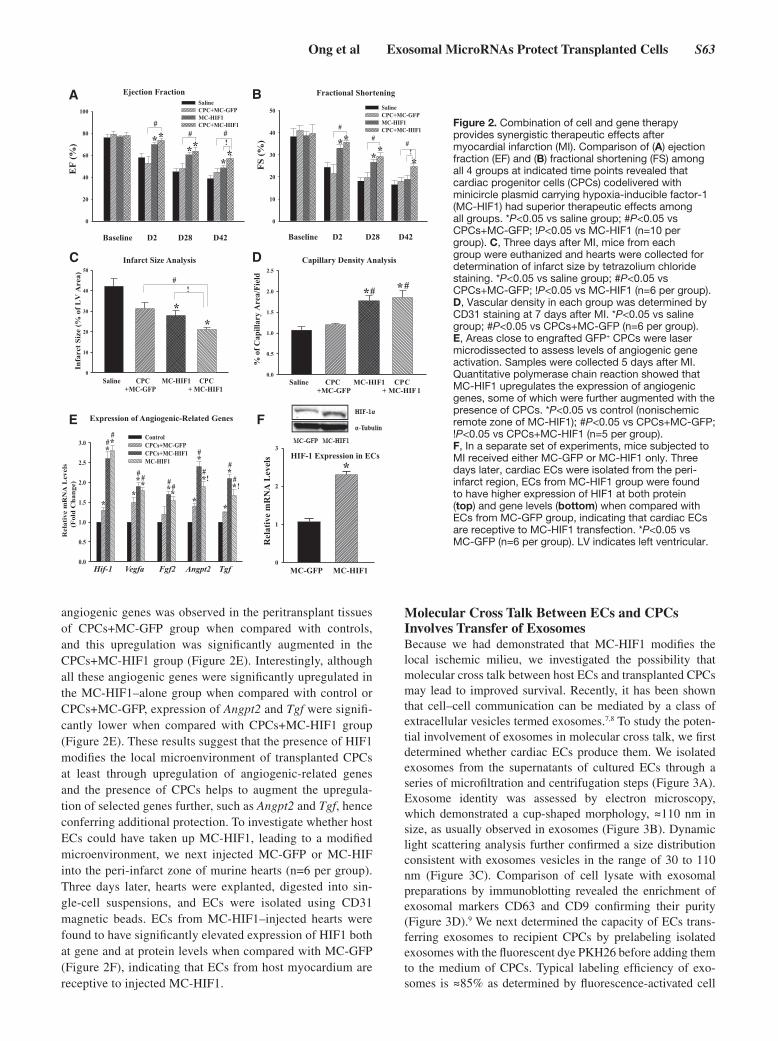
angiogenic genes was observed in the peritransplant tissues of CPCs+MC-GFP group when compared with controls, and this upregulation was significantly augmented in the CPCs+MC-HIF1 group (Figure 2E). Interestingly, although all these angiogenic genes were significantly upregulated in the MC-HIF1–alone group when compared with control or CPCs+MC-GFP, expression of Angpt2 and Tgf were signifi-cantly lower when compared with CPCs+MC-HIF1 group (Figure 2E). These results suggest that the presence of HIF1 modifies the local microenvironment of transplanted CPCs at least through upregulation of angiogenic-related genes and the presence of CPCs helps to augment the upregula-tion of selected genes further, such as Angpt2 and Tgf, hence conferring additional protection. To investigate whether host ECs could have taken up MC-HIF1, leading to a modified microenvironment, we next injected MC-GFP or MC-HIF into the peri-infarct zone of murine hearts (n=6 per group). Three days later, hearts were explanted, digested into sin-gle-cell suspensions, and ECs were isolated using CD31 magnetic beads. ECs from MC-HIF1–injected hearts were found to have significantly elevated expression of HIF1 both at gene and at protein levels when compared with MC-GFP (Figure 2F), indicating that ECs from host myocardium are receptive to injected MC-HIF1.
Molecular Cross Talk Between ECs and CPCs Involves Transfer of ExosomesBecause we had demonstrated that MC-HIF1 modifies the local ischemic milieu, we investigated the possibility that molecular cross talk between host ECs and transplanted CPCs may lead to improved survival. Recently, it has been shown that cell–cell communication can be mediated by a class of extracellular vesicles termed exosomes.7,8 To study the poten-tial involvement of exosomes in molecular cross talk, we first determined whether cardiac ECs produce them. We isolated exosomes from the supernatants of cultured ECs through a series of microfiltration and centrifugation steps (Figure 3A). Exosome identity was assessed by electron microscopy, which demonstrated a cup-shaped morphology, ≈110 nm in size, as usually observed in exosomes (Figure 3B). Dynamic light scattering analysis further confirmed a size distribution consistent with exosomes vesicles in the range of 30 to 110 nm (Figure 3C). Comparison of cell lysate with exosomal preparations by immunoblotting revealed the enrichment of exosomal markers CD63 and CD9 confirming their purity (Figure 3D).9 We next determined the capacity of ECs trans-ferring exosomes to recipient CPCs by prelabeling isolated exosomes with the fluorescent dye PKH26 before adding them to the medium of CPCs. Typical labeling efficiency of exo-somes is ≈85% as determined by fluorescence-activated cell
0
1
2
3
Rel
ativ
e m
RN
A L
evel
s
*
MC-GFP MC-HIF10.0
0.5
1.0
1.5
2.0
2.5
3.0ControlCPCs+MC-GFPCPCs+MC-HIF1MC-HIF1
R
elat
ive
mR
NA
Lev
els
(Fol
d C
hang
e)
*
**
**
**
*
*
**
**
*!
!
##
# # ##
#
##
#
Hif-1 Vegfa Fgf2 Angpt2 Tgf
Expression of Angiogenic-Related Genes
Ejection Fraction Fractional Shortening
Infarct Size Analysis Capillary Density Analysis
A B
C D
0
20
40
60
80
100SalineCPC+MC-GFPMC-HIF1CPC+MC-HIF1
EF
(%)
****
*
#
!
*
# #
0
10
20
30
40
50 SalineCPC+MC-GFPMC-HIF1CPC+MC-HIF1
FS (%
) ****
*
#
!
##
0.0
0.5
1.0
1.5
2.0
2.5
% o
f Cap
illar
y A
rea/
Fiel
d
* *
Saline CPC MC-HIF1 CPC +MC-GFP + MC-HIF1
# #
Saline CPC MC-HIF1 CPC +MC-GFP + MC-HIF1
0
10
20
30
40
50
Infa
rct S
ize
(% o
f LV
Are
a)
**
#!
Baseline D2 D28 D42 Baseline D2 D28 D42
E F
HIF-1 Expression in ECs
Figure 2. Combination of cell and gene therapy provides synergistic therapeutic effects after myocardial infarction (MI). Comparison of (A) ejection fraction (EF) and (B) fractional shortening (FS) among all 4 groups at indicated time points revealed that cardiac progenitor cells (CPCs) codelivered with minicircle plasmid carrying hypoxia-inducible factor-1 (MC-HIF1) had superior therapeutic effects among all groups. *P<0.05 vs saline group; #P<0.05 vs CPCs+MC-GFP; !P<0.05 vs MC-HIF1 (n=10 per group). C, Three days after MI, mice from each group were euthanized and hearts were collected for determination of infarct size by tetrazolium chloride staining. *P<0.05 vs saline group; #P<0.05 vs CPCs+MC-GFP; !P<0.05 vs MC-HIF1 (n=6 per group). D, Vascular density in each group was determined by CD31 staining at 7 days after MI. *P<0.05 vs saline group; #P<0.05 vs CPCs+MC-GFP (n=6 per group). E, Areas close to engrafted GFP+ CPCs were laser microdissected to assess levels of angiogenic gene activation. Samples were collected 5 days after MI. Quantitative polymerase chain reaction showed that MC-HIF1 upregulates the expression of angiogenic genes, some of which were further augmented with the presence of CPCs. *P<0.05 vs control (nonischemic remote zone of MC-HIF1); #P<0.05 vs CPCs+MC-GFP; !P<0.05 vs CPCs+MC-HIF1 (n=5 per group). F, In a separate set of experiments, mice subjected to MI received either MC-GFP or MC-HIF1 only. Three days later, cardiac ECs were isolated from the peri-infarct region, ECs from MC-HIF1 group were found to have higher expression of HIF1 at both protein (top) and gene levels (bottom) when compared with ECs from MC-GFP group, indicating that cardiac ECs are receptive to MC-HIF1 transfection. *P<0.05 vs MC-GFP (n=6 per group). LV indicates left ventricular.

S64 Circulation September 9, 2014
sorter (Figure IIA in the online-only Data Supplement). After 6 hours of incubation, confocal imaging revealed the ability of CPCs to uptake the fluorescently labeled exosomes in a time-dependent manner (Figure 3E; Figure IIB in the online-only Data Supplement). To confirm these findings further, we transfected ECs with a miRNA that is naturally present only in Caenorhabditis elegans (cel-miR-39). Forty-eight hours later, exosomes from these cel-miR-39–transfected ECs were iso-lated and added to CPCs. Analysis of cel-miR-39 expression levels in CPCs demonstrated the presence of cel-miR-39, and that cel-miR-39 is transferred from ECs to CPCs in a temporal manner (Figure 3F).
Overexpression of HIF1 Alters the MicroRNA Contents of EC-Derived ExosomesWe next investigated the effects of MC-HIF1 transfection on the EC-derived exosome composition. HIF1 overexpression did not lead to a change in the amount of exosomes released from ECs (Figure IIC in the online-only Data Supplement). We next studied the repertoires of microRNAs (miRs) con-tained in exosomes secreted by both groups of cells (ECGFP-Exo versus ECHIF-Exo) to determine potential miRs that might be regulated by overexpression of HIF1. Using a quantitative polymerase chain reaction array of 380 mouse miRs, profiling
of RNA isolated from exosomes showed that overexpression of HIF1 led to the upregulation of several exosomal miRs (Figure 4A). We chose to focus on 2 specific miRs, namely the endothelial-specific miR-126 and also miR-210, which has been previously reported to be regulated by HIF110 for sub-sequent experiments. We confirmed the involvement of HIF1 in upregulating these miRs by obtaining similar results using a pharmacological activator of HIF1, dimethyloxalylglycine (Figure IID in the online-only Data Supplement). In addi-tion, the requirement for exosome biogenesis in the increased expression of secreted miRs was demonstrated. Knockdown of sphingomyelinase (nSMase2), which has been shown to inhibit exosome generation,11 reduced the presence of both exosomal miR-126 and miR-210 from ECGFP cells or ECHIF cells (Figure 4B).
After incubation with control ECGFP-CM, CPCs expressed significantly higher levels of miR-126 but levels of miR-210 remain unchanged, consistent with the fact that miR-126 is predominantly expressed in ECs (Figure 4C) rather than CPCs. In keeping with the profiling results, CPCs incu-bated with ECHIF-CM had a significantly higher expression of both miR-126 and miR-210 when compared with CPCs supplemented with ECGFP-CM or untreated cells, indicat-ing that HIF1 leads to enriched expression of these miRs in
Cop
ies/
ng R
NA
0
1000
2000
3000
4000
5000
6000
4 h 8 h 12 h 24 h
**
*
*
Control cel-miR-39
Cell culture supernatant
Supernatant300 g, 10 min
2000 g, 10 min Supernatant
13,000 g, 15 minSupernatant
Filtration 0.2 µmSupernatant
Medium Concentration + Exosome PrecipitationThen 12,000 g 60 min
Exosomes
Supernatant (contaminating proteins)
CD63
#1 #2 #1 #2 Whole Cell Lysate Exosomal Prep
CD9
Expression of cel-miR-39 in CPC
Control
EC-Exo
DAPI GFP PKH26-Exo Merge
A
C
E
F
B
DFigure 3. Purified exosomes produced by endothelial cells (ECs) are actively internalized by cardiac progenitor cells (CPCs) in vitro. A, Purification scheme of exosomes from EC culture supernatant. B, Cup-shaped morphology of purified ECs’ exosomes assessed by transmission electron microscopy. C, Dynamic light scattering analysis of purified ECs’ exosomes demonstrating a physical size distribution of 10 to 110 nm. D, Immunoblotting revealed an enrichment of exosomal markers CD63 and CD9 in purified exosomal fraction compared to whole EC lysates. Two separate preparations were shown. E, CPCs were cultured in the presence of EC-derived PKH26-labeled exosomes or absence (control; same volume of PBS labeled similarly) for 6 hours. Exosomes were taken up by CPCs as shown by confocal microscopy. F, ECs were transfected with cel-miR-39 or left untransfected. CPCs were then treated with exosomes isolated from untransfected (control) or cel-miR-39 transfected ECs for the indicated times. *P<0.05 vs control (n=4 per group). DAPI indicates 4’,6-diamidino-2-phenylindole.

Ong et al Exosomal MicroRNAs Protect Transplanted Cells S65
target cells (Figure 4C). To investigate the requirement for exosomes in shuttling miRs directly, we depleted ECGFP-CM and ECHIF-CM of exosomes through ultracentrifugation. The depletion of exosomes considerably abrogated the expression of miR-126 and miR-210 in target CPCs (Figure 4C). The presence of a transcription inhibitor, actinomycin D, also did not affect levels of miR-126 and miR-210 in the treated CPCs, confirming the aforementioned effects as direct consequences of exosomal-mediated transfer (Figure 4C). Finally, degrada-tion analysis of exosomes using a combination of ribonucle-ase (RNase), proteinase K (for degrading proteins), and Triton X-100 (disrupts phospholipid membranes) further demon-strated that the majority of endothelial-derived miR-126 and miR-210 are transmitted into CPCs through vesicle-protected RNA (Figure 4D), instead of forming soluble ribonucleopro-tein complexes, as recently shown.12–14
Transferred MiR-126 Modulates Biological Properties of CPCsNext, to test the biological functionality of the transferred miR-126 and miR-210 in CPCs, cells were transfected with a 3′-untranslated region luciferase reporter vector harboring a binding site for either miR-126 or miR-210, respectively. We also included a second group with a control reporter vec-tor without a miR recognition site. As shown in Figure 5A, when CPCs were cultured with ECHIF-CM, luciferase activity was decreased significantly in cells transfected with specific miR recognition sequence vector, but not the control vector or when exosomes were depleted from the CM through ultracen-trifugation. Transfected CPCs in media alone displayed robust
luciferase production (data not shown). These results show the transferred miRs were functional in the recipient CPCs. We then examined the biological effect of these transferred miRs that could potentially confer the demonstrated in vivo toler-ance against ischemic stress in the recipient cells. We found that CPCs given ECGFP-Exo had increased phosphorylated lev-els of prosurvival kinases phospho-ERK and phospho-AKT and that these effects were further augmented when CPCs were given ECHIF-Exo, consistent with the established role of miR-126 in regulating these kinases (Figure 5B).15 Silencing of miR-126 blunted the phosphorylation of phospho-ERK and phospho-AKT, lending support to the importance of miR-126 in ECHIF-exosomes (Figure 5B). Furthermore, CPCs given ECHIF-Exo had elevated expression of several angiogenic-related genes, such as Vegfa and Fgf2, when compared with untreated or ECGFP-Exo cells (Figure 5C). These angiogenic factors have been previously shown to regulate phospho-ERK and phospho-AKT.16 However, the upregulation of Vegfa is in contrary to the previously described role of miR-126 as a direct repressor of this gene. Hence, we subjected CPCs to culture conditions in which they were supplemented with exosomes derived from ECHIF cotransfected with antagomirs targeting miR-126, and we found that these cells had signifi-cantly higher expression of both Vegfa and Fgf2 but not Il-8 and Kdr when compared with ECHIF transfected with scram-bled antagomirs (we did not observe a difference between ECHIF versus ECHIF-scrambled). These data suggest that although there were still partial repressive effects of miR-126, it was insufficient to lead to downregulation of Vegfa presum-ably because of the presence of other factors contained within
+ + + + + ++ - + + - +- - + - - +
0
10
20
30
40
50
Ct V
alue
mm
u-m
iR-3
0b
mm
u-m
iR-1
26-5
p
mm
u-m
iR-2
10
mm
u-m
iR-5
03
mm
u-m
iR-3
00
mm
u-m
iR-4
84
*
*
*
*
**
Exosomal microRNA profiling following HIF-1 Overexpression
0.0
0.5
1.0
4.0
8.0
16.0
20.0
*
*
* *
*
*
Exo miR-126
A
C
B
D
0.0
0.2
0.4
0.6
0.8
1.0
1.2
* **
*
Expression of miRs in CPCs Degradation Analysis of Exosomes
RNase Proteinase K Triton X-100
0
2
4
6
8
10
*
miR-126
miR-210
*
*
*
* *
*
MediaExo Actino-mycin D
- + + + +- - + - +- - - + +
Rel
ativ
e m
iRLe
vels
(Fol
d C
hang
e)
Nor
mal
ized
to E
CG
FP+s
iScr
ambl
ed
Rel
ativ
e m
iR L
evel
s vs
Unt
reat
ed (F
old
Cha
nge)
Rel
ativ
e m
iR L
evel
s(F
old
Cha
nge)
Exo miR-210
ECGFP+si-nSMase2ECHIF+siScrambledECHIF+si-nSMase2
ECGFP-ExoECHIF-Exo
ECGFP-CMECHIF-CM
miR-126miR-210
Exosomal microRNA profiling following HIF-1 Overexpression
Figure 4. Hypoxia-inducible factor-1 (HIF1) modulates the microRNA (miRs) repertoire in endothelial cell (EC)–derived exosomes (Exo), which are transferable to cardiac progenitor cells (CPCs). A, ECs were transfected with either minicircle-green fluorescent protein (MC-GFP; control) or MC-HIF1. After transfection, Exo were purified from both groups and subjected to miRs profiling. Expression of selected exosomal miRs was increased after MC-HIF1 transfection. *P<0.05 vs ECGFP-Exo (n=4). B, On the basis of profiling results, we chose miR-126 and miR-210 for detailed analysis. ECGFP and ECHIF were transfected with either scrambled siRNA or siRNA targeting nSMase2. Exo were then purified from each group, and the expression of miR-126 and miR-210 was determined by quantitative polymerase chain reaction (qPCR) using Taqman probes and expressed as relative fold-change normalized to ECGFP transfected with scrambled siRNA. *P<0.05 vs ECGFP+siScrambled (n=6 per group). C, Recipient CPCs were treated with vehicle or actinomycin D (5 μg/mL). CPCs were then grown in normal growth medium or supplemented with either ECGFP-CM or ECHIF-CM in the absence or presence of Exo as indicated. After 24 hours, the expression of miR-126 and miR-210 in CPCs was determined by qPCR and expressed as fold-change normalized against CPCs grown in normal growth medium. *P<0.05 (n=6). D, Exo were isolated from ECHIF and incubated with the indicated reagents for 45 minutes at 37°C before isolation of RNA and measurement of expression levels of miR-126 and miR-210 by qPCR. *P<0.05 vs untreated (n=4).

S66 Circulation September 9, 2014
ECHIF-Exo. We further confirmed the functionality of miR-126 in ECHIF-Exo through additional experiments by measur-ing the expression of Spred1, a validated miR-126 target gene in untreated CPCs, or CPCs given ECGFP-Exo, ECHIF-Exo, or ECHIF-miR126-KO-Exo. We found that exosomes derived from ECGFP led to partial suppression of Spred1 when compared with untreated CPCs; these effects were further augmented when ECHIF-Exo were added and reversed when CPCs were treated with antagomirs targeting miR-126 (Figure IIE in the online-only Data Supplement).
Transferred MiR-210 Modulates Biological Properties of CPCsHaving shown that miR-126 affects CPCs, we postulated that transferred miR-210 would also have a biological rel-evance in CPCs. Iron-sulfur cluster scaffold homolog (Iscu) is a validated target of miR-210, which on repression, reduces mitochondrial metabolism.17 A significant reduction of Iscu was observed in CPCs treated with ECHIF-Exo indicative of repression through miR-210 (Figure 5D). To assess the met-abolic profile of CPCs as a consequence of increased miR-210/repressed Iscu, we measured oxygen consumption rate in these cells by SeaHorse XF bioenergetic system. CPCs treated with ECHIF-Exo displayed a significant reduction in basal oxygen consumption (normalized against cell numbers)
when compared with untreated CPCs or CPCs treated with ECGFP-Exo, showing that increased miR-210 leads to reduced mitochondrial metabolism in cells (Figure 5E). In contrast, nonmitochondrial respiration was similar among all groups, suggesting that ECHIF-Exo act primarily in modulating mito-chondrial respiration (Figure 5F). In addition, we noted an increase in intracellular lactate with CPCs treated with ECHIF-Exo, indicating increased glycolysis as usually seen along reduced mitochondrial metabolism (Figure IIF in the online-only Data Supplement).
Exosomes Reduce Cellular Damage of CPCs Under Ischemic ConditionsGiven the known evidence for increased levels of prosurvival kinases, enhanced angiogenic response and reduced meta-bolic demand as adaptive beneficial responses,18,19 we hypoth-esized that these effects could collectively provide CPCs with increased tolerance to ischemic stress. Indeed, CPCs grown in ECHIF-CM had significantly reduced cellular dam-age as assessed by lactate dehydrogenase release when sub-jected to in vitro hypoxia when compared with untreated or cells grown in ECGFP-CM (Figure 6A). The protective effects of ECHIF-CM were dependent on the presence of exosomes as ultracentrifugation abrogated the aforementioned effects. Importantly, CM derived from ECs overexpressing HIF1 that
ECGFP-Exo ECHIF-Exo0.0
0.3
0.6
0.9
1.2
*
0
1
2
3
4
5
6
7
UntreatedECGFP-ExoECHIF-ExoECHIF/miR126-KO-Exo
R
elativ
e mRN
A Le
vels
(Fol
d Ch
ange
)
Vegfa Fgf2 Il-8 Kdr
*
**
*
*
*
** *
*
!!
##
## # #
##
Iscu Expression in CPCs
Lactate Production in CPCs
Luciferase Reporter Assay for miRs
0.0
0.3
0.6
0.9
1.2
**
0.0
0.3
0.6
0.9
1.2
Luc vector without miR Recognition SequenceLuc vector with miR Recognition Sequence
Luci
fera
se A
ctiv
ity
**
miR-126 miR-210
Normalized Oxygen Consumption Rate in CPCs
0.0
0.3
0.6
0.9
1.2
1.5
Non Mitochondrial Respiration in CPCs
A
C
E
B
D
F
Angiogenic Responses in CPCs
Rel
ativ
e m
RN
A L
evel
s(F
old
Cha
nge)
0.0
0.5
1.0
1.5
2.0
2.5
3.0
3.5
4.0
ControlECGFP-ExoECHIF-ExoECHIF/miR126-KO-Exo
**
pAKT/AKT
p-ERK/ERK
**
* *##
Prot
ein
Lev
els
(Fol
d C
hang
e)
Nor
mal
ized
Oxy
gen
Con
sum
ptio
n R
ate
Nor
mal
ized
Oxy
gen
Con
sum
ptio
n R
ate
ECHIF-CM + + + +
Exosomes - + - +
Ctrl ECGFP ECHIF ECHIF/miR126-KO
-Exo -Exo -Exo
p-AKT
AKT
p-ERK
ERK
ECGFP-Exo - + - -
ECHIF-Exo - - + -
DMOG - - - +
ECGFP-Exo - + - -
ECHIF-Exo - - + -
DMOG - - - +
Figure 5. Transferred exosomal miRs regulate biological properties of recipient cardiac progenitor cells (CPCs). A, CPCs were transfected with a luciferase reporter vector containing either miR-126 or miR-210 recognition sequence or a control luciferase vector lacking the sequence. CPCs were then given endothelial cell (EC)HIF-conditioned medium (CM) in the presence or absence of exosomes (Exo) as indicated. CPCs were then cultured for another 24 hours, and luciferase activity was determined and expressed as fold-decrease of cells transfected with the luciferase reporter containing specific miR recognition sequence over the vector lacking the recognition sequence for each culture condition. *P<0.05 (n=4) vs cells transfected with vector lacking miR recognition sequence. B, Representative immunoblots and densitometry quantification of indicated proteins in CPCs grown in either normal conditions, or supplemented with ECGFP-Exo, ECHIF-Exo, or ECHIF/miR126-KO-Exo. *P<0.05 vs untreated CPCs; #P<0.05 vs ECHIF-Exo (n=4). C, Expression of angiogenic genes in CPCs grown in each culture condition as indicated was determined by quantitative polymerase chain reaction. *P<0.05 vs untreated; #P<0.05 vs ECGFP-Exo; !P<0.05 vs ECHIF-Exo (n=8). D, Expression of Iscu, a validated miR-210 target gene, was determined in CPCs supplemented with either ECGFP-Exo or ECHIF-Exo. *P<0.05 (n=4) vs CPCs supplemented with ECGFP-Exo. CPCs were cultured in conditions as specified for 24 hours. E, Oxygen consumption and (F) nonmitochondrial respiration were then determined and expressed as fold-change when compared with untreated control cells. Dimethyloxalylglycine (DMOG), a known hypoxia-inducible factor-1 (HIF1) activator was used as positive control for reduced oxygen consumption. *P<0.05 vs untreated controls (n=6). KO indicates knockout.

Ong et al Exosomal MicroRNAs Protect Transplanted Cells S67
were additionally transfected with antagomirs against miR-126 and miR-210 failed to provide similar therapeutic effects when given to CPCs, indicating that these miRs are crucial for ECHIF-CM to afford CPCs with resistance against isch-emic stress (Figure 6B). Finally, to determine whether ECHIF-Exo can directly protect CPCs after transplantation into the ischemic heart, we delivered CPCs into mice after MI con-comitant with intravenous delivery of saline, PKH26-labeled ECGFP-Exo, or PKH26-labeled ECHIF-Exo, respectively. Flow cytometry analysis of isolated CPCs from explanted hearts demonstrated the presence of PKH26 exosomes, confirming the uptake of these exosomes by CPCs in vivo (Figure 6C). Longitudinal bioluminescence imaging of mice showed that CPCs that received ECHIF-Exo had better survival in the group when compared with saline or ECGFP-Exo groups at 1 week (Figure 6D).
DiscussionThis study revealed a multifaceted mechanism by which deliv-ery of HIF1 via a MC plasmid platform enhances survival of cotransplanted Sca1+ CPCs. First, using a murine model of MI followed by codelivery of CPCs with MC-GFP or MC-HIF, we observed that a combinatorial delivery with MC-HIF resulted in prolonged survival of transplanted CPCs, prevented cardiac remodeling, reduced infarct size, and enhanced vascularity. Second, HIF1 modulates the local milieu of the ischemic heart at least partly by effects on host cardiac ECs. Third, we found
that these cardiac ECs generate and release exosomes in vitro, which are actively internalized by CPCs when present in the growth medium. Fourth, HIF1 modifies the biological contents of these exosomes, resulting in a phenotypic change in recipi-ent cells. Fifth, in vitro hypoxia assays revealed that exosomes derived from ECs overexpressing HIF1 contributed to CPCs’ increased tolerance under hypoxic stress. These findings were validated in vivo because intravenous delivery of ECHIF-Exo significantly increased the survival of transplanted CPCs in the ischemic heart as determined by bioluminescence imaging.
HIF1 is a master transcriptional activator that mediates various physiological responses to hypoxia.3 Here, we dem-onstrated that codelivery of CPCs together with MC-HIF1 led to better survival of transplanted cells and was associated with preserved cardiac function. The synergistic effects of this combinatorial approach were evident early on, as documented by a smaller infarct size and increased vascularity when com-pared with other treatment groups. It is well known that stem/progenitor cells in adult organs reside in specialized niches that provide an ideal microenvironment for their mainte-nance.20,21 In this context, these cells are often found in close proximity to blood vessels, suggesting a role for these vessels in the regulation of stem cell self-renewal and differentiation. After an ischemic event, ECs are highly susceptible to under-going necrosis, potentially compromising the maintenance and expansion of resident stem cells. Using laser microdis-section analysis, we observed that MC-HIF1 changes the gene
ECHIF-Exo ECGFP-Exo Saline 0
10
20
30
40
50
60
70
% S
urvi
val o
f Cel
ls
* *
LD
H A
ctiv
ity0.0
0.5
1.0
1.5
2.0
* **
*
LD
H A
ctiv
ity
0.0
0.5
1.0
1.5
2.0
* ** *
PKH26-Exosomes
% o
f Max
Uptake of Exosomes In Vivo Survival of CPCs in the Presence of Intravenously-Delivered Exosomes
Lactate Dehydrogenase Assay for Cellular Damage in CPCs
Lactate Dehydrogenase Assay for Cellular Damage in CPCs
A
C
B
D
ECGFP-CM - + + - -ECHIF-CM - - - + +Exosomes - + - + -
ECGFP-CM - + + - -ECHIF-CM - - - + +Scrambled - + - + -Anti-miRs 126 & 210 - - + - +
Figure 6. Exosomes (Exo) directly provide cardiac progenitor cells (CPCs) with increased tolerance against ischemic stress both in vitro and in vivo. A, CPCs were grown in either normal conditions or supplemented with endothelial cell (EC) GFP-conditioned medium (CM) or ECHIF-CM with or without the presence of Exo as indicated, before being subjected to in vitro hypoxic stress. Lactate dehydrogenase (LDH) was then measured as an indicator of cellular damage and expressed as fold-change when compared with untreated cells kept in normoxic conditions. *P<0.05 (n=6) vs normoxic cells. B, ECHIF were cotransfected with either scrambled antagomirs or antagomirs targeting miR-126 and miR-210. CPCs were then grown in conditioned medium from each group as indicated before being subjected to LDH assay to assess the importance of miR-126 and miR-210 in ECHIF-CM–mediated protection seen in A. *P<0.05 vs normoxic control (n=6). C, After MI, CPCs were intramyocardially injected into mice hearts, and a bolus of PKH26-labeled ECs-Exo was delivered intravenously. After 12 hours, the animals were euthanized and CPCs were reisolated from the hearts. Samples were measured by flow cytometry for green fluorescent protein (GFP)+ and PKH26+ events. The histogram shows that PKH26-Exo (blue lines) were detectable in ≈10% of GFP+ CPCs (red lines) demonstrating that CPCs are capable of uptaking Exo in vivo. D, To determine whether ECHIF-Exo could confer increased tolerance to CPCs in vivo, cells were delivered intramyocardially after MI into mice concomitantly with intravenous injection of saline, ECGFP-Exo, or ECHIF-Exo. BLI was then performed at day 1 and day 7 after MI, and survival of CPCs was expressed as a percentage of signals intensity at day 7 when compared with initial signals intensity of day 1. *P<0.05 vs ECHIF-Exo (n=6 per group).
S68 Circulation September 9, 2014
expression landscape of the local milieu proximal to trans-planted cells, including resident cardiac ECs.
Given that HIF1 affects local cardiac ECs and improves sur-vival of transplanted CPCs, it is highly plausible that potential beneficial cross talk could occur between these 2 cell types. In recent years, exosomes have emerged as potential candidates for mediating cell–cell communication in various physiologi-cal or pathophysiological conditions through transfer of pro-teins, mRNAs, and miRs.7,8 We confirmed that cardiac ECs promoted release and transfer of exosomes to CPCs in vitro and demonstrated differences in miR profiling of ECGFP-Exo (control) versus ECHIF-Exo, most prominently miR-126 and miR-210. Our degradation analysis data also revealed that a significant amount of miR-126 and miR-210 is exosome-enclosed (unaffected by RNase treatment but susceptible to blockade of nSMase2), distinct from some previous studies indicating that miR-126 is transferred in a vesicle-free form or through apoptotic bodies.13,14 These data might reflect that additional mechanisms regulating the packaging of biological contents into exosomes are cell-type dependent. Recipient cell types might also differ in their ability to respond to exosomes because previous studies have reported receptor specificity as a crucial factor for internalization of exosomes.22
Endothelial miR-126 has been shown to regulate multiple pathways, including the regulation of prosurvival kinases phospho-ERK and phospho-AKT. Our findings demonstrated that CPCs internalizing ECGFP-Exo had a transient activation of these kinases that were further increased by ECHIF-Exo. The transient phosphorylation of these kinases is known to be pro-tective in the settings of ischemia by limiting both the apoptotic and the necrotic components of cell death. Likewise, miR-210, a known HIF1–regulated miR, was also shown to be upregu-lated preferentially in exosomes by HIF1, switching recipi-ent CPCs to a preferentially glycolytic state (reduced oxygen usage). This is consistent with previous studies documenting that one of the many targets of miR-210 is Iscu,17 and repression of this gene leads to lower mitochondrial metabolism; both phe-nomena were observed in our study. This metabolic adaptation to oxygen conservation reflects a lower use of the mitochondrial electron transport chain, which reduced the generation of reac-tive oxygen species. Indirectly, this phenomenon could poten-tially function as a preemptive measure against ischemic stress because the production of reactive oxygen species is known to be greatly increased during ischemia and is highly detrimental to cellular health. In keeping with our hypothesis, we observed that CPCs in ECHIF-CM had less cellular damage when exposed to in vitro hypoxic stress. This increased tolerance is attribut-able to exosomes because we found that depletion of exosomes from the CM abrogated the protective effects. In addition, the importance of both miR-126 and miR-210 mediating the pro-tective effects of the ECHIF-CM was corroborated because such effects were abolished when the donor cardiac EC cells were transfected with antagomirs targeting both miRs.
In the present study, a full comparison of the abundance of exosomes in vitro and in vivo was not feasible because the concentrations and transfer efficiencies are probably different. Nevertheless, we have generated evidence to support a physiolog-ical role for exosomes in mediating increased tolerance against ischemic stress, which helps to improve survival of transfected
cells. Although we focused on the transfer of genetic material after HIF1 overexpression from cardiac ECs to CPCs, we think that reciprocal cross talk could also occur as well with other resi-dent cell types, including fibroblasts and host cardiomyocytes, and even the possibility of CPCs themselves taking up MC-HIF1 that warrants deeper investigation. Likewise, although our data indicated a physiological role for exosomal miR-126 and miR-210 in recipient cells that was reversed on inhibition of both, this does not preclude the involvement of other miRs and proteins; further investigation is needed to show whether these proteins can improve survival of transplanted cells because various stud-ies have demonstrated the complexity of biological molecules enclosed within exosomes.23 Finally, our results demonstrating the upregulation of Vegfa despite being a direct target of miR-126 reveals the intricate mechanisms mediated by exosomes.
Collectively, we show for the first time an intricate exosome-mediated cross talk interface via HIF1 between the vascular endothelium and the transplanted stem cells. The improved tolerance against ischemic stress is afforded through activa-tion of prosurvival kinases, increased angiogenic responses, and reduced metabolic demand, and involves, at least par-tially, miR-126 and miR-210 (Figure III in the online-only Data Supplement). Importantly, these results suggest that the combination of gene- and cell-based therapies should be explored in future clinical trials.
Sources of FundingWe are grateful for the funding support by National Institutes of Health U01 HL099776, R01 EB009689, R01 HL093172, R01 HL095571, American Heart Association Established Investigator Award, and Fondation Leducq (Dr Wu).
DisclosuresNone.
References 1. Hausenloy DJ, Erik Bøtker H, Condorelli G, Ferdinandy P, Garcia-Dorado
D, Heusch G, Lecour S, van Laake LW, Madonna R, Ruiz-Meana M, Schulz R, Sluijter JP, Yellon DM, Ovize M. Translating cardioprotection for patient benefit: position paper from the Working Group of Cellular Biology of the Heart of the European Society of Cardiology. Cardiovasc Res. 2013;98:7–27.
2. Liu J, Narsinh KH, Lan F, Wang L, Nguyen PK, Hu S, Lee A, Han L, Gong Y, Huang M, Nag D, Rosenberg J, Chouldechova A, Robbins RC, Wu JC. Early stem cell engraftment predicts late cardiac functional recovery: preclinical insights from molecular imaging. Circ Cardiovasc Imaging. 2012;5:481–490.
3. Ong SG, Hausenloy DJ. Hypoxia-inducible factor as a therapeutic target for cardioprotection. Pharmacol Ther. 2012;136:69–81.
4. Huang M, Chen Z, Hu S, Jia F, Li Z, Hoyt G, Robbins RC, Kay MA, Wu JC. Novel minicircle vector for gene therapy in murine myocardial infarc-tion. Circulation. 2009;120(suppl 11):S230–S237.
5. Hu S, Huang M, Nguyen PK, Gong Y, Li Z, Jia F, Lan F, Liu J, Nag D, Robbins RC, Wu JC. Novel microRNA prosurvival cocktail for improv-ing engraftment and function of cardiac progenitor cell transplantation. Circulation. 2011;124(suppl 11):S27–S34.
6. Huang M, Nguyen P, Jia F, Hu S, Gong Y, de Almeida PE, Wang L, Nag D, Kay MA, Giaccia AJ, Robbins RC, Wu JC. Double knockdown of prolyl hydroxylase and factor-inhibiting hypoxia-inducible factor with nonviral minicircle gene therapy enhances stem cell mobilization and angiogenesis after myocardial infarction. Circulation. 2011;124(suppl 11):S46–S54.
7. Sahoo S, Klychko E, Thorne T, Misener S, Schultz KM, Millay M, Ito A, Liu T, Kamide C, Agrawal H, Perlman H, Qin G, Kishore R, Losordo DW. Exosomes from human CD34(+) stem cells mediate their proangiogenic paracrine activity. Circ Res. 2011;109:724–728.
Ong et al Exosomal MicroRNAs Protect Transplanted Cells S69
8. Hergenreider E, Heydt S, Tréguer K, Boettger T, Horrevoets AJ, Zeiher AM, Scheffer MP, Frangakis AS, Yin X, Mayr M, Braun T, Urbich C, Boon RA, Dimmeler S. Atheroprotective communication between endothelial cells and smooth muscle cells through miRNAs. Nat Cell Biol. 2012;14:249–256.
9. Mathivanan S, Ji H, Simpson RJ. Exosomes: extracellular organelles impor-tant in intercellular communication. J Proteomics. 2010;73:1907–1920.
10. Camps C, Buffa FM, Colella S, Moore J, Sotiriou C, Sheldon H, Harris AL, Gleadle JM, Ragoussis J. hsa-miR-210 Is induced by hypoxia and is an independent prognostic factor in breast cancer. Clin Cancer Res. 2008;14:1340–1348.
11. Trajkovic K, Hsu C, Chiantia S, Rajendran L, Wenzel D, Wieland F, Schwille P, Brügger B, Simons M. Ceramide triggers budding of exosome vesicles into multivesicular endosomes. Science. 2008;319:1244–1247.
12. Arroyo JD, Chevillet JR, Kroh EM, Ruf IK, Pritchard CC, Gibson DF, Mitchell PS, Bennett CF, Pogosova-Agadjanyan EL, Stirewalt DL, Tait JF, Tewari M. Argonaute2 complexes carry a population of circulating microRNAs independent of vesicles in human plasma. Proc Natl Acad Sci U S A. 2011;108:5003–5008.
13. Jansen F, Yang X, Hoyer FF, Paul K, Heiermann N, Becher MU, Abu Hussein N, Kebschull M, Bedorf J, Franklin BS, Latz E, Nickenig G, Werner N. Endothelial microparticle uptake in target cells is annexin I/phosphatidylserine receptor dependent and prevents apoptosis. Arterioscler Thromb Vasc Biol. 2012;32:1925–1935.
14. Zhou J, Li YS, Nguyen P, Wang KC, Weiss A, Kuo YC, Chiu JJ, Shyy JY, Chien S. Regulation of vascular smooth muscle cell turnover by endothelial cell-secreted microRNA-126: role of shear stress. Circ Res. 2013;113:40–51.
15. Chen JJ, Zhou SH. Mesenchymal stem cells overexpressing MiR-126 enhance ischemic angiogenesis via the AKT/ERK-related pathway. Cardiol J. 2011;18:675–681.
16. Wang S, Aurora AB, Johnson BA, Qi X, McAnally J, Hill JA, Richardson JA, Bassel-Duby R, Olson EN. The endothelial-specific microRNA
miR-126 governs vascular integrity and angiogenesis. Dev Cell. 2008;15:261–271.
17. Chan SY, Zhang YY, Hemann C, Mahoney CE, Zweier JL, Loscalzo J. MicroRNA-210 controls mitochondrial metabolism during hypoxia by repressing the iron-sulfur cluster assembly proteins ISCU1/2. Cell Metab. 2009;10:273–284.
18. Aragonés J, Schneider M, Van Geyte K, Fraisl P, Dresselaers T, Mazzone M, Dirkx R, Zacchigna S, Lemieux H, Jeoung NH, Lambrechts D, Bishop T, Lafuste P, Diez-Juan A, Harten SK, Van Noten P, De Bock K, Willam C, Tjwa M, Grosfeld A, Navet R, Moons L, Vandendriessche T, Deroose C, Wijeyekoon B, Nuyts J, Jordan B, Silasi-Mansat R, Lupu F, Dewerchin M, Pugh C, Salmon P, Mortelmans L, Gallez B, Gorus F, Buyse J, Sluse F, Harris RA, Gnaiger E, Hespel P, Van Hecke P, Schuit F, Van Veldhoven P, Ratcliffe P, Baes M, Maxwell P, Carmeliet P. Deficiency or inhibition of oxygen sensor Phd1 induces hypoxia tolerance by reprogramming basal metabolism. Nat Genet. 2008;40:170–180.
19. Hausenloy DJ, Yellon DM. Reperfusion injury salvage kinase signalling: taking a RISK for cardioprotection. Heart Fail Rev. 2007;12:217–234.
20. Yin T, Li L. The stem cell niches in bone. J Clin Invest. 2006;116:1195–1201. 21. Yoshida S, Sukeno M, Nabeshima Y. A vasculature-associated niche
for undifferentiated spermatogonia in the mouse testis. Science. 2007;317:1722–1726.
22. Christianson HC, Svensson KJ, van Kuppevelt TH, Li JP, Belting M. Cancer cell exosomes depend on cell-surface heparan sulfate proteogly-cans for their internalization and functional activity. Proc Natl Acad Sci U S A. 2013;110:17380–17385.
23. Kucharzewska P, Christianson HC, Welch JE, Svensson KJ, Fredlund E, Ringnér M, Mörgelin M, Bourseau-Guilmain E, Bengzon J, Belting M. Exosomes reflect the hypoxic status of glioma cells and mediate hypoxia-dependent activation of vascular cells during tumor development. Proc Natl Acad Sci U S A. 2013;110:7312–7317.