Comparison of ventral organ development across Pycnogonida ...
Upload
institut-clement-aderCategory
view
1download
0
Cross-Talk between Dopaminergic and NoradrenergicSystems in the Rat Ventral Tegmental Area,Locus Ceruleus, and Dorsal Hippocampus
Bruno P. Guiard, Mostafa El Mansari, and Pierre BlierUniversity of Ottawa Institute of Mental Health Research, Ottawa, Ontario, Canada (B.P.G, M.E.M., P.B.);and Department of Cellular and Molecular Medicine, University of Ottawa, Ottawa, Ontario, Canada (P.B.)
Received April 18, 2008; accepted August 14, 2008
ABSTRACTA decreased central dopaminergic and/or noradrenergic trans-mission is believed to be involved in the pathophysiology ofdepression. It is known that dopamine (DA) neurons in theventral tegmental area (VTA) and norepinephrine (NE) neuronsin the locus ceruleus (LC) are autoregulated by somatodendriticD2-like and �2-adrenoceptors, respectively. Complementingthese autoreceptor-mediated inhibitory feedbacks, anatomicaland functional studies have established a role for noradrenergicinputs in regulating dopaminergic activity, and reciprocally. Inthe present study, a microiontophoretic approach was used tocharacterize the postsynaptic catecholamine heteroreceptorsinvolved in such regulations. In the VTA, the application of DAand NE significantly reduced the firing activity of DA neurons. Inaddition to a role for D2-like receptors in the inhibitory effects ofboth catecholamines, it was demonstrated that the �2-adreno-ceptor antagonist idazoxan dampened the DA- and NE-in-duced attenuations of DA neuronal activity, indicating that both
of these receptors are involved in the responsiveness of VTADA neurons to catecholamines. In the LC, the effectiveness ofiontophoretically applied NE and DA to suppress NE neuronalfiring was blocked by idazoxan but not by the D2-like receptorantagonist raclopride, which suggested that only �2-adreno-ceptors were involved. In the dorsal hippocampus, a forebrainregion having a sparse dopaminergic innervation but receivinga dense noradrenergic input, the suppressant effects of DA andNE on pyramidal neurons were attenuated by idazoxan but notby raclopride. The suppressant effect of DA was prolonged byadministration of the selective NE reuptake inhibitor desipra-mine and, to lesser extent, of the selective DA reuptake inhibi-tor 1-(2-[bis(4-fluorophenyl)methoxy]ethyl)-4-(3-phenylpropyl)-piperazine (GBR12909), suggesting that both the NE and DAtransporters were involved in DA uptake in the hippocampus.These findings might help in designing new antidepressantstrategies aimed at enhancing DA and NE neurotransmission.
The catecholamines neurotransmitters dopamine (DA) andnorepinephrine (NE) are believed to be involved in psychiat-ric disorders, and a better knowledge of the reciprocal inter-actions between these two systems should improve our un-derstanding of the pathophysiology and treatment of mooddisorders. Anatomical evidence indicates that noradrenergicneurons of the locus ceruleus (LC) send projections to theventral tegmental area (VTA) in the vicinity of DA neuroncell bodies (Simon et al., 1979). Several subtypes of �-adren-
ergic receptors have been identified in the VTA (Lee et al.,1998), raising the possibility that NE inputs play a role inmodulating DA neuronal activity. Consistent with this as-sumption, it was recently demonstrated that a selective le-sion of LC NE neurons increases the mean firing activity ofDA neurons by 70% and their burst activity by almost 50%,thus revealing a net inhibitory effect of NE in the VTA(Guiard et al., 2008). In contrast, it had been reported thatthe systemic administration of the selective NE reuptakeinhibitor reboxetine enhanced the burst firing activity of DAneurons in the VTA, but without any apparent effect on theirmean firing rate (Linner et al., 2001). In earlier attempts atcharacterizing the mechanism of action of NE in the VTA, ithas also been shown that the systemic administration of the�1-adrenoceptor antagonist prazosin dose-dependently de-creased the burst firing of DA neurons (Grenhoff and Svens-son, 1993), apparently supporting an excitatory action of NE
This study was supported by the Canadian Institutes for Health Researchgrant (77838) and salary support from the University of Ottawa Institute ofMental Health Research (to B.P.G. and M.E.), as well as a Canada ResearchChair in Psychopharmacology from the Canadian Government, and an En-dowed Chair from the University of Ottawa Institute of Mental Health Re-search (to P.B.).
Article, publication date, and citation information can be found athttp://molpharm.aspetjournals.org.
doi:10.1124/mol.108.048033.
ABBREVIATIONS: DA, dopamine; CA, field of the hippocampus; DAT, dopamine transporter; GBR12909, 1-(2-[bis(4-fluorophenyl)methoxy]ethyl)-4-(3-phenylpropyl)piperazine; NE, norepinephrine; NET, norepinephrine transporter; LC, locus ceruleus; RT, recovery time; VTA, ventral tegmentalarea; (�)-3-PP, 3-(3-hydroxyphenyl)-N-n-propylpiperidine.
0026-895X/08/7405-1463–1475$20.00MOLECULAR PHARMACOLOGY Vol. 74, No. 5Copyright © 2008 The American Society for Pharmacology and Experimental Therapeutics 48033/3398207Mol Pharmacol 74:1463–1475, 2008 Printed in U.S.A.
1463
at ASPE
T Journals on Septem
ber 17, 2016m
olpharm.aspetjournals.org
Dow
nloaded from

in the VTA. However, the selective �2-adrenoceptor antago-nist idazoxan increased both the firing rate and burstingactivity of VTA DA neurons (Grenhoff and Svensson, 1993),thereby suggesting a complex regulation of DA neuronalactivity by NE inputs. As for the local application of NE inthe VTA, it usually reduced the spontaneous firing of DAneurons (Aghajanian and Bunney, 1977a,b; White and Wang,1984a,b), suggesting that a postsynaptic inhibitory elementsensitive to NE does exist in the VTA. However, whether NEexerts its effects in the VTA via �-adrenergic and/or dopami-nergic receptors remained an unresolved issue.
On the other hand, DA can influence the neuronal activityof LC NE neurons. Anatomical data support this possibilitybecause descending pathways from the VTA innervate theLC (Ornstein et al., 1987). Moreover, in rodents, althoughonly a small percentage of its VTA-derived afferents seem tobe dopaminergic (Swanson, 1982), DA receptors mainly of theD2-like subtype have been identified in the LC (Yokoyama etal., 1994). Lesion experiments have shed some light on thenature of the effects of DA in the LC. The selective lesion ofVTA DA neurons increased by 33% the mean firing rate of LCNE neurons discharging in a single spike mode and by almost60% that of LC neurons displaying both single spike andbusting activities (Guiard et al., 2008), revealing an inhibi-tory influence of DA inputs. In agreement with these find-ings, pharmacological studies have indicated that the sys-temic administration of the nonselective DA receptor agonist(�)-3-PP dose-dependently reduced the firing rate of LC NEneurons, whereas this effect was partially antagonized by the�2-adrenoceptor antagonist yohimbine but not the D2-likereceptor antagonist haloperidol (Elam et al., 1986). Theseresults stand in contrast with data showing that the systemicadministration of haloperidol enhances the firing rate of LCNE neurons, indicating the existence of a tonically activedopamine input that modulates the firing pattern of LC NEneurons (Piercey et al., 1994; Nilsson et al., 2005). The ion-tophoretic application of DA in the LC has been shown toinhibit the electrical activity of NE neurons (Cedarbaum andAghajanian, 1977; Elam et al., 1986). However, the lack orweak effect of the systemic administration of D2/D3 receptoragonists, such as pramipexole or apomorphine, on the firingrate of LC NE neurons (Cedarbaum and Aghajanian, 1977;Chernoloz et al., 2008) has not allowed the identification ofthe postsynaptic receptor(s) that mediate DA effects in the LC.
In rats, the dorsal hippocampus receives a dense noradren-ergic and sparse dopaminergic innervation arising from theLC (Jones and Moore, 1977) and the VTA (Swanson andHartman, 1975; Scatton et al., 1980), respectively. In addi-tion, because a significant decrease in hippocampal DA levelshad been reported when most of the noradrenergic neuronswere lesioned (Bischoff et al., 1979), it is possible that in thisbrain region, DA not only subserves a neurotransmitter rolein dopaminergic neurons but is also present as the precursorof NE in noradrenergic neurons. Indeed, it had been proposedthat approximately 40% of hippocampal DA is confined inthis population of neurons (Bischoff et al., 1979). Radioligandbinding studies have revealed the presence of �1-, �2-, �1-,and �2-adrenoceptors (Crutcher and Davis, 1980) and inD2-like receptors (Bischoff et al., 1986; Bruinink andBischoff, 1993) in the hippocampus, suggesting a role of bothcatecholamines in the modulation of CA3 pyramidal neurons.Therefore, in vivo electrophysiological evidence demon-
strated that NE generally decreased pyramidal neuronal ac-tivity, but both excitation and biphasic responses have alsobeen observed. Whereas the inhibitory effects of ionto-phoretic application of NE on rat dorsal hippocampus resultfrom the activation of �2-adrenoceptors (Curet and de Mon-tigny, 1988a,b), it had been shown that the excitatory effectsare mediated by �-adrenoceptors (Mueller et al., 1982; Curetand de Montigny, 1988). The existence of DA-sensitive recep-tor sites in the hippocampus was first suggested from obser-vations that the local application of DA decreases the firingrate of CA3 pyramidal neurons (Segal et al., 1973; Benardoand Prince, 1982). Despite these results, little is known aboutthe role of DA in the hippocampus and the nature of thereceptors mediating its electrophysiological effects.
The present study was therefore aimed at characterizingthe effects of microiontophoretically applied NE and DA inthe VTA, LC, and the dorsal hippocampus. This techniquehas the advantage of limiting the zone of possible drug-receptor interactions to the discrete area of application and,as a result, to allow the identification of postsynaptic recep-tor(s) mediating the response to catecholamines.
Materials and MethodsAnimals. Male Sprague-Dawley rats (Charles River Canada,
Montreal, QC, Canada) weighing 250 to 300 g were used in allexperiments. They were housed individually and kept under stan-dard laboratory conditions (12-h light/dark cycle with free access tofood and water). All animals were handled according to the guide-lines of the Canadian Council on Animal Care, and the protocolswere approved by the local Animal Care Committee (Institute ofMental Health Research, Ottawa, ON, Canada).
Fig. 1. Comparative effects of iontophoretically applied DA, NE, andquinpirole on the firing rate of VTA DA neurons. A, data are expressed asthe means of the number of spikes suppressed for DA neurons by DA (0.1M), NE (0.01 M), and quinpirole (Quin; 0.05 M). B and C, responsivenessof VTA DA neurons to iontophoretic applications of DA, NE, and quinpi-role. Data are expressed as mean � S.E.M. of the number of spikessuppressed by nanoamperes for DA neurons. The number of neuronstested is indicated in each histogram. ��, P � 0.01, significantly differentfrom the effect of DA alone by two-tailed Student’s t test.
1464 Guiard et al.
at ASPE
T Journals on Septem
ber 17, 2016m
olpharm.aspetjournals.org
Dow
nloaded from

In Vivo Microiontophoresis. Rats were anesthetized with chlo-ral hydrate (400 mg/kg i.p.) and placed into a stereotaxic frame. Theextracellular recordings of the DA, NE, and pyramidal neurons inthe VTA, LC, and CA3 region of the hippocampus, respectively, werecarried out using multibarreled glass micropipettes (ASI Instru-ments, Warren, MI). Five-barreled glass micropipettes preloadedwith fiberglass strands to promote capillary filling were pulled in theconventional manner. The central barrel used for extracellular uni-tary recording was filled with 2 M NaCl solution. One side barrel,filled with 2 M NaCl solution, was used for automatic current bal-ancing. The other side barrels were filled with three of the followingdrug solutions: dopamine (DA-HCl, 0.1 M in 0.2 M NaCl, pH 4),norepinephrine (l-NE-HCl, 0.01 M in 0.2 M NaCl, pH 4), quinpirole-HCl (0.05 M in 0.2 M NaCl, pH 4), raclopride-tartrate (0.025 M in 0.2M NaCl, pH 4), idazoxan-HCl (0.05 M in 0.2 M NaCl, pH 4), andquisqualate (0.0015 M in 0.2 M NaCl, pH 8). All drugs were ejectedas cations and retained with currents of �10 to �8 nA, except forquisqualate, which was ejected as an anion and retained with acurrent of �5 nA. The impedances of the central barrel were 2 to 5M� in the LC and hippocampus and 6 to 8 M� in the VTA. Theimpedances of the balance barrel and side barrels were 20 to 30 and50 to 100 M�, respectively. Haloperidol (200 �g/kg), desipramine (2,4, and 6 mg/kg), and GBR12909 (7.5 mg/kg i.v.) were administeredintravenously. All drugs were purchased from Sigma (St. Louis, MO).
Recording of VTA DA Neurons. The five-barreled glass mi-cropipettes were positioned using the following coordinates (frombregma): AP, �6 to �5.4 mm; L, 1 to 0.6 mm; and V, 7 to 9 mm. Thepresumed DA neurons were identified according to the well estab-lished electrophysiological properties in vivo: a typical triphasic ac-tion potential with a marked negative deflection; a characteristic
long duration (� 2.5 ms) often with an inflection or “notch” on therising phase; and a slow spontaneous firing rate (2–10 Hz) with anirregular single spiking pattern and slow bursting activity (charac-terized by spike-amplitude decrement).
Recording of LC NE Neurons. The five-barreled glass micro-pipettes were positioned using the following coordinates (fromlambda): AP, �1.0 to �1.2 mm; L, 1.0 to 1.3 mm; and V, 5 to 7 mm.Spontaneously active NE neurons were identified using the followingcriteria: regular firing rate (0.5–5.0 Hz) and positive action potentialof long duration (0.8–1.2 ms) exhibiting a brisk excitatory responseto a nociceptive pinch of the contralateral hind paw. The mesence-phalic fifth nucleus neurons were first located by a response to lowerjaw depression, and then the electrode was lowered medially torecord LC NE neurons.
Recording of Pyramidal Neurons in the CA3 Region of Dor-sal Hippocampus. The five-barreled glass micropipettes were po-sitioned using the following coordinates (from bregma): AP, 3.8 to 4.5mm; L, 4 to 4.2 mm; and V, 3 to 4.5 mm. Pyramidal neurons stimu-lated with quisqualate were identified by their high amplitude (0.5–1.2 mV), high frequency (13–15 Hz) and long duration (0.6–1.0 ms)action potential, and their characteristic “complex spike” discharge.
Assessment of Neuronal Responsiveness. In each brain region(i.e., the VTA, LC, and dorsal hippocampus), a current-responsecurve was obtained by determining for each current the number ofspikes suppressed during the 50-s drug ejection period. Two othersparameters were used to assess neuronal responsiveness to micro-iontophoretic application: 1) the number of spikes suppressed pernanoampere, obtained by dividing the number of spikes suppressedfrom the beginning of the ejection period by the current used (innanoamperes); and 2) the RT50 value (in seconds), which represents
Fig. 2. Effect of D2-like receptor antagonist raclopride on iontophoretically applied DA- or quinpirole-induced inhibition of VTA DA neurons. A,integrated firing rate histograms illustrating the effects of raclopride on DA- or quinpirole-induced decrease in VTA DA neuronal activity. Horizontalbars indicate the duration of iontophoretic ejection and current values in nanoamperes. B and C, responsiveness of VTA DA neurons to iontophoreticapplications of DA (B) or quinpirole (C) in the presence or absence of raclopride (0.025 M). Data are expressed as means � S.E.M. of the number ofspikes suppressed by nanoamperes for DA neurons. The number of neurons tested is indicated in each histogram. �, P � 0.05, and ��, P � 0.01,significantly different from the effect of DA alone by two-tailed Student’s t test.
Overlap of Function between Dopamine and Norepinephrine 1465
at ASPE
T Journals on Septem
ber 17, 2016m
olpharm.aspetjournals.org
Dow
nloaded from

the time required for the firing activity to recover by 50% from thecessation of the microiontophoretic application. In the present study,the RT50 value was used to provide an index of the capacity of NEterminals in the dorsal hippocampus to remove NE or DA from thesynaptic cleft in presence or not of intravenous cumulative doses ofthe selective NE reuptake inhibitor desipramine (from 2 to 6 mg/kgby adding 2 mg/kg after each injection) or of the selective DA re-uptake inhibitor GBR12909 (7.5 mg/kg). The doses of desipramineand GBR12909 were chosen on the basis of their capacity to signif-icantly block the NE and DA transporters, respectively (Curet and deMontigny, 1988; Einhorn et al., 1988).
Statistical Analysis. Electrophysiological data were expressedas means � S.E.M of number of spike suppressed per nanoampere,spontaneous firing rate, or RT50 values. The paired Student’s t testwas used to assess the statistical significance of the variations of aparameter measured from the same neurons under two conditions.When more than two groups were compared (e.g., RT50 values afterthe various doses of desipramine), a one-way analysis of variancewith repeated measures and treatment as main factor, followed by aFisher’s protected least-significance difference post hoc test, wasused.
ResultsPharmacological Characterization of the Effects of
Iontophoretically Applied DA and NE on DA Neuronsin the VTA. In the VTA, DA neurons displayed spontaneouselectrical activity in a range similar to that described previ-ously (i.e., 4.2 � 0.4 Hz, n � 26). DA neurons typically wereinhibited in a current-dependent manner to iontophoretically
applied DA (5–30 nA) and NE (5–60 nA; Fig. 1A). However,as found by other investigators (White and Wang, 1984a),even at high ejection currents, a complete suppression ofVTA-DA neuronal activity was usually not observed.
The iontophoretic application of the D2-like receptor ago-nist quinpirole also reduced the firing activity of all VTA DAneurons recorded in a current-dependent manner (10–40 nA;Fig. 1A), consistent with the well documented inhibitory roleof D2-like receptor subtype on this population of neurons. It isnoteworthy that the inhibition of the firing rate of VTA DAneurons was more pronounced after the iontophoretic appli-cation of DA than NE or quinpirole (Fig. 1, B and C). Com-plementing the latter results, it was observed that the ion-tophoretic application of the D2-like receptor antagonistraclopride (40 nA) blocked the suppressant effects of both DAand quinpirole on VTA DA neuronal activity (Fig. 2, A–C).For some neurons, the systemic administration of the D2-likereceptor antagonist haloperidol also prevented the suppres-sant effect of DA and NE (n � 3). It is noteworthy thatraclopride applied by microiontophoresis increased the spon-taneous firing rate of VTA DA neurons (4.7 � 0.7 versus6.3 � 0.6 Hz before and after its ejection, respectively; n � 5,P � 0.05). It thus seems that the inhibitory effect of DA wasmediated by D2-like receptors. However, the iontophoreticapplication of raclopride did not allow the complete blockadeof the electrophysiological effects of DA, raising the possibil-ity that other(s) receptor(s) might be involved in the inhibi-tory action of both pharmacological agents.
Fig. 3. Effect of �2-adrenoceptor antagonist idazoxan on iontophoretically applied DA- and NE-induced inhibition of VTA DA neurons. A, integratedfiring rate histograms illustrating the effects of idazoxan on DA- or NE-induced decrease in VTA DA neuronal activity. Horizontal bars indicate theduration of iontophoretic ejection and current values in nanoamperes. B and C, responsiveness of VTA DA neurons to iontophoretic applications of DA(B) or NE (C) alone and in the presence of idazoxan (0.05 M). Data are expressed as means � S.E.M. of the number of spikes suppressed bynanoamperes for DA neurons. The number of neurons tested is indicated in each histogram. �, P � 0.05, significantly different from DA or NE effectsalone by paired two-tailed Student’s t test.
1466 Guiard et al.
at ASPE
T Journals on Septem
ber 17, 2016m
olpharm.aspetjournals.org
Dow
nloaded from

The iontophoretic application of the �2-adrenoceptor an-tagonist idazoxan was used to test whether the effectsof DA could be mediated, at least in part, by a nondopa-minergic mechanism. The suppressant effect of DA waspartially blocked by idazoxan (Fig. 3, A and B), whereas
this drug per se did not affect the spontaneous firing ofVTA DA neurons (3.9 � 0.7 versus 4.1 � 0.8 Hz, before andafter its ejection; n � 5, P � 0.05). The suppressant ef-fect of NE was also blocked by idazoxan in the VTA (Fig. 3,A and C).
Fig. 4. Effect of D2-like receptor antagonist raclopride on iontophoretically applied NE- or DA-induced inhibition of LC NE neurons. A, comparativeeffects of microiontophoretically applied DA, NE, and quinpirole on the firing rates of LC NE neurons. Data are expressed as means � S.E.M. of thenumber of spikes suppressed for NE neurons by DA (0.1 M), NE (0.01 M), and quinpirole (0.05 M). B, integrated firing rate histograms illustratingthe effects of raclopride on DA- or NE-induced decrease in LC NE neuronal activity. Horizontal bars indicate the duration of iontophoretic ejection andcurrent values in nanoamperes. C and D, responsiveness of LC NE neurons to iontophoretic applications of DA (C) or NE (D) alone and in the presenceof raclopride (0.025 M). Data are expressed as mean � S.E.M. of the number of spikes suppressed by nanoamperes for NE neurons. The number ofneurons tested is indicated in each histogram.
Overlap of Function between Dopamine and Norepinephrine 1467
at ASPE
T Journals on Septem
ber 17, 2016m
olpharm.aspetjournals.org
Dow
nloaded from

Pharmacological Characterization of the Effects ofIontophoretically Applied DA on Noradrenergic Neu-rons in the LC. In the LC, the mean spontaneous electricalactivity of NE neurons was 2.1 � 0.2 Hz (n � 18). Theseneurons were inhibited in a current-dependent manner byiontophoretically applied NE and DA (10–15 nA), whereasthe D2-like receptor agonist quinpirole produced a relativelyweak and current-independent inhibition of LC NE neuronalactivity (10–20 nA; Fig. 4A). The latter results suggest thatD2-like receptors are not involved in the regulation of LC NEneurons. This inference was supported by the observationthat the iontophoretic application of raclopride failed to sig-nificantly attenuate the suppressant effects of DA on LC NEneuronal activity, as well as that of NE (Fig. 4, B–D). It isnoteworthy that raclopride alone did not modify the meanspontaneous firing activity of NE neurons (2.1 � 0.5 versus1.8 � 0.4 Hz, before and after its application, respectively;n � 6, P � 0.05).
In agreement with the presence of �2-adrenergic autore-ceptors in the LC, the iontophoretic application of idazoxanblocked the suppressant effect of NE on NE neuronal ac-tivity (Fig. 5, A and B). The fact that similar results wereobtained with DA suggests that DA may also activate
�2-adrenergic autoreceptors to exert an inhibitory effect onLC NE neurons (Fig. 5, A and C). Despite its blockingactivity on both NE and DA, idazoxan did not affect byitself the spontaneous firing of NE neurons (2.1 � 0.3versus 2.3 � 0.4 Hz, before and after its local application,respectively; n � 7, P � 0.05).
Pharmacological Characterization of the Effects ofIontophoretically Applied NE and DA on PyramidalNeurons of the CA3 Region of the Dorsal Hippocampus.The CA3 region of the dorsal hippocampus was chosen tofurther establish the possibility that DA could act via �-ad-renergic receptors because of it has sparse DA projections.The firing activity of dorsal hippocampus pyramidal CA3
neurons was activated by quisqualate to 14.3 � 0.7 Hz (n �29). All CA3 neurons responded to a marked and current-dependent inhibition to iontophoretically applied DA (5–80nA) and NE (5–30 nA), whereas the D2-like receptor agonistquinpirole (5–80 nA) had no effect (Fig. 6A). The suppressanteffect of DA on these pyramidal CA3 neurons was not blockedby the iontophoretic application of raclopride (Fig. 6, B andD) or by the systemic injection administration of haloperidol(Figs. 6E) but was significantly attenuated by idazoxan (Fig.7, A and B).
Fig. 5. Effect of �2-adrenoceptor antagonist idazoxan on iontophoretically applied DA- or NE-induced inhibition of LC NE neurons. A, integrated firingrate histograms illustrating the effects of raclopride on NE- or DA-induced decrease in LC NE neuronal activity. Horizontal bars indicate the durationof iontophoretic ejection and current values in nanoamperes. B and C, responsiveness of LC NE neurons to iontophoretic applications of NE (B) or DA(C) in the presence or not of idazoxan (0.05 M). Data are expressed as the mean � S.E.M. of the number of spikes suppressed by nanoamperes for NEneurons. The number of neurons tested is indicated in each histogram. ���, P � 0.001, significantly different from NE or DA alone by paired two-tailedStudent’s t test.
1468 Guiard et al.
at ASPE
T Journals on Septem
ber 17, 2016m
olpharm.aspetjournals.org
Dow
nloaded from

As demonstrated previously, NE suppressed the firingactivity of CA3 pyramidal neurons (Curet and de Mon-tigny, 1988). Although previous studies showed that theinhibitory action of NE was mediated by �2-adrenoceptors,the putative involvement of D2-like receptor in this re-sponse was examined. No modification of the suppressanteffect of microiontophoretically applied NE was detected inpresence of raclopride or haloperidol, thereby confirming
the lack of regulation of pyramidal CA3 neurons by D2
receptors in the hippocampus (Fig. 8, A–C). Finally, thespontaneous firing activity of CA3 pyramidal neurons re-mained unchanged after the local application of raclopride(17.3 � 2.1 versus 13.4 � 2.4 Hz, before and after itsapplication, respectively; n � 8, P � 0.05) or of idazoxan(13.4 � 4.2 versus 13.7 � 4.9 Hz, before and after itsapplication, respectively; n � 5, P � 0.05).
Fig. 6. Effects of D2-like receptor antagonist raclopride on iontophoretically applied DA-induced inhibition of dorsal hippocampus CA3 pyramidalneurons. A, comparative effects of microiontophoretically applied DA, NE, and quinpirole on the firing rates of CA3 pyramidal neurons. Data areexpressed as means � S.E.M. of the number of spikes suppressed by DA (0.1 M) or NE (0.01 M) for CA3 pyramidal neurons. B, integrated firing ratehistograms illustrating the effects of raclopride on DA-induced decrease in CA3 pyramidal neuronal activity. Horizontal bars indicate the duration ofiontophoretic ejection and current values in nanoamperes. (C, D, and E, responsiveness of CA3 pyramidal neurons to iontophoretic applications of DAor quinpirole (C) or DA in the presence of raclopride (0.025 M) (D) or haloperidol (200 �g/kg i.v.) (E). Data are expressed as mean � S.E.M. of thenumber of spikes suppressed by nanoamperes for CA3 pyramidal neurons. The number of neurons tested is indicated in each histogram.
Overlap of Function between Dopamine and Norepinephrine 1469
at ASPE
T Journals on Septem
ber 17, 2016m
olpharm.aspetjournals.org
Dow
nloaded from

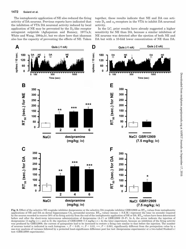
Inhibitory Action of Iontophoretically Applied DAon CA3 Pyramidal Neurons in the Presence of the Se-lective NE Reuptake Inhibitor Desipramine or the Se-lective DA Reuptake Inhibitor GBR12909. The recoverytime, from the suppression of hippocampus pyramidal neu-ron firing activity after microiontophoretic application of NEor DA, was assessed by determining RT50 values before andafter the short-term intravenous administration of desipra-mine or GBR12909. In agreement with previous data, cumu-lative doses of desipramine (2, 4, and 6 mg/kg i.v.) did notsignificantly modify the firing activity of CA3 pyramidal neu-rons. After the administration of desipramine (2 mg/kg i.v.),the RT50 values were significantly increased after the appli-cation of NE and DA (Fig. 9, A–C). Cumulative injections ofdesipramine (4 and 6 mg/kg) further increased RT50 valuesfor both monoamines, whereas GBR12909 (7.5 mg/kg i.v.)decreased the firing activity of pyramidal neurons by 42%. Asmall but significant increase in the RT50 value of DA but notNE was obtained after the short-term administration ofGBR12909 (Fig. 9, D–F).
DiscussionThe present electrophysiological data show that the micro-
iontophoretic application of DA and NE inhibits the sponta-neous firing activity of VTA DA, LC NE, and hippocampalCA3 pyramidal neurons. In the VTA, the suppressant effectsof DA and NE were blocked not only by the D2-like receptorantagonist raclopride, but also by the �2-adrenoceptor antag-onist idazoxan. In the LC and dorsal hippocampus, the sup-pressant effect of both catecholamines was only attenuatedby idazoxan.
In the VTA, the suppressant effect of both DA and quinpi-role on DA neurons and their blockade by the D2-like receptorantagonist raclopride further support the involvement of D2
receptor in the DA response. The observation that piperox-ane, a nonselective �-adrenoceptor antagonist, also attenu-ates the inhibitory effects of DA (Freedman and Aghajanian,1984) suggested actions on this class of receptors also. In-deed, the present study showed that the local application ofthe selective �2-adrenoceptor antagonist idazoxan attenu-ated the suppressant effect of DA on VTA DA neurons. Such
Fig. 7. Effect of �2-adrenoceptor antagonist idazoxan on iontophoretically applied DA-induced inhibition of dorsal hippocampus CA3 pyramidalneurons. A, integrated firing rate histograms illustrating the effects of idazoxan on DA-induced decrease in CA3 pyramidal neuronal activity.Horizontal bars indicate the duration of iontophoretic ejection and current values in nanoamperes. B, responsiveness of CA3 pyramidal neurons toiontophoretic applications of DA alone and in the presence of idazoxan (0.05 M). Data are expressed as mean � S.E.M. of the number of spikessuppressed by nanoamperes for CA3 pyramidal neurons. The number of neurons tested is indicated in each histogram. �, P � 0.05, significantlydifferent from DA alone by two-tailed Student’s t test.
1470 Guiard et al.
at ASPE
T Journals on Septem
ber 17, 2016m
olpharm.aspetjournals.org
Dow
nloaded from

an effect could not be attributed to a nonselective action ofidazoxan because this compound has no affinity for DA re-ceptors. Competition experiments in the rat cortex haveshown that idazoxan has at least 1000-fold lower affinity forD2 receptors than for �2-adrenoceptors (Ki values, �10 �Mand 8 nM, respectively) (Doxey et al., 1983; Neve et al., 1990).It thus seems that in addition to the stimulation of D2 recep-tors, DA also inhibits the firing activity of DA neurons byacting upon �2-adrenoceptors. Although functional studiespreviously emphasized such an unselective property of DA inthe central nervous system (Cornil et al., 2008), the role of�2-adrenoceptors in a direct regulation of DA neurons them-selves remained debatable, especially because, in rat brain,the affinity of DA for �2-adrenoceptors is approximately 3- to7-fold lower than that of NE (Boyajian et al., 1987). More-over, despite the expression of �2-adrenoceptors on DA neu-rons in the VTA (Lee et al., 1998), it was reported that themicroiontophoretic application of clonidine (20–120 nA) hasno depressant effect on VTA DA neuronal activity (White andWang, 1984a,b; Aghajanian and Bunney, 1977a,b). The ap-
parent discrepancy between the microiontophoretic effects ofclonidine and DA may be explained by the capacity of DA tobind and activate both D2 and �2-adrenoceptors. Indeed, thestimulation of both types of receptors by DA could be impor-tant for generating a robust inhibitory effect in the VTA. It isinteresting that the systemic administration of low doses ofclonidine, which probably activate presynaptic �2-adrenocep-tors, does not modify the mean firing rate of VTA DA neuronsbut decreases their bursting activity (Gobbi et al., 2001;Georges and Aston-Jones, 2003). In contrast, higher doses ofclonidine increase both the firing rate and bursting activity ofVTA DA neurons (Gobbi et al., 2001). These findings areparadoxical in the light of the present data, but it is possiblethat systemic administration of adrenergic agonists involveslong-loop feedback mechanisms that are not activated withlocal application. Another possibility is a blunting of theeffect of locally applied clonidine by its concomitant bindingto postsynaptic �1-adrenoceptors (Anden et al., 1976), whoseactivation stimulates VTA DA neurons (Grenhoff and Svens-son, 1993; Grenhoff et al., 1993, 1995).
Fig. 8. Effect of D2-like receptor antagonists on the responsiveness of dorsal hippocampus CA3 pyramidal neurons to iontophoretically applied NE.A, integrated firing rate histograms illustrating the effects of raclopride or haloperidol on NE-induced decreases in CA3 pyramidal neuronal activity.Horizontal bars indicate the duration of iontophoretic ejection and current values in nanoamperes. B and C, responsiveness of CA3 pyramidal neuronsto iontophoretic applications of NE in presence or not of idazoxan (0.05 M, 10 nA) (B) or haloperidol (200 �g/kg i.v.) (C). Data are expressed as mean �S.E.M. of the number of spikes suppressed by nA for CA3 pyramidal neurons. The number of neurons tested is indicated in each histogram.
Overlap of Function between Dopamine and Norepinephrine 1471
at ASPE
T Journals on Septem
ber 17, 2016m
olpharm.aspetjournals.org
Dow
nloaded from
The iontophoretic application of NE also reduced the firingactivity of DA neurons. Previous reports have indicated thatthe inhibition of VTA DA neuronal activity induced by localapplication of NE may be prevented by the D2-like receptorantagonist sulpiride (Aghajanian and Bunney, 1977a,b;White and Wang, 1984a,b), but we show here that idazoxanalso has the capacity of preventing the effects of NE. Taken
together, these results indicate that NE and DA can acti-vate D2 and �2-receptors in the VTA to inhibit DA neuronalactivity.
In the LC, prior results have already suggested a highersensitivity for NE than DA, because a similar inhibition ofNE neurons was detected after the ejection of both NE andDA but with a 10-fold lower concentration of NE than DA.
Fig. 9. Effect of the selective NE reuptake inhibitor desipramine or the selective DA reuptake inhibitor GBR12909 on RT50 values from iontophoreticapplications of NE and DA on dorsal hippocampus CA3 pyramidal neurons. RT50 values (means � S.E.M.) represent the time (in seconds) requiredby the neuron recorded to recover 50% of its firing activity from the end of the iontophoretic application of NE or DA. RT50 values have been determinedbefore and after the short-term intravenous administration of desipramine (A–C) or GBR12909 (D–F). In A, the arrow indicate the injection ofdesipramine (2 mg/kg i.v.), and in D, the injection of GBR12909 (7.5 mg/kg i.v.). In the later experiment, because an inhibition of the firing activitywas produced by GBR12909, the current of quisqualate had to be increased to restore a firing rate similar to that before the injection. The numberof neurons tested is indicated in each histogram. �, P � 0.05, ��, P � 0.01, ���, P � 0.001, significantly different from the preinjection value by aone-way analysis of variance followed by a protected least-significance difference post hoc test (desipramine experiments) or a two-tailed Student’s ttest (GBR12909 experiments).
1472 Guiard et al.
at ASPE
T Journals on Septem
ber 17, 2016m
olpharm.aspetjournals.org
Dow
nloaded from

The earlier observation that iontophoretically applied NE-induced inhibition of LC NE neurons is blocked by piperox-ane (Cedarbaum and Aghajanian, 1977) was compatible withan involvement of �-adrenoceptors. Consistent with theseresults, the inhibitory effects of both NE and DA here re-ported were antagonized by idazoxan. Although the inhibi-tory influence of NE through �2-adrenoceptor on LC NEneurons has been extensively studied (Svensson et al., 1975;Cedarbaum and Aghajanian, 1977), evidence that DA stim-ulates �2-adrenoceptors to reduce, at least in part, LC NEneuronal firing, was not clearly documented.
Because D2 receptors have been identified in the LC(Yokoyama et al., 1994), their direct activation on NE neu-rons after the iontophoretic application of NE or DA could notbe excluded. However, the present study showed that raclo-pride has no effect on NE- or DA-induced inhibition of LC NEneurons at concentrations and currents found to exert apotent inhibition of VTA DA neurons (Fig. 2C). These resultsare consistent with the previous demonstration that the non-selective DA receptor antagonist trifluoperazine is ineffectivein blocking the inhibition of LC neurons induced by both DAand NE (Cedarbaum and Aghajanian, 1977) and that thecapacity of clonidine to inhibit NE neuronal firing is unal-tered by haloperidol (Piercey et al., 1994). The putative lackof regulation of LC neurons by D2-like receptors is also evi-denced by the observation that these NE neurons are insen-sitive to the local application of D2-like receptor agonists,such as (�)-3-PP, apomorphine (Aghajanian and Bunney,1977; Elam et al., 1986), or quinpirole as shown in thepresent study. Altogether, these observations converge toshow that only �2-adrenoceptors are involved in the inhibi-tory control of LC NE exerted by NE and DA, even if thesystemic administration of haloperidol has been reported toenhance the firing rate of LC NE neurons (Piercey et al.,1994; Nilsson et al., 2005). Such an effect could not be relatedto an antagonistic activity of haloperidol on �2-adrenoceptor(U’Prichard et al., 1977), because the systemic administra-tion of haloperidol in the present study modified neither thespontaneous firing activity of LC NE neurons nor the inhib-itory actions of NE or DA. Other studies suggest an excita-tory effect of haloperidol on NE neurons that may initially bedriven through the local release of glutamate in the LC(Nilsson et al., 2005).
In the dorsal hippocampus, a possible role for DA had to beconsidered because the iontophoretic application of DA andNE inhibited the firing activity of CA3 pyramidal neurons.However, this DA-induced inhibition of firing was only par-tial, as if some of the DA effects were counterbalanced by anexcitatory component. Given that �-adrenoceptors exert anexcitatory action on hippocampus pyramidal neurons (Curetand de Montigny, 1988b), it is possible that the net biologicalresponse to DA results from opposite effects exerted on var-ious adrenoceptors. In keeping with a weak expression of DAreceptors and raclopride binding sites in the dorsal hip-pocampus (Dubois et al., 1986; Delis et al., 2004), it wasreported that neither the iontophoretic application of raclo-pride nor the systemic injection of haloperidol blocked DA- orNE-induced inhibition in the dorsal hippocampus. Althoughhaloperidol is known to bind and antagonize both D2 and�1-receptors (Cohen and Lipinski, 1986), the possibilitythat it blocked the adrenoceptors in the present study canbe excluded, because the administration of the selective
�1-adrenoceptor antagonist prazosin reduced the effect of NEin a dose-dependent manner (Curet and de Montigny, 1988a)and was here devoid of antagonistic activity. In contrast, theinhibitory action of DA was partially blocked by idazoxan atconcentrations that usually antagonize the inhibitory effectsof NE or clonidine on CA3 pyramidal neurons (Curet and deMontigny, 1988a). The involvement of postsynaptic �2-adre-noceptors in the electrophysiological response to DA, as de-scribed above in VTA and LC, was strongly supported by thelatter results. To better understand the physiological impor-tance of such a property, the possibility that NE neuronsthemselves could be the main source of DA in the hippocam-pus was addressed. The observation that the selective NEreuptake inhibitor desipramine prolonged the inhibitory ef-fects of microiontophoretic applied DA strongly suggestedthat the clearance of DA in the hippocampus is mediated, atleast in part, by the NE transporter (NET). This is consistentwith previous data showing that DA has a greater affinity forthe NET than the DA transporter (DAT) itself (Giros et al.,1994) and that DA reuptake by NE terminals occurs in brainregions in which DAT expression is minimal (e.g., the frontalcortex), intermediate, or maximal (e.g., nucleus accumbensshell and the bed nucleus, respectively) (Bymaster et al.,2002; Moron et al., 2002; Carboni and Silvagni, 2004). Recentexperiments have proposed that DA may be coreleased withNE from noradrenergic terminals in several cortical areas(Devoto et al., 2004). Although it is not clear whether thisfeature might be related to a previous nonspecific uptake ofDA by NE terminals, it is proposed here that DA is taken upby the NET in the hippocampus (Fig. 9), as reported previ-ously in the frontal cortex (Bymaster et al., 2002). It isinteresting that the selective DA reuptake inhibitorGBR12909 also produced a small but significant increase inthe RT50 value of iontophoretically applied DA, which couldhardly be ascribed to a nonselective binding of GBR12909 tothe NET because the RT50 value of NE was not altered in thisexperiment. Thus, in the dorsal hippocampus, DA uptake
Fig. 10. Model of the regulation of neuronal activities of ventral tegmen-tal area, locus ceruleus, and dorsal hippocampus by DA and NE at the cellbody level. In the VTA, DA, and NE act on both D2 and �2 receptor typesto inhibit the neuronal firing of DA neurons. In the LC, despite thepresence of D2 receptors, it seems that DA and NE exclusively stimulate�2 adrenoceptor thus inhibiting NE neuronal firing. The capacity of DA tobind and activate �2 adrenoceptor was also observed in the dorsal hip-pocampus. Although the release of DA from dopaminergic terminals inthe dorsal hippocampus has not yet been demonstrated, the presentresults indicate that exogenously applied DA can be removed from theextracellular space by the NE transporter.
Overlap of Function between Dopamine and Norepinephrine 1473
at ASPE
T Journals on Septem
ber 17, 2016m
olpharm.aspetjournals.org
Dow
nloaded from

could be mediated by distinct transport systems. The involve-ment of DAT was somewhat unexpected because in brainregions in which DAT is weakly expressed, as in the hip-pocampus (Delis et al., 2004; Dahlin et al., 2007), a limitedrole of DAT in removing DA from the extracellular space hasbeen reported (Moron et al., 2002). An alternative mecha-nism could be that residual DA uptake was mediated by therecently cloned and characterized polyspecific cation-mono-amine transporters organic cation transporters (2 and 3) orplasma membrane monoamine transporter. Indeed, thesetransporters are expressed in the rat hippocampus (Dahlin etal., 2007), but high concentrations of GBR12909 are neces-sary to block their activity (Moron et al., 2002). In view ofthese results and of the high ratio NET/DAT in hippocampus,it seems likely that, in this brain region, DA is captured byNET before it reaches other transport sites.
In summary, the present findings showed that, depend-ing on the brain structure studied, DA and NE activate D2
and/or �2-adrenoceptors to exert inhibitory postsynapticeffects (Fig. 10). This is in agreement with previous studiesshowing the in vivo and in vitro stereoselective interac-tions of DA with �2-adrenoceptors (Zhang et al., 2004;Cornil et al., 2008) and NE with D2-like receptors (New-man-Tancredi et al., 1997; Wedemeyer et al., 2007). Thefact that DA can act in concert with the NE system tostrengthen the intensity of postsynaptic noradrenergic re-sponses may have important implications in the treatmentof mood disorders.
ReferencesAghajanian GK and Bunney BS (1977a) Dopamine “autoreceptors”: pharmacological
characterization by microiontophoretic single cell recording studies. NaunynSchmiedebergs Arch Pharmacol 297:1–7.
Aghajanian GK and Bunney BS (1977b) Pharmacological characterization of dopa-mine “autoreceptors” by microiontophoretic single-cell recording studies. Adv Bio-chem Psychopharmacol 16:433–438.
Anden NE, Grabowska M, and Strombom U (1976) Different alpha-adrenoreceptorsin the central nervous system mediating biochemical and functional effects ofclonidine and receptor blocking agents. Naunyn Schmiedebergs Arch Pharmacol292:43–52.
Benardo LS and Prince DA (1982) Dopamine action on hippocampal pyramidal cells.J Neurosci 2:415–423.
Bischoff S, Heinrich M, Sonntag JM, and Krauss J (1986) The D-1 dopamine receptorantagonist SCH 23390 also interacts potently with brain serotonin (5-HT2) recep-tors. Eur J Pharmacol 129:367–370.
Bischoff S, Scatton B, and Korf J (1979) Biochemical evidence for a transmitter roleof dopamine in the rat hippocampus. Brain Res 165:161–165.
Boyajian CL, Loughlin SE, and Leslie FM (1987) Anatomical evidence for �-2adrenoceptor heterogeneity: differential autoradiographic distributions of [3H]-rauwolscine and [3H]idazoxan in rat brain. J Pharmacol Exp Ther 241:1079–1091.
Bruinink A and Bischoff S (1993) Dopamine D2 receptors are unevenly distributed inthe rat hippocampus and are modulated differently than in striatum. Eur J Phar-macol 245:157–164.
Bymaster FP, Katner JS, Nelson DL, Hemrick-Luecke SK, Threlkeld PG, Heiligen-stein JH, Morin SM, Gehlert DR, and Perry KW (2002) Atomoxetine increasesextracellular levels of norepinephrine and dopamine in prefrontal cortex of rat: apotential mechanism for efficacy in attention deficit/hyperactivity disorder. Neu-ropsychopharmacology 27:699–711.
Carboni E and Silvagni A (2004) Dopamine reuptake by norepinephrine neurons:exception or rule? Crit Rev Neurobiol 16:121–128.
Cedarbaum JM and Aghajanian GK (1977) Catecholamine receptors on locus coer-uleus neurons: pharmacological characterization. Eur J Pharmacol 44:375–385.
Chernoloz O, Mansari ME, and Blier P (2008) Sustained administration ofpramipexole modifies the spontaneous firing rate of dopamine, norepinephrine,and serotonin neurons in the rat brain. Neuropsychopharmacology, in press.
Cohen BM and Lipinski JF (1986) In vivo potencies of antipsychotic drugs in blockingalpha 1 noradrenergic and dopamine D2 receptors: implications for drug mecha-nisms of action. Life Sci 39:2571–2580.
Cornil CA, Castelino CB, and Ball GF (2008) Dopamine binds to alpha2-adrenergicreceptors in the song control system of zebra finches (Taeniopygia guttata). J ChemNeuroanat 35:202–215.
Crutcher KA and Davis JN (1980) Hippocampal alpha- and beta-adrenergic recep-tors: comparison of [3H]dihydroalprenolol and [3H]WB 4101 binding with norad-renergic innervation in the rat. Brain Res 182:107–117.
Curet O and de Montigny C (1988a) Electrophysiological characterization of adre-noceptors in the rat dorsal hippocampus. I. Receptors mediating the effect ofmicroiontophoretically applied norepinephrine. Brain Res 475:35–46.
Curet O and de Montigny C (1988b) Electrophysiological characterization of adre-noceptors in the rat dorsal hippocampus. II. Receptors mediating the effect ofmicroiontophoretically applied norepinephrine. Brain Res 475:47–57.
Dahlin A, Xia L, Kong W, Hevner R, and Wang J (2007) Expression and immuno-localization of the plasma membrane monoamine transporter in the brain. Neu-roscience 146:1193–1211.
Delis F, Mitsacos A, and Giompres P (2004) Dopamine receptor and transporterlevels are altered in the brain of Purkinje cell degeneration mutant mice. Neuro-science 125:255–268.
Devoto P, Flore G, Pira L, Longu G, and Gessa GL (2004) Mirtazapine-inducedcorelease of dopamine and noradrenaline from noradrenergic neurons in the me-dial prefrontal and occipital cortex. Eur J Pharmacol 487:105–111.
Doxey JC, Roach AG, and Smith CF (1983) Studies on RX 781094: a selective, potentand specific antagonist of alpha 2-adrenoceptors. Br J Pharmacol 78:489–505.
Dubois A, Savasta M, Curet O, and Scatton B (1986) Autoradiographic distributionof the D1 agonist [3H]SKF 38393, in the rat brain and spinal cord. Comparisonwith the distribution of D2 dopamine receptors. Neuroscience 19:125–137.
Einhorn LC, Johansen PA, and White FJ (1988) Electrophysiological effects ofcocaine in the mesoaccumbens dopamine system: studies in the ventral tegmentalarea. J Neurosci 8:100–112.
Elam M, Clark D, and Svensson TH (1986) Electrophysiological effects of the enan-tiomers of 3-PPP on neurons in the locus coeruleus of the rat. Neuropharmacology25:1003–1008.
Freedman JE and Aghajanian GK (1984) Idazoxan (RX 781094) selectively antago-nizes alpha 2-adrenoceptors on rat central neurons. Eur J Pharmacol 105:265–272.
Georges F and Aston-Jones G (2003) Prolonged activation of mesolimbic dopaminer-gic neurons by morphine withdrawal following clonidine: participation of imida-zoline and norepinephrine receptors. Neuropsychopharmacology 28:1140–1149.
Giros B, Wang YM, Suter S, McLeskey SB, Pifl C, and Caron MG (1994) Delineationof discrete domains for substrate, cocaine, and tricyclic antidepressant interac-tions using chimeric dopamine-norepinephrine transporters. J Biol Chem 269:15985–15988.
Gobbi G, Muntoni AL, Gessa GL, and Diana M (2001) Clonidine fails to modifydopaminergic neuronal activity during morphine withdrawal. Psychopharmacol-ogy (Berl) 158:1–6.
Grenhoff J and Svensson TH (1993) Prazosin modulates the firing pattern of dopa-mine neurons in rat ventral tegmental area. Eur J Pharmacol 233:79–84.
Grenhoff J, Nisell M, Ferre S, Aston-Jones G, and Svensson TH (1993) Noradrener-gic modulation of midbrain dopamine cell firing elicited by stimulation of the locuscoeruleus in the rat. J Neural Transm Gen Sect 93:11–25.
Grenhoff J, North RA, and Johnson SW (1995) Alpha 1-adrenergic effects on dopa-mine neurons recorded intracellularly in the rat midbrain slice. Eur J Neurosci7:1707–1713.
Guiard BP, El Mansari M, Merali Z, and Blier P (2008) Functional interactionsbetween dopamine, serotonin and norepinephrine neurons: an in-vivo electrophys-iological study in rats with monoaminergic lesions. Int J Neuropsychopharmacol11:625–639.
Jones BE and Moore RY (1977) Ascending projections of the locus coeruleus in therat. II. Autoradiographic study. Brain Res 127:25–53.
Lee A, Wissekerke AE, Rosin DL, and Lynch KR (1998) Localization of alpha2C-adrenergic receptor immunoreactivity in catecholaminergic neurons in the ratcentral nervous system. Neuroscience 84:1085–1096.
Linner L, Endersz H, Ohman D, Bengtsson F, Schalling M, and Svensson TH (2001)Reboxetine modulates the firing pattern of dopamine cells in the ventral tegmentalarea and selectively increases dopamine availability in the prefrontal cortex.J Pharmacol Exp Ther 297:540–546.
Moron JA, Brockington A, Wise RA, Rocha BA, and Hope BT (2002) Dopamineuptake through the norepinephrine transporter in brain regions with low levels ofthe dopamine transporter: evidence from knock-out mouse lines. J Neurosci 22:389–395.
Mueller AL, Palmer MR, Hoffer BJ, and Dunwiddie TV (1982) Hippocampal norad-renergic responses in vivo and in vitro. Characterization of alpha and beta com-ponents. Naunyn Schmiedebergs Arch Pharmacol 318:259–266.
Neve KA, Henningsen RA, Kinzie JM, De Paulis T, Schmidt DE, Kessler RM, andJanowsky A (1990) Sodium-dependent isomerization of Dopamine D-2 receptorscharacterized using [125I]epidepride, a high-affinity substituted benzamide ligand.J Pharmacol Exp Ther 252:1108–1116.
Newman-Tancredi A, Audinot-Bouchez V, Gobert A, and Millan MJ (1997) Nor-adrenaline and adrenaline are high affinity agonists at dopamine D4 receptors.Eur J Pharmacol 319:379–383.
Nilsson LK, Schwieler L, Engberg G, Linderholm KR, and Erhardt S (2005) Activa-tion of noradrenergic locus coeruleus neurons by clozapine and haloperidol: in-volvement of glutamatergic mechanisms. Int J Neuropsychopharmacol 8:329–339.
Ornstein K, Milon H, McRae-Degueurce A, Alvarez C, Berger B, and Wurzner HP(1987) Biochemical and radioautographic evidence for dopaminergic afferents ofthe locus coeruleus originating in the ventral tegmental area. J Neural Transm70:183–191.
Piercey MF, Smith MW, and Lum-Ragan JT (1994) Excitation of noradrenergic cellfiring by 5-hydroxytryptamine1A agonists correlates with dopamine antagonistproperties. J Pharmacol Exp Ther 268:1297–1303.
Scatton B, Simon H, Le Moal M, and Bischoff S (1980) Origin of dopaminergicinnervation of the rat hippocampal formation. Neurosci Lett 18:125–131.
Segal M, Pickel V, and Bloom F (1973) The projections of the nucleus locus coeruleus:an autoradiographic study. Life Sci 13:817–821.
Simon H, Le Moal M, Stinus L, and Calas A (1979) Anatomical relationships betweenthe ventral mesencephalic tegmentum—a 10 region and the locus coeruleus asdemonstrated by anterograde and retrograde tracing techniques. J Neural Transm44:77–86.
1474 Guiard et al.
at ASPE
T Journals on Septem
ber 17, 2016m
olpharm.aspetjournals.org
Dow
nloaded from

Svensson TH, Bunney BS, and Aghajanian GK (1975) Inhibition of both noradren-ergic and serotonergic neurons in brain by the alpha-adrenergic agonist clonidine.Brain Res 92:291–306.
Swanson LW and Hartman BK (1975) The central adrenergic system. An immuno-fluorescence study of the location of cell bodies and their efferent connections inthe rat utilizing dopamine-beta-hydroxylase as a marker. J Comp Neurol 163:467–505.
Swanson LW (1982) The projections of the ventral tegmental area and adjacentregions: a combined fluorescent retrograde tracer and immunofluorescence studyin the rat. Brain Res Bull 9:321–353.
U’Prichard DC, Greenberg DA, and Snyder SH (1977) Binding characteristics of aradiolabeled agonist and antagonist at central nervous system � noradrenergicreceptors. Mol Pharmacol 13:454–473.
Wedemeyer C, Goutman JD, Avale ME, Franchini LF, Rubinstein M, and Calvo DJ(2007) Functional activation by central monoamines of human dopamine D4 re-ceptor polymorphic variants coupled to GIRK channels in Xenopus oocytes. EurJ Pharmacol 562:165–173.
White FJ and Wang RY (1984a) A10 dopamine neurons: role of autoreceptors in deter-mining firing rate and sensitivity to dopamine agonists. Life Sci 34:1161–1170.
White FJ and Wang RY (1984b) Pharmacological characterization of dopamineautoreceptors in the rat ventral tegmental area: microiontophoretic studies.J Pharmacol Exp Ther 231:275–280.
Yokoyama C, Okamura H, Nakajima T, Taguchi J, and Ibata Y (1994) Autoradio-graphic distribution of [3H]YM-09151–2, a high-affinity and selective antagonistligand for the dopamine D2 receptor group, in the rat brain and spinal cord.J Comp Neurol 344:121–136.
Zhang WP, Ouyang M, and Thomas SA (2004) Potency of catecholamines and otherL-tyrosine derivatives at the cloned mouse adrenergic receptors. Neuropharma-cology 47:438–449.
Address correspondence to: Dr. Pierre Blier, University of Ottawa Instituteof Mental Health Research, 1145 Carling Avenue, Ottawa, K1Z 7K4, Ontario,Canada. E-mail: [email protected]
Overlap of Function between Dopamine and Norepinephrine 1475
at ASPE
T Journals on Septem
ber 17, 2016m
olpharm.aspetjournals.org
Dow
nloaded from
Copyright © 2022 FDOKUMEN