Correlating Magneto-Structural Properties to Hyperthermia Performance of Highly Monodisperse Iron...
11
Published: September 01, 2011 r2011 American Chemical Society 4170 dx.doi.org/10.1021/cm201078f | Chem. Mater. 2011, 23, 4170–4180 ARTICLE pubs.acs.org/cm Correlating Magneto-Structural Properties to Hyperthermia Performance of Highly Monodisperse Iron Oxide Nanoparticles Prepared by a Seeded-Growth Route Michael Levy, † Alessandra Quarta, ‡ Ana Espinosa, § Albert Figuerola,* ,||,^ Claire Wilhelm, † Mar García-Hern andez, § Alessandro Genovese, || Andrea Falqui, || Damien Alloyeau, # Raffaella Buonsanti, ‡ Pantaleo Davide Cozzoli, ‡,3 Miguel Angel García,* ,O,[ Florence Gazeau, † and Teresa Pellegrino ‡,|| † Laboratoire Mati ere et Syst emes Complexes (MSC), UMR 7057, CNRS and Universit e Paris Diderot, 10 rue Alice Domon et L eonie Duquet, 75205 Paris cedex 13, France ‡ National Nanotechnology Laboratory (NNL), Istituto Nanoscienze-CNR, via per Arnesano 16 Km.5, 73100 Lecce, Italy § Instituto de Ciencia de Materiales de Madrid, Consejo Superior de Investigaciones Científicas, Cantoblanco, Sor Juana In es de la Cruz 3, 28049 Madrid, Spain ) Istituto Italiano di Tecnologia, via Morego 30, 16169 Genoa, Italy; ^ Departament de Química Inorg anica, Universitat de Barcelona, Martí i Franqu es 1-11, 08028 Barcelona, Spain # Laboratoire Mat eriaux et Ph enom enes Quantiques (MPQ), UMR 7162, CNRS and Universit e Paris Diderot, 10 rue Alice Domon et L eonie Duquet, 75205 Paris cedex 13, France 3 Dipartimento di Ingegneria dell’Innovazione, Universit a del Salento, Via per Arnesano, 73100 Lecce, Italy O Instituto de Cer amica y Vidrio, CSIC and IMDEA Nanociencia, 28049 Madrid, Spain [ Departamento de Física de Materiales, Universidad Complutense de Madrid, 28040 Madrid, Spain b S Supporting Information ’ INTRODUCTION Controlled heat generation has been devised since years as an efficient way to fight cancer. 1 However, heat should be selectively delivered to the tumor area in order to avoid overheating of healthy tissues in the body. Nanotechnology has provided ther- mal probes which are more selective toward tumoral cells due to the nanometric dimensions of the particles employed as heating agents. Their nanoscaled sizes, well below the cell dimensions, are promising features for achieving targeted delivery and efficient cell loading. Magnetic nanoparticles (MNPs), such as of iron oxide, have the capability to release heat under the stimulation of an alternating magnetic field. 2,3 Unlike their use in diagnostics as contrast agents for magnetic resonance imaging (MRI), 4 the clinical use of MNPs for thermal therapy is just in its infancy. 5 Received: April 14, 2011 Revised: July 26, 2011 ABSTRACT: Monodisperse cubic spinel iron oxide magnetic nanoparticles with variable sizes were prepared following a multi-injection seeded-growth approach. As expected from such a well-known synthetic route, all samples were characterized by narrow size distributions, and showed excellent stability in both organic and aqueous media without the presence of aggregates, thus becoming ideal candidates for the study of their hyperthermia performance. Specific Loss Power measurements indicated low heating powers for all samples without a maximum for any specific size, contrary to what theory predicts. The magnetic study showed the formation of size-dependent nonsaturated magnetic regions, which enlarged with the particle size, evidencing a clear discrepancy between the crystal size and the effective magnetic volume. Strain map analysis of high resolution transmission electron micrographs indicated the presence of highly strained crystal areas even if nanoparticles were monocrystalline. The origin of the crystal strain was found to be strictly correlated with the seeded-growth synthetic procedure used for the preparation of the nanoparticles, which turned out to alter their magnetic structure by creating antiphase boundaries. Considering the calculated effective magnetic volumes and their magnetic dispersions in each sample, a reasonable agreement between hyperthermia experiments and theory was obtained. KEYWORDS: iron oxide nanoparticles, thermodecomposition colloidal synthesis, seeded-growth, hyperthermia, magnetic properties, antiphase boundaries, strain analysis
-
Upload
independent -
Category
Documents
-
view
5 -
download
0
Transcript of Correlating Magneto-Structural Properties to Hyperthermia Performance of Highly Monodisperse Iron...
Published: September 01, 2011
r 2011 American Chemical Society 4170 dx.doi.org/10.1021/cm201078f |Chem. Mater. 2011, 23, 4170–4180
ARTICLE
pubs.acs.org/cm
Correlating Magneto-Structural Properties to HyperthermiaPerformance of Highly Monodisperse Iron Oxide NanoparticlesPrepared by a Seeded-Growth RouteMichael Levy,† Alessandra Quarta,‡ Ana Espinosa,§ Albert Figuerola,*,||,^ Claire Wilhelm,†
Mar García-Hern�andez,§ Alessandro Genovese,|| Andrea Falqui,|| Damien Alloyeau,# Raffaella Buonsanti,‡
Pantaleo Davide Cozzoli,‡,3 Miguel Angel García,*,O,[ Florence Gazeau,† and Teresa Pellegrino‡,||
†Laboratoire Mati�ere et Syst�emes Complexes (MSC), UMR 7057, CNRS and Universit�e Paris Diderot, 10 rue Alice Domon et L�eonieDuquet, 75205 Paris cedex 13, France‡National Nanotechnology Laboratory (NNL), Istituto Nanoscienze-CNR, via per Arnesano 16 Km.5, 73100 Lecce, Italy§Instituto deCiencia deMateriales deMadrid, Consejo Superior de Investigaciones Científicas, Cantoblanco, Sor Juana In�es de la Cruz 3,28049 Madrid, Spain
)Istituto Italiano di Tecnologia, via Morego 30, 16169 Genoa, Italy;^Departament de Química Inorg�anica, Universitat de Barcelona, Martí i Franqu�es 1-11, 08028 Barcelona, Spain#Laboratoire Mat�eriaux et Ph�enom�enes Quantiques (MPQ), UMR 7162, CNRS and Universit�e Paris Diderot,10 rue Alice Domon et L�eonie Duquet, 75205 Paris cedex 13, France3Dipartimento di Ingegneria dell’Innovazione, Universit�a del Salento, Via per Arnesano, 73100 Lecce, ItalyOInstituto de Cer�amica y Vidrio, CSIC and IMDEA Nanociencia, 28049 Madrid, Spain[Departamento de Física de Materiales, Universidad Complutense de Madrid, 28040 Madrid, Spain
bS Supporting Information
’ INTRODUCTION
Controlled heat generation has been devised since years as anefficient way to fight cancer.1 However, heat should be selectivelydelivered to the tumor area in order to avoid overheating ofhealthy tissues in the body. Nanotechnology has provided ther-mal probes which are more selective toward tumoral cells due tothe nanometric dimensions of the particles employed as heatingagents. Their nanoscaled sizes, well below the cell dimensions,are promising features for achieving targeted delivery and efficient
cell loading. Magnetic nanoparticles (MNPs), such as of ironoxide, have the capability to release heat under the stimulation ofan alternating magnetic field.2,3 Unlike their use in diagnostics ascontrast agents for magnetic resonance imaging (MRI),4 theclinical use of MNPs for thermal therapy is just in its infancy.5
Received: April 14, 2011Revised: July 26, 2011
ABSTRACT:Monodisperse cubic spinel iron oxidemagnetic nanoparticles with variablesizes were prepared following a multi-injection seeded-growth approach. As expectedfrom such a well-known synthetic route, all samples were characterized by narrow sizedistributions, and showed excellent stability in both organic and aqueous media withoutthe presence of aggregates, thus becoming ideal candidates for the study of theirhyperthermia performance. Specific Loss Power measurements indicated low heatingpowers for all samples without a maximum for any specific size, contrary to what theorypredicts. The magnetic study showed the formation of size-dependent nonsaturatedmagnetic regions, which enlarged with the particle size, evidencing a clear discrepancybetween the crystal size and the effective magnetic volume. Strain map analysis of highresolution transmission electron micrographs indicated the presence of highly strainedcrystal areas even if nanoparticles were monocrystalline. The origin of the crystal strainwas found to be strictly correlated with the seeded-growth synthetic procedure used forthe preparation of the nanoparticles, which turned out to alter their magnetic structure by creating antiphase boundaries.Considering the calculated effective magnetic volumes and their magnetic dispersions in each sample, a reasonable agreementbetween hyperthermia experiments and theory was obtained.
KEYWORDS: iron oxide nanoparticles, thermodecomposition colloidal synthesis, seeded-growth, hyperthermia, magneticproperties, antiphase boundaries, strain analysis
4171 dx.doi.org/10.1021/cm201078f |Chem. Mater. 2011, 23, 4170–4180
Chemistry of Materials ARTICLE
One of the key limitations for their clinical application deals withthe fact that quite large doses are still required to generate anamount of heat sufficient to induce tumor regression. This couldbe due to the poor colloidal stability and thus to the low amountof magnetic material able to reach the tumoral site through thebloodstream.6,7 However, in many cases, in addition to thein vivo stability issue, the problem is mainly associated withthe inherently low heat generating properties of the MNPs. Thespecific loss power (SLP) is the parameter quantifying theheating power generated by MNPs and is expressed in W per gof magnetic material. In addition to iron oxide, other materialssuch as metals would definitely offer higher SLP values. Never-theless, their use in medicine is strongly limited by their hightoxicity.8 Hence, the understanding of the ideal characteristics ofiron oxideMNPs for enhanced heat generation is still a subject ofgreat interest. Even if theory predicts SLP values of up to 1 kW g�1
for iron oxide MNPs,2,9,10 such values have rarely been measuredexperimentally11 as they often reach only a few hundreds ofW g�1 or even less.12�15
In the past decades several studies have been undertaken tocorrelate the various structural and magnetic features with thehyperthermia performance of iron oxide MNPs. Among these,the size of the particles and their size distribution,9,12,16 theircrystallinity, chemical composition,10,14 and interparticle inter-actions13,17�19 are those intrinsically associated to the magneticmaterial. Besides these, other aspects such as the physiologicalconditions or the magnetic field amplitude and frequency used inthe measurement appear to be also determinant factors.20,21
Natural magnetosomes produced by magnetotactic bacteria haveso far shown the highest values of SLP, even if their large-scaleavailability is hard to achieve.22 Alternatively, iron oxide MNPscan be obtained by a wide variety of synthetic methods, includingprocedures based on coprecipitation in aqueous media23�25 orthermal decomposition of molecular precursors in organic sol-vents.26�29 To date, almost all studies have focused on iron oxideparticles prepared by coprecipitation of di/trivalent iron salts inbasic aqueous medium. Actually, commercially available ironoxide nanoparticles forMRI applications are exclusively preparedby this method.4 Even if the coprecipitation approach remains aneconomical route to synthesize large amounts of nanostructurediron oxide particles, it also entails several disadvantages in termsof poor degree of size control, crystallinity and colloidal stability.The theoretical model for heating power of single crystal MNPspredicts a sharp maximum for a well-defined diameter (at a givenfrequency) and a rapid decrease of the SLP when the particle sizediffers by only one nanometer with respect to the ideal size.2,9,10
Therefore, a tight control of the particle size and size dispersionappears to be the preliminary most important prerequisite tooptimizing the heating power of nanomagnets. In this scenario,the high temperature decomposition of organometallic precur-sors or metal complexes in mixtures of organic solvents and/orsurfactants represents a powerful alternative for the synthesis ofaggregation-free MNPs with a high degree of control over size,shape, size dispersion, and crystallinity.30,31 Several authors havealso reported on the enhanced magnetic properties of MNPsprepared by these methods compared to those prepared bycoprecipitation procedures.32,33 Among the numerous variants ofthe thermal decomposition route, the seeded-growth method hasbeen demonstrated to lead to high-quality nanoparticles withfinely tunable sizes and excellent monodispersity for a wide rangeof materials.29,30 Consequently, MNPs of certain sizes preparedby this method could, in principle, be ideal candidates to reach
the high SLP values predicted. However, the magnetic character-istics of MNPs prepared by seeded-growth approaches haverarely been studied in detail. MNPs of small enough sizes maysuffer from surface effects34�36 and can also show certain degreeof magnetic disorder strictly linked to the synthetic protocol usedfor their preparation, which can easily degrade their magneticperformance. Hence, the establishment of a correlation betweenthe synthetic method chosen and the magnetostructural char-acteristics of MNPs thereof represents a fundamental steptoward a better understanding of magnetic phenomena innanoparticles and for the validation of a specific synthetic routeto high-quality iron oxide MNPs suitable to be exploited inhyperthermia therapy.
In this manuscript we present an in-depth structural andmagnetic characterization of iron oxide MNPs of variable sizesand narrow size variances. All samples were prepared by asurfactant-assisted thermal decomposition approach in organicsolvents based on a seeded-growth technique.29 The presence ofamphiphilic surfactant molecules in the reaction mixture wasessential to guarantee attainment of unaggregated highly mono-disperse particles, with high solubility in nonpolar solvents. TheMNPs were further transferred to saline aqueous buffer solutionby using a well-established polymer coating process, after whichthe colloidal stability of the particles was still preserved.37 Thehyperthermia performances of both surfactant and polymer-coated MNPs was examined. We aimed at elucidating the struc-tural and magnetic factors responsible for the low SLP valuesmeasured for such samples in an attempt to fully correlate structuralfeatures, magnetic properties and hyperthermia performance inthese MNPs. The suitability of the seeded-growth approach for thepreparation of iron oxide MNPs has been also discussed.
’EXPERIMENTAL SECTION
Materials. Iron pentacarbonyl (Fe(CO)5, 98%), oleic acid(C17H33CO2H or OLAC, 90%), oleylamine (C17H33NH2 or OLAM,70%), hexadecane-1,2-diol (C16H32O2 or HDD, technical grade) and1-octadecene (C18H36 or ODE, 90%) were purchased from Aldrich.OLAC, OLAM andODE were degassed under vacuum at 120 �C for 1 hand stored under nitrogen in a glovebox at �20 �C before use.Synthesis of Iron Oxide MNPs. Iron oxide MNPs were prepared
by a suitably modified literature protocol38 implemented with a seeded-growth technique.29,39 The hydrophobic MNPs were transferred inwater by following a previously reported polymer coating procedure.37,40
More details on the synthesis and on the procedure for the transfer ofnanoparticles in water can be found in the Supporting Information (SI).Dynamic Light Scattering (DLS). DLS characterization was
performed on a Zetasizer Nano ZS90 (Malvern, U.S.) equipped witha 4.0 mW He�Ne laser, operating at 633 nm, and an Avalanchephotodiode detector. Measurements were carried out at 25 �C.Gel Electrophoresis. Gel electrophoresis was carried out on a 2%
agarose gel, applying a voltage of 100 V for 1 h. Prior to gel loading, asolution of 30% glycerol containing Orange G in gel-loading buffer(corresponding to 20% of the sample loaded volume) was added to eachsample.Elemental Analysis. The elemental analysis of the samples was
performed by digesting 25 μL of each sample in 2.5 mL of acqua regiaand 48 h later diluting them to a known volume with miliQ ultrapurewater. The measurement was performed with the iCap 6000 Seriesinduced coupled plasma-atomic emission spectroscopy (ICP-AES)instrument from Thermo Fisher.X-ray Diffraction (XRD). XRD spectra were obtained using the
Smartlab 9 kW Rigaku diffractometer equipped with a copper rotating

4172 dx.doi.org/10.1021/cm201078f |Chem. Mater. 2011, 23, 4170–4180
Chemistry of Materials ARTICLE
anode. The X-ray source was operated at 40 kV and 150 mA. A Gobelmirror was used to obtain a parallel beam and remove CuKβ radiation(1.392 Å). A flat mirror analyzer was used to remove X-ray fluorescence.A 2theta/omega scan was performed with a step width of 0.03� (2θ) andspeed scan of 0.023 deg/min.Low- and High-Resolution Transmission Electron Micro-
scopy (TEM). In view of the TEM analysis, a drop of the colloidalsolution in toluene or water was deposited onto a thin carbon filmsupported by a copper TEM grid and then allowing the solvent toevaporate. Low-magnification TEM measurements were carried outwith a Jeol JEM-1010 microscope, equipped with a tungsten electronsource and working at an acceleration voltage of 100 kV. Statisticalanalysis was carried out on several wide-field, low-magnification TEMimages, with the help of a dedicated software (Gatan Digital Micro-graph). For each sample, 200 particles were counted at least. High-resolution TEM (HRTEM) measurements were carried out with a JeolJEM-2200FS microscope with spherical aberration-corrected objectivelens, equipped with a field emission gun (FEG) and working at anacceleration voltage of 200 kV. The HRTEM images were recorded by aGatan Ultrascan 1000 CCD camera and the structural features of thenanostructures were studied by 2D-Fourier analysis, while the localstrain was studied by analyzing the HRTEM images with the peak pairanalysis (PPA) method.41
X-ray Absorption Spectroscopy. X-ray Absorption Near-EdgeStructure (XANES) and Extended X-ray Absorption Fine Structure(EXAFS) spectroscopy measurements at the Fe K-edge energy wereperformed at room temperature in transmission mode at the BM25Spanish CRG Beamline (SpLine) of the ESRF (European SynchrotronRadiation Facility). Two gas ionization chambers, filled with nitrogenand argon, were used to measure the incident and the transmitted beam,respectively. Powder samples were placed onto a kapton tape located onthe beam path. Different quantities of powders were used in theexperiments so as to achieve similar signal intensity for all the samples.Several scans were taken, in order to obtain a good signal-to-noise ratio.Bulk metallic Fe, FeO,R-Fe2O3, γ-Fe2O3, and Fe3O4 powders were alsomeasured for comparison purposes. Data were normalized applying thesame normalization parameters for all the spectra by means of AthenaSoftware.Magnetic Measurements. Magnetization curves of the colloidal
suspensions of MNPs ([Fe]∼ 10�2�10�3 M) were measured at 300 Kas a function of the magnetic field in the range 0�104 Oe using alaboratory-made vibratingmagnetometer.42Magnetization curves at lowtemperatures (5 K; from �50 kOe to +50 kOe), and the thermaldependence of the magnetization of liquid samples were also measuredin Zero Field Cooling (ZFC) and Field Cooling (FC) runs using asuperconducting quantum interference device (SQUID) fromQuantumDesign. To determine the magnetization per unit volume M, the netmagnetic moment of the suspension was normalized by the total volumeof particles, determined from the mass of iron (quantified by ICP-AES)and assuming the spinel crystal structure of magnetite (Fe3O4).Hyperthermia Experiments. The laboratory-made device used
for themagnetic hyperthermia experiments was described previously.9,11
It consists of a resonant RLC circuit producing a 700 kHz magnetic fieldof 21 kA m�1 in a coil of 16 mm diameter. The coil is cooled withcirculating nonane and the nonane temperature is controlled to reach anequilibrium temperature of 37 �C. The temperature is probed with afluoro-optic fiber thermometer (Luxtron Corp., CA) and is recordedevery 0.7 s after switching on the power supply. The initial linear rise oftemperature as a function of time, dT(t = 0)/dt, was measured in orderto deduce the power dissipated by one gram of nanoparticles, calledthe Specific Loss Power (SLP) of the particles, defined as SLP =((CVs)/m)((dT(t = 0)/dt), where C is the volume specific heat capacityof the sample (Cwater = 4185 J L�1 K�1), Vs = 300 μL is the samplevolume, and m is the mass of iron oxide.
’RESULTS
Synthesis of Size-Controlled Iron Oxide MNPs. Oleate-capped iron oxide nanocrystals with tunable sizes were synthe-sized by high-temperature decomposition of (Fe(CO)5) in aODE-dilutedmixture of OLAC, OLAM andHDD at 280 �C.38 Aone-pot, multistep seeded-growth technique was used to pro-duce MNPs with mean diameter precisely adjustable in the6�18 nm size range.29,39 The strategy involved the initialgeneration of small iron oxide MNP seeds (ca. 6 nm), whichcould then be grown to progressively larger sizes in the samesurfactant environment upon slow supply of extra iron precursorand capping agents. The enlargement of the presynthesized seedswas favored over the formation of new crystal embryos since theenergetic cost for the homogeneous nucleation at low super-saturation levels is higher than the one required for the deposi-tion of iron oxide onto the surface of the pre-existing seeds.43
This mechanism allows the separation of the primary nucleationand secondary growth processes, leading to size-tunable, highlymonodisperseMNPs by carefully controlling the relative amountof precursors added to the preformed seeds. The native hydro-phobic capping layer of the as-synthesized MNPs was exploitedfor their transfer into aqueous media following a well-knownpolymer-coating procedure.37,40 Amphiphilic polymer molecules(PC14 and PC18) having similar structure but different molec-ular weight were employed for transferring the nanoparticles intowater in order to assess whether the thickness of the polymerlayer could affect the heating ability of the magnetic core under-neath. Each MNP was enwrapped by polymer molecules thatformed a uniform and stable negatively charged shell around thenanoparticles, enabling their solubility into a saline aqueousenvironment.Size andSizeDispersionStudies.The left column of Figure 1
shows TEMmicrographs of seven iron oxide MNP samples withaverage dimensions ranging from 6 to 18 nm, which weredeposited from a toluene solution. The homogeneous andclose-packed arrangement of the particles on the TEM gridsdemonstrates their low degree of size dispersion and the absenceof aggregates in the solution. The length of the aliphatic chains ofthe capping molecules corresponds roughly to half the inter-particle distance observed in the images, confirming the efficientrole of the surfactants in isolating and stabilizing theMNPs in theorganic solution. The size dispersion of all samples was measuredover a minimum of 200 particles. In all cases, standard deviationvalues smaller than 20% with a slight increase with increasingMNP dimensions were deduced. The polymer coating proceduredid not affect the monodispersity of MNPs and ensured theirhomogeneous solubilization in water, as assessed by the TEMmicrographs (see the right column of Figure 1) and by DLSmeasurements (see SI) performed after water transfer. More-over, the gel electrophoresis of the water-soluble MNPs (FigureS1 of the SI) showed narrow migration bands, confirming thehomogeneous size (and charge) distribution of the nanocrystalsand evidenced the absence of multiparticle aggregates in solu-tion, regardless of the polymer used.Phase Determination. XRD spectra proved the crystalline
nature of all iron oxide samples. The peaks were comparedwith the diffraction patterns of standard magnetite (Fe3O4) andmaghemite (γ-Fe2O3) and all matched with the cubic spinelstructure even if it was not possible to discern between the twodifferent phases mentioned above. As an example, the X-ray diffrac-tion pattern for 15 nm iron oxideMNPs is shown in Figure 2. It is

4173 dx.doi.org/10.1021/cm201078f |Chem. Mater. 2011, 23, 4170–4180
Chemistry of Materials ARTICLE
well-known that nanostructured materials show diffraction peakswith larger widths with respect to their microstructured or bulk
analogues, as expressed in the Scherrer formula.44,45 However,the peak width should clearly narrow with increasing dimensions.Here we analyzed the size dependence of the full width at half-maximum (fwhm) of the most intense diffraction peak observedfor magnetite, corresponding to the (311) reflection (see theinset of Figure 2). As expected, the fwhm decreased by about 0.4�as the MNP size increased from 6 to 8 nm. However, for largersizes, a significant increase of the fwhm was observed, prelimi-narily indicating that the largerMNPs could be characterized by alimited length of structural coherence, as assessed in a recentatomic pair distribution function (PDF) analysis of synchrotronXRD patterns collected from iron oxide MNPs synthesized by ananalogous approach.46 The alternative hypothesis of a multi-domain polycrystalline structure could be fully discredited on thebasis of the results of HRTEM investigations, which will beillustrated in next paragraphs. XANES measurements were per-formed for all MNP samples in order to get additional structuralinformation and discard the presence of different iron species. Asreported in the SI for MNPs of 6 and 18 nm, nanocrystals arecomposed of pure magnetite regardless of their size.High-Resolution TEM measurements. HRTEM micro-
graphs of 18 nm iron oxide nanoparticles are shown in Figure 3.They revealed homogeneous structures of nanoparticles consis-tent with a monocrystalline phase as well as the absence ofevident lattice defects, such as dislocations or stacking faults. Inparticular, the numerical electron diffraction patterns (i.e., fil-tered 2D-FFT) obtained by HRTEM images were compatiblewith a single-crystal structure as no crystalline reflection splittingor satellite spots were in fact observed in the Fourier space.Magnetic Measurements. Magnetic measurements were
performed on both the starting surfactant-coated MNPs andthe corresponding water-soluble samples. No significant differ-ences were observed between the surfactant- and polymer- coatedanalogues. The magnetization of the MNP samples in solutionwas measured at room temperature as a function of the magneticfield up to 104 Oe. Volume magnetization curves are representedin Figure 4a for samples of different sizes. The magnetization at
Figure 2. X-ray diffraction pattern of iron oxide MNPs with averagediameter of 15 nm with the corresponding reference patterns ofmagnetite and maghemite for comparison. The reflection indexesindicated in the magnetite reference pattern are also valid for thecorresponding reflections of the maghemite pattern. The inset showsthe evolution of the full width at half-maximum (fwhm) of the peakcorresponding to the (311) reflection with the size of the MNPs.
Figure 1. TEM micrographs of iron oxide MNPs of different sizes,deposited from organic solvents (left column) and after their transfer toaqueous saline buffer by means of a polymer-coating procedure (rightcolumn). On the top of each panel the average size of the particles isindicated in black. The scale bar is marked at the bottom of eachmicrograph and represents 100 nm in all cases.

4174 dx.doi.org/10.1021/cm201078f |Chem. Mater. 2011, 23, 4170–4180
Chemistry of Materials ARTICLE
high fields clearly decreased with increasing particle size, thiseffect being more pronounced above 13 nm. Whereas a plateauwas observed for themagnetization of 6�10 nm diameterMNPs,a linear increase of the magnetization was seen at high fieldstrengths (typically above 6 kOe) for larger sizes (see in Figure 4a,the magnetization of the 18 nm sample). The highest magnetiza-tion value was found for a particle diameter of 6 nm and wasaround 55% of the saturation magnetization of the bulk magne-tite (MS bulk). In principle, the decrease of saturation magnetiza-tion compared to the bulk material can be explained by theexistence of nonfully coordinated atoms at the surface of theparticles, leading to the formation of a thin nonferromagnetic orfrustrated layer.34,36,47 Considering nanoparticles with a diameterof 6 nm and a magnetically frustrated layer of 0.5 nm, then only55% of the nanoparticle volume should actually be effectivelyferromagnetic, that is, with an exchange-based order, either ferro-or ferrimagnetic. When the MNP size increases, its surface-to-volume ratio decreases and the saturation magnetization of theparticle should approach the bulk value MS bulk. However,Figure 4a shows exactly the opposite trend, according to whichthe saturation magnetization decreases with increasing MNPsize: clearly, the volume that contributes to the overall magne-tization of the MNP was smaller than its crystalline volume, thelatter being calculated from the average diameter extracted fromTEMmeasurements (henceforth denoted as the TEM diameter)and assuming a spherical geometry. Actually, the ferromagneticvolume fractions (ΦFM) and a paramagnetic-like susceptibility(χ), the latter accounting for the nonsaturating behavior, werecalculated for all samples (see details in the SI). The resultsindicated that ΦFM represented around 55% of the crystalvolume for 6 nm particles and further diminished with increasingcrystal size. This behavior can be attributed to the presence of amagnetically frustrated phase which does not fully align even athigh magnetic fields and which accounts for more than 50% ofthe total particle volume in most of the samples.The next step was to deduce the whole distribution of effective
ferromagnetic diameters in the solution. This can be achieved byfitting the experimental magnetization curve with a Langevinfunction weighted by the distribution of ferromagnetic size. Toclarify, the normalized magnetization can be written asM/Mmax =RP(dFM)ddFML(ζFM), whereMmax is the maximum value of the
magnetization (from which the eventual “paramagnetic-like”contribution has been subtracted), P(dFM) is the probability densityof a ferromagnetic diameter dFM, L(ζFM) = coth(ζFM)� 1/ζFM is
the Langevin function and ζFM = MS bulkπdFM3 μ0H/6KBT is
the Langevin parameter, H the applied magnetic field, KB theBoltzmann constant and T the temperature. A log-normal distribu-tion of ferromagnetic diameters, P(dFM) = 1/((2π)1/2σFMdFM) �exp(�ln2(dFM/d0FM)2σFM
2 ), was found by fitting the experimentalcurves. The characteristic diameter d0FM and polydispersity indexσFM are shown in Figure 4b-c respectively, as a function of the TEMdiameter. Up to a MNP size of 10 nm, d0FM is slightly smaller thanthe TEM diameter and the polydispersity index σFM is close to zero,consistent with the uniform sizes observed in TEM micrographs.However, the ferromagnetic diameters diverge from TEM ones andthe polydispersity is drastically enhanced for larger particle sizes.Despite their quite homogeneous crystal size, the particles showheterogeneous magnetic behaviors with a high degree of disorder.For 18 nm particles, the ferromagnetic diameter is reduced to 8 nmwith a large dispersion and the appearance of a paramagnetic-likenonsaturated component of magnetization.Hyperthermia Measurements. To evaluate the efficiency
of the iron oxide MNPs as nanomediators for therapeutic hyper-thermia, we measured their SLP, expressed in Watts per iron
Figure 4. (a) Field-dependent magnetization curves of colloidal ironoxide MNPs with different sizes. Ferromagnetic diameters, d0FM,(b) and magnetic polydispersity indexes, σFM, (c) of magnetic size dis-tributions deduced from the fit of the magnetization curves, as a functionof TEM diameter.
Figure 3. HRTEM images of single iron oxide nanocrystals with theircorresponding numerical electron diffraction patterns (insets). (a)nanocrystal observed along the [112] zone axis, displaying [111] and(220) lattice fringes. (b) nanocrystal observed along the [011] zone axisshowing (111) and (400) lattice fringes. Numerical electron diffractionpatterns are consistent with a monocrystalline phase.

4175 dx.doi.org/10.1021/cm201078f |Chem. Mater. 2011, 23, 4170–4180
Chemistry of Materials ARTICLE
oxide unit mass (g), in amagnetic field of 21 kAm�1 oscillating ata frequency of 700 kHz. The measured values are reported inFigure 5 as a function of the TEM diameter. We also representedthe theoretical prediction for monodisperse nanoparticles ofmagnetite.2 SLPs increased with particle size, but exhibited muchlower values than those afforded by MNPs with similar sizessynthesized by coprecipitation.9 Moreover, the maximum of SLPpredicted theoretically for monodisperse 15 nm diameter MNPswas not observed here despite the highly monodisperse crystalsizes and the apparent crystallinity of the MNPs used.
’DISCUSSION
So far, the unusual magnetic behavior and the unexpected lowhyperthermia performance exhibited by our MNPs cannot bedirectly explained by the structural analyses provided by XRD,XAS and HRTEM. For this reason, an in-depth study of thesemonodisperse, single crystalline and well-dispersed MNPs canshed light on new aspects that could not be otherwise easilydisclosed in conventional samples affected by broad size disper-sions and/or aggregation phenomena.Strain Map Analysis by High Resolution TEM. Magnetite-
like single crystalline MNPs showed no direct evidence of anylattice defect, as reported previously. Starting fromHRTEM dataand using the analysis method known as peak pairs analysis(PPA), a complete strain tensors study was thus carried out forthe 18 nm MNPs in order to identify the presence/absence ofstrained regions inside the iron oxide monocrystalline structure(more details of the calculations can be found in the SI). Thestrain maps generated by the mean dilatation, the pure shear andthe rigid-body rotation tensor components are shown inFigure 6b�d. In particular, the mean dilatation tensor describesthe deformation due to tensile or compressive stress, defined asnormal to the body surface; the pure shear tensor describes thedeformation due to tangential stress; both the mean dilatationand pure shear tensor are defined so as to give no rotation of theatomic columns. Finally, the rigid body rotation tensor takes intoaccount only the angular rotation component of the atomiccolumns (see SI for further details). Defective areas are repre-sented by hot spots of blue/red/yellow colors while defect-freeareas are represented in green-brown colors.
As obtained from strain tensors analysis, a considerabledeformation (blue/red/yellow colors in strain maps) is accumu-lated along some “interfaces” across the nanocrystal and in theexternal region along a thickness of few nanometers. The strainedregions could be considered as a part of the single crystallinedomain with a certain degree of structural disorder, even if notenough to give rise to directly observable lattice distortion. Thisconclusion is consistent with the findings of the aforementionedPDF-synchrotron XRD study recently performed on analogousMNPs, according to which the limited length of structuralcoherence, which was much smaller than the TEM diameter,was ascribed to lattice strain effects.46 The results of the strainanalysis could provide a first explanation of the anomalousmagnetic behavior observed. The alignment of the iron spinscould be perturbed in the strained regions. As a consequence,such local lattice distortions could reduce the global magneticmoment of theMNP.Moreover, strained regions are expected tocreate a strain field spreading across the MNP: magnetic inter-actions are extremely sensitive to structural changes and hencethe structural strain at certain interfaces could easily frustrate thespins along wider regions.Low-Temperature Magnetic Analysis. To highlight the
peculiarities of our monodisperse MNPs, we carried out a moresystematic study of their field- and temperature-dependentmagnetic properties. We focused on the 6 nm MNPs (for whichthe paramagnetic-like component was absent) and the 18 nmMNPs (for which the paramagnetic-like component was large).Figure 7-top shows the field-dependent magnetization curvesmeasured at 5 K upon field cooling (FC) withHFC = 50 kOe andZero field cooling (ZFC). For the 6 nm MNPs, the magnetiza-tion curves saturate at about 2 kOe. The coercive field at 5 K is26Oe. ZFC and FCmagnetization curves are very similar althoughwith a 3% difference in magnetization values. On the contrary,the curves corresponding to the 18 nmMNPs exhibit a nonsaturated
Figure 5. Experimental specific loss power (SLP) as a function of TEMdiameters for different MNPs dispersed in toluene (red points) or in water(black and gray points). Theoretically predicted SLP values are displayed asa black line for monodisperse magnetite MNPs with a magnetic size equalto the TEM size. The inset represents a zoom on the experimental points.
Figure 6. Strain maps of a single crystal iron oxide MNP. (a) referenceHRTEM image for strain tensor analysis of a spinel ferrite nanocrystalviewed along the [011] zone axis showing (111) and (400) latticefringes; (b) mean dilation strain map; (c) pure shear strain map;(d) rigid-body rotation strain map.

4176 dx.doi.org/10.1021/cm201078f |Chem. Mater. 2011, 23, 4170–4180
Chemistry of Materials ARTICLE
component at 5 K (in a 50 kOe magnetic field). A ferromagneticphase with Curie temperature (TC) below 300 K would show aparamagnetic behavior at room temperature; however, thisbehavior would definitely disappear at 5 K, where the ferromag-netic component would undoubtedly be below its TC. The factthat we still observe such a nonsaturated magnetization at 5 Kindicates that this component is intrinsically nonferromagnetic.Figure 7-medium presents a detail of the low-field region for
the field-dependent magnetization curves at 5 K. For the 6 nmMNPs, ZFC and FC curves are identical. On the contrary, in thecase of the 18 nm MNPs, there are significant differencesbetween the ZFC and FC curves. Both magnetization curvesshow a coercive field of 395 Oe, but are shifted toward negativefields and positive magnetization values, by a field ofHEB = 30Oefor the ZFC condition and HEB = 126 Oe for the FC condition.This shift is the fingerprint of exchange bias, a phenomenon thattakes place at interfaces between different magnetic phases.48�50
It is important to remark that the existence of exchange biasphenomena requires the presence of magnetic interfaces intointimate contact and cannot take place between two particleswith different magnetic phases through dipolar interactions.Thus, we may conclude that the 18 nmMNPs can be consideredas magnetically complex structures, consisting of two phases thatcorrespond to a ferromagnetic phase and a nonsaturated one.
While exchange bias is characteristic of compositionally hybridstructures with domains showing different magnetic behavior(typically ferromagnetic-antiferromagnetic but also ferromag-netic-ferrimagnetic, ferrimagnetic-antiferromagnetic, etc),48,49,51
it can also appear in simpleMNPswith order/disorder interphases.The disorder does not need to be structural but can be a spindisorder such as antiphase boundaries, spin canting or spin glass.50 Itmust be noted that the initial seeds of 6 nm do not exhibit thiscomplex behavior. Therefore the exchange bias observed for thelarger MNPs is likely due to the features of the growing shell onthe initial 6 nm seeds. It turns out that the abnormal magneticbehavior is a consequence of the seeded-growth synthesis methodemployed here.The thermal dependence of the magnetization curves, obtai-
ned in FC and ZFC runs, for the 6 and 18 nmMNPs is presentedin Figure 7-bottom. The 6 nmMNPs exhibit a sharp maximum atthe blocking temperature TB = 11 K. The narrow maximum isconsistent with the small size dispersion evidenced by TEM andDLS measurements. The irreversibility temperature, (Tirr), cor-responding to the temperature where FC and ZFC curves diverge,coincides with TB, as expected for monocrystalline and mono-disperse superparamagnetic nanoparticles in absence of interparti-cle interactions. The effective anisotropy constant (K) of the 6 nmMNPswas deduced from the equationK=25KBTB/V, whereTB is
Figure 7. Top: Field-dependent magnetization curves for the 6 and 18 nm MNPs at 5 K under ZFC and FC conditions (HFC = 50 kOe). Medium:Details of the low field region of the magnetization curves shown at the top. For the 6 nmMNPs, bothM versusH curves upon FC and ZFC conditionsare identical and symmetric with respect to the origin. On the contrary, for the 18 nmMNPs, they are distinct and asymmetric with respect to the origin,showing the features of the exchange bias phenomenon. Bottom: Thermal dependence of the ZFC and FC magnetization for 6 and 18 nmMNPs (themagnetic field applied during cooling is HFC = 50 kOe while the measuring magnetic field is Hmeas = 500 Oe).

4177 dx.doi.org/10.1021/cm201078f |Chem. Mater. 2011, 23, 4170–4180
Chemistry of Materials ARTICLE
the blocking temperature, andV is the volume of theMNP.34,52Kwas found to be 5.8 � 104 J m�3 which is fairly larger than thetabulated ones for magnetite.53 However, it is well-known thatiron oxide MNPs exhibit enhanced anisotropy due to thecontribution of surface anisotropy that increases significantlyfor particle sizes below 10 nm.36 No fingerprint of the Verweytransition, which could account for an increase of anisotropy, isfound in the curves.54 For the 18 nm MNPs, the ZFC curveexhibits a maximum at about 194 K. This maximum is very broad,contrary to the expected behavior for a narrow size distribution ofnoninteracting particles in solution. The broadening of the ZFCcurve is commonly related to the presence of dipolar interactionsamong nanoparticles, but such interactions do not promote thesplitting of TB and Tirr, a phenomenon that is associated with thefeatures of individual nanoparticles. By contrast, the maximum ofmagnetization is found well below the irreversibility temperatureTirr for the 18 nm MNPs, suggesting that this is not a blockingtemperature but a freezing transition temperature Tg. Therefore,in our case, the wide cusp of the ZFC curve is associated with awide distribution of energy barriers (despite the narrow sizedistribution) leading to a spread distribution of magnetic frustra-tions. The frustrated regions would pin the magnetic moment ofthe ferromagnetic core as evidenced by the appearance ofexchange bias, increasing the effective anisotropy of individualMNPs. This increase of the anisotropy would be highly hetero-geneous promoting a wide maximum in the ZFC magnetizationcurve. At this maximum, the inner cores of the nanoparticlesbecome superparamagnetic but the frustrated regions (that
exhibit a much larger irreversibility field) remain blocked. Thus,the difference between FC and ZFC is due to the contribution ofmagnetically frustrated regions that need larger temperatures toovercome the energy barriers and defrost themagnetic moments.Mechanism of Formation of the Magnetic Disorder. The
magnetic and structural features of MNPs are strictly correlatedto the synthetic procedure and reagents chosen for their prepa-ration. Three different mechanisms could account for the observedmagnetic disorder:(a) During the growth of the iron oxideMNPs, and due to the
presence in the growth medium of Fe(0) species used asprecursors, the partial nucleation of small metallic Feclusters of a few atoms could take place, which would getenwrapped inside the iron oxide matrix during furthergrowth of the crystal. When the reaction mixture is finallyexposed to air at the end of the reaction, these metallicclusters could be easily oxidized leaving behind a signifi-cant amount of vacancies or defects. The mechanism isdepicted in Scheme 1a.
(b) Spinel ferrite iron oxide nanoparticles can be found in theform of pure magnetite, pure maghemite or a mixture ofthe two. The relative amount of the two phases, as well astheir spatial distribution in the particle, have been ob-served to be size-dependent in some cases.55 In addition,in our case the reaction temperature was decreased by30 �C for the growth of the shell onto the surface of the6 nm seeds to avoid the homogeneous nucleation of newiron oxide embryos. Such difference between the tem-perature at which core and shell were grown could alsoinduce a variation of the oxidation states between theinner and outer sections of the MNPs. The mechanism isdepicted in Scheme 1b.
(c) Alternatively, the highly symmetric cubic lattice of theiron oxide seeds could favor the simultaneous nucleationof iron oxide clusters in multiple sites of the seeds surfaceupon precursor injection. However, due to the slightdifferences in surface energy among the different seedfacets, together with their different degrees of surfactantcoverage, the small iron oxide nuclei forming the shellcould initially show different lattice orientations withrespect to the seed. Upon further precursor feeding andannealing of the system, the small iron oxide clusters willcoalesce forming a single crystalline shell with a clearepitaxy with the seed. Such growth mechanism hadalready been observed by some of us during the growthof iron oxide domains on fcc-FePt nanoparticles.56 Theshell will grow further with time until most of theprecursors are consumed. Nevertheless, the presence ofhighly strained regions in themonocrystalline shell shouldbe taken into consideration due its growth history. Thismechanism is depicted in Scheme 1c.
In principle, the three mechanisms are plausible and could takeplace simultaneously. However, the formation of metallic ironusually requires very low OLAC/Fe(CO)5 ratios or very hightemperatures,28,30 which is not the case here. In addition, sincethe 6 nm seeds show the expected magnetic behavior for highlycrystalline single domain particles, the mechanism a is unlikely totake place because it would also affect the seeds. Instead,mechanisms b and c are strictly related to the shell growth onthe seeds. For a 18nmnanoparticlewith a 6 nmseed, the 12nm-thickshell represents a 97% of its total volume; thus, and since XANES
Scheme 1. Possible Mechanisms of Strain Generation inSingle-Crystalline Iron Oxide MNPs Grown by a Seeded-Growth Route: (a) Sporadic Nucleation of Metallic IronClusters That Are Later Oxidized to Iron Oxide Can Lead tothe Formation of Atomic Vacancies; (b) the Thickness of theShell As Well As the Lower Temperature at Which This IsGrown Are Likely to Induce Structural Differences betweenthe Two Sections of theMNP; (c) Multiple-Site Nucleation ofIron Oxide Clusters on the Surface of the Seeds, Followed byTheir Coalescence to Form a Polycrystalline Thin Shell ThatFurther Grows upon Precursor Injection and Rearranges As aStrained Single Crystal along with the Annealing of theSample
4178 dx.doi.org/10.1021/cm201078f |Chem. Mater. 2011, 23, 4170–4180
Chemistry of Materials ARTICLE
measurements provide an average signal over the whole particle,if the crystalline phase of the shell was different to that of the core,the spectrum should result completely different, as it wouldbe basically that of the shell phase. On the contrary we find theXANES spectra for the 6 nm seeds and 18 nm nanoparticles tobe almost identical and thus we can conclude that the samples aremainly composed of magnetite regardless of the size of theMNPs. Consequently, mechanism c appears to be the mostprobable one for the formation of the magnetic disorder.During the second step of the synthesis (overgrowth of
additional material layers), a heterogeneous nucleation of severaliron oxide clusters can take place over the 6 nm seed. Theseclusters can grow until they become in contact with each other.Heterogeneous growth can induce a misalignment of the edgesamong neighboring nucleated clusters, leading to the develop-ment of strain at the interfaces. Actually, TEM measurementsshowed that the strain is accumulated at the facet edges in 18 nmnanoparticles. When the MNP grows, the contact regionsincrease, enhancing the strain field, and extending it to largerdistances of the growing layer to allow for relaxation. Strain isknown to induce strong modification of exchange interactionsleading to local spin freezing and disorder that results in reducedmagnetization and spin disorder.34,36
The reduction of magnetization in iron oxide MNPs is acommonly observed effect and thoroughly studied in theliterature.32,34 Particular mechanisms inducing the reduced mag-netization—spin canting,57�59 spin glass,60 or antiphase boun-daries61�63—could account for the observed phenomenology. Inour case, the volume of magnetically nonsaturated regionsincreases with the particle size and disappears for particles belowthe 8 nm size. Thus, we can rule out that the nonsaturatedcomponent is due to the surface spin canting or disorder asso-ciated with the surface broken symmetry of the particles. Spin glasscan also be discarded based on the large shift of the AC suscept-ibility cusp with the frequency (see SI), which is characteristic ofsuperparamagnetic nanoparticles, in contrast to the weak depen-dence of the cusp usually observed in spin glass systems.64
Antiphase boundaries have been demonstrated to inducemagnetic frustration leading to reduced magnetization andexchange bias in Fe3O4 epitaxial films grown on MgO.61 Themagnetic behavior is similar to that described here. Fe3O4 islandsgrow heterogeneously and when they become in contact, anti-phase boundaries are created, inducing exchange bias and mag-netic frustrations. In our case, heterogenerous growth on thedifferent facets of the initial seeds also occurs. Moreover, theshape of the magnetization curves at low temperature upon FCand ZFC (Figure 7) are very similar to that reported for mag-netite heterogeneous growth onMgO.61 Thus, antiphase bound-aries are likely the reason for the reduced magnetization in thesenanoparticles. Besides the existence of structural antiphaseboundaries, magnetic boundaries can also be present. In general,iron oxide nanostructures grown by a multistep method (thatincludes, for instance, coalescence of different domains) keep akind of magnetic memory effect, which indicates they arecomplex and that do not exhibit a single domain magneticbehavior.65,66 The fact that the MNPs are synthesized belowtheir Curie temperature makes difficult to reset this magneticmemory so as to have a single particle-like magnetic behavior.Calculation of SLP Values for Iron Oxide MNPs Consider-
ing the Effective Magnetic Volumes. In order to confirm ourhypotheses, the theoretical SLP values were recalculated usingthe linear theory which is valid only for superparamagnetic
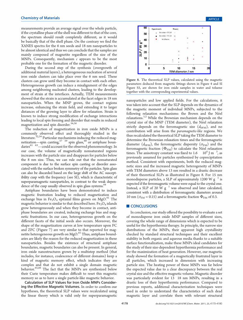
nanoparticles and low applied fields. For the calculations, itwas taken into account that the SLP depends on the dynamics ofthe magnetic moment of individual MNPs, subjected to thefollowing relaxation mechanisms: the Brown and the N�eelrelaxations.1,2 While the Brownian mechanism depends on thecrystal size of the MNP (TEM diameter), the N�eel relaxationstrictly depends on the ferromagnetic size (d0FM), and nocontribution will arise from the paramagnetic-like regions. Wethus recalculated the theoretical SLP taking the TEMdiameter todetermine the Brownian relaxation times and the ferromagneticdiameter (d0FM), the ferromagnetic dispersity (σFM) and theferromagnetic fraction (ΦFM) to calculate the N�eel relaxationtimes. The anisotropy constant was fixed to 1.6 � 104 J m�3, aspreviously assumed for particles synthesized by coprecipitationmethod. Consistent with experiments, both the reduced mag-netic sizes and the increased magnetic heterogeneity of MNPswith TEM diameters above 13 nm resulted in a drastic decreaseof their theoretical SLPs as illustrated in Figure 8. For 15 nmmonodisperse particles, a SLP of approximately 1200 W g�1 isexpected if the ferromagnetic volume were equal to the crystal one.Instead, a SLP of 20 W g�1 was observed and later calculated,consistent with a distribution of ferromagnetic diameters around10 nm (σFM = 0.15) and a ferromagnetic fractionΦFM of 0.3.
’CONCLUSIONS
In conclusion, our study offered the possibility to evaluate a setof monodisperse iron oxide MNP samples of different sizes,covering the whole range of dimensions which is expected to beuseful for the hyperthermia therapy. In principle, the narrow sizedistributions of the MNPs, their apparent high crystallinitychecked by standard structural techniques and their excellentstability in both organic and aqueous media thanks to a suitablesurface functionalization, make these MNPs ideal candidates forthe study of their size-dependent hyperthermia performance andfor the maximization of heat generation. However, our magneticstudy showed the formation of a magnetically frustrated layer inall particles, which increased in dimension with increasingparticle size. The heating power of these MNPs was far belowthe expected value due to a clear discrepancy between the realcrystal size and the effective magnetic volume. Magnetic disorderwas particularly evident for 13�18 nm MNPs, resulting in adrastic loss of their hyperthermia performance. Compared toprevious reports, additional characterization techniques wereused in order to understand the characteristics of such deadmagnetic layer and correlate them with relevant structural
Figure 8. The theoretical SLP values, calculated using the magneticparameters deduced from magnetic fittings shown in Figure 4 and SIFigure S5, are shown for iron oxide samples in water and toluenetogether with the corresponding experimental values.
4179 dx.doi.org/10.1021/cm201078f |Chem. Mater. 2011, 23, 4170–4180
Chemistry of Materials ARTICLE
features. Low-temperature magnetic measurements and strainmap analysis of single MNPs indicated the presence of structuraland magnetic antiphase boundaries originating from the highlystrained crystal lattice of the monocrystalline magnetite MNPs.As a consequence, the effective magnetic volumes of the particleswere significantly reduced as well as their magnetic dispersionsenlarged. The theoretical SLP values for each sample wererecalculated on the basis of the effective magnetic volume ofthe particles and their magnetic dispersion, thereby obtaining areasonable agreement with the experiments.
The origin of the crystal strain seems to be strictly related tothe two-step seeded-growth synthetic procedure used for theirpreparation. Generally speaking, the high temperature decom-position of organometallic precursors is a very promising proto-col for the preparation of nanoparticles in terms of size and sizedispersion control, as well as crystalinity. In particular, the seeded-growth route by which the present MNPs were grown ledefficiently to monocrystalline and monodisperse nanocrystals in awide range of sizes. However, this growth mechanism seems to beresponsible for the presence of strained lattice regionswhich turnedout to alter the magnetic structure of the crystals and which couldonly be detected by highly sensitive techniques. Hence, we high-light here the crucial role of the particular synthetic protocol chosenfor the preparation of iron oxide MNPs to be exploited as efficientheat nanomediators for hyperthermia treatments.
MNPs promise to become a powerful tool for cancer therapyin the next few years. However, all the works reported to dateindicate SLP values far below the ones theoretically predictedand practically required for the preparation of efficient thermalagents. The heating capacity of MNPs depends on the efficiencyof two heating mechanisms, that is, N�eel and Brown relaxations,which in turn depend on several structural and magnetic para-meters. Hence, the study of MNP-based hyperthermia faces thedifficulty of correlating several factors acting simultaneously andwhich are often difficult to decouple individually. Our workallowed us not only to discriminate among several structural andmagnetic features but also to establish correlations among them,which is a fundamental prerequisite to the understanding anddevelopment of a new generation of magnetic antitumoral drugs.
’ASSOCIATED CONTENT
bS Supporting Information. Synthesis of iron oxide MNPs;Transfer of iron oxide MNPs to aqueous solution and conditionsused for the purification of the polymer-coated iron oxide MNPsby means of ultracentrifugation; Dynamic light scattering data oniron oxide samples; gel electrophoresis characterization of water-soluble iron oxide MNPs; XANES characterization; analysis ofthe derivative curves of the X-ray absorption near edge spectra(XANES); X-ray absorption in a wider range of Fe K-edge;Calculation of the ferromagnetic volume fractions (ΦFM) andapparent paramagnetic-like susceptibilities (χ); Details on thestrain analysis by HRTEM; AC susceptibility data of iron oxideMNPs. This material is available free of charge via the Internet athttp://pubs.acs.org.
’AUTHOR INFORMATION
Corresponding Author*E-mail: [email protected] (A. F.); [email protected](M. A. G.).
’ACKNOWLEDGMENT
This work was supported by the European project Magnifyco(Contract NMP4-SL-2009-228622), the Spanish Ministerio deCiencia e Innovaci�on grants CSD2009-00013, MAT2008-06517-C02-01 and FIS-2008-06249 and Madrid Region Councilproject NANOBIOMAGNET (S2009/MAT-1726). A.F. acknowl-edges financial support from the Spanish Government through aRam�on yCajal Fellowship.We gratefully acknowledge R. Di Coratofor assistance with the polymer coating procedure, M. Povia forassistance with XRDmeasurements, and S. Nitti for assistance withthe synthesis of iron oxide MNPs.
’REFERENCES
(1) Gazeau, F.; Levy, M.; Wilhelm, C. Nanomedicine 2008, 3, 831.(2) Rosensweig, R. E. J. Magn. Magn. Mater. 2002, 252, 370.(3) Ma, M.; Wu, Y.; Zhou, J.; Sun, Y.; Zhang, Y.; Gu, N. J. Magn.
Magn. Mater. 2004, 268, 33.(4) Weinstein, J. S.; Varallyay, C. G.; Dosa, E.; Gahramanov, S.;
Hamilton, B.; Rooney, W. D.; Muldoon, L. L.; Neuwelt, E. A. J. Cereb.Blood Flow Metab. 2010, 30, 15.
(5) Johanssen, M.; Gneveckow, U.; Eckelt, L.; Feussner, A.; Waldo,N.; Scholz, R.; Deger, S.; Wust, P.; Loening, S. A.; Jordan, A Int. J.Hyperthermia 2005, 21, 637.
(6) Foy, S. P.; Manthe, R. L.; Foy, S. T.; Dimitrijevic, S.; Krishna-murthy, N.; Labhasetwar, V. ACS Nano 2010, 4, 5217.
(7) Owens, D. E.; Peppas, N. A. Int. J. Pharm. 2006, 307, 93.(8) Figuerola, A.; Di Corato, R.;Manna, L.; Pellegrino, T. Pharmacol.
Res. 2010, 62, 126.(9) Fortin, J.-P.; Wilhelm, C.; Servais, J.; M�enager, C.; Bacri, J.-C.;
Gazeau, F. J. Am. Chem. Soc. 2007, 129, 2628.(10) Purushotham, S.; Ramanujan, R.V. J. Appl. Phys.2010,107, 114701.(11) Levy, M.; Wilhelm, C.; Siaugue, J. M.; Horner, O.; Bacri, J. C.;
Gazeau, F. J. Phys.: Condes. Matter 2008, 20, 509901.(12) Gonzales-Weimuller, M.; Zeisberger, M.; Krishnan, K. M.
J. Magn. Magn. Mater. 2009, 321, 1947.(13) Dennis, C. L.; Jackson, A. J.; Borchers, J. A.; Hoopes, P. J.;
Strawbridge, R.; Foreman, A. R.; Lierop, J. v.; Gruttner, C.; Ivkov, R.Nanotechnology 2009, 20, 395103.
(14) Glockl, G.; Hergt, R.; Zeisberger, M.; Dutz, S.; Nagel, S.;Weitschies, W. J. Phys.: Condes. Matter 2006, 18, S2935.
(15) Kallumadil, M.; Tada, M.; Nakagawa, T.; Abe, M.; Southern, P.;Pankhurst, Q. A. J. Magn. Magn. Mater. 2009, 321, 1509.
(16) Rovers, S. A.; van der Poel, L. A. M.; Dietz, C. H. J. T.; Noijen,J. J.; Hoogenboom, R.; Kemmere, M. F.; Kopinga, K.; Keurentjes, J. T. F.J. Phys. Chem. C 2009, 113, 14638.
(17) Rovers, S. A.; Dietz, C.H. J. T.; v. d. Poel, L. A.M.; Hoogenboom,R.; Kemmere, M. F.; Keurentjes, J. T. F. J. Phys. Chem. C 2010, 114, 8144.
(18) B€uscher, K.; Helm, C. A.; Gross, C.; Gl€ockl, G.; Romanus, E.;Weitschies, W. Langmuir 2004, 20, 2435.
(19) Dennis, C. L.; Jackson, A. J.; Borchers, J. A.; Ivkov, R.; Foreman,A. R.; Lau, J. W.; Goernitz, E.; Gruettner, C. J. Appl. Phys. 2008,103, 07A319.
(20) Chen, S.; Chiang, C.-l.; Hsieh, S. J. Magn. Magn. Mater. 2010,322, 247.
(21) Hergt, R.; Dutz, S.; Roder, M. J. Phys.: Condes. Matter 2008,20, 385214.
(22) Hergt, R.; Dutz, S.; M€uller, R.; Zeisberger, M. J. Phys.: Condens.Matter 2006, 18, S2919.
(23) Frimpong, R. A.; Dou, J.; Pechan, M.; Hilt, J. Z. J. Magn. Magn.Mater. 2010, 322, 326.
(24) Kita, E.; Hashimoto, S.; Kayano, T.; Minagawa, M.; Yanagihara,H.; Kishimoto, M.; Yamada, K.; Oda, T.; Ohkohchi, N.; Takagi, T.;Kanamori, T.; Ikehata, Y.; Nagano, I. J. Appl. Phys. 2010, 107, 09B321.
(25) Zhao, D. L.; Teng, P.; Xu, Y.; Xia, Q. S.; Tang, J. T. J. AlloysCompd. 2010, 502, 392.

4180 dx.doi.org/10.1021/cm201078f |Chem. Mater. 2011, 23, 4170–4180
Chemistry of Materials ARTICLE
(26) Hyeon, T.; Lee, S. S.; Park, J.; Chung, Y.; Na, H. B. J. Am. Chem.Soc. 2001, 123, 12798.(27) Jun, Y.-w.; Huh, Y.-M.; Choi, J.-s.; Lee, J.-H.; Song, H.-T.;
KimKim; Yoon, S.; Kim, K.-S.; Shin, J.-S.; Suh, J.-S.; Cheon, J. J. Am.Chem. Soc. 2005, 127, 5732.(28) Park, J.; An, K.; Hwang, Y.; Park, J.-G.; Noh, H.-J.; Kim, J.-Y.;
Park, J.-H.; Hwang, N.-M.; Hyeon, T. Nat. Mater. 2004, 3, 891.(29) Sun, S.; Zeng, H. J. Am. Chem. Soc. 2002, 124, 8204.(30) Kwon, S. G.; Hyeon, T. Acc. Chem. Res. 2008, 41, 1696.(31) Jun, Y.-w.; Choi, J.-s.; Cheon, J. Angew. Chem., Int. Ed. 2006,
45, 3414.(32) Guardia, P.; Batlle-Brugal, B.; Roca, A. G.; Iglesias, O.; Morales,
M. P.; Serna, C. J.; Labarta, A.; Batlle, X. J. Magn. Magn. Mater. 2007,316, e756.(33) Roca, A. G.; Niznansky, D.; Poltierova-Vejpravova, J.; Bittova,
B.; Gonzalez-Fernandez, M. A.; Serna, C. J.; Morales, M. P. J. Appl. Phys.2009, 105, 114309.(34) Batlle, X.; Labarta, A. J. Phys. D: Appl. Phys. 2002, 35, R15.(35) Tronc, E.; Fiorani, D.; Nogu�es, M.; Testa, A. M.; Lucari, F.;
D’Orazio, F.; Gren�eche, J. M.; Wernsdorfer, W.; Galvez, N.; Chan�eac,C.; Mailly, D.; Jolivet, J. P. J. Magn. Magn. Mater. 2003, 262, 6.(36) Shendruk, T. N.; Desautels, R. D.; Southern, B. W.; van Lierop,
J. Nanotechnology 2007, 18, 455704.(37) Pellegrino, T.; Manna, L.; Kudera, S.; Liedl, T.; Koktysh, D.;
Rogach, A. L.; Keller, S.; R€adler, J.; Natile, G.; Parak, W. J. Nano Lett.2004, 4, 703.(38) Cozzoli, P. D.; Snoeck, E.; Garcia,M. A.; Giannini, C.; Guagliardi,
A.; Cervellino, A.; Gozzo, F.; Hernando, A.; Achterhold, K.; Ciobanu, N.;Parak, F. G.; Cingolani, R.; Manna, L. Nano Lett. 2006, 6, 1966.(39) Park, J.; Lee, E.; Hwang, N.-M.; Kang, M.; Kim, S. C.; Hwang,
Y.; Park, J.-G.; Noh, H.-J.; Kim, J.-Y.; Park, J.-H.; Hyeon, T. Angew.Chem., Int. Ed. 2005, 44, 2872.(40) Di Corato, R.; Quarta, A.; Piacenza, P.; Ragusa, A.; Figuerola,
A.; Buonsanti, R.; Cingolani, R.;Manna, L.; Pellegrino, T. J. Mater. Chem.2008, 18, 1991.(41) Galindo,P.L.;Kret, S.; Sanchez,A.M.; Laval, J. Y.; Yanez,A.; Pizarro,
J.; Guerrero, E.; Ben, T.; Molina, S. I. Ultramicroscopy 2007, 107, 1186.(42) Bacri, J. C.; Perzynski, R.; Salin, D.; Cabuil, V.; Massart, R.
J. Magn. Magn. Mater. 1986, 62, 36.(43) Yu, H.; Gibbons, P. C.; Kelton, K. F.; Buhro, W. E. J. Am. Chem.
Soc. 2001, 123, 9198.(44) Klug, H. P.; Alexander, L. E. X-ray Diffraction Procedures for
Polycrystalline and Amorphous Materials; John Wiley & Sons: New York,1962.(45) Cullity, B. D.; Stock, S. R. Elements of X-Ray Diffraction; 3rd ed.;
Prentice-Hall Inc.: New York, 2001.(46) Petkov, V.; Cozzoli, P. D.; Buonsanti, R.; Cingolani, R.; Ren, Y.
J. Am. Chem. Soc. 2009, 131, 14264.(47) Kim, T.; Shima, M. J. Appl. Phys. 2007, 101, 09M516.(48) Nogues, J.; Schuller, I. K. J. Magn. Magn. Mater. 1999, 192, 203.(49) Nogues, J.; Sort, J.; Langlais, V.; Skumryev, V.; Surinach, S.;
Munoz, J. S.; Baro, M. D. Phys. Rep. 2005, 422, 65.(50) Iglesias, O.; Labarta, A.; Batlle, X. J. Nanosci. Nanotechnol. 2008,
8, 2761.(51) Casavola, M.; Falqui, A.; Garcia, M. A.; García-Hern�andez, M.;
Giannini, C.; Cingolani, R.; Cozzoli, P. D. Nano Lett. 2009, 9, 366.(52) Caruntu, D.; Caruntu, G.; O’Connor, C. J. J. Phys. D: Appl. Phys.
2007, 40, 5801.(53) Bickford, L. R. J. Phys. Rev. 1950, 78, 449.(54) Walz, F. J. Phys.: Condens. Matter 2002, 14, R285.(55) Santoyo Salazar, J.; Perez, L.; de Abril, O.; Phuoc, L. T.;
Ihiawakrim, D.; Vazquez, M.; Greneche, J.-M.; Begin-Colin, S.; Pourroy,G. Chem. Mater. 2011, 23, 1379.(56) Figuerola, A.; Fiore, A.; Di Corato, R.; Falqui, A.; Giannini, C.;
Micotti, E.; Lascialfari, A.; Corti, M.; Cingolani, R.; Pellegrino, T.;Cozzoli, P. D.; Manna, L. J. Am. Chem. Soc. 2008, 130, 1477.(57) Pankhurst, Q. A.; Pollard, R. J. Phys. Rev. Lett. 1991, 67, 248.
(58) Morales, M. P.; Veintemillas-Verdaguer, S.; Montero, M. I.;Serna, C. J.; Roig, A.; Casas, L.; Martinez, B.; Sandiumenge, F. Chem.Mater. 1999, 11, 3058.
(59) Linderoth, S.; Hendriksen, P. V.; Bodker, F.; Wells, S.; Davies,K.; Charles, S. W.; Morup, S. J. Appl. Phys. 1994, 75, 6583.
(60) Mydosh, J. A. J. Magn. Magn. Mater. 1978, 7, 237.(61) Arora, S. K.; Sofin, R. G. S.; Nolan, A.; Shvets, I. V. J. Magn.
Magn. Mater. 2005, 286, 463.(62) Novakova, A. A.; Lanchinskaya, V. Y.; Volkov, A. V.; Gendler,
T. S.; Kiseleva, T. Y.; Moskvina, M. A.; Zezin, S. B. J. Magn. Magn. Mater.2003, 258, 354.
(63) Margulies, D. T.; Parker, F. T.; Rudee, M. L.; Spada, F. E.;Chapman, J. N.; Aitchison, P. R.; Berkowitz, A. E. Phys. Rev. Lett. 1997,79, 5162.
(64) Mulder, C. A.M.; van Duyneveldt, A. J.; Mydosh, J. A. Phys. Rev.B 1981, 23, 1384.
(65) Morales, M. P.; Gonzalez-Carre~no, T.; Serna, C. J. J. Mater. Res.1992, 7, 2538.
(66) Stachen, M.; Morales, M. P.; Ocan, M.; Serna, C. J. Phys. Chem.Chem. Phys. 1999, 1, 4465.