Micromechanical properties of biological silica in skeletons of deep-sea sponges

ORIGINAL PAPER
Coral cavity sponges depend on reef-derived food resources:stable isotope and fatty acid constraints
Fleur C. van Duyl • Leon Moodley • Gerard Nieuwland • Lennart van Ijzerloo •
Rob W. M. van Soest • Marco Houtekamer • Erik H. Meesters •
Jack J. Middelburg
Received: 28 April 2010 / Accepted: 16 March 2011 / Published online: 12 April 2011
� Springer-Verlag 2011
Abstract The diet of cavity sponges on the narrow
fringing reefs of Curacao, Caribbean was studied. The
origin and resources of the bulk food of these sponges, i.e.,
dissolved organic matter (DOM), were identified using
stable carbon and nitrogen isotopes and fatty acid bio-
markers. We found that phytoplankton and its derived
DOM from the adjacent open sea and from reef overlying
water is not the main source of food for most of the
sponges examined nor is bacterioplankton. Interestingly,
dual stable isotope signatures (d13Corg, d15Norg) and fatty
acid biomarkers appoint coral mucus and organic matter
derived from crustose coralline algae (CCA) as probable
food sources for encrusting sponges. Mucus-derived DOM
may contribute up to 66% to the diet of examined sponges
based on results of dual isotope mixing model analysis. The
contribution of CCA (as purported representative for ben-
thic algae) was smaller with values up to 31%. Together,
mucus- and CCA-derived substrates contributed for
48–73% to the diet of sponges. The presence of the
exogenous fatty acid 20:4x6 in sponges, which is abundant
in coral mucus of Madracis mirabilis and in CCA, high-
lights these reef-derived resources as sources of nutrition
for DOM feeding cavity sponges. The relatively high
concentrations of exogenous 20:4x6 in all sponges exam-
ined supports our hypothesis that the bulk of the food of the
cavity sponge community is reef-derived. Our results imply
that cavity sponges play an important role in conserving
food and energy produced within the reef.
Introduction
Sponges play an important role in the benthic-pelagic
coupling on coral reefs (Lesser 2006). The sponge com-
munity sequesters and processes organic matter and as such
conserve energy and nutrients for the reef ecosystem. It is
generally assumed that sponges on the fore-reef slope
mainly trap allochthonous organic matter that may com-
pensate the losses of material from reef ecosystems (e.g.,
Richter et al. 2001; Atkinson and Falter 2003; Ribes et al.
2005). Most reef sponges are extremely efficient in filter
feeding (e.g., Reiswig 1974; Lesser 2006). They preferably
filter the picoplankton fraction from the flowing water (e.g.,
Pile 1999; Ribes et al. 2003, 2005). The major food
resource of coral reef sponges, however, may well be
dissolved organic matter (Reiswig 1990; Yahel et al. 2003).
Various encrusting high microbial abundance sponges
(HMA sponges, sensu Hentschel et al. 2003) in coral
cavities are fueled mainly by dissolved organic matter
Communicated by U. Sommer.
F. C. van Duyl (&) � G. Nieuwland
Royal Netherlands Institute for Sea Research,
P.O. Box 59, 1790 AB Den Burg, Texel, The Netherlands
e-mail: [email protected]
L. Moodley � L. van Ijzerloo � M. Houtekamer �J. J. Middelburg
Netherlands Institute of Ecology (NIOO),
Korringaweg 7, 4401 NT Yerseke, The Netherlands
J. J. Middelburg
Faculty of Geosciences, University of Utrecht,
Budapestlaan 4, 3584 CD Utrecht, The Netherlands
E. H. Meesters
Wageningen IMARES, P.O. Box 167, Location Texel,
Landsdiep 4, 1797SZ ‘t Horntje, 1790 AD Den Burg,
The Netherlands
R. W. M. van Soest
Zoological Museum, University of Amsterdam,
P.O. Box 94766, 1090 GT Amsterdam, The Netherlands
123
Mar Biol (2011) 158:1653–1666
DOI 10.1007/s00227-011-1681-z

(DOM) (De Goeij et al. 2008a). Encrusting sponges cover
together with coralline algae [50% of the walls in coral
cavities, caves, and undersides of corals (Vasseur 1974;
Richter and Wunsch 1999; Wunsch et al. 2002; Scheffers
et al. 2004). This cryptic surface of 3-dimensionally
well-developed reefs may exceed the open reef surface
(Ginsburg 1983; Richter et al. 2001; Scheffers et al. 2003).
The sheer size of this reef habitat already preludes the
grand role of its biota in organic matter cycling. DOM
removal by the cryptic biota on coral reefs can be huge (up
to 500 mmol C m-2 cryptic surface d-1), with cavity
sponges accounting for 70% of the consumed DOM in
coral reef cavities (De Goeij and Van Duyl 2007; De Goeij
et al. 2008a).
These recent findings draw direct attention to the origin
and sources of DOM on coral reefs. Phytoplankton
(including photoautotrophic bacteria) usually is the main
source of reactive DOM in oligotrophic ocean water; it
releases DOM as photosynthetic product or through food
web processes and lysis (Carlson 2002). On coral reefs
extra sources of reactive DOM include release of DOM by
corals through mucus production (Johannes 1967; Richman
et al. 1975; Crossland et al. 1980; Naumann et al. 2010)
and extracellular release of DOM by benthic algae (e.g.,
Wada et al. 2007; Haas et al. 2010). The main consumers
of reactive DOM are usually heterotrophic bacteria (e.g.,
Harvey et al. 2006 and references therein) that may com-
pete with sponges for DOM. The fate of mucus and dis-
solved organic matter in reef overlying waters has been
ascribed to consumption and mineralization by bacteria in
the reef ambient water and in sediments (e.g., Gast et al.
1999; Ferrier-Pages et al. 2000; Van Duyl and Gast 2001;
Wild et al. 2004). The first indication that reef-derived
DOM may also be assimilated by higher trophic levels
came from the abundance of the dietary fatty acid bio-
marker 20:4x6 in the encrusting DOM feeding cavity
sponge Halisarca caerulea (De Goeij et al. 2008b). This
fatty acid is especially abundant in corals (Latyshev et al.
1991) and in coralline algae, Rhodophyta (Viso and Marty
1993) and its presence in sponges suggests that reef-
derived organic matter may be an important food source for
coral reef sponges.
This study aims (1) to determine whether cavity sponges
mainly rely on reef-produced or open water-derived dis-
solved organic matter and (2) to identify key sources of
DOM for cavity sponges. As putative food compounds
different fractions of suspended matter, bacterioplankton,
coral mucus, and coralline algae as proxies for DOM were
analyzed. Through stable isotopes and fatty acid bio-
markers, we characterized energy sources of cavity spon-
ges collected from coral reefs and open water on the
southwest coast of Curacao, Caribbean (128120N,
688560W). Stable isotopes are a classical way to trace food
sources of aquatic animals (Peterson and Fry 1987). The
carbon and nitrogen stable isotope signatures of 12 sponge
species were compared with those of putative food com-
pounds. Fatty acid biomarkers have been repeatedly used
as source-specific indicators of dissolved and particulate
organic matter both in environmental and in food web
studies (e.g. Hall et al. 2006; Thurber 2007). The FA pat-
tern of six sponge species and three types of reef food
resources were analyzed, and the relative input of different
sources to the diet of the sponges was determined.
Materials and methods
Sampling sites and sample collection
Samples were collected from March 14 to 22, 2006, along
four transects perpendicular to the SW coast of Curacao
(Fig. 1). Transects were placed 3–7 km apart from SE to
NW along the coast at Lagun Jan Thiel (LJT), at the ocean
Cruise Terminal in Otrabanda (CT), at Buoy 1 (B1) of the
Carmabi reef close to Piscaderabaai, and at Slangenbaai
(SB). Transects from the shore to open water cross the
narrow fringing reefs and water masses representative for
the variation in coral community composition and water
quality along the SW coast of Curacao (Van Duyl 1985;
Gast et al. 1999; Van Duyl and Gast 2001). Along each
transect (LJT, CT, B1, and SB), we sampled 2 stations:
1. an off-shore open water station about 1–2 km distance
from the reef where water was collected at 15–17 m
depth in the blue water and
2. a coral reef station where we collected bottom water at
15–17 m depth on the fore-reef slope, approximately
0.5 m above the coral bottom.
Water samples were taken with a 6 L Niskin bottle
operated by a SCUBA diver. The Niskin bottle was
Fig. 1 Position of sampling transects (LJT, CT, B1, and SB) along
the southwest coast of Curacao, with an inset of the geographic
position of the island in the Caribbean
1654 Mar Biol (2011) 158:1653–1666
123

repeatedly filled and hauled on board a small open boat and
emptied in a clean and sample washed 20 L container. At
each station (n = 8), we collected 20 L. On the fore-reef
slope stations (n = 4), we collected a few small pieces of
the stony corals Montastraea annularis and Madracis
mirabilis (also Siderastrea siderea and Porites astreoides
but only at B1) between 15 and 17 m depth, which were
kept in seawater under natural light conditions until mucus
collection on the next day. From the entrance of coral
cavities at 15–17 m depth on the fore-reef slope, we
chiseled crusts of calcareous algae. From the same cavities,
various species of encrusting sponges were collected, up to
eight different species per station. Specimens were chiseled
from the walls and collected in plastic bags while SCUBA
diving.
Sample treatment
The 20-L water samples were filtered over 47-mm-diam-
eter combusted glass microfiber filters (Whatman), first
over a GFC filter (nominal pore size 1.2 lm) and then over
a GFF filter (nominal pore size 0.7 lm). After sea water
filtration, filters were shortly washed with Milli-Q water
(MQ) to remove salt and were dried at 50�C in a stove and
stored in aluminum foil in the freezer until processing. The
remaining bacterioplankton in the GFF filtrate was con-
centrated with a Vivaspin filter cartridge driven by a
Master Volt tube pump. The concentrate was subsequently
filtered over a 0.2 lm pore size 25-mm-diameter Anopore
disc (aluminum oxide membrane filter, Whatman). The
discs were briefly washed with MQ after seawater filtration
and dried at 50�C in a stove (12 h). After drying, the filter
was crumbled in a clean acid-washed glass funnel, the
integrated polypropylene support ring of the filter was
removed and the filter fragments were transferred to
combusted silver capsules, which were closed with twee-
zers. Subsequently, the folded capsules were placed in
coded trays and stored in the freezer until processing.
Live corals were air-exposed during mucus collection
and positioned upside down connected to a stand. After
shortly spraying the live coral tissue with MQ, the mucus
produced by the coral was collected via an acid-washed
glass funnel in combusted glass vials. After collection of
approximately 2 mL mucus per coral, mucus samples were
dried in a stove at 50�C before being stored dry in the
freezer. Mucus samples were prepared in duplicate.
The cavity sponges and coralline algae were quickly
washed with MQ to remove salt and the tissue was sub-
sequently removed from the underlying substratum by
knife and scalpel. Small pieces were fixed in ethanol (80%)
for taxonomical identification of the different sponge spe-
cies. The rest of the pieces were collected in combusted
glass vials dried at 50�C and stored dry in the freezer.
Sponges and putative food sources were subject to stable
isotope analysis. Both carbon and nitrogen (except for
bacteria for which only d13Corg was measured) isotopic
composition of the samples were determined using a Fisons
CN analyser coupled on line via a Finnigan conflo 2
interface, to a Finnigan Delta S mass spectrometer. The
carbon and nitrogen isotope ratios are expressed as d13Corg
and d15N relative to the Vienna Pee Dee Belemnite stan-
dard and air, respectively, and the standard error of the
measurements is *0.15%. All carbon isotope samples
(except bacteria that were caught on aluminum filters) were
acidified (tissue directly with acid and glass filters in acid
vapor) prior to measurements and corrected for individual
sets of blanks (isotope mass balance corrections). Seawater
as source of carbonates leading to possible enrichment of
d13Corg in case of un-acidified aluminum bacteria filters
was eliminated by the rinse with MQ water at the end of
filtration. d15N values represent total nitrogen.
The dried sponge, coralline algae, and mucus material
were first homogenized in a mortar. For isotope measure-
ments, adequate amount of material was transferred to
pre-combusted silver boats for acidification within cups,
followed by oven drying to remove excess acid. From the
remaining grounded material, samples from the B1 reef
station were also analyzed for their total fatty acid com-
position using a method of one-step procedure of fatty acid
extraction and methylation (Masood et al. 2005). We
extracted three times 10 mg dry material of each individual
sample. Fatty acid extraction and preparation of methyl
esters (FAME) were carried out according to Masood et al.
(2005) with reagent volumes adapted for use in 2.5-mL
GC-vials using FAME C19:0 as internal standard to cal-
culate concentration of FAs (Van Gaever et al. 2009). These
individual samples were analyzed separately for their FA
compositions employing a Large Volume Splitless injection
method on a Thermo Finnigan Trace Ultra GC. Main items
of this method are the large volume liner with glass wool,
pre-column deactivated silica 5 m 9 0.53 lm, and analyt-
ical column SGE BPX-70 50 m 9 0.32 mm 9 0.25 lm.
The identification of FAMES was based on the comparison
of retention times with authentic commercially available
reference material and standards. Fatty acid source desig-
nation was achieved using data on distinctive FA for bac-
teria, algal carbon, and higher organisms (e.g., Parrish et al.
2000; Boschker and Middelburg 2002; Volkman 2006).
Statistical analyses
We ran one- and two-way ANOVAs with non-transformed
stable isotope data to determine whether variations in sta-
ble isotopes of sponges were due to sponge species and/or
sponge location assuming no interaction effects (not
enough replication for testing interaction). The Tukey’s
Mar Biol (2011) 158:1653–1666 1655
123
HSD method was used for pair wise comparisons of dif-
ferent sponge species (multiple comparisons of means).
ANOVA’s were also run for isotope signals of reef and
open water-derived suspended matter.
The SIAR stable isotope mixing model analysis (Parnell
et al. 2010) was applied to investigate probable solutions
for the diet of sponges based on dual stable isotope data of
four sources (GFC and GFF fractions of suspended matter,
crustose coralline algae, and coral mucus). We used the
following trophic enrichment factors with standard devia-
tions for the sponges: 3.5 ± 0.5 for d15N and 1 ± 1 for d13C (Vander Zanden and Rasmussen 2001; Behringer and
Butler IV 2006). The SIAR model takes data on organism
isotopes and fits a Bayesian model to their dietary habits
based upon a Gaussian likelihood with a mixture dirichlet-
distributed prior on the mean. Model fitting is via Markov
Chain Monte Carlo (MCMC) which produces simulations
of plausible values for each dietary source consistent with
the data. The software package SIAR (Stable Isotope
Analysis in R) is freely available from the package section
of the Comprehensive R Archive Network website—
http://cran.r-project.org/ (Parnell and Jackson 2010).
Fatty acid composition patterns were explored with
multivariate analyses of FAs composition profiles (non-
transformed percentage abundance) using the program
PRIMER Version 5 (Clarke and Gorley 2006). Multi-
dimensional scaling (MDS plots) of FA composition and
dual isotope label patterns were compared for six sponge
species and the three different reef-derived resources
(CCA, mucus of M. annularis, and mucus of M. mirabilis).
Similarities between FA profiles were investigated using
the SIMPER (Similarity of Percentages) function, and
statistical differences were determined using the ANOSIM
(Analysis of Similarities) function.
Results
The sponge species collected on the reef stations were
identified on basis of their tissue and skeletal characteris-
tics. Twelve different sponge species were recorded.
Sponges had d13Corg values ranging from -17.2%(Chondrilla caribensis, Ruetzler et al. 2007) to -20.7%(Scopalina ruetzleri) and d15N values ranging from 2.9%(Monanchora arbuscula) to 7.1% (Scopalina ruetzleri)
(Table 1). Variations in d13Corg and d15N data were sig-
nificantly related to sponge species (ANOVA, F(5,17) =
30.3 and F(5,17) = 15.8, P \ 0.01 for both) and not to reef
station (F(3,17) = 1.16 and F(3,17) = 0.83, P [ 0.35 for
both). Significant differences in d13C as well as d15N
(P \ 0.05) were found between S. ruetzleri and H. caeru-
lea, S. ruetzleri and Niphates erecta, N. erecta and
M. arbuscula, which were sampled at three to four reef
sites (Table 1). Differences in dual stable isotope values
between sponge species were small when compared to
isotope values of sources. Therefore, the sponge data of the
different transects were combined in the Fig. 2a, b. The
stable isotope signals of particulate organic matter size
fractions were not significantly different between the open
water and reef water (ANOVA, P [ 0.05), but were kept
separate in Fig. 2a, b. Figure 2a shows that open water and
reef water bacteria have on average slightly different d13C
signatures. Variations in d13Corg values of bacterioplankton
were large between stations (LJT and SB vs. CT and B1,
Table 2). Only the reef water bacteria of station SB (d13C:
-17.9%) were in trophic reach of sponges (\2% differ-
ence). The sponge trophic range overlaps with the range of
coralline algae (average d13C of -16.9 ± 4.2%). Varia-
tions in isotope signals were also large between algal
samples from different stations. Coral mucus of Montast-
raea annularis and Madracis mirabilis was on average
heavier in 13Corg content than the sponges (Table 2). Coral
mucus and coralline algae are potential sponge food
because of a trophic level d15N increase of 3–4% (Fig. 2b).
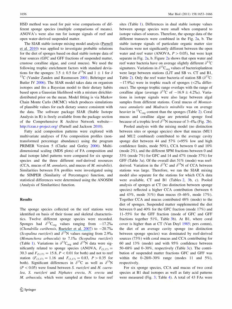
Pooled analysis with the mixing model (no distinction
between sites or sponge species) show that mucus (MU1
and MU2 combined) contributed to the average cavity
sponge diet between 44 and 57% (=95% low and high
confidence limits, mode 50%), CCA between 0 and 10%
(mode 2%), and the different SPM fractions between 0 and
33% (mode 7%) for GFC and 14 and 47% (mode 37%) for
GFF (Table 3a). Of the overall diet 51% (mode) was reef-
derived. Variation in the d13C and d15N of CCA between
stations was large. Therefore, we ran the SIAR mixing
model also separate for the stations for which CCA data
were available, CT and B1 (Tables 2, 3b, c). Pooled
analysis of sponges at CT (no distinction between sponge
species) reflected a higher CCA contribution (between 6
and 43%, mode 31%) than mucus (0–46%, mode 17%).
Together CCA and mucus contributed 48% (mode) to the
diet of sponges. Suspended matter supplemented the diet
between 0 and 40% for the GFC fraction (mode 17%) and
11–55% for the GFF fraction (mode of GFC and GFF
fractions together 51%, Table 3b). At B1, where coral
cover is higher than at CT (Van Duyl 1985; pers comm),
the diet of an average cavity sponge (no distinction
between sponge species) was dominated by reef-derived
sources (73%) with coral mucus and CCA contributing for
60 and 13% (mode) and with 95% confidence between
50–68% and 0–30%, respectively (Table 3c). The contri-
bution of suspended matter fractions GFC and GFF was
within the 0–28/0–30% range (modes 11 and 5%),
respectively.
For six sponge species, CCA and mucus of two coral
species at B1 dual isotopes as well as fatty acid patterns
were measured (Fig. 3; Table 4). A total of 43 FAs were
1656 Mar Biol (2011) 158:1653–1666
123

identified. FAs in sources were also present in most
sponges, but the diversity in FAs was higher in sponges.
Differences between sources (CCA and mucus) and spon-
ges in the fatty acid compositions are illustrated in Fig. 4.
The Euclidean distances show that the FAs of sponges
cluster together and are distinct from the sources. Main
FAs of the sponge cluster included, in order of importance
16:0, 20:4x6, 18:0, 20:5x3, 18:1x7c, and branched 20:0
(phytanic acid) whereas in the case of the potential energy
sources this included 16:0, 18:0, 18:1x9c, and 20:4x6
(Table 4). The source groups were significantly different
(ANOSIM, Global R = 0.8, P \ 0.05) from the sponge
group and SIMPER analysis revealed that the differences
are the relatively higher percentage abundance of 16:0,
20:4x6, 18:0, and 18:1x9c in coral mucus and coralline
algae but higher percentage abundance of phytanic acid,
24:0 and 20:5x3 in the sponges. These 7 FAs together
accounted for *60% of the FA dissimilarity (Table 4;
Fig. 4). Among the three different sources, coralline algae,
mucus of Montastraea annularis, and Madracis mirabilis,
composition patterns were also significantly different
(ANOSIM, Global R = 1, P \ 0.05). SIMPER analysis
revealed two very dominant fatty acids in the coralline
algae (16:0 and 20:4x6) that together accounted for *73%
of the FA; in mucus from M. annularis (MU1), four
abundant FAs made up *70% (16:0, 18:0, 20:5x3, and
18:1x9c); in mucus from M. mirabilis (MU2), three
abundant FAs made up *70% (16:0, 18:0, and 20:4x6,
Table 2). The major FAs accounting for the dissimilarity
between mucus from the two different coral species is the
relatively higher percentage abundance of 20:4x6 and 18:0
in M. mirabilis (MU2) but a relatively higher percentage
abundance of 18:1x7c and 20:5x3 in M. annularis (MU1)
with these 4 FAs accounting for *60% of the dissimilarity
between the coral species (Table 4). There was low simi-
larity in FA patterns between most of the sponges. Three
groups of sponges were distinguished among the 6 sponge
species (Fig. 4: H. caerulea (SP1), C. caribensis (SP3) and
Table 1 Sponge species collected at four reef stations with their respective average isotope signatures
Cryptic biota Station LJT
Lagun Jan Thiel
CT
Cruise terminal
B1
Carmabi reef
SB
Slangenbaai
Code d13C d15N d13C d15N d13C d15N d13C d15N
Halisarca caerulea
n = 7
SP1 -18.79 3.69 -18.45 4.12 -18.33 3.16
-18.48 4.02 -18.20 4.09
-17.87 4.07 -18.73 4.63
Clathria (Thalysis) raraechelae
n = 1
SP2 -18.91 4.03
Chondrilla caribensis
n = 1
SP3 -17.16 3.83
Chelonaplysilla erecta
n = 1
SP4 -19.94 4.68
Desmapsamma anchorata
n = 1
SP5 -18.81 5.61
Scopalina ruetzleri
n = 6
SP6 -19.76 5.90 -20.26 4.85 -20.79 5.48 -20.02 6.13
-19.41 7.07 -20.06 5.86
Eurypon laughlini
n = 2
-18.95 5.67 -18.51 5.78
Topsentia ophirhaphidites
n = 2
-19.63 5.37
-19.27 5.82
Niphates erecta
n = 6
-17.70 4.01 -17.65 4.79 -17.93 4.47 -17.99 4.21
-17.97 4.71
-18.00 4.71
Callyspongia vaginalis
n = 2
-17.72 3.79 -17.47 4.37
Monanchora arbuscula
n = 5
-20.38 3.83 -20.40 3.42 -19.82 3.28
-19.95 2.84 -19.47 3.63
Phorbas amaranthus
n = 1
-18.86 4.40
Codes SP1–SP6 refer to sponges collected at B1 and correspond to sponge data presented in Fig. 3
Mar Biol (2011) 158:1653–1666 1657
123

C. erecta (SP4) make up the first group; C. raraechelae
(SP2) and D. anchorata (SP5) the second and S. ruetzleri
(SP6) the third group). SIMPER analysis revealed different
combinations of FAs, in order of dominance, accounting
for *70% of sponge group similarity: for the first group
(SP1, SP3, and SP4) FAs 16:0, br 20:0 (phytanic acid),
18:0, 20:4x6, i15:0, and 16:1x7c are the main FAs. In the
second group (SP2 and SP5) FAs 16:0, 20:4x6, 24:0,
18:1x7c, 22:0, 20:5x3, and 20:6x3 determined primarily
the composition pattern. In the third group (SP6), these
were the five FAs: 16:0, 20:4x6, 18:0, 20:5x3, and 14:0
(Table 4).
For these three distinguished sponge groups at B1, we
also ran the isotope mixing model (Table 5). Results show
that the three groups make proportionally different use of
the sources. The first group appears to rely mainly on reef-
derived sources (mucus and CCA, mode and mean both
[75%). The second group probably relied less on mucus
and more on CCA and used more plankton-derived food
compared with group 1. The diet of group 3 (=S. ruetzleri
only) consists for about 51% (mean) of reef-derived and
49% (mean) of suspended matter (plankton) derived food.
Group 1 has a smaller centroid Euclidean distance (23.64)
to the average mucus mixture than groups 2 and 3 (32.43
and 24.55) in Fig. 4. This concurs with a larger contribu-
tion of mucus (66%) to the diet of group 1 than that of the
other two groups (36 and 33%) (Fig. 4; Table 5). The
lower contribution of CCA in the diet of group 1 (10%
mode) compared to group 3 (19% mode) was also reflected
in Fig. 4. We did not find this for group 2, which had the
longest Euclidean distance to CCA (41.81 vs. 33.78 of
group 3 and 37.37 of group 1), while CCA-derived food
comprised 22% in the diet of this group. To further explore
the relation between the stable isotope and FA data sets, we
compared the matrices of FAs and dual isotope signatures
of sponges and reef resources (Figs. 3, 4) and found a
significant correlation between the two matrices (PRIMER,
Rho = 0.625, P = \0.01).
Discussion
Cryptic encrusting sponges living in coral cavities on the
fore-reef slope of fringing reefs sequester most of their
organic matter from the passing water. They efficiently
filter the pico- and nanoplankton, but dissolved organic
matter (DOM) appears to be the major resource of various
sponge species living on the open reef and in coral cavities
(Yahel et al. 2003; De Goeij et al. 2008a). The origin and
source of this organic matter is unknown. It may be DOM
released from phytoplankton, DOM supplied to the reef
from outside or DOM released from the reef benthos.
Gradients in DOM concentrations over the reef suggest that
there is a net release of DOM from coral reef waters to
adjacent open waters (Van Duyl and Gast 2001). Only
10–20% of the DOM in reef water appears to be available
for sponges (De Goeij et al. 2008a). By analyses of dual
stable isotope signatures (d13Corg and d15N) and FA pat-
terns of sponges and their potential food sources, we
obtained circumstantial evidence that the reef itself mainly
feeds her cavity sponges. This remarkable finding implies
that cryptic sponges support reef internal nutrient recycling
and conservation of reef organic matter.
It was surprising that none of the 12 sponge species
examined appeared to live mainly on suspended particulate
organic matter or DOM derived from this source. The
d13Corg values of the SPM fractions were in the range
reported for marine suspended matter (Peterson and Fry
1987). Bacterioplankton values were within 2% of SPM in
open water, which indicates that bacteria were the primary
consumers of SPM. This makes it highly likely that SPM
represents predominantly phytoplankton and that the bac-
terioplankton reflects the d13C isotope ratio of reactive
DOM released by phytoplankton (e.g., Harvey et al. 2006;
Fig. 2 Plots of carbon (a) and nitrogen (b) stable isotope signatures
of material collected from the four reef and four open water stations
(average and standard error). Sponges (n = 35), Bacteria (Bact
n = 8), Coral mucus (n = 9), Glass microfiber filters coarse (GFC)
and fine (GFF) represent values of suspended matter (SPM) collected
either from reef water or from open water offshore (n = 4 or 5).
Crustose coralline algae from two reef stations (n = 3)
1658 Mar Biol (2011) 158:1653–1666
123

Van den Meersche et al. 2009). The d13C of SPM did not
change during passage over the reef, for instance by
obtaining a typical d13C SPM reef signal (-17 to -20%:
Land et al. 1975; Fry et al. 1982; Yamamuro et al. 1995).
The d15N of SPM did not change either and remained
*4%. Moreover, at two of the four stations (CT and B1)
comparable d13Corg values of predominantly heterotrophic
bacterioplankton (-26, -27%, Table 2) between open
water and reef bottom water suggest that this bacterio-
plankton remained dependent on phytoplankton DOC.
Such low d13C values have been reported, for e.g., Syn-
echococcus (d13C -33 to -25%, Brutemark et al. 2009)
and Prochlorococcus, both abundant in reef and open water
along Curacao (Van Duyl et al. 2002). Residence time of
the SPM and bacterioplankton in reef overlying waters may
have been too short for changes in isotopic ratios either due
to rapid water exchange with the open water and/or due to
SPM removal by benthic suspension feeders. Phytoplank-
ton concentrations in reef surrounding waters in Curacao
are usually low (van Duyl et al. 2002), and the organic
matter demand of cryptic biota, cryptic sponges in
particular, is high. Cavity biota between 10 and 15 m depth
on the fore-reef slope in Curacao consume on average
350 mmol C m-2 d-1 of which cavity sponges consume
on average 256 mmol C m-2 d-1 (73%) with the DOC
fraction comprising [90% of the diet (De Goeij et al.
2008a). This organic matter supply is orders of magnitude
higher than the supply of pelagic primary production to
reefs (Richter et al. 2001; Genin et al. 2009). It is evident
that the supply of open water-derived phytoplankton and its
DOM release is insufficient to satisfy the total organic
carbon requirements of coral reef cavity sponges. In
accordance, the isotope mixing model results confirmed a
SPM contribution of less than \50% in the diet of most
sponges. This implies that the examined sponges with
average d13Corg of -18.9% and d15N of 4.6% mainly
assimilate other sources than pelagic primary production.
Benthic primary production is the most likely other
source of DOM for sponges. Presence of distinct reactive
DOM in reef bottom water compared to open water was
demonstrated by the increased d13C value of bacterio-
plankton at two of the four stations (LJT and SB).
Table 2 Average isotope signatures of putative food sources for sponges collected from reef stations LJT, CT, B1, and SB (replicates, n = 2–3,
standard errors range from 0 to 0.4, not shown)
Substrate sources Station LJT
Lagun Jan Thiel
CT
Cruise terminal
B1
Carmabi reef
SB
Slangenbaai
Code d13C d15N d13C d15N d13C d15N d13C d15N
SPM-GFC
Open water -21.77 5.27 -23.87 3.37 -22.96 7.00 -21.61 3.94
Reef water -23.72 4.51 -23.38 5.17 -23.83 3.67 -22.53 2.66
Crevice water -21.40 2.96
SPM-GFF
Open water -20.86 3.81 -23.92 1.97 -25.87 2.84 -21.52 4.69
Reef water -21.19 4.53 -24.86 3.00 -24.36 4.40 -22.09 3.80
Crevice water -24.08 3.20
Bacteria
Open water -21.93 nd -26.02 nd -27.31 nd -19.42 nd
Reef water -15.36 nd -26.40 nd -27.41 nd -17.85 nd
Macroalgae
Coralline algae CCA -13.37 3.60 -25.20 1.30
-12.12 -4.05
Coral mucus
M. annularis MU1 -15.46 0.54 -15.78 4.12 -16.46 -2.35 -14.24 3.21
M. mirabilis MU2 -17.95 2.12 -16.53 4.24 -17.64 1.71
S. siderea -17.80 0.55
P. astreoides -16.87 1.11
Suspended particulate matter (SPM) fractions collected on coarse (GFC) and fine (GFF) glass fiber filters. Bacterioplankton (Bacteria) was
collected on aluminum oxide (anopore) membrane filters. Codes CCA and MU1, MU2 refer to data collected at B1 and correspond to data
presented in Fig. 3
nd not done
Mar Biol (2011) 158:1653–1666 1659
123

Bacterioplankton apparently assimilated a source of carbon
not available in the open water, a source, which may also
be available to the DOM feeding cavity sponges. Benthic
primary producers usually have higher d13Corg values than
pelagic primary producers (France 1995; Fry 2006). Thus,
assimilation of benthic algae-derived reactive DOC may
explain the elevated d13C values in bacterioplankton in reef
bottom water at LJT and SB. Bacteria respond quickly
when exposed to changing resources in the vicinity of the
reef bottom. Enhanced bacterioplankton production in reef
bottom waters, cavity waters, and coral mucus point to the
reef bottom as the source of inorganic nutrients and reac-
tive DOM (Van Duyl and Gast 2001; Scheffers et al. 2005).
Sponges and bacteria may compete for the released DOM.
However, the d13C reef signal of bacterioplankton (-17.9
to -15.4%) was higher than that of most examined
sponges (-20.8 to -17.2%) suggesting that sponges and
bacterioplankton may not fully depend on the same source
of reef-derived DOM. Sponges may exploit a wider size
range of the operationally defined DOM fraction than
bacterioplankton (De Goeij et al. 2008a).
Benthic primary producers such as corals and benthic
macroalgae have been reported to release reactive DOM
(e.g., Crossland 1983, 1987; Wild et al. 2004, 2010; Haas
et al. 2010; Naumann et al. 2010). The d13C values of the
benthic substrates (coral mucus of two dominant coral
species and crustose coralline algae) were on average
higher in d13C than the SPM and open water bacterio-
plankton and were more in range of the sponge stable
isotope signals. The d15N values of reef-derived substrates
with values 3–4% lower than those of sponges on average
suggest a direct trophic link (one trophic level distance).
The lower d15N values of reef substrates versus open water
substrates may be due to enhanced N2 fixation by the reef
benthos (Davey et al. 2008; Lesser et al. 2007) compared
with N2 fixation by microorganisms in open waters. Dis-
charge from land or pollution may counteract this effect by
introducing organic and inorganic N enriched in 15N. This
may partly explain the variations in d15N in mucus, cor-
alline algae, and sponges between different stations.
The range of d13Corg in crustose coralline algae (CCA)
of -25.2 to -12.1% was unexpectedly large, possibly due
to the unique ontogeny of CCA. Younger portions of the
CCA may have lower d13Corg values than older portions
possibly because rapid photosynthesis in younger parts
depends more on respired CO2 than photosynthesis in older
parts (Lee and Carpenter 2001). Anyway, the lower range
of d13Corg values of CCA was within 2% of our sponges.
Moreover, the d15N of CCA (1.3–4.0%) make it a likely
food source for the majority of cavity sponges with 3–4%higher d15N values than the CCA. Little is known of
exudation of organic matter by CCA apart from the
releases of allelochemicals (e.g., Kim et al. 2004). Total
DOM release by CCA has not been determined as far as we
know, but their productivity equals that of fleshy macro-
algae and turf algae on reefs (Chrisholm 2003). Reported
Table 3 Dual isotope mixing model analyses of the diet of sponges
Sources Low 95% High 95% Mode Mean Mode% Mean%
(a) Average diet of sponges (four stations)
GFC ro 0 0.326 0.071 0.138
GFF ro 0.140 0.474 0.371 0.321 44.2 45.9
CCA 0 0.095 0.015 0.039
MucMix 0.435 0.572 0.498 0.502 51.3 54.1
(b) Diet of sponges at CT
GFC ro 0 0.402 0.170 0.194
GFF ro 0.112 0.552 0.335 0.333 50.5 52.7
CCA 0.063 0.428 0.308 0.254
MucMix 0 0.455 0.172 0.218 48 47.2
(c) Diet of sponges at B1
GFC ro 0 0.275 0.106 0.122
GFF ro 0 0.300 0.053 0.130 15.9 25.2
CCA 0.001 0.301 0.131 0.155
MucMix 0.502 0.682 0.596 0.593 72.7 74.8
The sources comprise two suspended matter size fractions, GFC ro
[1.2 lm and GFF ro[0.7 and\1.2 lm from reef overlying and open
water (ro), CCA representing crustose coralline algae and MucMix
representing coral mucus of two different coral species, M. annularisand M. mirabilis. The 95% credibility intervals are presented of the
different sources as well as the mode and the mean. Mode% and
mean% show the contribution of plankton-derived (GFC and GFF)
and reef-derived organic matter (CCA and MucMix) in the diet as %
-26
-24
-22
-20
-18
-16
-4 -2 0 2 4 6 8
SP1 H. caerulea SP2 C. raraechelaeSP3 C. caribensisSP4 C. erectaSP5 D. anchorataSP6 S. ruetzleri GFC-SPM >1.2μm GFF-SPM >0.7 and <1.2μm MU1 M. annularis MU2 M. mirabilisCCA Crustose corallines
δ15N
δ13C
GFC-SPM
GFF-SPMCCA
SP6
MU1
MU2SP1
SP3
SP2
SP4
SP5
Fig. 3 Carbon and nitrogen isotopes of six sponge species (SP1–SP6)
and putative organic matter sources for sponges at reef site B1. The
average and standard error for SP1 (n = 3) and SP6 (n = 2), GFC-
SPM (n = 3), GFF-SPM (n = 3) is presented. Other data points (SP2,
3, 4, and 5, CCA and mucus from two coral species, Montastraeaannularis (MU1) and Madracis mirabilis (MU2) do not have
replicates other than repetitive sample analysis of the same speci-
men). See also Tables 1, 2
1660 Mar Biol (2011) 158:1653–1666
123

Table 4 Fatty acid composition of six sponge species (SP1–SP6, see Table 1), one crustose coralline alga (CCA) and mucus (MU) from two
coral species (see Table 2), all collected from reef site B1
SP1 SP2 SP3 SP4 SP5 SP6 CCA MU1 MU2
i14:0a 0 (0) 0 (0) 0.7 (0.02) 0 (0) 0 (0) 0 (0) 0 (0) 0 (0) 0 (0)
14:0 6.6 (0.55) 2.6 (0.05) 2.3 (0.08) 3.6 (0.40) 3.8 (0.55) 9.1 (0.24) 3.5 (0.10) 5.3 (0.21) 2.6 (0.05)
ai15:0a 0.6 (0.05) 0 (0) 2.6 (0.02) 0.5 (0.27) 0.1 (0.07) 0.3 (0.06) 0.04 (0.04) 0 (0) 0 (0)
i15:0a 5.3 (0.24) 0.3 (0.06) 9.8 (0.05) 5.4 (0.25) 0.8 (0.04) 0.5 (0.16) 0 (0) 3.8 (1.90) 4.2 (0.08)
15:0 1.2 (0.11) 0.5 (0.01) 1.1 (0.07) 1.4 (0.07) 1.7 (0.15) 7.5 (0.23) 0.3 (0.07) 0 (0) 0 (0)
i16:0a 0.6 (0.08) 0.4 (0.02) 1.3 (0.02) 0.4 (0.20) 0.7 (0.05) 1.9 (0.05) 0.3 (0.02) 0 (0) 0 (0)
10Me-16:0a 0 (0) 0 (0) 0 (0) 0 (0) 0.4 (0.07) 0 (0) 0 (0) 0 (0) 0 (0)
16:2x6 0 (0) 0 (0) 0 (0) 0 (0) 0 (0) 0 (0) 0 (0) 0 (0) 0.51 (0.03)
16:2x4 0 (0) 0.2 (0.03) 0 (0) 0 (0) 0.5 (0.00) 0.1 (0.09) 0.04 (0.04) 0 (0) 0 (0)
16:1x5c 0 (0) 0.7 (0.03) 0.9 (0.03) 0.4 (0.18) 0.6 (0.03) 0.4 (0.02) 0 (0) 0 (0) 0 (0)
16:1x7ca 3.3 (0.16) 1.6 (0.02) 12.6 (0.03) 4.0 (0.11) 4.2 (0.16) 2.1 (0.04) 0.5 (0.06) 3.9 (0.50) 0.6 (0.04)
16:1x7ta 0 (0) 0.15 (0.02) 0.79 (0.01) 0 (0) 0.1 (0.07) 0.3 (0.04) 0 (0) 0 (0) 0 (0)
16:0 17.1 (1.73) 13.2 (0.27) 18.4 (0.19) 20.5 (0.39) 12.1 (0.62) 19.6 (0.52) 40.7 (0.21) 30.8 (0.84) 27.8 (0.66)
10Me-17:0a 0 (0) 0 (0) 0.8 (0.15) 0 (0) 0 (0) 0 (0) 0 (0) 0 (0) 0 (0)
17:1x7c 0.9 (0.66) 0.1 (0.00) 1.9 (0.10) 8.5 (0.42) 0.5 (0.11) 0.7 (0.22) 0.4 (0.12) 0 (0) 0 (0)
17:0 1.9 (0.08) 0.5 (0.04) 0.5 (0.49) 2.6 (0.13) 1.4 (0.00) 0.5 (0.09) 0.3 (0.00) 0.5 (0.03) 0 (0)
i18:0a 0 (0) 0.6 (0.05) 0.3 (0.05) 0 (0) 0.6 (0.09) 0.4 (0.02) 0 (0) 0 (0) 0 (0)
18:5x3 0.2 (0.22) 1.1 (0.03) 0.2 (0.05) 0 (0) 1.7 (0.23) 0.1 (0.05) 0.1 (0.00) 1.5 (0.04) 0 (0)
18:5 0 (0) 0 (0) 0 (0) 0 (0) 0 (0) 0 (0) 0 (0) 2.1 (1.03) 0 (0)
18:3x6 0.6 (0.56) 0.3 (0.02) 0.1 (0.04) 0 (0) 0 (0) 0 (0) 0 (0) 4.1 (0.31) 0 (0)
18:3x3 0.5 (0.25) 0.1 (0.07) 0 (0) 0.8 (0.21) 0.3 (0.26) 0 (0) 0 (0) 0 (0) 0 (0)
18:2x6c 1.8 (0.12) 0.8 (0.07) 0.3 (0.02) 2.2 (0.09) 0.6 (0.30) 0.9 (0.06) 0.8 (0.02) 0.4 (0.20) 0.8 (0.05)
18:1x7c 5.7 (0.12) 9.7 (0.06) 3.3 (0.10) 5.5 (0.04) 6.6 (0.15) 2.0 (0.06) 4.5 (0.05) 8.1 (0.33) 0.7 (0.03)
18:1x9c 2.5 (0.50) 1.9 (0.06) 1.9 (0.11) 4.2 (0.14) 1.4 (0.26) 2.0 (0.08) 11.2 (0.10) 9.1 (0.32) 7.3 (0.12)
18:0 9.4 (0.25) 4.0 (1.61) 9.7 (0.23) 10.2 (0.29) 5.6 (0.46) 11.5 (0.06) 3.0 (0.02) 15.5 (0.34) 25.4 (0.74)
i20:0a 0.2 (0.20) 0.8 (0.01) 0 (0) 0 (0) 1.1 (0.14) 0 (0) 0 (0) 0 (0) 0 (0)
20:5x3 4.5 (0.26) 6.3 (0.24) 3.4 (0.01) 3.0 (0.25) 5.0 (0.36) 11.7 (0.26) 0.1 (0.05) 11.0 (0.15) 4.4 (0.44)
20:4x3 0 (0) 0.10 (0.05) 0 (0) 0 (0) 0.4 (0.08) 0.7 (0.35) 0.3 (0.30) 0 (0) 0 (0)
20:4x6 10.6 (0.68) 11.1 (0.24) 9.1 (0.25) 8.1 (0.11) 10.7 (0.48) 15.2 (0.27) 32.2 (0.75) 0 (0) 15.0 (0.34)
20:3x6 0.7 (0.13) 0.7 (0.12) 0.5 (0.06) 0.6 (0.08) 0.5 (0.03) 0.5 (0.05) 0.4 (0.03) 0 (0) 0 (0)
20:3x3 0 (0) 0.1 (0.07) 0 (0) 0.1 (0.12) 0.3 (0.13) 0.1 (0.13) 0 (0) 0 (0) 0 (0)
Phytanic
acid
15.5 (1.34) 2.9 (0.13) 13.4 (0.26) 8.3 (0.29) 1.8 (0.30) 1.3 (0.19) 0.2 (0.03) 0 (0) 0 (0)
20:1x9c 0.6 (0.39) 0.2 (0.05) 0.1 (0.00) 0 (0) 0.5 (0.10) 0.1 (0.05) 0.2 (0.04) 0 (0) 0.3 (0.28)
20:2x9 0.3 (0.15) 0.3 (0.05) 0 (0) 0.8 (0.02) 0.9 (0.40) 2.6 (0.04) 0.1 (0.04) 0 (0) 0 (0)
20:0 0.9 (0.04) 2.2 (0.06) 0.8 (0.04) 1.7 (0.05) 3.5 (0.16) 2.5 (0.03) 0.3 (0.05) 3.1 (0.14) 6.4 (0.21)
22:6x3 1.0 (0.01) 8.4 (0.14) 0.1 (0.06) 2.6 (0.27) 3.6 (0.28) 1.9 (0.02) 0 (0) 0 (0) 0 (0)
22:5x3 0 (0) 0 (0) 0.1 (0.09) 2.8 (0.30) 0 (0) 0 (0) 0.1 (0.03) 0 (0) 0 (0)
22:4x6 0.5 (0.06) 1.9 (0.14) 0 (0) 0.6 (0.58) 0.3 (0.12) 0 (0) 0 (0) 0.9 (0.44) 1.9 (0.14)
22:1x11 0.2 (0.20) 0.1 (0.03) 0.5 (0.06) 0.1 (0.11) 0 (0) 0 (0) 0.1 (0.03) 0 (0) 0 (0)
22:1x9c 0 (0) 0 (0) 0.1 (0.05) 0 (0) 0 (0) 1.0 (0.08) 0.1 (0.07) 0 (0) 0 (0)
22:0 1.9 (0.18) 4.2 (0.25) 0.7 (0.03) 0.4 (0.19) 18.1 (0.89) 0.8 (0.20) 0.2 (0.05) 0 (0) 2.2 (0.09)
23:0 1.5 (0.17) 2.1 (0.34) 0.9 (0.05) 0.1 (0.12) 2.0 (0.14) 0.1 (0.06) 0 (0) 0 (0) 0 (0)
24:0 3.5 (0.31) 20.3 (0.96) 1.1 (0.10) 0.6 (0.06) 7.8 (0.32) 1.8 (0.04) 0.3 (0.05) 0 (0) 0 (0)
% bact-spec 6.6/9.9 2.2/3.8 16.2/28.7 6.3/10.3 3.7/7.8 3.4/5.5 0.3/0.8 3.8/7.7 4.2/4.8
Average % ± SE (n = 3)a Bacteria-specific FA and last row is average % bacteria specific FA (bact-spec) with/without inclusion of 16:1x7c
Mar Biol (2011) 158:1653–1666 1661
123

d13C values of -18.7 to -16.8% for Dictyota sp.,-19.3%for Halimeda sp. (Raz-Guzman Macbeth and De la Lanza
Espina 1991; Lepoint et al. 2000), and d15N values of
0.5–3% of macroalgae on coral reefs (Yamamuro et al.
1995; France et al. 1998) fall in the same range as CCA
(d13Corg -25.2 to -12.1%, d15N 1.3–4%). Therefore,
DOM released from these brown and green macroalgae
may have also contributed to the isotope signal in sponges.
Trophic transfers of organic matter derived from benthic
macroalgae and mangroves, to sponges have been reported
before (Behringer and Butler IV 2006; Granek et al. 2009)
and appear to be important in the nutrition of sponges.
Unlike CCA, coral mucus has already been described as
an important food source on coral reefs (Wild et al. 2004,
2010). About 56–80% of coral mucus dissolves in sea
water (Crossland et al. 1980; Wild et al. 2004) and may be
a suitable source of DOM for cavity sponges. The d13C of
the mucus samples of Montastraea annularis and Madracis
mirabilis (range -17.9 to -14.2%) is higher than that of
most cavity sponges, implying that sponges do not feed
exclusively on mucus or mucus-derived DOM. Coral-
derived organic matter is a reactive substrate for bacte-
rioplankton growth (Ferrier-Pages et al. 2000) and may
have contributed to the d13C reef signal of bacterioplankton
at LJT (-15.4%). The average d15N of mucus (1.9%) and
that of cavity sponges (4.6%) are consistent with mucus/
mucus-derived DOM as a food source for most cavity
sponges.
It is evident that individually, neither phytoplankton,
bacterioplankton, CCA (benthic macroalgae) nor coral
mucus can account for the sponge dual isotope signatures.
The isotope mixing model analysis showed that cavity
sponges incorporate from half to three quarters of their
organic matter from mucus and CCA (benthic macroalgae)
with mucus as the dominant source of reef-derived organic
matter (up to 66% of the diet). It appears that with
decreasing coral cover the contribution of coral mucus to
the diet of sponges decreases and shifts to a larger contri-
bution of CCA (benthic macroalgae) and phytoplankton. At
station CT with a lower coral cover than at the other reef
stations, mucus only contributed 17% (mode) to the diet of
sponges while the bulk food was provided by CCA
including other benthic macroalgae (31%) and small phy-
toplankton (34%, GFF fraction). This was different at B1,
where coral cover on the slope is relatively high (Van Duyl
1985; Bak and Nieuwland 1995) and the sponges reflect a
diet of mainly mucus (60%, mode) with 13% CCA. This
suggests that the sponge community is opportunistic and
consumes OM according to availability irrespective of the
DOM source. This does not imply that all examined sponge
species at a particular reef site have similar diets. Signifi-
cant differences in stable isotope values of different sponge
species already suggest distinct diets. This is further cor-
roborated with our FA profiles of sponges at B1.
The overall similarity in FA composition pattern of six
examined sponge species at B1 was low. The three groups
within these six sponge species (based on significantly
different FA compositions) also had different diets
according to the isotope mixing model analysis, implying
differences in the proportional use of mucus, benthic algae
SP1 H. caeruleaSP2 C. raraechelaeSP3 C. caribensisSP4 C. erectaSP5 D. anchorataSP6 S. ruetzleriMU1 M. annularisMU2 M. mirabilisCCA Crustose corallines
MU2
MU1
SP6
SP1
SP4 SP3
SP5
SP2
CCA
Group 1
Group 2
Group 3
Resemblance: D1 Euclidean distance
Fig. 4 Multidimensional scaling (MDS) plot showing similarity in
fatty acid composition profiles of six sponge species (SP1–6), crustose
coralline algae (CCA) and the mucus of two coral species, Montast-raea annularis and Madracis mirabilis, at reef site B1 (see Table 4
for list of fatty acids)
Table 5 Results of dual isotope mixing model analyses for different
sponge groups at B1
Sources Low 95% High 95% Mode Mean Mode% Mean%
(a) Diet of group 1
H. caerulea, C. caribensis, C. erecta (SP1, SP3, SP4)
GFC ro 0 0.284 0.036 0.117
GFF ro 0 0.244 0.036 0.104 7.2 22.1
CCA 0 0.286 0.095 0.132
MucMix 0.504 0.796 0.658 0.647 75.3 77.9
(b) Diet of group 2
C. raraechelae, D. anchorata (SP2, SP5)
GFC ro 0 0.432 0.167 0.216
GFF ro 0 0.43 0.202 0.208 36.9 42.4
CCA 0 0.451 0.219 0.219
MucMix 0.077 0.606 0.357 0.357 57.6 57.6
(c) Diet of group 3
S. ruetzleri (SP6)
GFC ro 0.003 0.487 0.292 0.259
GFF ro 0 0.453 0.285 0.232 57.7 49.1
CCA 0 0.421 0.188 0.207
MucMix 0.023 0.516 0.327 0.302 51.5 50.9
For further explanation, see Table 3
1662 Mar Biol (2011) 158:1653–1666
123

(including CCA), and phytoplankton-derived DOM. This
suggests that there is trophic niche segregation among
sponges which may be an important factor facilitating
co-existence of different sponge species (Thurber 2007).
Furthermore, the differences in diet may to some extent be
reflected in the FA compositions of sponge groups sug-
gesting that the selected food resources may indeed form
part of the diet of sponges.
The fatty acid profiles of our coral mucus and CCA
sources resemble those reported for coral tissue and CCA
(e.g., Latyshev et al. 1991; Viso and Marty 1993; Papina
et al. 2003; Bachok et al. 2006). By assimilating these
substrates, distinct FAs may conservatively be transferred
to the consumer (Dalsgaard et al. 2003). Although many
dominant FAs such as 14:0, 16:0, and 18:0 (Table 2) are
common to many organisms, some FAs have been identi-
fied as potential tracer of food source in sponges. For this
study area specifically, for example, De Goeij et al.
(2008b) conclude from isotope tracer experiments that the
dominant FA 20:4x6 found in the sponge Halisarca
caerulea has an exogenous source. Interestingly, 20:4x6 is
found dominant in all 6 sponge species examined and it is
also dominant in CCA and M. mirabilis mucus. The high
cover of CCA in cavities (30% of the hard bottom surface
in cavities, Scheffers et al. 2004), its close proximity to
cavity sponges and the fact that CCA was rich in 20:4x6,
may have contributed to the 20:4x6 content in sponges. In
sponge groups 2 and 3, this FA was the second most
abundant ([10% of total FAs) and the diet of these sponge
groups consisted for 19–22% (mode) of CCA (including
other macroalgae). In sponge group 1, the predominantly
mucus feeding group, 20:4x6 was the third most abundant
FA with a lower relative abundance than in the other
groups (\10%). This coincided with a lower contribution
of CCA in their diet (10% mode). Trophic transfer of this
FA may be directly linked to the consumption of CCA as
part of macroalgal-derived organic matter.
Phytanic acid, which can be considered as a sponge
biomarker (De Goeij et al. 2008b) was the second most
abundant FA in group 1. For its synthesis, degradation
products of chlorophyll a are required, possibly delivered
to the sponge by sponge associated phototrophic microbes,
considering the relatively high amount of the likely bac-
terial specific FA 16:1x7c as well as the overall high % of
bacteria specific FAs in group 1 (up to 29% compared with
up to 10% of total FAs in the other groups, Table 4).
Sponge species in group 1 indeed harbor-rich microbial
communities (Hill et al. 2005; Erwin and Thacker 2007).
Mutualism between sponges and their associated microor-
ganisms may have influenced the diet of group 1 compared
with groups 2 and 3. Weisz et al. (2007) show that sponges
with high versus low abundances of associated microbes
may have distinct diets. In line, we show that the diet of
sponges (based on stable isotopes as well as FAs) varies
with abundance of bacteria specific FAs. Interestingly,
sponges with a relatively high abundance of associated
microbes (group 1) appear to be more dependent on reef-
derived food than other sponges.
The FA 20:5x3, a dominant FA in (benthic) diatoms
and the mucus of corals, particularly in mucus of
M. annularis was most abundant in S. ruetzleri (group 3).
The SIAR model analysis showed that the contribution of
phytoplankton-derived food was highest in this sponge
(mean 49% and mode 57%, both plankton size fractions
together), which may suggest that it feeds on diatoms or
diatom-derived OM rich in 20:5x3. The other sponge
groups (1 and 2) may obtain 20:5x3 predominantly by
consuming mucus considering the higher contribution of
mucus and lower plankton-derived food in the diet of these
groups than in the diet of S. ruetzleri (group 3). Diatom-
derived OM as well as mucus may have contributed to the
abundance of 20:5x3 in sponges.
Considering the congruencies between the FA compo-
sitions and the dual stable isotope signals in the trophic
transfer and relations between sources and consumers, it
was not surprising that the matrices (based on Euclidean
distances) of the reef sources and consumers of both
approaches were significantly correlated. Both methods
apparently lead to comparable results with FA composition
and biomarkers identifying the food items of consumers,
and evidencing trophic transfer and with dual stable isotope
analyses estimating the contribution of the different sour-
ces to the diet of consumers. Results of both methods
strongly support a trophic transfer from zooxanthellate
corals and CCA to cavity sponges. The quantitative con-
tribution of CCA to the diet of sponges remains unresolved,
because its dual isotope signal overlaps with that of other
benthic macroalgae.
Conclusion
Results unambiguously point to the reef benthos as the main
source of organic matter for encrusting cavity sponges on
coral reefs. The contribution of open water-derived organic
matter as food for the cavity sponge community was usually
less than 50%. Food sources for sponges on the reef com-
prise DOM derived from coral mucus and benthic macro-
algae (CCA). The prominent presence of a characteristic
fatty acid of coral mucus and CCA, 20:4x6 in sponges
further confirms this trophic transfer. These findings have
implications for our understanding of organic matter turn-
over on reefs. Cryptic sponges apparently sequester and
assimilate mainly DOM produced within the reef system.
De Goeij et al. (2008b, 2009) found that the cavity sponge
Halisarca caerulea converts assimilated DOM in sponge
Mar Biol (2011) 158:1653–1666 1663
123

biomass and detrital material. Combining this with the
knowledge that (1) the cover of cavity sponges is high on
fore-reef slopes (Scheffers et al. 2004), (2) DOM assimi-
lation by cavity sponges is high (De Goeij et al. 2008a), and
(3) most of the OM assimilated by sponges is reef-derived
(this study), we argue that cryptic sponges play a key role in
conserving reef-produced DOM for the reef system by
converting it into POM. Whether the ‘‘cryptic’’ sponge link
in the coral reef food web is favorable for the coral reef
health status remains to be investigated.
Acknowledgments We thank the CARMABI Foundation staff
(Curacao, Caribbean) and especially Carlos Winterdaal and Brian
Leysner for their hospitality and support. Oscar Frans and Jasper de
Goeij are acknowledged for support in the field. This study is financed
by the 6th EU framework project SPONGES (project FP6-COOP-CT-
2005-017800 to FCVD) and in part supported by NWO (PIONEER to
JJM). The reviewers are acknowledged for their comments that
improved the MS considerably.
References
Atkinson MJ, Falter JL (2003) Coral reefs. In: Black KD, Shimmield
GB (eds) Biogeochemistry of marine systems. Blackwell Publ
Ltd, Oxford, pp 40–64
Bachok Z, Mfilinge P, Tsuchiya M (2006) Characterization of fatty
acid composition in healthy and bleached corals from Okinawa,
Japan. Coral Reefs 25:545–554
Bak RPM, Nieuwland G (1995) Long-term change in coral
communities along depth gradients over leeward reefs in the
Netherlands Antilles. Bull Mar Sci 56:609–619
Behringer DC, Butler IV MJ (2006) Stable isotope analysis of
production and trophic relationships in a tropical marine hard-
bottom community. Oecologia 148:334–341
Boschker HTS, Middelburg JJ (2002) Stable isotopes and biomarkers
in microbial ecology. FEMS Microbiol Ecol 40:85–95
Brutemark A, Lindehoff E, Graneli E, Graneli W (2009) Carbon
isotope signature variability among cultured microalgae: influ-
ence of species, nutrients and growth. J Exp Mar Biol Ecol
372:98–105
Carlson CA (2002) Production and removal processes. In: Hansell D,
Carlson CA (eds) Biochemistry of dissolved organic matter.
Academic Press, Amsterdam, pp 91–152
Chrisholm JRM (2003) Primary productivity of reef-building crustose
coralline algae. Limnol Oceannogr 48:1376–1387
Clarke KR, Gorley RN (2006) PRIMER v6: user manual/tutorial.
PRIMER-E, Plymouth
Crossland CJ (1983) Dissolved nutrients in coral reef waters. In:
Barnes DJ (ed) Perspectives on coral reefs. Australian Institute
of Marine Science, Manuka, A.C.T., Australia, pp 56–68
Crossland CJ (1987) In situ release of mucus and DOC-lipid from the
corals Acropora variabilis and Stylophora pistillata in different
light regimes. Coral Reefs 6:35–42
Crossland CJ, Barnes DJ, Borowitzka MA (1980) Diurnal lipid and
mucus production in the staghorn coral Acropora acuminata.
Mar Biol 60:81–90
Dalsgaard J, St John M, Kattner G, Muller-Navarra D, Hagen W
(2003) Fatty acid trophic markers in the pelagic marine
environment. Adv Mar Biol 46:225–340
Davey M, Holmes G, Johnstone R (2008) High rates of nitrogen
fixation (acetylenen reduction) on coral skeletons following
bleaching mortality. Coral Reefs 27:227–236
De Goeij JM, Van Duyl FC (2007) Coral cavities are sinks of
dissolved organic carbon (DOC). Limnol Oceanogr 52:2608–
2617
De Goeij JM, Moodley L, Houtekamer M, Carballeira NM, Van Duyl
FC (2008a) Tracing 13C-enriched dissolved and particulate
organic carbon in the bacteria containing coral reef sponge
Halisarca caerulea: evidence for DOM-feeding. Limnol Ocea-
nogr 53:1376–1386
De Goeij JM, Van den Berg H, Van Oostveen MM, Epping EHG, Van
Duyl FC (2008b) Major bulk dissolved organic carbon (DOC)
removal by encrusting coral reef cavities sponges. Mar Ecol Prog
Ser 357:139–151
De Goeij JM, De Kluijver A, Van Duyl FC, Vacelet J, Wijffels RH,
De Goeij AFPM, Cleutjens JPM, Schutte B (2009) Cell kinetics
of the marine sponge Halisarca caerulea reveal rapid cell
turnover and shedding. J Exp Biol 212:3892–3900
Erwin PM, Thacker RW (2007) Incidence and identity of photosyn-
thetic symbionts in Caribbean coral reef sponge assemblages.
J Mar Biol Ass UK 87:1683–1692
Ferrier-Pages C, Leclerq N, Jaubert J, Pelegri SP (2000) Enhancement
of pico-and nanoplankton growth by coral exudates. Aquat
Microb Ecol 21:203–209
France RL (1995) Carbon-13 enrichment in benthic compared to
planktonic algae: foodweb implications. Mar Ecol Prog Ser
124:307–312
France R, Holmquist J, Chandler M, Cattaneo A (1998) Delta N-15
evidence for nitrogen fixation associated with macroalgae from a
seagrass-mangrove coral reef system. Mar Ecol Prog Ser
167:297–299
Fry B (2006) Stable isotope ecology. Springer, New York, p 308
Fry B, Lutes R, Northam M, Parker PL, Ogden JC (1982) A C-13/C-12
comparison of food webs in Caribbean seagrass meadows and
coral reefs. Aquat Bot 14:389–398
Gast GJ, Jonkers PJ, Van Duyl FC, Bak RPM (1999) Bacteria,
flagellates and nutrients in island fringing coral reef waters:
influence of the ocean, the reef and eutrophication. Bull Mar Sci
65:523–538
Genin A, Monismith SG, Reidenbach MA, Yahel G, Koseff JR (2009)
Intense benthic grazing of phytoplankton in a coral reef. Limnol
Oceanogr 54:938–951
Ginsburg RN (1983) Geological and biological roles of cavities in
coral reefs. In: Barnes D (ed) Perspectives on coral reefs.
Australian Institute of Marine Science, Manuka, A.C.T.,
Australia, pp 148–153
Granek EF, Compton JE, Philips DL (2009) Mangrove-exported
nutrient incorporation by sessile coral reef invertebrates. Eco-
systems 12:462–472
Haas AF, Jantzen C, Naumann MS, Iglesias-Prieto R, Wild C (2010)
Organic matter release by the dominant primary producers in a
Caribbean reef lagoon: implications for in situ O2 availability.
Mar Ecol Prog Ser 409:27–39
Hall D, Lee SY, Meziane T (2006) Fatty acids as trophic tracers in an
experimental estuarine food chain: tracer transfer. J Exp Mar
Biol Ecol 336:42–53
Harvey HR, Dyda RY, Kirchman DL (2006) Impact of DOM
composition on bacterial lipids and community structure in
estuaries. Aquat Microb Ecol 42:105–117
Hentschel U, Fieseler L, Wehrl M, Gernert C, Steinert M, Hacker J,
Horn M (2003) Microbial diversity of marine sponges. In:
Mueller WEG (ed) Molecular marine biology of sponges.
Springer Verlag, Heidelberg, pp 60–88
Hill M, Hill A, Lopez AN, Harriott O (2005) Sponge-specific
bacterial symbionts in the Caribbean sponge, Chondrilla carib-ensis (Demospongiae, Chondrosida). Mar Biol 148:1221–1230
Johannes RE (1967) Ecology of organic aggregates in the vicinity of a
coral reef. Limnol Oceanogr 12:189–195
1664 Mar Biol (2011) 158:1653–1666
123

Kim M-J, Choi J-S, Kang S-E, Cho J-Y, Jin H-J, Chun B-S, Hong
Y-K (2004) Multiple allelopathic activity of the crustose
coralline alga Lithophyllum yessoense against settlement and
germination of seaweed spores. J Appl Phycology 16:175–179
Land LS, Lang JC, Smith BN (1975) Preliminary observations of the
carbon isotope composition of some reef coral tissues and
symbiotic algae. Limnol Oceanogr 20:283–287
Latyshev NA, Naumenko NV, Svetashev VI, Latypov YY (1991) Fatty
acids of reef-building corals. Mar Ecol Prog Ser 76:295–301
Lee D, Carpenter SJ (2001) Isotopic disequilibrium in marine
calcareous algae. Chem Geol 172:307–329
Lepoint G, Nyssen F, Gobert S, Dauby P, Bouquegneau J-M (2000)
Relative impact of a seagrass bed and its adjacent epilithic algal
community in consumer diets. Mar Biol 136:513–518
Lesser MP (2006) Benthic-pelagic coupling on coral reefs: feeding
and growth of Caribbean sponges. J Exp Mar Biol Ecol
328:277–288
Lesser MP, Falcon LI, Rodriguez-Roman A, Enriquez S, Hoegh-
Guldberg O, Iglesias-Prieto R (2007) Nitrogen fixation by
symbiotic cyanobacteria provides a source of nitrogen for the
scleractinian coral Montastraea cavernosa. Mar Ecol Prog Ser
346:143–152
Masood A, Stark KD, Salem N Jr (2005) A simplified and efficient
method for the analysis of fatty acid methyl esters suitable for
large clinical studies. J Lipid Res 46:2299–2305
Naumann M, Haas AF, Struck U, Mayr C, El-Zibdah M, Wild C
(2010) Organic matter release by dominant hermatypic corals of
the Northern Red Sea. Coral Reefs 29:649–659
Papina M, Meziane T, Van Woesik R (2003) Symbiotic zooxanthellae
provide the host-coral Montipora digitata with polyunsaturated
fatty acids. Comp Biochem and Physiol B-Biochem Mol Biol
135:533–537
Parnell A, Jackson A (2010) siar: stable isotope analysis in R.R
package version 4.0.2. http://CRAN.R-project.org/package=siar
Parnell AC, Inger R, Bearhop R, Jackson AL (2010) Source
partitioning using stable isotopes: coping with too much
variation. PlosOne 5:1–5
Parrish CC, Abrajano TA, Budge SM, Helleur RJ, Hudson ED,
Pulchan K, Ramos C (2000) Lipid and phenolic biomarkers in
marine ecosystems. Analysis and applications. In: Wangersky P
(ed) The handbook of environmental chemistry, vol 5, D. Marine
chemistry, Springer-Verlag, Berlin, pp 193–223
Peterson BJ, Fry B (1987) Stable isotopes in ecosystem studies. Annu
Rev Ecol Syst 18:293–320
Pile AJ (1999) Resource partitioning by Caribbean coral reef sponges:
is there enough food for everyone? Mem Qld Mus 44:457–461
Raz-Guzman Macbeth A, De la Lanza Espina G (1991) Evaluation of
photosynthetic pathways of vegetation, and of sources of
sedimentary organic matter through d13C in Terminos Lagoon,
Campeche, Mexico. Anales Inst Biol Univ Nac Auton Mexico
Ser Bot 62:39–63
Reiswig HM (1974) Water transport, respiration and energetics of
three tropical marine sponges. J Exp Mar Biol Ecol 14:231–249
Reiswig HM (1990) In situ feeding in two shallow-water Hexacti-
nellid sponges. In: Ruetzler K (ed) New perspectives in sponge
biology. 3rd international conference on the biology of sponges.
Woods Hole MA, Smithonian Inst Press, pp 504–510
Ribes M, Coma R, Atkinson MJ, Kinzie RA III (2003) Particle
removal by coral reef communities: picoplankton is a major
source of nitrogen. Mar Ecol Prog Ser 257:13–23
Ribes M, Coma R, Atkinson MJ, Kinzie RA III (2005) Sponges and
ascidians control removal of particulate organic nitrogen from
coral reef waters. Limnol Oceanogr 50:1480–1489
Richman S, Loya Y, Slobodkin LR (1975) The rate of mucus
production by corals and its assimilation by the coral reef
copepod Acartia negligens. Limnol Oceanogr 20:918–923
Richter C, Wunsch M (1999) Cavity-dwelling suspension feeders in
coral reefs—a new link in reef trophodynamics. Mar Ecol Prog
Ser 188:105–116
Richter C, Wunsch M, Rasheed M, Kotter I, Badran MI (2001)
Endoscopic exploration of Red Sea coral reefs reveals dense
populations of cavity-dwelling sponges. Nature 413:726–730
Ruetzler K, Duran S, Piantoni C (2007) Adaptation of reef and
mangrove sponges to stress: evidence for ecological speciation
exemplified by Chondrilla caribensis new species (Demospon-
giae, Chondrosida). Mar Ecol 28:95–111
Scheffers SR, De Goeij JM, Van Duyl FC, Bak RPM (2003) The
cave-profiler: a simple tool to describe the 3D-structure of
inaccessible coral reef cavities. Coral Reefs 22:49–53
Scheffers SR, Nieuwland G, Bak RPM, Van Duyl FC (2004) Removal
of bacteria and nutrient dynamics within the coral reef frame-
work of Curacao (Netherlands Antilles). Coral Reefs 23:413–422
Scheffers SR, Nieuwland GN, Bak RPM, Van Duyl FC (2005) Why is
bacterioplankton growth in coral reef framework cavities
enhanced? Mar Ecol Prog Ser 299:89–99
Thurber AR (2007) Diets of Antarctic sponges: links between the
pelagic microbial loop and benthic metazoan food web. Mar
Ecol Prog Ser 351:77–89
Van den Meersche K, Van Rijswijk P, Soetaert K, Middelburg JJ
(2009) Autochthonous and allochthonous contributions to meso-
zooplankton diet in a tidal river and estuary: integrating carbon
isotope and fatty acid constraints. Limnol Oceanogr 54:62–74
Van Duyl FC (1985) Atlas of the living reefs of Curacao and Bonaire
Netherlands Antilles. Foundation for scientific studies in Suri-
nam and the Netherlands Antilles, vol 117, p 37, 42 reef maps
Van Duyl FC, Gast GJ (2001) Linkage of small-scale spatial variations
in DOC, inorganic nutrients and bacterioplankton growth with
different coral reef water types. Aquat Microb Ecol 24:17–24
Van Duyl FC, Gast GJ, Steinhoff W, Kloff S, Veldhuis MJW, Bak
RPM (2002) Factors influencing the short-term variation in
phytoplankton composition and biomass in coral reef waters.
Coral Reefs 21:293–306
Van Gaever S, Moodley L, Pasotti F, Houtekamer M, Middelburg JJ,
Danovaro R, Vanreusel A (2009) Trophic specialisation of
metazoan meiofauna at the Hakon Mosby Mud Volcano: fatty
acid biomarker isotope evidence. Mar Biol 156:1289–1296
Vander Zanden MJ, Rasmussen JB (2001) Variation in d15N and d13C
trophic fractionation: implications for aquatic foos web studies.
Limnol Oceanogr 46:2061–2066
Vasseur P (1974) The overhangs, tunnels and dark reef galleries of
Tulear (Madagascar) and their sessile invertebrate communities.
Proc 2nd Int Coral Reef Symp, p 143-160
Viso AC, Marty JC (1993) Fatty acids from 28 marine microalgae.
Phytochemistry 34:1521–1533
Volkman JK (2006) Lipid biomarkers for marine organic matter.
In: Volkman JK (ed) The handbook of environmental chemistry,
vol 2. Springer, Amsterdam, pp 27–70
Wada S, Aoki MN, Tsuchiya Y, Sato T, Shinagawa H, Hama T (2007)
Quantitative and qualitative analyses of dissolved organic matter
released from Ecklonia cava Kjellman, in Oura Bay, Shimoda,
Izu Peninsula, Japan. J Exp Mar Biol Ecol 349:344–358
Weisz JB, Hentschel U, Lindquist N, Martens CS (2007) Linking
abundance and diversity of sponge-associated microbial com-
munities to metabolic differences in host sponges. Mar Biol
152:475–483
Wild C, Huettel M, Klueter A, Kremb SG, Rasheed MYM, Jorgensen
BB (2004) Coral mucus functions as an energy carrier and
particle trap in the reef ecosystem. Nature 428:66–70
Wild C, Niggl W, Naumann MS, Haas AF (2010) Organic matter
release by Red Sea coral reef organisms-potential effects on
microbial activity and in situ O2 availability. Mar Ecol Prog Ser
411:61–71
Mar Biol (2011) 158:1653–1666 1665
123

Wunsch M, Al-Moghrabi SM, Kotter I (2002) Communities of coral
reef cavities in Jordan, Gulf of Aqaba (Red Sea). Proceedings of
the 9th international coral reef symposium, pp 595–600
Yahel G, Sharp JH, Marie D, Haese C, Genin A (2003) In situ feeding
and element removal in the symbiont-bearing sponge Theonella
swinhoei: bulk DOC is the major source for carbon. Limnol
Oceanogr 48:141–149
Yamamuro M, Kayanne H, Minagawa M (1995) Carbon and nitrogen
stable isotopes of primary producers in coral reef ecosystems.
Limnol Oceanogr 40:617–621
1666 Mar Biol (2011) 158:1653–1666
123
Copyright © 2022 FDOKUMEN