Contribution of outgrowth endothelial cells from human peripheral blood on in vivo vascularization...
9
Contribution of outgrowth endothelial cells from human peripheral blood on in vivo vascularization of bone tissue engineered constructs based on starch polycaprolactone scaffolds q Sabine Fuchs a, * ,1 , Shahram Ghanaati a, 1 , Carina Orth a , Mike Barbeck a , Marlen Kolbe a , Alexander Hofmann b , Markus Eblenkamp c , Manuela Gomes d , Rui L. Reis d , Charles J. Kirkpatrick a a Institute of Pathology, Langenbeckstr.1, Johannes Gutenberg University, 55101 Mainz, Germany b Department of Trauma Surgery, Johannes Gutenberg University, Mainz, Germany c Department of Medical Engineering, Technical University, Munich, Germany d 3B’s Research Group – Biomaterials, Biodegradables and Biomimetics, IBB – Institute for Biotechnology and Bioengineering, PT Government Associated Laboratory, University of Minho, Campus de Gualtar, Braga, Portugal article info Article history: Received 4 August 2008 Accepted 23 September 2008 Available online 31 October 2008 Keywords: Endothelial progenitor cells Vascularization Bone tissue engineering In vivo test Osteoblasts abstract In the present study we assessed the potential of human outgrowth endothelial cells (OEC), a subpopu- lation within endothelial progenitor cell cultures, to support the vascularization of a complex tissue engineered construct for bone. OEC cultured on starch polycaprolactone fiber meshes (SPCL) in mono- culture retained their endothelial functionality and responded to angiogenic stimulation by VEGF (vascular endothelial growth factor) in fibrin gel-assays in vitro. Co-culture of OEC with human primary osteoblasts (pOB) on SPCL, induced an angiogenic activation of OEC towards microvessel-like structures achieved without additional supplementation with angiogenic growth factors. Effects of co-cultures with pOB on the vascularization process by OEC in vivo were tested by subcutaneous implantation of Matrigel Ò plugs containing both, OEC and pOB, and resulted in OEC-derived blood vessels integrated into the host tissue and anastomosed to the vascular supply. In addition, morphometric analysis of the vascularization process by OEC indicated a better performance of OEC in the co-cultures with primary osteoblasts compared to monocultures of OEC. The contribution of OEC to vascular structures and the beneficial effect of the co-culture with primary human osteoblasts on the vascularization in vivo was additionally proven by subcutaneous implantation of pre-cellularized and pre-cultured SPCL constructs. OEC contributed to the vascular structures, by generating autogenic vessels or by incorporation into chimeric vessels consisting of both, human and mouse endothelial cells. The current data highlight the vasculogenic potential of OEC for bone tissue engineering applications and indicate a beneficial influence of constructs including both osteoblasts and endothelial cells for vascularization strategies. Ó 2008 Elsevier Ltd. All rights reserved. 1. Introduction Therapeutic success in tissue engineering and regenerative medicine is highly dependent on an appropriate vascularization of the bioengineered tissue. In this context, different strategies have been developed, including delivery systems for angiogenic growth factors embedded in the biomaterials, as well as the generation of prevascularized tissues by incorporation of endothelial cells. The latter might facilitate an anastomosis of bioengineered vascular structures with those in the peri-implant tissue, as has been recently shown in a prevascularized tissue engineered construct for muscle [1]. From several studies we have learned that a variety of cell types such as fibroblasts [2], smooth muscle cells [3] adipocytes [4] or osteoblasts [5,6] etc. can support the generation of pre- vascular structures by mechanisms which are currently under further investigation. The cellular cross talk between endothelial cells and other cell types comprises paracrine mechanisms based on several growth factors such as VEGF and PDGF [7], as well as direct cell to cell communication [7,8]. These molecular and cellular processes seem to control the formation of vascular structures in a close relation to the physiological conditions such as hypoxia [9]. Beyond the research interest in the molecular and cellular regu- lation of angiogenesis and vasculogenesis, the concept of co-cultures has also influenced research directed towards new therapeutic q This work was performed at the Institute of Pathology in Mainz, Germany. * Corresponding author. Tel.: þ49 6131 17 4204; fax: þ49 6131 17 5645. E-mail address: [email protected] (S. Fuchs). 1 Both authors are contributed equally. Contents lists available at ScienceDirect Biomaterials journal homepage: www.elsevier.com/locate/biomaterials 0142-9612/$ – see front matter Ó 2008 Elsevier Ltd. All rights reserved. doi:10.1016/j.biomaterials.2008.09.058 Biomaterials 30 (2009) 526–534
-
Upload
independent -
Category
Documents
-
view
1 -
download
0
Transcript of Contribution of outgrowth endothelial cells from human peripheral blood on in vivo vascularization...

lable at ScienceDirect
Biomaterials 30 (2009) 526–534
Contents lists avai
Biomaterials
journal homepage: www.elsevier .com/locate/biomater ia ls
Contribution of outgrowth endothelial cells from human peripheralblood on in vivo vascularization of bone tissue engineeredconstructs based on starch polycaprolactone scaffoldsq
Sabine Fuchs a,*,1, Shahram Ghanaati a,1, Carina Orth a, Mike Barbeck a, Marlen Kolbe a,Alexander Hofmann b, Markus Eblenkamp c, Manuela Gomes d, Rui L. Reis d, Charles J. Kirkpatrick a
a Institute of Pathology, Langenbeckstr. 1, Johannes Gutenberg University, 55101 Mainz, Germanyb Department of Trauma Surgery, Johannes Gutenberg University, Mainz, Germanyc Department of Medical Engineering, Technical University, Munich, Germanyd 3B’s Research Group – Biomaterials, Biodegradables and Biomimetics, IBB – Institute for Biotechnology and Bioengineering,PT Government Associated Laboratory, University of Minho, Campus de Gualtar, Braga, Portugal
a r t i c l e i n f o
Article history:Received 4 August 2008Accepted 23 September 2008Available online 31 October 2008
Keywords:Endothelial progenitor cellsVascularizationBone tissue engineeringIn vivo testOsteoblasts
q This work was performed at the Institute of Path* Corresponding author. Tel.: þ49 6131 17 4204; fa
E-mail address: [email protected] (S. Fuchs).1 Both authors are contributed equally.
0142-9612/$ – see front matter � 2008 Elsevier Ltd.doi:10.1016/j.biomaterials.2008.09.058
a b s t r a c t
In the present study we assessed the potential of human outgrowth endothelial cells (OEC), a subpopu-lation within endothelial progenitor cell cultures, to support the vascularization of a complex tissueengineered construct for bone. OEC cultured on starch polycaprolactone fiber meshes (SPCL) in mono-culture retained their endothelial functionality and responded to angiogenic stimulation by VEGF(vascular endothelial growth factor) in fibrin gel-assays in vitro. Co-culture of OEC with human primaryosteoblasts (pOB) on SPCL, induced an angiogenic activation of OEC towards microvessel-like structuresachieved without additional supplementation with angiogenic growth factors. Effects of co-cultures withpOB on the vascularization process by OEC in vivo were tested by subcutaneous implantation ofMatrigel� plugs containing both, OEC and pOB, and resulted in OEC-derived blood vessels integrated intothe host tissue and anastomosed to the vascular supply. In addition, morphometric analysis of thevascularization process by OEC indicated a better performance of OEC in the co-cultures with primaryosteoblasts compared to monocultures of OEC. The contribution of OEC to vascular structures and thebeneficial effect of the co-culture with primary human osteoblasts on the vascularization in vivo wasadditionally proven by subcutaneous implantation of pre-cellularized and pre-cultured SPCL constructs.OEC contributed to the vascular structures, by generating autogenic vessels or by incorporation intochimeric vessels consisting of both, human and mouse endothelial cells. The current data highlight thevasculogenic potential of OEC for bone tissue engineering applications and indicate a beneficial influenceof constructs including both osteoblasts and endothelial cells for vascularization strategies.
� 2008 Elsevier Ltd. All rights reserved.
1. Introduction
Therapeutic success in tissue engineering and regenerativemedicine is highly dependent on an appropriate vascularization ofthe bioengineered tissue. In this context, different strategies havebeen developed, including delivery systems for angiogenic growthfactors embedded in the biomaterials, as well as the generation ofprevascularized tissues by incorporation of endothelial cells. Thelatter might facilitate an anastomosis of bioengineered vascular
ology in Mainz, Germany.x: þ49 6131 17 5645.
All rights reserved.
structures with those in the peri-implant tissue, as has beenrecently shown in a prevascularized tissue engineered construct formuscle [1]. From several studies we have learned that a variety ofcell types such as fibroblasts [2], smooth muscle cells [3] adipocytes[4] or osteoblasts [5,6] etc. can support the generation of pre-vascular structures by mechanisms which are currently underfurther investigation. The cellular cross talk between endothelialcells and other cell types comprises paracrine mechanisms basedon several growth factors such as VEGF and PDGF [7], as well asdirect cell to cell communication [7,8]. These molecular and cellularprocesses seem to control the formation of vascular structures ina close relation to the physiological conditions such as hypoxia [9].
Beyond the research interest in the molecular and cellular regu-lation of angiogenesis and vasculogenesis, the concept of co-cultureshas also influenced research directed towards new therapeutic

S. Fuchs et al. / Biomaterials 30 (2009) 526–534 527
approaches for critical bone defects in tissue engineering. Recentfindings regarding the mutual information exchange between oste-oblasts and endothelial cells or their corresponding precursors,respectively, have led to marked interest in the generation of morecomplex and appropriately vascularized tissue engineered productsfor bone tissue [10–13].
Nevertheless, for such ‘‘pre-endothelialized’’ or ‘‘prevascular-ized’’ tissue constructs autologous cell sources are an importantprerequisite, before they may be considered for a clinical applica-tion. Therefore, endothelial progenitor cells become of interest asa potential autologous cell source for endothelial cells. In particular,one subpopulation within EPC, so called outgrowth endothelialcells (OEC) or late endothelial progenitor cells [14–16] with endo-thelial characteristics with respect to phenotype, as well as on thefunctional level, seem to meet the requirements for a therapeuticapplication. OEC can be isolated from the peripheral blood as aneasily obtainable source from an adult patient. Furthermore, OECsare a homogenous and distinct population of cells revealing thepotential for in vitro expansion as well as a stable endothelialphenotype [17]. Furthermore, OEC contribute to the neo-vascularization after implantation in vivo [16,18]. Nevertheless, invivo studies also revealed that stabilizing cells such as smoothmuscle cells are needed to stabilize vascular structures formed inlong term in vivo experiments [19].
In our previous in vitro studies OEC formed prevascularstructures with a vascular lumen in co-culture with osteoblasticcells lines or primary osteoblasts, indicating a beneficial effect ofthis co-culture technique on the angiogenic activation of OEC [6].Nevertheless, in bone tissue engineering biomaterials are used tofill bone defects to ensure mechanical stability and serve as scaf-folding material for the regenerating tissue and therapeutical cells.In previous studies fiber mesh scaffolds generated from a blend ofstarch with polycaprolactone (SPCL) have been shown to supportboth endothelial cells [20] and growth of bone marrow cells [21,22].In the present study, we evaluated the in vitro biofunctionality ofOEC on SPCL fiber meshes in terms of their endothelial phenotype,as well as the angiogenic activation in response to angiogenicstimuli in vitro. In addition, we generated more complex cellularconstructs based on SPCL fiber meshes consisting of both humanOEC and primary human osteoblast in a co-culture system. Finally,we assessed the formation of vascular structures by OEC in vivo andthe impact of co-cultures with pOB on the vascularization processin subcutaneous implantation models based on Matrigel� plugsand complex SPCL-based constructs.
2. Materials and methods
2.1. Isolation of human outgrowth endothelial cells
Outgrowth endothelial cells (OEC) were isolated according to previous pub-lished protocols [6,17,23]. In brief, mononuclear cells were isolated from humanperipheral blood buffy coats by ficoll- (Sigma–Aldrich, Steinbach, Germany) gradientcentrifugation and cultured in EGM-2 (endothelial cell growth medium-2, CC-3162,Lonza, Verviers, Belgium). Culture medium was supplemented with the kit ingre-dients, 5% fetal calf serum (FCS, GIBCO Life Technologies, Karlsruhe, Germany) and1% Pen/Strep. 5�106 cells/well were seeded on a 24 culture-well plate coated withcollagen (BD Europe, Heidelberg, Germany). Colonies of outgrowth endothelial cellswith endothelial morphology appearing after 3–4 weeks were trypsinized andexpanded during several passages in a splitting ratio 1:2 or 1:3. The purity andhomogeneity of OEC (about 95%) was determined by flow cytometry. HUVEC whichwere used as controls were isolated and cultured as previous described.
2.2. Culture of human osteoblasts
Endosteal cancellous bone fragments from hip or knee were derived from 4female or male patients (age 60–81) undergoing orthopaedic and trauma surgery,who had given their consent for material use. Investigations were approved by thelocal ethical committee. Primary osteoblasts were isolated and cultured according toan outgrowth protocol as previously described [24]. In brief, bone fragments wereminced and rinsed in PBS (phosphate buffered saline) for several times followed
digestion of the bone tissue by collagenase (Type IV C-5138, Sigma, Deisenhofen,Germany) for one hour at 37 �C. After this digestion step, bone fragments wererinsed again with PBS for several times. Bone fragments were incubated in DMEM/Hams F12 (GIBCO) including 20% FCS and 1% Pen/Strep to allow the outgrowth of thecells from the bone fragments. Confluent cultures were passaged using acutase(PAA) in a ratio of 1:3 and further cultured in DMEM/Hams F12 supplemented with10% FCS. For the present studies cells passage 1 to passage 4 were used to generateco-cultures.
2.3. Generation of SPCL constructs
SPCL scaffolds were generated and sterilized as previously described [22]. Priorto cell seeding scaffolds were coated with fibronectin according to standardprotocols [20]. SPCL were seeded with 500.000 OEC per scaffold with a base of0.33 cm2 or, respectively, 250.000 primary osteoblasts and 250.000 OEC for the co-cultures. The same cell culture medium, EGM-2 with 5% FCS; was used in both setups, monocultures or co-cultures according to the previously established protocols[6] without additional supplementation with angiogenic growth factors if not statedotherwise.
2.4. Scanning electron microscopy of outgrowth endothelial cells on SPCL
Samples of SPCL seeded with OEC for scanning electron microscopy (SEM) weretreated as previously described [23] using standard procedures. In brief, sampleswere fixed with 2% glutaraldehyde in 0.1 M cacodylate buffer for 30 min. Sampleswere treated with 1% osmium tetroxide for 1 h, followed by dehydration usingincreasing concentrations of acetone. Finally, samples were critically point dried andsputtered with gold before analysis.
2.5. Immunofluorescent staining of cellular constructs
Outgrowth endothelial cells alone or in co-culture with osteoblastic cells onSPCL were fixed using 2% paraformaldehyde (PFA). After washing with PBS cellswere permeabilized by 0.1% Triton-X, followed by the incubation with primaryantibodies, anti-human von Willebrand (vWF) (A0082 Dako, Hamburg, Germany,diluted 1:8000), anti-human CD31 (M0823Dako, Hamburg, Germany, diluted1:50), VE-Cadherin (C-26120 Transduction, Lexington, UK, diluted 1:100), in 1%bovine serum albumin (BSA)/PBS for 40 min at room temperature. Then cellswere washed and labeled with the corresponding Alexa anti-mouse or anti-rabbit conjugates, Alexa 488 anti-rabbit (A-21206), Alexa 488 anti-mouse (A-11029), Alexa 546 anti-rabbit (A-11010), Alexa 546 anti-mouse (A-11030) (allfrom Molecular Probes) diluted 1:1000 in 1% BSA/PBS for 40 min at roomtemperature. Nuclear counterstaining was routinely performed using Hoechst orPI (propidium iodide), respectively. Finally constructs were mounted with Gel-mount (M01, Biomeda, Foster City, USA) and studied by confocal microscopyusing a LeicaTCS-NT.
2.6. Gene expression in monoculture of OEC or in co-cultures on SPCL
Total RNA was extracted from OEC grown for 1 week on SPCL scaffolds usingRNeasy Kit (Qiagen, Hilden, Germany). From the co-cultures RNA was extracted aftertwo different time points after 1 and after 4 weeks of co-culturing. RNA isolation wasperformed including the treatment of isolated RNA with DNAse according to stan-dard protocols to prevent impurities by genomic DNA. RNA was extracted directlyfrom the cells grown on the scaffolds by adding RNA-later buffer. 1 mg of RNA wasthen transcribed into cDNA according to protocols from the Omniscript RT kit(Qiagen).
2.7. Relative quantification (RQ) of gene expression by real time PCR
Quantitative real time PCR was performed with the use of Applied Biosystems7300 Real Time PCR System (Applera Deutschland GmbH, Darmstadt, Germany). Thereaction composition was the following: 12.5 ml of 2� QuantiTect� SYBR� GreenPCR Master Mix, 2.5 ml of 10� QuantiTect� SYBR� Green Primer Assay (see Table 1),5 ml of RNase-free water and 5 ml of cDNA pre-diluted 1:100 with RNase-free water.All the reagents were provided by QIAGEN GmbH (Hilden, Germany). Each gene wasprocessed in triplicate. The specificity of PCR was assessed in a preliminary step onthe basis of dissociation curves. The relative quantification of gene expression wasassessed using the Applied Biosystems Sequence Detection Software v.1.2.2. Thehousekeeping gene GAPDH was found to have the most consistent expression leveland was therefore chosen as an endogenous control for the experiments. The ratiobetween the gene expression levels in OEC grown on SPCL versus tissue cultureplastic or in co-cultures of 1 or 4 weeks, respectively, was determined as indicated inthe individual graphs and the difference was considered to be significant, if p-valuesobtained from the paired t-test were less than 0.05.
2.8. 3D-fibrin angiogenesis assays
OEC in monoculture were grown on the SPCL scaffolds for 1 week asdescribed above. To assess the response of OEC towards angiogenic stimulation

Table 1QuantiTect� SYBR� Green Primer Assays used in the experiment
Gene name Primer Assay name Catalogue number
PECAM Hs_PECAM1_1_SG QuantiTectPrimer Assay
QT00081172
VE-Cadherin Hs_CDH5_1_SG QuantiTectPrimer Assay
QT00013244
vWF Hs_VWF_1_SG QuantiTectPrimer Assay
QT00051975
Alkaline phosphatase Hs_ALPL_1_SG QuantiTectPrimer Assay
QT00012957
Osteocalcin Hs_BGLAP_1_SG QuantiTectPrimer Assay
QT00232771
GAPDH Hs_GAPDH_1_SG QuantiTectPrimer Assay
QT00079247
S. Fuchs et al. / Biomaterials 30 (2009) 526–534528
constructs were embedded in fibrin gels as previously described [23] using thein vitro Angiogenesis Assay Kit (ECM 630, Chemicon). After gelation fibrin gelswere covered with 1 ml of EGM-2 containing 10 ng/ml VEGF and the mediumwas changed every second day. After 1 week the fibrin gels were fixed andfurther processed for immunofluorescent staining as described in the previoussection.
2.9. Subcutaneous implantation of Matrigel� plugs and in vivo assessment ofcellular constructs of SPCL
This experiment was approved by the Committee on the Use of Live Animals inTeaching and Research, Rheinland-Pfalz, Germany. Six male 6-week-old SCID mice(Charles River Laboratories, Germany) were bred and kept under standard condi-tions and provided with water ad libitum, artificial light and normal mouse pellet(Laboratory Rodent Chow, Altromin, Germany) at the Laboratory Animal Unit,Johannes Gutenberg University, Mainz, Germany. For Matrigel� plug experimentsOEC from 3 different donors were trypsinized and 3�106 OEC were resuspended in400 ml Matrigel�. For the co-implantation with pOB 1.5�106 and 1.5�106 OEC weremixed in 400 ml Matrigel�. For each donor two animals were treated either with OECMatrigel� or OEC/pOB –Matrigel� suspension. Samples were kept on ice before thecell containing Matrigel� was injected using a 20-gauge needle percutaneosly intothe subcutaneous region of the nude mice.
For the generation of cellular constructs SPCL were seeded either with OEC alone(500.000 cells per scaffold), or both cell types (pOB, OEC; 250.000 cells each). Thecellular constructs were prepared and pre-cultured for 1 week as previouslydescribed. For some experiments cellular constructs on SPCL were embedded in300–400 ml Matrigel�, before implantation and after gelation the constructs wereimplanted as described below.
Eight male 6-week-old SCID mice (Charles River Laboratories, Germany) werebred and kept under standard conditions and provided with water ad libitum,artificial light and normal mouse pellet (Laboratory Rodent Chow, Altromin, Ger-many). In vitro generated constructs were subcutaneously implanted in preformedsubcutaneous pockets of the subscapular region of the nude mice according to themodified method of Hafemann [25]. The experimental group consisted of fouranimals which were treated with SPCLþOEC from donor A or SPCLþOECþ pOBfrom donor A, SPCLþOEC from donor B or SPCLþOECþ pOB from donor B. AllSPCL scaffolds of this group were embedded in Matrigel� (BD Biosciences, USA).Two animals served as control for the the effects of Matrigel� and were implantedwith SPCLþOECþ pOB from either donor A or donor B, but these SPCL scaffoldswere not embedded in Matrigel�. As quality assurance measures for the in vivoexperiments as well as for the histomorphometrical analysis, one animal wastreated with SPCL embedded in Matrigel� (BD Biosciences, USA) and one animalunderwent a sham operation (i.e., a mock operation without biomaterialimplantation).
2.10. Histology and immunohistochemistry
After 14 days, mice were euthanized with an overdose of ketamine and xyla-zine. Biomaterials were explanted together with the surrounding peri-implanttissue, fixed in 4% formalin for 24 h, embedded in paraffin and prepared forhistological staining. Sections of 3 mm thickness were cut from the explanted tissueand stained with Mayer’s haematoxylin and eosin (HE-staining). Von WillebrandFactor antibodies (Dako, Sweden) detecting specifically human endothelial cells orisolectin B4 (Vector Laboratories, Burlingame, CA) used as a murine endothelial cellmarker were visualized with diaminobenzidine (Vector Laboratories). For negativecontrols, sections were treated similarly but omitting the primary antibody.Histopathological evaluation was performed using a Nikon ECLIPSE 80i microscope(Nikon, Japan) by two independent examiners blinded to the experimentalprotocol. Microphotographs were taken using a Nikon DS-Fi1/Digital camera anda digital sight control unit (Nikon, Japan) and prepared for further histomorpho-logical analysis.
2.11. Histomorphometrical analysis
All images showing human cells (immunopositive for von Willebrand Factor)were subjected to automatic quantification of the human cells using the S.COREImage Analysis System (S.CO LifeScience, Germany): In a first step all brown,immunopositive structures were separated from the background with a threshold-based approach. Then the brown elements were separated into areas with a weak-diffuse staining and those with an intensive-condensed staining. All objects witha weak-diffuse staining as well as very small objects with an intensive-condensedstaining were classified as ‘unspecific staining’. All other objects (bigger objects withintensive-condensed staining) were classified as ‘specific staining’. The total area ofall objects with specific staining was determined as the parameter for the content ofhuman cells within the histological sections.
3. Results
3.1. Endothelial phenotype of outgrowth endothelial cells on SPCL
Outgrowth endothelial cells in monoculture adhered to thesingle fibres of the SPCL scaffold material and formed an endo-thelial cell layer. OEC seemed to retain the typical endothelialphenotype on the SPCL fiber meshes which was demonstrated byseveral methods such as scanning electron microscopy (Fig. 1A andB). Immunofluorescent staining for endothelial markers such asCD31 (Fig. 1D), VE-cadherin and vWF (data not shown) revealed theformation of an interconnected endothelial cell layer. Cellularcontact molecules CD31 and VE-cadherin were situated along thecellular contacts, comparable to HUVECs used as endothelialcontrols (Fig. 1C). The morphology of the endothelial cell layers wassimilar to controls which were run on tissue culture plastic. Similarfindings with regard to endothelial cells were observed usingadditional methods investigating the expression of endothelial cellmarkers such as CD31, VE-cadherin and vWF by Real Time PCR(Fig. 1E). Relative gene expression of endothelial markers wascompared in cells grown on plastic (reference values set to 1) andcells grown on the SPCL scaffold material by quantitative Real TimePCR. The expression of CD31 and vWF was significantly reduced incontrast to VE-cadherin, for which we only observed a slightdecrease with no statistical significance.
3.2. Response of outgrowth endothelial cells to angiogenicstimulation
To assess the ability of OEC to respond to angiogenic stimuli andto form microvessel-like structures OEC grown for 1 week on theSPCL were embedded in a fibrin gel and the growth factor VEGF wasadded to the cell culture medium as an angiogenic stimulus. Byimmunostaining for the endothelial marker CD31 (depicted ingreen, nuclear counterstaining in red) and confocal laser scanningmicroscopy (Fig. 2A and B) we observed the angiogenic activation ofoutgrowth endothelial cells leading to angiogenic sprout formation.
The angiogenic activation of OEC in more complex tissue engi-neered constructs based on co-cultures with human primaryosteoblasts on SPCL fiber meshes was first assessed in vitro. Usingconfocal microscopy co-cultures of OEC and pOB were investigatedafter 1 week of co-culturing (Fig. 2C and D). OEC were identified bystaining for the endothelial marker CD31, depicted by green fluo-rescence. Nuclear counterstaining was used to detect all cells,including primary osteoblasts. OEC were embedded in a rich matrixproduced by primary osteoblasts and showed an elongatedmorphology and an organization into microvessel-like structuresindicating an angiogenic activation by the co-culture process. Thematurity of those vascular sprouts increased with culture time (4weeks, Fig. 2E and F). Nevertheless, as the formation of vascularstructures was already initiated after 1 week of co-culture, thecellular constructs based on SPCL were implanted after 1 week ofpre-culture for further in vivo evaluation in terms of the angiogenicpotential of OEC.

Fig. 1. (A, B) Scanning electron microscopy of OEC grown on SPCL fiber mesh scaffolds (A, scale bar 200 mm; B, scale bar 20 mm) in different magnifications, showing coverage of thefibres by endothelial cells of flattened morphology. (C, D) Immunofluorescence for endothelial marker CD31 depicted in green in HUVEC (C) and OEC (D) grown on SPCL scaffoldingmaterial. Nuclear counterstaining in red, scale bar 150 mm. (E) Quantitative real time PCR for endothelial markers for OEC grown on tissue culture plastic or SPCL fiber meshes.GAPDH was used as internal control. Expression levels on plastic were set as reference value (¼1). Relative quantification (RQ) of gene expression in OEC revealed statisticallysignificant downregulation of CD31 and vWF but not of VE-cadherin. * Indicate statistical significance� p-value 0.05.
S. Fuchs et al. / Biomaterials 30 (2009) 526–534 529
3.3. Osteogenic potential of SPCL constructs
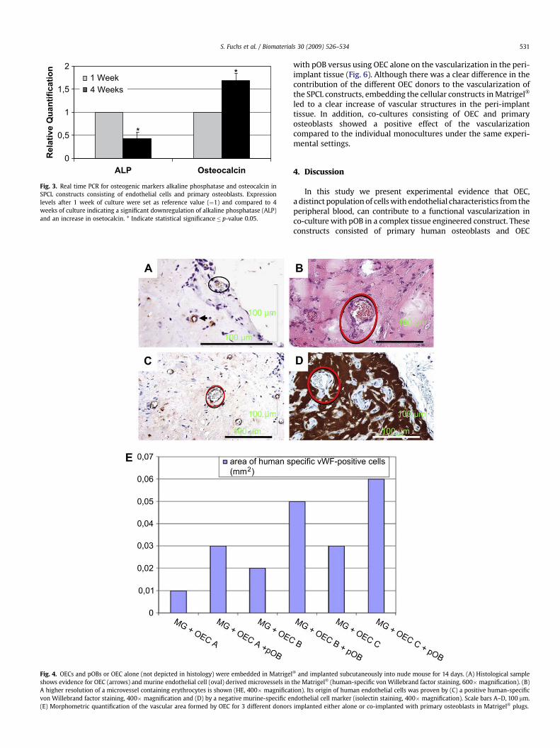
The potential of osteogenic differentiation of the SPCLconstructs under the applied culture conditions was assessed byquantitative real time PCR for alkaline phosphatase and osteocalcincomparing the expression rates after 1 and 4 weeks in the co-culture (Fig. 3). In the investigated time frame we observeda significant downregulation of alkaline phosphatase and a signifi-cant increase in the expression of osteocalcin. (p-values� 0.05).
3.4. Subcutaneous implantation of Matrigel plugs
In order to evaluate the angiogenic potential of OEC fromdifferent donors in vivo and to assess the impact of the co-culture
with primary osteoblasts on OEC-derived vascular structures,Matrigel� plugs containing either OEC alone or OEC and pOB wereimplanted subcutaneously (Fig. 4A–E). Perfused microvessels ofmouse (Fig. 4A, ovals, von Willebrand factor-negative) or of humanorigin (Fig. 4A, arrows, von Willebrand factor-positive) witha diameter of up to 60 mm were detected. In Fig. 4B–D the same areais depicted in stainings with differential specifity. The human originof the vessels in this area was confirmed by a positive staining usinghuman-specific van Willebrand antibodies (Fig. 4C) and the nega-tive staining in a murine-specific isolectin staining (Fig. 4D).Morphometric analysis was used to quantify vascular structurescomparing OEC implanted alone or co-implanted with pOB inMatrigel� plugs. For all tested donors (n¼ 3) the same trend wasobserved, indicating larger areas for human vascular structures in
E
4 w
eeks
OEC/poB
F
CD31/Hoechst
CD31/Hoechst
DC
1 w
eek
OEC/poB
OEC CD31/PI
A
OEC CD31/PI
B
Fig. 2. Angiogenic response of OEC on SPCL in monoculture using a fibrin gel based assay (A, B) and stimulation with VEGF or in the co-culture with primary osteoblasts (pOB)(without additional growth factors) at different time points of co-culture, 1 week (C, D) and 4 weeks (E, F). Endothelial cells are depicted by immunofluorescent staining for CD31depicted in green. Nuclear counterstaining of endothelial cells by propidium iodide in red (A, B) or for all cells in the co-culture by Hoechst (blue, D and F). Scale bar as indicated inthe picture¼ 150 mm. OEC show angiogenic activation using fibrin gels and supplementation with VEGF or in co-culture with primary osteoblasts.
S. Fuchs et al. / Biomaterials 30 (2009) 526–534530
co-implants of OEC and pOB compared to controls using OEC alone(Fig. 4E).
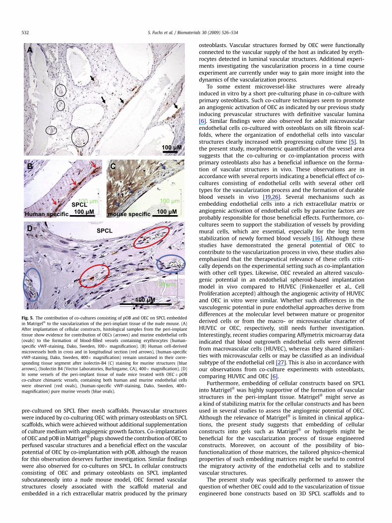
3.5. Vascularization of SPCL contructs in vivo containing OEC
The SPCL fibres were well integrated into the surrounding peri-implant tissue and were surrounded by both human (Fig. 5A;arrows) and murine microvessels (Fig. 5A; circles). Vascularstructures in the peri-implant tissue were observed in both caseswith (Fig. 5) or without using Matrigel� (not shown) as embeddingmatrix for the cellularized SPCL constructs. Nevertheless, embed-ding of constructs in Matrigel� seemed to have a beneficial effecton the formation of vascular structures in the peri-implant tissue(compare also Fig. 6). The contribution of OEC to the vasculariza-tion process was assessed using human-specific detection of
vascular structures or detection of vascular structures formed bythe host. Immunohistochemistry using the human-specific anti-bodies for vWF (Fig. 5A and B) revealed vascular structures formedby OEC mostly in close association with the SPCL fibres. Thesevascular structures formed by OEC revealed lumina (Fig. 5A and B)and contained erythrocytes as evidence of active perfusion. Incontrol staining, detecting vascular structures formed by mouse,those vascular structures formed by OEC (Fig. 5B, red arrows)where negative for the mouse-specific staining (Fig. 5C, bluearrows). In addition, blood-filled chimeric vessels formed by bothhuman and mouse endothelial cells were observed (Fig. 5D, high-lighted by ovals).
Histomorphometrical analysis was performed in addition toassess the effect of Matrigel� as embedding material for the cellularconstructs, as well as to gain insight into the effects of co-cultures
0
0,5
1
1,5
2
ALP Osteocalcin
Relative Q
uan
tificatio
n
1 Week4 Weeks
*
*
Fig. 3. Real time PCR for osteogenic markers alkaline phosphatase and osteocalcin inSPCL constructs consisting of endothelial cells and primary osteoblasts. Expressionlevels after 1 week of culture were set as reference value (¼1) and compared to 4weeks of culture indicating a significant downregulation of alkaline phosphatase (ALP)and an increase in osetocalcin. * Indicate statistical significance� p-value 0.05.
Fig. 4. OECs and pOBs or OEC alone (not depicted in histology) were embedded in Matrigelshows evidence for OEC (arrows) and murine endothelial cell (oval) derived microvessels inA higher resolution of a microvessel containing erythrocytes is shown (HE, 400� magnificatvon Willebrand factor staining, 400� magnification and (D) by a negative murine-specific en(E) Morphometric quantification of the vascular area formed by OEC for 3 different donors
S. Fuchs et al. / Biomaterials 30 (2009) 526–534 531
with pOB versus using OEC alone on the vascularization in the peri-implant tissue (Fig. 6). Although there was a clear difference in thecontribution of the different OEC donors to the vascularization ofthe SPCL constructs, embedding the cellular constructs in Matrigel�
led to a clear increase of vascular structures in the peri-implanttissue. In addition, co-cultures consisting of OEC and primaryosteoblasts showed a positive effect of the vascularizationcompared to the individual monocultures under the same experi-mental settings.
4. Discussion
In this study we present experimental evidence that OEC,a distinct population of cells with endothelial characteristics from theperipheral blood, can contribute to a functional vascularization inco-culture with pOB in a complex tissue engineered construct. Theseconstructs consisted of primary human osteoblasts and OEC
� and implanted subcutaneously into nude mouse for 14 days. (A) Histological samplethe Matrigel� (human-specific von Willebrand factor staining, 600�magnification). (B)ion). Its origin of human endothelial cells was proven by (C) a positive human-specificdothelial cell marker (isolectin staining, 400� magnification). Scale bars A–D, 100 mm.implanted either alone or co-implanted with primary osteoblasts in Matrigel� plugs.
A
100 µM
SPCLD
100 µM
SPCL
B
Human specific100 µM
100 µm
mouse specific
C
100 µM
100 µm
100 µm
Fig. 5. The contribution of co-cultures consisting of pOB and OEC on SPCL embeddedin Matrigel� to the vascularization of the peri-implant tissue of the nude mouse. (A)After implantation of cellular constructs, histological samples from the peri-implanttissue show evidence for contribution of OECs (arrows) and murine endothelial cells(ovals) to the formation of blood-filled vessels containing erythrocytes (human-specific vWF-staining, Dako, Sweden, 100� magnification). (B) Human cell-derivedmicrovessels both in cross and in longitudinal section (red arrows), (human-specificvWF-staining, Dako, Sweden, 400� magnification) remain unstained in their corre-sponding tissue segment after isolectin-B4 (C) staining for murine structures (bluearrows), (Isolectin B4 (Vector Laboratories, Burlingame, CA), 400� magnification). (D)In some vessels of the peri-implant tissue of nude mice treated with OECþ pOBco-culture chimaeric vessels, containing both human and murine endothelial cellswere observed (red ovals), (human-specific vWF-staining, Dako, Sweden, 400�magnification) pure murine vessels (blue ovals).
S. Fuchs et al. / Biomaterials 30 (2009) 526–534532
pre-cultured on SPCL fiber mesh scaffolds. Prevascular structureswere induced by co-culturing OEC with primary osteoblasts on SPCLscaffolds, which were achieved without additional supplementationof culture medium with angiogenic growth factors. Co-implantationof OEC and pOB in Matrigel� plugs showed the contribution of OEC toperfused vascular structures and a beneficial effect on the vascularpotential of OEC by co-implantation with pOB, although the reasonfor this observation deserves further investigation. Similar findingswere also observed for co-cultures on SPCL. In cellular constructsconsisting of OEC and primary osteoblasts on SPCL implantedsubcutaneously into a nude mouse model, OEC formed vascularstructures closely associated with the scaffold material andembedded in a rich extracellular matrix produced by the primary
osteoblasts. Vascular structures formed by OEC were functionallyconnected to the vascular supply of the host as indicated by eryth-rocytes detected in luminal vascular structures. Additional experi-ments investigating the vascularization process in a time courseexperiment are currently under way to gain more insight into thedynamics of the vascularization process.
To some extent microvessel-like structures were alreadyinduced in vitro by a short pre-culturing phase in co-culture withprimary osteoblasts. Such co-culture techniques seem to promotean angiogenic activation of OEC as indicated by our previous studyinducing prevascular structures with definitive vascular lumina[6]. Similar findings were also observed for adult microvascularendothelial cells co-cultured with osteoblasts on silk fibroin scaf-folds, where the organization of endothelial cells into vascularstructures clearly increased with progressing culture time [5]. Inthe present study, morphometric quantification of the vessel areasuggests that the co-culturing or co-implantation process withprimary osteoblasts also has a beneficial influence on the forma-tion of vascular structures in vivo. These observations are inaccordance with several reports indicating a beneficial effect of co-cultures consisting of endothelial cells with several other celltypes for the vascularization process and the formation of durableblood vessels in vivo [19,26]. Several mechanisms such asembedding endothelial cells into a rich extracellular matrix orangiogenic activation of endothelial cells by paracrine factors areprobably responsible for those beneficial effects. Furthermore, co-cultures seem to support the stabilization of vessels by providingmural cells, which are essential, especially for the long termstabilization of newly formed blood vessels [16]. Although thesestudies have demonstrated the general potential of OEC tocontribute to the vascularization process in vivo, these studies alsoemphasized that the therapeutical relevance of these cells criti-cally depends on the experimental setting such as co-implantationwith other cell types. Likewise, OEC revealed an altered vasculo-genic potential in an endothelial spheroid-based implantationmodel in vivo compared to HUVEC (Finkenzeller et al., CellProliferation accepted) although the angiogenic activity of HUVECand OEC in vitro were similar. Whether such differences in thevasculogenic potential in pure endothelial approaches derive fromdifferences at the molecular level between mature or progenitorderived cells or from the macro- or microvascular character ofHUVEC or OEC, respectively, still needs further investigation.Interestingly, recent studies comparing Affymetrix microarray dataindicated that blood outgrowth endothelial cells were differentfrom macrovascular cells (HUVEC), whereas they shared similari-ties with microvascular cells or may be classified as an individualsubtype of the endothelial cell [27]. This is also in accordance withour observations from co-culture experiments with osteoblasts,comparing HUVEC and OEC [6].
Furthermore, embedding of cellular constructs based on SPCLinto Matrigel� was highly supportive of the formation of vascularstructures in the peri-implant tissue. Matrigel� might serve asa kind of stabilizing matrix for the cellular constructs and has beenused in several studies to assess the angiogenic potential of OEC.Although the relevance of Matrigel� is limited in clinical applica-tions, the present study suggests that embedding of cellularconstructs into gels such as Matrigel� or hydrogels might bebeneficial for the vascularization process of tissue engineeredconstructs. Moreover, on account of the possibility of bio-functionalization of those matrices, the tailored physico-chemicalproperties of such embedding matrices might be useful to controlthe migratory activity of the endothelial cells and to stabilizevascular structures.
The present study was specifically performed to answer thequestion of whether OEC could add to the vascularization of tissueengineered bone constructs based on 3D SPCL scaffolds and to

Area human positive cells (mm2)
0
0,1
0,2
0,3
0,4
0,5
0,6
0,7
MG
, SPC
L, O
EC
A
MG
, SPC
L, O
EC
B
MG
, SPC
L, O
EC
A, pO
B
MG
, SPC
L, O
EC
B, pO
B
SPC
L, O
EC
A, pO
B
SPC
L, O
EC
B, pO
B
Fig. 6. The vessel area in mm2 which has been formed by human endothelial cells in dependence of the tested donors and the presence of Matrigel�. Monocultures of OEC wereused as additional controls.
S. Fuchs et al. / Biomaterials 30 (2009) 526–534 533
determine the effect of the co-culture with pOB on the angiogenicactivity of OEC. On the other hand, it is well known that osteo-genesis itself is dependent on a functional interaction of endothelialcells and osteoblasts. Initial characterization of complex SPCLscaffolds by quantitative real time PCR indicated the upregulationof the late osteoblastic marker osteocalcin and a decrease in theearly marker alkaline phosphatase with progressing pre-culturetime (Fig. 3). These data indicate the progressing differentiation ofosteoblastic cells in complex SPCL constructs under the cultureconditions mainly tailored for OEC. More detailed investigation ofthe dynamics of endothelial and osteogenic markers, matrixcomponents and vessel formation within complex constructs invitro is also provided in an additional study based on silk fibroinscaffolds (manuscript submitted), whereas the present paperfocuses on the beneficial effect of the co-cultures on the vascular-ization process by OEC. Nevertheless, the in vivo bone formation ofsuch constructs needs to be investigated in further studies usingadditional models suitable for monitoring bone formation.
5. Conclusions
This manuscript describes the contribution of human OEC,a distinct subpopulation within EPC cultures from the peripheralblood, to the vascularization of a bone tissue construct in a nudemouse model in vivo. Co-cultures or co-implantation of OEC withpOB exerted a beneficial effect on the vascularization process byOEC. The regeneration and repair of highly vascularized tissues andlarger tissue defects such as in bone might profit from the use ofOEC as autologous cell sources. Our findings might have an impacton further developments of prevascularized tissues for bone basedon endothelial progenitor cells.
Acknowledgements
The authors would like to thank B. Malenica, C. Braun, L. Meyer,K. Molter, M. Muller. J. Alonso-Monje for their excellent technicalassistance. This work was financially supported by grants from theEuropean commission (HIPPOCRATES N� NMP3-CT-2003-505758;EXPERTISSUES Contract nr.: 500283-2) and BMBF-Grant forGerman-Chinese Cooperation in Regenerative Medicine (grantnumber 0315033).
Appendix
Figures with essential colour discrimination. Certain figures inthis article, in particular parts of Figures 1,2,4 and 5 are difficult tointerpret in black and white. The full colour images can be found inthe on-line version, at doi: 10.1016/j.biomaterials.2008.09.058.
References
[1] Levenberg S, Rouwkema J, Macdonald M, Garfein ES, Kohane DS, Darland DC,et al. Engineering vascularized skeletal muscle tissue. Nat Biotechnol2005;23(7):879–84.
[2] Berthod F, Germain L, Tremblay N, Auger FA. Extracellular matrix depositionby fibroblasts is necessary to promote capillary-like tube formation in vitro.J Cell Physiol 2006;207(2):491–8.
[3] Elbjeirami WM, West JL. Angiogenesis-like activity of endothelial cells co-cultured with VEGF-producing smooth muscle cells. Tissue Eng 2006.
[4] Saiki A, Watanabe F, Murano T, Miyashita Y, Shirai K. Hepatocyte growth factorsecreted by cultured adipocytes promotes tube formation of vascular endo-thelial cells in vitro; 2006.
[5] Unger RE, Sartoris A, Peters K, Motta A, Migliaresi C, Kunkel M, et al. Tissue-like self-assembly in cocultures of endothelial cells and osteoblasts and theformation of microcapillary-like structures on three-dimensional porousbiomaterials. Biomaterials 2007;28(27):3965–76.
[6] Fuchs S, Hofmann A, Kirkpatrick CJ. Microvessel-like structures fromoutgrowth endothelial cells from human peripheral blood in 2-dimensionaland 3-dimensional co-cultures with osteoblastic lineage cells. Tissue Eng2007;13(10):2577–88.
[7] Wenger A, Stahl A, Weber H, Finkenzeller G, Augustin HG, Stark GB, et al.Modulation of in vitro angiogenesis in a three-dimensional spheroidalcoculture model for bone tissue engineering. Tissue Eng 2004;10(9–10):1536–47.
[8] Villars F, Guillotin B, Amedee T, Dutoya S, Bordenave L, Bareille R, et al. Effect ofHUVEC on human osteoprogenitor cell differentiation needs heterotypic gapjunction communication. Am J Physiol Cell Physiol 2002;282(4):C775–85.
[9] Spector JA, Mehrara BJ, Greenwald JA, Saadeh PB, Steinbrech DS, Bouletreau PJ,et al. Osteoblast expression of vascular endothelial growth factor is modulatedby the extracellular microenvironment. Am J Physiol Cell Physiol 2001;280(1):C72–80.
[10] Choong CS, Hutmacher DW, Triffitt JT. Co-culture of bone marrow fibroblastsand endothelial cells on modified polycaprolactone substrates for enhancedpotentials in bone tissue engineering. Tissue Eng 2006;12(9):2521–31.
[11] Guillotin B, Bareille R, Bourget C, Bordenave L, Amedee J. Interaction betweenhuman umbilical vein endothelial cells and human osteoprogenitors triggerspleiotropic effect that may support osteoblastic function. Bone 2008.
[12] Scherberich A, Galli R, Jaquiery C, Farhadi J, Martin I. Three-dimensionalperfusion culture of human adipose tissue-derived endothelial and osteoblasticprogenitors generates osteogenic constructs with intrinsic vascularizationcapacity. Stem Cells 2007;25(7):1823–9.
[13] Rouwkema J, de Boer J, Van Blitterswijk CA. Endothelial cells assemble intoa 3-dimensional prevascular network in a bone tissue engineering construct.Tissue Eng 2006;12(9):2685–93.

S. Fuchs et al. / Biomaterials 30 (2009) 526–534534
[14] Lin Y, Weisdorf DJ, Solovey A, Hebbel RP. Origins of circulating endothelial cellsand endothelial outgrowth from blood. J Clin Invest 2000;105(1):71–7.
[15] Gulati R, Jevremovic D, Peterson TE, Chatterjee S, Shah V, Vile RG, et al. Diverseorigin and function of cells with endothelial phenotype obtained from adulthuman blood. Circ Res 2003;93(11):1023–5.
[16] Melero-Martin JM, Khan ZA, Picard A, Wu X, Paruchuri S, Bischoff J. In vivovasculogenic potential of human blood-derived endothelial progenitor cells.Blood 2007;109(11):4761–8.
[17] Fuchs S, Hermanns M, Kirkpatrick C. Retention of a differentiated endo-thelial phenotype by outgrowth endothelial cells isolated from humanperipheral blood and expanded in long-term cultures. Cell Tissue Res2006:1–14.
[18] Au P, Daheron LM, Duda DG, Cohen KS, Tyrrell JA, Lanning RM, et al. Differ-ential in vivo potential of endothelial progenitor cells from human umbilicalcord blood and adult peripheral blood to form functional long-lasting vessels.Blood 2008;111(3):1302–5.
[19] Au P, Tam J, Fukumura D, Jain RK. Bone marrow-derived mesenchymal stemcells facilitate engineering of long-lasting functional vasculature. Blood2008;111(9):4551–8.
[20] Santos MI, Fuchs S, Gomes ME, Unger RE, Reis RL, Kirkpatrick CJ. Response ofmicro- and macrovascular endothelial cells to starch-based fiber meshes forbone tissue engineering. Biomaterials 2007;28(2):240–8.
[21] Gomes ME, Holtorf HL, Reis RL, Mikos AG. Influence of the porosity of starch-based fiber mesh scaffolds on the proliferation and osteogenic differentiation
of bone marrow stromal cells cultured in a flow perfusion bioreactor. TissueEng 2006;12(4):801–9.
[22] Gomes ME, Sikavitsas VI, Behravesh E, Reis RL, Mikos AG. Effect of flowperfusion on the osteogenic differentiation of bone marrow stromal cellscultured on starch-based three-dimensional scaffolds. J Biomed Mater Res A2003;67(1):87–95.
[23] Fuchs S, Motta A, Migliaresi C, Kirkpatrick CJ. Outgrowth endothelial cellsisolated and expanded from human peripheral blood progenitor cells asa potential source of autologous cells for endothelialization of silk fibroinbiomaterials. Biomaterials 2006;27(31):5399–408.
[24] Hofmann A, Konrad L, Gotzen L, Printz H, Ramaswamy A, Hofmann C. Bio-engineered human bone tissue using autogenous osteoblasts cultured ondifferent biomatrices. J Biomed Mater Res A 2003;67(1):191–9.
[25] Hafemann B, Ensslen S, Erdmann C, Niedballa R, Zuhlke A, Ghofrani K, et al.Use of a collagen/elastin-membrane for the tissue engineering of dermis.Burns 1999;25(5):373–84.
[26] Sanz L, Santos-Valle P, Alonso-Camino V, Salas C, Serrano A, Vicario JL, et al.Long-term in vivo imaging of human angiogenesis: Critical role of bonemarrow-derived mesenchymal stem cells for the generation of durable bloodvessels. Microvasc Res 2008;75(3):308–14.
[27] Jiang A, Pan W, Milbauer LC, Shyr Y, Hebbel RP. A practical question based oncross-platform microarray data normalization: are BOEC more like large vesselor microvascular endothelial cells or neither of them? J Bioinform Comput Biol2007;5(4):875–93.