Contemporary Fixed Prosthodontics_ 5ed_1.pdf
100
-
Upload
khangminh22 -
Category
Documents
-
view
2 -
download
0
Transcript of Contemporary Fixed Prosthodontics_ 5ed_1.pdf

Evolve Student Resources for Rosenstiel/Land/Fujimoto: Contemporary Fixed Prosthodontics, Fifth Edition, include the following:
• Practiceexaminationquestionsforeachchapter
• TheGlossaryofProsthodonticTerms
Activate the complete learning experience that comes with each textbook purchase by registering at
http://evolve.elsevier.com/Rosenstiel/prosthodontics/
YOU’VE JUST PURCHASED
MORE THAN A TEXTBOOK!
REGISTER TODAY!
You can now purchase Elsevier products on Evolve! Go to evolve.elsevier.com/html/shop-promo.html to search and browse for products.

CONTEMPORARY FIXED PROSTHODONTICS

This page intentionally left blank

CONTEMPORARY FIXED PROSTHODONTICS
FIFTH EDITION
Stephen F. Rosenstiel, BDS, MSDProfessor Emeritus, Restorative and Prosthetic DentistryCollege of DentistryThe Ohio State UniversityColumbus, Ohio
Martin F. Land, DDS, MSDProfessor of Fixed ProsthodonticsSchool of Dental MedicineSouthern Illinois University, EdwardsvilleAlton, Illinois
Junhei Fujimoto, DDS, MSD, DDScPart-Time Lecturer, Tokyo Medical and Dental UniversityDirector, J.F. Occlusion and Prosthodontic Postgraduate Course;Private PracticeTokyo, Japan
3251 Riverport LaneSt. Louis, Missouri 63043
CONTEMPORARY FIXED PROSTHODONTICS, FIFTH EDITION
ISBN: 978-0-323-08011-8
Copyright © 2016 by Elsevier Inc. All rights reserved.
No part of this publication may be reproduced or transmitted in any form or by any means, electronic or mechanical, including photocopying, recording, or any information storage and retrieval system, without permission in writing from the Publisher. Details on how to seek permission, further information about the Publisher’s permissions policies and our arrangements with organizations such as the Copyright Clearance Center and the Copyright Licensing Agency, can be found at our website: www.elsevier.com/permissions.
This book and the individual contributions contained in it are protected under copyright by the Publisher (other than as may be noted herein).
Notices
Knowledge and best practice in this field are constantly changing. As new research and experience broaden our understanding, changes in research methods, professional practices, or medical treatment may become necessary.
Practitioners and researchers must always rely on their own experience and knowledge in evaluating and using any information, methods, compounds, or experiments described herein. In using such information or methods, they should be mindful of their own safety and the safety of others, including parties for whom they have a professional responsibility.
With respect to any drug or pharmaceutical products identified, readers are advised to check the most current information provided (i) on procedures featured or (ii) by the manufacturer of each product to be administered, to verify the recommended dose or formula, the method and duration of administration, and contraindications. It is the responsibility of practitioners, relying on their own experience and knowledge of their patients, to make diagnoses, to determine dosages and the best treatment for each individual patient, and to take all appropriate safety precautions.
To the fullest extent of the law, neither the Publisher nor the authors, contributors, or editors, assume any liability for any injury and/or damage to persons or property as a matter of product liability, negligence or otherwise, or from any use or operation of any methods, products, instructions, or ideas contained in the material herein.
Previous editions copyrighted 2006, 2001, 1995, and 1988.
Library of Congress Cataloging-in-Publication Data
Rosenstiel, Stephen F., author. Contemporary fixed prosthodontics / Stephen F. Rosenstiel, Martin F. Land, Junhei Fujimoto.— Fifth edition. p. ; cm. Includes bibliographical references and index. ISBN 978-0-323-08011-8 (hardcover : alk. paper) I. Land, Martin F., author. II. Fujimoto, Junhei, author. III. Title. [DNLM: 1. Denture, Partial, Fixed. 2. Prosthodontics—methods. WU 515]
RK666 617.6′92—dc23 2015024195
Executive Content Strategist: Kathy FalkSenior Content Development Specialist: Courtney SprehePublishing Services Manager: Catherine JacksonSenior Project Manager: Rachel E. McMullenDesign Direction: Xiaopei Chen
Printed in China
Last digit is the print number: 9 8 7 6 5 4 3 2 1

v
Contributors
Robert F. Baima, DDS Clinical Associate Professor, Department of
Periodontology and Restorative Dentistry, School of Dentistry, University of Detroit Mercy, Detroit, Michigan; Diplomate, American Board of Periodontology, Diplomate, American Board of Prosthodontics
Rick K. Biethman, DMD Assistant Professor, Department of Restorative
Dentistry, School of Dental Medicine, Southern Illinois University, Alton, Illinois
William A. Brantley, PhD Professor and Director of the Graduate Program in
Dental Materials Science, Division of Restorative and Prosthetic Dentistry, College of Dentistry, The Ohio State University, Columbus, Ohio
Isabelle L. Denry, DDS, MS, PhD Professor, Department of Prosthodontics and Dows
Institute for Dental Research, College of Dentistry, The University of Iowa, City, Iowa
R. Duane Douglas, DMD, MS Associate Professor and Chair, Department of
Restorative Dentistry, School of Dental Medicine, Southern Illinois University, Alton, Illinois
A. Jon Goldberg, PhD Professor, Department of Reconstructive Sciences,
Director, Center for Biomaterials, School of Dental Medicine, University of Connecticut, Farmington, Connecticut
Julie A. Holloway, DDS, MS Professor and Head, Department of Prosthodontics,
College of Dentistry, The University of Iowa, Iowa City, Iowa
Christa D. Hopp, DMD, BS Associate Professor,Section Head, Operative Dentistry, School of Dental
Medicine, Southern Illinois University, Alton, Illinois
William M. Johnston, PhD Professor Emeritus, Division of General Practice
and Materials Science, College of Dentistry, The Ohio State University, Columbus, Ohio
Peter E. Larsen, DDS The Larry J. Peterson Endowed Professor,
and Chair of Oral and Maxillofacial Surgery, College of Dentistry, The Ohio State University, Columbus, Ohio
Edwin A. McGlumphy, DDS, MS Professor, Department of Restorative and Prosthetic
Dentistry, College of Dentistry, The Ohio State University, Columbus, Ohio
Jonathan C. Meiers, DMD, MS Chief, Dental Service, VA Connecticut Healthcare
System, 950 Campbell Ave. West Haven, Connecticut
Donald A. Miller, DDS, MS Private Practice, Chicago and Naperville, Illinois,
Clinical Associate Professor, University of Illinois at Chicago College of Dentistry; Diplomate, American Board of Endodontics
Van P. Thompson, DDS, PhD Professor, Division of Tissue Engineering and
Biophontics, King’s College London Dental Institute, London, United Kingdom
Alvin G. Wee, DDS, MS, MPH Associate Professor, Division of Oral Facial
Prosthetics/Dental Oncology, Department of Otolaryngology—Head and Neck Surgery; Member, Cancer Prevention and Control Program, University of Nebraska Medical Center Eppley Cancer Center, Courtesy Associate Professor, College of Dentistry, University of Nebraska Medical Center, Omaha, Nebraska
Burak Yilmaz, DDS, PhD Associate Professor, Division of Restorative Science
and Prosthodontics, College of Dentistry, The Ohio State University, Columbus, Ohio
vi
Preface
In the late summer of 1975, three young dentists from England, Holland, and Japan met for the very first time at the School of Dentistry in Indianapolis. They shared a common interest in “crown and bridge” prosthodon-tics. Little did they know that 40 years later they would take immense satisfaction in having completed the fifth edition of Contemporary Fixed Prosthodontics.
As the three of us embarked on mastering the art and science of fixed prosthodontics, we encountered the com-plete range of human emotions, ranging from insecurity to self-confidence, from frustration to fulfillment, and from anxiety to satisfaction and occasional pride. It is with much pride that we introduce this comprehensively updated and revised edition of one of the most widely used and translated texts in our field.
The task once again was overwhelming. Technology had developed far beyond our comfort zones of clinical sciences and dental materials. The technological advances in imaging and CAD/CAM tempted us to add a chapter (in an appropriate place in the text) summarizing new technological developments. Instead, we opted to inte-grate new technologies throughout the text, as we con-sistently tried to do in earlier editions. Early in the review process, we realized that describing some of the new systems would be impossible in a step-by-step format. Advances and improvements occur at such a pace that a chapter that was comprehensively rewritten would soon be out of date. As a result, we embarked on a process of breaking down the newer technologies into their under-lying principles and then integrating this information—often gleaned from sources outside the conventional dental literature—into the text.
The fifth edition now includes cone-beam technology for diagnostic purposes and implant placement. Impres-sion making was expanded with a section on optical imaging, and solid casts are contrasted with their virtual counterparts. Wax pattern fabrication is still presented in the classical manner but is augmented with specific information on computer-aided design followed by either printing or milling. Similarly, the section on the fabrica-tion of metal ceramic substructures and all-ceramic res-torations was revised to include the new CAD/CAM techniques.
Since the publication of the fourth edition, the dental laboratory industry has undergone revolutionary change.
Smaller laboratories must compete with large corpora-tions that can more easily invest in expensive new technologies. When we visited laboratories and manu-facturers, we realized that the transition to CAD/CAM was not a seamless process.
A number of years ago, a highly skilled and seasoned ceramist at a major dental laboratory was put in charge of the digital production of all-ceramic restorations. He shared with us that the learning curve had been particu-larly steep in the beginning when he tried to teach the very best and brightest computer personnel he could find the necessary dental knowledge. A number of years into this process, he started to teach his most experienced certified dental technicians how to operate some of the newly developed CAD/CAM tools, and, as he told us, “In six months, we were up and running, and we have never looked back.”
The foregoing experience reinforces the importance of establishing a solid foundation in the fundamental skill sets for fixed prosthodontics before successfully applying them to some of the new technologies. Any student of fixed prosthodontics must have a thorough knowledge of dental anatomy, tooth form and function before embark-ing on even the classical approaches to the fabrication of individual crowns or simple fixed dental prostheses. A thorough understanding of form and function is, in turn, prerequisite to achieving mastery of today’s cutting-edge technologies. A sincere effort was made to include new illustrations of commonly incorporated techniques in undergraduate curricula throughout the United States and Canada. Popular techniques have been added to the previously recommended approaches, illustrations of instrumentation have been replaced to incorporate con-temporary equipment, and other new materials have been incorporated throughout.
With this text, we hope to serve predoctoral students, postdoctoral fellows, practitioners, and researchers. The text is well indexed, and every effort has been made to ensure the rapid retrievability of evidence-based informa-tion for the busy practitioner or dental manufacturer.
Stephen F. RosenstielMartin F. Land
Junhei Fujimoto

vii
Acknowledgments
In recognition of so many colleagues and friends …Where to begin? After three decades, we are unlikely
to achieve absolute accuracy in crediting all of those whose selfless contributions to the development of this text have helped it evolve. Whenever we asked, we met with a willingness to share concepts, new technology, illustrations, photographs, materials, and whatever else we requested. Permissions were routinely granted and, invariably, most kindly approved. We again have made every effort to accurately and precisely give credit to all of those to whom credit is due. Any errors and omissions are absolutely unintentional and the sole responsibility of the authors—we apologize if such should have occurred.
A special thank you to the following:James Cockerill, RBP, who once again provided
selected photographic support consistent with his previ-ous contributions.
Our contributors: Robert F. Baima, Rick K. Biethman, William A. Brantley, Isabelle L. Denry, R. Duane Douglas, Martin A. Freilich, A. Jon Goldberg, Julie A. Holloway, Christa D. Hopp, William M. Johnston, Peter E. Larsen, Leon W. Laub, Edwin A. McGlumphy, Jonathan C. Meiers, Donald A. Miller, M. H. Reisbick, James L. Sandrik, Van P. Thompson, Alvin G. Wee, and Burak Yilmaz.
Faculty and staff at Southern Illinois University, School of Dental Medicine: Dr. Jeffrey Banker, Dr. Rick Biethman, Dr. Robert Blackwell, Dr. Duane Douglas, Dr. Randy Duncan, Dr. Christa Hopp, Ms. Nancy Inlow, Dr. Daniel Ketteman, Dr. Dennis Knobeloch, Ms. Robin Manning, Dr. Jack Marincel, Ms. Tobbi McEuen, Dr. Charles Poeschl, Dr. Steven Raney, Dr. Vincent Rapini, Dr. William Seaton, Dr. Joseph Sokolowski, Dr. Charles Thornton, Ms. Michele Wadlow, and Dr. Daniel Woodlock.
Faculty and staff at The Ohio State University: Dr. Shereen Azer, Dr. Nancy Clelland, Dr. Allen Firestone, Dr. Lisa Knobloch, Dr. John Nusstein, Dr. Robert Seghi, Dr. Burak Yilmaz, and Ms. Amy Barker for their many valu-able insights and their continuous support along the way.
Photographer Brodie Strum (Chicago, Illinois) who provided assistance with the post photographs in Chapter 12.
All of the medical illustrators who have been instru-mental in expanding and refining the art program through the many editions: Krystyna Srodulski (San
Francisco, California); Donald O’Connor (St. Peters, Missouri); Sandra Cello-Lang (Chicago, Illinois); Sue E. Cottrill (Chicago, Illinois); Kerrie Marzo (Chicago Heights, Illinois).
The outstanding team at Elsevier who continued to believe in our ability to complete this task, and whose relentless pursuit of quality has helped make this our finest edition yet: Kathy Falk, Executive Content Strategist; Courtney Sprehe, Senior Content Develop-ment Specialist; and Rachel McMullen, Senior Project Manager for their help, patience, and understanding throughout.
A special thank you to the many individuals in the dental manufacturing and marketing industries who pro-vided us with information and illustrations of their products.
In each of the previous editions, we have written a special thank you to our spouses, Enid, Karen, and Yoshiko. Sadly Karen Tolbert Land died on January 4, 2014, and was not able to see this fifth edition through to completion. Her home in Alton, Illinois, always served as our base when we met with Elsevier in St. Louis. Working on this edition was not the same without her hospitality.
“Looking back over my career, I can tell you a lot of things about fixed prosthodontics. Most importantly, I can tell you very sincerely: I have never been bored” said the experienced teacher.
The fifth edition of Contemporary Fixed Prosthodontics is close to what we originally envisioned as we embarked on this original journey. We hope that it will help advance the art and science of what is beyond question the most challenging clinical dental specialty. We know that we do not have all the answers, but we hope that students and practitioners, researchers and manufacturers, and all who demonstrate the interest and commitment necessary to achieve mastery in a discipline we have come to love and respect, may find many, if not most, of the answers they seek.
Stephen F. RosenstielMartin F. Land
Junhei Fujimoto

viii
Contents
PART IPLANNING AND PREPARATION1 HistoryTakingandClinicalExamination, 3
2 DiagnosticCastsandRelatedProcedures, 35
3 TreatmentPlanning, 70
4 PrinciplesofOcclusion, 92
5 PeriodontalConsiderations, 117
6 MouthPreparation, 138
PART IICLINICAL PROCEDURES: SECTION 17 PrinciplesofToothPreparation, 169
8 TheCompleteCastCrownPreparation, 209
9 TheMetal-CeramicCrownPreparation, 222
10 ThePartialVeneerCrown,Inlay,andOnlayPreparations, 236
11 ToothPreparationforAll-CeramicRestorations, 264
12 RestorationoftheEndodonticallyTreatedTooth, 278
13 Implant-SupportedFixedProstheses, 318
14 TissueManagementandImpressionMaking, 367
15 InterimFixedRestorations, 401
PART IIILABORATORY PROCEDURES16 CommunicatingwiththeDental
Laboratory, 443
17 DefinitiveCastsandDies, 457
18 WaxPatterns, 489
19 FrameworkDesignandMetalSelectionforMetal-CeramicRestorations, 521
20 PonticDesign, 546
21 RetainersforPartialRemovableDentalProstheses, 576
22 InvestingandCasting, 601
23 DescriptionofColor,Color-ReplicationProcess,andEsthetics, 624
24 Metal-CeramicRestorations, 647
25 All-CeramicRestorations, 674
26 Resin-BondedFixedDentalProstheses, 694
27 ConnectorsforPartialFixedDentalProstheses, 713
28 FinishingtheCastRestoration, 736
PART IVCLINICAL PROCEDURES: SECTION 229 Evaluation,Characterization,and
Glazing, 751
30 LutingAgentsandCementationProcedures, 774
31 PostoperativeCare, 792
Appendix:DentalMaterialsandEquipmentIndex, 829

PART I
PLANNING AND PREPARATION

This page intentionally left blank
3
History Taking and Clinical Examination
C H A P T E R 1
Fixed prosthodontic treatment involves the replacement and restoration of teeth by artificial substitutes that are not readily removable from the mouth. Its focus is to restore function, esthetics, and comfort. Fixed prostho-dontics can offer exceptional satisfaction for both patient and dentist. It can transform an unhealthy, unattractive dentition with poor function into a comfortable, healthy occlusion capable of years of further service while greatly enhancing esthetics (Fig. 1-1, A and B). Treatment can range from fairly straightforward measures—such as restoration of a single tooth with a cast crown (see Fig. 1-1, C), replacement of one or more missing teeth with a fixed dental prosthesis (see Fig. 1-1, D), or an implant-supported restoration (see Fig. 1-1, E)—to highly complex restorations involving all the teeth in an entire arch or the entire dentition (see Fig. 1-1, F).
To achieve predictable success in this technically and intellectually challenging field, meticulous attention to every detail is crucial: the initial patient interview and diagnosis, the active treatment phases, and a planned schedule of follow-up care. Otherwise, the result is likely to be unsatisfactory and frustrating for both dentist and patient, resulting in disappointment and loss of confi-dence in each other.
Problems encountered during or after treatment can often be traced to errors and omissions during history taking and initial examination. The inexperienced clini-cian may plunge into the treatment phase before collect-ing sufficient diagnostic information that helps predict likely pitfalls.
Making the correct diagnosis is prerequisite for for-mulating an appropriate treatment plan. All pertinent information must be obtained. A complete history includes a comprehensive assessment of the patient’s general and dental health, individual needs, preferences, and personal circumstances. This chapter is a review of the fundamentals of history taking and clinical examina-tion, with special emphasis on obtaining the necessary information to make appropriate decisions about fixed prosthodontic treatment.
HISTORY
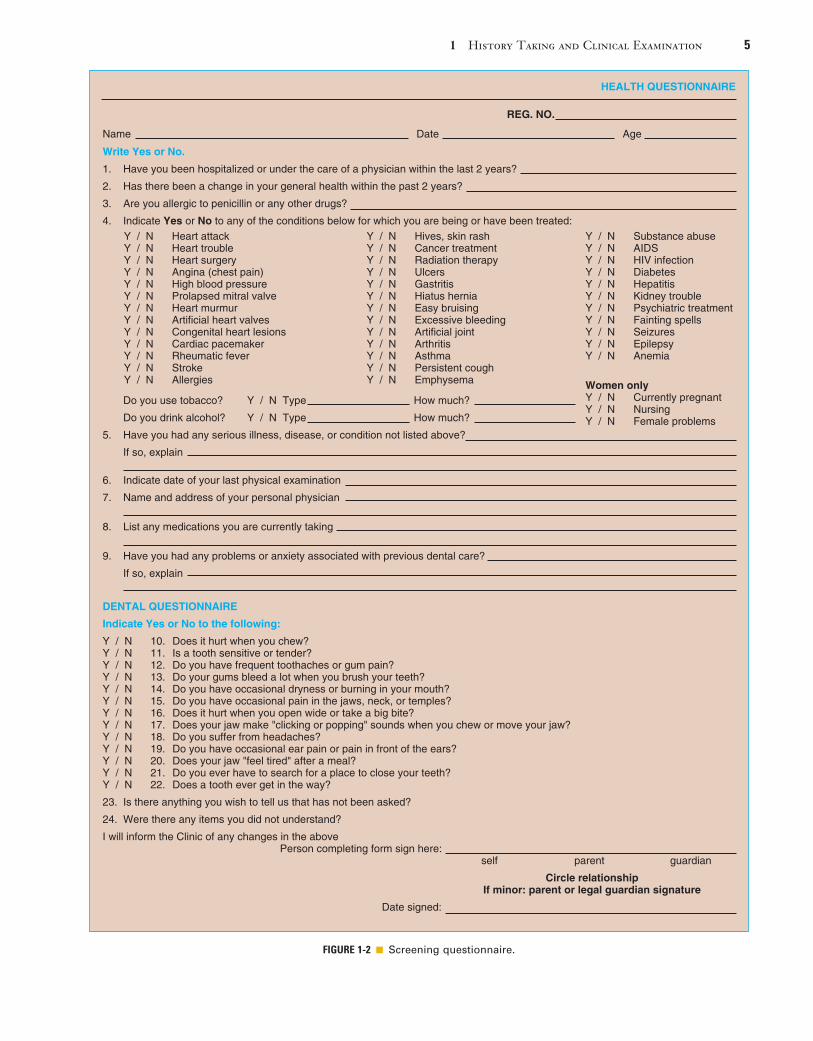
A patient’s history should include all pertinent informa-tion concerning the reasons for seeking treatment, along with any personal information, including relevant previ-ous medical and dental experiences. The chief complaint should be recorded, preferably in the patient’s own words. A screening questionnaire (Fig. 1-2) is useful for history taking; it should be reviewed in the patient’s presence to correct any mistakes and to clarify inconclusive entries.
If the patient is mentally impaired or a minor, the guard-ian or responsible parent must be present.
Chief ComplaintThe accuracy and significance of the patient’s primary reason or reasons for seeking treatment should be ana-lyzed first. These may be just the obvious features, and careful examination often reveals problems and disease of which the patient is unaware; nevertheless, the patient perceives the chief complaint as the major or only impor-tant problem. Therefore, when a comprehensive treat-ment plan is proposed, special attention must be given to how the chief complaint can be resolved. The inexperi-enced clinician who tries to prescribe an “ideal” treat-ment plan can easily lose sight of the patient’s wishes. The patient may then become frustrated because the dentist does not appear to understand or does not want to under-stand the patient’s point of view.
Chief complaints usually belong to one of the follow-ing four categories:
• Comfort (pain, sensitivity, swelling)• Function (difficulty in mastication or speech)• Social (bad taste or odor)• Appearance (fractured or unattractive teeth or res-
torations, discoloration)
Comfort
If pain is present, its location, character, severity, and frequency should be noted, as well as the first time it occurred, what factors precipitate it (e.g., pressure, hot, cold, or sweet things), any changes in its character, and whether it is localized or more diffuse in nature. It is often helpful for the patient to point at the area while the dentist pays close attention.
If swelling is present, the location, size, consistency, and color are noted, as well as how long it has been felt and whether it is increasing or decreasing.
Function
Difficulties in chewing may result from a local problem such as a fractured cusp or missing teeth; they may also indicate a more generalized malocclusion or neuromus-cular dysfunction.
Social Aspects
A bad taste or smell often indicates compromised oral hygiene and periodontal disease. Social pressures prompt many affected patients to seek care.

4 PART I Planning and Preparation
flexibility are noted. Much can be learned in a 5-minute, casual conversation during the initial visit. In addition to establishing rapport and developing a basis on which the patient can trust the dentist, small and seemingly unim-portant personal details often have considerable influence in establishing a correct diagnosis, prognosis, and treat-ment plan.
Medical HistoryAn accurate and current general medical history should include any medications the patient is taking and all relevant medical conditions. If necessary, the patient’s
Appearance
Compromised appearance is a strong motivating factor for patients to seek advice as to whether improvement is possible (Fig. 1-3). Such patients may have missing or crowded teeth, or a tooth or restoration may be fractured. The teeth may be unattractively shaped, malpositioned, or discolored, or there may be a developmental defect. A single discolored tooth may indicate pulpal disease.
Personal DetailsThe patient’s name, address, phone number, gender, occupation, work schedule, marital status, and budgetary
FIGURE 1-1 ■ A, Severely damaged maxillary dentition. B, Restoration with metal-ceramic fixed prostheses. C, Complete cast crown that restores mandibular molar. D, Three-unit fixed dental prosthesis that replaces missing mandibular premolar. E, Congenitally missing maxillary lateral incisors replaced with implant supported crowns. F, Extensive fixed prosthodontics involving restoration of multiple teeth. (C, Courtesy Dr. X. Lepe. D, Courtesy Dr. J. Nelson. E, Courtesy Dr. A. Hsieh.)
A B
E F
C D
1 History Taking and Clinical Examination 5
FIGURE 1-2 ■ Screening questionnaire.
HEALTH QUESTIONNAIRE
Name Date Age
REG. NO.
Write Yes or No.
1. Have you been hospitalized or under the care of a physician within the last 2 years?
2. Has there been a change in your general health within the past 2 years?
3. Are you allergic to penicillin or any other drugs?
4. Indicate Yes or No to any of the conditions below for which you are being or have been treated:
Do you use tobacco? Y / N Type How much?
Do you drink alcohol? Y / N Type How much?
5. Have you had any serious illness, disease, or condition not listed above?
If so, explain
6. Indicate date of your last physical examination
7. Name and address of your personal physician
8. List any medications you are currently taking
9. Have you had any problems or anxiety associated with previous dental care?
If so, explain
DENTAL QUESTIONNAIRE
Indicate Yes or No to the following:
Y / N 10. Does it hurt when you chew?Y / N 11. Is a tooth sensitive or tender?Y / N 12. Do you have frequent toothaches or gum pain?Y / N 13. Do your gums bleed a lot when you brush your teeth?Y / N 14. Do you have occasional dryness or burning in your mouth?Y / N 15. Do you have occasional pain in the jaws, neck, or temples?Y / N 16. Does it hurt when you open wide or take a big bite?Y / N 17. Does your jaw make "clicking or popping" sounds when you chew or move your jaw?Y / N 18. Do you suffer from headaches?Y / N 19. Do you have occasional ear pain or pain in front of the ears?Y / N 20. Does your jaw "feel tired" after a meal?Y / N 21. Do you ever have to search for a place to close your teeth?Y / N 22. Does a tooth ever get in the way?
23. Is there anything you wish to tell us that has not been asked?
24. Were there any items you did not understand?
I will inform the Clinic of any changes in the above Person completing form sign here: self parent guardian
Circle relationship If minor: parent or legal guardian signature
Date signed:
Y / N Heart attackY / N Heart troubleY / N Heart surgeryY / N Angina (chest pain)Y / N High blood pressureY / N Prolapsed mitral valveY / N Heart murmurY / N Artificial heart valvesY / N Congenital heart lesionsY / N Cardiac pacemakerY / N Rheumatic feverY / N StrokeY / N Allergies
Y / N Hives, skin rashY / N Cancer treatmentY / N Radiation therapyY / N UlcersY / N GastritisY / N Hiatus herniaY / N Easy bruisingY / N Excessive bleedingY / N Artificial jointY / N ArthritisY / N AsthmaY / N Persistent coughY / N Emphysema
Y / N Substance abuseY / N AIDSY / N HIV infectionY / N DiabetesY / N HepatitisY / N Kidney troubleY / N Psychiatric treatmentY / N Fainting spellsY / N SeizuresY / N EpilepsyY / N Anemia
Women onlyY / N Currently pregnantY / N NursingY / N Female problems

6 PART I Planning and Preparation
FIGURE 1-3 ■ Poor appearance is a common reason for seeking restorative dental treatment.
FIGURE 1-4 ■ Severe gingival hyperplasia associated with anti-convulsant drug use. (Courtesy Dr. P.B. Robinson.)
FIGURE 1-5 ■ A, Extensive damage caused by self-induced acid regurgitation. Note that the lingual surfaces are bare of enamel except for a narrow band at the gingival margin. B, Teeth prepared for partial-coverage restorations. C and D, The completed restoration.
A
C
B
D
physician or physicians can be contacted for clarification. The following classification may be helpful:
1. Conditions affecting the treatment methods (e.g., any disorders that necessitate the use of antibiotic premedication, any use of steroids or anticoagu-lants, and any previous allergic responses to medi-cation or dental materials). Once such conditions are identified, treatment usually can be modified as part of the comprehensive treatment plan, although some conditions may severely limit avail-able options.
2. Conditions affecting the treatment plan (e.g., pre-vious radiation therapy, hemorrhagic disorders, extremes of age, and terminal illness). These can be expected to affect the patient’s response to dental treatment and may influence the prognosis. For instance, patients who have previously received radiation treatment in the area of a planned extrac-tion require special measures (hyperbaric oxygen) to prevent serious complications.
3. Systemic conditions with oral manifestations. For example, periodontitis may be exacerbated by dia-betes, menopause, pregnancy, or the use of anticon-vulsant drugs (Fig. 1-4); in cases of gastroesophageal reflux disease, bulimia, or anorexia nervosa, teeth may be eroded by regurgitated stomach acid1,2 (Fig. 1-5); certain drugs may generate side effects that mimic temporomandibular disorders3 or reduce salivary flow.4,5
4. Possible risks to the dentist and auxiliary personnel (e.g., patients who are suspected or confirmed car-riers of hepatitis B, acquired immunodeficiency syndrome, or syphilis).
Dental offices practice “universal precautions” to ensure appropriate infection control. This means that full infection control is practiced for every patient; no addi-tional measures are needed when dentists treat known disease carriers.6
Dental HistoryClinicians should complete a thorough examination before establishing a diagnosis. With adequate experi-ence, a clinician can often assess preliminary treatment needs during the initial appointment, but review and
1 History Taking and Clinical Examination 7
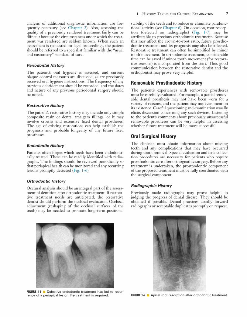
stability of the teeth and to reduce or eliminate parafunc-tional activity (see Chapter 6). On occasion, root resorp-tion (detected on radiographs) (Fig. 1-7) may be attributable to previous orthodontic treatment. Because this may affect the crown-to-root ratio, future prostho-dontic treatment and its prognosis may also be affected. Restorative treatment can often be simplified by minor tooth movement. In orthodontic treatment, considerable time can be saved if minor tooth movement (for restora-tive reasons) is incorporated from the start. Thus good communication between the restorative dentist and the orthodontist may prove very helpful.
Removable Prosthodontic HistoryThe patient’s experiences with removable prostheses must be carefully evaluated. For example, a partial remov-able dental prosthesis may not have been worn for a variety of reasons, and the patient may not even mention its existence. Careful questioning and examination usually elicits discussion concerning any such devices. Listening to the patient’s comments about previously unsuccessful removable prostheses can be very helpful in assessing whether future treatment will be more successful.
Oral Surgical HistoryThe clinician must obtain information about missing teeth and any complications that may have occurred during tooth removal. Special evaluation and data collec-tion procedures are necessary for patients who require prosthodontic care after orthognathic surgery. Before any treatment is undertaken, the prosthodontic component of the proposed treatment must be fully coordinated with the surgical component.
Radiographic History
Previously made radiographs may prove helpful in judging the progress of dental disease. They should be obtained if possible. Dental practices usually forward radiographs or acceptable duplicates promptly on request.
analysis of additional diagnostic information are fre-quently necessary (see Chapter 2). Also, assessing the quality of a previously rendered treatment fairly can be difficult because the circumstances under which the treat-ment was rendered are seldom known. When such an assessment is requested for legal proceedings, the patient should be referred to a specialist familiar with the “usual and customary” standard of care.
Periodontal History
The patient’s oral hygiene is assessed, and current plaque-control measures are discussed, as are previously received oral hygiene instructions. The frequency of any previous debridement should be recorded, and the dates and nature of any previous periodontal surgery should be noted.
Restorative History
The patient’s restorative history may include only simple composite resin or dental amalgam fillings, or it may involve crowns and extensive fixed dental prostheses. The age of existing restorations can help establish the prognosis and probable longevity of any future fixed prostheses.
Endodontic History
Patients often forget which teeth have been endodonti-cally treated. These can be readily identified with radio-graphs. The findings should be reviewed periodically so that periapical health can be monitored and any recurring lesions promptly detected (Fig. 1-6).
Orthodontic History
Occlusal analysis should be an integral part of the assess-ment of dentition after orthodontic treatment. If restora-tive treatment needs are anticipated, the restorative dentist should perform the occlusal evaluation. Occlusal adjustment (reshaping of the occlusal surfaces of the teeth) may be needed to promote long-term positional
FIGURE 1-6 ■ Defective endodontic treatment has led to recur-rence of a periapical lesion. Re-treatment is required. FIGURE 1-7 ■ Apical root resorption after orthodontic treatment.
8 PART I Planning and Preparation
recorded, rather than “gingival inflammation” (which implies a diagnosis).
Thorough examination and data collection are needed for prospective patients who desire fixed pros-thodontic treatment, and more detailed protocols for this effort can be obtained from various textbooks of oral diagnosis.7,8
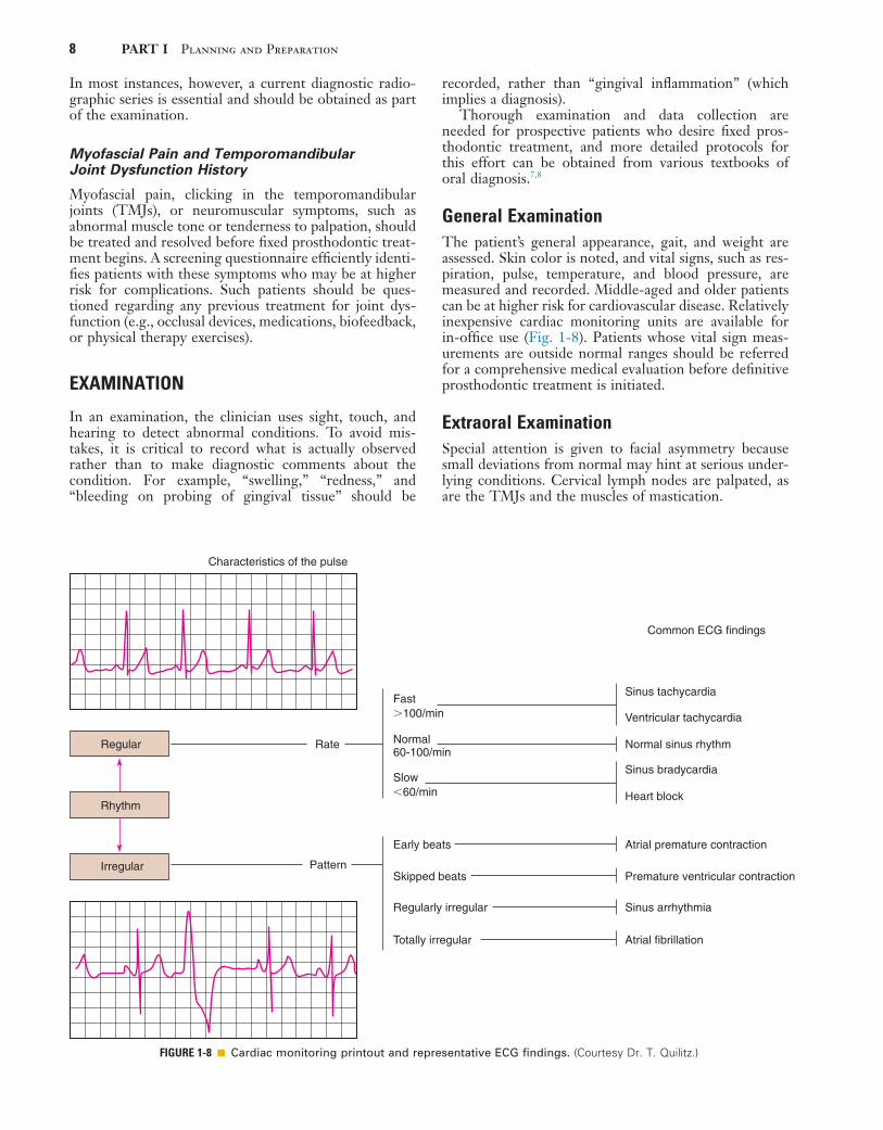
General ExaminationThe patient’s general appearance, gait, and weight are assessed. Skin color is noted, and vital signs, such as res-piration, pulse, temperature, and blood pressure, are measured and recorded. Middle-aged and older patients can be at higher risk for cardiovascular disease. Relatively inexpensive cardiac monitoring units are available for in-office use (Fig. 1-8). Patients whose vital sign meas-urements are outside normal ranges should be referred for a comprehensive medical evaluation before definitive prosthodontic treatment is initiated.
Extraoral ExaminationSpecial attention is given to facial asymmetry because small deviations from normal may hint at serious under-lying conditions. Cervical lymph nodes are palpated, as are the TMJs and the muscles of mastication.
In most instances, however, a current diagnostic radio-graphic series is essential and should be obtained as part of the examination.
Myofascial Pain and Temporomandibular Joint Dysfunction History
Myofascial pain, clicking in the temporomandibular joints (TMJs), or neuromuscular symptoms, such as abnormal muscle tone or tenderness to palpation, should be treated and resolved before fixed prosthodontic treat-ment begins. A screening questionnaire efficiently identi-fies patients with these symptoms who may be at higher risk for complications. Such patients should be ques-tioned regarding any previous treatment for joint dys-function (e.g., occlusal devices, medications, biofeedback, or physical therapy exercises).
EXAMINATION
In an examination, the clinician uses sight, touch, and hearing to detect abnormal conditions. To avoid mis-takes, it is critical to record what is actually observed rather than to make diagnostic comments about the condition. For example, “swelling,” “redness,” and “bleeding on probing of gingival tissue” should be
FIGURE 1-8 ■ Cardiac monitoring printout and representative ECG findings. (Courtesy Dr. T. Quilitz.)
Regular
Rhythm
Irregular
Rate
Fast�100/min
Early beats
Skipped beats
Regularly irregular
Totally irregular
Atrial premature contraction
Sinus tachycardia
Sinus bradycardia
Normal sinus rhythm
Heart block
Common ECG findings
Ventricular tachycardia
Premature ventricular contraction
Sinus arrhythmia
Atrial fibrillation
Normal60-100/min
Slow�60/min
Characteristics of the pulse
Pattern
1 History Taking and Clinical Examination 9
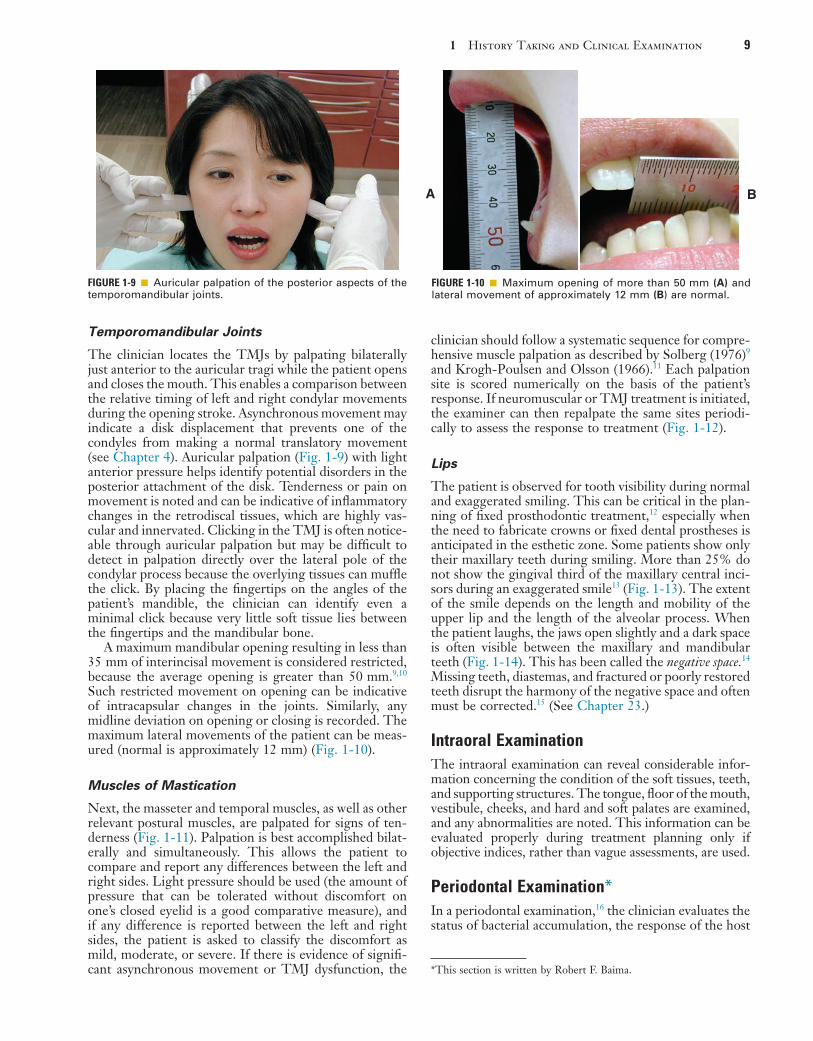
FIGURE 1-9 ■ Auricular palpation of the posterior aspects of the temporomandibular joints.
FIGURE 1-10 ■ Maximum opening of more than 50 mm (A) and lateral movement of approximately 12 mm (B) are normal.
A B
Temporomandibular Joints
The clinician locates the TMJs by palpating bilaterally just anterior to the auricular tragi while the patient opens and closes the mouth. This enables a comparison between the relative timing of left and right condylar movements during the opening stroke. Asynchronous movement may indicate a disk displacement that prevents one of the condyles from making a normal translatory movement (see Chapter 4). Auricular palpation (Fig. 1-9) with light anterior pressure helps identify potential disorders in the posterior attachment of the disk. Tenderness or pain on movement is noted and can be indicative of inflammatory changes in the retrodiscal tissues, which are highly vas-cular and innervated. Clicking in the TMJ is often notice-able through auricular palpation but may be difficult to detect in palpation directly over the lateral pole of the condylar process because the overlying tissues can muffle the click. By placing the fingertips on the angles of the patient’s mandible, the clinician can identify even a minimal click because very little soft tissue lies between the fingertips and the mandibular bone.
A maximum mandibular opening resulting in less than 35 mm of interincisal movement is considered restricted, because the average opening is greater than 50 mm.9,10 Such restricted movement on opening can be indicative of intracapsular changes in the joints. Similarly, any midline deviation on opening or closing is recorded. The maximum lateral movements of the patient can be meas-ured (normal is approximately 12 mm) (Fig. 1-10).
Muscles of Mastication
Next, the masseter and temporal muscles, as well as other relevant postural muscles, are palpated for signs of ten-derness (Fig. 1-11). Palpation is best accomplished bilat-erally and simultaneously. This allows the patient to compare and report any differences between the left and right sides. Light pressure should be used (the amount of pressure that can be tolerated without discomfort on one’s closed eyelid is a good comparative measure), and if any difference is reported between the left and right sides, the patient is asked to classify the discomfort as mild, moderate, or severe. If there is evidence of signifi-cant asynchronous movement or TMJ dysfunction, the *This section is written by Robert F. Baima.
clinician should follow a systematic sequence for compre-hensive muscle palpation as described by Solberg (1976)9 and Krogh-Poulsen and Olsson (1966).11 Each palpation site is scored numerically on the basis of the patient’s response. If neuromuscular or TMJ treatment is initiated, the examiner can then repalpate the same sites periodi-cally to assess the response to treatment (Fig. 1-12).
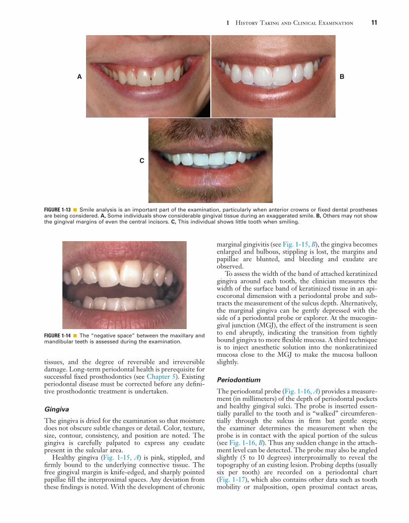
Lips
The patient is observed for tooth visibility during normal and exaggerated smiling. This can be critical in the plan-ning of fixed prosthodontic treatment,12 especially when the need to fabricate crowns or fixed dental prostheses is anticipated in the esthetic zone. Some patients show only their maxillary teeth during smiling. More than 25% do not show the gingival third of the maxillary central inci-sors during an exaggerated smile13 (Fig. 1-13). The extent of the smile depends on the length and mobility of the upper lip and the length of the alveolar process. When the patient laughs, the jaws open slightly and a dark space is often visible between the maxillary and mandibular teeth (Fig. 1-14). This has been called the negative space.14 Missing teeth, diastemas, and fractured or poorly restored teeth disrupt the harmony of the negative space and often must be corrected.15 (See Chapter 23.)
Intraoral ExaminationThe intraoral examination can reveal considerable infor-mation concerning the condition of the soft tissues, teeth, and supporting structures. The tongue, floor of the mouth, vestibule, cheeks, and hard and soft palates are examined, and any abnormalities are noted. This information can be evaluated properly during treatment planning only if objective indices, rather than vague assessments, are used.
Periodontal Examination*In a periodontal examination,16 the clinician evaluates the status of bacterial accumulation, the response of the host

10 PART I Planning and Preparation
FIGURE 1-11 ■ Muscle palpation of the masseter (A), the temporal muscle (B), the trapezius muscle (C), the sternocleidomastoid muscle (D), and the floor of the mouth (E).
A
C D
B
E
FIGURE 1-12 ■ Palpation sites for assessing muscle tenderness. A, Temporomandibular joint capsule: lateral and dorsal. B, Masseter: deep and superficial. C, Temporal muscle: anterior and posterior. D, Vertex. E, Neck: nape and base. F, Sternocleidomastoid muscle: insertion, body, and origin. G, Medial pterygoid muscle. H, Posterior digastric muscle. I, Temporal tendon. J, Lateral pterygoid muscle. (From Krogh-Poulsen WG, Olsson A: Occlusal disharmonies and dysfunction of the stomatognathic system. Dent Clin North Am 10:627, 1966.)
D
C C
B
BF
F
F
E
E
I
G
H
AA
J
Palpation is best donebilaterally; simultaneously,the patient is asked toidentify any differencesbetween left and right.
1 History Taking and Clinical Examination 11
FIGURE 1-13 ■ Smile analysis is an important part of the examination, particularly when anterior crowns or fixed dental prostheses are being considered. A, Some individuals show considerable gingival tissue during an exaggerated smile. B, Others may not show the gingival margins of even the central incisors. C, This individual shows little tooth when smiling.
A
C
B
FIGURE 1-14 ■ The “negative space” between the maxillary and mandibular teeth is assessed during the examination.
tissues, and the degree of reversible and irreversible damage. Long-term periodontal health is prerequisite for successful fixed prosthodontics (see Chapter 5). Existing periodontal disease must be corrected before any defini-tive prosthodontic treatment is undertaken.
Gingiva
The gingiva is dried for the examination so that moisture does not obscure subtle changes or detail. Color, texture, size, contour, consistency, and position are noted. The gingiva is carefully palpated to express any exudate present in the sulcular area.
Healthy gingiva (Fig. 1-15, A) is pink, stippled, and firmly bound to the underlying connective tissue. The free gingival margin is knife-edged, and sharply pointed papillae fill the interproximal spaces. Any deviation from these findings is noted. With the development of chronic
marginal gingivitis (see Fig. 1-15, B), the gingiva becomes enlarged and bulbous, stippling is lost, the margins and papillae are blunted, and bleeding and exudate are observed.
To assess the width of the band of attached keratinized gingiva around each tooth, the clinician measures the width of the surface band of keratinized tissue in an api-cocoronal dimension with a periodontal probe and sub-tracts the measurement of the sulcus depth. Alternatively, the marginal gingiva can be gently depressed with the side of a periodontal probe or explorer. At the mucogin-gival junction (MGJ), the effect of the instrument is seen to end abruptly, indicating the transition from tightly bound gingiva to more flexible mucosa. A third technique is to inject anesthetic solution into the nonkeratinized mucosa close to the MGJ to make the mucosa balloon slightly.
Periodontium
The periodontal probe (Fig. 1-16, A) provides a measure-ment (in millimeters) of the depth of periodontal pockets and healthy gingival sulci. The probe is inserted essen-tially parallel to the tooth and is “walked” circumferen-tially through the sulcus in firm but gentle steps; the examiner determines the measurement when the probe is in contact with the apical portion of the sulcus (see Fig. 1-16, B). Thus any sudden change in the attach-ment level can be detected. The probe may also be angled slightly (5 to 10 degrees) interproximally to reveal the topography of an existing lesion. Probing depths (usually six per tooth) are recorded on a periodontal chart (Fig. 1-17), which also contains other data such as tooth mobility or malposition, open proximal contact areas,

12 PART I Planning and Preparation
FIGURE 1-16 ■ A, Three types of sulcus/pocket-measuring probes. B, Correct position of a periodontal probe in the interproximal sulcular area, parallel to the root surface and in a vertical direction as far interproximally as possible. C and D, Graduated furcation probe. (A and C, From Boyd LB: Dental instruments, 5th ed. St. Louis, Saunders, 2015.)
A
C D
B
FIGURE 1-15 ■ A, Healthy gingiva is pink, knife-edged, and firmly attached. B, In gingivitis, plaque and calculus cause marginal inflam-mation, with changes in color, contour, and consistency of the free gingival margin. In this case, inflammation extends into the keratinized attached gingiva.
A B
inconsistent marginal ridge heights, missing or impacted teeth, areas of inadequate attached keratinized gingiva, gingival recession, furcation involvements, and malposi-tioned frenum attachments).
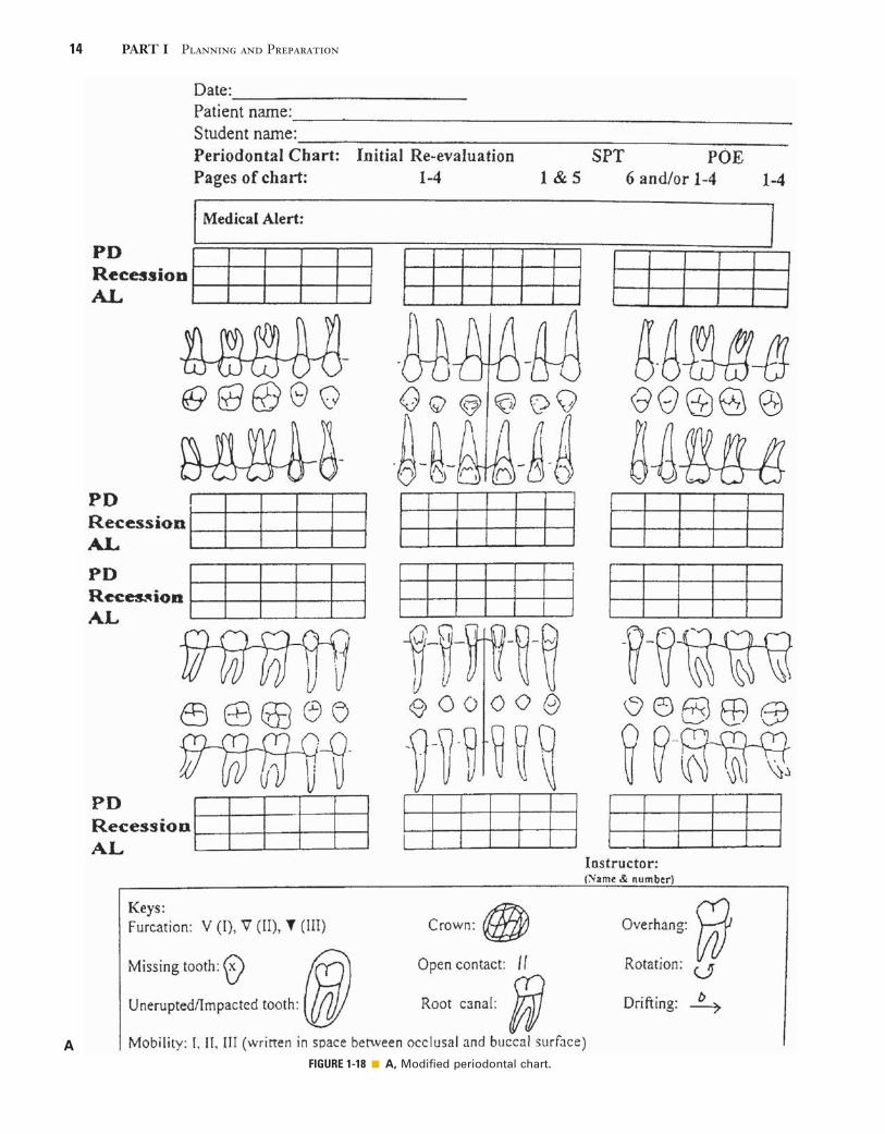
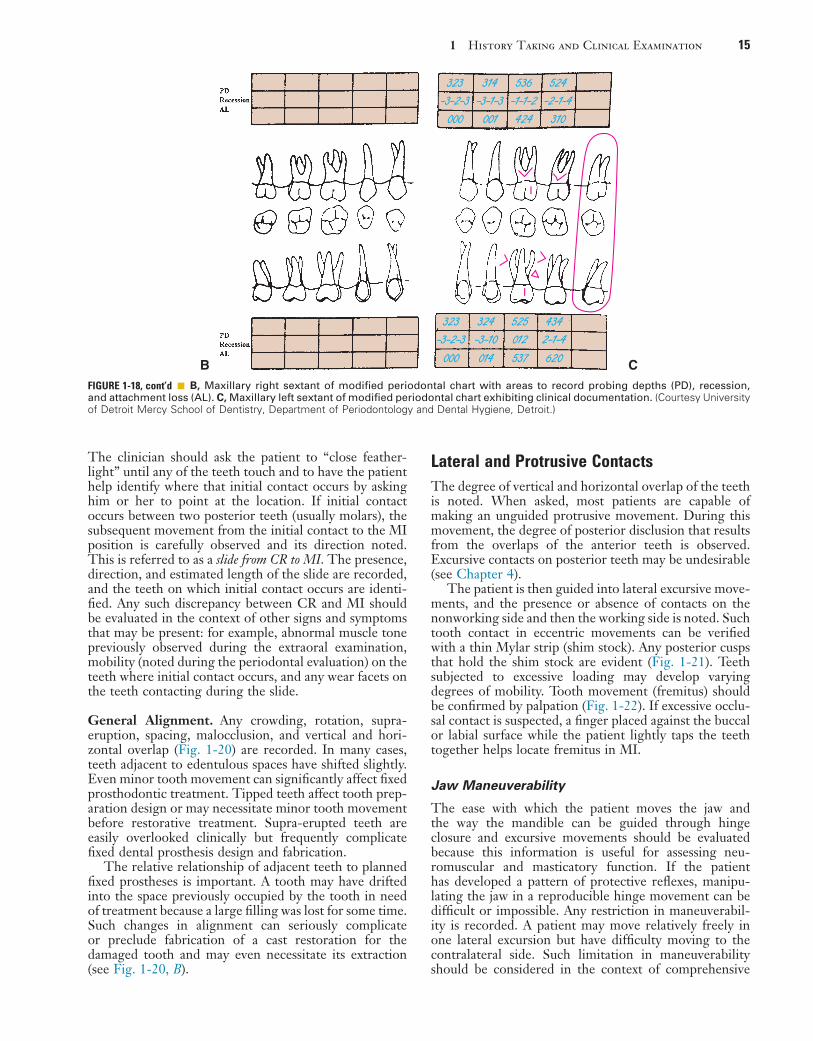
Clinical Attachment LevelDocumenting the level of epithelial attachment helps the clinician quantify periodontal destruction and is essential for rendering a diagnosis of periodontitis (loss of connec-tive tissue attachment).17,18 This measurement also pro-vides objective information regarding the prognosis of individual teeth. The clinical attachment level is deter-mined by measuring the distance between the apical extent of the probing depth and a fixed reference point on the tooth, most commonly either the apical extent of a restoration or the cementoenamel junction (CEJ). This is recorded on modified periodontal charts (Fig. 1-18). When the free margin of the gingiva is located on the clinical crown and the level of the epithelial attachment is at the CEJ, there is no attachment loss, and recession is noted as a negative number. When the attachment level is on root structure and the free gingival margin is at the CEJ, attachment loss equals the probing depth, and the
recession is scored 0. When increased periodontal destruction and recession are present, attachment loss equals the probing depth plus the measurement of reces-sion19 (see Fig. 1-18, B and C). Clinical attachment loss is a measure of periodontal destruction at a site, rather than of current disease activity; it may be considered the diagnostic “gold standard” for periodontitis20 and should be documented in the initial periodontal examination.21 It is an important consideration in the development of the overall diagnosis, treatment plan, and the prognosis of the dentition.
• • •
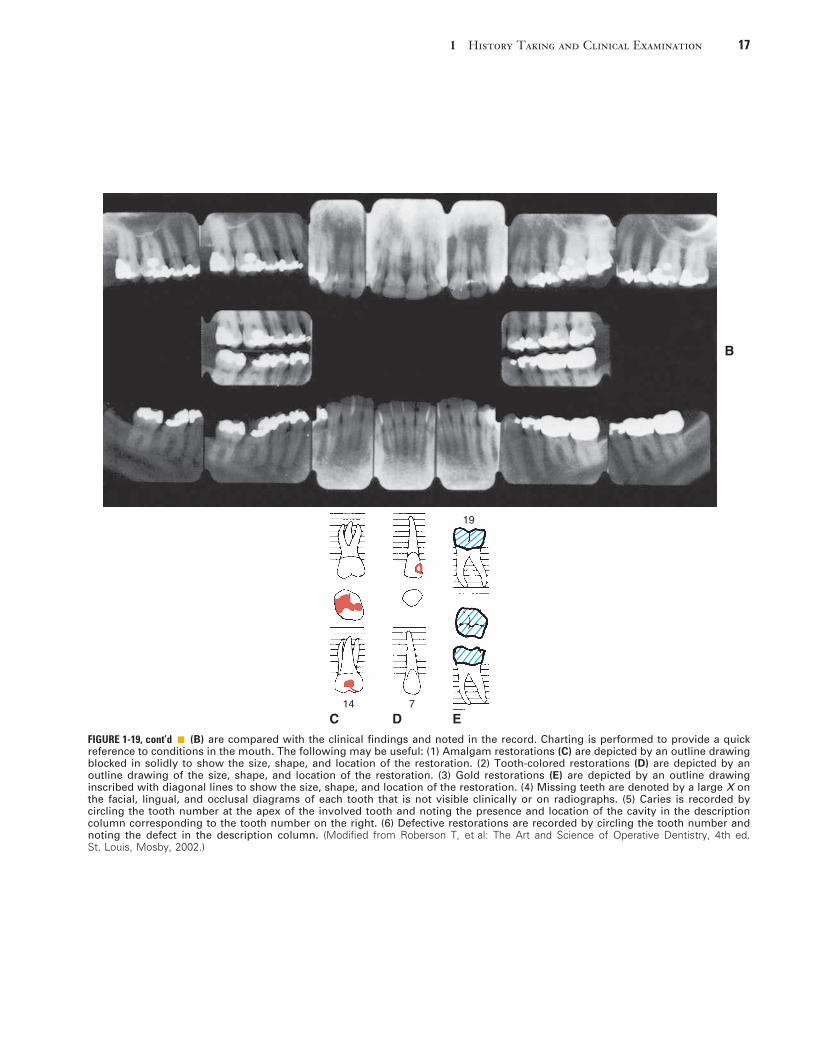
Dental ChartingAn accurate charting of the state of the dentition reveals important information about the condition of the teeth and facilitates treatment planning. Adequate charting (Fig. 1-19) shows the presence or absence of teeth, dental caries, restorations, wear faceting and abrasions, frac-tures, malformations, and erosions. Tooth loss often affects the position of adjacent teeth (see Treatment of Tooth Loss section, Chapter 3). The presence of caries

1 History Taking and Clinical Examination 13
FIGURE 1-17 ■ Chart for recording pocket depths. The parallel lines are approximately 2 mm apart. The notations involved in using the chart are as follows: 1, Block out any missing teeth. 2, Draw a red × through the crown of any tooth that is to be extracted. 3, Record the gingival level with a continuous blue line. 4, Record pocket depths with a red line interrupted at the proximal surfaces of each tooth. 5, Shade the pocket form on each tooth with a red pencil (between the red and blue lines). 6, Indicate bifurcation or trifurcation involvements with a small red × at the involved area. 7, Record open contacts with vertical parallel lines (‖) through the area. 8, Record improper contacts with a wavy red line through the area. 9, Record gingival overhang(s) with a red spur (∧) through the area. 10, Outline cavities and faulty restorations of periodontal significance in red. 11, Indicate rotated teeth by outlining in blue to show their actual position. (Modified from Goldman HM, Cohen DW: Periodontal Therapy, 5th ed. St. Louis, Mosby, 1973.)
Charting of conditions before periodontal treatment
Photographs, date Radiographs, date Casts, date
LeftRight
8 87 76 65 54 43 32 21 1
8 87 76 65 54 43 32 21 1
Occlusal
Occlusal
Lingual
Lingual
Facial
Facial
Max
illar
yM
andi
bula
r
on one interproximal surface should prompt the exam-iner to carefully inspect the adjacent proximal surface, even if caries is not apparent radiographically. The degree and extent of caries development over time can have a considerable effect on the eventual outcome of fixed prosthodontic treatment. The condition and type of the existing restorations are noted (e.g., amalgam, cast gold, composite resin, all-ceramic). Open contacts and areas where food impaction occurs must also be identified. The presence of wear facets is indicative of sliding contact sustained over time and thus may indicate parafunctional activity (see Chapter 4). Wear facets are often easier to see on diagnostic casts, however (see Chapter 2); during the clinical examination, the location of any observed facet is recorded. Fracture lines in teeth may necessitate fixed prosthodontic intervention, although minor hairline cracks in walls that are not subject to excessive loading can often go untreated and simply be observed at recall appointments (see Chapter 32). The location of fractures and any other abnormalities should be recorded.
Occlusal Examination
The clinician starts the occlusal examination by asking the patient to make a few simple opening and closing movements, which the clinician carefully observes. The objective is to determine to what extent the patient’s occlusion differs from the ideal (see Chapter 4) and how well the patient has adapted to any difference that may exist. Special attention is given to initial contact, tooth alignment, eccentric occlusal contacts, and jaw maneuverability.
Initial Tooth Contact. The relationship of teeth in both centric relation (see Chapter 4) and the maximum intercuspation should be evaluated. If all teeth come together simultaneously at the end of terminal hinge closure, the centric relation (CR) position of the patient is said to coincide with the maximum intercuspation (MI) (see also Chapters 2 and 4). The patient is guided into a terminal hinge closure to detect where initial tooth contact occurs (see also the sections on bimanual manipu-lation and terminal hinge closure in Chapters 2 and 4).
14 PART I Planning and Preparation
AFIGURE 1-18 ■ A, Modified periodontal chart.
1 History Taking and Clinical Examination 15
The clinician should ask the patient to “close feather-light” until any of the teeth touch and to have the patient help identify where that initial contact occurs by asking him or her to point at the location. If initial contact occurs between two posterior teeth (usually molars), the subsequent movement from the initial contact to the MI position is carefully observed and its direction noted. This is referred to as a slide from CR to MI. The presence, direction, and estimated length of the slide are recorded, and the teeth on which initial contact occurs are identi-fied. Any such discrepancy between CR and MI should be evaluated in the context of other signs and symptoms that may be present: for example, abnormal muscle tone previously observed during the extraoral examination, mobility (noted during the periodontal evaluation) on the teeth where initial contact occurs, and any wear facets on the teeth contacting during the slide.
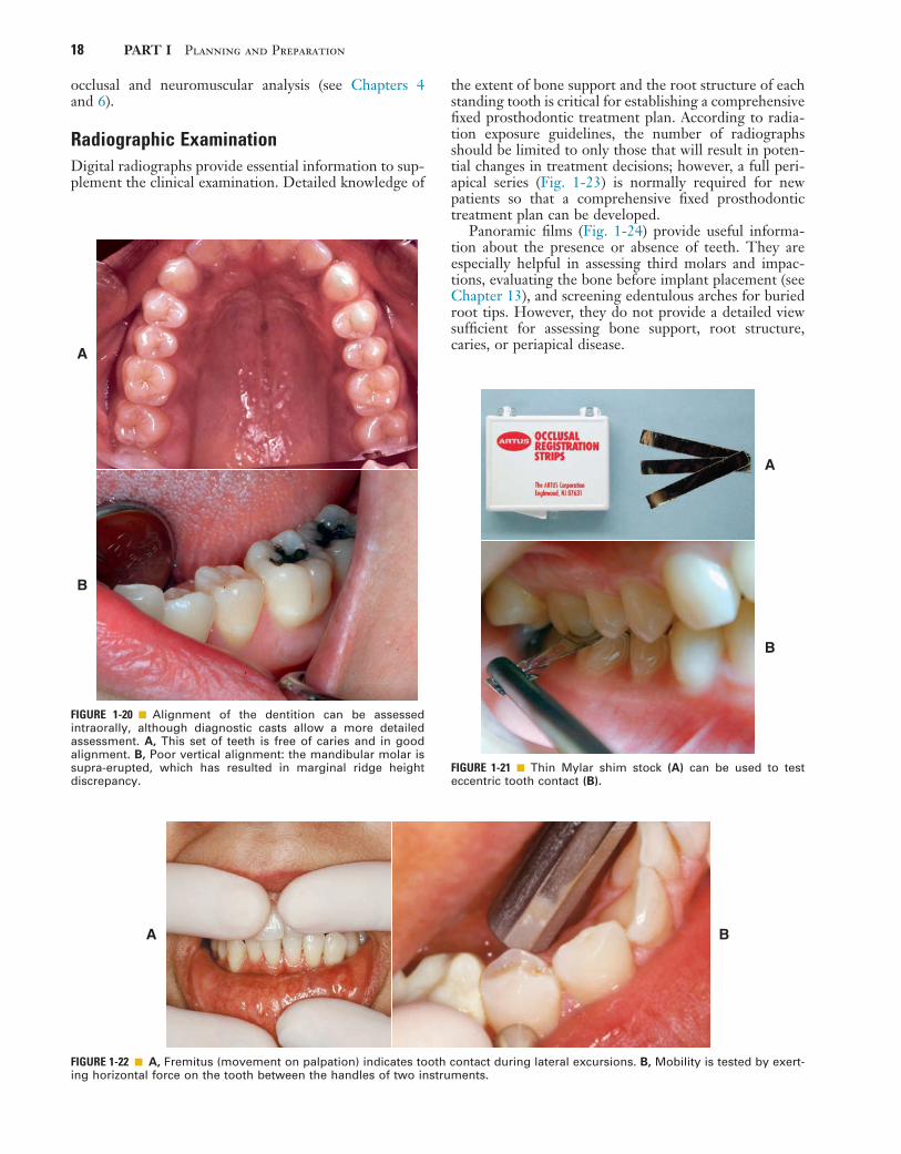
General Alignment. Any crowding, rotation, supra-eruption, spacing, malocclusion, and vertical and hori-zontal overlap (Fig. 1-20) are recorded. In many cases, teeth adjacent to edentulous spaces have shifted slightly. Even minor tooth movement can significantly affect fixed prosthodontic treatment. Tipped teeth affect tooth prep-aration design or may necessitate minor tooth movement before restorative treatment. Supra-erupted teeth are easily overlooked clinically but frequently complicate fixed dental prosthesis design and fabrication.
The relative relationship of adjacent teeth to planned fixed prostheses is important. A tooth may have drifted into the space previously occupied by the tooth in need of treatment because a large filling was lost for some time. Such changes in alignment can seriously complicate or preclude fabrication of a cast restoration for the damaged tooth and may even necessitate its extraction (see Fig. 1-20, B).
Lateral and Protrusive ContactsThe degree of vertical and horizontal overlap of the teeth is noted. When asked, most patients are capable of making an unguided protrusive movement. During this movement, the degree of posterior disclusion that results from the overlaps of the anterior teeth is observed. Excursive contacts on posterior teeth may be undesirable (see Chapter 4).
The patient is then guided into lateral excursive move-ments, and the presence or absence of contacts on the nonworking side and then the working side is noted. Such tooth contact in eccentric movements can be verified with a thin Mylar strip (shim stock). Any posterior cusps that hold the shim stock are evident (Fig. 1-21). Teeth subjected to excessive loading may develop varying degrees of mobility. Tooth movement (fremitus) should be confirmed by palpation (Fig. 1-22). If excessive occlu-sal contact is suspected, a finger placed against the buccal or labial surface while the patient lightly taps the teeth together helps locate fremitus in MI.
Jaw Maneuverability
The ease with which the patient moves the jaw and the way the mandible can be guided through hinge closure and excursive movements should be evaluated because this information is useful for assessing neu-romuscular and masticatory function. If the patient has developed a pattern of protective reflexes, manipu-lating the jaw in a reproducible hinge movement can be difficult or impossible. Any restriction in maneuverabil-ity is recorded. A patient may move relatively freely in one lateral excursion but have difficulty moving to the contralateral side. Such limitation in maneuverability should be considered in the context of comprehensive
I
I
323 314 536 524-3-2-3 -3-1-3 -1-1-2 -2-1-4
000 001 424 310
323 324 525 434-3-2-3 -3-10 012 2-1-4
000 014 537 620 CBFIGURE 1-18, cont’d ■ B, Maxillary right sextant of modified periodontal chart with areas to record probing depths (PD), recession, and attachment loss (AL). C, Maxillary left sextant of modified periodontal chart exhibiting clinical documentation. (Courtesy University of Detroit Mercy School of Dentistry, Department of Periodontology and Dental Hygiene, Detroit.)
16 PART I Planning and Preparation
FIGURE 1-19 ■ An appropriate charting system (A) designates the location, type, and extent of existing restorations and the presence of any disease condition, all of which become part of the patient’s permanent record. Radiographic findings obtained from a full-mouth series
11/12/82
I Anterior crowding, limited LL movement
Defective restorationOL defectiveD cariesDefectiveD caries, M defectiveL & M cariesD defective
L pit caries, D defectiveM cariesD overhangDefective; recurrent cariesM overhangCaries, broken tooth
M cariesD caries (lost amalgam)M defectiveD defective
D defectiveM cariesD overhang, M defectiveCaries, broken toothF & L caries
Gingivitis, bleeding upon probing,minimal pocket depth
11 12 82 16 II
A
1 History Taking and Clinical Examination 17
B
19
14 7
C D EFIGURE 1-19, cont’d ■ (B) are compared with the clinical findings and noted in the record. Charting is performed to provide a quick reference to conditions in the mouth. The following may be useful: (1) Amalgam restorations (C) are depicted by an outline drawing blocked in solidly to show the size, shape, and location of the restoration. (2) Tooth-colored restorations (D) are depicted by an outline drawing of the size, shape, and location of the restoration. (3) Gold restorations (E) are depicted by an outline drawing inscribed with diagonal lines to show the size, shape, and location of the restoration. (4) Missing teeth are denoted by a large X on the facial, lingual, and occlusal diagrams of each tooth that is not visible clinically or on radiographs. (5) Caries is recorded by circling the tooth number at the apex of the involved tooth and noting the presence and location of the cavity in the description column corresponding to the tooth number on the right. (6) Defective restorations are recorded by circling the tooth number and noting the defect in the description column. (Modified from Roberson T, et al: The Art and Science of Operative Dentistry, 4th ed. St. Louis, Mosby, 2002.)
18 PART I Planning and Preparation
FIGURE 1-20 ■ Alignment of the dentition can be assessed intraorally, although diagnostic casts allow a more detailed assessment. A, This set of teeth is free of caries and in good alignment. B, Poor vertical alignment: the mandibular molar is supra-erupted, which has resulted in marginal ridge height discrepancy.
A
B
FIGURE 1-21 ■ Thin Mylar shim stock (A) can be used to test eccentric tooth contact (B).
A
B
FIGURE 1-22 ■ A, Fremitus (movement on palpation) indicates tooth contact during lateral excursions. B, Mobility is tested by exert-ing horizontal force on the tooth between the handles of two instruments.
A B
occlusal and neuromuscular analysis (see Chapters 4 and 6).
Radiographic ExaminationDigital radiographs provide essential information to sup-plement the clinical examination. Detailed knowledge of
the extent of bone support and the root structure of each standing tooth is critical for establishing a comprehensive fixed prosthodontic treatment plan. According to radia-tion exposure guidelines, the number of radiographs should be limited to only those that will result in poten-tial changes in treatment decisions; however, a full peri-apical series (Fig. 1-23) is normally required for new patients so that a comprehensive fixed prosthodontic treatment plan can be developed.
Panoramic films (Fig. 1-24) provide useful informa-tion about the presence or absence of teeth. They are especially helpful in assessing third molars and impac-tions, evaluating the bone before implant placement (see Chapter 13), and screening edentulous arches for buried root tips. However, they do not provide a detailed view sufficient for assessing bone support, root structure, caries, or periapical disease.

1 History Taking and Clinical Examination 19
FIGURE 1-23 ■ A to C, A full-mouth radiographic survey should enable the dentist to make a detailed assessment of the structure of each tooth and its bone support.
A B
C
FIGURE 1-24 ■ A panoramic film cannot be substituted for a full-mouth series because the image is distorted. Nevertheless, it is very useful for assessing unerupted teeth, screening edentulous areas for buried root tips, and evaluating the bone before implant placement.
20 PART I Planning and Preparation
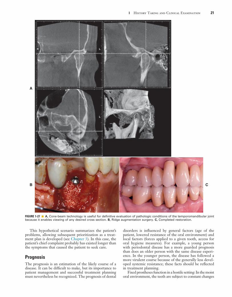
Special radiographs may be needed for the assessment of TMJ disorders and a wide variety of pathologic conditions ranging from bone and mineral disorders to metabolic disorders, genetic abnormalities, and soft tissue calcifications, such as carotid artery calcification.21 For assessment of the TMJs, a transcranial exposure (Fig. 1-25), with the help of a positioning device, reveals the lateral third of the mandibular condyle and can be used to detect some structural and positional changes. However, interpretation may be difficult,22 and more information may be obtained from other images23 (Fig. 1-26). Cone-beam imaging is considered prerequi-site to most dental implant placements. In this form of imaging, osseous contours and bone volume are visual-ized, which improves decision making about the size of implant fixtures that realistically can be accommodated (Fig. 1-27).
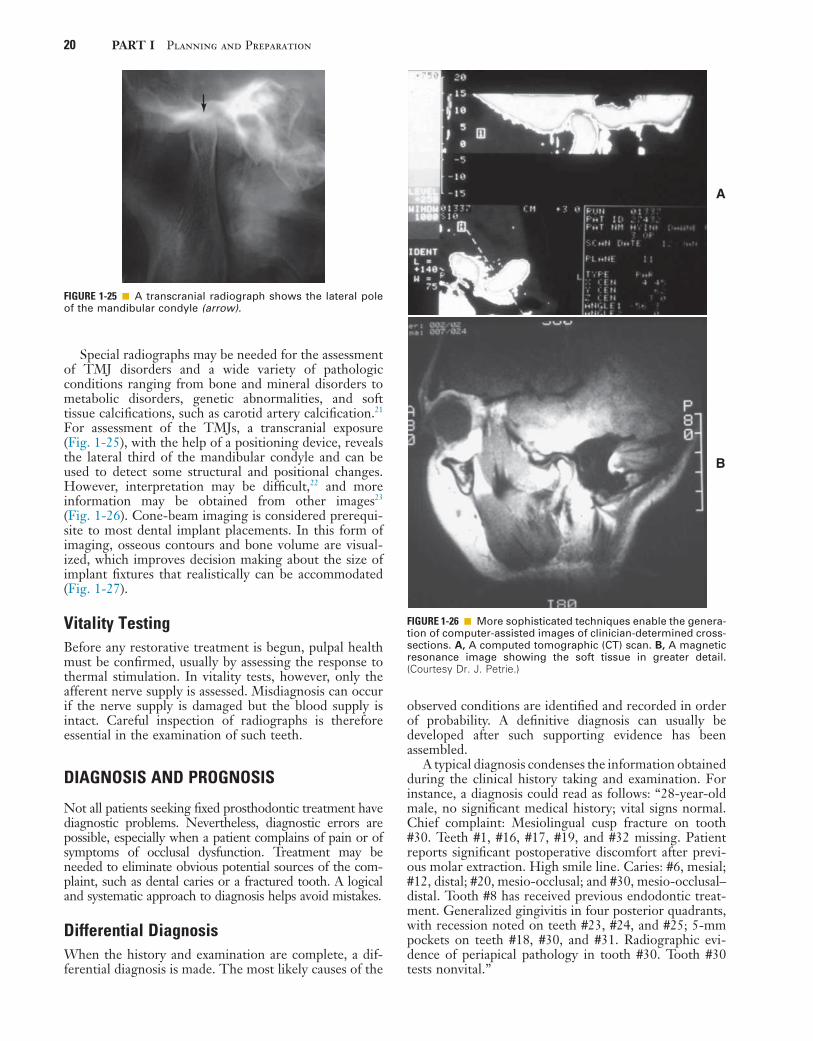
Vitality TestingBefore any restorative treatment is begun, pulpal health must be confirmed, usually by assessing the response to thermal stimulation. In vitality tests, however, only the afferent nerve supply is assessed. Misdiagnosis can occur if the nerve supply is damaged but the blood supply is intact. Careful inspection of radiographs is therefore essential in the examination of such teeth.
DIAGNOSIS AND PROGNOSIS
Not all patients seeking fixed prosthodontic treatment have diagnostic problems. Nevertheless, diagnostic errors are possible, especially when a patient complains of pain or of symptoms of occlusal dysfunction. Treatment may be needed to eliminate obvious potential sources of the com-plaint, such as dental caries or a fractured tooth. A logical and systematic approach to diagnosis helps avoid mistakes.
Differential DiagnosisWhen the history and examination are complete, a dif-ferential diagnosis is made. The most likely causes of the
FIGURE 1-25 ■ A transcranial radiograph shows the lateral pole of the mandibular condyle (arrow).
FIGURE 1-26 ■ More sophisticated techniques enable the genera-tion of computer-assisted images of clinician-determined cross-sections. A, A computed tomographic (CT) scan. B, A magnetic resonance image showing the soft tissue in greater detail. (Courtesy Dr. J. Petrie.)
A
B
observed conditions are identified and recorded in order of probability. A definitive diagnosis can usually be developed after such supporting evidence has been assembled.
A typical diagnosis condenses the information obtained during the clinical history taking and examination. For instance, a diagnosis could read as follows: “28-year-old male, no significant medical history; vital signs normal. Chief complaint: Mesiolingual cusp fracture on tooth #30. Teeth #1, #16, #17, #19, and #32 missing. Patient reports significant postoperative discomfort after previ-ous molar extraction. High smile line. Caries: #6, mesial; #12, distal; #20, mesio-occlusal; and #30, mesio-occlusal–distal. Tooth #8 has received previous endodontic treat-ment. Generalized gingivitis in four posterior quadrants, with recession noted on teeth #23, #24, and #25; 5-mm pockets on teeth #18, #30, and #31. Radiographic evi-dence of periapical pathology in tooth #30. Tooth #30 tests nonvital.”
1 History Taking and Clinical Examination 21
A
FIGURE 1-27 ■ A, Cone-beam technology is useful for definitive evaluation of pathologic conditions of the temporomandibular joint because it enables viewing of any desired cross section. B, Ridge augmentation surgery. C, Completed restoration.
B C
This hypothetical scenario summarizes the patient’s problems, allowing subsequent prioritization as a treat-ment plan is developed (see Chapter 3). In this case, the patient’s chief complaint probably has existed longer than the symptoms that caused the patient to seek care.
PrognosisThe prognosis is an estimation of the likely course of a disease. It can be difficult to make, but its importance to patient management and successful treatment planning must nevertheless be recognized. The prognosis of dental
disorders is influenced by general factors (age of the patient, lowered resistance of the oral environment) and local factors (forces applied to a given tooth, access for oral hygiene measures). For example, a young person with periodontal disease has a more guarded prognosis than does an older person with the same disease experi-ence. In the younger person, the disease has followed a more virulent course because of the generally less devel-oped systemic resistance; these facts should be reflected in treatment planning.
Fixed prostheses function in a hostile setting: In the moist oral environment, the teeth are subject to constant changes
22 PART I Planning and Preparation
and support of the ACP. These guidelines are intended to help practitioners determine appropriate treatments for their patients. For each index, four categories, class I to class IV, are defined; class I represents an uncompli-cated clinical situation and class IV represents a complex clinical situation. The indices are designed for use by dental professionals involved in the diagnosis and treatment of partially edentulous and completely dentate patients. Potential benefits of the system include (1) improved intraoperator consistency, (2) improved professional communication, (3) insurance reimburse-ment commensurate with complexity of care, (4) improved screening tool for dental school admission clinics, (5) standardized criteria for outcomes assessment and research, (6) enhanced diagnostic consistency, and (7) simplified decision to refer a patient.
Each class is differentiated by specific diagnostic cri-teria (ideal or minimal, moderately compromised, sub-stantially compromised, or severely compromised) of the following (for partially edentulous patients):
1. Location and extent of the edentulous area or areas2. Condition of the abutment teeth3. Occlusal scheme4. Residual ridge
For completely dentate patients, only tooth condition and occlusal scheme are evaluated.
Location and Extent of the Edentulous AreasIn the ideal or minimally compromised edentulous area, the edentulous span is confined to a single arch, and one of the following conditions is present:
• Any anterior maxillary span that does not exceed two missing incisors
• Any anterior mandibular span that does not exceed four missing incisors
• Any posterior maxillary or mandibular span that does not exceed two premolars or one premolar and one molar
In the moderately compromised edentulous area, the eden-tulous span is in both arches, and one of the following conditions exists:
• The span includes any anterior maxillary span that does not exceed two missing incisors.
• The span includes any anterior mandibular span that does not exceed four missing incisors.
• The span includes any posterior maxillary or man-dibular span that does not exceed two premolars or one premolar and one molar.
• The maxillary or mandibular canine tooth is missing.The substantially compromised edentulous area includes
the following conditions:• Any posterior maxillary or mandibular span that is
greater than three missing teeth or two molars• Any edentulous span, including anterior and poste-
rior areas of three or more missing teethThe severely compromised edentulous area includes the
following condition:• Any edentulous area or combination of edentulous
areas whose care requires a high level of patient compliance
in temperature and acidity and to considerable load fluctua-tion. A comprehensive clinical examination helps establish the likely prognosis. All facts and observations are first con-sidered individually and then correlated appropriately.
General Factors
The overall caries rate of the patient’s dentition indicates future risk to the patient if the condition is left untreated. Important variables include the patient’s understanding and comprehension of plaque-control measures, as well as the physical ability to perform those tasks. Analysis of systemic problems in the context of the patient’s age and overall health provides important information. For example, the incidence of periodontal disease is higher in diabetic patients than in the general population, and special precautionary measures may be indicated in those patients before treatment begins. Such conditions also affect the overall prognosis.
Some patients are capable of exerting an extremely high occlusal force (see Fig. 7-39), whereas others are not. If muscle tone of hypertrophied elevator muscles is identified as abnormal during the extraoral examination and multiple intraoral wear facets are observed, loading of the teeth is considerably higher than in the dentition of a frail 90-year-old patient who fatigues easily when asked to close. Other important factors in determining overall prognosis are the history and success of previous dental treatments. If a patient’s previous dental care has been successful over a period of many years, a better prognosis can be anticipated than when apparently prop-erly fabricated prostheses fail or become dislodged within a few years of initial placement.
Local Factors
The observed vertical overlap of the anterior teeth has a direct effect on the load distribution in the dentition and thus can have an effect on the prognosis. Minimal vertical overlap is generally less favorable because higher load on posterior teeth results (see Chapter 4). In the presence of favorable loading, minor tooth mobility is less of a concern than in the presence of unfavorably directed or high load. Impactions adjacent to a molar that will be crowned may pose a serious threat in a younger patient, in whom additional growth can be anticipated, but may be of lesser concern in an older patient.
Individual tooth mobility, root angulation, root struc-ture, crown-to-root ratios, and many other variables all have an effect on the overall prognosis for fixed prostho-dontic devices. They are addressed later in this book (see also Chapter 3).
Prosthodontic Diagnostic Index (PDI) for Partially Edentulous and Completely Dentate PatientsThe American College of Prosthodontists (ACP) has developed diagnostic indices for partial edentulism24 and for completely dentate patients25 on the basis of diagnos-tic findings that are summarized here with the permission
1 History Taking and Clinical Examination 23
Condition of the Abutment Teeth (Tooth Condition for Completely Dentate Patients)In cases of ideal or minimally compromised abutment teeth,
• No preprosthetic therapy is indicated.In cases of moderately compromised abutment
teeth,• Tooth structure is insufficient to retain or support
intracoronal restorations, in one or two sextants.• Localized adjunctive therapy (i.e., periodontal,
endodontic, or orthodontic procedures, in one or two sextants) is required for abutments.
In cases of substantially compromised abutment teeth,• Tooth structure is insufficient to retain or support
intracoronal or extracoronal restorations, in four or more sextants.
• Extensive adjunctive therapy (i.e., periodontal, endodontic or orthodontic procedures, in four or more sextants) is required for abutments.
In cases of severely compromised abutment teeth,• Abutments have a guarded prognosis.
Occlusal SchemeIdeal or minimally compromised occlusal schemes are characterized by the following conditions:
• No preprosthetic therapy required• Class I molar and jaw relationshipsModerately compromised occlusal schemes are char-
acterized by the following conditions:• Necessity for localized adjunctive therapy (e.g.,
enameloplasty on premature occlusal contacts)• Class I molar and jaw relationshipsSubstantially compromised occlusal schemes are char-
acterized by the following conditions:• Necessity for reestablishment of entire occlusal
scheme, but without any change in the occlusal ver-tical dimension
• Class II molar and jaw relationshipsSeverely compromised occlusal schemes are character-
ized by the following conditions:• Necessity for reestablishment of entire occlusal
scheme, with changes in the occlusal vertical dimension
• Class II, division 2, and class III molar and jaw relationships
Residual RidgeThe Classification System for Complete Edentulism26 is used to categorize any edentulous span present in a par-tially edentulous patient.
Classification SystemThe four criteria and their subclassifications are organ-ized into an overall classification system for partial eden-tulism; the two criteria provide the system for completely edentulous patients.
Class I
This class (Figs. 1-28 and 1-29) is characterized by ideal or minimal compromise in the location and extent of an edentulous area (which is confined to a single arch), abut-ment conditions, occlusal characteristics, and residual ridge conditions. All four of the diagnostic criteria are favorable.
1. The location and extent of the edentulous area are ideal or minimally compromised:• The edentulous area is confined to a single arch.• The edentulous area does not compromise the
physiologic support of the abutments.• The edentulous area may include any anterior
maxillary span that does not exceed two incisors, any anterior mandibular span that does not exceed four missing incisors, or any posterior span that does not exceed two premolars or one premolar and one molar.
2. The abutment condition is ideal or minimally com-promised, with no need for preprosthetic therapy.
3. The occlusion is ideal or minimally compromised, with no need for preprosthetic therapy; maxillo-mandibular relationship consists of class I molar and jaw relationships.
4. Residual ridge structure conforms to the class I complete edentulism description.
Class II
This class (Figs. 1-30 and 1-31) is characterized by mod-erately compromised location and extent of edentulous areas in both arches, abutment conditions that necessitate localized adjunctive therapy, occlusal characteristics that necessitate localized adjunctive therapy, and residual ridge conditions.
1. The location and extent of the edentulous area are moderately compromised:• Edentulous areas may exist in one or both arches.• The edentulous areas do not compromise the
physiologic support of the abutments.• Edentulous areas may include any anterior maxil-
lary span that does not exceed two incisors, any anterior mandibular span that does not exceed four incisors, any posterior span (maxillary or mandibular) that does not exceed two premolars, or one premolar and one molar or any missing canine (maxillary or mandibular).
2. Condition of the abutments is moderately compromised:• Abutments in one or two sextants have insuffi-
cient tooth structure to retain or support intra-coronal or extracoronal restorations.
• Abutments in one or two sextants necessitate localized adjunctive therapy.
3. Occlusion is moderately compromised:• Occlusal correction necessitates localized adjunc-
tive therapy.• Maxillomandibular relationship is characterized
as class I molar and jaw relationships.4. Residual ridge structure conforms to the class II
description of complete edentulism.

24 PART I Planning and Preparation
FIGURE 1-28 ■ Class I. This patient is categorized as class I because of an ideal or minimally compromised edentulous area, abutment condition, and occlusion. There is a single edentulous area in one sextant. The residual ridge is considered type A. A, Frontal view, maximum intercuspation. B, Right lateral view, maximum intercuspation. C, Left lateral view, maximum intercuspation. D, Occlusal view, maxillary arch. E, Occlusal view, mandibular arch. F, Frontal view, protrusive relationship. G, Right lateral view, right working movement. H, Left lateral view, left working movement. I, Full-mouth radiographic series. (From McGarry TJ, et al: Classification system for partial edentulism. J Prosthodont 11:181, 2002.)
A
D
G H
E,F
B,C
I

1 History Taking and Clinical Examination 25
FIGURE 1-29 ■ Class I. This patient is categorized as class I because an ideal or minimally compromising tooth condition and occlusal scheme are exhibited. A single large amalgam core restoration requires a complete coverage restoration in one sextant. A, Frontal view, maximum intercuspation. B, Right lateral view, maximum intercuspation. C, Left lateral view, maximum intercuspation. D, Occlusal view, maxillary arch. E, Occlusal view, mandibular arch. F, Panoramic radiograph. (From McGarry TJ, et al: Classification system for the completely dentate patient. J Prosthodont 13:73, 2004.)
A
C
E F
D
B

26 PART I Planning and Preparation
FIGURE 1-30 ■ Class II. This patient is categorized as class II because edentulous areas are present in two sextants in different arches. A, Frontal view, maximum intercuspation. B, Right lateral view, maximum intercuspation. C, Left lateral view, maximum intercuspa-tion. D, Occlusal view, maxillary arch. E, Occlusal view, mandibular arch. F, Frontal view, protrusive relationship. G, Right lateral view, right working movement. H, Left lateral view, left working movement. I, Full-mouth radiographic series. (From McGarry TJ, et al: Classification system for partial edentulism. J Prosthodont 11:181, 2002.)
A
D E,F
B,C
G H
I

1 History Taking and Clinical Examination 27
FIGURE 1-31 ■ Class II. This patient is categorized as class II because one sextant exhibits three defective restorations with an esthetic component. Additional variables of gingival architecture and individual tooth proportions increase the complexity of diagnosis. A, Frontal view, maximum intercuspation. B, Right lateral view, maximum intercuspation. C, Left lateral view, maximum intercuspa-tion. D, Occlusal view, maxillary arch. E, Occlusal view, mandibular arch. F, Panoramic radiograph. (From McGarry TJ, et al: Classifica-tion system for the completely dentate patient. J Prosthodont 13:73, 2004.)
A B
C D
E F
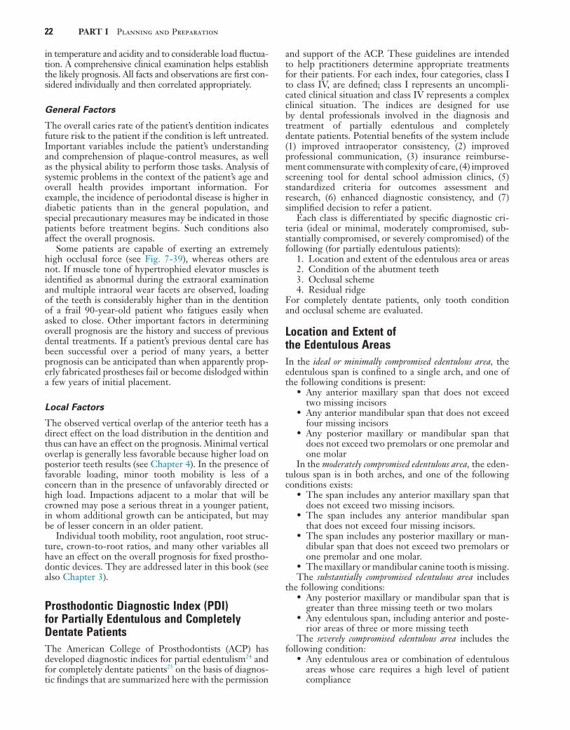
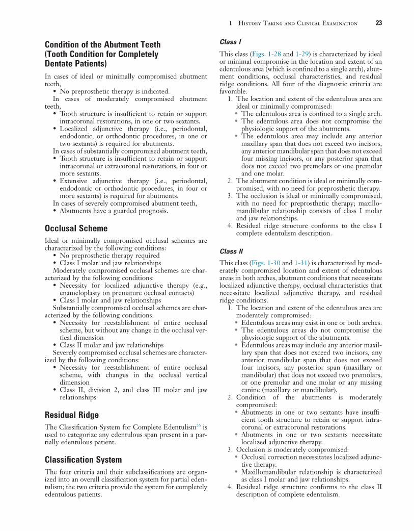
Class III
This class (Figs. 1-32 and 1-33) is characterized by sub-stantially compromised location and extent of edentulous areas in both arches, abutment condition that necessitates substantial localized adjunctive therapy, occlusal charac-teristics that necessitate reestablishment of the entire occlusion without a change in the occlusal vertical dimen-sion, and residual ridge conditions.
1. The location and extent of the edentulous areas are substantially compromised:• Edentulous areas may be present in one or both
arches.• Edentulous areas compromise the physiologic
support of the abutments.• Edentulous areas may include any posterior
maxillary or mandibular edentulous area greater
than three teeth or two molars or anterior and posterior edentulous areas of three or more teeth.
2. The condition of the abutments is moderately compromised:• Abutments in three sextants have insufficient
tooth structure to retain or support intracoronal or extracoronal restorations.
• Abutments in three sextants necessitate more sub-stantial localized adjunctive therapy (i.e., perio-dontal, endodontic, or orthodontic procedures).
• Abutments have a fair prognosis.3. Occlusion is substantially compromised:
• The entire occlusal scheme must be reestablished without an accompanying change in the occlusal vertical dimension.
• Maxillomandibular relationship is characterized as class II molar and jaw relationships.
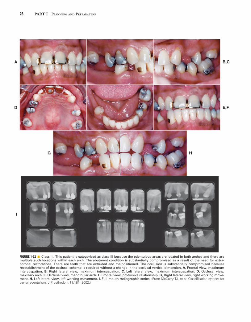
28 PART I Planning and Preparation
FIGURE 1-32 ■ Class III. This patient is categorized as class III because the edentulous areas are located in both arches and there are multiple such locations within each arch. The abutment condition is substantially compromised as a result of the need for extra-coronal restorations. There are teeth that are extruded and malpositioned. The occlusion is substantially compromised because reestablishment of the occlusal scheme is required without a change in the occlusal vertical dimension. A, Frontal view, maximum intercuspation. B, Right lateral view, maximum intercuspation. C, Left lateral view, maximum intercuspation. D, Occlusal view, maxillary arch. E, Occlusal view, mandibular arch. F, Frontal view, protrusive relationship. G, Right lateral view, right working move-ment. H, Left lateral view, left working movement. I, Full-mouth radiographic series. (From McGarry TJ, et al: Classification system for partial edentulism. J Prosthodont 11:181, 2002.)
A B,C
D E,F
G H
I
1 History Taking and Clinical Examination 29
• Edentulous areas compromise the physiologic support of the abutment teeth, and so the prog-nosis is guarded.
• Edentulous areas include acquired or congenital maxillofacial defects.
• At least one edentulous area has a guarded prognosis.
2. Abutments are severely compromised:• Abutments in four or more sextants have insuf-
ficient tooth structure to retain or support intra-coronal or extracoronal restorations.
• Abutment conditions in four or more sex-tants necessitate extensive localized adjunctive therapy.
• Abutments have a guarded prognosis.
4. Residual ridge structure conforms to the class III complete edentulism description.
Class IV
This class (Figs. 1-34 and 1-35) is characterized by severely compromised location and extent of edentulous areas with guarded prognosis, abutment conditions that necessitate extensive therapy, occlusion characteristics that necessitate reestablishment of the occlusion with a change in the occlu-sal vertical dimension, and residual ridge conditions.
1. The location and extent of the edentulous areas result in severe occlusal compromise:• Edentulous areas may be extensive and may be
present in both arches.
FIGURE 1-33 ■ Class III. This patient is categorized as class III because large defective amalgam and composite restorations are present in four sextants. The remaining tooth structure is substantially compromised in most posterior teeth. The occlusion is substantially compromised, which necessitates reestablishment of the occlusal scheme without a change in the occlusal vertical dimension. A, Frontal view, maximum intercuspation. B, Right lateral view, maximum intercuspation. C, Left lateral view, maximum intercuspa-tion. D, Occlusal view, maxillary arch. E, Occlusal view, mandibular arch. F, Panoramic radiograph. (From McGarry TJ, et al: Classifica-tion system for the completely dentate patient. J Prosthodont 13:73, 2004.)
A
C
E F
D
B
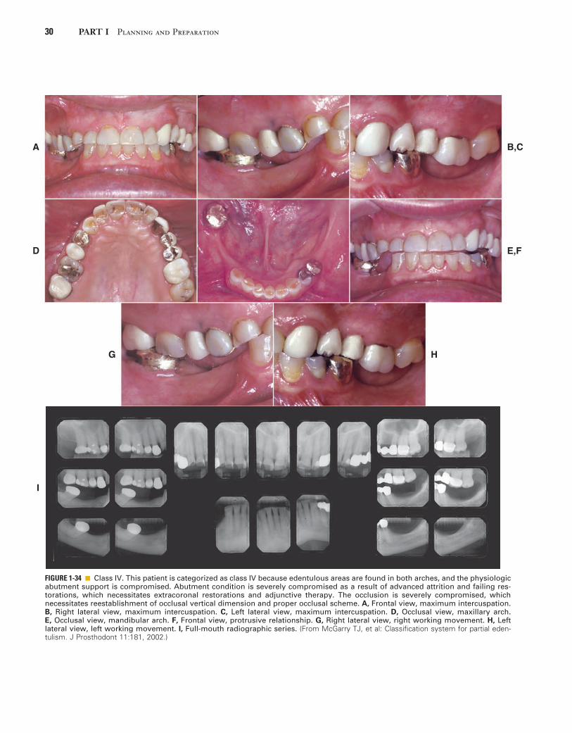
30 PART I Planning and Preparation
FIGURE 1-34 ■ Class IV. This patient is categorized as class IV because edentulous areas are found in both arches, and the physiologic abutment support is compromised. Abutment condition is severely compromised as a result of advanced attrition and failing res-torations, which necessitates extracoronal restorations and adjunctive therapy. The occlusion is severely compromised, which necessitates reestablishment of occlusal vertical dimension and proper occlusal scheme. A, Frontal view, maximum intercuspation. B, Right lateral view, maximum intercuspation. C, Left lateral view, maximum intercuspation. D, Occlusal view, maxillary arch. E, Occlusal view, mandibular arch. F, Frontal view, protrusive relationship. G, Right lateral view, right working movement. H, Left lateral view, left working movement. I, Full-mouth radiographic series. (From McGarry TJ, et al: Classification system for partial eden-tulism. J Prosthodont 11:181, 2002.)
A
D
B,C
E,F
G H
I

1 History Taking and Clinical Examination 31
Guidelines for the Use of PDI Classification System for Partial Edentulism and Complete DentitionThe analysis of diagnostic factors is facilitated with the use of a worksheet (Fig. 1-36 and Table 1-1). Each crite-rion is evaluated, and a checkmark is placed in the appro-priate box. In instances in which a patient’s diagnostic criteria overlap two or more classes, the more complex class is the selected diagnosis.
The following additional guidelines should be followed to ensure consistent application of the classification system:
1. Consideration of future treatment procedures must not influence the choice of diagnostic level.
3. Occlusion is severely compromised:• Reestablishment of the entire occlusal scheme,
including changes in the occlusal vertical dimen-sion, is necessary.
• Maxillomandibular relationship is characterized as class II, division 2, or class III molar and jaw relationships.
4. Residual ridge structure conforms to the class IV complete edentulism description.
Other characteristics include severe manifestations of local or systemic disease, including sequelae from onco-logic treatment, maxillomandibular dyskinesia or ataxia, and refractoriness (a patient’s presenting with chronic complaints after appropriate therapy).
FIGURE 1-35 ■ Class IV. This patient is categorized as class IV because advanced attrition of the occlusal surfaces is present in more than three sextants. The occlusion is severely compromised, which necessitates reestablishment of occlusal vertical dimension and a proper occlusal scheme. A, Frontal view, maximum intercuspation. B, Right lateral view, maximum intercuspation. C, Left lateral view, maximum intercuspation. D, Occlusal view, maxillary arch. E, Occlusal view, mandibular arch. F, Panoramic radiograph. (From McGarry TJ, et al: Classification system for the completely dentate patient. J Prosthodont 13:73, 2004.)
A B
C D
E F

32 PART I Planning and Preparation
FIG
URE
1-3
6 ■
Wo
rksh
eet
use
d t
o d
eter
min
e p
rost
ho
do
nti
c d
iag
no
stic
in
dex
cla
ssifi
cati
on
. N
ote
: In
div
idu
al d
iag
no
stic
cri
teri
a ar
e ev
alu
ated
, an
d t
he
app
rop
riat
e b
ox
is c
hec
ked
. T
he
mo
st a
dva
nce
d fi
nd
ing
det
erm
ines
th
e fi
nal
cla
ssifi
cati
on
. Gu
idel
ines
fo
r u
se o
f th
e w
ork
shee
t: 1
. Po
sses
sio
n o
f an
y si
ng
le c
rite
rio
n o
f a
mo
re c
om
ple
x cl
ass
pla
ces
the
pat
ien
t in
to t
he
mo
re c
om
ple
x cl
ass.
2.
Co
nsi
der
atio
n o
f fu
ture
tre
atm
ent
pro
ced
ure
s m
ust
no
t in
flu
ence
th
e d
iag
no
stic
lev
el.
3. I
nit
ial
pre
pro
sth
etic
tre
atm
ent
and
/or
adju
nct
ive
ther
apy
can
ch
ang
e th
e in
itia
l cl
assi
fica
tio
n l
evel
. 4.
If
ther
e is
an
est
het
ic c
on
cern
/ch
alle
ng
e, t
he
clas
sifi
cati
on
is
incr
ease
d i
n c
om
ple
xity
by
on
e le
vel
in c
lass
I a
nd
II
pat
ien
ts.
5. I
n t
he
pre
sen
ce o
f sy
mp
tom
s o
f te
mp
oro
man
dib
ula
r d
iso
rder
, th
e cl
assi
fica
tio
n is
incr
ease
d in
co
mp
lexi
ty b
y o
ne
or
mo
re le
vels
in c
lass
I an
d II
pat
ien
ts. 6
. In
th
e si
tuat
ion
in w
hic
h t
he
pat
ien
t p
rese
nts
wit
h a
n e
den
tulo
us
man
dib
le o
pp
osi
ng
a p
arti
ally
ed
entu
lou
s o
r d
enta
te m
axill
a, t
he
pat
ien
t is
cat
ego
rize
d a
s cl
ass
IV.
Des
crip
tio
nC
lass
IC
lass
II
Cla
ss I
IIC
lass
IV
Loca
tio
n a
nd
Ext
ent
of
Ed
entu
lou
s A
reas
Idea
l o
r m
inim
ally
co
mp
rom
ised
: si
ng
le a
rch
Mo
der
atel
y co
mp
rom
ised
: b
oth
arc
hes
Su
bst
anti
ally
co
mp
rom
ised
: m
ore
th
an t
hre
e te
eth
Sev
erel
y co
mp
rom
ised
: g
uar
ded
pro
gn
osi
sC
on
gen
ital
or
acq
uir
ed m
axill
ofa
cial
def
ect
Ab
utm
ent
Co
nd
itio
n
Idea
l o
r m
inim
ally
co
mp
rom
ised
Mo
der
atel
y co
mp
rom
ised
: o
ne
to t
wo
sex
tan
tsS
ub
stan
tial
ly c
om
pro
mis
ed:
thre
e se
xtan
tsS
ever
ely
com
pro
mis
ed:
fou
r o
r m
ore
sex
tan
ts
Occ
lusi
on
Idea
l o
r m
inim
ally
co
mp
rom
ised
Mo
der
atel
y co
mp
rom
ised
: lo
cal
adju
nct
ive
trea
tmen
tS
ub
stan
tial
ly c
om
pro
mis
ed:
occ
lusa
l sc
hem
eS
ever
ely
com
pro
mis
ed:
chan
ge
in o
cclu
sal
vert
ical
dim
ensi
on
Res
idu
al R
idg
e
Cla
ss I
ed
entu
lou
sC
lass
II
eden
tulo
us
Cla
ss I
II ed
entu
lou
sC
lass
IV
ed
entu
lou
s
Co
nd
itio
ns
Cre
atin
g a
Gu
ard
ed P
rog
no
sis
Sev
ere
ora
l m
anif
esta
tio
ns
of
syst
emic
dis
ease
Max
illo
man
dib
ula
r d
yski
nes
ia a
nd
/or
atax
iaR
efra
cto
ry c
on
dit
ion

1 History Taking and Clinical Examination 33
The classification system for partial edentulism is based on the most objective criteria available to facilitate uniform use of the system. Such standardization may help improve communications among dental professionals and third parties. This classification system serves to identify patients most likely to require treatment by a specialist or by a practitioner with additional training and experience in advanced techniques. This system should also be valuable to research protocols as different treat-ment procedures are evaluated. With the increasing com-plexity of treatment, this partial edentulism classification system, coupled with the complete edentulism classifica-tion system, helps dental school faculty assess entering patients for the most appropriate patient assignment for better care. On the basis of use and observations by prac-titioners, educators, and researchers, this system is modi-fied as needed.
SUMMARY
A comprehensive history and a thorough clinical exami-nation provide sufficient data for the practitioner to formulate a successful treatment plan. If they are too hastily accomplished, details may be missed, which can cause significant problems during treatment, when it may be difficult or impossible to make corrections. Also, the overall outcome and prognosis may be adversely affected. In particular, it is crucial to develop
2. Initial preprosthetic treatment or adjunctive therapy can change the initial classification level. Classification may need to be reassessed after exist-ing prostheses are removed.
3. Esthetic concerns or challenges raise the classifica-tion by one level in cases of class I and class II dentition.
4. The presence of symptoms of temporomandibular disorder raises the classification by one or more levels in patients with class I and class II dentition.
5. In a patient presenting with an edentulous maxilla opposing a partially edentulous mandible, each arch is diagnosed according to the appropriate clas-sification system; that is, the maxilla is classified according to the complete edentulism classification system, and the mandible is classified according to the partial edentulism classification system. The sole exception to this rule is the case of an edentu-lous mandible opposed by a partially edentulous or dentate maxilla. This clinical situation entails sig-nificant complexity and potential long-term mor-bidity and, as such, should be categorized as class IV in either system.
6. Periodontal health is intimately related to the diagnosis and prognosis for partially edentulous patients. For the purpose of this system, it is assumed that patients receive therapy to achieve and maintain periodontal health so that appropriate prosthodontic care can be accomplished.
TABLE 1-1 Worksheet Used to Determine Prosthodontic Diagnostic Index Classification of Completely Dentate Patients
Description Class I Class II Class III Class IV
Teeth Condition
Ideal or minimally compromised: three or fewer teeth in one sextant XModerately compromised: one or more teeth in one to two sextants XSubstantially compromised: one or more teeth in three to five sextants XSeverely compromised: four or more teeth, all sextants X
Occlusal Scheme
Ideal or minimally compromised XModerately compromised: anterior guidance intact XSubstantially compromised: extensive rest/same OVD XSeverely compromised: extensive rest/new OVD X
Conditions Creating a Guarded Prognosis
Severe oral manifestations of systemic disease XMaxillomandibular dyskinesia or ataxia XRefractory condition X
Note: Individual diagnostic criteria are evaluated, and the appropriate box is checked. The most advanced finding determines the final classification.
Guidelines for use of this worksheet:1. Consideration of future treatment procedures must not influence the diagnostic level.2. Initial preprosthetic treatment or adjunctive therapy can change the initial classification level.3. If there is an esthetic concern/challenge, the classification is increased in complexity by one or more levels.4. In the presence of temporomandibular joint symptoms, the classification is increased in complexity by one or more levels.5. It is assumed that the patient will receive therapy designed to achieve and maintain optimal periodontal health.6. Situations that fail to conform to the definition of completely dentate should be classified according to the classification system for
partial edentulism.OVD, Occlusal vertical dimension.
34 PART I Planning and Preparation
11. Krogh-Poulsen WG, Olsson A: Occlusal disharmonies and dysfunc-tion of the stomatognathic system. Dent Clin North Am 10:627, 1966.
12. Moskowitz ME, Nayyar A: Determinants of dental esthetics: a rational for smile analysis and treatment. Compend Contin Educ Dent 16:1164, 1995.
13. Crispin BJ, Watson JF: Margin placement of esthetic veneer crowns. I. Anterior tooth visibility. J Prosthet Dent 45:278, 1981.
14. Lombardi RE: The principles of visual perception and their clinical application to denture esthetics. J Prosthet Dent 29:358, 1973.
15. Rosenstiel SF, Rashid RG: Public preferences for anterior tooth variations: a web-based study. J Esthet Restor Dent 14:97, 2002.
16. Parameter on comprehensive periodontal examination. American Academy of Periodontology. J Periodontol 71:847, 2000.
17. Guidelines for periodontal therapy. American Academy of Perio-dontology. J Periodontol 69:405, 1998.
18. Carranza FA Jr, Newman MG: Clinical Periodontology, 8th ed. Philadelphia, WB Saunders, 1996.
19. Goodson JM: Selection of suitable indicators of periodontitis. In Bader JD, ed: Risk Assessment in Dentistry. Chapel Hill, N.C., University of North Carolina Dental Ecology, 1989.
20. American Academy of Periodontology: Parameters of Care. Chicago, American Academy of Periodontology, 1998.
21. Carter L: Clinical indications as a basis for ordering extraoral imaging studies. Compend Contin Educ Dent 25:351, 2004.
22. Van Sickels JE, et al: Transcranial radiographs in the evaluation of craniomandibular (TMJ) disorders. J Prosthet Dent 49:244, 1983.
23. Brooks SL, et al: Imaging of the temporomandibular joint: a posi-tion paper of the American Academy of Oral and Maxillofacial Radiology. Oral Surg Oral Med Oral Pathol Oral Radiol Endod 83:609, 1997.
24. McGarry TJ, et al: Classification system for partial edentulism. J Prosthodont 11:181, 2002.
25. McGarry TJ, et al: Classification system for the completely dentate patient. J Prosthodont 13:73, 2004.
26. McGarry TJ, et al: Classification system for complete edentulism. The American College of Prosthodontics. J Prosthodont 8:27, 1999.
a thorough understanding of each patient’s special concerns relating to previous care and his or her expec-tations about future treatment. Many problems encoun-tered during fixed prosthodontic treatment are directly traceable to factors overlooked during the initial exami-nation and data collection. A diagnosis is a summation of the observed problems and their underlying causes. The patient’s overall prognosis is influenced by general and local factors.
REFERENCES1. Moazzez R, et al: Dental erosion, gastro-oesophageal reflux disease
and saliva: how are they related? J Dent 32:489, 2004.2. Milosevic A: Eating disorders and the dentist. Br Dent J 186:109,
1999.3. Cope MR: Metoclopramide-induced masticatory muscle spasm. Br
Dent J 154:335, 1983.4. Pajukoski H, et al: Salivary flow and composition in elderly patients
referred to an acute care geriatric ward. Oral Surg Oral Med Oral Pathol Oral Radiol Endod 84:265, 1997.
5. Hunter KD, Wilson WS: The effects of antidepressant drugs on salivary flow and content of sodium and potassium ions in human parotid saliva. Arch Oral Biol 40:983, 1995.
6. Infection control recommendations for the dental office and labo-ratory. J Am Dent Assoc (Suppl):1, 1992.
7. Epstein O, et al: Pocket Guide to Clinical Examination, 4th ed. St. Louis, Elsevier, 2009.
8. Little JW, et al: Little and Falace’s Dental Management of the Medically Compromised Patient, 8th ed. St. Louis, Elsevier, 2012.
9. Solberg WK: Occlusion-related pathosis and its clinical evaluation. In Clark JW, ed: Clinical Dentistry, vol 2, chap 35. Hagerstown, Md, Harper & Row, 1976.
10. Pullinger AG, et al: Differences between sexes in maximum jaw opening when corrected to body size. J Oral Rehabil 14:291, 1987.
STUDY QUESTIONS
1. Discuss the importance of the chief complaint and its management during examination and presentation of the treatment plan.
2. What is the classification of conditions observed as part of the medical history?
3. Describe the various areas included in the documenta-tion of a comprehensive dental history.
4. What systemic conditions may cause oral manifesta-tions that can affect a plan for fixed prosthodontic treatment?
5. What is included in a comprehensively conducted extraoral examination? Specify all structures included in palpation.
6. Discuss three critical observations that are part of a comprehensive periodontal examination. Why are they important in the evaluation for fixed prosthodon-tic treatment?
7. What would be recorded as part of an intraoral charting?
8. Discuss the various types of radiographs available for diagnostic purposes. What are the advantages and limitations of each technique?
9. Give examples of general and local factors that may influence the patient’s prognosis.
35
Diagnostic Casts and Related Procedures
C H A P T E R 2
Accurate diagnostic casts transferred to a semiadjustable articulator (Fig. 2-1) are essential in planning fixed pros-thodontic treatment. This enables examination of static and dynamic relationships of the teeth without interfer-ence from protective neuromuscular reflexes, and unen-cumbered views from all directions reveal aspects of the occlusion not always easily detectable intraorally (e.g., the relationship of the lingual cusps in the occluded position). If the maxillary cast has been transferred with a facebow, a centric relation (CR) interocclusal record has been used for articulation of the mandibular cast, and the condylar elements have been appropriately set (as with protrusive and excursive interocclusal records), reproducing the patient’s movements with reasonable accuracy is possible. If the casts have been articulated in CR, both the CR and the maximum intercuspation (MI) position can be assessed because any slide can then be reproduced.
Other critical information not immediately apparent during the clinical examination includes the occlusocervi-cal dimension of edentulous spaces. On an articulator, these are readily assessed in the occluded position and throughout the entire range of mandibular movement. Relative alignment and angulation of proposed abutment teeth are easier to evaluate on casts than intraorally, as are many other subtle changes in individual tooth posi-tion. Articulated diagnostic casts enable a detailed analy-sis of the occlusal plane and the occlusion, and diagnostic procedures can be performed for a better diagnosis and treatment plan; tooth preparations can be “rehearsed” on the casts, and diagnostic waxing procedures allow evalu-ation of the eventual outcome of proposed treatment.
IMPRESSION MAKING FOR DIAGNOSTIC CASTS
Accurate impressions of both dental arches are required. Flaws in the impressions result in inaccuracies in the casts that easily multiply. For instance, a small void in the impression caused by the trapping of an air bubble on one of the occlusal surfaces results in a nodule on the occlusal table. If it is not recognized and carefully removed, it leads to an inaccurate articulator mounting, and the diagnostic data are incorrect.
As long as the impression extends several millimeters beyond the cervical line of the teeth, the borders of diag-nostic impressions are usually not of great concern for fixed prosthodontic purposes, unless a removable pros-thesis is also to be fabricated. Properly manipulated irre-versible hydrocolloid (alginate) is sufficiently accurate and offers adequate surface detail for planning purposes.
However, the material does require pouring within 2 hours to avoid dimensional change1 and does not repro-duce sufficient surface detail for suitable definitive casts and dies on which actual fixed prostheses are fabricated (see Chapter 17). Materials with a composition similar to that of vinyl polysiloxane materials (see Chapter 17) have been marketed as an alternative to traditional irreversible hydrocolloids.2 These materials should be considered if a delay in pouring is necessary.3
Irreversible HydrocolloidThe irreversible hydrocolloids, or alginates, are essen-tially sodium or potassium salts of alginic acid and are therefore water soluble. They react chemically with calcium sulfate to produce insoluble calcium alginate. These materials contain other ingredients, chiefly dia-tomaceous earth (for strength and body), trisodium phos-phate (Na3PO4), and similar compounds to control the setting rate as they react preferentially with calcium sulfate. When this reaction is complete and the retarder is consumed, gel formation begins. The clinician can control the reaction rate by varying the temperature of the mixing water. Because irreversible hydrocolloid is largely water, it readily absorbs (by imbibition) as well as gives off (by syneresis) liquid to the atmosphere, causing distortion of the impression. Alginate impressions must therefore be poured immediately.
Diagnostic Impression TechniqueArmamentarium
• Impression trays• Modeling compound• Mixing bowl• Mixing spatula• Gauze squares• Irreversible hydrocolloid• American Dental Association (ADA) type IV or V
stone• Vacuum mixer• Humidor• Disinfectant
Tray Selection
All impression materials must be retained in the impres-sion tray. This can be accomplished for irreversible hydrocolloid with an adhesive or by means of perfora-tions or undercuts around the rim of the tray. All types of trays are capable of producing impressions with

36 PART I Planning and Preparation
seated, impression material is often squeezed out of the mucobuccal fold or from underneath the upper lip.
A loss of tackiness of the material (gelation) implies initial set. The tray should be removed quickly 2 to 3 minutes after gelation. Teasing or wiggling the set impres-sion from the mouth causes excessive distortion as a result of viscous flow. Also, certain irreversible hydrocolloid materials become distorted if held in the mouth more than 2 or 3 minutes after gelation.7 After removal (see Fig. 2-3, D), the impression should be rinsed and disin-fected, dried slightly with a gentle air stream, and poured immediately. For disinfection, spraying with a suitable glutaraldehyde and placement in a self-sealing plastic bag for approximately 10 minutes is recommended, after which it can be poured. Alternatively, the impression can be immersed in iodophor or glutaraldehyde disinfectant. The disinfection protocol is an essential precaution for preventing cross infection and protecting laboratory per-sonnel (see Chapter 14); irreversible hydrocolloid impres-sions carry significantly higher numbers of bacteria than do elastomeric materials.8 No significant loss of accuracy or surface detail is caused by the disinfection procedure.9,10 To ensure accuracy, pouring should be completed within 15 minutes after the impression is removed from the mouth. Keeping an impression in a moist towel is no substitute for pouring within the specified time. Trimming off gross excess impression material before setting the tray down on the bench top is helpful. A vacuum-mixed ADA type IV or type V stone is recommended. The choice of the brand of stone is important because of the harmful surface interactions between specific irreversible hydrocolloid materials and gypsum products.11
clinically acceptable accuracy,4 although casts may be produced more accurately by rigid plastic trays than by perforated metal trays.5 For irreversible hydrocolloids, the largest tray that will fit comfortably in the patient’s mouth should be selected. A greater bulk of material produces a more accurate impression (i.e., a bulky impres-sion has a more favorable ratio of surface area to volume and is less susceptible to water loss or gain and therefore unwanted dimensional change). In contrast, elastomeric impression materials work well with a relatively tightly fitting custom impression tray in which a uniform thin layer of material is used. This produces the most accurate impression (see Chapter 14).
Distortion of irreversible hydrocolloid can occur if any part of the impression is unsupported by the tray or if there is movement of the tray during setting. For these reasons, the tray may need to be extended and its perim-eter modified with modeling compound (Fig. 2-2).
Impression Making
For optimum results, the teeth should be cleaned and the mouth thoroughly rinsed. Some drying is necessary, but excessively dried tooth surfaces cause the irreversible hydrocolloid impression material to adhere. The material is mixed to a homogenous consistency and loaded into the tray, and its surface is smoothed with a moistened gloved finger.6 Concurrently, a small amount of material is wiped into the crevices of the occlusal surfaces (Fig. 2-3, A and B) before the tray is seated (see Fig. 2-3, C). Also, a small amount can be applied by wiping it into the mucobuccal fold. As the tray is inserted into the patient’s mouth and seated, the patient is instructed to “close gently” on the tray. If the patient continues to stretch the mouth wide open while the tray is being fully
FIGURE 2-1 ■ Diagnostic casts mounted on a semi-adjustable articulator. (Courtesy Whip Mix Corporation, Louisville, Ky.)
FIGURE 2-2 ■ Stock impression trays can be readily modified with modeling compound to provide better support for the algi-nate. The posterior border typically needs extension. If the patient has a high palate, the alginate should be supported here, too, although the compound should not block out the retentive area of the tray.

2 Diagnostic Casts and Related Procedures 37
FIGURE 2-3 ■ Making an alginate impression for diagnostic casts. A and B, A small amount of alginate being wiped into the crevices of the occlusal surfaces. C, Seating of the tray. D, The completed impression.
A
C
B
D
After mixing, a small amount of stone is added in one location (e.g., the posterior aspect of one of the molars). Adding small amounts consistently in the same location helps minimize bubble formation (see the section on pouring stone dies in Chapter 17). If air is trapped, a small instrument (e.g., a periodontal probe or a wax spatula) can be used to poke the bubbles and eliminate them. While they are setting, the poured impressions must be stored tray side down, not inverted. Inverting freshly poured impressions results in a cast with a rough and grainy surface.12 Stone is added to create a sufficient base that provides adequate retention for mounting on the articulator. To achieve maximum strength and surface detail, the poured impression should be covered with wet paper and stored in a humidor for 1 hour. This minimizes distortion of the irreversible hydrocolloid during the setting period. The setting gypsum cast should never be immersed in water. If this is done, setting expansion of plaster, stone, or die stone doubles or even triples through the phenomenon of hygroscopic expansion (see Chapter 22). For best results, the cast should be separated from the impression 1 hour after being poured.
Evaluation
Although it is apparently a simple procedure, diagnostic cast fabrication is often mishandled. Seemingly minor inaccuracies can lead to serious diagnostic errors. Ques-tionable impressions and casts should be discarded and the process repeated (Fig. 2-4). Voids in the impression create nodules on the poured cast. These can prevent proper articulation and effectively render useless a sub-sequent occlusal analysis or other diagnostic procedure.
Articulator Selection
Handheld casts can provide information concerning alignment of the individual arches but do not enable analysis of functional relationships. For such analysis, the diagnostic casts need to be attached to an articulator, a mechanical device that simulates mandibular movement. Articulators can simulate the movement of the condyles in their corresponding fossae. They are classified accord-ing to how closely they can reproduce mandibular border movements. Because the movements are governed by the bones and ligaments of the temporomandibular joints, they are relatively constant and reproducible. With most
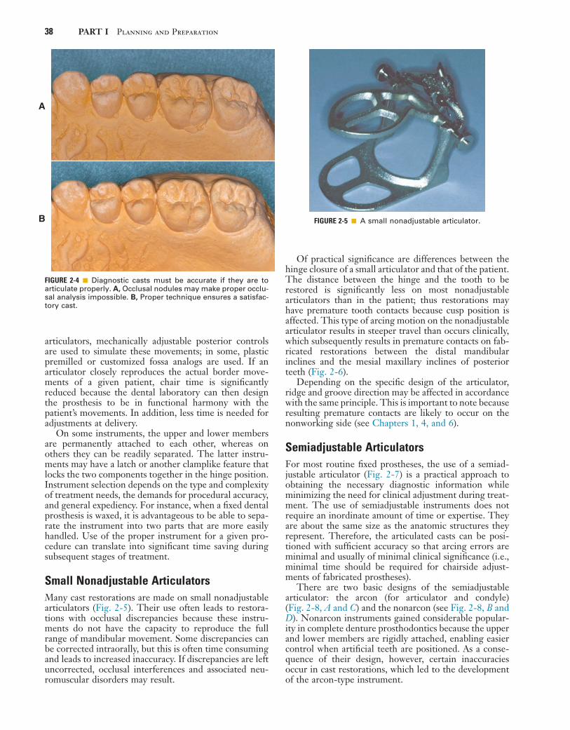
38 PART I Planning and Preparation
FIGURE 2-5 ■ A small nonadjustable articulator.
FIGURE 2-4 ■ Diagnostic casts must be accurate if they are to articulate properly. A, Occlusal nodules may make proper occlu-sal analysis impossible. B, Proper technique ensures a satisfac-tory cast.
A
B
articulators, mechanically adjustable posterior controls are used to simulate these movements; in some, plastic premilled or customized fossa analogs are used. If an articulator closely reproduces the actual border move-ments of a given patient, chair time is significantly reduced because the dental laboratory can then design the prosthesis to be in functional harmony with the patient’s movements. In addition, less time is needed for adjustments at delivery.
On some instruments, the upper and lower members are permanently attached to each other, whereas on others they can be readily separated. The latter instru-ments may have a latch or another clamplike feature that locks the two components together in the hinge position. Instrument selection depends on the type and complexity of treatment needs, the demands for procedural accuracy, and general expediency. For instance, when a fixed dental prosthesis is waxed, it is advantageous to be able to sepa-rate the instrument into two parts that are more easily handled. Use of the proper instrument for a given pro-cedure can translate into significant time saving during subsequent stages of treatment.
Small Nonadjustable ArticulatorsMany cast restorations are made on small nonadjustable articulators (Fig. 2-5). Their use often leads to restora-tions with occlusal discrepancies because these instru-ments do not have the capacity to reproduce the full range of mandibular movement. Some discrepancies can be corrected intraorally, but this is often time consuming and leads to increased inaccuracy. If discrepancies are left uncorrected, occlusal interferences and associated neu-romuscular disorders may result.
Of practical significance are differences between the hinge closure of a small articulator and that of the patient. The distance between the hinge and the tooth to be restored is significantly less on most nonadjustable articulators than in the patient; thus restorations may have premature tooth contacts because cusp position is affected. This type of arcing motion on the nonadjustable articulator results in steeper travel than occurs clinically, which subsequently results in premature contacts on fab-ricated restorations between the distal mandibular inclines and the mesial maxillary inclines of posterior teeth (Fig. 2-6).
Depending on the specific design of the articulator, ridge and groove direction may be affected in accordance with the same principle. This is important to note because resulting premature contacts are likely to occur on the nonworking side (see Chapters 1, 4, and 6).
Semiadjustable ArticulatorsFor most routine fixed prostheses, the use of a semiad-justable articulator (Fig. 2-7) is a practical approach to obtaining the necessary diagnostic information while minimizing the need for clinical adjustment during treat-ment. The use of semiadjustable instruments does not require an inordinate amount of time or expertise. They are about the same size as the anatomic structures they represent. Therefore, the articulated casts can be posi-tioned with sufficient accuracy so that arcing errors are minimal and usually of minimal clinical significance (i.e., minimal time should be required for chairside adjust-ments of fabricated prostheses).
There are two basic designs of the semiadjustable articulator: the arcon (for articulator and condyle) (Fig. 2-8, A and C) and the nonarcon (see Fig. 2-8, B and D). Nonarcon instruments gained considerable popular-ity in complete denture prosthodontics because the upper and lower members are rigidly attached, enabling easier control when artificial teeth are positioned. As a conse-quence of their design, however, certain inaccuracies occur in cast restorations, which led to the development of the arcon-type instrument.

2 Diagnostic Casts and Related Procedures 39
when nonarcon articulators are set with protrusive wax records because the condylar path is not fixed in relation to the maxillary occlusal plane. As the protrusive record used to adjust the instrument is removed from the arcon articulator, the maxillary occlusal plane and the condylar inclination become more parallel, which causes reduction in cuspal heights in subsequently fabricated prostheses (see Table 4-3).
Fully Adjustable ArticulatorsA fully (or highly) adjustable articulator (Fig. 2-9) has a wide range of positions and can be set to follow a patient’s border movements. The accuracy of reproduction of movement depends on the care and skill of the operator, the errors inherent in the articulator and recording device, and any malalignments resulting from slight flexing of the mandible and the nonrigid nature of the temporomandibular joints.
Rather than relying on wax records to adjust the instrument, a series of special pantographic tracings are used to record the patient’s border movements. The
In an arcon articulator, the condylar spheres are attached to the lower component of the articulator, and the mechanical fossae are attached to the upper member of the instrument. Thus the arcon articulator is anatomi-cally “correct,” which makes understanding of mandibu-lar movements easier, as opposed to the nonarcon articulator (whose movements are confusingly “back-wards”). The angulation of the mechanical fossae of an arcon instrument is fixed in relation to the occlusal plane of the maxillary cast; in the nonarcon design, it is fixed in relation to the occlusal plane of the mandibular cast.
Most semiadjustable articulators allow adjustments to the condylar inclination and progressive or immediate side shift. Some have straight condylar inclined paths, although newer instruments have curved condylar hous-ings, which are more anatomically correct.
The mechanical fossae on semiadjustable articulators can be adjusted to mimic the movements of the patient through the use of interocclusal records. These consist of several thicknesses of wax or another suitable material into which the patient has bitten. These records can be several millimeters thick, and so an error is introduced
FIGURE 2-6 ■ Discrepancies in the path of closure when a small nonadjustable articulator is used can cause restorations to have premature occlusal contacts. A, An anatomically accurate articulator shows an accurate path of closure. B, With the small nonadjust-able instrument, the radius of the path of closure is smaller, which results in premature contact at the clinical try-in between the premolars during hinge closure.
B AThe radius of the arc ofclosure affects the likeli-hood of interferences.
A B

40 PART I Planning and Preparation
they convey can save considerable chairside time. They can be very useful as treatment complexity increases (e.g., when all four posterior quadrants are to be restored simultaneously or when it is necessary to restore a patient’s entire dentition, especially in the presence of atypical mandibular movement).
FACEBOWS
Transverse Horizontal AxisThe mandibular hinging movement around the trans-verse horizontal axis is repeatable. Therefore, the
armamentarium used to generate these tracings is then transferred to the articulator, and the instrument is adjusted so that the articulator replicates the tracings, essentially reproducing the border movements of the patient. The ability of fully adjustable instruments to track irregular pathways of movement throughout entire trajectories enables the fabrication of complex prostheses, which require minimal adjustment at the evaluation and delivery appointment.
Fully adjustable articulators are not often required in general practice. Using and adjusting them can be time consuming and require a high level of skill and under-standing by the dentist and the technician. Once this skill has been acquired, however, the detailed information
FIGURE 2-7 ■ Semiadjustable arcon articulators. A, The Denar Mark 330 articulator. B, The Whip Mix model 2240 articulator. C, The Hanau Wide-Vue articulator. (A to C, Courtesy Whip Mix Corporation, Louisville, Ky.)
A
C
B
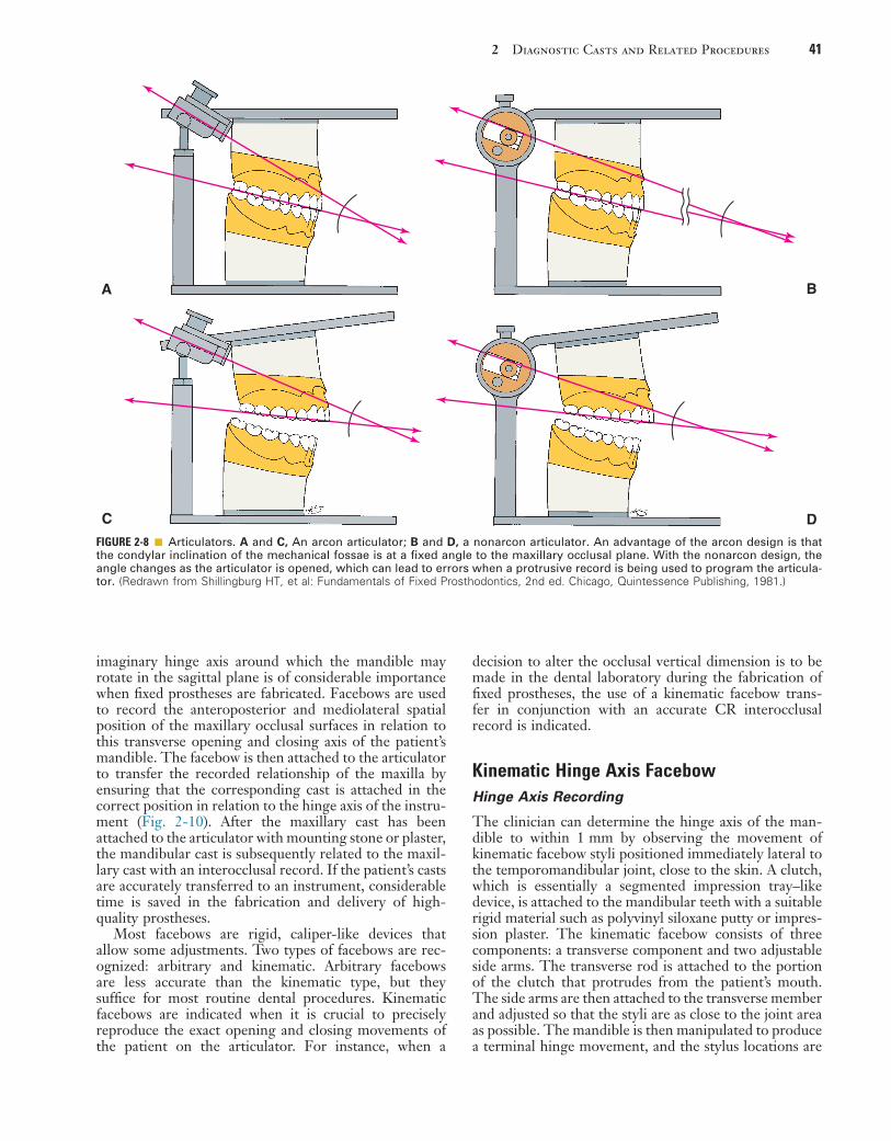
2 Diagnostic Casts and Related Procedures 41
decision to alter the occlusal vertical dimension is to be made in the dental laboratory during the fabrication of fixed prostheses, the use of a kinematic facebow trans-fer in conjunction with an accurate CR interocclusal record is indicated.
Kinematic Hinge Axis FacebowHinge Axis Recording
The clinician can determine the hinge axis of the man-dible to within 1 mm by observing the movement of kinematic facebow styli positioned immediately lateral to the temporomandibular joint, close to the skin. A clutch, which is essentially a segmented impression tray–like device, is attached to the mandibular teeth with a suitable rigid material such as polyvinyl siloxane putty or impres-sion plaster. The kinematic facebow consists of three components: a transverse component and two adjustable side arms. The transverse rod is attached to the portion of the clutch that protrudes from the patient’s mouth. The side arms are then attached to the transverse member and adjusted so that the styli are as close to the joint area as possible. The mandible is then manipulated to produce a terminal hinge movement, and the stylus locations are
imaginary hinge axis around which the mandible may rotate in the sagittal plane is of considerable importance when fixed prostheses are fabricated. Facebows are used to record the anteroposterior and mediolateral spatial position of the maxillary occlusal surfaces in relation to this transverse opening and closing axis of the patient’s mandible. The facebow is then attached to the articulator to transfer the recorded relationship of the maxilla by ensuring that the corresponding cast is attached in the correct position in relation to the hinge axis of the instru-ment (Fig. 2-10). After the maxillary cast has been attached to the articulator with mounting stone or plaster, the mandibular cast is subsequently related to the maxil-lary cast with an interocclusal record. If the patient’s casts are accurately transferred to an instrument, considerable time is saved in the fabrication and delivery of high-quality prostheses.
Most facebows are rigid, caliper-like devices that allow some adjustments. Two types of facebows are rec-ognized: arbitrary and kinematic. Arbitrary facebows are less accurate than the kinematic type, but they suffice for most routine dental procedures. Kinematic facebows are indicated when it is crucial to precisely reproduce the exact opening and closing movements of the patient on the articulator. For instance, when a
FIGURE 2-8 ■ Articulators. A and C, An arcon articulator; B and D, a nonarcon articulator. An advantage of the arcon design is that the condylar inclination of the mechanical fossae is at a fixed angle to the maxillary occlusal plane. With the nonarcon design, the angle changes as the articulator is opened, which can lead to errors when a protrusive record is being used to program the articula-tor. (Redrawn from Shillingburg HT, et al: Fundamentals of Fixed Prosthodontics, 2nd ed. Chicago, Quintessence Publishing, 1981.)
A B
DC

42 PART I Planning and Preparation
The kinematic facebow technique is time consuming, and so its use is generally limited to extensive prostho-dontics, particularly when a change in the occlusal verti-cal dimension is to be made. A less precisely derived transfer would then lead to unacceptable errors and com-promise the result.
Arbitrary Hinge Axis FacebowArbitrary hinge axis facebows (Fig. 2-13) approximate the horizontal transverse axis and rely on anatomic average values. Manufacturers design these facebows so that the relationship to the true axis falls within an acceptable degree of error. Typically, an easily identifiable landmark such as the external acoustic meatus is used to stabilize the bow, which is aligned with earpieces similar to those on a stethoscope. Such facebows can be used single-handedly because they are self-centering and assembly is not complicated. They depict a sufficiently accurate rela-tionship for most diagnostic and restorative procedures. However, regardless of which arbitrary position is chosen, a minimum error of 5 mm from the axis can be expected,13 as can errors in the steepness of the occlusal plane.14 When coupled with the use of a thick interocclusal record made at an increased vertical dimension, this error can lead to considerable inaccuracy.
Anterior Reference Point
The use of an anterior reference point (Fig. 2-14) enables the clinician to duplicate the recorded position on the articulator at future appointments. This saves time because previously recorded articulator settings can be used again. An anterior reference point, such as the inner canthus of the eye or a freckle or mole on the skin, is selected. After this point has been marked, it is used, along with the two points of the hinge axis, to define the position of the maxillary cast in space. This procedure has the following advantages:
• After the posterior controls have been adjusted ini-tially, subsequent casts can be mounted on the artic-ulator without the need to repeat the facebow determinations and reset the posterior articulator controls.
• Because the maxillary arch is properly positioned in relation to the axis, average values for posterior articulator controls can be used without the need to readjust the instrument on the basis of eccentric records.
• When the articulator has been adjusted, the result-ing numerical values for the settings can be com-pared with known average values to provide information about the patient’s individual variations and the likelihood of encountering difficulties during restorative procedures.
Facebow Transfer
Armamentarium• Arbitrary hinge axis facebow• Modeling compound• Cotton rolls
adjusted with thumbscrews (superiorly and inferiorly, anteriorly and posteriorly) until they make a purely rota-tional movement (Fig. 2-11). Because the entire assembly is rigidly attached to the mandible, a strictly rotational movement signifies that stylus position coincides with the hinge axis. When this purely rotational movement is veri-fied, the position of the hinge axis is marked with a dot on the patient’s skin, or it may be permanently tattooed if future use is anticipated or required.
Kinematic Facebow Transfer
An impression of the maxillary cusp tips is obtained in a suitable recording medium on a facebow fork (Fig. 2-12). The facebow is attached to the protruding arm of the fork. The side arms are adjusted until the styli are aligned with the hinge axis marks on the patient’s skin. To prevent skin movement from introducing any inaccuracy, the patient must be in the same position that was used when the axis was marked. A pointer device is usually attached to the bow and adjusted to a repeatable reference point selected by the clinician. The reference point is used later for reproducibil-ity. The kinematic facebow recording is then transferred to the articulator, and the maxillary cast is attached.
FIGURE 2-9 ■ Fully adjustable articulators. A, The Stuart articula-tor. B, The Denar D5A articulator. (Courtesy Whip Mix Corpora-tion, Louisville, Ky.)
A
B
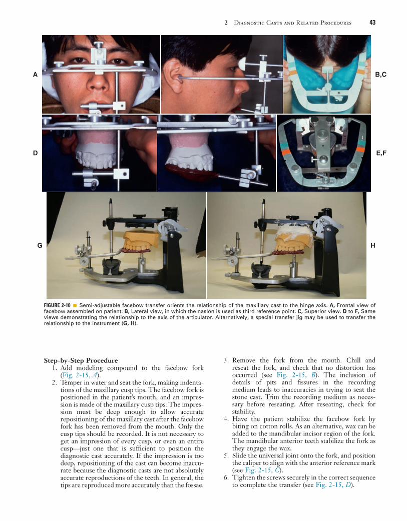
2 Diagnostic Casts and Related Procedures 43
FIGURE 2-10 ■ Semi-adjustable facebow transfer orients the relationship of the maxillary cast to the hinge axis. A, Frontal view of facebow assembled on patient. B, Lateral view, in which the nasion is used as third reference point. C, Superior view. D to F, Same views demonstrating the relationship to the axis of the articulator. Alternatively, a special transfer jig may be used to transfer the relationship to the instrument (G, H).
A B,C
D E,F
G H
Step-by-Step Procedure1. Add modeling compound to the facebow fork
(Fig. 2-15, A).2. Temper in water and seat the fork, making indenta-
tions of the maxillary cusp tips. The facebow fork is positioned in the patient’s mouth, and an impres-sion is made of the maxillary cusp tips. The impres-sion must be deep enough to allow accurate repositioning of the maxillary cast after the facebow fork has been removed from the mouth. Only the cusp tips should be recorded. It is not necessary to get an impression of every cusp, or even an entire cusp—just one that is sufficient to position the diagnostic cast accurately. If the impression is too deep, repositioning of the cast can become inaccu-rate because the diagnostic casts are not absolutely accurate reproductions of the teeth. In general, the tips are reproduced more accurately than the fossae.
3. Remove the fork from the mouth. Chill and reseat the fork, and check that no distortion has occurred (see Fig. 2-15, B). The inclusion of details of pits and fissures in the recording medium leads to inaccuracies in trying to seat the stone cast. Trim the recording medium as neces-sary before reseating. After reseating, check for stability.
4. Have the patient stabilize the facebow fork by biting on cotton rolls. As an alternative, wax can be added to the mandibular incisor region of the fork. The mandibular anterior teeth stabilize the fork as they engage the wax.
5. Slide the universal joint onto the fork, and position the caliper to align with the anterior reference mark (see Fig. 2-15, C).
6. Tighten the screws securely in the correct sequence to complete the transfer (see Fig. 2-15, D).

44 PART I Planning and Preparation
FIGURE 2-11 ■ Hinge axis recording. A, Left and right styli are attached via a facebow to a clutch affixed to the mandibular teeth. When the mandible makes a strictly rotational movement, the stylus remains stationary if aligned with the actual axis of rotation. If the stylus is positioned forward or backward, above or below the actual axis, it travels one of the arcs indicated by the arrows when the mandible makes a rotational movement. Thus the arc indicates in what direction an adjustment should be made to the stylus position. B, Hinge axis locator is positioned. C, Set screws allow side arm adjustment. D, Adjustment continues until no arcing of the pointer is seen.
B C,D
The locator is adjustedproperly when the pointerremains stationary duringhinge movement.
A
7. If the articulator has an adjustable intercondylar width, record this measurement (see Fig. 2-15, E). Remove the facebow from the mouth.
The technique is slightly different with other arbitrary facebows (see Fig. 2-15, F to K).
Centric Relation Record
A CR record (Fig. 2-16) provides the orientation of man-dibular to maxillary teeth in CR in the terminal hinge position, in which opening and closing are purely rota-tional movements. Centric relation is defined as the maxil-lomandibular relationship in which the condyles articulate
with the thinnest avascular portion of their respective disks, with the condyle-disk complex in the anterosupe-rior position against the articular eminences. This posi-tion is independent of tooth contact.
MI may or may not be coincident with the CR posi-tion. The CR record is transferred to the maxillary cast on the articulator and is used to relate the mandibular cast to the maxillary cast. Once the mandibular cast is attached to the articulator with mounting stone, the record is removed. The casts then occlude in precisely the CR position as long as the maxillary cast is correctly related to the hinge axis with a facebow (see Fig. 2-15). When the articulator controls are set properly, through
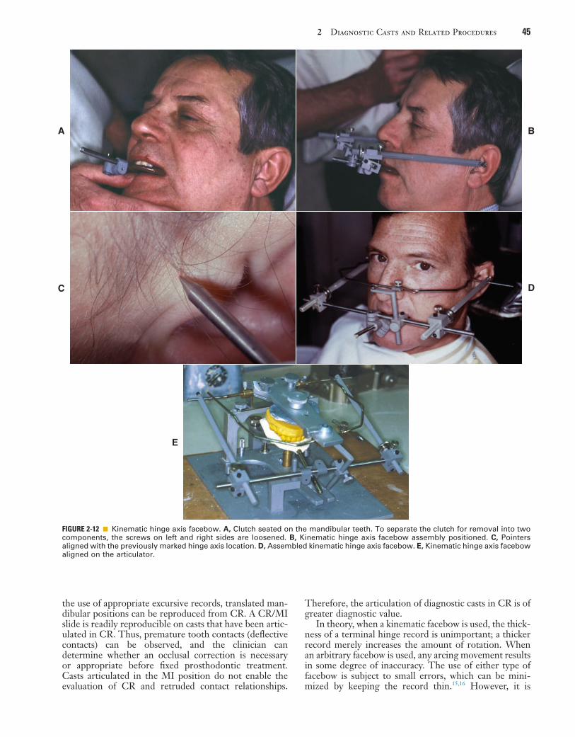
2 Diagnostic Casts and Related Procedures 45
FIGURE 2-12 ■ Kinematic hinge axis facebow. A, Clutch seated on the mandibular teeth. To separate the clutch for removal into two components, the screws on left and right sides are loosened. B, Kinematic hinge axis facebow assembly positioned. C, Pointers aligned with the previously marked hinge axis location. D, Assembled kinematic hinge axis facebow. E, Kinematic hinge axis facebow aligned on the articulator.
A
C
E
B
D
the use of appropriate excursive records, translated man-dibular positions can be reproduced from CR. A CR/MI slide is readily reproducible on casts that have been artic-ulated in CR. Thus, premature tooth contacts (deflective contacts) can be observed, and the clinician can determine whether an occlusal correction is necessary or appropriate before fixed prosthodontic treatment. Casts articulated in the MI position do not enable the evaluation of CR and retruded contact relationships.
Therefore, the articulation of diagnostic casts in CR is of greater diagnostic value.
In theory, when a kinematic facebow is used, the thick-ness of a terminal hinge record is unimportant; a thicker record merely increases the amount of rotation. When an arbitrary facebow is used, any arcing movement results in some degree of inaccuracy. The use of either type of facebow is subject to small errors, which can be mini-mized by keeping the record thin.15,16 However, it is
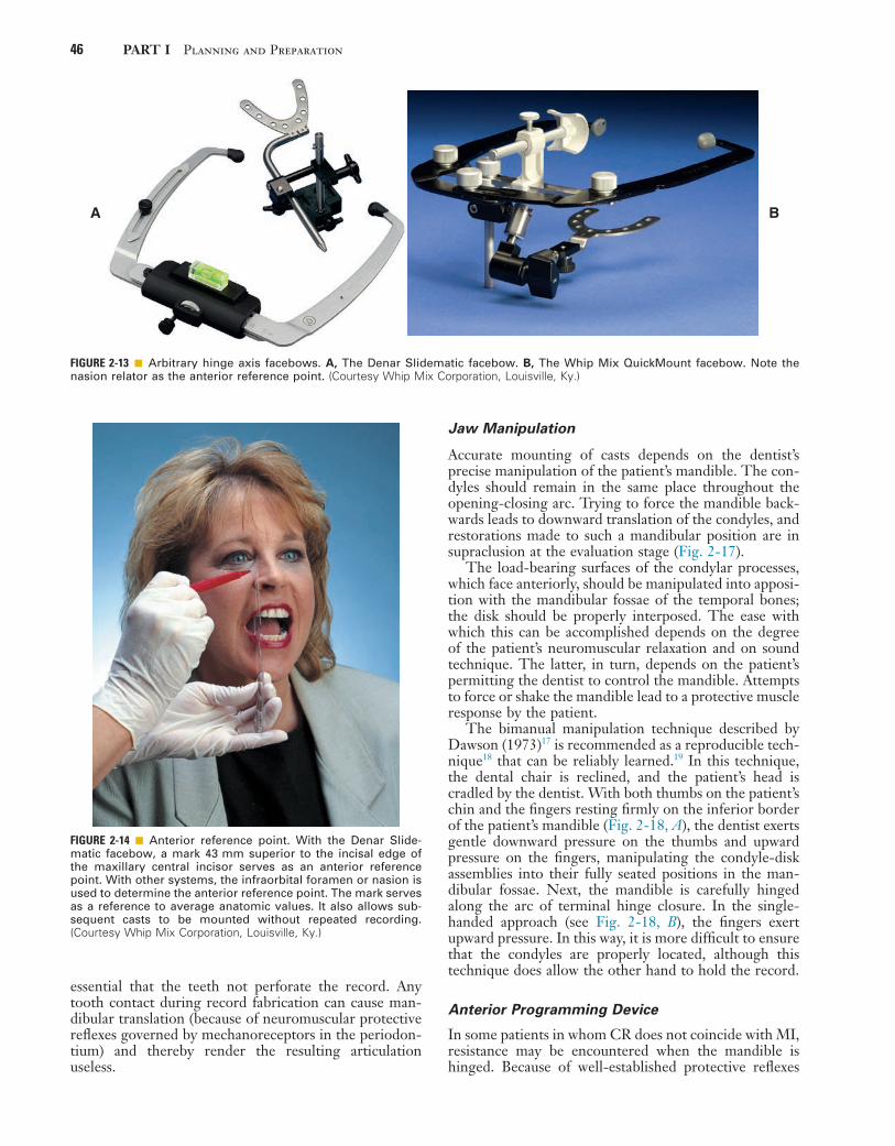
46 PART I Planning and Preparation
Jaw Manipulation
Accurate mounting of casts depends on the dentist’s precise manipulation of the patient’s mandible. The con-dyles should remain in the same place throughout the opening-closing arc. Trying to force the mandible back-wards leads to downward translation of the condyles, and restorations made to such a mandibular position are in supraclusion at the evaluation stage (Fig. 2-17).
The load-bearing surfaces of the condylar processes, which face anteriorly, should be manipulated into apposi-tion with the mandibular fossae of the temporal bones; the disk should be properly interposed. The ease with which this can be accomplished depends on the degree of the patient’s neuromuscular relaxation and on sound technique. The latter, in turn, depends on the patient’s permitting the dentist to control the mandible. Attempts to force or shake the mandible lead to a protective muscle response by the patient.
The bimanual manipulation technique described by Dawson (1973)17 is recommended as a reproducible tech-nique18 that can be reliably learned.19 In this technique, the dental chair is reclined, and the patient’s head is cradled by the dentist. With both thumbs on the patient’s chin and the fingers resting firmly on the inferior border of the patient’s mandible (Fig. 2-18, A), the dentist exerts gentle downward pressure on the thumbs and upward pressure on the fingers, manipulating the condyle-disk assemblies into their fully seated positions in the man-dibular fossae. Next, the mandible is carefully hinged along the arc of terminal hinge closure. In the single-handed approach (see Fig. 2-18, B), the fingers exert upward pressure. In this way, it is more difficult to ensure that the condyles are properly located, although this technique does allow the other hand to hold the record.
Anterior Programming Device
In some patients in whom CR does not coincide with MI, resistance may be encountered when the mandible is hinged. Because of well-established protective reflexes
essential that the teeth not perforate the record. Any tooth contact during record fabrication can cause man-dibular translation (because of neuromuscular protective reflexes governed by mechanoreceptors in the periodon-tium) and thereby render the resulting articulation useless.
FIGURE 2-13 ■ Arbitrary hinge axis facebows. A, The Denar Slidematic facebow. B, The Whip Mix QuickMount facebow. Note the nasion relator as the anterior reference point. (Courtesy Whip Mix Corporation, Louisville, Ky.)
A B
FIGURE 2-14 ■ Anterior reference point. With the Denar Slide-matic facebow, a mark 43 mm superior to the incisal edge of the maxillary central incisor serves as an anterior reference point. With other systems, the infraorbital foramen or nasion is used to determine the anterior reference point. The mark serves as a reference to average anatomic values. It also allows sub-sequent casts to be mounted without repeated recording. (Courtesy Whip Mix Corporation, Louisville, Ky.)
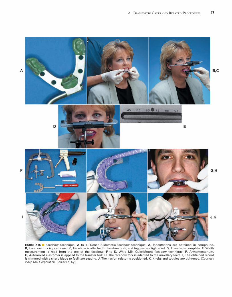
2 Diagnostic Casts and Related Procedures 47
FIGURE 2-15 ■ Facebow technique. A to E, Denar Slidematic facebow technique: A, Indentations are obtained in compound. B, Facebow fork is positioned. C, Facebow is attached to facebow fork, and toggles are tightened. D, Transfer is complete. E, Width measurement is read from the top of the facebow. F to K, Whip Mix QuickMount facebow technique: F, Armamentarium. G, Automixed elastomer is applied to the transfer fork. H, The facebow fork is adapted to the maxillary teeth. I, The obtained record is trimmed with a sharp blade to facilitate seating. J, The nasion relator is positioned. K, Knobs and toggles are tightened. (Courtesy Whip Mix Corporation, Louisville, Ky.)
A B,C
D E
G,H
J,KI
F
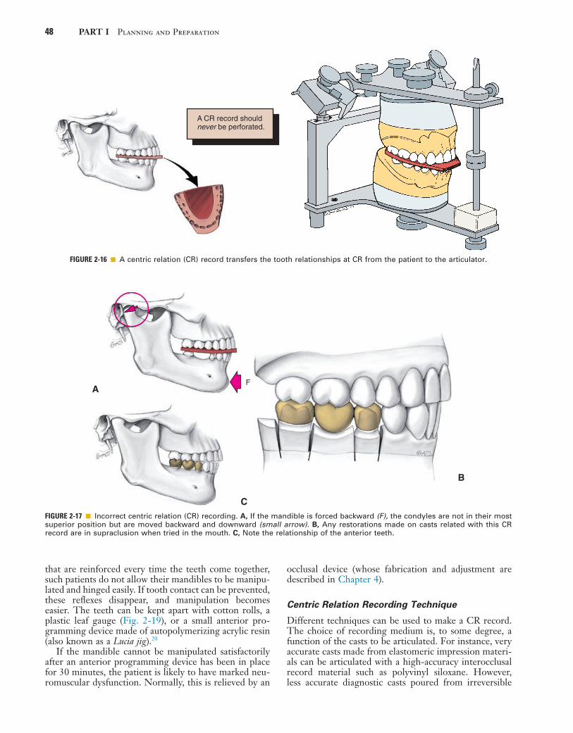
48 PART I Planning and Preparation
FIGURE 2-16 ■ A centric relation (CR) record transfers the tooth relationships at CR from the patient to the articulator.
A CR record shouldnever be perforated.
FIGURE 2-17 ■ Incorrect centric relation (CR) recording. A, If the mandible is forced backward (F), the condyles are not in their most superior position but are moved backward and downward (small arrow). B, Any restorations made on casts related with this CR record are in supraclusion when tried in the mouth. C, Note the relationship of the anterior teeth.
FA
B
C
that are reinforced every time the teeth come together, such patients do not allow their mandibles to be manipu-lated and hinged easily. If tooth contact can be prevented, these reflexes disappear, and manipulation becomes easier. The teeth can be kept apart with cotton rolls, a plastic leaf gauge (Fig. 2-19), or a small anterior pro-gramming device made of autopolymerizing acrylic resin (also known as a Lucia jig).20
If the mandible cannot be manipulated satisfactorily after an anterior programming device has been in place for 30 minutes, the patient is likely to have marked neu-romuscular dysfunction. Normally, this is relieved by an
occlusal device (whose fabrication and adjustment are described in Chapter 4).
Centric Relation Recording Technique
Different techniques can be used to make a CR record. The choice of recording medium is, to some degree, a function of the casts to be articulated. For instance, very accurate casts made from elastomeric impression materi-als can be articulated with a high-accuracy interocclusal record material such as polyvinyl siloxane. However, less accurate diagnostic casts poured from irreversible

2 Diagnostic Casts and Related Procedures 49
hydrocolloid are better articulated with the use of a more malleable material, such as interocclusal wax, provided the record is properly reinforced. Most studies have shown considerable variability among various registra-tion materials and techniques,21 and so particular care is needed with this procedure.
Reinforced Aluwax Record
Reinforced Aluwax is a malleable material for recording the CR position (Fig. 2-20, A). This type of record, originally described by Wirth (1971)22 and Wirth and Aplin (1971),23 is reliable and the technique has provided consistent results.24,25
Armamentarium• Heat-retaining wax sheet (i.e., Aluwax; see Appen-
dix A)• Soft metal sheet (No. 7 Ash’s soft metal; see Appen-
dix A)• Hard pink wax• Sticky wax• Scissors• Ice water
FIGURE 2-18 ■ Manipulating a patient’s mandible into centric relation: the bimanual technique (A) and the single-handed technique (B). Note the position of the dentist’s thumbs and fingers on the mandibular border.
A
B
Step-by-Step Procedure1. Soften half a sheet of occlusal wax in warm water, and
adapt it to the maxillary cusp tips (see Fig. 2-20, B). Allow the patient to close lightly, and make cuspal indentations of the mandibular teeth. These indenta-tions form no part of the record, but they thin the wax slightly and indicate the approximate positions of the mandibular teeth for later reference.
2. Add hard pink baseplate wax to the mandibular ante-rior region of the record (see Fig. 2-20, C), add soft metal sheet to reinforce the palatal area, and seal along the periphery with sticky wax (see Fig. 2-20, D).
3. Readapt the record to the maxillary teeth, resoften-ing if necessary. Guide the patient’s mouth into centric closure, making shallow indentations in the wax. Verify that no posterior tooth contact occurs.
A
B
C
FIGURE 2-19 ■ An anterior programming device is used to facili-tate centric relation recording. A, Autopolymerizing resin is mixed and adapted to the maxillary central incisors. The patient’s mouth is guided into closure and stopped when the posterior teeth are about 1 mm apart. B, The indentations are used as a guide during trimming of the device. C, The com-pleted device should allow the patient to make smooth lateral and protrusive movements. An inclined contact area must be avoided, because it will tend to cause the mandible to retrude excessively. Continued
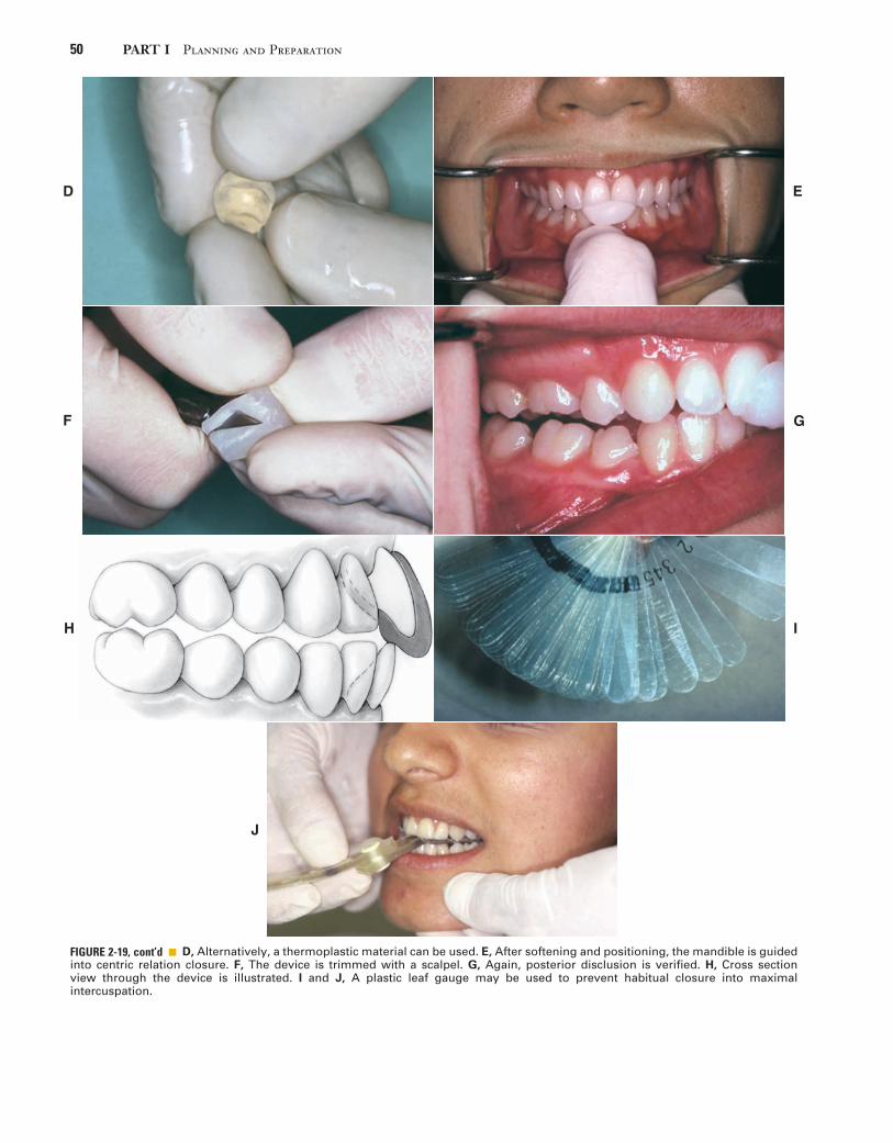
50 PART I Planning and Preparation
D
F
E
G
H I
J
D, Alternatively, a thermoplastic material can be used. E, After softening and positioning, the mandible is guided into centric relation closure. F, The device is trimmed with a scalpel. G, Again, posterior disclusion is verified. H, Cross section view through the device is illustrated. I and J, A plastic leaf gauge may be used to prevent habitual closure into maximal intercuspation.
FIGURE 2-19, cont’d ■
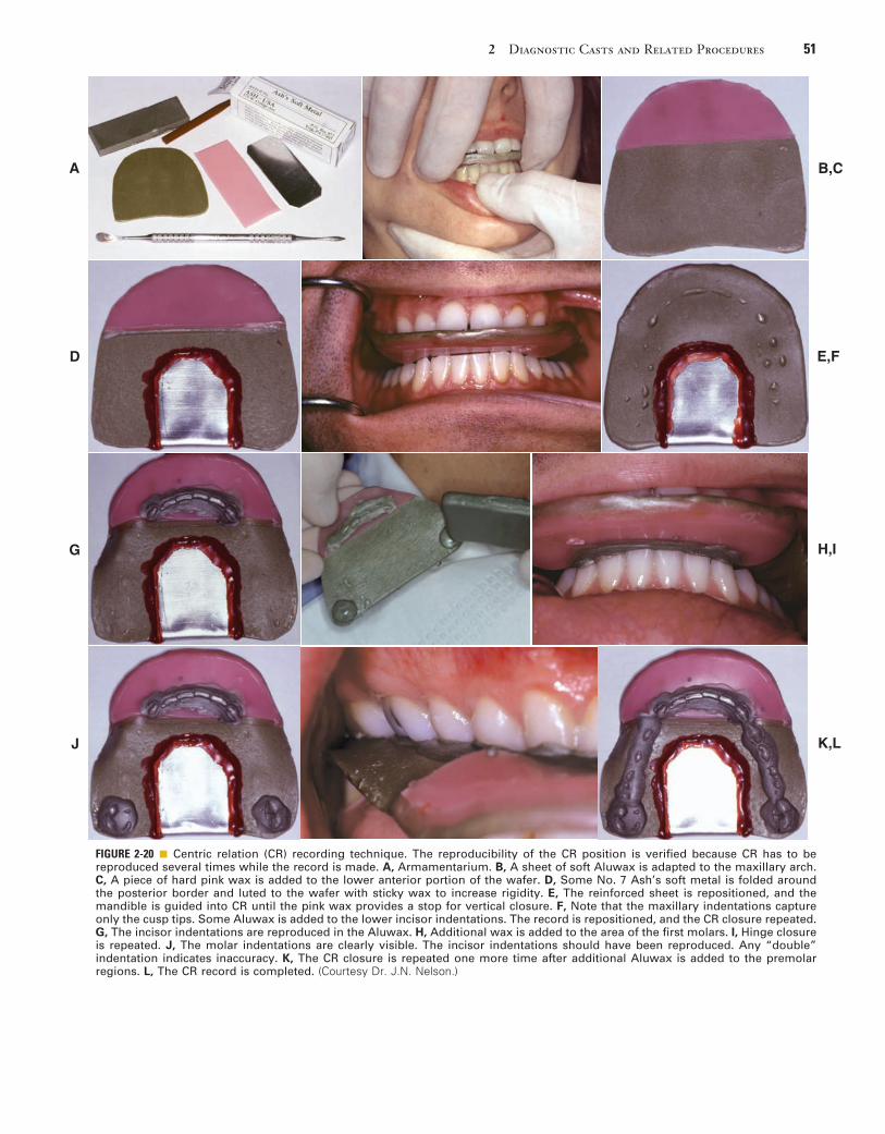
2 Diagnostic Casts and Related Procedures 51
FIGURE 2-20 ■ Centric relation (CR) recording technique. The reproducibility of the CR position is verified because CR has to be reproduced several times while the record is made. A, Armamentarium. B, A sheet of soft Aluwax is adapted to the maxillary arch. C, A piece of hard pink wax is added to the lower anterior portion of the wafer. D, Some No. 7 Ash’s soft metal is folded around the posterior border and luted to the wafer with sticky wax to increase rigidity. E, The reinforced sheet is repositioned, and the mandible is guided into CR until the pink wax provides a stop for vertical closure. F, Note that the maxillary indentations capture only the cusp tips. Some Aluwax is added to the lower incisor indentations. The record is repositioned, and the CR closure repeated. G, The incisor indentations are reproduced in the Aluwax. H, Additional wax is added to the area of the first molars. I, Hinge closure is repeated. J, The molar indentations are clearly visible. The incisor indentations should have been reproduced. Any “double” indentation indicates inaccuracy. K, The CR closure is repeated one more time after additional Aluwax is added to the premolar regions. L, The CR record is completed. (Courtesy Dr. J.N. Nelson.)
A B,C
D E,F
G H,I
K,LJ
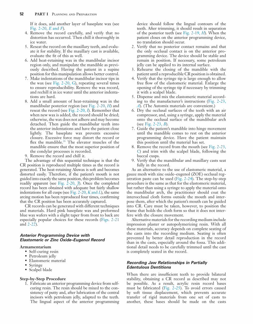
52 PART I Planning and Preparation
If it does, add another layer of baseplate wax (see Fig. 2-20, E and F).
4. Remove the record carefully, and verify that no distortion has occurred. Then chill it thoroughly in ice water.
5. Reseat the record on the maxillary teeth, and evalu-ate it for stability. If the maxillary cast is available, evaluate the fit of this as well.
6. Add heat-retaining wax in the mandibular incisor region only, and manipulate the mandible as previ-ously described. Having the patient in a supine position for this manipulation allows better control.
7. Make indentations of the mandibular incisor tips in the wax (see Fig. 2-20, G), repeating several times to ensure reproducibility. Remove the wax record, and rechill it in ice water until the anterior indenta-tions are hard.
8. Add a small amount of heat-retaining wax in the mandibular posterior region (see Fig. 2-20, H) and reseat the record (see Fig. 2-20, I). Remember that when new wax is added, the record should be dried; otherwise, the wax does not adhere and may become detached. Then guide the mandibular teeth into the anterior indentations and have the patient close lightly. The baseplate wax prevents excessive closure. Excessive force may distort the record or flex the mandible.26 The elevator muscles of the mandible ensure that the most superior position of the condylar processes is recorded.
9. Remove the record and chill it.The advantage of this sequential technique is that the
CR position is reproduced multiple times as the record is generated. The heat-retaining Aluwax is soft and becomes distorted easily. Therefore, if the patient’s mouth is not guided into exactly the same position, this problem becomes readily apparent (see Fig. 2-20, J). Once the completed record has been obtained with adequate but fairly shallow indentations for all cusps (see Fig. 2-20, K and L), the same arcing motion has been reproduced four times, confirming that the CR position has been accurately captured.
CR records can be generated with different techniques and materials. Hard pink baseplate wax and preformed blue wax wafers with a slight taper from front to back are especially popular choices for these records (Figs. 2-21 and 2-22).
Anterior Programming Device with Elastomeric or Zinc Oxide–Eugenol Record
Armamentarium• Self-curing resin• Petroleum jelly• Elastomeric material• Syringe• Scalpel blade
Step-by-Step Procedure1. Fabricate an anterior programming device from self-
curing resin. The resin should be mixed to the con-sistency of putty and, after lubrication of the central incisors with petroleum jelly, adapted to the teeth. The lingual aspect of the anterior programming
device should follow the lingual contours of the teeth. After trimming, it should result in separation of the posterior teeth (see Fig. 2-19, H). When the patient closes on the anterior programming device, no translation should occur.
2. Verify that no posterior contact remains and that the only occlusal contact is on the anterior pro-gramming device. The device should be stable and remain in position. If necessary, some petroleum jelly can be applied to its internal surface.
3. Rehearse the closing of the mandible with the patient until a reproducible CR position is obtained.
4. Verify that the syringe tip is large enough to allow free flow of the elastomeric material. Enlarge the opening of the syringe tip if necessary by trimming it with a scalpel blade.
5. Dispense and mix the elastomeric material accord-ing to the manufacturer’s instructions (Fig. 2-23, A). (The Automix materials are convenient.)
6. Dry the occlusal surfaces of the teeth with an air compressor, and, using a syringe, apply the material onto the occlusal surface of the mandibular arch (see Fig. 2-23, B).
7. Guide the patient’s mandible into hinge movement until the mandible comes to rest on the anterior programming device. Have the patient maintain this position until the material has set.
8. Remove the record from the mouth (see Fig. 2-23, C) and trim with the scalpel blade, following the buccal cusps.
9. Verify that the mandibular and maxillary casts seat fully in the record.
As an alternative to the use of elastomeric material, a gauze mesh with zinc oxide–eugenol (ZOE) occlusal reg-istration paste can be used (Fig. 2-24). The step-by-step procedure is the same as that for the elastomeric material, but rather than using a syringe to apply the material onto the mandibular arch, the practitioner should coat the interocclusal cloth forms outside the mouth and inter-pose them, after which the patient’s mouth can be guided into CR. Care must be taken, however, to position the frame that holds the cloth form so that it does not inter-fere with the closure movement.
Alternative materials for the recording medium include impression plaster or autopolymerizing resin. With all these materials, accuracy depends on complete seating of the casts into the recording medium. Seating is often prevented by better detail reproduction in the record than in the casts, especially around the fossa. This addi-tional detail needs to be carefully trimmed until the cast is completely seated in the record.
Recording Jaw Relationships in Partially Edentulous Dentitions
When there are insufficient teeth to provide bilateral stability, obtaining a CR record as described may not be possible. As a result, acrylic resin record bases must be fabricated (Fig. 2-25). To avoid errors caused by soft tissue displacement, which prevents accurate transfer of rigid materials from one set of casts to another, these bases should be made on the casts
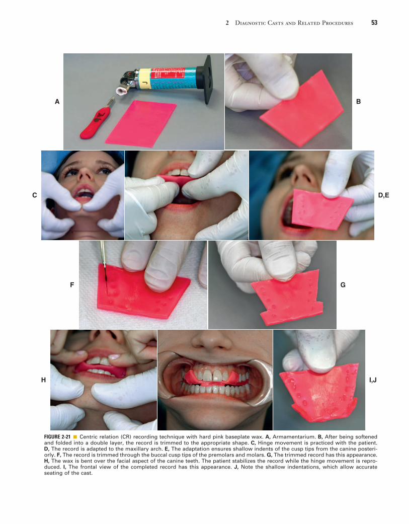
2 Diagnostic Casts and Related Procedures 53
FIGURE 2-21 ■ Centric relation (CR) recording technique with hard pink baseplate wax. A, Armamentarium. B, After being softened and folded into a double layer, the record is trimmed to the appropriate shape. C, Hinge movement is practiced with the patient. D, The record is adapted to the maxillary arch. E, The adaptation ensures shallow indents of the cusp tips from the canine posteri-orly. F, The record is trimmed through the buccal cusp tips of the premolars and molars. G, The trimmed record has this appearance. H, The wax is bent over the facial aspect of the canine teeth. The patient stabilizes the record while the hinge movement is repro-duced. I, The frontal view of the completed record has this appearance. J, Note the shallow indentations, which allow accurate seating of the cast.
A B
C D,E
F G
H I,J

54 PART I Planning and Preparation
FIGURE 2-22 ■ Centric relation (CR) recording technique with preformed wax wafer and leaf gauge. A, Preformed wax and a leaf gauge are used for occlusal registration. B, The leaf gauge is positioned to achieve separation of the posterior teeth. C, Lateral aspects of the wax are softened in a water bath. D, The wax record is adapted to the maxillary teeth. E, The record is trimmed through the buccal cusp tips. F, The wax is resoftened. G, The patient’s mandible is guided into centric relation, and indents of the mandibular cusp tips are obtained. H, The record is chilled with cold water. I, Excess wax can be removed with a sharp blade until only indents of the cusp tips remain. J, After clinical verification, the record is stored in cold water for subsequent use in the laboratory.
A B,C
D E,F
G H
I J

2 Diagnostic Casts and Related Procedures 55
Articulating the Diagnostic CastsMaxillary Cast. The maxillary cast (Fig. 2-26) is
seated in the indentations on the facebow fork after the facebow is attached to the articulator. Wedges or specially designed braces can be used to support the weight of the cast and to prevent the fork from flexing or moving. After it has been scored and wetted, the cast is attached to the mounting ring of the articulator with a low-expansion, fast-setting mounting stone or plaster.
Mandibular Cast. To relate the mandibular cast prop-erly to the maxillary cast, the incisal guide pin should be lowered sufficiently to compensate for the thickness of the CR record. The articulator is inverted, and the record is seated on the maxillary cast. The mandibular cast (Fig. 2-27) is then carefully seated in the record, and each cast is checked for stability. The maxillary and mandibular casts can be luted together with sticky wax and either metal rods or pieces of wooden tongue blade. The man-dibular member of the articulator is closed into mounting stone; the condylar balls should be fully seated in the cor-responding fossae. If the articulator has a centric latch, this step is simplified. Otherwise, the articulator should be held until the stone has reached its initial set. No attempt should be made to smooth the stone until it has fully set.
Evaluation. Accuracy is crucial in both CR and MI. Before the articulator controls are adjusted, the dentist must confirm the accuracy of CR by comparing the tooth
FIGURE 2-23 ■ Centric relation (CR) recording. A, Elastomeric material is used for CR recording. B, Once mandibular quadrants are coated, the dentist uses an anterior resin jig (see Fig. 2-10) to ensure that a reproducible recording position is obtained. The patient remains occluded until the material has set. C, This is the appearance of the record before trimming. (A, Courtesy Parkell, Inc., Edge-wood, N.Y.)
A B
C
FIGURE 2-24 ■ Gauze mesh cloth forms with plastic holders, and zinc oxide–eugenol (ZOE) paste can be used instead of elasto-meric paste.
that are to be articulated. If breakage of the casts is a concern, it may be advisable to make record bases on an accurate duplicate cast made with reversible agar hydrocolloid impression material in a flask designed for that purpose.
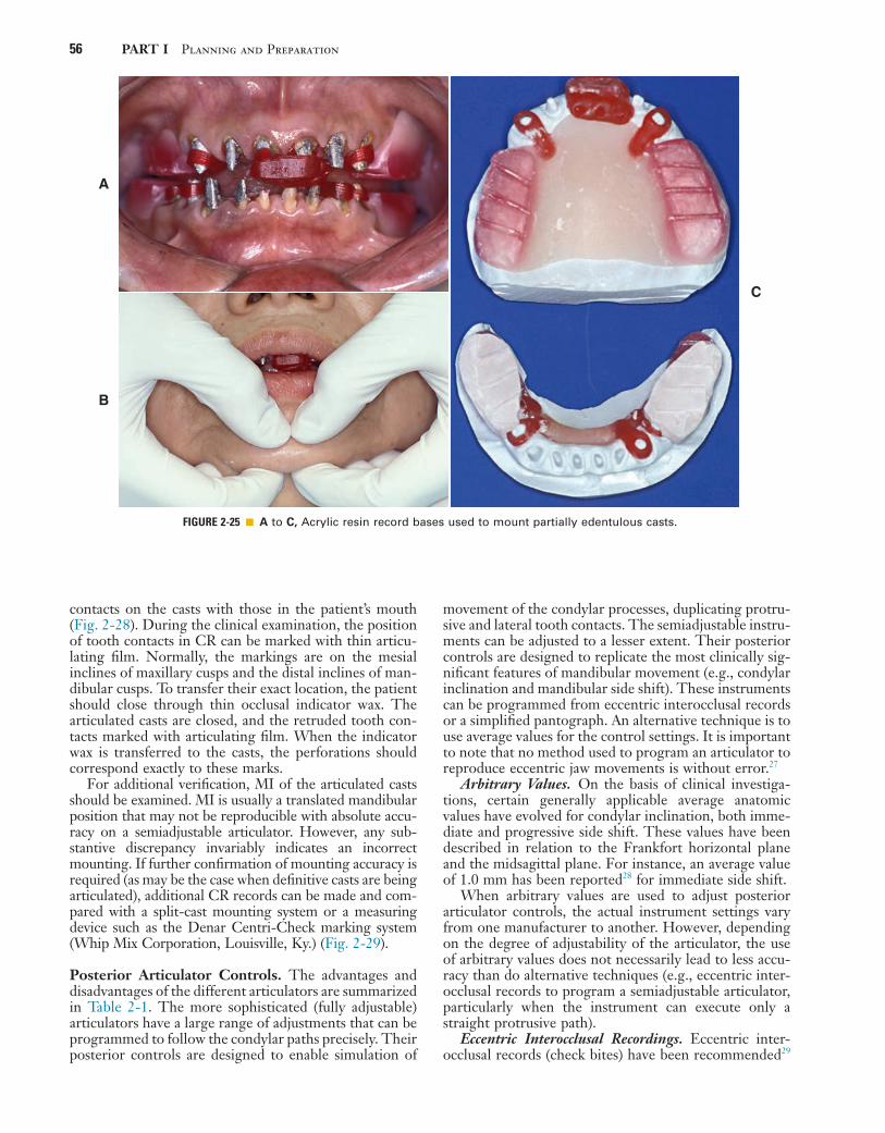
56 PART I Planning and Preparation
movement of the condylar processes, duplicating protru-sive and lateral tooth contacts. The semiadjustable instru-ments can be adjusted to a lesser extent. Their posterior controls are designed to replicate the most clinically sig-nificant features of mandibular movement (e.g., condylar inclination and mandibular side shift). These instruments can be programmed from eccentric interocclusal records or a simplified pantograph. An alternative technique is to use average values for the control settings. It is important to note that no method used to program an articulator to reproduce eccentric jaw movements is without error.27
Arbitrary Values. On the basis of clinical investiga-tions, certain generally applicable average anatomic values have evolved for condylar inclination, both imme-diate and progressive side shift. These values have been described in relation to the Frankfort horizontal plane and the midsagittal plane. For instance, an average value of 1.0 mm has been reported28 for immediate side shift.
When arbitrary values are used to adjust posterior articulator controls, the actual instrument settings vary from one manufacturer to another. However, depending on the degree of adjustability of the articulator, the use of arbitrary values does not necessarily lead to less accu-racy than do alternative techniques (e.g., eccentric inter-occlusal records to program a semiadjustable articulator, particularly when the instrument can execute only a straight protrusive path).
Eccentric Interocclusal Recordings. Eccentric inter-occlusal records (check bites) have been recommended29
contacts on the casts with those in the patient’s mouth (Fig. 2-28). During the clinical examination, the position of tooth contacts in CR can be marked with thin articu-lating film. Normally, the markings are on the mesial inclines of maxillary cusps and the distal inclines of man-dibular cusps. To transfer their exact location, the patient should close through thin occlusal indicator wax. The articulated casts are closed, and the retruded tooth con-tacts marked with articulating film. When the indicator wax is transferred to the casts, the perforations should correspond exactly to these marks.
For additional verification, MI of the articulated casts should be examined. MI is usually a translated mandibular position that may not be reproducible with absolute accu-racy on a semiadjustable articulator. However, any sub-stantive discrepancy invariably indicates an incorrect mounting. If further confirmation of mounting accuracy is required (as may be the case when definitive casts are being articulated), additional CR records can be made and com-pared with a split-cast mounting system or a measuring device such as the Denar Centri-Check marking system (Whip Mix Corporation, Louisville, Ky.) (Fig. 2-29).
Posterior Articulator Controls. The advantages and disadvantages of the different articulators are summarized in Table 2-1. The more sophisticated (fully adjustable) articulators have a large range of adjustments that can be programmed to follow the condylar paths precisely. Their posterior controls are designed to enable simulation of
FIGURE 2-25 ■ A to C, Acrylic resin record bases used to mount partially edentulous casts.
A
C
B

2 Diagnostic Casts and Related Procedures 57
recorded by the record (Fig. 2-30). This occurs because the path taken between these may differ signif-icantly on the articulator from what is actually performed by the mandible. A semiadjustable instru-ment may have a protrusive path and a side shift path that are straight lines, whereas the true paths are invar-iably curved. In an attempt to minimize errors, many contemporary semiadjustable articulators come with curved fossae.
Armamentarium• Interocclusal wax record material
for setting the posterior controls of a semiadjustable articulator. These consist of wax or another recording material interposed between the maxillary and mandibu-lar arches; they record the position of the condyles in eccentric mandibular positions. Static positional records are made in translated jaw positions: a protrusive record and two lateral records. The protrusive record can be used to adjust both condylar inclinations on the articula-tor, and the lateral records are used to adjust the side shift on semiadjustable articulators.
An articulator set by an eccentric record is accurate in only two positions: at CR and at the position
FIGURE 2-26 ■ Mounting the maxillary cast on a Whip Mix articulator. A, Armamentarium. B, The incisal pin is removed. C, The condylar inclination is adjusted to the facebow setting. D, The side shift is set to zero. E, A mounting plate is attached. F, The facebow earpieces are attached to the condylar elements. G, The facebow is attached to the articulator. H, The scored maxillary cast is posi-tioned on the facebow fork, and the cast is prewetted. I, The mounting stone is applied to the cast and the mounting plate. The upper member of the articulator is closed until it contacts the crossbar of the facebow. J, Additional stone is added as needed. (Courtesy Whip Mix Corporation, Louisville, Ky.)
B,CA
D E,F
G H,I
J
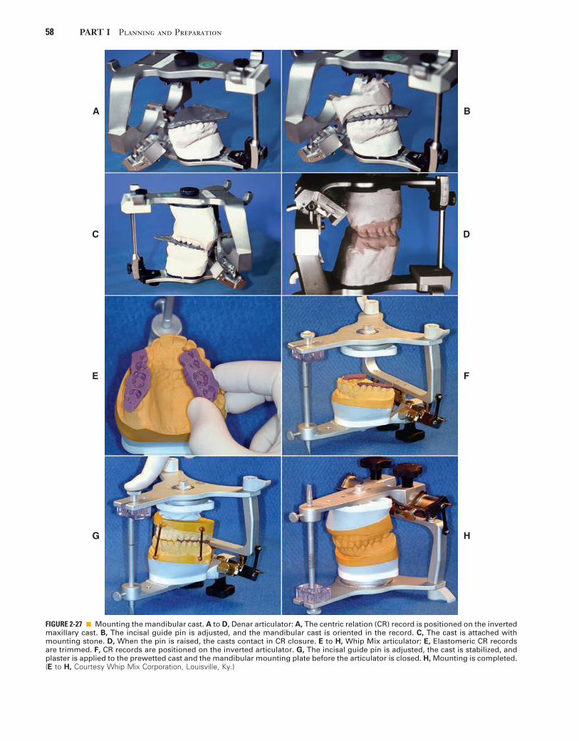
58 PART I Planning and Preparation
FIGURE 2-27 ■ Mounting the mandibular cast. A to D, Denar articulator: A, The centric relation (CR) record is positioned on the inverted maxillary cast. B, The incisal guide pin is adjusted, and the mandibular cast is oriented in the record. C, The cast is attached with mounting stone. D, When the pin is raised, the casts contact in CR closure. E to H, Whip Mix articulator: E, Elastomeric CR records are trimmed. F, CR records are positioned on the inverted articulator. G, The incisal guide pin is adjusted, the cast is stabilized, and plaster is applied to the prewetted cast and the mandibular mounting plate before the articulator is closed. H, Mounting is completed. (E to H, Courtesy Whip Mix Corporation, Louisville, Ky.)
A B
C
E F
D
G H
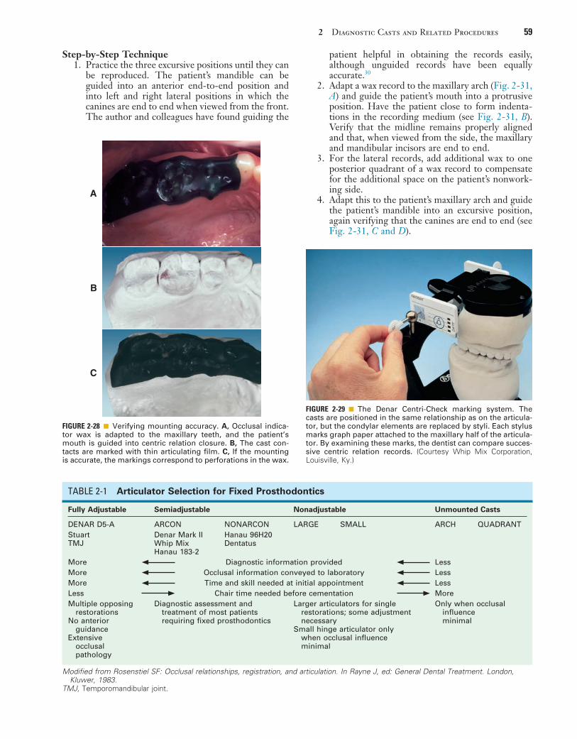
2 Diagnostic Casts and Related Procedures 59
FIGURE 2-28 ■ Verifying mounting accuracy. A, Occlusal indica-tor wax is adapted to the maxillary teeth, and the patient’s mouth is guided into centric relation closure. B, The cast con-tacts are marked with thin articulating film. C, If the mounting is accurate, the markings correspond to perforations in the wax.
A
B
C
FIGURE 2-29 ■ The Denar Centri-Check marking system. The casts are positioned in the same relationship as on the articula-tor, but the condylar elements are replaced by styli. Each stylus marks graph paper attached to the maxillary half of the articula-tor. By examining these marks, the dentist can compare succes-sive centric relation records. (Courtesy Whip Mix Corporation, Louisville, Ky.)
Modified from Rosenstiel SF: Occlusal relationships, registration, and articulation. In Rayne J, ed: General Dental Treatment. London, Kluwer, 1983.
TABLE 2-1 Articulator Selection for Fixed Prosthodontics
TMJ, Temporomandibular joint.
Fully Adjustable Semiadjustable Nonadjustable Unmounted Casts
DENAR D5-A ARCON NONARCON LARGE SMALL ARCH QUADRANTStuartTMJ
Denar Mark IIWhip MixHanau 183-2
Hanau 96H20Dentatus
More Diagnostic information provided LessMore Occlusal information conveyed to laboratory LessMore Time and skill needed at initial appointment LessLess Chair time needed before cementation MoreMultiple opposing
restorationsNo anterior
guidanceExtensive
occlusal pathology
Diagnostic assessment and treatment of most patients requiring fixed prosthodontics
Larger articulators for single restorations; some adjustment necessary
Small hinge articulator only when occlusal influence minimal
Only when occlusal influence minimal
Step-by-Step Technique1. Practice the three excursive positions until they can
be reproduced. The patient’s mandible can be guided into an anterior end-to-end position and into left and right lateral positions in which the canines are end to end when viewed from the front. The author and colleagues have found guiding the
patient helpful in obtaining the records easily, although unguided records have been equally accurate.30
2. Adapt a wax record to the maxillary arch (Fig. 2-31, A) and guide the patient’s mouth into a protrusive position. Have the patient close to form indenta-tions in the recording medium (see Fig. 2-31, B). Verify that the midline remains properly aligned and that, when viewed from the side, the maxillary and mandibular incisors are end to end.
3. For the lateral records, add additional wax to one posterior quadrant of a wax record to compensate for the additional space on the patient’s nonwork-ing side.
4. Adapt this to the patient’s maxillary arch and guide the patient’s mandible into an excursive position, again verifying that the canines are end to end (see Fig. 2-31, C and D).

60 PART I Planning and Preparation
are used to set a semiadjustable articulator to provide useful diagnostic information.
Simplified pantographs may reveal an excessively shallow condylar inclination or an exaggerated mandibular side shift. If either of these conditions is identified, restoration of the posterior teeth is likely to be complex, and the use of a fully adjustable articulator is recommended. Some manu-facturers offer inserts of standard “fossae” of varying con-figuration, whose selection depends on the measurements obtained with a simplified pantograph (Fig. 2-33).
5. Repeat this step for the other lateral excursion.6. Mark each record to facilitate its identification
when it is used to adjust the posterior articulator controls (see Fig. 2-31, E).
Simplified Pantographs. A simplified pantograph (Fig. 2-32) measures only certain components of man-dibular movement thought to be of greatest clinical sig-nificance: usually the condylar inclinations and mandibular side shift. This device can be assembled quickly. Numeri-cal values are measured directly from the recording and
FIGURE 2-30 ■ A, The typical condylar path is curved, with its steepest inclination near centric relation (CR). If a semiadjustable articu-lator with a straight condylar path is programmed from an eccentric record, very different values will be obtained (depending on where the record is made) from what is actually performed by the mandible. B, Record made at position 1. C, Record made at position 2.
A
B
C
Straight-line articulators areaccurate only in CR and theposition where the excursiverecords are made.
FIGURE 2-31 ■ Eccentric interocclusal records. A, Adaptation of wax to the maxillary arch. B, Protrusive record. C and D, Guiding the patient’s mandible into left and right lateral excursive movements. Records are made in the left and right canine edge-to-edge posi-tions. E, The completed records.
A B,C
D E
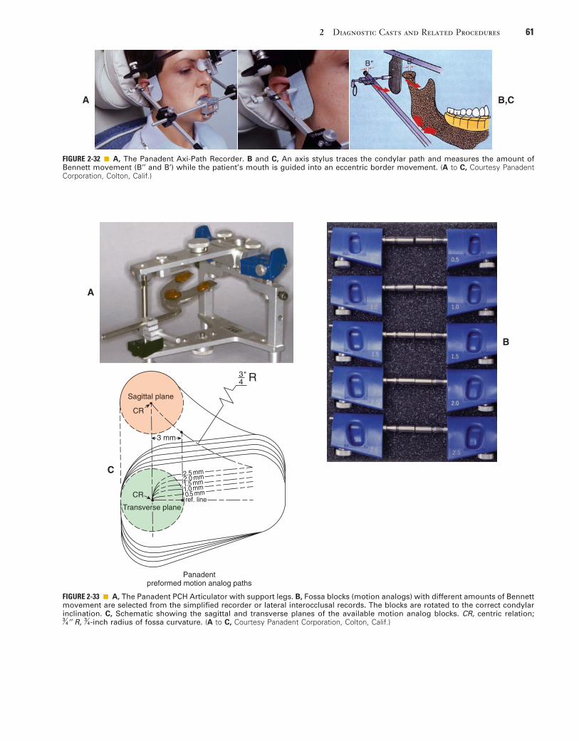
2 Diagnostic Casts and Related Procedures 61
FIGURE 2-32 ■ A, The Panadent Axi-Path Recorder. B and C, An axis stylus traces the condylar path and measures the amount of Bennett movement (B′′ and B′) while the patient’s mouth is guided into an eccentric border movement. (A to C, Courtesy Panadent Corporation, Colton, Calif.)
B" B'
A B,C
FIGURE 2-33 ■ A, The Panadent PCH Articulator with support legs. B, Fossa blocks (motion analogs) with different amounts of Bennett movement are selected from the simplified recorder or lateral interocclusal records. The blocks are rotated to the correct condylar inclination. C, Schematic showing the sagittal and transverse planes of the available motion analog blocks. CR, centric relation; 3
4′′ R, 34-inch radius of fossa curvature. (A to C, Courtesy Panadent Corporation, Colton, Calif.)
A
B
Sagittal plane
Panadentpreformed motion analog paths
CR
CR
3 mm
C
3"4 R
Transverse planeref. line
2.5 mm
1.5 mm1.0 mm0.5 mm
2.0 mm
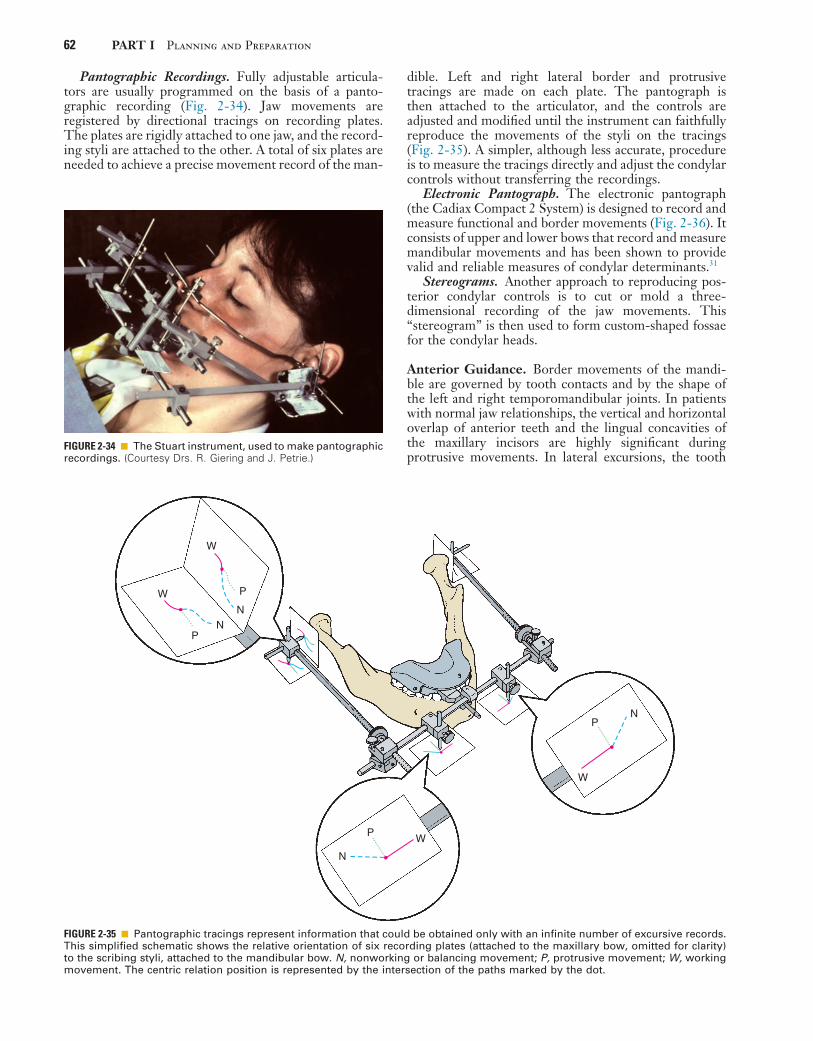
62 PART I Planning and Preparation
FIGURE 2-34 ■ The Stuart instrument, used to make pantographic recordings. (Courtesy Drs. R. Giering and J. Petrie.)
FIGURE 2-35 ■ Pantographic tracings represent information that could be obtained only with an infinite number of excursive records. This simplified schematic shows the relative orientation of six recording plates (attached to the maxillary bow, omitted for clarity) to the scribing styli, attached to the mandibular bow. N, nonworking or balancing movement; P, protrusive movement; W, working movement. The centric relation position is represented by the intersection of the paths marked by the dot.
PN
N
N
N
W
W
W
W P
P
P
Pantographic Recordings. Fully adjustable articula-tors are usually programmed on the basis of a panto-graphic recording (Fig. 2-34). Jaw movements are registered by directional tracings on recording plates. The plates are rigidly attached to one jaw, and the record-ing styli are attached to the other. A total of six plates are needed to achieve a precise movement record of the man-
dible. Left and right lateral border and protrusive tracings are made on each plate. The pantograph is then attached to the articulator, and the controls are adjusted and modified until the instrument can faithfully reproduce the movements of the styli on the tracings (Fig. 2-35). A simpler, although less accurate, procedure is to measure the tracings directly and adjust the condylar controls without transferring the recordings.
Electronic Pantograph. The electronic pantograph (the Cadiax Compact 2 System) is designed to record and measure functional and border movements (Fig. 2-36). It consists of upper and lower bows that record and measure mandibular movements and has been shown to provide valid and reliable measures of condylar determinants.31
Stereograms. Another approach to reproducing pos-terior condylar controls is to cut or mold a three-dimensional recording of the jaw movements. This “stereogram” is then used to form custom-shaped fossae for the condylar heads.
Anterior Guidance. Border movements of the mandi-ble are governed by tooth contacts and by the shape of the left and right temporomandibular joints. In patients with normal jaw relationships, the vertical and horizontal overlap of anterior teeth and the lingual concavities of the maxillary incisors are highly significant during protrusive movements. In lateral excursions, the tooth
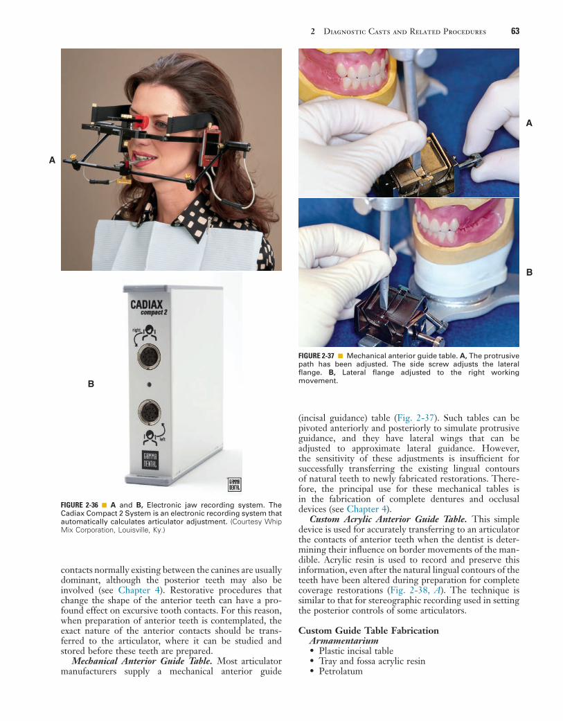
2 Diagnostic Casts and Related Procedures 63
FIGURE 2-36 ■ A and B, Electronic jaw recording system. The Cadiax Compact 2 System is an electronic recording system that automatically calculates articulator adjustment. (Courtesy Whip Mix Corporation, Louisville, Ky.)
A
B
A
B
FIGURE 2-37 ■ Mechanical anterior guide table. A, The protrusive path has been adjusted. The side screw adjusts the lateral flange. B, Lateral flange adjusted to the right working movement.
contacts normally existing between the canines are usually dominant, although the posterior teeth may also be involved (see Chapter 4). Restorative procedures that change the shape of the anterior teeth can have a pro-found effect on excursive tooth contacts. For this reason, when preparation of anterior teeth is contemplated, the exact nature of the anterior contacts should be trans-ferred to the articulator, where it can be studied and stored before these teeth are prepared.
Mechanical Anterior Guide Table. Most articulator manufacturers supply a mechanical anterior guide
(incisal guidance) table (Fig. 2-37). Such tables can be pivoted anteriorly and posteriorly to simulate protrusive guidance, and they have lateral wings that can be adjusted to approximate lateral guidance. However, the sensitivity of these adjustments is insufficient for successfully transferring the existing lingual contours of natural teeth to newly fabricated restorations. There-fore, the principal use for these mechanical tables is in the fabrication of complete dentures and occlusal devices (see Chapter 4).
Custom Acrylic Anterior Guide Table. This simple device is used for accurately transferring to an articulator the contacts of anterior teeth when the dentist is deter-mining their influence on border movements of the man-dible. Acrylic resin is used to record and preserve this information, even after the natural lingual contours of the teeth have been altered during preparation for complete coverage restorations (Fig. 2-38, A). The technique is similar to that for stereographic recording used in setting the posterior controls of some articulators.
Custom Guide Table FabricationArmamentarium• Plastic incisal table• Tray and fossa acrylic resin• Petrolatum
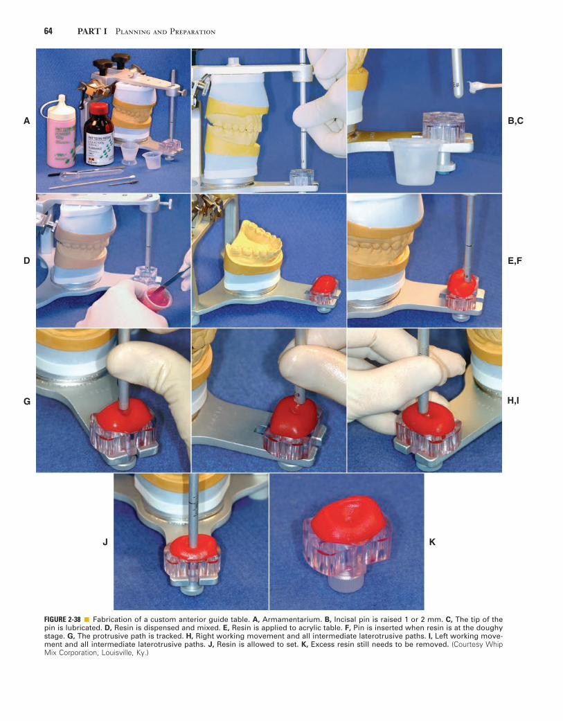
64 PART I Planning and Preparation
FIGURE 2-38 ■ Fabrication of a custom anterior guide table. A, Armamentarium. B, Incisal pin is raised 1 or 2 mm. C, The tip of the pin is lubricated. D, Resin is dispensed and mixed. E, Resin is applied to acrylic table. F, Pin is inserted when resin is at the doughy stage. G, The protrusive path is tracked. H, Right working movement and all intermediate laterotrusive paths. I, Left working move-ment and all intermediate laterotrusive paths. J, Resin is allowed to set. K, Excess resin still needs to be removed. (Courtesy Whip Mix Corporation, Louisville, Ky.)
A
D
G
B,C
E,F
H,I
J K
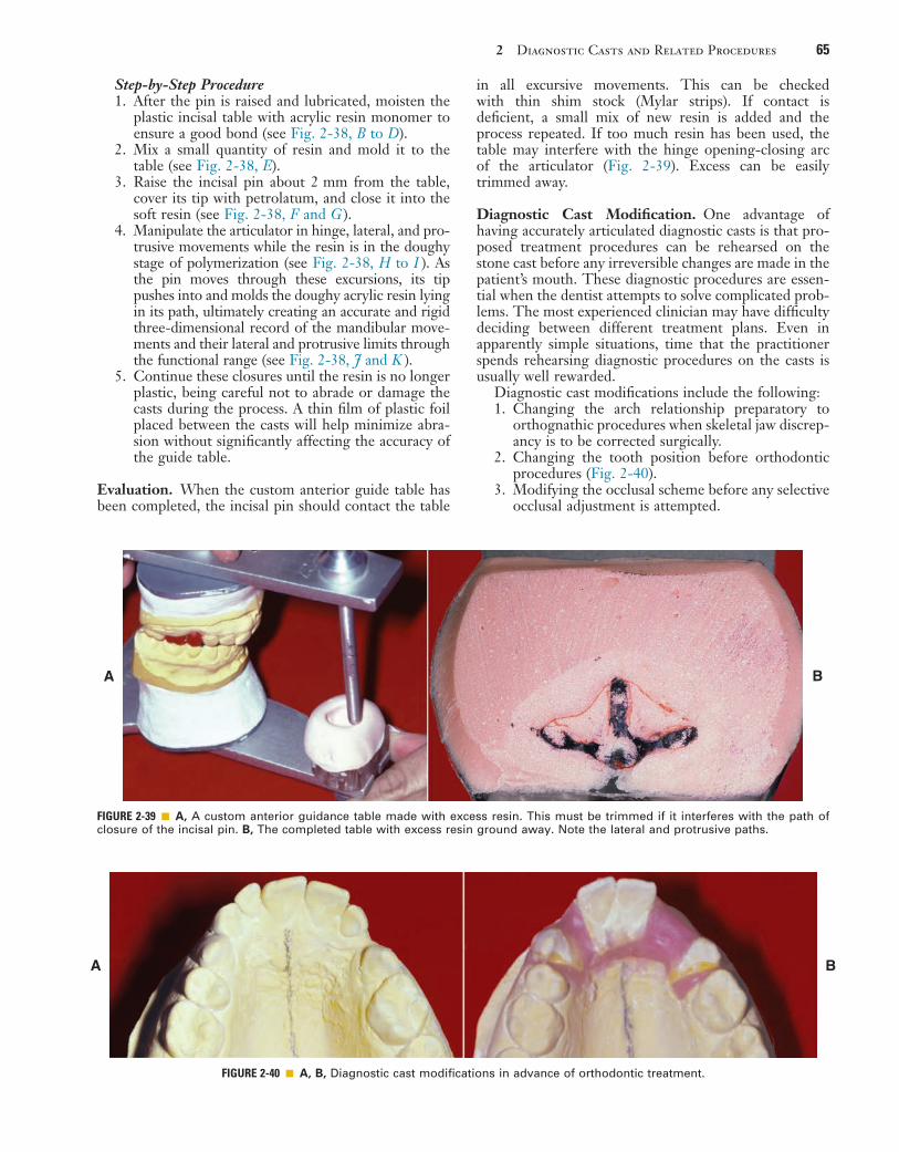
2 Diagnostic Casts and Related Procedures 65
Step-by-Step Procedure1. After the pin is raised and lubricated, moisten the
plastic incisal table with acrylic resin monomer to ensure a good bond (see Fig. 2-38, B to D).
2. Mix a small quantity of resin and mold it to the table (see Fig. 2-38, E).
3. Raise the incisal pin about 2 mm from the table, cover its tip with petrolatum, and close it into the soft resin (see Fig. 2-38, F and G ).
4. Manipulate the articulator in hinge, lateral, and pro-trusive movements while the resin is in the doughy stage of polymerization (see Fig. 2-38, H to I ). As the pin moves through these excursions, its tip pushes into and molds the doughy acrylic resin lying in its path, ultimately creating an accurate and rigid three-dimensional record of the mandibular move-ments and their lateral and protrusive limits through the functional range (see Fig. 2-38, J and K ).
5. Continue these closures until the resin is no longer plastic, being careful not to abrade or damage the casts during the process. A thin film of plastic foil placed between the casts will help minimize abra-sion without significantly affecting the accuracy of the guide table.
Evaluation. When the custom anterior guide table has been completed, the incisal pin should contact the table
in all excursive movements. This can be checked with thin shim stock (Mylar strips). If contact is deficient, a small mix of new resin is added and the process repeated. If too much resin has been used, the table may interfere with the hinge opening-closing arc of the articulator (Fig. 2-39). Excess can be easily trimmed away.
Diagnostic Cast Modification. One advantage of having accurately articulated diagnostic casts is that pro-posed treatment procedures can be rehearsed on the stone cast before any irreversible changes are made in the patient’s mouth. These diagnostic procedures are essen-tial when the dentist attempts to solve complicated prob-lems. The most experienced clinician may have difficulty deciding between different treatment plans. Even in apparently simple situations, time that the practitioner spends rehearsing diagnostic procedures on the casts is usually well rewarded.
Diagnostic cast modifications include the following:1. Changing the arch relationship preparatory to
orthognathic procedures when skeletal jaw discrep-ancy is to be corrected surgically.
2. Changing the tooth position before orthodontic procedures (Fig. 2-40).
3. Modifying the occlusal scheme before any selective occlusal adjustment is attempted.
FIGURE 2-39 ■ A, A custom anterior guidance table made with excess resin. This must be trimmed if it interferes with the path of closure of the incisal pin. B, The completed table with excess resin ground away. Note the lateral and protrusive paths.
A B
FIGURE 2-40 ■ A, B, Diagnostic cast modifications in advance of orthodontic treatment.
A B
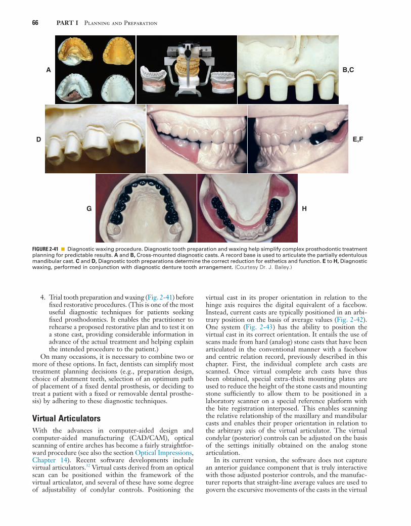
66 PART I Planning and Preparation
FIGURE 2-41 ■ Diagnostic waxing procedure. Diagnostic tooth preparation and waxing help simplify complex prosthodontic treatment planning for predictable results. A and B, Cross-mounted diagnostic casts. A record base is used to articulate the partially edentulous mandibular cast. C and D, Diagnostic tooth preparations determine the correct reduction for esthetics and function. E to H, Diagnostic waxing, performed in conjunction with diagnostic denture tooth arrangement. (Courtesy Dr. J. Bailey.)
A B,C
D E,F
G H
4. Trial tooth preparation and waxing (Fig. 2-41) before fixed restorative procedures. (This is one of the most useful diagnostic techniques for patients seeking fixed prosthodontics. It enables the practitioner to rehearse a proposed restorative plan and to test it on a stone cast, providing considerable information in advance of the actual treatment and helping explain the intended procedure to the patient.)
On many occasions, it is necessary to combine two or more of these options. In fact, dentists can simplify most treatment planning decisions (e.g., preparation design, choice of abutment teeth, selection of an optimum path of placement of a fixed dental prosthesis, or deciding to treat a patient with a fixed or removable dental prosthe-sis) by adhering to these diagnostic techniques.
Virtual ArticulatorsWith the advances in computer-aided design and computer-aided manufacturing (CAD/CAM), optical scanning of entire arches has become a fairly straightfor-ward procedure (see also the section Optical Impressions, Chapter 14). Recent software developments include virtual articulators.32 Virtual casts derived from an optical scan can be positioned within the framework of the virtual articulator, and several of these have some degree of adjustability of condylar controls. Positioning the
virtual cast in its proper orientation in relation to the hinge axis requires the digital equivalent of a facebow. Instead, current casts are typically positioned in an arbi-trary position on the basis of average values (Fig. 2-42). One system (Fig. 2-43) has the ability to position the virtual cast in its correct orientation. It entails the use of scans made from hard (analog) stone casts that have been articulated in the conventional manner with a facebow and centric relation record, previously described in this chapter. First, the individual complete arch casts are scanned. Once virtual complete arch casts have thus been obtained, special extra-thick mounting plates are used to reduce the height of the stone casts and mounting stone sufficiently to allow them to be positioned in a laboratory scanner on a special reference platform with the bite registration interposed. This enables scanning the relative relationship of the maxillary and mandibular casts and enables their proper orientation in relation to the arbitrary axis of the virtual articulator. The virtual condylar (posterior) controls can be adjusted on the basis of the settings initially obtained on the analog stone articulation.
In its current version, the software does not capture an anterior guidance component that is truly interactive with those adjusted posterior controls, and the manufac-turer reports that straight-line average values are used to govern the excursive movements of the casts in the virtual
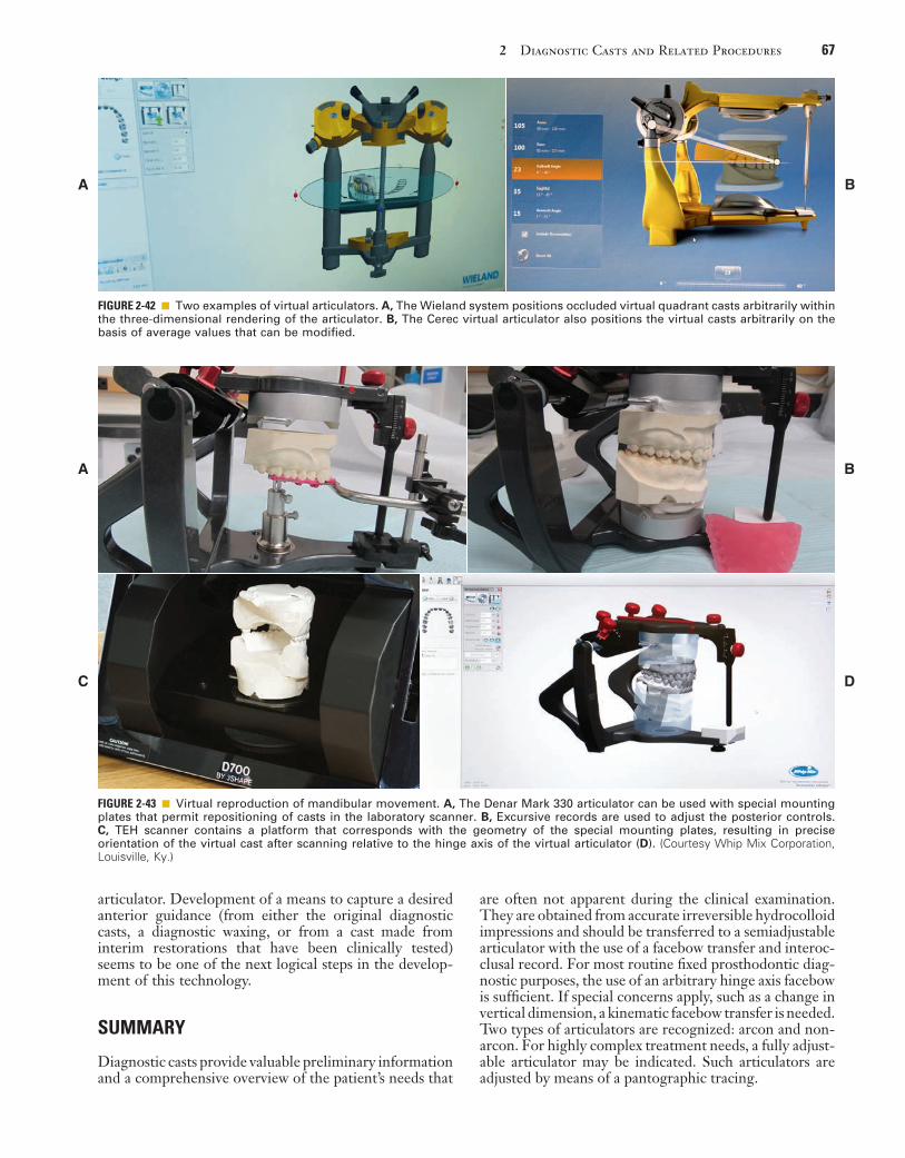
2 Diagnostic Casts and Related Procedures 67
FIGURE 2-42 ■ Two examples of virtual articulators. A, The Wieland system positions occluded virtual quadrant casts arbitrarily within the three-dimensional rendering of the articulator. B, The Cerec virtual articulator also positions the virtual casts arbitrarily on the basis of average values that can be modified.
A B
FIGURE 2-43 ■ Virtual reproduction of mandibular movement. A, The Denar Mark 330 articulator can be used with special mounting plates that permit repositioning of casts in the laboratory scanner. B, Excursive records are used to adjust the posterior controls. C, TEH scanner contains a platform that corresponds with the geometry of the special mounting plates, resulting in precise orientation of the virtual cast after scanning relative to the hinge axis of the virtual articulator (D). (Courtesy Whip Mix Corporation, Louisville, Ky.)
B
D
A
C
articulator. Development of a means to capture a desired anterior guidance (from either the original diagnostic casts, a diagnostic waxing, or from a cast made from interim restorations that have been clinically tested) seems to be one of the next logical steps in the develop-ment of this technology.
SUMMARY
Diagnostic casts provide valuable preliminary information and a comprehensive overview of the patient’s needs that
are often not apparent during the clinical examination. They are obtained from accurate irreversible hydrocolloid impressions and should be transferred to a semiadjustable articulator with the use of a facebow transfer and interoc-clusal record. For most routine fixed prosthodontic diag-nostic purposes, the use of an arbitrary hinge axis facebow is sufficient. If special concerns apply, such as a change in vertical dimension, a kinematic facebow transfer is needed. Two types of articulators are recognized: arcon and non-arcon. For highly complex treatment needs, a fully adjust-able articulator may be indicated. Such articulators are adjusted by means of a pantographic tracing.

68 PART I Planning and Preparation
Diagnostic casts should be articulated in CR to enable observation of deflective tooth contact and to assess any slide that may be present from CR to MI. Centric relation is defined as the maxillomandibular relationship in which the condyles articulate with the thinnest avascular portion of their respective disks with the complex in the antero-superior position against the shapes of the articular emi-nences. This position is independent of tooth contact. To record it, the dentist uses a suitable medium interposed between the maxillary and mandibular teeth and guides the patient into the CR position. This can be accom-plished through bimanual manipulation. If many teeth are absent, record bases with wax rims may need to be fabricated to obtain a CR record.
If it is difficult to manipulate a patient’s mandible into a reproducible hinge movement, a deprogramming device is helpful. This can be used to help minimize “muscle memory,” which would result in easier replication of the rotational hinge movement of the mandible.
Posterior articulator controls can be adjusted on the basis of arbitrary values according to anatomic averages by means of eccentric records, simplified pantographs, pantographs, or stereographs.
Anterior guidance can be approximated on articulators with a mechanical guide table. As an alternative, a custom acrylic guide table can be generated from the diagnostic casts. The latter is useful when anterior teeth are to be restored.
The development of virtual articulators is exciting. In its current form, the software used for such virtual instru-mentation is not quite capable of rendering the simula-tion of mandibular movements that can be observed on stone casts.
Diagnostic procedures such as diagnostic waxing, tooth preparation, and diagnostic cast modification can greatly enhance diagnosis and treatment planning.
REFERENCES1. Erbe C, et al: Dimensional stability of contemporary irreversible
hydrocolloids: humidor versus wet tissue storage. J Prosthet Dent 108:114, 2012.
2. Patel RD, et al: An in vitro investigation into the physical proper-ties of irreversible hydrocolloid alternatives. J Prosthet Dent 104:325, 2010.
3. Nassar U, et al: Dimensional stability of irreversible hydrocolloid impression materials as a function of pouring time: a systematic review. J Prosthet Dent 106:126, 2011.
4. Mendez AJ: The influence of impression trays on the accuracy of stone casts poured from irreversible hydrocolloid impressions. J Prosthet Dent 54:383, 1985.
5. Damodara EK, et al: A randomized clinical trial to compare diag-nostic casts made using plastic and metal trays. J Prosthet Dent 104:364, 2010.
6. Lim PF, et al: Adaptation of finger-smoothed irreversible hydrocol-loid to impression surfaces. Int J Prosthodont 8:117, 1995.
7. Khaknegar B, Ettinger RL: Removal time: a factor in the accuracy of irreversible hydrocolloid impressions. J Oral Rehabil 4:369, 1977.
8. al-Omari WM, et al: A microbiological investigation following the disinfection of alginate and addition cured silicone rubber impres-sion materials. Eur J Prosthodont Restor Dent 6:97, 1998.
9. Hall BD, et al: Effects of a chemical disinfectant on the physical properties of dental stones. Int J Prosthodont 17:65, 2004.
10. Johnson GH, et al: Dimensional stability and detail reproduction of irreversible hydrocolloid and elastomeric impressions disinfected by immersion. J Prosthet Dent 79:446, 1998.
11. Reisbick MH, et al: Irreversible hydrocolloid and gypsum interac-tions. Int J Prosthodont 10:7, 1997.
12. Young JM: Surface characteristics of dental stone: impression ori-entation. J Prosthet Dent 33:336, 1975.
13. Palik JF, et al: Accuracy of an earpiece face-bow. J Prosthet Dent 53:800, 1985.
14. O’Malley AM, Milosevic A: Comparison of three facebow/semi-adjustable articulator systems for planning orthognathic surgery. Br J Oral Maxillofac Surg 38:185, 2000.
15. Piehslinger E, et al: Computer simulation of occlusal discrepancies resulting from different mounting techniques. J Prosthet Dent 74:279, 1995.
16. Adrien P, Schouver J: Methods for minimizing the errors in mandibu-lar model mounting on an articulator. J Oral Rehabil 24:929, 1997.
17. Dawson PE: Temporomandibular joint pain-dysfunction problems can be solved. J Prosthet Dent 29:100, 1973.
18. Tarantola GJ, et al: The reproducibility of centric relation: a clini-cal approach. J Am Dent Assoc 128:1245, 1997.
19. McKee JR: Comparing condylar position repeatability for stand-ardized versus nonstandardized methods of achieving centric rela-tion. J Prosthet Dent 77:280, 1997.
20. Lucia VO: A technique for recording centric relation. J Prosthet Dent 14:492, 1964.
21. Gross M, et al: The effect of three different recording materials on the reproducibility of condylar guidance registrations in three semi-adjustable articulators. J Oral Rehabil 25:204, 1998.
22. Wirth CG: Interocclusal centric relation records for articulator mounted casts. Dent Clin North Am 15:627, 1971.
23. Wirth CG, Aplin AW: An improved interocclusal record of centric relation. J Prosthet Dent 25:279, 1971.
24. Lundeen HC: Centric relation records: the effect of muscle action. J Prosthet Dent 31:244, 1974.
25. Kepron D: Variations in condylar position relative to central mandibu-lar recordings. In Lefkowitz W, ed: Proceedings of the Second Inter-national Prosthodontic Congress, p 210. St. Louis, Mosby, 1979.
26. Teo CS, Wise MD: Comparison of retruded axis articular mount-ings with and without applied muscular force. J Oral Rehabil 8:363, 1981.
27. Tamaki K, et al: Reproduction of excursive tooth contact in an articulator with computerized axiography data. J Prosthet Dent 78:373, 1997.
28. Lundeen HC, Wirth CG: Condylar movement patterns engraved in plastic blocks. J Prosthet Dent 30:866, 1973.
29. Bell LJ, Matich JA: A study of the acceptability of lateral records by the Whip-Mix articulator. J Prosthet Dent 38:22, 1977.
30. Celar AG, et al: Guided versus unguided mandibular movement for duplicating intraoral eccentric tooth contacts in the articulator. J Prosthet Dent 81:14, 1999.
31. Chang WSW, et al: An in vitro evaluation of the reliability and validity of an electronic pantograph by testing with five different articulators. J Prosthet Dent 92:83, 2004.
32. Solaberrieta E, et al: Direct transfer of the position of digitized casts to a virtual articulator. J Prosthet Dent 109:411, 2013.
2 Diagnostic Casts and Related Procedures 69
STUDY QUESTIONS
1. Discuss the uses and limitations of irreversible hydro-colloid, and include an overview of its material properties.
2. Why are diagnostic casts articulated in centric rela-tion? Why are they not articulated in maximum intercuspation?
3. List five items that are determined more easily on diagnostic casts than intraorally.
4. What is accomplished with a facebow transfer? How do arbitrary facebows differ from kinematic facebows? When would one be selected over the other?
5. Describe the differences between arcon and nonarcon articulators. When would use of a simple hinge instru-ment be acceptable, and when would it be contraindi-cated? Why?
6. What is the role of excursive records in adjusting the articulator?
7. What does a simplified pantograph record? What does a pantograph record? When would either be indicated?
8. For what purpose is a custom acrylic guide table fab-ricated, and when is its use necessary?
9. Give two examples of situations in which a diagnostic waxing procedure is indicated.
70
Treatment Planning
C H A P T E R 3
Treatment planning consists of developing a logical sequence of treatment designed to restore the patient’s dentition to good health, optimal function, and optimal appearance. The plan should be presented in writing and discussed in detail with the patient. Good communica-tion with the patient is critical as the plan is formulated. Most dental problems can be solved in a number of dif-ferent ways; the patient’s preferences and concerns are paramount in establishing an appropriate treatment plan. In appropriate proper planning, the patient is informed about the current conditions and problems, the extent of dental treatment that is proposed, the time and cost of treatment, and the level of home care and professional follow-up necessary for success. Also, before any irrevers-ible procedures are initiated, the patient should under-stand that some of the planned procedures may need to be changed as treatment progresses and new information becomes available.
This chapter outlines the necessary decisions in plan-ning fixed prosthodontic treatment. Foremost among these is the identification of patients’ needs and their preferences, which must be correlated with the range of treatments available. For long-term success, when a fixed dental prosthesis (FDP) is being considered, the abut-ment teeth must be carefully evaluated. Finally, the treat-ment plan must be properly sequenced as part of an ongoing program of comprehensive dental care.
IDENTIFICATION OF PATIENT NEEDS
Successful treatment planning is based on proper identi-fication of the patient’s needs. If the dentist attempts to make the patient conform to the “ideal” treatment plan rather than make the treatment plan conform to the patient’s needs, success is unlikely. In many cases, several plans are presented and discussed, each with advantages and disadvantages. Indeed, failing to explain and present available options may be considered legally negligent.
Treatment is necessary to accomplish one or several of the following objectives: correcting an existing disease, preventing future disease, restoring function, and improv-ing appearance.
Correction of Existing DiseaseExisting disease is revealed during the clinical examina-tion (see Chapter 1). Active disease can usually be halted by identification and reduction of the initiating factors, identification and improvement of the resistive factors, or both (Fig. 3-1). For example, oral hygiene instruction helps reduce the amount of residual plaque, an initiating
factor, and thus helps reduce the likelihood of further dental caries. Such instruction also helps improve gingi-val health, and the resulting healthy tissue is more resist-ant to disease. In such patients with extensive caries, additional preventive measures are necessary (e.g., mouth rinses, high-fluoride toothpastes, dietary analysis). Restorative treatment replaces damaged or missing tooth structure, but additional treatment is crucial to control the underlying causes.
Prevention of Future DiseaseThe likelihood of future disease can be predicted from the patient’s disease experience and from the prevalence of the disease in the general population. Treatment should be proposed if future disease seems likely in the absence of such intervention.
One of the first phases of treatment is to stabilize active disease, which often includes replacement of defec-tive restorations and treatment of carious lesions. If a patient presents initially with poor oral hygiene, the dentist should monitor if improvement in plaque control results during the stabilization phase. If such does not occur, renewed emphasis on proper oral hygiene meas-ures is indicated. Subsequently identified limitations in the patient’s oral hygiene may justifiably affect the final treatment plan.
Restoration of FunctionAlthough objective measurement can be difficult, the level of function is assessed during the examination. Treatment may be proposed to correct impaired function (e.g., mastication or speech). Prerequisite treatment may include mandibular repositioning through occlusal reshaping before fixed prosthodontic treatment (see Chapters 4 and 6) and orthodontically repositioning teeth in more favorable locations before missing teeth are replaced.
Improvement of AppearancePatients often seek dental treatment because they are dissatisfied with their appearance. However, it is difficult to assess dental esthetics objectively (see Chapter 23). The dentist should develop expertise in this area and should be prepared to appraise the appearance of the patient’s dentition and listen carefully to the patient’s views. If the existing appearance is far outside socially accepted values, the feasibility (and limitations) of cor-rective procedures should be brought to the patient’s attention. Long-term dental health should not be
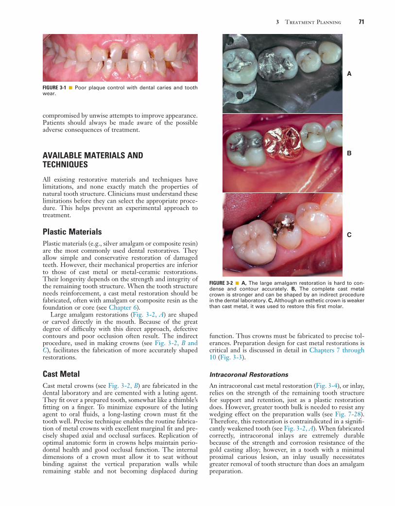
3 Treatment Planning 71
function. Thus crowns must be fabricated to precise tol-erances. Preparation design for cast metal restorations is critical and is discussed in detail in Chapters 7 through 10 (Fig. 3-3).
Intracoronal Restorations
An intracoronal cast metal restoration (Fig. 3-4), or inlay, relies on the strength of the remaining tooth structure for support and retention, just as a plastic restoration does. However, greater tooth bulk is needed to resist any wedging effect on the preparation walls (see Fig. 7-28). Therefore, this restoration is contraindicated in a signifi-cantly weakened tooth (see Fig. 3-2, A). When fabricated correctly, intracoronal inlays are extremely durable because of the strength and corrosion resistance of the gold casting alloy; however, in a tooth with a minimal proximal carious lesion, an inlay usually necessitates greater removal of tooth structure than does an amalgam preparation.
compromised by unwise attempts to improve appearance. Patients should always be made aware of the possible adverse consequences of treatment.
AVAILABLE MATERIALS AND TECHNIQUES
All existing restorative materials and techniques have limitations, and none exactly match the properties of natural tooth structure. Clinicians must understand these limitations before they can select the appropriate proce-dure. This helps prevent an experimental approach to treatment.
Plastic MaterialsPlastic materials (e.g., silver amalgam or composite resin) are the most commonly used dental restoratives. They allow simple and conservative restoration of damaged teeth. However, their mechanical properties are inferior to those of cast metal or metal-ceramic restorations. Their longevity depends on the strength and integrity of the remaining tooth structure. When the tooth structure needs reinforcement, a cast metal restoration should be fabricated, often with amalgam or composite resin as the foundation or core (see Chapter 6).
Large amalgam restorations (Fig. 3-2, A) are shaped or carved directly in the mouth. Because of the great degree of difficulty with this direct approach, defective contours and poor occlusion often result. The indirect procedure, used in making crowns (see Fig. 3-2, B and C), facilitates the fabrication of more accurately shaped restorations.
Cast MetalCast metal crowns (see Fig. 3-2, B) are fabricated in the dental laboratory and are cemented with a luting agent. They fit over a prepared tooth, somewhat like a thimble’s fitting on a finger. To minimize exposure of the luting agent to oral fluids, a long-lasting crown must fit the tooth well. Precise technique enables the routine fabrica-tion of metal crowns with excellent marginal fit and pre-cisely shaped axial and occlusal surfaces. Replication of optimal anatomic form in crowns helps maintain perio-dontal health and good occlusal function. The internal dimensions of a crown must allow it to seat without binding against the vertical preparation walls while remaining stable and not becoming displaced during
FIGURE 3-1 ■ Poor plaque control with dental caries and tooth wear.
FIGURE 3-2 ■ A, The large amalgam restoration is hard to con-dense and contour accurately. B, The complete cast metal crown is stronger and can be shaped by an indirect procedure in the dental laboratory. C, Although an esthetic crown is weaker than cast metal, it was used to restore this first molar.
A
B
C

72 PART I Planning and Preparation
FIGURE 3-4 ■ A, The mesio-occlusal-distal (MOD) inlay is gener-ally contraindicated because of the risk of tooth fracture. However, it can be a very long-lasting restoration. These, placed in 1948, were still satisfactory when the patient died in 2012. B, These small gold foil restorations were placed in 1943.
A
B
FIGURE 3-3 ■ A, An intracoronal cast restoration (inlay) can act as a wedge during cementation or function. If the cusps are weakened, fracture will occur. B, A cuspal-coverage onlay pro-vides better protection but often lacks retention. C, A complete crown provides the best protection against fracture. It also has the best retention, but it can be associated with periodontal disease and poor esthetics. (Redrawn from Rosenstiel SF: Fixed bridgework—the basic principles. In Rayne J, ed: General Dental Treatment. London, Kluwer Publishing, 1983.)
Fracture
Inlay Onlay Completecrown
Cuspal protection becomesmore important as thestructural durability of thecusps is compromised.
A B C
Extracoronal Restorations
An extracoronal cast metal restoration (Fig. 3-5), or crown, encircles all or part of the remaining tooth struc-ture and the occlusal surface. Crowns strengthen and protect teeth weakened by caries or trauma. To provide the necessary bulk of material for strength, considerably more tooth structure must be removed than for an intra-coronal restoration. The margins of an extracoronal res-toration often must be near or below the crest of the free gingiva, which can make maintenance of tissue health difficult. Tooth preparation for an extracoronal restora-tion may be combined with intracoronal features (e.g., grooves and pinholes) to obtain retention mechanical advantage (see the Retention and Resistance section, Chapter 7).
Metal-Ceramic MaterialMetal-ceramic crowns (Fig. 3-6) consist of a tooth-colored layer of porcelain bonded to a cast metal sub-structure. They are used when a complete crown is needed to restore appearance and function. Tooth struc-ture must be reduced sufficiently to provide space for the bulk of porcelain needed for a natural appearance. Thus the preparation design for a metal-ceramic crown is
FIGURE 3-5 ■ A, Complete cast metal crowns restore the molars. Occlusal (B) and buccal (C) views of two partial veneer crowns. With partial veneer crowns, more tooth structure is conserved.
A
B
C

3 Treatment Planning 73
do not withstand functional loads well over time. There-fore, they are very useful as long-term interim restora-tions. (See the Fiber-Reinforced Interim Restorations section, Chapter 15).
Complete CeramicCrowns, inlays, and laminate veneers made entirely of dental porcelain are among the most esthetically pleasing of all fixed restorations (Fig. 3-8). Drawbacks include a comparative lack of strength and—depending on the fab-rication method used—the difficulty in achieving an acceptable internal and marginal fit. Some all-ceramic restorations are fabricated in the dental office, whereas others must be fabricated in the dental laboratory. In general, the internal fit of some of the laboratory fabri-cated restorations is superior to that of those fabricated by milling in the dental office. The advantage of the latter is that the esthetic restoration can be placed in a single appointment without the need for an interim restoration. The current focus in improving the strength of esthetic restorations is on either veneering a high-strength alumina, zirconia, spinel, or lithium disilicate core6-8 with a more translucent porcelain or using a leucite-reinforced translucent material9-12 (see Chapter 25). Monolithic esthetic restorations are among the strongest ceramic restorations, and colored monolithic zirconia crowns have very acceptable esthetics for posterior teeth.13 Com-plete ceramic restorations are fabricated by an indirect technique, and etchable ceramic crowns are generally retained with composite resin. The acid etching of the internal crown surface is used to provide retention “keys.”
Fixed Dental ProsthesesAn FDP (Fig. 3-9) is often indicated when one or more teeth must be removed or are missing. Such teeth are replaced by pontics that are designed to fulfill the func-tional and often the esthetic requirements of the missing teeth (see Chapter 20). Pontics are attached with connec-tors to the FDP retainers, which are the restorations on prepared abutment teeth.
All the components of an FDP are fabricated and assembled in the laboratory before cementation in the mouth. This requires precise alignment of tooth prepara-tions. Because unseating forces on individual retainers can be considerable, retentive restorations are essential.
FIGURE 3-6 ■ A and B, Metal-ceramic restorations used to restore the maxillary anterior teeth.
A
B
FIGURE 3-7 ■ Fiber-reinforced fixed dental prosthesis.
FIGURE 3-8 ■ Complete ceramic restoration.
among the least conservative, although tooth structure can be conserved if only the most visible part of the res-toration is veneered.
The labial margins of a metal-ceramic restoration are often discernible and may detract from its appearance. They can be hidden by subgingival placement, although they then have the potential for increasing gingival inflammation; this should be avoided when possible.1 Appearance can be improved by omitting the metal shoulder and making the labial margin in porcelain. As discussed in Chapter 24, this is a more challenging labo-ratory procedure.
Fiber-Reinforced ResinAdvances in composite resin technology, especially the introduction of glass and polyethylene fibers,2-4 have prompted the use of indirect composite resin restorations for inlays, crowns, and FDPs.5 Excellent marginal adapta-tion and esthetic results are achievable (Fig. 3-7), but they

74 PART I Planning and Preparation
FDPs have been demonstrated to have exceptional long-term success,14 which is ensured by controlling the mag-nitude and direction of loading and by making sure the patient practices appropriate oral hygiene measures.
Implant-Supported ProsthesesSingle or multiple missing teeth can be replaced with an implant-supported prosthesis (Fig. 3-10). For the suc-cessful “osseointegrated” technique, the bone is atrau-matically drilled to receive precisely fitting titanium cylinders.15 These are either left in place without loading for several months until they are osseointegrated or restored immediately with an interim restoration. Then function and esthetics are restored with a prosthesis (see Chapter 13).
Partial Removable Dental ProsthesisA partial removable dental prosthesis (RDP) (Fig. 3-11) is designed to replace missing teeth and their supporting structures. Forces applied to a well-designed partial RDP are distributed to the remaining teeth and the residual alveolar ridges. These forces are most accurately control-led if the abutment teeth can be reshaped with fixed cast restorations that have carefully contoured guide planes and rest seats (see Chapter 21). Specific design requirements of the partial RDP can affect tooth prepara-tion design for such survey crowns.
FIGURE 3-9 ■ A, A maxillary right central incisor that will be replaced by a single-unit implant-supported prosthesis. The impression post is tightened into the implant. B, All-ceramic restoration. C, Illustration of a three-unit fixed dental prosthesis, showing the main components. D, The pontic rigidly attached to crowns on the abutment teeth. The connectors should occupy the normal interproximal contact area and be large enough for strength but not so large as to impede plaque control.
Abutmentpreparation
Abutment tooth
Edentulousridge
ConnectorPartial-coverageretainer
Pontic
C D
A B
FPDcomponents
FIGURE 3-10 ■ A, Single tooth implant with healing abutment in place. B, Implant supported crown replacing maxillary lateral incisor.
A
B
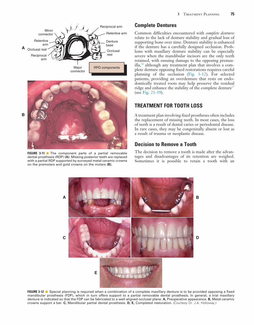
3 Treatment Planning 75
FIGURE 3-11 ■ The component parts of a partial removable dental prosthesis (RDP) (A). Missing posterior teeth are replaced with a partial RDP supported by surveyed metal-ceramic crowns on the premolars and gold crowns on the molars (B).
Minorconnector
Retentivearm
Reciprocalarm
Reciprocal arm
Retentive arm
Denturebase
Occlusalrest
Occlusal rest
Majorconnector
A
RPD components
B
Complete DenturesCommon difficulties encountered with complete dentures relate to the lack of denture stability and gradual loss of supporting bone over time. Denture stability is enhanced if the denture has a carefully designed occlusion. Prob-lems with maxillary denture stability can be especially severe when the mandibular incisors are the only teeth retained, with ensuing damage to the opposing premax-illa,16 although any treatment plan that involves a com-plete denture opposing fixed restorations requires careful planning of the occlusion (Fig. 3-12). For selected patients, providing an overdenture that rests on endo-dontically treated roots may help preserve the residual ridge and enhance the stability of the complete denture17 (see Fig. 21-39).
TREATMENT FOR TOOTH LOSS
A treatment plan involving fixed prostheses often includes the replacement of missing teeth. In most cases, the loss of teeth is a result of dental caries or periodontal disease. In rare cases, they may be congenitally absent or lost as a result of trauma or neoplastic disease.
Decision to Remove a ToothThe decision to remove a tooth is made after the advan-tages and disadvantages of its retention are weighed. Sometimes it is possible to retain a tooth with an
FIGURE 3-12 ■ Special planning is required when a combination of a complete maxillary denture is to be provided opposing a fixed mandibular prosthesis (FDP), which in turn offers support to a partial removable dental prosthesis. In general, a trial maxillary denture is indicated so that the FDP can be fabricated to a well-aligned occlusal plane. A, Preoperative appearance. B, Metal-ceramic crowns support a bar. C, Mandibular partial dental prosthesis. D, E, Completed restoration. (Courtesy Dr. J.A. Holloway.)
A B
C D
E
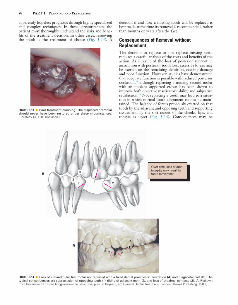
76 PART I Planning and Preparation
decision if and how a missing tooth will be replaced is best made at the time its removal is recommended, rather than months or years after the fact.
Consequences of Removal without ReplacementThe decision to replace or not replace missing teeth requires a careful analysis of the costs and benefits of the action. As a result of the loss of posterior support in association with posterior tooth loss, excessive forces may be exerted on the remaining dentition, causing damage and poor function. However, studies have demonstrated that adequate function is possible with reduced posterior occlusion,18 although replacing a missing second molar with an implant-supported crown has been shown to improve both objective masticatory ability and subjective satisfaction.19 Not replacing a tooth may lead to a situa-tion in which normal tooth alignment cannot be main-tained. The balance of forces previously exerted on that tooth by the adjacent and opposing teeth and supporting tissues and by the soft tissues of the cheeks, lips, and tongue is upset (Fig. 3-14). Consequences may be
apparently hopeless prognosis through highly specialized and complex techniques. In those circumstances, the patient must thoroughly understand the risks and bene-fits of the treatment decision. In other cases, removing the tooth is the treatment of choice (Fig. 3-13). A
FIGURE 3-13 ■ Poor treatment planning. The displaced premolar should never have been restored under these circumstances. (Courtesy Dr. P.B. Robinson.)
FIGURE 3-14 ■ Loss of a mandibular first molar not replaced with a fixed dental prosthesis: illustration (A) and diagnostic cast (B). The typical consequences are supraclusion of opposing teeth (1), tilting of adjacent teeth (2), and loss of proximal contacts (3). (A, Redrawn from Rosenstiel SF: Fixed bridgework—the basic principles. In Rayne J, ed: General Dental Treatment. London, Kluwer Publishing, 1983.)
1 223
3
Over time, loss of archintegrity may result intooth movement.A
3 2 2
1
B
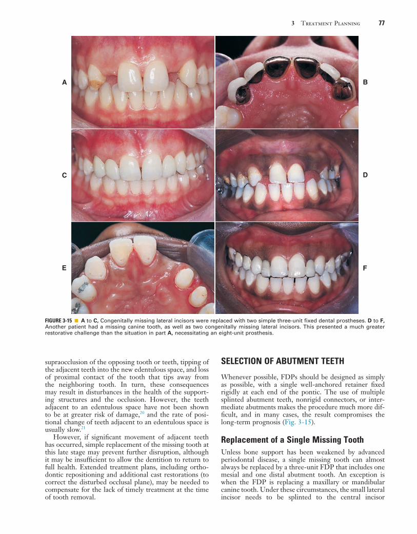
3 Treatment Planning 77
SELECTION OF ABUTMENT TEETH
Whenever possible, FDPs should be designed as simply as possible, with a single well-anchored retainer fixed rigidly at each end of the pontic. The use of multiple splinted abutment teeth, nonrigid connectors, or inter-mediate abutments makes the procedure much more dif-ficult, and in many cases, the result compromises the long-term prognosis (Fig. 3-15).
Replacement of a Single Missing ToothUnless bone support has been weakened by advanced periodontal disease, a single missing tooth can almost always be replaced by a three-unit FDP that includes one mesial and one distal abutment tooth. An exception is when the FDP is replacing a maxillary or mandibular canine tooth. Under these circumstances, the small lateral incisor needs to be splinted to the central incisor
supraocclusion of the opposing tooth or teeth, tipping of the adjacent teeth into the new edentulous space, and loss of proximal contact of the tooth that tips away from the neighboring tooth. In turn, these consequences may result in disturbances in the health of the support-ing structures and the occlusion. However, the teeth adjacent to an edentulous space have not been shown to be at greater risk of damage,20 and the rate of posi-tional change of teeth adjacent to an edentulous space is usually slow.21
However, if significant movement of adjacent teeth has occurred, simple replacement of the missing tooth at this late stage may prevent further disruption, although it may be insufficient to allow the dentition to return to full health. Extended treatment plans, including ortho-dontic repositioning and additional cast restorations (to correct the disturbed occlusal plane), may be needed to compensate for the lack of timely treatment at the time of tooth removal.
FIGURE 3-15 ■ A to C, Congenitally missing lateral incisors were replaced with two simple three-unit fixed dental prostheses. D to F, Another patient had a missing canine tooth, as well as two congenitally missing lateral incisors. This presented a much greater restorative challenge than the situation in part A, necessitating an eight-unit prosthesis.
A B
C D
FE

78 PART I Planning and Preparation
Evaluation of Abutment Teeth
The dentist can save considerable time and expense, and can reinforce a patient’s confidence, by thoroughly inves-tigating each abutment tooth before proceeding with tooth preparation. Radiographs are made, and the dentist assesses pulpal health by evaluating the response to thermal or electrical stimulation, or both. Existing resto-rations, cavity liners, and residual caries are removed27 (preferably under a rubber dam), and a careful check is made for possible pulpal exposure. Teeth in which pulpal health is doubtful should be treated endodontically before fixed prosthodontic treatment is initiated. Although a direct pulp cap may be an acceptable risk under a simple amalgam or composite resin, conventional endodontic treatment is normally preferred when crowns are planned. Fixed prosthodontic treatment is time consuming and costly; when endodontic treatment is needed after a complex prosthesis has been fabricated, access must be established through the occlusal surface of the newly fabricated prosthesis, which jeopardizes its long-term prognosis and the overall success of treatment.
Endodontically Treated Abutments
If a tooth is properly treated endodontically, it can serve well as an abutment with a post and core foundation for retention and strength (see Chapter 12). Failures occur, however, particularly on teeth with short roots or little remaining coronal tooth structure. Care is needed to obtain maximum retention for the post and core. Some-times it is better to remove a badly damaged tooth than to attempt endodontic treatment.
The type of restoration that is anticipated after endo-dontic treatment can help the dentist make the best
to prevent lateral drift of the FDP. This use of two abut-ment teeth on the anterior aspect is referred to as double-abutting.
Cantilever Fixed Dental Prostheses
FDPs in which only one side of the pontic is attached to a retainer are referred to as cantilevered. An example would be a lateral incisor pontic attached only to an extracoronal metal-ceramic retainer on a canine tooth. Cantilevered FDPs remain popular because some of the difficulties encountered in making a three-unit FDP are lessened. Also, many clinicians are reluctant to prepare an intact central incisor, preferring instead to use a cantilever.
However, the long-term prognosis of the single-abutment cantilever is poor.22 Forces are best tolerated by the periodontal supporting structures when directed along the long axes of the teeth.23 This is the case when a simple three-unit FDP is used. A cantilever induces lateral forces on the supporting tissues, which may be harmful and lead to tipping, rotation, or drifting of the abutment (Fig. 3-16). Laboratory analysis24,25 has con-firmed the potential harmful nature of such FDPs. However, clinical experience with resin-retained FDPs has suggested that cantilever designs may be preferred, especially because repeated adhesion after failure is greatly facilitated26 (see Chapter 26).
When multiple missing teeth are replaced, cantilever FDPs can have considerable application (see Fig. 3-20). The harmful tipping forces are resisted by multiple abut-ment teeth with sound periodontal support, and move-ment of the abutments is unlikely. Cantilevers are also successfully used with implant-supported prostheses (see Chapter 13).
FIGURE 3-16 ■ A, Forces applied to a cantilever fixed dental prosthesis are resisted on only one side, which leads to imbalance. Verti-cal forces can cause tipping, and horizontal forces can cause rotation, of abutment teeth. B, By including both adjacent teeth in the prosthesis, it is possible to resist forces much better because the teeth have to be moved bodily rather than merely rotated or tipped. (Redrawn from Rosenstiel SF: Fixed bridgework—the basic principles. In Rayne J, ed: General Dental Treatment. London, Kluwer Publishing, 1983.)
Tipping
Force
Rotation
To be successful, singleabutment cantileversrequire a very favorableocclusion.
A
Force
B
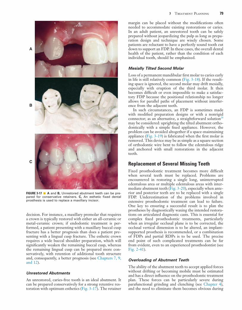
3 Treatment Planning 79
margin can be placed without the modifications often needed to accommodate existing restorations or caries. In an adult patient, an unrestored tooth can be safely prepared without jeopardizing the pulp as long as prepa-ration design and technique are wisely chosen. Some patients are reluctant to have a perfectly sound tooth cut down to support an FDP. In these cases, the overall dental health of the patient, rather than the condition of each individual tooth, should be emphasized.
Mesially Tilted Second Molar
Loss of a permanent mandibular first molar to caries early in life is still relatively common (Fig. 3-18). If the result-ing space is ignored, the second molar may drift mesially, especially with eruption of the third molar. It then becomes difficult or even impossible to make a satisfac-tory FDP because the positional relationship no longer allows for parallel paths of placement without interfer-ence from the adjacent teeth.
In such circumstances, an FDP is sometimes made with modified preparation designs or with a nonrigid connector; as an alternative, a straightforward solution28 may be considered: uprighting the tilted abutment ortho-dontically with a simple fixed appliance. However, the problem can be avoided altogether if a space-maintaining appliance (Fig. 3-19) is fabricated when the first molar is removed. This device may be as simple as a square section of orthodontic wire bent to follow the edentulous ridge and anchored with small restorations in the adjacent teeth.
Replacement of Several Missing TeethFixed prosthodontic treatment becomes more difficult when several teeth must be replaced. Problems are encountered in restoring a single long, uninterrupted edentulous area or multiple edentulous areas with inter-mediate abutment teeth (Fig. 3-20), especially when ante-rior and posterior teeth are to be replaced with a single FDP. Underestimation of the problems involved in extensive prosthodontic treatment can lead to failure. One key to ensuring a successful result is to plan the prostheses by diagnostically waxing the intended restora-tions on articulated diagnostic casts. This is essential for complex fixed prosthodontic treatments, particularly when an irregular occlusal plane is to be corrected, the occlusal vertical dimension is to be altered, an implant-supported prosthesis is recommended, or a combination of FDPs and partial RDPs is to be used. The precise end point of such complicated treatments can be far from evident, even to an experienced prosthodontist (see Fig. 2-41).
Overloading of Abutment Teeth
The ability of the abutment teeth to accept applied forces without drifting or becoming mobile must be estimated and has a direct influence on the prosthodontic treatment plan. These forces can be particularly severe during parafunctional grinding and clenching (see Chapter 4), and the need to eliminate them becomes obvious during
decision. For instance, a maxillary premolar that requires a crown is typically restored with either an all-ceramic or metal-ceramic crown; if endodontic treatment is per-formed, a patient presenting with a maxillary buccal cusp fracture has a better prognosis than does a patient pre-senting with a lingual cusp fracture. The esthetic crown requires a wide buccal shoulder preparation, which will significantly weaken the remaining buccal cusp, whereas the remaining lingual cusp can be prepared more con-servatively, with retention of additional tooth structure and, consequently, a better prognosis (see Chapters 7, 9, and 12).
Unrestored Abutments
An unrestored, caries-free tooth is an ideal abutment. It can be prepared conservatively for a strong retentive res-toration with optimum esthetics (Fig. 3-17). The retainer
FIGURE 3-17 ■ A and B, Unrestored abutment teeth can be pre-pared for conservative retainers. C, An esthetic fixed dental prosthesis is used to replace a maxillary incisor.
A
B
C
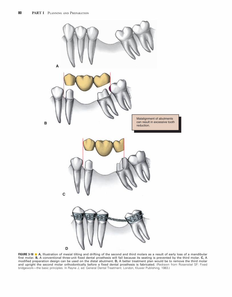
80 PART I Planning and Preparation
FIGURE 3-18 ■ A, Illustration of mesial tilting and drifting of the second and third molars as a result of early loss of a mandibular first molar. B, A conventional three-unit fixed dental prosthesis will fail because its seating is prevented by the third molar. C, A modified preparation design can be used on the distal abutment. D, A better treatment plan would be to remove the third molar and upright the second molar orthodontically before a fixed dental prosthesis is fabricated. (Redrawn from Rosenstiel SF: Fixed bridgework—the basic principles. In Rayne J, ed: General Dental Treatment. London, Kluwer Publishing, 1983.)
A
B
C
D
Malalignment of abutmentscan result in excessive toothreduction.
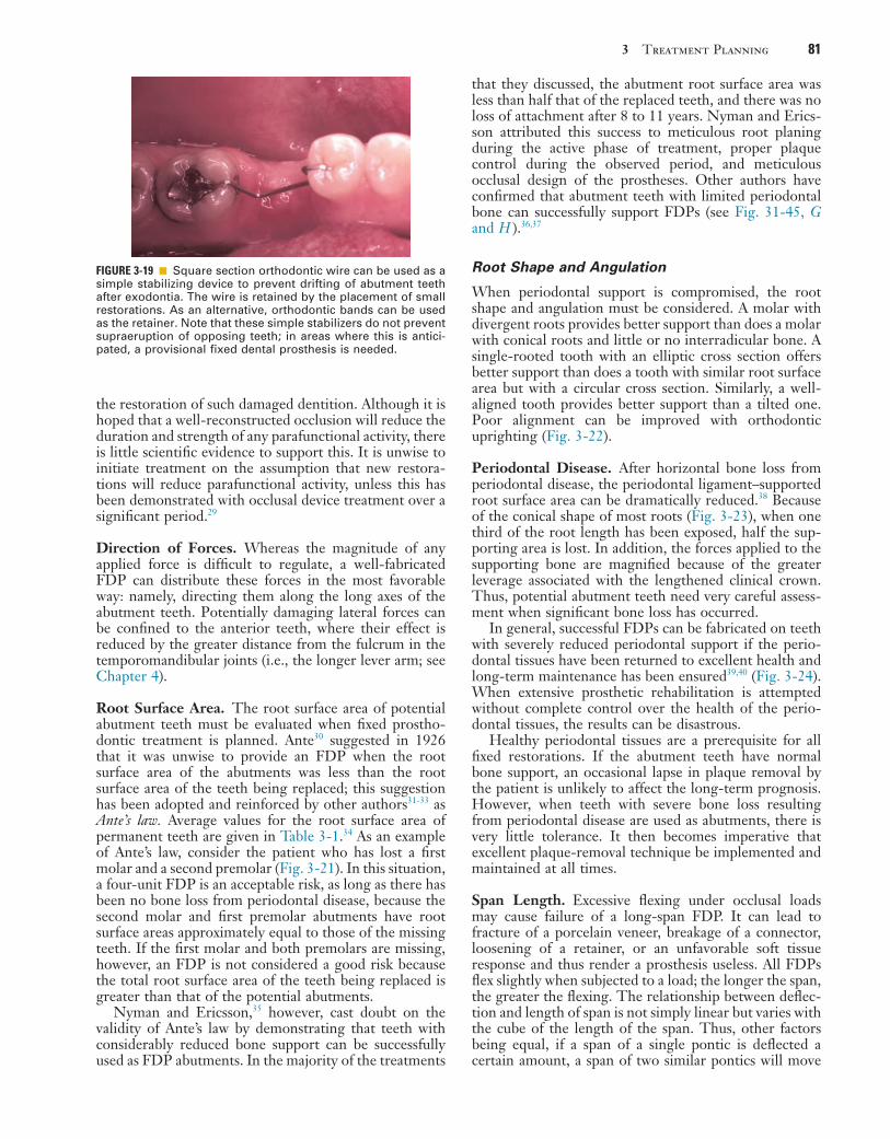
3 Treatment Planning 81
that they discussed, the abutment root surface area was less than half that of the replaced teeth, and there was no loss of attachment after 8 to 11 years. Nyman and Erics-son attributed this success to meticulous root planing during the active phase of treatment, proper plaque control during the observed period, and meticulous occlusal design of the prostheses. Other authors have confirmed that abutment teeth with limited periodontal bone can successfully support FDPs (see Fig. 31-45, G and H ).36,37
Root Shape and Angulation
When periodontal support is compromised, the root shape and angulation must be considered. A molar with divergent roots provides better support than does a molar with conical roots and little or no interradicular bone. A single-rooted tooth with an elliptic cross section offers better support than does a tooth with similar root surface area but with a circular cross section. Similarly, a well-aligned tooth provides better support than a tilted one. Poor alignment can be improved with orthodontic uprighting (Fig. 3-22).
Periodontal Disease. After horizontal bone loss from periodontal disease, the periodontal ligament–supported root surface area can be dramatically reduced.38 Because of the conical shape of most roots (Fig. 3-23), when one third of the root length has been exposed, half the sup-porting area is lost. In addition, the forces applied to the supporting bone are magnified because of the greater leverage associated with the lengthened clinical crown. Thus, potential abutment teeth need very careful assess-ment when significant bone loss has occurred.
In general, successful FDPs can be fabricated on teeth with severely reduced periodontal support if the perio-dontal tissues have been returned to excellent health and long-term maintenance has been ensured39,40 (Fig. 3-24). When extensive prosthetic rehabilitation is attempted without complete control over the health of the perio-dontal tissues, the results can be disastrous.
Healthy periodontal tissues are a prerequisite for all fixed restorations. If the abutment teeth have normal bone support, an occasional lapse in plaque removal by the patient is unlikely to affect the long-term prognosis. However, when teeth with severe bone loss resulting from periodontal disease are used as abutments, there is very little tolerance. It then becomes imperative that excellent plaque-removal technique be implemented and maintained at all times.
Span Length. Excessive flexing under occlusal loads may cause failure of a long-span FDP. It can lead to fracture of a porcelain veneer, breakage of a connector, loosening of a retainer, or an unfavorable soft tissue response and thus render a prosthesis useless. All FDPs flex slightly when subjected to a load; the longer the span, the greater the flexing. The relationship between deflec-tion and length of span is not simply linear but varies with the cube of the length of the span. Thus, other factors being equal, if a span of a single pontic is deflected a certain amount, a span of two similar pontics will move
FIGURE 3-19 ■ Square section orthodontic wire can be used as a simple stabilizing device to prevent drifting of abutment teeth after exodontia. The wire is retained by the placement of small restorations. As an alternative, orthodontic bands can be used as the retainer. Note that these simple stabilizers do not prevent supraeruption of opposing teeth; in areas where this is antici-pated, a provisional fixed dental prosthesis is needed.
the restoration of such damaged dentition. Although it is hoped that a well-reconstructed occlusion will reduce the duration and strength of any parafunctional activity, there is little scientific evidence to support this. It is unwise to initiate treatment on the assumption that new restora-tions will reduce parafunctional activity, unless this has been demonstrated with occlusal device treatment over a significant period.29
Direction of Forces. Whereas the magnitude of any applied force is difficult to regulate, a well-fabricated FDP can distribute these forces in the most favorable way: namely, directing them along the long axes of the abutment teeth. Potentially damaging lateral forces can be confined to the anterior teeth, where their effect is reduced by the greater distance from the fulcrum in the temporomandibular joints (i.e., the longer lever arm; see Chapter 4).
Root Surface Area. The root surface area of potential abutment teeth must be evaluated when fixed prostho-dontic treatment is planned. Ante30 suggested in 1926 that it was unwise to provide an FDP when the root surface area of the abutments was less than the root surface area of the teeth being replaced; this suggestion has been adopted and reinforced by other authors31-33 as Ante’s law. Average values for the root surface area of permanent teeth are given in Table 3-1.34 As an example of Ante’s law, consider the patient who has lost a first molar and a second premolar (Fig. 3-21). In this situation, a four-unit FDP is an acceptable risk, as long as there has been no bone loss from periodontal disease, because the second molar and first premolar abutments have root surface areas approximately equal to those of the missing teeth. If the first molar and both premolars are missing, however, an FDP is not considered a good risk because the total root surface area of the teeth being replaced is greater than that of the potential abutments.
Nyman and Ericsson,35 however, cast doubt on the validity of Ante’s law by demonstrating that teeth with considerably reduced bone support can be successfully used as FDP abutments. In the majority of the treatments
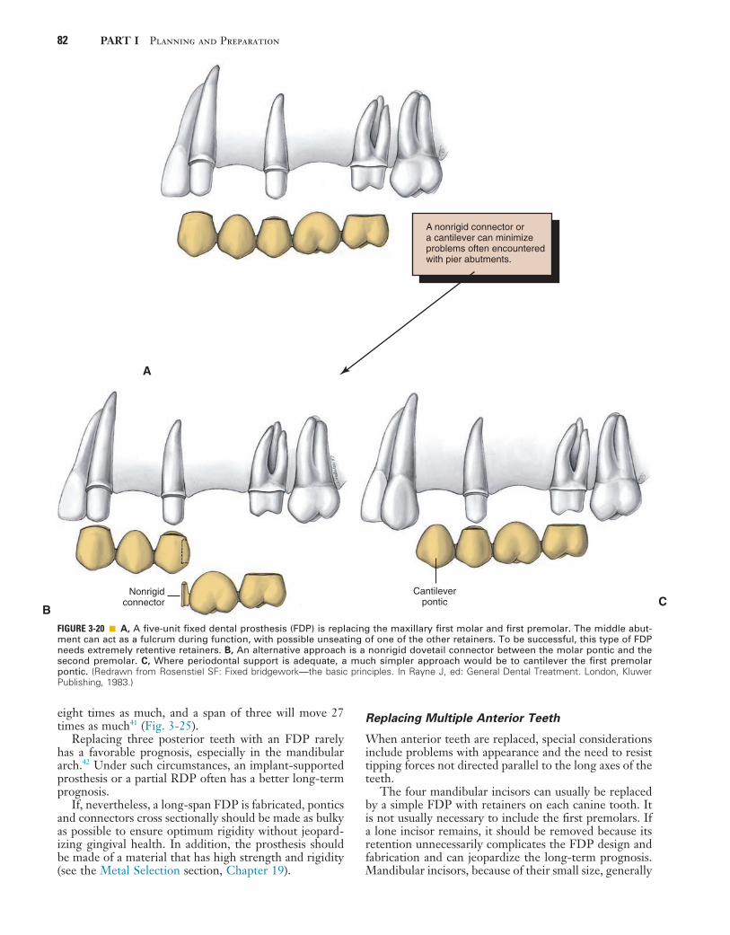
82 PART I Planning and Preparation
FIGURE 3-20 ■ A, A five-unit fixed dental prosthesis (FDP) is replacing the maxillary first molar and first premolar. The middle abut-ment can act as a fulcrum during function, with possible unseating of one of the other retainers. To be successful, this type of FDP needs extremely retentive retainers. B, An alternative approach is a nonrigid dovetail connector between the molar pontic and the second premolar. C, Where periodontal support is adequate, a much simpler approach would be to cantilever the first premolar pontic. (Redrawn from Rosenstiel SF: Fixed bridgework—the basic principles. In Rayne J, ed: General Dental Treatment. London, Kluwer Publishing, 1983.)
Nonrigidconnector
B
Cantileverpontic C
A nonrigid connector ora cantilever can minimizeproblems often encounteredwith pier abutments.
A
eight times as much, and a span of three will move 27 times as much41 (Fig. 3-25).
Replacing three posterior teeth with an FDP rarely has a favorable prognosis, especially in the mandibular arch.42 Under such circumstances, an implant-supported prosthesis or a partial RDP often has a better long-term prognosis.
If, nevertheless, a long-span FDP is fabricated, pontics and connectors cross sectionally should be made as bulky as possible to ensure optimum rigidity without jeopard-izing gingival health. In addition, the prosthesis should be made of a material that has high strength and rigidity (see the Metal Selection section, Chapter 19).
Replacing Multiple Anterior Teeth
When anterior teeth are replaced, special considerations include problems with appearance and the need to resist tipping forces not directed parallel to the long axes of the teeth.
The four mandibular incisors can usually be replaced by a simple FDP with retainers on each canine tooth. It is not usually necessary to include the first premolars. If a lone incisor remains, it should be removed because its retention unnecessarily complicates the FDP design and fabrication and can jeopardize the long-term prognosis. Mandibular incisors, because of their small size, generally

3 Treatment Planning 83
Data from Jepsen A: Root surface measurement and a method for x-ray determination of root surface area. Acta Odontol Scand 21:35, 1963.
TABLE 3-1 Root Surface Area of Abutment
Area in QuadrantRoot Surface Area (nm2)
Percentage of Root Surface
Maxillary
Central incisor 204 10Lateral incisor 179 9Canine tooth 273 14First premolar 234 12Second premolar 220 11First molar 433 22Second molar 431 22
Mandibular
Central incisor 154 8Lateral incisor 168 9Canine tooth 268 15First premolar 180 10Second premolar 207 11First molar 431 24Second molar 426 23
FIGURE 3-21 ■ To assess the support of a fixed dental prosthesis (FDP), Ante’s law proposes a relationship between the root surface areas of the missing teeth and those of the potential abutment teeth. (The numbers represent root surface area per-centages.) If the first molar (22%) and second premolar (11%) are missing, the abutments for a four-unit FDP have a slightly greater total root surface area (34%) than do the teeth being replaced. In that case, in the absence of other detrimental factors, an FDP’s prognosis is favorable. However, if the first premolar (12%) is also missing, the lost root surface area of potential abutment teeth is 45%, whereas the remaining abut-ments have only 36% of root surface area, which is much less favorable.
Ante’s law is useful fordetermining the prognosisof fixed dental prostheses.
14
1211
22 22
FIGURE 3-22 ■ A, A misaligned abutment tooth may be difficult or impossible to prepare for a fixed dental prosthesis abutment and provides poor support. B, The tilted mandibular molar was uprighted with a continuous flexible wire. C, Progress after 1 month. D, Uprighting essentially completed 2 months later. (From Proffit WR, Fields HW, Sarver DM: Contemporary Orthodontics, 5th ed. St. Louis, Mosby, 2013.)
A
C
B
D

84 PART I Planning and Preparation
FIGURE 3-23 ■ A, Because of the conical shape of most roots, the actual area of support (A) diminishes more than might be expected from the height of the bone (H). In addition, the center of rotation (R) moves apically (R′) and the lever arm (L′) increases, magnify-ing the forces on the supportive structure. B, A fixed dental prosthesis (FDP) replacing a maxillary first molar. The first premolar is an abutment providing additional stabilization for this FDP on abutment teeth with compromised bone support. (A, Redrawn from Rosenstiel SF: Fixed bridgework—the basic principles. In Rayne J, ed: General dental treatment. London, Kluwer Publishing, 1983.)
H
2/3 H
R
L
R9
A 1/2 A
L9
B
Horizontal bone loss canbe deceptive. A little canresult in a considerableloss of bone support be-cause of root morphology.
A
FIGURE 3-24 ■ A, Although this unusually long fixed dental prosthesis (FDP) provided 9 years of service, the connector between the distal pontic and the retainer eventually failed (B). C, Note the buccolingual crack through the ceramic as a result of flexure. D, Failure of a long-span FDP.
A B
C D

3 Treatment Planning 85
are poor abutment teeth. It is particularly important not to have overcontoured restorations on these teeth because plaque control may become nearly impossible. Thus, the clinician may have to make a choice among (1) compro-mised esthetics from too thin a ceramic veneer, (2) pulpal exposure during tooth preparation, and (3) selective tooth removal.
Restoration of appearance and providing support after the loss of several maxillary incisors presents a much greater challenge. Because of the curvature of the dental arch, forces directed against a maxillary incisor pontic tend to tip the abutment teeth outward. Unlike the man-dibular incisors, the maxillary incisors are not positioned in a straight line (particularly in patients with narrow or pointed dental arches). These tipping forces must be resisted by means of additional abutment teeth at each end of a long-span anterior FDP. Thus in general, when the four maxillary incisors are replaced, the canine teeth and first premolars should be used as abutment teeth.43
There may be considerable difficulty in achieving a good appearance when several maxillary incisors are replaced with an FDP. Obtaining optimal tooth contours and position for appearance and phonetics can be a chal-lenge. A diagnostic waxing procedure is extremely helpful to evaluate specific esthetic problems. As treatment progresses, an interim restoration is provided (see Chapter 15). This is used to test appearance, lip support, and phonetics. It may also be readily shaped and modified until the patient is satisfied with its appearance, after which the final restoration can be made to copy it, thereby
FIGURE 3-25 ■ The deflection of a fixed dental prosthesis is proportional to the cube of the length of its span. A, A single pontic deflects a small amount (D) when subjected to a certain force (F). B, Two pontics deflect 23 times as much (8D) to the same force. C, Three pontics deflect 33 times as much (27D).
Excessive span length isa common contraindicationfor fixed dental prostheses.
F
H
H
H
D
F
8D
F
27D
A
B
C
FIGURE 3-26 ■ Two incisors were lost in an accident. Consider-able alveolar bone has also been lost. An esthetic fixed dental prosthesis would be very difficult or impossible to fabricate without surgical ridge augmentation. (Courtesy Dr. N. Archambo.)
avoiding any embarrassing misunderstandings when the finished FDP is delivered.
If anterior bone loss has been severe, as can happen when teeth are lost as a result of trauma or periodontal disease, there may be a ridge defect (Fig. 3-26). In an affected patient, a partial RDP should be considered, especially when the patient has a high smile line, because an FDP generally replaces only the missing tooth struc-ture, not the supporting tissues. Again, an interim resto-ration may help the patient and dentist jointly determine the most appropriate treatment. A surgical ridge aug-mentation procedure44 may also be an option, although the surgical results can be somewhat unpredictable.
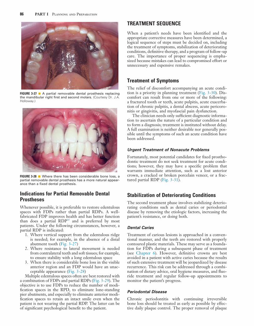
86 PART I Planning and Preparation
FIGURE 3-27 ■ A partial removable dental prosthesis replacing the mandibular right first and second molars. (Courtesy Dr. J.A. Holloway.)
FIGURE 3-28 ■ Where there has been considerable bone loss, a partial removable dental prosthesis has a more natural appear-ance than a fixed dental prosthesis.
Indications for Partial Removable Dental ProsthesesWhenever possible, it is preferable to restore edentulous spaces with FDPs rather than partial RDPs. A well-fabricated FDP improves health and has better function than does a partial RDP45 and is preferred by most patients. Under the following circumstances, however, a partial RDP is indicated:
1. Where vertical support from the edentulous ridge is needed; for example, in the absence of a distal abutment tooth (Fig. 3-27)
2. Where resistance to lateral movement is needed from contralateral teeth and soft tissues; for example, to ensure stability with a long edentulous space
3. When there is considerable bone loss in the visible anterior region and an FDP would have an unac-ceptable appearance (Fig. 3-28)
Multiple edentulous spaces often are best restored with a combination of FDPs and partial RDPs (Fig. 3-29). The objective is to use FDPs to reduce the number of modi-fication spaces in the RPD, to eliminate lone-standing pier abutments, and especially to eliminate anterior modi-fication spaces to retain an intact smile even when the patient is not wearing the partial RDP. The latter can be of significant psychological benefit to the patient.
TREATMENT SEQUENCE
When a patient’s needs have been identified and the appropriate corrective measures have been determined, a logical sequence of steps must be decided on, including the treatment of symptoms, stabilization of deteriorating conditions, definitive therapy, and a program of follow-up care. The importance of proper sequencing is empha-sized because mistakes can lead to compromised effort or unnecessary and expensive remakes.
Treatment of SymptomsThe relief of discomfort accompanying an acute condi-tion is a priority in planning treatment (Fig. 3-30). Dis-comfort can result from one or more of the following: a fractured tooth or teeth, acute pulpitis, acute exacerba-tion of chronic pulpitis, a dental abscess, acute pericoro-nitis or gingivitis, and myofascial pain dysfunction.
The clinician needs only sufficient diagnostic informa-tion to ascertain the nature of a particular condition and to form a diagnosis; treatment is instituted without delay. A full examination is neither desirable nor generally pos-sible until the symptoms of such an acute condition have been addressed.
Urgent Treatment of Nonacute Problems
Fortunately, most potential candidates for fixed prostho-dontic treatment do not seek treatment for acute condi-tions; however, they may have a specific problem that warrants immediate attention, such as a lost anterior crown, a cracked or broken porcelain veneer, or a frac-tured partial RDP (Fig. 3-31).
Stabilization of Deteriorating ConditionsThe second treatment phase involves stabilizing deterio-rating conditions such as dental caries or periodontal disease by removing the etiologic factors, increasing the patient’s resistance, or doing both.
Dental Caries
Treatment of carious lesions is approached in a conven-tional manner, and the teeth are restored with properly contoured plastic materials. These may serve as a founda-tion for FDPs during a subsequent phase of treatment (see Chapter 6). However, definitive crowns are best avoided in a patient with active caries because the results of such extensive treatment will be jeopardized by disease recurrence. This risk can be addressed through a combi-nation of dietary advice, oral hygiene measures, and fluo-ride treatment and regular follow-up appointments to monitor the patient’s progress.
Periodontal Disease
Chronic periodontitis with continuing irreversible bone loss should be treated as early as possible by effec-tive daily plaque control. The proper removal of plaque
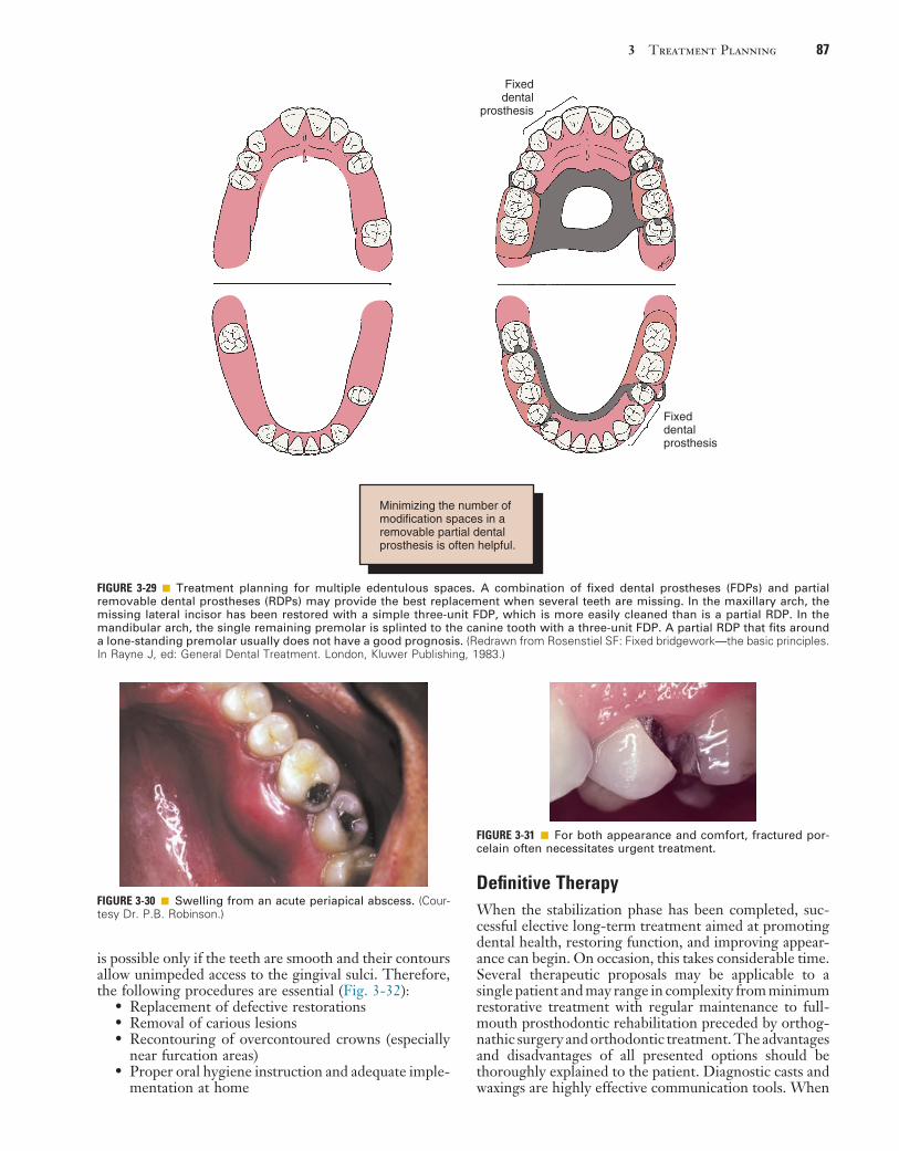
3 Treatment Planning 87
Definitive TherapyWhen the stabilization phase has been completed, suc-cessful elective long-term treatment aimed at promoting dental health, restoring function, and improving appear-ance can begin. On occasion, this takes considerable time. Several therapeutic proposals may be applicable to a single patient and may range in complexity from minimum restorative treatment with regular maintenance to full-mouth prosthodontic rehabilitation preceded by orthog-nathic surgery and orthodontic treatment. The advantages and disadvantages of all presented options should be thoroughly explained to the patient. Diagnostic casts and waxings are highly effective communication tools. When
is possible only if the teeth are smooth and their contours allow unimpeded access to the gingival sulci. Therefore, the following procedures are essential (Fig. 3-32):
• Replacement of defective restorations• Removal of carious lesions• Recontouring of overcontoured crowns (especially
near furcation areas)• Proper oral hygiene instruction and adequate imple-
mentation at home
FIGURE 3-29 ■ Treatment planning for multiple edentulous spaces. A combination of fixed dental prostheses (FDPs) and partial removable dental prostheses (RDPs) may provide the best replacement when several teeth are missing. In the maxillary arch, the missing lateral incisor has been restored with a simple three-unit FDP, which is more easily cleaned than is a partial RDP. In the mandibular arch, the single remaining premolar is splinted to the canine tooth with a three-unit FDP. A partial RDP that fits around a lone-standing premolar usually does not have a good prognosis. (Redrawn from Rosenstiel SF: Fixed bridgework—the basic principles. In Rayne J, ed: General Dental Treatment. London, Kluwer Publishing, 1983.)
Minimizing the number ofmodification spaces in aremovable partial dentalprosthesis is often helpful.
Fixeddental
prosthesis
Fixeddentalprosthesis
FIGURE 3-30 ■ Swelling from an acute periapical abscess. (Cour-tesy Dr. P.B. Robinson.)
FIGURE 3-31 ■ For both appearance and comfort, fractured por-celain often necessitates urgent treatment.
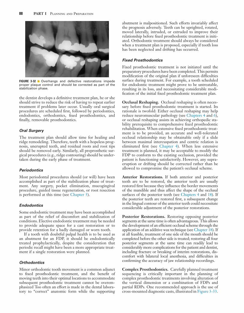
88 PART I Planning and Preparation
abutment is malpositioned. Such efforts invariably affect the prognosis adversely. Teeth can be uprighted, rotated, moved laterally, intruded, or extruded to improve their relationship before fixed prosthodontic treatment is initi-ated. Orthodontic treatment should always be considered when a treatment plan is proposed, especially if tooth loss has been neglected and drifting has occurred.
Fixed Prosthodontics
Fixed prosthodontic treatment is not initiated until the preparatory procedures have been completed. This permits modification of the original plan if unforeseen difficulties surface during treatment. For example, a tooth scheduled for endodontic treatment might prove to be untreatable, resulting in its loss, and necessitating considerable modi-fication of the initial fixed prosthodontic treatment plan.
Occlusal Reshaping. Occlusal reshaping is often neces-sary before fixed prosthodontic treatment is started. Its rationale is twofold: Either occlusal reshaping may help reduce neuromuscular pathology (see Chapters 4 and 6), or occlusal reshaping assists in achieving orthopedic sta-bility prerequisite to comprehensive fixed prosthodontic rehabilitation. When extensive fixed prosthodontic treat-ment is to be provided, an accurate and well-tolerated occlusal relationship may be obtainable only if a slide between maximal intercuspation and centric relation is eliminated first (see Chapter 4). When less extensive treatment is planned, it may be acceptable to modify the FDP to conform to the existing occlusion, provided the patient is functioning satisfactorily. However, any supra-eruption or drifting should be corrected rather than be allowed to compromise the patient’s occlusal scheme.
Anterior Restorations. If both anterior and posterior teeth are to be restored, the anterior teeth are usually restored first because they influence the border movements of the mandible and thus affect the shape of the occlusal surfaces of the posterior teeth (see Chapters 4 and 18). If the posterior teeth are restored first, a subsequent change in the lingual contour of the anterior teeth could necessitate considerable adjustment of the posterior restorations.
Posterior Restorations. Restoring opposing posterior segments at the same time is often advantageous. This allows the development of an efficient occlusal scheme through the application of an additive wax technique (see Chapter 18). If at all feasible, treatment of one side of the mouth should be completed before the other side is treated; restoring all four posterior segments at the same time can readily lead to considerably more complications for the patient and dentist, including fracture or breaking of interim restorations, dis-comfort with bilateral local anesthesia, and difficulties in confirming the accuracy of jaw relationship recordings.
Complex Prosthodontics. Carefully planned treatment sequencing is critically important in the planning of complex prosthodontic treatments involving alteration of the vertical dimension or a combination of FDPs and partial RDPs. One recommended approach is the use of cross-mounted diagnostic casts, illustrated in Figure 3-33.
the dentist develops a definitive treatment plan, he or she should strive to reduce the risk of having to repeat earlier treatment if problems later occur. Usually oral surgical procedures are scheduled first, followed by periodontics, endodontics, orthodontics, fixed prosthodontics, and finally, removable prosthodontics.
Oral Surgery
The treatment plan should allow time for healing and ridge remodeling. Therefore, teeth with a hopeless prog-nosis, unerupted teeth, and residual roots and root tips should be removed early. Similarly, all preprosthetic sur-gical procedures (e.g., ridge contouring) should be under-taken during the early phase of treatment.
Periodontics
Most periodontal procedures should (or will) have been accomplished as part of the stabilization phase of treat-ment. Any surgery, pocket elimination, mucogingival procedure, guided tissue regeneration, or root resection is performed at this time (see Chapter 5).
Endodontics
Some endodontic treatment may have been accomplished as part of the relief of discomfort and stabilization of conditions. Elective endodontic treatment may be needed to provide adequate space for a cast restoration or to provide retention for a badly damaged or worn tooth.
If a tooth with doubtful pulpal health is to be used as an abutment for an FDP, it should be endodontically treated prophylactically, despite the consideration that periodic recall might have been a more appropriate treat-ment if a single restoration were planned.
Orthodontics
Minor orthodontic tooth movement is a common adjunct to fixed prosthodontic treatment, and the benefit of moving teeth into their optimal locations to accommodate subsequent prosthodontic treatment cannot be overem-phasized Too often an effort is made in the dental labora-tory to “correct” anatomic form while the supporting
FIGURE 3-32 ■ Overhangs and defective restorations impede proper plaque control and should be corrected as part of the stabilization phase.
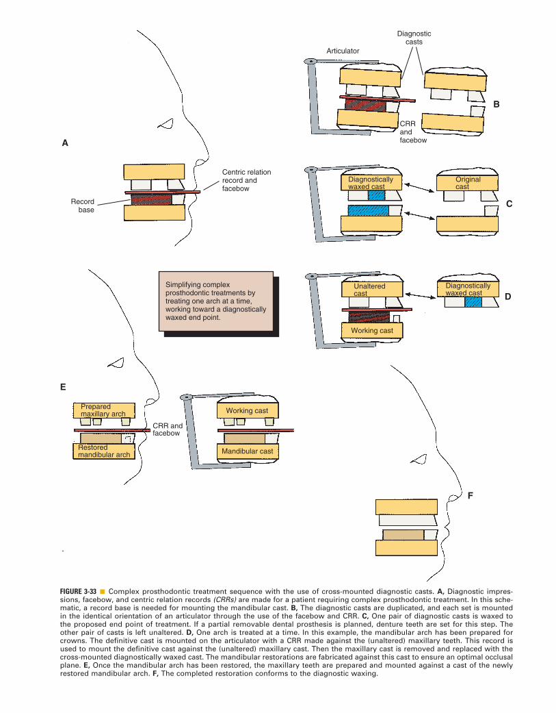
FIGURE 3-33 ■ Complex prosthodontic treatment sequence with the use of cross-mounted diagnostic casts. A, Diagnostic impres-sions, facebow, and centric relation records (CRRs) are made for a patient requiring complex prosthodontic treatment. In this sche-matic, a record base is needed for mounting the mandibular cast. B, The diagnostic casts are duplicated, and each set is mounted in the identical orientation of an articulator through the use of the facebow and CRR. C, One pair of diagnostic casts is waxed to the proposed end point of treatment. If a partial removable dental prosthesis is planned, denture teeth are set for this step. The other pair of casts is left unaltered. D, One arch is treated at a time. In this example, the mandibular arch has been prepared for crowns. The definitive cast is mounted on the articulator with a CRR made against the (unaltered) maxillary teeth. This record is used to mount the definitive cast against the (unaltered) maxillary cast. Then the maxillary cast is removed and replaced with the cross-mounted diagnostically waxed cast. The mandibular restorations are fabricated against this cast to ensure an optimal occlusal plane. E, Once the mandibular arch has been restored, the maxillary teeth are prepared and mounted against a cast of the newly restored mandibular arch. F, The completed restoration conforms to the diagnostic waxing.
Simplifying complexprosthodontic treatments bytreating one arch at a time, working toward a diagnosticallywaxed end point.
Recordbase
Centric relationrecord andfacebow
Articulator
CRRandfacebow
Originalcast
Diagnosticallywaxed cast
Diagnosticallywaxed cast
Unalteredcast
Working cast
Diagnosticcasts
Preparedmaxillary arch
Restoredmandibular arch
CRR andfacebow
Working cast
Mandibular cast
A
E
B
C
D
F
90 PART I Planning and Preparation
8. Zimmer D, et al: Survival rate of IPS-Empress 2 all-ceramic crowns and bridges: three years’ results. Schweiz Monatsschr Zahnmed 114:115, 2004.
9. Denry IL: Recent advances in ceramics for dentistry. Crit Rev Oral Biol Med 7:134, 1996.
10. Sorensen JA, et al: IPS Empress crown system: three-year clinical trial results. J Calif Dent Assoc 26:130, 1998.
11. Denry IL, et al: Effect of cubic leucite stabilization on the flexural strength of feldspathic dental porcelain. J Dent Res 75:1928, 1996.
12. Fabbri G, et al: Clinical evaluation of 860 anterior and posterior lithium disilicate restorations: retrospective study with a mean follow-up of 3 years and a maximum observational period of 6 years. Int J Periodontics Restorative Dent 34:165, 2014.
13. Dhima M, et al: Practice-based clinical evaluation of ceramic single crowns after at least five years. J Prosthet Dent 111:124, 2014.
14. Walton TR: An up to 15-year longitudinal study of 515 metal-ceramic FPDs: Part 1. Outcome. Int J Prosthodont 15:439, 2002.
15. Adell R, et al: A 15-year study of osseointegrated implants in the treatment of the edentulous jaw. Int J Oral Surg 10:387, 1981.
16. Saunders TR, et al: The maxillary complete denture opposing the mandibular bilateral distal-extension partial denture: treatment considerations. J Prosthet Dent 41:124, 1979.
17. Brewer AA, Morrow RM: Overdentures, 2nd ed. St. Louis, Mosby, 1980.
18. Sarita PTN, et al: Chewing ability of subjects with shortened dental arches. Community Dent Oral Epidemiol 31:328, 2003.
19. Nam DH, et al: Change in masticatory ability with the implant restoration of second molars. J Prosthet Dent 111:286, 2014.
20. Shugars DA, et al: Survival rates of teeth adjacent to treated and untreated posterior bounded edentulous spaces. J Am Dent Assoc 129:1089, 1998.
21. Gragg KL, et al: Movement of teeth adjacent to posterior bounded edentulous spaces. J Dent Res 80:2021, 2001.
22. Cheung GS, et al: A clinical evaluation of conventional bridge-work. J Oral Rehabil 17:131, 1990.
23. Glickman I, et al: Photoelastic analysis of internal stresses in the periodontium created by occlusal forces. J Periodontol 41:30, 1970.
24. Wright KWJ, Yettram AL: Reactive force distributions for teeth when loaded singly and when used as fixed partial denture abut-ments. J Prosthet Dent 42:411, 1979.
25. Yang HS, et al: Stress analysis of a cantilevered fixed partial denture with normal and reduced bone support. J Prosthet Dent 76:424, 1996.
26. Briggs P, et al: The single unit, single retainer, cantilever resin-bonded bridge. Br Dent J 181:373, 1996.
27. Christensen GJ: When to use fillers, build-ups or posts and cores. J Am Dent Assoc 127:1397, 1996.
28. Miller TE: Orthodontic therapy for the restorative patient. I. The biomechanic aspects. J Prosthet Dent 61:268, 1989.
29. Holmgren K, et al: The effects of an occlusal splint on the electro-myographic activities of the temporal and masseter muscles during maximal clenching in patients with a habit of nocturnal bruxism and signs and symptoms of craniomandibular disorders. J Oral Rehabil 17:447, 1990.
30. Ante IH: The fundamental principles of abutments. Mich State Dent Soc Bull 8:14, 1926.
31. Dykema RW, et al, eds: Johnston’s Modern Practice in Fixed Pros-thodontics, 4th ed, p 4. Philadelphia, WB Saunders, 1986.
32. Tylman SD, Malone WFP: Tylman’s Theory and Practice of Fixed Prosthodontics, 7th ed, p 15. St Louis, Mosby, 1978.
33. Shillingburg HT, et al: Fundamentals of Fixed Prosthodontics, 2nd ed, p 20. Chicago, Quintessence Publishing, 1981.
34. Jepsen A: Root surface measurement and a method for x-ray deter-mination of root surface area. Acta Odontol Scand 21:35, 1963.
35. Nyman S, Ericsson I: The capacity of reduced periodontal tissues to support fixed bridgework. J Clin Periodontol 9:409, 1982.
36. Freilich MA, et al: Fixed partial dentures supported by periodon-tally compromised teeth. J Prosthet Dent 65:607, 1991.
37. Decock V, et al: 18-Year longitudinal study of cantilevered fixed restorations. Int J Prosthodont 9:331, 1996.
38. Penny RE, Kraal JH: Crown-to-root ratio: its significance in restorative dentistry. J Prosthet Dent 42:34, 1979.
39. Nyman S, et al: The role of occlusion for the stability of fixed bridges in patients with reduced periodontal tissue support. J Clin Periodontol 2(2):53, 1975.
Two sets of diagnostic casts are accurately mounted so they can be precisely interchanged on the articulator. One set is prepared and waxed to the intended end point of treatment, with denture teeth inserted where partial RDPs are to be used. The waxing is carefully evaluated on the articulator in relation to occlusion and appearance. When anterior teeth are to be replaced, they can be assessed for appearance and phonetics directly in the mouth if they are mounted on a removable record base. Definitive tooth preparation starts in one arch only, so that the occlusal surfaces of the opposing arch are pre-served to act as an essential reference for mounting the definitive cast. The definitive restorations are waxed against the diagnostically waxed cast, which establishes optimal occlusion. When one arch has been completed, the opposing cast can be restored and the predicted result thus achieved.
FOLLOW-UP
A specific program of follow-up care and regular recall is an essential part of the treatment plan. Its aim is to monitor dental health, to identify newly developed signs of disease early, and to initiate corrective measures promptly as needed (see Chapter 32). Restorations do not last forever; they are subject to wear and may need replacement. Adequate follow-up care will help maintain long-term dental health.
SUMMARY
The basis of logical treatment planning consists of iden-tifying the patient’s needs, eliciting his or her expecta-tions and wishes, and comparing these with the available and feasible corrective materials and techniques. Plan-ning also involves evaluating whether a technique will yield a good prognosis. Then a rational sequence of treat-ment may be initiated for symptomatic relief, stabiliza-tion, definitive therapy, and follow-up care. The extent of treatment is modified throughout and is dictated by the patient’s attitude and ability to cooperate and by the defined goals and objectives for the specific individual.
REFERENCES1. Palomo F, Peden J: Periodontal considerations of restorative pro-
cedures. J Prosthet Dent 36:387, 1976.2. Karmaker AC, et al: Continuous fiber reinforced composite mate-
rials as alternatives for metal alloys used for dental appliances. J Biomater Appl 11:318, 1997.
3. Rosenthal L, et al: A new system for posterior restorations: a com-bination of ceramic optimized polymer and fiber-reinforced com-posite. Pract Periodontics Aesthet Dent 9(5 suppl):6, 1997.
4. Zanghellini G: Fiber-reinforced framework and Ceromer restora-tions: a technical review. Signature 4(1):1, 1997.
5. Frese C, et al: Fiber-reinforced composite fixed dental prostheses in the anterior area: A 4.5-year follow-up. J Prosthet Dent 112(2):143, 2014.
6. Claus H: Vita In-Ceram, a new procedure for preparation of oxide-ceramic crown and bridge framework. Quintessenz Zahntech 16:35, 1990.
7. Magne P, Belser U: Esthetic improvements and in vitro testing of In-Ceram Alumina and Spinell ceramic. Int J Prosthodont 10:459, 1997.
http://dentalebooks.com