Conserved and Distinct Modes of CREB/ATF Transcription Factor Regulation by PP2A/B56γ and Genotoxic...
10
Conserved and Distinct Modes of CREB/ATF Transcription Factor Regulation by PP2A/B56c and Genotoxic Stress Naval P. Shanware, Lihong Zhan, John A. Hutchinson, Sang Hwa Kim, Leah M. Williams, Randal S. Tibbetts* Department of Pharmacology, Program in Molecular and Cellular Pharmacology and Molecular and Environmental and Toxicology Center, University of Wisconsin School of Medicine and Public Health, Madison, Wisconsin, United States of America Abstract Activating transcription factor 1 (ATF1) and the closely related proteins CREB (cyclic AMP resonse element binding protein) and CREM (cyclic AMP response element modulator) constitute a subfamily of bZIP transcription factors that play critical roles in the regulation of cellular growth, metabolism, and survival. Previous studies demonstrated that CREB is phosphorylated on a cluster of conserved Ser residues, including Ser-111 and Ser-121, in response to DNA damage through the coordinated actions of the ataxia-telangiectasia-mutated (ATM) protein kinase and casein kinases 1 and 2 (CK1/2). Here, we show that DNA damage-induced phosphorylation by ATM is a general feature of CREB and ATF1. ATF1 harbors a conserved ATM/CK cluster that is constitutively and stoichiometrically phosphorylated by CK1 and CK2 in asynchronously growing cells. Exposure to DNA damage further induced ATF1 phosphorylation on Ser-51 by ATM in a manner that required prior phosphorylation of the upstream CK residues. Hyperphosphorylated ATF1 showed a 4-fold reduced affinity for CREB- binding protein. We further show that PP2A, in conjunction with its targeting subunit B56c, antagonized ATM and CK1/2- dependent phosphorylation of CREB and ATF1 in cellulo. Finally, we show that CK sites in CREB are phosphorylated during cellular growth and that phosphorylation of these residues reduces the threshold of DNA damage required for ATM- dependent phosphorylation of the inhibitory Ser-121 residue. These studies define overlapping and distinct modes of CREB and ATF1 regulation by phosphorylation that may ensure concerted changes in gene expression mediated by these factors. Citation: Shanware NP, Zhan L, Hutchinson JA, Kim SH, Williams LM, et al. (2010) Conserved and Distinct Modes of CREB/ATF Transcription Factor Regulation by PP2A/B56c and Genotoxic Stress. PLoS ONE 5(8): e12173. doi:10.1371/journal.pone.0012173 Editor: Vladimir N. Uversky, Indiana University, United States of America Received June 19, 2010; Accepted July 18, 2010; Published August 13, 2010 Copyright: ß 2010 Shanware et al. This is an open-access article distributed under the terms of the Creative Commons Attribution License, which permits unrestricted use, distribution, and reproduction in any medium, provided the original author and source are credited. Funding: This work was supported by grants from the NIH (CA124722, NS059001), The American Cancer Society, and a Shaw Scientist Award to R.S.T. from the Greater Milwaukee Foundation. N.P.S. was supported by an American Heart Association Predoctoral Fellowship. J.A.H was supported by a Molecular and Environmental Toxicology training grant (T32ES007015). The funders had no role in study design, data collection and analysis, decision to publish, or preparation of the manuscript. Competing Interests: The authors have declared that no competing interests exist. * E-mail: [email protected] Introduction Members of the CREB/ATF subfamily of bZIP transcription factors, including CREB, CREM, and ATF1 were among the first stimulus-induced transcription factors to be identified. The seminal member of this family, CREB, was identified some twenty years ago as the major nuclear binding protein of the somatostatin cyclic AMP response element (CRE), an octanucleotide palin- drome (TGACGTCA) that has been identified and functionally validated in several thousand mammalian genes [1,2,3]. CREB is now widely recognized as a critical regulator of gene expression, particularly in neuronal and metabolic contexts [4,5]. In the nervous system, CREB contributes to long-term potentiation, memory fear conditioning, circadian rhythm entrainment, and neuron survival [5]. CREB is also an important regulator of glucose homeostasis and metabolic rate in mammals [4]. CREB is activated in diabetes, and hepatic or adipocytic CREB promotes hyperglycemia and insulin resistance [4,6]. CREB has also been implicated as a proto-oncogene in acute myeloid leukemia, where it may promote cell survival and deregulation of the cell cycle [7,8]. Activation of CREB occurs through phosphorylation of Ser-133 within the kinase-inducible domain (KID) (Reviewed in Ref. [1]). Canonical activation of CREB occurs in response to cAMP, which induces PKA-dependent Ser-133 phosphorylation. The phosphor- ylation of CREB on Ser-133 promotes recruitment of the histone acetyl transferase CREB-binding protein (CBP) and coactivation of CREB target genes harboring octanucleotide CRE sequences or variants thereof [9,10]. CREB activation is also strongly dependent on the recruitment of CREB-regulated transcription co-activator (CRTC) proteins, which bind to the carboxyl-terminal bZip domain [11,12]. CRTC-dependent activation of CREB does not absolutely require phosphorylation of Ser-133; however, CRTC binding to CREB facilitates phospho-Ser-133-dependent CBP recruitment and transcriptional activation [13]. Like CREB, CRTC proteins are essential regulators of metabolism in mammals [14]. ATF1 and CREM are structurally and functionally related to CREB and mediate cyclic AMP responses as homodimers or as heterodimers with CREB [1]. ATF1 and CREM display high amino acid identity to CREB particularly within the KID and bZIP regions and are phosphorylated and activated by PKA on conserved sites analogous to Ser-133 in CREB [1,15]. Whereas CREB and ATF1 are expressed throughout development, and in most if not all tissues in the adult, the expression of CREM occurs in a more tissue-restricted fashion, with highest expression PLoS ONE | www.plosone.org 1 August 2010 | Volume 5 | Issue 8 | e12173
-
Upload
independent -
Category
Documents
-
view
2 -
download
0
Transcript of Conserved and Distinct Modes of CREB/ATF Transcription Factor Regulation by PP2A/B56γ and Genotoxic...

Conserved and Distinct Modes of CREB/ATF TranscriptionFactor Regulation by PP2A/B56c and Genotoxic StressNaval P. Shanware, Lihong Zhan, John A. Hutchinson, Sang Hwa Kim, Leah M. Williams, Randal S.
Tibbetts*
Department of Pharmacology, Program in Molecular and Cellular Pharmacology and Molecular and Environmental and Toxicology Center, University of Wisconsin School
of Medicine and Public Health, Madison, Wisconsin, United States of America
Abstract
Activating transcription factor 1 (ATF1) and the closely related proteins CREB (cyclic AMP resonse element binding protein)and CREM (cyclic AMP response element modulator) constitute a subfamily of bZIP transcription factors that play criticalroles in the regulation of cellular growth, metabolism, and survival. Previous studies demonstrated that CREB isphosphorylated on a cluster of conserved Ser residues, including Ser-111 and Ser-121, in response to DNA damage throughthe coordinated actions of the ataxia-telangiectasia-mutated (ATM) protein kinase and casein kinases 1 and 2 (CK1/2). Here,we show that DNA damage-induced phosphorylation by ATM is a general feature of CREB and ATF1. ATF1 harbors aconserved ATM/CK cluster that is constitutively and stoichiometrically phosphorylated by CK1 and CK2 in asynchronouslygrowing cells. Exposure to DNA damage further induced ATF1 phosphorylation on Ser-51 by ATM in a manner that requiredprior phosphorylation of the upstream CK residues. Hyperphosphorylated ATF1 showed a 4-fold reduced affinity for CREB-binding protein. We further show that PP2A, in conjunction with its targeting subunit B56c, antagonized ATM and CK1/2-dependent phosphorylation of CREB and ATF1 in cellulo. Finally, we show that CK sites in CREB are phosphorylated duringcellular growth and that phosphorylation of these residues reduces the threshold of DNA damage required for ATM-dependent phosphorylation of the inhibitory Ser-121 residue. These studies define overlapping and distinct modes of CREBand ATF1 regulation by phosphorylation that may ensure concerted changes in gene expression mediated by these factors.
Citation: Shanware NP, Zhan L, Hutchinson JA, Kim SH, Williams LM, et al. (2010) Conserved and Distinct Modes of CREB/ATF Transcription Factor Regulation byPP2A/B56c and Genotoxic Stress. PLoS ONE 5(8): e12173. doi:10.1371/journal.pone.0012173
Editor: Vladimir N. Uversky, Indiana University, United States of America
Received June 19, 2010; Accepted July 18, 2010; Published August 13, 2010
Copyright: � 2010 Shanware et al. This is an open-access article distributed under the terms of the Creative Commons Attribution License, which permitsunrestricted use, distribution, and reproduction in any medium, provided the original author and source are credited.
Funding: This work was supported by grants from the NIH (CA124722, NS059001), The American Cancer Society, and a Shaw Scientist Award to R.S.T. from theGreater Milwaukee Foundation. N.P.S. was supported by an American Heart Association Predoctoral Fellowship. J.A.H was supported by a Molecular andEnvironmental Toxicology training grant (T32ES007015). The funders had no role in study design, data collection and analysis, decision to publish, or preparationof the manuscript.
Competing Interests: The authors have declared that no competing interests exist.
* E-mail: [email protected]
Introduction
Members of the CREB/ATF subfamily of bZIP transcription
factors, including CREB, CREM, and ATF1 were among the first
stimulus-induced transcription factors to be identified. The
seminal member of this family, CREB, was identified some twenty
years ago as the major nuclear binding protein of the somatostatin
cyclic AMP response element (CRE), an octanucleotide palin-
drome (TGACGTCA) that has been identified and functionally
validated in several thousand mammalian genes [1,2,3]. CREB is
now widely recognized as a critical regulator of gene expression,
particularly in neuronal and metabolic contexts [4,5]. In the
nervous system, CREB contributes to long-term potentiation,
memory fear conditioning, circadian rhythm entrainment, and
neuron survival [5]. CREB is also an important regulator of
glucose homeostasis and metabolic rate in mammals [4]. CREB is
activated in diabetes, and hepatic or adipocytic CREB promotes
hyperglycemia and insulin resistance [4,6]. CREB has also been
implicated as a proto-oncogene in acute myeloid leukemia, where
it may promote cell survival and deregulation of the cell cycle
[7,8].
Activation of CREB occurs through phosphorylation of Ser-133
within the kinase-inducible domain (KID) (Reviewed in Ref. [1]).
Canonical activation of CREB occurs in response to cAMP, which
induces PKA-dependent Ser-133 phosphorylation. The phosphor-
ylation of CREB on Ser-133 promotes recruitment of the histone
acetyl transferase CREB-binding protein (CBP) and coactivation
of CREB target genes harboring octanucleotide CRE sequences or
variants thereof [9,10]. CREB activation is also strongly
dependent on the recruitment of CREB-regulated transcription
co-activator (CRTC) proteins, which bind to the carboxyl-terminal
bZip domain [11,12]. CRTC-dependent activation of CREB does
not absolutely require phosphorylation of Ser-133; however,
CRTC binding to CREB facilitates phospho-Ser-133-dependent
CBP recruitment and transcriptional activation [13]. Like CREB,
CRTC proteins are essential regulators of metabolism in mammals
[14].
ATF1 and CREM are structurally and functionally related to
CREB and mediate cyclic AMP responses as homodimers or as
heterodimers with CREB [1]. ATF1 and CREM display high
amino acid identity to CREB particularly within the KID and
bZIP regions and are phosphorylated and activated by PKA on
conserved sites analogous to Ser-133 in CREB [1,15]. Whereas
CREB and ATF1 are expressed throughout development, and in
most if not all tissues in the adult, the expression of CREM occurs
in a more tissue-restricted fashion, with highest expression
PLoS ONE | www.plosone.org 1 August 2010 | Volume 5 | Issue 8 | e12173

observed in testis [16]. CREB-deficient mice die perinatally from
respiratory insufficiency while ATF1-null mice are phenotypically
normal [17]. ATF1 heterozygosity on a CREB-null background,
however, causes early embryonic lethality [17]. Thus, CREB
appears to be responsible for the bulk of essential functions carried
out by CREB/ATF family members; however, the activities of
ATF1 and CREM are not fully redundant with those of CREB.
Previous work has demonstrated that, in addition to its
regulation by metabolic and growth signals, CREB is a target of
the DNA damage response [18,19,20]. CREB is directly
phosphorylated by the ATM protein kinase on Ser-111 in
response to ionizing radiation (IR) and other types of genotoxic
stimuli [18,20]. The phosphorylation of CREB on Ser-111 primes
phosphorylation of Ser-108, Ser-114, and Ser-117 by CK1 and
CK2, which is required for additional ATM-dependent phos-
phorylation of Ser-121 [19]. All told, ATM, CK1, and CK2
phosphorylate five sites within the KID in response to DNA
damage [19]. A net result of ATM/CK cluster phosphorylation is
a decrease in the binding affinity between the KID domain of
CREB and the KID-interacting (KIX) domain of CBP [19,20].
However, although the ATM/CK cluster is positionally conserved
in Drosophila CREB orthologs, its physiologic functions have not
been elucidated. It is also unclear whether DNA damage-
dependent phosphorylation is unique to CREB or represents a
general mechanism of CREB/ATF regulation.
In this study we compared phosphorylation mechanisms of
CREB and ATF1 both in the absence and presence of DNA
damage. We show that ATM phosphorylates ATF1 in response to
DNA damage on Ser-51, which is analogous to the Ser-121
phosphorylation site in CREB that inhibits CBP binding, and that
the PP2A/B56c phosphatase complex antagonizes DNA damage-
induced phosphorylation of both proteins. Although these aspects
of CREB and ATF1 phosphorylation are shared, the mechanisms
and extent of DNA damage-independent phosphorylation of CK
residues is divergent. We show that DNA damage-independent
phosphorylation of CREB is induced during cellular growth and
reduces the threshold of DNA damage required for subsequent
IR-induced phosphorylation by ATM. Our findings thus provide
greater insights into CREB/ATF1 regulation and suggest that
DNA damage signaling input into these structurally related
proteins is evolutionarily conserved.
Results
ATF1 is hyperphosphorylated in asynchronously growingcells
We have previously described a complex phosphorylation
cascade involving interplay between ATM, CK1 and CK2 in
the genotoxic stress-induced phosphorylation of CREB [19]. The
end consequence of this cascade is the phosphorylation of five
clustered.
Ser residues: Ser-108, Ser-111, Ser-114, Ser-117, and Ser-121
(designated the ATM/CK cluster) within the amino-terminal
region of the CREB KID. Although the functional consequences
of CREB phosphorylation are not well understood, evidence
suggested that ATM/CK cluster phosphorylation antagonized
CREB-CBP interaction in vitro [19]. A sequence comparison of the
KID region of the CREB, ATF1, and CREM shows strong
positional conservation of the CK sites in both CREM and ATF1;
however, only ATF1 showed co-conservation of the Ser-Gln
dipeptide motifs in CREB that are phosphorylated by ATM in vitro
and in intact cells (Fig. 1A and [21]). Based on this homology, we
sought to test if ATF1 possessed a functional ATM/CK cluster
that was a target of the DNA damage response.
CREB undergoes a rapid ATM dependent and phosphatase-
sensitive electrophoretic mobility shift on SDS-PAGE gels
following cellular exposure to IR [19,20]. ATF1, however,
migrated as a single band of 35 kDa on SDS-PAGE gels that
was not affected by IR (Fig. 1B). Nevertheless, phosphatase
treatment of cell extracts collapsed the major 35 kDa ATF1 band
to a species with an approximate molecular mass of 27 kDa
(Fig. 1B). This finding indicated that ATF1 was stoichiometrically
hyperphosphorylated in asynchronously growing cells and that IR
did not induce a detectable ATF1 electrophoretic mobility shift.
Constitutive hyperphosphorylation of ATF1 was also observed in
several other cell lines and primary mouse tissues from the spleen
and thymus (Fig. S1A).
CK sites regulate ATF1 hyperphosphorylationWe tested whether ATF1 hyperphosphorylation required the
conserved CK sites. Of four candidate sites mutated to Ala (Ser-
36, Ser-38, Ser-41, and Ser-44), Ser-36 and Ser-41 caused a
change in ATF1 electrophoretic mobility; a fraction of each
mutant migrated faster than the wild-type protein (Fig. 1C).
Combined mutation of Ser-36 and Ser-41, in the ATF1S36/41A
mutant totally abolished the ATF1 electrophoretic mobility shift
(Fig. 1D). This finding suggested that Ser-36 and Ser-41 within the
putative ATM/CK cluster were phosphorylated in HEK 293T
cells.
To demonstrate site-specific phosphorylation of ATF1 in vivo, we
attempted and failed to generate a phospho-specific antibody that
would recognize ATF1 phosphorylated on Ser-36, Ser-38, and
Ser-41. As an alternative approach, we exploited the strong
conservation between CREB and ATF1 in the KID region and
constructed an ATF1D39E,E41D mutant that rendered the ATF1
ATM/CK cluster identical to its CREB counterpart, which was
readily detected using an a-pCREB-108/111/114 CREB anti-
body (Fig. S1B). These data suggested that ATF1 is always
hyperphosphorylated on the CK sites in vivo. Also, the antibody
reactivity suggested that in addition to Ser-36 and Ser-41, Ser-38
and Ser-44 were phosphorylated in vivo.
The ATF1 KID harbors a Ser-X-X-Ser-X-X–Ser motif making
it a strong candidate for sequential phosphorylation by CK1 and
CK2 (Fig. 1A and [22,23]). To test this idea, we treated HEK
293T cells with the CK1 inhibitor D4476 [24] or the CK2-specific
inhibitor TBB [25] and examined ATF1 electrophoretic mobility.
Treatment with D4476 or TBB alone had no effect on ATF1
electrophoretic mobility (Fig. 1E). However, combined treatment
with both inhibitors caused ATF1 to migrate as the hypopho-
sphorylated species on SDS-PAGE gels (Fig. 1E). These findings
suggest that the activity of CK1 or CK2 is sufficient to maintain
ATF1 in a hyperphosphorylated state in HEK 293T cells.
DNA damage-induced phosphorylation of ATF1While our previous data showed that ATF1 was stoichiomet-
rically phosphorylated on CK sites in vivo, it was still not clear if
ATF1 is a target of the DNA damage response. Ser-51 is
positionally analogous to Ser-121 in CREB, which undergoes
ATM-dependent phosphorylation in response to DNA damage
(Fig. 1A). To test if Ser-51 is a target of the DNA damage in vivo,
we generated an antibody against a peptide containing phos-
phorylated Ser-47, Ser-50 and Ser-51, using the rationale that
Ser-47 and Ser-50 are likely phosphorylated by CK1 and CK2
(Fig. 2A). The a-pATF1-47/50/51 antibody was first tested
against HEK 293T cell extract overexpressing Myc-tagged
ATF1. The antibody displayed strong phosphatase-sensitive
reactivity with Myc-ATF1 in HEK 293T cells (Fig. 2B). We
further tested the specificity and IR-inducibility of the a-pATF1-
Regulation of CREB/ATFs
PLoS ONE | www.plosone.org 2 August 2010 | Volume 5 | Issue 8 | e12173

47/50/51 antibody by assessing the effects of single SerRAla
substitutions at Ser-50 and Ser-51 on basal and IR-induced
immunoreactivity. Whereas wild-type Myc-ATF1 exhibited
modest IR-induced phosphorylation, Myc-ATF1 harboring Ala
substitutions at Ser-50- or Ser-51- failed to react with a-pATF1-
47/50/51, indicating that the integrity of these sites was required
for antibody recognition (Fig. 2C).
We next tested the ATM-dependence of IR-induced ATF1
phosphorylation. Preincubation of HEK 293T cells with the ATM
inhibitor KU-55933 [26] completely blocked the IR-induced
phosphorylation of ATF1 on Ser-47/50/51, establishing that the
IR-induced phosphorylation of this motif required ATM (Fig. 2D).
Finally, we tested whether ATF1 phosphorylation on Ser-47/50/
51 required priming phosphorylation of the upstream CK sites.
HEK 293T cells overexpressing either Myc-ATF1WT or Myc-
ATF1S36/41A vectors were mock irradiated or exposed to IR.
Immunoblotting analyses with the a-pATF1-47/50/51 antibody
showed that the Myc-ATF1S36/41A mutant was completely
defective for Ser-47/50/51 phosphorylation (Fig. 2E). Thus
ATF1 hyperphosphorylation on upstream CK residues is a
prerequisite for DNA damage-induced phosphorylation of Ser-
47/50/51.
ATF1 hyperphosphorylation regulates the ATF1-KIXcomplex
DNA damage-induced phosphorylation of CREB antagonized
its association with the CBP KIX domain [19,20]. To test the
effects of ATF1 hyperphosphorylation on its KIX binding activity,
we transfected HEK 293T cells with Myc-ATF1WT or Myc-
ATF1S36/41A vectors, exposed the cells to IR, and performed
ATF1 pull-down assays using the GST-KIX fusion protein.
ATF1WT-KIX binding was only marginally reduced on exposure
to IR (Fig. 2F). The ATF1S36/41A mutant showed greater than
four-fold higher binding affinity for the KIX domain when
compared to wild-type ATF1, and the ATF1S36/41A-KIX
interaction was completely resistant to the effects of IR (Fig. 2F).
These data suggest that ATF1 hyperphosphorylation negatively
regulates CBP binding.
Figure 1. ATF1 is constitutively phosphorylated by CK1/CK2 in vivo. (A) Sequence overlay of ATM/CK cluster regions in CREB, CREM andATF1. Homologous putative phosphorylation sites are shown in boldface and defined phosphorylation sites in CREB underlined. (B) ATF1 is basallyphosphorylated in intact cells. HEK 293T cells were exposed to IR (10 Gy) or left untreated and cell extracts were prepared and treated with lphosphatase (with or without inhibitors) prior to analysis by SDS-PAGE followed by immunoblotting with a-bZIP antibody that recognizes CREB andATF1. The * denotes the position of a cross-reactive protein. (C) Phosphorylation site requirements for ATF1 electrophoretic mobility shift. HEK 293Tcells were transfected with plasmids encoding Myc-ATF1WT or the indicated Myc-ATF1 phosphorylation site mutants. Cell extracts were then madeand analyzed by immunoblotting using a-Myc antibody. (D) The ATF1S36/41A mimics in vitro dephosphorylated ATF1. HEK 293T cells were transfectedwith plasmids encoding Myc-ATF1WT or the Myc-ATF1S36/41A mutant. Cell extracts were prepared and treated with l phosphatase prior to analysis byimmunoblotting using a-Myc antibodies. (E) CK1 and CK2 inhibitors dephosphorylate ATF1. HEK 293T cells were treated with 75 mM D4476, 50 mMTBB or both compounds for 4 h. Cell extracts were then analyzed by immunoblotting using a-ATF1, a-CREB and a-pCREB-108/111/114 antibodies.doi:10.1371/journal.pone.0012173.g001
Regulation of CREB/ATFs
PLoS ONE | www.plosone.org 3 August 2010 | Volume 5 | Issue 8 | e12173

B56c-PP2A antagonizes CK and ATM-dependent CREBphosphorylation
The above findings indicated that the vast preponderance of
ATF1 molecules in intact cells are constitutively phosphorylated by
CK1/CK2 within the ATM/CK cluster and that Ser-51 is a DNA
damage-inducible site that is positionally analogous to the Ser-121
residue in CREB. To further explore the dynamic regulation of
these residues, we sought to identify the relevant cellular
phosphatases. Okadaic acid (OA) can be used to distinguish toxin-
sensitive (PP1, PP2A and PP5) from toxin-insensitive (PP2B/
calcineurin and PP7) phosphatases [27,28]. Additionally, PP2A is
extremely sensitive to OA inhibition and can be pharmacologically
distinguished from less sensitive PP1 and PP5 [27]. As seen in
Fig. 3A, both 10 nM OA treatment and 100 nM OA treatment led
to elevated IR-induced CREB phosphorylation on both Ser-108/
111/114 and Ser-121 in HEK 293T cells. In the absence of IR,
100 nM OA induced the phosphorylation of Ser-108/111/114, but
not Ser-121, which is consistent with the idea that Ser-121 is strictly
a DNA damage-inducible site. Neither overexpression of dominant-
negative PP1 nor knockdown of PP5 affected basal or IR-induced
CREB phosphorylation (data not shown). However, HEK 293T
cells stably transfected with a shRNA construct directed against the
PP2A catalytic subunit (PP2Ac) showed enhanced IR-induced
phosphorylation on both Ser-108/111/114 and Ser-121, despite
the partial nature of PP2A protein reduction in these cells (Fig. 3B).
Thus, RNAi and inhibitor data suggested a role for the PP2A family
of phosphatases in the regulation of CREB ATM/CK cluster
dephosphorylation.
The PP2A catalytic subunit is targeted to diverse substrates via
interaction with specificity-determining (B) subunits [29]. The
B56c B subunit has recently been implicated in the DNA damage
response by regulating p53 phosphorylation [30,31]. We evaluated
Figure 2. DNA damage induces ATM dependent phosphorylation of ATF1 on Ser-51. (A) Sequence of peptide antigen used to generate a-pATF1-47/50/51 antibody. (B) Phosphatase sensitivity of a-pATF1-47/50/51 antibody. HEK 293T cells were transfected with Myc-ATF1WT plasmid. Cellextracts were prepared and treated with l phosphatase prior to analysis by immunoblotting using a-Myc and a-pATF1-47/50/51 antibodies. (C)Phosphorylation site requirements and IR inducibility. HEK 293T cells were transfected with vector DNA (2), Myc-ATF1WT, Myc-ATF1S50A or Myc-ATF1S51A plasmids and exposed to IR (10 Gy, 2 h). Cell extracts were prepared and analyzed by immunoblotting using a-Myc and a-pATF1-47/50/51antibodies. (D) IR dependent ATF1 phosphorylation is ATM dependent. HEK 293T cells either left untreated or treated with IR in the presence of10 mM ATM inhibitor (KU-55933). Immunoprecipitation reactions were performed using a mock antibody or a-ATF1 antibody andimmunoprecipitates were analyzed by immunoblotting using a-Myc and a-pATF1-47/50/51 antibodies. (E) The ATF1S36/41A mutant is defective forIR induced pATF1-47/50/51 phosphorylation. HEK 293T cells were transfected with plasmid DNA encoding Myc-ATF1WT or the Myc-ATF1S36/41A
mutant and either left untreated or subjected to 10 Gy IR for 2 h. Cell extracts were prepared and analyzed by immunoblotting using a-Myc and a-pATF1-47/50/51 antibodies. (F) Hyperphosphorylated ATF1 shows reduced binding to the KIX domain of CBP. HEK 293T cells were transfected withplasmids encoding Myc-ATF1WT or the Myc-ATF1S36/41A mutant and either left untreated or subjected to 10 Gy IR. Cell extracts were prepared 2 hlater, incubated with GST-KIX-loaded beads, and bound protein analyzed by immunoblotting using a-Myc antibodies. Numbers under GST-KIX resultdemote fold changes in ATF1 levels.doi:10.1371/journal.pone.0012173.g002
Regulation of CREB/ATFs
PLoS ONE | www.plosone.org 4 August 2010 | Volume 5 | Issue 8 | e12173
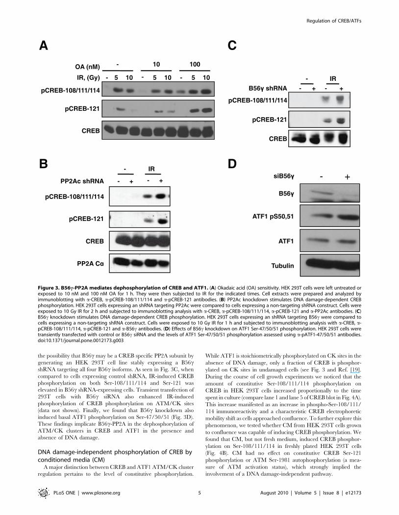
the possibility that B56c may be a CREB specific PP2A subunit by
generating an HEK 293T cell line stably expressing a B56cshRNA targeting all four B56c isoforms. As seen in Fig. 3C, when
compared to cells expressing control shRNA, IR-induced CREB
phosphorylation on both Ser-108/111/114 and Ser-121 was
elevated in B56c shRNA-expressing cells. Transient transfection of
293T cells with B56c siRNA also enhanced IR-induced
phosphorylation of CREB phosphorylation on ATM/CK sites
(data not shown). Finally, we found that B56c knockdown also
induced basal ATF1 phosphorylation on Ser-47/50/51 (Fig. 3D).
These findings implicate B56c-PP2A in the dephosphorylation of
ATM/CK clusters in CREB and ATF1 in the presence and
absence of DNA damage.
DNA damage-independent phosphorylation of CREB byconditioned media (CM)
A major distinction between CREB and ATF1 ATM/CK cluster
regulation pertains to the level of constitutive phosphorylation.
While ATF1 is stoichiometrically phosphorylated on CK sites in the
absence of DNA damage, only a fraction of CREB is phosphor-
ylated on CK sites in undamaged cells (see Fig. 3 and Ref. [19].
During the course of cell growth experiments we noticed that the
amount of constitutive Ser-108/111/114 phosphorylation on
CREB in HEK 293T cells increased proportionally to the time
spent in culture (compare lane 1 and lane 5 of CREB blot in Fig. 4A).
This increase manifested as an increase in phospho-Ser-108/111/
114 immunoreactivity and a characteristic CREB electrophoretic
mobility shift as cells approached confluence. To further explore this
phenomenon, we tested whether CM from HEK 293T cells grown
to confluence was capable of inducing CREB phosphorylation. We
found that CM, but not fresh medium, induced CREB phosphor-
ylation on Ser-108/111/114 in freshly plated HEK 293T cells
(Fig. 4B). CM had no effect on constitutive CREB Ser-121
phosphorylation or ATM Ser-1981 autophosphorylation (a mea-
sure of ATM activation status), which strongly implied the
involvement of a DNA damage-independent pathway.
Figure 3. B56c-PP2A mediates dephosphorylation of CREB and ATF1. (A) Okadaic acid (OA) sensitivity. HEK 293T cells were left untreated orexposed to 10 nM and 100 nM OA for 1 h. They were then subjected to IR for the indicated times. Cell extracts were prepared and analyzed byimmunoblotting with a-CREB, a-pCREB-108/111/114 and a-pCREB-121 antibodies. (B) PP2Ac knockdown stimulates DNA damage-dependent CREBphosphorylation. HEK 293T cells expressing an shRNA targeting PP2Ac were compared to cells expressing a non-targeting shRNA construct. Cells wereexposed to 10 Gy IR for 2 h and subjected to immunoblotting analysis with a-CREB, a-pCREB-108/111/114, a-pCREB-121 and a-PP2Ac antibodies. (C)B56c knockdown stimulates DNA damage-dependent CREB phosphorylation. HEK 293T cells expressing an shRNA targeting B56c were compared tocells expressing a non-targeting shRNA construct. Cells were exposed to 10 Gy IR for 1 h and subjected to immunoblotting analysis with a-CREB, a-pCREB-108/111/114, a-pCREB-121 and a-B56c antibodies. (D) Effects of B56c knockdown on ATF1 Ser-47/50/51 phosphorylation. HEK 293T cells weretransiently transfected with control or B56c siRNA and the levels of ATF1 Ser-47/50/51 phosphorylation assessed using a-pATF1-47/50/51 antibodies.doi:10.1371/journal.pone.0012173.g003
Regulation of CREB/ATFs
PLoS ONE | www.plosone.org 5 August 2010 | Volume 5 | Issue 8 | e12173

We used pharmacologic inhibitors to begin probing mecha-
nisms DNA damage-independent CREB phosphorylation Nei-
ther PI3 kinase nor MEK inhibitors had any effect on CM-
induced CREB phosphorylation in HEK 293T cells (data not
shown), suggesting that these mitogenic pathways are not
involved. On the other hand, preincubation of HEK 293T cells
with the proteasome inhibitor MG-132 abolished CM induced
CREB phosphorylation on Ser-108/111/114, suggesting a
critical role for proteasome-mediated protein degradation
(Fig. 4C). This finding is consistent with our previously published
observations showing CREB phosphorylation was induced by the
protein synthesis inhibitor, cycloheximide [32]. Although the
pathways controlling CM-induced CREB phosphorylation are
not clear, these findings suggest that the phosphorylation of these
sites is dynamically regulated during cell growth in response to
secreted factors.
Our previous studies showed that phosphorylation of CREB on
Ser-108/111/114 was required for DNA damage-induced phos-
phorylation of CREB on Ser-121 [19]. It follows that increased
phosphorylation of these residues in response to CM would
potentiate DNA damage-induced phosphorylation of CREB on
Ser-121 at low doses of IR. Consistent with this idea, we found
that pretreatment of HEK 293T cells with CM for 6 h increased
the phosphorylation of CREB in response to 1 Gy IR (Fig. 4D).
These findings suggest that cellular growth status is a determinant
of IR-induced CREB Ser-121 phosphorylation.
CREB and ATF1 regulate ATM mRNA expressionThe ATM promoter contains positionally conserved CRE
elements suggesting the interesting possibility that ATM is
regulated by its own substrates (Fig. 5A and Ref. [2,33]. To test
if ATM is a direct target of CREB/ATF1 we transfected human
MeWo melanoma cells with siRNA directed against CREB and
ATF1 either individually or in combination. While ATF1
knockdown and CREB knockdown significantly reduced ATM
mRNA levels, a double knockdown caused an almost 10-fold
reduction in ATM mRNA levels (Figs. 5B and C). ATM protein
levels were also significantly reduced in CREB/ATF1 knockdown
cells (Fig. 5D). However, in no case did we observe effects of DNA
damage on ATM mRNA or protein levels, indicating that
although CREB and ATF1 contribute to constitutive ATM
expression, it is unlikely that ATM-mediated phosphorylation of
these factors modulates ATM promoter activity during DNA
damage.
Figure 4. ATM and DNA damage-independent phosphorylation of the CREB ATM/CK cluster during cell growth. (A) CREBphosphorylation increases with time spent in culture. Replicate plates of HEK 293T cells were plated at 60–70% confluence and allowed to grow for14 h. Cells were then harvested at 2 h intervals and the extracts analyzed by immunoblotting with a-CREB and a-pCREB-108/111/114 antibodies. (B)Conditioned media (CM) induces CREB phosphorylation independent of DNA damage. HEK 293T cells were plated overnight followed by exposure tofresh media (FM), or CM. A 10 Gy IR exposure (2 h) was used as a positive control to induce Ser-121 phosphorylation. Cell extracts were prepared atthe indicated times and analyzed by immunoblotting with a-CREB, a-pCREB-108/111/114, a-pCREB-121, a-ATM and a- pATM-1981 antibodies. (C)MG-132 suppresses CM-induced CREB phosphorylation. HEK 293T cells were cultured in the presence of CM or CM supplemented with 10 mM MG132(or solvent). Cell extracts were prepared at the indicated times after CM treatment and analyzed by immunoblotting with a-CREB and a-pCREB-108/111/114 antibodies. (D) CM-induced phosphorylation of CREB on Ser-111 facilitates IR-induced phosphorylation of Ser-121. HEK 293T cells wereincubated with CM or not for 8 h and then mock irradiated or exposed to 1 Gy of IR. Cell extracts were analyzed by immunoblotting with a-CREB, a-pCREB-108/111/114 and a-pCREB-121 antibodies.doi:10.1371/journal.pone.0012173.g004
Regulation of CREB/ATFs
PLoS ONE | www.plosone.org 6 August 2010 | Volume 5 | Issue 8 | e12173

Discussion
In this study, we compared the DNA damage-dependent and
DNA damage-independent phosphorylation of CREB and ATF1
on an evolutionarily conserved ATM/CK phosphorylation cluster
within their respective KIDs. The principle findings of the study
include: (i) ATF1 is phosphorylated by ATM on Ser-51 in response
to DNA damage in a manner that requires phosphorylation of
upstream residues by CK1/CK2; (ii) ATF1 hyperphosphorylation
on CK residues inhibits CBP binding; (iii) B56c/PP2A antagonizes
CREB and ATF1 phosphorylation on CK/ATM sites; and (iv)
priming phosphorylation of CREB on CK sites during cell growth
reduces the DNA damage threshold required for ATM-dependent
phosphorylation of Ser-121. These findings illustrate conserved
similarities as well as differences in the phosphoregulation of CREB
and ATF1 and point toward the existence of a novel pathway
regulating CREB phosphorylation status during cell growth.
CK-dependent phosphorylation of ATF1 within its KID,
including Ser-36 and Ser-41, was reported by Masson et al, but
the functions of these sites has been elusive [34]. Our studies show
that virtually all ATF1 molecules are phosphorylated on the CK
sites in asynchronously growing cells and that dual inhibition of
CK1 and CK2 is required for their dephosphorylation (Fig. 1E).
By contrast, positionally conserved CK sites in CREB are
constitutively phosphorylated to a much smaller degree (Fig. 1E
and Ref. [20]. Although the CK clusters are virtually identical
between the two proteins (Fig. 1A), ATF1 contains three extra Ser
residues and may be a preferred substrate for CK1 and CK2.
Although the functional impact of CK-mediated ATF1
phosphorylation is still unclear, we found that mutation of Ser-
36 and Ser-41 increased CBP KIX domain binding by up to four
fold (Fig. 2G). This result is consistent with the negative impact of
CK-mediated phosphorylation on CBP binding affinity of CREB
that we previously reported [20]. On the other hand, IR-induced
Figure 5. CREB/ATF1 regulate ATM transcription. (A) Schematic of the human ATM with putative CREs is shown. CREs conserved betweenhuman and mouse ATM promoters are in boldface. The arrow indicates the transcription start site. (B) Knockdown of CREB and ATF1 in MeWo cells.MeWo cells were transfected with shRNA for ATF1, siRNA for CREB or both and compared to cells expressing a non-targeting construct for 48 h. Cellextracts were subjected to immunoblotting with a-CREB, a-ATF1 and a-b-Tubulin antibodies. (C) Real-time PCR analysis of ATM mRNA in CREB/ATF1-deficient MeWo cells. Relative fold-expression levels of ATM mRNA normalized for GAPDH is shown. Error bars denote standard deviation from themean from three independent experiments. (D) Effects of CREB and ATF1 knockdown on ATM protein levels. MeWo cells were transfected withshRNA for ATF1, siRNA for CREB or both and compared to cells expressing a non-targeting construct for 48 h before IR exposure. Cell extracts wereprepared and subjected to immunoblotting with a-CREB, a-ATF1, a-ATM and a-b-Tubulin antibodies.doi:10.1371/journal.pone.0012173.g005
Regulation of CREB/ATFs
PLoS ONE | www.plosone.org 7 August 2010 | Volume 5 | Issue 8 | e12173

phosphorylation of ATF1 on Ser-51 did not further reduce CBP-
binding affinity (Fig. 2F), which contrasts with the inhibitory effects
of IR-induced phosphorylation on CREB-CBP complex assembly
[19]. To accommodate these findings, we propose that constitutive
hyperphosphorylation by CK1/CK2 maintains ATF1 in an
inactive state that promotes transcriptional repression. This
conclusion is consistent with early studies showing that ATF1 is
only weakly activated by cAMP [35] and more recent reports
implicating ATF1 as a gene-specific repressor of transcription
[36,37]. An interesting question that emerges from this model
concerns the impact of ATF1 hyperphosphorylation on the
transcriptional activity of CREB/ATF1 heterodimers. It is possible
that hyperphosphorylation of ATF1 confers an intermediate CBP-
binding affinity to CREB/ATF1 heterodimers that allows for
tuning of CREB-dependent transcriptional responses. In fact, CBP
binding affinity is a primary determinant of CREB transcriptional
activity [38].
This study implicates B56c-PP2A as a negative regulator of
ATM/CK cluster phosphorylation. Specifically, the IR-induced
phosphorylation of CREB on both Ser-108/111/114 and Ser-121
was upregulated in PP2A- or B56c-deficient cells, indicating that
B56c-PP2A extinguishes DNA damage-dependent phosphoryla-
tion of CREB (Fig. 3). CREB is the second DNA damage-
regulated target for B56c, with p53 being the other [30]. In the
p53 paradigm, DNA damage-dependent phosphorylation of p53
on Ser-15 by ATM recruits B56c-PP2A, which dephosphorylates
the inhibitory phosphorylation site, Thr-55 [31]. By analogy, it is
possible that DNA damage-dependent phosphorylation of the
CREB ATM/CK cluster promotes B56c-PP2A recruitment and
dephosphorylation of proximal sites, such as Ser-133.
CREB differs from ATF1 with respect to the level of
constitutive, DNA damage-independent, phosphorylation on the
CK sites. Here, we show that the phosphorylation status of CREB
CK residues is influenced by cell growth status and that CM from
confluent cells induces Ser-108/111/114 phosphorylation in
freshly plated cells (Fig. 4A and 4B). Furthermore, CM-induced
phosphorylation of Ser-108/111/114 potentiated phosphorylation
of CREB on Ser-121 in response to low dose IR, suggesting that
the phosphorylation status of the CK residues determines the
DNA damage threshold required for Ser-121 phosphorylation by
ATM (Fig. 4D). Although the pathways governing DNA damage-
independent CREB Ser-108/111/114 phosphorylation are not
known, the proteasome inhibitor MG-132 effectively abolished the
response, suggesting that degradation of a cellular factor is
required (Fig. 4C). Although B56c and/or PP2Ac are intriguing
candidates, a 6 h treatment with MG-132 did not affect the
expression of either (data not shown). Thus, an unidentified
proteasome substrate apparently antagonizes DNA damage-
independent CREB Ser-108/111/114 phosphorylation in cell
lines. A model summarizing the ATF1 and CREB phosphoryla-
tion results is shown in Fig. 6.
The impact of DNA damage-dependent phosphorylation on
CREB and ATF1 transcriptional activity is yet to be elucidated.
The ATM gene harbors consensus CRE elements within its
promoter that were required for optimal activity in reporter assays
[33]. We showed that knockdown of CREB and ATF1
synergistically inhibited ATM mRNA expression, which supports
functionality of the CRE elements (Fig. 5C). Although basal levels
of ATM were reduced in CREB/ATF1-deficient cells, IR
exposure did not induce or repress ATM in MeWO or HEK
293T cells, irrespective of CREB/ATF1 status (Fig. 5D and data
not shown). These findings suggest that ATM does not regulate its
own expression via phosphorylation of CREB and ATF1, but do
not rule out this possibility. Additional studies using gene-targeted
mice expressing phospho-mutant alleles of CREB should illumi-
nate CREB-dependent transcriptional responses to DNA damage.
Materials and Methods
DNA constructs usedpCMV-Myc-hATF1 was constructed by cloning the BC029619
clone from Open Biosystems into the pCMV-Myc vector
(Clontech). Site-directed mutagenesis was performed using the
QuickChange method (Stratagene) and the indicated primers:
ATF1S36A (59-CAACAGGTATCATCTTT AGCAGAAAGTG-
AGGAGTCC CAG-39 and its reverse complement), ATF1S38A
(59-GGTATCATCTTTATCAGAAGCTGAGGAGTC CAGG-
A-39 and its reverse complement), ATF1S41A (59-GTCGGAT-
GAGTCCTGGGCCTCCTCACT TTCTG-39 and its reverse
complement), ATF1S36/41A (59-CAGGTATCATCTTTAGCA
GAAAGTGAGGAGGCCCAGGACTCATCC-39 and its reverse
complement), ATF1S50A (59-CTGACAGCATAGGCGCCTCA-
CAGAAAGCTCAC-39 and its reverse complement) and
ATF1S51A (59-GACAGCATAGGCTCCGCACAGAAAGCT C-
ACGGG -39 and its reverse complement). The ATF1 shRNA
construct was constructed by cloning the following oligonucleotide
into the pSuperior plasmid: 59-GATCCGAACTACACCTTCA-
39. The PP2Ac and B56c shRNA constructs were constructed by
cloning the following oligonucleotides into the pSuperior plasmid:
PP2Ac: 59-TGGAACTTGACGATACTC T-39 and B56c 59-TC-
AGTGACAACGCAGCGAA-39. siRNA SMARTpools against
CREB, ATF1 and B56c were obtained from Dharmacon Inc.
Cell culture, antibodies and inhibitorsHEK 293T, MeWo, and HeLa cells were purchased through
ATCC and maintained in Dulbecco’s Modified Eagle’s medium
(DMEM) containing 5% FBS. The pATF1-47/50/51 antibody was
generated by immunizing rabbits with a triply phosphorylated
ATF1 peptide (NH2-CD[pS]IG[pS][pS]QKAH-COOH) (Coca-
lico Biologicals, Reamstown, PA). Peptide synthesis and purification
of antisera was performed as described before for the pCREB-108/
111/114 antibody[19]. Other antibodies used in this study include:
a-Myc, a-CREB (bZip) and a-PP2Ac, and a-B56c (SCBT); a-
CREB, a-ATF1, and a-b-Tubulin (Millipore); a-ATM (Genetex);
and a-pATM-1981 (Rockland). The CK1 inhibitor D4476 (4-(4-
(2,3-Dihydrobenzo[1,4] dioxin-6-yl)-5-pyridin-2-yl-1H-imidazol-2-
yl)benzamide) and CK2 inhibitor TBB (4,5,6,7-Tetrabromo-
benzotriazole) were purchased from EMD Biosciences and used
at concentrations of 75 mM, 50 mM respectively. Okadaic acid (OA)
was obtained from EMD Biosciences and added to culture media
for 4 h at the indicated concentrations. Other reagents were from
commercial sources and used as indicated. For Conditioned media
(CM) generation, media from 3 million cells grown in 5 ml of
DMEM with 10% FBS for 60 h was collected. The CM was treated
for indicated lengths of time.
Transfections and immunoblottingTransfections were performed using the calcium phosphate DNA
precipitation procedure as described. Cells were harvested 48 h later
and extracts prepared as described previously [19]. Briefly, 75 mg of
total protein was separated on 10% SDS-PAGE gels and transferred
to Immobilon PVDF membranes (Millipore). Membranes were
blocked in Tris-buffered saline containing 0.2% Tween-20 (TBS-T)
and 5% dried milk and incubated overnight at 4uC with the indicated
primary antibodies diluted in blocking solution. After washing, the
blots were incubated with HRP-conjugated sheep anti-mouse or goat
anti-rabbit secondary antibodies (Jackson) and developed using
SuperSignal chemilluminescent substrate (Pierce). GST-KIX assays
Regulation of CREB/ATFs
PLoS ONE | www.plosone.org 8 August 2010 | Volume 5 | Issue 8 | e12173

were performed as described previously [19]. Band pixel intensities
for GST-KIX assay were determined using the density function of
Quantity One software (Biorad).
Real-time PCR analysisReal-time PCR analysis of ATM mRNA was performed using
standard procedures. Briefly RNA extraction was performed by
the Qiagen RNA extraction kit followed by real-time PCR analysis
using a Bio-Rad MyIQ single color real-time PCR detection
system with SyBr green. The following ATM primers were used:
59-CAAACGAACCTGGAGAGAGC-39 and 59- GGTGGAGG-
GATTTGGTAGG T -39.
Supporting Information
Figure S1 (A) ATF1 is hyperphosphorylated in mouse tissues.
Thymus or spleen extracts were treated with vehicle or lambda
phosphatase prior to analysis by immunoblotting with a-ATF1
and a-CREB antibodies. (B) ATF1E40D,D43E is detected by a-
pCREB108/111/114 antibodies. HEK 293T cells were transfect-
ed with wild-type FLAG-ATF1 or FLAG-ATF1E40D,D43E
expression plasmids. Overexpressed and endogenous ATF1
proteins were detected with a-ATF1 and a-pCREB108/111/
114 antibodies. The detection of FLAG-ATF1E40D,D43E with a-
pCREB108/111/114 provides evidence that the conserved
ATM/CK cluster is phosphorylated in intact cells.
Found at: doi:10.1371/journal.pone.0012173.s001 (1.82 MB EPS)
Acknowledgments
The authors would like to thank Dr. Gary Case at the UW Biotech Center
for expert assistance on peptide synthesis.
Author Contributions
Conceived and designed the experiments: NPS LZ SHK. Performed the
experiments: NPS LZ JH SHK LMW. Analyzed the data: NPS LZ RST.
Contributed reagents/materials/analysis tools: NPS. Wrote the paper:
NPS SHK RST.
Figure 6. Working model depicting regulation of CREB and ATF1 on ATM/CK cluster residues. Both proteins are constitutivelyphosphorylated on CK residues resulting in CREB-4P and ATF1-5P isoforms that are further phosphorylated in response to DNA damage on Ser-121and Ser-51, respectively to yield CREB-5P and ATF1-6P isoforms. Phosphorylation of CREB CK residues (Ser-108/111/114/117) is stimulated by a CMfactor, whereas ATF1 CK residues (Ser-36/38/41/44/47) are constitutively phosphorylated (indicated by bold arrow). PP2A/B56c antagonizesphosphorylation of ATM sites in both CREB and ATF1. Inhibition of CBP binding is one endpoint of ATM/CK cluster phosphorylation. The dashed linedenotes that phosphorylation of CK sites in ATF1 is sufficient to inhibit CBP binding in the absence of DNA damage.doi:10.1371/journal.pone.0012173.g006
Regulation of CREB/ATFs
PLoS ONE | www.plosone.org 9 August 2010 | Volume 5 | Issue 8 | e12173

References
1. Mayr B, Montminy M (2001) Transcriptional regulation by the phosphoryla-
tion-dependent factor CREB. Nat Rev Mol Cell Biol 2: 599–609.
2. Zhang X, Odom DT, Koo SH, Conkright MD, Canettieri G, et al. (2005)
Genome-wide analysis of cAMP-response element binding protein occupancy,
phosphorylation, and target gene activation in human tissues. Proc Natl Acad
Sci U S A 102: 4459–4464.
3. Impey S, McCorkle SR, Cha-Molstad H, Dwyer JM, Yochum GS, et al. (2004)
Defining the CREB regulon: a genome-wide analysis of transcription factor
regulatory regions. Cell 119: 1041–1054.
4. Montminy M, Koo SH, Zhang X (2004) The CREB family: key regulators of
hepatic metabolism. Ann Endocrinol (Paris) 65: 73–75.
5. Lonze BE, Ginty DD (2002) Function and regulation of CREB family
transcription factors in the nervous system. Neuron 35: 605–623.
6. Qi L, Saberi M, Zmuda E, Wang Y, Altarejos J, et al. (2009) Adipocyte CREB
promotes insulin resistance in obesity. Cell Metab 9: 277–286.
7. Shankar DB, Cheng JC, Kinjo K, Federman N, Moore TB, et al. (2005) The
role of CREB as a proto-oncogene in hematopoiesis and in acute myeloid
leukemia. Cancer Cell 7: 351–362.
8. Conkright MD, Montminy M (2005) CREB: the unindicted cancer co-
conspirator. Trends Cell Biol 15: 457–459.
9. Chrivia JC, Kwok RP, Lamb N, Hagiwara M, Montminy MR, et al. (1993)
Phosphorylated CREB binds specifically to the nuclear protein CBP. Nature
365: 855–859.
10. Parker D, Ferreri K, Nakajima T, LaMorte VJ, Evans R, et al. (1996)
Phosphorylation of CREB at Ser-133 induces complex formation with CREB-
binding protein via a direct mechanism. Mol Cell Biol 16: 694–703.
11. Conkright MD, Canettieri G, Screaton R, Guzman E, Miraglia L, et al. (2003)
TORCs: transducers of regulated CREB activity. Mol Cell 12: 413–423.
12. Iourgenko V, Zhang W, Mickanin C, Daly I, Jiang C, et al. (2003) Identification
of a family of cAMP response element-binding protein coactivators by genome-
scale functional analysis in mammalian cells. Proc Natl Acad Sci U S A 100:
12147–12152.
13. Ravnskjaer K, Kester H, Liu Y, Zhang X, Lee D, et al. (2007) Cooperative
interactions between CBP and TORC2 confer selectivity to CREB target gene
expression. Embo J 26: 2880–2889.
14. Koo SH, Flechner L, Qi L, Zhang X, Screaton RA, et al. (2005) The CREB
coactivator TORC2 is a key regulator of fasting glucose metabolism. Nature
437: 1109–1111.
15. Shaywitz AJ, Greenberg ME (1999) CREB: a stimulus-induced transcription
factor activated by a diverse array of extracellular signals. Annu Rev Biochem
68: 821–861.
16. Jaspers S, Gellersen B, Kempf R, Samalecos A, Bergmann M, et al. (2007)
Functional characterization of male germ cell-specific CREM isoforms. J Androl
28: 59–66.
17. Bleckmann SC, Blendy JA, Rudolph D, Monaghan AP, Schmid W, et al. (2002)
Activating transcription factor 1 and CREB are important for cell survival
during early mouse development. Mol Cell Biol 22: 1919–1925.
18. Dodson GE, Tibbetts RS (2006) DNA replication stress-induced phosphoryla-
tion of cyclic AMP response element-binding protein mediated by ATM. J Biol
Chem 281: 1692–1697.
19. Shanware NP, Trinh AT, Williams LM, Tibbetts RS (2007) Coregulated ataxia
telangiectasia-mutated and casein kinase sites modulate cAMP-response
element-binding protein-coactivator interactions in response to DNA damage.
J Biol Chem 282: 6283–6291.
20. Shi Y, Venkataraman SL, Dodson GE, Mabb AM, LeBlanc S, et al. (2004)
Direct regulation of CREB transcriptional activity by ATM in response togenotoxic stress. Proc Natl Acad Sci U S A 101: 5898–5903.
21. Kim ST, Lim DS, Canman CE, Kastan MB (1999) Substrate specificities andidentification of putative substrates of ATM kinase family members. J Biol Chem
274: 37538–37543.22. Knippschild U, Gocht A, Wolff S, Huber N, Lohler J, et al. (2005) The casein
kinase 1 family: participation in multiple cellular processes in eukaryotes. Cell
Signal 17: 675–689.23. Meggio F, Pinna LA (2003) One-thousand-and-one substrates of protein kinase
CK2? Faseb J 17: 349–368.24. Rena G, Bain J, Elliott M, Cohen P (2004) D4476, a cell-permeant inhibitor of
CK1, suppresses the site-specific phosphorylation and nuclear exclusion of
FOXO1a. EMBO Rep 5: 60–65.25. Ruzzene M, Penzo D, Pinna LA (2002) Protein kinase CK2 inhibitor 4,5,6,7-
tetrabromobenzotriazole (TBB) induces apoptosis and caspase-dependentdegradation of haematopoietic lineage cell-specific protein 1 (HS1) in Jurkat
cells. Biochem J 364: 41–47.
26. Hickson I, Zhao Y, Richardson CJ, Green SJ, Martin NM, et al. (2004)Identification and characterization of a novel and specific inhibitor of the ataxia-
telangiectasia mutated kinase ATM. Cancer Res 64: 9152–9159.27. Swingle M, Ni L, Honkanen RE (2007) Small-molecule inhibitors of ser/thr
protein phosphatases: specificity, use and common forms of abuse. Methods MolBiol 365: 23–38.
28. Bialojan C, Takai A (1988) Inhibitory effect of a marine-sponge toxin, okadaic
acid, on protein phosphatases. Specificity and kinetics. Biochem J 256: 283–290.29. Eichhorn PJ, Creyghton MP, Bernards R (2009) Protein phosphatase 2A
regulatory subunits and cancer. Biochim Biophys Acta 1795: 1–15.30. Li HH, Cai X, Shouse GP, Piluso LG, Liu X (2007) A specific PP2A regulatory
subunit, B56gamma, mediates DNA damage-induced dephosphorylation of p53
at Thr55. Embo J 26: 402–411.31. Shouse GP, Cai X, Liu X (2008) Serine 15 phosphorylation of p53 directs its
interaction with B56gamma and the tumor suppressor activity of B56gamma-specific protein phosphatase 2A. Mol Cell Biol 28: 448–456.
32. Shanware NP, Williams LM, Bowler MJ, Tibbetts RS (2009) Non-specific invivo inhibition of CK1 by the pyridinyl imidazole p38 inhibitors SB 203580 and
SB 202190. BMB Rep 42: 142–147.
33. Gueven N, Keating K, Fukao T, Loeffler H, Kondo N, et al. (2003) Site-directedmutagenesis of the ATM promoter: consequences for response to proliferation
and ionizing radiation. Genes Chromosomes Cancer 38: 157–167.34. Masson N, John J, Lee KA (1993) In vitro phosphorylation studies of a
conserved region of the transcription factor ATF1. Nucleic Acids Res 21:
4166–4173.35. Flint KJ, Jones NC (1991) Differential regulation of three members of the ATF/
CREB family of DNA-binding proteins. Oncogene 6: 2019–2026.36. Iwasaki K, Hailemariam K, Tsuji Y (2007) PIAS3 interacts with ATF1 and
regulates the human ferritin H gene through an antioxidant-responsive element.J Biol Chem 282: 22335–22343.
37. Ghoneim C, Soula-Rothhut M, Blanchevoye C, Martiny L, Antonicelli F, et al.
(2007) Activating transcription factor-1-mediated hepatocyte growth factor-induced down-regulation of thrombospondin-1 expression leads to thyroid
cancer cell invasion. J Biol Chem 282: 15490–15497.38. Shaywitz AJ, Dove SL, Kornhauser JM, Hochschild A, Greenberg ME (2000)
Magnitude of the CREB-dependent transcriptional response is determined by
the strength of the interaction between the kinase-inducible domain of CREBand the KIX domain of CREB-binding protein. Mol Cell Biol 20: 9409–9422.
Regulation of CREB/ATFs
PLoS ONE | www.plosone.org 10 August 2010 | Volume 5 | Issue 8 | e12173


![Anguilla anguilla L. Biochemical and Genotoxic Responses to Benzo[ a]pyrene](https://static.fdokumen.com/doc/165x107/631d4597f26ecf94330a787a/anguilla-anguilla-l-biochemical-and-genotoxic-responses-to-benzo-apyrene.jpg)