Conformational analysis of the MuBetaRho83-99 (Phe91) and MuBetaRho83-99 (Tyr91) peptide analogues...
17
Conformational analysis of the LBQ 83–99 (Phe 91 ) and LBQ 83–99 (Tyr 91 ) peptide analogues and study of their interactions with the HLA-DR2 and human TCR receptors by using Molecular Dynamics C. Potamitis • M.-T. Matsoukas • T. Tselios • T. Mavromoustakos • S. Golic ˇ Grdadolnik Received: 10 September 2010 / Accepted: 17 August 2011 / Published online: 6 September 2011 Ó Springer Science+Business Media B.V. 2011 Abstract The two new synthetic analogues of the MBP 83–99 epitope substituted at Lys 91 (primary TCR con- tact) with Phe [MBP 83–99 (Phe 91 )] or Tyr [MBP 83–99 (Tyr 91 )], have been structurally elucidated using 1D and 2D high resolution NMR studies. The conformational analysis of the two altered peptide ligands (APLs) has been per- formed and showed that they adopt a linear and extended conformation which is in agreement with the structural requirements of the peptides that interact with the HLA-DR2 and TCR receptors. In addition, Molecular Dynamics (MD) simulations of the two analogues in complex with HLA-DR2 (DRA, DRB1*1501) and TCR were performed. Similarities and differences of the binding motif of the two analogues were observed which provide a possible explanation of their biological activity. Their differences in the binding mode in comparison with the MBP 83–99 epitope may also explain their antagonistic versus agonistic activity. The obtained results clearly indicate that substitutions in crucial amino acids (TCR contacts) in combination with the specific con- formational characteristics of the MBP 83–99 immunodomi- nant epitope lead to an alteration of their biological activity. These results make the rational drug design intriguing since the biological activity is very sensitive to the substitution and conformation of the mutated MBP epitopes. Keywords Molecular Dynamics (MD) Á Myelin basic protein (MBP) Á Conformational analysis Á Binding motif Á NMR Á Multiple sclerosis (MS) Introduction Multiple sclerosis (MS) is an inflammatory disease, char- acterized by the demyelination of the white matter of the central nervous system (CNS). It is caused by aberrant responses of autoreactive T-cells that escape negative selection [1, 2]. T-cell receptors (TCRs) recognize self- peptide fragments bound to major histocompatibility complex II (MHC II) molecules (in human referred to as HLA, Human Leukocyte Antigens). This recognition results in T-cell activation and proliferation and the trig- gering of an autoimmune response [3, 4]. It is well known that MBP epitopes induce in mice experimental autoim- mune encephalomyelitis (EAE), the best studied animal model of MS [7, 8, 10, 11, 12]. Reported work has demonstrated that the HLA-DR2 (DRA, DRB1*1501) haplotype is present at an increased Electronic supplementary material The online version of this article (doi:10.1007/s10822-011-9467-4) contains supplementary material, which is available to authorized users. C. Potamitis Á T. Mavromoustakos National Hellenic Research Foundation, Institute of Organic and Pharmaceutical Chemistry, Vas. Constantinou 48, 11635 Athens, Greece C. Potamitis Á T. Mavromoustakos (&) Chemistry Department, University of Athens, Panepistimiopolis, Zographou, 15784 Athens, Greece e-mail: [email protected] M.-T. Matsoukas Á T. Tselios (&) Department of Chemistry, University of Patras, Patras 26500, Greece e-mail: [email protected] S. Golic ˇ Grdadolnik Laboratory of Biomolecular Structure, National Institute of Chemistry, Hajdrihova 19, 1001 Ljubljana, Slovenia S. Golic ˇ Grdadolnik (&) EN-FIST Centre of Excellence, Dunajska 156, 1000 Ljubljana, Slovenia e-mail: [email protected] 123 J Comput Aided Mol Des (2011) 25:837–853 DOI 10.1007/s10822-011-9467-4
Transcript of Conformational analysis of the MuBetaRho83-99 (Phe91) and MuBetaRho83-99 (Tyr91) peptide analogues...
Conformational analysis of the LBQ83–99 (Phe91) and LBQ83–99
(Tyr91) peptide analogues and study of their interactionswith the HLA-DR2 and human TCR receptors by usingMolecular Dynamics
C. Potamitis • M.-T. Matsoukas • T. Tselios •
T. Mavromoustakos • S. Golic Grdadolnik
Received: 10 September 2010 / Accepted: 17 August 2011 / Published online: 6 September 2011
� Springer Science+Business Media B.V. 2011
Abstract The two new synthetic analogues of the
MBP83–99 epitope substituted at Lys91 (primary TCR con-
tact) with Phe [MBP83–99 (Phe91)] or Tyr [MBP83–99
(Tyr91)], have been structurally elucidated using 1D and 2D
high resolution NMR studies. The conformational analysis
of the two altered peptide ligands (APLs) has been per-
formed and showed that they adopt a linear and extended
conformation which is in agreement with the structural
requirements of the peptides that interact with the HLA-DR2
and TCR receptors. In addition, Molecular Dynamics (MD)
simulations of the two analogues in complex with HLA-DR2
(DRA, DRB1*1501) and TCR were performed. Similarities
and differences of the binding motif of the two analogues
were observed which provide a possible explanation of their
biological activity. Their differences in the binding mode in
comparison with the MBP83–99 epitope may also explain
their antagonistic versus agonistic activity. The obtained
results clearly indicate that substitutions in crucial amino
acids (TCR contacts) in combination with the specific con-
formational characteristics of the MBP83–99 immunodomi-
nant epitope lead to an alteration of their biological activity.
These results make the rational drug design intriguing since
the biological activity is very sensitive to the substitution
and conformation of the mutated MBP epitopes.
Keywords Molecular Dynamics (MD) � Myelin basic
protein (MBP) � Conformational analysis � Binding motif �NMR � Multiple sclerosis (MS)
Introduction
Multiple sclerosis (MS) is an inflammatory disease, char-
acterized by the demyelination of the white matter of the
central nervous system (CNS). It is caused by aberrant
responses of autoreactive T-cells that escape negative
selection [1, 2]. T-cell receptors (TCRs) recognize self-
peptide fragments bound to major histocompatibility
complex II (MHC II) molecules (in human referred to as
HLA, Human Leukocyte Antigens). This recognition
results in T-cell activation and proliferation and the trig-
gering of an autoimmune response [3, 4]. It is well known
that MBP epitopes induce in mice experimental autoim-
mune encephalomyelitis (EAE), the best studied animal
model of MS [7, 8, 10, 11, 12].
Reported work has demonstrated that the HLA-DR2
(DRA, DRB1*1501) haplotype is present at an increased
Electronic supplementary material The online version of thisarticle (doi:10.1007/s10822-011-9467-4) contains supplementarymaterial, which is available to authorized users.
C. Potamitis � T. Mavromoustakos
National Hellenic Research Foundation, Institute of Organic
and Pharmaceutical Chemistry, Vas. Constantinou 48,
11635 Athens, Greece
C. Potamitis � T. Mavromoustakos (&)
Chemistry Department, University of Athens, Panepistimiopolis,
Zographou, 15784 Athens, Greece
e-mail: [email protected]
M.-T. Matsoukas � T. Tselios (&)
Department of Chemistry, University of Patras,
Patras 26500, Greece
e-mail: [email protected]
S. Golic Grdadolnik
Laboratory of Biomolecular Structure, National Institute
of Chemistry, Hajdrihova 19, 1001 Ljubljana, Slovenia
S. Golic Grdadolnik (&)
EN-FIST Centre of Excellence, Dunajska 156,
1000 Ljubljana, Slovenia
e-mail: [email protected]
123
J Comput Aided Mol Des (2011) 25:837–853
DOI 10.1007/s10822-011-9467-4

frequency in northern European caucasoid patients with
MS [5, 6]. Although the antigenic components of myelin in
MS have not been identified with certainty, myelin basic
protein (MBP) is believed to be the main candidate auto-
antigen, and more specifically MBP83–99 shows the stron-
gest binding with HLA-DR2 compared to other MBP
epitopes. MBP83–99 interacts with HLA-DR2 through
hydrophobic interactions with amino acids Val87 and
Phe90, while the amino acids that interact with TCR are
His88, Phe89 and Lys91 [7–9].
In a previous study, the solution structural motif of
MBP83–99 has been performed using 2D 1H-NMR spec-
troscopy in dimethyl sulfoxide (DMSO). A rather extended
conformation, along with the formation of a well defined a-
helix spanning residues Val87-Phe90 is proposed, as no
long-range correlations were observed. Moreover, the res-
idues of MBP87–99 peptide analogues which are important
for T-cell receptor recognition are found to be solvent
exposed [13].
Two phase II clinical trials using an altered peptide
ligand (APL) derived from the MBP83–99 have been
reported [14, 15]. Their biological data urged for the syn-
thesis of more novel analogues with a better biological
profile. Two analogues have been designed and synthesized
in which the Lys91 is substituted with Phe or Tyr. This
amino acid has been chosen for mutation because it plays a
crucial role in the recognition of the epitope by TCR
(Fig. 1).
H2NHC C
CH2
O
CH2
C
OH
O
HN
HC C
CH2
O
C
NH2
O
N
C
OHN
HC C
HC
O
CH3
CH3
HN
HC C
HC
O
CH3
CH3
HN
HC C
CH2
O
N
NH
HN CH C
CH2
O
HN CH C
CH2
OHN CH C
CH2
O
C
NH2
O
HN CH C
CH
O
CH3
CH2
CH3
HN CH C
CH
O
CH3
CH3
HN CH C
CH
O
OH
CH3
N
C
O
OH
MPB83-96
HN CH C
CH2
O
CH2
O
CH2
CH2
NH2
H2NHC C
CH2
O
CH2
C
OH
O
HN
HC C
CH2
O
C
NH2
O
N
C
OHN
HC C
HC
O
CH3
CH3
HN
HC C
HC
O
CH3
CH3
HN
HC C
CH2
O
N
NH
HN CH C
CH2
O
HN CH C
CH2
O
HN CH C
CH2
O
HN CH C
CH2
O
C
NH2
O
HN CH C
CH
O
CH3
CH2
CH3
HN CH C
CH
O
CH3
CH3
HN CH C
CH
O
OH
CH3
N
C
O
HN CH C
CH2
O
CH2
CH2
NH
C
NH2
NH
HN CH C
CH
O
OH
CH3
N
C OH
O
MPB83-99 (Phe91)
H2NHC C
CH2
O
CH2
C
OH
O
HN
HC C
CH2
O
C
NH2
O
N
C
OHN
HC C
HC
O
CH3
CH3
HN
HC C
HC
O
CH3
CH3
HN
HC C
CH2
O
N
NH
HN CH C
CH2
O
HN CH C
CH2
OHN CH C
CH2
O
C
NH2
O
HN CH C
CH
O
CH3
CH2
CH3
HN CH C
CH
O
CH3
CH3
HN CH C
CH
O
OH
CH3
N
C
O
HN CH C
CH2
O
CH2
CH2
NH
C
NH2
NH
HN CH C
CH
O
OH
CH3
N
C OH
O
Glu83 Asn84 Pro85 Val86 Val87 His88 Phe89 Phe90 Lys91 Asn92 Ile93 Val94 Thr95 Pro96
Glu83 Asn84 Pro85 Val86 Val87 His88 Phe89 Phe90 Phe91 Asn92 Ile93 Val94 Thr95 Pro96 Arg97 Thr98 Pro99
Glu83 Asn84 Pro85 Val86 Val87 His88 Phe89 Phe90 Tyr91 Asn92 Ile93 Val94 Thr95 Pro96 Arg97 Thr98 Pro99
MPB83-99 (Tyr91)
HN CH C
CH2
O
OH
O
Fig. 1 Structures of the wild type MBP83–96 (the numbering is in accordance with the human MBP [35]) and the two new synthetic analogues of
the MBP83–99 epitope substituted at Lys91 amino acid with Phe (MBP83–99 (Phe91)) or Tyr (MBP83–99 (Tyr91))
838 J Comput Aided Mol Des (2011) 25:837–853
123

The obtained biological results of mutated MBP83–99
analogues with crucial T-cell receptor (TCR) substitution
at position 91 (Lys) were promising. The MBP83–99 (Phe91)
and MBP83–99 (Tyr91) analogues were evaluated in SJL/J
mice emulsified in complete Freund’s adjuvant (CFA) [16].
Immunized mice with MBP83–99 (Phe91) analogue gener-
ated moderate levels of IFN-c, while with MBP83–99
(Tyr91) were negative. However, the linear peptides
resulted in non-cross reactivity by T-cells against native
MBP83–99 epitope [16]. Furthermore, the linear MBP83–99
(Phe91) and MBP83–99 (Tyr91) peptide analogues conju-
gated to reduced mannan were immunized in SJL/J mice
[17]. It was found that these peptides-mannan conjugations
induce T cell responses with the induction of high level of
IL-4 while only the MBP83–99 (Phe91) peptide generated
IFN-c secreting T cells. Moreover, high antibody levels
were induced by MBP83–99 (Tyr91) and low by MBP83–99
(Phe91) peptides [17]. A very significant observation is that
these peptides did not cross-react with native MBP protein
at 1/200 and 1/1,600 sera dilution [17]. The MBP83–99
(Tyr91) peptide on reduced mannan gave the best cytokine
and antibody profile and constitutes a promising candidate
peptide for the immunotherapy of Multiple Sclerosis (MS)
[17, 18].
The differential and promising biological properties
shown by the two peptidic analogues triggered our research
interest to study their conformational and binding proper-
ties to HLA-DR2. Such studies reveal not only the crucial
interactions between the epitope and the receptor, but also
aid in the designing novel analogues with better biological
profile.
Our laboratory has been engaged for the last decade in
the use of the powerful NMR spectroscopy that is well
known to be a capable technique to study the putative
bioactive conformers of biologically important molecules
and specifically MBP epitopes. NMR spectroscopy is
coupled with MD simulations to study the binding motif of
the altered peptide epitopes of MBP83–99 with HLA-DR2
[19–31].
Results and discussion
Structure identification and conformational analysis
The chemical shifts of the protons and carbon-13 of the two
peptides MBP83–99 (Phe91) and MBP83–99 (Tyr91) were
achieved with the aid of the combined use of 1H-NMR, 2D-
TOCSY, 2D-NOESY and 2D-HSQC spectra in DMSO-d6
solvent, as it provides an amphiphilic environment, mim-
icking the physiological conditions at the receptor binding
site [32] (Supporting information, Tables S1 and S2, Fig-
ures SF1–SF12).
The proton–proton NOE connectivities were identified
from 2D NOESY spectra acquired using Varian 800 MHz
at 25 �C and mixing time of 75 ms (Tables 1, 2). Along the
entire backbone of the two peptides strong sequential NOE
connectivities daN(i,i?1) are observed. These are indicating
a predominant population of extended backbone confor-
mation in DMSO-d6 solvent. In contrast to wild type
MBP83–99, no secondary structure elements or backbone
local folded structures can be identified in DMSO-d6 sol-
vent as the indicative medium or long range contacts of the
two peptides are absent.
The difference in the chemical shifts between Cb-Cc of
the three Pro was found to be 4.4–4.5 ppm, showing pre-
ferred trans conformation for Asn84–Pro85, Thr95–Pro96
and Thr98–Pro99 bonds [33].
Comparative solution NMR studies
Recently the crystallographic structure (PDB code:
1YMM) of the trimolecular complex between the
MBP83–96, HLA-DR2 (DRA, DRB1*1501) and a human
TCR isolated from a patient with relapsing-remitting MS
was reported [34]. In this complex, the MBP83–96 peptide
adopted an extended conformation with His88, Phe89 and
Lys91 residues to constitute the major TCR contact residues
for human MBP-specific T-cell clones which are solvent
exposed and accessible for recognition by TCR [34].
Table 1 MOE intensities for peptide MBP83–99 (Phe91) in DMSO-d6 solution
Glu83
Asn84
Pro85
Val86
Val87
His88
Phe89
Phe90
Phe91
Asn92
Ile93
Val94
Thr95
Pro96
Arg97
Thr98
Pro99
dNN(i,i+1)
dαN(i,i+1)
dβN(i,i+1)
dγN(i,i+1)
dαδ(i,i+1)
dγδ(i,i+1)
Strong (\ 2.5 A)
Medium (2.5–3.7 A)
Weak (3.7–5.0 A)
J Comput Aided Mol Des (2011) 25:837–853 839
123

Spyranti et al. published data using high resolution NMR
spectroscopy in DMSO-d6 solution for MBP83–99 peptide
and observed some local bending in the His88–Phe90, Asn92–
Thr95 and a-helix spanning residues Val87–Phe90 [13]. Fares
et al. published results for the MBP81-98, showing that this
epitope forms a stable, amphipathic, alpha helix under
organic and membrane-mimetic conditions, but has only a
partially helical conformation in aqueous solution [35].
Results obtained by Spyranti et al. [13] resembled more to
ours. This is expected, since Spyranti et al. used DMSO-d6
solvent like ours, while Fares et al. used either aqueous
solution or mixture of TFE and water or dispersion of
100 mM dodecylphosphocholine.
Insofar, in all our NMR studies performed in DMSO-d6
solution linear APLs for the epitopes MBP83–99 and
MBP87–99 showed predominant extended conformations
[22, 23, 26].
Molecular Dynamics results of the two peptides
on the trimolecular complex of MBP83–96, HLA-DR2
(DRA, DRB1*1501) and a human TCR
2D NOESY experiments revealed no long range NOEs for
the two peptides under study, implying an extended con-
formation in DMSO-d6 solution. Thus, the extended crystal
structure of MBP83–96 was modified to the altered peptides
of MBP83–99 and minimized. These structures were in
accordance to the observation of strong sequential daN(i,i?1)
NOE connectivities and the absence of long range NOEs.
The two peptides under study were then docked in the
pocket of the native peptide MBP83–96.
The best docking poses for each peptide were subjected
to Molecular Dynamics. RMSD values derived by com-
parison of the corresponding heavy atoms of the two
peptides versus simulation time indicate that after 1 ns both
of them have reached to a plateau (Fig. 2). The fluctuation
of the RMSD values for both peptides is around 0.15 A.
A total of 1,000 conformers from the trajectory file were
divided into 5 families according to their RMSD values
based on heavy atoms. The trimolecular complexes for the
lowest energy conformer from each cluster were subjected
to Truncated Newton Conjugate Gradient (TNCG) mini-
mization. The hydrogen bonds, hydrophobic and electro-
static interactions from the 5 conformers in the trimolecular
complex are shown in Tables 3 and 4.
Table 2 MOE intensities for peptide MBP83–99 (Tyr91) in DMSO-d6
Glu83
Asn84
Pro85
Val86
Val87
His88
Phe89
Phe90
Tyr91
Asn92
Ile93
Val94
Thr95
Pro96
Arg97
Thr98
Pro99
dNN(i,i+1)
dαN(i,i+1)
dβN(i,i+1)
dβN(i,i+2)
dγN(i,i+1)
dαδ(i,i+1)
dγδ(i,i+1)
dαγ (i,i+1)
dβδ (i,i+1)
dγδ (i,i+1)
dRβ (i,i+1)
dRβ (i,i+2)
dγR (i,i+2)
Strong (\ 2.5 A)
Medium (2.5–3.7 A)
Weak (3.7–5.0 A)
0.00
0.05
0.10
0.15
0.20
0.25
0.30
0.35
0.40
0.45
0 1 2 3 4 5 6 7 8 9 10
Time (ns)
RM
SD
(Å)
MBP83-99 (Phe91)
MBP83-99 (Tyr91)
Fig. 2 Fluctuation of the RMSD values of the peptides MBP83–99
(Phe91) and MBP83–99 (Tyr91) versus the simulation time of Molecular
Dynamics
840 J Comput Aided Mol Des (2011) 25:837–853
123

Table 3 Hydrogen bonds, hydrophobic and electrostatic interactions between the five conformers of MBP83-99(Phe91) and the proteins
HLA-DR2 and TCR of the trimolecular complex
Residues Interactions
MBP83–99 (Phe91) Receptor Hydrogen bonds Hydrophobic Electrostatic
Conformers Conformers Conformers
1 2 3 4 5 1 2 3 4 5 1 2 3 4 5
Glu 83 HLA-DR2a Phe51 4 8 8 8 8 8 8 8 8 8 8 8 8 8 8
Glu 83 TCR-Va Lys99 8 4 4 4 4 8 8 8 8 8 8 4 4 4 4
Glu 83 TCR-Va Thr97 8 8 4 8 4 8 8 8 8 8 8 8 8 8 8
Glu 83 TCR-Vb Glu59 8 4 4 8 8 8 8 8 8 8 8 8 8 8 8
Glu 83 TCR-Vb Gln60 8 8 8 8 4 8 8 8 8 8 8 8 8 8 8
Asn 84 TCR-Va Lys99 4 8 8 8 8 8 8 8 8 8 8 8 8 8 8
Asn 84 TCR-Va Ala52 8 8 4 8 8 8 8 8 8 8 8 8 8 8 8
Asn 84 TCR-Vb Tyr58 4 8 8 8 8 8 8 8 8 8 8 8 8 8 8
Asn 84 TCR-Vb Glu59 4 8 8 8 8 8 8 8 8 8 8 8 8 8 8
Asn 84 HLA-DR2a Phe51 8 8 8 8 4 8 8 8 8 8 8 8 8 8 8
Pro 85 HLA-DR2a Ser53 4 4 4 4 4 8 8 8 8 8 8 8 8 8 8
Pro 85 HLA-DR2a Phe51 8 8 8 8 8 4 8 8 8 8 8 8 8 8 8
Pro 85 HLA-DR2a Ala52 8 8 8 8 8 4 8 8 4 8 8 8 8 8 8
Pro 85 HLA-DR2b Val85 8 8 8 8 8 4 8 8 4 8 8 8 8 8 8
Pro 85 TCR-Va Thr97 8 8 8 8 8 4 8 8 8 8 8 8 8 8 8
Val 86 TCR-Va Gly96 4 4 4 4 4 8 8 8 8 8 8 8 8 8 8
Val 86 TCR-Va Thr97 8 8 8 8 8 4 4 4 4 4 8 8 8 8 8
Val 86 TCR-Va Tyr98 8 8 8 8 8 4 4 4 4 4 8 8 8 8 8
Val 86 TCR-Vb Tyr58 8 8 8 8 8 8 4 8 4 4 8 8 8 8 8
Val 86 HLA-DR2b His81 8 4 8 8 8 8 8 8 8 8 8 8 8 8 8
Val 87 HLA-DR2b Val85 8 8 8 8 8 4 4 4 4 4 8 8 8 8 8
Val 87 HLA-DR2b Val86 8 8 8 8 8 8 4 8 8 8 8 8 8 8 8
Val 87 HLA-DR2a Phe32 8 8 8 8 8 4 4 4 4 4 8 8 8 8 8
Val 87 HLA-DR2a Trp43 8 8 8 8 8 4 4 4 4 8 8 8 8 8 8
Val 87 HLA-DR2a Phe54 8 8 8 8 8 4 4 4 8 4 8 8 8 8 8
Val 87 HLA-DR2a Phe24 8 8 8 8 8 8 4 4 4 4 8 8 8 8 8
Val 87 HLA-DR2a Ser53 8 4 4 4 4 8 8 8 8 8 8 8 8 8 8
Val 87 TCR-Va Tyr98 8 8 8 4 4 8 8 8 8 8 8 8 8 8 8
His 88 HLA-DR2b Asn82 4 4 4 4 4 8 8 8 8 8 8 8 8 8 8
His 88 HLA-DR2b His81 8 8 8 8 8 8 8 8 8 8 8 8 4 4 4
His 88 TCR-Vb Asn30 8 4 8 8 8 8 8 8 8 8 8 8 8 8 8
His 88 TCR-Vb Asn104 8 8 8 8 8 8 8 8 8 8 8 8 4 8 8
His 88 TCR-Vb Ala103 8 8 8 8 8 8 8 8 8 8 8 8 8 4 4
Phe 89 TCR-Vb Ala103 4 4 4 4 4 8 8 8 8 8 8 8 8 8 4
Phe 89 TCR-Vb Thr31 8 8 8 8 8 4 4 4 4 4 8 8 8 8 8
Phe 89 HLA-DR2a Phe54 8 8 8 8 8 4 4 4 4 8 8 8 8 8 8
Phe 89 HLA-DR2a Ala59 8 8 8 8 8 4 4 8 8 8 8 8 8 8 8
Phe 90 HLA-DR2a Gln9 4 8 4 4 4 8 8 8 8 8 8 8 8 8 8
Phe 90 HLA-DR2a Asn62 4 4 8 4 4 8 8 8 8 8 8 8 8 8 8
Phe 90 HLA-DR2b Ile67 8 8 8 8 8 4 8 8 8 8 8 8 8 8 8
Phe 90 HLA-DR2b Tyr 78 8 8 8 8 8 8 8 4 8 8 8 8 8 8 8
Phe 90 HLA-DR2b Ala71 8 8 8 8 8 4 4 4 4 4 8 8 8 8 8
Phe 90 HLA-DR2b Phe26 8 8 8 8 8 8 4 4 4 4 8 8 8 8 8
Phe 90 HLA-DR2b Ala74 8 8 8 8 8 8 4 4 8 8 8 8 8 8 8
J Comput Aided Mol Des (2011) 25:837–853 841
123

MD results for the peptide MBP83–99 (Phe91)
Figure 3 shows the hydrogen bond donors (green) and
acceptors (red), the hydrophobic interactions (yellow)
and electrostatic interactions (red: negative charged
groups, blue: positive charged groups) of the most
energetically favored conformer, 3 of MBP83–99 (Phe91)
during its binding with amino acids of the receptors
HLA-DR2 and TCR as they are revealed from the
application of MD.
More specifically, the amino acids Val87 and Phe90 of
MBP83–99 (Phe91) which are required for the binding of
MBP83–99 to HLA-DR2, form hydrogen bonds with Ser53
and Gln9 of HLA-DR2a correspondingly. Furthermore,
Phe90 forms hydrogen bonding with Asn62 of HLA-DR2a,
not present in the conformer 3 (Tables 3 and 5) but its
occurrence through the MD simulations is 81% (Table 6).
In addition, Val87 develops hydrophobic interactions with
amino acids Phe24, Phe32, Trp43 and Phe54 of HLA-DR2aand Val85 of HLA-DR2b, while Phe90 with amino acids
Phe26, Ala71, Ala74 and Tyr78 of HLA-DR2b.
The interactions that are appearing during the recogni-
tion of MBP83–99 (Phe91) with TCR are: (1) hydrogen
bonds between Phe89 of the peptide MBP83–99 (Phe91) and
Ala103 of TCR-Vb and between Val86 and Gly96 of the
TCR-Va, (2) hydrophobic interactions between Val86 of
the peptide MBP83–99 (Phe91) and Thr97 and Tyr98 of the
TCR-Va and between Phe89 and Thr31 TCR-Vb. Interest-
ingly, the mutated amino acid Phe91 forms hydrophobic
interactions with Thr100 of TCR-Vb and hydrogen bonding
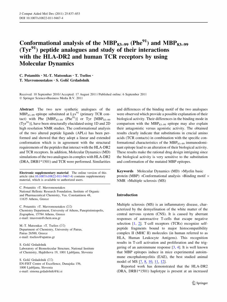
Table 3 continued
Residues Interactions
MBP83–99 (Phe91) Receptor Hydrogen bonds Hydrophobic Electrostatic
Conformers Conformers Conformers
1 2 3 4 5 1 2 3 4 5 1 2 3 4 5
Phe 91 HLA-DR2b Gln70 4 8 4 4 4 8 8 8 8 8 8 8 8 8 8
Phe 91 TCR-Vb Thr100 8 8 8 8 8 4 8 4 4 4 8 8 8 8 8
Asn 92 HLA-DR2a Asp66 4 8 4 4 4 8 8 8 8 8 8 8 8 8 8
Asn 92 TCR-Vb Thr100 4 8 8 8 8 8 8 8 8 8 8 8 8 8 8
Asn 92 HLA-DR2b Trp61 8 4 4 4 8 8 8 8 8 8 8 8 8 8 8
Asn 92 HLA-DR2b Tyr30 8 8 8 4 8 8 8 8 8 8 8 8 8 8 8
Ile 93 HLA-DR2b Tyr60 4 4 4 8 4 8 8 8 8 8 8 8 8 8 8
Ile 93 HLA-DR2b Ile67 8 8 8 8 8 4 4 4 4 4 8 8 8 8 8
Ile 93 HLA-DR2b Trp61 8 8 8 8 4 8 8 8 8 8 8 8 8 8 8
Val 94 HLA-DR2b Tyr60 4 4 4 4 4 8 8 8 8 8 8 8 8 8 8
Val 94 HLA-DR2a Val65 8 8 8 8 8 4 4 4 4 4 8 8 8 8 8
Val 94 HLA-DR2a Ala68 8 8 8 8 8 4 4 4 4 4 8 8 8 8 8
Val 94 HLA-DR2a Ile72 8 8 8 8 8 4 8 4 8 8 8 8 8 8 8
Thr 95 HLA-DR2a Asn69 4 4 4 4 8 8 8 8 8 8 8 8 8 8 8
Thr 95 HLA-DR2a Arg76 4 8 8 8 8 8 8 8 8 8 8 8 8 8 8
Thr 95 HLA-DR2b Asp57 4 4 4 4 4 8 8 8 8 8 8 8 8 8 8
Thr 95 HLA-DR2a Ile72 8 8 8 8 8 4 4 4 4 4 8 8 8 8 8
Thr 95 HLA-DR2a Met73 8 8 8 8 8 4 4 4 4 4 8 8 8 8 8
Thr 95 HLA-DR2b Trp61 8 8 8 8 8 4 4 4 4 4 8 8 8 8 8
Thr 95 HLA-DR2b Tyr60 8 8 8 8 8 8 8 8 4 4 8 8 8 8 8
Pro 96 HLA-DR2a Arg76 4 4 8 4 8 8 8 8 8 8 8 8 8 8 8
Pro 96 HLA-DR2b Tyr60 8 8 8 8 8 4 4 4 4 4 8 8 8 8 8
Arg 97 HLA-DR2a Ile72 8 8 8 8 8 4 8 8 8 8 8 8 8 8 8
Thr 98 HLA-DR2a Ile72 8 8 8 8 8 4 4 8 8 8 8 8 8 8 8
Thr 98 HLA-DR2a Lys75 8 4 8 8 8 8 8 8 8 8 8 8 8 8 8
Thr 98 HLA-DR2a Arg76 8 8 4 8 4 8 8 8 8 8 8 8 8 8 8
Pro 99 HLA-DR2a Lys75 8 8 8 8 8 8 8 8 4 8 8 4 4 8 8
Pro 99 HLA-DR2a Ile72 8 8 8 8 8 8 8 8 4 8 8 8 8 8 8
The two chains of HLA-DR2 are denoted with a and b, and of TCR with Va and Vb
842 J Comput Aided Mol Des (2011) 25:837–853
123

Table 4 Hydrogen bonds, hydrophobic and electrostatic interactions between the five conformers of MBP83–99 (Tyr91) and the proteins HLA-
DR2 and TCR of the trimolecular complex
Residues Interactions
MBP83–99 (Tyr91) Receptor Hydrogen bonds Hydrophobic Electrostatic
Conformers Conformers Conformers
1 2 3 4 5 1 2 3 4 5 1 2 3 4 5
Glu 83 TCR-Va Lys99 8 8 8 8 4 8 8 8 8 8 4 4 8 8 8
Glu 83 TCR-Va Thr97 4 8 8 8 8 8 8 8 8 8 8 8 8 8 8
Glu 83 TCR-Vb Glu59 8 4 8 4 8 8 8 8 8 8 8 8 8 8 8
Glu 83 TCR-Vb Gln60 8 4 8 8 8 8 8 8 8 8 8 8 8 8 8
Glu 83 TCR-Vb Tyr58 4 8 4 4 4 8 8 8 8 8 8 8 8 8 8
Glu 83 TCR-Vb Gly61 8 8 4 4 8 8 8 8 8 8 8 8 8 8 8
Asn 84 TCR-Vb Tyr58 8 4 4 4 8 8 8 8 8 8 8 8 8 8 8
Asn 84 TCR-Vb Glu59 8 4 4 8 8 8 8 8 8 8 8 8 8 8 8
Asn 84 TCR-Vb Phe51 8 8 8 4 8 8 8 8 8 8 8 8 8 8 8
Asn 84 TCR-Vb Thr97 8 8 8 4 8 8 8 8 8 8 8 8 8 8 8
Asn 84 HLA-DR2a Ser53 4 8 8 8 4 8 8 8 8 8 8 8 8 8 8
Asn 84 HLA-DR2a Arg50 4 8 8 8 8 8 8 8 8 8 8 8 8 8 8
Pro 85 HLA-DR2a Ser53 4 4 4 4 4 8 8 8 8 8 8 8 8 8 8
Pro 85 HLA-DR2b Val85 8 8 8 8 8 8 8 4 8 8 8 8 8 8 8
Pro 85 TCR-Va Thr97 8 8 8 8 8 8 8 4 8 8 8 8 8 8 8
Val 86 TCR-Va Gly96 4 4 4 4 4 8 8 8 8 8 8 8 8 8 8
Val 86 TCR-Va Thr97 8 8 8 8 8 4 4 4 4 4 8 8 8 8 8
Val 86 TCR-Va Tyr98 8 8 8 8 8 4 4 4 4 4 8 8 8 8 8
Val 86 TCR-Vb Tyr58 8 8 8 8 8 8 8 4 8 8 8 8 8 8 8
Val 87 HLA-DR2b Val85 8 8 8 8 8 4 4 4 4 4 8 8 8 8 8
Val 87 HLA-DR2b Val86 8 8 8 8 8 8 4 8 4 4 8 8 8 8 8
Val 87 HLA-DR2a Trp43 8 8 8 8 8 4 4 8 4 8 8 8 8 8 8
Val 87 HLA-DR2a Phe54 8 8 8 8 8 4 4 4 4 4 8 8 8 8 8
Val 87 HLA-DR2a Phe24 8 8 8 8 8 8 4 4 4 4 8 8 8 8 8
Val 87 HLA-DR2a Ser53 4 4 4 4 4 8 8 8 8 8 8 8 8 8 8
Val 87 HLA-DR2a Ala52 8 8 8 8 8 4 8 4 8 4 8 8 8 8 8
Val 87 HLA-DR2a Glu55 8 4 4 4 4 8 8 8 8 8 8 8 8 8 8
His 88 HLA-DR2b Asn82 4 4 4 4 4 8 8 8 8 8 8 8 8 8 8
His 88 HLA-DR2b His81 8 8 8 8 8 8 8 8 8 8 4 8 8 8 8
His 88 TCR-Va Asp92 4 8 8 8 8 8 8 8 8 8 4 8 8 8 8
His 88 TCR-Vb Ala103 8 8 8 8 8 4 4 8 8 8 8 8 8 8 4
Phe 89 TCR-Vb Ala103 4 4 4 4 4 4 4 8 8 8 8 8 8 8 4
Phe 89 TCR-Vb Thr31 8 8 8 8 8 8 4 4 8 4 8 8 8 8 8
Phe 89 HLA-DR2a Ala59 8 8 8 8 8 4 4 8 4 4 8 8 8 8 8
Phe 90 HLA-DR2a Gln9 4 8 4 4 4 8 8 8 8 8 8 8 8 8 8
Phe 90 HLA-DR2a Asn62 4 4 8 8 8 8 8 8 8 8 8 8 8 8 8
Phe 90 HLA-DR2b Tyr 78 8 8 8 8 8 4 8 4 8 8 8 8 8 8 8
Phe 90 HLA-DR2b Ala71 8 8 8 8 8 4 4 4 4 4 8 8 8 8 8
Phe 90 HLA-DR2b Phe26 8 8 8 8 8 4 4 4 4 4 8 8 8 8 8
Phe 90 HLA-DR2b Ala74 8 8 8 8 8 4 4 4 4 4 8 8 8 8 8
Phe 90 TCR-Vb Ala103 8 8 8 8 8 4 8 4 4 4 8 8 8 8 8
Tyr 91 HLA-DR2b Ile67 8 8 8 8 8 4 4 4 4 4 8 8 8 8 8
Tyr 91 TCR-Vb Thr100 8 8 8 8 8 4 8 4 8 8 8 8 8 8 8
Asn 92 HLA-DR2a Asp66 8 8 4 4 4 8 8 8 8 8 8 8 8 8 8
J Comput Aided Mol Des (2011) 25:837–853 843
123
with Gln70 of HLA-DR2b. The hydrogen bonds mentioned
above are stable during MD simulation with high occur-
rence (Table 6).
Furthermore, the peptide MBP83–99 (Phe91) is stabilized
in the HLA-DR2 pocket with the following interactions:
hydrogen bonds between Pro85–Ser53 of HLA-DR2a,
His88–Asn82 of HLA-DR2a, electrostatic interactions
between His88–His81 of HLA-DR2a, hydrophobic interac-
tions between Phe89–Phe54 of HLA-DR2a, hydrogen bonds
between Asn92–Asp66 of HLA-DR2a, Asn92–Trp61 of
HLA-DR2b, and Ile93–Tyr60 of HLA-DR2b, hydrophobic
interactions between Asn92–Asp66 of HLA-DR2a and
Ile93–Ile67 of HLA-DR2b, hydrogen bonding between
Val94–Tyr60 of HLA-DR2b, hydrophobic interactions
between Val94–Val65, Ala68 and Ile72 of HLA-DR2a,
hydrogen bonds between Thr95–Asn69 of HLA-DR2a and
Asp57 of HLA-DR2b, hydrophobic interactions between
Thr95–Ile72 and Met73 of HLA-DR2a and Trp61 of HLA-
DR2b, between Pro96–Tyr60 of HLA-DR2b, hydrogen
bonding between Thr98–Arg76 of HLA-DR2a and an
electrostatic interaction between Pro99–Lys75 of HLA-
DR2a.
The peptide MBP83–99 (Phe91) forms also further inter-
actions with TCR: hydrogen bonds between Glu83–Glu59 of
TCR-Vb and Thr97 of TCR-Va, Asn84–Ala52 of TCR-Vaand electrostatic interactions between His88–Asn104 of
TCR-Vb. Finally, there is also a combination of hydrogen
bonding and electrostatic interactions between Glu83 and
Lys99 of TCR-Va.
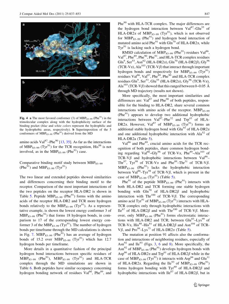
A visualization of the binding pocket’s hydrophilicity
surface for the most favored conformer 3 of the peptide
MBP83–99 (Phe91) in the trimolecular complex is presented
in Fig. 4a. From the superimposition of the five most
favored conformers of MBP83–99 (Phe91) produced by MD
simulations (Fig. 4b), it is evident that linear and extended
structures are the major characteristics of the low energy
conformers situated in the binding site of the trimolecular
complex.
Table 4 continued
Residues Interactions
MBP83–99 (Tyr91) Receptor Hydrogen bonds Hydrophobic Electrostatic
Conformers Conformers Conformers
1 2 3 4 5 1 2 3 4 5 1 2 3 4 5
Asn 92 HLA-DR2a Glu11 4 8 4 4 4 8 8 8 8 8 8 8 8 8 8
Asn 92 HLA-DR2a Asn62 8 4 8 8 8 8 8 8 8 8 8 8 8 8 8
Ile 93 HLA-DR2b Tyr30 4 8 8 8 8 4 8 8 8 8 8 8 8 8 8
Ile 93 HLA-DR2a Asn69 4 4 4 4 4 8 8 8 8 8 8 8 8 8 8
Ile 93 HLA-DR2b Trp61 8 8 8 8 8 4 4 4 4 4 8 8 8 8 8
Ile 93 HLA-DR2b Ile67 8 8 8 8 8 4 4 4 4 4 8 8 8 8 8
Ile 93 HLA-DR2b Tyr60 8 8 8 8 8 8 4 8 8 8 8 8 8 8 8
Val 94 HLA-DR2b Trp61 4 8 8 8 8 8 8 8 8 8 8 8 8 8 8
Val 94 HLA-DR2a Val65 8 8 8 8 8 4 4 4 4 4 8 8 8 8 8
Val 94 HLA-DR2a Ala68 8 8 8 8 8 4 4 8 4 4 8 8 8 8 8
Val 94 HLA-DR2a Ile72 8 8 8 8 8 4 4 8 4 8 8 8 8 8 8
Val 94 HLA-DR2b Tyr60 8 8 4 8 8 8 8 8 8 8 8 8 8 8 8
Thr 95 HLA-DR2a Asn69 4 8 4 4 4 8 8 8 8 8 8 8 8 8 8
Thr 95 HLA-DR2b Asp57 8 4 8 4 8 8 8 8 8 8 8 8 8 8 8
Thr 95 HLA-DR2a Ile72 8 8 8 8 8 4 4 4 4 4 8 8 8 8 8
Thr 95 HLA-DR2a Met73 8 8 8 8 8 4 4 4 4 4 8 8 8 8 8
Thr 95 HLA-DR2b Trp61 8 8 8 8 8 8 4 4 4 4 8 8 8 8 8
Thr 95 HLA-DR2b Tyr60 8 8 8 8 8 8 8 4 8 8 8 8 8 8 8
Pro 96 HLA-DR2b Tyr60 8 8 8 8 8 8 4 4 4 8 8 8 8 8 8
Thr 98 HLA-DR2a Ile72 8 8 8 8 8 4 8 8 8 8 8 8 8 8 8
Pro 99 HLA-DR2a Arg76 8 8 4 8 4 8 8 8 8 8 8 8 8 8 4
The two chains of HLA-DR2 are denoted with a and b, and of TCR with Va and Vb
844 J Comput Aided Mol Des (2011) 25:837–853
123

MD results for the peptide MBP83–99 (Tyr91)
The hydrogen bonding, the hydrophobic and electrostatic
interactions of the most energetically favored conformer 3
of the peptide MBP83–99 (Tyr91) during binding with amino
acids of the receptor HLA-DR2 and TCR are revealed from
the application of MD in Fig. 5.
More specifically, the amino acid Val87 forms hydrogen
bonding with Ser53 and Glu55 of HLA-DR2a and interacts
through hydrophobic interactions with Phe24, Ala52 and
Phe54 of HLA-DR2a and with Val85 of HLA-DR2b. Phe90,
the other crucial amino acid for the binding to HLA-DR2,
develops hydrogen bonding with Gln9 of HLA-DR2a and
hydrophobic interactions with Phe26, Ala71, Tyr78 and
Ala74 of HLA-DR2b.
The required interactions that are appearing during the
recognition of MBP83–99 (Tyr91) with TCR are: hydrogen
bonds between Phe89–Ala103 of TCR-Vb and Val86–Gly96
of TCR-Va, hydrophobic interaction between Val86–Thr97
and Tyr98 of TCR-Va, Val86–Tyr58 of TCR-Vb and
between Phe89–Thr31 of TCR-Vb. The mutated amino acid
Tyr91 forms hydrophobic interactions with Ile67 of HLA-
DR2b and with Thr100 of TCR-Vb. The mentioned
hydrogen bonds are stable during MD simulation as indi-
cated by their high occurrence (Table 6).
Further interactions which stabilize the peptide MBP83–99
(Tyr91) in the HLA-DR2 pocket are: hydrogen bonds
between Pro85–Ser53 of HLA-DR2a and His88–Asn82 of
HLA-DR2a, hydrophobic interactions between Pro85–Val85
of HLA-DR2b, hydrogen bonds between Asn92–Glu11 and
Asp66 of HLA-DR2a and between Ile93–Asn69 of HLA-
DR2a, hydrophobic interactions between Ile93–Trp61 and
Ile67 of HLA-DR2b, hydrogen bond between Val94–Tyr60 of
HLA-DR2b, hydrophobic interactions between Val94–Val65
of HLA-DR2a, hydrogen bonding between Thr95–Asn69 of
HLA-DR2a, hydrophobic interactions between Thr95–Ile72
and Met73 of HLA-DR2a, Thr95–Tyr60 and Trp61 of HLA-
DR2b and between Pro96–Tyr60 of HLA-DR2b and hydro-
gen bond between Pro99–Arg76 of HLA-DR2a.
MBP83–99 (Tyr91) forms also the following interactions
with the TCR: hydrogen bonds between Glu83–Tyr58 and
Gly61 of TCR-Vb, between Asn84–Tyr58 and Glu59 of
TCR-Vb, hydrophobic interactions between Pro85–Thr97 of
TCR-Va and between Phe90–Ala103 of TCR-Vb.
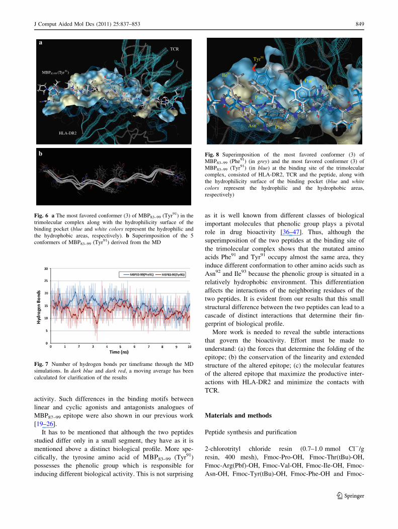
The hydrophilicity surface of binding pocket for the
most favored conformer 3 of the peptide MBP83–99 (Tyr91)
in the trimolecular complex is shown in Fig. 6a. From the
superimposition of the five most favored conformers of
MBP83–99 (Tyr91) derived from MD simulations (Fig. 6b),
it is obvious, also in this case, that linear and extended
conformations are the major characteristics of MBP83–99
(Tyr91) low energy conformers situated in the binding site
of the trimolecular complex.
Regarding the mutation of Lys91 MBP83–99 with the
amino acid Tyr91, as in the case of Phe91, it causes the
abolishment of the a-helix formation observed between
Glu83
Asn84
Pro85
Val86
Val87
His88
Phe90
Phe89
Phe91
Asn92
Ile93
Val94
Thr95
Pro96
Arg97
Thr98
Pro99
Fig. 3 Hydrogen bonds (donors
green, acceptors red),
hydrophobic interactions
(yellow) and electrostatic
interactions (red negative
charged groups, blue positive
charged groups) of the most
favored conformation (3) of
MBP83–99 (Phe91) during
binding with amino acids of the
receptor HLA-DR2 and TCR as
they are revealed from the
application of MD. The lettersthat accompany each amino acid
correspond to the following:
A = HLA-DR2a, B = HLA-
DR2b, D = TCR-Va and
E = TCR-Vb
J Comput Aided Mol Des (2011) 25:837–853 845
123
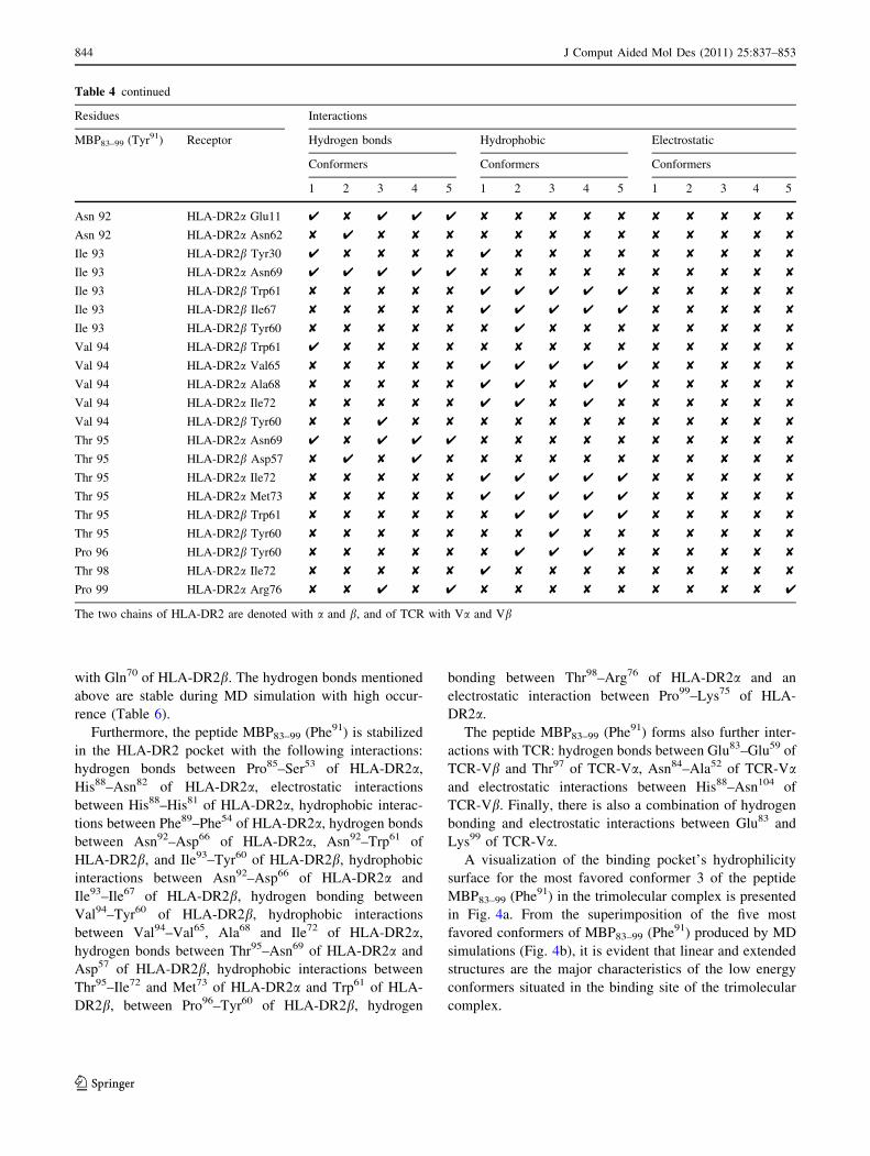
Table 6 Hydrogen bond interactions between specific residues
responsible for the binding to HLA-DR2 and recognition of TCR of
LBQ83–99 (Phe91) and LBQ83–99 (Tyr91) through the MD simulations
Hydrogen bond interaction Occurrence (%)
Peptide HLA-TCR complex LBQ83–99
(Phe91)
LBQ83–99
(Tyr91)
Val 86 TCR-Va Gly96 85 75
Val 87 HLA-DR2a Ser53 77 90
Val 87 HLA-DR2a Glu 55 0 76
Phe 89 TCR-Vb Ala103 92 95
Phe 90 HLA-DR2a Gln9 100 100
Phe 90 HLA-DR2a Asn62 81 77
Phe 91 HLA-DR2b Gln70 100 –
Asn 92 HLA-DR2a Asp66 97 82
Asn 92 HLA-DR2b Trp 61 67 0
Asn 92 HLA-DR2a Glu11 1 83
Ile 93 HLA-DR2b Tyr60 67 1
Ile 93 HLA-DR2a Asn69 7 86
The hydrogen bond interactions of other neighboring residues of
mutation site 91 are included
Table 5 Comparison of hydrogen bonds, hydrophobic and electro-
static interactions of the two lowest energy conformers of the peptides
MBP83–99 (Phe91) (conformer 3) and MBP83–99 (Tyr91) (conformer 3)
and the proteins HLA-DR2 and TCR of the trimolecular complex
Peptide Receptor Conformers
MBP83–99 (Phe91) MBP83–99 (Tyr91)
Conformer 3 Conformer 3
Hydrogen bonds
Glu 83 TCR-Va Lys99 4 8
Glu 83 TCR-Vb Tyr58 8 4
Glu 83 TCR-Vb Glu59 4 8
Glu 83 TCR-Vb Gly61 8 4
Glu 83 TCR-Va Thr97 4 8
Asn 84 TCR-Vb Tyr58 8 4
Asn 84 TCR-Vb Glu59 8 4
Asn 84 TCR-Va Ala52 4 8
Pro 85 HLA-DR2a Ser53 4 4
Val 86 TCR-Va Gly96 4 4
Val 87 HLA-DR2a Ser53 4 4
Val 87 HLA-DR2a Glu55 8 4
His 88 HLA-DR2b Asn82 4 4
Phe 89 TCR-Vb Ala103 4 4
Phe 90 HLA-DR2a Gln9 4 4
Phe 91 HLA-DR2b Gln70 4 –
Asn 92 HLA-DR2a Asp66 4 4
Asn 92 HLA-DR2b Trp61 4 8
Asn 92 HLA-DR2a Glu11 8 4
Ile 93 HLA-DR2b Tyr60 4 8
Ile 93 HLA-DR2a Asn69 8 4
Val 94 HLA-DR2b Tyr60 4 4
Thr 95 HLA-DR2a Asn69 4 4
Thr 95 HLA-DR2b Asp57 4 8
Thr 98 HLA-DR2a Arg76 4 8
Pro 99 HLA-DR2a Arg76 8 4
Hydrophobic interactions
Pro 85 HLA-DR2b Val85 8 4
Pro 85 TCR-Va Thr97 8 4
Val 86 TCR-Vb Tyr58 8 4
Val 86 TCR-Va Thr97 4 4
Val 86 TCR-Va Tyr98 4 4
Val 87 HLA-DR2b Val85 4 4
Val 87 HLA-DR2a Phe32 4 8
Val 87 HLA-DR2a Trp43 4 8
Val 87 HLA-DR2a Ala52 8 4
Val 87 HLA-DR2a Phe54 4 4
Val 87 HLA-DR2a Phe24 4 4
Phe 89 TCR-Vb Thr31 4 4
Phe 89 HLA-DR2a Phe54 4 8
Phe 90 HLA-DR2b Ala71 4 4
Phe 90 HLA-DR2b Phe26 4 4
Phe 90 HLA-DR2b Tyr 78 4 4
Table 5 continued
Peptide Receptor Conformers
MBP83–99 (Phe91) MBP83–99 (Tyr91)
Conformer 3 Conformer 3
Phe 90 HLA-DR2b Ala74 4 4
Phe 90 TCR-Vb Ala103 8 4
Phe 91 TCR-Vb Thr100 4 –
Tyr 91 TCR-Vb Thr100 – 4
Tyr 91 HLA-DR2b Ile67 – 4
Asn 92 HLA-DR2a Asp66 4 8
Asn 92 HLA-DR2a Glu11 8 8
Ile 93 HLA-DR2b Trp61 8 4
Ile 93 HLA-DR2b Ile67 4 4
Val 94 HLA-DR2a Val65 4 4
Val 94 HLA-DR2a Ala68 4 8
Val 94 HLA-DR2a Ile72 4 8
Thr 95 HLA-DR2a Ile72 4 4
Thr 95 HLA-DR2a Met73 4 4
Thr 95 HLA-DR2b Trp61 4 4
Thr 95 HLA-DR2b Tyr60 8 4
Pro 96 HLA-DR2b Tyr60 4 4
Electrostatic interactions
Glu 83 TCR-Va Lys99 4 8
His 88 HLA-DR2b His81 4 8
His 88 TCR-Vb Asn104 4 8
Pro 99 HLA-DR2a Lys75 4 8
The two chains of HLA-DR2 are denoted with a and b, and of TCR
with Va and Vb
846 J Comput Aided Mol Des (2011) 25:837–853
123
amino acids Val87–Phe90 [13, 35]. As far as the interactions
of MBP83–99 (Tyr91) for the TCR recognition, His88 is not
involved, as in the MBP83–99 (Phe91) case.
Comparative binding motif study between MBP83–99
(Phe91) and MBP83–99 (Tyr91)
The two linear and extended peptides showed similarities
and differences concerning their binding motif to the
receptor. Comparison of the most important interactions of
the two peptides on the receptor HLA-DR2 is shown in
Table 5. Peptide MBP83–99 (Phe91) forms with the amino
acids of the receptor HLA-DR2 and TCR more hydrogen
bonds relatively to the MBP83–99 (Tyr91). As a represen-
tative example, is shown the lowest energy conformer 3 of
MBP83–99 (Phe91) that forms 18 hydrogen bonds, in com-
parison to 17 of the corresponding lowest energy con-
former 3 of the MBP83–99 (Tyr91). The number of hydrogen
bonds per timeframe through the MD calculations is shown
in Fig. 7. LBQ83–99 (Phe91) has an average of hydrogen
bonds of 15.2 over LBQ83–99 (Tyr91) which has 12.7
hydrogen bonds per timeframe.
More details in a quantitative fashion of the principal
hydrogen bond interactions between specific residues of
LBQ83–99 (Phe91), LBQ83–99 (Tyr91) and HLA-TCR
complex through the MD simulations are shown in
Table 6. Both peptides have similar occupancy concerning
hydrogen bonding network of residues Val86, Phe89, and
Phe90 with HLA-TCR complex. The major differences are
the hydrogen bond interaction between Val87–Glu55 of
HLA-DR2a of LBQ83–99 (Tyr91), which is not observed
for LBQ83–99 (Phe91) and hydrogen bond interaction of
mutated amino acid Phe91 with Gln70 of HLA-DR2a, while
Tyr91 is lacking such a hydrogen bond.
RMSD calculation of LBQ83–99 (Phe91) residues Val86,
Val87, Phe89, Phe90, Phe91, and HLA-TCR complex residues
Gln9, Ser53, Asn62 (HLA-DR2a), Gln70 (HLA-DR2b), Gly96
(TCR-Va), Ala103 (TCR-Vb) that interact through important
hydrogen bonds and respectively for LBQ83–99 (Tyr91)
residues Val86, Val87, Phe89, Phe90 and HLA-TCR complex
residues Gln9, Ser53, Glu55 (HLA-DR2a), Gly96 (TCR-Va),
Ala103 (TCR-Vb) showed that this ranged between 0–0.05 A
through MD trajectory (results not shown).
More specifically, the most important similarities and
differences are: Val87 and Phe90 of both peptides, respon-
sible for the binding to HLA-DR2, share several common
interactions with amino acids of the receptor. LBQ83–99
(Phe91) appears to develop two additional hydrophobic
interactions between Val87–Phe32 and Trp43 of HLA-
DR2a. However, Val87 of LBQ83–99 (Tyr91) forms an
additional stable hydrogen bond with Glu55 of HLA-DR2aand one additional hydrophobic interaction with Ala52 of
HLA-DR2a (Table 5).
Val86 and Phe89, crucial amino acids for the TCR rec-
ognition of both peptides, share common hydrogen bond-
ing regarding Val86–Gly96 of TCR-Va, Phe89–Ala103 of
TCR-Vb and hydrophobic interactions between Val86–
Thr97, Tyr98 of TCR-Va and Phe89–Thr31 of TCR-Vb.
LBQ83–99 (Phe91) lacks the hydrophobic interaction
between Val86–Tyr58 of TCR-Vb, which is present in the
case of LBQ83–99 (Tyr91) (Table 5).
Phe91 of the peptide LBQ83–99 (Phe91) interacts with
both HLA-DR2 and TCR forming one stable hydrogen
bonding with Gln70 of HLA-DR2b and hydrophobic
interaction with Thr100 of TCR-Vb. The corresponding
amino acid Tyr91 of LBQ83–99 (Tyr91) interacts with HLA-
TCR complex only through hydrophobic interactions with
Ile67 of HLA-DR2b and with Thr100 of TCR-Vb. More-
over, only LBQ83–99 (Phe91) forms electrostatic interac-
tions with HLA-DR2 and TCR; between Glu83–Lys99 of
TCR-Va, His88–His81 of HLA-DR2b and Asn104 of TCR-
Vb, and Pro99–Lys75 of HLA-DR2a (Table 5).
The mutation at position 91 affects also the conforma-
tion and interactions of neighboring residues, especially of
Asn92 and Ile93 (Figs. 3, 6 and 8). More specifically, the
Asn92 of LBQ83–99 (Phe91) develops hydrogen bonds with
Asp66 of HLA-DR2a and Trp61 of HLA-DR2b while in the
case of LBQ83–99 (Tyr91) it interacts with Asp66 and Glu11
of HLA-DR2a. Regarding the Ile93 of LBQ83–99 (Phe91)
forms hydrogen bonding with Tyr60 of HLA-DR2b and
hydrophobic interactions with Ile67 of HLA-DR2b, but in
HLA-DR2
TCR
MBP83-99 (Phe91)
a
b
Fig. 4 a The most favored conformer (3) of MBP83–99 (Phe91) in the
trimolecular complex along with the hydrophilicity surface of the
binding pocket (blue and white colors represent the hydrophilic and
the hydrophobic areas, respectively). b Superimposition of the 5
conformers of MBP83–99 (Phe91) derived from the MD
J Comput Aided Mol Des (2011) 25:837–853 847
123

the case of LBQ83–99 (Tyr91) Ile93 forms hydrogen bond
with Asn69 of HLA-DR2a and hydrophobic interaction not
only with Ile67 of HLA-DR2b but also with Trp61 of HLA-
DR2b. All the above described hydrogen bonds are stable
through the MD simulations (Table 6).
When another strategy was used, more specifically,
when MD simulation was applied using the mutated crystal
structure of MBP83–96 at position 91 and extended until
position 99, identical results were obtained. Although, this
strategy is simpler, we find our above mentioned approach
more general in terms of simulating the system for the
following reasons: (a) We proved that synthetic peptides
accommodate in the same place as the wild peptide;
(b) Low energy conformers of the synthetic peptides are in
accordance with NMR results; (c) We explored, through
docking, the conformational preferences of the elongated,
regarding to the wild type, peptides (d) The applied
extended MD calculations allow the exploration of the
mobility of the system; (e) Our approach expands the
conformational space for the ligand as it combines the use
of experimental NMR and theoretical results (combination
of docking and MD calculations).
Conclusion
The two peptides under study show similarities and dif-
ferences in respect to their binding motif with HLA-DR2
and TCR receptors. In general, LBQ83–99 (Phe91) shows
more interactions at the formation of the trimolecular
complex compared to LBQ83–99 (Tyr91), concerning the
comparison of hydrogen bond occupancy over MD tra-
jectories, as well as the comparison of all interactions of
the most energetically favored conformers. The better
biological profile of the latter may be due to this reason,
thus its distinct number of interactions may be responsible
to reduce the immunological response through the activa-
tion of encephalitogenic T-cells and finally the appearance
of MS.
The two molecules also show differences in the binding
motif in comparison with the wild epitope LBQ83–96. This
is attributed to the fact that by mutating Lys91 by either Tyr
or Phe the stereoelectronic characteristics become differ-
ent. This modification of the stereoelectronic properties can
affect at least the binding motif of the regional amino acids.
This may also explain their antagonistic versus agonistic
Glu83
Asn84
Pro85
Val86
Val87
His88
Phe90
Phe89
Tyr91
Asn92
Ile93
Val94
Thr95
Pro96
Arg97 Thr98
Pro99
Fig. 5 Hydrogen bonds (donors
green, acceptors red),
hydrophobic interactions
(yellow) and electrostatic
interactions (red negative
charged groups, blue positive
charged groups) of the most
favored conformer (3) of
MBP83–99 (Tyr91) during
binding with amino acids of the
receptor HLA-DR2 and TCR as
they are revealed from the
application of MD. The lettersthat accompany each amino acid
correspond to the following:
A = HLA-DR2a, B = HLA-
DR2b, D = TCR-Va and
E = TCR-Vb
848 J Comput Aided Mol Des (2011) 25:837–853
123
activity. Such differences in the binding motifs between
linear and cyclic agonists and antagonists analogues of
MBP87–99 epitope were also shown in our previous work
[19–26].
It has to be mentioned that although the two peptides
studied differ only in a small segment, they have as it is
mentioned above a distinct biological profile. More spe-
cifically, the tyrosine amino acid of LBQ83–99 (Tyr91)
possesses the phenolic group which is responsible for
inducing different biological activity. This is not surprising
as it is well known from different classes of biological
important molecules that phenolic group plays a pivotal
role in drug bioactivity [36–47]. Thus, although the
superimposition of the two peptides at the binding site of
the trimolecular complex shows that the mutated amino
acids Phe91 and Tyr91 occupy almost the same area, they
induce different conformation to other amino acids such as
Asn92 and Ile93 because the phenolic group is situated in a
relatively hydrophobic environment. This differentiation
affects the interactions of the neighboring residues of the
two peptides. It is evident from our results that this small
structural difference between the two peptides can lead to a
cascade of distinct interactions that determine their fin-
gerprint of biological profile.
More work is needed to reveal the subtle interactions
that govern the bioactivity. Effort must be made to
understand: (a) the forces that determine the folding of the
epitope; (b) the conservation of the linearity and extended
structure of the altered epitope; (c) the molecular features
of the altered epitope that maximize the productive inter-
actions with HLA-DR2 and minimize the contacts with
TCR.
Materials and methods
Peptide synthesis and purification
2-chlorotrityl chloride resin (0.7–1.0 mmol Cl-/g
resin, 400 mesh), Fmoc-Pro-OH, Fmoc-Thr(tBu)-OH,
Fmoc-Arg(Pbf)-OH, Fmoc-Val-OH, Fmoc-Ile-OH, Fmoc-
Asn-OH, Fmoc-Tyr(tBu)-OH, Fmoc-Phe-OH and Fmoc-
Asn92
Phe89
Phe90
Phe91
Tyr91
Ile93
His88
Fig. 8 Superimposition of the most favored conformer (3) of
MBP83–99 (Phe91) (in grey) and the most favored conformer (3) of
MBP83–99 (Tyr91) (in blue) at the binding site of the trimolecular
complex, consisted of HLA-DR2, TCR and the peptide, along with
the hydrophilicity surface of the binding pocket (blue and whitecolors represent the hydrophilic and the hydrophobic areas,
respectively)
HLA-DR2
TCR
HLA-DR2
MBP83-99 (Tyr91)
a
b
Fig. 6 a The most favored conformer (3) of MBP83–99 (Tyr91) in the
trimolecular complex along with the hydrophilicity surface of the
binding pocket (blue and white colors represent the hydrophilic and
the hydrophobic areas, respectively). b Superimposition of the 5
conformers of MBP83–99 (Tyr91) derived from the MD
Fig. 7 Number of hydrogen bonds per timeframe through the MD
simulations. In dark blue and dark red, a moving average has been
calculated for clarification of the results
J Comput Aided Mol Des (2011) 25:837–853 849
123
His(Trt)-OH, Fmoc-Glu(tBu)-OH, were obtained from
Chemical to Biopharmaceutical Laboratories of Patras,
Patras, Greece. All solvents and other reagents were pur-
chased from Merck, Sigma-Aldrich and Fluka chemical
companies. DC-Alufolien Kieselgel 60 (Merck) was used
for Thin Layer Chromatography (TLC) analysis of syn-
thetic products with the following eluent solvent: n-buta-
nol/acetic acid/water (BAW) 4:1:1 (v/v/v). Peptides were
purified by semi-preparative Reverse Phase High Perfor-
mance Liquid Chromatography (RP-HPLC) on a Waters
system equipped with a 600E controller and a Waters 996
photodiode array UV detector. The analysis was controlled
by an Millenium 2.1 operating system and a Nucleosil C-18
reversed phase analytical column (250 9 10 mm with
7 lm packing material). Electron Spray Ionization Mass
Spectroscopy (ESI–MS) experiments were performed on a
TSQ 7000 spectrometer (Electrospray Platform LC of
Micromass) coupled to a MassLynx NT 2.3 data system.
The purity of the two compounds was found to be C95%.
Synthesis of MBP83–99 (Phe91) and MBP83–99 (Tyr91)
peptide analogues
The linear peptides were prepared on 2-chlorotrityl chloride
resin (CLTR-Cl) using the Fmoc/tBu solid-phase method-
ology [28–31]. The first Na Fmoc (9-fluorenylmethylox-
ycarboxyl)-protected amino acid (Fmoc-Pro-OH) was
esterified to the resin in the presence of diisopropylethyl-
amine (DIPEA) in dichloromethane (DCM) in 1 h at RT
[28–31]. DCM/MeOH/DIPEA (85:10:5) was then added and
the resulting mixture was stirred for another 10 min at RT.
The remaining protected peptide chains were assembled by
sequential couplings of the appropriate Fmoc protected
amino acids (2.5 equiv), in the presence of N,N’-diisopro-
pylcarbodiimide (DIC) (2.75 equiv) and 1-hydroxybenzo-
triazole (HOBt) (3.75 equiv) in N,N-dimethylformamide
(DMF) [28–31]. The completeness of each coupling
was verified by the Kaiser test and TLC using a BAW, 4:1:1
(v/v/v) eluent system and the Fmoc protecting group was
removed by treatment with piperidine solution (20% in
DMF). The synthesized protected peptide on the resin was
then cleaved with the splitting solution dichloromethane/
2,2,2-trifluoroethanol (DCM/TFE, 7/3, 2 h at RT). The
mixtures were filtered, the solvents were removed on a rotary
evaporator and the obtained oily products were precipitated
from cold dry diethyl ether as white solids. The linear pro-
tected peptides were treated with 70% TFA in DCM in the
presence of 0.3% 1,2-ethanedithiol, anisole and H2O as
scavengers for 5 h at RT. The purification of each peptide
was carried out using semi-preparative RP-HPLC and pep-
tide purity was assessed by analytical RP-HPLC (column:
Nucleosil C18, 5 lm, 4.6 9 250 mm). They were identified
by ESI–MS.
NMR spectroscopy
The high-resolution NMR spectra were recorded on a Varian
DirectDrive 800 MHz spectrometer at 25 �C. 2 mg of each
peptide were dissolved in 0.7 ml of DMSO-d6. The 2D
TOCSY [48] and 2D NOESY [49] experiments were recor-
ded using the standard pulse sequences in the phase-sensitive
mode. The 2D homonuclear proton spectra were acquired
with a spectral width of 8,012 Hz, 2,048–4,096 data points in
t2, 4–32 scans, 512 complex points in t1, and a relaxation
delay of 1.5 s. The mixing times in 2D NOESY and 2D
TOCSY experiments were 75 ms and 60 ms, respectively.
The 1H–13C HSQC was performed with gradients and was
recorded with 32 scans and 128 complex points in t1, 1H
spectral width of 8,013 Hz, 13C spectral width of 30,166 Hz,
512 data points in t2 and a relaxation delay of 1 s.
The data was processed and analyzed with Mnova
software from Mestrelab Research. Crosspeak volumes in
NOESY spectra were calculated by integration routine
within the Mnova software. A set of strong (up to 2.5 A),
medium (2.5–3.7 A), and weak (3.7–5 A) NOEs was
established according to the integrated intensity of the
geminal pair of protons c1 and c2 of Ile93, which have a
distance of 1.78 A in all conformations.
Molecular modeling
The extended crystal structure of MBP83–96 (pdb code:
1YMM) was mutated at amino acid 91 with either Phe91 or
Tyr91 and extended with amino acid residues 87–89 to
provide peptides MBP83–99 (Phe91) and MBP83–99 (Tyr91).
The resulting structures were optimized using the Steepest
Descent and Truncated Newton Conjugate Gradient
(TNCG) minimization algorithms. The optimized struc-
tures were in accordance to the observation of strong
sequential daN(i,i?1) NOE connectivities and the absence of
long range NOEs. The two peptides under study were then
docked in the pocket of the native peptide MBP83–96.
During docking the peptides were held flexible.
All docking simulations were performed on Discovery
Studio 2.0 by Accelrys Software Inc [50] with the
LigandFit method, on the Dreiding Energy Grid force field
[51] using the experimental crystal structure coordinates of
the major histocompatibility complex bonded to a human
autoimmune T cell receptor (pdb code: 1ymm) [34]. The
co-crystallized peptide of LBQ83–96 as well as water
molecules were removed. This approach was performed in
order to dock and obtain the optimal peptide–receptor
complexes in terms of binding affinity for the two mole-
cules, LBQ83–99 (Phe91) and LBQ83–99 (Tyr91). The
docking experiments resulted on various docked confor-
mations, for both molecules. Performing the LigandScore
function, top dock scoring conformations for each peptide
850 J Comput Aided Mol Des (2011) 25:837–853
123
were extracted to be further examined on their stability
through Molecular Dynamics runs.
The best docking poses for each peptide were subjected
to Molecular Dynamics (MD). A total of 1,000 conformers
from the trajectory file were divided into 5 families
according to their RMSD values based on heavy atoms.
The trimolecular complexes for the lowest energy con-
former from each cluster were subjected to Truncated
Newton Conjugate Gradient (TNCG) minimization.
Molecular Dynamics simulations were performed on the
GROMACS 4.5.3 software package [52] using the GRO-
MOS96 force field [53]. The best docked conformations of
the peptides MBP83–99 (Phe91) and MBP83–99 (Tyr91) were
subjected to Molecular Dynamics simulations where in
each case, the system included the peptide, the major his-
tocompatibility complex, the human autoimmune T cell
receptor and *66,000 water molecules in a cubic box with
dimensions 110 9 180 9 110 (A). Simulations were run at
300 K using the Berendsen pressure coupling method and
electrostatic interactions were calculated using the particle
mesh Ewald method [54]. Cutoff distances for the calcu-
lation of Coulomb and van der Waals interactions were 1.0
and 1.4 nm, respectively. Prior to the Molecular Dynamics
simulations, energy minimization was applied to the full
system without constraints using the Steepest Descent
integrator for 1,000 steps with a time step of 2 fs (the
minimization tolerance was set to 1,000 kJ/(mol.nm)). The
systems were then equilibrated via 300 ps simulations with
a time step of 2 fs. Finally, a 10 ns production run was
performed for the systems, respectively, at 300 K with a
time step of 2 fs using the Berendsen thermostat algorithm
as all bonds were constrained using the LINCS algorithm
[55]. Visualization of the dynamics trajectories was per-
formed with the VMD software package [56].
The clustering of the selected 1,000 conformers of the
peptides was performed by Cheminformatics/Canvas [57]
module of Schrodinger Suite 2010 followed by energy
minimization using Truncated Newton Conjugate Gradient
(TNCG) algorithm of Macromodel module on Maestro
platform of Schrodinger Suite 2010 [58].
LigandScout 3.0 (evaluation version) was used for the
visualization of the interactions of the peptides at the binding
site of the trimolecular complex. Hydrogen bonding default
angle ranges are used (donor: sp2 below 50�, sp3 below 34�,
acceptor: below 85�). For hydrophobic interactions also
default threshold values were used (rings 0.6 A, chains
1.0 A, groups 0.47 A and the surface accessibility threshold
was set 0.25 A [59].
Acknowledgments This work was partially supported by EN-FIST
Centre of Excellence (Dunajska 156, SI-1000 Ljubljana, Slovenia)
and Ministry of Higher Education, Science and Technology of
Slovenia. We acknowledge A. Suarez amd M. Mavromoustakos for
their linguistic amendment of the manuscript. We gratefully thank
Prof. Leonardo Pardo (Universitat Autonoma de Barcelona) for
computer facilities and software use.
References
1. Kenealy SJ, Perical-Vance MA, Haines JL (2003) The genetic
epidemiology of MS. J Neuroimmunol 143:7–12
2. Prat E, Martin R (2002) The immunopathogenesis of MS.
J Rehabil Res Dev 39:187–200
3. Aguado B, Bahram S, Beck S, Campbell RD, Forbes SA, Geraghty
D, Guillaudeux T, Hood L, Horton R, Inoko H, Janer M, Jasoni C,
Madan A, Milne S, Neville M, Oka A, Qin S, Ribas-Despuig G,
Rogers J, Rowen L, Shiina T, Spies T, Tamiya G, Tashiro H,
Trowsdale J, Vu Q, Williams L, Yamazaki M, Sequence Consor-
tium MHC (1999) Complete sequence and gene map of a human
MHC complex. Nature 401:921–923
4. Compston A, Coles A (2002) Multiple sclerosis. Lancet 359:1221
5. Barcellos LF, Thomson G (2003) Genetic analysis of MS.
J Neuroimmunol 143:1–6
6. Muraro PA, Vergelli M, Kalbus M, Banks DE, Nagle J, Tranquill
LR, Nepom GT, Biddison WE, McFarland HF, Martin R (1997)
Immunodominance of a low-affinity major histocompatibility
complex-binding myelin basic protein epitope in HLADR4.
J Clin Invest 100:339–349
7. Ota K, Matsui M, Milford EL, Mackin GA, Weiner HL, Hafler
DA (1990) T-cell recognition of an immunodominant
myelin basic protein epitope in multiple sclerosis. Nature 346:
183–187
8. Martin R, Howell MD, Jaraquemada D, Flerlage M, Richert J,
Brostoff S, Long EO, McFarlin DE, McFarland HF (1991) A
myelin basic protein peptide is recognized by cytotoxic T cells in
a context of four HLA-DR types associated with multiple scle-
rosis. J Exp Med 173:19–24
9. Zhang J, Markovic-Plese S, Lacet B, Raus J, Weiner HL, Hafler
DA (1994) Increased frequency of interleukin 2-responsive T
cells specific for myelin basic protein and proteolipid protein in
peripheral blood and cerebrospinal fluid of patients with multiple
sclerosis. J Exp Med 179:973–984
10. Zamvil SS, Steinman L (1990) The T lymphocyte in experimental
allergic encephalomyelitis. Annu Rev Immunol 8:579–621
11. Valli A, Sette A, Kappos L, Oseroff C, Sidney J, Miescher G,
Hochberger M, Albert ED, Adorini L (1993) Binding of myelin
basic protein peptides to human histocompatibility leukocyte
antigen class II molecules and their recognition by T cells from
multiple sclerosis patients. J Clin Invest 91:616–628
12. Kalbus M, Fleckenstein BT, Offenhausser M, Bluggel M, Melms
A, Meyer HF, Rammensee HG, Martin R, Jung G, Sommer N
(2001) Ligand motif of the autoimmune disease-associated mouse
MHC class II molecule H2-A(s). Eur J Immunol 31:551–562
13. Spyranti Z, Tselios T, Deraos G, Matsoukas J, Spyroulias GA
(2010) NMR structural elucidation of myelin basic protein epitope
83–99 implicated in multiple sclerosis. Amino Acids 38:929–936
14. Bielekova B, Goodwin B, Richert N, Cortese I, Kondo T, Afshar G,
Gran B, Eaton J, Antel J, Frank JA, McFarland HF, Martin R (2000)
Encephalitogenic potential of the myelin basic protein peptide
(amino acids 83–99) in multiple sclerosis: results of a phase II
clinical trial with an altered peptide ligand. Nat Med 6:1167–1175
15. Kappos L, Comi G, Panitch H, Oger J, Antel J, Conlon P, Steinman
L (2000) The altered peptide ligand in relapsing MS study NMR of
MBP 83–99 epitope group. Induction of a non-encephalitogenic
type 2 T helper-cell autoimmune response in multiple sclerosis
after administration of an altered peptide ligand in a placebo-con-
trolled, randomized phase II trial. Nat Med 6:1176–1182
J Comput Aided Mol Des (2011) 25:837–853 851
123

16. Katsara M, Yuriev E, Ramsland PA, Deraos G, Tselios T,
Matsoukas J, Apostolopoulos V (2008) A double mutation of
MBP(83–99) peptide induces IL-4 responses and antagonizes
IFN-gamma responses. J Neuroimmunol 200:77–89
17. Katsara M, Yuriev E, Ramsland PA, Deraos G, Tselios T,
Matsoukas J, Apostolopoulos V (2008) Mannosylation of muta-
ted MBP83–99 peptides diverts immune response from Th1 to
Th2. Molec Immunol 45:3661–3670
18. Katsara M, Deraos G, Tselios T, Matsoukas J, Apostolopoulos V
(2008) Design of novel cyclic altered peptide ligands of myelin
basic protein MBP83–99 that modulate immune responses in
SJL/J mice. J Med Chem 51:3971–3978
19. Pellecchia M, Bertini I, Cowburn D, Dalvit C, Giralt E, Jahnke
W, James TL, Homans SW, Kessler H, Luchinat C, Meyer B,
Oschkinat H, Peng J, Schwalbe H, Siegal G (2008) Perspectives
on NMR in drug discovery: a technique comes of age. Nat Rev
Drug Discov 7:738–745
20. Li X, Peterkofsky A, Wang G (2008) Solution structure of NPr,
bacterial signal-transducing protein that controls the phosphory-
lation state of the potassium transporter-regulating protein
IIANtr. Amino acids 35:531–539
21. Mantzourani ED, Mavromoustakos TM, Platts JA, Matsoukas
JM, Tselios T (2005) Structural requirements for binding of
myelin basic protein (MBP) peptides to MHC II: effects on
immune regulation. Curr Med Chem 12:1521–1535
22. Mantzourani ED, Tselios TV, Grdadolnik SG, Platts JA, Brancale
A, Deraos G, Matsoukas JM, Mavromoustakos TM (2006)
Comparison of proposed putative active conformations of linear
altered peptide ligands of myelin basic protein epitope 87–99 by
spectroscopic and modelling studies: the role of position 91 and
96 in T-cell receptor activation. J Med Chem 49:6683–6691
23. Mantzourani ED, Tselios TV, Grdadolnik SG, Brancale A, Platts
JA, Matsoukas JM, Mavromoustakos TM (2006) A putative bio-
active conformation for the altered peptide ligand of myelin basic
protein and inhibitor of experimental autoimmune encephalomy-
elitis [Arg91, Ala96] MBP87–99. J Mol Graph Mod 25:17–29
24. Mantzourani ED, Platts JA, Brancale A, Mavromoustakos TM,
Tselios TV (2007) Molecular dynamics at the receptor level of
immunodominant myelin basic protein epitope 87–99 implicated in
multiple sclerosis and its antagonists altered peptide ligands: trig-
gering of immune response. J Mol Graphics Model 26:471–481
25. Mantzourani E, Laimou D, Matsoukas MT, Tselios T (2008)
Peptides as therapeutic agents or drug leads for autoimmune,
hormone dependent and cardiovascular diseases. Anti-inflamm
Anti-allergy Agents Med Chem 7:294–306
26. Mantzourani ED, Blokar K, Tselios TV, Matsoukas JM, Platts
JA, Mavromoustakos TM, Grdadolnik SG (2008) A combined
NMR and molecular dynamics simulation study to determine the
conformational properties of agonists and antagonists against
experimental autoimmune encephalomyelitis. Bioorg Med Chem
16:2171–2182
27. Spyranti Z, Dalkas GA, Spyroulias GA, Mantzourani ED, Mav-
romoustakos T, Friligou I, Matsoukas JM, Tselios TV (2007)
Putative bioactive conformations of amide linked cyclic myelin
basic protein peptide analogues associated with experimental
autoimmune encephalomyelitis. J Med Chem 50:6039–6047
28. Tselios T, Probert L, Daliani I, Matsoukas E, Troganis A, Gero-
thanassis I, Mavromoustakos T, Moore G, Matsoukas J (1999)
Design and synthesis of a potent cyclic analogue of the myelin
basic protein epitope MBP72–85: importance of the Ala81 car-
boxyl group and of a cyclic conformation for induction of exper-
imental allergic encephalomyelitis. J Med Chem 42:1170–1177
29. Tselios T, Daliani I, Deraos S, Thymianou S, Matsoukas E,
Troganis A, Gerothanassis I, Mouzaki A, Mavromoustakos T,
Probert L, Matsoukas J (2000) Treatment of experimental allergic
encephalomyelitis (EAE) by a rationally designed cyclic
analogue of myelin basic protein (MBP) epitope 72–85. Bioorg
Med Chem Lett 10:2713–2717
30. Tselios T, Apostolopoulos V, Daliani I, Deraos S, Grdadolnik S,
Mavromoustakos T, Melachrinou M, Thymianou S, Probert L,
Mouzaki A, Matsoukas J (2002) Antagonistic effects of human
cyclic MBP(87–99) altered peptide ligands in experimental
allergic encephalomyelitis and human T-cell proliferation. J Med
Chem 45:275–283
31. Tselios T, Daliani I, Deraos S, Thymianou S, Matsoukas E,
Troganis A, Gerothanassis I, Mouzaki A, Mavromoustakos T,
Probert L, Matsoukas J (2000) Treatment of experimental allergic
encephalomyelitis (EAE) by a rationally designed cyclic ana-
logue of myelin basic protein (MBP) epitope 72–85. Bioorg Med
Chem Let 10:2713–2717
32. Soteriadou KP, Tzinia AK, Panou-Pamonis E, Tsikaris V,
Sakarellos-Daitsiotis M, Sakarellos C, Papapoulou Y, Matsas R
(1996) Antigenicity and conformational analysis of the Zn(2?)-
binding sites of two Zn(2?)-metalloproteases: Leishmania gp63
and mammalian endopeptidase-24.11. Biochem J 313:455–
466
33. LaPlanche LA, Rogers MT (1964) Cis and trans configurations of
the peptide bond in N-monosubstituted amides by nuclear mag-
netic resonance. J Am Chem Soc 86:337–341
34. Hahn M, Nicholson MJ, Pyrdol J, Wucherpfennig KW (2005)
Unconventional topology of self peptide-major histocompatibility
complex binding by a human autoimmune T cell receptor. Nat
Immunol 6:490–496
35. Fares C, Libich DS, Harauz G (2006) Solution NMR structure of
an immunodominant epitope of myelin basic protein. FEBS J
273:601–614
36. Martel P, Makriyannis A, Mavromoustakos T, Kelly K, Jeffrey KR
(1993) Topography of tetrahydrocannabinol in model membranes
using neutron diffraction. Biochim Biophys Acta 1151:51–58
37. Mavromoustakos T, Daliani I (1999) Effects of cannabinoids in
membrane bilayers containing cholesterol. Biochim Biophys
Acta 1420:252–265
38. Mavromoustakos T, Theodoropoulou E (1998) A combined use
of 13C-cross polarization/magic angle spinning, 13C-magic angle
spinning and 31P nuclear magnetic spectroscopy with differential
scanning calorimetry to study cannabinoid-membrane interac-
tions. Chem Phys Lipids 92:37–52
39. Mavromoustakos T, Yang DP, Broderick W, Fournier D, Her-
bette LG, Makriyannis A (1991) Small angle X-Ray diffraction
studies on the topography of cannabinoids in synaptic plasma
membranes. Pharmacol Biochem Behav 40:547–552
40. Mavromoustakos T, Yang D, Makriyannis A (1994) Topography
of Alphaxalone and D16-alphaxalone in membrane bilayers
containing cholesterol. Biochim Biophys Acta 1194:69–74
41. Mavromoustakos T, Yang DP, Makriyannis A (1995) Small angle
x-ray diffraction and differential scanning calorimetric studies
on O-methyl-(-)-D8-Tetrahydrocannabinol and its iodinated
derivative in membrane bilayers Biochim. Biophys Acta 1237:
183–188
42. Mavromoustakos T, Yang DP, Makriyannis A (1995) Effects of
the anesthetic steroids alphaxalone and its inactive analog D16-
analog on the thermotropic properties of membrane bilayers.
Amodel for membrane perturbation. Biochim Biophys Acta
1239:257–264
43. Mavromoustakos T, De-Ping Y, Makriyannis A (1996) Topog-
raphy and thermotropic properties of cannabinoids in brainsp-
hingomyelin bilayers. Life Sci 59:1969–1979
44. Mavromoustakos T, Theodoropoulou E, Papahatjis D, Kourouli
T, De-Ping Y, Trumbore M, Makriyannis A (1996) Studies on the
thermotropic effects of cannabinoids on phosphatidylcholine
bilayers using differential scanning calorimetry and small angle
X-ray diffraction. Biochim Biophys Acta 1281:235–244
852 J Comput Aided Mol Des (2011) 25:837–853
123
45. Mavromoustakos T, Theodoropoulou E, Yang DP (1997) The use
of high-resolution solid-state NMR spectroscopy and differential
scanning calorimetry to study interactions of anaesthetic steroids
with membrane. Biochim Biophys Acta 1328:65–73
46. Mavromoustakos T, Papahatjis D, Laggner P (2001) Differential
membrane fluidization by active and inactive cannabinoid ana-
logs. Biochim Biophys Acta 1512:183–190
47. Koukoulitsa C, Durdagi S, Siapi E, Villalonga-Barber C, Alexi X,
Sttele BR, Screttas MM, Alexis MN, Kakoulidou AT, Mavro-
moustakos T (2011) Comparison of thermal effects of stilbenoid
analogs in lipid bilayers using differential scanning calorimetry
and molecular dynamics:correlation of thermal effects and
topographical position with antioxidant activity. Eur Biophys J
40:865–875
48. Braunschweiler L, Ernst RR (1983) Coherence transfere by iso-
tropic mixing: application to proton correlation spectroscopy.
J Magn Reson 53:521–528
49. Jeener J, Meier BH, Bachmann P, Ernst RR (1979) Investigation
of exchange proccesses by two-dimensional NMR spectroscopy.
J Chem Phys 71:4546–4553
50. Discovery Studio v1.7, a molecular modeling system, supplied by
Accelrys Inc., San Diego, CA
51. Mayo SL, Olafson BD, Goddard WA (1990) DREIDING: A
Generic Force Field for Molecular Simulations. J Phys Chem 94:
8897–8909
52. van Der Spoel D, Lindahl E, Hess B, Groenhof G, Mark AE,
Berendsen HJ (2005) GROMACS: fast, flexible, and free.
J Comput Chem 26:1701–1717
53. van Gunsteren WF, Billeter SR, Eising AA, Hunenberger PH,
Kruger P, Mark AE, Scott WRP, Tironi IG (1996) Biomolecular
Simulation: the GROMOS96 Manual and User Guide; Vdf
Hochschulverlag AG an der ETH Zurich
54. Essmann U, Perera L, Berkowitz ML, Darden T, Lee H, Pedersen
LG (1995) A smooth particle mesh Ewald method. J Chem Phys
103:8577–8593
55. Hess B, Bekker H, Berendsen HJC, Fraaije JGEM (1997) LINCS:
a linear constraint solver for molecular simulations. J Comput
Chem 18:1463–1472
56. Humphrey W, Dalke A, Schulten K (1996) VMD: visual
molecular dynamics. J Mol Graph 14:33–38
57. Sastry M, Lowrie JF, Dixon SL, Sherman W (2010) Large-scale
systematic analysis of 2D fingerprint methods and parameters to
improve virtual screening enrichments. J Chem Inf Model 50:
771–784
58. MacroModel, version 9.8; Schrodinger, LLC: New York, NY,
2010
59. LigandScout 3.0, Inte:Ligand, Software-Entwicklungs und Con-
sulting GmbH: Vienna, Austria, 2010
J Comput Aided Mol Des (2011) 25:837–853 853
123