
Concise Review: Stem Cells, Myocardial Regeneration, and Methodological Artifacts
15
DOI: 10.1634/stemcells.2006-0623 2007;25;589-601; originally published online Nov 22, 2006; Stem Cells Urbanek, Jan Kajstura and Roberto Bolli Piero Anversa, Annarosa Leri, Marcello Rota, Toru Hosoda, Claudia Bearzi, Konrad Artifacts Concise Review: Stem Cells, Myocardial Regeneration, and Methodological This information is current as of August 30, 2007 http://www.StemCells.com/cgi/content/full/25/3/589 located on the World Wide Web at: The online version of this article, along with updated information and services, is 1066-5099. Online ISSN: 1549-4918. 260, Durham, North Carolina, 27701. © 2007 by AlphaMed Press, all rights reserved. Print ISSN: Journal is owned, published, and trademarked by AlphaMed Press, 318 Blackwell Street, Suite STEM CELLS® is a monthly publication, it has been published continuously since 1983. The genetics and genomics; translational and clinical research; technology development. embryonic stem cells; tissue-specific stem cells; cancer stem cells; the stem cell niche; stem cell STEM CELLS®, an international peer-reviewed journal, covers all aspects of stem cell research: by on August 30, 2007 www.StemCells.com Downloaded from
-
Upload
hms-harvard -
Category
Documents
-
view
2 -
download
0
Transcript of Concise Review: Stem Cells, Myocardial Regeneration, and Methodological Artifacts

DOI: 10.1634/stemcells.2006-0623 2007;25;589-601; originally published online Nov 22, 2006; Stem Cells
Urbanek, Jan Kajstura and Roberto Bolli Piero Anversa, Annarosa Leri, Marcello Rota, Toru Hosoda, Claudia Bearzi, Konrad
ArtifactsConcise Review: Stem Cells, Myocardial Regeneration, and Methodological
This information is current as of August 30, 2007
http://www.StemCells.com/cgi/content/full/25/3/589located on the World Wide Web at:
The online version of this article, along with updated information and services, is
1066-5099. Online ISSN: 1549-4918. 260, Durham, North Carolina, 27701. © 2007 by AlphaMed Press, all rights reserved. Print ISSN:Journal is owned, published, and trademarked by AlphaMed Press, 318 Blackwell Street, Suite STEM CELLS® is a monthly publication, it has been published continuously since 1983. The
genetics and genomics; translational and clinical research; technology development.embryonic stem cells; tissue-specific stem cells; cancer stem cells; the stem cell niche; stem cell STEM CELLS®, an international peer-reviewed journal, covers all aspects of stem cell research:
by on August 30, 2007
ww
w.Stem
Cells.com
Dow
nloaded from

Concise Review: Stem Cells, Myocardial Regeneration, andMethodological Artifacts
PIERO ANVERSA,a ANNAROSA LERI,a MARCELLO ROTA,a TORU HOSODA,a CLAUDIA BEARZI,a
KONRAD URBANEK,a JAN KAJSTURA,a ROBERTO BOLLIb
aCardiovascular Research Institute, Department of Medicine, New York Medical College, Valhalla, New York,USA; bInstitute of Molecular Cardiology, University of Louisville, Louisville, Kentucky, USA
Key Words. Heart • Confocal and light microscopy • Mitosis • Chimerism • Bone marrow cell transdifferentiationEnhanced green fluorescent protein autofluorescence
ABSTRACT
This review discusses the current controversy about therole that endogenous and exogenous progenitor cells havein cardiac homeostasis and myocardial regeneration fol-lowing injury. Although great enthusiasm was created bythe possibility of reconstituting the damaged heart, theopponents of this new concept of cardiac biology haveinterpreted most of the findings supporting this possibil-
ity as the product of technical artifacts. This article chal-lenges this established, static view of cardiac growth andfavors the notion that the mammalian heart has the in-herent ability to replace its cardiomyocytes through theactivation of a pool of resident primitive cells or theadministration of hematopoietic stem cells. STEM CELLS2007;25:589 – 601
INTRODUCTION
This article discusses the fundamental results that have contrib-uted to create the view that the postnatal and adult heart cannotrenew its parenchymal cells and that the number of myocytesthat are present at birth dictates the destiny of this organthroughout life [1, 2]. According to this premise, myocyteturnover does not occur, and the cells originally formed duringembryonic and fetal cardiac development are responsible for thepreservation of myocardial performance in the young, adult, old,and senescent heart. Coronary vascular endothelial cells (ECs)and smooth muscle cells (SMCs) divide rarely, have a very longcell cycle, and are replaced slowly by division of resident ECsand SMCs within the wall, without changing the interactionbetween the established, irreplaceable myocytes and the mod-estly more dynamic coronary vessels. Importantly, circulatingprogenitor cells or primitive cells mobilized from the bonemarrow do not transdifferentiate and therefore are incapable ofacquiring the myocyte, EC, and SMC lineages [3]. By necessity,myocyte apoptosis has to be an occasional event of no or, atmost, trivial consequence; if not, the heart would rapidly lose asignificant number of parenchymal cells, and the small group ofmyocytes left would be unable to sustain ventricular perfor-mance. Together, these concepts project a rather static biologi-cal view of cardiac homeostasis and pathology. In addition, thisperception of the heart imposes severe limitations on the devel-opment of therapeutic strategies that can be implemented for themanagement of human disease [4].
We will review the work that has promoted a shift inparadigm of the heart from a terminally differentiated postmi-totic organ to a self-renewing organ. This dramatic change in
understanding of cardiac behavior and function was originatedby observations that were made in the 1940s and that havecontinued to accumulate over the years (for reviews, see [5, 6]).However, the disagreement that persists among the members ofthe scientific community today was present then and has per-sisted for nearly 60 years. The reason for the controversy isunclear, but it seems to reflect unshakable positions based onpreconceived beliefs more than on careful analysis and properconsideration of published results [7]. In this article, we focuson methodological issues because, in the course of the contro-versy, recurrent statements have been made to undermine thetechnical protocols used in studies supporting the existence ofcardiac regeneration. Consistently, the data in favor of myocar-dial regeneration are claimed to be the product of methodolog-ical artifacts [1–4, 8–11]. To properly evaluate the validity ofthese criticisms or lack thereof, methodologies need to be com-pared to provide readers with a better understanding of thesubstrate that governs the debate.
The old dogma has profoundly conditioned basic and clin-ical research in cardiology for the last three decades [7, 12].Based on this paradigm, cardiomyocytes undergo cellular hy-pertrophy but cannot be replaced either by the entry into the cellcycle of a subpopulation of nonterminally differentiated myo-cytes or by the activation of a pool of primitive cells thatbecome committed to the myocyte lineage. However, the effortsmade to introduce a highly dynamic perspective of the hearthave led to the identification and characterization of a residentpool of stem cells that can generate myocyte, and ECs andSMCs organized in coronary vessels [13]. This discovery hascreated a new, heated debate concerning the implementation ofadult cardiac stem cells in the treatment of heart failure ofischemic and nonischemic origin.
Correspondence: Piero Anversa, M.D., Cardiovascular Research Institute, Vosburgh Pavilion, New York Medical College, Valhalla, NewYork 10595, USA. Telephone: 914-594-4168; Fax: 914-594-4406, e-mail: [email protected] Received October 3, 2006; acceptedfor publication November 7, 2006; first published online in STEM CELLS EXPRESS November 22, 2006; available online without subscriptionthrough the open access option. ©AlphaMed Press 1066-5099/2007/$30.00/0 doi: 10.1634/stemcells.2006-0623
TISSUE-SPECIFIC STEM CELLS
STEM CELLS 2007;25:589–601 www.StemCells.com
by on August 30, 2007
ww
w.Stem
Cells.com
Dow
nloaded from
POSSIBILITY 1: THE HEART IS A
TERMINALLY DIFFERENTIATED
POSTMITOTIC ORGAN
The majority of organs contain dividing and nondividing cells.However, nondividing cells can rest in the G0 phase and reenterthe cell cycle following growth activation or become terminallydifferentiated and die without undergoing further division [14].The latter category of cells is not dormant in G0 and cannotreenter the cell cycle. This is typically seen at the tip of the villiof the small intestine [15], in the auditory hair cells of the ear[16], and in the upper layer of the epidermis [17]. Hepatocytesin vivo are in a G0 state, and after partial hepatectomy or severeinjury, liver regeneration is accomplished by proliferation ofhepatocytes, biliary epithelial cells, and fenestrated endothelialcells [18]. Because the liver contains a population of primitivecells, the oval cells, the growth and maturation of this cellcategory may contribute to the reconstitution of liver mass [19].Probably both cell mechanisms are involved in the repair of theorgan, although controversy exists in the field [20]. Multipoten-tial cells have also been identified in the central nervous system,but questions have been raised concerning their ability to growand generate Purkinje neurons in the adult brain [21–24]. Con-versely, the regeneration of the skin [25], bone marrow [26], andskeletal muscle [27] is regulated by activation of skin stem cells,hematopoietic stem cells (HSCs), and satellite cells, respec-tively. These mechanisms of cellular growth have not beenconsidered possible in the myocardium, and the notion has beenproposed that ventricular myocytes cannot be replaced once celldivision ceases immediately after birth in the mammalian heart[1, 2, 9, 10].
Thus, the dogma was established that the postnatal heart iscomposed of a fixed number of myocytes and that, if myocytesdie, they are permanently lost and the myocardium must main-tain its vital role with a reduced number of cells. The remainingmyocytes are not in G0 and cannot be triggered into the repli-cating phase [28]; they continue to perform their physiologicalfunction, undergo cellular hypertrophy, and ultimately die [29].Based on this paradigm, the age of myocytes, organ, and organ-ism was assumed to coincide, implying that myocytes in humansmay have a lifespan that exceeds 100 years [30]. For severaldecades, no effort was made to reexamine this rather unusualview of the biology of the heart and cardiac homeostasis. Re-markably, there is not a single piece of evidence that demon-strates the inability of the heart to replace its dying myocytes. Itseems rather extravagant that cardiomyocytes can contract 70times per minute over 100 years and continue to be functional.During this period, they would have contracted 3.7 billion timesand still be operative. If this were to be the case, adult myocyteswould be essentially immortal cells.
POSSIBILITY 2: THE HEART IS NOT A
TERMINALLY DIFFERENTIATED
POSTMITOTIC ORGAN
Historically, the foundations for the view of the heart as aterminally differentiated postmitotic organ incapable of regen-eration were established in the mid-1920s [31]. This conclusionwas dictated by the difficulty in identifying mitotic figures inmyocyte nuclei of the human heart with the unsophisticatedhistologic preparations available 80–90 years ago. This notiongained support from autoradiographic studies of tritiated thymi-dine incorporation in the myocardium during postnatal devel-
opment and pathological overloads [32–35], conducted in themid- and late 1960s. DNA synthesis in myocyte nuclei wasnegligible, and this observation, together with the inability torecognize myocyte mitotic figures, led to the belief that the adultheart is composed of parenchymal cells that are permanentlywithdrawn from the cell cycle. Myocytes can increase in volumebut not in number [36]. Again, this work used standard histo-logic preparations in combination with photographic emulsions,which imposed severe limitations in terms of microscopic res-olution [37]. These factors made it highly problematic to dis-tinguish whether DNA replication or mitosis was present exclu-sively in interstitial cells or, at times, involved cardiomyocytesas well. In fact, the possibility of myocyte division was ac-knowledged in two of the early reports [34, 35].
The qualitative results discussed so far were in sharp con-trast with quantitative measurements of myocyte volume andnumber performed in human hearts obtained from patients whodied because of decompensated cardiac hypertrophy and con-gestive heart failure [5]. In the late 1940s and early 1950s,Linzbach documented that, in the presence of a cardiac weightequal to or greater than 500 g, myocyte proliferation representedthe predominant mechanism of increased muscle mass [38, 39].An interesting aspect of this work is that these determinationswere all based on two very simple but critical parameters:myocyte length, assessed by the distance between two nuclei oflargely mononucleated myocytes, and myocyte diameter acrossthe nucleus. These variables are easily recognizable in standardhistologic sections and do not require high resolution and animpeccable preparation. Based on a cylindrical shape configu-ration, myocyte cell volume was computed. Moreover, the ag-gregate volume of the myocyte compartment of the myocardiumwas obtained, and the quotient of this quantity and myocyte cellvolume yielded the total number of ventricular myocytes. Be-cause of the simplicity of the approach, the original results wereconfirmed several years later in other laboratories in which morerefined techniques were applied [40, 41]. In all cases, heartsweighing 500 g or more were characterized by a striking in-crease in myocyte number that was more prominent than cellu-lar hypertrophy; this adaptation involved both the left and rightventricles [38–43].
In the mid- and late 1990s, new studies of the human heartexamined the distribution of mononucleated and binucleatedmyocytes in 72 normal and 176 diseased hearts [43]. Aging,cardiac hypertrophy, and ischemic cardiomyopathy were char-acterized by the lack of changes in the relative proportion ofmononucleated and multinucleated myocytes in the ventricularmyocardium, confirming that the early and more recent mea-surements of myocyte proliferation were valid and did notrepresent and erroneous interpretation dictated by nuclear hy-perplasia in the absence of myocyte division [6, 44]. Mono-nucleated cells constitute �75% of myocytes in the humanheart, differing significantly from mice [45], rats [46], dogs [47,48], and pigs [49].
Mononucleated cells are smaller than binucleated cells, andthis cellular property may influence the ability of myocyte todivide [50]. Human myocytes larger than 30,000 �m3 cannotreenter the cell cycle and proliferate [50]. In fact, cycling andmitotic myocytes are predominantly small and mononucleated,and this feature may account for the massive cardiac hypertro-phy that can be achieved in humans (Fig. 1). It would beinefficient for large myocytes to divide once or at most twice toexpand the cardiac mass. Heart weight in humans can increasenearly threefold, reaching values of 1,000 g or larger [5, 38, 41,43]. Heart failure typically shows increases in myocyte numberthat vary from 20%–100% or more [5, 38, 41, 43, 51–53]. Thisphenomenon is not affected by age; in an analysis of 7,112human hearts, from birth to 110 years of age, Linzbach and
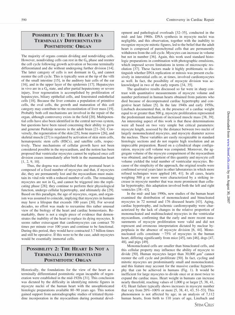
590 Controversy in Cardiac Repair
by on August 30, 2007
ww
w.Stem
Cells.com
Dow
nloaded from

Akuamoa-Boateng have shown that extreme forms of organhypertrophy are detectable up to the ninth decade of life, andheart weights of 500 and 600 g are present in patients at 100years of age and older [54].
Importantly, scar formation, foci of replacement fibrosisscattered throughout the ventricular wall, diffuse interstitialfibrosis, and ongoing myocyte death by apoptosis and necrosisare typically present in the hypertrophied failing heart [44, 55,56]. These factors markedly affect the number of cardiomyo-cytes and indicate that the extent of myocyte formation deter-mined by the morphometric methodologies summarized aboveconstitutes an underestimation of the actual magnitude of myo-cyte regeneration in the pathological heart. In addition, there isno alternative methodology able to demonstrate unequivocallymyocyte proliferation. By definition, myocyte replication cor-responds to an increase in the number of myocytes; in theabsence of this documentation, biochemical, molecular, physi-ological, autoradiographic, and structural results can only besuggestive of myocyte regeneration but in themselves cannotprovide the critical information, that is, an increased number ofcells. Quantitative morphology is the only technique that canidentify myocyte formation. Contrary to expectations, the ob-servations in favor of myocyte regeneration were not receivedwith enthusiasm by the scientific community [57–63]. Withsome small variations, the morphometric results in the humanheart were questioned, and the possibility of myocyte formationin the adult myocardium was first dismissed and subsequentlyattacked. This new idea challenged the comfortable paradigm ofmyocyte hypertrophy that had dominated the field of molecularcardiology and cell signaling for the previous 25 years. Heartfailure was viewed as the consequence of a defective myocytehypertrophy in which alterations of effector pathways regu-lating myocyte growth and contractility were considered re-sponsible for the depression in organ, tissue, and cell function[64, 65].
However, the quantitative data collected in the human heartsuggested that a reexamination of the mechanisms of myocytegrowth needed to be considered and that additional studiesneeded to be performed to clarify the apparently contradictoryresults. For this purpose, experiments were conducted in animalmodels of cardiac failure and in human diseased hearts toprovide relevant information in support of or against the notionof the regeneration potential of the adult myocardium. Newmethodologies were introduced to determine whether a subset ofcardiomyocytes had the ability to reenter the cell cycle anddivide and whether this process involved the activation of cellcycle-related genes, cyclins, and cyclin-dependent kinases andthe expression of proteins modulating karyokinesis and cytoki-nesis [45, 50, 55, 66–80]. Rounds of cell division result inprogressive telomere attrition, and critical telomeric shorteningtriggers irreversible growth arrest [81–83]. Thus, the telomere-telomerase axis, together with the telomere-binding proteins, isa fundamental component of the multiple mechanisms thatregulate the regeneration of non-postmitotic organs; because ofthis function, this system was evaluated and found to be oper-ative in the adult heart of animals and humans [50, 74–77, 79,
84]. Unfortunately, these multiple studies left the controversyunresolved.
MYOCYTE DIVISION
In 1925, a highly significant paper was published in which theclaim was made that there are no detectable mitotic figures inmyocytes of the human heart, and therefore myocyte regenera-tion does not occur in mammals [31]. This work challenged theview proposed in numerous studies published from 1850 to1911 in which the general belief was that cardiac hypertrophy isthe consequence of hyperplasia and hypertrophy of existingmyocytes [6]. The 1925 study can be considered the one thatintroduced the concept that the heart is a terminally differenti-ated postmitotic organ. The impact of this research was enor-mous. To the best of our knowledge, for several decades, manygenerations of pathologists and cardiovascular scientists haveaccepted the view that mitotic myocytes are not to be found inthe adult myocardium, and only a few rare exceptions have beenpublished, mostly restricted to the fetal-neonatal heart [85–88].
In the last 10–12 years, however, mitotic images in humancardiomyocytes have been documented in acute and chronicischemic cardiomyopathy [72, 89, 90], idiopathic dilated car-diomyopathy [72], chronic aortic stenosis with moderate ven-tricular dysfunction [50], myocardial aging [84], acromegaly(Fig. 2A), and diabetes (Fig. 2B). Using light microscopicexamination of routine histologic sections, an extensive searchwas required for the recognition of occasional mitotic figures inmyocytes. Because in the early successful observations, divid-ing myocytes were seen only in the fetal and diseased failingheart [89], the assumption was made that replicating myocytesare essentially undetectable in normal adult myocardium. Thishypothesis, in fact, suggested a rather minor role of myocyteregeneration in cardiac homeostasis and pathology in humans.However, in spite of the caution exercised in the interpretationof these results, this work was considered to reflect the incorrectinterpretation of proliferating interstitial fibroblasts as dividingmyocytes [1, 2]. Surprisingly, this conclusion was reached basedon electron microscopic images that illustrated nondividing fi-broblasts in proximity to differentiated nonreplicating cardio-myocytes [1]. This represents the normal organization of themyocardium and makes it difficult to understand how thiswell-established morphological characteristic of cardiac struc-ture can be used as an argument against myocyte division and acase in favor of its erroneous identification. So Artifact 1 wasintroduced in the literature as the only logical explanation forthese unexpected results.
An important point that necessitates discussion is thebelief by some that electron microscopy is the only tool thatcan unequivocally prove or disprove the presence of myocytemitotic images in the myocardium. In invited reviews ques-tioning the existence of myocyte regeneration, interstitialfibroblasts have been described as extremely mobile cellscapable of penetrating by several microns the thickness ofmyocytes, positioning themselves in the central portion of
Figure 1. Myocyte proliferation and isch-emic cardiomyopathy in humans. Small di-viding left ventricular myocytes (�-sarco-meric actin; red) are identified by Ki67labeling (A) (white, arrow), MCM5 expres-sion (B) (yellow, arrow), and metaphasechromosomes (C) (arrow). The boundariesof myocytes and interstitial cells are definedby laminin (green). Nuclei are stained by4,6-diamidino-2-phenylindole (blue).
591Anversa, Leri, Rota et al.
www.StemCells.com
by on August 30, 2007
ww
w.Stem
Cells.com
Dow
nloaded from

these cells [1, 2]. By this mechanism, the myocyte nucleuswas apparently displaced from its original location, whichwas occupied by the “penetrating fibroblast.” Schemes werecreated to strengthen this unlikely possibility [1]. However,this highly imaginative understanding of cardiac morphologyfell short on several accounts: the examples of “penetrating”interstitial cells did not show dividing cells, the myocytenucleus was at its expected site in the middle of the myocytecytoplasm, and the electron microscopic preparation was ofpoor quality. Myocytes were hypercontracted and had sarco-meres of �1.4 –1.5 �m in length. Normally, diastolic sarco-mere length is 2.1–2.2 �m, and during systolic contraction, itbecomes 1.8 –1.9 �m [91]. Hearts with sarcomeres �1.4 �min length are dead, since no contraction can be generatedunder this condition. Contraction bands produced by theinappropriate fixation of the myocardium were analyzed toreinterpret myocardial ultrastructure. Surprisingly, these ar-tifacts were accepted and introduced in the literature as facts.
The frequency of mitosis in myocytes of normal human,mouse, or rat heart, as assessed by confocal microscopy, is lowand averages 14/106 myocytes [72], 37/106 myocytes [45], and85/106 myocytes (J.K., unpublished data), respectively. Thevalues for dividing interstitial fibroblasts are similar to those ofcardiomyocytes [89]. Based on these data, �350, 140, and 60mm2 of tissue need to be examined to identify a dividingmyocyte in the human, mouse, and rat heart, respectively. Thisis why the suggestion to implement electron microscopy for thisanalysis is not realistic. The area of myocardium that is sampledin each thin section by electron microscopy is �0.2 mm2, andthe area of the microscopic field at a magnification of approx-imately �2,000 is 0.0054 mm2. Thus, the numbers of micro-graphs that would have to be collected to identify a singlemyocyte mitotic image are 66,000 pictures for humans, 25,000for mice, and 11,000 for rats. If one wishes to use electronmicroscopy to challenge the existence of myocyte division andto demonstrate that proliferating fibroblasts were improperlyinterpreted as mitotic myocytes, that is what needs to be in-vested to obtain at least one example in support of or against thisthesis [92].
Major advances in understanding myocyte replication inthe adult human heart have been made by the use of confocalmicroscopy and immunolabeling in the analysis of the normaland diseased heart. With this approach, the identification ofmitotic images in myocytes became easier and more accurate.These new results strengthened the notion that parenchymalcells are formed continuously in the normal myocardium andmyocyte regeneration is markedly potentiated in the patho-logical failing heart [6, 50, 72, 84, 90]. This technology was
also applied to the study of animal models of human diseases,and myocyte replication was unequivocally demonstrated inmyocardial sections and isolated cell preparations [6, 45, 73,93]. The latter observation conclusively refuted the claim thatdividing myocytes represent proliferating fibroblasts insertedwithin or attached to terminally differentiated myocytes. Theformation of the mitotic spindle by the arrangement of tubu-lin and the generation of the contractile ring by the accumu-lation of actin in the region of cytoplasmic separation wereclearly documented, together with karyokinesis and cytoki-nesis (Fig. 2C–2E). However, these results did not change theposition of certain groups, who continued to reject the regen-erative ability of the adult mammalian myocardium [2, 4, 8,9, 28]. These groups introduced the possibility of a newsource of artifacts, Artifact 2, related to confocal microscopy;they stated that traditional light microscopy is greatly supe-rior to confocal microscopy and, with this personal view-point, tried to invalidate the results obtained with the lattertechnique.
An important issue that requires discussion relates to thenotion that the analysis of histochemical preparations byconventional light microscopy is superior to fluorescencelabeling and confocal microscopy [94, 95]. The resolution ofthe micrographs provided by light microscopy is markedlyinferior to that obtained by confocal microscopy, �0.62versus 0.29 �m [37, 96]. In addition, only the very superficiallayer of a tissue section can be examined by light microscopy(Fig. 3). This inherent problem with light microscopy wasrecognized immediately when confocal technology becameavailable [97]. Clear examples were published in the Journalof Cell Biology in 1987 demonstrating the impossibility ofidentifying microtubules, mitotic spindle, cytoplasmic pro-teins, and chromosomal structures by light microscopy incultured cells. Conversely, these morphological details wereapparent when the same cells were examined by confocalmicroscopy [98]. The difference between epifluorescencelight microscopy and confocal microscopy is even greater intissue sections. When confocal microscopy was not availableto us, we estimated the myocyte mitotic index in chronicheart failure to be 11 myocytes per million [89]. Whencomparable hearts were evaluated by confocal microscopy,the mitotic index reached a value of 152 cells per million[72]. Even more striking is the difference between the mitoticindices obtained by light and confocal microscopy in theborder zone of acute infarcts in humans: light microscopyyielded 3.3 mitotic myocytes per million [89] and confocalmicroscopy 775 mitotic cells per million [90]. In all cases,
Figure 2. Myocyte generation in the dis-eased human heart. Metaphase chromosomes(blue, 4,6-diamidino-2-phenylindole [DAPI];arrows) are apparent in two small dividingcardiomyocytes (red, �-sarcomeric actin) inpatients affected by acromegaly (A) or diabetes(B). The organization of tubulin in the mitoticspindle (C) (white, arrows), the accumulationof actin in the contractile ring (D) (red, arrows),and cytokinesis (E) (arrows) in dividing cardi-omyocytes are apparent. In all cases, chromo-somes are labeled by DAPI (blue) and themyocyte cytoplasm by �-sarcomeric actin(red). Green indicates laminin.
592 Controversy in Cardiac Repair
by on August 30, 2007
ww
w.Stem
Cells.com
Dow
nloaded from

the measurements of myocytes in mitosis were made in thesame laboratory.
CARDIAC CHIMERISM AND
MYOCYTE FORMATION
The controversy concerning the recognition of dividing myo-cytes in the adult heart intensified when studies on cardiacchimerism were conducted in an attempt to determinewhether myocardial regeneration could be accounted for bythe activation of undifferentiated cells of recipient origin[99]. Although studies of the chimerism of the heart and otherorgans following sex-mismatched cardiac or bone marrowtransplantation have provided consistent results concerningthe migration of primitive cells from the host to the graft [94,95, 99 –114], findings from different laboratories have beeninconsistent in terms of the extent of this process, and thisvariability has promoted a heated debate in the scientificcommunity. After homing, host progenitor cells undergoreplication and differentiation generating mature parenchy-mal cells and vascular structures in the transplanted organ.There is little disagreement among authors on the occurrenceof this phenomenon in sex-mismatched heart transplant, butthe magnitude of cardiac chimerism varies significantly indifferent reports [94, 95, 99 –102, 104, 109, 110]. This dis-crepancy involves mostly ventricular myocytes and, to amuch lesser extent, newly formed coronary vessels. For myo-cytes, the published values range from as high as 18% to aslow as 0.02% or absent [99, 100, 104]. A plethora of inter-mediate results has also been obtained.
The surprising outcome of these findings was that the lowextent of myocyte chimerism detected by some groups wasused to question myocyte formation and to support the tra-ditional view that the heart is a terminally differentiatedpostmitotic organ incapable of undergoing meaningful regen-eration of any kind [2, 4, 8 –10]. However, to accomplish thistask, a rational explanation was necessary to account for thedata pointing to a high number of male myocytes withinfemale hearts transplanted in male recipients [99]. In fact,these observations indicated that a significant proportion ofmale myocytes was present in the female heart as a result ofactivation and commitment of the host male progenitor cells.Interestingly, the discrepancy in the degree of cardiac chi-merism was immediately adopted to initiate another contro-versy and introduced in the literature Artifact 3. The promot-ers of this new source of technical error reached thisconclusion by assuming that only the absence or minimallevels of chimerism were correct, whereas high levels ofchimerism had to be necessarily wrong [94, 95, 104, 115].The critics decided to take the prerogative of reinterpretingdata published by other groups, pointing to a number of crassmorphological errors. Surprisingly, these negative commentswere published and, once again, these questionable criticismsbecame facts.
The most obvious technical differences among the studiespublished so far is the use of conventional light microscopy [94,95, 100–102, 104] versus confocal microscopy [99, 109, 110].The probes used for the detection of the Y chromosome infemale hearts transplanted in male recipients were not alwaysthe same, and the procedures used for the recognition of the Ychromosome differed as well. These factors introduced criticalvariables in the acquisition of the data that, together with themethod of analysis, have been a major cause for the discrep-ancy.
A serious technical and conceptual concern is that similarhigh values of Y-chromosome-positive myocyte nuclei havebeen obtained in control male human hearts, averaging 50%.However, the values reported in control male human heartsby conventional fluorescence microscopy and confocal mi-croscopy in the absence of optical sectioning of the samplesare unrealistic and therefore cast doubts on this series ofstudies. By these two methods of analysis, the fraction ofY-chromosome-positive myocyte nuclei is low and rarelyexceeds 15% (Fig. 4A). This is due to the limited focal depthof the objective, �0.5 �m, [37] and the extent by which anucleus has to be embedded within the section to be visible(i.e., the penetration factor) [44]. Moreover, with conven-tional fluorescence microscopy, the out-of-focus fluores-cence blurs the image, complicating the identification of theY-chromosome signal in nuclei (Fig. 4B). When histologicsections of male myocardium are examined by confocalmicroscopy, the superficial layer of the section, which cor-responds to 0.6 �m, shows 12% Y-chromosome-positivemyocyte nuclei (Fig. 4C). Importantly, the evaluation of theentire thickness of the section by the analysis of consecutiveoptical planes markedly increases the detection of Y-chro-mosome-labeled myocyte nuclei, reaching a cumulative valueof 55% (Fig. 4D). Therefore, in several reports in whichconventional fluorescence microscopy or confocal micros-copy restricted to a single plane was used [94, 101, 102, 104],the finding of the Y chromosome in a large number ofmyocyte nuclei of control male hearts is at variance withreality. Because of this unacceptable premise, the extremelylow number or absence of Y-chromosome-positive myocytenuclei found in the transplanted female hearts can be seri-ously questioned.
The volume of a myocyte nucleus is �250 �m3, and thesignal for the Y chromosome has a volume of �0.5 �m3,occupying 0.2% of the entire nucleus. When the Y chromosomeis present in a mid-section of a nucleus, the area of the Y-chromosome signal is �1 �m2, and the area of the nucleusprofile is �75 �m2 [116]. Thus, the Y chromosome occupies�1.3% of the nuclear area (Fig. 4E). It follows that the proba-bility of hitting the Y chromosome in myocyte nuclear profilesof a tissue section can be calculated. Importantly, the chance ofa positive signal varies as a function of section thickness and isinfluenced by the orientation of the nuclei in myocytes. Thesevariables can be computed according to the following equations[44, 116].
Figure 3. Light epifluorescence microscopyof human myocardial tissue sections. Panelsshow different focal planes. (A): Superficiallayer of the section. (B): Focal plane located1 �m below the surface. (C): Focal planelocated 2 �m below the surface. Note thatsarcomere striations (green, �-sarcomericactin) and details of nuclear structure (red,propidium iodide) are well-defined only in(A) and are not clearly visible in (B) and (C).The yellowish area adjacent to the nucleusreflects the accumulation of lipofuscin.
593Anversa, Leri, Rota et al.
www.StemCells.com
by on August 30, 2007
ww
w.Stem
Cells.com
Dow
nloaded from

P � �dY/lN) � F
F � 1 � �k/�k � tS))
k � [wN � sin(�)] � [lN � cos(�)]
In these equations, P corresponds to the probability of finding aY-chromosome signal with a diameter dY within a nucleuswhose length is described by lN and width by wN. The parameterF represents the fraction of the nucleus embedded in the section.F is a function of the thickness of the section, tS, and theorientation and size of the nucleus, described by the variable k.The angle between the plane of the section and the long axis ofthe nucleus is given by �.
Supplemental online Table 1 lists the predicted minimal,maximal, and average values of Y-chromosome-positive myo-cyte nuclei in control male human hearts according to sectionthickness, nuclear orientation, and modality of analysis. Surpris-ingly, there is a dramatic inconsistency between the expectedvalues of Y-chromosome labeling of myocyte nuclei in normalconditions and in published results. Because of the questionablevalidity of control values in these studies, the number of malecells identified in female-transplanted hearts suffers from lackof reliability and necessitates reevaluation. This issue is cer-tainly more relevant than the claim concerning the difficulty ofdistinguishing myocyte nuclei from interstitial cell nuclei [1, 2,94, 115]. As recently emphasized, an experienced pathologistcan easily differentiate an interstitial cell from a cardiomyocyteor discriminate an interstitial cell nucleus from a myocyte nu-cleus, particularly by confocal microscopy [117]. This is con-firmed by the clear recognition of the Y-chromosome signal andnuclear profiles by confocal microscopy [99, 109, 110] and therather ill-defined Y-chromosome labeling and nuclear boundaryby light microscopy [94, 95, 104]. In some cases, by lightmicroscopy, the Y chromosome cannot be distinguished fromthe nucleolus [94] or properly identified because of little con-trast, weak intensity of the signal, and diffuse pattern of staining[104].
Several other factors have to be considered when humanmyocardium is examined. The age of the patients, time fromtransplantation, level of rejection, immunosuppressive regimen,and, most importantly, the condition of the tissue to be studiedmay all contribute to the variability in the levels of cardiacchimerism reported in the last several years. Poor fixation and/ordelayed fixation affect the integrity of the DNA and, thereby,the probability of detecting any nucleotide sequence [118].
DNA degradation precedes the alterations in protein antigenicity[119] so that Y-chromosome labeling of myocyte nuclei mayconstitute a significant underestimation of the actual degree ofmyocyte chimerism present in the organ [109, 117]. Likewise,prolonged preservation of the myocardium in formalin fixativeproduces excessive cross-linking of proteins [120], and thiscondition diminishes the accessibility of the Y-chromosomeprobe to the nuclear DNA [118, 121]. It is surprising that thesecrucial factors, together with elementary morphometric princi-ples, have been ignored in the studies pointing to the absence orminimal levels of cardiac chimerism following sex-mismatchedheart allograft or bone marrow transplantation.
An elegant protocol has been introduced in one study ofcardiac chimerism following sex-mismatched bone marrowtransplantation to correct, at least in part, this problem. Thefluorescence in situ hybridization (FISH) assay was performedwith two distinct probes, which detected, respectively, the Ychromosome and the X chromosome in myocyte nuclei [103,109]. Nuclei negative for the X chromosome had severelyaltered DNA and had to be excluded from the analysis. Whenthis approach was used, the extent of cardiomyocyte chimerismwas found to be 30-fold higher than in other comparable studies[108]. Because questions have been raised concerning some ofthe values obtained in our study of cardiac chimerism followingsex-mismatched heart transplantation [99], we have re-exam-ined cases with the highest levels of Y-chromosome-positivemyocyte nuclei using this dual-color FISH assay and evaluatedthe percentage of XX-positive and XY-positive myocyte nuclei(Fig. 5A, 5B). The original data were confirmed and strength-ened by this more sophisticated approach. In addition, the mea-surements of the number of X and Y chromosomes in nucleiallowed us to exclude cell fusion as the mechanism of malemyocyte formation in the female heart. Importantly, opticalsectioning by confocal microscopy prevented any possible mis-interpretation of interstitial cell nuclei as myocyte nuclei (Fig.5C–5L).
Together, the studies on cardiac chimerism in patientswho had sex-mismatched bone marrow transplantation pro-vide strong evidence that circulating progenitor cells mobi-lized from the bone marrow have the ability to repopulate theheart with cardiomyocytes and coronary vessels [103, 108,109]. The generation of parenchymal cells and vascular struc-tures mediated by engraftment and differentiation of primi-tive cells from the recipient is even greater following hearttransplantation [99, 109]. However, in this case, the source of
Figure 4. Detection of Y-chr. by fluores-cence in situ hybridization. (A): One myo-cyte nucleus positive for the Y-chr. (whitedot, arrow) is visible by conventional epi-fluorescence microscopy of 6-�m-thicksection of human male myocardium. Sevennuclei pertaining to myocytes (�-sarco-meric actin, red, arrowheads) are present inthe field. (B): Often, out-of-focus fluores-cence makes it difficult the recognition ofY-chr. labeling. The arrowhead points to anuclear dot that may correspond to theY-chr. signal. (C, D): Confocal micros-copy of a 6-�m-thick tissue section of hu-man male myocardium; the two panels il-lustrate the same field. (C): Superficiallayer of the section. (D): Combination of10 optical projections of 0.6 �m each ofthe entire section thickness. Arrowheadsindicate Y-chr.-positive myocyte nuclei in
(C) (2 of 15) and (D) (9 of 16). Laminin (green) defines the periphery of the cells. (E): High-magnification confocal image of a myocyte nucleuspositive for the Y-chr. The area occupied by the nucleus is 176 �m2, whereas the area occupied by the Y-chr. is 2.0 �m2, accounting for 1.1% ofthe nuclear cross-sectional area. Abbreviation: Y-chr., Y chromosome.
594 Controversy in Cardiac Repair
by on August 30, 2007
ww
w.Stem
Cells.com
Dow
nloaded from

the cells that translocate from the host to the implanted heartremains uncertain [99]. The progenitor cells could have beenof hematopoietic origin and migrated to the heart through thesystemic circulation, or they could have come from thecardiac remnant preserved during surgery for the attachmentof the donor heart [122]. These possibilities are not mutuallyexclusive.
The recognition that cardiac chimerism occurs rapidlyand in a noticeable manner has had a dramatic impact on ourunderstanding of the biology of the heart and mechanisms ofmyocardial repair. This work has formed the foundation forthe concept of myocardial regeneration and has created theopportunity for testing the role that HSCs have in the resto-ration of the injured heart [123]. The notion that HSCs retain
a significant degree of developmental plasticity and canacquire cell lineages distinct from the organ of origin haspromoted the entire field of cell therapy of the failing heart[124]. Opponents of this emerging new paradigm in cardiol-ogy, however, continued to claim that these concepts are theproduct of artifacts [1, 2, 10, 94, 95, 108, 125]. A recurrentunqualified statement is that they have introduced in theirwork “rigorous” and “stringent” criteria, which implies thatothers do not adhere to the same high scientific standards.The old paradigm has to survive, and the notion that myo-cytes cannot be generated postnatally, in adulthood or senes-cence, must be defended. An example of this approach can befound in a recent report in which levels of EC chimerism of25% and vascular SMC chimerism of 3.5% have been found
Figure 5. Cardiac chimerism in humans. Confocal images of a tissue section obtained from a female heart transplanted in a male recipient. (A, B):Nuclei (4,6-diamidino-2-phenylindole, blue) are positive for the X chromosome (magenta dots) and Y chromosome (white dots, arrowheads). Thecoexpression of X and Y chromosomes in the same nucleus indicates male diploid genotype. The majority of nuclei belong to myocytes (B)(�-sarcomeric actin, red). (B): Open arrow points to a male myocyte. The periphery of the cells is defined by laminin (green). (C–L): Y chromosome(white dots) and X chromosome (magenta dots) are detected in nine distinct optical sections 1 �m apart (D–L) of the area indicated by the inset in(C). The Y chromosome in the myocyte nucleus (C) (arrowhead) is not visible in (D) and (E) but is evident in (F–H). It is no longer present in (G–L).The X chromosome in the same myocyte nucleus is apparent only in (J) and (K). The nucleus of an interstitial cell is also visible in (C) (asterisk).
595Anversa, Leri, Rota et al.
www.StemCells.com
by on August 30, 2007
ww
w.Stem
Cells.com
Dow
nloaded from

in sex-mismatched heart transplants in the absence(0.0016%) of myocyte chimerism [95].
HEMATOPOIETIC STEM CELLS AND
MYOCYTE FORMATION
In the last decade, major discoveries have been made concerningthe biology of adult HSCs. They can differentiate into celllineages distinct from the organ in which they reside and intocells derived from a different germ layer [126]. These propertieswere considered to be restricted to embryonic stem cells, andbecause of their dramatic biological and clinical implications,they triggered a vigorous debate in the scientific community.The controversy reached unexpectedly high levels of intensitywhen HSCs were shown to be able to migrate to sites of injuryrepairing damage in various organs, in particular the brain andthe heart [127–129]. The brain and the heart have always beenviewed as nonpermissive organs for exogenous and endogenoustissue regeneration. Studies presenting positive results [127,129–135] were immediately confronted by negative reports[136–140], which challenged stem cell plasticity and the emerg-ing new paradigm of organ and organism homeostasis and repair
[141–143]. Although the possibility that HSCs differentiate intoPurkinje neurons was questioned based on data interpretation orinability to reproduce similar findings, a different reaction oc-curred in response to the documentation of the efficacy of adultHSCs in myocardial regeneration after infarction. Several lab-oratories have used genetically labeled HSCs from transgenicanimals to promote the restoration of infarcted myocardiumexperimentally to identify the progeny formed [129, 133–135].HSCs expressing enhanced green fluorescence protein (EGFP)and sex-mismatched transplantation have been used frequentlyto have two independent markers for the recognition of newlygenerated myocardium: EGFP expression and Y-chromosomelabeling [129, 134, 135]. 5-Bromo-2�-deoxyuridine (BrdU) wasalso administered chronically to animals after infarction and celltreatment to have a third marker of cell accumulation within theinfarcted region (Fig. 6A–6F).
Following the detection of EGFP-, Y-chromosome-, andBrdU-positive cells, transcription factors and cytoplasmic pro-teins specific to myocytes and vascular smooth muscle andendothelial cells were demonstrated (Fig. 6G–6O) so as to avoidpotential questions on the validity and accuracy of the results[129, 135]. However, these observations were attacked on meth-odological grounds and were claimed to be the product of a new
Figure 6. Bone marrow cell transdifferentiation. Male c-kit-positive bone marrow cells were obtained from EGFP-transgenic mice and injectedshortly after infarction in the female mouse heart. (A–C): The regenerated myocardium is composed of small myocytes positive for �-actinin (A, C)(red) that express EGFP (B, C) (green) and have a male genotype (C) (Y chromosome, white dots). (D–F): Again, the new myocytes express �-actinin(D, F) (red) and EGFP (E, F) (green). The vast majority of myocyte nuclei are positive for the cell cycle marker BrdU (F) (white). (G–I): Theregenerated myocytes express �-actinin (G, I) (red) and EGFP (H, I) (green), and the nuclei are positive for MEF2C (I) (yellow). (J–L): A newlyformed arteriole (J, L) (�-smooth muscle actin, red) expresses EGFP (K, L) (green); smooth muscle cells are positive for GATA-6 (L) (white).(M–O): The formed capillaries (M, O) (vWf, red) are EGFP-positive (N, O) (green), and endothelial cells express the transcription factor Ets1 (O)(white). Abbreviations: BrdU, 5-bromo-2�-deoxyuridine; EGFP, enhanced green fluorescence protein; vWf, von Willebrand factor.
596 Controversy in Cardiac Repair
by on August 30, 2007
ww
w.Stem
Cells.com
Dow
nloaded from

artifact [2, 9, 10, 138, 139, 144]. The characteristics of Artifact4 were unexpected because a new twist was introduced in the“erroneous” interpretation of morphological images obtained byimmunolabeling and confocal microscopy: autofluorescence.How these multiple markers of myocardial reconstitution byHSCs could all be the result of autofluorescence is an interestingissue, which unfortunately was never addressed. The autofluo-rescence was introduced by the same authors who raised theissues of Artifacts 1–3 and who, this time, joined forces withexperimental hematologists opposing the concept of HSC plas-ticity [8]. So the never-documented autofluorescence artifactwas invoked as the only logical explanation for the possibilitythat cardiac repair can be accomplished by exogenous cells.
Criticisms have focused on the need to use genetic markersto demonstrate whether HSCs can form heart muscle [2–4, 9,10]. Genetic markers have been used in the questioned positivestudies [129, 135], as well as in the negative reports that havebeen claimed to be valid [138–140]. Both positive and negativepublications used immunostaining, but in the former case,autofluorescence was assumed to be a factor, whereas in thelatter case, the technique used was considered immaculate.Unfortunately, the basis for this conclusion remains an enigma.Although EGFP labeling was applied in several of the papersthat opposed HSC plasticity [139, 140, 145], one of theselaboratories introduced the notion that green autofluorescence iscommonly present in skeletal muscle samples and therefore thedetection of EGFP-positive cells in the heart or skeletal muscle
following implantation of EGFP-positive HSCs is by necessityan artifact [146]. This concept was adopted immediately by theInternational Society of Stem Cell Research [147] and empha-sized in original studies and reviews [2, 10].
The rationale for Artifact 5 is incomprehensible. The greenautofluorescence observed in histologic sections of skeletalmuscle was largely due to the use of an inappropriate fixationprotocol for immunolabeling studies. A mixture of paraformal-dehyde and glutaraldehyde (Fig. 7A) was used rather thanphosphate-buffered formalin (Fig. 7B), which carries minimalbackground autofluorescence. But most importantly, immuno-labeling of glutaraldehyde-fixed tissue never works (Fig. 7C,7D) because of the high level of cross-linking of proteinsgenerated by glutaraldehyde [148]. Green autofluorescence canbe clearly distinguished from EGFP-expressing cells by the useof specific green fluorescence protein antibodies as opposed todirect identification of the EGFP protein by epifluorescencelight microscopy (Fig. 7E–7G). Therefore, immunolabeling canbe easily optimized so that the emission signal is orders ofmagnitude greater than background fluorescence, or autofluo-rescence can be totally excluded by direct labeling of antibodieswith quantum dots (Fig. 7H–7K).
What remains unclear is the ultimate goal of these unqual-ified criticisms. They add to the already enormous amount ofconfusion and uncertainty present in the field of regenerativemedicine. The critics of myocardial and skeletal muscle repairby exogenous or endogenous stem cells continue to implement
Figure 7. Autofluorescence of tissue sections and detection of EGFP in skeletal muscle. Autofluorescence (green) of tissue sections obtained fromskeletal muscle after fixation with paraformaldehyde and glutaraldehyde (A) or formalin (B), both in the absence of immunolabeling. Nuclei arestained with 4,6-diamidino-2-phenylindole (blue). (C, D): After fixation with paraformaldehyde and glutaraldehyde, skeletal muscle tissue sectionswere labeled with �-skeletal actin (C) and EGFP (D) antibodies. The lack of sarcomere striation indicates that only unspecific labeling of these twoantigens was obtained with this protocol. The signals in (C) and (D) correspond to autofluorescence. In this regard, compare (D) with (A). Afterfixation with formalin only, skeletal muscle tissue sections were labeled with �-skeletal actin (E, G) (red) and EGFP (F, G) (green) antibodies. Thepresence of striations in the cell cytoplasm indicates that specific labeling was obtained by this approach. Note the coexistence of EGFP-positive andEGFP-negative fibers in the same muscle. (H–J): After fixation with formalin, skeletal muscle tissue sections were labeled with �-skeletal actin (H,J) (red) and EGFP (I, J) (green) antibodies conjugated with quantum dots. The presence of striations in the cell cytoplasm indicates the specificityof labeling. With this approach, the autofluorescence of a tissue section can be totally avoided: the confocal image illustrated in (K) was acquiredwith the excitation and emission wavelengths used for quantum dot 655. Note the complete absence of autofluorescence. Importantly, the excitationand emission wavelengths used for quantum dot analysis are distinct from the autofluorescence wavelength of tissue sections. Abbreviation: EGFP,enhanced green fluorescence protein.
597Anversa, Leri, Rota et al.
www.StemCells.com
by on August 30, 2007
ww
w.Stem
Cells.com
Dow
nloaded from

in their work the same methodologies used by the investigatorsbeing criticized. The bias lies in the preconceived notion thatimmunolabeling and confocal microscopy are good for some butbad for others. We have examined two recent issues each ofNature, Nature Medicine, Science, and Proceedings of the Na-tional Academy of Sciences of the United States of America andfound that 7 of 17, 13 of 25, 3 of 11, and 14 of 22 publicationsin biomedical sciences, respectively, have used immunostainingand confocal microscopy. Remarkably, in none of these 37studies was the issue of autofluorescence discussed as a sourceof artifacts.
CARDIAC STEM CELLS AND
MYOCYTE FORMATION
In the last 3 years, several laboratories have identified progen-itor cells in the adult heart of small and large mammals, includ-ing humans. Cardiac progenitor cells are multipotent in vitro andgive rise to cardiomyocytes and coronary vessels in vivo, thuspossessing the fundamental properties of stem cells [13, 48,149–153]. The recognition that stem cells reside in the myocar-dium had a dramatic impact on the field of cardiology from abiological and clinical perspective. This discovery has providedthe missing link between the documentation of small dividingmyocytes [6, 7, 12] and the uncertainty concerning the origin ofthese repopulating cells, and it has laid the groundwork for thepossibility of introducing the use of these cells in the treatmentof the failing human heart [12, 154]. A number of laboratorieshave invested resources and staff in exploring this novel andradically different perspective of cardiac pathophysiology [151,152, 154]. Because of these efforts, major advances have beenmade, and preclinical studies are in progress with the objectiveof rapidly implementing this technology in humans.
The discovery that cardiac stem cells (CSCs) are present inthe adult myocardium led to the proposal of a new artifact. Theformulation of Artifact 6 required vivid imagination and unusuallogic; since myocytes cannot be formed [1, 2], the preparationsof CSCs must be a product of an artifact associated with the invitro isolation [10]. The critics then go on to suggest that thisartifact may, however, have some positive outcomes. But toquestion the presence and role of CSCs in the adult heart,
Artifact 6 could not be restricted to the in vitro work and had tobe extended to the demonstration that CSCs regenerate infarctedmyocardium in vivo [13]. This task was accomplished by statingthat cells that expressed cardiac myosin heavy chain, connexin43, and N-cadherin and responded to electrical stimulation byshortening and relengthening were in reality fibroblasts [10].
In summary, the controversy concerning stem cell therapyfor the damaged heart has its foundations in the debate thatoriginated nearly 80 years ago when it was concluded thatmyocardial regeneration ceases at birth and cannot occur inadulthood or senescence [31]. The biochemical characterizationof cardiac hypertrophy performed in the late 1960s and 1970shas contributed to reaffirming the notion that myocardial growthpostnatally can be accomplished only by enlargement of thepre-existing myocytes [36], strengthening the notion of the heartas an organ formed by a predetermined number of myocytesincapable of reentering the cell cycle and dividing. Molecularcardiology was built on this inherited inviolable paradigm [57],which has been strongly supported by traditional cardiovascularpathologists reluctant to introduce modern technology in theanalysis of the structure of the normal and diseased heart.Surprisingly, myocyte death and myocyte formation, which arethe two critical variables that control cardiac cell number, arerarely evaluated concurrently to obtain a dynamic view of theheart in its various phases of life [45, 75, 76, 155]. By thissimple approach, it would become clear whether the heart canfunction with the same cells throughout the lifespan of the organand organism or whether an alternative possibility has to beconsidered.
ACKNOWLEDGMENTS
Supported by NIH Grants HL-38132, AG-15756, HL-65577,HL-55757, HL-68088, HL-70897, HL-76794, HL-66923, HL-65573, HL-75480, AG-17042, and AG-23071.
DISCLOSURES
The authors indicate no potential conflicts of interest.
REFERENCES
1 Soonpaa MH, Field LJ. Survey of studies examining mammalian car-diomyocyte DNA synthesis. Circ Res 1998;83:15–26.
2 Rubart M, Field LJ. Cardiac regeneration: Repopulating the heart. AnnuRev Physiol 2006;68:29–49.
3 Menasche P. You can’t judge a book by its cover. Circulation 2006;13:1275–1277.
4 Murry CE, Field LJ, Menasche P. Cell-based cardiac repair reflectionsat the 10-year point. Circulation 2005;112:3174–3183.
5 Linzbach AJ. Heart failure from the point of view of quantitativeanatomy. Am J Cardiol 1960;5:370–382.
6 Anversa P, Kajstura J. Ventricular myocytes are not terminally differ-entiated in the adult mammalian heart. Circ Res 1998;83:1–14.
7 Anversa P, Kajstura J, Leri A et al. Life and death of cardiac stem cells.A paradigm shift in cardiac biology. Circulation 2006;113:1451–1463.
8 Wagers AJ, Weissman IL. Plasticity of adult stem cells. Cell 2004;116:639–648.
9 Chien KR. Stem cells: Lost in translation. Nature 2004;428:607–608.10 Laflamme MA, Murry CE. Regenerating the heart. Nat Biotechnol
2005;23:845–856.11 Menard C, Hagege AA, Onnik Agbulut O et al. Transplantation of
cardiac-committed mouse embryonic stem cells to infarcted sheep myo-cardium: A preclinical study. Lancet 2005;366:1005–1012.
12 Leri A, Kajstura J, Anversa P. Cardiac stem cells and mechanisms ofmyocardial regeneration. Physiol Rev 2005;85:1373–1416.
13 Beltrami AP, Barlucchi L, Torella D et al. Adult cardiac stem cellsare multipotent and support myocardial regeneration. Cell 2003;114:763–776.
14 Zhu O, Skoultchi AI. Coordinating cell proliferation and differentiation.Curr Opin Genet Dev 2001;1:91–97.
15 Mariadason JM, Nicholas C, L’Italien KE et al. Gene expression pro-filing of intestinal epithelial cell maturation along the crypt-villus axis.Gastroenterology 2005;4:1081–1088.
16 Chen P, Zindy F, Abdala C et al. Progressive hearing loss in micelacking the cyclin-dependent kinase inhibitor Ink4a. Nat Cell Biol2003;5:385–387.
17 Hotchin NA, Gandarillas A, Watt FM. Regulation of cell surface beta 1integrin levels during keratinocyte terminal differentiation. Cell Biol1995;6:1209–1219.
18 Taub R. Liver regeneration: From myth to mechanism. Nat Rev MolCell Biol 2004;5:836–847.
19 Vessey CJ, de la Hall PM. Hepatic stem cells: A review. Pathology2001;33:130–141.
20 Fausto N, Campbell JS. The role of hepatocytes and oval cells in liverregeneration and repopulation. Mech Dev 2003;120:117–130.
21 Eriksson PS, Perfilieva E, Bjork-Eriksson T et al. Neurogenesis in theadult human hippocampus. Nat Med 998;4:1313–1317.
22 Sanai N, Tramontin AD, Quinones-Hinojosa A et al. Unique astrocyteribbon in adult human brain contains neural stem cells but lacks chainmigration. Nature 2004;427:740–744.
23 Rakie P. Neuroscience: Immigration denied. Nature 2004;427:685–686.24 Lie DC, Colamarino SA, Song HJ et al. Wnt signalling regulates adult
hippocampal neurogenesis. Nature 2005;437:1370–1375.
598 Controversy in Cardiac Repair
by on August 30, 2007
ww
w.Stem
Cells.com
Dow
nloaded from

25 Blanpain C, Lowry WE, Geoghegan A et al. Self-renewal, multipo-tency, and the existence of two cell populations within an epithelial stemcell niche. Cell 2004;118:635–648.
26 Uchida N, Tsukamoto A, He D et al. High doses of purified stem cellscause early hematopoietic recovery in syngeneic and allogeneic hosts.J Clin Invest 1998;101:961–966.
27 Dhawan J, Rando TA. Stem cells in postnatal myogenesis: Molecularmechanisms of satellite cell quiescence, activation and replenishment.Trends Cell Biol 2005;15:666–673.
28 Nakamura T, Schneider MD. The way to a human’s heart is through thestomach. Circulation 2003;107:2638–2639.
29 MacLellan WR, Schneider MD. Genetic dissection of cardiac growthcontrol pathways. Annu Rev Physiol 2000;62:289–319.
30 Anversa P, Rota M, Urbanek K et al. Myocardial aging: A stem cellproblem. Basic Res Cardiol 2005;100:482–493.
31 Karsner HT, Saphir O, Todd TW. The state of the cardiac muscle inhypertrophy and atrophy. Am J Pathol 1925;1:351–371.
32 Petersen RO, Baserga R. Nucleic acid and protein synthesis in cardiacmuscle of growing and adult mice. Exp Cell Res 1965;40:340–352.
33 Morkin E, Ashford TP. Myocardial DNA synthesis in experimentalcardiac hypertrophy. Am J Physiol 1968;215:1409–1413.
34 Grove D, Nair KG, Zak R. Biochemical correlates of cardiac hypertro-phy. 3. Changes in DNA content: The relative contributions ofpolyploidy and mitotic activity. Circ Res 1969;25:463–471.
35 Grove D, Zak R, Nair KG et al. Biochemical correlates of cardiachypertrophy. IV. Observation on the cellular organization of growthduring myocardial hypertrophy in the rat. Circ Res 1969;25:473–485.
36 Zak R. Development and proliferative capacity of cardiac muscle cells.Circ Res 1974;35:17–26.
37 Spector DL, Goldman, RD, Leinwand LA. Light microscopy. In: Spec-tor DL, Goldman, RD, Leinwand LA, eds. Cells: Light Microscopy andCell Structure. Cold Spring Harbor, NY: Cold Spring Harbor Labora-tory Press, 1997:94.1–94.53.
38 Linzbach AJ. Mikrometrische und histologische Analyse hypertrophermenschlicher Herzen. Virchow Arch Pathol Anat Physiol 1947;314:534–594.
39 Linzbach AJ. Muskelfaserkonstante und das Wachstumsgesetz der men-schlichen Herzkammern. Virchows Arch 1950;318:575–618.
40 Astorri E, Chizzola A, Visioli O et al. Right ventricular hypertrophy: Acytometric study on 55 human hearts. J Mol Cell Cardiol 1971;2:99–110.
41 Astorri E, Bolognesi R, Colla B et al. Left ventricular hypertrophy: Acytometric study on 42 human hearts. J Mol Cell Cardiol 1977;9:763–775.
42 Adler CP, Friedburg H. Myocardial DNA content, ploidy level and cellnumber in geriatric hearts: Post-mortem examination of human myo-cardium in old age. J Mol Cell Cardiol 1986;18:39–53.
43 Olivetti G, Cigola E, Maestri R et al. Aging, cardiac hypertrophy andischemic cardiomyopathy do not affect the proportion of mononucleatedand multinucleated myocytes in the human heart. J Mol Cell Cardiol1996;29:1463–1477.
44 Anversa P, Olivetti G. Cellular basis of physiological and pathologicalmyocardial growth. In: Fozzard HA, Solaro RJ, eds. Handbook ofPhysiology: The Cardiovascular System. The Heart. New York: OxfordUniversity Press, 2002:75–144.
45 Limana F, Urbanek K, Chimenti S et al. bcl-2 overexpression promotesmyocyte proliferation. Proc Natl Acad Sci U S A 2002;99:6257–6262.
46 Anversa P, Palackal T, Sonnenblick EH et al. Myocyte cell loss andmyocyte cellular hyperplasia in the hypertrophied aging rat heart. CircRes 1990;67:871–885.
47 Kajstura J, Zhang X, Liu Y et al. The cellular basis of pacing-induceddilated cardiomyopathy. Myocyte cell loss and myocyte cellular reac-tive hypertrophy. Circulation 1995;92:2306–2317.
48 Linke A, Muller P, Nurzynska D et al. Stem cells in the dog heart areself-renewing, clonogenic, and multipotent and regenerate infarctedmyocardium, improving cardiac function. Proc Natl Acad Sci U S A2005;102:8966–8971.
49 Spinale FG, Zellner JL, Tomita A et al. Relation between ventricularand myocyte remodeling with the development and regression of su-praventricular tachycardia-induced cardiomyopathy. Circ Res 1991;69:1058–1067.
50 Urbanek K, Quaini F, Tasca G et al. Intense myocyte formation fromcardiac stem cells in human cardiac hypertrophy. Proc Natl Acad SciU S A 2003;100:10440–10445.
51 Adler C-P, Costabel U. Cell number in human heart in atrophy, hyper-trophy, and under the influence of cytostatics. In: Fleckenstein A, RomaG, eds. Recent Advances in Studies on Cardiac Structure and Metabo-lism: Pathophysiology and Morphology of Myocardial Cell Alteration.Baltimore, MD: University Park Press,1975:343–355.
52 Grajek S, Lesiak M, Pyda M et al. Hypertrophy or hyperplasia in cardiacmuscle: Postmortem human morphometric study. Eur Heart J 1993;14:40–47.
53 Olivetti G, Cigola E, Maestri R et al. Myocyte nuclear and possible cellularhyperplasia contribute to ventricular remodeling in the hypertrophic senes-cent heart in humans. J Am Coll Cardiol 1994;24:140–149.
54 Linzbach AM, Akuamoa-Boateng E. Die Alternsveranderungen desmenschlichen Herzens I. Die Herzgewicht im Alter. Klin Wochenschr1973;51:156–163.
55 Beltrami CA, Finato N, Rocco M et al. Structural basis of end-stagefailure in ischemic cardiomyopathy in humans. Circulation 1994;89:151–163.
56 Beltrami CA, Finato N, Rocco M et al. The cellular basis of dilatedcardiomyopathy in humans. J Mol Cell Cardiol 1995;27:291–305.
57 Nadal-Ginard B, Mahdavi V. Molecular basis of cardiac performance,plasticity of the myocardium generated through protein isoformswitches. J Clin Invest 1989;84:1693–1700.
58 Tam SK, Gu W, Mahdavi V et al. Cardiac myocyte terminal differen-tiation, potential for cardiac regeneration. Ann N Y Acad Sci 1995;752:72–79.
59 Soonpaa MH, Daud AI, Koh GY et al. Potential approaches for myo-cardial regeneration. Ann N Y Acad Sci 1995;752:446–454.
60 Chien KR. Cardiac muscle diseases in genetically engineered mice: Evo-lution of molecular physiology. Am J Physiol 1995;269:H755–H766.
61 Schneider MD. Myocardial infarction as a problem of growth control:Cell cycle therapy for cardiac myocytes? J Card Fail 1996;2:259–263.
62 Sugden PH. Signaling in myocardial hypertrophy: Life after cal-cineurin? Circ Res 1999;84:633–646.
63 Von Harsdorf Rudiger. Can cardiomyocytes divide? Heart 2001;86:481–482.
64 Pasumarthi KB, Field LJ. Cardiomyocyte cell regulation. Circ Res 200290:1044–1054.
65 Field LJ. Modulation of the cardiomyocyte cell cycle in geneticallyaltered animals. Ann N Y Acad Sci 2004;1015:160–170.
66 Marino TA, Haldar S, Williamson EC et al. Proliferating cell nuclearantigen in developing and adult rat cardiac muscle cells. Circ Res1991;69:1353–1360.
67 Reiss K, Cheng W, Ferber A et al. Overexpression of insulin-likegrowth factor-1 in the heart is coupled with myocyte proliferation intransgenic mice. Proc Natl Acad Sci U S A 1996;93:8630–8635.
68 Baba HA, Takeda A, Schmid C et al. Early proliferative changes inhearts of hypertensive Goldblatt rats: An immunohistochemical andflow-cytometrical study. Basic Res Cardiol 1996;91:275–282.
69 Reiss K, Cheng W, Giordano A et al. Myocardial infarction is coupledwith the activation of cyclins and cyclin-dependent kinases in myocytes.Exp Cell Res 1996;225:44–54.
70 Soonpaa MH, Koh GY, Pajak L et al. Cyclin D1 overexpressionpromotes cardiomyocytes DNA synthesis and multinucleation in trans-genic mice. J Clin Invest 1997;99:2644–2654.
71 Agah R, Kirshenbaum LA, Abdellatif M et al. Adenoviral delivery ofE2F-1 directs cell cycle reentry and p53-independent apoptosis in post-mitotic adult myocardium in vivo. J Clin Invest 1997;100:2722–2728.
72 Kajstura J, Leri A, Finato N et al. Myocyte proliferation in end-stagecardiac failure in humans. Proc Natl Acad Sci U S A 1998;95:8801–8805.
73 Setoguchi M, Leri A, Wang S et al. Activation of cyclins and cyclin-dependent kinases, DNA synthesis, and myocyte mitotic division inpacing-induced heart failure in dogs. Lab Invest 1999;79:1545–1548.
74 Leri A, Malhotra A, Liew CC et al. Telomerase activity in rat cardiacmyocytes is age and gender dependent. J Mol Cell Cardiol 2000;32:385–390.
75 Leri A, Barlucchi L, Limana F et al. Telomerase expression and activity arecoupled with myocyte proliferation and preservation of telemetric length inthe failing heart. Proc Natl Acad Sci U S A 2001;98:8626–8631.
76 Leri A, Franco S, Zacheo A et al. Ablation of telomerase and telomereloss leads to cardiac dilatation and heart failure associated with p53upregulation. EMBO J 2003;22:131–139.
77 Torella D, Rota M, Nurzynska D et al. Cardiac stem cell and myocyteaging, heart failure, and insulin-like growth factor-1 overexpression.Circ Res 2004;94:514–524.
78 Pasumarthi KB, Nakajima H, Nakajima HO et al. Targeted expressionof cyclin D2 results in cardiomyocytes DNA synthesis and infarctregression in transgenic mice. Circ Res 2005;96:110–118.
79 Urbanek K, Torella D, Sheikh F et al. Myocardial regeneration byactivation of multipotent cardiac stem cells in ischemic heart failure.Proc Natl Acad Sci U S A 2005;102:8692–8697.
80 Ledda-Columbano GM, Molotzu F, Pibiri M et al. Thyroid hormoneinduces cyclin D1 nuclear translocation and DNA synthesis in adult ratcardiomyocytes. FASEB J 2006;20:87–94.
81 Samper E, Fernandez P, Martin-Rivera L et al. Long-term repopulatingability of telomerase-deficient murine hematopoietic stem cells. Blood2002;99:2767–2775.
82 Baerlocher GM, Roth A, Lansdorp PM. Telomeres in hematopoieticstem cells. Ann N Y Acad Sci 2003;996:44–48.
83 Flores I, Cayuela ML, Blasco MA. Effects of telomerase and telomerelength on epidermal stem cell behavior. Science 2005;309:1253–1256.
84 Chimenti C, Kajstura J, Torella D et al. Senescence and death ofprimitive cells and myocytes lead to premature cardiac aging and heartfailure. Circ Res 2003;93:604–613.
599Anversa, Leri, Rota et al.
www.StemCells.com
by on August 30, 2007
ww
w.Stem
Cells.com
Dow
nloaded from

85 Klinge O. DNS-Synthese und Kernteilung im normalen und im infarz-ierten Rattenherzen. Verh Dtsch Ges Path 1967;51:157–161.
86 Manasek FJ. Mitosis in developing cardiac muscle. J Cell Biol 1968;37:191–196.
87 Anversa P, Vitali-Mazza L, Loud AV. Morphometric and autoradio-graphic study of developing ventricular and atrial myocardium in fetalrats. Lab Invest 1975;33:696–705.
88 Li F, Wang X, Capasso JM et al. Rapid transition of cardiac myocytesfrom hyperplasia to hypertrophy during postnatal development. J MolCell Cardiol 1996;28:1737–1746.
89 Quaini F, Cigola E, Lagrasta C et al. End-stage cardiac failure inhumans is coupled with the induction of proliferating cell nuclearantigen and nuclear mitotic division in ventricular myocytes. Circ Res1994;75:1050–1063.
90 Beltrami AP, Urbanek K, Kajstura J et al. Evidence that human cardiacmyocytes divide after myocardial infarction. N Engl J Med 2001;344:1750–1757.
91 Sonnenblick EH, Spiro D, Cottrell TS. Fine structural changes in heartmuscle in relation of the length-tension curve. Proc Natl Acad SciU S A 1963;49:193–200.
92 Anversa P. Myocyte death in the pathological heart. Circ Res 2000;86:121–124.
93 Kajstura J, Zhang X, Reiss K et al. Myocyte cellular hyperplasia andmyocyte cellular hypertrophy contribute to chronic ventricular remod-eling in coronary artery narrowing-induced cardiomyopathy in rats. CircRes 1994;74:383–400.
94 Laflamme MA, Myerson D, Saffitz JE et al. Evidence for cardiomyo-cyte repopulation by extracardiac progenitors in transplanted humanhearts. Circ Res 2002;90:634–640.
95 Minami E, Laflamme A, Saffitz JE et al. Extracardiac progenitor cellsrepopulate most major cell types in the transplanted human heart.Circulation 2005;112:2951–2958.
96 Carter D. Practical considerations for collecting confocal images. Meth-ods Mol Biol 1999;122:35–57.
97 Sheppard CJ, Wilson T. The theory of the direct-view confocal micro-scope. J Microsc 1981;124:107–117.
98 White JG, Amos WB, Fordham M. An evaluation of confocal versusconventional imaging of biological structures by fluorescence lightmicroscopy. J Cell Biol 1987;105:41–48.
99 Quaini F, Urbanek K, Beltrami AP et al. Chimerism of the transplantedheart. N Engl J Med 2002;346:5–15.
100 Hruban RH, Long PP, Perlman EJ et al. Fluorescence in situ hybrid-ization for the Y-chromosome can be used to detect cells of recipientorigin in allografted hearts following cardiac transplantation. Am JPathol 1993;142:975–980.
101 Muller P, Pfeidder P, Koglin J et al. Cardiomyocytes of noncardiacorigin in myocardial biopsies of human transplanted hearts. Circulation2002;106:31–35.
102 Bayes-Genis A, Salido M, Sole Ristol F et al. Host cell-derived cardi-omyocytes in sex-mismatched cardiac allografts. Cardiovasc Res 2002;56:404–410.
103 Thiele J, Varus E, Wickenhauser C et al. Chimerism of cardiomyocytesand endothelial cells after allogeneic bone marrow transplantation inchronic myeloid leukemia. An autopsy study [in German]. Pathologe2002;23:405–410.
104 Glaser R, Lu MM, Narula N et al. Smooth muscle cells, but notmyocytes, of host origin in transplanted human hearts. Circulation2002;106:17–19.
105 Korbling M, Katz RL, Khanna A et al. Hepatocytes and epithelial cellsof donor origin in recipients of peripheral-blood stem cells. N EnglJ Med 2002;346:738.
106 Caplice NM, Bunch TJ, Stalboerger PG et al. Smooth muscle cells inhuman coronary atherosclerosis can originate from cells adminis-tered at marrow transplantation. Proc Natl Acad Sci U S A 2003;100:4754 – 4759.
107 Kleeberger W, Versmold A, Rothamel T et al. Increased chimerism ofbronchial and alveolar epithelium in human lung allografts undergoingchronic injury. Am J Pathol 2003;162:1487–1494.
108 Deb A, Wang S, Skelding KA et al. Bone marrow-derived cardio-myocytes are present in adult human heart. Circulation 2003;107:1245–1247.
109 Thiele J, Varus E, Wickenhauser C et al. Mixed chimerism of cardio-myocytes and vessels after allogeneic bone marrow and stem-cell trans-plantation in comparison with cardiac allografts. Transplantation 2004;77:1902–1905.
110 Hocht-Zeisberg E, Kahnert H, Guan K et al. Cellular repopulation ofmyocardial infarction in patients with sex-mismatched heart transplan-tation. Eur Heart J 2004;25:749–758.
111 Mathur A, Martin JF. Stem cells and repair of the heart. Lancet 2004;364:183–192.
112 von Harsdorf R, Poole-Wilson PA, Dietz R. Regenerative capacity ofthe myocardium: Implications for treatment of heart failure. Lancet2004;363:1306–1313.
113 Cogle CR, Yachnis AT, Laywell ED et al. Bone marrow transdifferen-tiation in brain after transplantation: A retrospective study. Lancet2004;363:1432–1437.
114 Angelini P, Markwald RR. Stem cell treatment of the heart. Tex HeartInst J 2005;32:479–488.
115 Taylor DA, Hruban R, Rodriguez ER et al. Cardiac chimerism as amechanism for self-repair. Does it happen and if so to what degree?Circulation 2002;106:2–4.
116 Anversa P, Nadal-Ginard B. Cardiac chimerism: Methods matter. Cir-culation 2002;106:e129–131.
117 Kvasnicka HM, Wickenhauser C, Thiele J. Quantifying chimeric car-diomyocytes. Circulation 2003;108:e60.
118 Srinivasas M, Sedmak D, Jewell S. Effect of fixatives and tissueprocessing on the content and integrity of nucleic acids. Am J Pathol2002;161:1961–1971.
119 Bramwell NH, Burns BF. The effects of fixative type and fixation timeon the quantity and quality of extractable DNA for hybridization studieson lymphoid tissue. Exp Hematol 1988;16:730–732.
120 Shi SR, Cote RJ, Taylor CR. Antigen retrieval techniques: Currentperspectives. J Histochem Cytochem 2001;49:931–937.
121 Diaz-Cano SJ, Brady SP. DNA extraction from formalin-fixed, paraffin-embedded tissues: Protein digestion as a limiting step for retrieval ofhigh-quality DNA. Diagn Mol Pathol 1997;6:342–346.
122 Schwartz RS, Curfman GD. Can the heart repair itself? N Engl J Med2002;346:2–4.
123 Anversa P, Sussman MA, Bolli R. Molecular genetic advances incardiovascular medicine. Focus on the myocyte. Circulation 2004;109:2832–2838.
124 Wollert KC, Drexler H. Clinical applications of stem cells for the heart.Circ Res 2005;96:151–163.
125 Deb A, Wang S, Skelding KA et al. Quantifying chimeric cardiomyo-cytes. Response. Circulation 2003;108:e60.
126 Tosh D, Slack JM. How cells change their phenotype. Nat Rev Mol CellBiol 2002;3:187–194.
127 Eglitis MA, Mezey E. Hematopoietic cells differentiate into both mi-croglia and macroglia in the brains of adult mice. Proc Natl Acad SciU S A 1997;94:4080–4085.
128 Ferrari G, Cusella-DeAngelis G, Coletta M. Muscle regeneration by bonemarrow-derived myogenic progenitors. Science 1998;279:1528–1530.
129 Orlic D, Kajstura J, Chimenti S et al. Bone marrow cells regenerateinfarcted myocardium. Nature 2001;410:701–705.
130 Orlic D, Kajstura J, Chimenti S et al. Mobilized bone marrow cellsrepair in infarcted heart, improving function and survival. Proc NatlAcad Sci U S A 2001;98:10344–10349.
131 Brazelton TR, Rossi FM, Keshet GI et al. From marrow to brain:Expression of neuronal phenotypes in adult mice. Science 2000;290:1775–1779.
132 Weimann JM, Charlton CA, Brazelton TR et al. Contribution of trans-planted bone marrow cells to Purkinje neurons in human adult brains.Proc Natl Acad Sci U S A 2003;100:2088–2093.
133 Kawada H, Fujita J, Kinjo K et al. Non-hematopoietic mesenchymalstem cells can be mobilized and differentiate into cardiomyocytes aftermyocardial infarction. Blood 2004;104:3581–3587.
134 Yoon YS, Wecker A, Heyd L et al. Clonally expanded novel multi-potent stem cells from human bone marrow regenerate myocardiumafter myocardial infarction. J Clin Invest 2005;115:326–338.
135 Kajstura J, Rota M, Whang B et al. Bone marrow cells differentiate incardiac cell lineages after infarction independently of cell fusion. CircRes 2005;96:127–137.
136 Wagers AJ, Sherwood RI, Christensen JL et al. Little evidence fordevelopmental plasticity of adult hematopoietic stem cells. Science2002;297:2256–2259.
137 Castro RF, Jackson KA, Goodell MA et al. Failure of bone marrowcells to transdifferentiate into neural cells in vivo. Science 2002;297:1299.
138 Murry CE, Soonpaa MH, Reinecke H et al. Haematopoietic stem cellsdo not transdifferentiate into cardiac myocytes in myocardial infarcts.Nature 2004;428:664–668.
139 Balsam LB, Wagers AJ, Christensen JL et al. Haematopoietic stem cellsadopt mature haematopoietic fates in ischaemic myocardium. Nature2004;428:668–673.
140 Nygren JM, Jovinge S, Breitbach M et al. Bone marrow-derivedhematopoietic cells generate cardiomyocytes at a low frequencythrough cell fusion, but not transdifferentiation. Nat Med 2004;10:494 –501.
141 Blau H, Brazelton T, Keshet G et al. Something in the eye of thebeholder. Science 2002;298:361–362.
142 Mezey E, Nagy A, Szalayova I et. al. Comment on “Failure of bonemarrow cells to transdifferentiate into neural cells in vivo”. Science2003;299:1184.
143 Theise ND, Krause DS, Sharkis S. Comment on “Little evidence fordevelopmental plasticity of adult hematopoietic stem cells”. Science2003;299:1317.
600 Controversy in Cardiac Repair
by on August 30, 2007
ww
w.Stem
Cells.com
Dow
nloaded from

144 Unlisted authors. No consensus on stem cells. Nature 2004;428:587.145 Dawn B, Guo Y, Rezazadeh A et al. Postinfarct cytokine therapy
regenerates cardiac tissue and improves left ventricular function. CircRes 2006;98:1098–1105.
146 Jackson KA, Snyder DS, Goodell MA. Skeletal muscle fiber-specificgreen autofluorescence: Potential for stem cell engraftment artifacts.STEM CELLS 2004;22:180–187.
147 Unlisted authors. Literature highlights. Int Soc Stem Cell Res Newslet-ter 2004;3.
148 Steinkamp JA, Lehnert NM, Keij JF et al. Enhanced immunofluores-cence measurement resolution of surface antigens on highly autofluo-rescent, glutaraldehyde-fixed cells analyzed by phase-sensitive flowcytometry. Cytometry 1999;37:275–283.
149 Oh H, Bradfute SB, Gallardo TD et al. Cardiac progenitor cells fromadult myocardium: Homing, differentiation, and fusion after infarction.Proc Natl Acad Sci U S A 2003;100:12313–12318.
150 Matsuura K, Nagai T, Nishigaki N et al. Adult cardiac Sca-1-positive cellsdifferentiate into beating cardiomyocytes. J Biol Chem 2004;265:262–275.
151 Messina E, De Angelis L, Frati G et al. Isolation and expansion of adultcardiac stem cells from human and murine heart. Circ Res 2004;95:911–921.
152 Pfister O, Mouquet F, Jain M et al. CD31- but not CD31� cardiac sidepopulation cells exhibit functional cardiomyogenic differentiation. CircRes 2005;97:52–61.
153 Tomita Y, Matsumura K, Wakamatsu Y et al. Cardiac neural crest cellscontribute to the dormant multipotent stem cell in the mammalian heart.J Cell Biol 2005;170:1135–1146.
154 Dawn B, Stein AB, Urbanek K et al. Cardiac stem cells deliveredintravascularly traverse the vessel barrier, regenerate infarcted myocar-dium, and improve cardiac function. Proc Natl Acad Sci U S A 2005;102:3766–3771.
155 Limana F, Germani A, Zacheo A et al. Exogenous high-mobility groupbox 1 protein induces myocardial regeneration after infarction viaenhanced cardiac c-kit� cell proliferation and differentiation. Circ Res2005;97:e73–e83.
See www.StemCells.com for supplemental material available online.
601Anversa, Leri, Rota et al.
www.StemCells.com
by on August 30, 2007
ww
w.Stem
Cells.com
Dow
nloaded from

DOI: 10.1634/stemcells.2006-0623 2007;25;589-601; originally published online Nov 22, 2006; Stem Cells
Urbanek, Jan Kajstura and Roberto Bolli Piero Anversa, Annarosa Leri, Marcello Rota, Toru Hosoda, Claudia Bearzi, Konrad
ArtifactsConcise Review: Stem Cells, Myocardial Regeneration, and Methodological
This information is current as of August 30, 2007
& ServicesUpdated Information
http://www.StemCells.com/cgi/content/full/25/3/589including high-resolution figures, can be found at:
Supplementary Material http://www.StemCells.com/cgi/content/full/2006-0623/DC1
Supplementary material can be found at:
by on August 30, 2007
ww
w.Stem
Cells.com
Dow
nloaded from


![A Concise History of Ancient Kurdistan [in Kurdish]](https://static.fdokumen.com/doc/165x107/63335467b6829c19b80c5cc1/a-concise-history-of-ancient-kurdistan-in-kurdish.jpg)