American media's coverage of Vietnamese political dissidents
Upload
independentCategory
view
3download
0
Comprehensive analyses and characterization ofhaemophagocytic lymphohistiocytosis in Vietnamese children
Haemophagocytic lymphohistiocytosis (HLH) is a rapidly fatal
haematological disorder frequently associated with severe
infections and malignancies (Mathew et al, 2000; Henter et al,
2002; Gupta et al, 2009). The disease is characterized by
prolonged fever, jaundice, hepatosplenomegaly, pancytopenia
and coagulopathy (Henter et al, 2002). In past decades, a series
of studies have revealed that HLH results from the deregula-
tion of natural killer cells and cytotoxic T cell response, leading
to systemic proinflammatory cytokine injuries and macro-
phage activation with haemophagocytosis in bone marrow and
the lymphoid system (Ueda et al, 2006; Henter et al, 2007).
Current advances in molecular studies have further docu-
mented that a substantial percentage of HLH patients are
primarily associated with mutations of the SH2D1A (SAP),
PRF1, or UNC13D genes (Ohno et al, 2003; Henter et al, 2007;
Su, 2008). Furthermore, HLH may accompany autoimmune
disorders and malignancies, such as lymphomas (Gutierrez
et al, 2003; Krenova et al, 2007). Therefore, HLH encompasses
a diverse spectrum of diseases and their distribution may vary
among different ethnic populations or geographic regions.
Recently, a therapeutic protocol of HLH-2004 by the Histio-
cyte Society has been widely adopted with specific treatments
based on disease severity and particular genetic abnormalities
(Imashuku et al, 2000; Henter et al, 2007; Horne et al, 2008;
Su, 2008; Yamada et al, 2008).
Haemophagocytic lymphohistiocytosis is not well docu-
mented in Vietnamese children. Before 2000, the recognition
of this disease was hampered by the lack of appropriate
guidelines for diagnosis. Based on the guidelines for HLH-94
and HLH-2004 (Henter et al, 2002, 2007), HLH was increas-
ingly diagnosed and treated at the Children’s Hospital No. 1 of
Ho-Chi-Minh City (HCMC), Vietnam. The general outcome,
Lam T. My,1,2 Le B. Lien,1 Wen-Chuan
Hsieh,3 Toshihiko Imamura,4 Tran N. K.
Anh,1 Phan N. L. Anh,1 Nguyen T.
Hung,1 Fan-Chen Tseng,3 Chia-Yu Chi,3
Ngo T. H. Dao,5 Duong T. M. Le,5 Le
Q. Thinh,5 Tran T. Tung,5 Shinsaku
Imashuku,4 Tang C. Thuong1 and
Ih-Jen Su3,6
1Department of Clinical Haematology, Children
Hospital No. 1, 2Department of Paediatrics, The
University of Medicine and Pharmacy, Ho-Chi-
Minh City, Vietnam, 3Division of Infectious
Diseases, National Health Research Institutes,
Tainan, Taiwan, 4Department of Haematology,
Kyoto Prefecture University of Medicine, Kyoto,
Japan, 5Department of Laboratory Haematology,
Children’s Hospital No. 1, Ho-Chi-Minh City,
Vietnam, and 6Department of Pathology,
National Cheng Kung University College of
Medicine and Hospital, Tainan, Taiwan
Received 13 July 2009; accepted for
publication 1 September 2009
Correspondence: Dr Ih-Jen Su, Division of
Infectious Diseases, National Health Research
Institutes, 367 Sheng-Li Road, Tainan, Taiwan.
E-mail: [email protected]
Summary
Haemophagocytic lymphohistiocytosis (HLH) is a fatal haematological
disorder with diverse aetiology. This prospective study was undertaken to
characterize HLH cases in Vietnamese children. Clinical and laboratory data,
genetic analyses and outcome of the HLH patients were analysed. A total of
33 patients were enrolled from March 2007 to December 2008, with a median
age of 3 years. Mutations of the SH2D1A (SAP) and PRF1 genes were
detected in one patient, respectively. The virus association was high, up to
63Æ6% (21/33), including Epstein–Barr virus (19/33), cytomegalovirus (2/33)
and dengue virus (2/33). Five patients had malignant lymphoma and two had
autoimmune diseases. Twenty-eight patients were treated according to the
HLH-2004 protocol. The first response rate was 64Æ3% (18/28), with an early
death rate of 35Æ7% (10/28). High levels of interferon-c, interleukin-10, MIG
and interferon-inducible protein-10 (IP-10) were associated with early
mortality (P < 0Æ05). Reactivation among the responders was high (9/18) and
the uneventful resolution was low (3/18) after a median follow-up of
35 weeks. In conclusion, the majority of HLH cases are associated with virus
infections in Vietnamese children. Familial HLH is rare. The frequent
reactivation and high mortality demands a more appropriate therapeutic
regimen in tropical areas like Vietnam.
Keywords: haemophagocytic lymphohistiocytosis, Epstein–Barr virus, SAP,
cytokine storm, Vietnam.
research paper
First published online 27 October 2009ª 2009 Blackwell Publishing Ltd, British Journal of Haematology, 148, 301–310 doi:10.1111/j.1365-2141.2009.07957.x

however, was poor. The major challenge for the paediatricians
was, and continues to be, the diagnostic criteria and differen-
tial diagnosis of HLH, particularly in a tropical region where
dengue and other infectious diseases prevail (Mathew et al,
2000; Gurgey et al, 2005). In order to understand the potential
diagnostic and therapeutic problems and to characterize the
distribution of disease entities of HLH in Vietnam, a
prospective study on the diagnosis and treatment of HLH
was launched by collaboration between Children’s Hospital
No.1, HCMC, Vietnam and the Division of Infectious Diseases
of the National Health Research Institutes, Taiwan, with
consultation from Professor Shinsaku Imashuku, Kyoto, Japan.
Through this collaborative study, the clinical assessments,
cytokine profiles, virological etiologies, family studies and
genetic analyses and treatment outcome of HLH in Vietnamese
children were analysed.
Materials and methods
Patients and diagnosis
A prospective study on HLH was carried out from 1 March
2007 to 30 December 2008 at the Children’s Hospital No.1,
HCMC, Vietnam. The criteria for diagnosis of HLH were
based on the guideline of the HLH-2004 proposed by the
Haemophagocytic Lymphohistiocytosis Study Group (Henter
et al, 2007), and patients who fulfilled at least five of the
following signs or laboratory parameters were enrolled: fever
(higher than 38�C for more than 7 d), splenomegaly or
hepatomegaly, cytopenia (affecting ‡2 of three lineages in the
peripheral blood: haemoglobin (<90 g/l), leucopenia
(<5Æ0 · 109/l), neutropenia (<1Æ0 · 109/l), thrombocytopenia
(<100Æ0 · 109/l), hypertriglyceridemia (‡3 mmol/l) or hypo-
fibrinogenemia (£1 g/l), hyperferritinemia (‡500 lg/l) and
haemophagocytosis in bone marrow or lymph nodes. The
demographic, clinical and laboratory data of HLH patients
were recorded and collected beginning with the first hospital-
isation.
Routine laboratory studies
The general laboratory tests after admission included blood
count, blood smear, Coombs’ test, aspartate transaminase
(AST), alanine transaminase (ALT), bilirubin, activated partial
thromboplastin time (APTT), prothrombin time (PT), fibrin-
ogen, ferritin, triglyceride, routine chest X-ray and abdominal
echography. To identify pathogens, blood culture and sero-
logical tests for hepatitis B virus (HBV), hepatitis C virus
(HCV), human immunodeficiency virus (HIV), Epstein–Barr
virus (EBV), dengue virus, cytomegalovirus (CMV) and
Salmonella typhi (Widal test) were performed. Bone marrow
aspiration was done on all patients. Biopsy of the lymph
nodes or liver was performed if necessary. The viral loads of
EBV and CMV infection were determined by real-time
polymerase chain reaction (Q-PCR) on the plasma samples
taken at diagnosis. The diagnosis of systemic lupus erythe-
matosus (SLE) or other autoimmune diseases was based on
the criteria from the American Rheumatism Association (Tan
et al, 1982). Patients with malignancy-associated HLH were
transferred to the National Cancer Hospital or the Hospital of
Blood and Transfusion of HCMC for further treatment.
Patients with immune disorder-associated HLH, including
SLE or juvenile idiopathic arthritis (JIA), at first were treated
according to the guidelines of HLH-2004 with ciclosporin and
dexamethasone. But after enough criteria for the positive
diagnosis of SLE or JIA were reached, they were further
treated pursuant to the guidelines for SLE or JIA. Patients
with infectious diseases-associated HLH, such as bacterial (e.g.
typhoid fever, tuberculosis) or parasitic (malaria), were
treated with specific antibiotics or anti-malarial drugs accord-
ing to the guidelines of the Ministry of Public Health,
Vietnam.
Cytokine and chemokine studies
Cytokine [including c-interferon (IFN-c), tumour necrosis
factor-a (TNF-a), interleukin (IL)-2, IL4, IL-6 and IL-10]
and chemokine [including IL-8, RANTES, MCP-1 (monocyte
chemoattractant protein-1), IP-10 (interferon-inducible
protein-10) and MIG (Monokine induced by IFN-c)] levels
in the plasma of HLH patients were measured at diagnosis
by the Becton-Dickinson Human Th1/Th2 Cytokine
Cytometric Bead Array kit-II (Becton-Dickinson Pharmin-
gen, San Diego, CA, USA) and the Becton-Dickinson Human
Chemokine Cytometric Bead Array kit (Becton-Dickinson
Pharmingen), respectively. Both cytokine and chemokine
assays were performed according to the manufacturer’s
instructions.
Genetic analyses
Mutation analyses for SH2D1A and PRF1. Genomic DNA was
isolated from PBMC according to the manufacture’s
instructions for the QIAamp DNA blood kit (QIAGEN,
Valencia, CA, USA). To detect mutations, we used two
primer pairs for SH2D1A: (exon 2: F 5¢-GGAAACTGT
GGTTGGGCAGATACAATATGG-3¢ and R 5¢-GGCTAAACA
GGATGGGACCAAAATTCTC-3¢; exon 3: F 5¢-GCTCCTCTT
GCAGGG AAATTCAGCCAACC-3¢ and R 5¢-GTCACCTCT
CATTTGACTTGCTGGCTACATC-3¢); and another two
primer pairs for PRF1 (exon 2: F5¢-TGTGCCCTGATAAT
CTGTG-3¢ and R 5¢-GCAGCCTCCAAGTTTGA-3¢; exon 3: F
5¢-TCCTAGTTCTGCCCACTTAC-3¢ and R 5¢-GGCTCCCAC
TGTGAGA-3¢) as described elsewhere (Sayos et al, 1998;
Goransdotter Ericson et al, 2001). For PRF1 gene analysis,
two primer pairs were used: F5¢-TTCCCCCATGTGCC CT
GATAATC-3¢ and R5¢-AGCAGCCTCCAAGTTTGATTGG-3¢for exon 2; F5¢-CCAGTCC TAGTTCTGCCCACTTAC-3¢ and
R5¢-GAACCCCTTCAGTCCAAGCATAC-3¢ for exon 3. For
each PCR reaction, 35 cycles were performed on 100 ng of
L. T. My et al
302 ª 2009 Blackwell Publishing Ltd, British Journal of Haematology, 148, 301–310
DNA in 50 ll of PCR reaction mixture, which consisted of
50 nmol/l KCl, 10 mmol/l Tris–HCl (pH 8Æ3), 1Æ5 mmol/l
MgCl2, 200 nmol/l dNTP, 0Æ2 lmol/l primers and 1Æ25 units of
Taq polymerase (Takara, Otsu, Shiga, Japan). PCR products
were analysed using 2% agarose gels and then sequenced and
analysed on an ABI Genetic Analyzer (Applied Biosystems Inc.,
Foster City, CA, USA).
Treatment and outcome
The initial therapy (initial period) comprised an 8-week
treatment with dexamethasone, ciclosporin A, etoposide and
broad-spectrum antibiotics in the same doses as described
in the HLH-2004 guidelines. For patients who experienced
disease reactivation, a continuation therapy (follow-up period)
of 9–40 weeks was given after the initial therapy. The
treatment consisted of dexamethasone (every second week,
10 mg/m2 for 3 d) and ciclosporin (ciclosporin A, 6 mg/kg per
day). Because we could not differentiate between primary HLH
and secondary HLH at hospitalisation, the subsequent therapy
was only used for patients who had not fulfilled the criteria for
response. Dexamethasone was used after 40 weeks of follow-
up. The outcome of patients was categorized into the following
groups: (A) early death group, for patients who died during
the initial therapy period (8 weeks); (B) first response group,
for patients who survived after the initial therapy period; (C)
transfer group, for patients who were excluded from study
because of underlying diseases (malignant disease, autoim-
mune disease, or infectious disease from special pathogens
such as malaria, tuberculosis and typhoid); (D) reactivation
group, for patients who have achieved remission but who
again developed at least three of the following signs:
fever, hepatosplenomegaly, platelet count <100 · 109/l, hyper-
triglyceridaemia, hypofibrinogenaemia, haemophagocytosis,
hyperferritinaemia. This latter group was divided into two
subgroups: the survival reactivation group (D1) for patients
who had reactivation and were followed continuously; and the
death reactivation group (D2) for those who had activation
and died. Finally, the resolution group (E) included patients
who were still alive after finishing the HLH-2004 therapy.
Patients lost to follow-up after discharge were assigned to the
lost to follow-up group.
Statistical analysis
Categorical data analyses were performed for variables related
to clinical signs, aetiology, cytokine profiles and treatment.
Percentages of patients positive for a variable were compared
between the cases and control groups using the chi-square
test, or Fisher’s exact tests when expected counts were <5.
Continuous variables, such as biochemical laboratory data and
duration of activities, were described by median (range) value
and compared using the Wilcoxan–Mann–Whitney U-test.
Differences were considered significant if the P value was
<0Æ05.
Results
Demographic and clinical data
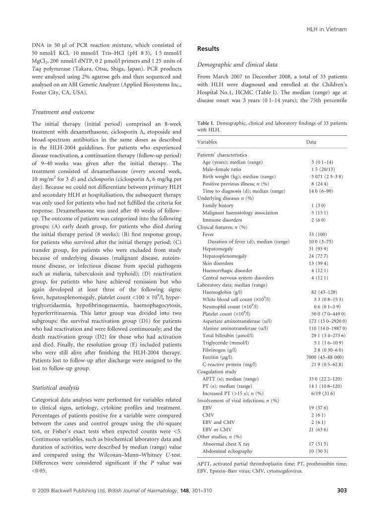
From March 2007 to December 2008, a total of 33 patients
with HLH were diagnosed and enrolled at the Children’s
Hospital No.1, HCMC (Table I). The median (range) age at
disease onset was 3 years (0Æ1–14 years); the 75th percentile
Table I. Demographic, clinical and laboratory findings of 33 patients
with HLH.
Variables Data
Patients’ characteristics
Age (years); median (range) 3 (0Æ1–14)
Male–female ratio 1Æ5 (20/13)
Birth weight (kg); median (range) 3Æ071 (2Æ3–3Æ8)
Positive previous illness; n (%) 8 (24Æ4)
Time to diagnosis (d); median (range) 14Æ0 (6–90)
Underlying diseases n (%)
Family history 1 (3Æ0)
Malignant haematology association 5 (15Æ1)
Immune disorders 2 (6Æ0)
Clinical features; n (%)
Fever 33 (100)
Duration of fever (d); median (range) 10Æ0 (3–75)
Hepatomegaly 31 (93Æ9)
Hepatosplenomegaly 24 (72Æ7)
Skin disorders 13 (39Æ4)
Haemorrhagic disorder 4 (12Æ1)
Central nervous system disorders 4 (12Æ1)
Laboratory data; median (range)
Haemoglobin (g/l) 82 (43–128)
White blood cell count (·109/l) 3Æ3 (0Æ8–15Æ3)
Neutrophil count (·109/l) 0Æ6 (0Æ1–5Æ9)
Platelet count (·109/l) 50Æ0 (7Æ0–449Æ0)
Aspartate aminotransferase (u/l) 173 (15Æ0–2920Æ0)
Alanine aminotransferase (u/l) 110 (14Æ0–1987Æ0)
Total bilirubin (lmol/l) 29Æ1 (3Æ4–273Æ6)
Triglyceride (mmol/l) 5Æ1 (1Æ6–10Æ9)
Fibrinogen (g/l) 2Æ8 (0Æ30–6Æ0)
Ferritin (lg/l) 7000 (43–88 000)
C-reactive protein (mg/l) 21Æ9 (0Æ5–62Æ8)
Coagulation study
APTT (s); median (range) 33Æ0 (22Æ2–120)
PT (s); median (range) 14Æ1 (10Æ8–120)
Increased PT (>15 s); n (%) 6/19 (31Æ6)
Involvement of viral infections; n (%)
EBV 19 (57Æ6)
CMV 2 (6Æ1)
EBV and CMV 2 (6Æ1)
EBV or CMV 21 (63Æ6)
Other studies; n (%)
Abnormal chest X ray 17 (51Æ5)
Abdominal echography 10 (30Æ3)
APTT, activated partial thromboplastin time; PT, prothrombin time;
EBV, Epstein–Barr virus; CMV, cytomegalovirus.
HLH in Vietnam
ª 2009 Blackwell Publishing Ltd, British Journal of Haematology, 148, 301–310 303
of age was 6 years; five patients were aged <1 year. The male–
female ratio was 1Æ5. The median (range) birth weight was
3Æ071 kg (2Æ3–3Æ8 kg). Two cases were diagnosed with auto-
immune disease. Only one case had a family history of sibling
death due to pneumonia (at 8 months); otherwise, no
familial histories were identified for the remaining 32
patients.
The median (range) duration of persistent fever was 10 d
(3–75 d). The duration between onset of symptoms and
diagnosis varied from 6 to 90 d with a median of 14 d.
Common clinical features at admission were hepatomegaly
(31/33; 93Æ9%), hepatosplenomegaly (24/33; 72Æ7%), skin
lesions (13/33; 39Æ4%) and neurological signs (4/33; 12Æ1%)
with lethargy in three and paresis of feet in one.
Laboratory data
At the time of diagnosis, the common laboratory findings
were hypertriglyceridaemia (25/29; 86Æ2%), thrombocyto-
penia (26/33; 78Æ8%), hyperferritinaemia (25/32; 78Æ1%)
and neutropenia (22/33; 66Æ7%). Haemophagocytosis was
detected in the bone marrow in all the patients. Other
abnormal tests were increased C-reactive protein (CRP; 24/
33; 72Æ7%), increased AST (15/28; 53Æ6%), ALT (15/28;
53Æ6%) and total bilirubin (14/29; 48Æ3%), pleural effusion
(7/33; 21Æ2%) and diffuse lung infiltrates (10/33; 30Æ3%)
detected by chest radiograph, and abdominal effusion
detected by ultrasound (10/33; 30Æ3%). Virus-associated
HLH was high (21/33; 63Æ6%). The serological tests for viral
infection revealed a high rate of EBV and CMV infection:
titres of EBV IgG higher than 640 (14/33; 42Æ4%), positive
test for EBV IgM (4/33; 12Æ1%), and titres of CMV IgG
higher than 640 (14/33; 42Æ4%). Two patients were diagnosed
with dengue haemorrhagic fever with positive IgM for
dengue virus (2/33; 6Æ0%). Real-time PCR for free EBV in
the plasma was interpreted as positive (19/33; 57Æ6%) if the
copy numbers were higher than 10 copies per ml; and real-
time PCR for free CMV in the plasma was positive (higher
than 10 copies per ml) (2/33; 6Æ0%). Two cases were both
real time PCR positive for EBV and CMV (2/33; 6%)
(Table I). Blood culture was positive in four cases (4/33;
12Æ1%): two cases were Acinetobacter and two cases coagulase-
negative Staphylococcus.
Associated underlying diseases
Haematological malignancy-associated HLH. Five patients had
associated malignant haematological diseases (5/33; 15Æ1%)
(Table I), including EBV-positive Hodgkin lymphoma; EBV-
positive peripheral T cell lymphoma (two cases) and
Langerhans cell histiocytosis. The remaining case was a
4-year-old male who had fulfilled the diagnosis for EBV-
associated HLH at the time of first hospitalisation, but after
12 months of follow-up developed reactivation and was
diagnosed with acute lymphoblastic leukaemia (ALL).
Immune disease-associated HLH. Two patients were
diagnosed with immune disease-associated HLH (2/33;
6Æ0%), (Table I). One was diagnosed with juvenile
rheumatoid arthritis (JRA). The other case concerned a
10-year-old female admitted with severe anaemia, acute
arthritis, positive Coombs’ test and positive anti-nuclear
antibody, who was diagnosed with SLE.
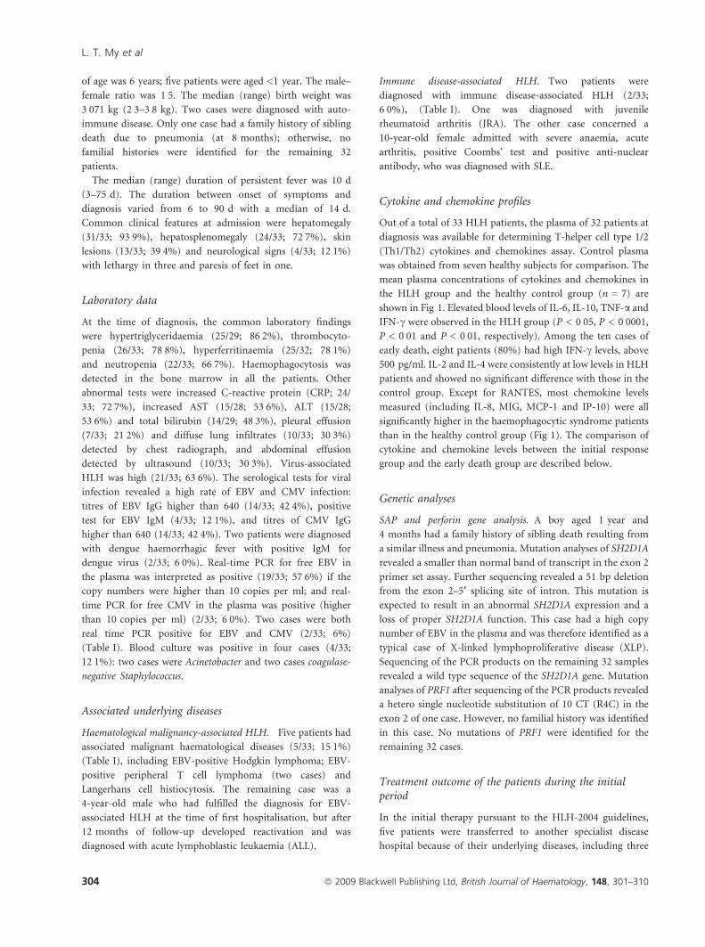
Cytokine and chemokine profiles
Out of a total of 33 HLH patients, the plasma of 32 patients at
diagnosis was available for determining T-helper cell type 1/2
(Th1/Th2) cytokines and chemokines assay. Control plasma
was obtained from seven healthy subjects for comparison. The
mean plasma concentrations of cytokines and chemokines in
the HLH group and the healthy control group (n = 7) are
shown in Fig 1. Elevated blood levels of IL-6, IL-10, TNF-a and
IFN-c were observed in the HLH group (P < 0Æ05, P < 0Æ0001,
P < 0Æ01 and P < 0Æ01, respectively). Among the ten cases of
early death, eight patients (80%) had high IFN-c levels, above
500 pg/ml. IL-2 and IL-4 were consistently at low levels in HLH
patients and showed no significant difference with those in the
control group. Except for RANTES, most chemokine levels
measured (including IL-8, MIG, MCP-1 and IP-10) were all
significantly higher in the haemophagocytic syndrome patients
than in the healthy control group (Fig 1). The comparison of
cytokine and chemokine levels between the initial response
group and the early death group are described below.
Genetic analyses
SAP and perforin gene analysis. A boy aged 1 year and
4 months had a family history of sibling death resulting from
a similar illness and pneumonia. Mutation analyses of SH2D1A
revealed a smaller than normal band of transcript in the exon 2
primer set assay. Further sequencing revealed a 51 bp deletion
from the exon 2–5¢ splicing site of intron. This mutation is
expected to result in an abnormal SH2D1A expression and a
loss of proper SH2D1A function. This case had a high copy
number of EBV in the plasma and was therefore identified as a
typical case of X-linked lymphoproliferative disease (XLP).
Sequencing of the PCR products on the remaining 32 samples
revealed a wild type sequence of the SH2D1A gene. Mutation
analyses of PRF1 after sequencing of the PCR products revealed
a hetero single nucleotide substitution of 10 CT (R4C) in the
exon 2 of one case. However, no familial history was identified
in this case. No mutations of PRF1 were identified for the
remaining 32 cases.
Treatment outcome of the patients during the initialperiod
In the initial therapy pursuant to the HLH-2004 guidelines,
five patients were transferred to another specialist disease
hospital because of their underlying diseases, including three
L. T. My et al
304 ª 2009 Blackwell Publishing Ltd, British Journal of Haematology, 148, 301–310
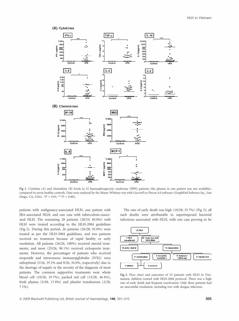
patients with malignancy-associated HLH, one patient with
JRA-associated HLH, and one case with tuberculosis-associ-
ated HLH. The remaining 28 patients (28/33; 84Æ8%) with
HLH were treated according to the HLH-2004 guidelines
(Fig 2). During this period, 26 patients (26/28; 92Æ8%) were
treated as per the HLH-2004 guidelines, and two patients
received no treatment because of rapid fatality or early
resolution. All patients (26/26, 100%) received steroid treat-
ments, and most (25/26, 96Æ1%) received ciclosporin treat-
ments. However, the percentages of patients who received
etoposide and intravenous immunopglobulin (IVIG) were
suboptimal (5/26, 19Æ2% and 9/26, 34Æ6%, respectively) due to
the shortage of supply or the severity of the diagnosis of most
patients. The common supportive treatments were whole
blood cell (10/28, 35Æ7%), packed red cell (13/28, 46Æ4%),
fresh plasma (5/28, 17Æ8%) and platelet transfusions (2/28,
7Æ1%).
The rate of early death was high (10/28; 35Æ7%) (Fig 2); all
such deaths were attributable to superimposed bacterial
infections associated with HLH, with one case proving to be
Fig 2. Flow chart and outcomes of 33 patients with HLH in Viet-
namese children treated with HLH-2004 protocol. There was a high
rate of early death and frequent reactivation. Only three patients had
an uneventful resolution, including two with dengue infection.
(A)
(B)
Fig 1. Cytokine (A) and chemokine (B) levels in 32 haemophoagocytic syndrome (HPS) patients (the plasma in one patient was not available),
compared to seven healthy controls. Data were analysed by the Mann–Whitney test with GraphPad Prism 4.0 software (GraphPad Software Inc., San
Diego, CA, USA). *P < 0Æ05, ***P < 0Æ001.
HLH in Vietnam
ª 2009 Blackwell Publishing Ltd, British Journal of Haematology, 148, 301–310 305
lymphoma association. The percentage of first response was
64Æ3% (18/28).
The median (range) durations of hospitalisation of the early
death group and the first response group were 16 d (3–48 d)
and 24 d (7–57 d), respectively. The median number of days
for hospitalisation for treatment with dexamethasone and
ciclosporin of the early death group was significantly lower
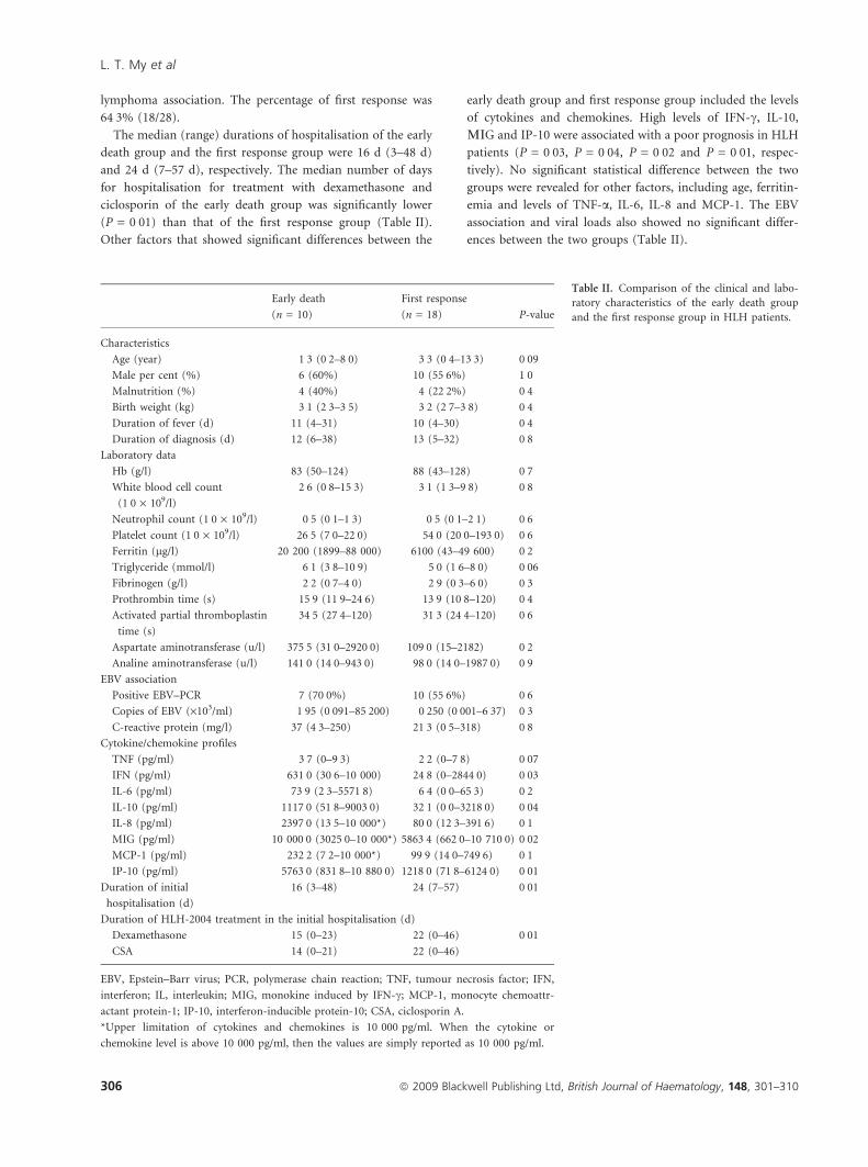
(P = 0Æ01) than that of the first response group (Table II).
Other factors that showed significant differences between the
early death group and first response group included the levels
of cytokines and chemokines. High levels of IFN-c, IL-10,
LIG and IP-10 were associated with a poor prognosis in HLH
patients (P = 0Æ03, P = 0Æ04, P = 0Æ02 and P = 0Æ01, respec-
tively). No significant statistical difference between the two
groups were revealed for other factors, including age, ferritin-
emia and levels of TNF-a, IL-6, IL-8 and MCP-1. The EBV
association and viral loads also showed no significant differ-
ences between the two groups (Table II).
Table II. Comparison of the clinical and labo-
ratory characteristics of the early death group
and the first response group in HLH patients.
Early death
(n = 10)
First response
(n = 18) P-value
Characteristics
Age (year) 1Æ3 (0Æ2–8Æ0) 3Æ3 (0Æ4–13Æ3) 0Æ09
Male per cent (%) 6 (60%) 10 (55Æ6%) 1Æ0Malnutrition (%) 4 (40%) 4 (22Æ2%) 0Æ4Birth weight (kg) 3Æ1 (2Æ3–3Æ5) 3Æ2 (2Æ7–3Æ8) 0Æ4Duration of fever (d) 11 (4–31) 10 (4–30) 0Æ4Duration of diagnosis (d) 12 (6–38) 13 (5–32) 0Æ8
Laboratory data
Hb (g/l) 83 (50–124) 88 (43–128) 0Æ7White blood cell count
(1Æ0 · 109/l)
2Æ6 (0Æ8–15Æ3) 3Æ1 (1Æ3–9Æ8) 0Æ8
Neutrophil count (1Æ0 · 109/l) 0Æ5 (0Æ1–1Æ3) 0Æ5 (0Æ1–2Æ1) 0Æ6Platelet count (1Æ0 · 109/l) 26Æ5 (7Æ0–22Æ0) 54Æ0 (20Æ0–193Æ0) 0Æ6Ferritin (lg/l) 20 200 (1899–88 000) 6100 (43–49 600) 0Æ2Triglyceride (mmol/l) 6Æ1 (3Æ8–10Æ9) 5Æ0 (1Æ6–8Æ0) 0Æ06
Fibrinogen (g/l) 2Æ2 (0Æ7–4Æ0) 2Æ9 (0Æ3–6Æ0) 0Æ3Prothrombin time (s) 15Æ9 (11Æ9–24Æ6) 13Æ9 (10Æ8–120) 0Æ4Activated partial thromboplastin
time (s)
34Æ5 (27Æ4–120) 31Æ3 (24Æ4–120) 0Æ6
Aspartate aminotransferase (u/l) 375Æ5 (31Æ0–2920Æ0) 109Æ0 (15–2182) 0Æ2Analine aminotransferase (u/l) 141Æ0 (14Æ0–943Æ0) 98Æ0 (14Æ0–1987Æ0) 0Æ9
EBV association
Positive EBV–PCR 7 (70Æ0%) 10 (55Æ6%) 0Æ6Copies of EBV (·103/ml) 1Æ95 (0Æ091–85Æ200) 0Æ250 (0Æ001–6Æ37) 0Æ3C-reactive protein (mg/l) 37 (4Æ3–250) 21Æ3 (0Æ5–318) 0Æ8
Cytokine/chemokine profiles
TNF (pg/ml) 3Æ7 (0–9Æ3) 2Æ2 (0–7Æ8) 0Æ07
IFN (pg/ml) 631Æ0 (30Æ6–10 000) 24Æ8 (0–2844Æ0) 0Æ03
IL-6 (pg/ml) 73Æ9 (2Æ3–5571Æ8) 6Æ4 (0Æ0–65Æ3) 0Æ2IL-10 (pg/ml) 1117Æ0 (51Æ8–9003Æ0) 32Æ1 (0Æ0–3218Æ0) 0Æ04
IL-8 (pg/ml) 2397Æ0 (13Æ5–10 000*) 80Æ0 (12Æ3–391Æ6) 0Æ1MIG (pg/ml) 10 000Æ0 (3025Æ0–10 000*) 5863Æ4 (662Æ0–10 710Æ0) 0Æ02
MCP-1 (pg/ml) 232Æ2 (7Æ2–10 000*) 99Æ9 (14Æ0–749Æ6) 0Æ1IP-10 (pg/ml) 5763Æ0 (831Æ8–10 880Æ0) 1218Æ0 (71Æ8–6124Æ0) 0Æ01
Duration of initial
hospitalisation (d)
16 (3–48) 24 (7–57) 0Æ01
Duration of HLH-2004 treatment in the initial hospitalisation (d)
Dexamethasone 15 (0–23) 22 (0–46) 0Æ01
CSA 14 (0–21) 22 (0–46)
EBV, Epstein–Barr virus; PCR, polymerase chain reaction; TNF, tumour necrosis factor; IFN,
interferon; IL, interleukin; MIG, monokine induced by IFN-c; MCP-1, monocyte chemoattr-
actant protein-1; IP-10, interferon-inducible protein-10; CSA, ciclosporin A.
*Upper limitation of cytokines and chemokines is 10 000 pg/ml. When the cytokine or
chemokine level is above 10 000 pg/ml, then the values are simply reported as 10 000 pg/ml.
L. T. My et al
306 ª 2009 Blackwell Publishing Ltd, British Journal of Haematology, 148, 301–310
Treatment outcome of the patients in the continuationperiod
There were 18 patients during the follow-up period, which
ranged from 9 weeks to more than 40 weeks. During this
period, four patients were lost to follow up (4/18; 22Æ2%). Two
other patients were transferred because of the development of
underlying diseases: one had acute lymphoblastic leukaemia
and the other had SLE. The rate of reactivation was high (9/18;
50Æ0%) including 22Æ2% (4/18) for the death reactivation
group and 27Æ8% (5/18) for the survival reactivation group
(Fig 2). The percentage of patients with resolution was low
(3/18; 16Æ7%). The median (range) follow-up duration for the
death reactivation group was 17 weeks (10–35 weeks), for the
survival reactivation group 36 weeks (33–87 weeks) and
72 weeks (55–82 weeks) for the resolution group.
Discussion
This study characterized, for the first time, childhood HLH in
HCMC, Vietnam. Considering there were 33 HLH cases in a
single institution in 1 year and 9 months, the projected
incidence of childhood HLH in Vietnam was relatively high,
as compared to that in Taiwan, Canada, Turkey or Thailand
(Veerakul et al, 2002; Chen et al, 2004; Gurgey et al, 2005).
Recently, a nationwide survey of HLH in Japan showed that
the mean number of 160 patients per year between 2001 and
2005 (Ishii et al, 2007). Our studies revealed a high prevalence
of EBV infection. Two HLH cases were associated with dengue
infection, due to the prevalence of dengue virus infection in
this tropical region. Familial HLH is rare in Vietnamese
children, with only one case found during this study of XLP
(1/33; 3%), a situation similar to that of Taiwan (Chen et al,
2004; Lee et al, 2009).
The diagnosis of HLH in tropical regions is complicated by
common clinical and laboratory findings which may present in
other tropical infections, such as severe malaria, tuberculosis,
typhoid fever or HIV infection (Mathew et al, 2000; Veerakul
et al, 2002). In this study, the time between the onset of
symptoms and the diagnosis varied from 6 to 90 d (median
14 d). The delay of diagnosis or prolonged duration of
diagnosis (14 d) again reflected the difficulties of the paedi-
atricians to diagnose HLH when facing these disorders. In
practice, persistent fever, hepatosplenomegaly, hyperferritina-
emia and hypertriglyceridaemia and cytopenia, together with
the progressive course, represent a good index for suspicion of
an HLH diagnosis and prompt the physician to study the
haemophagocytosis in bone marrow in order to establish the
diagnosis. Since early recognition of HLH and immediate
application of appropriate regimens to HLH patients have
been shown to improve or to arrest the disease progression
(Imashuku et al, 2004; Lee et al, 2005; Tang et al, 2008;
Yamada et al, 2008), the delayed diagnosis of our HLH
patients may partly explain the poor outcome of treatments in
this cohort.
Besides delayed diagnosis, this study attempted to identify
other potential prognostic factors, such as cytokine levels, virus
association and genetic factors. The cytokine and chemokine
studies revealed a consistent elevation of several cytokines and
chemokines, though not IL-2 and IL-4, which were usually not
elevated in our HLH patients. These findings indicated that a
cytokine storm occurred in most of our HLH patients and
were responsible for the clinical manifestations and abnormal
laboratory data of HLH (Ishii et al, 1991; Fujiwara et al, 1993;
Osugi et al, 1997). The high levels of IFN-c, IL-6, IP-10 and
MIG in HLH patients with poor prognosis or early death
support the observations of previous reports (Ohga et al, 1993;
Imashuku et al, 1994; Tang et al, 2008). Hypercytokinemia is
responsible for the systemic problems of HLH, such as
pancytopenia, depletion of the immune system, diffuse respi-
ratory infiltrates of lungs and liver damage (Imashuku, 2002).
Therefore, anti-inflammatory or immunoregulatory agents
targeted to the master genes of inflammation, such as NFKB1
and AP1, may ameliorate the outcome of HLH patients
(Chuang et al, 2007a; Su, 2008). Agonists of peroxisome
proliferator-activated receptors (PPAR) have been recently
found to exert such remarkable results with minimal side
effects in an animal model of virus-associated haemophago-
cytic syndrome and could represent potential therapeutic
agents (Chuang et al, 2007a).
Haemophagocytic lymphohistiocytosis can be classified into
genetic (primary) HLH and acquired (secondary) HLH
(Henter et al, 2007). Genetic HLH can be further divided into
familial HLH (FHLH) and immune deficiency HLH, including
Chediak–Higashi syndrome (CHS), Griscelli syndrome (GS)
and XLP (Henter et al, 1998; Janka, 2007). Both genetic
subgroups are associated with impaired NK cell function
(Sullivan et al, 1998; Kogawa et al, 2002; Henter et al, 2007).
This study did not include assessment of NK function or NK
cells due to the unavailability of flow cytometry in the hospital.
The prevalence of genetic HLH appeared to be low (2/33; 6%)
in this study, based on the genetic mutation analyses of
SH2D1A and PRF1 genes. The analysis of UNC13D gene,
however, was not performed in this study due to the large
UNC13D gene of 32 exons for such a big sample size of 33
cases. There was only one case of XLP involving a family
history, specifically a sibling death due to pneumonia at
8 months. XLP has been now recognized as a primary
immunological disorder in infants and children (Mischler
et al, 2007). The only case of a mutation of the PRF1 gene in
this study showed no family history. Other candidate genes
associated with HLH were not explored here and whether
other ‘acquired’ HLH may have underlying genetic or immu-
nological disorders remains to be clarified.
Although we detected a high association with EBV infection
(19/33, 57Æ6%), no prognostic significance of the EBV
association, for either reactivation or mortality, could be
identified in this study, similar to the report from Taiwan (Su
et al, 1994; Chen et al, 2004; Lee et al, 2009). EBV infects T
cells in acquired HLH and the EBV latent membrane protein 1
HLH in Vietnam
ª 2009 Blackwell Publishing Ltd, British Journal of Haematology, 148, 301–310 307
(LMP1) can transcriptionally inhibit the expression of
SH2D1A, leading to sustained T cell activation and the release
of Th1 cytokines, such as TNF-a and IFN-c, via the
TRAF2,5/nuclear factor jB (NF-jB) pathway (Chuang et al,
2005, 2007b). Interestingly, two HLH cases were noted to have
positive serology tests for dengue virus at the convalescent
stage of dengue haemorrhage fever. Both patients had a good
response to treatment with dexamethasone and ciclosporin
and recovered thereafter. In Thailand, Veerakul et al (2002)
also reported three paediatric HLH patients with positive
dengue virus detection. Several reports of adult dengue
haemorrhagic fever-associated HLH have been reported from
Taiwan (Lu et al, 2005), Thailand (Veerakul et al, 2002;
Srichaikul et al, 2008) and Japan (Nakamura et al, 2009).
Further studies are needed to clarify the mechanism of HLH
association with dengue virus infection.
For malignancy-associated HLH, four patients were identi-
fied with haematological malignancies, including two cases of
peripheral T cell lymphoma, one case of Hodgkin lymphoma
and one case of Langerhans cell histiocytosis (LCH). Interest-
ingly, EBV association was detected by in situ EBV-encoded
RNA 1 (EBER-1) hybridization in both cases of peripheral T
cell lymphoma and in the Hodgkin lymphoma case. Besides
malignancy-associated HLH, this study noted two patients
with autoimmune disease-associated HLH: an 11-year-old
female with SLE and a 13-year-old male with JRA. Both
patients had unusually high levels of hyperferritinemia (1130
and 5200 lg/l) and both are still alive, receiving therapy for
autoimmune diseases. This study also noted a high rate of
pleural and abdominal effusion, probably due to the conse-
quences of severe liver damage, immune reactions, or infiltra-
tion of the lymphatic system (Akcam et al, 2007). Our study
also noted the presence of neurological disorders in three HLH
cases. The development of central nervous system involvement
in HLH, which has received increasing attention in recent
years, possibly involves infiltration of the brain by lympho-
histiocytes, or therapeutic events (Akiyoshi et al, 2006; Horne
et al, 2008).
The high percentage of early deaths and reactivation deaths
in our HLH patients (14/28, 50%) drove us to clarify and
compare the potential prognostic factors between the early
response group and the early death group. Consistent with
previous reports (Tang et al, 2008), we noted that high serum
concentrations of IFN-c, IL-10, MIG and IP-10 were associ-
ated with early death (P = 0Æ03; P = 0Æ04; P = 0Æ02 and
P = 0Æ01, respectively). Therefore, measuring the levels of
these specific cytokine and chemokine profiles may provide
predictive markers of disease activity and severity for thera-
peutic consideration. Finally, some of our patients received
suboptimal therapy in this study due to the shortage of
medications, such as etoposide and IVIG, during the early
phase of the study, which partly explains the poor outcome of
the treatments. The high rate of recurrence and of reactivation
death in this study suggests that a more aggressive regimen or
bone marrow transplantation may be needed in the future.
In conclusion, this prospective, collaborative study provides
a comprehensive analysis and characterization of HLH cases in
Vietnamese children. Early diagnosis of HLH, measurements
of specific cytokine profiles, and a more appropriate thera-
peutic regimen may be needed in the future.
Acknowledgement
This project is sponsored by grants from National Health
Research Institutes, Taiwan.
References
Akcam, M., Artan, R., Yilmaz, A. & Akkaya, B. (2007) An unusual
cause of ascites: hemophagocytic lymphohistiocytosis. Indian Pedi-
atrics, 44, 371–374.
Akiyoshi, K., Hamada, Y., Yamada, H., Kojo, M. & Izumi, T. (2006)
Acute necrotizing encephalopathy associated with hemophagocytic
syndrome. Pediatric Neurology, 34, 315–318.
Chen, C.J., Huang, Y.C., Jaing, T.H., Hung, I.J., Yang, C.P., Chang,
L.Y. & Lin, T.Y. (2004) Hemophagocytic syndrome: a review of 18
pediatric cases. Journal of Microbiology, Immunology, and Infection,
37, 157–163.
Chuang, H.C., Lay, J.D., Hsieh, W.C., Wang, H.C., Chang, Y.,
Chuang, S.E. & Su, I.J. (2005) Epstein-Barr virus LMP1 inhibits
the expression of SAP gene and upregulates Th1 cytokines in the
pathogenesis of hemophagocytic syndrome. Blood, 106, 3090–
3096.
Chuang, H.C., Lay, J.D., Hsieh, W.C. & Su, I.J. (2007a) Pathogenesis
and mechanism of disease progression from hemophagocytic lym-
phohistiocytosis to Epstein–Barr virus-associated T-cell lymphoma:
nuclear factor-kappa B pathway as a potential therapeutic target.
Cancer Science, 98, 1281–1287.
Chuang, H.C., Lay, J.D., Chuang, S.E., Hsieh, W.C., Chang, Y. & Su,
I.J. (2007b) Epstein–Barr virus (EBV) latent membrane protein-1
down-regulates tumor necrosis factor-alpha (TNF-alpha) receptor-1
and confers resistance to TNF-alpha-induced apoptosis in T cells:
implication for the progression to T-cell lymphoma in EBV-asso-
ciated hemophagocytic syndrome. American Journal of Pathology,
170, 1607–1617.
Fujiwara, F., Hibi, S. & Imashuku, S. (1993) Hypercytokinemia in
hemophagocytic syndrome. The American Journal of Pediatric
Hematology/Oncology, 15, 92–98.
Goransdotter Ericson, K., Fadeel, B., Nilsson-Ardnor, S., Soderhall, C.,
Samuelsson, A., Janka, G., Schneider, M., Gurgey, A., Yalman, N.,
Revesz, T., Egeler, R., Jahnukainen, K., Storm-Mathiesen, I., Har-
aldsson, A., Poole, J., de Saint Basile, G., Nordenskjold, M. &
Henter, J. (2001) Spectrum of perforin gene mutations in familial
hemophagocytic lymphohistiocytosis. American Journal of Human
Genetics, 68, 590–597.
Gupta, A.A., Tyrrell, P., Valani, R., Benseler, S., Abdelhaleem, M. &
Weitzman, S. (2009) Experience with hemophagocytic lymphohist-
iocytosis/macrophage activation syndrome at a single institution.
Journal of Pediatric Hematology/Oncology, 31, 81–84.
Gurgey, A., Secmeer, G., Tavil, B., Ceyhan, M., Kuskonmaz, B., Cengiz,
B., Ozen, H., Kara, A., Cetin, M. & Gumruk, F. (2005) Secondary
hemophagocytic lymphohistiocytosis in Turkish children. Pediatric
Infectious Disease Journal, 24, 1116–1117.
L. T. My et al
308 ª 2009 Blackwell Publishing Ltd, British Journal of Haematology, 148, 301–310
Gutierrez, A., Solano, C., Ferrandez, A., Marugan, I., Terol, M.J., Benet,
I., Tormo, M., Bea, M.D. & Rodriguez, J. (2003) Peripheral T-cell
lymphoma associated consecutively with hemophagocytic lym-
phohistiocytosis and hypereosinophilic syndrome. European Journal
of Haematology, 71, 303–306.
Henter, J.I., Arico, M., Elinder, G., Imashuku, S. & Janka, G. (1998)
Familial hemophagocytic lymphohistiocytosis. Primary hemophag-
ocytic lymphohistiocytosis. Hematology/Oncology Clinics of North
America, 12, 417–433.
Henter, J.I., Samuelsson-Horne, A., Arico, M., Egeler, R.M., Elinder,
G., Filipovich, A.H., Gadner, H., Imashuku, S., Komp, D., Ladisch,
S., Webb, D. & Janka, G. (2002) Treatment of hemophagocytic
lymphohistiocytosis with HLH-94 immunochemotherapy and bone
marrow transplantation. Blood, 100, 2367–2373.
Henter, J.I., Horne, A., Arico, M., Egeler, R.M., Filipovich, A.H.,
Imashuku, S., Ladisch, S., McClain, K., Webb, D., Winiarski, J. &
Janka, G. (2007) HLH-2004: diagnostic and therapeutic guidelines
for hemophagocytic lymphohistiocytosis. Pediatric Blood & Cancer,
48, 124–131.
Horne, A., Trottestam, H., Arico, M., Egeler, R.M., Filipovich, A.H.,
Gadner, H., Imashuku, S., Ladisch, S., Webb, D., Janka, G. &
Henter, J.I. (2008) Frequency and spectrum of central nervous
system involvement in 193 children with haemophagocytic lym-
phohistiocytosis. British Journal of Haematology, 140, 327–335.
Imashuku, S. (2002) Clinical features and treatment strategies of
Epstein–Barr virus-associated hemophagocytic lymphohistiocytosis.
Critical Reviews in Oncology/Hematology, 44, 259–272.
Imashuku, S., Hibi, S., Fujiwara, F., Ikushima, S. & Todo, S. (1994)
Haemophagocytic lymphohistiocytosis, interferon-gamma-naemia
and Epstein–Barr virus involvement. British Journal of Haematology,
88, 656–658.
Imashuku, S., Tabata, Y., Teramura, T. & Hibi, S. (2000) Treatment
strategies for Epstein–Barr virus-associated hemophagocytic lym-
phohistiocytosis (EBV-HLH). Leukaemia & Lymphoma, 39, 37–49.
Imashuku, S., Teramura, T., Tauchi, H., Ishida, Y., Otoh, Y., Sawada,
M., Tanaka, H., Watanabe, A., Tabata, Y., Morimoto, A., Hibi, S. &
Henter, J.I. (2004) Longitudinal follow-up of patients with Epstein-
Barr virus-associated hemophagocytic lymphohistiocytosis. Hae-
matologica, 89, 183–188.
Ishii, E., Ohga, S., Aoki, T., Yamada, S., Sako, M., Tasaka, H., Kuwano,
A., Sasaki, M., Tsunematsu, Y. & Ueda, K. (1991) Prognosis of
children with virus-associated hemophagocytic syndrome and
malignant histiocytosis: correlation with levels of serum interleukin-
1 and tumor necrosis factor. Acta Haematologica, 85, 93–99.
Ishii, E., Ohga, S., Imashuku, S., Yasukawa, M., Tsuda, H., Miura, I.,
Yamamoto, K., Horiuchi, H., Takada, K., Ohshima, K., Nakamura,
S., Kinukawa, N., Oshimi, K. & Kawa, K. (2007) Nationwide survey
of hemophagocytic lymphohistiocytosis in Japan. International
Journal of Hematology, 86, 58–65.
Janka, G.E. (2007) Familial and acquired hemophagocytic lymphoh-
istiocytosis. European Journal of Pediatrics, 166, 95–109.
Kogawa, K., Lee, S.M., Villanueva, J., Marmer, D., Sumegi, J. &
Filipovich, A.H. (2002) Perforin expression in cytotoxic lympho-
cytes from patients with hemophagocytic lymphohistiocytosis and
their family members. Blood, 99, 61–66.
Krenova, Z., Sterba, J., Blatny, J., Kren, L. & Slany, J. (2007) A case of
anaplastic large cell lymphoma-induced hemophagocytic lymphoh-
istiocytosis in an adolescent female. Pediatric Blood & Cancer, 49,
1056.
Lee, J.S., Kang, J.H., Lee, G.K. & Park, H.J. (2005) Successful treatment
of Epstein–Barr virus-associated hemophagocytic lymphohistio-
cytosis with HLH-94 protocol. Journal of Korean Medical Science, 20,
209–214.
Lee, W.I., Chen, S.H., Hung, I.J., Yang, C.P., Jaing, T.H., Chen, C.J., Li,
S.P. & Huang, J.L. (2009) Clinical aspects, immunologic assessment,
and genetic analysis in Taiwanese children with hemophagocytic
lymphohistiocytosis. Pediatric Infectious Disease Journal, 28, 30–34.
Lu, P.L., Hsiao, H.H., Tsai, J.J., Chen, T.C., Feng, M.C., Chen, T.P. &
Lin, S.F. (2005) Dengue virus-associated hemophagocytic syndrome
and dyserythropoiesis: a case report. The Kaohsiung Journal of
Medical Sciences, 21, 34–39.
Mathew, L.G., Cherian, T., Sudarshanam, A., Korah, I., Kumar, N.K. &
Raghupathy, P. (2000) Hemophagocytic lymphohistiocytosis: a case
series. Indian Pediatrics, 37, 526–531.
Mischler, M., Fleming, G.M., Shanley, T.P., Madden, L., Levine, J.,
Castle, V., Filipovich, A.H. & Cornell, T.T. (2007) Epstein–Barr
virus-induced hemophagocytic lymphohistiocytosis and X-linked
lymphoproliferative disease: a mimicker of sepsis in the pediatric
intensive care unit. Pediatrics, 119, e1212–e1218.
Nakamura, I., Nakamura-Uchiyama, F., Komiya, N. & Ohnishi, K.
(2009) [A case of dengue fever with viral-associated hemophagocytic
syndrome]. Kansenshogaku Zasshi, 83, 60–63.
Ohga, S., Matsuzaki, A., Nishizaki, M., Nagashima, T., Kai, T., Suda,
M. & Ueda, K. (1993) Inflammatory cytokines in virus-associated
hemophagocytic syndrome. Interferon-gamma as a sensitive indi-
cator of disease activity. The American Journal of Pediatric Hema-
tology/Oncology, 15, 291–298.
Ohno, T., Ueda, Y., Nagai, K., Takahashi, T., Konaka, Y., Takamatsu,
T., Suzuki, T., Sasada, M. & Uchiyama, T. (2003) The serum cyto-
kine profiles of lymphoma-associated hemophagocytic syndrome: a
comparative analysis of B-cell and T-cell/natural killer cell lym-
phomas. International Journal of Hematology, 77, 286–294.
Osugi, Y., Hara, J., Tagawa, S., Takai, K., Hosoi, G., Matsuda, Y., Ohta,
H., Fujisaki, H., Kobayashi, M., Sakata, N., Kawa-Ha, K., Okada, S.
& Tawa, A. (1997) Cytokine production regulating Th1 and Th2
cytokines in hemophagocytic lymphohistiocytosis. Blood, 89, 4100–
4103.
Sayos, J., Wu, C., Morra, M., Wang, N., Zhang, X., Allen, D., van
Schaik, S., Notarangelo, L., Geha, R., Roncarolo, M.G., Oettgen, H.,
De Vries, J.E., Aversa, G. & Terhorst, C. (1998) The X-linked lym-
phoproliferative-disease gene product SAP regulates signals induced
through the co-receptor SLAM. Nature, 395, 462–469.
Srichaikul, T., Punyagupta, S., Kanchanapoom, T., Chanokovat, C.,
Likittanasombat, K. & Leelasiri, A. (2008) Hemophagocytic
syndrome in Dengue hemorrhagic fever with severe multiorgan
complications. Journal of the Medical Association of Thailand, 91,
104–109.
Su, I.J. (2008) Perspectives on the pathogenesis and therapy of he-
mophagocytic syndrome. Journal of the Formosan Medical Associa-
tion, 107, 277–280.
Su, I.J., Chen, R.L., Lin, D.T., Lin, K.S. & Chen, C.C. (1994) Epstein–
Barr virus (EBV) infects T lymphocytes in childhood EBV-associated
hemophagocytic syndrome in Taiwan. American Journal of Patho-
logy, 144, 1219–1225.
Sullivan, K.E., Delaat, C.A., Douglas, S.D. & Filipovich, A.H. (1998)
Defective natural killer cell function in patients with hemophago-
cytic lymphohistiocytosis and in first degree relatives. Pediatric
Research, 44, 465–468.
HLH in Vietnam
ª 2009 Blackwell Publishing Ltd, British Journal of Haematology, 148, 301–310 309
Tan, E.M., Cohen, A.S., Fries, J.F., Masi, A.T., McShane, D.J., Roth-
field, N.F., Schaller, J.G., Talal, N. & Winchester, R.J. (1982) The
1982 revised criteria for the classification of systemic lupus erythe-
matosus. Arthritis and Rheumatism, 25, 1271–1277.
Tang, Y., Xu, X., Song, H., Yang, S., Shi, S., Wei, J., Pan, B., Zhao, F.,
Liao, C. & Luo, C. (2008) Early diagnostic and prognostic signifi-
cance of a specific Th1/Th2 cytokine pattern in children with
haemophagocytic syndrome. British Journal of Haematology, 143,
84–91.
Ueda, I., Ishii, E., Morimoto, A., Ohga, S., Sako, M. & Imashuku, S.
(2006) Correlation between phenotypic heterogeneity and gene
mutational characteristics in familial hemophagocytic lymphohisti-
ocytosis (FHL). Pediatric Blood & Cancer, 46, 482–488.
Veerakul, G., Sanpakit, K., Tanphaichitr, V.S., Mahasandana, C. &
Jirarattanasopa, N. (2002) Secondary hemophagocytic lymphohist-
iocytosis in children: an analysis of etiology and outcome. Journal of
the Medical Association of Thailand, 85(Suppl. 2), S530–S541.
Yamada, K., Yamamoto, Y., Uchiyama, A., Ito, R., Aoki, Y., Uchida, Y.,
Nagasawa, H., Kimura, H., Ichiyama, T., Fukao, T. & Kohno, Y.
(2008) Successful treatment of neonatal herpes simplex-type 1
infection complicated by hemophagocytic lymphohistiocytosis and
acute liver failure. Tohoku Journal of Experimental Medicine, 214, 1–5.
L. T. My et al
310 ª 2009 Blackwell Publishing Ltd, British Journal of Haematology, 148, 301–310
Copyright © 2022 FDOKUMEN