Complete Genome Sequence of the Shrimp White Spot Bacilliform Virus
10
JOURNAL OF VIROLOGY, 0022-538X/01/$04.000 DOI: 10.1128/JVI.75.23.11811–11820.2001 Dec. 2001, p. 11811–11820 Vol. 75, No. 23 Copyright © 2001, American Society for Microbiology. All Rights Reserved. Complete Genome Sequence of the Shrimp White Spot Bacilliform Virus FENG YANG, JUN HE, XIONGHUI LIN, QIN LI, DENG PAN, XIAOBO ZHANG, AND XUN XU* The Third Institute of Oceanography, Xiamen 361005, People’s Republic of China Received 11 June 2001/Accepted 1 August 2001 We report the first complete genome sequence of a marine invertebrate virus. White spot bacilliform virus (WSBV; or white spot syndrome virus) is a major shrimp pathogen with a high mortality rate and a wide host range. Its double-stranded circular DNA genome of 305,107 bp contains 181 open reading frames (ORFs). Nine homologous regions containing 47 repeated minifragments that include direct repeats, atypical inverted repeat sequences, and imperfect palindromes were identified. This is the largest animal virus that has been completely sequenced. Although WSBV is morphologically similar to insect baculovirus, the two viruses are not detectably related at the amino acid level. Rather, some WSBV genes are more homologous to eukaryotic genes than viral genes. In fact, sequence analysis indicates that WSBV differs from all known viruses, although a few genes display a weak homology to herpesvirus genes. Most of the ORFs encode proteins that bear no homology to any known proteins, either suggesting that WSBV represents a novel class of viruses or perhaps implying a significant evolutionary distance between marine and terrestrial viruses. The most unique feature of WSBV is the presence of an intact collagen gene, a gene encoding an extracellular matrix protein of animal cells that has never been found in any viruses. Determination of the genome of WSBV will facilitate a better understanding of the molecular mechanism underlying the pathogenesis of the WSBV virus and will also provide useful infor- mation concerning the evolution and divergence of marine and terrestrial animal viruses at the molecular level. White spot bacilliform virus (WSBV) or white spot syn- drome virus (WSSV) is a major shrimp pathogen that is highly virulent in penaeid shrimp, the most important species used in aquaculture, and can also infect most species of crustacean (15, 32). Infection of penaeid shrimp by WSBV can result in mor- tality of up to 90 to 100% within 3 to 7 days (57). A major outbreak of WSBV infection in 1993 resulted in a 70% reduc- tion in shrimp production in China (14, 57) and has raised major concerns in aquaculture around the world. Prevention and inhibition of infection by this virus can be difficult due largely to the ability of WSBV to survive for a long time in the environment (2 years in a shrimp pond) and also due to a poor understanding of this virus at the molecular level. WSBV was originally classified as an unassigned member of the Baculoviridae because of its rod-shaped, enveloped mor- phology (20). However, it was recently excluded from the bac- ulovirus family and is temporarily unclassified due to the lack of molecular information (53). The virus is known generally as white spot syndrome virus (WSSV) (31), and a new genus name, Whispovirus, was proposed by Vlak et al. (48). Sequence analysis of individual genes and proteins later showed that most WSBV proteins bear poor sequence homology to bacu- lovirus proteins but have repeated regions similar to those of some baculoviruses. To understand the molecular basis of viral replication and infection, we decided to sequence the whole genome of WSBV. MATERIALS AND METHODS Isolation and sequencing of WSBV genomic DNA. Intact WSBV genomic DNA was isolated from dead and moribund WSBV-infected Penaeus japonicus shrimp which were collected from shrimp ponds in Tongan, Xiamen, east China, in October 1996 as previously described (56). A whole-genome random sequenc- ing method (19) was used to obtain the complete genome sequence for WSBV. Genomic DNA was cloned by the shotgun method into SmalI-linearized pUC18 vector, amplified, and sequenced using ABI BigDye Terminator chemistry on ABI 377 and ABI 3700 capillary sequencers. Large DNA fragments of 5 to 10 kb were also obtained by partial digestion with Sau3A1 and cloned into the pBlue- script vectors (41). This was used to form a genome scaffold and to verify the orientation and integrity of the contigs formed from the shotgun library. A total of 5,770 sequences for sevenfold coverage were assembled using the InnerPeace software by Charles Lawrence based on the Phred, Phrap, and Consed program originally developed at the University of Washington. The WSBV genome sequence was confirmed by comparison of the observed restriction fragments from seven restriction enzymes (BamHI, EcoRI, HindIII, KpnI, PstI, SalI, and XbaI) to those predicted from the sequence data and was also confirmed by the genome scaffold produced by sequence pairs from 1,495 large-insert clones, which covered 90% of the main genome. Gaps were closed by a combination of sequence-walking of shotgun and PCR large-fragment libraries. DNA sequence analysis. Genome DNA composition, structure, repeats, re- striction enzyme patterns, and translation were analyzed with the DNAMAN software (Lynnon BioSoft, Vaudreuil, Canada). Open reading frames (ORFs) consisted of more than 60 codons that are initiated with a methionine codon. For detection of potential protein-coding regions, the codon usage bias and posi- tional base preference were evaluated by determining the codon frequency of known WSBV genes or cDNA cloned from the WSBV cDNA library. Homology searches were performed with the FASTA (38) and BLAST programs (3). Pro- tein motifs were analyzed by using the PROSITE database, release 16 (25). Trans- membrane domains and signal peptides were predicted with ANTHEPROT (23). Preparation and screening of a WSBV cDNA library. Poly(A) mRNA was isolated from WSBV-infected shrimp tissue using the PolyATtract System 1000 kit (Promega). Double-stranded cDNAs were synthesized using the SUPER- SCRIPT plasmid system for cDNA synthesis and plasmid cloning (GIBCO BRL). WSBV cDNA clones were selected by hybridization with the digoxigenin (DIG)-labeled WSBV genomic DNA probe (DIG labeling kit; Boehringer Mannheim) and sequenced. The transcription of some ORFs was also verified by PCR on a cDNA cocktail using ORF-specific primers. Nucleotide sequence accession number. The complete WSBV sequence can be obtained from the GenBank database (accession no. AF332093). * Corresponding author. Mailing address: The Third Institute of Oceanography, Xiamen 361005, People’s Republic of China. Phone: 86-592-2195296. Fax: 86-592-2085376. E-mail: [email protected]. 11811
-
Upload
independent -
Category
Documents
-
view
4 -
download
0
Transcript of Complete Genome Sequence of the Shrimp White Spot Bacilliform Virus

JOURNAL OF VIROLOGY,0022-538X/01/$04.00�0 DOI: 10.1128/JVI.75.23.11811–11820.2001
Dec. 2001, p. 11811–11820 Vol. 75, No. 23
Copyright © 2001, American Society for Microbiology. All Rights Reserved.
Complete Genome Sequence of the Shrimp White SpotBacilliform Virus
FENG YANG, JUN HE, XIONGHUI LIN, QIN LI, DENG PAN, XIAOBO ZHANG, AND XUN XU*
The Third Institute of Oceanography, Xiamen 361005, People’s Republic of China
Received 11 June 2001/Accepted 1 August 2001
We report the first complete genome sequence of a marine invertebrate virus. White spot bacilliform virus(WSBV; or white spot syndrome virus) is a major shrimp pathogen with a high mortality rate and a wide hostrange. Its double-stranded circular DNA genome of 305,107 bp contains 181 open reading frames (ORFs). Ninehomologous regions containing 47 repeated minifragments that include direct repeats, atypical inverted repeatsequences, and imperfect palindromes were identified. This is the largest animal virus that has been completelysequenced. Although WSBV is morphologically similar to insect baculovirus, the two viruses are not detectablyrelated at the amino acid level. Rather, some WSBV genes are more homologous to eukaryotic genes than viralgenes. In fact, sequence analysis indicates that WSBV differs from all known viruses, although a few genesdisplay a weak homology to herpesvirus genes. Most of the ORFs encode proteins that bear no homology to anyknown proteins, either suggesting that WSBV represents a novel class of viruses or perhaps implying asignificant evolutionary distance between marine and terrestrial viruses. The most unique feature of WSBV isthe presence of an intact collagen gene, a gene encoding an extracellular matrix protein of animal cells that hasnever been found in any viruses. Determination of the genome of WSBV will facilitate a better understanding ofthe molecular mechanism underlying the pathogenesis of the WSBV virus and will also provide useful infor-mation concerning the evolution and divergence of marine and terrestrial animal viruses at the molecular level.
White spot bacilliform virus (WSBV) or white spot syn-drome virus (WSSV) is a major shrimp pathogen that is highlyvirulent in penaeid shrimp, the most important species used inaquaculture, and can also infect most species of crustacean (15,32). Infection of penaeid shrimp by WSBV can result in mor-tality of up to 90 to 100% within 3 to 7 days (57). A majoroutbreak of WSBV infection in 1993 resulted in a 70% reduc-tion in shrimp production in China (14, 57) and has raisedmajor concerns in aquaculture around the world. Preventionand inhibition of infection by this virus can be difficult duelargely to the ability of WSBV to survive for a long time in theenvironment (2 years in a shrimp pond) and also due to a poorunderstanding of this virus at the molecular level.
WSBV was originally classified as an unassigned member ofthe Baculoviridae because of its rod-shaped, enveloped mor-phology (20). However, it was recently excluded from the bac-ulovirus family and is temporarily unclassified due to the lackof molecular information (53). The virus is known generally aswhite spot syndrome virus (WSSV) (31), and a new genusname, Whispovirus, was proposed by Vlak et al. (48). Sequenceanalysis of individual genes and proteins later showed thatmost WSBV proteins bear poor sequence homology to bacu-lovirus proteins but have repeated regions similar to those ofsome baculoviruses. To understand the molecular basis of viralreplication and infection, we decided to sequence the wholegenome of WSBV.
MATERIALS AND METHODS
Isolation and sequencing of WSBV genomic DNA. Intact WSBV genomicDNA was isolated from dead and moribund WSBV-infected Penaeus japonicus
shrimp which were collected from shrimp ponds in Tongan, Xiamen, east China,in October 1996 as previously described (56). A whole-genome random sequenc-ing method (19) was used to obtain the complete genome sequence for WSBV.Genomic DNA was cloned by the shotgun method into SmalI-linearized pUC18vector, amplified, and sequenced using ABI BigDye Terminator chemistry onABI 377 and ABI 3700 capillary sequencers. Large DNA fragments of 5 to 10 kbwere also obtained by partial digestion with Sau3A1 and cloned into the pBlue-script vectors (41). This was used to form a genome scaffold and to verify theorientation and integrity of the contigs formed from the shotgun library. A totalof 5,770 sequences for sevenfold coverage were assembled using the InnerPeacesoftware by Charles Lawrence based on the Phred, Phrap, and Consed programoriginally developed at the University of Washington.
The WSBV genome sequence was confirmed by comparison of the observedrestriction fragments from seven restriction enzymes (BamHI, EcoRI, HindIII,KpnI, PstI, SalI, and XbaI) to those predicted from the sequence data and wasalso confirmed by the genome scaffold produced by sequence pairs from 1,495large-insert clones, which covered 90% of the main genome.
Gaps were closed by a combination of sequence-walking of shotgun and PCRlarge-fragment libraries.
DNA sequence analysis. Genome DNA composition, structure, repeats, re-striction enzyme patterns, and translation were analyzed with the DNAMANsoftware (Lynnon BioSoft, Vaudreuil, Canada). Open reading frames (ORFs)consisted of more than 60 codons that are initiated with a methionine codon. Fordetection of potential protein-coding regions, the codon usage bias and posi-tional base preference were evaluated by determining the codon frequency ofknown WSBV genes or cDNA cloned from the WSBV cDNA library. Homologysearches were performed with the FASTA (38) and BLAST programs (3). Pro-tein motifs were analyzed by using the PROSITE database, release 16 (25). Trans-membrane domains and signal peptides were predicted with ANTHEPROT(23).
Preparation and screening of a WSBV cDNA library. Poly(A) mRNA wasisolated from WSBV-infected shrimp tissue using the PolyATtract System 1000kit (Promega). Double-stranded cDNAs were synthesized using the SUPER-SCRIPT plasmid system for cDNA synthesis and plasmid cloning (GIBCOBRL). WSBV cDNA clones were selected by hybridization with the digoxigenin(DIG)-labeled WSBV genomic DNA probe (DIG labeling kit; BoehringerMannheim) and sequenced. The transcription of some ORFs was also verified byPCR on a cDNA cocktail using ORF-specific primers.
Nucleotide sequence accession number. The complete WSBV sequence can beobtained from the GenBank database (accession no. AF332093).
* Corresponding author. Mailing address: The Third Institute ofOceanography, Xiamen 361005, People’s Republic of China. Phone:86-592-2195296. Fax: 86-592-2085376. E-mail: [email protected].
11811

RESULTS AND DISCUSSION
General features of the WSBV genome. We have previouslydeveloped a unique method that enables us to highly purify theWSBV virus from infected shrimp tissue (56). A random shot-gun method was employed to sequence the entire genome ofWSBV; the sequence was subsequently confirmed by the ge-nome scaffold formed by sequencing a large-fragment DNAlibrary. The complete WSBV genome is a double-strandedcircular DNA of 305,107 bp, similar to a previous estimate of290 kb (56). Since the origin of replication was unknown, thestart of the largest BamHI fragment was chosen to be base 1(Fig. 1). Three percent of the WSBV genome is made up ofnine homologous regions (hrs), while the remaining 97% of thesequences are unique (see description below). The genome hasa total G�C content of 41%.
A total of 531 putative ORFs were identified by sequenceanalysis, among which 181 ORFs are likely to encode func-tional proteins (Table 1). This corresponds to an average gene
density of one gene per 1.7 kb. Thirty-six of the 181 ORFsannotated here either have been identified by screening andsequencing a WSBV cDNA library (Table 1) or have beenreported previously to encode functional proteins (45, 46, 48,49, 50). Transcription of another 52 ORFs was confirmed byreverse transcription-PCR (RT-PCR; see Material and Meth-ods) (Table 1). The relative positions of the ORFs and hrs inthe genome are shown in Fig. 1. For 80% of the putative 181ORFs there is a potential polyadenylation site (AATAAA)downstream of the ORF (Table 1).
WSBV ORFs encode gene products homologous to knownproteins. Table 1 contains a list of the 181 predicted WSBVORFs. Among 181 ORFs, the proteins encoded by 18 ORFsshow 40 to 68% identity to known proteins from other virusesor organisms or contain an identifiable functional domain.These proteins include enzymes involved in nucleic acid me-tabolism and DNA replication, a collagen-like protein, andthree viral structure proteins (for details, see below). Thirty
FIG. 1. Circular representation of the WSBV genome. Arrows, positions (outer ring) of the 181 ORFs (red and blue indicate the differentdirections of transcription); green rectangles, 9 hrs. B, sites of BamHI restriction enzymes (inner ring; their positions are in parentheses).
11812 YANG ET AL. J. VIROL.
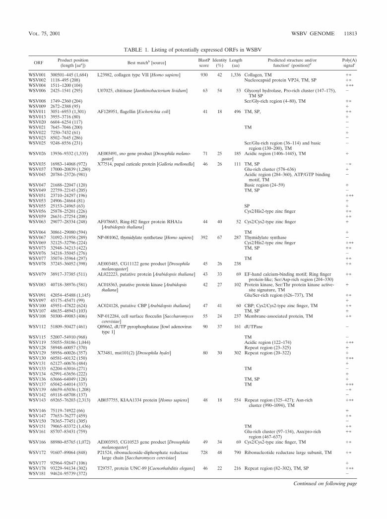
TABLE 1. Listing of potentially expressed ORFs in WSBV
ORFProduct position
(length [aaa])Best matchb [source]
BlastPscore
Identity(%)
Length(aa)
Predicted structure and/orfunctionc (position)d
Poly(A)signalc
WSV001 300501–445 (1,684) L23982, collagen type VII [Homo sapiens] 930 42 1,336 Collagen, TM ��WSV002 1118–495 (208) Nucleocapsid protein VP24, TM, SP ��WSV004 1511–1200 (104) ���WSV006 2425–1541 (295) U07025, chitinase [Janthinobacterium lividum] 63 54 53 Glycosyl hydrolase, Pro-rich cluster (147–175),
TM SP�
WSV008 1749–2360 (204) Ser/Gly-rich region (4–80), TM ��WSV009 2672–2388 (95) �WSV011 3051–6953 (1,301) AF128951, flagellin [Escherichia coli] 41 18 496 TM, SP, ��WSV013 3955–3716 (80) �WSV020 6604–6254 (117) �WSV021 7645–7046 (200) TM �WSV022 7250–7432 (61) �WSV023 8502–7645 (286) �WSV025 9248–8556 (231) Ser/Glu-rich region (36–114) and basic
region (130–200), TM�
WSV026 13936–9332 (1,535) AE003491, sno gene product [Drosophila melano-gaster]
71 25 185 Acidic region (1406–1445), TM �
WSV035 16983–14068 (972) X77514, pupal cuticule protein [Galleria mellonella] 46 26 111 TM, SP ��WSV037 17000–20839 (1,280) Glu-rich cluster (578–636) �WSV045 20784–23726 (981) Acidic region (284–360), ATP/GTP binding
motif, TM�
WSV047 21688–22047 (120) Basic region (24–59) �WSV049 22759–22145 (205) TM, SP �WSV051 23710–24297 (196) ���WSV053 24906–24664 (81) �WSV055 25153–24965 (63) SP �WSV056 25878–25201 (226) Cys2/His2-type zinc finger ��WSV059 26631–27254 (208) ��WSV063 29077–28334 (248) AF078683, Ring-H2 finger protein RHA1a
[Arabidopsis thaliana]44 40 52 Cys2/Cys2-type zinc finger �
WSV064 30861–29080 (594) TM �WSV067 31092–31958 (289) NP-001062, thymidylate synthetase [Homo sapiens] 392 67 287 Thymidylate synthase �WSV069 32125–32796 (224) Cys2/His2-type zinc finger ���WSV073 32948–34213 (422) TM, SP ��WSV076 34218–35045 (276) �WSV077 35074–35964 (297) TM ��WSV078 37245–36052 (398) AE003485, CG11122 gene product [Drosophila
melanogaster]45 26 238 ��
WSV079 38917–37385 (511) AL022223, putative protein [Arabidopsis thaliana] 43 33 69 EF-hand calcium-binding motif; Ring fingerprotein-like; Ser/Asp-rich region (204–330)
��
WSV083 40718–38976 (581) AC018363, putative protein kinase [Arabidopsisthaliana]
42 27 102 Protein kinase, Ser/Thr protein kinase active-site signature, TM
�
WSV091 42054–45488 (1,145) Glu/Ser-rich region (626–737), TM ��WSV097 45175–45471 (99) �WSV100 45951–47822 (624) AC024128, putative CBP [Arabidopsis thaliana] 47 41 60 CBP; Cys2/Cys2-type zinc finger, TM ��WSV107 48635–48943 (103) TM, SP �WSV108 50300–49083 (406) NP-012284, cell surface flocculin [Saccharomyces
cerevisiae]55 24 237 Membrane-associated protein, TM ��
WSV112 51809–50427 (461) Q89662, dUTP pyrophosphatase [fowl adenovirustype 1]
90 37 161 dUTPase �
WSV115 52007–54910 (968) TM �WSV119 55055–58186 (1,044) Acidic region (122–174) ���WSV128 58948–60057 (370) Repeat region (23–325) �WSV129 58956–60026 (357) X73481, mst101(2) [Drosophila hydei] 80 30 302 Repeat region (20–322) �WSV130 60581–60132 (150) ���WSV131 62127–60676 (484) �WSV133 62204–63016 (271) TM �WSV134 62991–63656 (222) �WSV136 63666–64049 (128) TM, SP �WSV137 65042–64014 (337) TM ���WSV139 68659–65036 (1,208) ��WSV142 69118–68708 (137) �WSV143 69265–76203 (2,313) AB037755, KIAA1334 protein [Homo sapiens] 48 18 554 Repeat region (325–427); Asn-rich
cluster (990–1094), TM���
WSV146 75119–74922 (66) �WSV147 77653–76277 (459) ��WSV150 78365–77451 (305) �WSV151 79065–83372 (1,436) TM ��WSV161 85707–83431 (759) Glu-rich cluster (97–134), Asn/pro-rich
region (467–637)��
WSV166 88980–85765 (1,072) AE003593, CG10523 gene product [Drosophilamelanogaster]
49 34 69 Cys2/Cys2-type zinc finger, TM ��
WSV172 91607–89064 (848) P21524, ribonucleoside-diphosphate reductaselarge chain [Saccharomyces cerevisiae]
728 48 790 Ribonucleotide reductase large subunit, TM ��
WSV177 92964–92647 (106) �WSV178 93229–94134 (302) T29757, protein UNC-89 [Caenorhabditis elegans] 46 22 216 Repeat region (82–302), TM, SP ���WSV181 94624–95739 (372) �
Continued on following page
VOL. 75, 2001 WSBV GENOME 11813

TABLE 1—Continued
ORFProduct position
(length [aaa])Best matchb [source]
BlastPscore
Identity(%)
Length(aa)
Predicted structure and/orfunctionc (position)d
Poly(A)signalc
WSV184 95744–97366 (541) Cys2/Cys2-type zinc finger, TM �WSV188 97548–98786 (413) AF117061, ribonucleotide reductase R2 subunit
[Aedes albopictus]364 59 313 Ribonucleotide reductase small subunit, TM ��
WSV191 98854–99786 (311) AJ133437, deoxyribonuclease I [Penaeus japonicus] 54 32 149 Nuclease, TM, SP ��WSV192 102885–99829 (1,019) TM ��WSV195 103071–103841 (257) TM, SP �WSV198 103844–104677 (278) �WSV199 104760–107327 (856) AF156271, Ring finger protein terf [Homo sapiens] 43 30 72 Ring-H2 finger motif, TM �WSV206 108550–109161 (204) ��WSV207 109261–110085 (275) Proline rich, TM ���WSV209 114953–110136 (1,606) TM �WSV214 115053–115292 (80) L41834, nuclear protein [Ensis minor] 59 46 73 DNA-binding protein ���WSV216 118987–115406 (1,194) Protein-splicing signature, TM ��WSV220 119057–121078 (674) �WSV222 121100–123631 (844) AK016037, putative [Mus musculus] 44 32 58 Ring-H2 finger motif, ATP/GTP binding motif,
TM���
WSV226 123758–126547 (930) TM �WSV230 126755–127000 (82) ���WSV231 129006–127162 (615) TM ��WSV234 130290–129409 (294) ��WSV235 129611–129811 (67) �WSV236 130076–130306 (77) TM �WSV237 130566–131441 (292) ��WSV238 131481–132938 (486) 38 30 139 Gly-rich cluster (50–138), TM, SP ��WSV242 132994–133893 (300) TM �WSV244 133969–136341 (791) TM ���WSV249 137589–139937 (783) AC024760, contains similarity to TR [Caenorhab-
ditis elegans]72 27 202 Ring-H2 finger motif, repeat region (454–633) ��
WSV252 140111–141613 (501) ���WSV254 141696–142538 (281) ��WSV256 142545–143696 (384) TM, SP ��WSV259 143760–144686 (309) ���WSV260 147517–144752 (922) Asp/Glu/Ser-rich region (344–485), TM ���WSV267 148612–147770 (281) ��WSV269 150145–148679 (489) TM �WSV270 150675–150166 (170) �WSV271 150688–154341 (1,218) S59310, probable membrane protein YMR317w
[Saccharomyces cerevisiae]46 20 369 Lys/Ser-rich region (455–526), TM �
WSV277 154557–156929 (791) D86346, crystal protein [Bacillus thuringiensis] 41 23 249 TM ��WSV282 159352–161253 (634) Ser-rich cluster (13–129), SP ��WSV284 161263–161562 (100) TM, SP �WSV285 161718–165017 (1,100) AE003824, CG13185 gene product [Drosophila
melanogaster]48 25 183 ATP/GTP binding motif, TM ���
WSV289 169814–165120 (1,565) NP-011856, serine/threonine protein kinase[Saccharomyces cerevisiae]
46 25 245 Protein kinase, TM, SP ��
WSV291 167278–167532 (85) TM, SP �WSV294 170113–170730 (206) �WSV295 170832–171458 (209) TM ���WSV299 172439–171513 (309) TM, SP ���WSV302 173075–172509 (189) �WSV303 173178–175850 (891) NP-069209, transcription initiation factor IID
[Archaeoglobus fulgidus]44 23 140 TBP Cys2/Cys2-type zinc finger, TM �
WSV306 175840–177096 (419) TM ��WSV308 177124–178521 (466) ��WSV310 178530–179345 (272) TM �WSV311 180036–179425 (204) Nucleocapsid protein VP26, TM, SP ���WSV313 183817–180279 (1,180) Glu-rich region (37–358) and Pro-rich
cluster (462–492), TM�
WSV321 184132–184482(117) TM, SP �WSV322 184499–185179(227) TM �WSV323 185082–184819(88) ���WSV324 185434–185189(82) �WSV325 185433–186827(465) TM, SP �WSV327 190743–188176(856) TM ���WSV331 190094–190306(71) �WSV332 190876–193233(786) ���WSV333 191135–190932(68) TM, SP �WSV338 194629–193331(433) TM, SP ���WSV339 195503–194655(283) �WSV340 196292–195510(261) �WSV342 196697–196398(100) �WSV343 209342–196803(4,180) TM ���WSV344 197221–197517(99) TM �WSV349 199510–199779(90) TM, SP �WSV360 209616–227846(6,077) U96166, srpA [Streptococcus cristatus] 46 23 222 Leucine-zipper motif, TM ���WSV386 228196–227993(68) TM, SP �
Continued on following page
11814 YANG ET AL. J. VIROL.

ORFs predicted proteins that show a partial homology (20 to39% identity) to known proteins or contain one or two se-quence motifs (versus a real functional domain). The remain-ing 133 ORFs encode proteins with no homology to any knownproteins or motifs.
Enzymes involved in nucleotide metabolism. Among the 18ORFs encoding proteins that show extensive homologies withpreviously identified proteins, WSV067, WSV112, WSV172,WSV188, and WSV395 may encode the WSBV homologues ofenzymes involved in nucleic acid metabolism (Table 1). The
TABLE 1—Continued
ORFProduct position
(length [aaa])Best matchb [source]
BlastPscore
Identity(%)
Length(aa)
Predicted structure and/orfunctionc (position)d
Poly(A)signalc
WSV387 228375–230561(729) �WSV390 230617–231579(321) �WSV394 231422–231724(101) �WSV395 231603–232796(398) T41553, thymidylate kinase [Schizosaccharomyces
pombe]157 41 200 Thymidylate kinase; ATP/GTP binding motif �
WSV397 232819–233331(171) �WSV398 233383–233763(127) TM �WSV399 234330–233782(183) �WSV403 236679–238601(641) Ring-H2 finger motif, repeat region (435–494),
SP��
WSV406 238659–239435(259) T27927, hypothetical protein ZK593.8 [Caenorhab-ditis elegans]
50 26 175 TM, SP ��
WSV407 240139–239459(227) �WSV412 240713–241189(159) �WSV414 241637–241275(121) Asp-rich cluster (63–86), TM, SP ���WSV415 241775–243406(544) TM �WSV419 243217–243795(193) �WSV421 244242–244853(204) Envelope protein VP28, TM, SP ���WSV423 247143–244954(730) T22255, hypothetical protein F45H7.4 [Caenorhab-
ditis elegans]45 26 165 Protein kinase, TM ��
WSV427 249230–247362(623) EF-hand calcium-binding motif, TM �WSV432 249151–249456(102) �WSV433 249426–253208(1,261) Pro-rich cluster (29–71), TM �WSV440 253297–255117(607) �WSV442 255075–257474(800) ATP/GTP binding motif, TM ��WSV446 257552–259129(526) ATP/GTP binding motif, TM, SP ��WSV447 264975–259168(1,936) Z70204, similarity to yeast hypothetical helicase
[Caenorhabditis elegans]42 40 52 Helicase, ATP/GTP binding motif, Asp-pro-
tease motif, TM��
WSV455 265079–265597(173) TM, SP �WSV457 265606–266400(265) TM, SP ��WSV459 266838–266446(131) TM �WSV461 267400–266930(157) �WSV462 267399–267647(83) �WSV464 268584–267721(288) �WSV465 272423–268695(1,243) Cys2/Cys2-type zinc finger, TM ��WSV477 274527–275150(208) Cys2/Cys2-type zinc finger, ATP/GTP binding
motif��
WSV479 276736–275210(509) Glu-rich cluster (467–485), TM ���WSV482 277035–277571(179) TM ���WSV483 277705–278076(124) �WSV484 278423–277776(216) TM ��WSV486 278637–280973(779) AF154037, surface protein PspC [Streptococcus
pneumoniae]46 24 194 Lysine-rich, TM �
WSV489 281865–281131(245) ��WSV492 282176–282583(136) �WSV493 283360–282677(228) Acidic region (59–103) ���WSV495 283754–284011(86) �WSV497 285773–284079(565) TM ���WSV500 286706–286080(209) Cys2/Cys2-type zinc finger, ATP/GTP binding
motif��
WSV502 286606–289632(1,009) AL352992, ariadne-like protein [Leishmania major] 51 51 33 Cys2/His2, Cys2/Cys2-type zinc finger, ATP/GTP binding motif, TM, SP
��
WSV508 291298–289685(538) TM ���WSV513 291720–292202(161) �WSV514 292190–298774(2,195) X61920, DNA polymerase III catalytic subunit [Sac-
charomyces cerevisiae]52 24 201 DNA polymerase, TM ���
WSV518 293724–293275(150) SP �WSV524 298729–298526(68) �WSV525 299033–298821(71) TM, SP �WSV526 300432–299089(448) TM ��
a aa, amino acid.b Accession numbers are from the GenBank or SwissProt database.c Function was deduced from the degree of amino acid similarity to either products of known genes or Prosite signatures. TM, transmembrane domains; SP, signal
peptides.d Positions of amino acid residues.e �, polyadenylation signal present; �, signal absent. �, the transcription of the ORF was also verified by RT-PCR; ��, the ORF was confirmed by cDNA sequencing.
VOL. 75, 2001 WSBV GENOME 11815
highest degree of homology (67% identity over 287 aminoacids) was detected between the product of WSV067 and thehuman thymidylate synthase. The 29-amino-acid thymidylatesynthase prosite motif (PS00091), which contains the catalyticcysteine residue, is 100% conserved in the product of WSV067.In addition, WSV112 may encode a WSBV homologue ofdUTPase (37% identity over 161 amino acids) since the fiveconserved regions of dUTPase, especially the highly conservedsubstrate-binding residues, were identified in the product ofWSV112 (13, 35). dUTPase has been shown to be essential forthe replication of DNA viruses (5). Consistent with the previ-ous reports by van Hulten et al. (48) and Tsai et al. (45, 46),WSBV contains ribonucleotide reductases (products ofWSV172 and WSV188) and also thymidylate/thymidine kinase(product of WSV395). Among these enzymes, thymidylate syn-thase catalyzes the methylation of dUTP to yield the nucleo-tide precursor dTMP. This is an important step in the de novopathway of biosynthesis of pyrimidine (12). Despite its ubiqui-tous distribution in nature, a viral thymidylate synthase wasfound only in a few herpesviruses (2, 10, 26, 39), Melanoplussanguinipes entomopoxvirus (MsEPV) (1), Chilo iridescent vi-rus (CIV) (36), and bacteriophages (9). Most viruses do notcontain thymidylate synthase, as they depend mostly on thehost enzymatic machinery for the replication of their genomesso as to keep the viral genome small (36). WSBV and otherthymidylate synthase-containing viruses may therefore exhibita considerable independence from the host deoxyribonucleo-tide synthesis. This may represent a significant advantage forviral genome replication that may ultimately lead to persis-tence of infection and a broad host range for viral infection(36). It is possible that WSBV acquires these replication-re-lated genes from its host and/or from a coinfecting virus thatmight occur at an earlier period in evolution. However, sincethe shrimp homologues of these genes have not been cloned,we are not able to test this hypothesis.
Proteins involved in DNA replication and transcription.WSBV contains genes encoding proteins involved in DNAreplication such as DNA polymerase (product of WSV514).The WSBV DNA polymerase was putatively identified by thepresence of three highly conserved motifs, YGDTDSVFC(DNA polymerase family B signature PS00116), KLGMNSMYG, and DMTSLYP (conserved amino acid residuesare underlined), that are found in most eukaryotic DNA poly-merases (4) as well as in some viral polymerases (18, 29, 43).However, since the degree of amino acid similarity between theproduct of WSV514 and known DNA polymerases is low (24%identity over 201 amino acids), its putative activity as a DNApolymerase still awaits future experimental verification. Inter-estingly, the size of this putative WSBV polymerase (2,195amino acids) is much larger than those of the regular poly-merases found in other organisms.
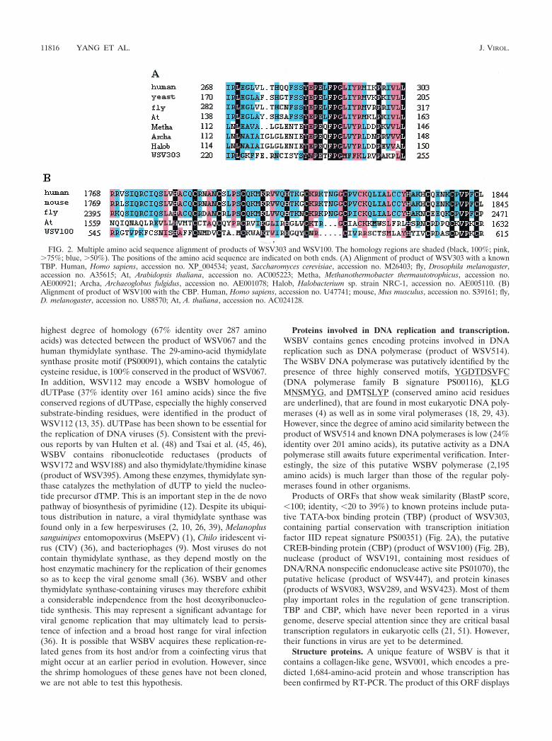
Products of ORFs that show weak similarity (BlastP score,�100; identity, �20 to 39%) to known proteins include puta-tive TATA-box binding protein (TBP) (product of WSV303,containing partial conservation with transcription initiationfactor IID repeat signature PS00351) (Fig. 2A), the putativeCREB-binding protein (CBP) (product of WSV100) (Fig. 2B),nuclease (product of WSV191, containing most residues ofDNA/RNA nonspecific endonuclease active site PS01070), theputative helicase (product of WSV447), and protein kinases(products of WSV083, WSV289, and WSV423). Most of themplay important roles in the regulation of gene transcription.TBP and CBP, which have never been reported in a virusgenome, deserve special attention since they are critical basaltranscription regulators in eukaryotic cells (21, 51). However,their functions in virus are yet to be determined.
Structure proteins. A unique feature of WSBV is that itcontains a collagen-like gene, WSV001, which encodes a pre-dicted 1,684-amino-acid protein and whose transcription hasbeen confirmed by RT-PCR. The product of this ORF displays
FIG. 2. Multiple amino acid sequence alignment of products of WSV303 and WSV100. The homology regions are shaded (black, 100%; pink,�75%; blue, �50%). The positions of the amino acid sequence are indicated on both ends. (A) Alignment of product of WSV303 with a knownTBP. Human, Homo sapiens, accession no. XP_004534; yeast, Saccharomyces cerevisiae, accession no. M26403; fly, Drosophila melanogaster,accession no. A35615; At, Arabidopsis thaliana, accession no. AC005223; Metha, Methanothermobacter thermautotrophicus, accession no.AE000921; Archa, Archaeoglobus fulgidus, accession no. AE001078; Halob, Halobacterium sp. strain NRC-1, accession no. AE005110. (B)Alignment of product of WSV100 with the CBP. Human, Homo sapiens, accession no. U47741; mouse, Mus musculus, accession no. S39161; fly,D. melanogaster, accession no. U88570; At, A. thaliana, accession no. AC024128.
11816 YANG ET AL. J. VIROL.

the highest degree of homology to human collagen type VII(42% identity over 1,336 amino acids) (Fig. 3). This is the firsttime that an intact collagen gene has been reported in a virusgenome. The collagen-like protein of WSBV contains a typicalrepeat of Gly-X-Y (X is mostly proline, and Y can be anyamino acid) that can form the triple-helical structure charac-teristics of animal collagen fiber. The presence of this collagen-like protein may help to protect the WSBV from environmen-tal factors and may contribute to its ability to survive for a longtime in a shrimp pond.
Previously only a short segment of collagen-homologous se-quence was found in the structural proteins of ectocarpus sil-iculosus virus 1 (EsV-1) (16), hepersvirus saimiri (HVS) (2,22), and bacteriophage PRD1 (6, 7) (Fig. 3). In EsV-1, thecollagen-like sequence was found in the N-terminal half ofboth vp55 and vp74 (16), which were encoded by the EsV-1genome and which are likely to be the components of the viralcore structure. In HVS, the Gly-X-Y motif is repeated 18 timesand is located in the central region of saimiri transformation-associated protein (STP). These collagen-like repeats mayserve as a hinge to extend the active domain of STP to its siteof action (2). Finally, in bacteriophage PRD1, a minor capsidprotein was found to contain a short collagen-like region (Gly-X-Y)6 (7). All of the collagen-like segments present in theseproteins are short. These segments may play only a supple-mentary role in protein functions.
In addition, WSV002 and WSV311 encode a nucleocapsidprotein, and the product of WSV421 shows characteristics ofan envelope protein. These proteins have recently been puri-fied from the nucleocapsid and envelope of WSBV (49, 50).
WSV214 encodes a polypeptide with 44.2% basic amino acidresidues (Arg/Lys) and 24.6% Ser residues. This amino acidcomposition is similar to that of the DNA-binding protein ofinsect baculoviruses (34, 40, 55). Homologs of these DNA-binding proteins have also been found in granulosis virus (47).The basic residues of these DNA-binding proteins have a highaffinity for the phosphate backbone of DNA, enabling the
generation of a highly compact form of viral genomic DNA.Upon entry into a host cell, the DNA-binding protein maybecome phosphorylated by a protein kinase, resulting in theunpacking of the viral DNA (54).
Protein motifs. ORFs containing zinc finger and leucinezipper motifs have been found in WSBV (Table 1). Thesemotifs have been shown to be involved in DNA-protein inter-action and in regulation of transcriptional activation. Ring-H2finger motifs, a variation of the Ring finger motif (30, 44)found in proteins critical for virus survival and replication (11,42), are also detected. Products of WSV079 and WSV427contain an EF-hand calcium-binding motif (PS00018). Proteinswith these motifs are found in some prokaryotic and all eu-karyotic organisms and play important roles in the regulationand control of normal cellular functions. The detection ofthese motifs in proteins of a marine virus suggests that some ofthese basic regulatory activities are well conserved throughoutevolution.
The remaining 133 ORFs encode novel proteins of unknownfunction. These novel genes obviously will provide ample op-portunities for future research and for exploration of molecu-lar mechanisms by which a virus and its host interact to survivein the marine environment.
Among the 181 ORFs examined, the products of 96 havepotential transmembrane domains and 32 proteins containboth signal peptide sequences and substantial hydrophobic do-mains, suggesting that they may be membrane-associated pro-teins and that they may play an important role in the WSBV-host cell interaction and host range determination. Other thanthe putative signal sequences and hydrophobic domains, theseproteins are not obviously related to other known proteins.
Repetitive regions. Three percent of the WSBV genome iscomposed of highly repetitive sequences, and the repeats aredistributed throughout the genome. We found nine hrs with atotal of 47 repeated minifragments encompassing direct re-peats, atypical inverted repeat sequences, and imperfect pal-indromic sequences. The nine hrs vary in size from 0.76 to 3.62
FIG. 3. Multiple amino acid sequence alignment of the product of WSV001 with human (Homo sapiens) type VII collagen, accession no.L23982; fruit fly (Drosophila melanogaster) collagen, accession no. P08120; sea urchin (Strongylocentrotus purpuratus) collagen, accession no.A43426; brown alga virus (BAV; ectocarpus siliculosus virus) collagen-like protein, accession no. NP_077542; HVS strain 484-77) collagen-likeprotein, accession no. P25050; and bacteriophage PRD1 (PRD1) coat protein, which contains a short collagen-like region, accession no. P22536.The homology regions are shaded (black, 100%; pink, �75%; blue, �50%). Repeat sequence density is shown as a ratio of a/b, in which a indicatesthe length of the typical repeat sequence and b indicates the full length of the protein.
VOL. 75, 2001 WSBV GENOME 11817
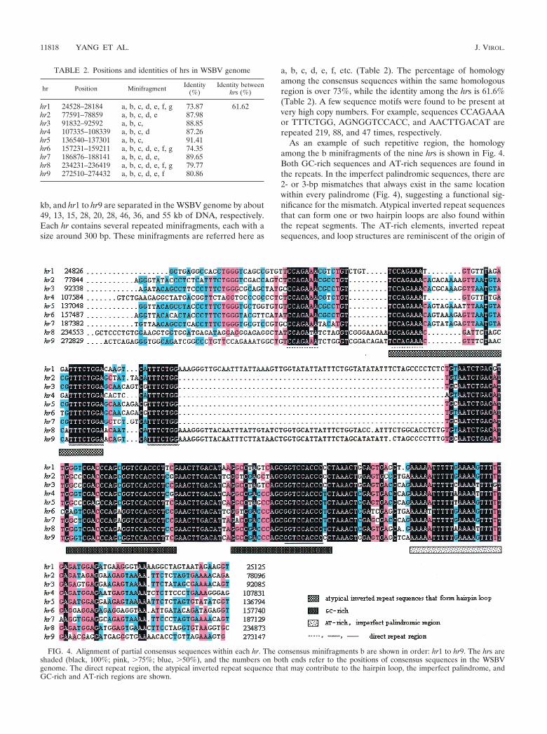
kb, and hr1 to hr9 are separated in the WSBV genome by about49, 13, 15, 28, 20, 28, 46, 36, and 55 kb of DNA, respectively.Each hr contains several repeated minifragments, each with asize around 300 bp. These minifragments are referred here as
a, b, c, d, e, f, etc. (Table 2). The percentage of homologyamong the consensus sequences within the same homologousregion is over 73%, while the identity among the hrs is 61.6%(Table 2). A few sequence motifs were found to be present atvery high copy numbers. For example, sequences CCAGAAAor TTTCTGG, AGNGGTCCACC, and AACTTGACAT arerepeated 219, 88, and 47 times, respectively.
As an example of such repetitive region, the homologyamong the b minifragments of the nine hrs is shown in Fig. 4.Both GC-rich sequences and AT-rich sequences are found inthe repeats. In the imperfect palindromic sequences, there are2- or 3-bp mismatches that always exist in the same locationwithin every palindrome (Fig. 4), suggesting a functional sig-nificance for the mismatch. Atypical inverted repeat sequencesthat can form one or two hairpin loops are also found withinthe repeat segments. The AT-rich elements, inverted repeatsequences, and loop structures are reminiscent of the origin of
TABLE 2. Positions and identities of hrs in WSBV genome
hr Position Minifragment Identity(%)
Identity betweenhrs (%)
hr1 24528–28184 a, b, c, d, e, f, g 73.87 61.62hr2 77591–78859 a, b, c, d, e 87.98hr3 91832–92592 a, b, c, 88.85hr4 107335–108339 a, b, c, d 87.26hr5 136540–137301 a, b, c, 91.41hr6 157231–159211 a, b, c, d, e, f, g 74.35hr7 186876–188141 a, b, c, d, e, 89.65hr8 234231–236419 a, b, c, d, e, f, g 79.77hr9 272510–274432 a, b, c, d, e, f 80.86
FIG. 4. Alignment of partial consensus sequences within each hr. The consensus minifragments b are shown in order: hr1 to hr9. The hrs areshaded (black, 100%; pink, �75%; blue, �50%), and the numbers on both ends refer to the positions of consensus sequences in the WSBVgenome. The direct repeat region, the atypical inverted repeat sequence that may contribute to the hairpin loop, the imperfect palindrome, andGC-rich and AT-rich regions are shown.
11818 YANG ET AL. J. VIROL.

replication in eukaryotic cells and also in some of the viruses(17, 37). The presence of hrs is a feature of many baculovirusgenomes. The hrs may serve as transcription enhancers andorigins of DNA replication and play a fundamental role in theviral life cycle (24, 27, 28, 33). The presence of nine hrs sug-gests that WSBV may contain multiple replication origins. Thismay account for the fast replication and the growth rate ofWSBV. Furthermore, although the organization of WSBV hrsis similar to that of baculovirus, no homology among most oftheir ORFs is detected. Thus, future investigations are neededto determine whether WSBV is a seawater baculovirus andwhether the ancestors of WSBV and insect baculovirusesevolved by separate routes, acquiring genes independently indifferent environments.
In summary, we have obtained the complete genome se-quence of WSBV. This is the first complete genome sequencefrom a marine invertebrate virus. It is also the largest animalvirus genome sequenced (8, 52). As the genomic data demon-strated, more than 80% of WSBV proteins bear no homologyto previously identified proteins. This leads us to consider aseparate evolutionary origin for this virus. Among the proteinsthat show homology with known proteins, most seem to berelated to eukaryotic proteins and relatively few seem to berelated to viral proteins (Table 1). Although a few genes showweak similarities to genes of herpesvirus (data not shown), themorphology and the double-stranded circular WSBV genomediffer significantly from those of herpesvirus, which contains anicosahedral capsid and a linear double-stranded DNA mole-cule. On the other hand, WSBV shares some complex mor-phological traits with the insect baculovirus, and a pattern ofinterspersed repetitive regions in WSBV is similar to thatfound in some of the insect baculoviruses, but sequence com-parison indicates that they are not detectably related at theamino acid level. Unfortunately, until now there were no ge-nome sequence data available for the nonoccluded baculovi-rus. Based on genetic analysis, WSBV clearly should not beincluded in any of the currently recognizable baculovirus sub-families and perhaps should be classified in a new virus family.It is possible that other WSBV-like viruses that can infect otherorganisms may exist. As the sequence of a representative of amarine DNA virus, the complete WSBV genome sequenceshould provide valuable information to serve as the geneticbasis for future studies. Future work may shed more light onthe evolution of these viruses.
ACKNOWLEDGMENTS
We thank Mei He and Yun Ye for their assistance, and we acknowl-edge the support of Mingwei Wang, Lin Zao, and Yan Shen. We thankMark Yandell, Jennifer R. Wortman, Chinnappa Kodira, P. W. Li, andZ. Deng of Celera Genomics for coordinating the project at Celera.We thank Kunxin Luo of Lawrence Berkeley National Laboratory andUC Berkeley for data analysis and critical reading of the manuscript.
This work is funded by the Chinese High Tech “863” Program(Z19-02-05-01), Fujian Science Fund (C97053), and Science Founda-tion of the State Oceanic Administration.
REFERENCES
1. Afonso, C. L., E. R. Tulman, Z. Lu, E. Oma, G. F. Kutish, and D. L. Rock.1999. The genome of Melanoplus sanguinipes Entomopoxvirus. J. Virol. 73:533–552.
2. Albrecht, J. C., J. Nicholas, D. Biller, K. R. Cameron, B. Biesinger, C.Newman, S. Wittmann, M. A. Craxton, H. Coleman, B. Fleckenstein, andR. W. Honess. 1992. Primary structure of the herpesvirus saimiri genome.J. Virol. 66:5047–5058.
3. Altschul, S. F., W. Gish, W. Miller, E. W. Myers, and D. J. Lipman. 1990.Basic local alignment search tool. J. Mol. Biol. 215:403–410.
4. Arif, P. 1988. A sequence motif in many polymerases. Nucleic Acids Res.16:9909–9916.
5. Baldo, A. M., and M. A. McClure. 1999. Evolution and horizontal transfer ofdUTPase-encoding genes in viruses and their hosts. J. Virol. 73:7710–7721.
6. Bamford, D. H., and J. K. Bamford. 1990. Collagenous proteins multiply.Nature 344:497.
7. Bamford, J. K., and D. H. Bamford. 1990. Capsomer proteins of bacterio-phage PRD1, a bacterial virus with a membrane. Virology 177:445–451.
8. Bankier, A. T., S. Beck, R. Bohni, C. M. Brown, R. Cerny, M. S. Chee, C. A.Hutchinson, T. Kouzarides, J. A. Martignetti, E. Preddie, S. C. Satchwell, P.Tomlinson, K. M. Weston, and B. G. Barrell. 1991. The DNA sequence ofthe human cytomegalovirus genome. DNA Seq. 2:1–12.
9. Belfort, M., A. Moelleken, G. F. Maley, and F. Maley. 1983. Purification andproperties of T4 phage thymidylate synthase produced by the cloned gene inan amplification vector. J. Biol. Chem. 258:2045–2051.
10. Bodemer, W., H. H. Niller, N. Nitsche, B. Scholz, and B. Fleckenstein. 1986.Organization of the thymidylate synthase gene of herpesvirus saimiri. J. Vi-rol. 60:114–123.
11. Borden, K. L. 2000. RING domains: master builders of molecular scaffolds?J. Mol. Biol. 295:1103–1112.
12. Carreras, C. W., and D. V. Santi. 1995. The catalytic mechanism and struc-ture of thymidylate synthase. Annu. Rev. Biochem. 64:721–762.
13. Cedergren-Zeppezauer, E. S., G. Larsson, P. O. Nyman, Z. Dauter, and K. S.Wilson. 1992. Crystal structure of a dUTPase. Nature 355:740–743.
14. Cen, F. 1998. The existing condition and development strategy of shrimpculture industry in China, p. 32–38. In Y. Q. Su (ed.), The health culture ofshrimps. China Ocean Press, Beijing, People’s Republic of China.
15. Chen, X. F., C. Chen, D. H. Wu, H. Huai, and X. C. Chi. 1997. A newbaculovirus of cultured shrimp. Sci. China Ser. C 40:630–635.
16. Delaroque, N., S. Wolf, D. G. Muller, and R. Knippers. 2000. Characteriza-tion and immunolocalization of major structural proteins in the brown algalvirus EsV-1. Virology 269:148–155.
17. DePamphilis, M. L. 1993. Origins of DNA replication that function in eu-karyotic cells. Curr. Opin. Cell Biol. 5:434–441.
18. Earl, P. L., E. V. Jones, and B. Moss. 1986. Homology between DNApolymerases of poxviruses, herpesviruses, and adenoviruses: nucleotide se-quence of the vaccinia virus DNA polymerase gene. Proc. Natl. Acad. Sci.USA 83:3659–3663.
19. Fleischmann, R. D., M. D. Adams, O. White, R. A. Clayton, E. F. Kirkness,A. R. Kerlavage, C. J. Bult, J. F. Tomb, B. A. Dougherty, J. M. Merrick, etal. 1995. Whole-genome random sequencing and assembly of haemophilusinfluenzae Rd. Science 269:496–512.
20. Francki, R. I. B., C. M. Fauquet, D. L. Knudson, and F. Brown. 1991.Classification and nomenclature of viruses. Fifth report of the InternationalCommittee on Taxonomy of Viruses. Arch. Virol. 1991(Suppl. 2):1–450.
21. Furukawa, T., and N. Tanese. 2000. Assembly of partial TFIID complexes inmammalian cells reveals distinct activities associated with individual TATAbox-binding protein-associated factors. J. Biol. Chem. 275:29847–29856
22. Geck, P., S. A. Whitaker, M. M. Medveczky, and P. G. Medveczky. 1990.Expression of collagenlike sequences by a tumor virus, herpesvirus saimiri.J. Virol. 64:3509–3515.
23. Geourjon, C., and G. Deleage. 1995. ANTHEPROT 2.0: a three-dimensionalmodule fully coupled with protein sequence analysis methods. J. Mol. Graph.13:209–212.
24. Guarino, L. A., and W. Dong. 1991. Expression of an enhancer-bindingprotein in insect cells transfected with the Autographa californica nuclearpolyhedrosis virus IE1 gene. J. Virol. 65:3676–3680.
25. Hofmann, K., P. Bucher, L. Falquet, and A. Bairoch. 1999. The PROSITEdatabase, its status in 1999. Nucleic Acids Res. 27:215–219.
26. Honess, R. W., W. Bodemer, K. R. Cameron, H. H. Niller, B. Fleckenstein,and R. E. Randall. 1986. The A�T-rich genome of Herpesvirus saimiricontains a highly conserved gene for thymidylate synthase. Proc. Natl. Acad.Sci. USA 83:3604–3608.
27. Kool, M., P. M. Van Den Berg, J. Tramper, R. W. Goldbach, and J. M. Vlak.1993. Location of two putative origins of DNA replication of Autographacalifornica nuclear polyhedrosis virus. Virology 192:94–101.
28. Kool, M., J. T. Voeten, R. W. Goldbach, J. Tramper, and J. M. Vlak. 1993.Identification of seven putative origins of Autographa californica multiplenucleocapsid nuclear polyhedrosis virus DNA replication. J. Gen. Virol.74:2661–2668.
29. Larder, B. A., S. D. Kemp, and G. Darby. 1987. Related functional domainsin virus DNA polymerases. EMBO J. 6:169–175.
30. Leverson, J. D., C. A. Joazeiro, A. M. Page, H. K. Huang, P. Hieter, and T.Hunter. 2000. The APC11 RING-H2 finger mediates E2-dependent ubiq-uitination. Mol. Biol. Cell 11:2315–2325.
31. Lightner, D. V. 1996. A handbook of pathology and diagnostic proceduresfor diseases of penaeid shrimp. World Aquaculture Society, Baton Rouge,La.
32. Lo, C. F., C. H. Ho, S. E. Peng, C. H. Chen, H. C. Hsu, Y. L. Chiu, C. F.Chang, K. F. Liu, M. S. Su, C. H. Wang, and G. H. Kou. 1996. White spot
VOL. 75, 2001 WSBV GENOME 11819
syndrome baculovirus (WSBV) detected in cultured and captured shrimp,crabs and other arthropods. Dis. Aquat. Org. 27:215–226.
33. Lu, A., P. Krell, J. M. Vlak, and G. F. Rohrmann. 1997. Baculovirus DNAreplication. In L. K. Miller (ed.), The baculoviruses. Plenum Press, NewYork, N.Y.
34. Maeda, S., S. G. Kamita, and H. Kataska. 1991. The basic DNA-bindingprotein of Bombyx mori nuclear polyhedrosis virus: the existence of anadditional arginine repeat. Virology 180:807–810.
35. McGeoch, D. J. 1990. Protein sequence comparisons show that the ’pseudo-proteases’ encoded by poxviruses and certain retroviruses belong to thedeoxyuridine triphosphatase family. Nucleic Acids Res. 18:4105–4110.
36. Muller, K., C. A. Tidona, U. Bahr, and G. Darai. 1998. Identification of athymidylate synthase gene within the genome of Chilo iridescent virus. VirusGenes 17:243–258.
37. Pearson, M., R. Bjornson, G. Pearson, and G. Rohrmann. 1992. The Au-tographa californica baculovirus genome: evidence for multiple replicationorigins. Science 257:1382–1384.
38. Pearson, W. R. 1990. Rapid and sensitive sequence comparison with FASTPand FASTA. Methods Enzymol. 183:2444–2448.
39. Richter, J., I. Puchtler, and B. Fleckenstein. 1988. Thymidylate synthasegene of herpesvirus ateles. J. Virol. 62:3530–3535.
40. Russell, R. L., and G. F. Rohrmann. 1990. The p6.5 gene region of a nuclearpolyhedrosis virus of Orgyia pseudotsugata: DNA sequence and transcrip-tional analysis of four late genes. J. Gen Virol. 71:551–560.
41. Sambrook, J., E. F. Fritsch, and T. Maniatis. 1989. Molecular cloning: alaboratory manual, 2nd ed. Cold Spring Harbor Laboratory Press, ColdSpring Harbor, N.Y.
42. Saurin, A. J., K. L. Borden, M. N. Boddy, and P. S. Freemont. 1996. Doesthis have a familiar RING? Trends Biochem. Sci. 21:208–214.
43. Tomalski, M. D., J. G. Wu, and L. K. Miller. 1988. the location, sequence,transcription, and regulation of a baculovirus DNA polymerase gene. Virol-ogy 167:591–600.
44. Torii, K. U., C. D. Stoop-Myer, H. Okamoto, J. E. Coleman, M. Matsui, andX. W. Deng. 1999. The ring finger of photomorphogenic repressor COP1specifically interacts with the RING-H2 motif of a novel Arabidopsis protein.J. Biol. Chem. 274:27674–27681.
45. Tsai, M. F., C. F. Lo, M. C. van Hulten, H. F. Tzeng, C. M. Chou, C. J.Huang, C. H. Wang, J. Y. Lin, J. M. Vlak, and G. H. Kou. 2000. Transcrip-
tional analysis of the ribonucleotide reductase genes of shrimp white spotsyndrome virus. Virology 277:92–99.
46. Tsai, M. F., H. T. Yu, H. F. Tzeng, J. H. Leu, C. M. Chou, C. J. Huang, C. H.Wang, J. Y. Lin, G. H. Kou, and C. F. Lo. 2000. Identification and charac-terization of a shrimp white spot syndrome virus (WSSV) gene that encodesa novel chimeric polypeptide of cellular-type thymidine kinase and thymidy-late kinase. Virology 277:100–110.
47. Tween, K. A., L. A. Bulla, and R. A. Consigli. 1980. Characterization of anextremely basic protein derived from granulosis virus nucleocapsid. J. Virol.33:866–876.
48. van Hulten, M. C., M. F. Tsai, C. A. Schipper, C. F. Lo, G. H. Kou, and J. M.Vlak. 2000. Analysis of a genomic segment of white spot syndrome virus ofshrimp containing ribonucleotide reductase genes, and repeat regions.J. Gen. Virol. 81:307–316.
49. van Hulten, M. C., M. Westenberg, S. D. Goodall, and J. M. Vlak. 2000.Identification of two major virion protein genes of white spot syndrome virusof shrimp. Virology 266:227–236.
50. van Hulten, M. C., R. W. Goldbach, and J. M. Vlak. 2000. There functionallydiverged major structural proteins of white spot syndrome virus evolved bygene duplication. J. Gen. Virol. 81:2525–2529.
51. Van Orden, K., and J. K. Nyborg. 2000. Insight into the tumor suppressorfunction of CBP through the viral oncoprotein tax. Gene Expr. 9:29–36.
52. Vink, C., E. Beuken, and C. A. Bruggeman. 2000. Complete DNA sequenceof the rat cytomegalovirus genome. J. Virol. 74:7656–7665.
53. Volkman, L. E. 1995. Baculoviridae, p. 104–113. In F. A. Murphy and C. M.Fauquet (ed.), Virus taxonomy. Sixth report of the International Committeeon Taxonomy of Viruses. Springer-Verlag, New York, N.Y.
54. Wilson, M. E., and R. A. Consigli. 1985. Functions of a protein kinase activityassociated with purified capsids of the granulosis virus infecting plodia inter-punctella. Virology 143:526–535.
55. Wilson, M. E., T. H. Mainprize, P. D. Friesen, and L. K. Miller. 1987.Location, transcription, and sequence of a baculovirus gene encoding a smallarginine-rich polypeptide. J. Virol. 61:661–666.
56. Yang, F., W. Wang, and X. Xu. 1997. A simple and efficient methods forpurification of prawn baculovirus DNA. J. Virol. Methods 67:1–4.
57. Zhan, W. B., and Y. H. Wang. 1998. White spot syndrome virus infection ofcultured shrimp in China. J. Aquat. Anim. Health 10:405–410.
11820 YANG ET AL. J. VIROL.