Endovascular stent graft repair for aneurysms on the descending thoracic aorta
Upload
independentCategory
view
1download
0
P1
Comparison of carbamylated versus recombinant erythropoietin during spinal cord ischemia/reperfusion injuryF Simon, A Scheuerle, A Soell, M Groeger, O McCook, P Radermacher,
M Georgieff , E Calzia, H Schelzig
Ulm University, Ulm, Germany
Critical Care 2010, 14(Suppl 1):P1 (doi: 10.1186/cc8233)
Introduction We previously showed that erythropoietin (EPO) attenuates
the morphological signs of spinal cord ischemia/reperfusion (I/R) injury
in swine [1] without, however, improving neurological function. The
clinical use of EPO has been cautioned most recently due to serious safety
concerns arising from an increased mortality in acute stroke patients
treated with EPO and simultaneously receiving systemic thrombolysis
[2]. Carbamylated EPO (cEPO) is an EPO derivative without erythropoietic
activity and devoid of the EPO side eff ects, but with apparently well
maintained cytoprotective qualities [3]. We therefore tested the hypothesis
whether cEPO may be equally effi cient as EPO in reducing morphological
as well as functional aortic occlusion-induced spinal cord I/R injury.
Methods In a randomized and blinded trial pigs received either vehicle
(control, n = 9), EPO or cEPO, respectively (n = 9 each; 5,000 IU/kg over
30 minutes before and during the fi rst 4 hours of reperfusion). Animals
underwent 30 minutes of thoracic aortic balloon occlusion with catheters
placed immediately downstream of the A. subclavia and upstream of the
aortic trifurcation. Spinal cord function was assessed by motor evoked
potentials (MEP as percentage of the amplitude before aortic occlusion)
and lower limb refl exes (assessed as the subjective strength of response)
for a period of 10 hours after reperfusion. Tissue damage was evaluated
using Nissl staining.
Results Both EPO-treated and cEPO-treated animals presented with
attenuated spinal cord injury in the Nissl staining (median (quartile)
percentage of damaged neurons in the thoracic segments: control 27
(25,44), cEPO 8 (4,10), and EPO 5 (5,7), P <0.001 vs control group; in the
lumbar segments: control 26 (19,32), cEPO 7 (5,13), EPO 8 (5,10), P <0.001
vs control group). However, while only cEPO treatment was associated
with recovery of the MEP amplitude to pre-occlusion values when
compared with the control group (P <0.05), lower limb refl ex response
was comparably restored stronger in both treatment groups (P <0.05 vs
control).
Conclusions In a clinically relevant porcine model mimicking aortic cross-
clamping during vascular surgery repair of thoracic aortic aneurysm, cEPO
protected spinal cord function and integrity as eff ective as EPO when
applied at equipotent doses.
Acknowledgements Supported by the Deutsche Forschungs gemein-
schaft (SCHE 899/2-2).
References1. Crit Care Med 2008, 36:2143-2150.
2. Stroke 2009. [Epub ahead of print]
3. J Int Med 2008, 264:405-432.
P2
Sodium 4-phenylbutylate protects against myocardial ischemia-reperfusion injury by reducing unfolded protein response-mediated apoptosis in miceM Okajima1, M Takamura2, S Usui2, T Taniguchi1, S Kaneko2
1Kanazawa University Hospital, Kanazawa, Japan; 2Kanazawa University
Graduate School of Medical Science, Kanazawa, Japan
Critical Care 2010, 14(Suppl 1):P2 (doi: 10.1186/cc8234)
Introduction Unfolded protein response (UPR)-mediated apoptosis plays a
pivotal role in ischemia-reperfusion injury. Sodium 4-phenylbutyrate (PBA)
has been reported to act as a chemical chaperone inhibiting UPR-mediated
apoptosis triggered by ischemia in various organs other than the heart.
Therefore we investigated whether PBA reduces UPR-mediated apoptosis
and protects against myocardial ischemia-reperfusion injury in mice.
Methods C57BL/6 mice were subjected to 30 minutes LAD ischemia
followed by reperfusion. PBA (100 mg/kg) or PBS (control) was adminis-
trated intraperitoneally just before ischemia. Apoptosis, infarct size and
tissue protein levels of Grp78 and caspase-12 (UPR-mediated apoptosis-
associated protein) were evaluated by TUNEL, TTC stain and western blot
analyses, respectively, at 48 hours after ischemia (n = 5 for each group).
Echocardiography was performed at 3 weeks after ischemia and the
survival ratio was observed (n = 9 for each group).
Results Compared with PBS, PBA reduced apoptotic cells (30.8 ± 0.2% vs
20.5 ± 0.5%, P <0.05) and infarct size (32.0 ± 3.8% vs 13.0 ± 2.1%, P <0.01)
after ischemia-reperfusion. Grp78 and caspase-12 were increased in
mice with PBS, but PBA attenuated the increase in Grp78 (P <0.05) and
caspase-12 (P <0.05). PBA inhibited the deterioration of cardiac parameters
including LVEDD (3.35 ± 0.08 mm vs 2.74 ± 0.11 mm, P <0.01), LVESD
(2.30 ± 0.08 mm vs 1.54 ± 0.12 mm, P <0.01), and %FS (31.3 ± 2.2% vs 39.4 ±
2.2%, P <0.05). All mice with PBA survived, but 33% animals with PBS died.
Conclusions PBA maintained cardiac function and improved survival
ratio after myocardial ischemia-reperfusion by reducing UPR-mediated
apoptosis in mice.
References1. Qi X, et al.: Mol Pharmacol 2004, 66:899-908.
2. Vilatoba M, et al.: Surgery 2005, 138:342-351.
© 2010 BioMed Central Ltd
30th International Symposium on Intensive Care and Emergency MedicineBrussels, Belgium, 9-12 March 2010
Published: 1 March 2010
M E E T I N G A B S T R AC T S
Figure 1 (abstract P2). Phenylbutyrate reduced the unfolded protein
response.
Critical Care 2010, Volume 14 Suppl 1 http://ccforum.com/supplements/14/S1
© 2010 BioMed Central Ltd

P3
Time-dependent eff ects of intravenous H2S during long-term,
resuscitated porcine hemorrhagic shockH Bracht1, F Simon1, B Hauser1, M Groeger1, A Soell1, O McCook1,
M Georgieff 1, P Radermacher1, C Szabo2, E Calzia1
1Ulm University, Ulm, Germany; 2University of Texas Medical Branch,
Galveston, TX, USA
Critical Care 2010, 14(Suppl 1):P3 (doi: 10.1186/cc8235)
Introduction In awake, spontaneously breathing mice, inhaling hydrogen
sulfi de (H2S) induced a hibernation-like metabolic state characterised by
reduced energy expenditure and hypothermia [1], which protected against
otherwise lethal hypoxia [2] and hemorrhage [3]. In contrast, other authors
reported that inhibition of endogenous H2S synthesis attenuated post-
hemorrhage organ dysfunction [4,5]. All these data originate, however,
from unresuscitated models using a pre-treatment design. Therefore we
investigated the time-dependent eff ect of intravenous H2S in a clinically
relevant, long-term model of porcine hemorrhage and resuscitation.
Methods After surgical instrumentation, pigs were subjected to 4 hours
of hemorrhagic shock induced by removal of 40% of the calculated blood
volume and thereafter by additional removal or retransfusion of blood boli
as needed to maintain MAP = 30 mmHg. Animals randomly received vehicle
(control, n = 14) or the intravenous H2S donor Na
2S started 2 hours before
hemorrhage (pre-treatment, n = 11), at the beginning of blood removal
(early post-treatment, n = 10) or at the beginning of resuscitation (late
post-treatment, n = 10). In all groups the Na2S infusion was continued over
the fi rst 10 hours of reperfusion. Resuscitation comprised retransfusion of
shed blood, colloid volume expansion, and noradrenaline titrated to keep
MAP at pre-shock levels. Systemic, renal and liver perfusion, O2 exchange,
and organ function were assessed before and at the end of hemorrhage
as well as at 10 and 22 hours of resuscitation.
Results Survival (71% in the control vs 100, 91, and 90% in the pre-
treatment, early post-treatment and late post-treatment groups, respec-
tively) was signifi cantly improved in all treatment groups. The noradrenaline
infusion rate required to maintain hemodynamic targets was signifi cantly
reduced in the early post-treatment group only, which coincided with a
progressive drop in core temperature and attenuated kidney dysfunction
(blood creatinine levels, creatinine clearance) in these animals.
Conclusions Na2S application improved survival regardless of the drug
timing. The less benefi cial eff ect of pre-treatment on organ function may
be due to the higher total amount of drug infused, possibly suggesting
some toxicity at these doses.
Acknowledgements Supported by the German Ministry of Defence, and
Ikaria Inc., Seattle, WA, USA.
References1. Science 2005, 308:518.
2. Shock 2007, 27:370-372.
3. J Trauma 2008, 65:183-187.
4. Br J Pharmacol 2004, 143:881-889.
5. Infl amm Res 2008, 57:512-518.
P4
An ovine intensive care model of septic shockM Chapman1, M Maiden1, J Fraser1, C Nash1, F Crichton1, P Sideris2,
T Kuchel2
1Royal Adelaide Hospital, Adelaide, Australia; 2IMVS, Adelaide, Australia
Critical Care 2010, 14(Suppl 1):P4 (doi: 10.1186/cc8236)
Introduction Translation of previous animal studies into human ICU
clinical trials has frequently produced negative results. Most of these
animal studies have had high baseline mortality and have not employed
standardised management of sepsis as usually provided in an ICU. The
aim of this study was to develop a large animal model of septic shock
receiving standardised intensive care management, thus replicating the
management of septic shock in humans.
Methods Eleven Marino ewes (weight 60 to 70 kg, hemiazygous vein
ligated) were anaesthetised and had radiological guided catheters
inserted into the iliac, renal, and hepatic veins, coronary sinus, and the
pulmonary and carotid arteries. Tracheostomy tubes were inserted and
the animals mechanically ventilated while supported in a sling. Six sheep
were administered intravenous E. coli (ATCC 25922) 1.0 x 10⁸ orgs/kg over
1 hour (septic sheep), fi ve received placebo (nonseptic sheep). For 24 hours,
animals were monitored and received sedation (midazolam + ketamine),
ventilation, fl uids and inotropes according to a protocol. Primary end-point
was noradrenaline (NA) dose to maintain mean arterial pressure (MAP)
of 75 mmHg. Secondary end-points included haemodynamic variables,
respiratory, hepatic, and renal function, haematology, acid–base status and
global, hind-limb, renal, hepatic and coronary oxygen extraction ratio (OER).
Results Sheep were successfully instrumented, monitored and supported
for 24 hours. Septic sheep required NA (mean dose 0.28 μg/kg/min vs 0.00,
P <0.001), developed a higher cardiac index (6.6 l/m2 vs 4.3, P <0.05) and
lower SVRI (769 dynes/m2 vs 1,804, P <0.05). At 24 hours, septic sheep had
renal impairment (creatinine 286 mmol/l vs 76, P <0.05; urea 12 mmol/l vs
7, P <0.05), metabolic acidosis (pH 7.21 vs 7.39, P <0.05; lactate 10.9 mmol/l
vs 1.2, P <0.01; pCO2 32 vs 31, P = 0.63), coagulopathy (INR 5.9 vs 1.9,
P <0.05; fi brinogen 0.9 g/l vs 2.7, P <0.05) but preserved respiratory and
hepatic function. Global OER was lower in septic sheep (0.16 vs 0.29,
P <0.05) as was coronary OER (0.36 vs 0.68, P <0.05). OER did not change
with sepsis in the kidney (0.09 vs 0.11, P = 0.52), liver (0.24 vs 0.31, P = 0.48)
and hind-limb (0.31 vs 0.42, P = 0.23).
Conclusions We have developed a large animal model of septic shock
that receives intensive care support and standardised management. This
model replicates much of the pathophysiology and management that
occurs in human septic shock. It allows a large range of physiological
parameters to be assessed when investigating new therapies for sepsis.
P5
Eff ects of temperature and H2S inhalation on glucose metabolism in
murine resuscitated septic shockK Baumgart1, F Wagner1, V Hysa1, J Vogt1, U Wachter1, S Weber1,
M Georgieff 1, P Radermacher1, C Szabo2, E Calzia1
1Ulm University, Ulm, Germany; 2University of Texas Medical Branch,
Galveston, TX, USA
Critical Care 2010, 14(Suppl 1):P5 (doi: 10.1186/cc8237)
Introduction In awake, spontaneously breathing mice, inhaling hydrogen
sulfi de (H2S) induced a hibernation-like metabolic state characterised by
reduced energy expenditure and hypothermia [1], which protected against
otherwise lethal hypoxia [2] and hemorrhage [3] as a result of impaired
cellular energy metabolism [4]. Therefore, we investigated the metabolic
eff ects of inhaled H2S in our model of resuscitated murine septic shock.
Methods Sixteen hours after induction of sepsis by cecal ligation and
puncture (CLP) or sham operation, anesthetized and mechanically ventilated
mice received 100 ppm H2S or vehicle over 5 hours at body temperatures
of 38 and 27°C, respectively. During the observation period, hyperdynamic
hemodynamics were maintained by colloid resuscitation and noradrenaline
infusion [5]. Endogenous glucose production was calculated from blood 13C6-glucose isotope enrichment derived from the rate of appearance of
stable, non-radioactive labeled 1,2,3,4,5,6-13C6 glucose during continuous
isotope infusion [6]. Whole-body glucose oxidation rate was derived from
the total CO2-production rate, the mixed expiratory 13CO
2/12CO
2 isotope ratio
and the 13 C6-glucose infusion rate after the steady state was achieved.
Results While endogenous glucose production was not aff ected by
hypothermia, it was signifi cantly higher in the septic animals when
compared with the corresponding sham operated groups, most likely
due to the ongoing noradrenaline infusion. In contrast, despite the
catecholamine infusion and higher glucose release, whole body glucose
oxidation was signifi cantly reduced in normothermic septic animals.
During hypothermia, H2S shifted substrate towards preferential glucose
utilisation, but this eff ect disappeared in the septic mice.
Conclusions H2S inhalation alone does not infl uence glucose metabolism
once temperature is maintained at normothermic levels in anesthetised
and mechanically ventilated mice. The H2S-related shift of energy meta-
bo lism towards preferential carbohydrate oxidation present during
hypothermia is blunted during sepsis, possibly as a result of the ongoing
catecholamine treatment.
Acknowledgements Supported by the Deutsche Sepsis Gesellschaft, the
DFG KFO 200, and Ikaria Inc., Seattle, WA, USA.
Critical Care 2010, Volume 14 Suppl 1 http://ccforum.com/supplements/14/S1
S2
References1. Science 2005, 308:518.
2. Shock 2007, 27:370-372.
3. J Trauma 2008, 65:183-187.
4. J Am Soc Nephrol 2009, 20:1901-1905.
5. Intensive Care Med 2009, 35:344-349.
6. Crit Care Med 2010, 38:in press.
P6
Mitochondrial respiration and cytochrome c inhibition by sulfi de in peritoneal macrophages in vitro: eff ects of temperature and pHM Groeger1, F Wagner1, K Baumgart1, M Huber-Lang1, M Knoeferl1,
M Georgieff 1, P Radermacher1, C Szabo2, E Calzia1
1Ulm University, Ulm, Germany; 2University of Texas Medical Branch,
Galveston, TX, USA
Critical Care 2010, 14(Suppl 1):P6 (doi: 10.1186/cc8238)
Introduction Hydrogen sulfi de (H2S) is a potent inhibitor of cytochrome
c oxidase (COX) and, thus, of mitochondrial respiration [1]. Since H2S was
reported to induce a suspended animation-like status characterized by
reduced energy expenditure and hypothermia [2], we sought to determine
the eff ect of hypothermia on mitochondrial respiratory capacity and H2S-
related COX inhibition. We further studied the infl uence of variations in pH
on both variables.
Methods All measurements were conducted in digitonin-permeabilised
cultured peritoneal macrophages using high-resolution respirometry [3]
(Oxygraph-2k, Oroboros, Austria). Maximum mitochondrial respiration (1
to 2 Mio cells/ml respiration medium) was achieved in the uncoupled
state by adding pyruvate, malate, glutamate and succinate as respiratory
substrates. Then, in one of the two chambers of the oxygraph, mitochondrial
respiration was inhibited stepwise by incremental concentrations of the
H2S donor Na2S (1 to 64 μM). In the parallel chamber, the identical inhibitor
titration sequence was preceded by the inhibition of the respiratory chain
by rotenone and antimycin A followed by the selective stimulation of the
COX after addition of ascorbate and TMPD. COX excess capacity (% of
OXPHOS) was calculated based on the ratio of inhibition of mitochondrial
respiration with full operating respiratory chain versus the COX-stimulated
condition. This experimental sequence was repeated at 37°C and 25°C
with a medium pH of 7.1 and then at 37°C with a pH of 6.8 and 7.7.
Results COX excess capacity (median (quartiles)) was signifi cantly higher
at 25°C than at 37°C (134 (113; 140) vs 61 (47; 79)), most likely due to the
almost halved mitochondrial respiratory capacity at hypothermia (50 (37;
63) vs 95 (81; 103) pmolO2/s x Mio cells). Changing the medium pH from
6.8 to 7.7 signifi cantly increased the COX excess capacity (91 (79; 103) vs 71
(64; 82) pmolO2/s x Mio cells), which again was related to the signifi cantly
lower mitochondrial respiratory capacity with more acidic conditions (80
(70; 89) vs 94 (85; 98)).
Conclusions Our results suggest that COX excess capacity is temperature
as well as pH dependent in peritoneal macrophages. This eff ect may
protect cells from H2S toxicity at low temperatures and high pH values.
Acknowledgements Supported by the Deutsche Forschungs ge mein-
schaft (KFO 200).
References1. Science 2005, 308:518.
2. Toxicol Sci 2002, 65:18-25.
3. Biochim Biophys Acta 2006, S14:201-202.
P7
Alterations of caspase 9 mRNA gene expression in survivors and nonsurvivors of severe sepsisM White1, D Doherty2, D Kelleher2, R McManus2, T Ryan1
1St James Hospital, Dublin, Ireland; 2Trinity College, Dublin, Ireland
Critical Care 2010, 14(Suppl 1):P7 (doi: 10.1186/cc8239)
Introduction Sepsis-induced lymphocyte apoptosis plays a fundamental
role in the pathophysiology of sepsis. Recent animal models of sepsis have
identifi ed anomalies in the extrinsic apoptotic pathway, a key pathological
occurrence in sepsis [1]. Specifi cally apoptosis markers such as caspases
1, 3, 8, 9 and FADD have been shown to be signifi cant in animal models
of infection [2].We investigated mRNA transcription of these markers in a
human model of severe sepsis. We hypothesized that ICU mortality from
severe sepsis is associated with distinctive gene expression of extrinsic
apoptosis markers.
Methods A prospective observational study of patients with severe sepsis
was performed. Mononuclear cells were isolated from 48 patients with
severe sepsis. Total RNA was extracted from samples for day 1 of admission
and again on day 7. FADD, caspase 1, 3, 8, and 9 mRNA was quantifi ed
with quantitative real-time polymerase chain reaction (qRT-PCR). Standard
demographic and outcome data were recorded. Between-group
comparisons were performed by Wilcoxon rank sum test. All values are
stated as median and interquartile range.
Results Sixteen of the 48 patients died in the ICU. Caspase 9 mRNA copy
numbers were signifi cantly increased on day 7 in the survivor group (5.4 x
106; 7.4 x 10⁶ to 8.9 x 10⁶) compared with death in the ICU group (1.9 x
106; 3.0 x 10⁶ to 1.2 x 10⁶) P = 0.001. FADD, caspase 1, 3 and 8 mRNA copy
numbers were not signifi cantly diff erent between patients who died and
those discharged from the ICU on either day 1 or day 7 of admission.
Conclusions Caspase 9 may be an important regulator of apoptotic
mechanisms in humans with late sepsis. Pro-apoptotic mechanisms may
have a role in the resolution of severe sepsis.
Acknowledgements This study is funded by the Association of Anaes-
thetists of Great Britain and Ireland and the Intensive Care Society Ireland.
References1. Oberholzer C, et al.: Apoptosis in sepsis: a new target for therapeutic
exploration. FASEB J 2001, 15:879-892.
2. Hotchkiss R, et al.: Accelerated lymphocyte death in sepsis occurs by both the death receptor and mitochrondrial pathways. J Immunol 2005,
174:5110-5118.
P8
The TLR4 antagonist CRX-526 reduces LPS-induced leukocyte activation and improves capillary perfusion of the rat intestineM Soltow1, K Zimmermann1, D Pavlovic1, J Zhou2, S Whynot2, O Hung2,
M Murphy2, B Johnston2, C Lehmann2
1Ernst Moritz Arndt University, Greifswald, Germany; 2Dalhousie University,
Halifax, Canada
Critical Care 2010, 14(Suppl 1):P8 (doi: 10.1186/cc8240)
Introduction Toll-like receptor 4 (TLR4) represents an important mediator
of endotoxin-related signal transduction. The aim of our study was to
evaluate whether TLR4 inhibition after onset of experimental endotoxemia
is able to improve the intestinal microcirculation, which is crucial in the
pathogenesis of septic multiple organ failure.
Methods We studied four groups of animals (Lewis rats, n = 10 per
group): healthy controls (CON group), endotoxemic animals (15 mg/kg
lipopolysaccharide, LPS group), endotoxemic animals treated with TLR4
antagonist (1 mg/kg CRX-526, LPS + CRX group), and CRX-526 treated
controls (CRX group). Intravital microscopy of the intestinal microcirculation
was performed following 2 hours of observation in all animals. Blood samples
were taken for cytokine measurements at the end of the experiments.
Results Following 2 hours of endotoxemia we observed a signifi cant
increase of leukocyte adhesion in the intestinal submocosal venules
(for example, V1 venules: CON 20.4 ± 6.5 n/mm2, LPS 237.5 ± 36.2 n/
mm2, P <0.05). Capillary perfusion of the muscular and mucosal layers of
the intestinal wall was signifi cantly reduced (for example, longitudinal
muscular layer: CON 112.5 ± 5.9 cm/cm2, LPS 71.3 ± 11.0 cm/cm2). TLR4
inhibition reduced leukocyte activation (V1 venules: 104.3 ± 7.8 n/mm2)
and improved capillary perfusion (longitudinal muscular layer: 111.0 ±
12.3 cm/cm2) signifi cantly. Cytokine release was not aff ected.
Conclusions Administration of the TLR4 antagonist CRX-526 improved
intes tinal microcirculation in a post-treatment model of experimental endo-
toxemia. The TLR4 pathway may be a target in clinical Gram-negative sepsis.
References1. Cristofaro et al.: Drugs 2006, 66:15.
2. Fort et al.: J Immunol 2005, 74:6416.
3. Moue et al.: Biochim Biophys Acta 2008, 1780:134.
4. Zanotti et al.: Am J Physiol Lung Cell Mol Physiol 2009, 297:L52.
Critical Care 2010, Volume 14 Suppl 1 http://ccforum.com/supplements/14/S1
S3

P9
Eff ects of lipopolysaccharide on isolated muscle mitochondrial respiration are dose and time dependentS Brandt1, S Djafarzadeh2, J Takala2, SM Jakob2
1University of Bern, Switzerland; 2Department of Intensive Care Medicine,
University of Bern, Switzerland
Critical Care 2010, 14(Suppl 1):P9 (doi: 10.1186/cc8241)
Introduction There is evidence that mitochondrial dysfunction plays a
role in sepsis-related tissue damage. Several studies described the uptake
and endocytosis of lipopolysaccharide (LPS) by various cells. LPS has been
localized in diff erent parts of the cytoplasm, including at close proximity
to or within mitochondria [1]. Whether eff ects of LPS on mitochondrial
respiration are time and/or dose dependent is unknown.
Methods Quadriceps muscle biopsy was taken from seven anaesthetized
pigs. Mitochondria were isolated using diff erential centrifugation
and immediately incubated with 0.1, 1, 10, 50 and 100 μg LPS per mg
mito chondria protein on ice for 2 and 4 hours. Respiration rates were
determined polarographically using glutamate and succinate as substrates
to test the function of complex I and II. Respiration Control Ratio (State 3/
State 4) was derived for each substrate. Repeated-measures ANOVA was
used to analyze time and dose eff ect of LPS on respiration rates.
Results The results are shown in Figure 1.
Conclusions In vitro, LPS has time-dependent and dose-dependent
eff ects on muscle mitochondrial respiration. This has consequences for
design and interpretation of experimental studies.
References1. Diaz-Laviada I, et al.: Histochem J 1991, 23:221-228.
P10
Clinical severity and local infl ammatory responses in animal models of sepsisS Saeed, D Gilroy, M Singer
University College London, UK
Critical Care 2010, 14(Suppl 1):P10 (doi: 10.1186/cc8242)
Introduction Severe sepsis carries high morbidity and mortality. Pre clinical
research predominantly utilises animal models although their reproducibility
may vary, thus impairing understanding of disease. We sought to determine
the reproducibility of two murine models by assessing clinical severity and
local immune cell response 24 hours after septic insult.
Methods Intraperitoneal faecal slurry (FS) or zymosan was given to induce
acute peritonitis in 11 and 12 male C57/Bl mice (8 to 12 weeks, 18 to
32 g). A control group received saline only (n = 5). In surviving animals at
24 hours, clinical severity was scored as severe, moderate or mild according
to appearance and alertness. Peritoneal lavage was performed to obtain
immune cells. Analysis by antibody labelling (F4/80, GR-1, CD3 and CD19)
for fl uorescence-assisted cell sorting identifi ed numbers of macrophages,
neutrophils, T and B cells. Logistic regression (odds ratio, OR) was used to
determine the relationship of cell numbers with severity (reported if P <0.05).
Results Clinical severity varied markedly despite similar dosing (see Table 1).
At 24 hours, total intraperitoneal immune cells increased in both models and
with clinical severity (OR 0.83). Neutrophils predominated after septic insult
and also rose with severity (OR 0.75). Compared with control, macrophage
populations did not change in either model while B and T lymphocytes fell.
A cell population that expressed both F4/80 and GR-1 – that is, markers for
macrophages and neutrophils, respectively – occurred only in the FS model.
Table 1 (abstract P10). Number of animals according to clinical severity (with
inclusive mortality)
Severe Moderate Mild
Faecal slurry 6 (2) 0 5
Zymosan 4 (2) 5 3
Conclusions Individual variability occurs in both faecal and zymosan
peritonitis models as shown by heterogeneous clinical responses and local
immune cell numbers to the same dose in similar animals. The cellular
immune response in both models is consistent with current understanding
of infection-induced infl ammation. Neutrophils, but not macrophages, rose
in proportion to worsening clinical severity. The signifi cance of F4/80+/GR-1+
cells in the FS model requires further evaluation.
P11
Adenosine increases during human experimental endotoxemia, but does not infl uence the immune response and subsequent organ injuryB Ramakers, N Riksen, B Franke, P Pickkers, H Van der Hoeven
Radboud University Nijmegen Medical Centre, Nijmegen, the Netherlands
Critical Care 2010, 14(Suppl 1):P11 (doi: 10.1186/cc8243)
Introduction Although the innate immune response protects the
host from invading pathogens, an excessive response may also lead to
Critical Care 2010, Volume 14 Suppl 1 http://ccforum.com/supplements/14/S1
Figure 1 (abstract P9). Mean ± SD. Boxes: μg/mg LPS.
S4

collateral damage to normal tissues. Adenosine has been proposed as an
immunomodulator capable of inhibiting infl ammation and preventing
tissue injury. The C34T nonsense mutation in the AMP deaminase 1
(AMPD1) gene is thought to increase the endogenous adenosine concen-
tration and has been associated with improved prognosis and survival in
ischemic heart disease. Caff eine on the other hand, acting as a nonselective
adenosine receptor antagonist, could diminish adenosine-mediated
eff ects. The present study evaluated the endotoxemia-induced adenosine
response, subclinical renal damage and endothelium dysfunction in
healthy male subjects. Furthermore, we investigated whether the LPS-
induced infl ammatory response is attenuated by AMPD1 and enhanced
by caff eine, as well as its eff ects on markers of endothelium activation
(plasma ICAM, VCAM) and renal damage (urinary excretion of GSTA1-1 and
GSTP1-1).
Methods Thirty healthy male subjects received 2 ng/kg E. coli LPS. Three
groups were evaluated in a double-blind randomized controlled setting; a
LPS-placebo group (n = 10), LPS-placebo in AMPD1 subjects (n = 10), and
a LPS-caff eine group (4 mg/kg, n = 10).
Results During endotoxemia, the adenosine concentration increased from
10.0 ng/ml (9.0 to 15.3) at baseline to 15.5 ng/ml (13.0 to 22.3) 2 hours after
LPS infusion (P = 0.003; Friedman). The response was similar between LPS
groups. The increase in proinfl ammatory and anti-infl ammatory cytokines
(TNFα, IL-6, IL-10 and IL1RA) was similar in the three groups. Experimental
endotoxemia resulted in endothelial dysfunction, measured by an increase
in adhesion molecules and subclinical renal injury as measured by GSTA1-
1 and GSTP1-1. Infl ammation induced subclinical end-organ damage was
not infl uenced by either the AMPD1 SNP or treatment with caff eine.
Conclusions Human experimental endotoxemia induces an increase in
circulating cytokine levels and subclinical endothelial and renal damage.
Acute systemic infl ammation is also associated with an increase in
endogenous adenosine concentrations. Modulation of the adenosine
metabolism through the presence of the AMPD1 and administration of
caff eine does not aff ect the innate immune response and its subsequent
subclinical organ dysfunction.
P12
Bacterial load plays a crucial role for survival in experimental peritonitis and modulates immunoparalysis of monocytesS Atmatzidis1, K Louis2, A Pistiki2, I Koutelidakis1, T Adamis2,
E Giamarellos-Bourboulis2
1Thessaloniki, Medical School, Thessaloniki, Greece; 2ATTIKON University
Hospital, Athens, Greece
Critical Care 2010, 14(Suppl 1):P12 (doi: 10.1186/cc8244)
Introduction Peritonitis is the prototype of polymicrobial sepsis with
kinetics of bacterial growth diff ering from those of other infections leading
to sepsis. The eff ect of the extent of leaking of gut content in the response
of the host was studied.
Methods A total of 21 rabbits were studied divided into two groups;
A: high-load peritonitis; and B: normal load peritonitis. After a midline
abdominal incision, the ileocecal calve was ligated. Three holes were
performed in the cecum wall of group A followed by masturbation to drain
cecal content in the peritoneal cavity. Two holes without masturbation
were performed in group B. After closure of the abdominal cavity, blood
was sampled at 2, 4, 24 and 48 hours. Peripheral blood mononuclear cells
(PBMCs) were isolated and stimulated in microplate wells with 10 ng/ml
LPS. Concentrations of TNFα were estimated in supernatants by a bioassay
in L929 fi brosarcoma cell line. In parallel, monocytes were separated from
lymphocytes by plastic adherence. Apoptosis was estimated after staining
with ANNEXIN-V and PI and fl ow cytometric analysis. Tissue bacterial
growth was estimated after death.
Results Mortality after 14 days was 84.6% in group A and 62.5% in
group B (log-rank: 3.83, P = 0.050). Mean respective rates of apoptosis
of lymhocytes of groups A and B were 32.2 and 44.5% at 2 hours; 33.5
and 51.9% at 4 hours (P = 0.028); 35.6 and 39.1% at 24 hours; and 28.5
and 43.7% at 48 hours (P = 0.029). Mean respective rates of apoptosis of
monocytes of groups A and B were 48.2 and 64.1% at 2 hours (P = 0.036);
57.9 and 66.3% at 4 hours; 47.3 and 69.9% at 24 hours (P = 0.041); and
60.5 and 73.5% at 48 hours. Respective TNFα released ex vivo from PBMCs
isolated at 24 hours from groups A and B after LPS stimulation was 2,579.1
and 31.3 pg/ml (P = 0.048). Mean respective log10
of enterobacteriaceae of
groups A and B were 6.52 and 1.39 cfu/ml in liver (P = 0.012); 7.79 and 1.43
cfu/ml in spleen (P = 0.012); and 7.98 and 1.79 cfu/ml in the right kidney
(P = 0.012).
Conclusions Experimental peritonitis with enormous bacterial leaking
from the gut is accompanied by reduced survival, increased tissue
bacterial growth and reduced apoptosis of lymphocytes and monocytes.
Peritonitis with low bacterial leaking is characterized by immunoparalysis
of PBMCs. These results may project in the effi cacy of immunotherapy in
abdominal sepsis.
P13
Decreased whole blood TNFα production capacity after acute alcohol exposure and LPS stimulation ex vivoA Gavala1, K Venetsanou2, C Kittas3, E Manolis4, A Yiambides2,
P Myrianthefs2, G Baltopoulos2
1KAT, Kifi sia, Greece; 2ICU, KAT Hospital, School of Nursing, Athens University,
Athens, Greece; 3Medical School, Athens University, Athens, Greece; 4School of
Nursing, Athens University, Athens, Greece
Critical Care 2010, 14(Suppl 1):P13 (doi: 10.1186/cc8245)
Introduction Acute alcohol exposure is related to increased susceptibility
to infections [1]. The purpose of the study was to investigate the eff ect of
acute exposure to diff erent alcohol concentrations in whole blood TNFα
production capacity after LPS stimulation ex vivo in healthy men.
Methods Whole blood was taken from healthy volunteers and was
placed in tubes containing EDTA and immediately transferred to the lab.
Heparinized blood samples diluted 1:10 in RPMI 1640 culture medium
(100 μl whole blood added in 900 μl RPMI 1640). Samples were pre-
incubated with 0‚ 5‚ 12.5‚ 25‚ 50‚ 100 and 200 mM alcohol (EtOH) for
10 minutes at room temperature. After incubation, 500 pg LPS was added
in each sample for 4 hours at 37°C. At the end of the process, samples
were centrifuged (1,800 rpm, 5 minutes, r.t.). Culture supernatants were
collected and stored at –70°C until measurements. TNFα levels were
determined in culture supernatant with ELISA [2].
Results We studied 17 healthy males volunteers aged 36.9 ± 1.6 (X ±
SEM). TNFα levels are shown in Figure 1. There was no TNFα production
detected in samples without alcohol in the absence of LPS stimulation
(control). TNFα production was signifi cantly decreased at a dose of alcohol
of 50 mM after LPS stimulation (P <0.05) but more apparently at doses of
100 and 200 mM alcohol (P <0.001) compared with LPS-induced samples.
Conclusions Alcohol is related to inhibition of TNFα production of
whole blood stimulated with LPS ex vivo [3]. This eff ect occurred shortly
after alcohol exposure. Our observation indicates a suppression of
proinfl ammatory response during acute alcoholic intoxication which may
be related to increased susceptibility to infections.
References1. Brown LA‚ et al.: Alcohol Clin Exp Res 2006, 30:1624-1631.
2. Myrianthefs P, et al.: Cytokine 2003, 24:286-292.
3. Nair M‚ et al.: Alcohol Clin Exp Res 1994, 18:602-607.
Figure 1 (abstract P13).
Critical Care 2010, Volume 14 Suppl 1 http://ccforum.com/supplements/14/S1
S5
P14
Leukocyte immunophenotyping: methodological aspectsJ Jämsä, V Huotari, ER Savolainen, H Syrjälä, T Ala-Kokko
Oulu University Hospital, University of Oulu, Finland
Critical Care 2010, 14(Suppl 1):P14 (doi: 10.1186/cc8246)
Introduction Flow cytometric analysis of leukocyte surface receptors
has been performed and shown benefi cial, for example to characterize
infectious and septic patients [1,2]. For many surface antigens the results
may vary depending on the sampling temperature, the anticoagulant used
and the storage of the sample before analysis [3]. In order to obtain reliable
data on leukocyte immunophenotyping for patient diagnostic purposes,
we wanted to carry out a thorough evaluation on these methodological
issues with a wide range of leukocyte surface antigens.
Methods Four blood samples, two using acid citrate dextrose (ACD) and
two using heparin as an anticoagulant, were taken from fi ve ICU patients
with severe sepsis and from fi ve healthy volunteers. The patients and the
healthy volunteers were combined into one study population (n = 10).
The samples were taken, processed and stored either at +4°C or at room
temperature. The surface antigen staining and fl ow cytometry were
performed immediately after sample collection or after 6 or 24 hours
at the above-mentioned temperatures. Antibodies of interest were for
monocytes and neutrophils CD11b and CD64, for monocytes CD14, CD40,
CD80, human leukocyte antigen (HLA)-DR, and for lymphocytes (CD4+ and
CD8+ T cells, B cells, and NK cells) CD69. The fl ow cytometry analysis was
done in three diff erent time points, after 1, 6 or 24 hours of sampling. Inter-
assay standardization and fl uorescence quantifi cations were performed
using microspheres.
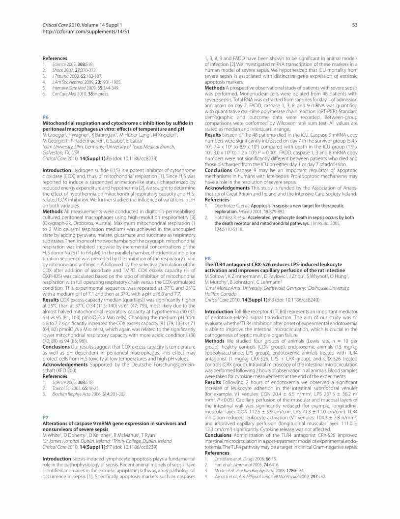
Results The fl uorescence intensities were higher at room temperature
compared with +4°C and they increased after storage (Figure 1). The eff ect
was observed especially for CD11b-antigen in monocytes and neutrophils
and for HLA-DR in monocytes.
Conclusions According to our results, fl ow cytometry leukocyte analysis
using CD antigens should be performed using +4°C temperature
throughout the measurement including sample collection, preparation
and storage, and the analysis should be performed within 6 hours.
References1. Payden et al.: Minerva Anest 2009, 75:484-493.
2. Nuutila et al.: Hum Immunol 2009, 70:237-243.
3. Li et al.: Eur J Haematol 2000, 65:57-65.
P15
Phenotypical analysis of peripheral human T lymphocytes in early sepsisM Nalos, B Santner-Nanan, L Weisbrodt, AS Mclean, R Nanan
Sydney Medical School – Nepean, The University of Sydney, Penrith, Australia
Critical Care 2010, 14(Suppl 1):P15 (doi: 10.1186/cc8247)
Introduction T lymphocytes are crucial immune cells. We analysed T-cell
subsets phenotypes and tested, on a single cell level, their ability to
produce key cytokines in early human sepsis.
Methods Whole blood was collected from septic patients on ICU
admission. Peripheral blood mononuclear cells (PBMC) were isolated and
T-cell subsets analysed. To study cytokine production, PMBC were cultured
in the presence of PMA/ionomycin (50/750 ng/ml) in supplemented RPMI
1640 for 5 hours. Intracellular cytokines IL-4, IL-10, IL-17, IFNγ were stained
in CD3+/CD4+, CD3+/CD8+ cells using fl ow cytometry for both. The number
of cytokine producing cells was compared with age/sex-matched healthy
human volunteers. Data are expressed as mean ± SEM.
Results There were 12 patients (66 years old, six males, APACHE II-24,
eight survivors) and nine volunteers. We found a relative increase in
the frequency of Treg cells while the proportion of CD4+ cells remained
unchanged in septic patients. The PMA/ionomycin lead to maximal
T-cell stimulation, testing the ability of individual cell subsets to produce
cytokines. Septic patients displayed reduction of IFNγ (10.5 ± 0.8% vs
14.7 ± 1.9%, P <0.01) and a tendency to higher number of IL-10 (1.7 ±
0.3% vs 0.5 ± 0.1%, P = 0.10) producing CD4+ cells, while the proportion
of IFNγ-positive CD8+ cells increased (42.8 ± 5.8% vs 28.1 ± 4.9%, P = 0.03).
However, the overall CD8+ T-cell population was reduced (14.29 ± 1.6% vs
25 ± 1.2%) following ex vivo activation in patients. The number of IL-4 and
IL-17 staining cells was unchanged (Figure 1).
Conclusions Our results confi rm a relative increase of Treg [1] and a skew
towards Th2 lineage in the CD4+ cells. The highly activated CD8+ cells
appear to be more susceptible to activation-induced cell death.
References1. Venet F et al.: Intensive Care Med 2009, 35:678-686.
Figure 1 (abstract P14). CD11b fl uorescence intensity for neutrophils
(n = 10). Figure 1 (abstract P15).
Critical Care 2010, Volume 14 Suppl 1 http://ccforum.com/supplements/14/S1
S6
P16
CD64, a marker of leucocyte activation kinetics after uncomplicated cardiac surgeryS Djebara, P Cauchie, A Alewaeters, A Daper, E Fosse, K Zouaoui Boujelta,
P Biston
CHU Charleroi, Charleroi, Belgium
Critical Care 2010, 14(Suppl 1):P16 (doi: 10.1186/cc8248)
Introduction The aim of this study was to assess the CD64 kinetics after
normal cardiac surgery, an essential step to defi ne the potential use of
CD64 as an early and specifi c infectious marker in this postoperative
setting. CD64 is a high-affi nity receptor for the Fc portion of IgG. It is weakly
expressed on the polynuclear neutrophils (PMNs) at rest [1,2] but increases
specifi cally after bacterial stimulation. Thus, CD64 analysis is proposed as
an early infectious marker.
Methods Prospective study realised in the medico-surgical ICU of CHU
Charleroi (Belgium). Twenty-two patients (mean age 64 ± 13 years)
scheduled for cardiac surgery were included in the analysis. The CD64
expression on neutrophils was quantifi ed by the haematologic cell dyn
sapphire method (Abott, USA). C-reactive protein, leukocyte count, white
blood cells, platelets and temperature were also monitored. Values are
expressed as median (25th to 75th) percentile.
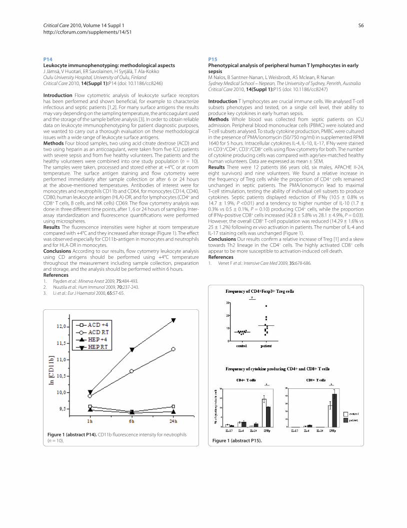
Results CD64 index (Figure 1) slightly rose from the day 0, median
value 0.86 units (1.06 to 1.38 units) to day 5 after the surgery, 1.01 (1.32
to 1.68 units). However, this increase was moderate, and the median
values did not exceed the usual threshold of 1.5 units. Conversely, CRP
showed a large increase from normal values on day 0 to 8, 67 mg/dl (6.64
to 13.56 mg/dl) on day 5. No signifi cant correlation (P >0.05) was found
between CD64 and the others parameters of infl ammation (CRP, leukocyte
count, PMNs, platelets and temperature).
Conclusions Our results show that the neutrophil CD64 expression after
cardiac surgery with ECC is only moderately increased. The role of this new
biomarker in the early diagnosis of infection (after major surgery) should
be assessed in prospective studies.
References1. Loan-Facsinay A, de Kimpe SJ, Hellwig SMM, et al.: FcγRI(CD64) contributes
substantially to severities of arthritis, hypersensitivity responses, and protection from bacterial infection. Immunity 2002, 16:391-402.
2. Harpaz Y, Chothia C: Many of the immunoglobulin superfamily domains in cell adhesion molecules and surface receptors belong to a new structural set which is close to that containing variable domains. J Mol Biol 1994,
238:528-539.
P17
Interferon-gamma reverses sepsis-induced immunoparalysis of monocytes in vitroM Mouktaroudi, A Savva, A Pistiki, M Raftogiannis, A Antonopoulou,
E Giamarellos-Bourboulis
ATTIKON University Hospital, Athens, Greece
Critical Care 2010, 14(Suppl 1):P17 (doi: 10.1186/cc8249)
Introduction It is currently being understood that most of the agents
modulating host response in sepsis have failed because they act to
refrain an over-exaggerated immune response whereas immunoparalysis
takes place on the time of their administration. The eff ect of IFNγ on
immunoparalysis of monocytes in sepsis was assessed.
Figure 1 (abstract P16). CD64 kinetics (units).
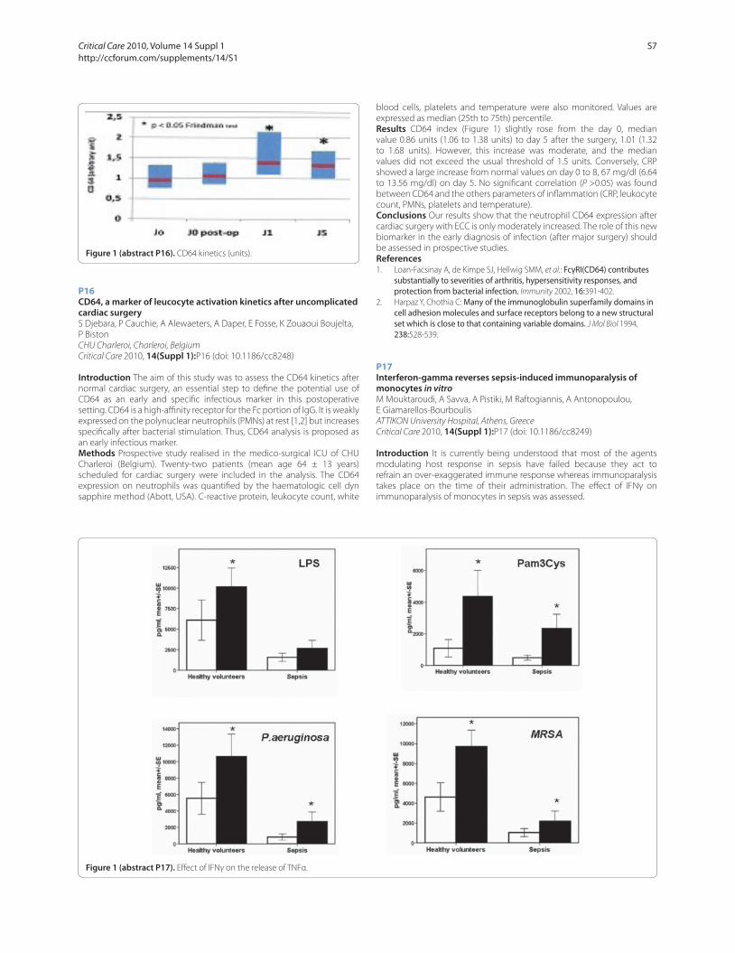
Figure 1 (abstract P17). Eff ect of IFNγ on the release of TNFα.
Critical Care 2010, Volume 14 Suppl 1 http://ccforum.com/supplements/14/S1
S7

Methods Blood was isolated within the fi rst 24 hours from the advent
of signs of sepsis from 10 healthy donors and from 33 patients; 14 with
sepsis and 19 with severe sepsis/shock. Peripheral blood mononuclear
cells (PBMCs) were isolated and stimulated with 10 ng/ml LPS; 5 μg/ml
Pam3Cys; and heat-killed isolates of Candida albicans, multidrug-resistant
Pseudomonas aeruginosa and methicillin-resistant Staphylococcus aureus
(MRSA). Stimulations were done in the absence and presence of 10 ng/
ml IFNγ. Concentrations of cytokines were estimated in supernatants by
an enzyme immunoassay.
Results Eff ects of IFNγ on the release of TNFα are shown in Figure 1.
Open bars represent stimulation in the absence of IFNγ and solid bars
stimulation in the presence of IFNγ. Asterisks signify diff erences compared
with the absence of IFNγ. IL-6 and IL-1β had similar kinetics to TNFα.
Conclusions IFNγ reverses in vitro sepsis-induced immunoparalysis of
monocytes. The type of aff ected cytokine depends on the agonist and
probably refl ects the mechanism of action of IFNγ.
P18
Early hydrocortisone treatment counteracts circulatory derangement but not cytokine response in porcine endotoxemiaES Söderberg1, ML Lipcsey1, JS Sjölin2, AL Larsson3, ME Eriksson1
1Section of Anaesthesiology and Intensive Care, Uppsala, Sweden; 2Section of
Infectious Diseases, Uppsala, Sweden; 3Section of Clinical Chemistry, Uppsala,
Sweden
Critical Care 2010, 14(Suppl 1):P18 (doi: 10.1186/cc8250)
Introduction We evaluated whether treatment with hydrocortisone
administered just after establishment of endotoxin-mediated circulatory
dysfunction would have anti-infl ammatory and shock reversing eff ects.
Establishment of endotoxin-mediated circulatory dysfunction was
defi ned as the moment when the mean pulmonary arterial pressure
(MPAP) reached the double baseline value.
Methods All pigs were anesthetized and given endotoxin infusion
during the 6 hours of the experiment. Eight pigs were randomized to the
hydrocortisone group, receiving hydrocortisone at 5 mg/kg intravenously,
or to the control group, receiving a corresponding volume of saline, as
soon as the MPAP doubled the baseline value. P <0.05 was considered
signifi cant.
Results No diff erence in baseline data was noted between the groups.
No diff erences in TNFα (Figure 1), IL-6 and core temperature were seen
between the groups. Pigs in the hydrocortisone group had signifi cantly
higher mean arterial pressure and systemic vascular resistance index
during the 6-hour experimental period than pigs in the control group,
while heart rate was signifi cantly lower in the hydrocortisone group at 1 to
6 hours as compared with controls.
Conclusions Early treatment with hydrocortisone, administered after the
onset of endotoxemia, counteracted circulatory deterioration, but did not
aff ect the plasma levels of proinfl ammatory cytokines in this model. Thus,
TNFα and IL-6 might not be involved in the development of circulatory
dysfunction during the early phase of experimental endotoxemia.
P19
CD40L is selectively expressed on platelets from thrombocytopenic septic patientsAM Montalbano1, F Turani2, EC Ciotti1, MF Fede1, MP Piperno1, PD David1,
DF Fiume1, GC Caiazza1, SN Natoli1, FL Leonardis1, AB Bergamini1
1University Tor Vergata, Rome, Italy; 2European Aurelia Hospital, Rome, Italy
Critical Care 2010, 14(Suppl 1):P19 (doi: 10.1186/cc8251)
Introduction It has been recently hypothesized that septic microangio-
pathy is caused or at least promoted by the interaction between endo-
thelial surface receptor CD40 and its ligand CD40L, expressed by activated
platelets. This interaction produces procoagulative changes in endothelial
cells, endothelial apoptosis, subendothelial matrix exposition and
microthrombi formation. Since virtually all septic patients show a certain
degree of coagulation abnormalities, we hypothesized that low platelet
count is associated with a diff erent degree of CD40L expression and that
this could correlate with the severity of disease.
Methods To determine the infl uence of sepsis on levels of platelet-derived
CD40L expression, we performed a prospective observational study in a
polyvalent university hospital ICU. Eighteen consecutively septic patients
were enrolled in the study, independently of the platelet count and the
severity of disease (SOFA score). Flow cytometry of fresh blood from septic
patients (n = 18) and age-matched controls (n = 8) was performed for
membrane-bound CD40L and CD62P on circulating platelets.
Results Flow cytometry demonstrated low levels of CD62P in controls
while the levels in patients were high. CD40L+ platelets were selectively
found from patients with thrombocytopenia (platelet count ≤60,000/
mm3). Furthermore a direct correlation between CD40L expression and
the SOFA score was found in patients with sepsis and thrombocytopenia
compared to patients with sepsis without thrombocytopenia.
Conclusions These results suggest that CD40L expression on platelets is
somehow related to the degree of thrombocytopenia and possibly can
be a marker of the severity of sepsis. Although the role of endothelial-
derived CD40/platelet-derived CD40L interaction is not fully understood
during sepsis, the expression of CD40L on platelets could be related to
the severity of organ disease due to the possible bursting of endothelial
damage through this pathway. Further investigation is needed to
determine whether platelets CD40L contributes to endothelial and
subsequent organ damage, its role in thrombocytopenia and its
correlation with the outcome of sepsis. The microvascular injury seems
to be a central event in sepsis, so understanding the mechanisms
underlying its development is crucial for the individuation of new and
specifi c therapeutic strategies.
P20
Neuronal NOS-inhibition in the setting of septic cardiomyopathyA Van de Sandt1, R Windler1, A Gödecke2, J Ohlig3, S Becher1, T Rassaf1,
J Schrader2, M Kelm1, M Merx1
1Department of Cardiology, Pulmonology and Vascular Medicine, University
Hospital Düsseldorf, Germany; 2Department of Cardiovascular Physiology,
Heinrich-Heine-University, Düsseldorf, Germany; 3RWTH, Aachen, Germany
Critical Care 2010, 14(Suppl 1):P20 (doi: 10.1186/cc8252)
Introduction Nitric oxide (NO) plays a central role in the pathogenesis
of sepsis. Recently, we demonstrated that endothelial NOS (eNOS)
contributes to endogenous NO-production and modulates infl ammation,
associated with preserved cardiac function resulting in prolonged survival
of eNOS–/– compared with wild-type (WT) mice. The role of neuronal NOS
(nNOS) in septic cardiomyopathy remains unclear. This study’s aim is to
elucidate the infl uence of nNOS in the presence/absence of eNOS on
cardiac function and survival in a clinically relevant model of sepsis.
Methods Inhibition of nNOS was achieved via continuous application of
the selective nNOS-inhibitor Vinyl-L-NIO (VL-NIO) (0.02 mg/kg BW/hour)
using an osmotic mini pump. B6/c57 WT and eNOS–/– mice were rendered
septic by cecum ligation and puncture (CLP). After 12 hours heart function
was analyzed using a pressure–volume catheter placed in the left ventricle.
For catecholamine responsiveness, norepinephrine was applied (0.4 μg/kg
BW/minute, intraperitoneally). NOx-levels in plasma were measured using
high-pressure performing liquid chromatography.
Results Inhibition of nNOS via VL-NIO application resulted in signifi cantly
reduced nitrate plasma levels and prolonged survival (WT CLP + VL-NIO = 38 Figure 1 (abstract P18). TNFα over time (mean ± SEM).
Critical Care 2010, Volume 14 Suppl 1 http://ccforum.com/supplements/14/S1
S8

hours vs WT CLP = 29 hours). However, cardiac function and norepinephrine
responsiveness were not improved compared with untreated septic WT.
In contrast to WT, application of VL-NIO in eNOS–/– had no infl uence on
plasma nitrate levels, while cardiac function and survival were signifi cantly
impaired compared with untreated septic eNOS–/–. Impaired cardiac
function was accompanied by decreased survival time (eNOS–/– CLP +
VL-NIO = 29.5 hours vs eNOS–/– CLP = 69.5 hours).
Conclusions Pharmacologic inhibition of nNOS result in signifi cant
reduction of plasma nitrate levels and prolonged survival compared
with untreated septic WT despite unimproved septic cardiomyopathy.
In contrast, the signifi cant survival benefi t of septic eNOS–/– compared
with septic WT was abrogated by pharmacologic nNOS inhibition.
Furthermore, the latter developed severe septic cardiomyopathy.
Whether eNOS acts as modulator of nNOS in this setting remains to be
clarifi ed by further studies.
P21
Urinary albumin excretion is elevated in sepsis, but does not correlate with circulating VEGF-A levelsS Basu1, M Bhattacharyya1, T Chatterjee2, S Todi1, A Majumdar1
1AMRI Hospitals, Kolkata, India; 2Jadavpur University, Kolkata, India
Critical Care 2010, 14(Suppl 1):P21 (doi: 10.1186/cc8253)
Introduction In critically ill patients admitted with SIRS, endothelial
dysfunction leads to increased capillary permeability. In the glomerulus,
it manifests as increased albumin excretion. Microalbuminuria is a
common fi nding in various acute conditions like sepsis, trauma and
surgery. Studies have shown increased levels of vascular endothelial
growth factor-A (VEGF-A), a potent vascular permeability inducing
agent, in LPS-induced endotoxemia, severe sepsis and septic shock.
The pathogenic mechanism of the glomerular leakage of albumin in
acute infl ammatory conditions remains to be clarifi ed. We wished to
investigate the causative role of VEGF-A, in this regard which might have
therapeutic implications.
Methods Prospective study in the 43-bed ICU of a tertiary care hospital. Of
the consecutive patients admitted to the ICUs between September 2008
and January 2009, 597 patients were included, after excluding patients
with ICU stay <24 hours, pregnancy, menstruation, anuria, hematuria and
proteinuria due to renal and post- renal diseases. Of these, 30 consecutive
patients with sepsis, severe sepsis and septic shock (sepsis group) and 30
randomly chosen patients without sepsis were recruited for the VEGF-A
study. Spot urine samples for the albumin–creatinine ratio (ACR, mg/g)
and serum for VEGF-A (ELISA) were collected on ICU admission. Correlation
was analyzed using Spearman’s correlation coeffi cient.
Results Sixty critically ill patients, with a median age (IQR) of 60 years (48
to 72), 39:21 male:female ratio, median APACHE II score (IQR) of 16 (7 to
23), and 3 median days of ICU stay, had a median ACR (IQR) of 125 mg/g
(51.6 to 239.1) and a median VEGF-A (IQR) of 111 pg/ml (54.3 to 286.9) on
ICU admission. The median ACR on admission in the sepsis group (n = 30)
of 161.8 mg/g was signifi cantly higher than the median ACR (78.3 mg/g)
of the group without sepsis (n = 30) (P = 0.011). On statistical analysis, no
signifi cant correlation was obtained between levels of urinary ACR and
serum VEGF-A (P = 0.327) in the entire group. Analysis for correlation
between ACR and VEGF-A in the sepsis patients (P = 0.396) and in the
group without sepsis (P = 0.518) yielded similar results.
Conclusions Despite a strong physiologic rationale, our pilot study did not
show an association between microalbuminuria and VEGF-A in critically ill
patients. Larger studies are needed to come to a defi nitive conclusion.
P22
High mobility group box protein-1 preconditions isolated rat hearts and protects human pulmonary and renal cells against hypoxic injuryCM Walshe, D O’Toole, B Higgins, JG Laff ey, LG Kevin
National University of Ireland, Galway, Ireland
Critical Care 2010, 14(Suppl 1):P22 (doi: 10.1186/cc8254)
Introduction High mobility group box protein-1 (HMGB) is released by
activated monocytes/macrophages during sepsis. Levels correlated with
mortality in patients and anti-HMGB antibodies prevented endotoxin-
induced death in animal models [1]. In marked contrast to these fi ndings
are reports of cytoprotective eff ects because HMGB protected liver from
ischemia/reperfusion (IR) [2] and HMGB-transgenic mice were resistant to
myocardial infarction [3]. Despite these reports, little is known of the role
or mechanism of HMGB. We aimed to further evaluate HMGB in rat hearts
and in pulmonary and renal cell cultures.
Methods Isolated hearts received HMGB/vehicle before 30 minutes
ischemia and 120 minutes reperfusion. In separate studies, rats were
rendered septic by caecal ligation and puncture (CLP) prior to IR. Left
ventricular (LV) function was measured with a latex LV balloon. Infarct
size was measured by TTC staining. For human cell culture studies, A549
alveolar cells or HK-2 renal tubular cells were treated with HMGB/vehicle
before incubation in normoxia or hypoxia. Fluorimetric caspase-3 activity
was used as measure of apoptosis. Cytochrome C was measured by ELISA.
For HK-2 cells, the MTT assay was used to test viability.
Results HMGB, prior to cardiac IR, preserved developed (d) LVP at
120 minutes reperfusion compared with controls (44 mmHg vs 27 mmHg,
P <0.05) and infarct size, expressed as %V weight ± SEM, was reduced (31
± 5.4 vs 44 ± 4.2, P <0.05), n = 6/group. Hearts from CLP rats had reduced
baseline dLVP (59 mmHg ± 5.1 vs 97 ± 5.3, P <0.05) but infarct sizes after IR
were signifi cantly reduced (27 ± 8.8, P <0.05 vs 44 ± 6.2, P <0.05). In A549
cells, HMGB inhibited apoptosis (49% reduced caspase-3 activity (8.6 vs
17.1 a.u., P <0.01)). This was associated with reduced cytochrome C release
(250 vs 389 pg/ml, P <0.05). Using MTT assay, HMGB protected against
hypoxia-induced cell death vs controls (viability 75% vs 61%, P <0.05).
Conclusions HMGB, or sepsis, similarly precondition the heart against IR
injury. HMGB has anti-apoptotic eff ects that protect against hypoxic renal
and alveolar cell injury. We conclude that HMGB has potent anti-ischemic
eff ects in multiple organs and may function as an innate cytoprotective
mediator in sepsis.
References1. Wang H, et al.: Science 1999, 285:248-251.
2. Tsung A, et al.: J Exp Med 2005, 201:1135-1143.
3. Kitahara T, et al.: Cardiovasc Res 2008, 80:40-46.
P23
Early persisting elevation of plasma Pentraxin 3 is associated with mortality and with coagulation impairment in severe sepsis and septic shockA Coppadoro1, T Mauri1, G Bellani1, N Patroniti1, M Cugno2, A Grassi1,
G Iapichino2, L Gattinoni2, A Mantovani2, A Pesenti1
1Universita’ degli Studi di Milano-Bicocca, Monza, Italy; 2Universita’ degli Studi
di Milano, Italy
Critical Care 2010, 14(Suppl 1):P23 (doi: 10.1186/cc8255)
Introduction Pentraxin 3 (PTX3) is an infl ammatory mediator produced by
a variety of tissue cells, like neutrophils, macrophages, myeloid dendritic,
endothelial and epithelial cells [1]. During sepsis a massive infl ammatory
activation occurs, which often leads to an important coagulation system
dysfunction. PTX3 upregulates coagulation promoters in vitro [2]. PTX3
may represent an early marker of severity and outcome and may be
related to coagulation impairment in septic patients.
Methods We studied 90 patients aff ected by severe sepsis or septic
shock previously enrolled in a prospective trial regarding the impact of
glycemic control on infl ammation and coagulation. At enrollment we
recorded sepsis signs, plasminogen activator inhibitor 1 (PAI-1, an inhibitor
of fi brinolysis) activity and concentration, prothrombin fragments 1 + 2
(F1+2) concentration (a marker of coagulation activation), disease severity
and organ dysfunctions. We measured plasma PTX3 levels at enrollment
and everyday until day 7. Mortality was recorded at day 90.
Results Although not diff erent on day 1, PTX3 remained signifi cantly
higher in nonsurvivors than in survivors over the fi rst 5 days (P = 0.002 by
general linear model). On day 1, septic shock patients had higher PTX3
levels than patients with severe sepsis (274.41 ± 321.92 vs 114.95 ± 129.93
ng/ml, P = 0.029, t test). Day 1 PTX3 was signifi cantly correlated with SAPS
II score (R2 = 0.08, P = 0.006, linear regression), platelets count (R2 = 0.14,
P <0.001) and SOFA score (R2 = 0.16, P <0.001). Day 1 PTX3 was correlated
with PAI-1 concentration (R2 = 0.211, P <0.001) and activity (R2 = 0.231,
P <0.001) and with F1+2 concentration (R2 = 0.09, P = 0.045).
Conclusions Persisting high levels of circulating PTX3 over the fi rst days
from sepsis onset may be associated with mortality. PTX3 correlates with
severity of sepsis and with sepsis-associated coagulation dysfunction.
Critical Care 2010, Volume 14 Suppl 1 http://ccforum.com/supplements/14/S1
S9
References1. Mauri T, et al.: Crit Care Med 2008, 36:2302-2308.
2. Napoleone E, et al.: J Leukoc Biol 2004, 76:203-209.
P24
Intravenous immunoglobulins prevent breakdown of the blood–brain barrier in experimental sepsisF Esen1, E Senturk1, P Ergin Ozcan1, B Ahishali1, N Arıcan1, N Orhan1,
O Ekizoğlu2, D Ustek1, M Kucuk1, M Kaya1
1Istanbul University Istanbul Medical Faculty, Istanbul, Turkey; 2Sadi Konuk
Training and Research Hospital, Istanbul, Turkey
Critical Care 2010, 14(Suppl 1):P24 (doi: 10.1186/cc8256)
Introduction The eff ect of intravenous immunoglobulin preparations –
immunoglobulin G (IgG), immunoglobulin G enriched with IgA and IgM
(IgGAM) – on blood–brain barrier (BBB) integrity and survival rate was
comparatively investigated in septic rats.
Methods Sepsis was induced by cecal ligation and perforation (CLP)
in Sprague–Dawley rats. The animals received either IgG (250 mg/kg,
intravenously) or IgGAM (250 mg/kg, intravenously) 5 minutes before CLP
surgery. All rats were observed for behavioral changes for 24 hours after CLP
operation. To show the alterations in BBB integrity, Evans blue (EB; 69 kDa)
dye extravasation was determined for quantitative measurement of BBB
permeability, and horseradish peroxidase (HRP; 40 kDa) extravasation was
evaluated to provide ultrastructural evidence for the transport pathways
involved in BBB permeability. Immunohistochemistry was performed
to show the alterations in immunoreactivity for tight junction protein
occludin.
Results A high mortality rate (34%) was noted in septic animals and
the mortality rate was decreased to 15% and 3% by IgG and IgGAM,
respectively. Both IgG and IgGAM alleviated the sickness behavior in
septic rats and the animals were observed to be healthy and active.
Increased extravasation of EB dye into brain tissue of septic animals was
markedly decreased by both IgG and IgGAM. Occludin immunoreactivity
remained essentially unchanged in all groups including CLP. In addition,
strong staining for HRP was seen around vessels located in the cerebral
cortex and the hippocampus in septic animals. Increased luminal and
abluminal vesicles containing electron-dense HRP reaction product were
noted in the cytoplasm of endothelial cells in the cerebral cortex and
hippocampus of septic rats, emphasizing an increased BBB permeability
predominantly by transendothelial route. In these animals, tight junctions
were ultrastructurally intact suggesting that paracellular pathway of BBB is
not responsible for the BBB breakdown in sepsis. Following IgG or IgGAM
treatment, no ultrastructural evidence of leaky capillaries in brain was
observed in septic animals indicating the blockade of the transcellular
pathway.
Conclusions The present study indicates that IgG and IgGAM improve the
integrity of the BBB and inhibit CLP-induced sickness behavior in rats.
P25
Early use of immunoglobulin in septic shockIC Cavazzuti, LR Rinaldi, LD Donno, SB Braccini, SB Busani, MG Girardis
Policlinico di Modena, Italy
Critical Care 2010, 14(Suppl 1):P25 (doi: 10.1186/cc8257)
Introduction In addition to the interventions suggested by the guidelines
of the Surviving Sepsis Campaign, in the recent years other therapeutic
options have been proposed and tested for patients with severe sepsis
and septic shock. Polyclonal intravenous immunoglobulins seem to be
an interesting and useful option because of its activity in neutralizing
endotoxin and in modulating the host immune response. In this retro-
spective study we assessed the eff ects of the early use of polyclonal
intravenous immunoglobulins enriched with IgA-M (IgGAM) in addition
to standard therapies on clinical outcome of patients with septic shock.
Methods In January 2008, IgGAM was introduced into our ICU protocol
for the management of septic shock patients (within 24 hours after shock
appearance and at the dosage of 5 ml/kg for 3 days). From January 2008
to September 2009, we studied 52 consecutive patients with septic shock
admitted to our ICU. In each patient we recorded the SAPS II and SOFA
scores, the compliance to the 6-hour and the 24-hour sepsis bundles, the
compliance to IgGAM use, the length of stay in the ICU and the 30-day
mortality rate.
Results Due to low compliance of medical staff to protocol application,
IgGAM has been used only in 27 patients (IgGAM group). At ICU admission,
the severity scores were similar in patients with and without (Control
group) IgGAM therapy (SAPS II: control 54 (22 to 99), IgGAM: 59 (39 to
101); P >0.05 and SOFA: control 9 (2 to 17); IgGAM 9 (4 to 15); P >0.05).
The compliance to 6-hour and 24-hour bundles was also similar in the
two groups, apart from compliance to fl uid therapy that was larger in the
IgGAM group (85% vs 56% in Control group). The length of stay in the ICU
was 15 ± 9 days in the IgGAM group and 18 ± 31 days in the Control group
(P >0.05); the 30-day mortality was lower (P <0.05) in the IgGAM group
(25%) than in the Control group (52%).
Conclusions These preliminary data indicate that the early use of IgGAM,
associated with interventions proposed by the SSC guidelines, reduces
mortality of patients with septic shock.
References1. Ballow M: Mechanism of action of intravenous immune serum globulin in
autoimmune and infl ammatory diseases. J Allergy Clin Immunol 1997,
100:151-157.
2. Werdan K: Mirror, mirror on the wall, which is the fairest meta-analysis of all? Crit Care Med 2007, 35:2852-2854.
P26
Low levels of immunoglobulin G in patients with sepsis or septic shock: a signum mali ominis?S Dietz, C Lautenschlaeger, U Mueller-Werdan, K Werdan
University Clinics of the Martin-Luther-University Halle-Wittenberg, Halle
(Saale), Germany
Critical Care 2010, 14(Suppl 1):P26 (doi: 10.1186/cc8258)
Introduction The role of intravenous immunoglobulin therapy in patients
with sepsis and septic shock is still controversially discussed. In a retro-
spective analysis of the data from the SBITS-trial [1] we investigated
whether the initial level of serum IgG on admission to the hospital in
patients with sepsis and septic shock (before the fi rst administration of the
fi rst dose of intravenous immunoglobulins) could be seen as a prognostic
parameter for the primary outcome, lethality on day 28, or the secondary
endpoints, lethality on day 7 or on the ICU.
Methods A total of 543 were included in the trial: the patients were
divided into four groups (quartiles) based on the 25th, 50th and 75th
percentiles of their initial level of IgG on admission to the hospital. The
fi rst group contained 140 patients with a range in the IgG serum level up
to 6.1 g/dl. The second and third groups contained 134 patients and 136
patients, respectively, the immunoglobulin G levels were greater than
6.1 g/dl to 8.4 g/dl and greater than 8.4 g/dl to 11.9 g/dl. The fourth group
containing 133 patients had IgG levels higher than 11.9 g/dl. In a logistic
regression model we adjusted for potential confounders (that is, sex, age,
APACHE II score, presence of shock on admission).
Results On basis of this model we could show that lethality between the
fi rst and second groups did not diff er signifi cantly with the lethality in the
third (reference) group with physiologic levels of IgG on day 0. Surprisingly
the lethality of the fourth group, with IgG levels higher than 11.9 g/dl, was
signifi cantly higher compared with the reference quartile (OR 1.69, CI 1.01
to 2.81, P = 0.047).
Conclusions Our post-hoc analysis did show no prognostic relevance of
a low level of serum IgG on admission to hospital for the 543 patients
with sepsis and septic shock. Thus the initial serum level of IgG seems to
be of no aid in making the decision to initiate therapy with intravenous
immunoglobulins for these patients. Whether a high level of serum IgG
on admission is an independent risk factor for an increased lethality in
critically ill patients, as shown in our analysis, needs to be investigated in
further studies.
Reference1. Werdan K, et al.: Score-based immunoglobulin G therapy of patients with
sepsis: the SBITS study. Crit Care Med 2007, 35:2693-2701.
Critical Care 2010, Volume 14 Suppl 1 http://ccforum.com/supplements/14/S1
S10

P27
Background factors in patients receiving immunoglobulin administration and changes in sepsis markersT Ikeda1, K Ikeda1, H Taniuchi1, S Suda1, Y Takahashi2
1Tokyo Medical University, Hachioji Medical Center, Tokyo, Japan; 2Sannoudai
Hospital, Ibaraki, Japan
Critical Care 2010, 14(Suppl 1):P27 (doi: 10.1186/cc8259)
Introduction The potentially envisaged actions of intravenous immuno-
globulin (IVIg) on severe infectious disease include virus or toxin neutralizing
action, opsonic eff ect, complement bacteriolytic activity, and enhancement
of sensitivity to antibiotics. In the case of severe infectious disease, antibiotics
are often supplemented with administration of IVIg. The aim of this study
is that the changes in sepsis markers followed by IVIg administration are
investigated with severe sepsis or severe septic shock patients.
Methods The subjects were 52 patients admitted to an ICU with a diagnosis
of severe sepsis or septic shock and from whom informed consent had been
obtained for the present study. IVIg was administered intravenously for 3 days
(5.0 g/day) and measurements were undertaken before administration (Day
1), on the day after completion of administration (Day 4), and on Day 7. The
items measured were IL-6, C-reactive protein (CRP) and, as indices of vascular
endothelial cell activation, plasminogen activator inhibitor-1 (PAI-1) and
adhesion molecules (endothelial leukocyte adhesion molecule-1 (ELAM-1));
the eff ect of IVIg administration on these markers was then studied. The IVIg
studied was polyethylene glycol-treated human immunoglobulin injection
fl uid (2.5 g, 50 ml, one vial).
Results The APACHE II score was 25.3 ± 7.8, the SOFA score 9.5 ± 3.5, and
the survival rate after 28 days 76.9%. The values before IVIg administration
were: procalcitonin 28.8 ± 44.5 (median value 13.6) ng/ml, CRP 16.3 ± 8.3
(median value 16.0) mg/dl, IL-6 8,873 ± 19,362 (median value 744) pg/ml,
PAI-1 374 ± 428 (median value 178) ng/ml, and ELAM-1 198 ± 359 (median
value 61) ng/ml. All values were thus elevated. After the completion of IVIg
administration, the level of mediators other than ELAM-1 (procalcitonin,
CRP, IL-6, PAI-1) decreased signifi cantly.
Conclusions The results of the present study found signifi cant decreases
of IL-6 and PAI-1 resulting from immunoglobulin administration, but did
not indicate a protective eff ect on vascular endothelial cell function.
P28
C1-esterase inhibitor (C1INH) implication in systemic infl ammation in sepsisA Igonin, N Lazareva, VY Uvarov
BioGenius Research Centre, Moscow, Russia Federation
Critical Care 2010, 14(Suppl 1):P28 (doi: 10.1186/cc8260)
Introduction C1INH is the most potent endogenous regulator of compli ment
as well as intrinsic coagulation pathways and the kallekrein–kinin system.
Methods C1INH systemic activity was studied in patients who were
enrolled within 48 hours after onset of sepsis (ACCP, 1992). The analysis of
C1INH activity in quartiles (Q) was conducted in terms of RCT of human
purifi ed C1INH (Bicizar, Russia).
Results Sepsis patients (n = 40) responded with an increase of C1INH
activity in comparison with healthy individuals (Figure 1). Thirty percent of
Q1 patients had ARDS and septic shock whereas in Q4 everyone showed
only signs of sepsis. The CRP level was higher in Q1 patients (243.4 ±
39.9 mg/l) than in Q4 (144.0 ± 20.07 mg/l; P = 0.04), whereas the C4 subunit
was lower in Q1 (0.19 ± 0.04 g/l) than in Q4 (0.32 ± 0.04 g/l; P = 0.05).
Conclusions Inability to upregulate C1INH activity in sepsis was associated
with enhanced systemic infl ammation, higher number of ARDS and septic
shock cases.
P29
Prognostic value of B-type natriuretic peptide in critically ill patients with new onset of fever: preliminary studyV Soulountsi1, V Voutsas1, P Kontou1, A Giakamozis1, K Hatzimanolis2,
T Lazaridis1, V Hatsiou2, M Mpitzani1
1ICU, G. Papanicolaou, Thessaloniki, Greece; 2Microbiology Laboratory,
G. Papanicolaou Hospital, Thessaloniki, Greece
Critical Care 2010, 14(Suppl 1):P29 (doi: 10.1186/cc8261)
Introduction The purpose of the study is the evaluation of B-type
natriuretic peptide (BNP) as a predictor of septic complications and ICU
mortality in patients with a new onset of fever during the fi rst 3 days of
hospitalization in the ICU.
Methods Thirty-one ICU patients (21 males and 10 females) with new
onset of fever and leukocytosis within the fi rst 3 days of ICU admission
were prospectively included in the study. Exclusion criteria were heart or
renal failure, chronic obstructive pulmonary disease and head trauma.
Serial plasma samples were taken on days 1, 2 and 4 after the onset of
fever for BNP level measurement. BNP values were correlated with severity
scores (Acute Physiology and Chronic Health Evaluation (APACHE II) and
Sequential Organ Failure Assessment (SOFA)), the progression to septic
shock and the fi nal outcome.
Results According to the clinical and laboratory fi ndings within the fi rst
3 days of hospitalization, the patients included in the study were divided
into three groups: Group A = systemic infl ammatory response syndrome
(SIRS) (seven patients), Group B = sepsis (14 patients) and Group C =
septic shock (10 patients). The BNP value on days 1 and 2 was signifi cantly
associated with the SOFA Max value (P <0.001). The BNP value on day 4
was signifi cantly associated with ICU mortality (P = 0.006). The optimal
cutoff BNP value for diff erentiating between nonsurvivors and survivors
was estimated to be 203.55 pg/ml (sensitivity = 100%, specifi city = 61.1%).
In Group B patients, BNP value on day 2 was signifi cantly higher in patients
who fi nally progressed to septic shock (P = 0.001). The optimal cutoff BNP
value for identifying these patients was estimated to be 212.45 pg/ml
(sensitivity = 85.7%, specifi city = 64.3%).
Conclusions In ICU patients with new onset of fever during the fi rst
3 days of ICU hospitalization, the BNP value on day 4 seems to be a good
predictor of ICU mortality. In patients with sepsis, a cut-off BNP value of
212.45 pg/ml on day 2 could be a predictor of progression to septic shock.
Due to the small number of patients included in our study, further studies
are needed to confi rm these fi ndings.
P30
Microalbuminuria evaluated as a biomarker in patients with septic shockC Grion, L Cardoso, C Carrilho, J Altafi n, S Barros, L Carvalho, J Festti,
F Mansano, T Okamoto, K Uehara, J Dias, GC Silva
Hospital Universitário de Londrina, Brazil
Critical Care 2010, 14(Suppl 1):P30 (doi: 10.1186/cc8262)
Introduction The level of microalbuminuria is thought to refl ect the
severity of infl ammation during acute phase response and may have
prognostic value with regard to survival. This study aimed to describe
urinary albumin excretion in patients with septic shock and changes in
microalbuminuria levels in the fi rst 3 days of treatment.
Figure 1 (abstract P28). C1INH activity in patients with sepsis analyzed in
quartiles (Q1, Q2, Q3, Q4) in comparison with healthy individuals.
Critical Care 2010, Volume 14 Suppl 1 http://ccforum.com/supplements/14/S1
S11

Methods Prospective observational study of 49 consecutive patients with
septic shock admitted to a general ICU. During the fi rst 24 hours after severe
sepsis was diagnosed, we collected data to calculate APACHE II and SOFA
scores and urinary sample to measure urinary albumin. Another sample of
urine was collected on the third day of septic shock. Microalbuminuria was
measured by immunoturbidimetry with a limit of detection of 3.5 mg/l.
The study was approved by the local Ethic Committee.
Results The mean age was 65.8 ± 15 years and male sex was more frequent
(63.3%). Pneumonia (59.2%) was the most common source of sepsis. Mean
APACHE II and SOFA scores on the fi rst day of sepsis were 24.4 ± 7.7 and
8.7 ± 3.7, respectively. Observed mortality was 71.4%. Median urinary
albumin was 7.0 mg/l (IQT: 4.7 to 60.3 mg/l) for survivors and 15.5 mg/l
(IQT: 7.9 to 77.1 mg/l) for nonsurvivors on the fi rst day of septic shock.
Urinary albumin decreased from 7.0 mg/l (IQT: 4.7 to 60.3 mg/l) to 3.9 mg/l
(IQT: 3.6 to 44.1 mg/l) in survivors during the fi rst 3 days of septic shock,
and in nonsurvivors remained at the same level from 15.5 mg/l (IQT: 7.9 to
77.1 mg/l) to 14.5 mg/l (IQT: 3.9 to 45.3 mg/l) in the period of observation.
Conclusions Patients with septic shock showed high frequency of
microalbuminuria. Levels of urinary albumin showed a tendency to
decrease in survivors, suggesting prognostic signifi cance.
References1. Thorevska N, et al.: Crit Care Med 2003, 31:1075-1081.
2. Abid O, et al.: Chest 2001, 120:1984-1988.
P31
Evaluation of automated hematologic VCS parameters in severe sepsis and septic shock: a case–control studyA Cortegiani1, A Di Benedetto1, L Marino1, G Virga1, R Chiaramonte1,
C Sarno2, SM Raineri1, A Giarratano1
1University of Palermo, Italy; 2Fondazione Istituto S. Raff aele, Cefalù (PA), Italy
Critical Care 2010, 14(Suppl 1):P31 (doi: 10.1186/cc8263)
Introduction A cheap and quick hematologic diagnostic parameter for
detection of sepsis would have both economic and therapeutic benefi ts.
The Coulter LH series hematology analyzer uses the VCS technology
(Volume, Conductivity, Laser Scatter) providing information about cell
volume, size, internal structure, and surface morphology. Many authors
analyzed the clinical usefulness of VCS parameters in reactive neutrophils
for detection of sepsis. An increase in mean cell volume and a decrease in
mean light scatter in septic patients have been described. Our aim is to
verify the correlation between VCS parameters and sepsis.
Methods We enrolled 92 patients admitted in our ICU and divided them
into two groups: Controls (48) and Sepsis (44). We collected blood samples
immediately after making the diagnosis. All samples were analyzed by
Coulter LH 500 hematology analyzer. We realized ROC curves for each V,
C, S parameter.
Results The AUC was 0.816 (95% CI, 0.722 to 0.889) for mean-V value; 0.546
(95% CI, 0.439 to 0.650) for mean-C value; 0.604 (95% CI, 0.497 to 0.704) for
mean S value (Figure 1). The correlation with sepsis was a linear positive
one for mean-V (strong) and mean-C (weak) instead of a linear negative
one for mean-S. The best cut-off value for mean-V was >163 (Sens: 84.09%;
Spec: 68.75%); >137 for mean-C (Sens: 72.73%; Spec: 41.67%); <134 for
mean-S (Sens: 56.82%; Spec: 64.58%).
Conclusions Our study shows the high diagnostic performance of mean-V
and the weak one of mean-C and mean-S in septic patients. The clinical
usefulness of mean-V is strengthened by the quickness and cheapness of
its analysis. It could be tested daily or more as a valid laboratorial help for
critical patient’s clinical assessment.
P32
Use of CD64 for the diagnosis of sepsis: a case–control studyA Cortegiani1, L Marino1, A Di Benedetto1, G Virga1, G Evangelico1,
C Sarno2, SM Raineri1
1University of Palermo, Italy; 2Fondazione Istituto S. Raff aele, Cefalù (PA), Italy
Critical Care 2010, 14(Suppl 1):P32 (doi: 10.1186/cc8264)
Introduction CD64 is the high-affi nity receptor of IgG. It is upregulated by
infl ammatory cytokines on neutrophils. The upregulation of CD64 is linked
with PMN activation in SIRS or sepsis. Our aim is to verify these correlations.
Methods We enrolled 48 ICU critical patients and three groups were
created: SIRS– (17), SIRS+ (13), Sepsis (17). We evaluated CD64 X-mean
values among the three groups as shown in Figure 1. We used a t test to
check the correlation between uprising CD64 and diff erent groups. We
verifi ed the correlation CD64/SOFA (Figure 2), WBC, NE, and patients’ age
using r.
Results We found a CD64 higher level in sepsis compared with SIRS– (t =
8.095; P <0.001) and with SIRS+ (t = 4.938; P <0.001). There were a high
positive linear correlation between CD64 and SOFA score (r = 0.806;
Figure 1 (abstract P31).
Figure 1 (abstract P32).
Figure 2 (abstract P32).
Critical Care 2010, Volume 14 Suppl 1 http://ccforum.com/supplements/14/S1
S12
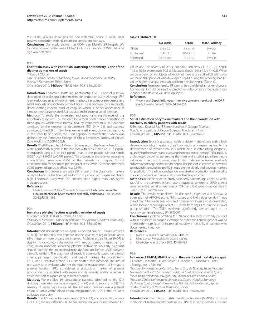
P <0.0001), a weak linear positive one with WBC count, a weak linear
positive correlation with NE count; no correlation with age.
Conclusions Our study shows that CD64 can identify SIRS/sepsis. We
found a correlation between CD64/SOFA; no infl uence of WBC, NE and
age was detected.
P33
Endotoxin assay with endotoxin scattering photometry is one of the diagnostic markers of sepsisY Kase1, T Obata2
1Jikei University School of Medicine, Tokyo, Japan; 2Microbial Chemistry
Research Foundation, Tokyo, Japan
Critical Care 2010, 14(Suppl 1):P33 (doi: 10.1186/cc8265)
Introduction Endotoxin scattering photometry (ESP) is one of a newly
developed clinically applicable method for endotoxin assay. Although ESP
is an analogous assay of turbidimetric method, it enables one to detect very
small amounts of endotoxin within 1 hour. This is because ESP can directly
detect clotting enzyme product, coagulin, which is the fi rst appearance of
Limulus amebocyte lysate (LAL) cascade and the precursor of gel clots.
Methods To study the correlates and prognostic signifi cance of the
endotoxin assay with ESP, we enrolled a total of 80 people consisting of
three groups which were normal healthy volunteers (n = 35), patients
admitted to the emergency department (ED) (n = 21) and patients
admitted to the ICU (n = 24). To examine whether endotoxin is infl uencing
in the severity of disease, we used sepsis/SIRS stratifi cation which was
defi ned by the American College of Chest Physicians/Society of Critical
Care Medicine (ACCP/SCCM).
Results Of all 80 people, 33.7% (n = 27) was sepsis. The levels of endotoxin
were signifi cantly higher in the patients with sepsis (median, 18.4 pg/ml;
interquartile range, 3 to 54.1 pg/ml) than in the people without sepsis
(0.227 pg/ml; 0.031 to 0.493 pg/ml). The area under the receiver operating
characteristic curve was 0.951 in the patients with sepsis. Cut-off
concentrations for optimum prediction of sepsis with ESP were endotoxin
>7.06 pg/ml (diagnostic effi ciency: 92.6%).
Conclusions Endotoxin assay with ESP is one of the diagnostic markers
of sepsis because the levels of endotoxin in patient with sepsis are clearly
high. Endotoxin assay with ESP concentration higher than 7.06 pg/ml
indicates sepsis.
References1. Obata T, Nomura M, Kase Y, Sasaki H, Shirasawa Y: Early detection of the
Limulus amebocyte lysate reaction evoked by endotoxins. Anal Biochem
2008, 373:281-286.
P34
Immature platelet fraction as predictive index of sepsisC Scaramucci, R De Blasi, S Tribuzi, A Carlini
II Faculty of Medicine – University of Rome ‘La Sapienza’ S. Andrea, Rome, Italy
Critical Care 2010, 14(Suppl 1):P34 (doi: 10.1186/cc8266)
Introduction The incidence of sepsis is reported around 37% in European
ICUs [1]. The mortality rate depends on the severity of organ failure, up to
65% if four or more organs are involved. Multiple organ failure (MOF) is
due to microcirculatory dysfunction with microthrombosis resulting from
coagulation disorders including platelets’ activation. An early diagnosis
should identify the microcirculatory dysfunction before MOF became
clinically evident. The diagnosis of sepsis is commonly based on clinical
criteria, pathogen identifi cation and use of markers like procalcitonin
(PCT) and C-reactive protein (PCR) associated with infection. The aim of
our study is to evaluate whether the routine measurement of immature
platelet fraction (IPF), considered a precocious marker of platelet
production, is associated with sepsis and its severity and/or whether it
could be used as a predicting marker of sepsis.
Methods We enrolled 66 consecutive patients admitted to the ICU,
dividing them into two groups: septic (n = 44) and no septic (n = 22). The
severity of sepsis was evaluated. The exclusion criterion was a platelet
count <150,000/mm3. Blood count, coagulation, PCR, PCT, and IPF were
collected every day.
Results The IPF values between septic (4.6 ± 3.1) and no septic patients
(3.3 ± 1.5) did not diff er (P = 0.16). No correlation was found between IPF
values and the severity of septic condition (no sepsis 11.7 ± 10.1; sepsis
14.3 ± 10.5; severe sepsis 10.5 ± 9.1; septic shock 19.5 ± 12.4; P = 0.3). When
we considered only subjects who did not have sepsis at the ICU admission
we found that patients who developed sepsis during the recovery had IPF
values higher than patients who did not develop sepsis (Table 1).
Conclusions From our results IPF cannot be considered a marker of sepsis.
Conversely it could be used as predictive index of sepsis because it can
identify patients who will develop sepsis.
References1. Vincent et al.: Sepsis in European intensive care units: results of the SOAP
study. Intensive Care Med 2006, 34:344-353.
P35
Serial estimation of cytokine markers and their correlation with mortality in elderly patients with sepsisR Bhanot, J Kaur, M Rao, T Ramachandran, K George, D Veliath
Pondicherry Institute of Medical Sciences, Pondicherry, India
Critical Care 2010, 14(Suppl 1):P35 (doi: 10.1186/cc8267)
Introduction Sepsis is a serious health problem in the elderly with a high
degree of mortality. The study of pathophysiology of sepsis has lead to the
development of cytokine markers which help in establishing diagnosis,
quantifying the severity and assessing the response to therapy. TNFα and IL-6,
a pleotropic cytokine, are among the most well-studied proinfl ammatory
cytokines in sepsis. However, very limited data are available in elderly
subjects regarding the markers for sepsis. The present study was undertaken
to establish the cytokine profi le in sepsis in the elderly and develop markers
for prediction. The infl uence of gender on cytokine production and mortality
in elderly patients with sepsis was considered in particular.
Methods In this prospective study, 50 elderly patients, age above 65 years,
satisfying the systemic infl ammatory response syndrome (SIRS) criteria
were included. Serial estimations of TNFα and IL-6 were done on days 1,
3 and 7 of ICU admission.
Results The results were drawn on the basis of gender and survival. A
correlation of APACHE score, TNFα values and IL-6 values on day 1, day
3 and day 7 between survivors and nonsurvivors was also documented,
which showed reducing levels of IL-6 levels from days 1 to 7 in the survivor
group (P <0.01). The TNFα level was signifi cantly low on day 1 in the
nonsurvivor female group (P <0.00001).
Conclusions Cytokine profi ling for TNFαand IL-6 level in elderly patients
with sepsis helps in prognosticating the outcome. Female gender was an
independent predictor of increased morality in critically ill patients with
documented infection.
References1. Martin GS, et al.: Crit Care Med 2006, 34:15-21.
2. Ulloa L, et al.: Trends Mol Med 2005, 11:56-63.
3. Oberholzer A, et al.: Shock 2005, 23:488-493.
P36
Infl uence of TIMP-1/MMP-9 ratio on the severity and mortality in sepsisL Lorente1, M Martín2, J Solé-Violán3, J Blanquer4, L Labarta5, C Díaz6,
J Borreguero1, J Páramo7
1Hospital Universitario de Canarias, Santa Cruz de Tenerife, Spain; 2Hospital
Universitario Nuestra Señora de Candelaria, Santa Cruz de Tenerife, Spain; 3Hospital Universitario Dr Negrín, Las Palmas de Gran Canaria, Spain; 4Hospital Clínico Universitario de Valencia, Spain; 5Hospital San Jorge
de Huesca, Spain; 6Hospital Insular, Las Palmas de Gran Canaria, Spain; 7CIMA-University of Navarra, Pamplona, Spain
Critical Care 2010, 14(Suppl 1):P36 (doi: 10.1186/cc8268)
Introduction The role of matrix metalloproteinases (MMPs) and tissue
inhibitors of matrix metalloproteinases (TIMPs) in sepsis remains unclear.
Table 1 (abstract P34)
No sepsis Sepsis Mann–Whitney
IPF (%) 5.4 ± 2.9 3.3 ± 1.5 P = 0.04
PCT (ng/ml) 0.68 ± 1.1 0.57 ± 1.3 P = 0.4
PCR (mg/dl) 9.57 ± 10.2 11.7 ± 10 P = 0.46
Critical Care 2010, Volume 14 Suppl 1 http://ccforum.com/supplements/14/S1
S13
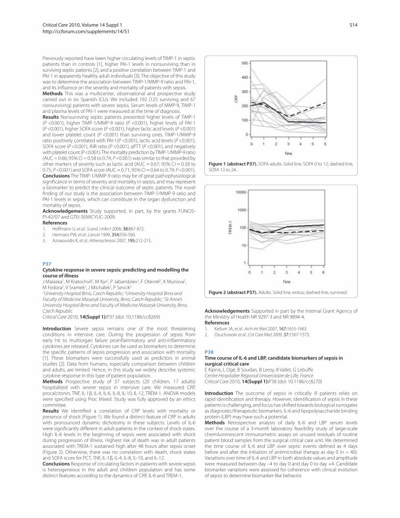
Previously reported have been higher circulating levels of TIMP-1 in septic
patients than in controls [1], higher PAI-1 levels in nonsurviving than in
surviving septic patients [2], and a positive correlation between TIMP-1 and
PAI-1 in apparently healthy adult individuals [3]. The objective of this study
was to determine the association between TIMP-1/MMP-9 ratio and PAI-1,
and its infl uence on the severity and mortality of patients with sepsis.
Methods This was a multicenter, observational and prospective study
carried out in six Spanish ICUs. We included 192 (125 surviving and 67
nonsurviving) patients with severe sepsis. Serum levels of MMP-9, TIMP-1
and plasma levels of PAI-1 were measured at the time of diagnosis.
Results Nonsurviving septic patients presented higher levels of TIMP-1
(P <0.001), higher TIMP-1/MMP-9 ratio (P <0.001), higher levels of PAI-1
(P <0.001), higher SOFA score (P <0.001), higher lactic acid levels (P <0.001)
and lower platelet count (P <0.001) than surviving ones. TIMP-1/MMP-9
ratio positively correlated with PAI-1(P <0.001), lactic acid levels (P <0.001),
SOFA score (P <0.001), INR ratio (P <0.001), aPTT (P <0.001), and negatively
with platelet count (P <0.001). The mortality prediction by TIMP-1/MMP-9 ratio
(AUC = 0.66; 95% CI = 0.58 to 0.74; P <0.001) was similar to that provided by
other markers of severity such as lactic acid (AUC = 0.67; 95% CI = 0.58 to
0.75; P <0.001) and SOFA score (AUC = 0.71; 95% CI = 0.64 to 0.79; P <0.001).
Conclusions The TIMP-1/MMP-9 ratio may be of great pathophysiological
signifi cance in terms of severity and mortality in sepsis, and may represent
a biomarker to predict the clinical outcome of septic patients. The novel
fi nding of our study is the association between TIMP-1/MMP-9 ratio and
PAI-1 levels in sepsis, which can contribute in the organ dysfunction and
mortality of sepsis.
Acknowledgements Study supported, in part, by the grants FUNCIS-
PI-42/07 and GTEI-SEMICYUC-2009.
References1. Hoff mann U, et al.: Scand J Infect 2006, 38:867-872.
2. Hermans PW, et al.: Lancet 1999, 354:556-560.
3. Aznaouridis K, et al.: Atherosclerosis 2007, 195:212-215.
P37
Cytokine response in severe sepsis: predicting and modelling the course of illnessJ Malaska1, M Kratochvil2, M Kyr2, P Jabandziev2, F Otevrel2, K Muriova2,
M Fedora2, V Sramek3, J Michalek2, P Sevcik2
1University Hospital Brno, Czech Republic; 2University Hospital Brno and
Faculty of Medicine Masaryk University, Brno, Czech Republic; 3St Anne’s
University Hospital Brno and Faculty of Medicine Masaryk University, Brno,
Czech Republic
Critical Care 2010, 14(Suppl 1):P37 (doi: 10.1186/cc8269)
Introduction Severe sepsis remains one of the most threatening
conditions in intensive care. During the progression of sepsis from
early hit to multiorgan failure proinfl ammatory and anti-infl ammatory
cytokines are released. Cytokines can be used as biomarkers to determine
the specifi c patterns of sepsis progression and association with mortality
[1]. These biomarkers were successfully used as predictors in animal
studies [2]. Data from humans, especially comparison between children
and adults, are limited. Hence, in this study we widely describe systemic
cytokine response in this type of patient population.
Methods Prospective study of 37 subjects (20 children, 17 adults)
hospitalized with severe sepsis in intensive care. We measured CRP,
procalcitonin, TNF, IL-1β, IL-4, IL-6, IL-8, IL-10, IL-12, TREM-1. ANOVA models
were specifi ed using Proc Mixed. Study was fully approved by an ethics
committee.
Results We identifi ed a correlation of CRP levels with mortality or
presence of shock (Figure 1). We found a distinct feature of CRP in adults
with pronounced dynamic dichotomy in these subjects. Levels of IL-6
were signifi cantly diff erent in adult patients in the context of shock states.
High IL-6 levels in the beginning of sepsis were associated with shock
during progression of illness. Highest risk of death was in adult patients
associated with TREM-1 sustained high after 48 hours after sepsis onset
(Figure 2). Otherwise, there was no correlation with death, shock states
and SOFA score for PCT, TNF, IL-1β, IL-4, IL-8, IL-10, and IL-12.
Conclusions Response of circulating factors in patients with severe sepsis
is heterogeneous in the adult and children population and has some
distinct features according to the dynamics of CRP, IL-6 and TREM-1.
Acknowledgements Supported in part by the Internal Grant Agency of
the Ministry of Health NR 9297-3 and NR 9894-4.
References1. Kellum JA, et al.: Arch Int Med 2007, 167:1655-1663.
2. Osuchowski et al.: Crit Care Med 2009, 37:1567-1573.
P38
Time course of IL-6 and LBP, candidate biomarkers of sepsis in surgical critical careE Kipnis, L Ogé, B Soudan, B Leroy, B Vallet, G Lebuff e
Centre Hospitalier Régional Universitaire de Lille, France
Critical Care 2010, 14(Suppl 1):P38 (doi: 10.1186/cc8270)
Introduction The outcome of sepsis in critically ill patients relies on
rapid identifi cation and therapy. However, identifi cation of sepsis in these
patients is challenging, and focus has shifted towards biological surrogates
as diagnostic/therapeutic biomarkers. IL-6 and lipopolysaccharide binding
protein (LBP) may have such a potential.
Methods Retrospective analysis of daily IL-6 and LBP serum levels
over the course of a 3-month laboratory feasibility study of large-scale
chemiluminescent immunometric assays on unused residuals of routine
patient blood samples from the surgical critical care unit. We determined
the time course of IL-6 and LBP over septic events defi ned as 4 days
before and after the initiation of antimicrobial therapy as day 0 (n = 40).
Variations over time of IL-6 and LBP in both absolute values and amplitude
were measured between day –4 to day 0 and day 0 to day +4. Candidate
biomarker variations were assessed for coherence with clinical evolution
of sepsis to determine biomarker-like behavior.
Figure 1 (abstract P37). SOFA adults. Solid line, SOFA 0 to 12; dashed line,
SOFA 13 to 24.
Figure 2 (abstract P37). Adults. Solid line, exitus; dashed line, survived.
Critical Care 2010, Volume 14 Suppl 1 http://ccforum.com/supplements/14/S1
S14
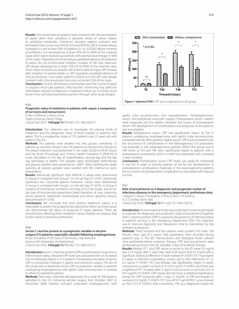
Results Only amplitudes of variation were coherent with clinical evolution
of sepsis rather than variations in absolute values or values relative
to published thresholds. Coherence between patient evolution and
biomarker time course was 93% for IL-6 and 85% for LBP. IL-6 levels always
increased to over at least 50% of baseline (51 to 2,523%) before initiation
of antibiotics. IL-6 decreased by at least 45% (45 to 98%) of the maximal
value when sepsis resolved successfully with antimicrobial therapy in 92%
of the cases. Stagnating IL-6 levels always paralleled absence of resolution
of sepsis. No pre-antimicrobial initiation increase of LBP was observed.
LBP always decreased by at least 16% (16 to 85%) of the maximal value
when sepsis resolved successfully with antimicrobial therapy. LBP increase
after initiation of antimicrobials or LBP stagnation paralleled absence of
clinical resolution. Concordant patterns of both IL-6 and LBP were always
coherent with clinical evolution but only concerned 35% of the cases.
Conclusions IL-6 and LBP behave as biomarkers over the course of sepsis
in surgical critical care patients. Only dynamic monitoring may yield any
information relevant to diagnosis or treatment follow-up. It remains to be
shown how well these biomarkers perform through other study designs.
P39
Prognostic value of melatonin in patients with sepsis: a comparison of survivors and nonsurvivorsK Alan, D Memis, G Altun, N Sut
Trakya University, Edirne, Turkey
Critical Care 2010, 14(Suppl 1):P39 (doi: 10.1186/cc8271)
Introduction Our objective was to investigate the plasma levels of
melatonin and the prognostic value of these markers in patients with
sepsis. This is a prospective study of 131 patients over 22 years old with
sepsis in the ICU.
Methods The patients were divided into two groups containing 72
patients as survivors (Group I) and 59 patients as nonsurvivors (Group II).
The blood collection was performed in the night (02.00 to 04.00 hours)
and the Acute Physiologic and Chronic Health Evaluation II (APACHE II)
score calculated on the day of hospitalisation, second day and the last
day (discharge or death). The samples were centrifuged methodically
and plasma samples were preserved at –200°C. After reaching the target
quantity, all plasma samples were studied with RIA method at room
temperature.
Results Statistically signifi cant high APACHE II values were determined
in Group II compared with Group I on the all days (P <0.05). Statistically
signifi cant low nocturnal plasma melatonin values were determined
in Group II compared with Group I on the last day (P <0.05). In Group II,
duration of mechanical ventilation and stay of ICU are longer, and at the
last days of biochemistry parameters failed (elevation of urea, creatinine,
serum glutamic oxaloacetic transaminase and serum glutamic pyruvic
transaminase) (P <0.05).
Conclusions We conclude that from plasma melatonin values it is
impossible to predict the prognosis but during the follow-up these values
can demonstrate the status of prognosis in septic patients. There are
several factors aff ecting these melatonin values; thereby we propose that
further research should be performed.
P40
Serum C-reactive protein as a prognostic variable in elective surgery ICU patients: especially valuable following esophagectomyM Van Genderen, H De Geus, D Gommers, J Van Bommel
Erasmus MC, Rotterdam, the Netherlands
Critical Care 2010, 14(Suppl 1):P40 (doi: 10.1186/cc8272)
Introduction Serum C-reactive protein (CRP) is synthesized in response to
infl ammatory status. Elevated CRP levels are associated with an increased
risk of multiorgan failure in ICU patients. Preoperative elevation of serum
CRP is a prognostic indicator in gastric and colorectal surgery. The aim of
this study was to determine serum CRP as prognostic variable in patients
undergoing esophagectomy with gastric tube reconstruction in contrast
to other ICU admitted patients.
Methods Data were collected retrospectively for a total of 208 patients
admitted to the ICU following elective surgery from October 2007 to
December 2008. Patients included underwent esophagectomy with
gastric tube reconstruction, liver transplantation, hemihepatectomy,
neuro- and abdominal aneurysm surgery. Postoperative serum markers
were measured and the relation between the course of postoperative
serum CRP, development of complications and prognosis of the patients
was investigated.
Results Postoperative serum CRP was signifi cantly higher at T24 in
patients undergoing esophagectomy with gastric tube reconstruction,
compared with all other patients. Higher serum CRP levels correlated with
the occurrence of complications in the heterogeneous ICU population
but especially in the esophagectomy patients. Within this group, serum
CRP levels at T24 and T48 were signifi cantly higher in patients with a
postoperative pneumonia, which in itself was associated with increased
1-year mortality.
Conclusions Postoperative serum CRP levels can easily be monitored
in the ICU in order to identify patients at risk for the development of
postoperative complications. Especially in the esophagectomy patients,
the occurrence of postoperative complications is associated with reduced
survival.
P41
Role of procalcitonin as a diagnostic and prognostic marker of infectious diseases in the emergency department: preliminary dataL Magrini, V Mura, F Travaglino, O Piras, E Ferri, S Di Somma
A. O. S.Andrea, Rome, Italy
Critical Care 2010, 14(Suppl 1):P41 (doi: 10.1186/cc8273)
Introduction An observational study was conducted in a teaching hospital
to evaluate the diagnostic and prognostic value of procalcitonin together
with C-reactive protein (CRP) in assessing the presence of infection/sepsis
in patients arriving in the emergency department (ED). The objective
was to improve diagnostic and therapeutic time to intervention by the
emergency physician.
Methods Three hundred and fi ve patients were studied (145 male, 160
female, mean age 67.2 years). Vital parameters were recorded during
patient’s stay in the ED. Hemocultures and biological fl uids cultures
were performed before antibiotic therapy. CRP and procalcitonin were
performed at arrival in the ED, and after 5 days of antibiotic therapy.
Results Median PCT and CRP values at arrival in the ED were 0.3 ng/ml
and 12.9 mg/dl, after 5 days they were 0.09 ng/ml and 4.2 mg/dl with a
signifi cant statistical diff erence in both markers (P <0.001). PCT was higher
in sepsis vs infections (respiratory, urinary, gut or skin infections) (3.1 vs
0.2 ng/ml, P <0.001). PCT pre-therapy was signifi cantly higher in septic
nonsurvivors vs septic survivors (1.0 vs 0.2 ng/ml, P <0.001), and there was
a signifi cant PCT increase after 5 days in nonsurvivors vs survivors (3.6 vs
0.07 ng/ml) (P <0.001). CRP results did not show a statistical signifi cance,
except for CRP measured after 5 days (3 mg/dl) vs CRP pre-therapy in
survivors (12 mg/dl, P <0.001). PCT (cut-off 0.5 ng/ml) ROC curve showed
an AUC 0.72 (P <0.001), with a sensitivity 75% as a diagnostic marker for
Figure 1 (abstract P40). CRP and complications in all groups.
Critical Care 2010, Volume 14 Suppl 1 http://ccforum.com/supplements/14/S1
S15
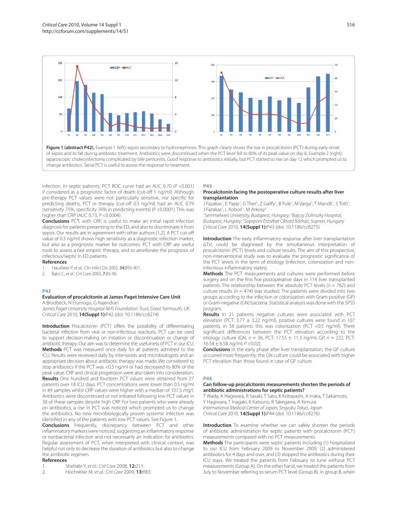
infection. In septic patients, PCT ROC curve had an AUC 0.70 (P <0.001)
if considered as a prognostic factor of death (cut-off 1 ng/ml). Although
pre-therapy PCT values were not particularly sensitive, nor specifi c for
predicting deaths, PCT in therapy (cut-off 0.5 ng/ml) had an AUC 0.79
(sensitivity 75%, specifi city 76% in predicting events) (P <0.0001). This was
higher than CRP (AUC 0.73, P <0.0004).
Conclusions PCT, with CRP, is useful to make an initial rapid infection
diagnosis for patients presenting to the ED, and also to discriminate it from
sepsis. Our results are in agreement with other authors [1,2]. A PCT cut-off
value of 0.5 ng/ml shows high sensitivity as a diagnostic infection marker,
but also as a prognostic marker for outcomes. PCT with CRP are useful
tools to assess a fast empiric therapy, and to ameliorate the prognosis of
infectious/septic in ED patients.
References1. Hausfater P, et al.: Clin Infect Dis 2002, 34:895-901.
2. Balci C, et al.: Crit Care 2003, 7:85-90.
P42
Evaluation of procalcitonin at James Paget Intensive Care UnitA Brodbeck, N Elumogo, G Rajendran
James Paget University Hospital NHS Foundation Trust, Great Yarmouth, UK
Critical Care 2010, 14(Suppl 1):P42 (doi: 10.1186/cc8274)
Introduction Procalcitonin (PCT) off ers the possibility of diff erentiating
bacterial infection from viral or non-infectious reactions. PCT can be used
to support decision-making on initiation or discontinuation or change of
antibiotic therapy. Our aim was to determine the usefulness of PCT in our ICU.
Methods PCT was measured once daily for all patients admitted to the
ICU. Results were reviewed daily by intensivists and microbiologists and an
appropriate decision about antibiotic therapy was made. We considered to
stop antibiotics if the PCT was <0.5 ng/ml or had decreased to 80% of the
peak value. CRP and clinical progression were also taken into consideration.
Results One hundred and fourteen PCT values were obtained from 27
patients over 18 ICU days. PCT concentrations were lower than 0.5 ng/ml
in 49 samples whilst CRP values were higher with a median of 107.5 mg/l.
Antibiotics were discontinued or not initiated following low PCT values in
38 of these samples despite high CRP. For two patients who were already
on antibiotics, a rise in PCT was noticed which prompted us to change
the antibiotics. No new microbiologically proven systemic infection was
identifi ed in any of the patients with low PCT values. See Figure 1.
Conclusions Frequently, discrepancy between PCT and other
infl ammatory markers were noticed, suggesting an infl ammatory response
or nonbacterial infection and not necessarily an indication for antibiotics.
Regular assessment of PCT, when interpreted with clinical context, was
helpful not only to decrease the duration of antibiotics but also to change
the antibiotic regimen.
References1. Shehabi Y, et al.: Crit Care 2008, 12:211.
2. Hochreiter M, et al.: Crit Care 2009, 13:R83.
P43
Procalcitonin facing the postoperative culture results after liver transplantationJ Fazakas1, E Papp1, G Ther1, Z Gallfy1, B Fule1, M Varga1, T Mandli1, S Toth1,
J Fazakas2, L Kobori1, M Arkosy3
1Semmelweis University, Budapest, Hungary; 2Bajcsy Zsilinszky Hospital,
Budapest, Hungary; 3Soprponi Erzsébet Oktató Kórház, Sopron, Hungary
Critical Care 2010, 14(Suppl 1):P43 (doi: 10.1186/cc8275)
Introduction The early infl ammatory response after liver transplantation
(LTx) could be diagnosed by the simultaneous interpretation of
procalcitonin (PCT) levels and culture results. The aim of this prospective,
non-interventional study was to evaluate the prognostic signifi cance of
the PCT levels in the term of etiology (infection, colonization and non-
infectious infl ammatory states).
Methods The PCT measurements and cultures were performed before
surgery and on the fi rst fi ve postoperative days in 114 liver transplanted
patients. The relationship between the absolute PCT levels (n = 762) and
culture results (n = 474) was studied. The patients were divided into two
groups according to the infection or colonization with Gram-positive (GP)
or Gram-negative (GN) bacteria. Statistical analysis was done with the SPSS
program.
Results In 21 patients negative cultures were associated with PCT
elevation (PCT: 3.77 ± 3.22 ng/ml), positive cultures were found in 107
patients, in 58 patients this was colonization (PCT <0.5 ng/ml). There
signifi cant diff erences between the PCT elevation according to the
etiology culture (GN, n = 36, PCT: 17.55 ± 11.3 ng/ml; GP, n = 222, PCT:
16.54 ± 6.58 ng/ml; P <0.02).
Conclusions In the early phase after liver transplantation, the GP culture
occurred more frequently; the GN culture could be associated with higher
PCT elevation than those found in case of GP culture.
P44
Can follow-up procalcitonin measurements shorten the periods of antibiotic administrations for septic patients?T Wada, A Hagiwara, R Sasaki, T Sato, K Kobayashi, A Inaka, T Sakamoto,
Y Hagiwara, T Inagaki, K Katsuno, R Takegawa, A Kimura
International Medical Center of Japan, Sinjyuku Tokyo, Japan
Critical Care 2010, 14(Suppl 1):P44 (doi: 10.1186/cc8276)
Introduction To examine whether we can safely shorten the periods
of antibiotic administration for septic patients with procalcitonin (PCT)
measurements compared with no PCT measurements.
Methods The participants were septic patients including (1) hospitalized
to our ICU from February 2009 to November 2009, (2) administered
antibiotics for 4 days and over, and (3) stopped the antibiotics during their
ICU stays. We treated the patients from February to June without PCT
measurements (Group A). On the other hand, we treated the patients from
July to November referring to serum PCT level (Group B). In group B, when
Figure 1 (abstract P42). Example 1 (left): sepsis secondary to hydronephrosis. This graph clearly shows the rise in procalcitonin (PCT) during early onset
of sepsis and its fall during antibiotic treatment. Antibiotics were discontinued when the PCT level fell to 80% of its peak value on day 8. Example 2 (right):
laparoscopic cholecystectomy complicated by bile peritonitis. Good response to antibiotics initially, but PCT started to rise on day 12 which prompted us to
change antibiotics. Serial PCT is useful to assess the response to treatment.
Critical Care 2010, Volume 14 Suppl 1 http://ccforum.com/supplements/14/S1
S16
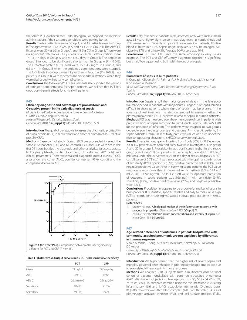
the serum PCT level decreases under 0.5 ng/ml, we stopped the antibiotic
administrations if their systemic conditions were getting better.
Results Twelve patients were in Group A, and 15 patients were in Group
B. The ages were 65 ± 18 in Group A, and 64 ± 23 in Group B. The APACHE
II scores were 20.4 ± 6.3 in Group A, and 18.5 ± 7.5 in Group B. There were
no signifi cant diff erences. The periods of antibiotic administrations were
14.1 ± 7.7 days in Group A, and 9.7 ± 6.0 days in Group B. The periods in
Group B tended to be signifi cantly shorter than in Group A (P = 0.049).
The C-reactive protein (CRP) levels were 3.5 ± 4.2 mg/dl in Group A, and
6.5 ± 4.1 in Group B when the antibiotic administrations were stopped.
The CRP levels in Group B were higher than in Group A (P = 0.071). Two
patients in Group B were repeated antibiotic administrations, while they
were discharged without any complications.
Conclusions The follow-up PCT measurements safely shorten the periods
of antibiotic administrations for septic patients. We believe that PCT has
good cost–benefi t eff ects for critically ill patients.
P45
Effi ciency diagnostic and advantages of procalcitonin and C-reactive protein in the early diagnosis of sepsisM De la Torre-Prados, A García-de la Torre, A García-Alcántara,
C Ortíz-García, A Enguix-Armada
Hospital Virgen de la Victoria, Málaga, Spain
Critical Care 2010, 14(Suppl 1):P45 (doi: 10.1186/cc8277)
Introduction The goal of our study is to assess the diagnostic profi tability
of procalcitonin (PCT) in septic shock and another biomarker as C-reactive
protein (CRP).
Methods Case–control study. During 2009 we proceeded to select the
sample: 54 patients (ICU) and 61 controls. PCT and CRP were set in the
fi rst 24 hours besides the diagnosis and other analytical (glucose, lactate,
leukocytes, platelets, white blood cells, LIC cells and ALY cells) and
clinical parameters. There were realized diagnostic output curves (ROC),
area under the curve (AUC), confi dence interval (95%), cut-off and the
comparison between AUC.
Results Fifty-four septic patients were assessed, 66% were males; mean
age, 63 years. Eighty-eight percent was diagnosed as septic shock and
11% severe sepsis. Seventy-six percent were medical patients. Positive
blood cultures in 42.5%. Sepsis origin: respiratory 46%, neurological 5%,
digestive 37% and urinary 3%. Average SOFA score was 10.4.
Conclusions PCT and CRP have the same effi ciency in early sepsis
diagnosis. The PCT and CRP effi ciency diagnostic together is signifi cant
but small. We suggest using both with the doubt of sepsis.
P46
Biomarkers of sepsis in burn patientsH Oueslati1, K Bousselmi1, I Rahmani1, A Mokline1, J Haddad1, Y Yahyia1,
R Ghanem2, A Messadi1
1Burn and Trauma Center, Tunis, Tunisia; 2Microbiology Department, Tunis,
Tunisia
Critical Care 2010, 14(Suppl 1):P46 (doi: 10.1186/cc8278)
Introduction Sepsis is still the major cause of death in the late post-
traumatic period in patients with major burns. Diagnosis of sepsis remains
diffi cult in these patients where signs of sepsis may be present in the
absence of real infection. This study attempted to assess whether the
plasma procalcitonin (PCT) level was related to sepsis in burned patients.
Methods PCT was measured over the entire course of stay in patients with
predictive signs of sepsis according to Burn French Society Criteria (SFETB)
for the presence of infection. The patients were assigned to two groups
depending on the clinical course and outcome: A = no septic patients, B =
septic patients. Optimum sensitivity, predictive values, and area under the
receiver operating characteristic (ROC) curve were evaluated.
Results Over a 6-month period starting from 1 July 2008 to 31 December
2008, 157 patients were admitted. Sixty-two were investigated, 40 in group
A and 22 in group B. Procalcitonin was signifi cantly higher in the septic
group (7.26 ± 7 ng/ml) compared with the no septic group (0.25 ± 0.32 ng/
ml). Area under the curve was 0.94 on the day of sepsis diagnosis. A PCT
cut-off value of 0.75 ng/ml was associated with the optimal combination
of sensitivity (85%), specifi city (87%), positive predictive value (91%), and
negative predictive value (73%). In surviving septic patients the PCT value
was signifi cantly lower than in deceased septic patients (3.5 ± 0.87 ng/
ml vs 10.18 ± 9.6 ng/ml). The PCT cut-off value for optimum prediction
of outcome in septic patients was 3.66 ng/ml with sensitivity (91%),
specifi city (75%), positive predictive value (78%), and negative predictive
value (90%).
Conclusions Procalcitonin appears to be a powerful marker of sepsis in
burn patients. It is sensitive, specifi c, reliable and easy to measure. A high
PCT concentration (>3.66 ng/ml) would indicate poor outcome in septic
patients.
References1. Gramm HJ, et al.: A biological marker of the infl ammatory response with
prognostic properties. Clin Intens Care 1995, 6(Suppl):71.
2. Zeni F, et al.: Procalcitonin serum concentrations and severity of sepsis. Clin
Intens Care 1994, 5(Suppl):2.
P47
Age-related diff erences of outcomes in patients hospitalized with community-acquired pneumonia are not explained by diff erences in immune responseS Kale, S Yende, L Kong, A Perkins, JA Kellum, AN Vallejo, AB Newman,
DC Angus
University of Pittsburgh School of Medicine, Pittsburgh, PA, USA
Critical Care 2010, 14(Suppl 1):P47 (doi: 10.1186/cc8279)
Introduction We hypothesized that the higher risk of severe sepsis and
mortality observed after infection in prior epidemiologic studies are due
to age-related diff erences in immune response.
Methods We analyzed 2,183 subjects from a multicenter observational
cohort of patients hospitalized with community-acquired pneumonia
(CAP). We divided subjects into fi ve age groups (<50, 50 to 64, 65 to 74,
74 to 84, ≥85). To compare immune response, we measured circulating
infl ammatory (IL-6 and IL-10), coagulation-fi brinolysis (D-dimer, factor
IX (F-IX), thrombin–antithrombin complex (TAT), antithrombin (AT) and
plasminogen-activator inhibitor (PAI)), and cell surface markers (TLR2,
Figure 1 (abstract P45). Comparison between AUC not signifi cantly
diff erent for PCT and CRP (P = 0.445).
Table 1 (abstract P45). Output curve results: PCT/CRP, sensitivity, specifi city
PCT CRP
Mean 24 ng/ml 227 mg/day
AUC 0.983 0.965
95% CI 0.93 to 0.99 0.91 to 0.99
Sensitivity 92.6% 91.1%
Specifi city 93.1% 100%
Critical Care 2010, Volume 14 Suppl 1 http://ccforum.com/supplements/14/S1
S17
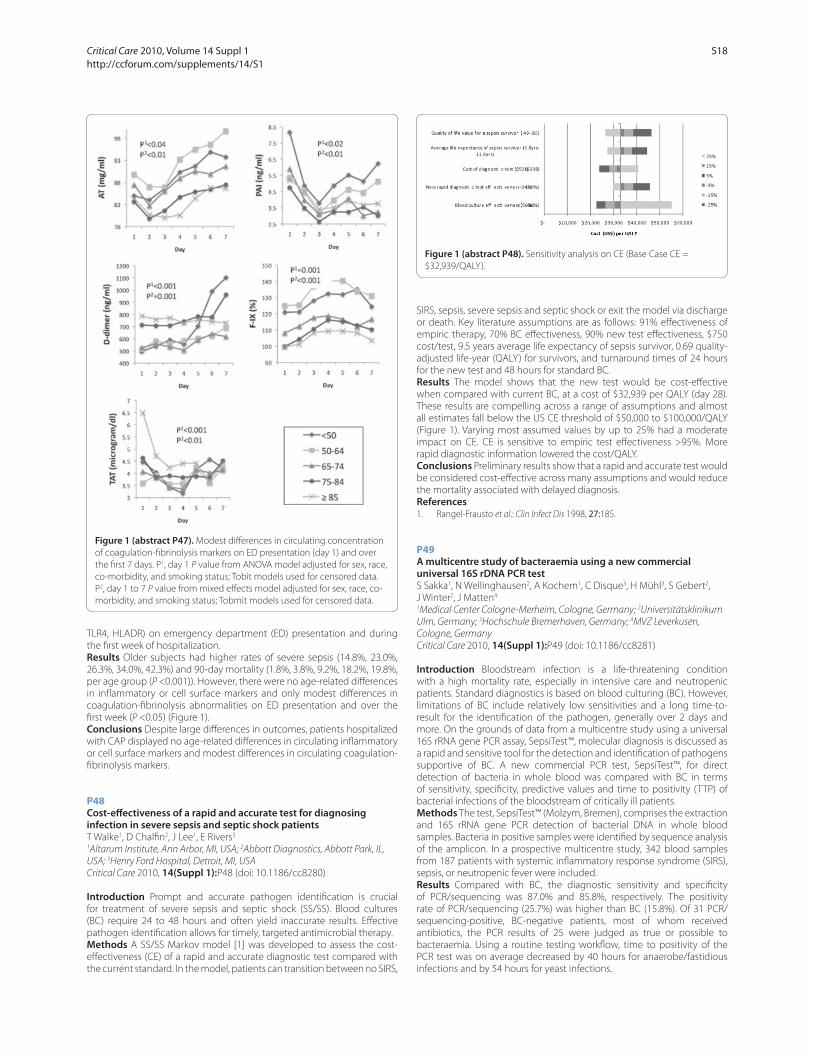
TLR4, HLADR) on emergency department (ED) presentation and during
the fi rst week of hospitalization.
Results Older subjects had higher rates of severe sepsis (14.8%, 23.0%,
26.3%, 34.0%, 42.3%) and 90-day mortality (1.8%, 3.8%, 9.2%, 18.2%, 19.8%,
per age group (P <0.001)). However, there were no age-related diff erences
in infl ammatory or cell surface markers and only modest diff erences in
coagulation-fi brinolysis abnormalities on ED presentation and over the
fi rst week (P <0.05) (Figure 1).
Conclusions Despite large diff erences in outcomes, patients hospitalized
with CAP displayed no age-related diff erences in circulating infl ammatory
or cell surface markers and modest diff erences in circulating coagulation-
fi brinolysis markers.
P48
Cost-eff ectiveness of a rapid and accurate test for diagnosing infection in severe sepsis and septic shock patientsT Walke1, D Chalfi n2, J Lee1, E Rivers3
1Altarum Institute, Ann Arbor, MI, USA; 2Abbott Diagnostics, Abbott Park, IL,
USA; 3Henry Ford Hospital, Detroit, MI, USA
Critical Care 2010, 14(Suppl 1):P48 (doi: 10.1186/cc8280)
Introduction Prompt and accurate pathogen identifi cation is crucial
for treatment of severe sepsis and septic shock (SS/SS). Blood cultures
(BC) require 24 to 48 hours and often yield inaccurate results. Eff ective
pathogen identifi cation allows for timely, targeted antimicrobial therapy.
Methods A SS/SS Markov model [1] was developed to assess the cost-
eff ectiveness (CE) of a rapid and accurate diagnostic test compared with
the current standard. In the model, patients can transition between no SIRS,
SIRS, sepsis, severe sepsis and septic shock or exit the model via discharge
or death. Key literature assumptions are as follows: 91% eff ectiveness of
empiric therapy, 70% BC eff ectiveness, 90% new test eff ectiveness, $750
cost/test, 9.5 years average life expectancy of sepsis survivor, 0.69 quality-
adjusted life-year (QALY) for survivors, and turnaround times of 24 hours
for the new test and 48 hours for standard BC.
Results The model shows that the new test would be cost-eff ective
when compared with current BC, at a cost of $32,939 per QALY (day 28).
These results are compelling across a range of assumptions and almost
all estimates fall below the US CE threshold of $50,000 to $100,000/QALY
(Figure 1). Varying most assumed values by up to 25% had a moderate
impact on CE. CE is sensitive to empiric test eff ectiveness >95%. More
rapid diagnostic information lowered the cost/QALY.
Conclusions Preliminary results show that a rapid and accurate test would
be considered cost-eff ective across many assumptions and would reduce
the mortality associated with delayed diagnosis.
References
1. Rangel-Frausto et al.: Clin Infect Dis 1998, 27:185.
P49
A multicentre study of bacteraemia using a new commercial universal 16S rDNA PCR testS Sakka1, N Wellinghausen2, A Kochem1, C Disque3, H Mühl3, S Gebert2,
J Winter2, J Matten4
1Medical Center Cologne-Merheim, Cologne, Germany; 2Universitätsklinikum
Ulm, Germany; 3Hochschule Bremerhaven, Germany; 4MVZ Leverkusen,
Cologne, Germany
Critical Care 2010, 14(Suppl 1):P49 (doi: 10.1186/cc8281)
Introduction Bloodstream infection is a life-threatening condition
with a high mortality rate, especially in intensive care and neutropenic
patients. Standard diagnostics is based on blood culturing (BC). However,
limitations of BC include relatively low sensitivities and a long time-to-
result for the identifi cation of the pathogen, generally over 2 days and
more. On the grounds of data from a multicentre study using a universal
16S rRNA gene PCR assay, SepsiTest™, molecular diagnosis is discussed as
a rapid and sensitive tool for the detection and identifi cation of pathogens
supportive of BC. A new commercial PCR test, SepsiTest™, for direct
detection of bacteria in whole blood was compared with BC in terms
of sensitivity, specifi city, predictive values and time to positivity (TTP) of
bacterial infections of the bloodstream of critically ill patients.
Methods The test, SepsiTest™ (Molzym, Bremen), comprises the extraction
and 16S rRNA gene PCR detection of bacterial DNA in whole blood
samples. Bacteria in positive samples were identifi ed by sequence analysis
of the amplicon. In a prospective multicentre study, 342 blood samples
from 187 patients with systemic infl ammatory response syndrome (SIRS),
sepsis, or neutropenic fever were included.
Results Compared with BC, the diagnostic sensitivity and specifi city
of PCR/sequencing was 87.0% and 85.8%, respectively. The positivity
rate of PCR/sequencing (25.7%) was higher than BC (15.8%). Of 31 PCR/
sequencing-positive, BC-negative patients, most of whom received
antibiotics, the PCR results of 25 were judged as true or possible to
bacteraemia. Using a routine testing workfl ow, time to positivity of the
PCR test was on average decreased by 40 hours for anaerobe/fastidious
infections and by 54 hours for yeast infections.
Figure 1 (abstract P47). Modest diff erences in circulating concentration
of coagulation-fi brinolysis markers on ED presentation (day 1) and over
the fi rst 7 days. P1, day 1 P value from ANOVA model adjusted for sex, race,
co-morbidity, and smoking status; Tobit models used for censored data.
P2, day 1 to 7 P value from mixed eff ects model adjusted for sex, race, co-
morbidity, and smoking status; Tobmit models used for censored data.
Figure 1 (abstract P48). Sensitivity analysis on CE (Base Case CE =
$32,939/QALY).
Critical Care 2010, Volume 14 Suppl 1 http://ccforum.com/supplements/14/S1
S18

Conclusions The PCR approach enables the detection and identifi cation
of bacteraemia in blood samples within a few hours. Despite the
indispensability of BC diagnostics, the rapid detection of bacteria by
SepsiTest™ appears to be a valuable tool, allowing earlier pathogen-
adapted antimicrobial therapy in critically ill patients.
P50
Time to antibiotics in sepsisR Appelboam, R Tilley, J Blackburn
Royal Devon and Exeter Hospital, Exeter, UK
Critical Care 2010, 14(Suppl 1):P50 (doi: 10.1186/cc8282)
Introduction Mortality from sepsis increases with delay in administration
of antibiotics. The Surviving Sepsis Guidelines suggest that antibiotics are
administered within 1 hour of diagnosis of severe sepsis and septic shock.
This retrospective study investigated the time to appropriate antibiotics in
patients with sepsis and identifi es reasons for delay where possible.
Methods All adult patients in our institution who had positive blood
cultures in 2008 were identifi ed. Included were patients with sepsis, severe
sepsis, or septic shock. Excluded were the isolation of low-pathogenicity
organisms from blood culture (for example, coagulase-negative Staphylo-
coccus). Case notes were reviewed to identify the time between diagnosis
of sepsis and administration of antibiotics. Time zero was taken as the
time recorded in the notes by the doctor that the patient was seen. Times
of prescription and administration of antibiotics were taken from the
prescription chart.
Results There were 1,477 sets of positive blood cultures in 1,098 patients.
Following exclusions there were 503 episodes of positive blood cultures
in 494 patients. Three hundred and seventy-fi ve fulfi lled the criteria
and were analysed: 52 did not fulfi l entry criteria, in 16 cases sepsis was
not suspected, and 60 sets of notes were incomplete or unavailable.
Average age was 69 years. Mean time between diagnosis and antibiotic
administration was 8.1 hours, median 4 hours. A delay of 3 hours or more
between diagnosis and administration of antibiotics occurred in 218 cases
(58%). The 28-day mortality for all patients overall was 21%; 19% in the
no-delay group compared with 24% in the delayed group. The 1-year
mortality was 35% for both groups. Causes of delay in administration
included prescription error, awaiting senior review, and patient movement
between departments.
Conclusions These retrospective data demonstrate that there is a delay in
administering antibiotics patients with sepsis. These data are likely to be
an underestimation as an accurate time zero is diffi cult to ascertain.
References
1. Kumar A, Roberts D, Wood KE, et al.: Crit Care Med 2006, 34:1589-1596
2. Dellinger RP, Levy MM, Carlet JM, et al.: Crit Care Med 2008, 36:296-327.
3. Bone RC, Sibbald WJ, Sprung CL: Chest 1992, 101:1481-1483.
P51
Utility of an antibiotic guideline in hospital-associated infectionsM Shankar-Hari, D Wyncoll
Guy’s and St Thomas’ NHS Foundation Trust, London, UK
Critical Care 2010, 14(Suppl 1):P51 (doi: 10.1186/cc8283)
Introduction Prompt initiation of appropriate antibiotic therapy improves
outcome in critically ill patients [1]. In a tertiary-level ICU, we evaluated
the appropriateness of, and adherence to an antibiotic guideline (based
on local bacterial epidemiology) in CDC-defi ned hospital-associated
infections.
Methods We conducted a 6-month prospective observational study (April
2008 to October 2008) in consecutive ICU admissions of patients who
satisfi ed investigator-adjudicated classifi cation of ICU-acquired infections
according to CDC criteria [2]. We assessed patient characteristics including
severity of illness at admission, ICU length of stay (LOS), appropriateness
of initial antibiotic choice as judged by in vitro sensitivity results and
appropriateness of the current guideline. Results are presented as mean
(SD) or median as appropriate.
Results During the study period there were 101 antibiotic starts in 65
patients with sepsis secondary to ICU-acquired infections. Medical patients
formed 44% of the study cohort; whilst 23% of patients were general
surgical and the remaining 33% were post cardiothoracic surgery. The age
and admission APACHE II score of the study cohort was 61.8 (16.3) years
and 18.4 (5.6). The median LOS and ICU mortality of the cohort was 24 days
and 27.6%. The most common CDC reportable diagnosis was clinical or
microbiological confi rmed pneumonia (PNU1/PNU2/LRI) (n = 57), followed
by intra-abdominal infection (SSI-GIT) (n = 10) and urinary tract infection
(SUTI) (n = 8). The culture positivity rate was 71.2%. The appropriateness of
the ICU antibiotic guideline is summarised in Table 1. Monotherapy was
used in 52.5% of episodes. The median length of antibiotic treatment with
positive cultures was 7 days, and 5 days for culture negative episodes. In
sepsis episodes with negative culture, antibiotics were stopped within
3 days in 17% of the episodes.
Table 1 (abstract P51). Appropriateness of guideline-based antibiotic therapy
Septic shock Severe sepsis Sepsis
Parameter (n = 35) (n = 30) (n = 36)
Positive cultures 23 (66%) 25 (83%) 24 (67%)
Appropriateness 18/23 (78%) 21/25 (84%) 14/24 (58%)
Conclusions The study cohort had a high culture positivity rate (71%) in
ICU-acquired sepsis. Our antibiotic guidelines gave an optimal empiric
initial therapy in over 74% of episodes, with more than 50% of antibiotics
started being monotherapy.
References
1. Kumar et al.: Crit Care Med 2006, 34:1589-1596.
2. Horan et al.: Am J Infect Control 2008, 36:309-332.
P52
Assessment of antibiotic prescriptions in two French intensive care unitsMP Parisot1, CT Tassin2, AL Lepape2, RG Gauzit1
1Hotel Dieu, Paris, France; 2CHU Lyon Sud, Lyon, France
Critical Care 2010, 14(Suppl 1):P52 (doi: 10.1186/cc8284)
Introduction In the setting of antimicrobial policy, the French authorities
recommend assessing the quality of antibiotic therapy [1]. The aim of this
study is to assess the adequacy of antibiotic therapy in the ICU, to good
practice guidelines and unit protocols. The three steps of prescription are
analysed: choice of treatment, modalities of prescription and treatment
follow-up.
Methods This preliminary observational trial was conducted in two ICUs
(October 2005 to August 2006 and March to August 2009). All patients with
curative antibiotherapy were prospectively included. For each patient, we
collected individual data of treatment and we evaluated them according
to the recommendations of good use published in the literature [1,2].
Results One hundred and thirteen patients were included. Infections
were community-acquired infection in 30% of the cases. Respiratory
tract infections were the most frequently encountered, followed by intra-
abdominal infections. The three most frequently isolated pathogens were
P. aeruginosa, E. coli and S. aureus.
Initial antibiotic therapy, n = 113: lack of sample 14%, inadequacy with unit
protocol 13%, too broad spectrum 7%, not justifi ed association 8%, wrong
posology 20%, incorrect route of administration 1%, wrong interval 6%,
lack of plasmatic dosage (n = 46) 7%.
Treatment follow-up, n = 108: wrong delay of reappraisal 14%, no adap-
tation to microbiological results 10%, lack of de-escalation 5%, no
reassessment of posology 3%, wrong posology (if change of treatment)
2%, wrong interval (if change of treatment) 3%, unjustifi ed duration 10%.
Conclusions The rate of prescriptions that did not conform to at least
one criterion in this study was over 70%. This high rate is partly explained
by the high number of assessment criterions (n = 15). The most frequent
criterion of inadequacy was a wrong initial dosage, followed by the
initiation of an antibiotherapy without a microbiologic sample, a wrong
delay of reassessment, and a nonconformity of treatment to unit protocols.
That last criterion must be assessed in a more qualitative way, as some
transgressions could be justifi ed.
References1. Stratégie d’antibiothérapie et prévention des résistances bactériennes en
établissement de santé [www.has-sante.fr]
2. Van der Meer JW, et al.: Clin Microbiol Infect 2001, 7(Suppl 6):12.
Critical Care 2010, Volume 14 Suppl 1 http://ccforum.com/supplements/14/S1
S19
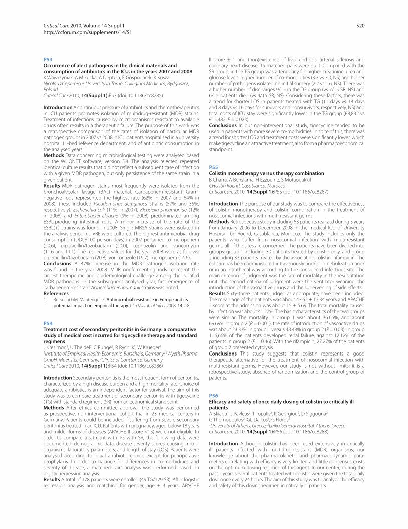
P53
Occurrence of alert pathogens in the clinical materials and consumption of antibiotics in the ICU, in the years 2007 and 2008K Wawrzyniak, A Mikucka, A Deptuła, E Gospodarek, K Kusza
Nicolaus Copernicus University in Toruń, Collegium Medicum, Bydgoszcz,
Poland
Critical Care 2010, 14(Suppl 1):P53 (doi: 10.1186/cc8285)
Introduction A continuous pressure of antibiotics and chemotherapeutics
in ICU patients promotes isolation of multidrug-resistant (MDR) strains.
Treatment of infections caused by microorganisms resistant to available
drugs often results in a therapeutic failure. The purpose of this work was
a retrospective comparison of the rates of isolation of particular MDR
pathogen groups in 2007 vs 2008 in ICU patients hospitalised in a university
hospital 11-bed reference department, and of antibiotic consumption in
the analysed years.
Methods Data concerning microbiological testing were analysed based
on the WHONET software, version 5.4. The analysis rejected repeated
identical culture results that did not refl ect a subsequent case of infection
with a given MDR pathogen, but only persistence of the same strain in a
given patient.
Results MDR pathogen stains most frequently were isolated from the
bronchoalveolar lavage (BAL) material. Carbapenem-resistant Gram-
negative rods represented the highest rate (62% in 2007 and 64% in
2008); these included Pseudomonas aeruginosa strains (57% and 35%,
respectively). Escherichia coli (11% in 2007), Klebsiella pneumoniae (12%
in 2008) and Enterobacter cloacae (9% in 2008) predominated among
ESBL-producing intestinal rods. A minor increase of the rate of the
ESBL(+) strains was found in 2008. Single MRSA strains were isolated in
the analysis period, no VRE were cultured. The highest antimicrobial drug
consumption (DDD/100 person-days) in 2007 pertained to meropenem
(20.6), piperacillin/tazobactam (20.0), cephazolin and vancomycin
(11.6 and 11.1). The respective values for the year 2008 were as follows:
piperacillin/tazobactam (20.8), voriconazole (19.7), meropenem (14.6).
Conclusions A 47% increase in the MDR pathogen isolation rate
was found in the year 2008. MDR nonfermenting rods represent the
largest therapeutic and epidemiological challenge among the isolated
MDR pathogens. In the subsequent analysed year, fi rst emergence of
carbapenem-resistant Acinetobacter baumanii strains was noted.
References
1. Rossolini GM, Mantengoli E: Antimicrobial resistance in Europe and its potential impact on empirical therapy. Clin Microbiol Infect 2008, 14:2-8.
P54
Treatment cost of secondary peritonitis in Germany: a comparative study of medical cost incurred for tigecycline therapy and standard
regimensJ Kresimon1, U Theidel2, C Runge2, R Rychlik1, W Krueger3
1Institute of Empirical Health Economic, Burscheid, Germany; 2Wyeth Pharma
GmbH, Muenster, Germany; 3Clinics of Constance, Germany
Critical Care 2010, 14(Suppl 1):P54 (doi: 10.1186/cc8286)
Introduction Secondary peritonitis is the most frequent form of peritonitis,
characterized by a high disease burden and a high mortality rate. Choice of
adequate antibiotics is an independent factor for survival. The aim of this
study was to compare treatment of secondary peritonitis with tigecycline
(TG) with standard regimens (SR) from an economical standpoint.
Methods After ethics committee approval, the study was performed
as prospective, non-interventional cohort trial in 23 medical centers in
Germany. Patients could be included if suff ering from severe secondary
peritonitis treated in an ICU. Patients with pregnancy, aged below 18 years
and milder forms of diseases (APACHE II score <15) were not eligible. In
order to compare treatment with TG with SR, the following data were
documented: demographic data, disease severity scores, causing micro-
organisms, laboratory parameters, and length of stay (LOS). Patients were
analysed according to initial antibiotic choice except for perioperative
prophylaxis. In order to balance for diff erences in co-morbidities and
severity of disease, a matched-pairs analysis was performed based on
logistic regression analysis.
Results A total of 178 patients were enrolled (49 TG/129 SR). After logistic
regression analysis and matching for gender, age ± 3 years, APACHE
II score ± 1 and (non)existence of liver cirrhosis, arterial sclerosis and
coronary heart disease, 15 matched pairs were built. Compared with the
SR group, in the TG group was a tendency for higher creatinine, urea and
glucose levels, higher number of co-morbidities (3.3 vs 3.0, NS) and higher
number of pathogens isolated on initial surgery (2.2 vs 1.6, NS). There was
a higher number of discharges 9/15 in the TG group (vs 7/15 SR, NS) and
6/15 patients died (vs 4/15 SR, NS). Considering these factors, there was
a trend for shorter LOS in patients treated with TG (11 days vs 18 days
and 8 days vs 16 days for survivors and nonsurvivors, respectively, NS) and
total costs of ICU stay were signifi cantly lower in the TG group (€8,832 vs
€15,482, P = 0.023).
Conclusions In our non-interventional study, tigecycline tended to be
used in patients with more severe co-morbidities. In spite of this, there was
a trend for shorter LOS and treatment costs were signifi cantly lower, which
make tigecycline an attractive treatment, also from a pharmacoeconomical
standpoint.
P55
Colistin monotherapy versus therapy combinationB Charra, A Benslama, H Ezzouine, S Motaouakkil
CHU Ibn Rochd, Casablanca, Morocco
Critical Care 2010, 14(Suppl 1):P55 (doi: 10.1186/cc8287)
Introduction The purpose of our study was to compare the eff ectiveness
of colistin monotherapy and colistin combination in the treatment of
nosocomial infections with multi-resistant germs.
Methods Retrospective study including 63 patients realized during 3 years
from January 2006 to December 2008 in the medical ICU of University
Hospital Ibn Rochd, Casablanca, Morocco. The study includes only the
patients who suff er from nosocomial infection with multi-resistant
germs, all of the sites are concerned. The patients have been divided into
groups: group 1 including 30 patients treated by colistin only, and group
2 including 33 patients treated by the association colistin–rifampicin. The
colistin has been administered intravenously and/or in nebulization and/
or in an intrathecal way according to the considered infectious site. The
main criterion of judgment was the rate of mortality in the resuscitation
unit, the second criteria of judgment were the ventilator weaning, the
introduction of the vasoactive drugs and the supervening of side eff ects.
Results Sixty-three patients judged as appropriate, have been included.
The mean age of the patients was about 43.62 ± 17.34 years and APACHE
2 score at the admission was about 15 ± 5.69. The total mortality caused
by infection was about 41.27%. The basic characteristics of the two groups
were similar. The mortality in group 1 was about 36.66%, and about
69.69% in group 2 (P = 0.001), the rate of introduction of vasoactive drugs
was about 23.33% in group 1 versus 48.48% in group 2 (P = 0.03). In group
1, 6,66% of the patients developed renal failure, against 12.12% of the
patients in group 2 (P = 0,46). With the rifampicin, 27.27% of the patients
of group 2 presented cytolysis.
Conclusions This study suggests that colistin represents a good
therapeutic alternative for the treatment of nosocomial infection with
multi-resistant germs. However, our study is not without limits; it is a
retrospective study, absence of randomization and the control group of
patients.
P56
Effi cacy and safety of once daily dosing of colistin to critically ill patientsA Skiada1, J Pavleas2, T Topalis2, K Georgiou2, D Siggouna2,
G Thomopoulos2, GL Daikos1, G Floros2
1University of Athens, Greece; 2Laiko General Hospital, Athens, Greece
Critical Care 2010, 14(Suppl 1):P56 (doi: 10.1186/cc8288)
Introduction Although colistin has been used extensively in critically
ill patients infected with multidrug-resistant (MDR) organisms, our
knowledge about the pharmacokinetic and pharmacodynamic para-
meters correlating with effi cacy is very limited and little consensus exists
on the optimum dosing regimen of this agent. In our center, during the
past 2 years several patients treated with colistin were given the total daily
dose once every 24 hours. The aim of this study was to analyze the effi cacy
and safety of this dosing regimen in critically ill patients.
Critical Care 2010, Volume 14 Suppl 1 http://ccforum.com/supplements/14/S1
S20
Methods The medical fi les of all patients who were admitted to the ICU
in 2008 and who received colistin for an infection due to MDR bacteria
were retrospectively analyzed. Criteria for inclusion were: dosing regimen
of colistin 9 million units (MU) every 24 hours (colistin methanesulphonate
– CMS, Norma, 1 vial = 1 MU, 1 mg = 13,300 U), administration of CMS for at
least 7 days and normal renal function at initiation of treatment with CMS
(serum creatinine ≤1 mg/dl or creatinine clearance >80 ml/min). Mortality
was evaluated at 28 days.
Results Thirteen patients were included in the study (69% male, median
age 71 years). The underlying diseases were surgical in seven patients and
medical in six patients. The median APACHE and SOFA scores on the fi rst
day of CMS administration were 15 and 10, respectively. Eleven patients
had nosocomial pneumonia, two had peritonitis and one osteomyelitis.
Causative bacteria were isolated in 10 patients (six Acinetobacter baumanii,
three Pseudomonas aeruginosa and one Klebsiella pneumonia) and
were all MDR. Of the 10 isolated bacteria, six were susceptible only to
colistin, while the other four were also susceptible either to aztreonam
or sulbactam. In cases of susceptibility of the isolate only to colistin, it
was given in combination with a carbapenem, while in the other cases
either aztreonam or sulbactam was added. The median length of CMS
administration was 11 days (mean 19; range 7 to 79 days). Median serum
creatinine on initiation and completion of treatment with CMS was 0.7 and
0.9 mg/dl, respectively (P = 0.357). No cases of neurotoxicity were found.
Mortality at 28 days was 15%.
Conclusions The total daily dose of 9 MU of CMS, given as one dose
every 24 hours in critically ill patients, seems to be effi cient and safe,
without resulting in increased nephrotoxicity or other adverse events.
A prospective study should be done to confi rm the fi ndings we have
presented herein.
P57
A comparison of the eff ect on renal function of fl ucloxacillin and vancomycin antibiotic prophylaxis in cardiac surgery patientsTO Brougham, K Giraud, A Vuylsteke
Critical Care Unit, Cambridge, UK
Critical Care 2010, 14(Suppl 1):P57 (doi: 10.1186/cc8289)
Introduction This study compared the eff ect on renal function of
fl ucloxacillin and vancomycin antibiotic prophylaxis for elective fi rst-
time coronary artery bypass grafting (CABG) surgery, using both direct
biochemical markers and indirect clinical outcome measures. Recent
evidence has suggested that vancomycin may be nephrotoxic in patients
undergoing cardiac surgery.
Methods A retrospective observational study of patients undergoing
elective fi rst-time CABG was performed, covering a 13-month period. All
patients received prophylactic antibiotics: fl ucloxacillin 1 g pre-operatively
and three 1 g doses post-operatively. Patients who were MRSA-positive,
MRSA unknown or penicillin allergic received an alternative regimen:
vancomycin 1 g pre-operatively and 1 g post-operatively. Exclusion criteria: pre-
operative creatinine >133mmol/l, any antibiotics other than prophylaxis
and haemodynamic support except <5 μg/kg/hour dopamine.
Results Of 1,413 patients in the study period, 415 met the study criteria:
360 patients received fl ucloxacillin and 55 patients received vancomycin.
There were no signifi cant diff erences between the two groups in sex,
age, BMI, euroSCORE, diabetes status, ejection fraction, pre-operative
creatinine, eGFR, sodium, or potassium. Comparing change in renal
function pre-operatively to post-operatively, there were no signifi cant
group diff erences in change in: creatinine (mmol/l; VAN median 0 (IQR
11); FLU –2 (19); P = 0.22), eGFR (ml/min; VAN 0 (14); FLU 2.4 (19.3); P =
0.22), sodium (mmol/l; VAN 1 (4); FLU 1 (4); P = 0.28). Change in potassium
diff ered signifi cantly (mmol/l; VAN 0.7 (0.9); FLU 0.5 (0.7); P <0.05). In clinical
outcome measures, the groups were similar. Most patients in both groups
stayed in ITU for 1 day and there was no signifi cant diff erence in the
number of patients staying for longer than 1 day (VAN 7/55 (13%); FLU
29/360 (8%); P = 0.30). There was no diff erence in hospital length of stay
(days; VAN 7 (4); FLU 6 (3); P = 0.19).
Conclusions In elective fi rst-time CABG patients, there is no signifi cant
diff erence in change in renal function between those given vancomycin
antibiotic prophylaxis and those given fl ucloxacillin prophylaxis, as
assessed by creatinine, eGFR and sodium levels, and indirect clinical
outcome measures. Potassium increased more in the vancomycin
group but the clinical signifi cance of this is unclear. Our data suggest
that prophylactic vancomycin does not impair renal function relative to
fl ucloxacillin.
P58
Microdialysis study of meropenem cerebral distribution in patients with acute brain injuryC Dahyot-Fizelier1, I Timofeev2, S Marchand1, W Couet1, P Hutchinson2,
B Debaene1, D Menon2, O Mimoz1, A Gupta2
1University Hospital, Poitiers, France; 2Addenbrooke’s Hospital, Cambridge, UK
Critical Care 2010, 14(Suppl 1):P58 (doi: 10.1186/cc8290)
Introduction Antibiotic dosing recommendations are usually based
on plasma or cerebral spinal fl uid pharmacokinetic (PK) studies.
However, as infections mainly occur in extracellular tissue fl uid (ECF),
corresponding unbound ECF antibiotic concentrations are responsible
for the antimicrobial eff ect. Because of the blood–brain barrier, the
cerebral antibiotic distribution is thought to be reduced compared with
tissues without any physiological barrier. This study aims to determine
meropenem (MPM) unbound concentrations in the brain and compare
them with MPM concentrations in plasma to explore cerebral distribution
of MPM in patients with acute brain injury.
Methods After local ethic approval and written informed consent, two
brain-injured patients, sedated, mechanically ventilated, receiving MPM for
an infection and monitored by cerebral microdialysis (CMA 71, membrane
length 10 mm, membrane diameter 0.6 mm, molecular cut-off 100 kDa;
CMA, Stockholm, Sweden) were enrolled. The PK study succeeded to 1 g
meropenem over 30 minutes and brain dialysates and blood samples were
collected over 420 minutes. Probe recoveries were evaluated individually
by retrodialysis. MPM was assayed by HPLC coupled with tandem mass
spectrometry (LC-MS/MS).
Results For each of the two patients, the MPM brain AUC is much
lower than plasma AUC and accordingly brain to serum AUC ratios
are respectively 0.73 for Patient 1 (P1) and 0.14 for Patient 2 (P2). MPM
concentration versus time curves in brain are delayed (time-to-peak
in P1 = 100 minutes, in P2 = 80 minutes) and present a smooth peak
compared with the corresponding curves in plasma (Figure 1). Mean
probe recoveries are respectively 19 ± 7% for P1 and 29 ± 7% for P2.
Conclusions The MPM brain AUC is much lower than plasma AUC for
the two patients enrolled, consistent with the PK theory in the presence
of tissue with effl ux transporters. More patients are needed to better
understand MPM brain distribution characteristics.
P59
Eff ects of two regimens of antibiotic prophylaxis on colonizing fl ora of the respiratory tract in patients with cardiovascular surgeryEM Berg, R Wesselink, M Tersmette
Sint Antonius Hospital, Nieuwegein, the Netherlands
Critical Care 2010, 14(Suppl 1):P59 (doi: 10.1186/cc8291)
Introduction Antibiotic prophylaxis in cardiovascular (CV) surgery has
greatly reduced the rate of postoperative infections. First-generation or
second-generation cefalosporines are used for this purpose. Our hospital
changed its antibiotic prophylaxis protocol for elective CV surgery in June
2007 from cefuroxime to cefazolin in accordance with national guidelines
Figure 1 (abstract P58). Individual MPM plasma (�) and brain (�)
concentrations versus time in P1 and P2.
Critical Care 2010, Volume 14 Suppl 1 http://ccforum.com/supplements/14/S1
S21

[1]. The aim of this study was to explore the eff ect of changing this
antibiotic prophylaxis protocol on microbial fl ora in sputum.
Methods Retrospective study. All patients admitted >10 days on the ICU
after elective CV surgery between 1 June 2006 and 1 June 2007 (Group
1) were compared with the same group of patients between 1 July 2007
and 1 August 2008 (Group 2). Patients received single-dose prophylaxis
(bypass surgery), or 2 days of prophylaxis (cardiac prosthetic surgery).
Group 1 received cefuroxime and Group 2 received cefazolin. Patients
did not receive selective digestive tract decontamination. Isolated
pathogens from elective sputum cultures of all patients were registered
and compared between groups after dividing pathogens into six classes
based on the profi le of intrinsic antibiotic susceptibility. Comparative data
between groups were analyzed with Pearson’s Χ test.
Results One hundred and fi fty-eight patients had positive sputum cultures.
Comparing total amounts of positive cultures, there was no signifi cant
diff erence between colonization with pathogenic micro-organisms
between Group 1 (n = 77 patients) and Group 2 (n = 76 patients). In Group
1 more colonization with pathogens from class 4 (β-lactamase-producing
Enterobacteriaceae and Pseudomonas aeruginosa) was observed compared
with Group 2 (45% vs 34%, P = 0.079). This nonsignifi cant diff erence
between groups was mainly attributable to a diff erence in colonization
with Pseudomonas. In the other classes of pathogens, no diff erences were
observed. No diff erence in postoperative wound infections was noted
between groups.
Conclusions Prophylactic use of cefazolin instead of cefuroxime after
CV surgery resulted in a trend towards reduction of colonization of the
respiratory tract with intrinsic β-lactam-resistant microbial fl ora in patients
with prolonged ICU stay, without adverse eff ects on the incidence of
postoperative wound infection.
Reference1. van Kasteren MEE, et al.: Ned Tijdschr Geneeskd 2000, 144:2049-2055.
P60
Prevalence of infections among patients admitted to ICUs in Nordic countries and the Netherlands in comparison with Mediterranean countries: a report from the EPIC II studyH Hanberger1, H Gill1, H Njimi2, S Walther1, JL Vincent2
1Antibiotic Research Unit, Linkoping, Sweden; 2Erasme Hospital, Brussels,
Belgium
Critical Care 2010, 14(Suppl 1):P60 (doi: 10.1186/cc8292)
Introduction The prevalence of infection is high among patients
admitted to the ICU and one of the main causes of mortality. The aim of
this study was to determine the burden of infectious diseases in patients
admitted to ICUs in Nordic countries and the Netherlands in comparison
with Mediterranean countries.
Methods The EPIC II 1-day point prevalence study of infections and
demographics of critically ill patients was performed on 8 May 2007. A
comparison of severity score, source of infection, co-morbidity and
patient outcome was done for patients admitted to ICUs in low antibiotic
resistance countries (LARC) (Denmark, Finland, the Netherlands, Norway,
Sweden) and high antibiotic resistance countries (HARC) (Greece, Israel,
Italy, Malta, Portugal, Spain, Turkey).
Results The number of patients included was 2,270 in HARC and 581 in
LARC. On the day of the study, 45.3% of patients in LARC were considered
as infected compared with 51.6% in HARC (P = 0.007). The mean SAPS
II scores were (95% CI) 32.7 (31.6 to 33.8) in LARC and 35.6 (35 to 36.2)
in HARC (P <0.001). Of all patients in LARC/HARC, 27.0%/33.9% (P =
0.002) had a respiratory infection, 10.8%/9.8% (P = 0.45) an abdominal
infection, 7.2%/10.4% (P = 0.023) a blood infection, 2.8%/3.2% (P = 0.60)
a skin infection, 2.1%/3.4% (P = 0.09) a catheter infection, 3.8%/6.6% (P =
0.012) a genitourinary infection and 0.7%/1.7% (P = 0.08) a CNS infection.
Chronic renal failure was seen in 2.6% of patients in LARC and 7.3% in
HARC (P <0.001) and the corresponding prevalence of chronic obstructive
disease was 13.4% and 20.2%, respectively (P <0.001). Admissions were
more often seen from OR/monitoring and less often from emergency
rooms among patients admitted to ICUs in LARC compared with HARC.
Median ICU length of stay (LOS) in LARC (IQR) was 5 days (1 to 19) and
in HARC was 12 days (3 to 31) (P <0.001). The corresponding hospital
LOS was 16 days (7 to 38) and 24 days (10 to 54), respectively (P <0.001).
The ICU mortality rates in LARC/HARC were 14.9%/19.9% (P = 0.008) for
all patients and for infected patients 21.2%/25.4% (P = 0.17), whereas the
corresponding hospital mortality rates were 20.8%/26.7% (P = 0.005) for all
patients and for infected patients 29.6%/34.3% (P = 0.16). The prevalence
of MRSA was 0.8% among patients in LARC (5.8% in HARC) (P <0.001) and
the prevalence of P. aeruginosa was 9.9% and 14.0% (P = 0.07), respectively.
Conclusions Infections and serious co-morbidities were more prevalent
among patients admitted to ICUs in HARC than in LARC. This was
associated with a longer stay and greater mortality in the ICU.
References1. Vincent JL, et al.: JAMA 1995, 274:639-644.
P61
Risk factors for the development of carbapenem-resistant Klebsiella pneumoniae infections in critically ill patientsKZ Vardakas, DK Matthaiou, E Antypa, A Grammatikos, E Chasou,
E Antoniadou
G. Gennimatas, Thessaloniki, Greece
Critical Care 2010, 14(Suppl 1):P61 (doi: 10.1186/cc8293)
Introduction We sought to study the characteristics and outcomes of ICU
patients with carbapenem-resistant (CRKp) and carbapenem-sensitive
(CSKp) K. pneumoniae infections.
Methods A retrospective cohort of patients requiring ICU treatment. The
study was conducted in an eight-bed ICU between January 2006 and
September 2009.
Results During the study period, 104 patients were diagnosed with
K. pneumoniae infection. The mean age of patients was 66.3 ± 14.3 years.
Fifty-one (49%) were males. The mean APACHE II score was 17.9 ± 6.9. The
median duration of hospital stay until the infection was 28 days. Forty-
eight patients (46.2%) had bacteremia, 27 (30%) urinary tract infections, 15
(14.4%) pneumonia, seven (6.7%) peritonitis and seven (6.7%) skin and soft
tissue infections. Fifty-eight (56.9%) and 39 (39%) patients had previous
and concurrent infections, respectively. Seventy-six patients (73.1%)
died. The univariate analysis showed that prior hospitalization (P = 0.049),
dialysis (P = 0.034), and history of urologic neoplasia (P = 0.041) were
associated with the development of carbapenem-resistant infections. No
independent risk factors were found in the multivariate analysis. APACHE
II score (P = 0.003), need for dialysis (P = 0.034), shock prior and after the
infection (P = 0.006 and P <0.001, respectively), respiratory distress prior
and after the infection (P = 0.021 and P = 0.032, respectively), multiorgan
failure prior and after the infection (P = 0.02 and P = 0.003, respectively),
treatment failure (P <0.001), and acidosis after the development of
infection (P = 0.003) were associated with death in the univariate analysis.
Shock after the infection (P = 0.016) and treatment failure (P = 0.001)
were independent predictors of mortality in the multivariate analysis. No
diff erence in mortality was found between patients with CRKp and CSKp
isolates.
Conclusions Infection due to K. pneumoniae in the ICU is associated with
high mortality. Infection treatment and hemodynamic support of the
patient may be important determinants of the clinical course in critically ill
patients with such infections.
Reference
1. Falagas ME, et al.: Risk factors of carbapenem-resistant Klebsiella pneumoniae infections: a matched case control study. J Antimicrob
Chemother 2007, 6:1124-1130.
P62
Outcome of unimicrobial versus polymicrobial sepsisN Abed1, A Hussein1, S Salaheldine1, A Hassan2, M Mahfouz1,
H Khaled Nagi1, H Khaled Nagi1
1Cairo University, Cairo, Egypt; 2National Heart Institute, Cairo, Egypt
Critical Care 2010, 14(Suppl 1):P62 (doi: 10.1186/cc8294)
Introduction Polymicrobial sepsis is associated with immunosuppression
caused by the predominance of anti-infl ammatory mediators and
profound loss of lymphocytes through apoptosis, and so deaths directly
related to sepsis are twofold higher in polymycrobial sepsis. The aim of
study is to compare unimicrobial versus polymicrobial sepsis as regards
microbiology, complications, length of stay, mortality, factor aff ecting
mortality.
Critical Care 2010, Volume 14 Suppl 1 http://ccforum.com/supplements/14/S1
S22
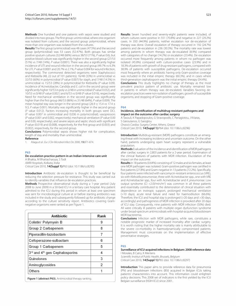
Methods One hundred and one patients with sepsis were studied and
divided into two groups. The fi rst group: unimicrobial, where one organism
was isolated from cultures; and the second group: polymicrobial, where
more than one organism was isolated from the cultures.
Results The fi rst group (unimicrobial) was 48 cases (47.5%) and the second
group (polymicrobial) was 53 cases (52.47%). Both groups has similar
positive sputum cultures (32/48 (66%) versus 38/53 (71%), P value 0.6), but
positive blood culture was signifi cantly higher in the second group (27/53
(51%) vs 7/48 (14%), P value 0.0001). There was also a signifi cantly higher
incidence of UTI and wound infection in the second group (49.7% vs 6.3%
for UTI, P value 0.0001 and 20.7% vs 2.1%, P value 0.004 for wound infection,
respectively). The commonest detected organisms were Staphylococci
and Klebsiella (48, 22 out of 101 patients); 16/48 (33%) in unimicrobial vs
32/53 (60%) in polymicrobial (P value 0.007) for staph, and 7/48 (14.5%) in
unimicrobial vs 15/53 (28%) in polymicrobial for Klebsiella (P value 0.09).
Incidence of Acinetobacter, Candida and E. coli in the second group were
signifi cantly higher 10/53 in poly vs 2/48 in unimicrobial (P value 0.032), and
10/53 vs 0/48 (P value 0.002 ) and 6/53 vs 0/48 (P value 0.016), respectively.
Need for mechanical ventilation in the second group was signifi cantly
higher than the fi rst group (46/53 (86%) vs 31/48 (64%), P value 0.009). The
mean hospital stay was longer in the second group (26.9 ± 15.4 vs 17.4 ±
9.3, P value 0.001). Mortality was signifi cantly higher in the second group
(P value 0.012). Factors increasing mortality in both groups were DCL
(P value 0.007 in unimicrobial and 0.036 in polymicrobial); vasopressors
(P value 0.001 and 0.002, respectively); mechanical ventilation (P value 0.00
and 0.00, respectively), and severe sepsis and septic shock with signifi cant
P value (0.019) and (0.008), respectively, for the fi rst group and (0.003) and
(0.002), respectively, for the second group.
Conclusions Polymicrobial sepsis shows higher risk for complication,
length of stay and mortality than unimicrobial.
Reference1. Aliaga et al.: Eur J Clin Microbiol Infect Dis 2000, 19:871-874.
P63
De-escalation practice pattern in an Indian intensive care unitA Bhakta, M Bhattacharyya, S Todi
AMRI Hospitals, Kolkata, India
Critical Care 2010, 14(Suppl 1):P63 (doi: 10.1186/cc8295)
Introduction Antibiotic de-escalation is thought to be benefi cial by
reducing the selection pressure for resistance. This study was carried out
to identify variables that infl uence de-escalation practices.
Methods Prospective observational study during a 1-year period (July
2008 to June 2009) in a 50-bed ICU in a tertiary care hospital. Any patient
admitted to the ICU during this period in whom at least one specimen
was sent for microbiological culture at or before starting antibiotics was
included in the study and subsequently followed up for antibiotic change
according to the culture sensitivity report. Antibiotics covering Gram-
negative organisms were ranked as per Figure 1.
Results Seven hundred and seventy-eight patients were included, of
whom cultures were positive in 551 (70.8%) and negative in 227 (29.2%)
cases. In 350 (44.9%) patients, neither escalation nor de-escalation of
therapy was done. Overall escalation of therapy occurred in 192 (24.7%)
patients and de-escalation in 236 (30.3%). The mortality rate was lowest
among patients in whom therapy was de-escalated (8.9%) compared
with categories of no change (14.2%) or escalation (23.4%). De-escalation
occurred more frequently among patients in whom no pathogen was
isolated (45.8%) compared with culture-positive cases (23.9%) and in
18.3% of patients with growth of drug-resistant pathogens, compared with
38.9% of patients with susceptible pathogens. De-escalation occurred
most frequently where an antibiotic having only Gram-positive coverage
was included in the initial empiric therapy (60.3%), and in cases where
third-generation cephalosporin was the initial empiric therapy (54.4%).
Conclusions This study highlights no change of therapy as the most
prevalent practice pattern of antibiotic use. Mortality remained low
in patients in whom therapy was de-escalated. Variables favoring de-
escalation practices were non-isolation of pathogen, growth of susceptible
organisms, and stopping of Gram-positive coverage.
P64
Incidence, identifi cation of multidrug-resistant pathogens and impact on the outcome after cardiac surgeryA Tasouli, K Papadopoulos, G Stravopodis, C Panagiotou, J Kriaras,
S Geroulanos, G Saroglou
Onassis Cardiac Surgery Center, Athens, Greece
Critical Care 2010, 14(Suppl 1):P64 (doi: 10.1186/cc8296)
Introduction Multidrug-resistant (MDR) pathogens constitute an emerg-
ing threat with increasing incidence and uncertain outcome. On the other
hand, patients undergoing open heart surgery represent a vulnerable
population.
Methods Evaluation of the incidence and identifi cation of MDR pathogens
after cardiac surgery in 2,803 patients for a 2-year period. Examination of
the clinical features of patients with MDR infection. Elucidation of the
impact on the outcome.
Results In 18 patients (0.64%) consisting of 12 males and six females, at least
one MDR pathogen was isolated. Gram-positive pathogen was certifi ed in
fi ve patients (27.8%) and Gram-negative in 13 patients (72.2%). Specifi cally,
four patients were infected with vancomycin-resistant enterococcus (VRE),
six with Klebsiella pneumoniae, three with Acinetobacter spp., one with VRE
and Acinetobacter spp., four with Acinetobacter and K. pneumoniae. Low
output syndrome (CI <2.0l/min/m2) was common in all these patients
and essentially contributed to the deterioration of clinical situation with
dependence on inotropic support, prolonged mechanical ventilation
(>10 days), acute renal failure and need for haemodilution (66.6%).
Therefore, the ICU and hospital stay is prolonged (>20 days and >30 days,
accordingly) and pathogenesis of MDR infection is provoked after 20 days
of ICU stay. Consequently, nine patients with MDR infection (50%) died.
All were critically ill patients with multiple organ dysfunction syndrome
under broad-spectrum antimicrobials with hospital-acquired bloodstream
MDR bacteremia.
Conclusions Infection with MDR pathogens, while rare, constitutes a
notable prognostic marker of increased mortality after cardiac surgery.
It is worth noting that the higher mortality rate is mainly attributable to
the severe co-morbidity in haemodynamically compromised patients.
Management must concentrate on the implementation of eff ective
preventative strategies.
P65
Surveillance of ICU-acquired infections in Belgium: 2008 reference dataI Morales, B Catry, K Mertens
Scientifi c Institut of Public Health, Brussels, Belgium
Critical Care 2010, 14(Suppl 1):P65 (doi: 10.1186/cc8297)
Introduction This paper aims to provide reference data for pneumonia
(PN) and bloodstream infections (BSI) acquired in Belgian ICUs taking
patients’ characteristics into account. This information could enlighten
policy decisions. This 2008 set of indicators is the fi rst yielded by the ICU
Belgian surveillance (NSIH-ICU) since 2001.Figure 1 (abstract P63). Antimicrobial therapy ranking.
Critical Care 2010, Volume 14 Suppl 1 http://ccforum.com/supplements/14/S1
S23
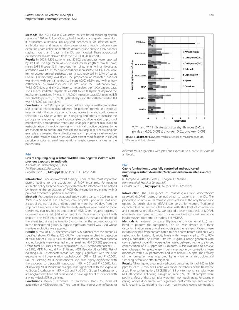
Methods The NSIH-ICU is a voluntary, patient-based reporting system
set up in 1992 to follow ICU-acquired infections and guide prevention.
It establishes a national risk-adjusted benchmark for infection rates,
antibiotics use and invasive device-use ratios through uniform case
defi nitions, data-collection methods, data entry and analysis. Only patients
staying more than 2 days in the ICU are included. These aggregated
database means are derived from the NSIH-ICU 2008 report.
Results In 2008, 4,355 patients and 35,802 patient-days were reported
by 19 ICUs. The age mean was 67.2 years; mean length of stay 8.1 days;
mean SAPS II score 43.8; the proportion of patients with antibiotics at
admission was 47.1%; medical admissions represented 63.4%, 4.2% were
immunocompromised patients; trauma was reported in 6.7% of cases.
Overall ICU mortality was 8.5%. The proportion of intubated patients
was 44.4%, with central venous catheters (CVC) 68.3% and with urinary
catheters 58.3%. Invasive-device use rates were: 358.5 intubation-days,
746.5 CVC-days and 644.2 urinary catheter-days per 1,000 patient-days.
The ICU-acquired PN/100 patients was 9.8, 16.5/1,000 patient-days and the
intubation-associated PN was 11.1/1,000 intubation days. ICU-acquired BSI
was 3.6/100 patients, 5.3/1,000 patient-days and the catheter-related BSI
was 4.3/1,000 catheter-days.
Conclusions The 2008 report provided Belgian hospitals with comparative
ICU-acquired infection data adjusted for patients’ intrinsic and extrinsic
infection risks. The participation changed across time and could cause a
selection bias. Outlier verifi cation is ongoing and eff orts to increase the
participation are being made. Indicator rates could be related to protocol
modifi cation, demographic trends and changes in patient mix linked to
restructuration of medical services or in clinical practice patterns. Some
are vulnerable to continuous medical and nursing in-service training, for
example at surveying the antibiotics use and improving invasive devices
use. Further studies could assess to what extent modifi cations in clinical
practice and/or external interventions might cause changes in the
patient mix.
P66
Risk of acquiring drug-resistant (MDR) Gram-negative isolates with previous exposure to antibioticA Bhakta, M Bhattacharyya, S Todi
AMRI Hospitals, Kolkata, India
Critical Care 2010, 14(Suppl 1):P66 (doi: 10.1186/cc8298)
Introduction Prior antimicrobial therapy is one of the most important
factors leading to the acquisition of MDR organisms. Formulating
antibiotic policy and choice of empirical antibiotic selection will be helped
by knowing the association of MDR Gram-negative organisms with a
previous exposure of particular antibiotic.
Methods Prospective observational study during January 2008 to June
2009 in a 50-bed ICU in a tertiary care hospital. Specimens sent after
2 days of the start of the antibiotic and no more than 90 days from the
stop date have been included in the study. Analyses were based on those
specimens that resulted in detection of MDR Gram-negative organism.
Observed relative risk (RR) of an antibiotic class was computed with
respect to an MDR infection. RR was computed as the ratio of the risk of
the event (acquiring the infection) occurring in the exposed group vs
in the nonexposed group. A logistic regression model was used where
multiple antibiotics were applied.
Results A total of 1,072 specimens from 500 patients met the criteria as
specifi ed above. Of these, 423 (39.4%) specimens resulted in detection
of MDR bacteria, 186 (17.4%) resulted in detection of non-MDR bacteria
and no bacteria were detected in the remaining 463 (43.2%) specimens.
Of the total 423 cases of MDR acquisitions, ESBL Enterobacteriaceae (151
or 35%), MDR Acineto (89 or 21%) and MDR Pseudo (58 or 14%). Risk of
isolating ESBL Enterobacteriaceae was highly signifi cant with the prior
exposure to third-generation cephalosporin (RR = 5.8 and P <0.001).
Risk of isolating MDR Acinetobacter spp. was highly signifi cant with
the exposure to piperacillin-tazobactam (RR = 2.7 and P <0.001). Risk
of isolating MDR Pseudomonas spp. was signifi cant with the exposure
to Group 2 carbapenem (RR = 2.2 and P >0.001). Group 1 carbapenem,
aminoglycosides have not been found to have signifi cant association with
any individual MDR organisms.
Conclusions Previous exposure to antibiotics leads to increased
acquisition of MDR organisms. There is a signifi cant association of isolating
diff erent MDR organisms with previous exposure to a particular class of
antibiotic.
P67
Ozone fumigation successfully controlled and eradicated multidrug-resistant Acinetobacter baumanii from an intensive care unitR Stümpfl e, A Castello-Cortes, F Coogan, PB Nielsen
Northwick Park Hospital, London, UK
Critical Care 2010, 14(Suppl 1):P67 (doi: 10.1186/cc8299)
Introduction The emergence of multidrug-resistant Acinetobacter
baumanii (MDRAB) poses a serious threat to patients on the ICU. The
production of metallo-β-lactamase leaves colistin as the only therapeutic
option. Outbreaks due to MDRAB can persist for months. Traditional
decontamination methods fail to deal with this level of colonisation
and contamination eff ectively. We tackled a recent outbreak of MDRAB
eff ectively using gaseous ozone. To our knowledge it is the fi rst time ozone
has been used to control an outbreak of MDRAB.
Methods An external company (Hydrozone Environmental Ltd) was
hired to perform the fumigation. The ICU was divided into three
decontamination areas using heavy-duty polythene sheets. Patients were
in turn relocated from contaminated to clean areas before each area was
sealed and fumigated. Humidity levels within were raised to 70 to 80%
using a humidifi er. An Ozone Ultra Pro 16 g/hour ozone generator with
ozone destruct capability, operated remotely, delivered ozone to a target
concentration of >2.0 ppm for 15 minutes. A fan was used to achieve
even dispersal. For safety reasons perimeter ozone concentrations were
monitored with a UV photometer and kept below 0.05 ppm. The effi cacy
of the fumigation was measured by environmental microbiological
sampling before and after fumigation.
Results All fumigated areas received ozone concentrations of 4.62 to 5.66
ppm for 21 to 32 minutes. Ozone was not detected outside the treatment
areas. Prior to fumigation, 72 (38%) of 188 environmental samples were
MDRAB-positive. Following fumigation, nine (5%) of 158 samples were
positive. Most of these samples were from nontouch areas, for example
ceiling, above door frame with signifi cant dust collection and without
daily cleaning. Considering that dust may impede ozone penetration,
Figure 1 (abstract P66). Observed relative risk of MDR infections for
diff erent antibiotic classes.
Critical Care 2010, Volume 14 Suppl 1 http://ccforum.com/supplements/14/S1
S24

basic cleaning was improved and duration of fumigation was increased
to 30 minutes. Subsequent samples all proved negative and no further
cross-infections occurred.
Conclusions This is the fi rst study using ozone fumigation as a disinfectant
to control an outbreak in an ICU. Ozone fumigation is an eff ective tool
for eradicating MDRAB from heavily contaminated clinical sites. Used in
conjunction with good infection control measures, ozone can be used to
control outbreaks such as MRSA, Clostridium diffi cile, Norovirus and swine fl u.
P68
Viral infections in the ICU: should we search for them?M Sousa, M Monteiro, V Alves, C Granja
Hospital Pedro Hispano, Matosinhos, Portugal
Critical Care 2010, 14(Suppl 1):P68 (doi: 10.1186/cc8300)
Introduction Respiratory viral infections are usually self-limited in adults
but 4 to 30% can be clinically severe and lead to intensive care needs.
The aim of this study was to determine the impact of viral respiratory
infections in an intensive care setting and the role of systematic viral
testing in patients admitted to an ICU.
Methods A retrospective analysis of all 114 viral tests of respiratory
samples of 98 patients admitted to the ICU for four consecutive years was
performed. Molecular biology test and immunofl uorescence assay for
adenovirus, infl uenza A and B, parainfl uenza 1 to 3, metapneumovirus and
syncytial respiratory virus (SRV) were performed in tracheal aspirate (TA)
(89%) and bronchoalveolar lavage (BAL) (11%). SAPS II was used as the
severity index. Patients were stratifi ed according to the primary diagnostic.
Results Viral tests were performed in 98 patients, 60% were male, mean
age 58 years old and 45% had previous respiratory disease. SAPS II was
48. Ninety-two percent need mechanical ventilation (MV) for 8.5 days.
Primary diagnostics were community-acquired pneumonia (CAP) (50%) or
tracheobronchitis (32%), chronic pulmonary disease exacerbations (6%),
aspiration pneumonia (3%), nosocomial infection (5%), septic shock (3%)
and meningitis (1%). Length of stay (LOS) was 11 days and ICU mortality was
17.5%. Virus identifi cation was positive in 13 (12.2%) respiratory samples of
12 patients, 12 in TA and one in BAL. Twelve were identifi ed in the winter.
Demographic variables, LOS, co-morbidities and severity index of patients
with viral infection were similar to the main group. Infl uenza A (in three
CAP, one tracheobronchitis, one nosocomial infection and one septic
shock), metapneumovirus (two tracheobronchitis and one CAP), infl uenza
B (one CAP), parainfl uenza 3 (one CAP) and SRV (one tracheobronchitis)
were identifi ed. In these patients, C-reactive protein was higher and
leucocytes were lower. Bacterial co-infection was identifi ed in 33% of the
patients, all of them with acquired community pneumonia. Antibiotic
step down was done in 62% of the patients with isolated viral infection.
In these patients LOS and days of mechanical ventilation were 9.6 and 6.2,
respectively, and invasive ventilation-associated pneumonia was reduced.
Conclusions Although viral tests should not be required for all ICU
patients, respiratory samples for viral tests should be performed in patients
with tracheobronchitis/pneumonia requiring intensive care, especially
in the winter. Positive identifi cation of viral agents could be useful in
antibiotic policy.
P69
Oseltamivir dosing with haemofi ltrationM Tomlin1, B Skinner1, J Fennell1, EP Pelosi1, SK Khoo2, NL Lindegardh3
1Southampton University Hospitals NHS Trust, Southampton, UK; 2University
of Liverpool, UK; 3Mahidol University, Bangkok, Thailand
Critical Care 2010, 14(Suppl 1):P69 (doi: 10.1186/cc8301)
Introduction In the current pandemic it is likely that some patients
will be admitted to hospital and require respiratory support including
mechanical ventilation. These patients are likely to have a profound
systemic infl ammatory response syndrome (SIRS); consequently they may
have multiorgan failure (MOF) requiring renal replacement therapy (RRT)
with haemofi ltration. Two questions then arise – what dose of oseltamivir
(Tamifl u) should we give these patients to shorten the duration of the
H1N1 infection? How should we modify the dose in response to altered
renal drug clearance and in those requiring RRT?
Methods A young adult female patient with H1N1 infection and MOF was
given oseltamivir 75 mg BD nasogastrically. Failure to respond changed
the risk: benefi t ratio and justifi ed doubling the dose despite uncertainties
over an overall reduced clearance. Enteral nutrition absorption was
uncertain, and thus we sampled her blood to ensure adequate oseltamivir
absorption and that activation of the pro-drug was not inhibited. We
undertook serial sampling for blood concentration assay to determine the
pharmacokinetic parameters in this diffi cult scenario. Blood samples were
collected in plain serum tubes (without sodium fl uoride). The samples
were spun and refrigerated within half an hour, then batched and shipped
to Bangkok for drug concentration measurement.
Results We report both the parent oseltamivir phosphate (OP) and that of
the active metabolite oseltamivir carboxylate (OC). OP levels were low at
10 to 77 ng/ml. but OC concentrations were high at 2,600 to 5,000 ng/ml.
Conclusions The population normal parameter for half-life OP is 1 hour
and for OC it is 3 to 5 hours. A single dose of 150 mg OP is expected
to achieve an OP level of 50 to 150 ng/ml and an OC level of 1,000 to
1,500 ng/ml. Our slightly low OP levels are likely to be due to ex vivo
hydrolysis in the collection tube due to a lack of esterase inhibitor. The
high OC levels are most probably due to reduced renal elimination despite
being on haemofi ltration. Our concerns were focused on the possibility
of viral mutation (subsequently shown to be negative) or poor enteral
absorption/activation. What we found was that 150 mg BD produces
more than adequate OC levels to treat H1N1 infection.
P70
Effi cacy and utility of a protocol for pre-emptive antimycotic therapySG Milanov, G Georgiev, V Todorova, L Kozarov, M Milanov
Pirogov Emergency Institute, Sofi a, Bulgaria
Critical Care 2010, 14(Suppl 1):P70 (doi: 10.1186/cc8302)
Introduction Invasive candidiasis (IC) is associated with increasing
morbidity and mortality in critically ill patients. This, in conjunction
with diffi culties in diagnosis, underscores the need for novel treatment
strategies based on the identifi cation of signifi cant risk factors for IC. The
aim of the study was to evaluate the effi cacy and safety of a protocol for
pre-emptive antimycotic treatment.
Methods A randomized prospective controlled trial was carried out in a
general ICU for 2 years. After the implication of the inclusion and exclusion
criteria, patients were submitted to block randomization and stratifi ed
on the basis of their initial SAPS II expanded score. We have developed
a protocol for pre-emptive antimycotic treatment. Having reviewed the
current literature, we combined the most signifi cant risk factors for IC with
tree major clinical criteria for persistent nonbacterial sepsis and assumed
this algorithm as an indication for starting pre-emptive therapy. According
to the protocol, antimycotic therapy was started on the day of inclusion
in the treatment group and only with proven IC in the control group.
Initial data were gathered on demographic characteristics of the patients,
proven risk factors for IC-related mortality (malnutrition, non-albicans
colonization, creatinine clearance) and severity of infl ammatory response
and organ dysfunction. Dynamics of SIRS and SOFA, subsequent Candida
isolates, ventilator-free days, length of ICU stay, outcome and eventual
adverse reactions were followed.
Results A total of 110 patients (equal in both groups) were enrolled.
No statistically signifi cant diff erences in the basal characteristics of the
patients, length of ICU stay and the number of ventilator-free days were
found. The delta SOFA score was signifi cantly lower in the treatment group
(P = 0.019).The in-hospital mortality was 38.2% in the treatment group vs
61.8% in the control group (P = 0.013). The associated with pre-emptive
therapy relative risk was 0.62 (95% CI = 0.4 to 0.94). Signifi cant diff erences
between the Kaplan–Meyer estimates of survival were found (log-rank test
P = 0.007). A total of 15 (13.6%) adverse reactions were observed among
treated patients in both groups which was not associated with higher
mortality risk.
Conclusions The implementation of the developed protocol reduced the
degree of organ dysfunction severity and was associated with signifi cant
survival benefi t.
Critical Care 2010, Volume 14 Suppl 1 http://ccforum.com/supplements/14/S1
S25
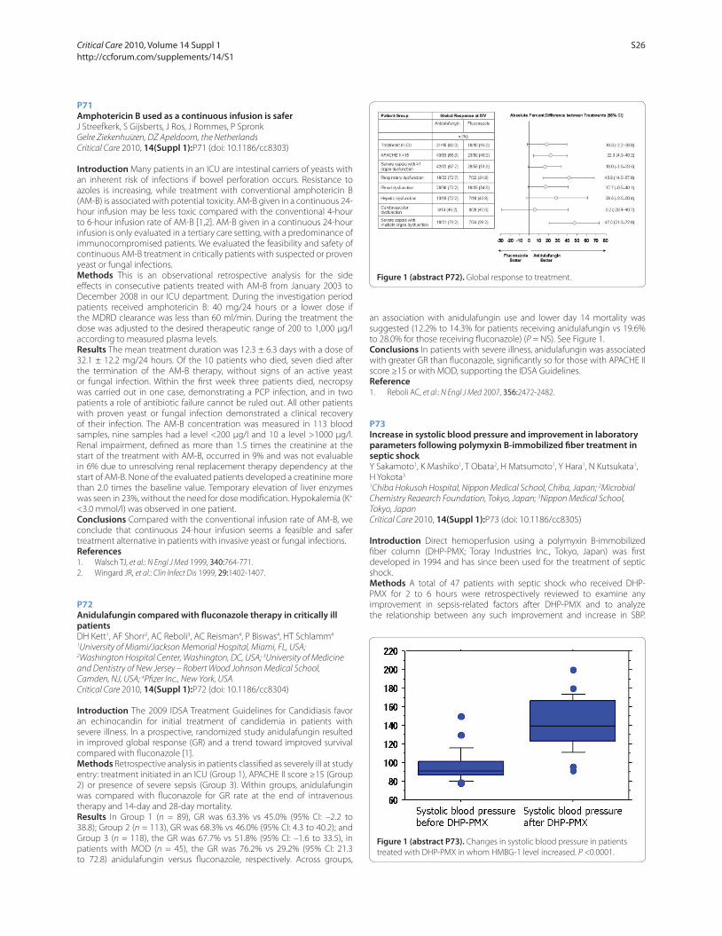
P71
Amphotericin B used as a continuous infusion is saferJ Streefkerk, S Gijsberts, J Ros, J Rommes, P Spronk
Gelre Ziekenhuizen, DZ Apeldoorn, the Netherlands
Critical Care 2010, 14(Suppl 1):P71 (doi: 10.1186/cc8303)
Introduction Many patients in an ICU are intestinal carriers of yeasts with
an inherent risk of infections if bowel perforation occurs. Resistance to
azoles is increasing, while treatment with conventional amphotericin B
(AM-B) is associated with potential toxicity. AM-B given in a continuous 24-
hour infusion may be less toxic compared with the conventional 4-hour
to 6-hour infusion rate of AM-B [1,2]. AM-B given in a continuous 24-hour
infusion is only evaluated in a tertiary care setting, with a predominance of
immunocompromised patients. We evaluated the feasibility and safety of
continuous AM-B treatment in critically patients with suspected or proven
yeast or fungal infections.
Methods This is an observational retrospective analysis for the side
eff ects in consecutive patients treated with AM-B from January 2003 to
December 2008 in our ICU department. During the investigation period
patients received amphotericin B: 40 mg/24 hours or a lower dose if
the MDRD clearance was less than 60 ml/min. During the treatment the
dose was adjusted to the desired therapeutic range of 200 to 1,000 μg/l
according to measured plasma levels.
Results The mean treatment duration was 12.3 ± 6.3 days with a dose of
32.1 ± 12.2 mg/24 hours. Of the 10 patients who died, seven died after
the termination of the AM-B therapy, without signs of an active yeast
or fungal infection. Within the fi rst week three patients died, necropsy
was carried out in one case, demonstrating a PCP infection, and in two
patients a role of antibiotic failure cannot be ruled out. All other patients
with proven yeast or fungal infection demonstrated a clinical recovery
of their infection. The AM-B concentration was measured in 113 blood
samples, nine samples had a level <200 μg/l and 10 a level >1000 μg/l.
Renal impairment, defi ned as more than 1.5 times the creatinine at the
start of the treatment with AM-B, occurred in 9% and was not evaluable
in 6% due to unresolving renal replacement therapy dependency at the
start of AM-B. None of the evaluated patients developed a creatinine more
than 2.0 times the baseline value. Temporary elevation of liver enzymes
was seen in 23%, without the need for dose modifi cation. Hypokalemia (K+
<3.0 mmol/l) was observed in one patient.
Conclusions Compared with the conventional infusion rate of AM-B, we
conclude that continuous 24-hour infusion seems a feasible and safer
treatment alternative in patients with invasive yeast or fungal infections.
References1. Walsch TJ, et al.: N Engl J Med 1999, 340:764-771.
2. Wingard JR, et al.: Clin Infect Dis 1999, 29:1402-1407.
P72
Anidulafungin compared with fl uconazole therapy in critically ill patientsDH Kett1, AF Shorr2, AC Reboli3, AC Reisman4, P Biswas4, HT Schlamm4
1University of Miami/Jackson Memorial Hospital, Miami, FL, USA; 2Washington Hospital Center, Washington, DC, USA; 3University of Medicine
and Dentistry of New Jersey – Robert Wood Johnson Medical School,
Camden, NJ, USA; 4Pfi zer Inc., New York, USA
Critical Care 2010, 14(Suppl 1):P72 (doi: 10.1186/cc8304)
Introduction The 2009 IDSA Treatment Guidelines for Candidiasis favor
an echinocandin for initial treatment of candidemia in patients with
severe illness. In a prospective, randomized study anidulafungin resulted
in improved global response (GR) and a trend toward improved survival
compared with fl uconazole [1].
Methods Retrospective analysis in patients classifi ed as severely ill at study
entry: treatment initiated in an ICU (Group 1), APACHE II score ≥15 (Group
2) or presence of severe sepsis (Group 3). Within groups, anidulafungin
was compared with fl uconazole for GR rate at the end of intravenous
therapy and 14-day and 28-day mortality.
Results In Group 1 (n = 89), GR was 63.3% vs 45.0% (95% CI: –2.2 to
38.8); Group 2 (n = 113), GR was 68.3% vs 46.0% (95% CI: 4.3 to 40.2); and
Group 3 (n = 118), the GR was 67.7% vs 51.8% (95% CI: –1.6 to 33.5), in
patients with MOD (n = 45), the GR was 76.2% vs 29.2% (95% CI: 21.3
to 72.8) anidulafungin versus fl uconazole, respectively. Across groups,
an association with anidulafungin use and lower day 14 mortality was
suggested (12.2% to 14.3% for patients receiving anidulafungin vs 19.6%
to 28.0% for those receiving fl uconazole) (P = NS). See Figure 1.
Conclusions In patients with severe illness, anidulafungin was associated
with greater GR than fl uconazole, signifi cantly so for those with APACHE II
score ≥15 or with MOD, supporting the IDSA Guidelines.
Reference
1. Reboli AC, et al.: N Engl J Med 2007, 356:2472-2482.
P73
Increase in systolic blood pressure and improvement in laboratory parameters following polymyxin B-immobilized fi ber treatment in septic shockY Sakamoto1, K Mashiko1, T Obata2, H Matsumoto1, Y Hara1, N Kutsukata1,
H Yokota3
1Chiba Hokusoh Hospital, Nippon Medical School, Chiba, Japan; 2Microbial
Chemistry Reaearch Foundation, Tokyo, Japan; 3Nippon Medical School,
Tokyo, Japan
Critical Care 2010, 14(Suppl 1):P73 (doi: 10.1186/cc8305)
Introduction Direct hemoperfusion using a polymyxin B-immobilized
fi ber column (DHP-PMX; Toray Industries Inc., Tokyo, Japan) was fi rst
developed in 1994 and has since been used for the treatment of septic
shock.
Methods A total of 47 patients with septic shock who received DHP-
PMX for 2 to 6 hours were retrospectively reviewed to examine any
improvement in sepsis-related factors after DHP-PMX and to analyze
the relationship between any such improvement and increase in SBP.
Figure 1 (abstract P72). Global response to treatment.
Figure 1 (abstract P73). Changes in systolic blood pressure in patients
treated with DHP-PMX in whom HMBG-1 level increased. P <0.0001.
Critical Care 2010, Volume 14 Suppl 1 http://ccforum.com/supplements/14/S1
S26
In addition, we analyzed the eff ectiveness of long-term DHP-PMX (5 to
6 hours) with respect to improvement in sepsis-related factors.
Results N-arachidonoylethanolamine (AEA), 2-arachidonoyl glycerol
(2-AG), and plasminogen activator inhibitor-1 (PAI-1) were signifi cantly
improved after DHP-PMX treatment. SBP increased signifi cantly in the
group showing improved high mobility group box protein 1 (HMGB-1)
level (P <0.0122). AEA, 2-AG and HMGB-1 were improved in all four patients
who were treated with long-term DHP-PMX.
Conclusions We observed a relationship between hemodynamic
improve ment and a decrease in serum HMGB-1 level in septic shock
patients treated with DHP-PMX. We suggest that long-term DHP-PMX
improves sepsis-related factors.
References1. Sakamoto Y, et al.: ASAIO J 2007, 53:646-650.
P74
Selection of acute blood purifi cation therapy according to lipid mediator adsorption and blood purifi cation in patients with septic shockY Sakamoto1, K Mashiko1, T Obata2, H Yokota3
1Chiba Hokusoh Hospital, Nippon Medical School, Chiba, Japan; 2Microbial
Chemistry Research Foundation, Tokyo, Japan; 3Nippon Medical School,
Tokyo, Japan
Critical Care 2010, 14(Suppl 1):P74 (doi: 10.1186/cc8306)
Introduction Direct hemoperfusion using a polymyxin B-immobilized
fi ber column (DHP-PMX) has been used for the treatment of septic
shock. As an alternative method for acute blood purifi cation therapy,
continuous venovenous hemodiafi ltration (CVVHDF) has been reported
to be an eff ective clinical treatment for critically ill patients; however,
the optimal column for performing CVVHDF remains controversial. On
the other hand, recently, one of the lipid mediators, endocannabinoids
(N-arachidonoylethanolamine (AEA) and 2-arachidonoyl glycerol (2-AG))
have been reported with a blood pressure decreased eff ect.
Methods We investigated 14 polymethylmethacrylate (PMMA) membrane
hemofi lters and three polyacrylonitrile (PAN) membrane hemofi lters
after use in patients with septic shock. Therefore, in clinical study, we
used CVVHDF after DHP-PMX to treat 32 patients with septic shock. To
determine the optimal acute blood purifi cation therapy, we subsequently
divided the patients into two groups: group A underwent CVVHDF using
a PMMA membrane hemofi lter after undergoing DHP-PMX (n = 25);
group B underwent CVVHDF using a PAN membrane hemofi lter after
undergoing DHP-PMX (n = 7). In addition, the levels of endocannabinoids
(AEA, 2-AG) were measured. The severity scores and the improvement of
endocannabinoids were compared between the two groups.
Results Endocannabinoids (AEA, 2-AG) were adsorbed more in the PMMA
column (AEA; 506.3 ± 680.2 ng/column, 2-AG; 23.0 ± 38.5 μg/column) than
in the PAN column (AEA; 1.5 ± 0.7 ng/column, 2-AG; 0.1 ± 0.1 μg/column).
The average Acute Physiology and Chronic Health Evaluation (APACHE) II
score and the average sepsis-related organ failure assessment (SOFA) score
did not diff er signifi cantly between the two groups. Group A showed a better
outcome compared with Group B (P = 0.05). In addition, only group A showed
a signifi cant improvement in the blood AEA level on day 1 (P = 0.0185).
Conclusions Our study suggests that the PMMA column might be the
better column for performing CVVHDF after DHP-PMX treatment, as
suggested by the adsorption and blood purifi cation of endocannabinoids.
Reference1. Sakamoto Y, et al.: ASAIO J 2008, 54:129-132.
P75
Prognostic assessment in community-acquired pneumonia by pneumonia severity scores and biomarkersF Dusemund1, W Albrich1, P Schuetz2, B Müller1
1Kantonsspital Aarau, Switzerland; 2University Hospital Basel, Switzerland
Critical Care 2010, 14(Suppl 1):P75 (doi: 10.1186/cc8307)
Introduction Classical biomarkers like C-reactive protein (CRP) or the
leucocyte count are only inaccurate tools for predicting the severity of
community-acquired pneumonia (CAP). Procalcitonin (PCT) was found to
predict 28-day mortality in CAP (area under the curve (AUC) 0.8) similar to
the combination of PCT and CRB65 score (AUC 0.83) [1]. Novel prognostic
biomarkers, such as Pro-ET1 and MR-ProADM, were shown to correlate
with CAP severity [2]. We aimed to compare the diagnostic accuracy to
predict mortality and ICU admission of clinical severity scores, biomarkers
and their combination.
Methods Nine hundred and twenty-fi ve CAP patients enrolled in the
ProHOSP trial [3] were analyzed by assessing clinical severity scores (SMART-
COP, PSI, CURB65) and biomarker levels (PCT, MR-ProADM, Pro-ET1). Receiver
operating characteristic curves for 30-day mortality and ICU admission were
used to calculate and compare the diff erent predictive values.
Results The AUC for the prediction of 30-day mortality was 0.84 with
SMART-COP, 0.82 with PSI, 0.72 with CURB65, 0.59 with PCT, 0.75 with
MR-ProADM and 0.75 with ProET1. ICU admission was predicted best
by SMART-COP (AUC 0.83), compared with the other severity scores and
biomarkers (PSI: 0.68, CURB65: 0.65, PCT: 0.7, Pro-ET1: 0.73, ProADM: 0.72).
The combination of SMART-COP and MR-ProADM was superior to SMART-
COP alone (AUC 0.84, P = 0.04).
Conclusions The combination of MR-ProADM with SMART-COP
signifi cantly improved the prediction of ICU admission. Prognostic
biomarkers should complement the clinical assessment of patients with
LRTI to improve allocation of healthcare resources to high-risk patients.
References
1. Krüger S, et al.: Eur Respir J 2008, 31:349-355.
2. Schuetz P, et al.: BMC Inf Dis 2008, 8:22-30.
3. Schuetz P, et al.:: JAMA 2009, 302:1059-1066.
P76
Current progress in pneumonia researchT Smelaya1, V Moroz1, A Goloubev1, L Salnikova2, A Rubanovich2
1V.A. Negovsky Research Institute of General Reanimatology RAMS, Moscow,
Russia Federation; 2N.I. Vavilov Research Institute of General Genetics,
Moscow, Russia Federation
Critical Care 2010, 14(Suppl 1):P76 (doi: 10.1186/cc8308)
Introduction Diagnosis and treatment of acute pneumonia (Pn) is a
problem of high signifi cance in modern medicine. The incidence of
complicated and lethal acute Pn has increased [1]. Candidate genes are of
great importance in the course of acute diseases and their complications.
Cytokines and xenobiotic detoxication genes are the most investigated. The
aim of the investigation was to study genetic predisposition to acute Pn.
Methods Results of the associative DNA polymorphism studies in 243
patients with acute community-acquired Pn are presented; 178 healthy
individuals formed a control group. Genetic variability of the candidate
loci was studied: renin–angiotensin ACE system gene, chemokine receptor
CCR5 gene and four genes controlling xenobiotic detoxifi cation (CYP1A1,
GSTM1, GSTT1, GSTP1). Multiplex polymerase chain reaction was utilized
for genotyping of insertion–deletion polymorphism on loci ACE (287
nucleotide pairs) and CCR5 (deletion of 32 nucleotide pairs). The odds ratio
index was used to describe the degree of association of the genotypes
with the diseases. Statistical analysis was done by means of Fisher’s exact
test and the online program SNPStats (http://bioinfo.iconcologia.net).
Results An increased predisposition to Pn development was registered in
homozygotes in deletion at the ACE locus (OR = 1.8; P = 0.013), positive
genotypes of the GSTM1 locus (OR = 1.7; P = 0.010) and homozygotes in
allele 606T of the CYP1A1 gene (OR = 1.6; P = 0.020).
Conclusions A combination of positive genotypes of the GSTM1 locus and
homozygotes in allele 606T of the CYP1A1 gene (OR = 1.9, P = 0.006; incidence
in controls >20%) presented with a most eff ective prognostic power.
Reference1. Sinopalnikov AI, et al.: Military Med J 1996, 2:30-33.
P77
Bacterial colonization and infections of the lower respiratory tract in an interdisciplinary ICUM Sartzi, M Agrafi otis, P Peppa, F Nanu, M Charitidi, I Basiliadis, G Kallitsi,
M Michalia, P Clouva-Molyvdas
General Hospital Thriassio Elefsinas, Magoula, Greece
Critical Care 2010, 14(Suppl 1):P77 (doi: 10.1186/cc8309)
Introduction Tracheal intubation and mechanical ventilation are often
associated with infection of the lower respiratory tract (LRT). The morbidity
Critical Care 2010, Volume 14 Suppl 1 http://ccforum.com/supplements/14/S1
S27

associated with the progression of airway colonization (AC) to ventilator-
associated tracheobronchitis (VAT) and from VAT to ventilator-associated
pneumonia (VAP) has been analyzed but not been fully elucidated. We
endeavored to study the relationship between AC and development of
infections (VAT and VAP) of the LRT in ICU patients.
Methods Retrospective study of 400 consecutive ICU patients ventilated
>48 hours in the past 4 years. Patients age, gender, APACHE II, prior illness,
cause of admittance, length of stay (LOS), time of mechanical ventilation
(MV), outcome, time of appearance of AC and infection of the LRT were
registered. MODS and CPIS were measured at onset of infection (VAT–
VAP) and 3 days after. MODS was also calculated on the day AC appeared.
Bronchial secretions were cultured at admission, at least once a week and
whenever there was a change in the amount and quality of bronchial
secretions or clinical infection was suspected. The Mann–Whitney test was
used for statistical analysis and statistical signifi cance was set at P <0.05.
Results From the 400 studied patients 68 (17%) were colonized and
153 (48.25%) developed infections: 54 (13.5%) VAT and 99 (24.7%) VAP.
Colonization appeared after 3.1 ± 0.8 days of ICU admission exclusively
with Gram-negative microorganisms. Twenty-fi ve (36.7%) of the colonized
patients developed infections of the LRT (with the same pathogen): 8
(11.7%) VAT after 4.6 ± 2.7 days and 17 (25%) were diagnosed with VAP
after 9.8 ± 1.8 days. From the eight VAT patients, four developed VAP (50%)
after 5.4 ± 0.4 days. None of the colonized patients died. The colonized
patients who developed infections were elderly (P <0.05), more severely ill
(P <0.04) and had at the time of infection a diagnosis higher temperature
(P <0.01), more severe leukocytosis (P <0.02) but not statistically signifi cant
organ dysfunction (P >0.3). The appearance of VAP caused more severe
organ dysfunction (P <0.002), longer MV duration (P <0.01) and longer
LOS (P <0.001) but did not infl uence mortality. On the day of AC detection
MODS was 2.4 ± 0.5, on VAT detection MODS was 4.8 ± 0.9 and on VAP
appearance MODS was 9.6 ± 0.7. CPIS at VAT detection was 3.9 ± 0.9 and at
VAP detection 6.1 ± 0.5 (day I) and 7.2 ± 0.8 (on day III).
Conclusions AC resulted in LRT infections in only one-third of our patients
and the majority of LRT infections are not preceded by AC.
P78
Rehydration does not aff ect pulmonary immune responses to infl uenza or susceptibility to secondary bacterial pneumoniaRD Sanders1, A Godlee1, JC Goulding1, M Maze2, T Hussell1
1Imperial College London, UK; 2UCSF, San Francisco, CA, USA
Critical Care 2010, 14(Suppl 1):P78 (doi: 10.1186/cc8310)
Introduction In our murine model of infl uenza, signifi cant weight loss
occurs up to day 7 post-infection [1]. We sought to determine whether
weight loss from infl uenza could be altered by rehydration and whether
this aff ects pulmonary immune responses.
Methods Adult BALB/c mice were infected with X31 (H3N2) infl uenza (1:80)
via the intranasal route and randomized to intraperitoneal rehydration
with 20 ml/kg compound sodium lactate (CSL), normal saline (NS) or
no rehydration (NR) starting on day 3 following infection and continued
for 4 days (n = 5/group). On day 7, mice were challenged with 1 x 10⁶
Streptococcus pneumoniae (serotype 2). Two further cohorts of mice were
challenged with diff erent doses of infl uenza and rehydrated from day 3 to
7 to investigate pulmonary immune responses in the absence of bacteria.
Mice were infected with 1:80 (n = 10/group) infl uenza and rehydrated
once daily or 1:60 (n = 5/group) infl uenza and rehydrated twice (1:60)
daily with 20 ml/kg CSL or not. Daily weight, survival following secondary
bacterial pneumonia, number of colony-forming units (48 hours after
bacterial challenge) from peripheral blood, lung, and nasal wash and
cellularity in lung compartments were measured.
Results Rehydration did not aff ect weight loss following 1:80 infl uenza
infection (naïve mice (+0.3 ± 0.4 g), infl uenza plus NR (–1.58 ± 0.4 g),
infl uenza plus CSL (–1.1 ± 0.7 g) and infl uenza with NS (–1.3 ± 0.4 g)). A
repeat experiment with CSL once daily or twice daily did not alter weight
loss compared with NR (P >0.05). Survival or CFU counts following bacterial
pneumonia did not diff er between the groups (P >0.05). The total number or
activational status of bronchoalveolar, lung macrophages/monocytes and
lymphocytes was not aff ected by rehydration following infl uenza infection
or 48 hours following bacterial pneumonia (P >0.05; P <0.05 vs naïve mice).
Conclusions Rehydration does not aff ect immunity or pathophysiology
in a murine infl uenza infection model. Assuming these results can be
extrapolated to the clinical setting, our fi ndings support the use of
conservative fl uid resuscitation strategies in patients with infl uenza.
Reference1. Snelgrove et al.: Nat Immunol 2008, 9:1074-1083.
P79
Bacteremic nosocomial pneumonia cases from the ATTAIN studiesE Rubinstein1, SL Barriere2, FC Genter2, GR Corey3, C Luna4, A Lentnek5,
ME Stryjewski6
1University of Manitoba, Winnipeg, Canada; 2Theravance, Inc., South San
Francisco, CA, USA; 3DCRI, Durham, NC, USA; 4University of Buenos Aires,
Argentina; 5Wellstar Infectious Disease, Marietta, GA, USA; 6CEMIC, Buenos
Aires, Argentina
Critical Care 2010, 14(Suppl 1):P79 (doi: 10.1186/cc8311)
Introduction Bacteremic pneumonia is associated with worse outcome
including higher mortality. The ATTAIN program compared telavancin
(TLV), a lipoglycopeptide antibiotic, with vancomycin (VAN) for treatment
of nosocomial pneumonia (NP) due to Gram-positive pathogens including
MRSA. This subgroup analysis examined the baseline characteristics and
clinical outcomes in bacteremic HAP cases.
Methods ATTAIN 1 and 2 were methodologically identical, randomized,
double-blind, phase 3 studies. Adult patients with NP due to presumed or
confi rmed Gram-positive pathogens were randomized (1:1) to TLV 10 mg/
kg intravenously every 24 hours or VAN 1 g intravenously every 12 hours
(adjusted per site-specifi c guidelines) for 7 to 21 days. The modifi ed all-
treated (MAT) population consisted of patients who received ≥1 dose of
study medication and who had a respiratory pathogen recovered from
baseline cultures. Bacteremic NP was defi ned by the identifi cation of a
pneumonia-causing pathogen in the blood or of the same pathogen in
lung and blood with identical susceptibility profi les. Clinical outcomes
were assessed at test-of-cure (TOC) 7 to 14 days after end of study
treatment.
Results All MAT patients with bacteremic NP (n = 73) were included in
this analysis. At baseline, more TLV patients than VAN patients were in the
ICU (TLV 74%, VAN 62%) and had ventilator-associated pneumonia (TLV
59%, VAN 44%); APACHE II scores were similar between groups (mean ±
SD, TLV 16 ± 6, VAN 17 ± 6). S. aureus was the most common pathogen
(TLV 76%, VAN 69%) and included MRSA (TLV 41%, VAN 49%). Cure rates for
TLV and VAN were 44% and 36%, respectively (diff erence TLV – VAN (95%
CI) = 7.3% (–15.9%, 30.5%)). On-study mortality was similar, 41% in each
treatment group. Incidences of adverse events (AE) were similar between
groups, except for nausea (TLV 21%, VAN 3%) and vomiting (TLV 15%, VAN,
0%). Proportions of patients who discontinued the study medication due
to AEs were similar (TLV 12%, VAN 13%).
Conclusions TLV and VAN had similar cure rates in a subgroup of ATTAIN
patients with bacteremic NP. The safety profi les of TLV and VAN were
mostly comparable in these patients.
P80
Late ventilator-associated pneumonia: analysis of baseline characteristics and clinical outcomes in the ATTAIN studiesE Rubinstein1, SL Barriere2, FC Genter2, GR Corey3, PC Lee4, T Lalani3
1University of Manitoba, Winnipeg, Canada; 2Theravance, Inc., South San
Francisco, CA, USA; 3DCRI, Durham, NC, USA; 4Baystate Medical Centre,
Springfi eld, MA, USA
Critical Care 2010, 14(Suppl 1):P80 (doi: 10.1186/cc8312)
Introduction Pneumonia is a leading cause of death associated with
hospital-acquired infections. Late ventilator-associated pneumonia
(VAP), defi ned as disease onset after ≥4 days of mechanical ventilation,
is associated with worse outcomes than other forms of hospital-acquired
pneumonia (HAP).
Methods ATTAIN 1 and 2 were methodologically identical, randomized,
phase 3 studies of telavancin (TLV) 10 mg/kg intravenously every 24 hours
vs vancomycin (VAN) 1 g intravenously every 12 hours for treatment of
HAP, including VAP. VAN doses could be adjusted per investigative site
guidelines. Clinical outcome was assessed at the test-of-cure (TOC) visit
7 to 14 days after the end of study treatment. Baseline characteristics,
outcomes, and safety in the late VAP sub-group were analyzed in the
Critical Care 2010, Volume 14 Suppl 1 http://ccforum.com/supplements/14/S1
S28
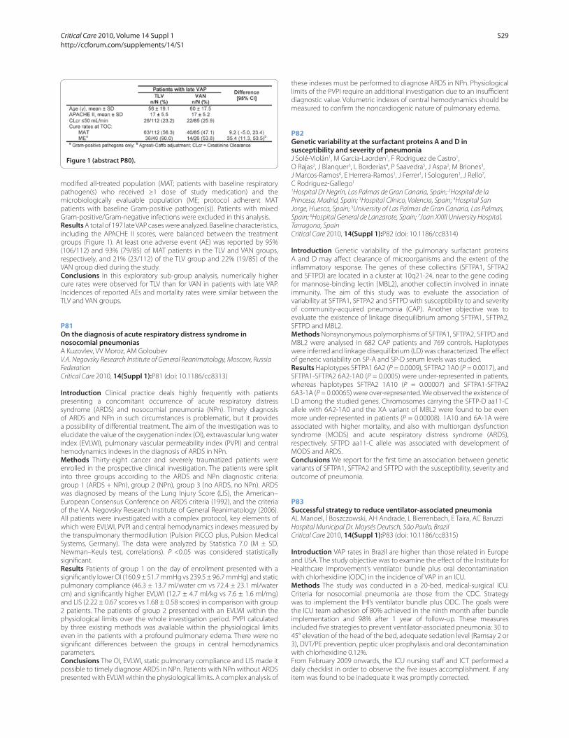
modifi ed all-treated population (MAT; patients with baseline respiratory
pathogen(s) who received ≥1 dose of study medication) and the
microbiologically evaluable population (ME; protocol adherent MAT
patients with baseline Gram-positive pathogen(s)). Patients with mixed
Gram-positive/Gram-negative infections were excluded in this analysis.
Results A total of 197 late VAP cases were analyzed. Baseline characteristics,
including the APACHE II scores, were balanced between the treatment
groups (Figure 1). At least one adverse event (AE) was reported by 95%
(106/112) and 93% (79/85) of MAT patients in the TLV and VAN groups,
respectively, and 21% (23/112) of the TLV group and 22% (19/85) of the
VAN group died during the study.
Conclusions In this exploratory sub-group analysis, numerically higher
cure rates were observed for TLV than for VAN in patients with late VAP.
Incidences of reported AEs and mortality rates were similar between the
TLV and VAN groups.
P81
On the diagnosis of acute respiratory distress syndrome in nosocomial pneumoniasA Kuzovlev, VV Moroz, AM Goloubev
V.A. Negovsky Research Institute of General Reanimatology, Moscow, Russia
Federation
Critical Care 2010, 14(Suppl 1):P81 (doi: 10.1186/cc8313)
Introduction Clinical practice deals highly frequently with patients
presenting a concomitant occurrence of acute respiratory distress
syndrome (ARDS) and nosocomial pneumonia (NPn). Timely diagnosis
of ARDS and NPn in such circumstances is problematic, but it provides
a possibility of diff erential treatment. The aim of the investigation was to
elucidate the value of the oxygenation index (OI), extravascular lung water
index (EVLWI), pulmonary vascular permeability index (PVPI) and central
hemodynamics indexes in the diagnosis of ARDS in NPn.
Methods Thirty-eight cancer and severely traumatized patients were
enrolled in the prospective clinical investigation. The patients were split
into three groups according to the ARDS and NPn diagnostic criteria:
group 1 (ARDS + NPn), group 2 (NPn), group 3 (no ARDS, no NPn). ARDS
was diagnosed by means of the Lung Injury Score (LIS), the American–
European Consensus Conference on ARDS criteria (1992), and the criteria
of the V.A. Negovsky Research Institute of General Reanimatology (2006).
All patients were investigated with a complex protocol, key elements of
which were EVLWI, PVPI and central hemodynamics indexes measured by
the transpulmonary thermodilution (Pulsion PiCCO plus, Pulsion Medical
Systems, Germany). The data were analyzed by Statistica 7.0 (M ± SD,
Newman–Keuls test, correlations). P <0.05 was considered statistically
signifi cant.
Results Patients of group 1 on the day of enrollment presented with a
signifi cantly lower OI (160.9 ± 51.7 mmHg vs 239.5 ± 96.7 mmHg) and static
pulmonary compliance (46.3 ± 13.7 ml/water cm vs 72.4 ± 23.1 ml/water
cm) and signifi cantly higher EVLWI (12.7 ± 4.7 ml/kg vs 7.6 ± 1.6 ml/mg)
and LIS (2.22 ± 0.67 scores vs 1.68 ± 0.58 scores) in comparison with group
2 patients. The patients of group 2 presented with an EVLWI within the
physiological limits over the whole investigation period. PVPI calculated
by three existing methods was available within the physiological limits
even in the patients with a profound pulmonary edema. There were no
signifi cant diff erences between the groups in central hemodynamics
parameters.
Conclusions The OI, EVLWI, static pulmonary compliance and LIS made it
possible to timely diagnose ARDS in NPn. Patients with NPn without ARDS
presented with EVLWI within the physiological limits. A complex analysis of
these indexes must be performed to diagnose ARDS in NPn. Physiological
limits of the PVPI require an additional investigation due to an insuffi cient
diagnostic value. Volumetric indexes of central hemodynamics should be
measured to confi rm the noncardiogenic nature of pulmonary edema.
P82
Genetic variability at the surfactant proteins A and D in susceptibility and severity of pneumoniaJ Solé-Violán1, M Garcia-Laorden1, F Rodriguez de Castro1,
O Rajas2, J Blanquer3, L Borderías4, P Saavedra5, J Aspa2, M Briones3,
J Marcos-Ramos6, E Herrera-Ramos1, J Ferrer1, I Sologuren1, J Rello7,
C Rodriguez-Gallego1
1Hospital Dr Negrín, Las Palmas de Gran Canaria, Spain; 2Hospital de la
Princesa, Madrid, Spain; 3Hospital Clínico, Valencia, Spain; 4Hospital San
Jorge, Huesca, Spain; 5University of Las Palmas de Gran Canaria, Las Palmas,
Spain; 6Hospital General de Lanzarote, Spain; 7Joan XXIII University Hospital,
Tarragona, Spain
Critical Care 2010, 14(Suppl 1):P82 (doi: 10.1186/cc8314)
Introduction Genetic variability of the pulmonary surfactant proteins
A and D may aff ect clearance of microorganisms and the extent of the
infl ammatory response. The genes of these collectins (SFTPA1, SFTPA2
and SFTPD) are located in a cluster at 10q21-24, near to the gene coding
for mannose-binding lectin (MBL2), another collectin involved in innate
immunity. The aim of this study was to evaluate the association of
variability at SFTPA1, SFTPA2 and SFTPD with susceptibility to and severity
of community-acquired pneumonia (CAP). Another objective was to
evaluate the existence of linkage disequilibrium among SFTPA1, SFTPA2,
SFTPD and MBL2.
Methods Nonsynonymous polymorphisms of SFTPA1, SFTPA2, SFTPD and
MBL2 were analysed in 682 CAP patients and 769 controls. Haplotypes
were inferred and linkage disequilibrium (LD) was characterized. The eff ect
of genetic variability on SP-A and SP-D serum levels was studied.
Results Haplotypes SFTPA1 6A2 (P = 0.0009), SFTPA2 1A0 (P = 0.0017), and
SFTPA1-SFTPA2 6A2-1A0 (P = 0.0005) were under-represented in patients,
whereas haplotypes SFTPA2 1A10 (P = 0.00007) and SFTPA1-SFTPA2
6A3-1A (P = 0.00065) were over-represented. We observed the existence of
LD among the studied genes. Chromosomes carrying the SFTP-D aa11-C
allele with 6A2-1A0 and the XA variant of MBL2 were found to be even
more under-represented in patients (P = 0.00008). 1A10 and 6A-1A were
associated with higher mortality, and also with multiorgan dysfunction
syndrome (MODS) and acute respiratory distress syndrome (ARDS),
respectively. SFTPD aa11-C allele was associated with development of
MODS and ARDS.
Conclusions We report for the fi rst time an association between genetic
variants of SFTPA1, SFTPA2 and SFTPD with the susceptibility, severity and
outcome of pneumonia.
P83
Successful strategy to reduce ventilator-associated pneumoniaAL Manoel, Í Boszczowski, AH Andrade, L Bierrenbach, E Taira, AC Baruzzi
Hospital Municipal Dr. Moysés Deutsch, São Paulo, Brazil
Critical Care 2010, 14(Suppl 1):P83 (doi: 10.1186/cc8315)
Introduction VAP rates in Brazil are higher than those related in Europe
and USA. The study objective was to examine the eff ect of the Institute for
Healthcare Improvement’s ventilator bundle plus oral decontamination
with chlorhexidine (ODC) in the incidence of VAP in an ICU.
Methods The study was conducted in a 20-bed, medical-surgical ICU.
Criteria for nosocomial pneumonia are those from the CDC. Strategy
was to implement the IHI’s ventilator bundle plus ODC. The goals were
the ICU team adhesion of 80% achieved in the ninth month after bundle
implementation and 98% after 1 year of follow-up. These measures
included fi ve strategies to prevent ventilator-associated pneumonia: 30 to
45° elevation of the head of the bed, adequate sedation level (Ramsay 2 or
3), DVT/PE prevention, peptic ulcer prophylaxis and oral decontamination
with chlorhexidine 0.12%.
From February 2009 onwards, the ICU nursing staff and ICT performed a
daily checklist in order to observe the fi ve issues accomplishment. If any
item was found to be inadequate it was promptly corrected.
Figure 1 (abstract P80).
Critical Care 2010, Volume 14 Suppl 1 http://ccforum.com/supplements/14/S1
S29
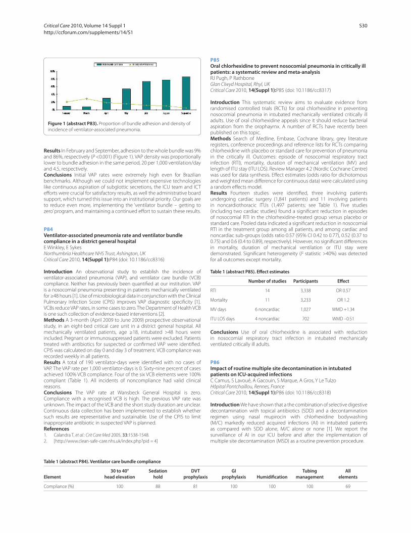
Results In February and September, adhesion to the whole bundle was 9%
and 86%, respectively (P <0.001) (Figure 1). VAP density was proportionally
lower to bundle adhesion in the same period, 20 per 1,000 ventilation/day
and 4.5, respectively.
Conclusions Initial VAP rates were extremely high even for Brazilian
benchmarks. Although we could not implement expensive technologies
like continuous aspiration of subglottic secretions, the ICU team and ICT
eff orts were crucial for satisfactory results, as well the administrative board
support, which turned this issue into an institutional priority. Our goals are
to reduce even more, implementing the ‘ventilator bundle – getting to
zero’ program, and maintaining a continued eff ort to sustain these results.
P84
Ventilator-associated pneumonia rate and ventilator bundle compliance in a district general hospitalE Winkley, E Sykes
Northumbria Healthcare NHS Trust, Ashington, UK
Critical Care 2010, 14(Suppl 1):P84 (doi: 10.1186/cc8316)
Introduction An observational study to establish the incidence of
ventilator-associated pneumonia (VAP), and ventilator care bundle (VCB)
compliance. Neither has previously been quantifi ed at our institution. VAP
is a nosocomial pneumonia presenting in patients mechanically ventilated
for ≥48 hours [1]. Use of microbiological data in conjunction with the Clinical
Pulmonary Infection Score (CPIS) improves VAP diag nostic specifi city [1].
VCBs reduce VAP rates, in some cases to zero. The Department of Health VCB
is one such collection of evidence-based interventions [2].
Methods A 3-month (April 2009 to June 2009) prospective observational
study, in an eight-bed critical care unit in a district general hospital. All
mechanically ventilated patients, age ≥18, intubated >48 hours were
included. Pregnant or immunosuppressed patients were excluded. Patients
treated with antibiotics for suspected or confi rmed VAP were identifi ed.
CPIS was calculated on day 0 and day 3 of treatment. VCB compliance was
recorded weekly in all patients.
Results A total of 190 ventilator-days were identifi ed with no cases of
VAP. The VAP rate per 1,000 ventilator-days is 0. Sixty-nine percent of cases
achieved 100% VCB compliance. Four of the six VCB elements were 100%
compliant (Table 1). All incidents of noncompliance had valid clinical
reasons.
Conclusions The VAP rate at Wansbeck General Hospital is zero.
Compliance with a recognised VCB is high. The previous VAP rate was
unknown. The impact of the VCB and the short study duration are unclear.
Continuous data collection has been implemented to establish whether
such results are representative and sustainable. Use of the CPIS to limit
inappropriate antibiotic in suspected VAP is planned.
References1. Calandra T, et al.: Crit Care Med 2005, 33:1538-1548.
2. [http://www.clean-safe-care.nhs.uk/index.php?pid = 4]
P85
Oral chlorhexidine to prevent nosocomial pneumonia in critically ill patients: a systematic review and meta-analysisRJ Pugh, P Rathbone
Glan Clwyd Hospital, Rhyl, UK
Critical Care 2010, 14(Suppl 1):P85 (doi: 10.1186/cc8317)
Introduction This systematic review aims to evaluate evidence from
randomised controlled trials (RCTs) for oral chlorhexidine in preventing
nosocomial pneumonia in intubated mechanically ventilated critically ill
adults. Use of oral chlorhexidine appeals since it should reduce bacterial
aspiration from the orophayrnx. A number of RCTs have recently been
published on this topic.
Methods Search of Medline, Embase, Cochrane library, grey literature
registers, conference proceedings and reference lists for RCTs comparing
chlorhexidine with placebo or standard care for prevention of pneumonia
in the critically ill. Outcomes: episode of nosocomial respiratory tract
infection (RTI), mortality, duration of mechanical ventilation (MV) and
length of ITU stay (ITU LOS). Review Manager 4.2 (Nordic Cochrane Centre)
was used for data synthesis. Eff ect estimates (odds ratio for dichotomous
and weighted mean diff erence for continuous data) were calculated using
a random eff ects model.
Results Fourteen studies were identifi ed, three involving patients
undergoing cardiac surgery (1,841 patients) and 11 involving patients
in noncardiothoracic ITUs (1,497 patients; see Table 1). Five studies
(including two cardiac studies) found a signifi cant reduction in episodes
of nosocomial RTI in the chlorhexidine-treated group versus placebo or
standard care. Pooled data indicated a signifi cant reduction in nosocomial
RTI in the treatment group among all patients, and among cardiac and
noncardiac sub-groups (odds ratio 0.57 (95% CI 0.42 to 0.77), 0.52 (0.37 to
0.75) and 0.6 (0.4 to 0.89), respectively). However, no signifi cant diff erences
in mortality, duration of mechanical ventilation or ITU stay were
demonstrated. Signifi cant heterogeneity (I2 statistic >40%) was detected
for all outcomes except mortality.
Table 1 (abstract P85). Eff ect estimates
Number of studies Participants Eff ect
RTI 14 3,338 OR 0.57
Mortality 11 3,233 OR 1.2
MV days 6 noncardiac 1,027 WMD +1.34
ITU LOS days 4 noncardiac 702 WMD –0.51
Conclusions Use of oral chlorhexidine is associated with reduction
in nosocomial respiratory tract infection in intubated mechanically
ventilated critically ill adults.
P86
Impact of routine multiple site decontamination in intubated patients on ICU-acquired infectionsC Camus, S Lavoué, A Gacouin, S Marque, A Gros, Y Le Tulzo
Hôpital Pontchaillou, Rennes, France
Critical Care 2010, 14(Suppl 1):P86 (doi: 10.1186/cc8318)
Introduction We have shown that a the combination of selective digestive
decontamination with topical antibiotics (SDD) and a decontamination
regimen using nasal mupirocin with chlorhexidine bodywashing
(M/C) markedly reduced acquired infections (AI) in intubated patients
as compared with SDD alone, M/C alone or none [1]. We report the
surveillance of AI in our ICU before and after the implementation of
multiple site decontamination (MSD) as a routine prevention procedure.
Figure 1 (abstract P83). Proportion of bundle adhesion and density of
incidence of ventilator-associated pneumonia.
Table 1 (abstract P84). Ventilator care bundle compliance
30 to 40° Sedation DVT GI Tubing All
Element head elevation hold prophylaxis prophylaxis Humidifi cation management elements
Compliance (%) 100 88 81 100 100 100 69
Critical Care 2010, Volume 14 Suppl 1 http://ccforum.com/supplements/14/S1
S30
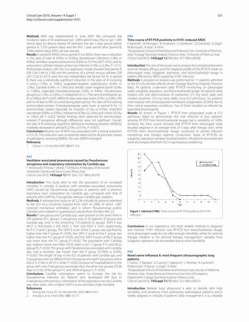
Methods MSD was implemented in June 2007. We compared the
incidence rates of AI (expressed per 1,000 patient-days (‰) or per 1,000
device-days for device-related AI) between the last 1-year period before
(period A, 7,723 patient-days) and the fi rst 1-year period after (period B,
7,646 patient-days) MSD use was started.
Results In period B (MSD) versus period A (no MSD), there was a reduction
in the rates of total AI (9.4‰ vs 23.6‰), bloodstream infections (1.0‰ vs
4.0‰), ventilator-acquired pneumonia (5.6‰ vs 19.1‰) (all P <0.01), and to
a less extent catheter-related urinary tract infection (1.0‰ vs 2.2‰, P = 0.11).
Multivariate analysis with the Cox regression model showed that period A
(OR 2.34 (1.64 to 3.20)) and the presence of a central venous catheter (OR
2.07 (1.22 to 3.57)) were the two independent risk factors for AI. In period
B, there was a statistically signifi cant reduction in the rates of AI involving
S. aureus (1.0‰ vs 3.0‰), coagulase-negative staphylococci (0.4‰ vs
1.3‰), Candida (0.4‰ vs 1.3‰) and aerobic Gram-negative bacilli (5.0‰
vs 15.8‰), especially Enterobacteriaceae (3.4‰ vs 9.4‰), Pseudomonas
aeruginosa (1.4‰ vs 3.9‰), S. maltophilia (0 vs 1.7‰) and Acinetobacter sp.
(0 vs 0.8‰) (all P ≤0.05). MRSA AI rates were also lower (0.3‰ vs 0.9‰, NS)
and no AI due to VRE occurred during both period. The rates of AI involving
antimicrobial-resistant Enterobacteriaceae were lower in period B for 12
antimicrobials tested, especially for ticarcillin (2.1‰ vs 6.5‰), ticarcillin/
clavulanate (0.8‰ vs 4.0‰), cefotaxime (0.3‰ vs 2.2‰) and colistin (0.5‰
vs 1.8‰) (all P ≤0.02). Similar fi ndings were observed for antimicrobial-
resistant P. aeruginosa although diff erences were not signifi cant. Overall,
the rate of AI involving organisms defi ned as multidrug-resistant (MDROs)
markedly decreased in period B (2.5‰ vs 9.1‰, P <0.001).
Conclusions Routine use of MDS was associated with a strong reduction
in ICU AI. The reduction was consistently observed for all principal classes
of pathogens, including MDROs. No new MDRO emerged.
Reference1. Camus C: Crit Care Med 2005, 33:307-314.
P87
Ventilator-associated pneumonia caused by Pseudomonas aeruginosa and respiratory colonization by Candida spp.F Antonicelli, R Festa, F Idone, F Di Muzio, R Maviglia, M Antonelli
Università Cattolica del Sacro Cuore, Roma, Italy
Critical Care 2010, 14(Suppl 1):P87 (doi: 10.1186/cc8319)
Introduction This study aims to test the association of an increased
mortality in critically ill patients with ventilator-associated pneumonia
(VAP) caused by Pseudomonas aeruginosa in patients with a previous
respiratory tract colonization by Candida spp. compared with that of
patients with a VAP by P. aeruginosa without Candida spp. isolation.
Methods A retrospective study on all 5,236 critically ills patients admitted
to the ICU of a university hospital from 2001 to 2008, of which 1,097
received mechanical ventilation and in whom Pseudomonas and/or
Candida were isolated in quantitative cultures from the Mini-BAL [1].
Results P. aeruginosa and Candida spp. were present at the same time in
295 patients (P-C group), P. aeruginosa only in 92 patients (P group) and
Candida spp. only in the remaining 710 patients (C group). SAPS II was
44.27 ± 14.8 (mean ± SD), 39.07 ± 14.61 and 39.07 ± 14.61, respectively,
for P-C, P and C groups. The SAPS II score of the C group was signifi cantly
higher than the P group (P <0.05), the SAPS II score of the C group was
higher than the P-C group (P >0.05), and the SAPS II score of the P group
was lower than the P-C group (P >0.05). The population with Candida
spp. isolation alone was older (70.25 years vs 63.17 (group P-C) and 60.22
(group P), P <0.05). The group with Pseudomonas associated with Candida
spp. had a mortality rate higher than the P group (57.96% vs 6.05%,
P <0.05). The length of stay in the ICU of patients with Candida spp. and
P. aeruginosa was not diff erent from the group who had P. aeruginosa alone
(26.22 ± 31.80 vs 29.15 ± 25.66, P >0.05). Duration of hospitalization in the
group with only P. aeruginosa was longer than the other two groups (79.38
days vs 51.85 of the group P-C and 28.40 of group C, P <0.05).
Conclusions Candida colonization seems to increase the risk for
Pseudomonas infection [2]. Patients who developed VAP due to
P. aeruginosa with previous colonization of the respiratory tract by Candida
spp. were older, with a higher SAPS II score and had a high mortality.
References1. Koenig SM, Truwit JD: Clin Microbiol Rev 2006, 19:637-657.
2. Azoulay E, et al.: Chest 2006, 129:110-117.
P88
Time course of RT-PCR positivity in H1N1-induced ARDSE Mannelli1, M Murtigni1, M Andreani1, S Cianferoni1, D Colosimo1, G Zagli2,
M Bonizzoli2, A Azzi3, A Peris2
1Postgraduate School of Anesthesia and Intensive Care, University of Florence,
Italy; 2Careggi Teaching Hospital, Florence, Italy; 3University of Florence, Italy
Critical Care 2010, 14(Suppl 1):P88 (doi: 10.1186/cc8320)
Introduction The aim of this study was to analyze the correlation between
antiviral therapy effi cacy and the negative profi le of the RT-PCR made on
pharyngeal swab, subglottic aspiration, and bronchoalveolar lavage in
patient aff ected by ARDS caused by H1N1 infection.
Methods A prospective analysis was performed on 11 patients admitted
to the ICU of a tertiary referral center (Careggi Teaching Hospital, Florence,
Italy). All patients underwent daily RT-PCR monitoring on pharyngeal
swab, subglottic aspiration, and bronchoalveolar lavage. All patients were
treated with oral administration of oseltamivir (75 mg twice daily) and
inhaled zanamivir (10 mg twice daily) since ICU admission. Six patients
were treated with extracorporeal membrane oxygenation (ECMO) due to
their critical respiratory conditions. Two of them resulted co-infected by
legionella pneumophila.
Results As shown in Figure 1, RT-PCR from pharyngeal swab at ICU
admission failed to demonstrate the viral infection in four patients,
whereas RT-PCR from bronchoalveolar lavage had a sensibility of 100%.
Similarly, the time course showed that RT-PCR from pharyngeal swab
resulted negative in an average time of 3 days after therapy start, while
RT-PCRs from bronchoalveolar lavage continued to permit infection
monitoring and therapy regimen conduction. None of RT-PCRs on
subglottic aspiration samples resulted positive. All patients recovered and
were discharged alive from ICU in spontaneous breathing.
Conclusions In our experience, the most reliable method to diagnose
and monitor H1N1 infection was RT-PCR from bronchoalveolar lavage,
since pharyngeal swab do not off er enough sensibility, either for antiviral
therapy initiation or for antiviral therapy management. Samples from
subglottic aspiration can be avoided due to a low sensibility.
P89
Novel swine infl uenza A: most frequent ultrasonographic lung patternsL Tutino1, F Barbani1, G Cianchi2, S Batacchi2, L Perretta2, R Cammelli2,
M Bonizzoli2, R Spina2, G Zagli2, A Peris2
1Postgraduate School of Anesthesia and Intensive Care, Faculty of Medicine,
Florence, Italy; 2Anaesthesia and Intensive Care Unit of Emergency
Department, Careggi Teaching Hospital, Florence, Italy
Critical Care 2010, 14(Suppl 1):P89 (doi: 10.1186/cc8321)
Introduction Bedside lung ultrasound is able to identify with high
sensibility and sensitivity most pulmonary pathological patterns and is
widely adopted in critically ill patients’ daily management. It is a feasible
Figure 1 (abstract P88). Time course of RT-PCR positivity for H1N1
infection.
Critical Care 2010, Volume 14 Suppl 1 http://ccforum.com/supplements/14/S1
S31
and reliable method for the identifi cation of the lung pathological patterns
caused by H1N1 infl uenza infection.
Methods The study took place in the ICU of a regional referral center,
with ECMO availability, for respiratory failure (Careggi Teaching Hospital,
Florence, Italy). Eight patients admitted for H1N1-induced ARDS
(September to October 2009) underwent daily LU examination. The
examination was standardized with a procedure ad hoc in order to
achieve complete and comparable reports for every patient. Patients were
examined supine, taking lateral and anterior views and, if possible, on
one side. Intercostal spaces were used as acoustic windows. Every single
examination and its fi nding have been reported in our ICU database.
Results Pleural thickness was present in 100% of cases, mostly related
to little and multiple pleural consolidations. Anyway, pleural gliding was
present most of the time (87.5%), even if with a visible decrease of its
movement. This was replaced with the lung pulse only in the presence
of important lung consolidation. Alveolar interstitial syndrome (AIS) was
always present on the whole lung, ranging from moderate to severe (100%),
with high positivity at the base, posteriorly (100%). White lung appeared in
every patient, most at the lung base and in the middle fi elds, posteriorly.
As far as consolidation is concerned, its presence was confi rmed in 100%
of the patients, associated with satellite multiple subpleural consolidations
in 37.5% of patients. Basal lung was always involved (100%), followed
by middle (50%) and apical fi elds(25%). Bronchogram was present in
100% of patients in bigger consolidations. Their aspect was aerial and
only in one patient, with severe consolidations, turned into a fl uid one.
Anechoic pleural eff usions were found in 37.5% of patients. No cases of
pneumothorax were detected.
Conclusions In this group of patients, H1N1 infection shows diff erent
lung patterns altogether, where the most frequent seemed to be severe
basal posterior AIS, multiple subpleural lung consolidations, and multiple
parenchymal consolidation with bronchogram. The presence of spared
areas did not seem to belong to H1N1 LU patterns.
P90
Outcome and prognostic factors in patients with HIV-negative pneumocystis pneumonia requiring mechanical ventilationE Choi
Asan Medical Center, Seoul, Republic of Korea
Critical Care 2010, 14(Suppl 1):P90 (doi: 10.1186/cc8322)
Introduction Pneumocystis pneumonia (PCP) in HIV-negative patients
frequently presents as fulminant respiratory failure and is associated with a
high mortality rate when the patient requires mechanical ventilation. The
aims of this study were to evaluate the outcome and prognostic factors in
the patients with HIV-negative PCP requiring mechanical ventilation (MV).
Methods We retrospectively reviewed the medical records and collected
the HIV-negative patients who were microbiologically confi rmed as PCP
and required MV in ICU over a 10-year period in a tertiary care teaching
hospital.
Results A total of 51 patients were identifi ed. Mean age was 55.4 ± 15.0
years. Mean APACHE II score at ICU admission was 25.7 ± 5.8. The 28-day
mortality and in-hospital mortality were 45.8% and 66.7%, respectively.
Between survivors and nonsurvivors, there were no signifi cant diff erences
in baseline characteristics, APACHE II score, PaO2/FiO
2 ratio, and absolute
neutrophil counts on ICU admission day. Also the mortality was not
diff erent in relation to the presence of barotrauma, application of non-
invasive ventilation, timing of susceptible antibiotic administration,
changing or not to salvage regimens, presence of cytomegalovirus co-
infection and even the microbiologic persistency in follow-up specimens.
Based on the types and intensity of previous immunosuppressive therapy,
we classifi ed patients into three subgroups: patients receiving low-dose
steroid maintenance ± other immunosuppressive agents (LS), which
represent previously stable organ transplants; another group consisting
of patients receiving recent intensive chemotherapy (CTx); and the other
group refers to patients receiving high dose (defi ned as >2 weeks at
least 1 mg/kg dose) steroid therapy ± other immunosuppressive agents
(HS). Signifi cant diff erences of outcome were observed among the three
diff erent groups (28-day mortality: LS = 22.2%, CTx = 29.4%, HS = 71.4%,
P = 0.01; 60-day mortality: LS = 33.3%, CTx = 64.7%, HS = 81.0%, P = 0.04).
Conclusions Our data showed that the mortality of fulminant HIV-
negative PCP requiring MV was signifi cantly diff erent according to the
types and intensity of previous immunosuppressive treatment despite
similar clinical features on ICU admission.
P91
Acute respiratory failure from pandemic infl uenza A (H1N1) in an intensive care unit in southern BrazilS Beduschi Filho, D Siqueira, SC Carvalho Flores, I Yoshiko Masukawa,
L Kretzer, AC Burigo Grumann, I Silva Maia, M Pimentel Pincelli
Hospital Nereu Ramos, Florianópolis, Brazil
Critical Care 2010, 14(Suppl 1):P91 (doi: 10.1186/cc8323)
Introduction In the winter of 2009 the infl uenza A (H1N1) pandemic
reached Brazil, aff ecting severely its southern states. A total 68,806 cases
of suspected pandemic infl uenza A infection had been reported in Brazil
and 91% of the specimens tested were positive. Incidence of pandemic
infl uenza A in southern Brazil was 137/100,000. In the southern Brazilian
state of Santa Catarina, Nereu Ramos Hospital is the referral center for the
treatment of infectious diseases, and it provided eight intensive care beds
for the management of acute respiratory failure from suspected cases of
infl uenza A during the 2009 pandemic.
Methods We collected retrospective data on the epidemiological features,
clinical course and ventilatory characteristics of patients with laboratory-
confi rmed infl uenza A (H1N1) infection who were admitted to our ICU
during the winter of 2009.
Results Thirty-four adult patients were admitted to our ICU with acute
respiratory failure and suspected pandemic infl uenza A infection. Of these,
14 cases tested positive for infl uenza A (H1N1). The majority of the patients
were male (61.5%) with median age of 27.5 years and interquartile range
(IR) of 26.5 to 48.5. The APACHE II median was 14.5 (IR 10.0 to 18.25) with
median predicted mortality of 20.0 (IR 10.73 to 27.7). Risk factors were:
obesity (23.1%), obstructive respiratory diseases (15.4%); pregnancy,
immunosuppressive and neuromuscular disorders were present in one
patient each. All 14 patients presented severe respiratory failure, with a
median lowest PaO2/IOF of 113.3 (IR 83.53 to 159.55) and 92.3% of them
requiring mechanical ventilation. The patients in mechanical ventilation
were all ventilated in pressured-controlled mode, demanded high PEEP
levels (mean of 18.5 ± 5.16 cmH2O), presented high peak inspiratory
pressure (mean of 37.83 ± 7.35 cmH2O) and required elevated IOF
(81.15 ± 20.83%). A total of 57.1% of the patients were submitted to lung
recruitment, while the prone position was used in 42.8% of them. The
median ICU stay was 13.0 days (IR 4.75 to 23.50) and the median hospital
stay was 16.5 days (IR 9.75 to 25.0). The ventilatory-associated pneumonia
(VAP) rate was unusually high (69.2%) as well as the mortality rate during
the intensive care stay (38.5%).
Conclusions Patients admitted to our ICU with confi rmed pandemic
infl uenza A infection presented severe acute respiratory failure and an
unusually high incidence of VAP and mortality rates.
Reference1. Perez-Padilla R, et al.: Pneumonia and respiratory failure from swine-origin
infl uenza A (H1N1) in Mexico. N Engl J Med 2009, 361:680-689.
P92
Extracorporeal membrane oxygenation for infl uenza A (H1N1): experience in a regional referral centerA Pasquini1, S Di Valvasone2, S Biondi2, S Batacchi1, G Cianchi1, M Ciapetti1,
M Bonizzoli1, R Spina1, L Turrisi1, E Mascitelli3, M Bonacchi3, G Zagli1,
GF Gensini3, A Peris1
1Careggi Teaching Hospital, Florence, Italy; 2Postgraduate School of
Anesthesia and Intensive Care, Florence, Italy; 3University of Florence, Italy
Critical Care 2010, 14(Suppl 1):P92 (doi: 10.1186/cc8324)
Introduction The novel infl uenza A H1N1 virus can cause, in a restricted
subgroup of infected patients, an acute respiratory failure not responding
to conventional treatment. In selected cases, extracorporeal membrane
oxygenation (ECMO) has been applied with a 21% mortality rate. Here we
report the experience of the ICU of a regional referral center for ECMO
(Careggi Teaching Hospital, Florence, Italy).
Methods An Emergency Medical Service has been established for the
novel pandemic infl uenza to guarantee the possibility of ECMO initiation in
the peripheral hospitals by our ECMO Team and the subsequent transport
Critical Care 2010, Volume 14 Suppl 1 http://ccforum.com/supplements/14/S1
S32
on extracorporeal circulation to our referral center. The ECMO Team is
composed of an intensivist, a cardiac surgeon, a cardiologist, a perfusionist
and a nurse. All of the diff erent fi gures were properly trained and formed
on ECMO treatment. According to our internal protocol, eligible patients
for ECMO treatment are aged 15 to 70 years old with acute respiratory
failure with one of the following conditions: a ratio PaO2/FiO
2 <60 or
pH <7.20 under protective ventilation conditions. ECMO insertion was
achieved percutaneously and a high-fl ow approach (5 to 6 l/minute) was
initially established according to patient need. ECMO device is a Rotafl ow
Maquet Centrifugal Pump with a Quadrox-D oxygenator (Maquet, Rastatt,
Germany) and biocoated circuits. H1N1 infection was monitored by RT-
PCR examination on pharyngeal swab and bronchial aspirate. Antiviral
therapy was conducted by oral administration of oseltamivir (75 mg twice
daily), and inhaled zanamivir (10 mg twice daily).
Results From October 2009 to November 2009, six patients with infl uenza
A (H1N1) have been treated with ECMO support. Three patients were
cannulated in the district hospital and transported safety to our ICU by
our ECMO Team. Median SAPS II at admission, median age and sex were
as listed as follows: 46, 35 years old and fi ve males/one female. All patients
had an acute respiratory failure accompanied by a multiorgan dysfunction.
ECMO was established and maintained for 200 hours. All patients were
successfully weaned from ECMO support, extubated and discharged from
our ICU. No major procedure-related complications were observed.
Conclusions Well-timed ECMO use in cases of infl uenza A H1N1 acute
respiratory failure could improve overall the survival rate.
P93
Budd–Chiari syndrome complicated by abdominal compartment syndrome: evidence of central hypovolaemia?D Joshi, S Saha, W Bernal, J Wendon, G Auzinger
King’s College Hospital, London, UK
Critical Care 2010, 14(Suppl 1):P93 (doi: 10.1186/cc8325)
Introduction Budd–Chiari syndrome (BCS) is characterised by hepatic
venous outfl ow obstruction (HVOO) leading to post-sinusoidal portal
hypertension, congestion of the liver with caudate lobe hypertrophy. In
addition to intra-abdominal hypertension (IAH), caused by severe ascites,
HVOO may aggravate the cardiovascular disturbances seen in patients
with decompensated disease. The aim was to study the haemodynamic
response to abdominal decompression in BCS compared with patients
with decompensated cirrhosis.
Methods Ten patients with BCS admitted to the Liver ICU, King’s
College Hospital were studied. Transpulmonary thermodilution cardiac
output monitoring and calculation of volumetric indices of preload was
performed with the PiCCO system. Haemodynamic variables and IAP
were analysed pre and post intervention. The control group comprised of
cirrhotic patients with IAH requiring abdominal paracentesis (PC).
Results Ten patients with BCS were studied, median age 39 years (range
(R) 20 to 52); eight had liver transplantation and two had a surgical shunt
procedure. Eight patients (PC), median age 59 (33 to 65), underwent
abdominal paracentesis for tense ascites. IAP was raised in both groups
pre intervention (23, R 17 to 40, BCS vs 26, R 20 to 40, PC). ITBVI remained
low in the BCS group (632, R 453 to 924) pre intervention despite
aggressive volume resuscitation (median positive FB10L, R 0.5 to 39). Post
intervention, a reduction in IAP was seen in both groups (BC P <0.001,
PC P <0.0001). ITBVI increased (633, R 453 to 924 vs 736, R 512 to 1,110,
P = 0.001) in BCS patients. No change in ITBVI was noted (pre 870, R 598
to 1,619 vs post 1,036, R 763 to 1,762) in the CP group despite albumin
replacement. An increase in CI and SVI was noted in both groups: BCS (CI
P = 0.003, SVI P = 0.007), CP (CI P = 0.005, SVI P = 0.02). There was an inverse
relationship between IAP, CI (P = 0.003), SVI (P = 0.004) and ITBVI (P = 0.01)
in BCS patients. In the CP group, IAP did not correlate with ITBVI.
Conclusions Compared with cirrhotic patients with ascites, patients with
BCS and IAH have evidence of central hypovolaemia. We postulate that
in addition to raised IAP, hepatic venous obstruction and caudate lobe
hypertrophy limit venous return in patients with BCS. Reduction in IAP
restores preload with improvement in cardiac output.
References1. Cheatham et al.: Intensive Care Med 2007, 33:951-962.
2. Malbrain et al.: Intensive Care Med 2006, 32:1722-1732.
P94
Adenosine signalling has a protective role in intestinal ischemia and reperfusion injuryK Zimmermann1, T Krieg2, M Soltow1, D Pavlovic1, J Zhou3, S Whynot3,
O Hung3, M Murphy3, C Lehmann3
1Ernst Moritz Arndt University Greifswald, Germany; 2Department of
Cardiology, Cambridge, UK; 3Dalhousie University, Halifax, Canada
Critical Care 2010, 14(Suppl 1):P94 (doi: 10.1186/cc8326)
Introduction Gut ischemia and reperfusion (IR), for example in small
bowel transplantation or following resuscitation, may result in severe
impairment of the intestinal microcirculation. Potential sequelae are
mucosal damage, loss of barrier function, bacterial translocation, systemic
infl ammation, multiple organ failure and death. We hypothesized that
extracellular adenosine signalling has a protective role in intestinal IR
injury. Using intravital microscopy we investigated the eff ects of the
adenosine receptor agonist NECA (5΄-N-ethyl carboxamide adenosine) on
leukocyte–endothelial interactions and capillary perfusion in the intestinal
microcirculation following intestinal IR.
Methods Six groups of animals (n = 7 per group) were studied: control
(CON), NECA, IR (30 minutes of intestinal ischemia, 2 hours of reperfusion),
IR + NECA, IR + NECA + MRS1754 (adenosine A2B
receptor antagonist), IR
+ NECA + DPCPX (adenosine A1 receptor antagonist). All substances were
administered immediately after declamping of the superior mesenteric
artery. Intravital microscopy was performed after 2 hours of reperfusion.
Leukocyte adhesion (rolling/fi rm adherence) and functional capillary
density (FCD) were measured offl ine by blinded investigators.
Results Following IR we observed a signifi cant increase of leukocyte
adherence in the intestinal submucosal venules (for example, V1 venules:
CON 20.1 ± 5.8 n/mm2, IR 237.8 ± 24.2 n/mm2, P <0.05). Capillary perfusion
of the muscular layers of the intestinal wall was reduced (for example,
longitudinal muscular layer: CON 112.3 ± 5.3 cm/cm2, IR 92.4 ± 8.4 cm/
cm2). NECA administration reduced signifi cantly leukocyte adherence
(V1 venules: 67.8 ± 6.8 n/mm2, P <0.05) and improved capillary perfusion
(longitudinal muscular layer: 113.9 ± 8.3 cm/cm2). Administration of the
adenosine A2B
receptor antagonist completely reversed the NECA eff ects
(for example, leukocyte adherence V1 venules: 228.8 ± 33.2 n/mm2),
whereas A1 receptor inhibition only partially abolished the action of NECA
(for example, leukocyte adherence V1 venules 124.5 ± 14.5 n/mm2).
Conclusions The data support the hypothesis, that adenosine signalling
is involved in intestinal IR injury. The adenosine A2B
receptors are more
important than adenosine A1 receptors since inhibition of A
2B receptor
by MRS1754 completely reversed the eff ect of the adenosine receptor
agonist NECA.
References1. Förster et al.: Basic Res Cardiol 2006, 101:319-326.
2. Solenkova et al.: Am J Physiol 2005, 290:H441-449.
3. Hart et al.: J Immunol 2009, 182:3965-3968.
P95
Hemodynamic states during the course of norepinephrine weaningE Kipnis, H Tytgat, N Bruneau, B Leroy, G Lebuff e, B Tavernier, B Vallet
Centre Hospitalier Régional Universitaire de Lille, France
Critical Care 2010, 14(Suppl 1):P95 (doi: 10.1186/cc8327)
Introduction Norepinephrine is a standard of care in the hemodynamic
management of septic shock concomitantly to fl uid administration.
However, no guidelines exist concerning norepinephrine weaning.
Further more, norepinephrine may rapidly restore macrocirculatory hemo-
dynamics through stressed volume without treating the underlying
volume defi cit, especially in the absence of optimal volume expansion.
Norepinephrine weaning may reveal latent/persisting need for volume
expansion in apparently stable patients.
Methods Observational study of norepinephrine weaning in seven
resuscitated SICU septic shock patients. Weaning was performed in 0.1 mg/
hour decrements every 5 to 15 minutes until hemodynamic instability,
defi ned as a mean arterial pressure (MAP) ≤70 mmHg, interrupted the
process, at which point pulse pressure variation (PPV) was measured
and transthoracic echocardiography (TTE) performed as per standard
procedures. Upon instability with PPV ≥13%, 250 ml fl uid challenge was
performed and the MAP increase allowed pursuit of weaning, whereas
Critical Care 2010, Volume 14 Suppl 1 http://ccforum.com/supplements/14/S1
S33

instability with PPV <13% halted weaning. Nonparametric correlations
were sought between norepinephrine decrements and variations over
weaning (and fl uid challenge) in PPV, arterial compliance, and indexed
systolic ejection volume (iSEV). Global resuscitation endpoints (serum
lactate, ScvO2) were compared before and after the entire weaning
process.
Results Two behaviors were observed. First, norepinephrine weaning
unmasked latent preload dependency in steps characterized by marked
PPV increase (16.5%, 8 to 36), correlated to norepinephrine decrements
(ρ = 0.971; r2 = 0.745; P = 0.005) resulting in PPV ≥13% and fl uid challenge
increasing the MAP and allowing pursuit of weaning. Second, weaning
revealed norepinephrine dependency in steps halted by instability and
characterized by slight PPV increases (3.9%, 0.3 to 8.4), resulting in PPV
<10%, and a decrease in iSEV that was correlated to norepinephrine
decrements (ρ = 0.943; r2 = 0.830; P = 0.001). Globally, weaning decreased
serum lactate to under 2 mmol/l with ScvO2 maintained above 70%.
Conclusions Hemodynamic stability through norepinephrine use in
septic shock might be at the cost of occulting residual fl uid requirements
which can be revealed during norepinephrine weaning.
P96
Threshold levels of extreme body surface area that may cause a misrepresentation of cardiac performanceWT McGee1, BH Nathanson2
1Baystate Medical Center, Springfi eld, MA, USA; 2OptiStatim, LLC,
Longmeadow, MA, USA
Critical Care 2010, 14(Suppl 1):P96 (doi: 10.1186/cc8328)
Introduction Stroke volume (SV) and cardiac output (CO) are standardized
into the stroke index (SI) and cardiac index (CI) by dividing by the patient’s
body surface area (BSA). Commonly used algorithms in hemodynamic
management suggest diverse therapeutic interventions based on low,
high, or normal SI or CI. Morbid obesity is increasingly common. When
morbidly obese patients have their SV or CO indexed, high absolute values
become low SI and CI, and may be misleadingly low. This would then
cause therapeutic interventions opposite to their needs.
Methods BSA was derived using the Mosteller formula on the metric
equivalent of simulated patients ranging from 5 ft to 7 ft and 100 lbs
to 700 lbs. A priori, we defi ned normal CO = 4 to 8, normal CI = 2.5 to
5.0, normal SV = 60 to 100 ml/beat, and normal SI = 33 to 47. Algebraic
analysis was used to determine BSA levels that would classify an SV or CO
as abnormal.
Results Critical BSA thresholds (T) are presented in Table 1. For example,
at SV = 100, a BSA higher than 3.03 (to the second decimal place) would
classify the patient as having a low SI.
Table 1 (abstract P96)
SV T CO T
90 2.72 7 2.80
100 3.03 8 3.20
110 3.33 9 3.60
120 3.64 10 4.00
130 3.94 11 4.40
Conclusions Patients with extreme BSAs are increasingly encountered
in the ICU, especially larger BSAs related to obesity. We provide threshold
values where extreme BSAs will classify high SV or CO values as low indexed
values. The ranges considered normal for SI and CI may be inappropriate for
patients with extreme BSAs, particularly in the obese. We caution against
relying solely on the SI and CI to assess hemodynamic performance.
Instead, the SV and CO along with other physiological parameters should
also be considered before making therapeutic decisions.
P97
Cardiac output measurement is feasible in the presence of left-to-right shunt with ultrasound dilution method: a validation study in lambsS Vrancken, W De Boode, J Hopman, S Singh, K Liem
Radboud University Nijmegen Medical Centre, Nijmegen, the Netherlands
Critical Care 2010, 14(Suppl 1):P97 (doi: 10.1186/cc8329)
Introduction It remains a great challenge to measure systemic blood
fl ow in critically ill newborns, especially during the transitional period
with intracardiac and extracardiac shunts. Due to technical restraints, size
limitations, the necessity for a relatively large amount of blood withdrawn
and possible indicator toxicity, many methods of cardiac output
monitoring are not feasible, hence cardiac output is generally estimated
from indirect parameters of systemic blood fl ow. In a former study we
assessed the agreement for cardiac output using the ultrasound dilution
method (UDCO) and ultrasound transit time-based measurement of main
pulmonary blood fl ow in a juvenile piglet model without shunts [1]. In the
present study we analyzed the infl uence of a left-to-right shunt on the
agreement between UDCO and ultrasonic transit time pulmonary blood
fl ow in a juvenile lamb model.
Methods In this prospective, experimental animal study, which
was approved by the Ethical Committee on Animal Research of the
Radboud University Nijmegen, we placed a Gore-Tex® shunt between
the left pulmonary artery and the descending aorta in eight random-
bred newborn lambs (3.5 to 8.3 kg). This aortopulmonary shunt was
intermittently opened and closed while cardiac output was manipulated
by creating hemorrhagic hypotension. Ultrasound dilution cardiac output
(Q-UDCO) – using repeated injection of 1.0 ml/kg isotonic saline at body
temperature – was compared with pulmonary blood fl ow (Q-MPA)
invasively measured by a perivascular fl ow probe around the main
pulmonary artery.
Results A total of 220 measurements were performed. Bias, defi ned as
Q-UDCO minus Q-MPA, was calculated for each measurement. With an
open shunt there was a signifi cant left-to-right shunt (mean Qp-/Qs- ratio
1.8; range 1.6 to 2.6). Mean bias (SD) was 6.80 (18.0) ml/kg/minute with a
closed shunt and 11.1 (19.8) ml/kg/minute with an open shunt. Limits of
agreement (±1.96 SD) were ±35.2 ml/kg/minute and ±38.8 ml/kg/minute,
respectively. Percentage error was 22.0% and 24.2% for measurements
with a closed and open shunt, respectively.
Conclusions Cardiac output measurement with the UDCO method is
reliable and easily applicable in ventilated juvenile lambs, even in the
presence of a signifi cant left-to-right shunt.
Reference
1. de Boode WP, et al.: Pediatr Crit Care Med 2009, 9. [Epub ahead of print]
P98
The PAPIKAS trial: a comparative clinical trial of pulmonary catheter versus the PiCCO device during therapy of patients with acute heart failure and cardiogenic shockT Schwab, B Schmid, S Richter, C Bode, HJ Busch
University Hospital, Freiburg, Germany
Critical Care 2010, 14(Suppl 1):P98 (doi: 10.1186/cc8330)
Introduction Haemodynamic measurement plays an important role in
the treatment of patients with acute heart failure and cardiogenic shock
(CS). The pulmonary catheter (PAC) is a common device for enhanced
haemodynamic measurement. The aim of this study was to evaluate the
comparability of the PiCCO device with the PAC during the treatment of
patients with CS; for example, the use of intra-aortic counterpulsion (intra-
aortic balloon pumping (IABP)), and therapeutic hypothermia after cardiac
arrest.
Methods Seventy-seven measurements were taken in 11 critically ill
patients during the therapy of cardiogenic shock. The cardiac index (CI),
stroke volume (SVI), and systemic vascular resistance (SVRI) were measured
by pulmonary catheter and PiCCO device, as well as the pulmonary aortic
occlusion pressure (PAWP) with a pulmonary catheter and the global
end-diastolic volume (GEDI) with the PiCCO device, and registered in a
commercially available computer system.
Results A good correlation between the two techniques in all data, as
well as during the use of IABP or during hypothermia was shown. The
Critical Care 2010, Volume 14 Suppl 1 http://ccforum.com/supplements/14/S1
S34
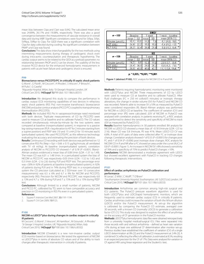
mean bias between CIpa and CIart was 0.042. The calculated mean error
was 24.89%, 26.17% and 19.08%, respectively. There was also a good
convergence between the measurements of vascular resistance in overall
data and during IABP. Signifi cant correlations were found for SVIpa, SVIpi,
SVRIpa, SVRIpi to CIpa. For GEDI there was a signifi cant correlation with
CIpa for data collected during cooling. No signifi cant correlation between
PAWP und CIpa was found.
Conclusions The data show interchangeability for the two methods using
intermitting measurements during therapy of cardiogenic shock even
during intra-aortic counterpulsation and therapeutic hypothermia. The
cardiac output seems to be related to the GEDI as a preload parameter; no
relationship between PAOP and CI can be shown. The usability of the less
invasive PiCCO device for the enhanced hemodynamic measurement in
patients with acute heart failure and cardiogenic shock is ensured.
P99
Bioreactance versus PICCOTD/PC in critically ill septic shock patientsG Monti1, G Pizzilli1, M Cecconi2, A Rhodes2, S Vesconi1, P Brioschi1,
M Pulici1, G Casella1
1Niguarda Hospital, Milan, Italy; 2St George’s Hospital, London, UK
Critical Care 2010, 14(Suppl 1):P99 (doi: 10.1186/cc8331)
Introduction We designed this study to compare the performance in
cardiac output (CO) monitoring capabilities of two devices in refractory
septic shock patients (RSS Pts): non-invasive transthoracic bioreactance
(NICOM) and pulse contour analysis (PICCO PC) coupled to transpulmonary
thermodilution (PICCOTD).
Methods We included RSS Pts in vasopressor/inotrope need monitored
with both devices. Triplicate measurements of CO by PICCOTD were
used to measure CO at baseline and to calibrate PulseCO. The CO values
recorded simultaneously minute-by-minute by the two systems were
compared at baseline (nonperturbated system), in response to a passive
leg-raising maneuver (PLR = leg elevation to 45° for 2 minutes starting from
a supine position) and PEEP test (10 and 15 cmH2O for 10 minutes each)
(perturbated system). We used PICCOTD/PC as the reference technology
evaluating the accuracy and estimating the precision of both devices.
Results Continuous CO recording with both devices was performed on 12
consecutive RSS Pts (Nep + Epi = 0.66 ± 0.15 μg/kg/minute, all ventilated
with TV <8 ml/kg). At baseline (nonperturbated system), correlation
analysis of NICOM vs PiCCOTD CO showed r2 of 0.78 (P <0.001). Bland–
Altman analysis evidenced a mean bias of 0.08 l/min (LOA –1.31 to 1.49).
The mean CO was 6.01 ± 1.48 l/min. In a perturbated system the bias of
NICOM vs PICCO PC was respectively–0.05 l/min (LOA –1.52 to 1.42) and
0.3 l/min (LOA –2 to 2.6) during PLR and PEEP test. The percentage error
was <30% in 92% of patients at baseline (nonperturbated system), in 92%
of patients during PLR and in 74% during PEEP test. In a nonperturbated
system the CO precision (calculated as 2 SD/mean over 10 consecutive
measurements) was 6.5 ± 6% and 6.7 ± 9% for NICOM and PiCCOTD,
respectively (NS). Precision for NICOM and PICCOPC was respectively 6.8
± 13% and 4.7 ± 10% during PLR and 7 ± 15% and 7.6 ± 15% during PEEP
test.
Conclusions Although limited to a small number of patients, NICOM
and PICCO PC, calibrated by TD, seem to have comparable accuracy and
precision in CO monitoring in RSS Pts even in a perturbated system.
References1. Squara P: Intensive Care Med 2007, 33:1191-1194.
2. Squara P: Crit Care 2009, 13:R125.
P100
NICOM vs LiDCO™plus during changes in cardiac output in critically ill patientsM Cecconi1, G Monti2, S Vesconi2, M Hamilton1, M Grounds1, A Rhodes1
1St George ‘s Hospital, London, UK; 2Niguarda Hospital, Milan, Italy
Critical Care 2010, 14(Suppl 1):P100 (doi: 10.1186/cc8332)
Introduction NICOM (Cheetah) is a new non-invasive cardiac output
(CO) monitor based on bioreactance. We tested the agreement of NICOM
vs LiDCO™plus in terms of absolute CO values and of the ability to track
changes after therapeutic intervention in critically ill patients.
Methods Patients requiring haemodynamic monitoring were monitored
with LiDCO™plus and NICOM. Three measurements of CO by LiDCO
were used to measure CO at baseline and to calibrate PulseCO. After
fl uid challenges (FC = 250 ml colloid/5 minutes) or inotropic therapy
alterations, the change in stroke volume (SV) for PulseCO and NICOM CO
was recorded. Patients able to increase SV ≥10% as measured by PulseCO
were considered responders (R). Bland–Altman analysis was performed
for NICOM vs LiDCO CO at baseline. The coeffi cient of variation (CV) and
percentage error (PE) were calculated. ΔSV of NICOM vs PulseCO was
analysed with correlation analysis. In patients receiving FC, a ROC analysis
was performed to detect the sensitivity and specifi city of NICOM to track
ΔSV as measured by PulseCO CO.
Results Baseline haemodynamics in 30 patients enrolled: BA analysis for
NICOM vs LiDCO CO showed a mean bias of –0.18 l/minute (LOA –2.5 to
2.16). Mean CO was 5.8 l/minute, PE was 41%. Mean LiDCO CO CV was
6.8%. A total of 81 pairs of data were collected after FC or inotrope dose
change. Correlation analysis showed r2 of 0.55 (P <0.0001) for changes post
FC and r2 of 0.54 (P <0.006) post inotrope dose changes. ROC analysis for
NICOM CO in R and NR after a FC showed an area under the curve (AUC) of
0.8 (P <0.0001; Figure 1). An increase in NICOM SV >8% showed a sensitivity
of 76% and a specifi city of 75% to predict PulseCO changes >10%.
Conclusions NICOM demonstrated a moderate agreement with LiDCO
but showed excellent agreement with PulseCO in tracking CO changes
following therapeutic interventions.
P101
Eff ect of cardiac arrhythmias on PulseCO calibration and performanceM Jonas1, E Mills2, C Wolff 2, T O’Brien2
1Southampton University Hospital, Southampton, UK; 2LiDCO Ltd, London, UK
Critical Care 2010, 14(Suppl 1):P101 (doi: 10.1186/cc8333)
Introduction Arrhythmias are common among high-risk surgical and
ICU patients. The PulseCO pressure waveform algorithm is used for
both LiDCO™plus and LiDCOrapid hemodynamic monitors, which are
frequently used to estimate cardiac output (CO) in critically ill patients.
Cardiac arrythmias could increase the variation of both the lithium dilution
(LiDCO) and/or the PulseCO measurement. At set-up the algorithm
is calibrated by comparing the PulseCO CO estimate, averaged over
30 seconds, with a known CO (normally LiDCO) to generate a calibration
factor (CF) [1]. This study was designed to explore the eff ect of arrythmias
on the accuracy of CF generation in the PulseCO monitor.
Methods LiDCO™plus hemodynamic data fi les were obtained retro spectively
from a university hospital medical/surgical ICU. Files were separated into
those records with and without arrhythmia – defi ned as heart rate variation
>5% during at least one additional CF determination after monitor set-up.
Previous studies have established the coeffi cient of variation (CV) of a single
LiDCO determination at 8% [2] and the PulseCO measurement at 2.4% [3]. A
combined CV, refl ecting the eff ect of calibration, is estimated at 8.5%, resulting
in an expected precision for the CF of 17%. Data were analysed for variation in
CF against HRV using linear regression and the Student’s t test.
Figure 1 (abstract P100). ROC analysis for NICOM CO in R and NR.
Critical Care 2010, Volume 14 Suppl 1 http://ccforum.com/supplements/14/S1
S35
Results Twenty-eight records were collected and analysed. Twenty-one
records contained 32 post set-up calibration events. Of these 17 occurred
with HRV ≤5% (median = 1%, range: 0 to 5%) and 15 occurred while HRV
>5% (median: 19%, range: 7 to 26%). The average variation in CF during
HRV was 5.4 ± 4.0% and for high HRV was 8.9 ± 8.1% of the initial value.
The t test indicated no diff erence in the mean variation of CF (P = 0.162)
or median. There was no correlation between HRV and CF variation (r2 =
0.002). Ninety-one per cent (29/32) of the observed CF variation were less
than 17% of the initial CF value.
Conclusions CF determinations are not signifi cantly aff ected by HRV.
PulseCO estimates CO acceptably in the presence of arrhythmias.
Interpretation of the data is enhanced by using at least 30 second averaging.
References1. Rhodes A, et al.: Functional Hemodynamic Monitoring Update in Intensive Care
and Emergency Medicine, volume 42, 2005:183-192.
2. Cecconi M, et al.: Intensive Care Med 2009, 35:498-504.
3. Kemps H, et al.: J Appl Physiol 2008, 105:1822-1829.
P102
PulseCO consistency: variation in calibration factor over 24 and 48 hoursE Mills1, M Jonas2, C Wolff 1, T O’Brien1
1LiDCO Ltd, London, UK; 2Southampton University Hospital, Southampton, UK
Critical Care 2010, 14(Suppl 1):P102 (doi: 10.1186/cc8334)
Introduction The PulseCO pressure waveform algorithm is used for both
LiDCO™plus and LiDCOrapid hemodynamic monitors to estimate cardiac
output (CO). The accuracy of this CO estimate is achieved by comparing
(calibrating) the PulseCO with a known CO, for example from the lithium
dilution or LiDCO which scales the software algorithm and reduces the
bias to the actual CO [1]. This calibration/scaling factor (CF) is patient
specifi c and should not signifi cantly change over a period of a few days as
it refl ects the patient’s aortic capacitance.
Methods LiDCO™plus hemodynamic data fi les were obtained
retrospectively from a university hospital medical/surgical ICU. Files
were screened to obtain records with at least 24 hours of continuous
hemodynamic data and at least one calibration at 24 hours following the
initial set-up calibration. Data were analysed for change in CF from initial
calibration over 24 and up to 48 hours. Relative change in CF was analyzed
for correlation with changes in SVR and CO.
Results Twenty-one records contained at least 24 hours of data with a
calibration at 24 hours. Eleven contained at least 48 hours of data and a further
calibration at 48 hours. The 32 calibrations were reviewed for consistency of
PulseCO and LiDCO results. The average variation in CF over 24 hours (7.4 ±
6.9% (mean ± SD)) and 48 hours (6.5 ± 6.2%) were similar (P = 0.362). Average
changes in SVR were 24.7 ± 27.4% (range: 0 to 130%); average CO changed by
21.4 ± 17% (range: 0 to 57%) over 24 hours. There was no correlation between
the variation in CF and changes in either CO or SVR.
Conclusions It is known that the estimated coeffi cient of variation of
a single measurement of LiDCO is 8% [2] and PulseCO is 2.4% [3]. An
orthogonally combined CV, refl ecting the eff ects on CF, is estimated at
8.5%, or precision of 17%. Ninety-one per cent (29/32 ) of the observed
CF changes were less than 17% of the initial CF. The PulseCO CF remains
constant despite changes in SVR of up to 130% and CO of up to 57% over
periods of 24 to 48 hours.
References1. Rhodes A, et al.: Functional Hemodynamic Monitoring Update in Intensive Care
and Emergency Medicine, volume 42, 2005:183-192.
2. Cecconi M, et al.: Intensive Care Med 2009, 35:498-504.
3. Kemps H, et al.: J Appl Physiol 2008, 105:1822-1829.
P103
Performance of cardiac output measurement derived from arterial pressure waveform analysis in patients undergoing triple-H-therapy of cerebral vasospasms after subarachnoidal hemorrhageSM Metzelder, R Kopp, M Fries, M Reinges, S Reich, R Rossaint, G Marx, S Rex
RWTH Aachen, Germany
Critical Care 2010, 14(Suppl 1):P103 (doi: 10.1186/cc8335)
Introduction The validity of an arterial waveform-based device for
measuring cardiac output (CO) without the need for invasive calibration
(FloTrac/Vigileo) in patients needing large doses of vasoactive medication
has not yet been thoroughly studied. We performed the present study
to assess the validity of both the second-generation and the third-
generation software compared with transpulmonary thermodilution
CO measurement using the PiCCO technology in patients undergoing
triple-H-therapy (hypertonia, hypervolemia, hemodilution) of cerebral
vasospasms after subarachnoidal hemorrhage.
Methods Twenty-three patients (18 females and fi ve males) were
included in this study. All of them were suff ering from a subarachnoidal
hemorrhage (Hunt&Hess grade I to V) due to rupture of a cerebral
aneurysm. Triple-H-therapy was initiated for the treatment of cerebral
vasospasm. Simultaneous CO measurements by bolus thermodilution and
the FloTrac/Vigileo device were obtained at baseline as well as 2 hours,
6 hours, 12 hours, 24 hours, 48 hours and 72 hours after inclusion. A
percentage error of 30% or less was established as the criterion for method
interchangeability.
Results Patients received vasoactive support with 0.53 ± 0.46 μg/kg/
minute norepinephrine, resulting in a mean arterial pressure of 104 ±
13.6 mmHg and a systemic vascular resistance index of 1,741.17 ± 432.50
dyn·s/cm5/m2. One hundred and fi fty-one CO-data pairs were analyzed.
Transpulmonary thermodilution CO ranged from 5.18 to 14.28 l/minute
(mean 8.61 ± 1.93 l/minute) and FloTrac/Vigileo CO ranged from 4.1 to
13.7 l/minute (mean 7.62 ± 1.79 l/minute). Bias and precision (1.96SD of
the bias) were 0.99 l/minute and 2.46 l/minute, resulting in an overall
percentage error of 28.55%. Subgroup analysis revealed a percentage error
of 29.53% for 67 data pairs measured using the second-generation FloTrac
software and 26.44% for 84 data pairs analyzed by the third-generation
software.
Conclusions In patients undergoing triple-H-therapy and needing
extensive vasoactive support, CO values obtained by arterial waveform
analysis showed good agreement with intermittent transpulmonary
thermodilution CO measurements, which was improved by the intro-
duction of a new software generation.
References
1. Mayer J, Boldt J, Wolf MW, Lang J, Suttner S: Cardiac output derived from arterial pressure waveform analysis in patients undergoing cardiac surgery: validity of a second generation device. Anesth Analg 2008,
106:867-872, table.
P104
Infl uence of systemic vascular resistance on cardiac output measured by new non-invasive cardiac output monitorK Yamashita, T Yamatabe, H Abe, M Yokoyama
Kochi Medical School, Kochi, Japan
Critical Care 2010, 14(Suppl 1):P104 (doi: 10.1186/cc8336)
Introduction Early manipulation of hemodynamic variables has
been reported to be able to improve the outcome of patients. Cardiac
output (CO) was an important parameter to understand the status of
hemodynamics. Recently, the pulse-contour method is widely used to
estimate CO, because it is non-invasive and easy to use. However, the
change in the systemic vascular resistance (SVR) aff ects CO measured
by the pulse-contour method. In this study, we evaluated the new non-
invasive cardiac output monitor [1] based on pulse pressure analysis
combined with pulse-wave transition time (estimated continuous CO;
esCCO) compared with the conventional thermodilution method under
a clinical setting.
Methods Twenty-fi ve surgical patients who underwent cardiac or
vascular surgery (ASA physical status 2) were enrolled in this study.
After anesthesia induction, radial arterial catheter and pulmonary artery
catheter were inserted. Intermittent cardiac output (ICO) was measured
by thermodilution method in triplicate and averaged (<5°C saline 10 ml
in each measurement) using Vigilance (Edwards Life Science, Irvine, CA,
USA). Echocardiogram, pulse oximetry and arterial blood pressure were
also monitored and connected to personal computer to calculate esCCO.
Bland and Altman plots were used to evaluate the percentage diff erence
in CO in relation to SVR.
Results One hundred matched sets of data were obtained. The limit of
agreement (bias ± 2SD of bias) was –2.9 ± 31.9%. Only fi ve measurements
were exceeded – 30% against ICO. Each SVR was 1,159, 1,376, 1,418, 2,567,
3,150 dyn·s/m5.
Critical Care 2010, Volume 14 Suppl 1 http://ccforum.com/supplements/14/S1
S36
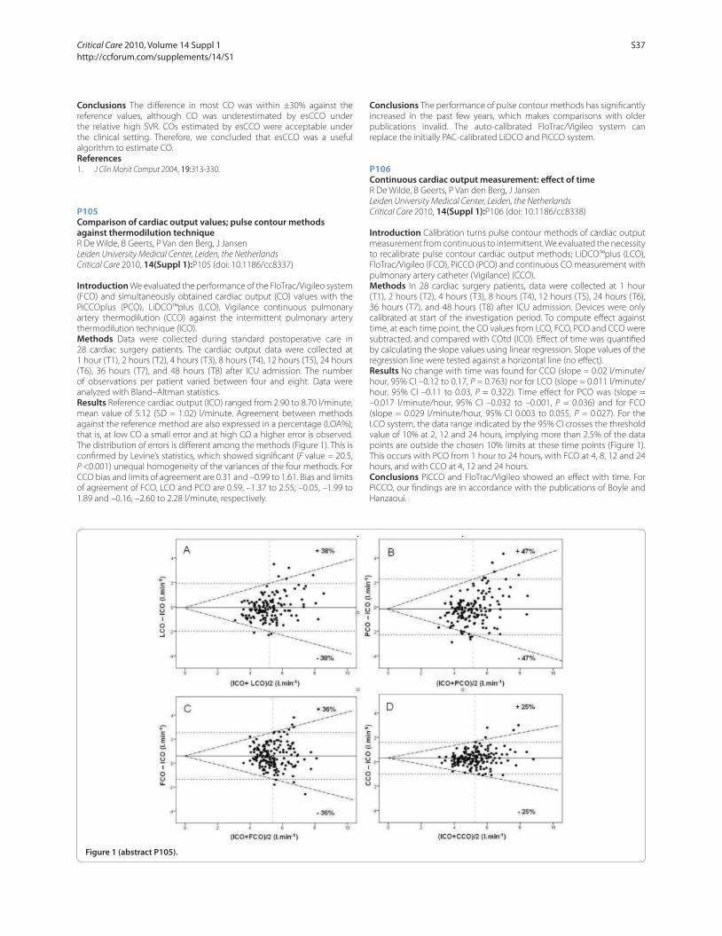
Conclusions The diff erence in most CO was within ±30% against the
reference values, although CO was underestimated by esCCO under
the relative high SVR. COs estimated by esCCO were acceptable under
the clinical setting. Therefore, we concluded that esCCO was a useful
algorithm to estimate CO.
References1. J Clin Monit Comput 2004, 19:313-330.
P105
Comparison of cardiac output values; pulse contour methods against thermodilution techniqueR De Wilde, B Geerts, P Van den Berg, J Jansen
Leiden University Medical Center, Leiden, the Netherlands
Critical Care 2010, 14(Suppl 1):P105 (doi: 10.1186/cc8337)
Introduction We evaluated the performance of the FloTrac/Vigileo system
(FCO) and simultaneously obtained cardiac output (CO) values with the
PiCCOplus (PCO), LiDCO™plus (LCO), Vigilance continuous pulmonary
artery thermodilution (CCO) against the intermittent pulmonary artery
thermodilution technique (ICO).
Methods Data were collected during standard postoperative care in
28 cardiac surgery patients. The cardiac output data were collected at
1 hour (T1), 2 hours (T2), 4 hours (T3), 8 hours (T4), 12 hours (T5), 24 hours
(T6), 36 hours (T7), and 48 hours (T8) after ICU admission. The number
of observations per patient varied between four and eight. Data were
analyzed with Bland–Altman statistics.
Results Reference cardiac output (ICO) ranged from 2.90 to 8.70 l/minute,
mean value of 5.12 (SD = 1.02) l/minute. Agreement between methods
against the reference method are also expressed in a percentage (LOA%);
that is, at low CO a small error and at high CO a higher error is observed.
The distribution of errors is diff erent among the methods (Figure 1). This is
confi rmed by Levine’s statistics, which showed signifi cant (F value = 20.5,
P <0.001) unequal homogeneity of the variances of the four methods. For
CCO bias and limits of agreement are 0.31 and –0.99 to 1.61. Bias and limits
of agreement of FCO, LCO and PCO are 0.59, –1.37 to 2.55; –0.05, –1.99 to
1.89 and –0.16, –2.60 to 2.28 l/minute, respectively.
Conclusions The performance of pulse contour methods has signifi cantly
increased in the past few years, which makes comparisons with older
publications invalid. The auto-calibrated FloTrac/Vigileo system can
replace the initially PAC-calibrated LiDCO and PiCCO system.
P106
Continuous cardiac output measurement: eff ect of timeR De Wilde, B Geerts, P Van den Berg, J Jansen
Leiden University Medical Center, Leiden, the Netherlands
Critical Care 2010, 14(Suppl 1):P106 (doi: 10.1186/cc8338)
Introduction Calibration turns pulse contour methods of cardiac output
measurement from continuous to intermittent. We evaluated the necessity
to recalibrate pulse contour cardiac output methods; LiDCO™plus (LCO),
FloTrac/Vigileo (FCO), PiCCO (PCO) and continuous CO measurement with
pulmonary artery catheter (Vigilance) (CCO).
Methods In 28 cardiac surgery patients, data were collected at 1 hour
(T1), 2 hours (T2), 4 hours (T3), 8 hours (T4), 12 hours (T5), 24 hours (T6),
36 hours (T7), and 48 hours (T8) after ICU admission. Devices were only
calibrated at start of the investigation period. To compute eff ect against
time, at each time point, the CO values from LCO, FCO, PCO and CCO were
subtracted, and compared with COtd (ICO). Eff ect of time was quantifi ed
by calculating the slope values using linear regression. Slope values of the
regression line were tested against a horizontal line (no eff ect).
Results No change with time was found for CCO (slope = 0.02 l/minute/
hour, 95% CI –0.12 to 0.17, P = 0.763) nor for LCO (slope = 0.011 l/minute/
hour, 95% CI –0.11 to 0.03, P = 0.322). Time eff ect for PCO was (slope =
–0.017 l/minute/hour, 95% CI –0.032 to –0.001, P = 0.036) and for FCO
(slope = 0.029 l/minute/hour, 95% CI 0.003 to 0.055, P = 0.027). For the
LCO system, the data range indicated by the 95% CI crosses the threshold
value of 10% at 2, 12 and 24 hours, implying more than 2.5% of the data
points are outside the chosen 10% limits at these time points (Figure 1).
This occurs with PCO from 1 hour to 24 hours, with FCO at 4, 8, 12 and 24
hours, and with CCO at 4, 12 and 24 hours.
Conclusions PiCCO and FloTrac/Vigileo showed an eff ect with time. For
PiCCO, our fi ndings are in accordance with the publications of Boyle and
Hanzaoui.
Figure 1 (abstract P105).
Critical Care 2010, Volume 14 Suppl 1 http://ccforum.com/supplements/14/S1
S37
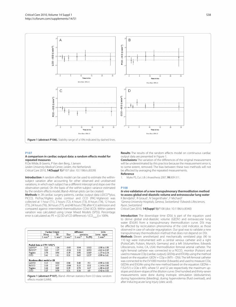
P107
A comparison in cardiac output data: a random eff ects model for repeated measuresR De Wilde, B Geerts, P Van den Berg, J Jansen
Leiden University Medical Center, Leiden, the Netherlands
Critical Care 2010, 14(Suppl 1):P107 (doi: 10.1186/cc8339)
Introduction A random eff ects model can be used to estimate the within-
subject variation after accounting for other observed and unobserved
variations, in which each subject has a diff erent intercept and slope over the
observation period. On the basis of the within-subject variance estimated
by the random eff ects model, Bland–Altman plots can be created.
Methods In 28 cardiac surgery patients, cardiac output data LiDCO™plus,
PICCO, FloTrac/Vigileo pulse contour and CCO (PAC-Vigilance) was
collected at 1 hour (T1), 2 hours (T2), 4 hours (T3), 8 hours (T4), 12 hours
(T5), 24 hours (T6), 36 hours (T7), and 48 hours (T8) after ICU admission and
compared against intermitted thermodilution COtd (ICO). Within patient
variation was calculated using Linear Mixed Models (SPSS). Percentage
error is calculated as: PE = [(2.SD of CO diff erence) / (COmean
)] x 100%.
Results The results of the random eff ects model on continuous cardiac
output data are presented in Figure 1.
Conclusions The variation of the diff erences of the original measurement
will be underestimated by this practice because the measurement error is,
to some extent, removed. The bias between these two methods will not
be aff ected by averaging the repeated measurements.
Reference1. Myles PS, Cui J: Br J Anaesthesia 2007, 99:309-311.
P108
In vivo validation of a new transpulmonary thermodilution method to assess global end-diastolic volume and extravascular lung water
K Bendjelid1, R Giraud1, N Siegenthaler1, F Michard2
1Geneva University Hospitals, Geneva, Switzerland; 2Edwards Lifesciences,
Nyon, Switzerland
Critical Care 2010, 14(Suppl 1):P108 (doi: 10.1186/cc8340)
Introduction The downslope time (DSt) is part of the equation used
to derive global end-diastolic volume (GEDV) and extravascular lung
water (EVLW) from a transpulmonary thermodilution curve. DSt may
be aff ected by recirculation phenomena of the cold indicator, as those
observed in case of valvular regurgitation. Our goal was to validate a new
transpulmonary thermodilution method that does not depend on DSt.
Methods Eleven anesthetized and mechanically ventilated pigs (90 to
110 kg) were instrumented with a central venous catheter and a right
(PulsioCath; Pulsion, Munich, Germany) and a left (VolumeView; Edwards
Lifesciences, Irvine, CA, USA) thermodilution femoral arterial catheter. The
right femoral catheter was connected to a PiCCO2 monitor (Pulsion) and
used to measure COp (cardiac output), GEDVp and EVLWp using the method
based on the equation: GEDV = COp x (MTt – DSt). The left femoral catheter
was connected to the EV1000 monitor (Edwards) and used to measure COe,
GEDVe and EVLWe using the new method based on the equation: GEDVe =
f (S2/S1) x COe x MTt, where S1 and S2 are respectively the maximum up-
slopes and down-slopes of the dilution curve. One hundred and thirty-seven
measurements were done during inotropic stimulation (dobutamine),
during hypovolemia (bleeding), during hypervolemia (fl uid overload), and
after inducing acute lung injury (oleic acid).
Figure 1 (abstract P106). Stability range of ±10% indicated by dashed lines.
Figure 1 (abstract P107). Bland–Altman statistics from CO data: random
eff ects model (LMM).
Critical Care 2010, Volume 14 Suppl 1 http://ccforum.com/supplements/14/S1
S38
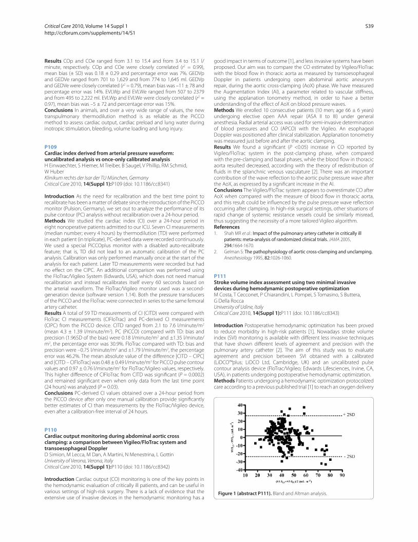
Results COp and COe ranged from 3.1 to 15.4 and from 3.4 to 15.1 l/
minute, respectively. COp and COe were closely correlated (r2 = 0.99),
mean bias (± SD) was 0.18 ± 0.29 and percentage error was 7%. GEDVp
and GEDVe ranged from 701 to 1,629 and from 774 to 1,645 ml. GEDVp
and GEDVe were closely correlated (r2 = 0.79), mean bias was –11 ± 78 and
percentage error was 14%. EVLWp and EVLWe ranged from 507 to 2379
and from 495 to 2,222 ml. EVLWp and EVLWe were closely correlated (r2 =
0.97), mean bias was –5 ± 72 and percentage error was 15%.
Conclusions In animals, and over a very wide range of values, the new
transpulmonary thermodilution method is as reliable as the PiCCO
method to assess cardiac output, cardiac preload and lung water during
inotropic stimulation, bleeding, volume loading and lung injury.
P109
Cardiac index derived from arterial pressure waveform: uncalibrated analysis vs once-only calibrated analysisH Einwaechter, S Hiemer, M Treiber, B Saugel, V Phillip, RM Schmid,
W Huber
Klinikum rechts der Isar der TU München, Germany
Critical Care 2010, 14(Suppl 1):P109 (doi: 10.1186/cc8341)
Introduction As the need for recalibration and the best time point to
recalibrate has been a matter of debate since the introduction of the PiCCO
monitor (Pulsion, Germany), we set out to analyze the performance of its
pulse contour (PC) analysis without recalibration over a 24-hour period.
Methods We studied the cardiac index (CI) over a 24-hour period in
eight nonoperative patients admitted to our ICU. Seven CI measurements
(median number; every 4 hours) by thermodilution (TD) were performed
in each patient (in triplicate), PC-derived data were recorded continuously.
We used a special PiCCOplus monitor with a disabled auto-recalibrate
feature; that is, TD did not lead to an automatic calibration of the PC
analysis. Calibration was only performed manually once at the start of the
analysis for each patient. Later TD measurements were recorded but had
no eff ect on the CIPC. An additional comparison was performed using
the FloTrac/Vigileo System (Edwards, USA), which does not need manual
recalibration and instead recalibrates itself every 60 seconds based on
the arterial waveform. The FloTrac/Vigileo monitor used was a second-
generation device (software version 1.14). Both the pressure transducers
of the PiCCO and the FloTrac were connected in series to the same femoral
artery catheter.
Results A total of 59 TD measurements of CI (CITD) were compared with
FloTrac CI measurements (CIFloTrac) and PC-derived CI measurements
(CIPC) from the PiCCO device. CITD ranged from 2.1 to 7.6 l/minute/m2
(mean 4.3 ± 1.39 l/minute/m2). PC (PiCCO) compared with TD: bias and
precision (1.96SD of the bias) were 0.18 l/minute/m2 and ±1.35 l/minute/
m2, the percentage error was 30.9%. FloTrac compared with TD: bias and
precision were –0.75 l/minute/m2 and ±1.79 l/minute/m2, the percentage
error was 46.2%. The mean absolute value of the diff erence |CITD – CIPC|
and |CITD – CIFloTrac| was 0.48 ± 0.49 l/minute/m2 for PiCCO pulse contour
values and 0.97 ± 0.76 l/minute/m2 for FloTrac/Vigileo values, respectively.
This higher diff erence of CIFloTrac from CITD was signifi cant (P = 0.0002)
and remained signifi cant even when only data from the last time point
(24 hours) was analyzed (P = 0.03).
Conclusions PC-derived CI values obtained over a 24-hour period from
the PiCCO device after only one manual calibration provide signifi cantly
better estimates of CI than measurements by the FloTrac/Vigileo device,
even after a calibration-free interval of 24 hours.
P110
Cardiac output monitoring during abdominal aortic cross clamping: a comparison between Vigileo/FloTrac system and transoesophageal DopplerD Simion, M Lecca, M Dan, A Martini, N Menestrina, L Gottin
University of Verona, Verona, Italy
Critical Care 2010, 14(Suppl 1):P110 (doi: 10.1186/cc8342)
Introduction Cardiac output (CO) monitoring is one of the key points in
the hemodynamic evaluation of critically ill patients, and can be useful in
various settings of high-risk surgery. There is a lack of evidence that the
extensive use of invasive devices in the hemodynamic monitoring has a
good impact in terms of outcome [1], and less invasive systems have been
proposed. Our aim was to compare the CO estimated by Vigileo/FloTrac
with the blood fl ow in thoracic aorta as measured by transoesophageal
Doppler in patients undergoing open abdominal aortic aneurysm
repair, during the aortic cross-clamping (AoX) phase. We have measured
the Augmentation Index (AI), a parameter related to vascular stiff ness,
using the applanation tonometry method, in order to have a better
understanding of the eff ect of AoX on blood pressure waves.
Methods We enrolled 10 consecutive patients (10 men; age 66 ± 6 years)
undergoing elective open AAA repair (ASA II to III) under general
anesthesia. Radial arterial access was used for semi-invasive determination
of blood pressures and CO (APCO) with the Vigileo. An esophageal
Doppler was positioned after clinical stabilization. Applanation tonometry
was measured just before and after the aortic clamping.
Results We found a signifi cant (P <0.05) increase in CO reported by
Vigileo/FloTrac system in the post-clamping phase, when compared
with the pre-clamping and basal phases, while the blood fl ow in thoracic
aorta resulted decreased, according with the theory of redistribution of
fl uids in the splanchnic venous vasculature [2]. There was an important
contribution of the wave refl ection to the aortic pulse pressure wave after
the AoX, as expressed by a signifi cant increase in the AI.
Conclusions The Vigileo/FloTrac system appears to overestimate CO after
AoX when compared with the measure of blood fl ow in thoracic aorta,
and this result could be infl uenced by the pulse pressure wave refl ection
occurring after clamping. In high-risk surgical settings, other situations of
rapid change of systemic resistance vessels could be similarly misread,
thus suggesting the necessity of a more tailored Vigileo algorithm.
References
1. Shah MR et al.: Impact of the pulmonary artery catheter in critically ill patients: meta-analysis of randomized clinical trials. JAMA 2005,
294:1664-1670.
2. Gelman S: The pathophysiology of aortic cross-clamping and unclamping. Anesthesiology 1995, 82:1026-1060.
P111
Stroke volume index assessment using two minimal invasive devices during hemodynamic postoperative optimizationM Costa, T Cecconet, P Chiarandini, L Pompei, S Tomasino, S Buttera,
G Della Rocca
University of Udine, Italy
Critical Care 2010, 14(Suppl 1):P111 (doi: 10.1186/cc8343)
Introduction Postoperative hemodynamic optimization has been proved
to reduce morbidity in high-risk patients [1]. Nowadays stroke volume
index (SVI) monitoring is available with diff erent less invasive techniques
that have shown diff erent levels of agreement and precision with the
pulmonary artery catheter [2]. The aim of this study was to evaluate
agreement and precision between SVI obtained with a calibrated
(LiDCO™plus; LiDCO Ltd, Cambridge, UK) and an uncalibrated pulse
contour analysis device (FloTrac/Vigileo; Edwards Lifesciences, Irvine, CA,
USA), in patients undergoing postoperative hemodynamic optimization.
Methods Patients undergoing a hemodynamic optimization protocolized
care according to a previous published trial [1] to reach an oxygen delivery
Figure 1 (abstract P111). Bland and Altman analysis.
Critical Care 2010, Volume 14 Suppl 1 http://ccforum.com/supplements/14/S1
S39
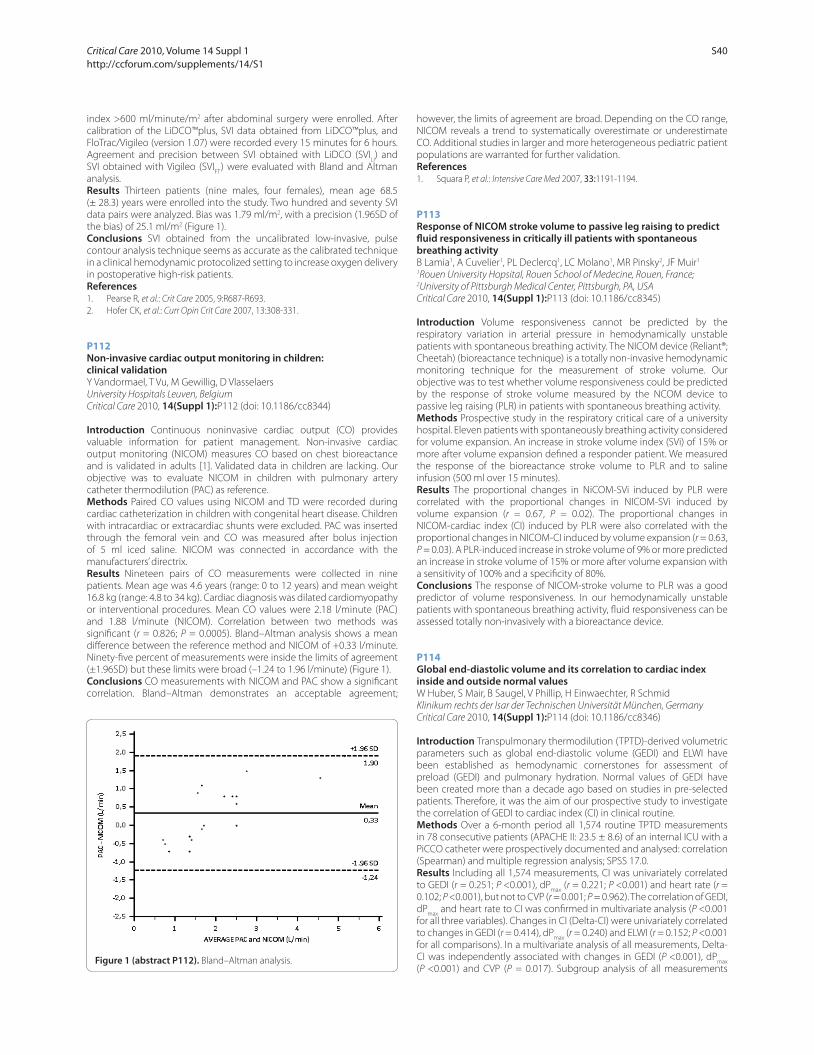
index >600 ml/minute/m2 after abdominal surgery were enrolled. After
calibration of the LiDCO™plus, SVI data obtained from LiDCO™plus, and
FloTrac/Vigileo (version 1.07) were recorded every 15 minutes for 6 hours.
Agreement and precision between SVI obtained with LiDCO (SVILI) and
SVI obtained with Vigileo (SVIFT
) were evaluated with Bland and Altman
analysis.
Results Thirteen patients (nine males, four females), mean age 68.5
(± 28.3) years were enrolled into the study. Two hundred and seventy SVI
data pairs were analyzed. Bias was 1.79 ml/m2, with a precision (1.96SD of
the bias) of 25.1 ml/m2 (Figure 1).
Conclusions SVI obtained from the uncalibrated low-invasive, pulse
contour analysis technique seems as accurate as the calibrated technique
in a clinical hemodynamic protocolized setting to increase oxygen delivery
in postoperative high-risk patients.
References1. Pearse R, et al.: Crit Care 2005, 9:R687-R693.
2. Hofer CK, et al.: Curr Opin Crit Care 2007, 13:308-331.
P112
Non-invasive cardiac output monitoring in children: clinical validationY Vandormael, T Vu, M Gewillig, D Vlasselaers
University Hospitals Leuven, Belgium
Critical Care 2010, 14(Suppl 1):P112 (doi: 10.1186/cc8344)
Introduction Continuous noninvasive cardiac output (CO) provides
valuable information for patient management. Non-invasive cardiac
output monitoring (NICOM) measures CO based on chest bioreactance
and is validated in adults [1]. Validated data in children are lacking. Our
objective was to evaluate NICOM in children with pulmonary artery
catheter thermodilution (PAC) as reference.
Methods Paired CO values using NICOM and TD were recorded during
cardiac catheterization in children with congenital heart disease. Children
with intracardiac or extracardiac shunts were excluded. PAC was inserted
through the femoral vein and CO was measured after bolus injection
of 5 ml iced saline. NICOM was connected in accordance with the
manufacturers’ directrix.
Results Nineteen pairs of CO measurements were collected in nine
patients. Mean age was 4.6 years (range: 0 to 12 years) and mean weight
16.8 kg (range: 4.8 to 34 kg). Cardiac diagnosis was dilated cardiomyopathy
or interventional procedures. Mean CO values were 2.18 l/minute (PAC)
and 1.88 l/minute (NICOM). Correlation between two methods was
signifi cant (r = 0.826; P = 0.0005). Bland–Altman analysis shows a mean
diff erence between the reference method and NICOM of +0.33 l/minute.
Ninety-fi ve percent of measurements were inside the limits of agreement
(±1.96SD) but these limits were broad (–1.24 to 1.96 l/minute) (Figure 1).
Conclusions CO measurements with NICOM and PAC show a signifi cant
correlation. Bland–Altman demonstrates an acceptable agreement;
however, the limits of agreement are broad. Depending on the CO range,
NICOM reveals a trend to systematically overestimate or underestimate
CO. Additional studies in larger and more heterogeneous pediatric patient
populations are warranted for further validation.
References1. Squara P, et al.: Intensive Care Med 2007, 33:1191-1194.
P113
Response of NICOM stroke volume to passive leg raising to predict fl uid responsiveness in critically ill patients with spontaneous breathing activityB Lamia1, A Cuvelier1, PL Declercq1, LC Molano1, MR Pinsky2, JF Muir1
1Rouen University Hopsital, Rouen School of Medecine, Rouen, France; 2University of Pittsburgh Medical Center, Pittsburgh, PA, USA
Critical Care 2010, 14(Suppl 1):P113 (doi: 10.1186/cc8345)
Introduction Volume responsiveness cannot be predicted by the
respiratory variation in arterial pressure in hemodynamically unstable
patients with spontaneous breathing activity. The NICOM device (Reliant®;
Cheetah) (bioreactance technique) is a totally non-invasive hemodynamic
monitoring technique for the measurement of stroke volume. Our
objective was to test whether volume responsiveness could be predicted
by the response of stroke volume measured by the NCOM device to
passive leg raising (PLR) in patients with spontaneous breathing activity.
Methods Prospective study in the respiratory critical care of a university
hospital. Eleven patients with spontaneously breathing activity considered
for volume expansion. An increase in stroke volume index (SVi) of 15% or
more after volume expansion defi ned a responder patient. We measured
the response of the bioreactance stroke volume to PLR and to saline
infusion (500 ml over 15 minutes).
Results The proportional changes in NiCOM-SVi induced by PLR were
correlated with the proportional changes in NICOM-SVi induced by
volume expansion (r = 0.67, P = 0.02). The proportional changes in
NICOM-cardiac index (CI) induced by PLR were also correlated with the
proportional changes in NICOM-CI induced by volume expansion (r = 0.63,
P = 0.03). A PLR-induced increase in stroke volume of 9% or more predicted
an increase in stroke volume of 15% or more after volume expansion with
a sensitivity of 100% and a specifi city of 80%.
Conclusions The response of NICOM-stroke volume to PLR was a good
predictor of volume responsiveness. In our hemodynamically unstable
patients with spontaneous breathing activity, fl uid responsiveness can be
assessed totally non-invasively with a bioreactance device.
P114
Global end-diastolic volume and its correlation to cardiac index
inside and outside normal valuesW Huber, S Mair, B Saugel, V Phillip, H Einwaechter, R Schmid
Klinikum rechts der Isar der Technischen Universität München, Germany
Critical Care 2010, 14(Suppl 1):P114 (doi: 10.1186/cc8346)
Introduction Transpulmonary thermodilution (TPTD)-derived volumetric
parameters such as global end-diastolic volume (GEDI) and ELWI have
been established as hemodynamic cornerstones for assessment of
preload (GEDI) and pulmonary hydration. Normal values of GEDI have
been created more than a decade ago based on studies in pre-selected
patients. Therefore, it was the aim of our prospective study to investigate
the correlation of GEDI to cardiac index (CI) in clinical routine.
Methods Over a 6-month period all 1,574 routine TPTD measurements
in 78 consecutive patients (APACHE II: 23.5 ± 8.6) of an internal ICU with a
PiCCO catheter were prospectively documented and analysed: correlation
(Spearman) and multiple regression analysis; SPSS 17.0.
Results Including all 1,574 measurements, CI was univariately correlated
to GEDI (r = 0.251; P <0.001), dPmax
(r = 0.221; P <0.001) and heart rate (r =
0.102; P <0.001), but not to CVP (r = 0.001; P = 0.962). The correlation of GEDI,
dPmax
and heart rate to CI was confi rmed in multivariate analysis (P <0.001
for all three variables). Changes in CI (Delta-CI) were univariately correlated
to changes in GEDI (r = 0.414), dPmax
(r = 0.240) and ELWI (r = 0.152; P <0.001
for all comparisons). In a multivariate analysis of all measurements, Delta-
CI was independently associated with changes in GEDI (P <0.001), dPmax
(P <0.001) and CVP (P = 0.017). Subgroup analysis of all measurements Figure 1 (abstract P112). Bland–Altman analysis.
Critical Care 2010, Volume 14 Suppl 1 http://ccforum.com/supplements/14/S1
S40

with GEDI below the lower normal level 680 ml/kg/m2 demonstrated an
independent association of CI to GEDI (P <0.001), dPmax
(P <0.001) and ELWI
(P = 0.041) but not to CVP. Similarly, Delta-CI was independently associated
with changes in GEDI and dPmax
(P <0.001). Similar results were found for
the measurements with GEDI within the normal range (680 to 800 ml/kg/
m2): signifi cant and independent correlation of CI to GEDI (P <0.017) and
dPmax
(P <0.001). Changes in CI were independently correlated to changes
in GEDI (P <0.001), dPmax
(P <0.001) and CVP (P = 0.035). Interestingly,
even in measurements with GEDI >800 ml/kg/m2, CI was independently
correlated to GEDI (P = 0.009) and dPmax
(P <0.001). Changes in CI in this
group were independently associated with changes in dPmax
and GEDI
(P <0.001).In the subgroup of measurements with GEDI >1,000 ml/kg/m2
there was no correlation of any parameter to CI, however changes in CI
were independently correlated to changes in GEDI (P <0.001) and dPmax
(P = 0.003).
Conclusions GEDI and dPmax
and their changes have an independent and
positive correlation to CI and its changes even in patients with increased
GEDI.
P115
Relationship of stroke volume variation, pulse pressure variation and global end-diastolic volume in patients undergoing brain surgeryA Rieß1, S Wolf2, C Lumenta2, L Schürer2, P Friederich1
1Klinikum Bogenhausen, Department of Anesthesiology, München, Germany; 2Department of Neurosurgery, München, Germany
Critical Care 2010, 14(Suppl 1):P115 (doi: 10.1186/cc8347)
Introduction Monitoring intravascular volume in patients with intra-
cranial pathology is often mandatory for maintaining hemodynamic
stability [1,2]. Cyclic changes in cardiac stroke volume [1,3] and pulse
pressure induced by positive pressure ventilation as well as target values
of global end-diastolic volume index (GEDVI) (ml/m2) [2] allow volume
therapy guidance. The relationship between stroke volume variation (SVV)
(%) and pulse pressure variation (PPV) (%), as well as between SVV or PPV
and values of GEDVI has not been established in patients with intracranial
pathology.
Methods In this prospective investigation the correlation between
dynamic and static hemodynamic parameters of 38 patients undergoing
brain surgery was studied. Measurements were performed using the
PiCCO technology. For statistical analysis, nonparametric correlation
analysis and hypothesis testing were applied.
Results SVV correlated signifi cantly with PPV (r2 = 0.87, P <0.001). Neither
SVV (r2 = 0.14, P = 0.13) nor PPV (r
2 = 0.07, P = 0.81) correlated with GEDVI.
Threshold values for SVV (9.5%, 11.6%) as well as for PPV (12.5%) allowed
discrimination between groups with signifi cantly diff erent values of
stroke volume index, while failing to discriminate between groups with
signifi cantly diff erent values of GEDVI. Dichotomizing the patients into
groups of GEDVI ≤680 ml/m2 and >680 ml/m2 resulted in groups with
signifi cantly diff erent values of stroke volume index as well while failing
to discriminate between groups with signifi cantly diff erent values of SVV
and PPV.
Conclusions Static (GEDVI) and dynamic (SVV, PPV) parameters of cardiac
preload may refl ect diff erent properties of the cardiovascular system. The
combination of SVV, PPV, and GEDVI may off er more precise information
on the cardiovascular system than either parameter alone.
References1. Berkenstadt H, et al.: Anesth Analg 2001, 92:984-989.
2. Mutoh T, et al.: Stroke 2009, 40:2368-2374.
3. Marik PE, et al.: Crit Care Med 2009, 37:2642-2647.
P116
The value of pulse pressure variation to predict volume response in patients ventilated with low VTGF Friedman, CD Costa, SR Vieira, L Fialkow
Hospital de Clínicas de Porto Alegre, Brazil
Critical Care 2010, 14(Suppl 1):P116 (doi: 10.1186/cc8348)
Introduction The prediction value of pulse pressure variation (ΔPP) in
patients ventilated with low VT is not well studied. A ΔPP of 12 to 13%
is validated as a predictor of volume response in several studies, but in
patients ventilated with VT >8 ml/kg. One study has shown that the ΔPP
of 12 to 13% does not predict volume response in patients ventilated with
a low VT. We hypothesized that a lower cut-off value for ΔPP can predict
volume response in patients with low VT.
Methods Thirty-seven adult patients mechanically ventilated with a tidal
volume <8 ml/kg (PBW), without cardiac arrhythmias, with a pulmonary
artery catheter and a peripheral arterial catheter were included. An
increase in cardiac index (thermodilution)) >15% output after a fl uid
challenge (Crystalloid 1,000 ml or Colloid 500 ml) was considered a
positive response.
Results Seventeen patients were responders. The ROC curve showed that
the best cut-off value for ΔPP was 10% (ROC area = 0.74, 95% CI: 0.51 to 0.9;
sensitivity 53%, specifi city 95%, positive likelihood ratio 9.4 and negative
0.34).Twelve patients consisted of a heterogeneous group of patients
(liver transplant, acute pancreatitis, aortic surgery). Among 25 septic
shock patients, a ΔPP >10% showed a ROC area of 0.84 (sensitivity 78%,
specifi city 93%). In any case, the greater ΔPP, the greater the fl uid response.
ΔPP >10% was a better predictor than CVP or PAOP.
Conclusions ΔPP has a limited value in patients ventilated with low VT.
However, a ΔPP >10% may help identify septic shock patients that will
respond to a fl uid challenge.
References
1. Vincent JL, et al.: Pulse pressure variations to predict fl uid responsiveness: Infl uence of tidal volume. Intensive Care Med 2005, 31:517-523.
P117
Pre-ejection period is a reliable parameter to estimate cardiac preload in a hemorrhagic shockR Giraud, N Siegenthaler, JA Romand, K Bendjelid
Hôpitaux Universitaires de Genève, Switzerland
Critical Care 2010, 14(Suppl 1):P117 (doi: 10.1186/cc8349)
Introduction We have already demonstrated that in mechanically
ventilated patients, the respiratory change in the pre-ejection period
(ΔPEP) is a reliable dynamic index for the prediction of increase in cardiac
output after volume infusion [1]. However, in an animal study, Kubitz and
colleagues showed that the pre-ejection period is not sensitive to the
changes in intravascular volume status [2].
Methods This study investigated the infl uence of changes in intravascular
volume status on ΔPEP. In 17 pigs, ECG, arterial pressure and cardiac output
derived from a Swan–Ganz catheter were recorded. Measurements were
performed during normovolaemic conditions, after haemorrhage (25 ml/
kg) and following re-transfusion (25 ml/kg) with constant tidal volume
(10 ml/kg) and respiration rate (15/minute).
Figure 1 (abstract P117). Box plot representing pulse pressure variations
(PPV) (%) and pre-ejection period variations (ΔPEP) (%) at the three times
of the protocol. §P <0.05, †P <0.05.
Critical Care 2010, Volume 14 Suppl 1 http://ccforum.com/supplements/14/S1
S41
Results At baseline, respiratory changes in pulse pressure (PPV) and ΔPEP
were both <12%. PPV signifi cantly correlated with ΔPEP (r = 0.96, P <0.001).
Volume loss induced by haemorrhage increased PPV and ΔPEP. Moreover,
during this state, PPV correlated with ΔPEP with a signifi cant correlation
coeffi cient (r = 0.88, P <0.001). Retransfusion signifi cantly decreased PPV
and ΔPEP and PPV signifi cantly correlated with ΔPEP (r = 0.94, P <0.001)
(Figure 1).
Conclusions Available correlations between PPV and ΔPEP at each time
of the study were observed, meaning that ΔPEP is a reliable parameter to
estimate the changes in intravascular volume status.
References1. Bendjelid K, Suter PM, Romand JA: The respiratory change in preejection
period: a new method to predict fl uid responsiveness. J Appl Physiol 2004,
96:337-342.
2. Kubitz JC, Kemming GI, Schultheib G, Starke J, Podtschaske A, Goetz AE,
Reuter DA: The infl uence of cardiac preload and positive end-expiratory pressure on the pre-ejection period. Physiol Meas 2005, 26:1033-1038
P118
Predictive value of pulse pressure variation for fl uid responsiveness after maneuver to change tidal volume in patients with protective ventilatory strategyF Machado, F Freitas, M Assuncao, B Mazza, M Jackiu
Federal University of São Paulo, Brazil
Critical Care 2010, 14(Suppl 1):P118 (doi: 10.1186/cc8350)
Introduction After the early phase of sepsis, excessive fl uid administration
may worsen pulmonary edema and prolong mechanical ventilation [1].
Accurately predicting fl uid responsiveness obviates unnecessary fl uid
loading, and helps to detect patients who may benefi t from a volume
expansion. Pulse pressure variation (DPP) is a reliable predictor of fl uid
responsiveness in mechanically ventilated patients only when tidal volume
is at least 8 ml/kg [2]. The aim of this study was to evaluate the predictive
value of DPP for fl uid responsiveness after a maneuver to change tidal
volume to 8 ml/kg in patients ventilated with 6 ml/kg.
Methods Prospective clinical study in 40 patients ventilated with 6 ml/kg
after resuscitation phase of severe sepsis and septic shock. Fluid challenge
was indicated by the attending physician (7 ml/kg of 6% hydroxyethyl
starch 130/0.4). Complete hemodynamic measurements including DPP
(DPP 6 ml/kg) were obtained at baseline. The tidal volume was then
changed to 8 ml/kg and the DPP (DPP 8 ml/kg) was measured after
5 minutes. The ventilatory settings were returned to 6 ml/kg before fl uid
challenge. Patients whose cardiac output (CO) increased by ≥15% were
considered to be fl uid responders. Receiver operating characteristic (ROC)
curve analysis was used to evaluate the predictive value of DPP.
Results In 19 patients (responders), CO increased by >15% after fl uid
infusion. Fluid responsiveness was better predicted with DPP 6 ml/kg (ROC
curve area 0.92 ± 0.05) than with pulmonary artery occluded pressure
(0.56 ± 0.09) and right atrial pressures (0.74 ± 0.08). Increasing tidal volume
to 8 ml/kg did not improved prediction as the ROC curve area with DPP
8 ml/kg was 0.94 ± 0.03. The best cut-off values defi ned by the ROC curve
analysis was 6.5% and 10.5% for DPP 6 ml/kg and DPP 8 ml/kg, respectively.
Conclusions The maneuver to change tidal volume to 8 ml/kg in
patients ventilated with protective ventilatory strategy to better predict
fl uid responsiveness is not useful. Fluid responsiveness can be correctly
predicted in patients ventilated with tidal volume of 6 ml/kg.
References
1. Wiedemann HP, et al.: N Engl J Med 2006, 354:2564-2575.
2. De Backer D, et al.: Intensive Care Med 2005, 31:517-523.
P119
Does stroke volume increase after a fl uid challenge? A study on the management of patients undergoing major head and neck free fl ap surgery: preliminary dataL Wijayasiri, D Garewal, M Khpal, A Rhodes, A Dewhurst, M Cecconi
St George’s Hospital, London, UK
Critical Care 2010, 14(Suppl 1):P119 (doi: 10.1186/cc8351)
Introduction Major head and neck surgery involving reconstructive free
fl aps for oropharyngeal cancers are complex and prolonged operations
during which appropriate fl uid management can become diffi cult.
Potential adverse eff ects of fl uid mismanagement in this group of patients
include fl ap hypoperfusion related to hypovolaemia or fl ap oedema
and deterioration in alveolar–arterial gradients related to excessive
fl uid administration. This study looked at ongoing standard practice, to
determine whether the use of a cardiac output monitor could improve
fl uid management in this subset of patients.
Methods Single-blinded, prospective observational study conducted
on consecutive adult patients undergoing major head and neck
reconstructive free fl ap surgery. All patients were anaesthetised by the
same individual, using a standardised technique. Volume-controlled
positive pressure ventilation was initiated in all patients. Patients received
maintenance crystalloid fl uids at a rate of 5 ml/kg/hour. Additional
fl uid challenges (250 ml crystalloid boluses given over 5 minutes) were
administered at the discretion of the anaesthetist. A priori criterion of fl uid
responsiveness was defi ned as an increase in stroke volume (SV) >10%
as measured with the LiDCOrapid after a fl uid challenge. The anaesthetist
was blinded to the LiDCOrapid data and observations were made by an
independent investigator. Data were collected on: heart rate (HR), mean
arterial pressure (mAP) and central venous pressure (CVP).
Results Forty-seven fl uid boluses were assessed. The median age of the
patients was 72 years (range 68 to 81 years) and their median weight was
71.2 kg (range 63 to 88 kg). The ventilatory set-up used a median tidal
volume of 7 ml/kg (range 450 to 600 ml). Fifteen out of 47 (32%) of the
fl uid challenges were positive when assessed against the change in stroke
volume. When comparing the fl uid responders with the nonresponders,
there were no diff erences in HR (62 ± 10 bpm vs 62 ± 10 bpm), mAP (63 ±
6 mmHg vs 63 ± 7 mmHg) or CVP (8 ± 3 mmHg vs 10 ± 3 mmHg).
Conclusions This preliminary report suggests that only 32% of fl uid
challenges given in theatre were eff ective in increasing the SV >10% and
potentially two-thirds of the fl uid challenges may have been detrimental.
This can be seen only if SV is monitored continuously. If further data
confi rm this, LiDCOrapid may be useful to guide fl uid administration in
order to optimise the SV, thereby minimizing the risk of fl uid overload.
P120
Optimizing stroke volume and oxygen delivery in elective abdominal aortic surgeryJ Bisgaard1, E Rønholm1, T Gilsaa1, P Toft2
1Kolding Hospital, Kolding, Denmark; 2Odense University Hospital, Odense,
Denmark
Critical Care 2010, 14(Suppl 1):P120 (doi: 10.1186/cc8352)
Introduction Patients undergoing elective abdominal aortic surgery
(EAAS) are at risk of developing complications due to preoperative
co-morbidity, surgical trauma, blood loss and infl ammatory injury [1].
Individualized goal-directed therapy (IGDT) has been proposed to
improve outcome in patients undergoing high-risk surgery [2]. The aim of
this study was to investigate whether IGDT, targeting stroke volume (SV)
and oxygen delivery (DO2), can be performed safely in EAAS.
Methods Sixty-three EAAS patients were randomized to IGDT or
conventional therapy. The LiDCO™plus system was used for SV and DO2
monitoring. SV was optimized by 250 ml fl uid challenges intraoperatively and
the fi rst 6 hours postoperatively. DO2 was optimized 6 hours postoperatively
targeting a DO2I level of 600 ml/min/m2, by infusion of dobutamine, if
necessary. Hemodynamic data were collected at baseline (t0) preoperatively
(t1), before aortic cross-clamping (t2), at the end of surgery (t3), and the fi rst
6 hours postoperatively (p1 to p6). All patients were monitored with fi ve-
lead ECG during dobutamine infusion and dosage was reduced at signs
of ischemia or heart rate >20% above baseline. Dobutamine dosage was
limited to a maximum of 10 μg/kg/minute.
Results The mean SVI level was 19.4% higher at p4 to p6 in the IGDT group
compared with the control group (P = 0.02), and 12.1% higher in the entire
intervention period (t1 to p6) (P = 0.07). The mean DO2I was 18.0% higher
at p4 to p6 (P = 0.01) and 12.9% higher in the entire intervention period (t1
to p6) in the IGDT group (P = 0.03). Mean arterial pressure and heart rate
did not diff er signifi cantly (P = 0.12 and P = 0.21). There was no diff erence in
the frequency of postoperative cardiac complications between the groups.
Conclusions The results of this study demonstrate that IGDT targeting SV
and DO2 can be performed safely in patients undergoing EAAS. Whether
this intervention is benefi cial is being evaluated in the current study.
Critical Care 2010, Volume 14 Suppl 1 http://ccforum.com/supplements/14/S1
S42

References1. Dardik et al.: Results of elective abdominal aortic aneurism repair in the 1990s:
A population-based analysis of 2335 cases. J Vasc Surg 1999, 30:985-995.
2. Bundgaard-Nielsen et al.: Monitoring of perioperative fl uid administration by individualized goal-directed therapy. Acta Anaestesiol Scand 2007, 51:331-340.
P121
Oxygen saturation determined from the mouth is an early indicator of central hypovolemia in humansA Lima, M Alamyar, C Ince, J Bakker
Erasmus MC University Medical Centre, Rotterdam, the Netherlands
Critical Care 2010, 14(Suppl 1):P121 (doi: 10.1186/cc8353)
Introduction Studies have suggested that tissue oxygenation (StO2)
measured on the thenar muscle is not sensitive to track acute changes
in hemodynamics due to reductions in central blood volume. We aimed
to investigate the feasibility of StO2 measurements in the mouth as a
quantitative indicator comparable with StO2 measurements obtained
from the thenar eminence during changes in central blood volume (CBV).
Methods We performed a head-up tilt (HUT) test in 10 healthy volunteers
as an experimental model to reduce CBV. StO2 was continuously measured
using two devices (InSpectra model 650): a multiple depth optical probe
was placed over the thenar eminence (15 and 25 mm) and a 1-mm probe
was placed in the mouth. Subjects were placed on an electrically driven
tilt table with a footboard. After 5 minute baseline measurements in the
supine position, the table was tilted up to 70° and returned to the supine
position after 10 minutes. StO2 readings were analyzed at the lowest stroke
volume value.
Results All subjects (mean age: 23 ± 6; six males) tolerated well the supine
and head-up positions. Cardiac output signifi cantly decreased in the HUT
position; simultaneous decrease in StO2 was observed in the mouth, but
not in the thenar. The general results of the HUT test are shown in Tables
1 and 2.
Table 1 (abstract P121). Hemodynamics in the baseline and in the head-up
tilt (HUT)
Baseline HUT
HR 69 85*
MAP 91 93
CO 5.6 4.9*
*P <0.05 vs baseline.
Table 2 (abstract P121). StO2 in the baseline and in the head-up tilt (HUT)
Baseline HUT Baseline 2 StO2 15 thenar
76 ± 6 75 ± 6 78 ± 7 StO2 25 thenar
76 ± 5 75 ± 6 78 ± 8 StO2 buccal
88 ± 3 78 ± 3* 87 ± 3
*P <0.05 vs baseline 1 and 2.
Conclusions Near-infrared spectroscopy can be used in the mouth to
track changes in central blood volume. Our results showed that buccal
StO2 is an earlier indicator of acute hemodynamic responses to the HUT-
induced central hypovolemia than thenar StO2.
P122
Plethysmographic indices as predictors of fl uid responsiveness: a meta-analysisCM Marano, FC Cavallaro, CS Sandroni, AD Dell’Anna, MA Antonelli
Catholic University School of Medicine, Rome, Italy
Critical Care 2010, 14(Suppl 1):P122 (doi: 10.1186/cc8354)
Introduction The aim of the study was to assess the ability of respiratory
variations in pulse oximetry plethysmographic waveform amplitude
(ΔPOP) to predict the increase of cardiac output (CO) after fl uid infusion
(fl uid responsiveness).
Methods Two authors independently performed a search iterated until
November 2009 in MEDLINE, Embase and Cochrane Database using
keywords: ‘fl uid OR preload OR volume responsiveness’, ‘cardiovascular
monitoring’, ‘fl uid challenge’, ‘functional hemodynamic monitoring’, ‘dynamic
indices OR indexes’, ‘delta POP’, ‘pleth index’, ‘plethysmographic waveform’.
Predictive value of ΔPOP was estimated by: diff erence in mean value of
ΔPOP between responders and nonresponders; correlation coeffi cient
between pre-infusion ΔPOP and CO increase after fl uids; and sensitivity,
specifi city and area under the ROC curve (AUC) for ΔPOP to predict a
responder state. A meta-analysis was performed using Comprehensive
Meta-Analysis v. 2.2. Pooled values of diagnostic odds ratio (DOR), sensitivity,
specifi city and summary ROC (SROC) curve were calculated using MetaDiSC
v. 1.4. Heterogeneity was evaluated with Q and I2 tests.
Results From the initial dataset of 839 records we identifi ed six articles
[1-6] on 135 patients to whom 195 fl uid boluses were administered. All
patients were in sinus rhythm and adapted to a ventilator. An increase
of cardiac index or stroke volume index >10 to 15% defi ned responders.
Mean responder rate was 58.9%. Pooled diff erence in mean was 10% (95%
CI 5.8 to 14.2), correlation coeffi cient 0.59 (0.41 to 0.73), AUC 0.85 (0.76 to
0.94), sensitivity 81.1% (72.5 to 87.9), specifi city 78.6% (68.3 to 86.8), DOR
21.7 (6.3 to 74.4), area under SROC 0.88 (0.80 to 0.96). The best threshold
value for identifi cation of responders was 9.5 to 15%. Heterogeneity was
signifi cant for all evaluations.
Conclusions ΔPOP is a non-invasive dynamic parameter able to predict
fl uid responsiveness with only a moderate level of accuracy. The main
limitation of this analysis is heterogeneity between included studies.
References
1. Cannesson M, et al.: Anesthesiology 2007, 106:1105-1111.
2. Cannesson M, et al.: Br J Anaesth 2008, 101:200-206.
3. Feissel M, et al.: Intensive Care Med 2007, 33:993-999.
4. Wyff els PA, et al.: Anesth Analg 2007, 105:448-452.
5. Natalini G, et al.: Anesth Analg 2006, 103:1478-1484.
6. Solus-Biguenet H, et al.: Br J Anaesth 2006, 97:808-816.
P123
Maintenance of cardiac index within normal range is associated with mortality reduction in patients undergoing major urological surgeryPS Szturz, JM Maca, JT Tichy, PS Sukenik, VC Chylek, PS Sklienka, JJ Jahoda,
RK Kula
Faculty Hospital Ostrava, Czech Republic
Critical Care 2010, 14(Suppl 1):P123 (doi: 10.1186/cc8355)
Introduction Hemodynamic optimisation based on fl ow variables allows
an early detection and correction of possible occult organ hypoperfusion
in patients undergoing major surgery. Shoemaker described a markedly
decreased cardiac index (CI) in nonsurvivors, which remained signifi cantly
below the values compared with survivors during the surgery. The aim of
this study was to evaluate the length of ICU stay, overall in-hospital stay
and the postoperative outcome in a group of patients undergoing major
urological surgery, while the CI is maintained within the normal range
during intraoperative period.
Methods Patients were randomised into groups the day before surgery –
conventional management group (decision about fl uid therapy and
vasoactive support was based on internal guidelines to preserve normal
macrohemodynamic variables), and protocol group. Each patient in the
protocol group received an oesophageal Doppler probe (TED) (Hemosonic
100; Arrow International, USA) after the start of general anaesthesia and
then hemodynamic optimisation (fl uid management and vasoactive
drugs), according to TED variables, was performed to keep CI between 2.6
and 3.8 l/minute/m2.
Results We enrolled 230 patients. The control group: n = 115 and the
protocol group: n = 115. High-risk criteria surgery was fulfi lled in 43%
patients in protocol group and 45% in control group. There were no
signifi cant diff erences in baseline variables between both groups (age,
gender, length of surgical procedure, estimated blood loss and also in
intraoperative values of MAP and CVP). In the protocol group was observed
a high frequency of CI <2.6 l/minute/m2 after induction of anesthesia 75%
with fast recovery of CI. The volume of fl uids (Ringer’s solution and HES
6% 130/0.4) administered during surgery was lower in the control group
(medians: 2,800 ml vs 3,800 ml, P <0.05). Amount of used blood units (RBC
71 vs 133, P = 0.001; FFP 71 vs 142, P <0.001) was higher in control group.
Signifi cant diff erences have also been found in the use of vasoactive
Critical Care 2010, Volume 14 Suppl 1 http://ccforum.com/supplements/14/S1
S43
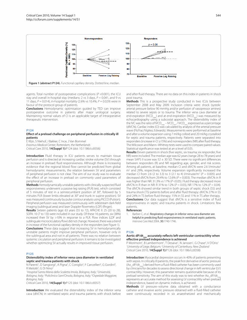
agents. Total number of postoperative complications (P <0.001), the ICU
stay and overall in-hospital stay (medians: 2 vs 3 days, P = 0.041, and 9 vs
11 days, P = 0.014), in-hospital mortality (2.6% vs 10.4%, P = 0.029) were in
favour of the protocol group of patients.
Conclusions Hemodynamic optimisation guided by TED can improve
postoperative outcome in patients after major urological surgery.
Maintain ing normal values of CI is an applicable target of intraoperative
therapeutic intervention.
P124
Eff ect of a preload challenge on peripheral perfusion in critically ill patientsE Klijn, S Niehof, J Bakker, C Ince, J Van Bommel
Erasmus Medical Center, Rotterdam, the Netherlands
Critical Care 2010, 14(Suppl 1):P124 (doi: 10.1186/cc8356)
Introduction Fluid therapy in ICU patients serves to maintain tissue
perfusion and is directed at increasing cardiac stroke volume (SV) through
an increase in preload: fl uid responsiveness. Although there is increasing
evidence that the regional blood fl ow cannot be predicted from global
hemodynamic measurements, the relation between SV and parameters
of peripheral perfusion is not clear. The aim of our study was to evaluate
the eff ect of an increase in preload on commonly used parameters of
peripheral perfusion.
Methods Hemodynamically unstable patients with clinically suspected fl uid
responsiveness underwent a passive leg raising (PLR) test, which consisted
of 5 minutes of rest in a semirecumbent position of 30°, followed by 5
minutes PLR (lower limbs elevated at 30° and trunk in supine position). SV
was measured continuously by pulse contour analysis using PiCCO (Pulsion).
Peripheral perfusion was measured continuously with sidestream dark fi eld
imaging (sublingual area) and laser Doppler fl owmetry (LDF) (fi nger).
Results Sixteen patients (age: 63 years (55 to 72), APACHE II: 25 (20 to 28),
SOFA: 10 (7 to 13)) were included in our study. Of these 16 patients, six (38%)
increased their SV by >10% in response to a PLR. Flow indices (LDF and
sublinguale microcirculatory fl ow) did not change. However, there was a trend
in increase of the functional capillary density in the responders (see Figure 1).
Conclusions These data suggest that increasing SV in hemodynamically
unstable patients might improve peripheral perfusion, however only in
the sublingual area and not in all patients. There was no relation between
systemic circulation and peripheral perfusion: it remains to be investigated
whether optimizing SV actually results in improved tissue perfusion.
P125
Distensibility index of inferior vena cava diameter in ventilated septic and trauma patients with shockN Parenti1, D Sangiorgi2, A Pigna3, C Coniglio4, F Cancellieri4, G Gordini4,
R Melotti3, G Di Nino3
1Hospital Santa Maria della Scaletta Imola, Bologna, Italy; 2Università,
Bologna, Italy; 3Policlinico Sant’Orsola, Bologna, Italy; 4Ospedale Maggiore,
Bologna, Italy
Critical Care 2010, 14(Suppl 1):P125 (doi: 10.1186/cc8357)
Introduction We evaluated the distensibility index of the inferior vena
cava (dIVC%) in ventilated septic and trauma patients with shock before
and after fl uid therapy. There are no data on this index in patients in shock
post trauma.
Methods This is a prospective study conducted in two ICUs between
September 2008 and May 2009. Inclusion criteria were: shock (systolic
arterial pressure below 90 mmHg and/or perfusion of vasopressor amines)
related to severe sepsis or to trauma. The inferior vena cava diameter at
end-expiration (IVCDmax
) and at end-inspiration (IVCDmin
) was measured by
echocardiography using a subcostal approach. The distensibility index of
the IVC was the ratio of IVCDmax
– IVCDmin
/ IVCDmin
expressed as a percentage
(dIVC%). Cardiac index (CI) was calculated by analysis of the arterial pressure
wave (FloTrac/Vigileo; Edwards). Measurements were performed at baseline
and after a volume expansion using 7 ml/kg colloid and 20 ml/kg crystalloid
for septic and trauma patients, respectively. Patients were separated into
responders (increase in CI ≥15%) and nonresponders (NR) after fl uid therapy.
The Wilcoxon and Mann–Whitney tests were used to compare paired values.
Statistical signifi cance was tested at an α level of 0.05.
Results Eleven patients in shock (fi ve septic, six trauma; six responder, fi ve
NR) were included. The median age was 62 years (range 28 to 78 years) and
mean SAPS II score was 52 ± 30 SD. There were no signifi cant diff erences
between responders (R) and NR regarding age, gender, and risk scores.
Among all patients, at baseline, median CI and dIVC% were 2.6 l/minute/
m2 and 29%, respectively. Volume expansion signifi cantly increased the
median CI from 2.6 (2 to 3.3) to 3 (2.1 to 4) l/minute/m2 (P = 0.005) and
decreased dIVC% from 29.4% to 12.6% (P = 0.003). The median dIVC% in R
was higher than NR: 31.3% vs 17% (P <0.05). Fluid therapy decreased more
dIVC% in R than in NR: R 31% to 12% (P = 0.03), NR 17% to 12% (P = 0.04).
The dIVC% showed similar trend in both groups of septic shock (SS) and
trauma shock (TS) patients before and after fl uid therapy: dIVC% 27% in SS
and 24% in TS before fl uid therapy; 15% in SS and 11% in TS after therapy.
Conclusions Our data suggest that dIVC% is a sensitive index of fl uid
responsive ness in septic and trauma patients in shock. Limitations: few
patients.
References1. Barbier C, et al.: Respiratory changes in inferior vena cava diameter are
helpful in predicting fl uid responsiveness in ventilated septic patients. Intensive Care Med 2004, 30:1740-1746.
P126
Aortic dP/dtmax
accurately refl ects left ventricular contractility when eff ective preload independence is achievedP Morimont1, B Lambermont1, T Desaive1, N Janssen1, G Chase2, V D’Orio1
1University of Liege, Belgium; 2University of Canterbury, New Zealand
Critical Care 2010, 14(Suppl 1):P126 (doi: 10.1186/cc8358)
Introduction Myocardial depression occurs in 40% of patients presenting
with sepsis. In critically ill patients, the peak fi rst derivative of aortic pressure
(Ao_dP/dtmax
) derived from a fl uid-fi lled catheter has been commonly used
by clinicians for decades to assess directional change in left ventricular (LV)
contractility. However, this parameter remains questionable because of its
preload sensitivity. The aim of this study was to test whether Ao_dP/dtmax
represents an accurate method for assessing LV contractility when preload
independence, based on dynamic indices, is achieved.
Methods LV pressure–volume data obtained with a conductance
catheter and invasive aortic pressure obtained with a fl uid-fi lled catheter
were continuously recorded in six anaesthetized and mechanically
Figure 1 (abstract P124). Functional capillary density. Dotted line, median.
Critical Care 2010, Volume 14 Suppl 1 http://ccforum.com/supplements/14/S1
S44
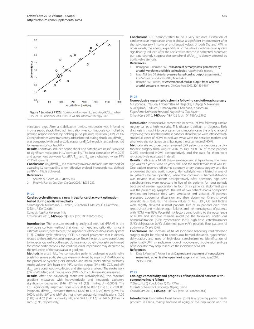
ventilated pigs. After a stabilization period, endotoxin was infused to
induce septic shock. Fluid administration was continuously controlled by
preload responsiveness by holding pulse pressure variation (PPV) <13%.
Catecholamines were transiently administrated during shock. Ao_dP/dtmax
was compared with end-systolic elastance (Ees
), the gold standard method
for assessing LV contractility.
Results Endotoxin-induced septic shock and catecholamine infusion lead
to signifi cant variations in LV contractility. The best correlation (r2 = 0.76)
and agreement between Ao_dP/dTmax
and Ees
were obtained when PPV
<11% (Figure 1).
Conclusions Ao_dP/dTmax
is a minimally invasive and accurate method for
assessing LV contractility when eff ective preload independence, defi ned
as PPV <11%, is achieved.
References1. Sharma AC: Shock 2007, 28:265-269.
2. Pinsky MR, et al.: Curr Opin Crit Care 2005, 11:235-239.
P127
Cardiac cycle effi ciency: a new index for cardiac work estimation tested during aortic valve plastyS Romagnoli, M Romano, C Lazzeri, G Santoro, F Meucci, D Quattrone,
D Dini, A De Gaudio
Careggi Hospital, Florence, Italy
Critical Care 2010, 14(Suppl 1):P127 (doi: 10.1186/cc8359)
Introduction The pressure recording analytical method (PRAM) is the
only pulse contour method that does not need any calibration since it
estimates in vivo, beat to beat, the impedance of the cardiovascular system
[1-3]. Cardiac cycle effi ciency (CCE) is a novel parameter that is directly
related to the cardiovascular impedance. Since the aortic valve contributes
to impedance, we hypothesized during an aortic valvuloplasty, performed
for severe aortic stenosis, the cardiovascular impedance may decrease by
the reduction of the transvalvular gradient.
Methods In a cath lab, fi ve consecutive patients undergoing aortic valve
plasty for severe aortic stenosis were monitored by means of PRAM during
the procedure. Systolic (SAP), diastolic, and mean (MAP) arterial pressures,
stroke volume (SV), heart rate (HR), cardiac output (SV x HR), CCE, and dP/
dtmax
were continuously collected and afterwards analyzed. The stroke work
(SW = SV x MAP) and minute work (MW = SBP x CO) were also measured.
Results After the ballooning maneuver (valvuloplasty), the maximal
gradient measured with intraventricular and intraaortic catheters
signifi cantly decreased (146 (37) vs 43 (12) mmHg; P <0.0001). The
CCE signifi cantly improved from –0.72 (0.4) to 0.02 (0.19) U; P <0.0001.
Peripheral dP/dtmax
increased from 0.8 (0.27) to 1.16 (0.23) mmHg/ms; P =
0.001, while SW and MW did not show substantial modifi cations (4.06
(1.03) vs 4.02 (1.4) l x mmHg; NS, and 549.8 (111.5) vs 544.6 (153.4) l x
mmHg; NS, respectively).
Conclusions CCE demonstrated to be a very sensitive estimation of
cardiovascular impedance since it shows a signifi cant improvement after
the valvuloplasty in spite of unchanged values of both SW and MW. In
other words, the energy expenditure of the whole cardiovascular system
signifi cantly reduced after the aortic valve stenosis is corrected. Moreover,
our data strongly suggest that peripheral dP/dtmax
is deeply aff ected by
aortic valve stenosis.
References1. Romagnoli S, Romano SM: Estimation of hemodynamic parameters by
arterial waveform: available technologies. Anesth Analg in press.
2. Maus TM, Lee DE: Arterial pressure-based cardiac output assessment. J
Cardiothorac Vasc Anesth 2008, 22:468-473.
3. Romano SM, Pistolesi M: Assessment of cardiac output from systemic arterial pressure in humans. Crit Care Med 2002, 30:1834-1841.
P128
Nonocclusive mesenteric ischemia following cardiothoracic surgeryN Kiyonaga, T Yasuda, T Yonemitsu, M Nagaoka, T Oryoji, M Nakahara,
N Okayama, T Kikuchi, T Imabayashi, Y Kakihana, Y Kanmura
Kagoshima University Hospital, Kagoshima City, Japan
Critical Care 2010, 14(Suppl 1):P128 (doi: 10.1186/cc8360)
Introduction Nonocclusive mesenteric ischemia (NOMI) following cardiac
surgery carries a high mortality. This disease is diffi cult to diagnose. Early
diagnosis is thought to be of paramount importance as the only chance of
improving the survival rate in these patients. Therefore, we were retrospectively
studied all cases of NOMI to evaluate what were the sensitive markers and
what were the risk factors contributing to the occurrence of NOMI.
Methods We retrospectively reviewed 279 patients undergoing cardio-
thoracic surgery from August 2007 to July 2008. Six of these patients
(2.2%) developed NOMI postoperatively and the data for them were
retrospectively evaluated in detail.
Results In all cases of NOMI, they were diagnosed at laparotomy. The mean
age was 69.7 years (50 to 83 years old), and the male:female ratio was 1:1.
One patient received off -pump coronary artery bypass surgery, and fi ve
underwent thoracic aortic surgery. Hemodialysis was initiated in one of
six patients before operation, while the continuous hemodiafi ltration
was initiated in all patients postoperatively. After operation, high-dose
catecholamines were necessary in fi ve of six patients for long periods
because of severe hypotension. In four of six patients, abdominal pain
was the presenting symptom. The rest of two patients had a nonspecifi c
presentation because they were ventilated and sedated. All patients
presented abdominal distension and their abdominal X-rays showed
paralytic ileus features. The serum values of AST, LDH, CK, and lactate
were slightly elevated in most patients. Five of six patients died from
septic shock and multiple organ failures, and the mortality rate of patients
with NOMI was 83%. Potential risk factors contributing to the occurrence
of NOMI and sensitive markers might be the following: continuous
hemodiafi ltration (6/6); hypotension (5/6); high-dose catecholamines
(5/6); dehydration (6/6); abdominal pain (4/6); paralytic ileus patterns of
abdominal X-rays (6/6).
Conclusions The increase of NOMI incidence following cardiothoracic
surgery might be related to continuous hemodiafi ltration, hypotension,
dehydration, and uses of high-dose catecholamines. Identifi cation of
patients at NOMI risk and prevention of hypovolemic hypotension and use
of vasodilator may help to reduce the incidence of NOMI.
References
1. Klotz S, Vestring T, Rotker J, et al.: Dagnosis and treatment of nonocclusive mesenteric ischemia after open heart surgery. Ann Thorac Surg 2001,
72:1583-1586.
P129
Etiology, comorbidity and prognosis of hospitalized patients with congestive heart failureY Zhao, J Li, Q Xue, L Gao, Q Xu, X Wu
Institute of Geriatric Cardiology, Beijing, China
Critical Care 2010, 14(Suppl 1):P129 (doi: 10.1186/cc8361)
Introduction Congestive heart failure (CHF) is a growing public health
problem in China, mainly because of aging of the population and the
Figure 1 (abstract P126). Correlation between Ees
and Ao_dP/dtmax
when
PPV <11%. Incidence of ICR-BSI in WCNN intensive therapy unit.
Critical Care 2010, Volume 14 Suppl 1 http://ccforum.com/supplements/14/S1
S45

increased prevalence of CHF in the elderly. The aim of present study was
to investigate the major causes, comorbidities and in-hospital mortality of
patients with CHF.
Methods A retrospective study was performed in 6,960 patients (4,352
males, 2,608 females) with a validated primary discharge diagnosis of CHF
hospitalized from 1 January 1993 through 31 December 2007, at Chinese
PLA General Hospital in Beijing. The patients were divided into fi ve groups
based on the number of etiologies and comorbidities from one to fi ve
or more than fi ve. A comparative analysis was performed to explore the
major causes, comorbidities and in-hospital mortality of patients among
the groups.
Results The mean (± SD) age of patients was 53 ± 17 years in the one-
comorbidity group, 60 ± 16 years in the two-comorbidity group, 65 ±
14 years in the three-comorbidity group, 70 ± 13 years in the four-
comorbidity group and 72 ± 11 years in the fi ve-comorbidity group.
The major causes of hospitalized patients with CHF were coronary artery
disease (44.9%), vavular heart disease (27.5%), cor pulmonale (9.6%) and
cardiomyopathy (7.4%). The comorbidities of CHF were hypertension
(38.6%), atrial fi brillation (23.1%), diabetes mellitus (18.3%), pneumonia
(11.6%) and renal failure (7.1%). The single comorbidity was predominant
in younger patients while multiple comorbidity was predominant in the
elderly (P <0.001). The most common single etiology was vavular heart
disease, the most common triple etiology was coronary artery disease
complicated with hypertension and diabetes mellitus. Cox regression
analysis showed higher hospital mortality rates associated with increased
numbers of etiology and comorbidity (hazard ratio (HR) from 0.98, 95% CI
0.71 to 1.36 to HR 1.59, 95% CI 1.23 to 2.05, to HR 1.90, 95% CI 1.43 to 2.51,
to HR 2.47, 95% CI 1.81 to 3.35, P <0.001).
Conclusions This study demonstrates that the older a hospitalized
patient with CHF is, the more comorbidities they have. The major
causes and comorbidities of hospitalized patients with CHF are coronary
artery disease, vavular heart disease, cor pulmonale, cardiomyopathy,
hypertension, atrial fi brillation, diabetes mellitus, pneumonia and renal
failure. The single etiology or comorbidity is predominant in younger
patients, while multiple etiology or comorbidity is predominant in the
elderly. A higher in-hospital mortality rate of CHF is associated with an
increased number of etiologies and comorbidities.
P130
Factors associated with intensive care morbidity following surgical repair of tetralogy of fallotA Aralihond, A Krishnaiah, B Mimic, K Brown
Great Ormond Street Hospital, London, UK
Critical Care 2010, 14(Suppl 1):P130 (doi: 10.1186/cc8362)
Introduction The outcome of surgical repair of tetralogy of fallot (TOF)
is a useful benchmark for the assessment of congenital cardiac surgical
programs. The aim of this study was to describe postoperative morbidity
following TOF repair and assess the factors linked to a longer duration of
postoperative ventilation.
Methods Retrospective study of all patients admitted to cardiac intensive
care between January 2003 and December 2008 following classic TOF
repair. More complex repairs were excluded. Factors were investigated
for a relationship with intubation hours using linear regression analysis.
Intubation hours were log transformed for the analysis since the data were
skewed.
Results A total of 174 children were included, 97 (56%) male, of whom
31 (18%) had previous palliation with a BT shunt and 23 (13%) had extra
medical problems. The median age at repair was 8.9 (range 1.6 to 112.8)
months and the median weight was 8.2 (3.5 to 28.5) kg. The median
cardiopulmonary bypass (CPB) time was 101 (42 to 292) minutes and 94
(54%) required transannular patch. The median postoperative intubation
time was 23 (0 to 566) hours and 11 patients (6%) did not require
postoperative ventilation. The median ICU stay was 66 (13 to 791) hours
and there was one early death before hospital discharge. One hundred and
seventy (95%) patients received an inodilator. Postoperative complications
included renal failure and fl uid overload requiring peritoneal dialysis
(PD) in 31 (17%); junctional ectopic tachycardia (JET) in 29 (16%); other
arrhythmias in 10 (6%), delayed chest closure in six (3%) of which two had
emergency mediastinal exploration due to bleeding and ECMO in one
patient. A multiple regression model for the outcome measure intubation
hours indicated that younger age at repair (P = 0.03), associated medical
problems (P = 0.04), longer CPB time (P = 0.057), JET, PD, noradrenalin use
and adrenaline use (P <0.01 for all) were independently linked with longer
postoperative ventilation times.
Conclusions Younger children, those with extra medical problems and
children that required higher levels of inotropes (probably refl ecting low
cardiac output syndrome), had longer intubation times. The commonest
and most important complications that infl uenced duration of ventilation
were JET and PD. In our series, mortality and length of stay were
comparable with other published data.
P131
Sildenafi l attenuates pulmonary vascular remodeling and upregulates Kv1.5 mRNA expression in pulmonary hypertension secondary to left-to-right shunt in ratsL Hu, LH Tan
Children’s Hospital, Zhejiang University School of Medicine, Hangzhou, China
Critical Care 2010, 14(Suppl 1):P131 (doi: 10.1186/cc8363)
Introduction Sildenafi l is a phosphodiesterase-type-5 inhibitor and
selectively decreases pulmonary artery pressure. So far, the mechanism
underlying sildenafi l’s eff ects on pulmonary vascular remodeling and
potassium channel activity in pulmonary artery smooth muscle cells
(PASMCs) has not been clearly addressed in pulmonary hypertension
secondary to increased pulmonary blood fl ow.
Methods A total of 27 male SD rats were randomly divided into a sham
group (n = 9), a shunt group (n = 9) and a shunt + sildenafi l group (n = 9).
A left-to-right shunt was established by performing an abdominal aorta to
inferior vena cava fi stula both in the shunt group and the shunt + sildenafi l
group. Rats in the shunt + sildenafi l group received oral sildenafi l 10 mg/kg/
day, whereas the rats in the sham group and the shunt group were fed with
normal saline of the same volume. Eleven weeks later, mean pulmonary
artery pressure (mPAP) was measured. Meanwhile, the ratio of right
ventricular mass to left ventricular plus septal mass (RV/(LV+S)) was detected
as a marker of the degree of right ventricular hypertrophy. The relative medial
thickness (RMT) of middle and small pulmonary muscularized arteries was
calculated as a sign of pathological changes of pulmonary vasculature. The
voltage-gated potassium channel Kv1.5 mRNA expression of pulmonary
vasculature was detected using real-time PCR.
Results Eleven weeks later, the rats in the shunt group developed
pulmonary hypertension as evidenced by signifi cantly increased
mPAP, RV/(LV+S), as well as higher RMT of middle and small pulmonary
muscularized arteries (all P = 0.01). In addition, the rats in shunt group had
decreased Kv1.5 mRNA expression in pulmonary vasculature (P = 0.01).
The rats in the shunt + sildenafi l group had a signifi cantly decreased mPAP,
and RV/(LV+S) ratio, and a lower RMT as well (all P = 0.01), whereas the
levels of Kv1.5 mRNA expression were signifi cantly upregulated (P = 0.01).
Furthermore, there were no statistically signifi cant diff erence in mPAP, in
RV/(LV+S) ratio, RMT of middle and small pulmonary muscularized arteries
and Kv1.5 mRNA expression between the shunt + sildenafi l group and the
sham group (all P = NS).
Conclusions Oral sildenafi l attenuated pulmonary vascular remodeling
and upregulated Kv1.5 mRNA expression in the rats with pulmonary
hypertension secondary to left-to-right shunt.
P132
Validation of echocardiographic indices of right ventricular afterload: an experimental, open pericardium pig modelA Knook, D Reis Miranda, J Van Bommel, A Bogers, D Duncker,
D Gommers, J Bakker
Erasmus Medical Centre, Rotterdam, the Netherlands
Critical Care 2010, 14(Suppl 1):P132 (doi: 10.1186/cc8364)
Introduction Pulmonary vascular resistance (PVR) has been used to assess
right ventricular afterload; however, this is considered meaningless due to
several physiologic shortcomings. Echocardiography has often been used to
assess right ventricular (RV) afterload, but this has never been validated as an
index of RV afterload. The purpose of this study is to evaluate echocardiographic
indices of RV afterload in an experimental, open-pericardium pig model with
induced, variable main pulmonary artery stenosis.
Critical Care 2010, Volume 14 Suppl 1 http://ccforum.com/supplements/14/S1
S46

Methods Eleven anesthesized pigs were instrumented for the measure-
ment of arterial blood pressure, central venous pressure, RV and pulmonary
pressure. An ultrasonic fl owprobe (MA14PAX; Transonic) was positioned on
the main pulmonary artery to obtain pulmonary fl ow. Distal to the fl owprobe,
a balloon-occluder was positioned facilitating gradual constriction of the
pulmonary artery. To obtain a stepwise pressure diff erence increment over the
banding of 10 mmHg at each measurement, we gradually infl ated the balloon-
occluder. After 10 minutes, all invasive hemodynamic data were registered
and an epicardial echocardiography was performed to obtain tricuspid fl ow
velocities, isovolumetric and isovolumetric relaxation time. To calculate the
TEI index during one heartbeat, echocardiographic measurements were
synchronized with fl owprobe measurements to obtain ejection time. The
E/E΄ ratio was obtained with tissue Doppler echocardiography of the lateral
tricuspid annulus. All echocardiographic measurements were performed in
triple and averaged. The ejection period and mean pulmonary acceleration
and cardiac output were calculated from the pulmonary fl ow curve derived
from the ultrasonic fl owprobe. Resistance over the pulmonary banding was
calculated by pressure gradient divided by cardiac output.
Results Of the 11 pigs, two pigs died prematurely, one inferior caval vein
rupture, and one due to asystole while inserting the pulmonary artery
catheter. After pulmonary banding, central venous pressure did not
increase signifi cantly. Mean acceleration correlated with the resistance
over the banding (r = 0.59, P <0.0001). The TEI index and E/E΄ neither
correlated with the pressure gradient over the banding nor fl ow.
Conclusions Mean acceleration correlated with RV afterload in this model.
P133
Activation of apoptotic pathways in experimental acute afterload-induced right ventricular failureC Dewachter1, L Dewachter1, B Rondelet1, P Fesler2, S Brimioulle1,
F Kerbaul3, R Naeije1
1Faculty of Medicine, ULB, Brussels, Belgium; 2Lapeyronie Hospital, Montpellier,
France; 3La Timone University Hospital, Marseille, France
Critical Care 2010, 14(Suppl 1):P133 (doi: 10.1186/cc8365)
Introduction The pathobiology of persistent right ventricular (RV) failure
observed after an acute increase in pulmonary artery pressure (Ppa
)
remains incompletely understood. We hypothesized that these severe
complications might be related to an activation of apoptotic pathways.
Methods Fourteen anesthetized dogs were randomised to a transient
90 minutes pulmonary artery constriction or to a SHAM operation,
followed 30 minutes later by hemodynamic measurements including
eff ective arterial elastance (Ea) to estimate RV afterload and end-systolic
elastance (Ees
) to estimate RV contractility, and sampling of cardiac tissue
to assess apoptosis by real-time quantitative polymerase chain reaction,
enzyme-linked immunosorbent assay and immunohistochemistry.
Results Transient increase in Ppa
persistently increased Ea from 0.75 ± 0.08
to 1.37 ± 0.18 mmHg/ml, and decreased Ees
from 1.06 ± 0.09 to 0.49 ± 0.09
mmHg/ml, Ees
/Ea from 1.44 ± 0.06 to 0.34 ± 0.03 and cardiac output from
3.78 ± 0.16 to 1.46 ± 0.10 l/minute, indicating RV failure. As compared with
the SHAM-operated group, and with left ventricular tissue in animals with
persistent RV failure, there were decreased gene expressions of RV and
septal Bcl-2, with no changes in the gene expressions of Bax and Bak, and
an increase in the Bax/Bcl-2 ratio. RV and septal Bcl-XL, and RV Bcl-w gene
expressions were decreased as compared with the SHAM-operated group.
There were activations of RV caspases 8 and 9, and of RV and septal caspase
3. Diff use RV and septal apoptosis was confi rmed by TUNEL staining. There
were also increased RV and septal protein expressions of TNFα.
Conclusions Acute afterload-induced persistent RV failure appears to be
related to an early activation of apoptotic pathways, and to a myocardial
increase of TNFα.
P134
High incidence of severely prolonged QT interval after cardiac surgeryM Biry, U Schurr, S Ritter, K Baenziger, A Zollinger, M Genoni
Triemli City Hospital, Zurich, Switzerland
Critical Care 2010, 14(Suppl 1):P134 (doi: 10.1186/cc8366)
Introduction A prolonged QT interval may be associated with torsades
de pointes and lead to sudden cardiac death. The aim of this study was to
determine both the frequency of QT prolongation and administration of
QT prolonging drugs perioperatively in cardiac surgery.
Methods This prospective observational study included 82 patients
(mean age 67 years) undergoing elective cardiac surgery (coronary
bypass grafting 37, valve surgery 29, combination 16). The QT interval was
manually measured in lead II and V2 of a 12-lead electrocardiogram (ECG)
and corrected for heart rate (corrected QT interval, QTc, calculated from
Bazett’s formula) on the day before surgery, immediately after surgery,
daily until day 5 and before hospital discharge. A QTc interval >440 ms
in men and >460 ms in women in either ECG lead II or V2 was defi ned
as moderately prolonged. A QTc interval >500 ms was considered to be
moderately prolonged in patients showing complete left or right bundle
block, and severely prolonged in patients without complete bundle block.
All administered drugs that may prolong the QT interval were identifi ed
and classifi ed according to the QTdrugs.org Advisory Board: drugs with
a defi nite risk of torsades de pointes (class 1, for example amiodarone,
sotalol, haloperidol) or with a possible risk (class 2, for example granisetron,
risperidone).
Results A total of 489 ECG were analyzed (mean six ECG per patient).
The QTc interval was moderately prolonged in 40/82 patients before
surgery (48.8%). Five out of 40 patients (12.5%) had received drugs from
class 1 preoperatively and 1/40 (2.5%) from class 2. The QTc interval was
severely prolonged in 3/82 patients before surgery (3.7%), of which none
had received QT prolonging drugs preoperatively. In 38/82 patients
(46.3%) moderate QTc prolongation was newly discovered in at least
one postoperative ECG. Seventeen put of 38 patients (44.7%) received
drugs from class 1, and 3/38 (7.9%) from class 2 perioperatively. In 16/82
patients (19.5%) severe QTc prolongation newly appeared in at least one
postoperative ECG. Seven out of 16 patients (43.8%) received drugs from
class 1 and 1/16 (6.3%) from class 2 perioperatively.
Conclusions Severe QT interval prolongation >500 ms occurs in one-
quarter of cardiac surgical patients in the perioperative period while
moderate prolongation occurs in most of them. In nearly one-half of
these cases, QT prolonging drugs like amiodarone and haloperidol may
be involved. Before and during administration of such drugs in cardiac
surgical patients perioperatively, assessment of the QT interval from
routine ECG is recommended.
P135
Endothelial protein C receptor expression after cardiopulmonary bypass in adult cardiac surgical patientsR Hajek1, J Ruzickova2, L Slavik1, V Krcova1, M Simek1, I Fluger1
1University Hospital Olomouc, Czech Republic; 2Hospital Prerov, Czech
Republic
Critical Care 2010, 14(Suppl 1):P135 (doi: 10.1186/cc8367)
Introduction Endothelial protein C receptor (EPCR) is a transmembrane
glyco protein primarily localised on vessel endothelium. EPCR binds
protein C (PC) on the endothelial surface and presents it to the thrombin/
thrombomodulin complex. Thrombin activates PC to APC, which exerts
anticoagulant and anti-infl ammatory eff ect. Proteolysis of membrane-
bound EPCR releases soluble EPCR which can be detected in plasmatic
circulation. This reaction indirectly correlates with thrombin generation
and can be interpreted as marker of endothelial activation during
cardiopulmonary bypass (CPB).
Methods In a prospective study, the group of 35 adult patients (mean age
68 years) scheduled for cardiac surgery with standard cardiopulmonary
bypass (duration 80 ± 30 minutes, heparin dose 3 mg/kg, mild hypo-
thermia about 34°C) was evaluated. The markers of endothelial activation
and coagulation parameters (EPCR, PAI-1, antithrombin, tPA, PTT, aPTT,
fi brinogen, platelets and thromboelastography (TEG)) immediately
before and after surgery were recorded. We hypothesized that the
EPCR level (detected with ELISA monoclonal antibodies labeled with
myeloperoxidase, 450 nm reader – detection limit 10 ng/ml soluble
EPCR) increases after CPB and correlates with other markers of endothelial
activation.
Results The cut-off value of EPCR was set down 200 ng/ml. In 17% of
patients the value above the cut-off (366 ng/ml before, 209 ng/ml after CPB)
was detected. The EPCR value decreases after CPB (150.7 ± 124 vs 99.4 ±
70.6) whereas tPA increases (1.5 vs 9.3 ng/ml) and PAI-1 was unchangeable.
A trend to hypocoagulation after CPB was noticed (Δfi brinogen –1.04 g/l,
Critical Care 2010, Volume 14 Suppl 1 http://ccforum.com/supplements/14/S1
S47
ΔaPTT +7.7 sec, Δplatelets –67 G/l) but no correlation between EPCR and
TEG parameters (coagulation index ΔCI –1.46, ΔR +0.22 minutes, Δα΄ –3.9°,
ΔMA –9.3 mm) was recorded. No correlation between EPCR and CPB
duration was recorded as well.
Conclusions CPB is not associated with increase of EPCR – a novel endothelial
marker – measured immediately after operation. We need further studies to
explain mechanisms of endothelial protection in this clinical setting.
References1. Esmon CT: The endothelial protein C receptor. Curr Opin Hematol 2006,
13:382-385.
2. Thiyagarajan M, Cheng T, Zlokovic V: Endothelial cell protein C receptor: role beyond endothelium? Circ Res 2007, 100:155-157.
3. Lopez-Sagasetza J, Montes R, Puy C, et al.: Binding of factor VIIa to the endothelial cell protein C receptor reduces its coagulant activity. J Thromb
Haemost 2007, 5:1813-1816.
P136
Urapidyl for hypertension control in severe pre-eclampsia: a comparative study with nicardipineR Vizitiu1, M Krauss-Grignard2, V Garcia1, L Valentin2, E Samain2, P Diemunsch1
1Hôpitaux Universitaires de Strasbourg, France; 2Hôpital Saint Jacques,
Besançon, France
Critical Care 2010, 14(Suppl 1):P136 (doi: 10.1186/cc8368)
Introduction During severe pre-eclampsia (PE), control of blood
pressure (BP) is crucial in order to prevent systemic complications.
Currently approved treatments such as nicardipine (N) and dihydralazine
have drawbacks. Since hypertension in PE is associated with increased
sympathetic activity, urapidil (U), a peripheral α1 antagonist, has potential
for BP control in PE, but no controlled comparison of U with N is available.
This preliminary randomized controlled trial aims to compare effi cacy and
safety of U and N to reduce BP in severe PE.
Methods After IRB approval and signed informed consent, 18 women with
severe PE without previous antihypertensive treatment were randomized
to U or N groups. The therapeutic goal was to achieve a mean BP (MBP)
between 105 and 125 mmHg. The U patients fi rst received U 6.25 mg
boluses every 5 minutes until the diastolic BP dropped below 105 mmHg,
followed by a 4 mg/hour infusion adjusted as needed. In the N group,
patients fi rst received a N 1 γ/kg/minute infusion until a 15% reduction in
mean BP, followed by a N 0.75 γ/kg/minute infusion adjusted as needed.
Non-invasive BP was assessed every 5 minutes during treatment titration
and then every 15 minutes. The time needed to reach the therapeutic
goal was registered. The main endpoint was the achievement of the BP
goal in 2 hours or less. Tolerance was assessed by the number of episodes
of hypotension (HO) (defi ned as MBP below 100 mmHg) and side eff ects.
Severe HO, defi ned as MBP below 80 mmHg or two episodes of HO, was
considered as treatment failure and led to exclusion. Further assessment
was limited to safety, amount of ocytocics used and neonatal evaluation by
ICU paediatricians until discharge from ICU. Results were compared using
analysis of variance. Side eff ects were compared with the chi-square test.
Results One U patient was excluded from the effi cacy assessment due to
a protocol violation. The main endpoint was reached in all 17 patients, after
50 minutes in both groups. During the fi rst 2 hours, the needed treatment
adjustment median value was 1 (0 to 10) in the U group and 1 (0 to 13) in
the N group. Side eff ects attributable to the study treatment were observed
in six of the nine cases in the N group and in one of the nine cases in the U
group (P <0.02). There were no severe side eff ects or neonatal side eff ects.
Conclusions No diff erence in effi cacy could be shown in this preliminary
series. Both treatments were easy to titrate. Fewer side eff ects were recorded
in the U group. Further studies are needed in order to compare U and N.
P137
Comparison of the eff ects of furosemide, captopril and lorazepam on noncomplicated hypertensive patients in the emergency departmentA Uzun, L Yamanel, O Cinar, H Hasman, B Comert
Gulhane Military Medical Academy, Ankara, Turkey
Critical Care 2010, 14(Suppl 1):P137 (doi: 10.1186/cc8369)
Introduction Patients frequently refer to the emergency department with
hypertension and related disorders. If hypertensive crisis is not diagnosed
in these patients, urgent treatment is not necessary. However, taking
patient satisfaction into consideration, the emergency physicians usually
discharge these patients after lowering the blood pressure with various
medications [1]. This prospective, randomized, placebo-controlled study
is designed in order to compare the eff ects of captopril, furosemide and
lorazepam on lowering blood pressure and increasing patient satisfaction.
Methods One hundred patients with uncomplicated hypertension were
included in the study. All were randomized into four groups: (1) captopril
group, (2) furosemide group, (3) lorazepam group, (4) placebo group. The
blood pressure was measured at baseline, 30th, 60th and 90th minute.
The patient satisfaction was assessed with a visual analog scale (VAS) at
baseline and 90th minute.
Results Captopril (23.64 mmHg), lorazepam (24.90 mmHg) and
furosemide (24.10 mmHg) were found similarly eff ective in lowering blood
pressure when we compare baseline and 90th minute, but all three drugs
were superior to placebo (15.94 mmHg) (P <0.05) (Table 1). When patient
satisfaction was assessed with the VAS, captopril (30.12 mm), furosemide
(28.04 mm) and lorazepam (32.88 mm) were statistically similar and all
three drugs were superior to placebo (22.76 mm).
Table 1 (abstract P137). Mean arterial pressure
Group MAP (mmHg) SD (mmHg) Level of signifi cance
Furosemide 24.1 10.7 19.9 to 28.5
Captopril 23.6 11.7 18.8 to 28.4
Lorazepam 24.9 10.1 20.7 to 29.1
Placebo 15.9 10.1 11.7 to 20.1
Total 22.1 11.1 19.9 to 24.3
Conclusions In conclusion, all three drugs can be used in subjects referred
to the emergency department with uncomplicated hypertension. They
are similarly eff ective in both lowering the blood pressure and increasing
patient satisfaction.
References1. Chiang WK: Asymptomatic hypertension in the ED. Am J Emergency Med
1998, 16:701.
P138
Incidence, risk factors and outcome of hypertensive crises in critically ill patients: a retrospective surveyC Chelazzi, G Villa, V Selmi, AR De Gaudio
University of Florence, Italy
Critical Care 2010, 14(Suppl 1):P138 (doi: 10.1186/cc8370)
Introduction Hypertensive crises (HC) are common among patients
admitted to emergency rooms [1]. However, data are lacking about
prevalence in the critically ill admitted to the ICU. The aim of the study
was to assess the rate of HC in a cohort of patients admitted to a medical–
surgical ICU, to look for risk factors for HC and to assess outcome of
patients with HC.
Methods Data have been collected from clinical charts of patients
consecutively admitted to a mixed ICU (1 January 2008 to 31 January
2009). HC diagnosis was made based on JNC defi nitions [2]. Patients were
divided into two groups, the HC group and the control group. Diff erences
between groups were evaluated regarding age, gender distribution,
admission diagnosis, SAPS II score, ICU length of stay, ICU mortality, and in-
hospital mortality (P <0.05). Clinical and behavioural conditions associated
with HC were considered in the two groups and diff erences tested for
statistical signifi cance (P <0.05).
Results Of a total of 409 patients, 63 had one or more HC (15.4%).
Conditions signifi cantly associated with HC were age, cigarette smoking,
cancer surgery, vasculopathy, medical indication to ICU admission,
coronary artery disease, and chronic atrial fi brillation. HC patients showed
higher ICU length of stay and ICU mortality.
Conclusions HC were common among critically ill patients admitted
to the ICU, irrespective of their admission diagnosis. Conditions strongly
Critical Care 2010, Volume 14 Suppl 1 http://ccforum.com/supplements/14/S1
S48

associated with HC included cigarette smoking, cancer and age. HC were
associated with worse outcomes.
References1. Zampaglione B, Pascale C, Marchisio M, Cavallo-Perin P: Hypertensive
urgencies and emergencies. Prevalence and clinical presentation. Hypertension 1996, 27:144-147.
2. Joint National Committee on Prevention, Detection, Evaluation, and
Treatment of High Blood Pressure; National Heart, Lung, and Blood Institute;
National High Blood Pressure Education Program Coordinating Committee:
Seventh report of the Joint National Committee on Prevention, Detection, Evaluation and Treatment of High Blood Pressure. Hypertension 2003,
42:1206-1252.
P139
Comparative study to evaluate blood pressure levels measured by an invasive method versus a non-invasive method in patients in an intensive care unitS Kiff er Macedo, T Bissoli, L Carvalho Dias, M Rodrigues Lacerda, R Paiva
Barbosa de Castro, D Leite Cordeiro, R Janetti Carrara, PV Falcão Duarte
Hospital Sao José do Avai, Itaperuna, Brazil
Critical Care 2010, 14(Suppl 1):P139 (doi: 10.1186/cc8371)
Introduction The blood pressure is an important variable in medical
practice. A mistaken measurement can change the management of some
diseases, modifying the patient’s prognosis. Therefore a correct defi nition
and control of blood pressure is an essential item in an ICU environment.
Methods A prospective and comparative study of the blood pressure
assessed by arterial catheterization (pressure referenced to atmospheric
pressure at mid-chest level and adequate curve in the monitor) and by
non-invasive blood pressure device (using the cuff of the monitor with
automatic oscillometric measurement) in the upper and lower limbs.
Results Included were 52 patients with average age of 54.7 years. In the
right arm (RA), a diff erence between IBP and NIBP larger than or equal
to 20 mmHg in systolic blood pressure (SBP) (plus or minus) occurred in
26.5% of the patients; 14.3% in diastolic blood pressure (DBP) and 8.7% in
mean blood pressure (MBP). In the left arm (LA), 30.6% for SBP; 4.1% for
DBP and 4.2% for MBP. In the left leg (LL), 40% for SBP, 17.8% for DBP and
17.8% for MBP. In the right leg (RL), 47.8% for SBP; 8.7% for DBP and 17.4%
for MBP.
Conclusions There were diff erences between BP measured by invasive
and non-invasive methods mainly in SBP. Lower limbs presented a larger
diff erence, which means that non-invasive methods in this place are not
reliable. The DBP in both methods presented less diff erence.
References
1. Crit Care 2006, 10:R64.
P140
Chronic atrial fi brillation in intensive care unit treated with electrical cardioversion: is it appropriate?M Murtigni1, E Mannelli1, D Colosimo1, S Cianferoni1, S Degl’innocenti2,
J Parodo2, M Bonizzoli2, G Zagli2, A Peris2
1Postgraduate School of Anesthesia and Intensive Care, University of Florence,
Italy; 2Careggi Teaching Hospital, Florence, Italy
Critical Care 2010, 14(Suppl 1):P140 (doi: 10.1186/cc8372)
Introduction Chronic atrial fi brillation is known to be an important co-
morbidity factor in critically ill patients admitted to the ICU. The aim of
this study was to evaluate whether electrical cardioversion in the ICU can
reduce mortality of patients with chronic atrial fi brillation.
Methods From January 2005 to November 2009, 50 patients with history of
chronic atrial fi brillation admitted to the ICU were retrospectively studied.
Patients were divided into two groups: the fi rst group included patients
who underwent electrical cardioversion (CVE group, n = 10) and were
discharged in sinus rhythm; the second included patients who did not
undergo electrical cardioversion and were discharged with chronic atrial
fi brillation (CAF group, n = 40). In both groups, mortality, antiarrhythmic
drug therapy and oral anticoagulation regimen were monitored. The fi rst
group’s follow-up evaluated maintenance of sinus rhythm. The number of
DC shocks required for the maintenance of sinus rhythm in patients of the
CVE group was also collected. The main outcome parameter considered
was 28-day mortality.
Results Patients of both groups resulted in similar demographic and
clinical parameters. In patients included in the CVE group, the maintenance
of sinus rhythm was achieved with one DC shock in 66.6%, two DC shocks
in 22.2% and three DC shocks in 11.1% of cases. The mortality rate at 24
hours was 0% while at 28 days was 11.1%, whereas patients in the CAF
group had a 28-day mortality rate of 35%. See Figure 1.
Conclusions Our pilot study indicates that the improvement of ventricular
performance with the contribution of atrial systole might improve critically
ill patients’ outcome. Considering the small number of cases, a prospective
study, based on these preliminary results, is ongoing.
P141
Applanation tonometry application in ICU patients with acute cardiogenic edemaN Menestrina1, A Martini1, D Simion1, M Dan2, L Gottin1
1University Hospital, Verona, Italy; 2Ospedale Civile, Verona, Italy
Critical Care 2010, 14(Suppl 1):P141 (doi: 10.1186/cc8373)
Introduction The incidence of acute cardiogenic pulmonary edema
(ACPE) is increasing and it is now one of the leading causes of morbidity
and mortality in our society. As the global population of European
countries is getting older, the impact of arterial stiff ness is becoming more
and more important in the pathophysiology of ACPE, besides coronary
vascular disease and valvular disease. The aim of the study was to evaluate
whether acute modifi cations in elastic artery distensibility can be involved
in the pathogenesis of ACPE.
Methods Six consecutive patients (four men and two women; age 76 ±
7.1 years) were admitted to our ICU with the clinical diagnosis of ACPE. All
patients were studied with transthoracic echocardiography (TTE) and by
evaluation of pulse wave analysis (PWA) and pulse wave velocity (PWV)
with the applanation tonometry method, performed at admission and
after clinical stabilization.
Results In all patients, left ventricular systolic function was not signifi cantly
reduced when evaluated with transthoracic echocardiography, while
a diastolic dysfunction was always demonstrated. We have recorded
a signifi cant (P <0.05) decrease in blood pressure values after clinical
stabilization. Estimation of the central aortic pressure waveform by
mathematical transformation of radial tonometry pressure was similarly
reduced. Other tonometric parameters, such as the Augmentation Index,
which represents the contribution of the wave refl ection to the global
pulse pressure wave, and PWV, which is inversely correlated to the arterial
compliance, were also signifi cantly decreased, when compared with the
values at the admission time. The subendocardial viability ratio (SEVR),
an indirect index of myocardial perfusion relative to cardiac workload,
increased after treatment with vasodilatators.
Conclusions Our data confi rm the determining role of the increased
arterial stiff ness in the pathogenesis of ACPE, and how a therapeutic
strategy able to ameliorate this target can be associated with a clinical
improvement for the patient. The applanation tonometry could be an
interesting method to evaluate these patients in the ICU setting.
Reference1. Nichols WN, O’Rourke MF: McDonalds Blood Flow in Arteries: Theoretical,
Experimental and Clinical Principles. 4th edition. London: Hodder Arnold; 1998.
Figure 1 (abstract P140).
Critical Care 2010, Volume 14 Suppl 1 http://ccforum.com/supplements/14/S1
S49

P142
A better prognosis for septic patients with left ventricular dysfunction?P Carrilho-Ferreira, A Pais-de-Lacerda, H Côrte-Real, C França
Hospital de Santa Maria, Lisbon, Portugal
Critical Care 2010, 14(Suppl 1):P142 (doi: 10.1186/cc8374)
Introduction Several studies using angiographic or echocardiographic
methods have shown that in septic shock (SSh) or severe sepsis (SeS),
cardiac abnormalities, which are often documented [1], seem to have a
relevant prognostic signifi cance [2,3].
Methods A retrospective study was conduced during 6 months (April
to September 2009) in an ICU of a university tertiary hospital, identifying
all patients with SSh or SeS and reviewing their echocardiographic data.
All the patients with known or clinical/echocardiographic evidence of
signifi cant prior cardiac disease were excluded from this analysis.
Results Forty-nine patients were identifi ed with SSh or SeS from a total
of 211 ICU patients (23.2%). Among these, 26 had echocardiograms
done during their ICU stay (53.1%). We analyzed 19 echocardiograms
from 15 patients. The remaining 11 patients were excluded, having
ischemic heart disease (seven patients) and signifi cant valvular heart
disease (four patients). Within the SSh/SeS group of patients, those with
echocardiograms were younger (mean age 56.3 vs 64.4) and more often
women (53.3% vs 36.7%). The ICU mortality was nevertheless similar
in the two groups (46.7% vs 46.9%). The primary sites of infection were
the lungs and abdomen (26.7% each) or unknown (20%). The agents
most frequently isolated were Gram-negative bacilli (26.7%). In 53.3%
of cases there was no identifi ed agent. Echocardiographic evaluation
revealed signifi cant changes in 26.7% of the patients, with the remaining
being considered normal. Of those with signifi cant changes, 75% (n = 3)
showed impairment of left ventricular (LV) systolic function with global
hypokinesia simultaneously with diastolic dysfunction. Either abnormality
was not found in any other patients. The mortality rate during ICU stay was
lower in the group of patients with systolic and diastolic LV dysfunction
compared with those with no apparent modifi cation of LV function (0 vs
58.3%). The right ventricle was dilated and showed high systolic pressures
present in only one patient.
Conclusions This study reinforces the concept that changes in systolic
and diastolic functions are common in septic shock and severe septic
patients, and that the development of these changes (as an adaptation
mechanism) seems to correlate inversely with the acute mortality rate
during ICU stay [2,4].
References
1. Zanotti-Cavazzoni SL, et al.: Curr Opin Crit Care 2009, 15:392-397.
2. Parker MM, et al.: Ann Intern Med 1984, 100:483-490.
3. Poelaert J, et al.: Intensive Care Med 1997, 23:553-560.
4. Jardin F, et al.: Chest 1999, 116:1354-1359.
P143
Microvascular eff ects of pulsatile versus nonpulsatile perfusion during cardiopulmonary bypassPW Elbers1, J Wijbenga2, F Solinger2, A Yilmaz2, M Van Iterson2,
E Van Dongen2, C Ince3
1OLVG, Amsterdam, the Netherlands; 2St Antonius Hospital, Nieuwegein, the
Netherlands; 3Academic Medical Center, Amsterdam, the Netherlands
Critical Care 2010, 14(Suppl 1):P143 (doi: 10.1186/cc8375)
Introduction While advantages of pulsatile perfusion (PP) during
cardiopulmonary bypass (CPB) in terms of clinical outcome remain the
subject of debate, possible benefi ts are generally thought to occur via
improvements in microvascular fl ow [1]. However, this is currently not
supported by human clinical data. Therefore we used real-time human
microvascular imaging to test our hypothesis that pulsatile perfusion
would enhance microvascular perfusion.
Methods We used sidestream dark fi eld imaging to record video clips
of the human microcirculation in 16 patients undergoing routine CPB
for cardiac surgery. Following administration of cardioplegia, CPB was
continued in either pulsatile (PP, n = 8) or nonpulsatile (NP) mode. After
10 minutes, microvascular recordings were made. The perfusion mode was
then switched from PP to NP or vice versa. Ten minutes later, a second series
of microvascular video recordings were obtained. Global hemodynamic
and laboratory data were recorded and the energy equivalent pressure
(EEP) and pulse pressure (both mean ± SD) were calculated to quantify
PP generated surplus energy. Microvascular analysis was performed both
for smaller and larger microvessels with a diameter cut-off of 20 μm.
Assessments included perfused vessel density (PVD) (mean ± SD, 95%
confi dence intervals of the diff erence between NP and PP (95% CID)) and
the Microvascular Flow Index (MFI) (mean ± SD and interquartile range).
Results Pulsatile perfusion resulted in higher pulse pressure (27 ± 6 vs 7 ±
2 mmHg, P <0.0001) and CPB circuit EEP (184 ± 33 vs 150 ± 27 mmHg, P
<0.0001) as compared with NP while MAP was similar between these
perfusion modes (52 ± 11 vs 56 ± 13 mmHg, P = 0.09). Both for small and
larger microvessels, we found no diff erences in indices of microvascular
perfusion between PP and NP. Small microvessel PVD was similar between
PP and NP (6.65 ± 1.39 vs 6.83 ± 1.23/mm; 95% CID –0.50 to 0.87/mm, P =
0.58). The same was true for larger microvessel PVD (2.16 ± 0.64 vs 1.96 ±
0.48; 95% CID –0.50 to 0.11, P = 0.20). Similarly, the MFI did not diff er between
groups either for smaller (3.00 (2.83 to 3.00) vs 3.00 (2.75 to 3.0), P = 0.41) and
larger microvessels (3.00 (2.75 to 3.00) 3.00 (3.00 to 3.00), P = 0.50).
Conclusions PP during CPB does not alter human microvascular perfusion
using standard equipment in routine cardiac surgery despite yielding
higher CPB circuit energy equivalent pressure and patient pulse pressure.
Reference
1. Murphy GS, et al.: Anesth Analg 2009, 108:1394.
P144
Microcirculation and intravascular coagulopathy in patients with severe sepsis and septic shockR Wimmer, S Franz, S Siebelist, M Uschner, C Seidelmann, H Ebelt,
S Hettwer, J Wilhelm, H Loppnow, K Werdan
Universitätsklinikum Halle (Saale), Halle, Germany
Critical Care 2010, 14(Suppl 1):P144 (doi: 10.1186/cc8376)
Introduction Sidestream darkfi eld imaging (SDF) technology is a new
method to visualize directly microcirculation. In this study we examined
the perfusion of the oral mucosa in patients with severe sepsis and
septic shock in order to evaluate the link between the severity of illness,
microcirculation and intravascular coagulopathy.
Methods Microcirculation was analysed in 46 ICU patients with clinical
suspicion for sepsis and procalcitonin >2 ng/ml. Microcirculation was
recorded daily over a 7-day period (d0 to d6). Each day, at least fi ve single
fi lm clips were taken by SDF and analysed with special software on a PC. We
used the functional vessel density and distribution of the vessel diameters
as well-established parameters to qualify microcirculation. Laboratory
parameters, hemodynamic data and vital signs were also registered. The
degree of disseminated intravascular coagulopathy was evaluated by
calculating the overt DIC score (according to ISTH), and severity of illness
was determined using SAPS II and the SOFA score.
Results During the stay in the ICU we observed a continuous descent of
the DIC score (1.82 ± 1.91 on d0 vs 1.3 ± 1.94 on d6) as well as a decline
of SAPS II (63.1 ± 14.1 on d0 vs 57.1 ± 18.4 on d6) and SOFA score (12.2 ±
3.9 on d0 vs 9.2 ± 5.8 on d6). The percentage of smallest vessels below
25 μm diameter decreased initially from 79.5 ± 21.9% (d1) to rise up then
persistently 86.2 ± 10.3% (d6). Nonsurvivors (n = 11) had, compared with
survivors (n = 35), a signifi cantly higher initial DIC score on day 0 (1.3 ± 1.6
vs 2.05 ± 4.1; P <0.05).
Conclusions Our data show a clear relation between the time courses of
DIC and microcirculation in patients with severe sepsis and septic shock.
This fi nding is in line with previous reports stating the profound impact of
DIC on microcirculation and fi nally on patient outcome.
P145
Prevalence and prognostic signifi cance of cardiac abnormalities in the ICU: an echocardiographic studyA Dell’Anna, C Sandroni, F Cavallaro, C Marano, M Antonelli
UCSC School of Medicine, Rome, Italy
Critical Care 2010, 14(Suppl 1):P145 (doi: 10.1186/cc8377)
Introduction The aim was to describe the prevalence and type of cardiac
abnormalities in ICU patients detected using echocardiography and to
assess whether those abnormalities are correlated with ICU survival.
Critical Care 2010, Volume 14 Suppl 1 http://ccforum.com/supplements/14/S1
S50

Methods Prospective cohort study. Consecutive patients hospitalized in the
UCSC medical–surgical ICU for >24 hours were evaluated using transthoracic
echocardiography (TTE). Patients admitted for postoperative monitoring
or in an agonal state were excluded. Transesophageal echocardiography
(TEE) was used when TTE was technically impossible. Measured parameters
included: volume, thickness, global and regional kinesis of the left ventricle
(LV), systolic function of the right ventricle (RV), and valvular function.
Results From 1 March to 30 June 2009 a total of 100 patients (median age
68.5 years (IQR 50 to 77.5)) were included in the study. Medium SAPS II was
46 ± 15. ICU mortality rate was 29%. Median LOS in ICU was 14 days. TTE
was technically feasible in 97 patients; the other three patients underwent
TEE. Signifi cant cardiac abnormalities were found in 66 patients, 41 of
whom had no history of cardiac disease. The most frequent abnormalities
were: tricuspidal regurgitation (45.3%), regional asynergies of LV (32%),
left heart failure (30.2%) and mitral regurgitation (28%). The median left
ventricular ejection fraction (EF) in our population was 50% (IQR 15 to
72%). Left ventricular end-diastolic volume was 100 ml (IQR 78 to 130 ml).
Mean tricuspid annular systolic excursion (TAPSE) was lower in patients
who died (18.9 vs 20.8 mm; P = 0.09). TAPSE was signifi cantly correlated
to EF (r coeffi cient = 0.48; P <0.0001). Pulmonary artery systolic pressure
was signifi cantly higher in patients who died than in those who survived
to the ICU (49.8 ± 13.0 mmHg vs 35.0 ± 12.8 mmHg; P = 0.045). On logistic
regression, prediction of ICU mortality was better (AUC = 0.901 vs 0.787)
when cardiac abnormalities detected by echocardiography were added to
a model based on age and SAPS II.
Conclusions In our study, a complete echocardiographic assessment
was possible using TTE in 97% of patients. Two-thirds of our population
had echocardiographic abnormalities, 62.1% of whom were previously
unknown. The presence of LV asynergies and reduced RV systolic function
were associated with a worse prognosis. Detection of cardiac abnormalities
increased prediction of ICU mortality based on logistic regression.
P146
BNP and NT-proBNP in patients with acute myocardial infarction complicated by cardiogenic shock: results from the IABP Shock trialH Lemm1, R Prondzinsky1, A Geppert2, M Russ1, K Huber2, K Werdan1, M Buerke1
1Martin-Luther-University Hospital, Halle, Germany; 2Wilhelminenspital,
Vienna, Austria
Critical Care 2010, 14(Suppl 1):P146 (doi: 10.1186/cc8378)
Introduction B-type natriuretic peptide (BNP) and N-terminal proBNP
(NT-proBNP) are routine diagnostic and monitoring markers in patients
with heart failure. LV function and prognosis of these patients have been
shown to be refl ected by BNP and NT-proBNP levels. However, in patients
with cardiogenic shock after myocardial infarction, the relationship and
relevance of these markers has not been elucidated.
Methods The IABP Shock trial was as a monocentric, randomised and
prospective clinical trial to determine the role of therapeutic intraaortic
balloon pump (IABP) counterpulsation after primary percutaneous
transluminal coronary angioplasty (PCI) in acute myocardial infarction
(AMI) complicated by cardiogenic shock. Cardiac catheterization was
performed in 40 patients within 12 hours of onset of hemodynamic
instability. Creatinine, hemodynamic parameters and survival were deter-
mined. Further, BNP and NT-proBNP levels were measured on admission as
well as 24, 48 and 72 hours afterwards.
Results BNP levels detected diff erences in treatment regarding LV-
unloading under IABP (632 ± 194 pg/ml vs 1,370 ± 475 pg/ml, P <0.05).
However, there was no signifi cant diff erence between the group treated
with IABP and no IABP with regard to NT-proBNP levels. Interestingly, NT-
proBNP levels clearly diff erentiated between survivors and nonsurvivors
(4,590 ± 1,230 pg/ml vs 14,370 ± 4,886 pg/ml, P <0.05), contrary to no
signifi cant diff erence between survivors and nonsurvivors with regard to
BNP levels (NS). Elevated levels of NT-proBNP in patients with cardiogenic
shock might be more dependent on impaired renal function, which might
refl ect additional organ dysfunction (creatinine >200 μmol/l vs creatinine
≤200 μmol/l: 24,965 ± 9,567 pg/ml vs 7,246 ± 2,650 pg/ml, P <0.05).
Conclusions In myocardial infarction complicated by cardiogenic shock,
levels of BNP and NT-proBNP both provide valuable additional information.
BNP seems to closely refl ect the cardiac status and eff ects of therapy,
while NT-pro-BNP seems a good indicator for prognosis in patients with
cardiogenic shock by its dependency on organ dysfunction.
P147
Understanding vital-sign abnormalities in critical care patientsS Hugueny1, DA Clifton1, M Hravnak2, MR Pinsky2, L Tarassenko1
1University of Oxford, UK; 2University of Pittsburgh, PA, USA
Critical Care 2010, 14(Suppl 1):P147 (doi: 10.1186/cc8379)
Introduction We have previously shown how a data fusion index,
based on the integration of vital signs continuously monitored in a step-
down unit (SDU), can identify the cardiorespiratory instability that often
precedes adverse events [1]. We show how the correlations between vital
signs, captured in a data fusion index based on a probabilistic model of
normality [2], change during times of cardiorespiratory instability.
Methods An observational study was carried out in a 24-bed SDU, in
which vital signs from 326 patients were continuously recorded [1].
The existing standard of care used single-channel Medical Emergency
Team (MET) activation criteria to determine when individual vital signs
became abnormal. Retrospective evaluation of the continuous vital sign
data identifi ed that MET criteria were exceeded on 238 nonartefactual
occasions. One hundred and eleven of these were indicative of suffi cient
cardiorespiratory instability that they should have required an MET call
(critical events). Vital-sign dynamics during these events were compared
with data from stable periods, using order statistics and covariance analysis.
Results We found the probability distributions of the most extreme and
the median data for each parameter over 1-minute intervals. The bivariate
Gaussian distributions of best fi t for data from critical events (abnormal
respiratory rate (RR) or heart rate (HR)) show signifi cant diff erences in
covariance when compared with those calculated for data intervals from
stable patients (see Figure 1, in which covariance is indicated by ellipse
orientation). Under normal conditions (black ellipses), RR is correlated with
HR. During critical events, tachycardia occurs with little or no variation in
RR (red ellipses); tachypnoea occurs with little or no variation in HR (blue
ellipses).
Conclusions Dynamics and correlations that exist in vital signs during
periods of stability change signifi cantly during periods of abnormality. A
statistical approach that integrates vital signs and captures correlations
between them will be sensitive to cardiorespiratory deterioration.
References
1. Hravnak M, et al.: Arch Intern Med 2008, 168:1300.
2. Tarassenko et al.: Br J Anaesth 2006, 97:64.
P148
Tissue microdialysis in critically ill septic patients: associations with sepsis severity and mortalityM Theodorakopoulou1, S Orfanos1, A Diamantakis1, E Tsafou2,
G Karkouli1, N Nikitas3, I Ikonomidis1, I Ilias1, I Tsangaris1, A Armaganidis1,
I Dimopoulou1
1Attikon University Hospital, Athens, Greece; 2Athens Veterans Hospital,
Athens, Greece; 3Department of Therapeutics, Alexandra General Hospital,
Athens, Greece
Critical Care 2010, 14(Suppl 1):P148 (doi: 10.1186/cc8380)
Introduction In vivo microdialysis (MD) is a bedside sampling method that
permits continuous analysis of a patient’s extracellular tissue chemistry
without consuming blood. MD is performed by implanting a commercially
available catheter that mimics a blood capillary at the site of interest.
Figure 1 (abstract P147).
Critical Care 2010, Volume 14 Suppl 1 http://ccforum.com/supplements/14/S1
S51
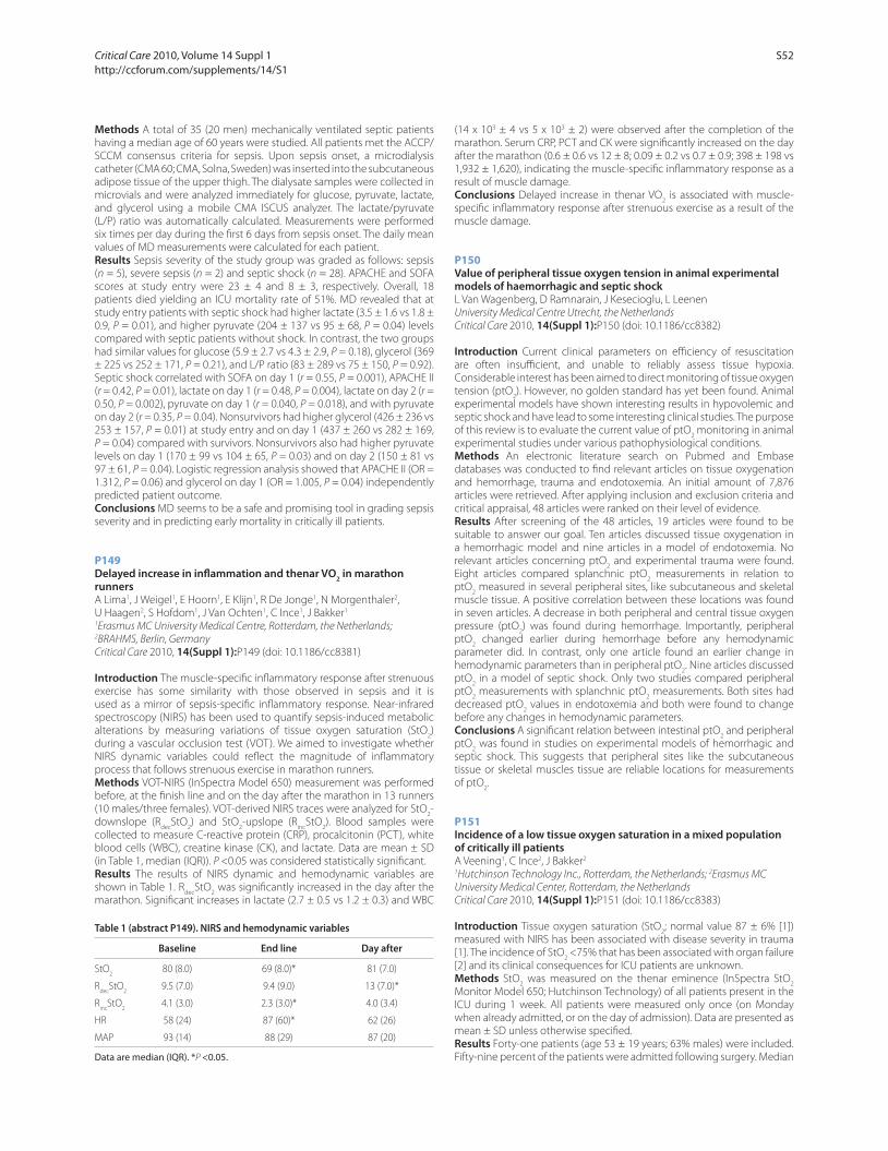
Methods A total of 35 (20 men) mechanically ventilated septic patients
having a median age of 60 years were studied. All patients met the ACCP/
SCCM consensus criteria for sepsis. Upon sepsis onset, a microdialysis
catheter (CMA 60; CMA, Solna, Sweden) was inserted into the subcutaneous
adipose tissue of the upper thigh. The dialysate samples were collected in
microvials and were analyzed immediately for glucose, pyruvate, lactate,
and glycerol using a mobile CMA ISCUS analyzer. The lactate/pyruvate
(L/P) ratio was automatically calculated. Measurements were performed
six times per day during the fi rst 6 days from sepsis onset. The daily mean
values of MD measurements were calculated for each patient.
Results Sepsis severity of the study group was graded as follows: sepsis
(n = 5), severe sepsis (n = 2) and septic shock (n = 28). APACHE and SOFA
scores at study entry were 23 ± 4 and 8 ± 3, respectively. Overall, 18
patients died yielding an ICU mortality rate of 51%. MD revealed that at
study entry patients with septic shock had higher lactate (3.5 ± 1.6 vs 1.8 ±
0.9, P = 0.01), and higher pyruvate (204 ± 137 vs 95 ± 68, P = 0.04) levels
compared with septic patients without shock. In contrast, the two groups
had similar values for glucose (5.9 ± 2.7 vs 4.3 ± 2.9, P = 0.18), glycerol (369
± 225 vs 252 ± 171, P = 0.21), and L/P ratio (83 ± 289 vs 75 ± 150, P = 0.92).
Septic shock correlated with SOFA on day 1 (r = 0.55, P = 0.001), APACHE II
(r = 0.42, P = 0.01), lactate on day 1 (r = 0.48, P = 0.004), lactate on day 2 (r =
0.50, P = 0.002), pyruvate on day 1 (r = 0.040, P = 0.018), and with pyruvate
on day 2 (r = 0.35, P = 0.04). Nonsurvivors had higher glycerol (426 ± 236 vs
253 ± 157, P = 0.01) at study entry and on day 1 (437 ± 260 vs 282 ± 169,
P = 0.04) compared with survivors. Nonsurvivors also had higher pyruvate
levels on day 1 (170 ± 99 vs 104 ± 65, P = 0.03) and on day 2 (150 ± 81 vs
97 ± 61, P = 0.04). Logistic regression analysis showed that APACHE II (OR =
1.312, P = 0.06) and glycerol on day 1 (OR = 1.005, P = 0.04) independently
predicted patient outcome.
Conclusions MD seems to be a safe and promising tool in grading sepsis
severity and in predicting early mortality in critically ill patients.
P149
Delayed increase in infl ammation and thenar VO2 in marathon
runnersA Lima1, J Weigel1, E Hoorn1, E Klijn1, R De Jonge1, N Morgenthaler2,
U Haagen2, S Hofdom1, J Van Ochten1, C Ince1, J Bakker1
1Erasmus MC University Medical Centre, Rotterdam, the Netherlands; 2BRAHMS, Berlin, Germany
Critical Care 2010, 14(Suppl 1):P149 (doi: 10.1186/cc8381)
Introduction The muscle-specifi c infl ammatory response after strenuous
exercise has some similarity with those observed in sepsis and it is
used as a mirror of sepsis-specifi c infl ammatory response. Near-infrared
spectroscopy (NIRS) has been used to quantify sepsis-induced metabolic
alterations by measuring variations of tissue oxygen saturation (StO2)
during a vascular occlusion test (VOT). We aimed to investigate whether
NIRS dynamic variables could refl ect the magnitude of infl ammatory
process that follows strenuous exercise in marathon runners.
Methods VOT-NIRS (InSpectra Model 650) measurement was performed
before, at the fi nish line and on the day after the marathon in 13 runners
(10 males/three females). VOT-derived NIRS traces were analyzed for StO2-
downslope (Rdec
StO2) and StO
2-upslope (R
incStO
2). Blood samples were
collected to measure C-reactive protein (CRP), procalcitonin (PCT), white
blood cells (WBC), creatine kinase (CK), and lactate. Data are mean ± SD
(in Table 1, median (IQR)). P <0.05 was considered statistically signifi cant.
Results The results of NIRS dynamic and hemodynamic variables are
shown in Table 1. Rdec
StO2 was signifi cantly increased in the day after the
marathon. Signifi cant increases in lactate (2.7 ± 0.5 vs 1.2 ± 0.3) and WBC
(14 x 103 ± 4 vs 5 x 10³ ± 2) were observed after the completion of the
marathon. Serum CRP, PCT and CK were signifi cantly increased on the day
after the marathon (0.6 ± 0.6 vs 12 ± 8; 0.09 ± 0.2 vs 0.7 ± 0.9; 398 ± 198 vs
1,932 ± 1,620), indicating the muscle-specifi c infl ammatory response as a
result of muscle damage.
Conclusions Delayed increase in thenar VO2 is associated with muscle-
specifi c infl ammatory response after strenuous exercise as a result of the
muscle damage.
P150
Value of peripheral tissue oxygen tension in animal experimental models of haemorrhagic and septic shockL Van Wagenberg, D Ramnarain, J Kesecioglu, L Leenen
University Medical Centre Utrecht, the Netherlands
Critical Care 2010, 14(Suppl 1):P150 (doi: 10.1186/cc8382)
Introduction Current clinical parameters on effi ciency of resuscitation
are often insuffi cient, and unable to reliably assess tissue hypoxia.
Considerable interest has been aimed to direct monitoring of tissue oxygen
tension (ptO2). However, no golden standard has yet been found. Animal
experimental models have shown interesting results in hypovolemic and
septic shock and have lead to some interesting clinical studies. The purpose
of this review is to evaluate the current value of ptO2 monitoring in animal
experimental studies under various pathophysiological conditions.
Methods An electronic literature search on Pubmed and Embase
databases was conducted to fi nd relevant articles on tissue oxygenation
and hemorrhage, trauma and endotoxemia. An initial amount of 7,876
articles were retrieved. After applying inclusion and exclusion criteria and
critical appraisal, 48 articles were ranked on their level of evidence.
Results After screening of the 48 articles, 19 articles were found to be
suitable to answer our goal. Ten articles discussed tissue oxygenation in
a hemorrhagic model and nine articles in a model of endotoxemia. No
relevant articles concerning ptO2 and experimental trauma were found.
Eight articles compared splanchnic ptO2 measurements in relation to
ptO2 measured in several peripheral sites, like subcutaneous and skeletal
muscle tissue. A positive correlation between these locations was found
in seven articles. A decrease in both peripheral and central tissue oxygen
pressure (ptO2) was found during hemorrhage. Importantly, peripheral
ptO2 changed earlier during hemorrhage before any hemodynamic
parameter did. In contrast, only one article found an earlier change in
hemodynamic parameters than in peripheral ptO2. Nine articles discussed
ptO2 in a model of septic shock. Only two studies compared peripheral
ptO2 measurements with splanchnic ptO
2 measurements. Both sites had
decreased ptO2 values in endotoxemia and both were found to change
before any changes in hemodynamic parameters.
Conclusions A signifi cant relation between intestinal ptO2 and peripheral
ptO2 was found in studies on experimental models of hemorrhagic and
septic shock. This suggests that peripheral sites like the subcutaneous
tissue or skeletal muscles tissue are reliable locations for measurements
of ptO2.
P151
Incidence of a low tissue oxygen saturation in a mixed population of critically ill patientsA Veening1, C Ince2, J Bakker2
1Hutchinson Technology Inc., Rotterdam, the Netherlands; 2Erasmus MC
University Medical Center, Rotterdam, the Netherlands
Critical Care 2010, 14(Suppl 1):P151 (doi: 10.1186/cc8383)
Introduction Tissue oxygen saturation (StO2; normal value 87 ± 6% [1])
measured with NIRS has been associated with disease severity in trauma
[1]. The incidence of StO2 <75% that has been associated with organ failure
[2] and its clinical consequences for ICU patients are unknown.
Methods StO2 was measured on the thenar eminence (InSpectra StO
2
Monitor Model 650; Hutchinson Technology) of all patients present in the
ICU during 1 week. All patients were measured only once (on Monday
when already admitted, or on the day of admission). Data are presented as
mean ± SD unless otherwise specifi ed.
Results Forty-one patients (age 53 ± 19 years; 63% males) were included.
Fifty-nine percent of the patients were admitted following surgery. Median
Table 1 (abstract P149). NIRS and hemodynamic variables
Baseline End line Day after
StO2 80 (8.0) 69 (8.0)* 81 (7.0)
Rdec
StO2 9.5 (7.0) 9.4 (9.0) 13 (7.0)*
Rinc
StO2 4.1 (3.0) 2.3 (3.0)* 4.0 (3.4)
HR 58 (24) 87 (60)* 62 (26)
MAP 93 (14) 88 (29) 87 (20)
Data are median (IQR). *P <0.05.
Critical Care 2010, Volume 14 Suppl 1 http://ccforum.com/supplements/14/S1
S52

StO2 was 83% (IQR: 75 to 88) (Figure 1). No relationship was found between
day of admission and measured StO2 (Figure 2). Thirty-one patients (76%)
had StO2 below normal (Figure 2, top line). Twelve (29%) patients had StO
2
≤75% (Figure 2, dotted line). Seven patients had StO2 <70; mortality of
these patients was 29% vs 9% in patients with StO2 >70% (NS).
Conclusions The incidence of an abnormal StO2 in critically ill patients is
high. Also very low StO2 values are frequently found and may be associated
with increased mortality.
References1. J Trauma 2005, 58:806-813.
2. J Trauma 2007, 62:44-54.
P152
Use of near-infrared spectroscopy during a vascular occlusion test to assess the microcirculatory response during fl uid responsivenessE Futier1, B Vallet2, E Robin2, M Vignaud1, J Constantin1, J Bazin1
1University Hospital of Clermont-Ferrand, France; 2University Hospital of Lille,
France
Critical Care 2010, 14(Suppl 1):P152 (doi: 10.1186/cc8384)
Introduction Adequate volume expansion for hypovolemia should
improve cardiac preload and stroke volume leading to improved
microvascular blood fl ow, tissue perfusion and oxygenation. The dynamic
recovery slope of tissue oxygen saturation (StO2) during a standardized
vascular occlusion test (VOT) is proposed to refl ect microvascular
reactivity. We aimed at evaluating the dynamic StO2 variations during fl uid
challenge in high-risk surgical patients.
Methods Thirteen adult Caucasian patients, ASA II to III, undergoing
major abdominal surgery, were included. In all, cardiac output (CO), stroke
volume (SV), pulse pressure variation (PPV), and standard hemodynamic
parameters were continuously recorded. Fluid responsiveness was defi ned
as an increase in SV >15%, after a 500 ml colloid (HEA 130/0.4) infusion
over 10 minutes. StO2 measurements and VOT (sphygmomanometer
infl ated until >50 mmHg above systolic pressure and kept infl ated until
StO2 decreased to 40%) were performed after anaesthesia induction
(baseline), and before (PPV >13%, therefore defi ning hypovolemia) and
after fl uid challenge.
Results At baseline, no patient was preload dependent (mean PPV 7.9 ± 2%),
and none required vasopressors during the procedure. Baseline mean StO2
was 85 ± 6%, while CO and SV were 4.9 ± 0.9 l/minute and 68 ± 21 ml,
respectively. During hypovolemia, compared with baseline, there was no
diff erence in both StO2 (85 ± 6 to 84 ± 9%, P = 0.76) and StO
2 desaturation
rate (–9.6 ± 1.6 to –11 ± 2.7%/minute, P = 0.10), while the StO2 recovery
slope was lowered (5.1 ± 1.6 to 3.8 ± 1.5%/second, P = 0.04). Patients were
all responders to fl uid challenge. After volume expansion, SV (84 ± 20 vs
62 ± 12 ml, P <0.01) and CO (6.5 ± 2 vs 4.8 ± 1.2 l/minute, P <0.01) were
higher, with no signifi cant changes in both StO2 and StO
2 desaturation
rate. Fluid challenge induced a 46% increase in StO2 recovery slope
(P <0.01; Figure 1), which was comparable with baseline.
Conclusions Whilst redistribution of blood fl ow may occur during
circulatory failure (vasoconstriction of lesser vital organs), impairment
of microcirculation and tissue hypoperfusion indicators are of particular
importance. Both hypovolemia and intravascular volume expansion are
associated with signifi cant modifi cations in the dynamic recovery slope
of StO2. Future studies are needed to better clarify its potential clinical
applications.
P153
Infl uence of mechanical ventilation on intestinal microcirculation and infl ammation in septic ratsD Henzler1, M Haeusler1, O Gil1, R Chankalal2, N Ismaiel2, C Lehmann2
1QEII Health Sciences Center, Halifax, Canada; 2Dalhousie University, Halifax,
Canada
Critical Care 2010, 14(Suppl 1):P153 (doi: 10.1186/cc8385)
Introduction Although the adverse eff ects of mechanical ventilation on
circulation and intestinal organ perfusion are well known, the infl uence of
assisted breathing versus controlled ventilation has not been investigated.
We hypothesized that pressure support ventilation causes less decrease
in gut perfusion and less neutrophil adhesion than controlled ventilation.
Methods Male SD rats (n = 40) were anesthetized and tracheotomized
and 10 animals each ventilated in pressure support (PSV) or pressure
controlled (PCV) mode. PCV animals were paralyzed. After 5 hours of
ventilation, intravital microscopy was performed. Endotoxin (15 mg/kg
lipopolysaccharide) was given to 20 rats, 20 animals served as controls.
Figure 1 (abstract P151). Frequency distribution of StO2.
Figure 2 (abstract P151). Day of admission at time of measurement vs
StO2.
Figure 1 (abstract P152).
Critical Care 2010, Volume 14 Suppl 1 http://ccforum.com/supplements/14/S1
S53
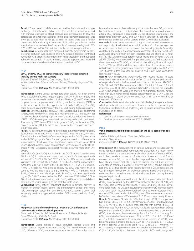
Results There were no diff erences in baseline hemodynamics or gas
exchange. Animals were stable over the whole observation period
with minimal changes in blood pressure and oxygenation. In PCV, the
percentage of functional capillaries in the longitudinal mucosal layer was
lower (57 ± 39%) than in PSV (87 ± 33%). In sepsis, there was a further
decrease in PCV and a major decrease in PSV. Leukocyte adhesion in the
intestinal submocosal venules (for example, V1 venules) was higher in PCV
(228 ± 119) than in PSV (93 ± 65) in controls, but not in septic animals.
Conclusions In septic rats with preserved macrohemodynamic stability,
intestinal perfusion was impaired after LPS infusion. Pressure-controlled
ventilation had a signifi cant infl uence on microcirculation and neutrophil
adhesion in controls. In septic animals, pressure support ventilation did
not attenuate these adverse eff ects as compared with PCV.
P154
ScvO2 and Pcv-aCO
2 as complementary tools for goal-directed
therapy during high-risk surgeryE Futier1, B Vallet2, E Robin2, J Constantin1, J Bazin1
1University Hospital of Clermont-Ferrand, France; 2University Hospital of Lille,
France
Critical Care 2010, 14(Suppl 1):P154 (doi: 10.1186/cc8386)
Introduction Central venous oxygen saturation (ScvO2) has been shown
to be a useful therapeutic target in septic shock or high-risk surgery. The
central venous-to-arterial carbon dioxide diff erence (Pcv-aCO2) has been
proposed as a complementary tool for goal-directed therapy (GDT) in
septic shock. We tested the hypothesis that both ScvO2 and Pcv-aCO
2
could be used as complementary tools for GDT during high-risk surgery.
Methods Seventy adult patients, ASA I to III, undergoing major abdominal
surgery, were randomly assigned to 6 ml/kg/hour (R-GDT group, n = 36)
or 12 ml/kg/hour (C-GDT group, n = 34) of crystalloids. Additional boluses
of HES (130/0.4) were given to maintain respiratory variation in peak aortic
fl ow velocity (ΔPV) below 13%. In both groups, ScvO2, cardiac output (CO),
oxygen delivery (DO2i), Pcv-aCO
2 and postoperative complications were
blindly recorded.
Results At baseline, there were no diff erences in hemodynamic variables,
ScvO2 (79 ± 7 vs 80 ± 6, P = 0.37) and Pcv-aCO
2 (6 ± 3 vs 6 ± 2, P = 0.95).
The total volume of fl uid perfused was larger in the C-GDT group than
in the R-GDT group (P <0.01). The two groups showed no diff erences in
intraoperative blood loss, blood transfusion, mean CO and mean DO2i
values. Overall, postoperative complications were increased in the R-GDT
group (P <0.01), especially postoperative sepsis occurred more often (P =
0.0064).
Minimal ScvO2 (minScvO
2) was higher in the C-GDT group (72 ± 6 vs 69 ±
6%, P = 0.04). In patients with complications, minScvO2 was signifi cantly
reduced (72 ± 6 vs 67 ± 6%, P = 0.0017). minScvO2 <70% was independently
associated with sepsis (OR 4.2 (95% CI 1.1 to 14.4), P = 0.025). Intraoperative
mean Pcv-aCO2 was higher in the R-GDT group (7 ± 3 vs 5 ± 2 mmHg,
P <0.01). In patients who develop sepsis, Pcv-aCO2 was higher than in
patients who did not (8 ± 2 vs 5 ± 2 mmHg, P <0.01). In patients with
ScvO2 >70% and who develop sepsis, Pcv-aCO
2 was also signifi cantly
higher (P <0.01). The area under the ROC curve was 0.758 (95% CI 0.71 to
0.81) for discrimination of patients with ScvO2 >70% who did and did not
develop sepsis, with 5 mmHg as the best threshold value.
Conclusions ScvO2 refl ects important changes in oxygen delivery in
relation to oxygen needs during the perioperative period and might
help guiding GDT better than ΔPV alone. Pcv-aCO2 appears a useful tool
to identify persistent hypoperfusion when GDT is associated with ScvO2
>70%.
P155
Prognostic value of central venous–arterial pCO2 diff erence in
severe sepsis and septic shock patientsF Machado, A Guarnieri, FG Freitas, M Assuncao, B Mazza, M Jackiu
Federal University of São Paulo, Brazil
Critical Care 2010, 14(Suppl 1):P155 (doi: 10.1186/cc8387)
Introduction Global indices of tissue perfusion include lactate and
central venous oxygen saturation (SvcO2). The mixed venous–arterial
pCO2 diff erence (ΔCO
2m) cannot serve as a marker of tissue hypoxia. It
is a marker of venous fl ow adequacy to remove the total CO2 produced
by peripheral tissues [1]. Substitution of a central for a mixed venous–
arterial pCO2 diff erence is acceptable [2]. The objective was to assess the
relationship between ΔCO2 and SOFA score variation in patients with
severe sepsis and septic shock. Lactate and ScO2 were also evaluated.
Methods Prospective observational study in patients with severe sepsis
and septic shock admitted to an adult tertiary ICU. The management
of sepsis was carried out as proposed by Surviving Sepsis Campaign
guidelines. The earliest simultaneous measurement of lactate, SvcO2, and
ΔCO2 were obtained when clinically indicated (T0) and after 8 to 12 hours
(T8-12). The SOFA score was determined at T0 and after 24 hours (T24) and
ΔSOFA (T24-T0) was calculated. The patients were classifi ed according to
their parameters at T0 (ΔCO2 ≥6 vs <6, lactate ≥28 mg/dl vs <28 mg/dl,
SvcO2 ≥70% vs <70%) and their trends after 8 to 12 hours (adequate vs
not adequate). Those groups were compared regarding their ΔSOFA score.
The Student’s t test was used for analysis and results were considered
signifi cant if P <0.05.
Results Forty-three patients were included with mean of 60.2 ± 18.6 years,
time from intensive care admission to T0 10.3 ± 9.3 hours and duration
of organ dysfunction before enrollment 21.4 ± 15.6 hours. APACHE II,
SOFA T0, and SOFA T24 scores were 18.8 ± 6.5, 7.9 ± 3.8, and 8.0 ± 4.3,
respectively. ΔCO2 at T0 (P = 0.60) and its trend (P = 0.36) are not related to
ΔSOFA. The analysis of SvcO2 also showed no signifi cant fi nding. Patients
with high (≥28 mg/dl) blood lactate concentration at T0 (P = 0.03) and
not adequate in blood lactate concentration at T8-12 (P = 0.04) had an
increase in SOFA score.
Conclusions Patients with hyperlactatemia in the beginning of admission,
which persists with increased levels of lactate, evolve to a worsening of
SOFA score in 24 hours. ΔCO2 and SvcO
2 and its trends are unrelated to the
SOFA score in 24 hours.
References1. Lamia B, et al.: Minerva Anestesiol 2006, 72:597-604.
2. Cuschieri J, et al.: Intensive Care Med 2005, 31:818-822.
P156
Veno-arterial carbon dioxide gradient at the early stage of septic shockJ Mallat, P Salaun, G Gasan, L Tronchon, D Thevenin
Hospital of Lens, France
Critical Care 2010, 14(Suppl 1):P156 (doi: 10.1186/cc8388)
Introduction The measurement of cardiac output and its adequacy to
tissue needs are essential for hemodynamic evaluation. In a recent review
it was stated that the venous-to-arterial carbon dioxide diff erence (dPCO2)
could be considered a marker of adequacy of venous blood fl ow to
remove the total CO2 produced by the peripheral tissues. Several studies
have already shown that dPCO2 and the cardiac index (CI) are inversely
correlated in critically ill patients. However, the dPCO2 can be infl uenced
not only by CI, but also by other factors governing CO2 production and
CO2 elimination. The aim of this work was to study the behaviour of dPCO
2,
measured from central venous blood, and its evolution during the early
stage of septic shock.
Methods Forty-six patients with septic shock were prospectively included.
dPCO2 was calculated by the diff erence between the arterial PCO
2 and
the PCO2 from central venous blood. A value of dPCO
2 >6 mmHg was
considered high. The CI was measured by transpulmonary thermodilution.
ScvO2 and serum lactate were obtained. Patients were separated into a
normal dPCO2 group and a high dPCO
2 group. The results were compared
by Student t test or Mann–Whitney test. P <0.05 was chosen as signifi cance.
Results At inclusion 24 patients (52%) had a high dPCO2. These patients
had a lower CI (3.5 ± 1.1 vs 4.2 ± 0.99 l/min/m2, P = 0.04) and lower ScvO2
(57 ± 17 vs 71 ± 8%, P <0.001) than patients with normal dPCO2. No
diff erence was found in PaCO2 or PaO
2/FiO
2 levels between both groups,
suggesting a similar CO2 elimination. Thirteen patients had a decrease in
dPCO2 from above to below 6 mmHg (from 8 ± 2 to 5 ± 1 mmHg, P =
0.001) associated with an increase in CI (from 3.7 ± 1.3 to 4.2 ± 1.3 l/min/
m2, P = 0.003). Conversely, 11 patients had an increase in dPCO2 from
below to above 6 mmHg (from 3.8 ± 2.4 to 7.7 ± 2.6 mmHg, P = 0.002).
In these patients, CI decreased (from 4.3 ± 1.3 to 3.5 ± 1 l/min/m2, P =
0.013) equally. A negative correlation was established between dPCO2 and
CI (r2 = 0.40, P <0.001) and between changes in dPCO2 and change in CI
Critical Care 2010, Volume 14 Suppl 1 http://ccforum.com/supplements/14/S1
S54

(r2 = 0.16, P = 0.008) during the study. The lactate level was similar in both
groups. The ScvO2 was correlated with CI (r2 = 0.44, P <0.001).
Conclusions dPCO2 patients with septic shock seem to be related
principally to CI. dPCO2 might be a marker of tissue perfusion adequacy to
patient’s metabolism and could be a resuscitation target for management
of septic shock patients.
P157
Venous–arterial PCO2 diff erence as an early predictor of organ
dysfunction in the ICUE Biagioni, E Boni, M Girardis
Azienda Ospedaliera Universitaria Policlinico di Modena, Italy
Critical Care 2010, 14(Suppl 1):P157 (doi: 10.1186/cc8389)
Introduction The early identifi cation and scrupulous monitoring of tissue
dysoxia can improve the management of critical patient. In this light,
the fi nal product of aerobe and anaerobe metabolism (that is, carbon
dioxide) can provide useful information on adequacy of tissue perfusion
and metabolism [1,2]. The aim of our study was to evaluate whether
the venous–arterial PCO2 gradient provides useful information on tissue
dysfunction in patients admitted to the ICU.
Methods We retrospectively studied 135 patients admitted to our ICU in
2006 with a length of stay >24 hours and a gas analysis from arterial and
central venous blood at admission. Venous–arterial PCO2 gradient (ΔpCO
2),
organ dysfunction in the fi rst 24 hours and ICU mortality were collected.
Organ dysfunction was defi ned as a SOFA score ≥2 for each organ. The
patients were subdivided and compared on the basis of ΔpCO2 value:
ΔpCO2 ≥ 6 mmHg (High group) and ΔpCO
2 <6 mmHg (Normal group).
Results Thirty-nine patients (29%) showed a ΔpCO2 ≥ 6 mmHg (ΔpCO
2
9.6 ± 3.1). In the Normal group the mean ΔpCO2 value was 1.2 ± 4.1. The
High group showed a larger rate (44%) of respiratory failure than the
Normal group (25%) Similarly, cardiovascular dysfunction was observed
in 49% of the patients of the High group and only in 19% of the Normal
group (P <0.05). Renal failure was also slightly larger in the High group
(31%) than in the Normal group (22%) (P >0.05). As expected, patients of
the High group showed an ICU mortality (33%) three times larger (P <0.05)
than patients of the Normal group (12%).
Conclusions The above data support the hypothesis that ΔpCO2 can
provide useful information on the tissue perfusion and metabolism in ICU
patients and can be used as a reliable biomarker for early prediction of
organ dysfunction.
References1. Mekontso A, Castelain V, Anguel N, et al.: Combination of venoarterial PCO2
diff erence with arteriovenous O2 content diff erence to detect anaerobic metabolism in patients. Intensive Care Med 2002, 28:272-277.
2. Lamia B, Monnet X, Teboul L: Meaning of arterio-venous PCO2 diff erence in circulatory shock. Minerva Anestesiol 2006, 72:597-604.
P158
Lactate and ScvO2 combinations do not predict mortality in ICU
patientsA Ali, S Gearay, D Bennett, M Terblanche
St Thomas’s Hospital, London, UK
Critical Care 2010, 14(Suppl 1):P158 (doi: 10.1186/cc8390)
Introduction Lactate (LCT) is used to identify critically ill patients at risk
of death. Low central venous oxygen saturation (SvO2) is also associated
with increased mortality. It is physiologically plausible that patients with
low SvO2 and high LCT do worse versus those with only one of SvO
2 or
LCT abnormal, or with both normal. However, the prognostic value of
combining LCT and SvO2 is unknown. We studied the association between
ICU mortality and combinations of SvO2 and LCT abnormality.
Methods We used a retrospective single-centre cohort methodology
using data obtained from the comprehensive electronic clinical infor-
mation system (CareVue™). All patients who underwent LCT and SvO2
measurement (from internal jugular or subclavian vein) during the fi rst
24 hours after ICU admission (2004 to 2009) were included. Baseline
(demographic, physiological), daily follow-up (physiological, SOFA) and
30-day mortality data were recorded. Worst admission values were used
to combine SvO2/LCT into four groups (SL groups: 0 to 3) dichotomised by
mean (SvO2) and median (LCT): 0 – N/N; 1 – L/N; 2 – N/H; 3 – L/H. Descriptive
analysis used standard statistical techniques. Variables individually
associated (P <0.20) with SvO2 and/or LCT, and mortality were included in
a multivariate logistic regression model (with mortality as the dependent
variable) using forward stepwise inclusion. Variables with adjusted P <0.05
remained in the fi nal model.
Results A total of 1,544 patients were included. Mean (SD) SvO2 was
63.6 (11.3) and median (IQR) LCT 3.85 (4.3). Overall mortality was 20.2%.
Univariate analysis showed a statistically signifi cant association between
SL group and mortality (P <0.001). This association did not remain
signifi cant in multivariate analysis: only LCT (not SvO2) was associated with
mortality when adjusted for other variables.
Conclusions In this population LCT but not SvO2 predicts 30-day ICU
mortality. These data does not support the hypothesis that patients with
low SvO2 and high LCT do worse compared with when only one of SvO
2 or
LCT is abnormal, or if both are normal.
P159
Early changes in lactate and central venous saturation combinations do not predict subsequent changes in organ performanceA Ali, S Gearay, D Bennett, M Terblanche
St Thomas’ Hospital, London, UK
Critical Care 2010, 14(Suppl 1):P159 (doi: 10.1186/cc8391)
Introduction High lactate (LCT) identifi es critically ill patients, predicts
risk of mortality and guides resuscitation. Low central venous oxygen
saturation (SvO2) is associated with mortality and is used as a resuscitation
target. A change in LCT, but not in SvO2, has been shown to relate to
outcome. It is physiologically plausible that those with low SvO2 and high
LCT do worse versus those with only one of SvO2 or LCT abnormal, or with
both normal. Early changes in these combinations may also determine
subsequent changes in organ performance. We hypothesised that
changes in combinations in SvO2 and LCT from day 1 to day 2 predicted
changes in total SOFA between day 2 and day 4.
Methods We used a retrospective cohort methodology using data
obtained from the electronic clinical information system. All included
patients underwent LCT and SvO2 measurement (from internal jugular
or subclavian vein) in the fi st 24 hours after ICU admission. Baseline
(demographic, physiological), daily follow-up (physiological, SOFA) and
30-day mortality data were recorded. Worst admission values were used
to combine SvO2/LCT into four groups (SL groups: 0 to 3) dichotomised by
mean (SvO2) and median (LCT): 0 – N/N; 1 – L/N; 2 – N/H; 3 – L/H. Variables
individually associated (P <0.20) with a change in total SOFA between day
2 and day 4 were included in a multivariate linear regression model (with
change in total SOFA as the dependent variable) using forward stepwise
inclusion. Variables with adjusted P <0.05 remained in the fi nal model.
Results A total of 1,544 patients were included. Complete data on all
change variables were available for 675 patients. Mean (SD) SvO2 was
63.6 (11.3) and median (IQR) LCT 3.85 (4.3). Mean (SD) APACHE II and age
were 22.4 (6.2) and 64.9 (15.9), respectively, and 61.9% were male. The
mean fall (SD) in total SOFA from day 2 to day 4 was 1.05 (2.78). APACHE
II (adj. P <0.001), day 1 SL group (adj. P = 0.019) and SOFA (adj. P <0.001),
and C-reactive protein >209 mg/l (adj. P = 0.006) were independently
associated with Δ-SOFA. Changes in SL group were not associated with
improvements in organ function (adj. P = 0.12).
Conclusions APACHE II, SL group and total SOFA (both on day 1) and
C-reactive protein >209 mg/l were associated with worsening organ
function between day 2 and day 4. Improvement in combinations of SvO2
and LCT were not associated with changes in organ performance.
P160
Regional lactate and oxygen saturation gradients: preliminary experimental dataA Pereira, P Rehder, C Dias, L Poli de Fugueiredo, E Silva
Universidade de São Paulo, Brazil
Critical Care 2010, 14(Suppl 1):P160 (doi: 10.1186/cc8392)
Introduction Regional diff erences among blood fl ow and oxygen
uptake are widely cited but not well explained. The lactate shuttle
Critical Care 2010, Volume 14 Suppl 1 http://ccforum.com/supplements/14/S1
S55
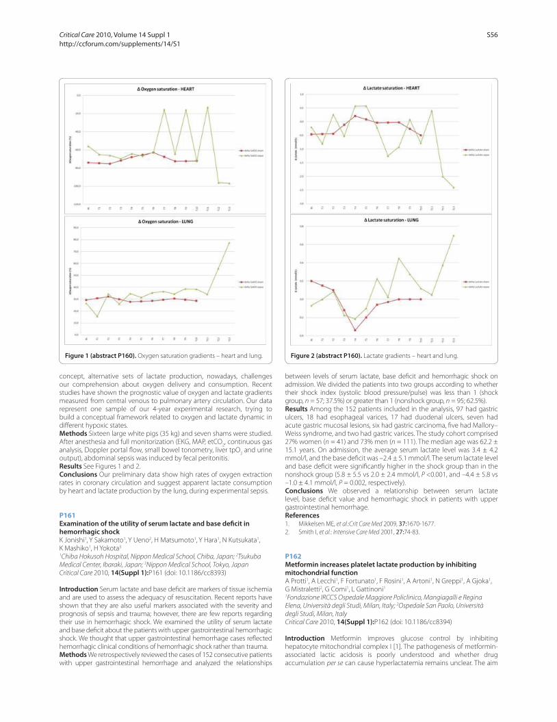
concept, alternative sets of lactate production, nowadays, challenges
our comprehension about oxygen delivery and consumption. Recent
studies have shown the prognostic value of oxygen and lactate gradients
measured from central venous to pulmonary artery circulation. Our data
represent one sample of our 4-year experimental research, trying to
build a conceptual framework related to oxygen and lactate dynamic in
diff erent hypoxic states.
Methods Sixteen large white pigs (35 kg) and seven shams were studied.
After anesthesia and full monitorization (EKG, MAP, etCO2, continuous gas
analysis, Doppler portal fl ow, small bowel tonometry, liver tpO2 and urine
output), abdominal sepsis was induced by fecal peritonitis.
Results See Figures 1 and 2.
Conclusions Our preliminary data show high rates of oxygen extraction
rates in coronary circulation and suggest apparent lactate consumption
by heart and lactate production by the lung, during experimental sepsis.
P161
Examination of the utility of serum lactate and base defi cit in hemorrhagic shockK Jonishi1, Y Sakamoto1, Y Ueno2, H Matsumoto1, Y Hara1, N Kutsukata1,
K Mashiko1, H Yokota3
1Chiba Hokusoh Hospital, Nippon Medical School, Chiba, Japan; 2Tsukuba
Medical Center, Ibaraki, Japan; 3Nippon Medical School, Tokyo, Japan
Critical Care 2010, 14(Suppl 1):P161 (doi: 10.1186/cc8393)
Introduction Serum lactate and base defi cit are markers of tissue ischemia
and are used to assess the adequacy of resuscitation. Recent reports have
shown that they are also useful markers associated with the severity and
prognosis of sepsis and trauma; however, there are few reports regarding
their use in hemorrhagic shock. We examined the utility of serum lactate
and base defi cit about the patients with upper gastrointestinal hemorrhagic
shock. We thought that upper gastrointestinal hemorrhage cases refl ected
hemorrhagic clinical conditions of hemorrhagic shock rather than trauma.
Methods We retrospectively reviewed the cases of 152 consecutive patients
with upper gastrointestinal hemorrhage and analyzed the relationships
between levels of serum lactate, base defi cit and hemorrhagic shock on
admission. We divided the patients into two groups according to whether
their shock index (systolic blood pressure/pulse) was less than 1 (shock
group, n = 57; 37.5%) or greater than 1 (nonshock group, n = 95; 62.5%).
Results Among the 152 patients included in the analysis, 97 had gastric
ulcers, 18 had esophageal varices, 17 had duodenal ulcers, seven had
acute gastric mucosal lesions, six had gastric carcinoma, fi ve had Mallory–
Weiss syndrome, and two had gastric varices. The study cohort comprised
27% women (n = 41) and 73% men (n = 111). The median age was 62.2 ±
15.1 years. On admission, the average serum lactate level was 3.4 ± 4.2
mmol/l, and the base defi cit was –2.4 ± 5.1 mmol/l. The serum lactate level
and base defi cit were signifi cantly higher in the shock group than in the
nonshock group (5.8 ± 5.5 vs 2.0 ± 2.4 mmol/l, P <0.001, and –4.4 ± 5.8 vs
–1.0 ± 4.1 mmol/l, P = 0.002, respectively).
Conclusions We observed a relationship between serum lactate
level, base defi cit value and hemorrhagic shock in patients with upper
gastrointestinal hemorrhage.
References1. Mikkelsen ME, et al.:Crit Care Med 2009, 37:1670-1677.
2. Smith I, et al.: Intensive Care Med 2001, 27:74-83.
P162
Metformin increases platelet lactate production by inhibiting mitochondrial functionA Protti1, A Lecchi1, F Fortunato1, F Rosini1, A Artoni1, N Greppi1, A Gjoka1,
G Mistraletti2, G Comi1, L Gattinoni1
1Fondazione IRCCS Ospedale Maggiore Policlinico, Mangiagalli e Regina
Elena, Università degli Studi, Milan, Italy; 2Ospedale San Paolo, Università
degli Studi, Milan, Italy
Critical Care 2010, 14(Suppl 1):P162 (doi: 10.1186/cc8394)
Introduction Metformin improves glucose control by inhibiting
hepatocyte mitochondrial complex I [1]. The pathogenesis of metformin-
associated lactic acidosis is poorly understood and whether drug
accumulation per se can cause hyperlactatemia remains unclear. The aim
Figure 1 (abstract P160). Oxygen saturation gradients – heart and lung. Figure 2 (abstract P160). Lactate gradients – heart and lung.
Critical Care 2010, Volume 14 Suppl 1 http://ccforum.com/supplements/14/S1
S56
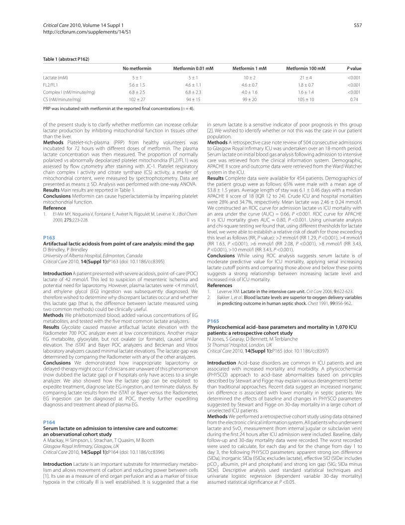
of the present study is to clarify whether metformin can increase cellular
lactate production by inhibiting mitochondrial function in tissues other
than the liver.
Methods Platelet-rich-plasma (PRP) from healthy volunteers was
incubated for 72 hours with diff erent doses of metformin. The plasma
lactate concentration was then measured. The proportion of normally
polarized vs abnormally depolarized platelet mitochondria (FL2/FL1) was
assessed by fl ow cytometry after staining with JC-1. Platelet respiratory
chain complex I activity and citrate synthase (CS) activity, a marker of
mitochondrial content, were measured by spectrophotometry. Data are
presented as means ± SD. Analysis was performed with one-way ANOVA.
Results Main results are reported in Table 1.
Conclusions Metformin can cause hyperlactatemia by impairing platelet
mitochondrial function.
Reference1. El-Mir MY, Nogueira V, Fontaine E, Avéret N, Rigoulet M, Leverve X: J Biol Chem
2000, 275:223-228.
P163
Artifactual lactic acidosis from point of care analysis: mind the gapD Brindley, P Brindley
University of Alberta Hospital, Edmonton, Canada
Critical Care 2010, 14(Suppl 1):P163 (doi: 10.1186/cc8395)
Introduction A patient presented with severe acidosis, point-of-care (POC)
lactate of 42 mmol/l. This led to suspicion of mesenteric ischemia and
potential need for laparotomy. However, plasma lactates were <4 mmol/l,
and ethylene glycol (EG) ingestion was subsequently diagnosed. We
therefore wished to determine why discrepant lactates occur and whether
this lactate gap (that is, the diff erence between lactate measured using
two common methods) could be clinically useful.
Methods We phlebotomized blood, added various concentrations of EG
metabolites, and tested with the fi ve most common lactate analyzers.
Results Glycolate caused massive artifactual lactate elevation with the
Radiometer 700 POC analyzer even at low concentrations. Another major
EG metabolite, glyoxylate, but not oxalate (or formate), caused similar
elevation. The iSTAT and Bayer POC analyzers and Beckman and Vitros
laboratory analyzers caused minimal lactate elevations. The lactate gap was
determined by comparing the Radiometer with any of the other analyzers.
Conclusions We demonstrated how inappropriate laparotomy or
delayed-therapy might occur if clinicians are unaware of this phenomenon
(now dubbed the lactate gap) or if hospitals only have access to a single
analyzer. We also showed how the lactate gap can be exploited: to
expedite treatment, diagnose late EG ingestion, and terminate dialysis. By
comparing lactate results from the iSTAT or Bayer versus the Radiometer,
EG ingestion can be diagnosed at POC, thereby further expediting
diagnosis and treatment ahead of plasma EG.
P164
Serum lactate on admission to intensive care and outcome: an observational cohort studyA Mackay, H Simpson, L Strachan, T Quasim, M Booth
Glasgow Royal Infi rmary, Glasgow, UK
Critical Care 2010, 14(Suppl 1):P164 (doi: 10.1186/cc8396)
Introduction Lactate is an important substrate for intermediary metabo-
lism and allows movement of carbon and reducing power between cells
[1]. Its use as a measure of end organ perfusion and as a marker of tissue
hypoxia in the critically ill is well established. It is suggested that a rise
in serum lactate is a sensitive indicator of poor prognosis in this group
[2]. We wished to identify whether or not this was the case in our patient
population.
Methods A retrospective case note review of 504 consecutive admissions
to Glasgow Royal Infi rmary ICU was undertaken over an 18-month period.
Serum lactate on initial blood gas analysis following admission to intensive
care was retrieved from the clinical information system. Demographic,
APACHE II score and outcome data were retrieved from the Ward Watcher
system in the ICU.
Results Complete data were available for 454 patients. Demographics of
the patient group were as follows: 65% were male with a mean age of
53.8 ± 1.5 years. Average length of stay was 6.1 ± 0.46 days with a median
APACHE II score of 18 (IQR 12 to 24). Crude ICU and hospital mortalities
were 28% and 34.7%, respectively. Mean lactate was 2.46 ± 0.24 mmol/l.
We constructed an ROC curve for admission lactate vs ICU mortality with
an area under the curve (AUC) = 0.66, P <0.001. ROC curve for APACHE
II vs ICU mortality gives AUC = 0.80, P <0.001. Using univariate analysis
and chi-square testing we found that, using diff erent thresholds for lactate
level, we were able to establish a relative risk of death for those exceeding
this level as follows (RR, P value): >2 mmol/l (RR 1.29, P <0.001), >4 mmol/l
(RR 1.63, P <0.001), >6 mmol/l (RR 2.08, P <0.001), >8 mmol/l (RR 3.43,
P <0.001), >10 mmol/l (RR 3.43, P <0.001).
Conclusions While using ROC analysis suggests serum lactate is of
moderate predictive value for ICU mortality, applying serial increasing
lactate cutoff points and comparing those above and below these points
suggests a strong relationship between increasing lactate level and
increased risk of ICU mortality.
References1. Leverve XM: Lactate in the intensive care unit. Crit Care 2006, 9:622-623.
2. Bakker J, et al.: Blood lactate levels are superior to oxygen delivery variables in predicting outcome in human septic shock. Chest 1991, 99:956-962.
P165
Physicochemical acid–base parameters and mortality in 1,070 ICU patients: a retrospective cohort studyN Jones, S Gearay, D Bennett, M Terblanche
St Thomas’ Hospital, London, UK
Critical Care 2010, 14(Suppl 1):P165 (doi: 10.1186/cc8397)
Introduction Acid–base disorders are common in ICU patients and are
associated with increased mortality and morbidity. A physicochemical
(PHYSCO) approach to acid–base abnormalities based on principles
described by Stewart and Figge may explain various derangements better
than traditional approaches. Recent data suggest an increased inorganic
ion diff erence is associated with lower mortality in septic patients. We
determined the eff ects of baseline and changes in PHYSCO parameters
suggested by Stewart and Figge on 30-day mortality in a large cohort of
unselected ICU patients.
Methods We performed a retrospective cohort study using data obtained
from the electronic clinical information system. All patients who underwent
lactate and SvO2 measurement (from internal jugular or subclavian vein)
during the fi rst 24 hours after ICU admission were included. Baseline, daily
follow-up and 30-day mortality data were recorded. The worst recorded
were used to calculate, for each day and for the change from day 1 to
day 3, the following PHYSCO parameters: apparent strong ion diff erence
(SIDa), inorganic SIDa (iSIDa; excludes lactate), eff ective SID (SIDe: includes
pCO2, albumin, pH and phosphate) and strong ion gap (SIG; SIDa minus
SIDe). Descriptive analysis used standard statistical techniques and
univariate logistic regression (dependent variable 30-day mortality)
assumed statistical signifi cance at P <0.05.
Table 1 (abstract P162)
No metformin Metformin 0.01 mM Metformin 1 mM Metformin 100 mM P value
Lactate (mM) 5 ± 1 5 ± 1 10 ± 2 21 ± 4 <0.001
FL2/FL1 5.6 ± 1.5 4.6 ± 1.1 4.6 ± 0.7 1.8 ± 0.7 <0.001
Complex I (nM/minute/mg) 6.8 ± 2.5 6.8 ± 2.3 4.0 ± 1.6 1.6 ± 1.4 <0.001
CS (nM/minute/mg) 102 ± 27 94 ± 15 99 ± 20 105 ± 10 0.74
PRP was incubated with metformin at the reported fi nal concentrations (n = 4).
Critical Care 2010, Volume 14 Suppl 1 http://ccforum.com/supplements/14/S1
S57

Results A total of 1,544 patients were included. Complete data to calculate
day 1 and day 3 parameters were available in 1,070 and 656 patients,
respectively. Overall 30-day mortality was 21.3%. Mean (SD) age and
APACHE II score were 63.0 (16.4) and 21.6 (6.9), respectively. For survivors
versus nonsurvivors there were signifi cant diff erences in baseline SIDa (P =
0.03), iSIDa, SIDe, lactate and base excess (all P <0.001), but not in SIG (P =
0.1). An increased SIDa (P = 0.015) and SIDe (P <0.001), and decreased iSIDa
and lactate (all P <0.001) but not SIG (P = 0.09) were predictive of mortality.
For day 1 to day 3, only Δ-SIG was weakly predictive of mortality (OR 1.03
per 1 unit increase, P = 0.04, 95% CI = 1.00 to 1.05, n = 656) but there was
no diff erence between survival groups (P = 0.051).
Conclusions This study confi rms recent data suggesting baseline
diff erences (but not changes over 3 days) in the relationships between
strong and weak ions and pCO2 (refl ected in SIDa, lactate, and impacts on
mortality). The diff erent eff ects observed for SIG and BE imply a signifi cant
unmeasured acid load infl uencing mortality. Bias and confounding may
aff ect these fi ndings and they should therefore be confi rmed prospectively.
P166
Accuracy of predicting arterial blood gases following mechanical ventilator adjustmentH Al-Otaibi1, N Bedforth2, R Mahajan1, J Hardman1
1The University of Nottingham, UK; 2Nottingham University Hospitals NHS
Trust, Nottingham, UK
Critical Care 2010, 14(Suppl 1):P166 (doi: 10.1186/cc8398)
Introduction Clinicians adjust mechanical ventilator (MV) in critical care
units to optimise arterial blood gases (ABG). The present study evaluates
their accuracy in predicting ABG values following MV adjustment.
Methods Thirty-one sets of data were collected from 16 patients requiring
mechanical ventilation on the ICU. Each set consisted of two subsets –
before and after MV adjustment. Each of subset consisted of the following
variables: FIO2, respiratory rate (breaths/minute), tidal volume (ml/kg),
body temperature (°C), haemoglobin concentration (g/dl), and ABG values
(pH, PaCO2, PaO
2, base excess). All data, with the exception of ABG values
after MV adjustment, were presented to clinicians. Thirty-three clinicians
(11 respiratory therapists (RTs), 11 nurses, and 11 medical staff ) were asked
to predict the new ABG values as they would in clinical settings. All data
are presented as mean ± SD unless stated otherwise.
Results All datasets include changes in FIO2, while only 13 datasets
include changes in minute ventilation. Increases in FIO2 were 0.19 ± 0.04
and decreases were –0.12 ± 0.04. Size of change of minute ventilation was
1.38 ± 0.95 l/minute. The absolute magnitudes of change for measured
pH, PaCO2, and PaO
2 were 0.024 ± 0.027, 0.40 ± 0.37 kPa and 5.91 ± 3.35
kPa, respectively. The magnitudes of change for predicted pH, PaCO2, and
PaO2, respectively, were 0.025 ± 0.033, 0.34 ± 0.23 kPa, and 3.45 ± 2.03 kPa
for medical staff ; 0.024 ± 0.021, 0.40 ± 0.41 kPa, and 3.91 ± 1.75 kPa for RTs;
and 0.022 ± 0.015, 0.32 ± 0.22 kPa, and 3.21 ± 2.1 kPa for nurses. The 95%
limits of agreement between predicted and measured size of changes for
pH, PaCO2, and PaO
2, respectively, were 0.043 to –0.041, 0.724 to –0.596
kPa, and 3.22 to –8.14 kPa for medical staff ; 0.036 to –0.036, 0.66 to –0.66
kPa, and 2.64 to –6.64 KPa for RTs; and 0.036 to –0.076, 0.74 to –0.9 kPa,
and 3.91 to –9.31 kPa for nurses. Correlation between duration of clinicians’
clinical experience and errors in predicting size of change for pH, PaCO2,
and PaO2 (–0.0225, –0.054, and 0.173, respectively) were weak and not
statistically signifi cant.
Conclusions Clinicians show considerable variation in predicting ABG
values. Medical staff and nurses underestimate the magnitude of PaCO2
changes whereas RTs show some accuracy. All clinicians have a large bias
in predicting the size of PaO2 changes.
P167
Use of capnography and the availability of airway equipment on UK intensive care unitsA Georgiou, S Gouldson, A Amphlett
Frenchay Hospital, Bristol, UK
Critical Care 2010, 14(Suppl 1):P167 (doi: 10.1186/cc8399)
Introduction Provisional data from the 4th National Audit Project of the
Royal College of Anaesthetists has identifi ed that at least 20% of major
airway events take place on the ICU and that these are more frequently
fatal than events elsewhere (TM Cook, personal communication). Over
23% of these incidents relate to the instigation or maintenance of an
artifi cial airway [1]. Both the use of capnography and the immediate
availability of airway rescue devices to avert or manage such incidents
have been recommended. This study seeks to determine the adherence
to these recommendations, the availability of such equipment on UK ICUs
and to identify areas for safety improvement.
Methods Every adult general, hepatobiliary and satellite ICU (grouped
together as GICU), neurointensive care unit (NICU) and cardiac intensive
care unit (CICU) in the UK was telephoned, thereby covering every area
in the UK where ventilated adult patients are routinely cared for outside
the operating theatre. Data were obtained from the nurse in charge over
a 3-month period in 2009.
Results In total, 315 of 328 contacted units responded (96% response).
Capnography during intubation: 32% units use always, 43% sometimes
and 25% never. Capnography for continuous monitoring of intubated
patients: 25% units use always, 35% sometimes and 40% never. Of
note, 45% of NICUs never use capnography for continuous monitoring
(where doing so may prove most benefi cial). Basic airway equipment
(oropharyngeal airways, laryngoscopes and bougies), alternative means of
patient ventilation and equipment checking procedures are seen on 97 to
100% of ICUs. A standard laryngeal mask (LMA) is available on 97% of ICUs.
More advanced airway rescue devices are seen less commonly: 71% have
a McCoy laryngoscope and 55% have a bougie through which ventilation
is possible. Forty-three per cent have an intubating LMA and 21% have
supraglottic airway with a gastric port immediately available. Forty-nine
per cent of units have a bronchoscope on the ICU. Seventy per cent of
units have equipment for emergency trans-tracheal access. CICUs are least
likely to use capnography or have airway rescue equipment immediately
available.
Conclusions Basic standards of airway safety are not being met on UK
ICUs and are far from the standards being routinely employed in the
operating theatre. The potential airway diffi culties presented by the ICU
patient, together with the lack of routine use of capnography, exposes
these patients to signifi cant risks that may be easily addressed.
Reference1. Thomas et al.: Anaesthesia 2009, 64:358-365.
P168
Combined oximetry and transcutaneous capnography is a reliable non-invasive monitoring in patients after cardiac surgeryW Baulig, RR Roediger, O Theusinger, D Rusch, D Spahn
University Hospital Zurich, Switzerland
Critical Care 2010, 14(Suppl 1):P168 (doi: 10.1186/cc8400)
Introduction The aim of this study was to validate the revised V-Sign™ 2
ear lobe sensor for combined assessment of pulse rate (PR), pulse oximetry
(SpO2) and transcutaneous carbon dioxide tension (PtcCO
2) in adults after
cardiac surgery.
Methods In 20 patients coevally readings of blood gases were compared
with values of the V-Sign™ 2 ear lobe sensor. Measurements were
performed during periods of hypercapnia, normocapnia and hypocapnia
and then in intervals of 30 minutes up to 5 hours. Agreement was assessed
by Bland–Altman analysis and single regression analysis.
Results Two hundred and ninety-six data pairs were analysed. Detection
failures for PtcCO2 were 1.3%, for SpO
2 10% and for PR 5%. PtcCO
2 and
PaCO2 ranged between 3.58 and 7.70 kPa. Mean bias and limits of
agreement (LOA) between PtcCO2 and PaCO
2 was excellent (Table 1). The
best agreement between PtcCO2 and PaCO
2 was found during periods of
Table 1 (abstract P168). Agreement between V-Sign™ 2 sensor and
co-oximetry and ECG
Limits of
Parameter n (data pairs) r2 Mean bias agreement
PtcCO2–PaCO
2 (kPa) 296 0.92 +0.14 –0.45/+0.73
V-Sign™ SpO2–SaO
2 (%) 270 0.59 –1.68 –6.81/+3.45
V-Sign™ PR-ECG (bpm) 285 0.98 +1.22 –3.31/+5.75
Critical Care 2010, Volume 14 Suppl 1 http://ccforum.com/supplements/14/S1
S58
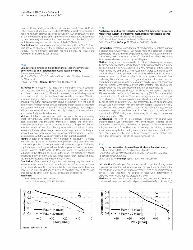
hyperventilation and hypoventilation with a mean bias (LOA) of +0.18 kPa
(–0.31/+0.67 kPa) and 0.07 kPa (–0.45/+0.59 kPa), respectively. At about 5
hours no relevant drift was observed between PtcCO2 and PaCO
2. V-Sign™
2 SpO2 moderately agreed and slightly underestimated SaO
2 determined
by direct oximetry, whereas pulse rate values detected by the V-Sign™ 2
ear lobe sensor agreed well with those of the ECG (Table 1).
Conclusions Transcutaneous capnography using the V-Sign™ 2 ear
lobe sensor reliably detects the ventilation state of patients after cardiac
surgery. This non-invasive approach has the potential to reduce the
number of arterial blood gas samples.
P169
Computerized lung sound monitoring to assess eff ectiveness of physiotherapy and secretion removal: a feasibility studyG Ntoumenopoulos1, Y Glickman2
1Guy’s and St Thomas’ NHS Foundation Trust, London, UK; 2Deep Breeze Ltd,
Or-Akiva, Israel
Critical Care 2010, 14(Suppl 1):P169 (doi: 10.1186/cc8401)
Introduction Intubation and mechanical ventilation impair secretion
clearance and can lead to lung collapse, consolidation and ventilator-
associated pneumonia [1]. There is, however, no valid diagnosis of
secretion retention in the intubated and ventilated patient. Vibration
response imaging (VRI) is a commercially developed acoustic lung
imaging system that displays breath sound distribution [2]. VRI should be
able to identify relationships between specifi c breath sound distributions
and secretion retention. This preliminary study investigated the changes in
VRI measurements before and after chest physiotherapy in adult intubated
and mechanically ventilated patients.
Methods Intubated and ventilated adult patients who were receiving
chest physiotherapy were investigated. Lung sound amplitude at
peak inspiration was measured immediately before and after chest
physiotherapy using two arrays of sensors attached to the patient’s back in
a supine position. Chest physiotherapy included combinations of closed
airway suctioning, saline lavage, postural drainage, manual techniques
and/or lung hyperinfl ation, dependent upon clinical indications. Means
are compared with the Wilcoxon matched-pairs signed-ranks test.
Results A total of 16 patients were included in the study (12 males,
four females, age 65 ± 14). Patients were predominantly ventilated with
continuous positive airway pressure and pressure support. Following
physiotherapy, total lung sound amplitude at peak inspiration decreased
twofold from 37 ± 58.106 to 18 ± 23.106 arbitrary units (AU), with signifi cant
reduction in the left lung (P = 0.03). Furthermore, the diff erence in sound
amplitude between right and left lungs signifi cantly decreased post-
treatment compared with pretreatment (P = 0.03).
Conclusions Computerized lung sound monitoring may be useful to
assess secretion retention and the eff ectiveness of secretion removal
in mechanically ventilated patients. Further investigation is, however,
necessary in order to distinguish between secretion-related eff ects and
changes due to other factors such as airfl ow rate and pattern.
References1. Konrad et al.: Chest 1994, 105:237-241.
2. Jean et al.: Anesth Analg 2008, 107:1243-1247.
P170
Analysis of sound waves recorded with the VRI pulmonary acoustic monitoring system in critically ill mechanically ventilated patientsS Lev1, YA Glickman2, M Shapiro1, PS Singer1
1RMC, Petach Tiqva, Israel; 2Deep Breeze, Or Akiva, Israel
Critical Care 2010, 14(Suppl 1):P170 (doi: 10.1186/cc8402)
Introduction Posterior auscultation of mechanically ventilated patient
is challenging. Environmental ICU noises make the detection of subtle
auscultatory features diffi cult. Digitalized pulmonary acoustic monitoring
has recently been introduced in the ICU. We explored the possibility to
listen to sound waves recorded by the VRI system.
Methods Lung sounds were recorded for 20 seconds using two arrays of
17 piezoelectric contact sensors attached to the patient’s back in a supine
position. Sound fi les were archived in a WAV format for offl ine analysis.
Three physicians and one trained respiratory therapist, blinded to the
patient’s clinical status, provided their fi ndings while listening to sound
waves recorded by 12 sensors distributed from apex to base, six from
each lung. Breath sounds were categorized as normal versus abnormal
and adventitious lung sounds were characterized as wheezes, rhonchi or
crackles. Findings were compared with anterior stethoscope auscultation
performed at the time of the recording by one of the physicians.
Results Eighteen critically ill mechanically ventilated patients (age 65 ±
17) were enrolled in this study. Chest radiography (CXR) fi ndings included
consolidation in 13 patients. There was an agreement among at least
three clinicians in the normal/abnormal classifi cation. Furthermore, in
10 out of these 15 patients (67%), this assessment based on sound wave
analysis was in agreement with anterior stethoscope auscultation. Finally,
the physician who performed both sound wave analysis and stethoscope
auscultation reported crackles/rhonchi in fi ve out of 13 patients with
consolidation (38%) during sound wave analysis but only in one patient
during auscultation (8%).
Conclusions The level of interobserver variability for sound wave
characterization was comparable with levels usually reported during
stethoscope auscultation. However, in patients with consolidation,
a higher number of crackles/rhonchi was reported during posterior
sound wave analysis than during anterior stethoscope auscultation. This
discrepancy may be either due to the anterior/posterior orientation or to
the higher sensitivity of the offl ine sound wave analysis.
P171
Lung tissue properties obtained by optical alveolar elastometryD Schwenninger1, H Runck1, S Schumann1, K Möller2
1University Medical Center of Freiburg, Germany; 2Furtwangen University,
VS-Schwenningen, Germany
Critical Care 2010, 14(Suppl 1):P171 (doi: 10.1186/cc8403)
Introduction Knowledge of micromechanical properties of lung paren-
chyma is essential for understanding macroscopic lung mechanics. The
fi rst results of in vivo and in situ measurements using an endomicroscopic
device [1] are reported. The degree of local lung deformation in
dependence of locally applied pressure is shown.
Methods An endoscopic system including two concentric trocars was
constructed to apply a defi ned local pressure within the fi eld of view (Pfov
).
Figure 1 (abstract P171).
Critical Care 2010, Volume 14 Suppl 1 http://ccforum.com/supplements/14/S1
S59
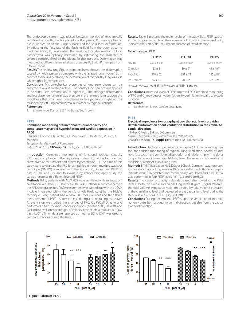
The endoscopic system was placed between the ribs of mechanically
ventilated rats with the tip placed on the pleura. Pfov
was applied to
a circular area on to the lungs’ surface and led to a local deformation.
By adjusting the fl ow rate of the fl ushing fl uid from the outer trocar to
the inner trocar, Pfov
was varied. The resulting local deformation of lung
parenchyma was optically measured by estimating the diameter of
ceramic particles, fi xed on the pleura for that purpose. Deformation was
measured at diff erent levels of airway pressure (Paw
) with Pfov
ramped from
0 to –40 mbar.
Results The healthy lung (Figure 1A) parenchyma showed less deformation
caused by fl uidic pressure compared with the lavaged lung (Figure 1B). In
contrast to the lavaged lung, the deformation of the healthy lung was less
when higher Paw
was present.
Conclusions Micromechanical properties of lung parenchyma can be
analyzed in vivo at an alveolar level. The healthy lung parenchyma appears
to be stiff er (less deformation) at higher Paw
. The stronger deformation
and less dependence on airway pressure in the lavaged lung support the
hypothesis that small lung compliance in lavaged lungs might not be
reasoned by stiff lung parenchyma, but rather by regional collapse.
References1. Schwenninger D, et al.: IEEE Trans Biomed Eng in press.
P172
Combined monitoring of functional residual capacity and compliance may avoid hyperinfl ation and cardiac depression in ARDSF Turani, L Cococcia, R Barchetta, F Mounayerfi , E Di Marzio, M Falco, A
Marinelli
European Aurelia Hospital, Rome, Italy
Critical Care 2010, 14(Suppl 1):P172 (doi: 10.1186/cc8404)
Introduction Combined monitoring of functional residual capacity
(FRC) and compliance of the respiratory system (Crs) at the bedside may
allow alveolar recruitment and detect hyperinfl ation [1]. The aims of this
study were to evaluate the FRC by a modifi ed nitrogen multiple washout
technique (NMBW) combined with the study of Crs, to set best PEEP on
data of FRC and Crs, and to evaluate by echocardiography study the
cardiac response to diff erent levels of PEEP.
Methods Thirty patients with ALI/ARDS were ventilated with an Engstrom
carestation ventilator (GE Healthcare, Helsinki, Finland) in accordance with
the ARDS net guidelines. FRC measurement was carried out with the COVX
module integrated within the ventilator (GE Healthcare) by the NMBW
technique. Every patient had a basal FRC measurement and then three
measurements at PEEP 15/10/5 cm H2O during a de-recruiting maneuver.
At every step we studied the changes of FRC, Crs, PaO
2/FIO
2 ratio and
performed a transthoracic echocardiography (Agilent 5500; Hewlett and
Packard) to evaluate the integral of velocity time of left ventricular outfl ow
tract (LVOT VTI). All data are reported as mean ± SD. ANOVA was used to
compare changes during the time.
Results Table 1 presents the main results of the study. Best PEEP was set
at 10 cmH2O, at which level the decrease of FRC and improvement of C
rs
indicates the start of de-recruitment and end of overdistention.
Table 1 (abstract P172)
PEEP 15 PEEP 10 PEEP 5
FRC ml 2,975 ± 644 2,412 ± 585* 2,059 ± 556**
Crs ml/cm 33 ± 8 39 ± 9* 45 ± 10**
PaO2/FIO
2 310 ± 62 291 ± 78 185 ± 89†
LVOT VTI cm 16.3 ± 2 20 ± 3* 22 ± 6**
*P <0.05. **P <0.01 vs PEEP 15. †P <0.001 vs PEEP 15 and 10.
Conclusions Increased levels of PEEP improve FRC. Combined monitoring
of FRC and Crs may detect hyperinfl ation. Hyperinfl ation impairs LV systolic
function.
References1. Lambermont B, et al.: Crit Care 2008, 12:R91.
P173
Electrical impedance tomography at two thoracic levels provides detailed information about ventilation distribution in the cranial to caudal directionI Bikker, C Preis, J Bakker, D Gommers
Erasmus Medical Center, Rotterdam, the Netherlands
Critical Care 2010, 14(Suppl 1):P173 (doi: 10.1186/cc8405)
Introduction Electrical impedance tomography (EIT) is a promising new
tool for bedside monitoring of regional lung ventilation. Several studies
have focused on the ventilation distribution and relationship with regional
lung volume on a lower, caudal lung level. However, no information is
available at a higher, cranial lung level.
Methods EIT (EIT Evaluation Kit 2; Dräger, Lübeck, Germany) was measured
at cranial and caudal lung levels in 10 patients after cardiothoracic surgery.
Patients were fully sedated and mechanically ventilated and a PEEP trial
was performed at four PEEP levels (15, 10, 5 and 0 cmH2O).
Results The center of gravity index decreased after lowering the PEEP
level at both the caudal and cranial lung levels (Figure 1 right). Whereas
the tidal volume impedance variation divided by tidal volume increased
at the cranial lung level and decreased at the caudal lung level during the
step-wise reductions in PEEP (Figure 1 left).
Conclusions During decremental PEEP steps, the ventilation distribution
not only shifts from a dorsal to ventral direction, but also from the caudal
to cranial direction.
Figure 1 (abstract P173).
Critical Care 2010, Volume 14 Suppl 1 http://ccforum.com/supplements/14/S1
S60

P174
Changes in regional ventilation determined by volume and pressure-controlled ventilation as assessed by electrical impedance tomography in an experimental model of acid-induced acute lung injuryCA Holms, DA Otsuki, J Noel-Morgan, M Kahvegian, DT Fantoni, JO Auler Jr
Faculdade de Medicina da Universidade de São Paulo, Brazil
Critical Care 2010, 14(Suppl 1):P174 (doi: 10.1186/cc8406)
Introduction Electrical impedance tomography (EIT) is a promising bedside
tool for monitoring regional lung ventilation during mechanical ventilation
[1]. The purpose of this study was to assess regional lung ventilation by
EIT in a pig model of acid-induced acute lung injury (ALI) during pressure-
controlled (PCV) and volume-controlled ventilation (VCV).
Methods Ten anesthetized and tracheotomized female pigs (25 to 30
kg) were randomized into two groups: mechanical ventilation by PCV
(pressure adjusted to achieve and maintain 8 ml/kg tidal volume, n =
5) or VCV (8 ml/kg, n = 5) in FiO2 50%. ALI was induced in both groups
by intratracheal instillation of 4 ml/kg hydrochloric acid (HCl) 0.1 N.
Impedance changes (Draeger, Germany) were recorded by a 16-electrode
belt placed at the level of the sixth intercostal space, and four horizontal,
equally-sized regions of interest (ROIs) were defi ned for offl ine data
analysis. Measurements were performed before (T-BL) and after lung
injury (T-ALI). Lung tidal volume (Vt), pulmonary static compliance (Cstat
),
pulmonary mean artery pressure (mPAP), peak pressure (Pp), respiratory
rate (RR) and PaO2/FiO
2 ratio were also recorded. Statistical analysis was
based on two-way ANOVA followed by the Tukey test for analysis of data
within and between groups, and a t test was used analysis of impedance
changes induced by ALI.
Results After ALI, both PCV and VCV groups showed signifi cant decreases
in the PaO2/FiO
2 ratio (479.6 ± 46.5 to 244.5 ± 64.7 and 428.8 ± 22.9 to
212.4 ± 26.7) and Cstat
(29.2 ± 5.1 to 13.2 ± 2.3 and 26.6 ± 3.4 to 12.3 ± 3.2)
and signifi cant increases in Pp (14.2 ± 1.9 to 25.6 ± 2.6 and 13.8 ± 0.8 to
24.6 ± 4.16) and mPAP (19.6 ± 0.5 to 35.2 ± 5.8 and 19.4 ± 1.5 to 26.8 ±
4.5). Between groups, only mPAP was statistically diff erent at T-ALI. Global
impedance decreased slightly after ALI in both groups, with no statistical
signifi cance. At T-ALI, there were statistically signifi cant diff erences in tidal
distributions of ROIs 2 (38.9 ± 2.6% to 42.4 ± 3.9%) and 3 (39.7 ± 3.1 to
35.1 ± 4.3%) of animals in the VCV group, when compared with T-BL. No
signifi cant diff erence was found in the PCV group.
Conclusions EIT assessment of ventilation revealed a ventral shift in the
tidal distribution in the VCV group after ALI, while PCV preserved tidal
distribution observed at baseline.
Acknowledgements Grants from FAPESP (08/55376-7 and 08/56792-4)
and LIM08/FMUSP.
Reference1. Frerichs I, et al.: Physiol Meas 2007, 28:S261-S267.
P175
Changes in lung tidal distribution assessed by electrical impedance tomography in a model of hemorrhagic shock with endotoxemia and resuscitationJ Noel-Morgan, DT Fantoni, DA Otsuki, CA Holms, JO Auler Jr
Faculdade de Medicina da Universidade de São Paulo, Brazil
Critical Care 2010, 14(Suppl 1):P175 (doi: 10.1186/cc8407)
Introduction Electrical impedance tomography (EIT) is a non-invasive
bedside imaging tool with the potential to assess regional lung ventilation
reliably [1]. The purpose of this study was to monitor changes in lung
impedance in a pig model of severe shock followed by fl uid resuscitation.
Methods Twelve anesthetized, mechanically ventilated (8 ml/kg, PEEP
5 cmH2O), supine pigs were submitted to acute hemorrhagic shock with
infusion of endotoxin. Animals were allocated to positive control (PC, n =
6) or treatment group with lactated Ringer’s to achieve and maintain pulse
pressure variation 13% and mean arterial pressure 65 mmHg (PPV, n = 6).
Ventilatory and hemodynamic parameters were recorded at baseline,
1 hour after hemorrhagic shock (Tshock), and hourly for 3 hours (T1 to T3).
A 16-electrode belt was placed at the level of the sixth intercostal space
for EIT measurements (Dräger, Germany). Offl ine analysis was based on
four horizontal regions of interest (ROIs) over the ventrodorsal lung area.
Statistical analysis was based on two-way ANOVA followed by the Tukey
test (P <0.05).
Results At Tshock there was hemodynamic compromise, statistical
decrease in lung compliance (Cstat
) and signifi cant increases in pulmonary
vascular resistance index, mean pulmonary artery pressure and peak
pressure (Ppeak
), with no statistical diff erence between groups. Endpoints
and hemodynamic stability were achieved in the PPV group in 117 ±
28 minutes. Cstat
continued to deteriorate and Ppeak
continued to rise in
both groups from T1 to T3. When compared with PC, the PPV group had
signifi cant impedance increases in ROIs 1 and 2 at T2 and T3 and, at T3
the increase in ROI 1 was also statistically greater than Tshock within the
group. Statistical decrease in the percentage tidal distribution in ROI 3 and
increase in ROI 2 of the PPV group, in relation to the PC group, were also
noted.
Conclusions Despite re-establishment of hemodynamic adequacy in PPV
group and although ventilatory parameters were similar in both groups
over time, resuscitation as performed in the study induced signifi cant
changes in tidal impedance toward nondependent lung regions, implying
greater lung impairment in treated animals.
Acknowledgements Grants from FAPESP 08/50063-0, 08/50062-4 and
LIM08/FMUSP.
Reference1. Victorino et al.: Am J Respir Crit Care Med 2004, 169:791-800.
P176
Estimating the intratidal compliance profi le with the new gliding-SLICE methodS Schumann1, B Burcza1, C Haberthuer2, M Lichtwarck-Aschoff 3,
J Guttmann1
1University Medical Center, Freiburg, Germany; 2Kantonsspital, Luzern,
Switzerland; 3University of Uppsala, Sweden
Critical Care 2010, 14(Suppl 1):P176 (doi: 10.1186/cc8408)
Introduction At the bedside, but even in most research work, the analysis
of respiratory system mechanics is limited to quasi-static conditions
excluding any insight into what happens during the breath. The new
gliding-SLICE method helps looking into the breath. It is a further
development of the SLICE method for calculating compliance and
resistance of subsequent intratidal volume ranges (slices) of the pressure–
volume (PV) loop by multiple linear regression analysis in a continuous
way. This allows for detecting intratidal compliance and resistance
nonlinearity during ongoing ventilation. Our objective was to determine
whether the nonlinear intratidal compliance profi le hints at what level to
set PEEP and tidal volume (VT) to make lung ventilation protective.
Methods In 12 piglets, atelectasis was induced by application of negative
pressure. The PV relationship and the ECG signal were recorded during
mechanical ventilation at diff erent levels of end-expiratory pressure (PEEP:
0, 5, 10 and 15 cmH2O) and a VT of 12 ml/kg BW. Using the gliding-SLICE
method [1], intratidal compliance profi les were calculated and compared
with the conventional quasi-static compliance.
Results In contrast to quasi-static compliance, the gliding-SLICE method
revealed pronounced intratidal nonlinearity of the compliance profi le
under ongoing ventilation (Figure 1). At low levels of PEEP, intratidal
compliance increased in the low volume range, remained at a high level
while further volume was delivered, and fi nally decreased with volume
>6 ml/kg BW. With higher levels of PEEP, intratidal compliance decreased
from the onset of inspiration.
Conclusions The gliding-SLICE method gives detailed insights into the
intratidal course of compliance during uninterrupted ventilation. From
Figure 1 (abstract P176).
Critical Care 2010, Volume 14 Suppl 1 http://ccforum.com/supplements/14/S1
S61
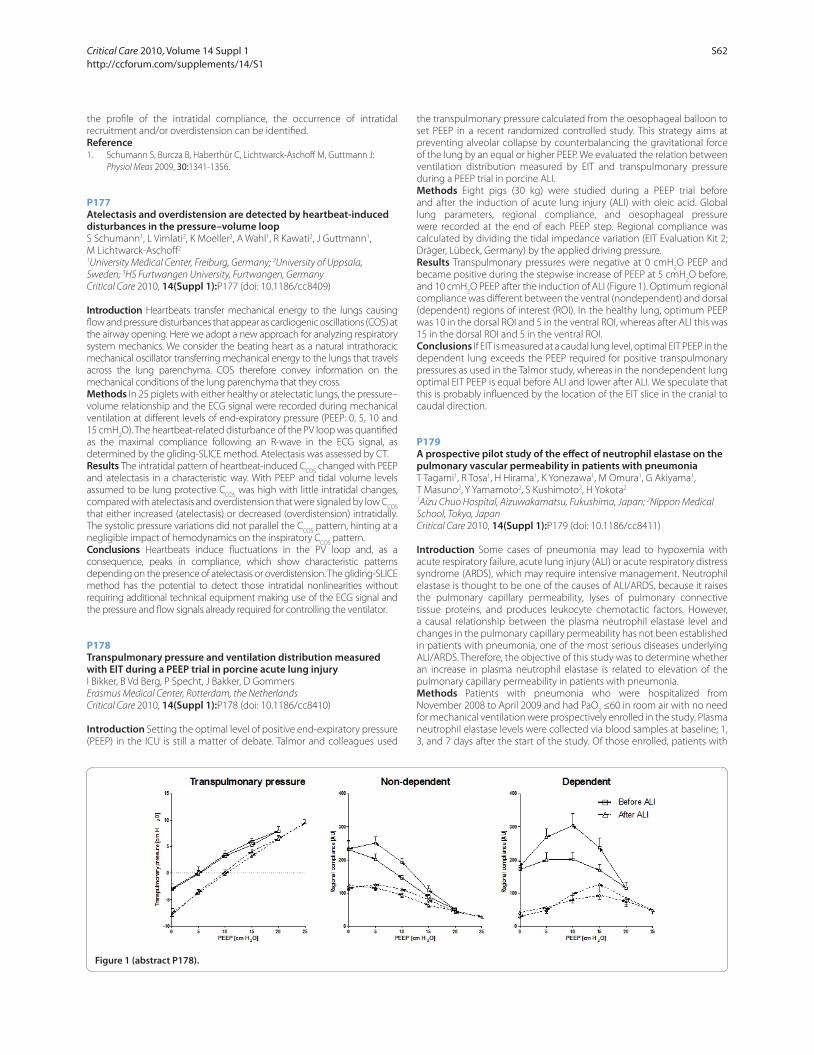
the profi le of the intratidal compliance, the occurrence of intratidal
recruitment and/or overdistension can be identifi ed.
Reference1. Schumann S, Burcza B, Haberthür C, Lichtwarck-Aschoff M, Guttmann J:
Physiol Meas 2009, 30:1341-1356.
P177
Atelectasis and overdistension are detected by heartbeat-induced disturbances in the pressure–volume loopS Schumann1, L Vimlati2, K Moeller3, A Wahl1, R Kawati2, J Guttmann1,
M Lichtwarck-Aschoff 2
1University Medical Center, Freiburg, Germany; 2University of Uppsala,
Sweden; 3HS Furtwangen University, Furtwangen, Germany
Critical Care 2010, 14(Suppl 1):P177 (doi: 10.1186/cc8409)
Introduction Heartbeats transfer mechanical energy to the lungs causing
fl ow and pressure disturbances that appear as cardiogenic oscillations (COS) at
the airway opening. Here we adopt a new approach for analyzing respiratory
system mechanics. We consider the beating heart as a natural intrathoracic
mechanical oscillator transferring mechanical energy to the lungs that travels
across the lung parenchyma. COS therefore convey information on the
mechanical conditions of the lung parenchyma that they cross.
Methods In 25 piglets with either healthy or atelectatic lungs, the pressure–
volume relationship and the ECG signal were recorded during mechanical
ventilation at diff erent levels of end-expiratory pressure (PEEP: 0, 5, 10 and
15 cmH2O). The heartbeat-related disturbance of the PV loop was quantifi ed
as the maximal compliance following an R-wave in the ECG signal, as
determined by the gliding-SLICE method. Atelectasis was assessed by CT.
Results The intratidal pattern of heartbeat-induced CCOS
changed with PEEP
and atelectasis in a characteristic way. With PEEP and tidal volume levels
assumed to be lung protective CCOS
was high with little intratidal changes,
compared with atelectasis and overdistension that were signaled by low CCOS
that either increased (atelectasis) or decreased (overdistension) intratidally.
The systolic pressure variations did not parallel the CCOS
pattern, hinting at a
negligible impact of hemodynamics on the inspiratory CCOS
pattern.
Conclusions Heartbeats induce fl uctuations in the PV loop and, as a
consequence, peaks in compliance, which show characteristic patterns
depending on the presence of atelectasis or overdistension. The gliding-SLICE
method has the potential to detect those intratidal nonlinearities without
requiring additional technical equipment making use of the ECG signal and
the pressure and fl ow signals already required for controlling the ventilator.
P178
Transpulmonary pressure and ventilation distribution measured with EIT during a PEEP trial in porcine acute lung injuryI Bikker, B Vd Berg, P Specht, J Bakker, D Gommers
Erasmus Medical Center, Rotterdam, the Netherlands
Critical Care 2010, 14(Suppl 1):P178 (doi: 10.1186/cc8410)
Introduction Setting the optimal level of positive end-expiratory pressure
(PEEP) in the ICU is still a matter of debate. Talmor and colleagues used
the transpulmonary pressure calculated from the oesophageal balloon to
set PEEP in a recent randomized controlled study. This strategy aims at
preventing alveolar collapse by counterbalancing the gravitational force
of the lung by an equal or higher PEEP. We evaluated the relation between
ventilation distribution measured by EIT and transpulmonary pressure
during a PEEP trial in porcine ALI.
Methods Eight pigs (30 kg) were studied during a PEEP trial before
and after the induction of acute lung injury (ALI) with oleic acid. Global
lung parameters, regional compliance, and oesophageal pressure
were recorded at the end of each PEEP step. Regional compliance was
calculated by dividing the tidal impedance variation (EIT Evaluation Kit 2;
Dräger, Lübeck, Germany) by the applied driving pressure.
Results Transpulmonary pressures were negative at 0 cmH2O PEEP and
became positive during the stepwise increase of PEEP at 5 cmH2O before,
and 10 cmH2O PEEP after the induction of ALI (Figure 1). Optimum regional
compliance was diff erent between the ventral (nondependent) and dorsal
(dependent) regions of interest (ROI). In the healthy lung, optimum PEEP
was 10 in the dorsal ROI and 5 in the ventral ROI, whereas after ALI this was
15 in the dorsal ROI and 5 in the ventral ROI.
Conclusions If EIT is measured at a caudal lung level, optimal EIT PEEP in the
dependent lung exceeds the PEEP required for positive transpulmonary
pressures as used in the Talmor study, whereas in the nondependent lung
optimal EIT PEEP is equal before ALI and lower after ALI. We speculate that
this is probably infl uenced by the location of the EIT slice in the cranial to
caudal direction.
P179
A prospective pilot study of the eff ect of neutrophil elastase on the pulmonary vascular permeability in patients with pneumoniaT Tagami1, R Tosa1, H Hirama1, K Yonezawa1, M Omura1, G Akiyama1,
T Masuno2, Y Yamamoto2, S Kushimoto2, H Yokota2
1Aizu Chuo Hospital, Aizuwakamatsu, Fukushima, Japan; 2Nippon Medical
School, Tokyo, Japan
Critical Care 2010, 14(Suppl 1):P179 (doi: 10.1186/cc8411)
Introduction Some cases of pneumonia may lead to hypoxemia with
acute respiratory failure, acute lung injury (ALI) or acute respiratory distress
syndrome (ARDS), which may require intensive management. Neutrophil
elastase is thought to be one of the causes of ALI/ARDS, because it raises
the pulmonary capillary permeability, lyses of pulmonary connective
tissue proteins, and produces leukocyte chemotactic factors. However,
a causal relationship between the plasma neutrophil elastase level and
changes in the pulmonary capillary permeability has not been established
in patients with pneumonia, one of the most serious diseases underlying
ALI/ARDS. Therefore, the objective of this study was to determine whether
an increase in plasma neutrophil elastase is related to elevation of the
pulmonary capillary permeability in patients with pneumonia.
Methods Patients with pneumonia who were hospitalized from
November 2008 to April 2009 and had PaO2 ≤60 in room air with no need
for mechanical ventilation were prospectively enrolled in the study. Plasma
neutrophil elastase levels were collected via blood samples at baseline; 1,
3, and 7 days after the start of the study. Of those enrolled, patients with
Figure 1 (abstract P178).
Critical Care 2010, Volume 14 Suppl 1 http://ccforum.com/supplements/14/S1
S62
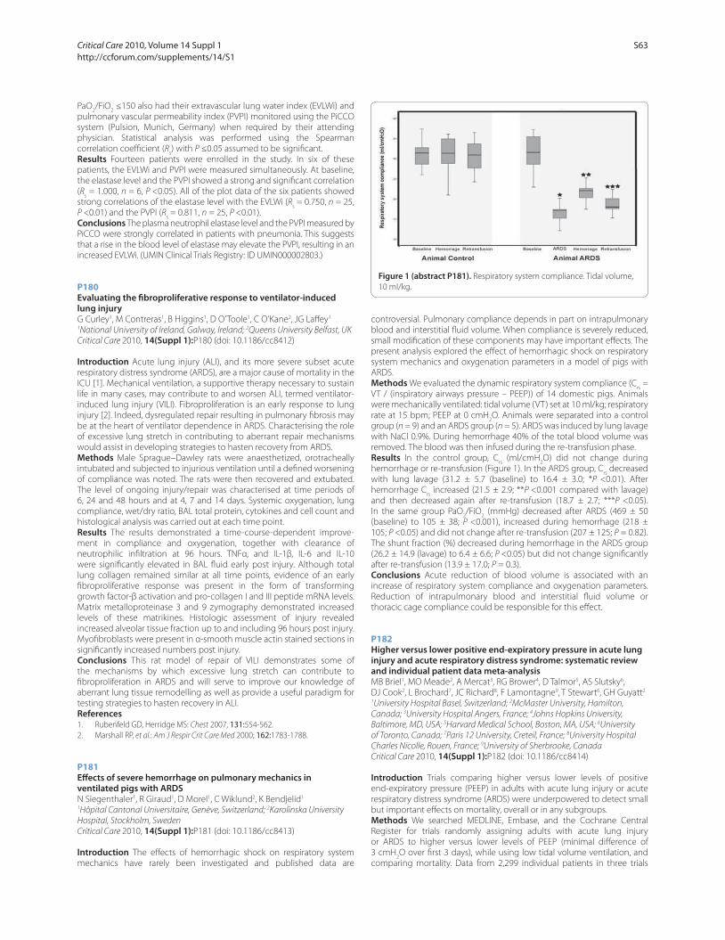
PaO2/FiO
2 ≤150 also had their extravascular lung water index (EVLWi) and
pulmonary vascular permeability index (PVPI) monitored using the PiCCO
system (Pulsion, Munich, Germany) when required by their attending
physician. Statistical analysis was performed using the Spearman
correlation coeffi cient (Rs) with P ≤0.05 assumed to be signifi cant.
Results Fourteen patients were enrolled in the study. In six of these
patients, the EVLWi and PVPI were measured simultaneously. At baseline,
the elastase level and the PVPI showed a strong and signifi cant correlation
(Rs = 1.000, n = 6, P <0.05). All of the plot data of the six patients showed
strong correlations of the elastase level with the EVLWi (Rs = 0.750, n = 25,
P <0.01) and the PVPI (Rs = 0.811, n = 25, P <0.01).
Conclusions The plasma neutrophil elastase level and the PVPI measured by
PiCCO were strongly correlated in patients with pneumonia. This suggests
that a rise in the blood level of elastase may elevate the PVPI, resulting in an
increased EVLWi. (UMIN Clinical Trials Registry: ID UMIN000002803.)
P180
Evaluating the fi broproliferative response to ventilator-induced lung injuryG Curley1, M Contreras1, B Higgins1, D O’Toole1, C O’Kane2, JG Laff ey1
1National University of Ireland, Galway, Ireland; 2Queens University Belfast, UK
Critical Care 2010, 14(Suppl 1):P180 (doi: 10.1186/cc8412)
Introduction Acute lung injury (ALI), and its more severe subset acute
respiratory distress syndrome (ARDS), are a major cause of mortality in the
ICU [1]. Mechanical ventilation, a supportive therapy necessary to sustain
life in many cases, may contribute to and worsen ALI, termed ventilator-
induced lung injury (VILI). Fibroproliferation is an early response to lung
injury [2]. Indeed, dysregulated repair resulting in pulmonary fi brosis may
be at the heart of ventilator dependence in ARDS. Characterising the role
of excessive lung stretch in contributing to aberrant repair mechanisms
would assist in developing strategies to hasten recovery from ARDS.
Methods Male Sprague–Dawley rats were anaesthetized, orotracheally
intubated and subjected to injurious ventilation until a defi ned worsening
of compliance was noted. The rats were then recovered and extubated.
The level of ongoing injury/repair was characterised at time periods of
6, 24 and 48 hours and at 4, 7 and 14 days. Systemic oxygenation, lung
compliance, wet/dry ratio, BAL total protein, cytokines and cell count and
histological analysis was carried out at each time point.
Results The results demonstrated a time-course-dependent improve-
ment in compliance and oxygenation, together with clearance of
neutro philic infi ltration at 96 hours. TNFα, and IL-1β, IL-6 and IL-10
were signifi cantly elevated in BAL fl uid early post injury. Although total
lung collagen remained similar at all time points, evidence of an early
fi broproliferative response was present in the form of transforming
growth factor-β activation and pro-collagen I and III peptide mRNA levels.
Matrix metalloproteinase 3 and 9 zymography demonstrated increased
levels of these matrikines. Histologic assessment of injury revealed
increased alveolar tissue fraction up to and including 96 hours post injury.
Myofi broblasts were present in α-smooth muscle actin stained sections in
signifi cantly increased numbers post injury.
Conclusions This rat model of repair of VILI demonstrates some of
the mechanisms by which excessive lung stretch can contribute to
fi broproliferation in ARDS and will serve to improve our knowledge of
aberrant lung tissue remodelling as well as provide a useful paradigm for
testing strategies to hasten recovery in ALI.
References1. Rubenfeld GD, Herridge MS: Chest 2007, 131:554-562.
2. Marshall RP, et al.: Am J Respir Crit Care Med 2000; 162:1783-1788.
P181
Eff ects of severe hemorrhage on pulmonary mechanics in ventilated pigs with ARDSN Siegenthaler1, R Giraud1, D Morel1, C Wiklund2, K Bendjelid1
1Hôpital Cantonal Universitaire, Genève, Switzerland; 2Karolinska University
Hospital, Stockholm, Sweden
Critical Care 2010, 14(Suppl 1):P181 (doi: 10.1186/cc8413)
Introduction The eff ects of hemorrhagic shock on respiratory system
mechanics have rarely been investigated and published data are
controversial. Pulmonary compliance depends in part on intrapulmonary
blood and interstitial fl uid volume. When compliance is severely reduced,
small modifi cation of these components may have important eff ects. The
present analysis explored the eff ect of hemorrhagic shock on respiratory
system mechanics and oxygenation parameters in a model of pigs with
ARDS.
Methods We evaluated the dynamic respiratory system compliance (Crs =
VT / (inspiratory airways pressure – PEEP)) of 14 domestic pigs. Animals
were mechanically ventilated: tidal volume (VT) set at 10 ml/kg; respiratory
rate at 15 bpm; PEEP at 0 cmH2O. Animals were separated into a control
group (n = 9) and an ARDS group (n = 5). ARDS was induced by lung lavage
with NaCl 0.9%. During hemorrhage 40% of the total blood volume was
removed. The blood was then infused during the re-transfusion phase.
Results In the control group, Crs (ml/cmH
2O) did not change during
hemorrhage or re-transfusion (Figure 1). In the ARDS group, Crs decreased
with lung lavage (31.2 ± 5.7 (baseline) to 16.4 ± 3.0; *P <0.01). After
hemorrhage Crs increased (21.5 ± 2.9; **P <0.001 compared with lavage)
and then decreased again after re-transfusion (18.7 ± 2.7; ***P <0.05).
In the same group PaO2/FiO
2 (mmHg) decreased after ARDS (469 ± 50
(baseline) to 105 ± 38; P <0.001), increased during hemorrhage (218 ±
105; P <0.05) and did not change after re-transfusion (207 ± 125; P = 0.82).
The shunt fraction (%) decreased during hemorrhage in the ARDS group
(26.2 ± 14.9 (lavage) to 6.4 ± 6.6; P <0.05) but did not change signifi cantly
after re-transfusion (13.9 ± 17.0; P = 0.3).
Conclusions Acute reduction of blood volume is associated with an
increase of respiratory system compliance and oxygenation parameters.
Reduction of intrapulmonary blood and interstitial fl uid volume or
thoracic cage compliance could be responsible for this eff ect.
P182
Higher versus lower positive end-expiratory pressure in acute lung injury and acute respiratory distress syndrome: systematic review and individual patient data meta-analysisMB Briel1, MO Meade2, A Mercat3, RG Brower4, D Talmor5, AS Slutsky6,
DJ Cook2, L Brochard7, JC Richard8, F Lamontagne9, T Stewart6, GH Guyatt2
1University Hospital Basel, Switzerland; 2McMaster University, Hamilton,
Canada; 3University Hospital Angers, France; 4Johns Hopkins University,
Baltimore, MD, USA; 5Harvard Medical School, Boston, MA, USA; 6University
of Toronto, Canada; 7Paris 12 University, Creteil, France; 8University Hospital
Charles Nicolle, Rouen, France; 9University of Sherbrooke, Canada
Critical Care 2010, 14(Suppl 1):P182 (doi: 10.1186/cc8414)
Introduction Trials comparing higher versus lower levels of positive
end-expiratory pressure (PEEP) in adults with acute lung injury or acute
respiratory distress syndrome (ARDS) were underpowered to detect small
but important eff ects on mortality, overall or in any subgroups.
Methods We searched MEDLINE, Embase, and the Cochrane Central
Register for trials randomly assigning adults with acute lung injury
or ARDS to higher versus lower levels of PEEP (minimal diff erence of
3 cmH2O over fi rst 3 days), while using low tidal volume ventilation, and
comparing mortality. Data from 2,299 individual patients in three trials
Figure 1 (abstract P181). Respiratory system compliance. Tidal volume,
10 ml/kg.
Critical Care 2010, Volume 14 Suppl 1 http://ccforum.com/supplements/14/S1
S63
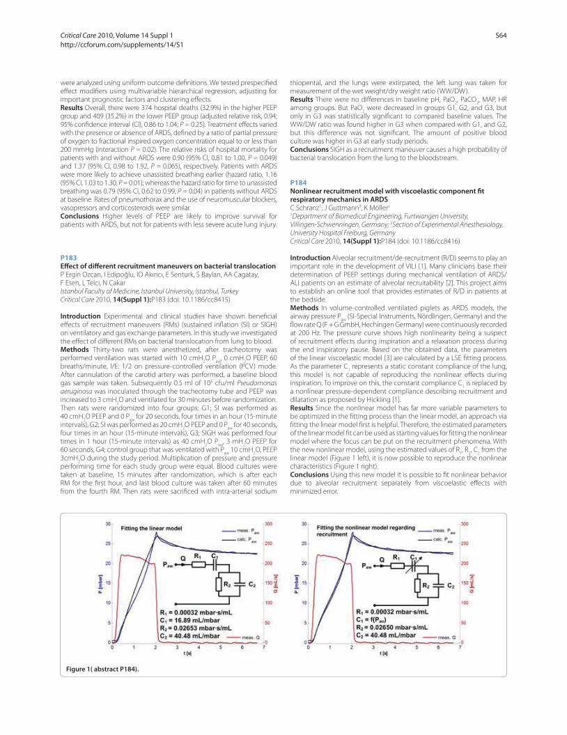
were analyzed using uniform outcome defi nitions. We tested prespecifi ed
eff ect modifi ers using multivariable hierarchical regression, adjusting for
important prognostic factors and clustering eff ects.
Results Overall, there were 374 hospital deaths (32.9%) in the higher PEEP
group and 409 (35.2%) in the lower PEEP group (adjusted relative risk, 0.94;
95% confi dence interval (CI), 0.86 to 1.04; P = 0.25). Treatment eff ects varied
with the presence or absence of ARDS, defi ned by a ratio of partial pressure
of oxygen to fractional inspired oxygen concentration equal to or less than
200 mmHg (interaction P = 0.02). The relative risks of hospital mortality for
patients with and without ARDS were 0.90 (95% CI, 0.81 to 1.00, P = 0.049)
and 1.37 (95% CI, 0.98 to 1.92, P = 0.065), respectively. Patients with ARDS
were more likely to achieve unassisted breathing earlier (hazard ratio, 1.16
(95% CI, 1.03 to 1.30, P = 0.01); whereas the hazard ratio for time to unassisted
breathing was 0.79 (95% CI, 0.62 to 0.99, P = 0.04) in patients without ARDS
at baseline. Rates of pneumothorax and the use of neuromuscular blockers,
vasopressors and corticosteroids were similar.
Conclusions Higher levels of PEEP are likely to improve survival for
patients with ARDS, but not for patients with less severe acute lung injury.
P183
Eff ect of diff erent recruitment maneuvers on bacterial translocationP Ergin Ozcan, I Edipoğlu, IO Akıncı, E Senturk, S Baylan, AA Cagatay,
F Esen, L Telci, N Cakar
Istanbul Faculty of Medicine, Istanbul University, Istanbul, Turkey
Critical Care 2010, 14(Suppl 1):P183 (doi: 10.1186/cc8415)
Introduction Experimental and clinical studies have shown benefi cial
eff ects of recruitment maneuvers (RMs) (sustained infl ation (SI) or SIGH)
on ventilatory and gas exchange parameters. In this study we investigated
the eff ect of diff erent RMs on bacterial translocation from lung to blood.
Methods Thirty-two rats were anesthetized, after tracheotomy was
performed ventilation was started with 10 cmH2O P
aw, 0 cmH
2O PEEP, 60
breaths/minute, I/E: 1/2 on pressure-controlled ventilation (PCV) mode.
After cannulation of the carotid artery was performed, a baseline blood
gas sample was taken. Subsequently 0.5 ml of 105 cfu/ml Pseudomonas
aeruginosa was inoculated through the tracheotomy tube and PEEP was
increased to 3 cmH2O and ventilated for 30 minutes before randomization.
Then rats were randomized into four groups: G1; SI was performed as
40 cmH2O PEEP and 0 P
aw for 20 seconds, four times in an hour (15-minute
intervals), G2; SI was performed as 20 cmH2O PEEP and 0 P
aw for 40 seconds,
four times in an hour (15-minute intervals), G3; SIGH was performed four
times in 1 hour (15-minute intervals) as 40 cmH2O P
aw, 3 mH
2O PEEP for
60 seconds, G4; control group that was ventilated with Paw
10 cmH2O, PEEP
3cmH2O during the study period. Multiplication of pressure and pressure
performing time for each study group were equal. Blood cultures were
taken at baseline, 15 minutes after randomization, which is after each
RM for the fi rst hour, and last blood culture was taken after 60 minutes
from the fourth RM. Then rats were sacrifi ced with intra-arterial sodium
thiopental, and the lungs were extirpated; the left lung was taken for
measurement of the wet weight/dry weight ratio (WW/DW).
Results There were no diff erences in baseline pH, PaO2, PaCO
2, MAP, HR
among groups. But PaO2 were decreased in groups G1, G2, and G3, but
only in G3 was statistically signifi cant to compared baseline values. The
WW/DW ratio was found higher in G3 when compared with G1, and G2,
but this diff erence was not signifi cant. The amount of positive blood
culture was higher in G3 at early study periods.
Conclusions SIGH as a recruitment maneuver causes a high probability of
bacterial translocation from the lung to the bloodstream.
P184
Nonlinear recruitment model with viscoelastic component fi t respiratory mechanics in ARDSC Schranz1, J Guttmann2, K Möller1
1Department of Biomedical Engineering, Furtwangen University,
Villingen-Schwenningen, Germany; 2Section of Experimental Anesthesiology,
University Hospital Freiburg, Germany
Critical Care 2010, 14(Suppl 1):P184 (doi: 10.1186/cc8416)
Introduction Alveolar recruitment/de-recruitment (R/D) seems to play an
important role in the development of VILI [1]. Many clinicians base their
determination of PEEP settings during mechanical ventilation of ARDS/
ALI patients on an estimate of alveolar recruitability [2]. This project aims
to establish an online tool that provides estimates of R/D in patients at
the bedside.
Methods In volume-controlled ventilated piglets as ARDS models, the
airway pressure Paw
(SI-Special Instruments, Nördlingen, Germany) and the
fl ow rate Q (F + G GmbH, Hechingen Germany) were continuously recorded
at 200 Hz. The pressure curve shows high nonlinearity being a suspect
of recruitment eff ects during inspiration and a relaxation process during
the end inspiratory pause. Based on the obtained data, the parameters
of the linear viscoelastic model [3] are calculated by a LSE fi tting process.
As the parameter C1 represents a static constant compliance of the lung,
this model is not capable of reproducing the nonlinear eff ects during
inspiration. To improve on this, the constant compliance C1 is replaced by
a nonlinear pressure-dependent compliance describing recruitment and
dilatation as proposed by Hickling [1].
Results Since the nonlinear model has far more variable parameters to
be optimized in the fi tting process than the linear model, an approach via
fi tting the linear model fi rst is helpful. Therefore, the estimated parameters
of the linear model fi t can be used as starting values for fi tting the nonlinear
model where the focus can be put on the recruitment phenomena. With
the new nonlinear model, using the estimated values of R1, R
2, C
2 from the
linear model (Figure 1 left), it is now possible to reproduce the nonlinear
characteristics (Figure 1 right).
Conclusions Using this new model it is possible to fi t nonlinear behavior
due to alveolar recruitment separately from viscoelastic eff ects with
minimized error.
Figure 1( abstract P184).
Critical Care 2010, Volume 14 Suppl 1 http://ccforum.com/supplements/14/S1
S64

References1. Hickling KG: Am J Respir Crit Care Med 2001, 163:69-78.
2. Gattinoni L, et al.: N Engl J Med 2006, 354:1775-1786.
3. Bates JH, et al.: J Appl Physiol 2002, 93:705-713.
P185
Lung recruitment and PEEP response in ARDS-related H1N1 virus patientsD Chiumello1, C Mietto2, V Berto2, A Marino2, E Gallazzi2, D Tubiolo1
1Fondazione IRCCS, ‘Ospedale Maggiore Policlinico Mangiagalli Regina Elena’
di Milano, Milan, Italy; 2Università degli Studi di Milano, Milan, Italy
Critical Care 2010, 14(Suppl 1):P185 (doi: 10.1186/cc8417)
Introduction Starting in April 2009 the swine-origin infl uenza (H1N1) virus
is considered a worldwide pandemic and is associated with a large number
of patients admitted to intensive care. In a proportion of these patients,
acute respiratory distress syndrome (ARDS) can develop. We described the
case of two patients that presented ARDS related to H1N1 virus confi rmed
by a nasal swab tested with real-time reverse transcription-polymerase
chain reaction. Using a lung CT scan we measured the potential of lung
recruitment and the response to positive end-expiratory pressure (PEEP) in
terms of gas exchange and respiratory mechanics.
Methods Patients underwent a lung CT scan at an airway pressure of 5
and 45 cmH2O; lung profi les were outlined and quantitative analysis was
performed with custom-made software. The potential of lung recruitment
was computed as the amount of nonaerated lung tissue at 5 cmH2O in
which aeration can be restored at an airway pressure of 45 cmH2O. The
PEEP test was performed at two PEEP levels (5 and 15 cmH2O), maintaining
constant the minute ventilation and the inspiratory oxygen fraction; gas
exchange and respiratory mechanics were recorded 20 minutes after each
PEEP change.
Results Two sedated and paralyzed subjects with acute lung injury (ALI)/
ARDS (age 46 ± 5 years, BMI 38.6 ± 5.5 kg/m2, PaO2/FiO
2 187 ± 30 mmHg,
PEEP 17 ± 1 cmH2O; both patients were obese and one of them suff ered
from diabetes) were enrolled. One patient required extracorporeal
membrane oxygenation support for 5 days. Lung CT scans were performed
5 ± 3 days after ICU admission. The total lung weight at 5 cmH2O was
1,598 ± 142 g. The percentage of potentially recruitable lung was 24 ±
8% of the total lung weight, corresponding to an absolute weight of 371
± 109 g recruitable lung tissue. A 15 cmH2O compared with 5 cmH
2O of
PEEP improved the compliance of the respiratory system from 42.2 ± 7.0 to
46.6 ± 2.1 ml/cmH2O and the gas exchange from PaO
2 62.7 ± 0.1 to 123.2 ±
61.8 mmHg and PaCO2 47.1 ± 0.9 to 45.9 ± 3.2 mmHg, respectively.
Conclusions The ARDS-related H1N1 virus presented, in the early phase,
a high potentially recruitable lung in which elevated PEEP levels can be
benefi cial.
P186
Maximal recruitment strategy minimizes tidal recruitment in severe ARDS: a CT scan studyG Matos1, J Borges2, V Okamoto2, C Carvalho2, M Amato2, C Barbas1
1Hospital Albert Einstein, São Paulo, Brazil; 2Universidade de São Paulo, Brazil
Critical Care 2010, 14(Suppl 1):P186 (doi: 10.1186/cc8418)
Introduction The maximal recruitment strategy (MRS) is considered an
effi cient maneuver to reverse nonaerated lung in ARDS patients. There is
no information regarding tidal recruitment during MRS.
Methods Early and severe ARDS patients were submitted to MRS
guided by thoracic CT: A – recruitment phase (PEEP 10 to 45 cmH2O and
constant driving pressure 15 cmH2O); B – PEEP titration phase (PEEP 25
to 10 cmH2O). Images were obtained at end expiration and inspiration.
Global and regional quantitative CT analyses from each step of PEEP were
compared [1].
Results Analyses of 12 patients showed that MRS reduced signifi cantly
the global amount of nonaerated tissue (54 ± 8% to 7 ± 6%, P <0.01), tidal
recruitment (4 ± 4% to 1 ± 1%, P = 0.029) (Figure 1). Most dependent
regional tidal recruitment signifi cantly increased from PEEP 10 to 20 cmH2O
(2 ± 3% to 11 ± 7%, P <0.01), but signifi cantly decreased after MRS (11 ±
7% to 2 ± 2%, P <0.01). High PEEP (25 cmH2O) was necessary to sustain
recruitment.
Conclusions MRS decreased nonaerated areas and tidal recruitment.
Increasing PEEP without full recruitment may cause lung injury exacer-
bation in the severe ARDS population.
References1. Borges JB, et al.: Reversibility of lung collapse and hypoxemia in early acute
respiratory distress syndrome. Am J Respir Crit Care Med 2006, 174:268-278.
P187
Mechanical ventilation with diff erent alveolar pressures improves alveolar recruitment: a model studyZ Zhao1, J Guttmann2, K Möller1
1Furtwangen University, Villingen-Schwenningen, Germany; 2University
Medical Center Freiburg, Germany
Critical Care 2010, 14(Suppl 1):P187 (doi: 10.1186/cc8419)
Introduction In patients with acute respiratory distress syndrome (ARDS)
tidal ventilation is always inhomogeneous, with atelectasis in dependent
lung and overdistention in nondependent lung. The aim of our study was
to develop a fl ow pattern that simultaneously produces locally diff erent
alveolar pressures, in order to recruit collapsed alveoli and at the same
time to prevent hyperinfl ation of already open alveoli.
Methods We modifi ed Horsfi eld’s model of the canine lung [1] to
predict alveolar pressure diff erences between the collapsed and opened
alveoli. The morphometric model has an asymmetrical branching airway
system with 47 orders, whereby order 47 corresponds to the trachea.
Each branch is terminated with a viscoelastic tissue unit and there are in
total 150,077 acini for the whole model. Using this model, atelectasis of
alveoli was simulated by reducing the diameter of small airways (order
1, 2 and 3) up to 90% of their original sizes. The tissue damping factor G
was 104 times higher in the collapsed alveoli than that in normal alveoli.
The shunt gas compression compliance Cg for the alveolus was 106 times
smaller. Diff erent percentages of atelectatic area from 0 to 90% were
simulated. Flow distribution and airway input impedance were calculated
at diff erent frequencies from 0.1 to 5 Hz. Alveolar pressure diff erences were
obtained by analyzing the products of fl ow distribution and alveolar input
impedance.
Results The alveolar pressure Palv
reduced as the frequency increased
(for both collapsed and open alveoli). Compared with the situation at 0.2
Hz, Palv
reduced to 9.1 ± 1.0% at 4 Hz. On the other hand, Palv
increased
as the percentage of atelectatic area increased (for all frequencies). Palv
increased to 267 ± 6% when 70% of alveoli collapsed compared with 10%
of collapse. The pressure diff erences between collapsed and open alveoli
increased as the frequency increased. At 0.2 Hz the diff erences were <3%,
while at 4 Hz the diff erences were >20% (relative to the lower value).
Figure 1 (abstract P186). Percentage of regional (I to IV) tidal recruitment
during all steps of the MRS protocol.
Critical Care 2010, Volume 14 Suppl 1 http://ccforum.com/supplements/14/S1
S65

Conclusions Increasing the high-frequency contents of the inspiratory
fl ow pattern may help to develop a pressure diff erence between collapsed
and open alveoli. The low-frequency contents of the fl ow pattern, however,
may be still needed to guarantee the minimum ventilation.
Reference1. Horsfi eld K, et al.: J Appl Physiol 1982, 52:21-26.
P188
Decreasing tidal volume from 6 to 4 ml/kg: feasibility and eff ects on repeated opening and closingJ Retamal, J Libuy, M Jimenez, M Delgado, C Besa, G Bugedo, A Bruhn
Pontifi cia Universidad Católica de Chile, Santiago, Chile
Critical Care 2010, 14(Suppl 1):P188 (doi: 10.1186/cc8420)
Introduction Low tidal volumes (Vt) are thought to protect the lung
by avoiding overdistension. We recently have shown that Vt may also
infl uence repeated opening and closing (O/C) [1]. The goal of this study
was to determine whether decreasing Vt from 6 to 4 ml/kg was eff ective to
reduce O/C, and whether it was possible to maintain alveolar ventilation
at such low Vt.
Methods Cross-over study at two Vt levels: 6 versus 4 ml/kg IBW. We
included ALI/ARDS patients, ventilated <48 hours, and who would have a
chest computed tomography (CT) scan. For the 4 ml/kg arm: we replaced
the heat and moisture exchange fi lter by a heated humidifi er, and the
respiratory rate was increased to keep minute ventilation constant. The
protocol had two parts: one bedside and other in the CT room. Both Vts
were applied in a random order. For the bedside protocol each Vt arm
was applied for 30 minutes. Data on lung mechanics and gas exchange
were taken at baseline and 30 minutes. For the CT scan protocol each Vt
arm was applied for 5 minutes and then a dynamic CT (4 images/second
for 8 seconds) was taken at each Vt at a fi xed transverse region at the
lower third of the lungs. Afterwards, CT images were analyzed by software
(MALUNA) and repeated O/C was determined as nonaerated tissue
variation between inspiration and expiration, expressed as a percentage
of lung tissue weight.
Results We analyzed nine patients (six male), who had a median age of 39
(21 to 72) years, APACHE II score 14 (5 to 23) and SOFA score 9 (6 to 15). All
patients had a pulmonary origin of their ARDS and were on their fi rst day
of ventilation. At baseline patients had a PaO2/FiO
2 ratio of 141 (71 to 280),
compliance of 32 (17 to 43) ml/cmH2O, and PEEP of 12 (10 to 16) cmH
2O. In
the Vt arms 4 and 6 ml/kg, Vts were 260 (210 to 300) and 350 (310 to 400)
ml, respectively (P <0.01), respiratory rates were 37 (31 to 42) and 25 (21 to
28) breaths per minute (P <0.01), and PaO2 levels were 84 (54 to 148) and
83 (61 to 162) mmHg (P = 0.3). PEEP and FiO2 were kept constant. PaCO
2
did not signifi cantly increase with Vt 4 but repeated O/C (delta nonaerated
tissue) consistently decreased (Figure 1).
Conclusions Decreasing Vt from 6 to 4 ml/kg reduces repeated O/C.
Hypercapnia can be eff ectively prevented by decreasing the instrumental
dead space and increasing the respiratory rate.
Acknowledgements Grant was Fondecyt 11060350.
Reference1. Bruhn A, et al.: Intensive Care Med 2009, 35(Suppl 1):S227.
P189
Barriers to providing lung-protective ventilation to patients with ALI/ARDSRH Passos, RH Oliveira, MQ Trabuco, RR Rodrigues, SP Souza, PB Batista
Hospital São Rafael, Salvador, Brazil
Critical Care 2010, 14(Suppl 1):P189 (doi: 10.1186/cc8421)
Introduction Acute lung injury (ALI) and acute respiratory distress
syndrome (ARDS) are associated with signifi cant morbidity and mortality.
Mechanical ventilation is the cornerstone of supportive therapy. However,
the optimal strategy of ventilation and adjunctive therapies are still
evolving. There is evidence to support the use of volume-limited and
pressure-limited lung-protective ventilation but practice variability in
the clinical management is still a concern mainly in sicker patients. The
purpose of this study was to examine the ventilatory prescriptions of
clinicians caring for patients with ALI/ARDS.
Methods We prospectively examined demographic characteristics,
APACHE II score and choice of ventilatory settings at the day of diagnosis
of ALI/ARDS (AECC criteria) of 84 mechanically ventilated patients from
May 2007 through October 2009 in a mixed medical–surgical critical care
unit.
Results ALI/ARDS occurred in 7.1% of admissions and 16.1% of all
mechanically ventilated patients. Sepsis was the main cause. All patients
were ventilated with pressure control mode. Median tidal volume/
predicted body weight was 6.1 (3.5 to 9.9) ml/kg, plateau pressure was
26 (15 to 55) cmH2O and mean PEEP was 12 (5 to 25) cmH
2O. Univariate
analysis showed sepsis-related ALI/ARDS (P = 0.026) and APACHE II score
>18 (P <0.001) were barriers to initiate lung-protective ventilation. In
multivariate analysis, APACHE score >18 was an independent predictor.
Ventilatory settings were signifi cantly diff erent in this subset of patients
(n = 26). The tidal volume/predicted body weight (6.2 (3.5 to 9.9) vs 5.1
(4.3 to 8.5) ml/kg), PEEP (15 (10 to 25) vs 7 (6 to 12) cmH2O) and plateau
pressure (38 (32 to 55) vs 22 (15 to 30) cmH2O) were higher in the group
with APACHE II score >18 (P <0.001). PaO2/FiO
2 pH and paCO
2 did not diff er
between groups. As expected, mortality was higher in this group, 60% vs
22% (P <0.001).
Conclusions Protective mechanical ventilation is not completely feasible
in the subset of ALI/ARDS patients with higher risk of mortality (APACHE
II >18).
Reference1. Acute Respiratory Distress Syndrome Network: Ventilation with lower tidal
volumes as compared with traditional tidal volumes for acute lung injury and the acute respiratory distress syndrome. N Engl J Med 2000, 342:1301.
P190
Infl uence of aetiology of acute respiratory distress syndrome and early diff erences in oxygenation on outcomeM Shankar-Hari1, CI Olvera2, S Webb2, S Phelps2, S Batson2, A Puddicome2,
R Marshall2, D Howell2, G Bellingan2
1Guy’s and St Thomas’ NHS Foundation Trust, London, UK; 2University College
Hospital, London, UK
Critical Care 2010, 14(Suppl 1):P190 (doi: 10.1186/cc8422)
Introduction Studies evaluating the relationship between the severity of
hypoxemia in patients with acute respiratory distress syndrome (ARDS)
Figure 1 (abstract P188).
Critical Care 2010, Volume 14 Suppl 1 http://ccforum.com/supplements/14/S1
S66
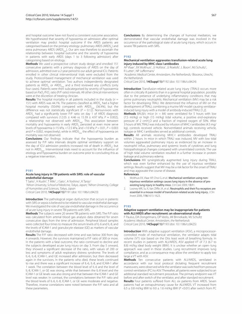
and hospital outcome have not found a consistent outcome association.
We hypothesized that severity of hypoxemia on admission after optimal
ventilation may predict hospital outcome if the ARDS patients are
categorised based on the primary etiology: pulmonary ARDS (ARDSp) and
extra pulmonary ARDS (ARDSexp
). Our aim was therefore to ascertain the
relationship between hospital outcome and the severity of hypoxemia
in patients with early ARDS (days 1 to 3 following admission) after
categorising based on etiology.
Methods We used a prospective cohort study design and enrolled 151
consecutive patients with a primary diagnosis of ARDS on the day of
admission, admitted over a 2-year period to our adult general ICU. Patients
enrolled in other clinical interventional trials were excluded from the
study. Protocol-based management of mechanical ventilation was used
to achieve optimal ventilation. Two authors independently designated
patients as ARDSp or ARDS
exp and a third reviewed any confl icts (only
four cases). Patients were then subcategorised by severity of hypoxaemia
based on PaO2/FiO
2 ratio (P/F ratio) intervals. All other clinical interventions
were at the discretion of treating clinician.
Results The hospital mortality in all patients included in the study (n =
151) with ARDS was 44.1%. The patients classifi ed as ARDSp had a higher
hospital mortality (50.6%) compared with ARDSexp
(36.4%), but the
diff erence was not statistically signifi cant (P = 0.12). Nonsurvivors with
ARDSp had a signifi cantly lower P/F ratio on day 1 of ARDS diagnosis
compared with survivors (12.05 ± 4.46 vs 15.39 ± 4.97 kPa; P = 0.002),
a relationship not observed with ARDSexp
. This association between
mortality and hypoxaemia in early ARDSp persisted at even more serve
levels of hypoxaemia (<15 kPa, <12.5 kPa, and <10 kPa; P = 0.01, P = 0.003
and P = 0.002, respectively), while in ARDSexp
the eff ect of hypoxaemia on
mortality was not observed.
Conclusions Our fi ndings indicate that the hypoxaemia burden as
assessed by P/F ratio intervals despite optimal ventilatory support on
the day of ICU admission predicts increased risk of death in ARDSp; but
not in ARDSexp
. Interventional trials need to account for the infl uence of
etiology and hypoxaemia burden on outcome prior to concluding this as
a negative intervention.
P191
Acute lung injury in TBI patients with SIRS: role of vascular endothelial damageT Saito1, H Kushi2, T Miki1, J Sato1, A Yoshino1, K Tanjo1
1Nihon University, School of Medicine, Tokyo, Japan; 2Nihon University, College
of Humanities and Sciences, Tokyo, Japan
Critical Care 2010, 14(Suppl 1):P191 (doi: 10.1186/cc8423)
Introduction The pathological organ dysfunction that occurs in patients
with SIRS or sepsis is believed to be related to vascular endothelial damage.
We investigated the role of vascular endothelial damage in the occurrence
of acute lung injury in severe TBI patients with SIRS.
Methods The subjects were 20 severe TBI patients with SIRS. The P/F ratio
was calculated from arterial blood gas analysis data obtained for seven
consecutive days from the time of admission. Peripheral blood samples
were collected four times to measure the serum levels of IL-6 and IL-8, and
the levels of ICAM-1 and granulocyte elastase (GE) as markers of vascular
endothelial damage.
Results The P/F ratio decreased with time and was below 300 from day
4 onwards. However, the survivors maintained a P/F ratio of 300 or more.
In the patients with a fatal outcome, the ratio continued to decline and
the subjects developed acute lung injury on day 3. From day 5 onward,
they showed a signifi cant decrease of the ratio, with values of 200 or
less and symptoms of adult respiratory distress syndrome. The levels of
IL-6, IL-8, ICAM-1, and GE increased after admission, but then decreased
again in the survivors. In the patients who died, these levels continued
to rise and there was a signifi cant increase of IL-6, IL-8, and ICAM-1 after
1 week. The correlation between the blood level of IL-6 and the level of
IL-8, ICAM-1, or GE was strong, while that between the IL-8 level and the
ICAM-1 or GE levels was also strong and that between the ICAM-1 and GE
level was weaker. In contrast, the correlations between the P/F ratio and
the blood levels of IL-6, IL-8, ICAM-1, or GE were moderate and negative.
Therefore, inverse correlations were noted between the P/F ratio and all
these parameters.
Conclusions By determining the changes of humoral mediators, we
demonstrated that vascular endothelial damage was involved in the
occurrence of the pathological state of acute lung injury, which occurs in
severe TBI patients with SIRS.
P192
Mechanical ventilation aggravates transfusion-related acute lung injury induced by MHC class I antibodiesAP Vlaar1, EK Wolthuis1, JJ Hofstra1, JJ Roelofs1, L Boon2, MJ Schultz1,
R Lutter1, NP Juff ermans1
1Academic Medical Center, Amsterdam, the Netherlands; 2Bioceros, Utrecht,
the Netherlands
Critical Care 2010, 14(Suppl 1):P192 (doi: 10.1186/cc8424)
Introduction Transfusion-related acute lung injury (TRALI) occurs more
often in critically ill patients than in a general hospital population, possibly
due to the presence of underlying infl ammatory conditions that may
prime pulmonary neutrophils. Mechanical ventilation (MV) may be a risk
factor for developing TRALI. We determined the infl uence of MV on the
development of TRALI, combining a murine MV model causing ventilator-
induced lung injury with a model of antibody-induced TRALI [1,2].
Methods BALB/c mice (n = 84) were ventilated for 5 hours with low
(7.5 ml/kg) or high (15 ml/kg) tidal volume, a positive end-expiratory
pressure of 2 cmH2O and a fraction of inspired oxygen of 50%. After
3 hours of MV, TRALI was induced by infusion of MHC-I antibodies (4.5 mg/
kg), controls received vehicle. Nonventilated animals receiving vehicle,
isotype or MHC-I antibodies served as additional controls.
Results All animals receiving MHC-I antibodies developed TRALI
within 2 hours. In mice in which TRALI was induced, MV with low tidal
volumes aggravated pulmonary injury as evidenced by an increase in
neutrophil infl ux, pulmonary and systemic levels of cytokines and lung
histopathological changes compared with unventilated controls. The use
of high tidal volume ventilation resulted in a further increase in protein
leakage and pulmonary edema.
Conclusions MV synergistically augmented lung injury during TRALI,
which was even further enhanced by the use of injurious ventilator
settings. Results suggest that MV may be a risk factor for the onset of TRALI
and may aggravate the course of disease.
References1. Wolthuis EK, Vlaar AP, Choi G, et al.: Mechanical ventilation using non-
injurious ventilation settings causes lung injury in the absence of pre-existing lung injury in healthy mice. Crit Care 2009, 13:R1.
2. Looney MR, Su X, Van Ziffl e JA, et al.: Neutrophils and their Fcγ receptors are essential in a mouse model of transfusion-related acute lung injury. J Clin
Invest 2006, 116:1615-1623.
P193
Adaptive support ventilation may be inappropriate for patients with ALI/ARDS after recruitment: an observational studyF Paulus, DA Dongelmans, DP Veelo, JM Binnekade, MJ Schultz
Academic Medical Center, Amsterdam, the Netherlands
Critical Care 2010, 14(Suppl 1):P193 (doi: 10.1186/cc8425)
Introduction With adaptive support ventilation (ASV), a microprocessor-
controlled mode of mechanical ventilation, the ventilator adapts tidal
volume (VT) size based on the Otis least work of breathing formula. In
recent studies in patients with ALI/ARDS, ASV applied VT of 7.3 (6.7 to
8.8) ml/kg ideal body weight (IBW). It is unclear whether an open–lung
approach was used in these studies. Lung recruitment improves lung
compliance, and as a consequence may allow the ventilator to apply too
large a VT with ASV.
Methods Ten consecutive patients with ALI/ARDS, ventilated in
accordance with our local protocol dictating frequent recruitment
maneuvers, were observed while the ventilator was switched from pressure
control ventilation (PC) to ASV. Thereafter, all patients were subjected to an
additional standard recruitment procedure. The primary endpoint was VT
before and after switch of the ventilator, and after standard recruitment.
Results Four patients suff ered from ALI, six patients from ARDS. Seven
patients had an extrapulmonary cause for ALI/ARDS. VT increased from
6.5 ± 0.8 ml/kg IBW to 9.0 ± 1.6 ml/kg IBW (P <0.01) after switch from PC
Critical Care 2010, Volume 14 Suppl 1 http://ccforum.com/supplements/14/S1
S67
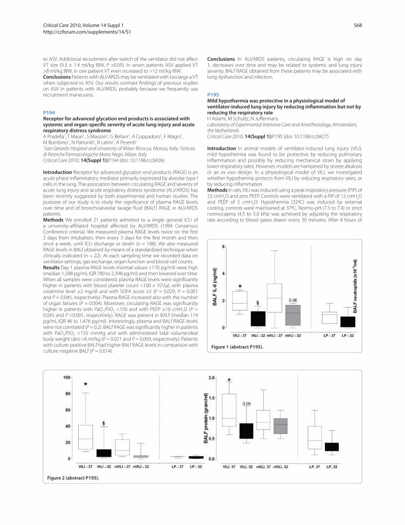
to ASV. Additional recruitment after switch of the ventilator did not aff ect
VT size (9.3 ± 1.4 ml/kg IBW, P >0.05). In seven patients ASV applied VT
>8 ml/kg IBW, in one patient VT even increased to >12 ml/kg IBW.
Conclusions Patients with ALI/ARDS may be ventilated with too large a VT
when subjected to ASV. Our results contrast fi ndings of previous studies
on ASV in patients with ALI/ARDS, probably because we frequently use
recruitment maneuvers.
P194
Receptor for advanced glycation end products is associated with systemic and organ-specifi c severity of acute lung injury and acute respiratory distress syndromeA Pradella1, T Mauri1, S Masson2, G Bellani1, A Coppadoro1, F Magni1,
M Bombino1, N Patroniti1, R Latini2, A Pesenti1
1San Gerardo Hospital and University of Milan-Bicocca, Monza, Italy; 2Istituto
di Ricerche Farmacologiche Mario Negri, Milan, Italy
Critical Care 2010, 14(Suppl 1):P194 (doi: 10.1186/cc8426)
Introduction Receptor for advanced glycation end products (RAGE) is an
acute phase infl ammatory mediator primarily expressed by alveolar type I
cells in the lung. The association between circulating RAGE and severity of
acute lung injury and acute respiratory distress syndrome (ALI/ARDS) has
been recently suggested by both experimental and human studies. The
purpose of our study is to study the signifi cance of plasma RAGE levels
over time and of bronchoalveolar lavage fl uid (BALf ) RAGE in ALI/ARDS
patients.
Methods We enrolled 21 patients admitted to a single general ICU of
a university-affi liated hospital aff ected by ALI/ARDS (1994 Consensus
Conference criteria). We measured plasma RAGE levels twice on the fi rst
2 days from intubation, then every 3 days for the fi rst month and then
once a week, until ICU discharge or death (n = 188). We also measured
RAGE levels in BALf obtained by means of a standardized technique when
clinically indicated (n = 22). At each sampling time we recorded data on
ventilator settings, gas exchange, organ function and blood cell counts.
Results Day 1 plasma RAGE levels (normal values <170 pg/ml) were high
(median 1,588 pg/ml, IQR 780 to 2,398 pg/ml) and then lowered over time.
When all samples were considered, plasma RAGE levels were signifi cantly
higher in patients with blood platelet count <100 x 103/μl, with plasma
creatinine level ≥2 mg/dl and with SOFA score ≥5 (P = 0.029, P = 0.001
and P = 0.045, respectively). Plasma RAGE increased also with the number
of organ failures (P = 0.004). Moreover, circulating RAGE was signifi cantly
higher in patients with PaO2/FiO
2 <150 and with PEEP ≥10 cmH
2O (P =
0.045 and P <0.001, respectively). RAGE was present in BALf (median 174
pg/ml, IQR 46 to 1,476 pg/ml). Interestingly, plasma and BALf RAGE levels
were not correlated (P = 0.2). BALf RAGE was signifi cantly higher in patients
with PaO2/FiO
2 <150 mmHg and with administered tidal volume:ideal
body weight ratio >6 ml/kg (P = 0.021 and P = 0.009, respectively). Patients
with culture-positive BALf had higher BALf RAGE levels in comparison with
culture-negative BALf (P = 0.014).
Conclusions In ALI/ARDS patients, circulating RAGE is high on day
1, decreases over time and may be related to systemic and lung injury
severity. BALf RAGE obtained from these patients may be associated with
lung dysfunction and infection.
P195
Mild hypothermia was protective in a physiological model of ventilator-induced lung injury by reducing infl ammation but not by reducing the respiratory rateH Aslami, M Schultz, N Juff ermans
Laboratory of Experimental Intensive Care and Anesthesiology, Amsterdam,
the Netherlands
Critical Care 2010, 14(Suppl 1):P195 (doi: 10.1186/cc8427)
Introduction In animal models of ventilator-induced lung injury (VILI),
mild hypothermia was found to be protective by reducing pulmonary
infl ammation and possibly by reducing mechanical strain by applying
lower respiratory rates. However, models are hampered by severe alkalosis
or an ex vivo design. In a physiological model of VILI, we investigated
whether hypothermia protects from VILI by reducing respiratory rates, or
by reducing infl ammation.
Methods In rats, VILI was induced using a peak inspiratory pressure (PIP) of
23 cmH2O and zero PEEP. Controls were ventilated with a PIP of 12 cmH
2O
and PEEP of 5 cmH2O. Hypothermia (32ºC) was induced by external
cooling, controls were maintained at 37ºC. Normo-pH (7.3 to 7.4) or strict
normocapnia (4.5 to 5.0 kPa) was achieved by adjusting the respiratory
rate according to blood gases drawn every 30 minutes. After 4 hours of
Figure 1 (abstract P195).
Figure 2 (abstract P195).
Critical Care 2010, Volume 14 Suppl 1 http://ccforum.com/supplements/14/S1
S68

ventilation, bronchoalveolar lavage (BAL) was done. Statistics include
Kruskal–Wallis and Mann–Whitney U tests.
Results A physiological model of VILI was established. In the normo-pH
group, hypothermia decreased pulmonary IL-6 and neutrophil infl ux and
tended to decrease pulmonary protein leak (Figure 1). In the normocapnia
group, hypothermia allowed for lower respiratory rates compared with
normo-pH (11 ± 1 vs 17 ± 2 breaths/minute) (Figure 2). However, this did
not further reduce parameters of lung injury.
Conclusions Hypothermia was protective in a physiological model of VILI,
by reduction of infl ammation, but not by reducing the repetitive strain of
respiratory cycles.
P196
Eff ect of induced hypothermia on respiratory parameters in mechanically ventilated patientsH Aslami, J Binnekade, S Huissoon, N Juff ermans
Laboratory of Experimental Intensive Care and Anesthesiology, Amsterdam,
the Netherlands
Critical Care 2010, 14(Suppl 1):P196 (doi: 10.1186/cc8428)
Introduction Therapeutic hypothermia is applied to reduce hypoxia-
induced organ injury. In the past decades, the use of hypothermia has
increased in critically ill patients who are mechanically ventilated (MV).
Data on the eff ect of hypothermia on gas exchange and lung mechanics
in these patients are limited. In this retrospective study, we describe the
eff ect of induced hypothermia and rewarming on respiratory parameters
in patients after a cardiac arrest.
Methods Patients with a Glasgow Coma Scale <8 after resuscitation for
a cardiac arrest in whom hypothermia was applied (32 to 34°C), were
enrolled. Patients with PaO2/FiO
2 ratio <200 or MV with positive end-
expiratory pressure (PEEP) >15 cmH2O were excluded. Ventilator settings
and arterial blood gasses were retrieved from the electronic patient
database during hypothermia and at every °C grade increase during
rewarming. Statistics include z paired t test.
Results From a cohort of 98 patients, 35 patients were excluded, leaving
62 patients for analysis. During hypothermia, arterial pCO2 decreased,
while end tidal (et) CO2 was low at unchanged minute volume ventilation
(Figure 1). Hypothermia increased the P/F ratio from 255 ± 55 to 283 ±
12 at unchanged PEEP (P <0.05), while fl uid balances were positive in all
patients (2.5 ± 1.6 l). After rewarming, arterial pCO2 was unchanged while
etCO2 increased. The P/F ratio after rewarming was unchanged compared
with the start of hypothermia, while lower PEEP levels were applied (7.0 ±
0.4 cmH2O vs 6.1 ± 0.3 cmH
2O, P <0.05).
Conclusions Induced hypothermia improved ventilation and oxygenation
in critically ill patients. Hypothermia may be considered in patients with
acute lung injury, in whom low minute ventilation results in severe
hypercapnia.
P197
Extracorporeal life support service in a regional referral center: the Florence experienceS Biondi1, A Pasquini2, S Batacchi2, G Cianchi2, M Ciapetti2, S Di Valvasone1,
S Bacci2, L Varegliano2, M Solaro2, M Bonizzoli2, R Spina2, M Bonacchi3,
G Zagli2, C Nozzoli2, A Peris2, GF Gensini3
1Postgraduate School of Anesthesia and Intensive Care, University of Florence,
Italy; 2Careggi Teaching Hospital, Florence, Italy; 3University of Florence, Italy
Critical Care 2010, 14(Suppl 1):P197 (doi: 10.1186/cc8429)
Introduction Extracorporeal life support (ECLS) represents a therapeutic
choice in cases of acute reversible lung and/or heart failure. An emergency
ICU can safely manage both veno-arterial (V-A) and veno-venous (V-V)
ECLS.
Methods In our experience, the ECLS treatment implementation started
on April 2008. The ECLS team is composed of intensivists, cardiac surgeons,
cardiologists, perfusionists and nurses. All of these fi gures were trained in
ECLS management. An Emergency Medical Service has been equipped
to guarantee rapid and safe ECLS positioning in peripheral hospitals and
transportation to our referral center by the ECLS team. According to our
internal protocol, eligible patients for V-V ECLS treatment are aged 15
to 70 years old and aff ected by ARDS with a PaO2/FiO
2 ratio <60 or pH
<7.20, under a protective ventilation setting. The use of ECLS for cardiac
support is reserved for those cases of cardiac shock refractory to standard
treatments and cardiac arrest not responding to advanced cardiac life
support (ACLS). The ECLS device used is a Rotafl ow Maquet Centrifugal
Pump with a Quadrox-D oxygenator (Maquet, Rastatt, Germany) and
biocoated circuits.
Results From April 2008 to November 2009, 21 patients were treated
with ECLS. In 13 patients V-V ECLS was established due to severe ARDS
not responding to conventional treatment. Median SAPS II at admission
was 49 and median duration was 235 hours. Three out of 13 patients
were cannulated by the ECLS team in peripheral hospitals and safety
transported. The intra-ICU survival rate was 62%. ECLS for cardiac support
was performed in eight patients: four cases of intrahospital cardiac arrest,
four cases of cardiogenic shock. Median stay of V-A ECLS was 110 hours. In
cases of V-A ECLS due to cardiac arrest, no positive outcome was observed.
The median stay of ACLS maneuvers before ECLS start was 62.5 minutes
In cases of cardiogenic shock undergoing ECLS, two out of four patients
were discharged from our ICU.
Conclusions A complete competence acquisition for ECLS management
makes this system a safe and feasible technique. The possibility to
guarantee a safe treatment must involve diff erent specialists and properly
trained nurses. We found the importance of a well-timed start of ECLS.
P198
Interhospital ground transportation of severe acute respiratory distress syndrome patients on extracorporeal membrane oxygenation: Monza’s experienceS Isgro1, M Milan1, A Zanella1, M Bombino1, G Foti1, M Giani1,
S Abd El Aziz El Sayed Deab1, N Patroniti2, A Pesenti2
1AO San Gerardo Monza, Italy; 2Milano-Bicocca University, Milan, Italy
Critical Care 2010, 14(Suppl 1):P198 (doi: 10.1186/cc8430)
Introduction Severe acute respiratory distress syndrome (ARDS)
patient transportation is an extremely high-risk procedure. We report
our experience in transferring these patients to our centre while on
extracorporeal membrane oxygenation (ECMO).
Methods After telephone referral and bed availability confi rmation,
patients matching entry criteria for ECMO are evaluated for transportation
to our centre. A skilled crew consisting of two expert plus one training
physician, one expert plus one training ICU nurse and one ECMO specialist
reaches the referral hospital for re-evaluation. If eligible, cannulation,
ECLS circuit set up and ECLS start are accomplished. Ground transport
is performed with a specially equipped ambulance, endowing enlarged
oxygen, fuel and energy supplies. The ambulance is loaded with all of the
items required for cannulation and ECMO circuit set up, additional oxygen
and a nitric oxide tank. Entry criteria are: potentially reversible respiratory
failure, Murray Score ≥3 or respiratory acidosis with pH <7.2, no intracranial
bleeding and absolute contraindication to heparinization.Figure 1 (abstract P196).
Critical Care 2010, Volume 14 Suppl 1 http://ccforum.com/supplements/14/S1
S69

Results Between 2004 and 2009 our crew evaluated for transfer on ECMO
15 ARDS patients (10 males), age 38 ± 15 years, BMI 28 ± 7, APACHE II
score 26 ± 9, SOFA score 9 ± 4, Oxygenation Index 39 ± 17. The average
distance was 133 ± 124 km. Two patients improved after NO trial and were
transferred without ECMO. All of the other patients underwent venovenous
ECMO: 11 with cannulation of femoral veins, one femoral–jugular veins
and one with a DL cannula in the jugular vein. ECMO settings were
(mean ± SD) BF 2.9 ± 0.8, GF 3.6 ± 1.6, GF FiO2 1. Data have been recorded
30 minutes before and 1 hour after ECLS began: vv-ECMO granted a better
clearance of pCO2 (75 ± 20.5 vs 49.7 ± 7.9 mmHg, P <0.01), thus improving
the pH (7.279 ± 0.10 vs 7.41 ± 0.06, P <0.01) and mean pulmonary arterial
pressure (41 ± 11 vs 31 ± 5 mmHg, P <0.05) and allowing a reduction in
respiratory rate (28 ± 11 vs 9 ± 4, P <0.01), minute ventilation (10.2 ± 4.6
vs 3.3 ± 1.7 l/min, P <0.01) and mean airway pressure (26 ± 6 vs 22 ± 5
cmH2O, P <0.01). Arterial pO
2, mean blood pressure and heart rate did not
show signifi cant variations. After ECMO began, vasoconstrictor therapy
(being administered to fi ve patients) was quickly tapered. Neither clinical
nor technical major complications were reported.
Conclusions ECMO employment at referral centers enabled long-
distance, high-risk ground transportation.
P199
Eff ects of hypertonic saline on a pig model of acute lung injury induced by hydrochloric acid instillationCA Holms1, DA Otsuki1, M Kahvegian1, J Noel-Morgan1, C Massoco2,
DT Fantoni1, P Gutierrez3, JO Auler Jr1
1Faculdade de Medicina da Universidade de São Paulo, Brazil; 2Genoa, São
Paulo, Brazil; 3INCOR, São Paulo, Brazil
Critical Care 2010, 14(Suppl 1):P199 (doi: 10.1186/cc8431)
Introduction Controversy exists over the possible benefi cial eff ects of
hypertonic saline (HS) in pulmonary infl ammatory response, particularly
in neutrophil immunomodulation [1,2]. This study was designed to
investigate possible benefi ts of HS in the treatment of pigs submitted to
experimental acute lung injury.
Methods Twelve anesthetized, tracheotomized pigs (25 to 30 kg) were
mechanically ventilated by pressure, adjusted to 8 ml/kg tidal volume,
with FiO2 50%, and submitted to intratracheal instillation of 4 ml/kg
hydrochloric acid (HCl) 0.1 N. They were then randomized to ALI group
(n = 7) or ALI + HS group (n = 5), where animals of the latter group
received 4 ml/kg intravenous hypertonic saline, 15 minutes after injury.
Hemodynamic parameters, pulmonary compliance (Cstat
), peak pressure
(Ppeak
), plateau pressure (Pplat
) and tidal volume (Vt) were analyzed at
baseline (TBL), 15 minutes after HCl instillation (TALI) and hourly thereafter
for 4 hours (T0 to T3). Bronchoalveolar lavage was performed at the end
of the observation period for fl ow cytometry analysis of neutrophil burst
activity. Postmortem histopathology of the right diaphragmatic lung was
also performed in all animals.
Results After TALI, animals of both groups presented signifi cant increases
in Ppeak
and Pplat
, and a decrease in Cstat
, at all time points, when compared
with TBL. Vt was preserved in both groups over time. There were signifi cant
diff erences between groups ALI and ALI + HS, respectively, in: central
venous pressure (T0, T1 and T2), pulmonary artery occlusion pressure
(T1 and T2) and pulmonary vascular resistance index (TALI). In the ALI
group, signifi cant diff erences related to TBL were found in mean arterial
pressure (T1), mean pulmonary artery pressure (TALI, T0, T1, T2 and T3) and
pulmonary vascular resistance index (TALI, T0, T3). In the ALI + HS group,
there were signifi cant diff erences related to TBL in mean arterial pressure
(T0, T1, T2), mean pulmonary artery pressure (TALI, T0, T1, T2, T3), cardiac
index (T0, T1, T2) and pulmonary vascular resistance index (T1, T2, T3). No
diff erences were found between groups regarding histopathology and
fl ow cytometry analyses.
Conclusions HS produced no signifi cant benefi t in the studied parameters
regarding the lungs, in the proposed model of ALI.
Acknowledgements Grants from FAPESP 08/55376-7 and 08/56792-4.
References1. Roch A et al.: Intensive Care Med 2007, 33:1645-1654.
2. Deree J et al.: J Trauma 2007, 62:104-111.
P200
Conventional mechanical ventilation can injury intact lungs in severe trauma patientsO Ignatenko, D Protsenko, A Yaroshetskiy, B Gelfand
State Medical Unversity of Russia, Moscow, Russia Federation
Critical Care 2010, 14(Suppl 1):P200 (doi: 10.1186/cc8432)
Introduction Conventional mechanical ventilation (MV) may cause
additional lung injury in ALI/ARDS due to overdistention of aerated
lung regions (high Vt) and cyclic lung reopening (low PEEP level).
Hyperproduction of infl ammatory mediators is one of the side eff ects in
these cases. This factor could delay or prevent resolution of respiratory
failure [1,2]. However, it is not clear whether conventional mechanical
ventilation damages intact lungs. The aim of this study was to evaluate the
eff ects of conventional and protective mechanical ventilation on intact
lungs in patients with severe trauma.
Methods A prospective, randomized controlled trial in trauma patients
with mechanical ventilation for extrapulmonary indications. The protocol
was approved by the local ethics committee. Seventy-eight patients were
randomized to conventional (Vt 10 to 12 ml/kg IBW, PEEP 5 cmH2O – n =
39) or protective (Vt 5 to 6 ml/kg IBW, PEEP 10 cmH2O – n = 39) mechanical
ventilation. TNFα, IL-1β and IL-6 levels in plasma and BAL fl uids were
measured on 1, 2, 3, 5 and 7 days of MV. Frequency of ALI (AECC criteria)
and VAP were evaluated. The endpoints of this study were the length of
MV, LOS in ICU and outcome on 28 days.
Results In fi rst 3 days ALI was revealed in 26 patients (66.6%) in the
conventional and 10 patients (26.5%) in the protective MV groups (P =
0.001; OR 4.375, 95% CI 2.227 to 8.189). ARDS occurred in four patients
(10,2%) of the conventional MV group (LIS >2) and no one in the protective
MV group (P <0.0001). Levels of TNFα, IL-1β and IL-6 in BAL fl uids were
signifi cantly higher in the conventional MV group from 1 to 7 days with
maximal increase on day 3 (542 ± 44/91 ± 11; 315 ± 35/86 ± 10; 1,092 ±
160/111 ± 18, P <0.0001). No diff erences were found in levels of TNFα,
IL-1β and IL-6 in plasma samples. VAP occurred in 31 patients (83.7%) of
the conventional and nine patients (23%) of the protective MV groups (P =
0.0001; OR 17.2, 95% CI 5.5 to 54.3). The length of MV was 17.4 ± 6 vs 12.8
± 3 (P = 0.0001; OR 4.2, 95% CI 1.5 to 11.5), LOS in the ICU was 21.9 ± 5.6 vs
15.75 ± 2.9 (P = 0.0002; OR 2.0, 95% CI 0.18 to 23.6). The 28-day mortality
was not signifi cantly diff erent in the groups.
Conclusions Conventional MV for more than 72 hours in patients with
severe trauma and intact lungs can cause lung injury, and increase
duration of MV and LOS in the ICU.
References1. Dreyfuss D, Saumon G: Ventilator-induced lung injury: lessons from
experimental studies. Am J Respir Crit Care Med 1998, 157:294-323.
2. Parker JC, Hernandez LA, Peevy KJ: Mechanisms of ventilator-induced lung injury. Crit Care Med 1993, 21:131-143.
P201
Protective eff ect of methylprednisolone on ventilator-induced diaphragm dysfunction is dose dependentK Maes1, A Agten1, A Smuder2, SK Powers2, M Decramer1, G Gayan-Ramirez1
1KU Leuven, Belgium; 2University of Florida, Gainesville, FL, USA
Critical Care 2010, 14(Suppl 1):P201 (doi: 10.1186/cc8433)
Introduction Administration of 80 mg/kg methylprednisolone has been
shown to prevent controlled mechanical ventilation (CMV) diaphragm
dysfunction in rats, partly by inhibiting the calpain system [1]. The current
experiments determined whether lower doses of corticosteroids will also
provide protection against ventilator-induced diaphragm dysfunction.
Methods Rats were assigned to a control group or to 24 hours of CMV
receiving a single injection of saline or 5 mg/kg (low MP) or 30 mg/kg
(high MP) of methylprednisolone.
Results Diaphragm force production was decreased after CMV but
signifi cantly more in the low MP group while similar to controls in the high
MP group. Atrophy of the type IIa fi bers was only present in the low MP
group. Atrophy of the type IIx/b fi bers was more severe in the low MP
group than in the CMV group while no atrophy was observed in the high
MP group. Diaphragm calpain activity was increased after CMV (+93%,
P <0.05 vs C) and in the low MP group (+83%, P <0.05 vs C), while it was
similar to controls in the high MP group. Expression of calpastatin was
Critical Care 2010, Volume 14 Suppl 1 http://ccforum.com/supplements/14/S1
S70
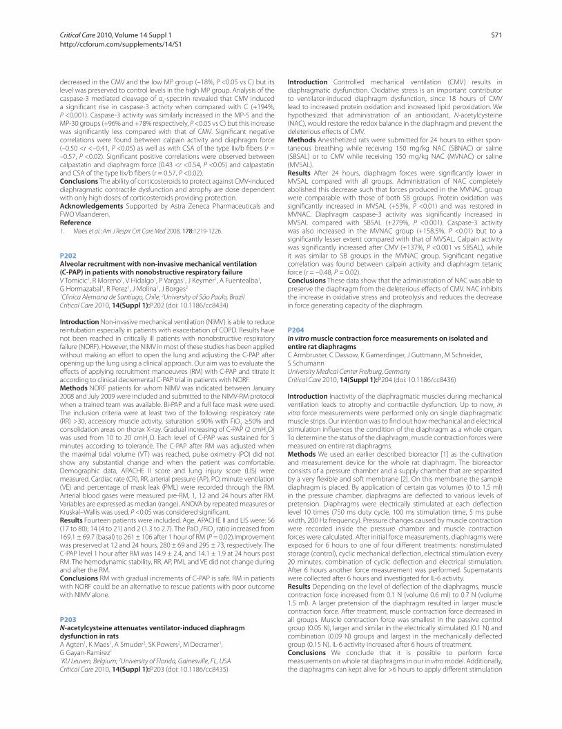
decreased in the CMV and the low MP group (–18%, P <0.05 vs C) but its
level was preserved to control levels in the high MP group. Analysis of the
caspase-3 mediated cleavage of αII-spectrin revealed that CMV induced
a signifi cant rise in caspase-3 activity when compared with C (+194%,
P <0.001). Caspase-3 activity was similarly increased in the MP-5 and the
MP-30 groups (+96% and +78% respectively, P <0.05 vs C) but this increase
was signifi cantly less compared with that of CMV. Signifi cant negative
correlations were found between calpain activity and diaphragm force
(–0.50 <r <–0.41, P <0.05) as well as with CSA of the type IIx/b fi bers (r =
–0.57, P <0.02). Signifi cant positive correlations were observed between
calpastatin and diaphragm force (0.43 <r <0.54, P <0.05) and calpastatin
and CSA of the type IIx/b fi bers (r = 0.57, P <0.02).
Conclusions The ability of corticosteroids to protect against CMV-induced
diaphragmatic contractile dysfunction and atrophy are dose dependent
with only high doses of corticosteroids providing protection.
Acknowledgements Supported by Astra Zeneca Pharmaceuticals and
FWO Vlaanderen.
Reference1. Maes et al.: Am J Respir Crit Care Med 2008, 178:1219-1226.
P202
Alveolar recruitment with non-invasive mechanical ventilation (C-PAP) in patients with nonobstructive respiratory failureV Tomicic1, R Moreno1, V Hidalgo1, P Vargas1, J Keymer1, A Fuentealba1,
G Hormazabal1, R Perez1, J Molina1, J Borges2
1Clinica Alemana de Santiago, Chile; 2University of São Paulo, Brazil
Critical Care 2010, 14(Suppl 1):P202 (doi: 10.1186/cc8434)
Introduction Non-invasive mechanical ventilation (NIMV) is able to reduce
reintubation especially in patients with exacerbation of COPD. Results have
not been reached in critically ill patients with nonobstructive respiratory
failure (NORF). However, the NIMV in most of these studies has been applied
without making an eff ort to open the lung and adjusting the C-PAP after
opening up the lung using a clinical approach. Our aim was to evaluate the
eff ects of applying recruitment manoeuvres (RM) with C-PAP and titrate it
according to clinical decremental C-PAP trial in patients with NORF.
Methods NORF patients for whom NIMV was indicated between January
2008 and July 2009 were included and submitted to the NIMV-RM protocol
when a trained team was available. Bi-PAP and a full face mask were used.
The inclusion criteria were at least two of the following: respiratory rate
(RR) >30, accessory muscle activity, saturation ≤90% with FiO2 ≥50% and
consolidation areas on thorax X-ray. Gradual increasing of C-PAP (2 cmH2O)
was used from 10 to 20 cmH2O. Each level of C-PAP was sustained for 5
minutes according to tolerance. The C-PAP after RM was adjusted when
the maximal tidal volume (VT) was reached, pulse oximetry (PO) did not
show any substantial change and when the patient was comfortable.
Demographic data, APACHE II score and lung injury score (LIS) were
measured. Cardiac rate (CR), RR, arterial pressure (AP), PO, minute ventilation
(VE) and percentage of mask leak (PML) were recorded through the RM.
Arterial blood gases were measured pre-RM, 1, 12 and 24 hours after RM.
Variables are expressed as median (range). ANOVA by repeated measures or
Kruskal–Wallis was used, P <0.05 was considered signifi cant.
Results Fourteen patients were included. Age, APACHE II and LIS were: 56
(17 to 80); 14 (4 to 21) and 2 (1.3 to 2.7). The PaO2/FiO
2 ratio increased from
169.1 ± 69.7 (basal) to 261 ± 106 after 1 hour of RM (P = 0.02).Improvement
was preserved at 12 and 24 hours, 280 ± 69 and 295 ± 73, respectively. The
C-PAP level 1 hour after RM was 14.9 ± 2.4, and 14.1 ± 1.9 at 24 hours post
RM. The hemodynamic stability, RR, AP, PML and VE did not change during
and after the RM.
Conclusions RM with gradual increments of C-PAP is safe. RM in patients
with NORF could be an alternative to rescue patients with poor outcome
with NIMV alone.
P203
N-acetylcysteine attenuates ventilator-induced diaphragm dysfunction in ratsA Agten1, K Maes1, A Smuder2, SK Powers2, M Decramer1,
G Gayan-Ramirez1
1KU Leuven, Belgium; 2University of Florida, Gainesville, FL, USA
Critical Care 2010, 14(Suppl 1):P203 (doi: 10.1186/cc8435)
Introduction Controlled mechanical ventilation (CMV) results in
diaphragmatic dysfunction. Oxidative stress is an important contributor
to ventilator-induced diaphragm dysfunction, since 18 hours of CMV
lead to increased protein oxidation and increased lipid peroxidation. We
hypothesized that administration of an antioxidant, N-acetylcysteine
(NAC), would restore the redox balance in the diaphragm and prevent the
deleterious eff ects of CMV.
Methods Anesthetized rats were submitted for 24 hours to either spon-
taneous breathing while receiving 150 mg/kg NAC (SBNAC) or saline
(SBSAL) or to CMV while receiving 150 mg/kg NAC (MVNAC) or saline
(MVSAL).
Results After 24 hours, diaphragm forces were signifi cantly lower in
MVSAL compared with all groups. Administration of NAC completely
abolished this decrease such that forces produced in the MVNAC group
were comparable with those of both SB groups. Protein oxidation was
signifi cantly increased in MVSAL (+53%, P <0.01) and was restored in
MVNAC. Diaphragm caspase-3 activity was signifi cantly increased in
MVSAL compared with SBSAL (+279%, P <0.001). Caspase-3 activity
was also increased in the MVNAC group (+158.5%, P <0.01) but to a
signifi cantly lesser extent compared with that of MVSAL. Calpain activity
was signifi cantly increased after CMV (+137%, P <0.001 vs SBSAL), while
it was similar to SB groups in the MVNAC group. Signifi cant negative
correlation was found between calpain activity and diaphragm tetanic
force (r = –0.48, P = 0.02).
Conclusions These data show that the administration of NAC was able to
preserve the diaphragm from the deleterious eff ects of CMV. NAC inhibits
the increase in oxidative stress and proteolysis and reduces the decrease
in force generating capacity of the diaphragm.
P204
In vitro muscle contraction force measurements on isolated and entire rat diaphragmsC Armbruster, C Dassow, K Gamerdinger, J Guttmann, M Schneider,
S Schumann
University Medical Center Freiburg, Germany
Critical Care 2010, 14(Suppl 1):P204 (doi: 10.1186/cc8436)
Introduction Inactivity of the diaphragmatic muscles during mechanical
ventilation leads to atrophy and contractile dysfunction. Up to now, in
vitro force measurements were performed only on single diaphragmatic
muscle strips. Our intention was to fi nd out how mechanical and electrical
stimulation infl uences the condition of the diaphragm as a whole organ.
To determine the status of the diaphragm, muscle contraction forces were
measured on entire rat diaphragms.
Methods We used an earlier described bioreactor [1] as the cultivation
and measurement device for the whole rat diaphragm. The bioreactor
consists of a pressure chamber and a supply chamber that are separated
by a very fl exible and soft membrane [2]. On this membrane the sample
diaphragm is placed. By application of certain gas volumes (0 to 1.5 ml)
in the pressure chamber, diaphragms are defl ected to various levels of
pretension. Diaphragms were electrically stimulated at each defl ection
level 10 times (750 ms duty cycle, 100 ms stimulation time, 5 ms pulse
width, 200 Hz frequency). Pressure changes caused by muscle contraction
were recorded inside the pressure chamber and muscle contraction
forces were calculated. After initial force measurements, diaphragms were
exposed for 6 hours to one of four diff erent treatments: nonstimulated
storage (control), cyclic mechanical defl ection, electrical stimulation every
20 minutes, combination of cyclic defl ection and electrical stimulation.
After 6 hours another force measurement was performed. Supernatants
were collected after 6 hours and investigated for IL-6 activity.
Results Depending on the level of defl ection of the diaphragms, muscle
contraction force increased from 0.1 N (volume 0.6 ml) to 0.7 N (volume
1.5 ml). A larger pretension of the diaphragm resulted in larger muscle
contraction force. After treatment, muscle contraction force decreased in
all groups. Muscle contraction force was smallest in the passive control
group (0.05 N), larger and similar in the electrically stimulated (0.1 N) and
combination (0.09 N) groups and largest in the mechanically defl ected
group (0.15 N). IL-6 activity increased after 6 hours of treatment.
Conclusions We conclude that it is possible to perform force
measurements on whole rat diaphragms in our in vitro model. Additionally,
the diaphragms can kept alive for >6 hours to apply diff erent stimulation
Critical Care 2010, Volume 14 Suppl 1 http://ccforum.com/supplements/14/S1
S71
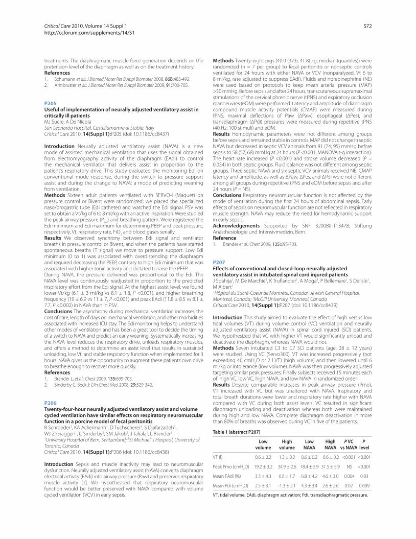
treatments. The diaphragmatic muscle force generation depends on the
pretension level of the diaphragm as well as on the treatment history.
References1. Schumann et al.: J Biomed Mater Res B Appl Biomater 2008, 86B:483-492.
2. Armbruster et al.: J Biomed Mater Res B Appl Biomater 2009, 91:700-705.
P205
Useful of implementation of neurally adjusted ventilatory assist in critically ill patientsMJ Sucre, A De Nicola
San Leonardo Hospital, Castellamamre di Stabia, Italy
Critical Care 2010, 14(Suppl 1):P205 (doi: 10.1186/cc8437)
Introduction Neurally adjusted ventilatory assist (NAVA) is a new
mode of assisted mechanical ventilation that uses the signal obtained
from electromyography activity of the diaphragm (EAdi) to control
the mechanical ventilator that delivers assist in proportion to the
patient’s respiratory drive. This study evaluated the monitoring Edi on
conventional mode response, during the switch to pressure support
assist and during the change to NAVA: a mode of predicting weaning
from ventilation.
Methods Sixteen adult patients ventilated with SERVO-I (Maquet) on
pressure control or Bivent were randomized; we placed the specialized
naso/orogastric tube (Edi catheter) and watched the Edi signal. PSV was
set to obtain a Vt/kg of 6 to 8 ml/kg with an active inspiration. Were studied
the peak airway pressure (Paw
) and breathing pattern. Were registered the
Edi minimum and Edi maximum for determining PEEP and peak pressure,
respectively, Vt, respiratory rate, FiO2 and blood gases serially.
Results We observed synchrony between Edi signal and ventilator
breaths in pressure control or Bivent, and when the patients have started
spontaneous breaths (T signal) we move to pressure support. Low Edi
minimum (0 to 1) was associated with overdistending the diaphragm
and required decreasing the PEEP, contrary to high Edi minimum that was
associated with higher tonic activity and dictated to raise the PEEP.
During NAVA, the pressure delivered was proportional to the Edi. The
NAVA level was continuously readjusted in proportion to the predicted
inspiratory eff ort from the Edi signal. At the highest assist level, we found
lower Vt/kg (6.1 ± 3 ml/kg vs 8.1 ± 1.8, P <0.001), and higher breathing
frequency (19 ± 6.9 vs 11 ± 7, P <0.001) and peak EAdi (11.8 ± 8.5 vs 8.1 ±
7.7, P <0.002) in NAVA than in PSV.
Conclusions The asynchrony during mechanical ventilation increases the
cost of care, length of days on mechanical ventilation, and other morbidities
associated with increased ICU stay. The Edi monitoring helps to understand
other modes of ventilation and has been a great tool to decide the timing
of a switch to NAVA and predict an early weaning. Systematically increasing
the NAVA level reduces the respiratory drive, unloads respiratory muscles,
and off ers a method to determine an assist level that results in sustained
unloading, low Vt, and stable respiratory function when implemented for 3
hours. NAVA gives us the opportunity to augment these patients’ own drive
to breathe enough to recover more quickly.
References1. Brander L, et al.: Chest 2009, 135:695-703.
2. Sinderby C, Beck J: Clin Chest Med 2008, 29:329-342.
P206
Twenty-four-hour neurally adjusted ventilatory assist and volume cycled ventilation have similar eff ects on respiratory neuromuscular function in a porcine model of fecal peritonitisR Schroeder1, KA Ackermann1, D Tuchscherer1, S Djafarzadeh1,
WJ Z`Graggen1, C Sinderby2, SM Jakob1, J Takala1, L Brander1
1University Hospital of Bern, Switzerland; 2St Michael`s Hospital, University of
Toronto, Canada
Critical Care 2010, 14(Suppl 1):P206 (doi: 10.1186/cc8438)
Introduction Sepsis and muscle inactivity may lead to neuromuscular
dysfunction. Neurally adjusted ventilatory assist (NAVA) converts diaphragm
electrical activity (EAdi) into airway pressure (Paw) and preserves respiratory
muscle activity [1]. We hypothesised that respiratory neuromuscular
function would be better preserved with NAVA compared with volume
cycled ventilation (VCV) in early sepsis.
Methods Twenty-eight pigs (40.0 (37.6; 41.8) kg; median (quartiles)) were
randomized (n = 7 per group) to fecal peritonitis or nonseptic controls
ventilated for 24 hours with either NAVA or VCV (nonparalyzed, Vt 6 to
8 ml/kg, rate adjusted to suppress EAdi). Fluids and norepinephrine (NE)
were used based on protocols to keep mean arterial pressure (MAP)
>50 mmHg. Before sepsis and after 24 hours, transcutaneous supramaximal
stimulations of the cervical phrenic nerve (tPNS) and expiratory occlusion
manoeuvres (eOM) were performed. Latency and amplitude of diaphragm
compound muscle activity potentials (CMAP) were measured during
tPNS; maximal defl ections of Paw (ΔPaw), esophageal (ΔPes), and
transdiaphragm (ΔPdi) pressures were measured during repetitive tPNS
(40 Hz, 100 stimuli) and eOM.
Results Hemodynamic parameters were not diff erent among groups
before sepsis and remained stable in controls. MAP did not change in septic
NAVA but decreased in septic VCV animals from 91 (74; 95) mmHg before
sepsis to 58 (57; 68) mmHg at 24 hours (P <0.001, MANOVA t-g interaction).
The heart rate increased (P <0.001) and stroke volume decreased (P =
0.034) in both septic groups. Fluid balance was not diff erent among septic
groups. Three septic NAVA and six septic VCV animals received NE. CMAP
latency and amplitude, as well as ΔPaw, ΔPes, and ΔPdi were not diff erent
among all groups during repetitive tPNS and eOM before sepsis and after
24 hours (P = NS).
Conclusions Respiratory neuromuscular function is not aff ected by the
mode of ventilation during the fi rst 24 hours of abdominal sepsis. Early
eff ects of sepsis on neuromuscular function are not refl ected in respiratory
muscle strength. NAVA may reduce the need for hemodynamic support
in early sepsis.
Acknowledgements Supported by SNF 3200B0-113478; Stiftung
Anästhesiologie und Intensivmedizin, Bern.
Reference1. Brander et al.: Chest 2009, 135:695-703.
P207
Eff ects of conventional and closed-loop neurally adjusted ventilatory assist in intubated spinal cord injured patientsJ Spahija1, M De Marchie2, K Trufl andier1, A Moga3, P Bellemare1, S Delisle1,
M Albert1
1Hôpital du Sacré-Coeur de Montréal, Canada; 2Jewish General Hospital,
Montreal, Canada; 3McGill University, Montreal, Canada
Critical Care 2010, 14(Suppl 1):P207 (doi: 10.1186/cc8439)
Introduction This study aimed to evaluate the eff ect of high versus low
tidal volumes (VT) during volume control (VC) ventilation and neurally
adjusted ventilatory assist (NAVA) in spinal cord injured (SCI) patients.
We hypothesized that VC with higher VT would signifi cantly unload and
deactivate the diaphragm, whereas NAVA would not.
Methods Seven intubated C3 to C7 SCI patients (age: 28 ± 12 years)
were studied. Using VC (Servo300), VT was increased progressively (not
exceeding 40 cmH2O or 2 l VT) (high volume) and then lowered until 6
ml/kg or intolerance (low volume). NAVA was then progressively adjusted
targeting similar peak pressures. Finally subjects received 15 minutes each
of: high VC, low VC, high NAVA, and low NAVA in randomized order.
Results Despite comparable increases in peak airway pressure (Pmo),
VT increased with VC but was unaltered with NAVA. Inspiratory and
total breath durations were lower and respiratory rate higher with NAVA
compared with VC during both assist levels. VC resulted in signifi cant
diaphragm unloading and deactivation whereas both were maintained
during high and low NAVA. Complete diaphragm deactivation in more
than 80% of breaths was observed during VC in fi ve of the patients.
Table 1 (abstract P207)
Low High Low High P VC P volume volume NAVA NAVA vs NAVA level
VT (l) 0.6 ± 0.2 1.3 ± 0.2 0.6 ± 0.2 0.6 ± 0.2 <0.001 <0.001
Peak Pmo (cmH2O) 19.2 ± 3.2 34.9 ± 2.6 18.4 ± 5.9 31.5 ± 5.9 NS <0.001
Mean EAdi (%) 3.3 ± 4.3 0.8 ± 1.7 6.8 ± 4.2 4.6 ± 3.0 0.004 0.03
Mean Pdi (cmH2O) 2.5 ± 3.1 -1.3 ± 2.1 4.3 ± 3.4 2.6 ± 2.6 0.02 0.009
VT, tidal volume; EAdi, diaphragm activation; Pdi, transdiaphragmatic pressure.
Critical Care 2010, Volume 14 Suppl 1 http://ccforum.com/supplements/14/S1
S72

Conclusions VC ventilation results in signifi cant diaphragm unloading
and deactivation especially at high assist levels, whereas diaphragm
activation (EAdi) is maintained with NAVA.
P208
A level of neurally adjusted ventilatory assist corresponds to low levels of continuous positive airway pressure and pressure support ventilation in patients preparing to be extubated from mechanical ventilationY Klai-on, A Navasakulpong, A Wattanathum
Phramongkutklao Hospital, Bangkok, Thailand
Critical Care 2010, 14(Suppl 1):P208 (doi: 10.1186/cc8440)
Introduction Neurally adjusted ventilator assist (NAVA) is a new mode
of mechanical ventilation which delivers ventilator assist in proportion to
electrical activity of the diaphragm (EAdi), as assessed by trans-esophageal
electromyography, and an adjustable supporting level called the NAVA
level. NAVA can be used as a supporting and weaning mode of ventilation
[1-3]. However, a level of NAVA corresponding to continuous positive
airway pressure (CPAP) and pressure support ventilation (PSV) in patients
ready for extubation has not been established.
Methods We carried out a prospective study in the medical ICU at
Phramongkutklao Hospital involving patients who were considered
by their physicians to be ready for extubation. Patients initially received
PSV of 5 cmH2O, PEEP of 5 cmH
2O, and FiO
2 of 40% for 15 minutes (PS).
Subsequently, each patient received a trial of NAVA mode with PEEP of
5 cmH2O, and FiO
2 of 40% for 15 minutes (NAVA), and, fi nally, a 30-minute
spontaneously breathing trial without ventilatory support (SBT). During
these trials, the minute volume (MV), respiratory rate, EAdi, and airway
pressure were measured. We determined the corresponding NAVA level
with the level of PSV of 5 cmH2O and PEEP of 5 cmH
2O using the NAVA
level matching MV (±10%) received from the PSV.
Results The NAVA level corresponding to the PSV of 5 cmH2O and PEEP
of 5 cmH2O was 0.52 cmH
2O/μV (0.2 to 1.2). The airway pressure during
NAVA was 5.97 cmH2O (3.2 to 8) above PEEP level. The peak EAdi was not
signifi cantly diff erent between the PS, NAVA, and SBT (11.58, 12.06 vs
12.65 μV; P = 0.6). Also, there was no signifi cant diff erence in MV during
the PS, NAVA, and SBT.
Conclusions NAVA mode can be used as a weaning mode by decreasing
the NAVA level. The reduced level of NAVA to 0.5 cmH2O/μV did not load
respiratory muscle more than a low level of PSV and SBT. Thus, a NAVA level
of 0.5 cmH2O/μV might be the level that is safe to discontinue ventilator
support in patients without problems for resuming spontaneous
breathing.
References1. Navalesi P, et al.: Curr Opin Crit Care 2003, 9:51-58.
2. Sinderby C, et al.: Chest 2007, 131:711-717.
3. Beck J, et al.: Pediatr Res 2007, 61:289-294.
P209
Towards partially automated ventilation: adapting decision-making according to medical preferencesS Lozano-Zahonero1, D Gottlieb2, J Guttmann2, K Möller1
1Furtwangen University, VS-Schwenningen, Germany; 2University Hospital
Freiburg, Germany
Critical Care 2010, 14(Suppl 1):P209 (doi: 10.1186/cc8441)
Introduction A novel methodology is proposed to adapt decision-
making strategies into our fuzzy-based expert system, AUTOPILOT-BT [1].
The special features of this approach are: knowledge from clinical experts
can be extracted in a setup simulating daily ICU routine; an automated
process serves to obtain the required information and to create new fuzzy
sets; and the fuzzy controller from AUTOPILOT-BT employs the newly
derived fuzzy rules. Thus, the knowledge base can easily be modifi ed
and the resulting mechanical ventilation therapies may be adapted to
individual preferences of the clinician.
Methods The methodology consists of: (i) Acquisition of decision-making
strategies from single or groups of anesthesiologists. This can either be
done with a questionnaire or with a PC-based program simulating the
doctors every day situation in diagnosis. (ii) Defi nition of fuzzy membership
functions based on the acquired knowledge (fuzzifi cation of the input).
(iii) Construction of fuzzy inference rules and defuzzifi cation or calculation
of change in ventilator settings (controller action). This approach allows
implementing clinician-dependent decision-making that refl ects
individual preferences. Thus guidelines from EBM can be used but as well
ICU specifi cs can be realized, for example diff erent levels of acceptable
hypercapnia in patients with acute respiratory distress syndrome (ARDS).
Results Exemplarily Figure 1 shows, for healthy and ARDS lungs, the
diff erence between two fuzzy sets for our paCO2 controller given from two
clinicians (C10 and C51) with diff erent expertise in mechanical ventilation.
With the newly designed fuzzy sets, our AUTOPILOT-BT reacts according to
the clinicians’ preferences, but still minimizes the time in which the patient
is not ventilated within the specifi ed limits.
Conclusions The system automatically implements the know-how of
medical experts in ventilation management if the clinicians are willing to
interact with the query system. The resulting strategy is mainly infl uenced
by the expertise, experience and demands of the clinician. Thus the
AUTOPILOT-BT, has the potential to select established guidelines or to
adapt the system to modifi ed ventilation therapies. Further clinical trials
will test the actual clinical effi ciency of diff erent controllers.
References1. Lozano S, et al.: Tech Health Care 2008, 16:1-11.
P210
Postoperative pulmonary complications in adult elective surgery patients in the US: severity, outcomes and resources useWL Linde-Zwirble1, JD Bloom2, RS Mecca3, DM Hansell4
1ZD Associates LLC, Perksaie, PA, USA; 2Covidien, Boulder, CO, USA; 3University
of California at Irvine, Orange, CA, USA; 4Massachusetts General Hospital,
Boston, MA, USA
Critical Care 2010, 14(Suppl 1):P210 (doi: 10.1186/cc8442)
Introduction Postoperative pulmonary complications (PPCs) are asso-
ciated with adverse outcomes and added resource use. However, PPCs
vary in severity, and so may have very diff erent outcomes.
Methods We selected adult elective surgical cases from the Premier database
for 2008. The PPC conditions identifi ed were: pneumonia; respiratory failure;
bronchospasm; tracheobronchitis; pleural eff usion; pulmonary collapse;
ARDS; and pneumothorax. We stratifi ed PPC cases into levels: those with
bronchospasm and no other PPC (BS), those with respiratory failure (RF)
and all remaining (NRF). We examined incidence, length of stay, hospital
mortality, and total cost. US national projections were made using Premier
supplied projection weights. We calculated incremental resource use and
outcomes by comparing cases with and without PPCs for each surgical
condition and summarizing across all conditions.
Results There were 738,039 cases in our cohort. At least one PPC was
identifi ed in 85,435. BS occurred on 6.4% of cases (45,005), NRF in 3.8%
(27,796) and RF in 1.7% of cases (12,634). BS was not associated with an
increase in the risk of death (one added death per 5611 cases), but had
signifi cant increases in resource use: one added ICU admission per 14 cases;
one added ICU day per fi ve cases; and one added hospital day and $1,563
added cost per case. NRF was associated with a modest increase in risk of
death (one added death per 157 NRF cases) and substantial increases in
resource use: one added ICU admission for every six cases; 0.66 ICU days
per case; and three hospital days and $5,771 added cost per NRF case. RF
was associated with greatly increased risk of death (one added death per
Figure 1 (abstract P209). Normal ventilation for healthy vs ARDS patients –
fuzzy sets for paCO2 given from two clinicians: (left) C10, (right) C51.
Critical Care 2010, Volume 14 Suppl 1 http://ccforum.com/supplements/14/S1
S73
10 cases) and larger increases in resource use: one added ICU admission per
two cases; 5 ICU days, 8 hospital days and $24,000 in added cost per RF case.
Projecting to the US population there were 583,300 PPC cases, associated
with 9,500 added deaths, 92,200 added ICU admissions, 584,200 added ICU
days, 867,400 added fl oor days and $3.42 billion US in added costs. PPC add
$717 to the average cost of elective surgery in the US.
Conclusions PPCs are very common, occurring in one in eight elective
postoperative patients. While BS was not associated with added deaths, all
three PPC strata were associated with substantial increase in resource use.
Better strategies for the prevention and management of PPCs could lead
to greatly improved outcomes and substantial savings.
P211
Increased duration of mechanical ventilation is associated with decreased diaphragmatic forceG Hermans, A Agten, D Testelmans, M Decramer, G Gayan-Ramirez
UZ Leuven, Belgium
Critical Care 2010, 14(Suppl 1):P211 (doi: 10.1186/cc8443)
Introduction Respiratory muscle weakness is an important risk factor
for prolonged mechanical ventilation, and may be part of critical illness
related polyneuropathy and myopathy. Animal data also strongly point to
atrophy and weakness of the diaphragm due to mechanical ventilation
itself, called ventilator-induced diaphragmatic dysfunction. Recently,
measuring transdiaphragmatic pressure following magnetic stimulation
(TwPdi BAMPS) was introduced in the ICU to evaluate diaphragm function
[1,2]. We aimed to evaluate reproducibility of twitch TwPdi BAMPS in
critically ill, mechanically ventilated patients. We also aimed to describe
the relationship between TwPdi and duration of mechanical ventilation.
Methods Prospective observational study in a medical ICU of a university
hospital. TwPdi BAMPS was measured in critically ill and mechanically
ventilated patients. Briefl y, the phrenic nerves were stimulated
bilaterally from the anterior approach, at the posterior border of the
sternocleidomastoid muscle, at the level of the cricoids using two fi gure-
of-eight 45 mm magnetic coils (Magstim, Dyfed, Wales) and a bistim
(Magstim, Dyfed, Wales). A custom-built occlusion valve was used to create
isometric conditions during stimulation. Oesophageal and abdominal
pressure changes were measured using balloon catheters (UK Medical,
Sheffi eld, UK) inserted through the nose after local anaesthesia.
Results Nineteen measurements were made in a total of 10 patients at
various intervals after starting mechanical ventilation. In seven patients,
measurements were made on at least two occasions with a minimal
interval of 24 hours. The between-occasion coeffi cient of variation of
TwPdi was 9.7%, which is comparable with data from healthy volunteers.
Increasing duration of mechanical ventilation was associated with a
logarithmic decline in TwPdi (R = 0.69, P = 0.038). This association was also
found when cumulative time on pressure control ventilation (R = 0.71, P =
0.03) and pressure support ventilation (P = 0.05, R = 0.66) were considered
separately, as well as for cumulative dose of propofol (R = 0.66, P = 0.05)
and piritramide (R = 0.79, P = 0.01).
Conclusions Increased duration of mechanical ventilation is associated
with a logarithmic decline in diaphragmatic force. These fi ndings are
compatible with the concept of ventilator-induced diaphragmatic
dysfunction. The observed decline may also be due to the cumulative
dose of sedatives/analgesics or other co-factors, such as sepsis.
References1. Watson AC, et al.: Crit Care Med 2001, 29:1325-1331.
2. Laghi F, et al.: Am J Respir Crit Care Med 2003, 167:120-127.
P212
Haemodynamics and volhaemic stage in patients with respiratory disorders to thermal injuryK Shatovkin, I Shlyk
Djanelidze Research Institute of Emergency Medicine, Saint-Petersburg, Russia
Federation
Critical Care 2010, 14(Suppl 1):P212 (doi: 10.1186/cc8444)
Introduction The aim of the investigation was to study the parameters
of haemodynamics, gas exchange and volhaemic status in patients with
severe thermal injury.
Methods The trial has covered 30 injured patients aged from 21 to
60 years with 25 to 78% skin burns and PO2/FiO
2 <300. Patients were
randomized into two groups. The fi rst group was n = 13, PO2/FiO
2 <300,
24 to 72 h after burn. The second group was n = 17, PO2/FiO
2 <300, 4
to 11 days after burn. Injured persons were subjected to evaluation of
cardiac index (CI), intrathoracic blood volume index (ITBI), extravascular
lung water index (ELWI0 and pulmonary vascular permeability index
(PVPI) by single transpulmonary thermodilution (PiCCOplus; Pulsion
Medical Systems, Germany), calculation of oxygen delivery (DO2), and
colloidal osmotic pressure of plasma. Grade of pulmonary damage was
evaluated by Murrey, severity of dispragia by SOFA. Correlation analysis
was performed using Pearson and Spearman criteria (r; P). Diff erences
were signifi cant at P <0.05.
Results Hypoxaemia (PO2/FiO
2 = 259 ± 19) in the injured of the fi rst group
was developing at the background of reduction of CI = 3.2 ± 0.5; ITBI = 751
± 114; DO2 = 596 ± 124 (r = –0.92; P = 0.02). ELWI level was normal (7.3 ±
0.8). In the second group, reduction of PO2/FiO
2 has been developing at
the background of burn sepsis (average SOFA point = 7.0 ± 1.9, Murrey
= 1.6 ± 0.4) and was caused by increasing of ELWI up to 9.0 ± 1.4 (r =
–0.66; P = 0.002) and correlated with PVPI (r = 0.57; P = 0.01). There was
no statistically signifi cant correlation ELWI with ITBI and colloidal osmotic
pressure of plasma (r = 0.13, r = –0.42).
Conclusions Gas exchange disorders in patients of the fi rst group were
caused by lack of perfusion and misbalance between oxygen delivery and
demand. Reduction of oxygenation index in patients from the second
group was accompanied by ELWI at the background of alteration of
intravascular penetration and sepsis.
P213
Invasive mechanical ventilation in cancer patients: prior non-invasive ventilation is a poor prognostic factorA Meert, T Berghmans, N Nayer, M Paesmans, E Markiewicz, M Hardy,
J Sculier
Institut Jules Bordet, Bruxelles, Belgium
Critical Care 2010, 14(Suppl 1):P213 (doi: 10.1186/cc8445)
Introduction Prior non-invasive ventilation (NIV) is associated with an
increased mortality in patients with haematological malignancies and
acute respiratory failure treated by invasive mechanical ventilation (IMV).
Methods We have assessed whether NIV failure is an independent
prognostic factor for hospital discharge in a general cancer population
treated by IMV. One hundred and six patients with solid tumours and
58 patients with haematological malignancies were eligible for this
retrospective study; 41 were treated by NIV before IMV.
Results The main indications for mechanical ventilation were sepsis/
shock (35%), acute respiratory failure (33%), cardiopulmonary resuscitation
(16%) and neurologic disease (10%). Respectively, 35%, 28% and 24%
of the patients were extubated, discharged from the ICU and from the
hospital. For patients treated with NIV prior to IMV, the rates were 22%,
17% and 10%, respectively. In multivariate analysis, three variables
were independently associated with a decreased probability of being
discharged from the hospital: NIV use before IMV (OR = 0.30, 95% CI: 0.09
to 0.95; P = 0.04); leucopenia (OR = 0.21, 95% CI: 0.06 to 0.77; P = 0.02) and
serum bilirubin >1.1 mg/dl (OR = 0.38, 95% CI: 0.16 to 0.94; P = 0.04).
Conclusions NIV failure before IMV is an independent poor prognostic
factor in cancer patients treated by IMV.
P214
Laryngeal angioedema: do we need a new treatment algorithm in the ER?K Stelter1, A Leunig1, M Jacob1, M Bas2
1Ludwig Maximilians University, Munich, Germany; 2Technical University,
Munich, Germany
Critical Care 2010, 14(Suppl 1):P214 (doi: 10.1186/cc8446)
Introduction Laryngeal angioedema (AE) could be life-threatening and
is frequently due to allergic reactions. Other causes, that is bradykinin-
mediated conditions, have to be taken into account. The aim of our survey
is to provide information on the incidence, diagnosis and potentially new
treatment options for laryngeal AE.
Critical Care 2010, Volume 14 Suppl 1 http://ccforum.com/supplements/14/S1
S74
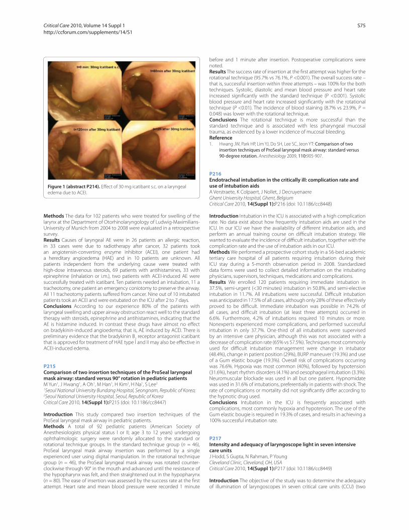
Methods The data for 102 patients who were treated for swelling of the
larynx at the Department of Otorhinolaryngology of Ludwig-Maximilians-
University of Munich from 2004 to 2008 were evaluated in a retrospective
survey.
Results Causes of laryngeal AE were in 26 patients an allergic reaction,
in 33 cases were due to radiotherapy after cancer, 32 patients took
an angiotensin-converting enzyme inhibitor (ACEI), one patient had
a hereditary angioedema (HAE) and in 10 patients are unknown. All
patients independent from the underlying cause were treated with
high-dose intravenous steroids, 69 patients with antihistamines, 33 with
epinephrine (inhalation or i.m.), two patients with ACEI-induced AE were
successfully treated with icatibant. Ten patients needed an intubation, 11 a
tracheotomy, one patient an emergency coniotomy to preserve the airway.
All 11 tracheotomy patients suff ered from cancer. Nine out of 10 intubated
patients took an ACEI and were extubated on the ICU after 2 to 7 days.
Conclusions According to our experience 80% of the patients with
laryngeal swelling and upper airway obstruction react well to the standard
therapy with steroids, epinephrine and antihistamines, indicating that the
AE is histamine induced. In contrast these drugs have almost no eff ect
on bradykinin-induced angioedema; that is, AE induced by ACEI. There is
preliminary evidence that the bradykinin B2 receptor antagonist icatibant
that is approved for treatment of HAE type I and II may also be eff ective in
ACEI-induced edema.
P215
Comparison of two insertion techniques of the ProSeal laryngeal mask airway: standard versus 90° rotation in pediatric patientsM Yun1, J Hwang1, A Oh1, M Han1, H Kim2, H Na1, S Lee2
1Seoul National University Bundang Hospital, Seongnam, Republic of Korea; 2Seoul National University Hospital, Seoul, Republic of Korea
Critical Care 2010, 14(Suppl 1):P215 (doi: 10.1186/cc8447)
Introduction This study compared two insertion techniques of the
ProSeal laryngeal mask airway in pediatric patients.
Methods A total of 92 pediatric patients (American Society of
Anesthesiologists physical status I or II; age 3 to 12 years) undergoing
ophthalmologic surgery were randomly allocated to the standard or
rotational technique groups. In the standard technique group (n = 46),
ProSeal laryngeal mask airway insertion was performed by a single
experienced user using digital manipulation. In the rotational technique
group (n = 46), the ProSeal laryngeal mask airway was rotated counter-
clockwise through 90° in the mouth and advanced until the resistance of
the hypopharynx was felt, and then straightened out in the hypopharynx
(n = 80). The ease of insertion was assessed by the success rate at the fi rst
attempt. Heart rate and mean blood pressure were recorded 1 minute
before and 1 minute after insertion. Postoperative complications were
noted.
Results The success rate of insertion at the fi rst attempt was higher for the
rotational technique (95.7% vs 76.1%, P <0.001). The overall success rate –
that is, successful insertion within three attempts – was 100% for the both
techniques. Systolic, diastolic and mean blood pressure and heart rate
increased signifi cantly with the standard technique (P <0.001). Systolic
blood pressure and heart rate increased signifi cantly with the rotational
technique (P <0.01). The incidence of blood staining (8.7% vs 23.9%, P =
0.048) was lower with the rotational technique.
Conclusions The rotational technique is more successful than the
standard technique and is associated with less pharyngeal mucosal
trauma, as evidenced by a lower incidence of mucosal bleeding.
Reference1. Hwang JW, Park HP, Lim YJ, Do SH, Lee SC, Jeon YT: Comparison of two
insertion techniques of ProSeal laryngeal mask airway: standard versus 90-degree rotation. Anesthesiology 2009, 110:905-907.
P216
Endotracheal intubation in the critically ill: complication rate and use of intubation aidsA Verstraete, K Colpaert, J Nollet, J Decruyenaere
Ghent University Hospital, Ghent, Belgium
Critical Care 2010, 14(Suppl 1):P216 (doi: 10.1186/cc8448)
Introduction Intubation in the ICU is associated with a high complication
rate. No data exist about how frequently intubation aids are used in the
ICU. In our ICU we have the availability of diff erent intubation aids, and
perform an annual training course on diffi cult intubation strategy. We
wanted to evaluate the incidence of diffi cult intubation, together with the
complication rate and the use of intubation aids in our ICU.
Methods We performed a prospective cohort study in a 56-bed academic
tertiary care hospital of all patients requiring intubation during their
ICU stay during a 5-month observation period in 2008. Standardized
data forms were used to collect detailed information on the intubating
physicians, supervisors, techniques, medications and complications.
Results We enrolled 120 patients requiring immediate intubation in
37.5%, semi-urgent (<30 minutes) intubation in 50.8%, and semi-elective
intubation in 11.7%. All intubations were successful. Diffi cult intubation
was anticipated in 17.5% of all cases, although only 28% of these eff ectively
proved to be diffi cult. Immediate intubation was possible in 74.2% of
all cases, and diffi cult intubation (at least three attempts) occurred in
6.6%. Furthermore, 4.2% of intubations required 10 minutes or more.
Nonexperts experienced more complications, and performed successful
intubation in only 37.7%. One-third of all intubations were supervised
by an intensive care physician, although this was not associated with a
decrease of complication rate (65% vs 57.5%). Techniques most commonly
used for diffi cult intubation management were change in intubator
(48.4%), change in patient position (29%), BURP maneuver (19.3%) and use
of a Gum elastic bougie (19.3%). Overall risk of complications occurring
was 76.6%. Hypoxia was most common (40%), followed by hypotension
(31.6%), heart rhythm disorders (4.1%) and oesophageal intubation (3.3%).
Neuromuscular blockade was used in all but one patient. Hypnomidate
was used in 31.6% of intubations, preferentially in patients with shock. The
rate of complications or mortality did not signifi cantly diff er according to
the hypnotic drug used.
Conclusions Intubation in the ICU is frequently associated with
complications, most commonly hypoxia and hypotension. The use of the
Gum elastic bougie is required in 19.3% of cases, and results in achieving a
100% successful intubation rate.
P217
Intensity and adequacy of laryngoscope light in seven intensive care unitsJ Hodd, S Gupta, N Rahman, P Young
Cleveland Clinic, Cleveland, OH, USA
Critical Care 2010, 14(Suppl 1):P217 (doi: 10.1186/cc8449)
Introduction The objective of the study was to determine the adequacy
of illumination of laryngoscopes in seven critical care units (CCU) (two
Figure 1 (abstract P214). Eff ect of 30 mg icatibant s.c. on a laryngeal
edema due to ACEI.
Critical Care 2010, Volume 14 Suppl 1 http://ccforum.com/supplements/14/S1
S75
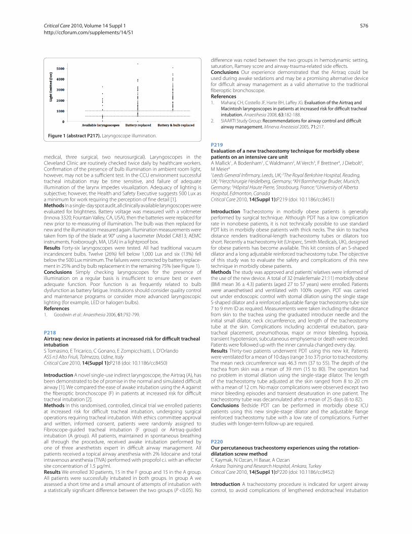
medical, three surgical, two neurosurgical). Laryngoscopes in the
Cleveland Clinic are routinely checked twice daily by healthcare workers.
Confi rmation of the presence of bulb illumination in ambient room light,
however, may not be a suffi cient test. In the CCU environment successful
tracheal intubation may be time sensitive, and failure of adequate
illumination of the larynx impedes visualization. Adequacy of lighting is
subjective; however, the Health and Safety Executive suggests 500 Lux as
a minimum for work requiring the perception of fi ne detail [1].
Methods In a single-day spot audit, all clinically available laryngoscopes were
evaluated for brightness. Battery voltage was measured with a voltmeter
(Innova 3320; Fountain Valley, CA, USA), then the batteries were replaced for
new prior to re-measuring of illumination. The bulb was then replaced for
new and the illumination measured again. Illumination measurements were
taken from tip of the blade at 90° using a luxometer (Model CA813; AEMC
instruments, Foxborough, MA, USA) in a lightproof box.
Results Forty-six laryngoscopes were tested. All had traditional vacuum
incandescent bulbs. Twelve (26%) fell below 1,000 Lux and six (13%) fell
below the 500 Lux minimum. The failures were corrected by battery replace-
ment in 25% and by bulb replacement in the remaining 75% (see Figure 1).
Conclusions Simply checking laryngoscopes for the presence of
illumination on a regular basis is insuffi cient to ensure best or even
adequate function. Poor function is as frequently related to bulb
dysfunction as battery fatigue. Institutions should consider quality control
and maintenance programs or consider more advanced laryngoscopic
lighting (for example, LED or halogen bulbs).
References1. Goodwin et al.: Anaesthesia 2006, 61:792-799.
P218
Airtraq: new device in patients at increased risk for diffi cult tracheal intubationS Tomasino, E Tricarico, C Gonano, E Zompicchiatti, L D’Orlando
ASS n3 Alto Friuli, Tolmezzo, Udine, Italy
Critical Care 2010, 14(Suppl 1):P218 (doi: 10.1186/cc8450)
Introduction A novel single-use indirect laryngoscope, the Airtraq (A), has
been demonstrated to be of promise in the normal and simulated diffi cult
airway [1]. We compared the ease of awake intubation using the A against
the fi beroptic bronchoscope (F) in patients at increased risk for diffi cult
tracheal intubation [2].
Methods In this randomised, controlled, clinical trial we enrolled patients
at increased risk for diffi cult tracheal intubation, undergoing surgical
operations requiring tracheal intubation. With ethics committee approval
and written, informed consent, patients were randomly assigned to
Fibroscope-guided tracheal intubation (F group) or Airtraq-guided
intubation (A group). All patients, maintained in spontaneous breathing
all through the procedure, received awake intubation performed by
one of three anesthetists expert in diffi cult airway management. All
patients received a topical airway anesthesia with 2% lidocaine and total
intravenous anesthesia (TIVA) performed with propofol c.i. with an eff ecter
site concentration of 1.5 μg/ml.
Results We enrolled 30 patients, 15 in the F group and 15 in the A group.
All patients were successfully intubated in both groups. In group A we
assessed a short time and a small amount of attempts of intubation with
a statistically signifi cant diff erence between the two groups (P <0.05). No
diff erence was noted between the two groups in hemodynamic setting,
saturation, Ramsey score and airway-trauma-related side eff ects.
Conclusions Our experience demonstrated that the Airtraq could be
used during awake sedations and may be a promising alternative device
for diffi cult airway management as a valid alternative to the traditional
fi beroptic bronchoscope.
References1. Maharaj CH, Costello JF, Harte BH, Laff ey JG: Evaluation of the Airtraq and
Macintosh laryngoscopes in patients at increased risk for diffi cult tracheal intubation. Anaesthesia 2008, 63:182-188.
2. SIAARTI Study Group: Recommendations for airway control and diffi cult airway management. Minerva Anestesiol 2005, 71:217.
P219
Evaluation of a new tracheostomy technique for morbidly obese patients on an intensive care unitA Mallick1, A Bodenham1, C Waldmann2, M Verch3, F Brettner4, J Diebolt5,
M Meier6
1Leeds General Infi rmary, Leeds, UK; 2The Royal Berkshire Hospital, Reading,
UK; 3Herzchirurgie Heidelberg, Germany; 4KH Barmherzige Bruder, Munich,
Germany; 5Hôpital Haute Pierre, Strasbourg, France; 6University of Alberta
Hospital, Edmonton, Canada
Critical Care 2010, 14(Suppl 1):P219 (doi: 10.1186/cc8451)
Introduction Tracheostomy in morbidly obese patients is generally
performed by surgical technique. Although PDT has a low complication
rate in nonobese patients, it is not technically possible to use standard
PDT kits in morbidly obese patients with thick necks. The skin to trachea
distance renders traditional-length tracheostomy tubes or dilators too
short. Recently a tracheostomy kit (Uniperc, Smith Medicals, UK), designed
for obese patients has become available. This kit consists of an S-shaped
dilator and a long adjustable reinforced tracheostomy tube. The objective
of this study was to evaluate the safety and complications of this new
technique in morbidly obese patients.
Methods The study was approved and patients’ relatives were informed of
the use of the new device. A total of 32 (male:female 21:11) morbidly obese
(BMI mean 36 ± 4.3) patients (aged 27 to 57 years) were enrolled. Patients
were anaesthetised and ventilated with 100% oxygen. PDT was carried
out under endoscopic control with stomal dilation using the single stage
S-shaped dilator and a reinforced adjustable fl ange tracheostomy tube size
7 to 9 mm ID as required. Measurements were taken including the distance
from skin to the trachea using the graduated introducer needle and the
initial small dilator, neck circumference, and length of the tracheostomy
tube at the skin. Complications including accidental extubation, para-
tracheal placement, pneumothorax, major or minor bleeding, hypoxia,
transient hypotension, subcutaneous emphysema or death were recorded.
Patients were followed up with the inner cannula changed every day.
Results Thirty-two patients underwent PDT using this new kit. Patients
were ventilated for a mean of 10 days (range 3 to 37) prior to tracheostomy.
The mean neck circumference was 46.3 mm (37 to 55). The depth of the
trachea from skin was a mean of 39 mm (15 to 80). The operators had
no problem in stomal dilation using the single-stage dilator. The length
of the tracheostomy tube adjusted at the skin ranged from 8 to 20 cm
with a mean of 12 cm. No major complications were observed except two
minor bleeding episodes and transient desaturation in one patient. The
tracheostomy tube was decannulated after a mean of 25 days (6 to 82).
Conclusions Bedside PDT can be performed in morbidly obese ICU
patients using this new single-stage dilator and the adjustable fl ange
reinforced tracheostomy tube with a low rate of complications. Further
studies with longer-term follow-up are required.
P220
Our percutaneous tracheostomy experiences using the rotation-dilatation screw methodC Kaymak, N Ozcan, H Basar, A Ozcan
Ankara Training and Research Hospital, Ankara, Turkey
Critical Care 2010, 14(Suppl 1):P220 (doi: 10.1186/cc8452)
Introduction A tracheostomy procedure is indicated for urgent airway
control, to avoid complications of lengthened endotracheal intubation
Figure 1 (abstract P217). Laryngoscope illumination.
Critical Care 2010, Volume 14 Suppl 1 http://ccforum.com/supplements/14/S1
S76
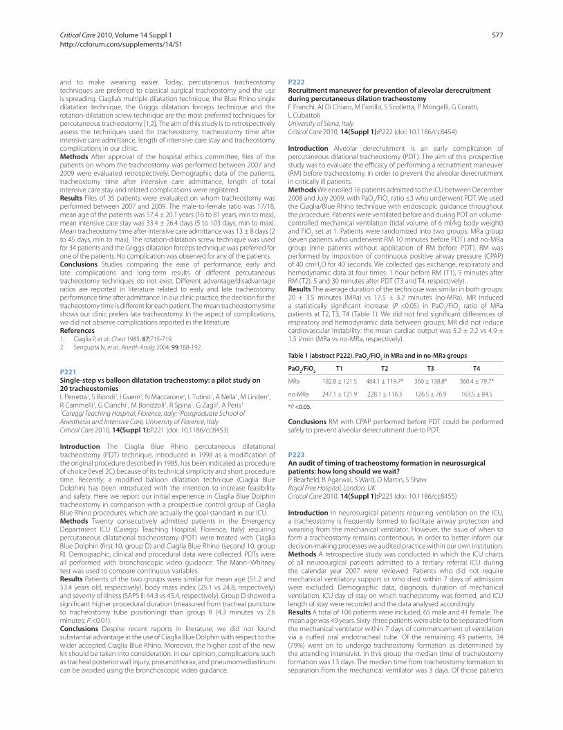
and to make weaning easier. Today, percutaneous tracheostomy
techniques are preferred to classical surgical tracheostomy and the use
is spreading. Ciaglia’s multiple dilatation technique, the Blue Rhino single
dilatation technique, the Griggs dilatation forceps technique and the
rotation-dilatation screw technique are the most preferred techniques for
percutaneous tracheostomy [1,2]. The aim of this study is to retrospectively
assess the techniques used for tracheostomy, tracheostomy time after
intensive care admittance, length of intensive care stay and tracheostomy
complications in our clinic.
Methods After approval of the hospital ethics committee, fi les of the
patients on whom the tracheostomy was performed between 2007 and
2009 were evaluated retrospectively. Demographic data of the patients,
tracheostomy time after intensive care admittance, length of total
intensive care stay and related complications were registered.
Results Files of 35 patients were evaluated on whom tracheostomy was
performed between 2007 and 2009. The male-to-female ratio was 17/18,
mean age of the patients was 57.4 ± 20.1 years (16 to 81 years, min to max),
mean intensive care stay was 33.4 ± 26.4 days (5 to 103 days, min to max).
Mean tracheostomy time after intensive care admittance was 13 ± 8 days (2
to 45 days, min to max). The rotation-dilatation screw technique was used
for 34 patients and the Griggs dilatation forceps technique was preferred for
one of the patients. No complication was observed for any of the patients.
Conclusions Studies comparing the ease of performance, early and
late complications and long-term results of diff erent percutaneous
tracheostomy techniques do not exist. Diff erent advantage/disadvantage
ratios are reported in literature related to early and late tracheostomy
performance time after admittance. In our clinic practice, the decision for the
tracheostomy time is diff erent for each patient. The mean tracheostomy time
shows our clinic prefers late tracheostomy. In the aspect of complications,
we did not observe complications reported in the literature.
References1. Ciaglia P, et al.: Chest 1985, 87:715-719.
2. Sengupta N, et al.: Anesth Analg 2004, 99:188-192.
P221
Single-step vs balloon dilatation tracheostomy: a pilot study on 20 tracheostomiesL Perretta1, S Biondi2, I Guerri2, N Maccarone2, L Tutino1, A Nella1, M Linden1,
R Cammelli1, G Cianchi1, M Bonizzoli1, R Spina1, G Zagli1, A Peris1
1Careggi Teaching Hospital, Florence, Italy; 2Postgraduate School of
Anesthesia and Intensive Care, University of Florence, Italy
Critical Care 2010, 14(Suppl 1):P221 (doi: 10.1186/cc8453)
Introduction The Ciaglia Blue Rhino percutaneous dilatational
tracheostomy (PDT) technique, introduced in 1998 as a modifi cation of
the original procedure described in 1985, has been indicated as procedure
of choice (level 2C) because of its technical simplicity and short procedure
time. Recently, a modifi ed balloon dilatation technique (Ciaglia Blue
Dolphin) has been introduced with the intention to increase feasibility
and safety. Here we report our initial experience in Ciaglia Blue Dolphin
tracheostomy in comparison with a prospective control group of Ciaglia
Blue Rhino procedures, which are actually the goal-standard in our ICU.
Methods Twenty consecutively admitted patients in the Emergency
Department ICU (Careggi Teaching Hospital, Florence, Italy) requiring
percutaneous dilatational tracheostomy (PDT) were treated with Ciaglia
Blue Dolphin (fi rst 10, group D) and Ciaglia Blue Rhino (second 10, group
R). Demographic, clinical and procedural data were collected. PDTs were
all performed with bronchoscopic video guidance. The Mann–Whitney
test was used to compare continuous variables.
Results Patients of the two groups were similar for mean age (51.2 and
53.4 years old, respectively), body mass index (25.1 vs 24.8, respectively)
and severity of illness (SAPS II: 44.3 vs 45.4, respectively). Group D showed a
signifi cant higher procedural duration (measured from tracheal puncture
to tracheostomy tube positioning) than group R (4.3 minutes vs 2.6
minutes; P <0.01).
Conclusions Despite recent reports in literature, we did not found
substantial advantage in the use of Ciaglia Blue Dolphin with respect to the
wider accepted Ciaglia Blue Rhino. Moreover, the higher cost of the new
kit should be taken into consideration. In our opinion, complications such
as tracheal posterior wall injury, pneumothorax, and pneumomediastinum
can be avoided using the bronchoscopic video guidance.
P222
Recruitment maneuver for prevention of alevolar derecruitment during percutaneous dilation tracheostomyF Franchi, AI Di Chiaro, M Fiorillo, S Scolletta, P Mongelli, G Coratti,
L Cubattoli
University of Siena, Italy
Critical Care 2010, 14(Suppl 1):P222 (doi: 10.1186/cc8454)
Introduction Alveolar derecruitment is an early complication of
percutaneous dilational tracheostomy (PDT). The aim of this prospective
study was to evaluate the effi cacy of performing a recruitment maneuver
(RM) before tracheostomy, in order to prevent the alveolar derecruitment
in critically ill patients.
Methods We enrolled 16 patients admitted to the ICU between December
2008 and July 2009, with PaO2/FiO
2 ratio ≤3 who underwent PDT. We used
the Ciaglia/Blue Rhino technique with endoscopic guidance throughout
the procedure. Patients were ventilated before and during PDT on volume-
controlled mechanical ventilation (tidal volume of 6 ml/kg body weight)
and FiO2 set at 1. Patients were randomized into two groups: MRa group
(seven patients who underwent RM 10 minutes before PDT) and no-MRa
group (nine patients without application of RM before PDT). RM was
performed by imposition of continuous positive airway pressure (CPAP)
of 40 cmH2O for 40 seconds. We collected gas exchange, respiratory and
hemodynamic data at four times: 1 hour before RM (T1), 5 minutes after
RM (T2), 5 and 30 minutes after PDT (T3 and T4, respectively).
Results The average duration of the technique was similar in both groups:
20 ± 3.5 minutes (MRa) vs 17.5 ± 3.2 minutes (no-MRa). MR induced
a statistically signifi cant increase (P <0.05) in PaO2/FiO
2 ratio of MRa
patients at T2, T3, T4 (Table 1). We did not fi nd signifi cant diff erences of
respiratory and hemodynamic data between groups; MR did not induce
cardiovascular instability: the mean cardiac output was 5.2 ± 2.2 vs 4.9 ±
1.5 l/min (MRa vs no-MRa, respectively).
Table 1 (abstract P222). PaO2/FiO
2 in MRa and in no-MRa groups
PaO2/FiO
2 T1 T2 T3 T4
MRa 182.8 ± 121.5 464.1 ± 119.7* 360 ± 138.8* 360.4 ± 79.7*
no-MRa 247.1 ± 121.9 228.1 ± 116.3 126.5 ± 76.9 163.5 ± 84.5
*P <0.05.
Conclusions RM with CPAP performed before PDT could be performed
safely to prevent alveolar derecruitment due to PDT.
P223
An audit of timing of tracheostomy formation in neurosurgical patients: how long should we wait?P Bearfi eld, B Agarwal, S Ward, D Martin, S Shaw
Royal Free Hospital, London, UK
Critical Care 2010, 14(Suppl 1):P223 (doi: 10.1186/cc8455)
Introduction In neurosurgical patients requiring ventilation on the ICU,
a tracheostomy is frequently formed to facilitate airway protection and
weaning from the mechanical ventilator. However, the issue of when to
form a tracheostomy remains contentious. In order to better inform our
decision-making processes we audited practice within our own institution.
Methods A retrospective study was conducted in which the ICU charts
of all neurosurgical patients admitted to a tertiary referral ICU during
the calendar year 2007 were reviewed. Patients who did not require
mechanical ventilatory support or who died within 7 days of admission
were excluded. Demographic data, diagnosis, duration of mechanical
ventilation, ICU day of stay on which tracheostomy was formed, and ICU
length of stay were recorded and the data analysed accordingly.
Results A total of 106 patients were included, 65 male and 41 female. The
mean age was 49 years. Sixty-three patients were able to be separated from
the mechanical ventilator within 7 days of commencement of ventilation
via a cuff ed oral endotracheal tube. Of the remaining 43 patients, 34
(79%) went on to undergo tracheostomy formation as determined by
the attending intensivist. In this group the median time of tracheostomy
formation was 13 days. The median time from tracheostomy formation to
separation from the mechanical ventilator was 3 days. Of those patients
Critical Care 2010, Volume 14 Suppl 1 http://ccforum.com/supplements/14/S1
S77

who could not be extubated within 7 days of ventilatory support, nine
(14%) were successfully separated from the mechanical ventilator without
the need for a tracheostomy. There were no signifi cant diff erences in age
or diagnosis between the two groups.
Conclusions Our data suggest that the failure of a neurosurgical patient
to separate from the mechanical ventilator within 7 days is predictive of
the eventual requirement for tracheostomy formation. In light of this we
intend to expand our sample size over a 5-year period and subject the
data to multivariate regression.
References1. [www.tracman.org.uk]
2. Griffi ths J, Barber VS, Morgan L, Young JD: Systematic review and meta-analysis of studies of the timing of tracheostomy in adult patients undergoing artifi cial ventilation. Br Med J 2005, 330:1243-1246.
P224
Percutaneous dilational tracheotomy in adult burn patientsP Zeyneloglu, A Pirat, C Aydogan, G Arslan, M Haberal
Baskent University Faculty of Medicine, Ankara, Turkey
Critical Care 2010, 14(Suppl 1):P224 (doi: 10.1186/cc8456)
Introduction Few studies have focused on the safety and potential
benefi ts of tracheotomy in burn patients. The purpose of this study is to
determine the safety and effi cacy of percutaneous dilational tracheotomy
(PDT) in patients with burn injury.
Methods A retrospective chart review was completed for patients
admitted to Baskent University Burn Institute, from January 2003 through
December 2008, who underwent tracheotomy during their hospitalization.
Parameters recorded included demographics, extent of burn, timing of
tracheotomy and presence of inhalation injury. Other variables measured
were the arterial blood gas fi ndings and lung compliance before and after
PDT and PDT-related complications and mortality.
Results Out of 325 patients, 17 patients (67% male) with a mean age of
33.1 ± 15.9 years and a mean total body surface area involvement of 51 ±
16% with a 40% of inhalation injury underwent PDT. Tracheotomies were
performed within 48 hours after endotracheal intubations. All PDTs were
performed at bedside by experienced staff anesthesiologists with direct
bronchoscopic guidance. The site of intended tracheotomy was aff ected
in 52% of patients. There were no procedural failures and no PDT-related
complications and deaths. The calculated lung compliance and PaO2:FiO
2
ratio improved after PDT (36.2 ± 12.9 vs 47.3 ± 16.5 ml/cmH2O and 243 ±
81 vs 350 ± 104, respectively).
Conclusions The results suggest that PDT is an effi cacious and safe
technique for airway management in patients with burn injuries. Besides
improved ventilatory mechanics, PDT may also benefi t the patient with
severe burns by earlier mobilization and ease of suctioning, oral intake and
ability to communicate by mouthing words.
P225
Safety of percutaneous tracheostomy in trauma patientsIM Maldonado, SB Becker
Maimonides Medical Center, Brooklyn, NY, USA
Critical Care 2010, 14(Suppl 1):P225 (doi: 10.1186/cc8457)
Introduction Percutaneous tracheostomy is a common procedure in
many trauma ICUs. A concern about the safety of performing percutaneous
tracheostomy in patients with no cervical spine clearance or cervical
spine injury has limited its adoption for some surgeons. Most experts
recommend the use of fi beroptic bronchoscopy during the insertion of
percutaneous tracheostomy.
Methods: From January 2001 to December 2007 we retrospectively
evaluated the medical records of all patients with blunt trauma who
required tracheostomy in a level II trauma center. Data were gathered from
a trauma registry database and medical records. Patients were divided in
two groups, open tracheostomy (OT) and percutaneous tracheostomy
(PT). Patient age, Injury Severity Score (ISS), type of tracheostomy insertion
method, cervical spine clearance status prior to tracheostomy, presence
of cervical spine injury, use of bronchoscopy assistance in percutaneous
tracheostomy, and immediate complications post tracheostomy were
recorded for each patient.
Results The total number of tracheotomies during the study period was
220, of which 125 (56%) were PT and 95(44%) were OT. Both groups were
similar in age, sex and ISS distribution. Of the OT group, 60 (63%) were
done in patients with no cervical spine clearance or cervical spine injury.
There were no immediate complications reported in the OT group. The
PT group had 63 cases (50.4%) done with no preoperative cervical spine
clearance or positive for cervical spine injury. The PT group underwent
the procedure without bronchoscopy assistance in 95% of the cases. Two
cases (1.5%) in the PT group were reported with postoperative bleeding
from the tracheostomy site that did not required intervention. Both
cases were PT done without bronchoscopy assistance and did not have
preoperative cervical spine clearance. No other immediate complications
were reported.
Conclusions The results of this study suggest that PT is safe in trauma
patients without preoperative cervical spine clearance or with cervical
injuries as compared with the OT group. Most of the PT cases were done
without bronchoscopy assistance (95%). This fi nding suggests the need
for further study to clarify the role of bronchoscopy assistance in PT.
P226
Safety of balloon dilatational tracheostomy with the Ciaglia Blue Dolphin deviceA De Nicola, MJ Sucre
San Leonardo Hospital, Castellamamre di Stabia, Italy
Critical Care 2010, 14(Suppl 1):P226 (doi: 10.1186/cc8458)
Introduction Percutaneous dilatational tracheostomy (PDT) has gained
widespread acceptance in many ICU and trauma centers as a viable
alternative approach. Numerous investigative reports show that all
traditional techniques for PDT have similar complications. The most
dangerous complication is posterior tracheal wall damage during insertion
of the dilational device employing downward pressure. The Ciaglia Blue
Dolphin (CBD) is an innovative device for PDT using radial balloon dilation.
This technique has the potential advantage of less tracheal damage over
other methods by avoiding downward pressure to enter the trachea [1,2].
Methods Case series of consecutive ICU patients with acute respiratory
failure who required tracheostomy for failure to wean and continued
mechanical ventilatory support. We report our clinical experience of
22 percutaneous tracheostomies performed using the CBD system
(Cook, Bloomington, IN, USA). Informed consent was obtained from
each patient, or family member. All procedures were performed under
direct visualization using a fi breoptic bronchoscope on anaesthetised
patients. The technique relies on the employ of a balloon-tipped catheter
loaded over the guidewire. The balloon was infl ated with saline up to
11 atmospheric pressure for 30 seconds to create a tracheostoma. A
preloaded tracheostomy tube was then inserted through the stoma
following balloon defl ation.
Results Bedside PDT with the CBD system was successfully performed in
the ICU for all 22 patients. Tracheostomies were performed within a mean
time of 4.1 ± 1.4 minutes of the tracheal needle entry and with negligible
blood loss in all patients. The procedure was not associated with a
decrease in SpO2 at the time of balloon infl ation. Procedural complications
were limited to one patient requiring the repetition of balloon infl ation for
tracheostomy tube placement. There were no other immediate medical
or technical problems.
Conclusions The CBD has been shown to be a safe, quick and successful
alternative to the traditional PDT (PercuTwist, guidewire dilating forceps,
and so forth) that have a tendency to go too far and perforate the tracheal
rings: the intensivist performing PDT should be aware when making the
decision as to which approach to use. In our hospital, the CBD method has
become the procedure of choice.
References1. Byhahn C, et al.: Crit Care 2008, 12(Suppl 2):P333.
2. Gromann TW, et al.: Anesth Analg 2009, 108:1862-1866.
Critical Care 2010, Volume 14 Suppl 1 http://ccforum.com/supplements/14/S1
S78
P227
Infl uence of the cuff pressure on the swallowing refl ex in tracheostomized ICU patientsR Amathieu, D Luis, W Kamoun, V Slavov, G Dhonneur
CHU APHP Jean Verdier, Bondy, France
Critical Care 2010, 14(Suppl 1):P227 (doi: 10.1186/cc8459)
Introduction Because recovery of an effi cient swallowing refl ex is a
determining factor for patient’s spontaneous rehabilitation of airway
protective refl exes, we have studied the infl uence of cuff pressure of the
tracheostomy tube on the swallowing refl ex elicited in tracheostomized
ICU patients.
Methods Twelve conscious eupneic adult patients were studied.
Simultaneous electromyography of submental muscles [1] and
acceleromyography of laryngeal elevation [2] were measured during
refl ex swallows elicited by pharyngeal injection of distilled water. After cuff
defl ation, instrumental characteristics of the swallowing refl ex (latency
time, electromyography and acceleromyography peak and amplitude)
were measured at 10, 15, 20, 25, 30, 40, 50, and 60 cmH2O of air-fi lled
induced randomly imposed cuff pressure.
Results Cuff pressure was inversely correlated to both electromyography
and acceleromyography characteristics of the swallowing refl ex. Latency
time of the swallowing refl ex and cuff pressure were linearly correlated.
Instrumental characteristics of the swallowing refl ex deteriorated for a cuff
pressure threshold >22 cmH2O (extremes 15 to 40).
Conclusions We have demonstrated that instrumental characteristics of
the swallowing refl ex were infl uenced by cuff pressure in the tracheostomy
tube. Increasing cuff pressure aff ected both the setting and the muscular
activity of submental muscles during refl ex swallows. Our data suggest that
peripheral mechanical constraints and centrally controlled inhibition may
be involved in cuff pressure rise induced deterioration of the swallowing
refl ex. Our report has several implications for intensive care daily clinical
practice. In case of dysphagia, swallowing function should be evaluated
and re-educated with a cuff pressure tidily controlled at 20 cmH2O.
References1. Nishino T: J Appl Physiol 1991, 70:888-993.
2. Reddy NP: J Rehabil Res Dev 2000, 37:361-372.
P228
Microaspirations during mechanical ventilation: polyurethane versus polyvinyl cuff ed endotracheal tubesP Bulpa, S Bouhon, F Schryvers, J Jamart, P Evrard, I Michaux, A Dive,
T Vander Borght, B Krug
Mont-Godinne University Hospital, Yvoir, Belgium
Critical Care 2010, 14(Suppl 1):P228 (doi: 10.1186/cc8460)
Introduction Mechanically ventilated (MV) patients are prone to develop
ventilator-associated pneumonia. One of the major risk factors is micro-
aspirations of supraglottic secretions past the endotracheal tube cuff
(usually in polyvinyl (PV)). A novel polyurethane (PUE) cuff was designed
to minimize these leakages. We therefore compared the sealing capacities
of the two tubes in MV patients.
Methods Twenty-nine consecutive MV patients (mean age ± SD: 68 ±
13, 21 males), were randomly allocated to receive either a PV (HI-LO
Evac, Mallinckrodt) or a PUE (SEALGUARD Evac, Mallinckrodt) cuff ed
endotracheal tube (size: 9 for men; 8 or 8.5 for women, as a rule). We
excluded patients with emergency intubation, unstable haemodynamics,
severe respiratory failure or patients with history of tracheal/laryngeal
disease. In each patient, cuff pressure was maintained at 30 cmH2O,
and ventilator parameters were set to plateau pressure ≤30 cmH2O;
patients were fasting and placed in a strict semirecumbent position
(45°). Radioactivity of tracheal aspirates was assessed sequentially (hourly
samplings from T0 to T6 hours, then T8 hours and T12 hours) after injection
of 74 MBq 99mTc-DTPA diluted in 5 ml of 0.9% NaCl just above the cuff via
the aspiration channel of the tube. Additionally, kinetics of respiratory tract
contamination was followed by simultaneous pulmonary images using a
scintillation camera. Data were blindly analysed by nuclear physicians.
The study was approved by the hospital ethics committee and informed
consent was obtained from relatives.
Results Sixteen PUE and 13 PV cuff ed tubes were compared. The study
was performed 3.2 ± 2.8 days after intubation and 8.3 ± 9.6 days after ICU
admission (mean ± SD). Ventilator parameters were the following: volume
control or pressure support but one on T tube, FiO2 was 0.43 ± 0.14, PEEP
6 ± 2 cmH2O. Leakages were observed in 11/29 patients (38%), with similar
rate of aspiration in PUE (5/16) and PV (6/13) groups (P = NS). Leakages
were more frequently observed in female (7/8) than in male patients
(4/21) (P <0.001). There was a trend to decreased frequency of aspiration
in patients with larger tubes (size 9 vs 8.5: P = 0.062).
Conclusions Both PUE and PV cuff ed endotracheal tubes are poorly
eff ective in preventing microaspirations in MV patients. Tube size and/or
gender may be more important than cuff composition in the prevention
of aspiration during MV.
P229
Do endotracheal tubes prevent microaspiration?P Lichtenthal1, U Borg2, D Maul3
1University of Arizona, Tucson, AZ, USA; 2Covidien, Boulder, CO, USA; 3Pre-Clinical Research Services, Fort Collins, CO, USA
Critical Care 2010, 14(Suppl 1):P229 (doi: 10.1186/cc8461)
Introduction For decades it was assumed that cuff ed endotracheal tubes
prevented aspiration. However, currently used cuff ed tubes do not prevent
the development of post-intubation pulmonary complications caused by
microaspiration [1,2]. A new taper-shaped cuff ed tube (TaperGuard (TG))
has recently been introduced. The aim of this study was to compare this
tube with a traditional tube (Hi-Lo (HL)) in an aspiration pig model.
Methods Fourteen pigs between 65 and 75 kg were studied. The pigs
were randomly intubated with either TaperGuard (tapered cuff ) or Hi-
Lo (barrel cuff ) tubes. The tube size was either 7.0 or 8.5 depending on
weight. Cuff pressures were maintained between 24 and 27 cm water.
After intubation, 0.3 ml/kg acidic blue dye (ph 2.5) was placed on top of
the cuff . The animals were ventilated for 3 hours. The animals were then
sacrifi ced and the tracheobronchial tree and lungs examined. Aspiration
was characterized as follows: dye leak, ulceration/erosion, hemorrhagic
pneumonia, bronchitis/bronchiolitis.
Results Of the 14 pigs, one had to be excluded due to accidental cuff
defl ation. Tube sizes were evenly distributed amongst the groups. Cuff
pressures were equal: TG 23.7, HL 25.2 – P <0.2. As seen in Table 1, the
incidence of microaspiration was signifi cantly less for TG in the Blue Dye
and bronchitis groups.
Table 1 (abstract P229). Incidence of microaspiration
Hi-Lo TaperGuard P value
Dye leak 7/7 1/6 0.005
Ulceration 5/7 1/6 0.07
Hemorrhagic pneumonia 5/7 1/6 0.07
Bronchitis 5/7 0/6 0.016
Conclusions The TG provided signifi cant microaspiration protection
compared with the conventional tube in the dye and bronchitis
categories. Although not statistically signifi cant, the diff erence in the other
two categories may be of clinical signifi cance. Further clinical studies are
necessary to confi rm this point.
References1. Oikkonen M, Aromaa U: Leakage of fl uid around low-pressure tracheal
tube cuff s. Anaesthesia 1997, 52:567-569.
2. Rawlinson E, Minchom A: Pulmonary aspiration. Anaesth Intensive Care Med
2007, 8:365-367.
P230
A cuff -leak pressure test is a simple method for assessing severe laryngeal edema in postoperative patientsH Hayami, S Nagai, S Ohama, A Sakurai, Y Sugawara, H Yamada,
O Yamaguchi, Y Koide
Yokohama City University Medical Center, Yokohama, Japan
Critical Care 2010, 14(Suppl 1):P230 (doi: 10.1186/cc8462)
Introduction The cuff -leak test has been proposed as a clinical method
for predicting stridor or respiratory distress caused by laryngeal edema
Critical Care 2010, Volume 14 Suppl 1 http://ccforum.com/supplements/14/S1
S79

following extubation. However, the true incidence of laryngeal edema
in postoperative patients is not clear. We assessed the relationship
between upper airway obstruction and the values of cuff -leak pressure in
postoperative patients.
Methods One hundred and fi fty-eight postoperative patients (123
elective, 35 emergency) were included. After ventilator weaning was
accomplished, we measured the airway pressure at which a sound of cuff
leakage was audible by a cuff pressure monitor. In 28 cases, the cuff leak
pressure was measured during both awake and sedated states.
Results The cuff leak pressure value was 12.8 ± 10.1 (median 10) mmHg
for elective cases and 12.6 ± 10.0 (median 10) for emergency cases (NS).
Six patients (3.8%) were not extubated because of high leak pressure
and the value was 40.5 ± 16.0 (24 to 60) mmHg. One hundred and forty-
three patients were extubated and nine of those (6.3%) were diagnosed
as laryngeal edema by laryngoscopy. Seven of those (5.0%) needed
reintubation, one was for the reason of massive sputa, three were for
granuloma formation, and three (2.1%) were diagnosed as severe laryngeal
edema. Patients who developed severe laryngeal edema had a higher
leak pressure (27.2 ± 22.7 mmHg) than those who did not, and all of such
patients had pressure above 20 mmHg. The sensitivity and the specifi city
of the test using a threshold value of 20 mmHg for severe laryngeal edema
were 97.2% and 40.0%, respectively. The occurrence of severe laryngeal
edema was not associated with age, gender, perioperative weight gain,
duration of translaryngeal intubation, inner diameter of the endotracheal
tube, or serum albumin concentration. However, the cuff leak pressure
value >20 mmHg was associated with gender (female, P = 0.02) and inner
diameter of the endotracheal tube (P = 0.0017) by multivariate regression.
In 28 patients, the cuff leak pressure was 17.8 ± 10.6 during the awake
state and 9.9 ± 4.6mmHg under sedative. Because tonus of larynx is
considered to be related to cuff leak pressure, it is useful to measure the
cuff leak pressure in a sedated state if the patient had a high value in the
awake state.
Conclusions Cuff leak pressure values <20 mmHg at any time are useful to
rule out severe laryngeal edema. It may be useful to measure the pressure
in a sedated state if the value in the awake state is high.
P231
Code critical airway teams improves patient safetyG Sample, P Mccabe, T Vandruff
Washington Hospital Center, Washington, DC, USA
Critical Care 2010, 14(Suppl 1):P231 (doi: 10.1186/cc8463)
Introduction It takes an experienced multiprofessional team to handle
emergent airway situations. We created a Code Critical Airway team to
manage airway emergencies. Critical (diffi cult) airway emergencies are
increasing due to advances in medical treatments, obesity and sleep
apnea. Diffi cult airway events are low volume and high risk, requiring
expert skill and communication. In the United States a leading level I
adverse patient safety event is ‘death or serious disability associated with
airway management’ [1].
Methods Utilizing a complex framework with the goal of successful airway
management outcomes, the project was divided into six components:
patient safety, includes the development of an airway management
plan focused on improved team effi ciency and culture change from
Panic Button to Bridge to Safety; patient assessment, includes the
development of educational programs focusing on early recognition of
potential critical airway patients; teamwork, includes the development of
improved communication, paging and handoff processes; performance
improvement, includes after-action reviews of each event to assess and
address system and process issues; equipment, includes the development
of standardized airway equipment at the bedside; and team simulation
training, includes consistency in communication, handoff , teamwork and
equipment use.
Results The average time to establish an airway across all techniques
has been drastically reduced (55 minutes to 22 minutes), of special note
is the dramatic improvement in the time to reintubation (18 minutes to
5 minutes). The number of surgical airways required reduced from fi ve
pre-project to zero in phase two. Thirty-one percent of the airway patients
were obese or had a short thick neck. We have seen an increase in our
respiratory distress calls versus respiratory arrest calls, reinforcing the
benefi t of proactive over reactive calling. Code Critical Airway education
programs improved the number of appropriate Code Critical Airways
being called, as well as improved overall Code Critical Airway Team
response time. As a by-product of this process, reliability of available
appropriate airway equipment is now 100%.
Conclusions This project made signifi cant process improvements in the
areas of patient safety, team communication, equipment availability and
response effi ciency. Hospitals should develop a specialized airway team to
decrease adverse airway events.
References1. Offi ce of Health Care Quality: Maryland Hospital Patient Safety Program Annual
Report Fiscal Year 2006. Annapolis, MD: Offi ce of Health Care Quality; 2007.
2. Rochlen L, et al.: Proceedings from the Annual Meeting of the American
Society Anesthesiologists. Designated Airway Emergency Team May Improve
Survival Rates at Hospital Discharge; 17–21 October 2009; New Orleans, LA.
P232
A prospective evaluation of hemodynamic indexes to predict weaning from mechanical ventilation in patients after cardiac surgeryF Galas, L Hajjar, T Lara, E Nozawa, M Feltrim, M Sundin, R Kalil, J Otavio
Heart Institute, São Paulo, Brazil
Critical Care 2010, 14(Suppl 1):P232 (doi: 10.1186/cc8464)
Introduction Weaning from mechanical ventilation represents an
important issue after cardiac surgery because a delayed extubation can
burden the patient’s recovery health, increasing the risk of infections
and the length of hospital stay. Recognized parameters such as minute
ventilation, negative inspiratory force, maximal inspiratory pressure ratio,
and Tobin index do not predict very well the success of extubation in
these patients. We tested whether hemodynamic parameters and indexes
predict better successful extubation after cardiac surgery.
Methods We prospectively evaluated 100 patients undergoing cardiac
surgery with pump admitted to the InCor ICU and monitored with
Swan–Ganz. Exclusion criteria were congenital diseases, transplantation
procedures, chronic renal failure, and ejection fraction lower than 20%.
Results Patients were considered for extubation if they tolerated a T-piece
for 30 minutes. From 100 patients, 88 were successfully extubated and 12
patients failed. There was no diff erence between groups regarding baseline
characteristics, including ejection fraction and co-morbidities. Univariate
analysis showed in the successful group a lower duration of pump (55
minutes x 90 minutes, P = 0.038), a lower length of stay in ICU (12 x 32 days,
P <0.0001), a lower BNP level (144 x 422, P = 0.002), and a higher PO2/FiO
2
(312 x 266, P = 0.043) and a higher VeRT (time recovery minute ventilation) 4
x 2, P <0.01. Multivariate analysis showed high levels of BNP and of PO2/FiO
2
at admission are strong predictors of successful weaning.
Conclusions After cardiac surgery, lower levels of BNP and higher PO2/
FiO2 are predictors of successful weaning. This suggests that adequate
hemodynamic optimization must be achieved in patients to improve
outcomes as early weaning of mechanical ventilation.
References1. Epstein SK, et al.: Eff ect of failed extubation on the outcome of mechanical
ventilation. Chest 1997, 112:186-192.
2. Epstein SK: Decision to extubate. Intensive Care Med 2002, 28:535-546.
P233
Rapid shallow breathing index predicts readiness to wean?R Janssen-Dean, K Vrints, I Blum, B Speelberg
St Anna Hospital Geldrop, the Netherlands
Critical Care 2010, 14(Suppl 1):P233 (doi: 10.1186/cc8465)
Introduction The rapid shallow breathing index (RSBI) has been shown to
be a parameter that predicts successful weaning. A RSBI of less than 105
is a predictor for successful weaning. We investigated whether the RSBI
could be a useful parameter in our patient population.
Methods From June 2008 until March 2009 all patients who had been
mechanically ventilated on our six-bed ICU were studied. Only the patients
who were invasively mechanically ventilated, for longer than 6 hours, who
had no missing information and did not die whilst on the ventilator were
included in this study. RSBIs were measured by these patients at two
diff erent time points of weaning, fi rstly at the point of reduction in pressure
support and then at the point of extubation. The RSBI was measured using
Critical Care 2010, Volume 14 Suppl 1 http://ccforum.com/supplements/14/S1
S80

the ventilator (Maquet servo-i) by reducing the pressure support level to
zero and adjusting the PEEP to 5 cmH2O. A signifi cance level <0.05 was
considered signifi cant. The nonparametric Kruskall–Wallis test was used to
analyse the collected data.
Results One hundred and nineteen patients were ventilated over this time
period: 74 patients did not meet the inclusion criteria; 45 were included. At
the time point of further reduction in pressure support, the RSBI showed
signifi cant diff erences (P = 0.038) between the group who was ready for
weaning and the group who was not ready for weaning. In the group with
successful reduction of PS (n = 28) the RSBI was 83.54 ± 32.12, and in the
group without successful reduction of PS (n = 2) the RSBI was 158.50 ±
38.89. At the point of extubation the RSBI was signifi cant in predicting
successful extubation (P <0.001). In the group who was successfully
extubated (n = 24) the RSBI was 75.25 ± 21.62, and in the group who was
unsuccessfully extubated (n = 5) the RSBI was 155.20 ± 25.23.
Conclusions At the moment of extubation, the RSBI showed signifi cant
low values in the successful group. This was also for the time point further
reduction in pressure support level.
P234
Increasing pressure support during vibrocompression is useful during respiratory therapy?W Naue, A Guntzel, A Silva, R Condessa, R Oliveira, S Vieira
Hospital de Clínicas de Porto Alegre, Brazil
Critical Care 2010, 14(Suppl 1):P234 (doi: 10.1186/cc8466)
Introduction ICU inpatients are susceptible to events of transient
hypoxemia. The physical therapist makes use of bronchial hygiene
techniques to solve or alleviate this problem. The goal of this paper was
to compare two physiotherapy techniques: vibrocompression versus
vibrocompression plus increase of 10 cmH2O in inspiratory pressure (IP)
during pressure support ventilation (PSV).
Methods After being placed in the supine position in bed, with head angle
elevation at 30°, patients were randomized to: group 1 (G1): vibrocompression
for 10 minutes in the chest, or group 2 (G2): vibrocompression plus increase
of 10 cmH2O in IP in PSV for 10 minutes in the chest. Clinical variables and
APACHE II score were registered. Parameters analyzed at the beginning (1)
and at the end (2) of the protocol were: variation of peak pressure (ΔPP =
Pp2 – Pp1), variation of tidal volume (ΔVT = VT2 – VT1), variation of dynamic
compliance (ΔCdyn = Cdyn2 – Cdyn1). The amount of variation of mucus
secretion aspirated (ΔSa = Sa2 – Sa1) at the end was also measured. The
results are showed in mean ± SD.
Results Both groups (G1: n = 30 and G2: n = 39) were similar in clinical
characteristics and APACHE II score. The variation of variables in G1 and G2,
were respectively: ΔPp (cmH2O) = 0.58 ± 1.41 and –0.10 ± 0.98, P = 0.02; ΔVT
(ml) = 28.90 ± 119.40 and 64.26 ± 93.82, P = 0.96; ΔCdyn (cmH2O) = 3.03 ±
9.56 and 4.26 ± 8.24, P = 0.89; ΔSa (g) 0.29 ± 1.73 and 0.93 ± 2.28, P = 0.36.
Conclusions We found no diff erences between groups, except in peak
pressure (Pp2 >Pp1) that was greater in G1. This result could be probably
due to decreased airway resistance.
References1. Stiller K: Chest 2000, 118:6.
2. Passam F, et al.: Respiration 2003, 70:4.
P235
Predicting success in weaning from mechanical ventilation in patients with and without cardiac diseaseL Cassel, C Hahn, S Vieira
Hospital de Clínicas de Porto Alegre, Brazil
Critical Care 2010, 14(Suppl 1):P235 (doi: 10.1186/cc8467)
Introduction Failure in weaning from mechanical ventilation (MV) is
frequent (25 to 30%) and associated with high mortality. Indexes predicting
success can be helpful clinically. However their predictive capacity can be
low, principally in patients with cardiac disease. The goal of this study is
to evaluate weaning predictor indexes in patients with cardiac disease
during weaning from MV.
Methods We included patients with and without cardiac disease under
MV for at least 48 hours, submitted to spontaneous breathing trial (SBT)
for 30 minutes, extubated according to clinical decision and followed for
48 hours. They were evaluated concerning age, sex, clinical characteristics,
length of hospital and ICU stay and of MV. At the fi rst and 30th minutes
from SBT there were analyzed: arterial blood gases, hemodynamic and
respiratory parameters as respiratory rate (f ), tidal volume (VT), rapid
shallow breathing index (f/VT), maximal inspiratory and expiratory
pressures. Comparisons were done between this group of patients and
success x failure, defi ning failure as a return to MV in the fi rst 48 hours.
Results Four hundred and thirteen patients were studied, 81 with cardiac
disease and 332 without. Overall mortality rate was 14%. Return to MV
occurred in 19.7%. The most important diff erences comparing patients
with cardiac disease with the control group were: lower mortality rate
(21% x 13.5%, P <0.006), shorter length of ICU stay (9 ± 3 x 16 ± 13 days).
Comparing patients who failed in the weaning process with and without
heart disease (20% x 20%, P <0.55). Comparing f/VT in 30th minute of
patients who have had success with those who failed the weaning process
(60 ± 38 x 74 ± 47, P <0.012), a lower increase in f/VT (Δf/VT) (4 ± 27 x 11 ±
33, P <0.075) during the test.
Conclusions In this group of patients a great number failed in the weaning
process, showing, as expected, a higher mortality rate. Parameters most
related to failure in the literature were higher age, longer length of ICU
stay, mortality and f/VT. In this study, just the last parameter was sensitive
principally in the 30th minute, and higher increase in f/VT (Δf/VT) during
the test, demonstrating that patients with cardiac disease not fail more
than others during the weaning process, as well as the effi ciency of the
test to predict success in weaning.
Members of Weaning Study Group R Wickert, LG Borges, ME Alves, ACT
Silva, R Condessa, MB Blom, R Zancanaro, F Callefe, KB Pinto, K Hartmann, P
Pinheiro, ES Oliveira, C Trevisan.
References1. Frutos-Vivar F, et al.: Chest 2006, 130:1664-1671.
2. Tanios MA, et al.: Crit Care Med 2006, 34:2530-2535.
P236
Implementation of a standardised weaning protocol in patients with prolonged mechanical ventilation in a post-acute care ICUK Schneider1, F Oehmichen1, M Pohl1, M Ragaller2
1Bavaria Klinik Kreischa, Germany; 2University Hospital Dresden, Germany
Critical Care 2010, 14(Suppl 1):P236 (doi: 10.1186/cc8468)
Introduction Due to the improvement of nearly all fi elds of acute care in
the past decades, the burden of long-term dependency from mechanical
ventilation (MV) increases incessantly. The aim of this study was to
implement and test a standardised weaning protocol in patients suff ering
from long-term mechanical ventilation.
Methods After approval by the local ethics committee and informed
consent, 644 patients were enrolled in a prospective cohort study in
1 year. The mean time on the ventilator before inclusion was 39.4 (6 to 357)
days. The reasons for long-term MV were: cerebral 33.1%; cardiovascular
31.5%; pulmonary 28.7% and 5.6% neuromuscular diseases. The weaning
protocol started with 6 x 5 minutes spontaneous breathing (intermission
of mechanical ventilation) on day 1 and was increased stepwise up to
24 hours spontaneous breathing on day 22. If there was no improvement
in weaning steps over 5 days or there were more than three steps
backwards the patient was switched to the individual weaning approach.
The weaning protocol was carried out by the previously trained ICU staff
as well as by specialized physiotherapists.
Results A total of 77.3% (n = 498) of the patients could be weaned off
the ventilator by the fi rst eff ort. Out of these, 85.9% of the patients were
weaned by the protocol whereas 14.1% needed an individual approach.
The mean time of weaning was 17 (5 to 67) days using the protocol and
29 (1 to 88) days due to the individual approach. In 12.6% both weaning
procedures failed and these patients were discharged from the hospital
into a home-care ventilation program. In total, 10.1% of the patients died
on mechanical ventilation during their ICU stay. Patients who had needed
a second weaning eff ort (n = 111) were weaned in 36.4% by the protocol,
transferred to home-care ventilation (28.8%) or died (35.1%).
Conclusions Using the standardised weaning protocol, more than
three-quarters of the prolonged mechanically ventilated patients could
be weaned off the ventilator in a mean of 17 days. Only 14.1% needed
an individual strategy that takes 29 days until complete release from the
ventilator. Therefore we suggest this kind of weaning protocol as a useful
Critical Care 2010, Volume 14 Suppl 1 http://ccforum.com/supplements/14/S1
S81
tool in patients with prolonged mechanical ventilation in a post-acute
care facility.
References1. Scheinhorn DJ, et al.: Chest 2001, 119:236-242.
P237
Comparison of pressure control and pressure support modes for non-invasive mechanical ventilation in acute hypercapnic respiratory failureM Aydogdu, G Gursel, F Yıldırım, S Tasyurek, F Aykan, G Teksut, S Nazik, A Demir
Gazi University Medical Faculty, Ankara, Turkey
Critical Care 2010, 14(Suppl 1):P237 (doi: 10.1186/cc8469)
Introduction Non-invasive positive pressure ventilation (NIPPV) has
generally been accepted as benefi cial to use in acute hypercapnic
respiratory failure (AHcRF), but it is not still standardized how to perform it,
and with which ventilation modes. In this study it was aimed to compare
the effi ciency of pressure control (PCV) and pressure support ventilation
(PSV) modes on patients with AHcRF.
Methods A prospective randomized controlled study, performed in a
pulmonary ICU of a university hospital. Patients who were admitted with
the diagnosis of AHcRF were included in the study consecutively and they
were randomized to have either NIPPV with PSV or PCV for the fi rst 2 hours.
Then NIPPV was stopped for 4 hours to achieve a basal PaCO2 level, and
after this waiting period NIPPV was continued with the other mode. The
mode that leads to a greater decrease in PaCO2 value was accepted as the
successful mode.
Results A total of 40 patients with mean age of 69 ± 12 years were
included in the study. In only two (5%) patients were endotracheal
intubation and mechanical ventilation needed. Among them, only one
(2.5%) patient died. After NIPPV there was a signifi cant decrease in PaCO2
level with both PSV and PCV modes. But when ΔPaCO2 (PaCO
2 after
NIPPV – PaCO2 baseline) of both groups was compared, no statistically
signifi cant diff erence was identifi ed. Among the 20 patients that NIPPV
was started with PSV, in nine (45%) of them PSV was assessed as successful
and continued; in 11 of them (55%) NIPPV was switched to PCV (P >0.05).
Among the 20 patients in which NIPPV was started with PCV, in one of
them NIPPV was unsuccessful ending up with intubation, in 16 of them
PCV (84%) was successful (P = 0.051). The presence of apnea and apnea
plus hypopnea with the initial mode was found as the factor aff ecting the
assessment of PCV as the successful mode (P <0.05).
Conclusions Both PSV and PCV can be used effi ciently in the treatment
of AHcRF of various causes. But in the presence of apneas and hypopneas
(either central or obstructive) PCV can be preferred instead of PSV.
P238
Non-invasive CPAP with face mask: comparison between diff erent devicesA Galimberti, G Mistraletti, F Rapido, M Gomarasca, B Salihovic, L D’Amato,
T Crespi, C Reali Forster, P Spanu, G Iapichino
Università degli Studi di Milano, Milan, Italy
Critical Care 2010, 14(Suppl 1):P238 (doi: 10.1186/cc8470)
Introduction This study compared the reliability and the effi cacy of
three diff erent non-invasive CPAP systems, Ventumask® low fl ow (LF),
Ventumask® high fl ow (HF) and Boussignac® CPAP valve (B), both in a lung
model and in healthy subjects.
Methods Lung model: pneumatic simulator fi rst set at respiratory rate 16/
minute with 400 ml tidal volume (L1), then RR 40/minute with 800 ml tidal
volume (L2). Human study: 10 healthy subjects were asked to make an
expiratory pause (T1), fi ve breaths at tidal volume (T2), and a short sequence
of tachypnea (T3). The diff erences between set and eff ective PEEP and FiO2
were used as indexes of reliability. The minimum airway pressure below the
PEEP level and the airway pressure swing around PEEP were used as indexes
of effi cacy. In the lung simulator protocol, the pressure–time product was
measured and then correlated to the other effi cacy indexes.
Results Lung model: HF showed a tendency towards a more stable PEEP
(P = 0.067). In B a signifi cant fall in the FiO2 value from L1 (96.9 (95.5; 97.6)%)
to L2 (54.2 (50.4; 56.9)%) was observed (P <0.001). The airway pressure swing
around PEEP was greater in LF and B compared with HF in L1 (P <0.001), while
during L2 it was lower in B (P = 0.007). The pressure–time product was better
described by the airway pressure swing around PEEP (r2 = 0.95) rather than
the minimum airway pressure below the PEEP level (r2 = 0.85). Human study:
a lower diff erence between set and eff ective FiO2 was registered with HF
(P <0.001). Ventumask® systems showed an overall higher minimum airway
pressure below the PEEP level compared with B (P <0.001). During T3, HF
(+10.1 (+7.4; +13.2) cmH2O) showed a smaller airway pressure swing around
PEEP relative to LF (+11.9 (+10.1; +16.6) cmH2O, P <0.001) but not to B (+12.2
(+6.6; +15.1) cmH2O, P = 0.40). During the whole respiratory sequence, an
increase in end-expiratory pressure was observed. This increase was greater
in B valve than in HF and LF (P <0.001).
Conclusions According to observations, HF seemed to be the most
reliable device. In conditions of high fl ow demand, B reached FiO2 values
lower than expected. The dynamic hyper-pressurization, higher with
B, is probably due to the relationship between expiratory fl ow and the
resistance off ered by the expiratory valve. B, even if handy and lightweight,
is less reliable in terms of fl ow supply and PEEP stability.
P239
Eff ects of non-invasive mechanical ventilation on hemodynamic and gas exchanges parameters after cardiac surgery in patients with 300 >PaO
2/FiO
2 >150
A Preisig1, VB Lagni1, V Almeida1, EA Lucio1, SR Vieira2
1Irmandade da Santa Casa de Porto Alegre, Brazil; 2Hospital de Clínicas de
Porto Alegre, Brazil
Critical Care 2010, 14(Suppl 1):P239 (doi: 10.1186/cc8471)
Introduction Non-invasive mechanical ventilation (NIV) has been used in
hypoxic postoperative cardiac patients but more studies are necessary to
clarify its respiratory and hemodynamic eff ects. Therefore, our objective
was to study its eff ects in the oxygenation index (PaO2/FiO
2) and in
hemodynamic variables in this group of patients.
Methods This is a randomized trial in which all postoperative cardiac
patients having a pulmonary artery catheter and showing a PaO2/FiO
2
between 150 and 300 (with FIO2 0.31), 1 hour after extubation, were
included. The intervention group used NIV with a bilevel positive airway
pressure (an inspiratory pressure to generate a tidal volume of 6 ml/kg and
an expiratory pressure of 7 cmH2O) with FIO
2 0.4 during 3 hours. The control
group used oxygen by Venturi mask in order to keep good oxygenation. In
both groups, measurements were done in a basal situation (FiO2 0.31), in
the fi rst hour after beginning the treatment (FiO2 0.4) and 1 hour after the
end of the intervention (FiO2 0.31). Variables studied included: pH, PaO
2,
PaCO2 and PaO
2/FiO
2 ratio, heart rate (HR), mean arterial pressure (MAP),
pulmonary capillary wedge pressure (PCWP), central venous pressure
(CVP), mean pulmonary arterial pressure (MPAP) and cardiac output (CO).
Results Forty-two patients were included in the study period. The mean
age was 65.7 ± 10 years. The basal variables were similar in the two groups.
There was an increase in the PaO2/FiO
2 ratio in the NIV group in the fi rst
hour (P <0.05, 95% CI 74.8 to 6.6) and 1 hour after stopping the treatment
(P <0.05, 95% CI 90.7 to 10.8) compared with the control group. There were
no signifi cant changes in hemodynamic parameters, pH and PaCO2 during
NIV compared with the control group.
Conclusions There were no changes in hemodynamic variables during
the NIV period. However, these results suggest that NIV improved
oxygenation even 1 hour after stopping the treatment.
References1. Zarbock A, et al.: Chest 2009, 135:1252-1259.
2. De Santo LS, et al.: J Thorac Cardiovasc Surg 2009, 137:342-346.
3. Kindgen-Milles D, et al.: Chest 2005, 128:821-828.
4. Matte P, et al.: Acta Anaesthesiol Scand 2000, 44:75-81.
P240
Non-invasive positive pressure ventilation in burnsJ Haddad, A Mokline, I Rahmani, H Oueslati, I El Jami, K Brini, K Bousselmi,
A Messadi
Burn and Trauma Center, Tunis, Tunisia
Critical Care 2010, 14(Suppl 1):P240 (doi: 10.1186/cc8472)
Introduction Acute respiratory failure is a common complication of
severely burn-injured patients. Non-invasive positive pressure ventilation
Critical Care 2010, Volume 14 Suppl 1 http://ccforum.com/supplements/14/S1
S82

(NIPPV) has been used with success in hypercapnic and hypoxaemic acute
respiratory failure. However, the outcome of NIPPV with burn patients is
less well documented. The purpose of this study is to report our experience
with NIPPV in a series of burn-injured patients.
Methods The records of all burn patients from July 2008 to September
2009, in whom NIPPV was used in the intensive burn care unit, were
reviewed. The criteria for selecting patients for NIPPV included a
combination of the following factors: patients with acute respiratory
failure, haemodynamically stable, conscious and cooperative with their
treatment. There had to be no need for endotracheal intubation.
Results Thirty-one patients were treated with NIPPV. Nineteen were
female. Mean age was 44.22 years, mean total body surface area (TBSA)
was 38.37%. NIPPV was used to treat hypoxia in 21 patients, hypercapnia
in four patients and both of them in six patients. The mean PaO2/FiO
2 ratio
before NIPPV was 188.53. NIPPV was used to treat ARDS in 12 patients,
pneumonia in eight patients, atelectasis in six patients and cardiogenic
oedema in fi ve patients. The mean PaO2/FiO
2 ratio after NIPPV was 256.43.
Intubation was successfully avoided in 12 out of the 31 (38.7%) patients.
All of these patients progressed to self-ventilation status following NIPPV.
Conclusions The use of NIPPV with burn-injured patients is, as yet,
unclear because little work has been documented. In our experience, the
use of NIPPV can lead to avoid the need for endotracheal intubation and
mechanical ventilation.
Reference1. Smailes ST: Burns 2002, 28:795-801.
P241
Non-invasive mechanical ventilation in the weaning process: continuous hemodynamic monitoringF Franchi, C Palomba, E Mastrocinque, L Cubattoli, P Mongelli, E Casadei
University of Siena, Italy
Critical Care 2010, 14(Suppl 1):P241 (doi: 10.1186/cc8473)
Introduction The purpose of this prospective study is to describe
the hemodynamic and cardiac variation during a trial of non-invasive
ventilation (NIV) in the process of weaning from ventilatory support. A
continuous hemodynamic monitoring was performed with a pulse contour
method, the MostCare (Vytech Health, Laboratoires Pharmaceutiques
Vygon, Ecouen, France). This device could be used to identify the early
warnings of cardiovascular dysfunction, which may contribute to
unsuccessful weaning.
Methods Fourteen patients, admitted to our ICU between January and
July 2009, were included in the study: six intubated on respiratory failure
due to cardiogenic pulmonary edema (CPE; 52 ± 18 years, four male
(M), two female (F)), eight intubated on chronic obstructive pulmonary
disease (COPD; 71 ± 16 years, four M, four F). The NIV trial was performed
with a face mask: pressure support (PS) = 5 to 10 cmH2O, positive end-
expiratory pressure (PEEP) = 5 to 7 cmH2O. Cardiovascular variables and
gas exchange data were measured at three defi ned points in time: 1 hour
before the extubation (T1), continuously during the NIV trial (a mean value
was calculated and expressed as T2), and at the end of the NIV trial with
the patient in spontaneous breathing (T3).
Results The variation of cardiac index (CI) was: T1 2.53 ± 0.43 ml/minute/m2
CPE vs 3.2 ± 0.64 ml/minute/m2 COPD; T2 2.93 ± 0.93 ml/minute/m2 CPE vs
2.9 ± 1.33 ml/minute/m2 COPD; T3 1.96 ± 0.61ml/minute/m2 CPE vs 3.3 ±
1.37 ml/minute/m2 COPD. The variation of CI depended on the variation of
stroke volume (SV), while the heart rate (HR) did not change during the trial.
We calculated the oxygen delivery (DO2) by the correlation of CI and gas
exchange: T1 767 ± 218 ml/minute CPE vs 839 ± 134.3 ml/minute COPD,
T2 917 ± 417 ml/minute CPE vs 701 ± 349 ml/minute COPD, T3 760 ± 404
ml/minute CPE vs 753 ± 340 ml/minute COPD. The variation of CI and DO2,
observed within and among the two groups was never signifi cant.
Conclusions Both groups of patients were successful in achieving
spontaneous ventilation. In our opinion, continuous hemodynamic
monitoring may provide helpful beat-to-beat information and it might
be used, combined with the gas exchange and oxygen saturation
monitoring, during the weaning process as a predictor of cardiovascular
instability or respiratory failure. Moreover, continuous hemodynamic
monitoring enables one to be aware of the variation of systemic oxygen
delivery, and these data could be used to value critically ill patients during
the weaning process.
P242
Infl uence of severe obesity on non-invasive ventilation strategies and responses in patients with acute hypercapnic respiratory failure in ICUG Gursel1, M Aydogdu1, G Gulbas2, S Ozkaya3, S Tasyurek1, F Yıldırım1
1Gazi University Medical Faculty, Ankara, Turkey; 2Inonu University Medical
Faculty Pulmonary Diseases Department, Malatya, Turkey; 3Rize University
Medical Faculty Pulmonary Diseases Department, Rize, Turkey
Critical Care 2010, 14(Suppl 1):P242 (doi: 10.1186/cc8474)
Introduction Obesity rates are increasing in the general population and
it is also prevalent in ICUs. Patients are sometimes admitted to ICUs for
hypercapnic respiratory failure or cor pulmonale but in general they are
admitted for pneumonia, excessive daytime sleepiness, heart failure,
COPD or asthma attacks or pulmonary embolism; and hypercapnic
respiratory failure is noticed during this period. On the other hand, optimal
non-invasive mechanical ventilation strategy is not known during their
ICU treatment. The aim of this study is to assess the diff erences between
NIV strategies and outcomes between obese and nonobese patients with
acute hypercapnic respiratory failure.
Methods In this retrospective cohort study, 73 patients were studied
and all of them were ventilated with a face mask. Patients divided into
two groups as obese (BMI >35 kg/m2) and nonobese (BMI <35 kg/m2),
and whether necessary pressure, volume, mode, ventilator and time to
reduce PaCO2 below 50 mmHg were signifi cantly diff erent in obese and
nonobese patients was investigated.
Results Mean age of the patients was 66 ± 14 years and mean admission
APACHE II score was 18 ± 4; 41 (56%) of them were female. ICU admission
reasons for the obese patients were signifi cantly more frequently
pulmonary edema and less frequently pulmonary infections (P = 0.003
and 0.043, respectively) than the nonobese patients. While there were
no signifi cant diff erences across the groups between the ventilators,
modes, and inspiratory pressure levels, obese patients required higher
end-expiratory pressure levels and more time to reduce the PaCO2 level
below 50 mmHg than the nonobese group. Length of NIV and ICU stay,
intubation and the mortality rate were similar across the groups.
Conclusions These results suggest that improvement of hypercapnia in
obese patients may require higher PEEP levels and longer times than the
non-obese ones during acute hypercapnic respiratory failure attack.
P243
A simple predictive scoring system for prolonged mechanical ventilation in severe sepsis and septic shockN Saito, Y Sakamoto, K Mashiko
Chiba Hokusou Hospital, Nippon Medical School, Chiba, Japan
Critical Care 2010, 14(Suppl 1):P243 (doi: 10.1186/cc8475)
Introduction Prolonged mechanical ventilation (MV) is associated
with high morbidity and mortality in septic patients. However, limited
data are available on the prediction of prolonged MV. We conducted
an observational cohort study aimed at developing a prolonged MV
predictive scoring system for severe sepsis and septic shock.
Methods We retrospectively analyzed 120 consecutive patients with
severe sepsis or septic shock who were ventilated for more than 72 hours
between January 2005 and October 2009. Clinical features and physiologic
parameters were examined for more than 15 days for use as predictors of
MV. Patients were divided into two groups: group 1, those requiring MV for
<15 days; group 2, those requiring MV >15 days.
Results The mean (± SD) age and SOFA scores were 65.3 ± 16.7 years and
10.0 ± 3.9, respectively, and 35% required prolonged MV. Univariate analysis
indicated that the length of ICU and hospital stays, hospital mortality, the
rate of transfusion, incidence of ARDS, ventilator-associated pneumonia
(VAP), other nosocomial infection (NI) and drug-resistant bacteria, the ratio
of steroid therapy and muscle relaxant use, and the mean PaO2/FiO
2 ratio
during the fi rst 3 days after admission were signifi cantly diff erent between
the two groups. The independent predictors for prolonged MV were ARDS
(OR 5.24 (P = 0.001; 95% CI: 1.9 to 14.1)), VAP (OR 7.75 (P <0.001; 95% CI:
2.7 to 22.0)), and transfusion (OR 2.84 (P = 0.036; 95% CI: 1.0 to 7.5)) Using
these results, we were able to develop a prolonged MV predictive scoring
system. This simplifi ed clinical risk assessment tool was developed from
Critical Care 2010, Volume 14 Suppl 1 http://ccforum.com/supplements/14/S1
S83
the results of univariate and multivariate analysis, with scoring based on a
cut-off point related to the adjusted odds ratio. Prolonged MV score: VAP
= 7 points; ARDS = 5 points; transfusion = 2 points; incidence of drug-
resistant bacteria, steroid therapy, muscle relaxant use, other NI, the mean
PaO2/FiO
2 ratio during the fi rst 3 days after admission <220 = each 1 point.
The area under the receiver operating characteristic curve for prolonged
MV in this scoring system was 0.866 (P <0.001; 95% CI: 0.802 to 0.931).
Conclusions The simple predictive scoring system for prolonged MV in
severe sepsis/septic shock developed here will therefore help in planning
the long-term care of such patients.
P244
Simplifi ed approach to ICU severity scoring with MPM and EUROSCOREJH Reeves, JA Barrett, G Hawdon, F Hawker
Cabrini Hospital, Malvern, Australia
Critical Care 2010, 14(Suppl 1):P244 (doi: 10.1186/cc8476)
Introduction Severity scoring is a powerful tool for quality control in the
ICU. We introduced a new severity scoring system to our ICU. The aim of
this report is to describe the database and present the results of the fi rst
12 months.
Methods We analysed needs for database and severity scoring. We chose
on-admission scoring with EUROSCORE for cardiac surgical patients,
and Mortality Prediction Model II time zero for other patients. Data were
collected via highly simplifi ed forms onto a spreadsheet. Demographic
entry was by ward clerk, on-admission severity scoring was by resident
medical staff , and risk of death calculation and data cleaning was by senior
attending medical staff . Individual patient risk of death was presented as a
logit. Combined risk of death for the whole cohort was calculated from the
arithmetic mean of individual logits. We estimated the time to completion
of each step in data acquisition to calculate a total time spent per patient.
Results There were 1,355 admissions, mean (SD) age 69.2 (15.9) years,
57.1% male; median (range) length of stay 23.1 (1.7 to 1,882.5) hours. Fifty-
four per cent of patients were ventilated for a median (range) 7.5 (0.8 to
1,877) hours. Predicted mortality for cardiothoracic and noncardiothoracic
patients combined was 8.64% and the observed mortality was 2.8%.
EUROSCORE-predicted mortality for the 535 cardiothoracic patients
was 5.72% and the observed mortality was 0.75% (four patients). MPM-
predicted mortality for the 820 noncardiothoracic patients was 11.23%
and the observed mortality was 4% (34 patients). Estimated time to
complete the severity scoring form was 30 seconds, to enter a new patient
on the database was 90 seconds and to calculate risk of death and check
data integrity was 90 seconds. Total was 3.5 minutes per patient.
Conclusions Data collection/analysis is essential for quality management
in the ICU. Proprietary systems are expensive. Traditional scoring systems
(APACHE II) are poorly calibrated to some case mixes. To overcome these
problems, we devised a simple, inexpensive and highly valuable ICU
database. The key features were well calibrated, on-admission severity
scoring, highly simplifi ed forms, a basic spreadsheet and collaborative
staff involvement. Senior medical staff performed the fi nal data checking.
The project provided abundant high-quality data with a total input of
3.5 minutes per patient. We are trialling the database in a large ICU in
China and we would welcome input from other ICUs that would like to
copy our methods.
P245
Validation of a computerised system to calculate the sequential organ failure assessment scoreC Bourdeaux1, M Thomas2, T Gould2, Z Evans2, D Bryant2
1Frenchay Hospital, Bristol, UK; 2Bristol Royal Infi rmary, Bristol, UK
Critical Care 2010, 14(Suppl 1):P245 (doi: 10.1186/cc8477)
Introduction The sequential organ failure assessment (SOFA) score was
introduced with the aim of quantifying the severity of illness, based on
organ dysfunction, serially over time. A previous study has suggested that
the reliability and accuracy of SOFA scoring by intensive care physicians
is good but there may be room for improvement [1]. Manual calculation
of the score can be time consuming. We developed a computer program
that derives a SOFA score from the electronic patient record, the Innovian
system (Draeger, Germany). To date, no automated method for SOFA score
calculation has been validated. We validated the automated SOFA score
collection method in order to assess its accuracy and reliability. We also
measured the time for manual collection of SOFA scores in order to assess
the resource saving potential of the computer system.
Methods Fifty patient records were selected from the database as a
stratifi ed random sample in order to represent the patient population of
our teaching hospital adult ICU. Two ICU physicians calculated a total SOFA
score and an individual organ score on each of the 50 patient records. A
gold standard SOFA score was then generated after discussion between
the two physicians with the aid of a third adjudicating ICU physician. SOFA
scores generated by the computer were compared with the gold standard
to assess accuracy. Reasons for inaccurate scoring were recorded.
Results SOFA scores varied from 1 to 15 in this sample, the mean SOFA
score for the Gold standard was 8.1 and the standard deviation was
2.9. The agreement between the diff erent ratings was very good. The
computer score had a better agreement with the gold standard score
(Pearson correlation coeffi cient 0.92), compared with the individual
physician scores (Pearson correlation coeffi cient 0.890 and 0.895).
The computer calculated the SOFA score correctly in 41 cases and the
physicians calculated the SOFA score correctly in 32 cases. The average
time to calculate a SOFA score was 4.91 minutes and was not signifi cantly
diff erent between the physicians.
Conclusions The results show that this computer system is highly
accurate at calculating SOFA scores from the electronic patient record and
is more accurate than physicians. The time saved is considerable.
Reference1. Arts DGT, de Keizer NF, Vroom MB, de Jonge E: Reliability and accuracy of
Sequential Organ Failure Assessment (SOFA) scoring. Crit Care Med 2005,
33:1988-1993.
P246
Sequential Organ Failure Assessment scores and mortalityC Borni, L Meynaud-Kraemer, G Laplatte, HL Lessire
CH Pasteur, Colmar, France
Critical Care 2010, 14(Suppl 1):P246 (doi: 10.1186/cc8478)
Introduction In 2005, in a multicenter study in 79 ICUs in Spain, Cabré
and colleagues [1] found patients older than 60 years and with SOFA score
higher than 9 for at least 5 days unlikely to survive, suggesting that it could
provide a basis for deciding whether to withhold or withdraw life support.
We tried to control their hypothesis in our patient population.
Methods We reviewed the data of the patients older than 60 years and
with a LOS higher than 5 days admitted to our ICU in 2007. We calculated
the daily SOFA score of the patients with an initial SOFA score higher than
9. IGSII, LOS and mortality were compared between patients with a SOFA
score higher than 9 for at least 5 days (SOFA(+) group) and the other
selected patients (SOFA(–) group).
Results In 2007, 430 patients were admitted to our ICU. One hundred and
forty were older than 60 years and remained more than 5 days in the ICU.
Eleven of these patients had a SOFA score higher than 9 during 5 days
or more. In the SOFA(+) group, LOS (22.6 ± 13.1 vs 10.8 ± 6.9 days) and
mortality (55% vs 33%) are signifi cantly higher (P <0.05). The mortality of
55% is in good agreement with the predicted IGSII mortality but far less
than the 100% predicted by Cabré and colleagues.
Conclusions A SOFA score higher than 9 for at least 5 days in patients older
than 60 years seems useless to defi ne futility in our patient population. It
is applicable to only less than 3% of the 430 patients admitted in 2007.
The mortality rate of the SOFA(+) group increased but remained far from
values permitting to withdraw or withhold intensive care. A weakness
of the use of sequential SOFA score to predict outcome is that some
therapeutic options can infl uence the value of the SOFA score.
Reference1. Cabré L, et al.: Intensive Care Med 2005, 31:927–933
Critical Care 2010, Volume 14 Suppl 1 http://ccforum.com/supplements/14/S1
S84
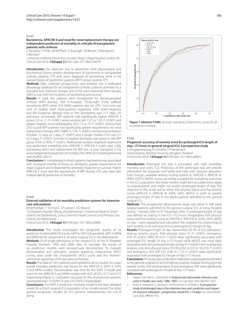
P247
Bacteremia, APACHE II and need for renal replacement therapy are independent predictors of mortality in critically ill transplanted patients with cirrhosisC Karvellas1, F Pink2, M McPhail2, G Auzinger2, W Bernal2, I Eltringham2,
J Wendon2
1University of Alberta, Edmonton, Canada; 2King’s College Hospital, London, UK
Critical Care 2010, 14(Suppl 1):P247 (doi: 10.1186/cc8479)
Introduction Our objective was to determine what physiological and
biochemical factors predict development of bacteremia in transplanted
cirrhotic patients (TP) and, upon diagnosis of bacteremia, what is the
natural history of bacteremic patients (BTP) versus controls (TP).
Methods Data collected prospectively and entered into a dedicated
physiology database for all transplanted cirrhotic patients admitted to a
specialist liver intensive therapy unit (LITU) were examined from January
2003 to July 2005 for incidence of bacteremia and survival.
Results In total, 362 patients were transplanted for decompensated
cirrhosis (64% elective, 36% in-hospital). Thirty-eight (10%) suff ered
bacteremia (BTP) while 324 (90%) patients did not (TP). Forty-nine per
cent of isolates were Gram-positive organisms, 45% Gram-negatives
and 6% fungemia. Median time to fi rst bacteremia was 11.5 days. On
admission (univariate), BTP patients had signifi cantly higher APACHE II
scores (23 vs 11, P <0.001), worse acidosis (pH 7.27 vs 7.35, P <0.001) and
greater degrees of encephalopathy (GCS 12 vs 15, P <0.001). During their
LITU course BTP patients had signifi cantly greater requirements for renal
replacement therapy (RRT) (68% vs 17%, P <0.001), mechanical ventilation
(median 15 days vs 2 days, P <0.001) and a longer median LITU stay (21
vs 3 days, P <0.001). Survival to hospital discharge was worse in the BTP
group (55% vs 95%, P <0.001). Multivariate analysis (logistical regression)
was performed modelling with APACHE II. APACHE II (odds ratio 1.06),
bacteremia (4.01) and requirement for RRT pre or post transplant (12.5)
were independent predictors of mortality. The APACHE II model performed
well (AUROC 0.817).
Conclusions In transplanted cirrhotic patients, bacteremia was associated
with increased severity of illness on admission, greater requirements for
organ support and independently adversely impacted on survival. Higher
APACHE II score and the requirement of RRT during LITU stay were also
independently predictive of mortality.
P248
External validation of six mortality prediction systems for intensive care unit patientsM Verhofstad1, T Timmers2, LP Leenen2, KG Moons3
1St Elizabeth Hospital, Tilburg, the Netherlands; 2Universitair Medical Centre
Utrecht, the Netherlands; 3Julius Centre for Health Sciences and Primary Care,
Utrecht, the Netherlands
Critical Care 2010, 14(Suppl 1):P248 (doi: 10.1186/cc8480)
Introduction This study investigates the prognostic quality of six
prediction models (APACHE II score, SAPS II, SAPS II (Expanded), SAPS 3, MPM
and MPM II0) for assessment in an adult surgical ICU in the Netherlands.
Methods Of all single admissions to the surgical ICU of the St Elisabeth
Hospital between 1995 and 2000, data to calculate the results of
six prediction models were prospectively documented. To evaluate
discrimination and calibration, receiver operating characteristic (ROC)
curves, area under the characteristic (AUC) curve and the Hosmer–
Lemeshow goodness-of-fi t test were performed.
Results The data of 1,821 patients were applied to all six models. Accurate
overall mortality prediction was found for the APACHE II, SAPS II, SAPS
3 and MPM models. Discrimination was best for the SAPS 3 model and
worst for the APACHE II and MPM models with AUC of 0.81, 0.77 and 0.77,
respectively (Figure 1). Calibration was poor for the six prediction systems,
varying between 23 (SAPS 3) and 233 (SAPS II (Expanded)).
Conclusions The SAPS 3 prediction mortality model is the best validated
model for a Dutch surgical ICU population of six models tested. The other
general prognostic models for ICU patients underestimate the risk of
dying.
P249
Prognostic accuracy of severity score for prolonged ICU length of stay >72 hours in general surgical ICU: a prospective studyS Kongsayreepong, N Lomarat, S Thamtanavit
Siriraj Hospital, Mahidol University, Bangkok, Thailand
Critical Care 2010, 14(Suppl 1):P249 (doi: 10.1186/cc8481)
Introduction Prolonged ICU stay is associated with high morbidity,
mortality and costs [1,2]. Prediction of this prolonged stay will provide
information for physician and family and help with resource allocation.
Even though, available severity scoring system ie. APACHE II, APACHE III,
MPM, SAPS II, MODS scores are widely accepted for evaluating outcomes
in the ICU population. But these models might be inaccurate when apply
to subpopulation and might not predict prolonged length of stay. The
objective of this study was to refi ne ASA physical status and fi ve severity
scores (APACHE II, APACHE III, SOFA, SAPS, MOFS) in order to predict
prolonged length of stay in the adults patients admitted to the general
surgical ICU.
Methods This prospective observational study was done in 948 adult
surgical patients admitted to the general surgical ICU at Siriraj Hospital
during 1 January 2005 to 31 December 2005. A prolonged length of stay
was defi ned as staying in the ICU >72 hours. Preoperative ASA physical
status and fi ve severity scores as APACHE II, APACHE III, SOFA, SAPS, MOFS
on day 1 were registered and calculated. We analyzed the relationship of
each severity score and outcome as prolonged ICU length of stay.
Results Prolonged length of stay represented 20.1% of ICU admissions.
Among severity scores, ASA physical status IV (P <0.001), emergency
ASA (P <0.001), APACHE III (P = 0.025) were signifi cantly associated with
prolonged ICU length of stay (>72 hours) while MODS was most likely
associated with this prolonged length of stay (P = 0.059). From multivariate
analysis, only ASA physical status IV (OR 6.503 (2.314 to 18.275), P <0.001)
and emergency ASA (OR 4.52 (2.86 to 7.12), P <0.001) were signifi cantly
associated with prolonged ICU length of stay (>72 hours).
Conclusions The study was conducted in 948 adult surgical patients admitted
to the general surgical ICU at a 3rd referral university hospital. Among all of the
severity scores, ASA physical status IV and emergency ASA were signifi cantly
correlated with prolonged ICU length of stay >72 hours.
References1. Halpern NA, Betts L, Greenstein R: Federal and nationwide intensive care
units in health care costs: 1986–1992. Crit Care Med 1994, 22:2001-2007.
2. Arabi Y, Venkatesh S, Haddad S, Al Shimemeri A, Al Malik S: A prospective study of prolonged stay in the intensive care unit: predictors and impact on resource utilization., prospective, mixed ICU, >14 days. Int J Qual Health
Care 2002, 14:403-410.
Figure 1 (abstract P248). Receiver operating characteristic curves for all
six prediction models.
Critical Care 2010, Volume 14 Suppl 1 http://ccforum.com/supplements/14/S1
S85

P250
Perioperative scores to predict mortality in surgical oncologic patients: a review of 1,362 casesC Simoes1, L Hajjar2, M Carmona1, F Galas1, J Auler Jr1
1Instituto do Cancer do Estado de São Paulo, Brazil; 2Heart Institute, São Paulo,
Brazil
Critical Care 2010, 14(Suppl 1):P250 (doi: 10.1186/cc8482)
Introduction Surgical outcomes depend on many factors, including
baseline characteristics and diff erences in surgical and anesthetic
practices. Few data are available in general surgical oncological patients.
We aimed to evaluate the predictive value of Porthsmouth POSSUM and
American Society of Anesthesiologists (ASA) scores in oncologic patients
submitted to surgical procedures.
Methods Data were collected from a database fi lled with demographic
characteristics, neoplasm defi nitions, co-morbidities and data from
intraoperative room and hospital mortality. The predicted mortality risk was
calculated using Porthsmouth POSSUM and ASA physical status grading.
Results From 1,362 surgical patients, the observed general hospital
mortality was 2.34%. ASA grading underestimated the mortality for
low-complexity patients, overestimated in patients with severe systemic
disease (ASA III), and was a strong predictor of mortality in patients with
life-threatening conditions (ASA IV) (Table 1). On the other hand, the
Porthsmouth POSSUM scoring system overestimated mortality in all
groups (Table 2).
Table 1 (abstract P250). Observed and predicted mortality of oncologic
patients evaluated through ASA physical status grading
ASA Number Observed Predicted
ASA I 209 4 (1.91%) 1 (0.05%)
ASA II 867 10 (1.15%) 1 (0.4%)
ASA III 247 7 (2.83%) 11 (4.5%)
ASA IV 39 11 (28.2%) 10 (25%)
Total 1,362 32 (2.34%)
Table 2 (abstract P250). Observed and predicted mortality of oncologic
patients evaluated through Portsmouth POSSUM scoring
Number Observed Predicted 0 to 5
0 0 0 (3.1%) 5 to 30
1,055 15 (1.42%) 142 (13.5%) 30 to 100
307 17 (5.53%) 149 (48.7%) 0 to 100
1,322 32 (2.34%)
Conclusions In surgical oncologic patients, Porthsmouth POSSUM and
ASA are not good predictors of mortality. These data suggest that a more
sensible and specifi c score is needed in this population.
Reference1. Wakabayashi H, et al.: Int J Surg 2007, 5:323-327.
P251
Scoring patients with a suspected infection in the emergency department (ED): comparison of the ED-specifi c MEDS score with APACHE II and SOFA scoreJ Wilhelm, S Hettwer, D Hammer, M Schürmann, M Amoury, H Ebelt, K
Werdan
University Hospital Halle (Saale), Germany
Critical Care 2010, 14(Suppl 1):P251 (doi: 10.1186/cc8483)
Introduction Several scores exist for characterization of critically ill
patients, developed in part for septic patients especially for the ICU patient
(for example, Acute Physiology and Chronic Health Evaluation, APACHE II;
Sequential Organ Failure Assessment, SOFA), but also for the septic patient
in the emergency department (ED) (Mortality in Emergency Department
Sepsis, MEDS). The practicability of these scores in the ED is discussed
controversially. The aim of our study was to directly compare these scores
in the setting of an ED on a cohort of patients with suspected infection.
Methods Patients with suspected severe infection from which blood
cultures were taken were included in the study. Procalcitonin (PCT) levels
were determined, and APACHE II, MEDS and SOFA scores were calculated
at admission. Endpoints were 28-day-mortality and necessity for therapy
on an ICU. Receiver operating characteristic (ROC) curve analysis was
performed to compare the ability of the three scores to predict the
endpoints.
Results We analyzed 211 patients. Eighty-two patients (43.6%) had a PCT
value ≥2 ng/ml and were considered septic. Mean values of APACHE II,
MEDS and SOFA score were 16.1 ± 8.5, 7.4 ± 4.7, and 3.4 ± 3.1 respectively.
Tables 1 and 2 show the area under the curve (AUC) values of the ROC
curves for mortality (Table 1) and ICU therapy (Table 2).
Table 1 (abstract P251). ROC analysis for mortality
Score AUC Standard error Signifi cance
APACHE II 0.804 0.045 <0.001
MEDS 0.785 0.048 <0.001
SOFA 0.708 0.051 0.006
Table 2 (abstract P251). ROC analysis for ICU therapy
Score AUC Standard error Signifi cance
APACHE II 0.757 0.038 <0.001
MEDS 0.648 0.042 0.001
SOFA 0.731 0.039 <0.001
Conclusions All three scores are useful prognostic factors for mortality
and for ICU therapy in the ED, with usually lower patients’ severity of
infection than in the ICU. The ICU-validated APACHE II and SOFA scores
were of similar prognostic value as the ED-specifi c MEDS score.
P252
Validation of the new proposed Emergency Trauma Score (EMTRAS)M Mangini1, S Di Valvasone2, C Greco2, A Ognibene1, G Cappuccini1,
R Spina1, R Tartaglia3, G Zagli1, A Peris1
1Careggi Teaching Hospital, Florence, Italy; 2Postgraduate School of
Anesthesia and Intensive Care, University of Florence, Italy; 3Centre for Clinical
Risk Management and Patient Safety of Tuscany Region, Florence, Italy
Critical Care 2010, 14(Suppl 1):P252 (doi: 10.1186/cc8484)
Introduction Recently, Raum and colleagues [1] generated and validated
a new trauma score (Emergency Trauma Score, EMTRAS) based on age,
prehospital GCS, prothrombin time and base excess. Each parameter is
subdivided into four classes, scored from 0 to 3 points. Scores of each class
are summed to obtain the EMTRAS, ranging from 0 to 12. Here we present
preliminary results of a study with the aim to validate this new scoring
system and to compare it with other commonly used illness scores.
Methods One hundred and fi fty trauma patients admitted to the regional
referral trauma center (Careggi Teaching Hospital, Florence, Italy) were
studied. Predictive value of the EMTRAS score was compared with the
Injury Severity Score (ISS), Revised Trauma Score (RTS), Trauma Injury
Table 1 (abstract P252). Area under the curve (AUC) for EMTRAS and other
scoring systems
Variable AUC 95% CI
EMTRAS 0.809 0.74 to 0.87
ISS 0.758 0.68 to 0.83
RTS 0.818 0.74 to 0.88
TRISS 0.909 0.84 to 0.96
SAPS II 0.845 0.78 to 0.90
Critical Care 2010, Volume 14 Suppl 1 http://ccforum.com/supplements/14/S1
S86
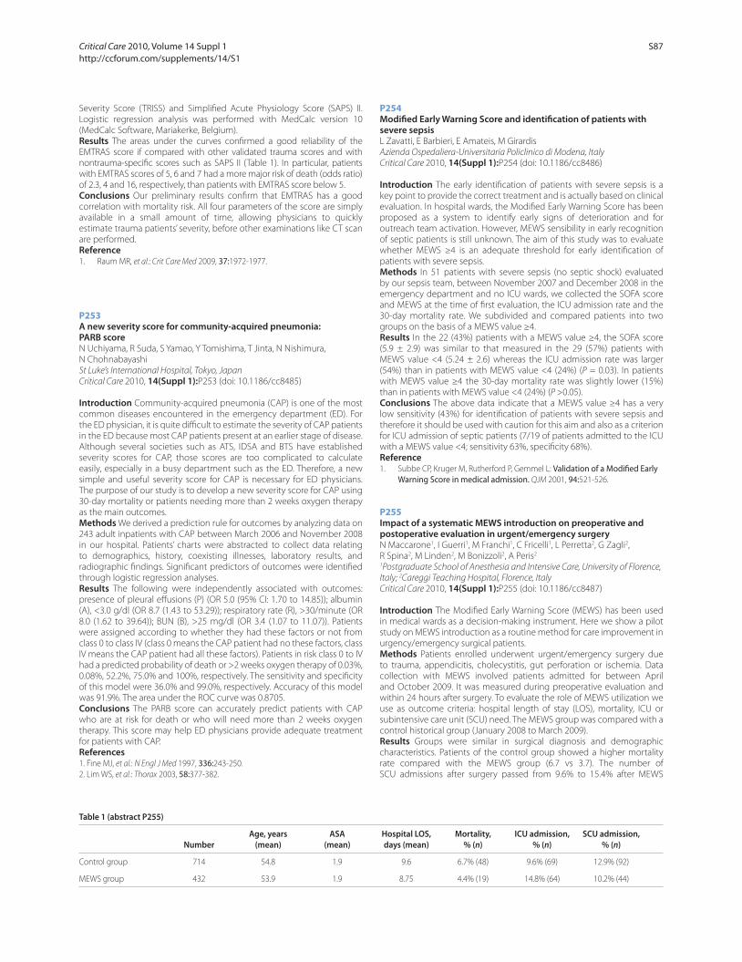
Severity Score (TRISS) and Simplifi ed Acute Physiology Score (SAPS) II.
Logistic regression analysis was performed with MedCalc version 10
(MedCalc Software, Mariakerke, Belgium).
Results The areas under the curves confi rmed a good reliability of the
EMTRAS score if compared with other validated trauma scores and with
nontrauma-specifi c scores such as SAPS II (Table 1). In particular, patients
with EMTRAS scores of 5, 6 and 7 had a more major risk of death (odds ratio)
of 2.3, 4 and 16, respectively, than patients with EMTRAS score below 5.
Conclusions Our preliminary results confi rm that EMTRAS has a good
correlation with mortality risk. All four parameters of the score are simply
available in a small amount of time, allowing physicians to quickly
estimate trauma patients’ severity, before other examinations like CT scan
are performed.
Reference1. Raum MR, et al.: Crit Care Med 2009, 37:1972-1977.
P253
A new severity score for community-acquired pneumonia: PARB scoreN Uchiyama, R Suda, S Yamao, Y Tomishima, T Jinta, N Nishimura,
N Chohnabayashi
St Luke’s International Hospital, Tokyo, Japan
Critical Care 2010, 14(Suppl 1):P253 (doi: 10.1186/cc8485)
Introduction Community-acquired pneumonia (CAP) is one of the most
common diseases encountered in the emergency department (ED). For
the ED physician, it is quite diffi cult to estimate the severity of CAP patients
in the ED because most CAP patients present at an earlier stage of disease.
Although several societies such as ATS, IDSA and BTS have established
severity scores for CAP, those scores are too complicated to calculate
easily, especially in a busy department such as the ED. Therefore, a new
simple and useful severity score for CAP is necessary for ED physicians.
The purpose of our study is to develop a new severity score for CAP using
30-day mortality or patients needing more than 2 weeks oxygen therapy
as the main outcomes.
Methods We derived a prediction rule for outcomes by analyzing data on
243 adult inpatients with CAP between March 2006 and November 2008
in our hospital. Patients’ charts were abstracted to collect data relating
to demographics, history, coexisting illnesses, laboratory results, and
radiographic fi ndings. Signifi cant predictors of outcomes were identifi ed
through logistic regression analyses.
Results The following were independently associated with outcomes:
presence of pleural eff usions (P) (OR 5.0 (95% CI: 1.70 to 14.85)); albumin
(A), <3.0 g/dl (OR 8.7 (1.43 to 53.29)); respiratory rate (R), >30/minute (OR
8.0 (1.62 to 39.64)); BUN (B), >25 mg/dl (OR 3.4 (1.07 to 11.07)). Patients
were assigned according to whether they had these factors or not from
class 0 to class IV (class 0 means the CAP patient had no these factors, class
IV means the CAP patient had all these factors). Patients in risk class 0 to IV
had a predicted probability of death or >2 weeks oxygen therapy of 0.03%,
0.08%, 52.2%, 75.0% and 100%, respectively. The sensitivity and specifi city
of this model were 36.0% and 99.0%, respectively. Accuracy of this model
was 91.9%. The area under the ROC curve was 0.8705.
Conclusions The PARB score can accurately predict patients with CAP
who are at risk for death or who will need more than 2 weeks oxygen
therapy. This score may help ED physicians provide adequate treatment
for patients with CAP.
References1. Fine MJ, et al.: N Engl J Med 1997, 336:243-250.
2. Lim WS, et al.: Thorax 2003, 58:377-382.
P254
Modifi ed Early Warning Score and identifi cation of patients with severe sepsisL Zavatti, E Barbieri, E Amateis, M Girardis
Azienda Ospedaliera-Universitaria Policlinico di Modena, Italy
Critical Care 2010, 14(Suppl 1):P254 (doi: 10.1186/cc8486)
Introduction The early identifi cation of patients with severe sepsis is a
key point to provide the correct treatment and is actually based on clinical
evaluation. In hospital wards, the Modifi ed Early Warning Score has been
proposed as a system to identify early signs of deterioration and for
outreach team activation. However, MEWS sensibility in early recognition
of septic patients is still unknown. The aim of this study was to evaluate
whether MEWS ≥4 is an adequate threshold for early identifi cation of
patients with severe sepsis.
Methods In 51 patients with severe sepsis (no septic shock) evaluated
by our sepsis team, between November 2007 and December 2008 in the
emergency department and no ICU wards, we collected the SOFA score
and MEWS at the time of fi rst evaluation, the ICU admission rate and the
30-day mortality rate. We subdivided and compared patients into two
groups on the basis of a MEWS value ≥4.
Results In the 22 (43%) patients with a MEWS value ≥4, the SOFA score
(5.9 ± 2.9) was similar to that measured in the 29 (57%) patients with
MEWS value <4 (5.24 ± 2.6) whereas the ICU admission rate was larger
(54%) than in patients with MEWS value <4 (24%) (P = 0.03). In patients
with MEWS value ≥4 the 30-day mortality rate was slightly lower (15%)
than in patients with MEWS value <4 (24%) (P >0.05).
Conclusions The above data indicate that a MEWS value ≥4 has a very
low sensitivity (43%) for identifi cation of patients with severe sepsis and
therefore it should be used with caution for this aim and also as a criterion
for ICU admission of septic patients (7/19 of patients admitted to the ICU
with a MEWS value <4; sensitivity 63%, specifi city 68%).
Reference1. Subbe CP, Kruger M, Rutherford P, Gemmel L: Validation of a Modifi ed Early
Warning Score in medical admission. QJM 2001, 94:521-526.
P255
Impact of a systematic MEWS introduction on preoperative and postoperative evaluation in urgent/emergency surgeryN Maccarone1, I Guerri1, M Franchi1, C Fricelli1, L Perretta2, G Zagli2,
R Spina2, M Linden2, M Bonizzoli2, A Peris2
1Postgraduate School of Anesthesia and Intensive Care, University of Florence,
Italy; 2Careggi Teaching Hospital, Florence, Italy
Critical Care 2010, 14(Suppl 1):P255 (doi: 10.1186/cc8487)
Introduction The Modifi ed Early Warning Score (MEWS) has been used
in medical wards as a decision-making instrument. Here we show a pilot
study on MEWS introduction as a routine method for care improvement in
urgency/emergency surgical patients.
Methods Patients enrolled underwent urgent/emergency surgery due
to trauma, appendicitis, cholecystitis, gut perforation or ischemia. Data
collection with MEWS involved patients admitted for between April
and October 2009. It was measured during preoperative evaluation and
within 24 hours after surgery. To evaluate the role of MEWS utilization we
use as outcome criteria: hospital length of stay (LOS), mortality, ICU or
subintensive care unit (SCU) need. The MEWS group was compared with a
control historical group (January 2008 to March 2009).
Results Groups were similar in surgical diagnosis and demographic
characteristics. Patients of the control group showed a higher mortality
rate compared with the MEWS group (6.7 vs 3.7). The number of
SCU admissions after surgery passed from 9.6% to 15.4% after MEWS
Table 1 (abstract P255)
Age, years ASA Hospital LOS, Mortality, ICU admission, SCU admission,
Number (mean) (mean) days (mean) % (n) % (n) % (n)
Control group 714 54.8 1.9 9.6 6.7% (48) 9.6% (69) 12.9% (92)
MEWS group 432 53.9 1.9 8.75 4.4% (19) 14.8% (64) 10.2% (44)
Critical Care 2010, Volume 14 Suppl 1 http://ccforum.com/supplements/14/S1
S87
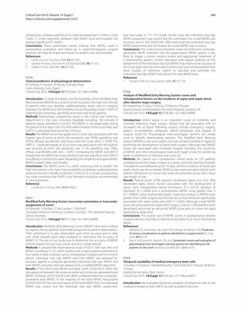
introduction, whereas need for an ICU bed decreased from 12.9% to 10.6%
(Table 1). Linear regression between high MEWS score and hospital LOS
resulted signifi cant (P <0.001).
Conclusions These preliminary results indicate that MEWS, used in
preoperative evaluation and follow-up in urgent/emergency surgical
patients, can have an important impact on patient care and mortality.
References1. Cei M, et al.: Int J Clin Pract 2009, 63:591-595.
2. Gardner-Thorpe J, et al.: Ann R Coll Surg Engl 2006, 88:571-575.
3. Subbe CP, et al.: QJM 2001, 94:521-526.
P256
Clinical predictors of physiological deteriorationM Shehata, A Hussein, A Farouk, H Khaled Nagi
Cairo University, Cairo, Egypt
Critical Care 2010, 14(Suppl 1):P256 (doi: 10.1186/cc8488)
Introduction To study the ability and the feasibility of the Modifi ed Early
Warning Score (MEWS) as a screening tool to predict the high-risk critically
ill patients who may develop cardiorespiratory arrest, and to compare
between the MEWS and the Simplifi ed Acute Physiology Score II (SAPS II)
regarding the sensitivity, specifi city and more easy to be applied.
Methods Randomized, prospective study in the critical care medicine
department in the Cairo University Hospitals including 100 critically ill
patients newly admitted to the ICU. The MEWS is calculated daily during
the period of the stay in the ICU with determination of the score Max, and
SAPS II is calculated during the fi rst 24 hours.
Results The MEWS score max grade of 8 or more was associated with the
highest rate of arrest at which the sensitivity was 78.9%, specifi city was
93.5%, effi cacy was 88.00%, with area under the curve (AUC) = 0.928. The
SAPS II – Expanded grade of 50 or more was associated with the highest
rate of arrest at which the sensitivity was 71.1%, specifi city was 100%,
effi cacy was 89.00%, with AUC = 0.872. Comparing the MEWS and SAPS II
reveals that the MEWS has more sensitivity but less specifi city than SAPS II
and effi cacy is almost the same. Regarding the simplicity and applicability:
MEWS is easier, faster and simpler.
Conclusions The MEWS score is a useful screening tool to predict the
high-risk patients who have high probability to develop cardiorespiratory
arrest among the critically ill patients in the ICU. It is simple and practical,
has more sensitivity than SAPS II and therefore should be recommended
in clinical practice.
Reference1. Le Gall et al.: Crit Care 2005, 9:R645-R652.
P257
Modifi ed Early Warning Scores: inaccurate summation or inaccurate assignment of score?M Edwards1, H McKay2, C Van Leuvan2, I Mitchell2
1Australian National University, Canberra, Australia; 2The Canberra Hospital,
Garran, Australia
Critical Care 2010, 14(Suppl 1):P257 (doi: 10.1186/cc8489)
Introduction Modifi ed Early Warning Scores (MEWS) have been utilised
to improve the recognition and medical response to patient deterioration.
Their usefulness is, in part, dependent upon their accuracy and to date
only small datasets have been analysed to determine the accuracy of
MEWS [1]. The aim of our study was to determine the accuracy of MEWS
and the reasons for any inaccuracies found in a large dataset.
Methods A prospective observational study of 9,672 vital sign sets and
MEWS recordings in 315 adult medical and surgical patients admitted to
four wards in both a tertiary and a metropolitan hospital over a 4-month
period. Individual vital sign MEWS and total MEWS was assessed for
accuracy against a computer-generated individual vital sign MEWS and
total MEWS using the vital sign dataset and a modifi ed MEWS algorithm.
Results Of the 9,672 total MEWS recorded, 3,504 (3,504/9,672, 36%) had
discrepancies between the nurse-recorded and computer-generated total
MEWS. Of these, 3,029 (3,029/3,504, 86%) underestimated the computer-
generated total MEWS. In the majority of the total MEWS inaccuracies
(2,443/3,504, 69.7%), the summation of the total MEWS from the individual
MEWS was correct but the individual vital sign MEWS assignment
was inaccurate. In 711 (711/3,504, 20.3%) cases the individual vital sign
MEWS assignment was correct but the summation for a total MEWS was
incorrect, and in 350 (350/3,504, 10%) cases both the individual vital sign
MEWS assignment and summation for a total MEWS was incorrect.
Conclusions The underscoring between nurse-recorded and computer-
generated MEWS indicates that the paper-based MEWS system is less
likely to trigger a timely medical review and appropriate treatment of
a deteriorating patient. Further education and regular auditing on the
assignment of the individual vital sign MEWS may improve the accuracy of
the score given that the summation of the score was not the predominant
issue. Equally, an electronic system to calculate and summate the
individual vital sign MEWS may reduce the total MEWS error.
Reference1. Prytherch DR, et al.: Resuscitation 2006, 70:173-178.
P258
Analysis of Modifi ed Early Warning System scores and intraoperative factors on the incidence of sepsis and septic shock after elective major surgeryPA Hampshire, A Guha, A Strong, D Parsons, P Rowan
Royal Liverpool and Broadgreen University Hospital, Liverpool, UK
Critical Care 2010, 14(Suppl 1):P258 (doi: 10.1186/cc8490)
Introduction Severe sepsis is an important cause of morbidity and
mortality following major surgery. Factors that are associated with an
increased risk of sepsis following surgery include emergency surgery,
patient co-morbidities, allogeneic blood transfusion and degree of
surgical insult [1]. Physiological track-and-trigger systems are widely
used to identify deteriorating patients. The Modifi ed Early Warning
System (MEWS) is one such system, but has not been studied in regard to
predicting the development of sepsis after surgery. Although high MEWS
scores are associated with increased hospital mortality, the sensitivity
of MEWS and other physiological track and trigger scores for predicting
death or admission to intensive care is low [2].
Methods We carried out a prospective cohort study on 101 patients
undergoing elective major surgery in a large university teaching hospital.
The patients were followed up for 10 days, and the incidence of sepsis and
septic shock was documented. MEWS scores were recorded daily for each
patient. Admissions to critical care were documented, along with critical
care length of stay.
Results Twenty-seven (27%) patients developed sepsis and nine (9%)
developed septic shock. Factors associated with the development of
sepsis were intraoperative blood transfusion (P = 0.013), duration of
operation (P = 0.004) and a postoperative MEWS score greater than 3
(P = 0.0003). Using multivariate logistic regression analysis, a MEWS score
greater than 3 after surgery was the only factor that remained signifi cantly
associated with sepsis (odds ratio 4.89, P = 0.003). Although a high MEWS
score was associated with sepsis after surgery, only fi ve (19%) patients who
developed sepsis had an abnormal MEWS score prior to (mean 4.6 days)
sepsis being diagnosed.
Conclusions The routine use of MEWS scores in postoperative elective
surgical patients may help to identify those patients at risk of developing
sepsis.
References1. Veltkamp SC, Kemmeren JM, Graaf YVD, Edlinger M, Werken CVD: Prediction
of serious complications in patients admitted to a surgical ward. Br J Surg
2002, 89:94-102.
2. Gao H, McDonnell A, Harrison DA, et al.: Systematic review and evaluation of physiological track and trigger warning systems for identifying at-risk patients on the ward. Intensive Care Med 2007, 33:667-679.
P259
Temporal variability of medical emergency team callsJ Tourtier, E Fontaine, J Vandenbossche, S De Rudnicki, E Falzone, M Borne,
Y Auroy
Hopital Val-de-Grâce, Paris, France
Critical Care 2010, 14(Suppl 1):P259 (doi: 10.1186/cc8491)
Introduction We evaluated temporal variability of telephone calls to the
medical emergency team (MET), as well as patient outcome.
Critical Care 2010, Volume 14 Suppl 1 http://ccforum.com/supplements/14/S1
S88
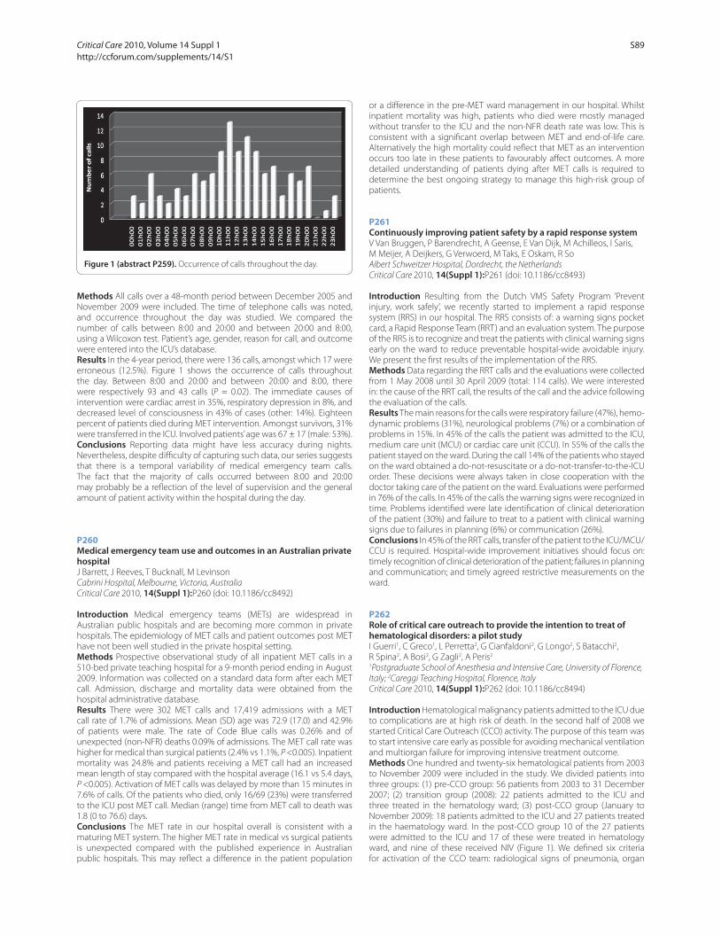
Methods All calls over a 48-month period between December 2005 and
November 2009 were included. The time of telephone calls was noted,
and occurrence throughout the day was studied. We compared the
number of calls between 8:00 and 20:00 and between 20:00 and 8:00,
using a Wilcoxon test. Patient’s age, gender, reason for call, and outcome
were entered into the ICU’s database.
Results In the 4-year period, there were 136 calls, amongst which 17 were
erroneous (12.5%). Figure 1 shows the occurrence of calls throughout
the day. Between 8:00 and 20:00 and between 20:00 and 8:00, there
were respectively 93 and 43 calls (P = 0.02). The immediate causes of
intervention were cardiac arrest in 35%, respiratory depression in 8%, and
decreased level of consciousness in 43% of cases (other: 14%). Eighteen
percent of patients died during MET intervention. Amongst survivors, 31%
were transferred in the ICU. Involved patients’ age was 67 ± 17 (male: 53%).
Conclusions Reporting data might have less accuracy during nights.
Nevertheless, despite diffi culty of capturing such data, our series suggests
that there is a temporal variability of medical emergency team calls.
The fact that the majority of calls occurred between 8:00 and 20:00
may probably be a refl ection of the level of supervision and the general
amount of patient activity within the hospital during the day.
P260
Medical emergency team use and outcomes in an Australian private hospitalJ Barrett, J Reeves, T Bucknall, M Levinson
Cabrini Hospital, Melbourne, Victoria, Australia
Critical Care 2010, 14(Suppl 1):P260 (doi: 10.1186/cc8492)
Introduction Medical emergency teams (METs) are widespread in
Australian public hospitals and are becoming more common in private
hospitals. The epidemiology of MET calls and patient outcomes post MET
have not been well studied in the private hospital setting.
Methods Prospective observational study of all inpatient MET calls in a
510-bed private teaching hospital for a 9-month period ending in August
2009. Information was collected on a standard data form after each MET
call. Admission, discharge and mortality data were obtained from the
hospital administrative database.
Results There were 302 MET calls and 17,419 admissions with a MET
call rate of 1.7% of admissions. Mean (SD) age was 72.9 (17.0) and 42.9%
of patients were male. The rate of Code Blue calls was 0.26% and of
unexpected (non-NFR) deaths 0.09% of admissions. The MET call rate was
higher for medical than surgical patients (2.4% vs 1.1%, P <0.005). Inpatient
mortality was 24.8% and patients receiving a MET call had an increased
mean length of stay compared with the hospital average (16.1 vs 5.4 days,
P <0.005). Activation of MET calls was delayed by more than 15 minutes in
7.6% of calls. Of the patients who died, only 16/69 (23%) were transferred
to the ICU post MET call. Median (range) time from MET call to death was
1.8 (0 to 76.6) days.
Conclusions The MET rate in our hospital overall is consistent with a
maturing MET system. The higher MET rate in medical vs surgical patients
is unexpected compared with the published experience in Australian
public hospitals. This may refl ect a diff erence in the patient population
or a diff erence in the pre-MET ward management in our hospital. Whilst
inpatient mortality was high, patients who died were mostly managed
without transfer to the ICU and the non-NFR death rate was low. This is
consistent with a signifi cant overlap between MET and end-of-life care.
Alternatively the high mortality could refl ect that MET as an intervention
occurs too late in these patients to favourably aff ect outcomes. A more
detailed understanding of patients dying after MET calls is required to
determine the best ongoing strategy to manage this high-risk group of
patients.
P261
Continuously improving patient safety by a rapid response systemV Van Bruggen, P Barendrecht, A Geense, E Van Dijk, M Achilleos, I Saris,
M Meijer, A Deijkers, G Verwoerd, M Taks, E Oskam, R So
Albert Schweitzer Hospital, Dordrecht, the Netherlands
Critical Care 2010, 14(Suppl 1):P261 (doi: 10.1186/cc8493)
Introduction Resulting from the Dutch VMS Safety Program ‘Prevent
injury, work safely’, we recently started to implement a rapid response
system (RRS) in our hospital. The RRS consists of: a warning signs pocket
card, a Rapid Response Team (RRT) and an evaluation system. The purpose
of the RRS is to recognize and treat the patients with clinical warning signs
early on the ward to reduce preventable hospital-wide avoidable injury.
We present the fi rst results of the implementation of the RRS.
Methods Data regarding the RRT calls and the evaluations were collected
from 1 May 2008 until 30 April 2009 (total: 114 calls). We were interested
in: the cause of the RRT call, the results of the call and the advice following
the evaluation of the calls.
Results The main reasons for the calls were respiratory failure (47%), hemo-
dynamic problems (31%), neurological problems (7%) or a combination of
problems in 15%. In 45% of the calls the patient was admitted to the ICU,
medium care unit (MCU) or cardiac care unit (CCU). In 55% of the calls the
patient stayed on the ward. During the call 14% of the patients who stayed
on the ward obtained a do-not-resuscitate or a do-not-transfer-to-the-ICU
order. These decisions were always taken in close cooperation with the
doctor taking care of the patient on the ward. Evaluations were performed
in 76% of the calls. In 45% of the calls the warning signs were recognized in
time. Problems identifi ed were late identifi cation of clinical deterioration
of the patient (30%) and failure to treat to a patient with clinical warning
signs due to failures in planning (6%) or communication (26%).
Conclusions In 45% of the RRT calls, transfer of the patient to the ICU/MCU/
CCU is required. Hospital-wide improvement initiatives should focus on:
timely recognition of clinical deterioration of the patient; failures in planning
and communication; and timely agreed restrictive measurements on the
ward.
P262
Role of critical care outreach to provide the intention to treat of hematological disorders: a pilot studyI Guerri1, C Greco1, L Perretta2, G Cianfaldoni2, G Longo2, S Batacchi2,
R Spina2, A Bosi2, G Zagli2, A Peris2
1Postgraduate School of Anesthesia and Intensive Care, University of Florence,
Italy; 2Careggi Teaching Hospital, Florence, Italy
Critical Care 2010, 14(Suppl 1):P262 (doi: 10.1186/cc8494)
Introduction Hematological malignancy patients admitted to the ICU due
to complications are at high risk of death. In the second half of 2008 we
started Critical Care Outreach (CCO) activity. The purpose of this team was
to start intensive care early as possible for avoiding mechanical ventilation
and multiorgan failure for improving intensive treatment outcome.
Methods One hundred and twenty-six hematological patients from 2003
to November 2009 were included in the study. We divided patients into
three groups: (1) pre-CCO group: 56 patients from 2003 to 31 December
2007; (2) transition group (2008): 22 patients admitted to the ICU and
three treated in the hematology ward; (3) post-CCO group (January to
November 2009): 18 patients admitted to the ICU and 27 patients treated
in the haematology ward. In the post-CCO group 10 of the 27 patients
were admitted to the ICU and 17 of these were treated in hematology
ward, and nine of these received NIV (Figure 1). We defi ned six criteria
for activation of the CCO team: radiological signs of pneumonia, organ
Figure 1 (abstract P259). Occurrence of calls throughout the day.
Critical Care 2010, Volume 14 Suppl 1 http://ccforum.com/supplements/14/S1
S89
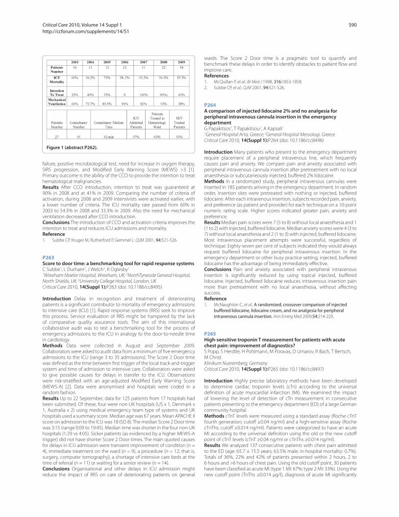
failure, positive microbiological test, need for increase in oxygen therapy,
SIRS progression, and Modifi ed Early Warning Score (MEWS) >3 [1].
Primary outcome is the ability of the CCO to provide the intention to treat
hematological malignancies.
Results After CCO introduction, intention to treat was guaranteed at
90% in 2008 and at 41% in 2009. Comparing the number of criteria of
activation, during 2008 and 2009 intensivists were activated earlier, with
a lower number of criteria. The ICU mortality rate passed from 60% in
2003 to 54.5% in 2008 and 33.3% in 2009. Also the need for mechanical
ventilation decreased after CCO introduction.
Conclusions The introduction of CCO and activation criteria improves the
intention to treat and reduces ICU admissions and mortality.
Reference1. Subbe CP, Kruger M, Rutherford P, Gemmel L: QJM 2001, 94:521-526.
P263
Score to door time: a benchmarking tool for rapid response systemsC Subbe1, L Durham2, J Welch3, K Oglesby1
1Wrexham Maelor Hospital, Wrexham, UK; 2NorthTyneside General Hospital,
North Shields, UK; 3University College Hospital, London, UK
Critical Care 2010, 14(Suppl 1):P263 (doi: 10.1186/cc8495)
Introduction Delay in recognition and treatment of deteriorating
patients is a signifi cant contributor to mortality of emergency admissions
to intensive care (ICU) [1]. Rapid response systems (RRS) seek to improve
this process. Service evaluation of RRS might be hampered by the lack
of comparative quality assurance tools. The aim of this international
collaborative audit was to test a benchmarking tool for the process of
emergency admissions to the ICU in analogy to the door-to-needle time
in cardiology.
Methods Data were collected in August and September 2009.
Collaborators were asked to audit data from a minimum of fi ve emergency
admissions to the ICU (range 3 to 35 admissions). The Score 2 Door time
was defi ned as the time between fi rst trigger of the local track-and-trigger
system and time of admission to intensive care. Collaborators were asked
to give possible causes for delays in transfer to the ICU. Observations
were risk-stratifi ed with an age-adjusted Modifi ed Early Warning Score
(MEWS-A) [2]. Data were anonymised and hospitals were coded in a
random fashion.
Results Up to 22 September, data for 125 patients from 17 hospitals had
been submitted. Of these, four were non-UK hospitals (US x 1, Denmark x
1, Australia x 2) using medical emergency team type of systems and UK
hospitals used a summary score. Median age was 67 years. Mean APACHE II
score on admission to the ICU was 18 (SD 8). The median Score 2 Door time
was 3:15 (range 0:09 to 19:45). Median time was shorter in the four non-UK
hospitals (1:29 vs 4:05). Sicker patients (as evidenced by a higher MEWS-A
trigger) did not have shorter Score 2 Door times. The main quoted causes
for delays in ICU admission were transient improvement of condition (n =
4), immediate treatment on the ward (n = 9), a procedure (n = 12; that is,
surgery, computer tomography), a shortage of intensive care beds at the
time of referral (n = 11) or waiting for a senior review (n = 14).
Conclusions Organisational and other delays in ICU admission might
reduce the impact of RRS on care of deteriorating patients on general
wards. The Score 2 Door time is a pragmatic tool to quantify and
benchmark these delays in order to identify obstacles to patient fl ow and
improve care.
References1. McQuillan P, et al.: Br Med J 1998, 316:1853-1858.
2. Subbe CP, et al.: QJM 2001, 94:521-526.
P264
A comparison of injected lidocaine 2% and no analgesia for peripheral intravenous cannula insertion in the emergency departmentG Papakitsos1, T Papakitsou2, A Kapsali1
1General Hospital Arta, Greece; 2General Hospital Messologi, Greece
Critical Care 2010, 14(Suppl 1):P264 (doi: 10.1186/cc8496)
Introduction Many patients who present to the emergency department
require placement of a peripheral intravenous line, which frequently
causes pain and anxiety. We compare pain and anxiety associated with
peripheral intravenous cannula insertion after pretreatment with no local
anaesthesia or subcutaneously injected, buff ered 2% lidocaine.
Methods In a randomized study, peripheral intravenous cannulas were
inserted in 185 patients arriving in the emergency department. In random
order, insertion sites were pretreated with nothing or injected, buff ered
lidocaine. After each intravenous insertion, subjects recorded pain, anxiety,
and preference (as patient and provider) for each technique on a 10-point
numeric rating scale. Higher scores indicated greater pain, anxiety and
preference.
Results Median pain scores were 7 (5 to 8) without local anaesthesia and 1
(1 to 2) with injected, buff ered lidocaine. Median anxiety scores were 4 (3 to
7) without local anaesthesia and 2 (1 to 3) with injected, buff ered lidocaine.
Most intravenous placement attempts were successful, regardless of
technique. Eighty-seven per cent of subjects indicated they would always
request buff ered lidocaine for peripheral intravenous insertion. In the
emergency department or other busy practice setting, injected, buff ered
lidocaine has the advantage of being immediately eff ective.
Conclusions Pain and anxiety associated with peripheral intravenous
insertion is signifi cantly reduced by using topical injected, buff ered
lidocaine. Injected, buff ered lidocaine reduces intravenous insertion pain
more than pretreatment with no local anaesthesia, without aff ecting
success.
Reference1. McNaughton C, et al.: A randomized, crossover comparison of injected
buff ered lidocaine, lidocaine cream, and no analgesia for peripheral intravenous cannula insertion. Ann Emerg Med 2009,54:214-220.
P265
High-sensitive troponin T measurement for patients with acute chest pain: improvement of diagnostics?S Popp, S Herdtle, H Pohlmann, M Poravas, D Umarov, R Bach, T Bertsch,
M Christ
Klinikum Nueremberg, Germany
Critical Care 2010, 14(Suppl 1):P265 (doi: 10.1186/cc8497)
Introduction Highly precise laboratory methods have been developed
to determine cardiac troponin levels (cTn) according to the universal
defi nition of acute myocardial infarction (MI). We examined the impact
of lowering the level of detection of cTn measurement in consecutive
patients presenting to the emergency department (ED) of a large German
community hospital.
Methods cTnT levels were measured using a standard assay (Roche cTnT
fourth generation; cutoff ≥0.04 ng/ml) and a high-sensitive assay (Roche
cTnThs; cutoff ≥0.014 ng/ml). Patients were categorized to have an acute
MI according to the universal defi nition using the old or the new cutoff
point of cTnT levels (cTnT ≥0.04 ng/ml or cTnThs ≥0.014 ng/ml).
Results We analyzed 137 consecutive patients with chest pain admitted
to the ED (age: 65.7 ± 15.5 years; 63.5% male; in-hospital mortality: 0.7%).
Totals of 36%, 22% and 42% of patients presented within 2 hours, 2 to
6 hours and >6 hours of chest pain. Using the old cutoff point, 30 patients
have been classifi ed as acute MI (type 1 MI: 67%; type 2 MI: 33%). Using the
new cutoff point (TnThs ≥0.014 μg/l), diagnosis of acute MI signifi cantly
Figure 1 (abstract P262).
Critical Care 2010, Volume 14 Suppl 1 http://ccforum.com/supplements/14/S1
S90
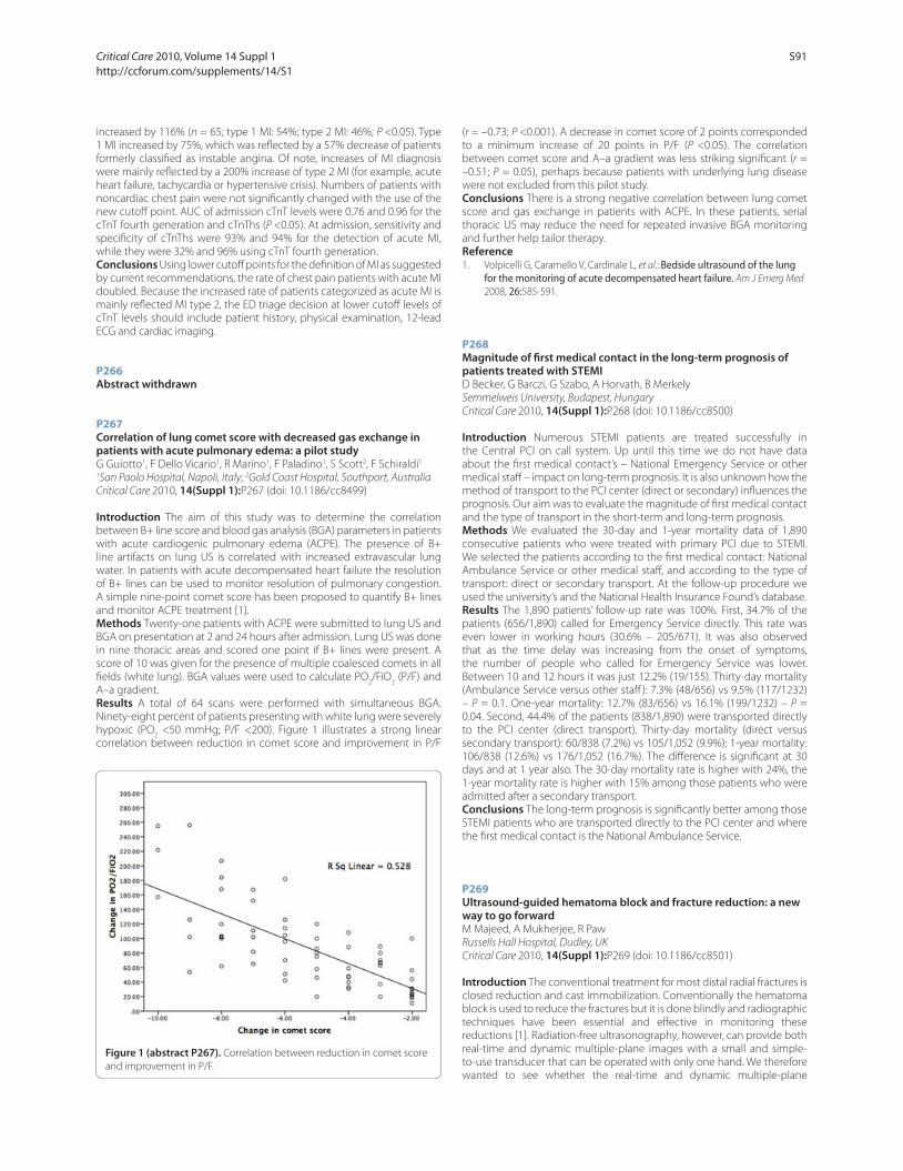
increased by 116% (n = 65; type 1 MI: 54%; type 2 MI: 46%; P <0.05). Type
1 MI increased by 75%, which was refl ected by a 57% decrease of patients
formerly classifi ed as instable angina. Of note, increases of MI diagnosis
were mainly refl ected by a 200% increase of type 2 MI (for example, acute
heart failure, tachycardia or hypertensive crisis). Numbers of patients with
noncardiac chest pain were not signifi cantly changed with the use of the
new cutoff point. AUC of admission cTnT levels were 0.76 and 0.96 for the
cTnT fourth generation and cTnThs (P <0.05). At admission, sensitivity and
specifi city of cTnThs were 93% and 94% for the detection of acute MI,
while they were 32% and 96% using cTnT fourth generation.
Conclusions Using lower cutoff points for the defi nition of MI as suggested
by current recommendations, the rate of chest pain patients with acute MI
doubled. Because the increased rate of patients categorized as acute MI is
mainly refl ected MI type 2, the ED triage decision at lower cutoff levels of
cTnT levels should include patient history, physical examination, 12-lead
ECG and cardiac imaging.
P266
Abstract withdrawn
P267
Correlation of lung comet score with decreased gas exchange in patients with acute pulmonary edema: a pilot studyG Guiotto1, F Dello Vicario1, R Marino1, F Paladino1, S Scott2, F Schiraldi1
1San Paolo Hospital, Napoli, Italy; 2Gold Coast Hospital, Southport, Australia
Critical Care 2010, 14(Suppl 1):P267 (doi: 10.1186/cc8499)
Introduction The aim of this study was to determine the correlation
between B+ line score and blood gas analysis (BGA) parameters in patients
with acute cardiogenic pulmonary edema (ACPE). The presence of B+
line artifacts on lung US is correlated with increased extravascular lung
water. In patients with acute decompensated heart failure the resolution
of B+ lines can be used to monitor resolution of pulmonary congestion.
A simple nine-point comet score has been proposed to quantify B+ lines
and monitor ACPE treatment [1].
Methods Twenty-one patients with ACPE were submitted to lung US and
BGA on presentation at 2 and 24 hours after admission. Lung US was done
in nine thoracic areas and scored one point if B+ lines were present. A
score of 10 was given for the presence of multiple coalesced comets in all
fi elds (white lung). BGA values were used to calculate PO2/FiO
2 (P/F) and
A–a gradient.
Results A total of 64 scans were performed with simultaneous BGA.
Ninety-eight percent of patients presenting with white lung were severely
hypoxic (PO2 <50 mmHg; P/F <200). Figure 1 illustrates a strong linear
correlation between reduction in comet score and improvement in P/F
(r = –0.73; P <0.001). A decrease in comet score of 2 points corresponded
to a minimum increase of 20 points in P/F (P <0.05). The correlation
between comet score and A–a gradient was less striking signifi cant (r =
–0.51; P = 0.05), perhaps because patients with underlying lung disease
were not excluded from this pilot study.
Conclusions There is a strong negative correlation between lung comet
score and gas exchange in patients with ACPE. In these patients, serial
thoracic US may reduce the need for repeated invasive BGA monitoring
and further help tailor therapy.
Reference1. Volpicelli G, Caramello V, Cardinale L, et al.: Bedside ultrasound of the lung
for the monitoring of acute decompensated heart failure. Am J Emerg Med
2008, 26:585-591.
P268
Magnitude of fi rst medical contact in the long-term prognosis of patients treated with STEMID Becker, G Barczi, G Szabo, A Horvath, B Merkely
Semmelweis University, Budapest, Hungary
Critical Care 2010, 14(Suppl 1):P268 (doi: 10.1186/cc8500)
Introduction Numerous STEMI patients are treated successfully in
the Central PCI on call system. Up until this time we do not have data
about the fi rst medical contact’s – National Emergency Service or other
medical staff – impact on long-term prognosis. It is also unknown how the
method of transport to the PCI center (direct or secondary) infl uences the
prognosis. Our aim was to evaluate the magnitude of fi rst medical contact
and the type of transport in the short-term and long-term prognosis.
Methods We evaluated the 30-day and 1-year mortality data of 1,890
consecutive patients who were treated with primary PCI due to STEMI.
We selected the patients according to the fi rst medical contact: National
Ambulance Service or other medical staff , and according to the type of
transport: direct or secondary transport. At the follow-up procedure we
used the university’s and the National Health Insurance Found’s database.
Results The 1,890 patients’ follow-up rate was 100%. First, 34.7% of the
patients (656/1,890) called for Emergency Service directly. This rate was
even lower in working hours (30.6% – 205/671). It was also observed
that as the time delay was increasing from the onset of symptoms,
the number of people who called for Emergency Service was lower.
Between 10 and 12 hours it was just 12.2% (19/155). Thirty-day mortality
(Ambulance Service versus other staff ): 7.3% (48/656) vs 9.5% (117/1232)
– P = 0.1. One-year mortality: 12.7% (83/656) vs 16.1% (199/1232) – P =
0.04. Second, 44.4% of the patients (838/1,890) were transported directly
to the PCI center (direct transport). Thirty-day mortality (direct versus
secondary transport): 60/838 (7.2%) vs 105/1,052 (9.9%); 1-year mortality:
106/838 (12.6%) vs 176/1,052 (16.7%). The diff erence is signifi cant at 30
days and at 1 year also. The 30-day mortality rate is higher with 24%, the
1-year mortality rate is higher with 15% among those patients who were
admitted after a secondary transport.
Conclusions The long-term prognosis is signifi cantly better among those
STEMI patients who are transported directly to the PCI center and where
the fi rst medical contact is the National Ambulance Service.
P269
Ultrasound-guided hematoma block and fracture reduction: a new way to go forwardM Majeed, A Mukherjee, R Paw
Russells Hall Hospital, Dudley, UK
Critical Care 2010, 14(Suppl 1):P269 (doi: 10.1186/cc8501)
Introduction The conventional treatment for most distal radial fractures is
closed reduction and cast immobilization. Conventionally the hematoma
block is used to reduce the fractures but it is done blindly and radiographic
techniques have been essential and eff ective in monitoring these
reductions [1]. Radiation-free ultrasonography, however, can provide both
real-time and dynamic multiple-plane images with a small and simple-
to-use transducer that can be operated with only one hand. We therefore
wanted to see whether the real-time and dynamic multiple-plane
Figure 1 (abstract P267). Correlation between reduction in comet score
and improvement in P/F.
Critical Care 2010, Volume 14 Suppl 1 http://ccforum.com/supplements/14/S1
S91

observation capabilities of ultrasonography would allow an ED physician
to perform under direct vision a hematoma block and closed reduction
without multiple attempts of either as are frequently required when only
conventional techniques are used.
Methods Sonographically guided hematoma block and closed reduction
was performed in three patients in the emergency department for an
acute distal radial fracture. The effi cacy of this method was evaluated and
compared with that of conventional techniques.
Results The ultrasound images helped giving the hematoma block
under direct vision and delineated the fractures as accurately as did the
conventional radiographs. All parameters measured on the ultrasound
images showed substantial restoration of anatomic alignment after
reduction. Under direct vision, the hematoma block helped reduce the
number of attempts and was less painful and achieved more patient
satisfaction.
Conclusions Sonography is an accurate, simple, time-saving, less painful
and radiation-free tool. Reducing badly displaced or angulated forearm
fractures in the emergency department can be diffi cult. Multiple attempts
at reduction may be required, with repeated trips to the radiology
department, before an adequate reduction is achieved [2]. Therefore
ultrasound-guided hematoma block and reduction of diffi cult forearm
fractures reduces the number of needle attempts and allows the physician
to assess the adequacy of the reduction at the patient’s bedside.
References1. Sonography for monitoring closed reduction of displaced extra-articular
distal radial fractures. J Bone Joint Surg Am 2002, 84:194-203.
2. Ultrasound guided reduction of pediatric forearm fractures in the ED. Am J
Emerg Med 2000, 18:72-77.
P270
Pulmonary edema in the prehospital setting: the importance of early treatmentS Lourenco, J Figueira Coelho, A Martins Baptista
Curry Cabral Hospital, Lisbon, Portugal
Critical Care 2010, 14(Suppl 1):P270 (doi: 10.1186/cc8502)
Introduction Patients with acute cardiogenic pulmonary edema require
rapid assessment and therapy to prevent progression to respiratory failure
and cardiovascular collapse. Lisbon city has emergency medical teams
that respond to situations where the life of the patient is at risk and whose
goal is to begin treatment if indicated and assure transport to the hospital
in the best conditions possible. We studied the intervention of one of
these teams on patients with acute pulmonary edema.
Methods Medical records of the activity of our prehospital emergency
vehicle from April 2005 to October 2009 were analyzed. All records of
patients having a clinical diagnosis of acute pulmonary edema at the
scene were considered. Incomplete records were excluded.
Results In the 55 months considered our emergency vehicle attended
9,856 calls, from which 493 (5%) were for acute pulmonary edema. Patients
ages range from 20 to 99 years, with a mean age of 75.8 years, and 47.8%
were males. A total of 58.6% of calls were made on the night period (from
8 pm to 8 am), most between 8 pm and 0 pm. Risk factors included arterial
hypertension (62.3%), ischemic heart disease (43.2%), diabetes mellitus
(31.8%), heart failure (23.3%), atrial fi brillation (13.2%) and renal failure
(9.9%). On arrival of the prehospital medical team at the scene, 24 patients
(4.9%) required immediate intubation and fi ve evolved to cardiac arrest,
four of whom were successfully resuscitated and transported to hospital.
Conclusions Our prehospital medical teams attended a large number of
patients with pulmonary edema during this period. We recorded a small
number of patients requiring mechanical intubation and a high rate of
survival on those suff ering from cardiac arrest in this context. We believe
this is largely because of the decreased response time that prehospital
emergency off ers in this situation, allowing the institution of early
treatment.
References1. Dickstein K, et al.: ESC guidelines for the diagnosis and treatment of acute
and chronic heart failure 2008: the Task Force for the diagnosis and treatment of acute and chronic heart failure 2008 of the European Society of Cardiology. Eur J Heart Fail 2008, 10:933-989.
2. Tresch DD, et al.: Out-of-hospital pulmonary edema: diagnosis and treatment. Ann Emerg Med 1983, 12:533-537.
P271
Effi cacy of bedside ultrasonography in early diagnosis of patients admitted to the emergency department with dyspneaE Dogan, S Girisgin, D Dündar, B Cander
Selcuk University Meram Faculty of Medicine, Konya, Turkey
Critical Care 2010, 14(Suppl 1):P271 (doi: 10.1186/cc8503)
Introduction We designed our study to fi nd a useful USG pattern for
diagnosis and assessment of dyspnea. We also compared utilities of chest
radiograms in a supine position, thorax CT and bedside USG techniques.
Methods In Selcuk University Meram Medical Faculty, during the year
2009, 60 patients who had acute dyspnea were included in our study (30
patients with chest trauma and 30 patients with nontraumatic causes). We
designed our study as prospective. We tried to determine pathologies that
have positive and negative Sliding Lung Sign (SLS) in USG examinations.
We also investigated its reliability in diff erential diagnosis of acute dyspnea.
Results First of all we performed chest radiograms on all patients in
a supine position. After that we performed CT scans. All of the results
were interpreted by a detached radiologist. Then we performed bedside
thoracic USG to fi nd SLS by physicians in the ER. We conceded CT results as
the golden standard and compared the results with chest radiograms and
USG fi ndings. The SPSS 13.0 program and Χ2 test were used for statistical
analysis (P <0.05). According to our fi ndings, SLS in USG examinations has
a sensitivity of 84% and a specifi city of 97% with a 97% positive predictive
value and 83% negative predictive value.
Conclusions Throughout the years, USG examinations were defi ned
as inappropriate in lung examinations but in our study we reached a
conclusion that they are a necessity. Bedside sonographic examination
allows impetuous diagnosis. But every respiratory failure has its own
sonographic pattern so mistakes are inevitable. Nevertheless we believe
that if profound studies like ours keep coming and if more and more
clinicians begin to use USG examinations, the SLS diagnostic value will rise
and become a routine element in the ER.
References1. Razzaq QM: Use of the ‘sliding lung sign’ in emergency bedside ultrasound.
Eur J Emerg Med 2008, 15:238-241.
2. Blaivas M, Lyon M, Duggal S: A prospective comparison of supine chest radiography and bedside ultrasound for the diagnosis of traumatic pneumothorax. Acad Emerg Med 2005, 12:844-849.
3. Wüstner A, Gehmacher O, Hämmerle S, Schenkenbach C, Häfele H, Mathis G:
Ultraschalldiagnostik beim stumpfen Thoraxtrauma. Ultraschall Med 2005,
26:285-290.
P272
Auscultation in fl ight: comparison of amplifi ed and traditional stethoscopesJ Tourtier, S Coste, E Fontaine, N Libert, M Viaggi, E Forsans, M Borne
Hopital Val-de-Grâce, Paris, France
Critical Care 2010, 14(Suppl 1):P272 (doi: 10.1186/cc8504)
Introduction Many air medical transport programs use pulse oximeters,
end-tidal carbon dioxide monitors and other devices as indirect measures
of respiratory and cardiac status. Thus, these methods do not replace
cardiac auscultation during fl ight, which may be needed to identify sudden
critical change. And the ability to compare left-sided and right-sided
breath sounds may be essential to confi rm the appropriate placement
of endotracheal tubes as well as to diagnose pneumothorax. The ability
for auscultation during air medical transport is compromised by high
ambient noise levels. The aim of this study was to assess the capabilities of
a traditional and an amplifi ed stethoscope (which is expected to reduce
background and ambient noise) to assess heart and breath sounds during
medical transport in a FALCON 50 plane.
Methods A prospective, double-blind, randomized study was performed.
We tested one model of traditional stethoscope (Littman® cardiology
III) and one model of amplifi ed stethoscope (Littman 3100). The sound
level was amplifi ed in six of the eight increments of the amplifi ed
stethoscope. Practitioners on board were all experienced in air medical
transport. They had normal audiologic testing. We studied 18 heart and
lung auscultations, during real medical evacuations on board the Falcon
50 (medically confi gured; Dassault aviation). For each, the quality of
Critical Care 2010, Volume 14 Suppl 1 http://ccforum.com/supplements/14/S1
S92

auscultation was described using a numeric rating scale (ranging from 0 to
10, 0 corresponding to ‘I hear nothing’, 10 to ‘I hear perfectly’). Comparisons
were accomplished using a t test for paired values.
Results Age of patients was 42 ± 11 years, 78% were males. The body mass
index was 29.5 ± 4.7. For cardiac auscultation, the value of the rating scale
was 5.7 ± 1.4 and 6.5 ± 1.8, respectively, for the traditional and amplifi ed
stethoscope (P = 0.027). For lung sounds, quality of auscultation was
estimated at 3.6 ± 2.3 for the traditional stethoscope, at 3.9 ± 2.9 for the
amplifi ed stethoscope (P = 0.193).
Conclusions We concluded that fl ight practitioners in the Falcon 50 are
more able to hear cardiac sounds with an amplifi ed than with a traditional
stethoscope, whereas there is no signifi cant diff erence concerning breath
sound auscultation. These fi ndings suggested to the investigators that
Falcon 50 noise and human breath sounds might share a common or
substantially overlapping frequency spectrum, with amplifi cation of one
necessarily amplifying the other. To assess this hypothesis, further studies
are needed to evaluate the sound frequency spectrum in the medically
confi gured Falcon 50.
P273
Performance of ventilators at simulated altitude: study of fraction of inspired oxygenJ Tourtier, S Ramsang, E Forsans, S De Rudnicki, A Chrissment, J Schaal,
M Borne
Hopital Val-de-Grâce, Paris, France
Critical Care 2010, 14(Suppl 1):P273 (doi: 10.1186/cc8505)
Introduction We studied the performance of two respirators employing
an advanced turbine delivery system: LTV 1000, Tbird VSO2. We assessed
the ability of the ventilators to deliver to a normal lung model a set fraction
of inspired oxygen (FiO2) at diff erent simulated cabin altitudes.
Methods We used a decompression chamber to mimic the hypobaric
environment at a range of simulated cabin altitudes of 1,500, 2,000 and
3,000 meters (4,000, 5,333, 8,000 feet). A model of normal lung was used.
Concerning the Tbird VSO2, cabin altitude was input. Ventilators were
tested with FiO2 set at 50% and Vt set at 700 ml. We noted the eff ective FiO
2
assessed by the ventilators (paramagnetic analysis). We measured the FiO2
really delivered with a dedicated instrument of the French physiological
laboratory of aviation and space medicine of the Air Force. Comparisons
of preset and assessed FiO2 to actual measured FiO
2 were accomplished
using a t test for each altitude.
Results Figure 1 shows the data with FiO2 set at 50%.
Conclusions On one hand, both ventilators showed a moderate variation
between FiO2 set and delivered. On the other hand, variations between
FiO2 delivered and assessed are high, suggesting ineffi cacity of ventilators’
paramagnetic analysis in hypobaric conditions.
P274
Aeromedical evacuation of the French Army: survey of the past 10 yearsS Coste, J Tourtier, C Daudé, M Viaggi, E Morgand, M Borne
Hopital Val-de-Grâce, Paris, France
Critical Care 2010, 14(Suppl 1):P274 (doi: 10.1186/cc8506)
Introduction The Military Medical Service of the French Forces insures the
support of 29,000 French servicemen spread around the world each year
and performs as such their medical evacuation by air towards France to
guarantee their adequate treatment as soon as possible. This knowledge is
sometimes also used for a national civilian abroad. We propose to describe
the epidemiology of medical evacuation (MEDEVAC) made in the past
10 years.
Methods We analysed data for every patient evacuated since 1 January
2000 and noted the destination, the number of patients per fl ight, the age,
the gender, the status (military or civilian), the pathology, the presence of
a specialist in the medical team (intensivist, cardiologist, psychiatrist) and
the recipient hospital. The presence of an intubated ventilated patient or
transfusion in fl ight was studied.
Results A total of 420 strategic MEDEVACs were carried out in 10 years (per
year: 42 ± 6.9) in favor of 529 patients, among whom 90% were French
servicemen. Each fl ight carried between one and 15 patients. Europe
represents 42% of the original location of evacuation, Africa 39% and the
Middle East 17%. An anesthetist–resuscitator reinforced the medical team
in 51.7% of the evacuations. The patients with respiratory assistance are
present in 19.8% of the fl ights and transfusion occurred in 3.1% of the
fl ights. Traumatic pathology is predominant (49.5% of patients), divided
into three major causes nearly equally: road traffi c accident, ballistic and
others. Burns represent a signifi cant proportion (6.8%) of the causes of
evacuation. Other causes are represented by 30.2% for medical conditions
(n = 160 among which 59 cardiac aff ections), 10.2% for nontraumatic
surgical aff ections and 2% for psychiatry. Finally 25.5% of the patients were
accommodated in an ICU, 6% in a specifi c center for treatment of burns,
35.3% in a service of surgery and 24.6% in medicine.
Conclusions If the number of MEDEVACs is relatively stable over the past
decade, destinations and causes are changing. Africa remains an usual
location of evacuation, the part of Europe tends to decrease for the benefi t
of the Middle East, which became in 2009 the predominant destination
(21/47) stretching out the duration of fl ights. The causes of evacuations
testify to the intensity of the commitment of French forces through
traumatic aff ections, and in particular of ballistic origin, which are in 2009
the main traumatic aff ections (15/31). The corollary is an increase of the
appeal to the resuscitators to deal with trauma patients.
P275
Comparison of two disaster drills’ management performed by trained and not-trained students: key times evaluationFL Barra, L Carenzo, PL Ingrassia, M Tengattini, F Prato, D Colombo,
F Della Corte
University of Eastern Piedmont ‘Amedeo Avogadro’, Novara, Italy
Critical Care 2010, 14(Suppl 1):P275 (doi: 10.1186/cc8507)
Introduction The aim of this report is to compare two disaster exercises’
management of students with diff erent backgrounds. To our knowledge
nobody has ever compared two exercises, probably because of the
diffi culty in their evaluation. We implemented a tool for an objective
evaluation [1] and we used it for this purpose.
Methods Both drills represented a ceiling collapse over a crowded room
with a similar amount of casualties and similar severity index. The START
triage system was used.
The trained students (T) were attending the European Master in Disaster
Medicine (EMDM), while the not-trained students (NT) were at the
beginning of an introductory course in disaster medicine. During the
exercises we recorded key victim–provider interaction times [2] using
victim-based data collection. Each victim had their own data card to
record triage and time information.
Results In this preliminary report we present data regarding the scene
length of stay (LOS) and triage to collecting area/advanced medical post
time (T-AMP). The LOS was 67.5 (50.0 to 111.0) minutes (25 to 75 IQR) for
T as compared with 145.0 (110.0 to 150.0) minutes (25 to 75 IQR) for NT Figure (abstract P273).
Critical Care 2010, Volume 14 Suppl 1 http://ccforum.com/supplements/14/S1
S93

(P <0.001). Stratifi cation according to assigned triage code showed no
diff erence for high-priority codes (reds and yellows) as opposed to the
green code (55.0 (47.0 to 75.0) minutes for T vs 145.0 (141.0 to 155.0)
minutes for NT with P <0.01). T-AMP was 10.0 (3.0 to 34.5) minutes for T as
compared with 63.5 (19.5 to 104.3) minutes for NT (P <0.001). Stratifi cation
according to triage code showed no diff erence for red codes between T
and NT but showed a diff erence for yellow codes (36.5 (15.0 to 82.0) vs 71.0
(30.0 to 99.0) minutes) and green codes (7.0 (3.0 to 12.0) vs 85.0 (17.3 to
115.0) minutes) with P <0.01.
Conclusions Both teams evacuated red codes before the yellow ones
in similar time. T-AMP was shorter considering global, yellow and green
codes for T as opposed to NT. Global and green LOS was also shorter in
the T group as opposed to NT. Training seems to infl uence global exercise
management, less aff ecting red codes but with an impact on yellow and
green evacuation strategies.
References1. Ingrassia PL, et al.: Evaluation of medical management during a mass
casualty incident exercise: an objective assessment tool to enhance direct observation. J Emerg Med 2009. [Epub ahead of print]
2. Green GB, et al.: Generic evaluation methods for disaster drills in developing countries. Ann Emerg Med 2003, 41:689-699.
P276
Acute severe asthma: performance of ventilator at simulated altitudeJ Tourtier, E Falzone, J Schaal, L Franck, G Edjo Nkilly, A Chrisment, M Borne
Military Hospital Val-de-Grâce, Paris, France
Critical Care 2010, 14(Suppl 1):P276 (doi: 10.1186/cc8508)
Introduction Exacerbation of asthma can appear during air transport.
Severe patients, not responding to conventional therapy can require
ventilator support. We evaluated the performance of two transport
ventilators, built with turbine technology – the T-bird VSO2 and the LTV
1000 – for use during aeromedical evacuation of acute severe asthma. We
have assessed the ability of both ventilators to deliver to an acute severe
asthma model a tidal volume (Vt) set at diff erent simulated altitudes, by
changing the ambient air pressure.
Methods We used a decompression chamber to mimic the hypobaric
environment at a range of simulated cabin altitudes of 1,500, 2,500
and 3,000 meters (4,000, 6,670, 8,000 feet). Ventilators were tested with
realistic parameters. Vt was set at 700 ml and 400 ml in an acute severe
asthma lung model. The protocol included three measurements for each
simulated altitude. Comparisons of preset to actual measured values were
accomplished using a t test for each altitude.
Results Figure 1 summarizes the data. With altitude, the T-bird VSO2
showed a decrease in volume delivered. Comparisons of actual delivered
Vt and set Vt demonstrated a signifi cant diff erence starting at 1,500 m for
a Vt set of 700 ml, at 2,500 m for Vt set of 400 ml, with a negative variation
of more than 10% compared with tidal volume set at respectively 3,000
and 2,500 m. With decreasing barometric pressure, the LTV 1000 showed
mostly an increase in volume delivered. Comparisons of actual delivered
Vt and set Vt demonstrated a signifi cant diff erence at 1,500 m for a Vt set
of 700 ml, at 2,500 m for a V test of 400 ml. The delivered tidal volume
remained within 10% of the set Vt.
Conclusions The T-bird VSO2 progressively delivered lower volumes as
barometric pressure decreased; whereas the LTV 1000 showed a moderate
increase in volume delivered for the acute severe asthma lung model, with
increasing altitude, but maintained the delivered volume within 10% of
the set Vt up to 3,000 m.
P277
Validation of a new French-language triage algorithm: the ELISA scaleJ Jobé1, A Ghuysen1, P Gérard2, G Hartstein1, V D’Orio1
1University Hospital, Liège, Belgium; 2University of Liège, Belgium
Critical Care 2010, 14(Suppl 1):P277 (doi: 10.1186/cc8509)
Introduction For some time, most emergency departments have been
experiencing severe overcrowding which could lead to reductions in the
quality of care. Consequently, nurse triage tools have been developed
with the aim of getting ‘the right patient to the right resources, at the right
place and at the right time’. However, diff erences in emergency medical
services in diff erent countries have limited the generalisation of pre-
existing triage systems; for this reason, specifi c algorithms corresponding
to local characteristics are needed. Accordingly, we developed a specifi c
French-language triage system called ELISA (Echelle Liegeoise d’Index de
Sévérité à l’Admission – Liege admission severity scale). We aimed to test
its validity in the present study.
Methods The ELISA is a fi ve-category (U1 to U5) nursing triage algorithm,
using fl ow charts based on vital signs and anamnestic data. Each category
is associated with a pre-established time delay for fi rst medical contact.
We tested inter-rater and intra-rater agreement respectively. As concerns
inter-rater agreement, nine triage nurses sorted the same 100 patients.
The assigned classifi cations compared the reference assignment and were
analyzed with Cohen’s kappa coeffi cient. Intra-rater agreement was tested
by asking 30 triage nurses to sort the same 10 patients at two diff erent
times (T1 was early morning and T2 was late night). We compared the
classifi cation at T1 and T2 with the null hypothesis being no diff erence and
determined whether diff erences were nurse dependent.
Results Statistical analysis of intra-rater agreement revealed that the
mean diff erence between classifi cation at T1 and T2 did not signifi cantly
depend on the nurse (P = 0.3487). This result allowed pooling of the data
and testing to determine whether the mean was diff erent from zero. We
used the Sign test and found that classifi cation at T1 was not diff erent from
classifi cation at T2 (P = 0.581). As concerns inter-rater agreement, we used
Cohen’s kappa coeffi cient, which revealed an almost perfect agreement
between classifi cation by nurses and the reference.
Conclusions The need for a specifi c triage tool in our emergency
department led us to develop a new French-language triage scale. The
present study demonstrates that this scale is a valid triage tool with very
good inter-rater and intra-rater agreement. Such results now allow a future
study to evaluate its effi ciency.
P278
Examining the suitability of medical admissions to the emergency short stay ward of a large UK hospitalE Hartley, E Hood, N Bashir, R Zahir
New Cross Hospital, Wolverhampton, UK
Critical Care 2010, 14(Suppl 1):P278 (doi: 10.1186/cc8510)
Introduction In 1996, a UK audit suggested suboptimal involvement of
consultant care in acute medicine [1]. Acute medicine has since evolved
as a specialty to improve medical inpatient care in the fi rst 48 hours [2].
Many hospitals now incorporate an acute short stay unit through which
up to 70% of medical admissions can be directed [3]. These units reduce
the length of hospital inpatient stay [4].
Methods We conducted a review of case notes for 100 consecutive
medical admissions to the Emergency Short Stay ward (ESS). Data collected
included diagnoses on admission and discharge, length of stay on ESS
and discharge destination. On reviewing each initial medical clerking, an
assessment was made regarding the suitability of the admission to ESS,
standardized against local criteria.
Results Data on 100 patients (age 17 to 89) were collected. Eighty-nine per
cent were admitted from the medical admissions unit, most commonly Figure 1 (abstract P276). Acute severe asthma, Vt delivered in altitude.
Critical Care 2010, Volume 14 Suppl 1 http://ccforum.com/supplements/14/S1
S94

for respiratory and cardiac problems. Average length of stay on ESS was
2 days. Twenty-fi ve per cent of patients remained on the ward for over
72 hours and 6% for over 5 days. Whilst 68% of patients were discharged
home from ESS, 32% of patients were later transferred to a diff erent
ward. When compared with the available admissions criteria, 27% of all
admissions to ESS were found to be inappropriate. Most (18/27) of these
patients were later transferred to a diff erent inpatient ward. Twenty-six per
cent of inappropriate admissions remained on ESS for >48 hours. The total
number of medical admissions to the hospital on each day of the study
did not aff ect the number of inappropriate transfers to ESS.
Conclusions The 24-bed Emergency Short Stay Ward at our UK hospital
is for assessment and treatment of selected acute conditions requiring an
inpatient stay <48 hours. ESS was not always being used appropriately
according to the admission criteria defi ned by the ward managers.
Hospital admissions of >72 hours are associated with signifi cant increase
in workload [5]. It is important that a short stay ward does not increase
the length of admission by becoming a transit lounge for patients before
transfer to a specialty lead ward.
References1. Houghton A, et al.: J R Coll Phys Lond 1996, 30:551-559.
2. Dowdle JR: Emerg Med J 2004, 21:652-653.
3. Cooke MW, et al.: Emerg Med J 2003, 20:138-142.
4. Downing H, et al.: Clin Med 2008, 8: 8-20.
5. Rainer TH, et al.: J Accid Emerg Med 1996, 13:325-329.
P279
A study on reliability of a new triage emergency method among nursing studentsN Parenti1, D Sangiorgi2, V Serventi3, L Sarli3
1Hospital Santa Maria della Scaletta Imola, Bologna, Italy; 2Università
Bologna, Italy; 3Università Parma, Italy
Critical Care 2010, 14(Suppl 1):P279 (doi: 10.1186/cc8511)
Introduction We analysed triage reliability among nursing students at an
Italian university before and after a course on a new Triage Emergency
Method, TEM v2, which showed good reliability in a previous study [1]. Few
studies compared triage reliability in nursing students. To our knowledge
there are no Italian studies on this topic.
Methods This is an observational study conducted in the University of
Parma using a triage scenarios database used in previous studies [1,2].
Fifty students attending the third year of a nursing course were selected
to assign a triage level to 105 paper scenarios, without triage protocols
(before course) and with TEMv2 (1 month after a 2-hour course on triage
and TEMv2). To prevent communication between participants, they
assigned triage codes in diff erent rooms, and in the presence of two
investigators. The triage scenarios were given randomly to the participants.
We measured: the inter-reliability using the weighted kappa statistics; the
complete disagreement (when nurses of the same group assigned to the
same scenario triage codes that diff ered in more than two priority levels);
the complete agreement (when all fi ve nurses assigned the same triage
code) before and after the course.
Results Of the 105 patients included in triage scenarios, 66 (63%) were
women, the mean age was 43.7 years (SD ± 26.3), and 22 were under the
age of 18 years. The most frequent presentation at triage was minor trauma
(30%). There were 30 hospital admissions: 27 in non-intensive wards and
three in ICUs. The mean age of students was 24 ± 3 SD. Few participants
attended triage training before (18%) and they declared scarce triage
knowledge. Inter-rater reliability was k = 0.42 (95% CI: 0.37 to 0.46) and
k = 0.61 (95% CI: 0.56 to 0.67) before the course (without triage protocol)
and after the course (with TEMv2), respectively. Complete disagreement
occurred in 98% of scenarios evaluated before and in 64% after the course.
Complete agreement was always zero.
Conclusions Our data suggest that TEM v2 improves triage reliability
among nursing students. It seems to be easy to understand and to use.
References1. Parenti N, et al.: Reliability and validity of two four-level emergency triage
systems. Eur J Emerg Med 2009, 16:115-120.
2. Parenti N, et al.: Affi dabilità e validità di un Metodo di Triage di Pronto Soccorso a 4 codici di priorità. Emerg Care J 2008, 5:30-33.
P280
Aggression and violence against staff of ambulance servicesA Auer
Vienna Ambulance Service, Vienna, Austria
Critical Care 2010, 14(Suppl 1):P280 (doi: 10.1186/cc8512)
Introduction Aggression and violence in the workplace have increasingly
become topical for various professions in the past years. Studies, however,
are mostly published for the areas of psychiatry and education. The fi rst
part of the study investigates the question of how emergency services in
general deal with aggressive assaults against their staff . In the second part,
the monitoring system for aggression incidents of the Vienna Ambulance
Service will be presented and evaluated.
Methods By the means of an email questionnaire, 35 emergency
medical services and EMS training facilities were interrogated about their
procedures in case of aggression incidents. The monitoring system of the
Vienna Ambulance Service, which is based on the SOAS-R questionnaire,
was statistically evaluated.
Results Emergency medical services survey: 21 of the 35 included
organizations returned the questionnaire with usable answers. For 80% of
the responding organizations, aggression against their staff is an issue; 50%
installed a staff reporting system and 58% off er de-escalation courses to
their staff . Vienna Ambulance Service: during the analysis period of 2 years,
203 aggressive and/or violent assaults were reported within the Vienna
Ambulance Service. In relation to the total number of 268,528 treated
patients, the rate of incident is 0.08%. Males committed, with 71.7%, most
of these aggressions. With regard to diagnoses, the diagnostic group of
alcohol-related incidents was most frequent with 59 cases. Most events
took place on weekends and in the evening hours. Verbal aggression was
with 86.2% predominant; however, more than 20% of the incidents lead to
physical injuries of the medical personnel.
Conclusions Aggression against emergency medical service staff
is brought up for discussion in many organizations. Despite several
approaches in diff erent states of development, standardized reporting
systems are the exception. The aggression incident reporting formula of
the Vienna Ambulance Service was proved to be practical for recording
aggression events as well as for evaluation of de-escalation education. The
study showed that aggression is a serious problem in emergency medical
services and appropriate precautions need to be institutionalized.
P281
Analysis of the disease spectrum of treated patients after Wenchuan earthquakeZ He, Y Wangy, L Huang
First Hospital Affi liated to General Hospital of PLA, Beijing, China
Critical Care 2010, 14(Suppl 1):P281 (doi: 10.1186/cc8513)
Introduction To investigate the data of patients treated by this fi eld hospital,
and to analyze the rule of spectrum of diseases in Wenchuan earthquake.
Methods Sexes, ages and diagnoses of the patients were collected and
the percentage was respectively calculated. The amount tendencies of the
time distribution of patients were also analyzed.
Results Sex diff erences of patients were not obvious in the total 2,331
cases. The amount in the group 15 to 60 years old, 1,535 (65.9%) cases, was
signifi cantly higher than those of other groups, but the relative ratios of the
group 0 to 14 years old, 382 (16.4%) cases, and the group >60 years old,
414 (17.7%) cases, were higher than that of 15 to 60 years old. The top four
diseases consist of respiratory system 601 (25.8%), trauma (fracture, wound
or injury) 586 (24.9%), dermatology system 295 (12.7%) and others 243
(10.4%). The amount of internal medicine patients exceeded that of surgery.
Conclusions The reasonable allocations of medical personnel and drugs
after earthquake may help to improve the remedy results and to prevent
waste of medical resource during future disasters.
References1. Hogan DE, Burstein JL: Disaster Medicine. Lippincott Williams & Wilkins; 2003:8.
2. Zhongjie He: The platinum ten mins – new ideal and concept. Chinese J Crit
Care Med 2004, 24:745-746.
3. Watts J: China’s health challenges after the earthquake. Lancet 2008,
371:1825-1826.
4. Conly J, Johnston B: Natural disasters, corpses and the risk of infectious diseases. Can J Infect Dis Med Microbiol 2005, 16:269-270.
Critical Care 2010, Volume 14 Suppl 1 http://ccforum.com/supplements/14/S1
S95
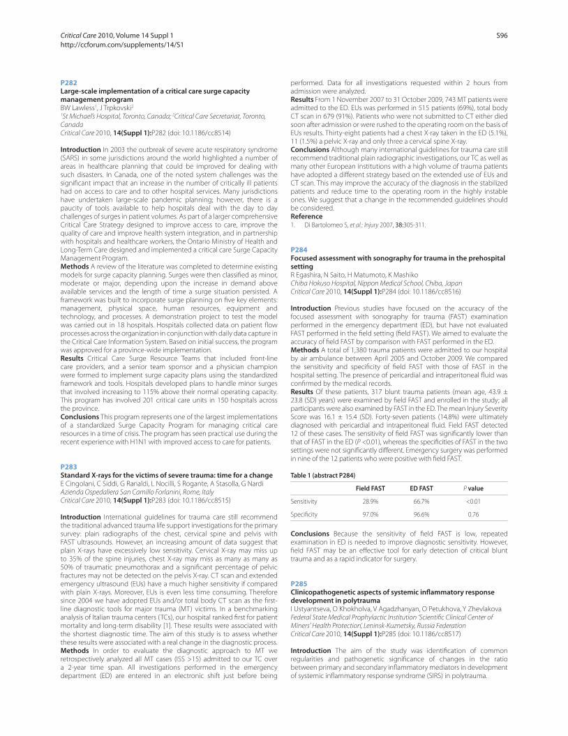
P282
Large-scale implementation of a critical care surge capacity management programBW Lawless1, J Trpkovski2
1St Michael’s Hospital, Toronto, Canada; 2Critical Care Secretariat, Toronto,
Canada
Critical Care 2010, 14(Suppl 1):P282 (doi: 10.1186/cc8514)
Introduction In 2003 the outbreak of severe acute respiratory syndrome
(SARS) in some jurisdictions around the world highlighted a number of
areas in healthcare planning that could be improved for dealing with
such disasters. In Canada, one of the noted system challenges was the
signifi cant impact that an increase in the number of critically ill patients
had on access to care and to other hospital services. Many jurisdictions
have undertaken large-scale pandemic planning; however, there is a
paucity of tools available to help hospitals deal with the day to day
challenges of surges in patient volumes. As part of a larger comprehensive
Critical Care Strategy designed to improve access to care, improve the
quality of care and improve health system integration, and in partnership
with hospitals and healthcare workers, the Ontario Ministry of Health and
Long-Term Care designed and implemented a critical care Surge Capacity
Management Program.
Methods A review of the literature was completed to determine existing
models for surge capacity planning. Surges were then classifi ed as minor,
moderate or major, depending upon the increase in demand above
available services and the length of time a surge situation persisted. A
framework was built to incorporate surge planning on fi ve key elements:
management, physical space, human resources, equipment and
technology, and processes. A demonstration project to test the model
was carried out in 18 hospitals. Hospitals collected data on patient fl ow
processes across the organization in conjunction with daily data capture in
the Critical Care Information System. Based on initial success, the program
was approved for a province-wide implementation.
Results Critical Care Surge Resource Teams that included front-line
care providers, and a senior team sponsor and a physician champion
were formed to implement surge capacity plans using the standardized
framework and tools. Hospitals developed plans to handle minor surges
that involved increasing to 115% above their normal operating capacity.
This program has involved 201 critical care units in 150 hospitals across
the province.
Conclusions This program represents one of the largest implementations
of a standardized Surge Capacity Program for managing critical care
resources in a time of crisis. The program has seen practical use during the
recent experience with H1N1 with improved access to care for patients.
P283
Standard X-rays for the victims of severe trauma: time for a changeE Cingolani, C Siddi, G Ranaldi, L Nocilli, S Rogante, A Stasolla, G Nardi
Azienda Ospedaliera San Camillo Forlanini, Rome, Italy
Critical Care 2010, 14(Suppl 1):P283 (doi: 10.1186/cc8515)
Introduction International guidelines for trauma care still recommend
the traditional advanced trauma life support investigations for the primary
survey: plain radiographs of the chest, cervical spine and pelvis with
FAST ultrasounds. However, an increasing amount of data suggest that
plain X-rays have excessively low sensitivity. Cervical X-ray may miss up
to 35% of the spine injuries, chest X-ray may miss as many as many as
50% of traumatic pneumothorax and a signifi cant percentage of pelvic
fractures may not be detected on the pelvis X-ray. CT scan and extended
emergency ultrasound (EUs) have a much higher sensitivity if compared
with plain X-rays. Moreover, EUs is even less time consuming. Therefore
since 2004 we have adopted EUs and/or total body CT scan as the fi rst-
line diagnostic tools for major trauma (MT) victims. In a benchmarking
analysis of Italian trauma centers (TCs), our hospital ranked fi rst for patient
mortality and long-term disability [1]. These results were associated with
the shortest diagnostic time. The aim of this study is to assess whether
these results were associated with a real change in the diagnostic process.
Methods In order to evaluate the diagnostic approach to MT we
retrospectively analyzed all MT cases (ISS >15) admitted to our TC over
a 2-year time span. All investigations performed in the emergency
department (ED) are entered in an electronic shift just before being
performed. Data for all investigations requested within 2 hours from
admission were analyzed.
Results From 1 November 2007 to 31 October 2009, 743 MT patients were
admitted to the ED. EUs was performed in 515 patients (69%), total body
CT scan in 679 (91%). Patients who were not submitted to CT either died
soon after admission or were rushed to the operating room on the basis of
EUs results. Thirty-eight patients had a chest X-ray taken in the ED (5.1%),
11 (1.5%) a pelvic X-ray and only three a cervical spine X-ray.
Conclusions Although many international guidelines for trauma care still
recommend traditional plain radiographic investigations, our TC as well as
many other European institutions with a high volume of trauma patients
have adopted a diff erent strategy based on the extended use of EUs and
CT scan. This may improve the accuracy of the diagnosis in the stabilized
patients and reduce time to the operating room in the highly instable
ones. We suggest that a change in the recommended guidelines should
be considered.
Reference1. Di Bartolomeo S, et al.: Injury 2007, 38:305-311.
P284
Focused assessment with sonography for trauma in the prehospital settingR Egashira, N Saito, H Matumoto, K Mashiko
Chiba Hokuso Hospital, Nippon Medical School, Chiba, Japan
Critical Care 2010, 14(Suppl 1):P284 (doi: 10.1186/cc8516)
Introduction Previous studies have focused on the accuracy of the
focused assessment with sonography for trauma (FAST) examination
performed in the emergency department (ED), but have not evaluated
FAST performed in the fi eld setting (fi eld FAST). We aimed to evaluate the
accuracy of fi eld FAST by comparison with FAST performed in the ED.
Methods A total of 1,380 trauma patients were admitted to our hospital
by air ambulance between April 2005 and October 2009. We compared
the sensitivity and specifi city of fi eld FAST with those of FAST in the
hospital setting. The presence of pericardial and intraperitoneal fl uid was
confi rmed by the medical records.
Results Of these patients, 317 blunt trauma patients (mean age, 43.9 ±
23.8 (SD) years) were examined by fi eld FAST and enrolled in the study; all
participants were also examined by FAST in the ED. The mean Injury Severity
Score was 16.1 ± 15.4 (SD). Forty-seven patients (14.8%) were ultimately
diagnosed with pericardial and intraperitoneal fl uid. Field FAST detected
12 of these cases. The sensitivity of fi eld FAST was signifi cantly lower than
that of FAST in the ED (P <0.01), whereas the specifi cities of FAST in the two
settings were not signifi cantly diff erent. Emergency surgery was performed
in nine of the 12 patients who were positive with fi eld FAST.
Table 1 (abstract P284)
Field FAST ED FAST P value
Sensitivity 28.9% 66.7% <0.01
Specifi city 97.0% 96.6% 0.76
Conclusions Because the sensitivity of fi eld FAST is low, repeated
examination in ED is needed to improve diagnostic sensitivity. However,
fi eld FAST may be an eff ective tool for early detection of critical blunt
trauma and as a rapid indicator for surgery.
P285
Clinicopathogenetic aspects of systemic infl ammatory response development in polytraumaI Ustyantseva, O Khokholva, V Agadzhanyan, O Petukhova, Y Zhevlakova
Federal State Medical Prophylactic Institution ‘Scientifi c Clinical Center of
Miners’ Health Protection’, Leninsk-Kuznetsky, Russia Federation
Critical Care 2010, 14(Suppl 1):P285 (doi: 10.1186/cc8517)
Introduction The aim of the study was identifi cation of common
regularities and pathogenetic signifi cance of changes in the ratio
between primary and secondary infl ammatory mediators in development
of systemic infl ammatory response syndrome (SIRS) in polytrauma.
Critical Care 2010, Volume 14 Suppl 1 http://ccforum.com/supplements/14/S1
S96

Methods Three hundred and eighty-seven patients with polytrauma
hospitalized to the ICU were examined. There was infection confi rmed
by microbiology in 175 (pneumonia, bronchitis, osteomyellitis, acute
urethritis, and so forth); 212 patients had no infection (acute respiratory
distress syndrome, disseminated intravascular coagulation, fat embolism,
and so forth). In 87% two or more signs of SIRS were noted according to
the criteria of the Consensus Conference ACCP/SCCM (2003). C-reactive
protein (CRP) in venous blood serum was measured on 1, 3, 5, 7, 10, 15 and
17 days after admission using the biochemical analyzer HITACI-912 with
reagents Tina-Quant CRP (Roche Diagnostic GMBH), lipopolysaccharide-
binding protein (LPSBP) and cytokines TNFα, IL-2R, IL-6, IL-8, IL-10 using
the immunochemiluminescent analyzer IMMULITE with the reagents DPC
(USA). Constant variables were compared using Student’s t test and the
Mann–Whitney U test. The diff erences were signifi cant at P <0.05.
Results On admission, increased serum LPSBP and TNFα were noted
in all patients with infection compared with the patients without it (1.8
and 3 times respectively, P <0.01). Moreover, the infectious patients had
the apparent increase of IL-2R, IL-6, IL-8 and CRP: 5, 13, 18 and 5 times,
respectively (P <0.05). At that, generalized manifestation of infl ammatory
reaction in the infectious patients was characterized by at mean fi vefold
increase in IL-8 on day 3 of the follow-up. It was not accompanied by
adequate anti-infl ammatory response that was indicated by an absence
of IL-10 dynamics. A direct correlation was found between an increase
in infl ammatory mediator concentration (IL-6, IL-8, IL-10) and respiratory
rate (r = 0.73; 0.46; 0.59), CRP and heart rate (r = 0.48, P <0.05), and an
inverse correlation between infl ammatory mediators (TNFα, IL-2R, LPSPB)
and arterial pressure level (r = –0.42; –0.63; –0.48) and respiratory function
values (vital lung capacity and forced expiratory volume, r = –0.42).
Conclusions Hyperproduction of proinfl ammatory cytokines is closely
connected with ventilation disturbances and clinical variants of the post-
traumatic period course in patients with polytrauma.
P286
Diagnostic and prognostic signifi cance of serum apoptosis markers in the patients with severe trauma injuryEG Grigoryev1, AR Radivilko1, AK Kiseleva2, YC Churliaev2, GV Vavin1
1Kemerovo State Medical Academy, Kemerovo, Russia Federation; 2Branch of
Scientifi c Institute of General Reanimatology RAMS, Novokuznetck, Russia
Federation
Critical Care 2010, 14(Suppl 1):P286 (doi: 10.1186/cc8518)
Introduction The aim of the investigation was to analyze the diagnostic
and prognostic signifi cance of serum proapoptosis and antiapoptosis
markers in the patients with severe trauma injury.
Methods Thirty-three patients with severe trauma injury were enrolled
into our investigation (we excluded patients with severe brain injury, the
middle age was 39 ± 8, initial TRISS scale 7.8 ± 1.8, APACHE II 14 ± 5). All of
the patients demonstrated signs of systemic infl ammatory response, in 12
patients the infectious pulmonary complications were revealed and in 21
patients the infectious complications were not developed. Venous blood
serum EDTA samples were investigated every day during a week in the
–25°C freeze. We investigated antiapoptotic markers (soluble Fas-L, soluble
APO-1/Fas, Cu/Zn superoxide dismutase), proapoptotic markers (protein
p53, protein Bcl-2), and high-sensitivity CRP (ELISA, Bender Medsystem).
Results We observed an increase in proapoptotic markers in patients
with infectious complications from the fi rst day in the ICU, antiapoptotic
markers (Cu/Zn superoxide dismutase) were lower with regard to
reference points. The patients demonstrated a maximum of Fas-L, protein
p53, and protein Bcl-2 (near 50% relative to fi rst day) on the second day. In
patients without infection complications we identifi ed a decreased level
of proapoptotic markers and an increased level of antiapoptotic markers
(SOD, soluble Fas-L, soluble APO-1/Fas). High-sensitivity CRP correlated
with the severity of infectious complications.
Conclusions Serum proapoptotic and antiapoptotic markers have
demonstrated diagnostic and prognostic signifi cance in the patients with
severe trauma injury.
Reference1. De Freitas I, Fernández-Somoza M, Essenfeld-Sekler E, et al.: Serum levels of the
apoptosis-associated molecules, tumor necrosis factor-alfa/tumor necrosis factor type-I receptor and Fas/FasL, in sepsis. Chest 2004, 125:2238-2246.
P287
Predictors of mechanical ventilation after burn injuryA Pirat, P Zeyneloglu, A Kundakci, C Aydogan, G Arslan, M Haberal
Baskent University Faculty of Medicine, Ankara, Turkey
Critical Care 2010, 14(Suppl 1):P287 (doi: 10.1186/cc8519)
Introduction It is important to predict the need for mechanical
ventilation (MV) in burn patients in order to improve their management
and also to optimize resource ultilization. However, there are scant data
on the predictors of MV in burn patients. The purpose of this study was to
determine the risk factors for MV in a cohort of our burn patients.
Methods A retrospective chart review was completed for patients
admitted to Baskent University Burn Institute, from January 2003 through
December 2008. Patients were divided into two groups based on whether
they required MV during their fi rst 3 days of hospitalization or not. After
comparing the two groups for the recorded variables, a binary logistic
regression model was developed using statistical and clinical signifi cance
to identify the risk factors for MV.
Results Out of 160 patients, 33 patients (21%) required MV during their fi rst
3 days of hospitalization. Patients who required MV were similar to those
who did not in terms of demographic features. Patients who required MV
were signifi cantly diff erent from those who did not regarding the mean
burn percentage (56% ± 23% vs 19% ± 15%, P <0.001), mechanism of burn
(fl ame, 73% vs 37%, P = 0.001), inhalational injury (27% vs 3%, P <0.001),
and burn aff ecting head and neck (72% vs 37%, P <0.001). Binary logistic
regression revealed that inhalational injury (OR, 8.4; 95% CI, 1.1 to 66.1;
P = 0.042) and burn percentage (OR, 1.1; 95% CI, 1.1 to 1.2; P <0.001) were
independent risk factors for MV.
Conclusions Our results demonstrate that inhalational injury and burn
percentage are predictors of MV in our series of burn patients. Particularly,
patients with inhalational injury were eight times more likely to require MV
during their fi rst 3 days of hospitalization in our series.
P288
Impact of regional imaging guidelines for head injury and unconscious polytrauma patientsR Gibbs1, R Walker2, E Thomas3
1Musgrove Park Hospital, Taunton, UK; 2Torbay Hospital, Torbay, UK; 3Derriford
Hospital, Plymouth, UK
Critical Care 2010, 14(Suppl 1):P288 (doi: 10.1186/cc8520)
Introduction Inadequate or delayed imaging of the unconscious patient
with traumatic brain injury or of the unconscious polytrauma patient may
lead to catastrophic consequences. The need for comprehensive imaging
needs to be balanced against the patient’s condition, the resources
available and excessive radiation exposure. We have introduced regional
evidence-based guidelines in the Southwest region of the UK. The aim of
these guidelines was to standardise practice, minimise delayed or missed
diagnosis of serious injuries, facilitate treatment of associated injuries
(such as head injuries) and to obviate the need for repeated imaging.
Methods The notes of all unconscious polytrauma patients transferred
from the emergency department or other hospitals and all patients trans-
ferred to our institution for ongoing neurosurgical care were retrospectively
reviewed over a 1-year period. Adherence to the imaging protocol was
assessed and the need for any further radiological investigations within
48 hours was also documented.
Results A total of 46 patients were identifi ed who fulfi lled the criteria
for the introduced guidelines. Of these patients, 21% were transferred
from other hospitals while the remainder was admitted from the onsite
emergency department. Eighty-three per cent of all eligible patients
adhered to the protocol. Two patients (4%) required further radiological
investigation in the 48 hours following admission. Both had isolated head
injury and required further imaging to exclude cervical injuries following
inadequate imaging which did not follow protocol. One further patient
(2%) required repeated imaging to exclude mesenteric injury.
Conclusions Regional adoption of imaging guidelines aimed at obtaining
early comprehensive imaging of both head-injured and polytrauma
patients results in high compliance and a low rate of re-imaging.
References1. Trauma: Who Cares? NCEPOD; 2009.
2. Eff ect of whole-body CT during trauma resuscitation on survival: a retrospective, multicentre study. Lancet 2009, 373:1455-1461.
Critical Care 2010, Volume 14 Suppl 1 http://ccforum.com/supplements/14/S1
S97
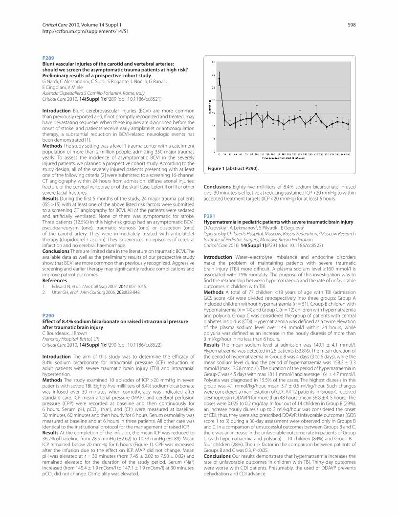
P289
Blunt vascular injuries of the carotid and vertebral arteries: should we screen the asymptomatic trauma patients at high risk? Preliminary results of a prospective cohort studyG Nardi, C Alessandrini, C Siddi, S Rogante, L Nocilli, G Ranaldi,
E Cingolani, V Miele
Azienda Ospedaliera S Camillo Forlanini, Rome, Italy
Critical Care 2010, 14(Suppl 1):P289 (doi: 10.1186/cc8521)
Introduction Blunt cerebrovascular injuries (BCVI) are more common
than previously reported and, if not promptly recognized and treated, may
have devastating sequelae. When these injuries are diagnosed before the
onset of stroke, and patients receive early antiplatelet or anticoagulation
therapy, a substantial reduction in BCVI-related neurologic events has
been demonstrated [1].
Methods The study setting was a level 1 trauma center with a catchment
population of more than 2 million people, admitting 350 major traumas
yearly. To assess the incidence of asymptomatic BCVI in the severely
injured patients, we planned a prospective cohort study. According to the
study design, all of the severely injured patients presenting with at least
one of the following criteria [2] were submitted to a screening 16-channel
CT angiography within 24 hours from admission: diff use axonal injuries;
fracture of the cervical vertebrae or of the skull base; Lefort II or III or other
severe facial fractures.
Results During the fi rst 5 months of the study, 24 major trauma patients
(ISS >15) with at least one of the above listed risk factors were submitted
to a screening CT angiography for BCVI. All of the patients were sedated
and artifi cially ventilated. None of them was symptomatic for stroke.
Three patients (12.5%) in this high-risk group had an asymptomatic BCVI:
pseudoaneurysm (one), traumatic stenosis (one) or dissection (one)
of the carotid artery. They were immediately treated with antiplatelet
therapy (clopidogrel + aspirin). They experienced no episodes of cerebral
infarction and no cerebral haemorrhage.
Conclusions There are limited data in the literature on traumatic BCVI. The
available data as well as the preliminary results of our prospective study
show that BCVI are more common than previously recognized. Aggressive
screening and earlier therapy may signifi cantly reduce complications and
improve patient outcomes.
References1. Edward N, et al.: J Am Coll Surg 2007, 204:1007-1015.
2. Utter GH, et al.: J Am Coll Surg 2006, 203:838-848.
P290
Eff ect of 8.4% sodium bicarbonate on raised intracranial pressure after traumatic brain injuryC Bourdeaux, J Brown
Frenchay Hospital, Bristol, UK
Critical Care 2010, 14(Suppl 1):P290 (doi: 10.1186/cc8522)
Introduction The aim of this study was to determine the effi cacy of
8.4% sodium bicarbonate for intracranial pressure (ICP) reduction in
adult patients with severe traumatic brain injury (TBI) and intracranial
hypertension.
Methods The study examined 10 episodes of ICP >20 mmHg in seven
patients with severe TBI. Eighty-fi ve milliliters of 8.4% sodium bicarbonate
was infused over 30 minutes when osmotherapy was indicated after
standard care. ICP, mean arterial pressure (MAP), and cerebral perfusion
pressure (CPP) were recorded at baseline and then continuously for
6 hours. Serum pH, pCO2, (Na+), and (Cl–) were measured at baseline,
30 minutes, 60 minutes and then hourly for 6 hours. Serum osmolality was
measured at baseline and at 6 hours in three patients. All other care was
identical to the institutional protocol for the management of raised ICP.
Results At the completion of the infusion, the mean ICP was reduced to
36.2% of baseline, from 28.5 mmHg (±2.62) to 10.33 mmHg (±1.89). Mean
ICP remained below 20 mmHg for 6 hours (Figure 1). CPP was increased
after the infusion due to the eff ect on ICP. MAP did not change. Mean
pH was elevated at t = 30 minutes (from 7.45 ± 0.02 to 7.50 ± 0.02) and
remained elevated for the duration of the study period. Serum (Na+)
increased (from 145.4 ± 1.9 mOsm/l to 147.1 ± 1.9 mOsm/l) at 30 minutes.
pCO2 did not change. Osmolality was elevated.
Conclusions Eighty-fi ve milliliters of 8.4% sodium bicarbonate infused
over 30 minutes is eff ective at reducing sustained ICP >20 mmHg to within
accepted treatment targets (ICP <20 mmHg) for at least 6 hours.
P291
Hypernatremia in pediatric patients with severe traumatic brain injuryD Azovskiy1, A Lekmanov2, S Pilyutik1, E Gegueva2
1Speranskiy Children’s Hospital, Moscow, Russia Federation; 2Moscow Research
Institute of Pediatric Surgery, Moscow, Russia Federation
Critical Care 2010, 14(Suppl 1):P291 (doi: 10.1186/cc8523)
Introduction Water–electrolyte imbalance and endocrine disorders
make the problem of maintaining patients with severe traumatic
brain injury (TBI) more diffi cult. A plasma sodium level ≥160 mmol/l is
associated with 75% mortality. The purpose of this investigation was to
fi nd the relationship between hypernatraemia and the rate of unfavorable
outcomes in children with TBI.
Methods A total of 77 children <18 years of age with TBI (admission
GCS score <8) were divided retrospectively into three groups: Group A
included children without hypernatraemia (n = 51), Group B children with
hypernatraemia (n = 14) and Group C (n = 12) children with hypernatraemia
and polyuria. Group C was considered the group of patients with central
diabetes insipidus (CDI). Hypernatraemia was defi ned as a twice elevation
of the plasma sodium level over 149 mmol/l within 24 hours, while
polyuria was defi ned as an increase in the hourly diuresis of more than
3 ml/kg/hour in no less than 6 hours.
Results The mean sodium level at admission was 140.1 ± 4.1 mmol/l.
Hypernatraemia was detected in 26 patients (33.8%). The mean duration of
the period of hypernatraemia in Group B was 4 days (3 to 6 days), while the
mean sodium level during the period of hypernatraemia was 158.3 ± 3.3
mmol/l (max 176.8 mmol/l). The duration of the period of hypernatraemia in
Group C was 4.5 days with max 181.1 mmol/l and average 161 ± 4.7 mmol/l.
Polyuria was diagnosed in 15.5% of the cases. The highest diuresis in this
group was 4.1 mmol/kg/hour, mean 3.7 ± 0.5 ml/kg/hour. Such changes
were considered a manifestation of CDI. All 12 patients in Group C received
desmopressin (DDAVP) for more than 48 hours (mean 56.8 ± 4. 5 hours). The
doses were 0.025 to 0.2 mg/day. In four out of 14 children in Group B (29%),
an increase hourly diuresis up to 3 ml/kg/hour was considered the onset
of CDI; thus, they were also prescribed DDAVP. Unfavorable outcomes (GOS
score 1 to 3) during a 30-day assessment were observed only in Groups B
and C. In a comparison of unsuccessful outcomes between Groups B and C,
there was an increase in the unfavorable outcome rate in patients of Group
C (with hypernatraemia and polyuria) – 10 children (84%) and Group B –
four children (28%). The risk factor in the comparison between patients of
Groups B and C was 0.3, P <0.05.
Conclusions Our results demonstrate that hypernatraemia increases the
rate of unfavorable outcomes in children with TBI. Thirty-day outcomes
were worse with CDI patients. Presumably, the used of DDAVP prevents
dehydration and CDI advance.
Figure 1 (abstract P290).
Critical Care 2010, Volume 14 Suppl 1 http://ccforum.com/supplements/14/S1
S98
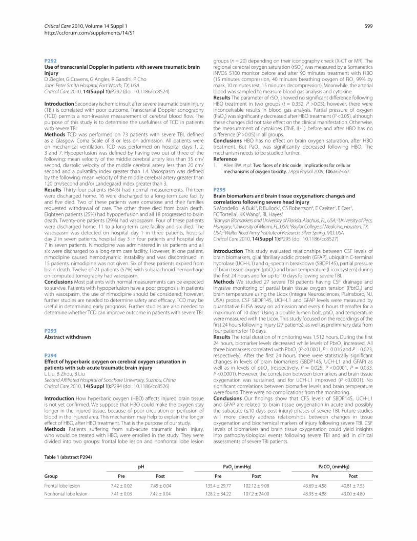
P292
Use of transcranial Doppler in patients with severe traumatic brain injuryD Ziegler, G Cravens, G Angles, R Gandhi, P Cho
John Peter Smith Hospital, Fort Worth, TX, USA
Critical Care 2010, 14(Suppl 1):P292 (doi: 10.1186/cc8524)
Introduction Secondary ischemic insult after severe traumatic brain injury
(TBI) is correlated with poor outcome. Transcranial Doppler sonography
(TCD) permits a non-invasive measurement of cerebral blood fl ow. The
purpose of this study is to determine the usefulness of TCD in patients
with severe TBI.
Methods TCD was performed on 73 patients with severe TBI, defi ned
as a Glasgow Coma Scale of 8 or less on admission. All patients were
on mechanical ventilation. TCD was performed on hospital days 1, 2,
3 and 7. Hypoperfusion was defi ned by having two out of three of the
following: mean velocity of the middle cerebral artery less than 35 cm/
second, diastolic velocity of the middle cerebral artery less than 20 cm/
second and a pulsatility index greater than 1.4. Vasospasm was defi ned
by the following: mean velocity of the middle cerebral artery greater than
120 cm/second and/or Landegaard index greater than 3.
Results Thirty-four patients (64%) had normal measurements. Thirteen
were discharged home, 16 were discharged to a long-term care facility
and fi ve died. Two of these patients were comatose and their families
requested withdrawal of care. The other three died from brain death.
Eighteen patients (25%) had hypoperfusion and all 18 progressed to brain
death. Twenty-one patients (29%) had vasospasm. Four of these patients
were discharged home, 11 to a long-term care facility and six died. The
vasospasm was detected on hospital day 1 in three patients, hospital
day 2 in seven patients, hospital day 3 in four patients and hospital day
7 in seven patients. Nimodipine was administered in six patients and all
six were discharged to a long-term care facility. However, in one patient,
nimodipine caused hemodynamic instability and was discontinued. In
15 patients, nimodipine was not given. Six of these patients expired from
brain death. Twelve of 21 patients (57%) with subarachnoid hemorrhage
on computed tomography had vasospasm.
Conclusions Most patients with normal measurements can be expected
to survive. Patients with hypoperfusion have a poor prognosis. In patients
with vasospasm, the use of nimodipine should be considered; however,
further studies are needed to determine safety and effi cacy. TCD may be
useful in determining early prognosis. Further studies are also needed to
determine whether TCD can improve outcome in patients with severe TBI.
P293
Abstract withdrawn
P294
Eff ect of hyperbaric oxygen on cerebral oxygen saturation in patients with sub-acute traumatic brain injuryL Liu, B Zhou, B Liu
Second Affi liated Hospital of Soochow University, Suzhou, China
Critical Care 2010, 14(Suppl 1):P294 (doi: 10.1186/cc8526)
Introduction How hyperbaric oxygen (HBO) aff ects injured brain tissue
is not yet confi rmed. We suppose that HBO could make the oxygen stay
longer in the injured tissue, because of poor circulation or perfusion of
blood in the injured area. This mechanism may help to explain the longer
eff ect of HBO, after HBO treatment. That is the purpose of our study.
Methods Patients suff ering from sub-acute traumatic brain injury,
who would be treated with HBO, were enrolled in the study. They were
divided into two groups: frontal lobe lesion and nonfrontal lobe lesion
groups (n = 20) depending on their iconography check (X-CT or MR). The
regional cerebral oxygen saturation (rSO2) was measured by a Somanetics
INVOS 5100 monitor before and after 90 minutes treatment with HBO
(15 minutes compression, 40 minutes breathing oxygen of FiO2 99% by
mask, 10 minutes rest, 15 minutes decompression). Meanwhile, the arterial
blood was sampled to measure blood gas analysis and cytokine.
Results The parameter of rSO2 showed no signifi cant diff erence following
HBO treatment in two groups (t = 0.352, P >0.05); however, there were
inconceivable results in blood gas analysis. Partial pressure of oxygen
(PaO2) was signifi cantly decreased after HBO treatment (P <0.05), although
these changes did not take eff ect on the clinical manifestation. Otherwise,
the measurement of cytokines (TNF, IL-1) before and after HBO has no
diff erence (P >0.05) in all groups.
Conclusions HBO has no eff ect on brain oxygen saturation, after HBO
treatment. But PaO2 was signifi cantly decreased following HBO. The
mechanism needs to be studied further.
Reference1. Allen BW, et al.: Two faces of nitric oxide: implications for cellular
mechanisms of oxygen toxicity. J Appl Physiol 2009, 106:662-667.
P295
Brain biomarkers and brain tissue oxygenation: changes and correlations following severe head injuryS Mondello1, A Buki2, R Bullock3, CS Robertson4, E Czeiter2, E Ezer2,
FC Tortella5, KK Wang1, RL Hayes1
1Banyan Biomarkers and University of Florida, Alachua, FL, USA; 2University of Pecs,
Hungary; 3University of Miami, FL, USA; 4Baylor College of Medicine, Houston, TX,
USA; 5Walter Reed Army Institute of Research, Silver Spring, MD, USA
Critical Care 2010, 14(Suppl 1):P295 (doi: 10.1186/cc8527)
Introduction This study evaluated relationships between CSF levels of
brain biomarkers, glial fi brillary acidic protein (GFAP), ubiquitin C-terminal
hydrolase (UCH-L1) and αII-spectrin breakdown (SBDP145), partial pressure
of brain tissue oxygen (ptiO2) and brain temperature (Licox system) during
the fi rst 24 hours and for up to 10 days following severe TBI.
Methods We studied 27 severe TBI patients having CSF drainage and
invasive monitoring of partial brain tissue oxygen tension (PbtO2) and
brain temperature using the Licox (Integra Neurosciences, Plainsboro, NJ,
USA) probe. CSF SBDP145, UCH-L1 and GFAP levels were measured by
quantitative ELISA assay on admission and every 6 hours thereafter for a
maximum of 10 days. Using a double lumen bolt, ptiO2 and temperature
were measured with the Licox. This study focused on the recordings of the
fi rst 24 hours following injury (27 patients), as well as preliminary data from
four patients for 10 days.
Results The total duration of monitoring was 1,512 hours. During the fi rst
24 hours, biomarker levels decreased while levels of PbrO2 increased. All
three biomarkers correlated with PbrO2 (P <0.0001, P = 0.016 and P = 0.023,
respectively). After the fi rst 24 hours, there were statistically signifi cant
changes in levels of brain biomarkers (SBDP145, UCH-L1 and GFAP) as
well as in levels of ptiO2 (respectively, P = 0.025, P <0.0001, P = 0.033,
P <0.0001). However, the correlation between biomarkers and brain tissue
oxygenation was sustained, and for UCH-L1 improved (P <0.0001). No
signifi cant correlations between biomarker levels and brain temperature
were found. There were no complications from the monitoring.
Conclusions Our fi ndings show that CFS levels of SBDP145, UCH-L1
and GFAP are related to brain tissue oxygenation in acute and possibly
the subacute (≤10 days post injury) phases of severe TBI. Future studies
will more directly address relationships between changes in tissue
oxygenation and biochemical markers of injury following severe TBI. CSF
levels of biomarkers and brain tissue oxygenation could yield insights
into pathophysiological events following severe TBI and aid in clinical
assessments of severe TBI patients.
Table 1 (abstract P294)
pH PaO2 (mmHg) PaCO
2 (mmHg)
Group Pre Post Pre Post Pre Post
Frontal lobe lesion 7.42 ± 0.02 7.45 ± 0.04 135.4 ± 29.77 102.12 ± 9.08 43.69 ± 4.58 40.81 ± 7.53
Nonfrontal lobe lesion 7.41 ± 0.03 7.42 ± 0.04 128.2 ± 34.22 107.2 ± 24.00 43.93 ± 4.88 43.00 ± 4.80
Critical Care 2010, Volume 14 Suppl 1 http://ccforum.com/supplements/14/S1
S99

P296
Cortex and deep grey matter have diff erent sensitivities to hypoxia-hypotension and traumatic brain injury: a combined PtiO
2
and microdialysis study in ratsA Odri, B Vigué, J Duranteau, T Geeraerts
CHU de Bicetre, Le Kremlin Bicetre, France
Critical Care 2010, 14(Suppl 1):P296 (doi: 10.1186/cc8528)
Introduction After traumatic brain injury (TBI), structural lesions are
heterogeneous, but the spatial heterogeneity of consequences of insults
as hypoxia-hypotension (HH) and/or TBI has never been studied. The
objective of this study was to compare the eff ect of standardized insults
(HH, TBI and both) on brain energy metabolism in two diff erent regions:
frontal cortex and thalamus.
Methods Twenty-eight Sprague–Dawley rats were randomized into four
groups: Sham, TBI (impact acceleration alone, 450 g weight drop from
1.8 m), HH (blood depletion to mean arterial pressure 40 mmHg, FiO2 10%,
15 minutes) and TBI-HH (TBI followed by HH, 45 minute delay). Cerebral
perfusion pressure (CPP) was continuously and invasively measured. Brain
microdialysis and PtiO2 probes were both inserted stereotaxically in the
right thalamus and frontal cortex.
Results Except during the HH phase, CPP was always greater than
60 mmHg. During the hour following the HH period, a signifi cant increase
in cerebral lactate/pyruvate ratio (Figure 1), glycerol and glutamate was
observed. This increase was higher in the cortex than in the thalamus in
all groups subjected to HH (P <0.001). In the TBI-HH groups, the increase
in glycerol in the cortex was signifi cantly higher compared with the
HH group (P <0.001), as well as thalamic and cortical glutamate. During
15 minutes following the HH phase (after reinjection and reoxygenation),
an increase in PtiO2 was observed in the cortex and thalamus, but with
diff erent profi les (lower increase in the cortex) (Figure 1).
Conclusions Diff erent profi les of cerebral response to HH and TBI were
observed with higher sensitivity in the cortex than in the thalamus. The
post-ischemic hyperemia seems to be altered in the traumatized cortex
but conserved into the thalamus and nontraumatized brain.
P297
Diagnosis of carotid and vertebral artery injury in major trauma with head injuryA Circelli1, M Ciapetti2, R Spina2, A Pasquini2, L Migliaccio2, M Pierini3,
G Zagli2, A Peris2
1Postgraduate School of Anesthesia and Intensive Care, University of Florence,
Italy; 2Careggi Teaching Hospital, Florence, Italy; 3Mechanical and Technology
Department, University of Florence, Italy
Critical Care 2010, 14(Suppl 1):P297 (doi: 10.1186/cc8529)
Introduction The purpose of this study is to describe a single institutional
experience in diagnosis and management of patients with blunt cerebro-
vascular injury (BCVI) after severe traumatic brain injury (TBI).
Methods We considered blunt major trauma patients consecutively
admitted from January 2008 to October 2009 to our ICU of a regional referral
hospital (Careggi Teaching Hospital, Florence, Italy). Patients were screened
on the basis of Memphis criteria to identify risk factors for BCVI: complex
cervical spine fractures, neurological conditions not sustained by imaging,
Horner’s syndrome, LeFort II/III fractures, basilar skull fractures with petrous/
neck soft tissue injury. We modifi ed this protocol to investigate all petrous
fracture and to consider also cervical spine fractures (subluxation, transverse
foramen, upper cervical spine involvement). In all patients judged at risk for
BCVI, a 64-slice angio-CT scan (CTA) was performed.
Results During the study period, 266 patients were admitted to the ICU
for BMT with an ISS >15; 162 patients (60.9%) presented TBI or cervical
spine fracture. According to our proposed modifi ed Memphis criteria, 46
patients showed risk factors for BCVI (17.3% of the total of BMT; 28.4% of
TBI), and six of them had carotid injury (2.2% of all BMT; 13.0% of patients
with risk factors). No vertebral injury was detected. Anticoagulant therapy
within 72 hours was initiated in all patients. No complications occurred
and all patients were discharged from the ICU. Clinical examination during
the 6-month follow-up showed no neurological defi ciency related to
previous vascular injury.
Conclusions A higher percentage of BCVI was observed using the
proposed modifi ed Memphis criteria if compared with other studies
(2.2% vs 1%). The mortality rate was lower than previously reported for
patients with BCVI (0% vs 13%) and it could be explained by the early
diagnosis and anticoagulant therapy initiation. An extended screening
protocol application allows prompt recognition of BCVI. In regard of the
higher percentage of BCVI found in our population, an advanced study on
protective devices (for example, helmets) is actually in progress with the
aim to reduce biomechanical eff ects of head injury on extracranial vessels.
P298
Decompressive craniectomy in severe intracranial hypertension after brain injury: early or late?S diValvasone1, S Biondi2, A Nella2, L Migliaccio2, M Ciapetti2, G Cianchi2,
M Bonizzoli2, G Zagli2, F Mariotti2, F Posteraro3, A Peris2
1Postgraduate School of Anesthesia and Intensive Care, University of Florence,
Italy; 2Careggi Teaching Hospital, Florence, Italy; 3Auxilium Vitae, Volterra, Italy
Critical Care 2010, 14(Suppl 1):P298 (doi: 10.1186/cc8530)
Introduction Decompressive craniectomy is indicated for treatment of
severe intracranial hypertension. However, this procedure is invasive and
potentially associated with complications. We present a preliminary result
of a study comparing early and late decompressive craniectomy in severe
traumatic brain injury.
Methods Patients studied were all admitted to the ICU of a tertiary
referral center (Careggi Teaching Hospital, Florence, Italy) during 4 years
(2005 to 2009). In total, data of 62 brain-injured patients, who underwent
decompressive craniectomy, were retrospectively examined and included
in two groups based on decompressive craniectomy execution:
Figure 1 (abstract P296). Brain lactate/pyruvate ratios and PtiO2 in the thalamus and cortex.
Critical Care 2010, Volume 14 Suppl 1 http://ccforum.com/supplements/14/S1
S100
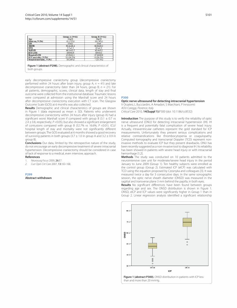
early decompressive craniectomy group (decompressive craniectomy
performed within 24 hours after brain injury, group A; n = 41) and late
decompressive craniectomy (later than 24 hours, group B; n = 21). For
all patients, demographic, scores, clinical data, length of stay and fi nal
outcome were collected from the institutional database. Traumatic lesions
were compared at admission using the Marshall score and 24 hours
after decompressive craniectomy execution with CT scan. The Glasgow
Outcome Scale (GOS) at 6 months was also collected.
Results Demographic and clinical characteristics of groups are shown
in Figure 1 (data expressed as mean ± SD). Patients who underwent
decompressive craniectomy within 24 hours after injury (group A) had a
signifi cant worst Marshall score if compared with group B (3.1 ± 0.7 vs
2.4 ± 0.8, respectively; P <0.05), but also showed a signifi cant enlargement
of contusions compared with group B (52.7% vs 16.6%; P <0.01). ICU/
hospital length of stay and mortality were not signifi cantly diff erent
between groups. The GOS evaluated at 6 months showed a good recovery
of surviving patients in both groups (3.7 ± 1.0 in group A and 3.2 ± 0.9 in
group B).
Conclusions Our data, limited by the retrospective nature of the study,
do not encourage an early decompressive treatment of severe intracranial
hypertension. Decompressive craniectomy should be considered in case
of lack of response to a medical, even intensive, approach.
References1. Neurosurg Focus 2009, 26:E7.
2. Curr Opin Crit Care 2007, 13:163-168.
P299
Abstract withdrawn
P300
Optic nerve ultrasound for detecting intracranial hypertensionV Orzalesi, L Bucciardini, A Amadori, S Marchiani, P Innocenti
AOU Careggi, Florence, Italy
Critical Care 2010, 14(Suppl 1):P300 (doi: 10.1186/cc8532)
Introduction The purpose of this study is to verify the reliability of optic
nerve ultrasound (ONU) for detecting intracranial hypertension (IH). IH
is a frequent and potentially fatal complication of severe head injury.
Actually, intraventricular catheters represent the gold standard for ICP
measurements. Unfortunately they present serious complications and
relative contraindications like thrombocytopenia or coagulopathy.
Computed tomography and transcranial Doppler (TCD) represent non-
invasive methods to evaluate ICP but they present drawbacks. ONU has
been recently suggested as a non-invasive tool to diagnose IH. Its reliability
has been showed in patients with severe head injury or with intracranial
hemorrhage [1,2].
Methods The study was conducted on 10 patients admitted to the
neurointensive care unit for moderate/severe head injury in the period
January to June 2009 (Group 1). Ten healthy subjects were enrolled as
the control group (Group 2). Estimated ICP (eICP) was calculated with
TCD using the equation proposed by Czosnyka and colleagues [3]. It was
measured twice a day for 3 consecutive days. In the same sonographic
session, the optic nerve sheath diameter (ONSD) was measured in the
sagittal and transverse plane 3 mm behind the papilla, in both eyes.
Results No signifi cant diff erences have been found between groups
regarding age and sex. The ONSD distribution is shown in Figure 1.
ONSD, eICP and ICP values were signifi cantly higher in Group 1 than in
Group 2. Linear regression analysis identifi ed a signifi cant relationship
Figure 1 (abstract P298). Demographic and clinical characteristics of
both groups.
Figure 1 (abstract P300). ONSD distribution in patients with ICP less
than and more than 20 mmHg.
Critical Care 2010, Volume 14 Suppl 1 http://ccforum.com/supplements/14/S1
S101
between ONSD and ICP (r = 0.588). By calculating the receiver operating
characteristic curve, an ONSD value of 5.35 mm resulted as the optimal
cut-off point with a sensitivity of 95.1% and a specifi city of 96.2%.
Conclusions ONSD measurements correlated with invasive and non-
invasive (TCD) measurements of ICP. It is a useful, non-invasive bedside tool
to diagnose IH. It is safe, easy to perform and can rapidly give reproducible
information on patients’ ICP.
References1. Moretti R, et al.: Reliability of optic nerve ultrasound for the evaluation of
patients with spontaneous intracranial hemorrhage. Neurocrit Care 2009.
[Epub ahead of print]
2. Geeraerts, et al.: Non-invasive assessment of intracranial pressure using ocular sonography in neurocritical care patients. Intensive Care Med 2008,
34:2062-2067.
3. Czosnyka M, et al.: Cerebral perfusion pressure in head-injured patients: a noninvasive assessment using transcranial Doppler ultrasonography. J
Neurosurg 1998, 88:802-808.
P301
Measurement of neuronal specifi c enolase as a predictor of outcome in patients suff ering from severe traumatic brain injuryJW Gatson, AH Idris, PE Pepe, JP Minei, J Wigginton
University of Texas Southwestern Medical Center at Dallas and the Parkland
Health and Hospital System, Dallas, TX, USA
Critical Care 2010, 14(Suppl 1):P301 (doi: 10.1186/cc8533)
Introduction Traumatic brain injury (TBI) is a multiphasic disease.
Follow ing the initial impact, secondary injury, including oxidative stress
and infl ammation, is thought to contribute signifi cantly to death and
disability. This ongoing damage in the penumbra of the brain leads to the
demise of neuronal populations, and ultimately decreased brain function.
Identifi cation of various neural markers such as neuronal specifi c enolase
(NSE) at various time points following TBI may help us better understand
the magnitude of secondary injury following TBI, and may help predict
outcome in these patients.
Methods Early serial cerebrospinal fl uid (CSF) samples were collected
from patients with severe TBI (GCS 3 to 8) who required placement of a
therapeutic ventriculostomy. Following ventriculostomy placement, CSF
samples were obtained every 4 hours for the fi rst 24 hours post-injury,
then every 8 hours for post-injury days 2 to 5. The levels of NSE were then
measured using the ELISA method.
Results Following sample analysis in 11 patients, we found that in TBI
patients that had a good neurological outcome, the levels of NSE in the
CSF rose early (between 25 and 60 ng/ml) within 12 to 16 hours following
the initial injury. The NSE levels then rapidly decreased to normal levels
(~5 ng/ml) at approximately 20 hours following injury, with these levels
persisting until day 5 (day of fi nal sample collection). Patients with a poor
outcome (lack of ability to return to pre-injury activities or death) showed
signifi cantly higher levels of NSE persisting out to 5 days post injury, with
late levels ranging from 35 to 50 ng/ml.
Conclusions In TBI patients with a good outcome, there was an increase
in NSE levels in the CSF noted at early time points (~16 hours), which
abated at approximately 20 hours after injury. In TBI patients with a poor
clinical outcome, CSF levels of NSE were signifi cantly elevated at later time
points over the fi rst 5 days post injury.
P302
Hyperphosphorylated neurofi lament NF-H, S100β and NSE as indicators of severity of brain injury in childrenJ Žurek, L Marek, P Košut, M Fedora
Faculty of Medicine, Masaryk University, University Children´s Hospital, Brno,
Czech Republic
Critical Care 2010, 14(Suppl 1):P302 (doi: 10.1186/cc8534)
Introduction The aim of the study was to determine whether serum
levels of biomarkers – hyperphosphorylated neurofi lament NF-H, S100β protein and NSE – correlate with severity of brain injury and outcome in
children with traumatic brain injury (TBI).
Methods Thirty-eight patients with TBI were enrolled in the prospective
study. TBI was verifi ed by computerized tomography according to
the Marshall classifi cation. Venous blood samples were taken after
admission and every 24 hours for a maximum of 6 consecutive days.
Serum NF-H concentrations were quantifi ed by ELISA, S100β and NSE
by electrogenerated chemiluminescence. The outcome was evaluated
6 months after TBI using the Glasgow Outcome Scale (GOS) in all patients.
Results Serum concentrations of NF-H (median 86 vs 12; P = 0.01), S100β (median 3.47 vs 0.54; P = 0.001), and NSE (median 193.2 vs 37.03; P = 0.001)
were higher in nonsurvivors than survivors. Serum NF-H (median 86 vs 12;
P = 0.004), S100β (median 3.47 vs 0.54; P = 0.001) and NSE (median 193.2
vs 37.03; P = 0.001) was signifi cantly higher in patients with unfavourable
outcome (GOS ≤3 vs GOS >3). Spearman’s rank correlation coeffi cient on
fi rst day of hospitalization S100B x NSE = 0.58 (P <0.05), GCS x GOS = 0.50
(P <0.05), GOS x Marshall classifi cation = 0.46 (P <0.05).
Conclusions Plasma levels of hyperphosphorylated neurofi lament, S100β and NSE correlate with severity of TBI and may be useful as predictors of
outcome in children with TBI.
P303
A simple hospital discharge score predicts Glasgow Outcome Scale at 12 months in patients with traumatic brain injuryGW Fuller, P Yeoman
Queens Medical Centre, Nottingham, UK
Critical Care 2010, 14(Suppl 1):P303 (doi: 10.1186/cc8535)
Introduction This study examines whether a hospital discharge score
predicts future Glasgow Outcome Scale (GOS) following traumatic brain
injury (TBI). Loss to follow-up is a major problem in TBI studies and an early
prediction score could be used for interpolation of missing outcome data
or as a surrogate outcome measure.
Methods Adults admitted to the Queens Medical Centre between 1993
and 2002 with TBI and GCS <12 were included prospectively in the
Nottingham Head Injury Register. At hospital discharge the modifi ed
Barthel Index was recorded by nursing staff and a Discharge Disability
Score (DDS) calculated: 1. Independent, 2. Dependent <24 hour care, 3.
Fully dependent, 4. Dead [1]. Glasgow Outcome Scale (GOS) was calculated
at 12 months by the patient’s general practitioner [2]. Unfavourable
outcome was defi ned as DDS 3, 4 or GOS 1, 2, 3. Correlation between
DDS and GOS was measured and the ability of DDS to predict GOS at
12 months in survivors was assessed using logistic regression. Sensitivity
and specifi city of dichotomised DDS for prediction of unfavourable
outcome at 12 months was also calculated.
Results Data were available on 1,227 patients. A highly correlated linear
relationship was evident between DDS and GOS (spearman correlation
coeffi cient 0.77, P <0.0001). Unfavourable outcome measured by DDS
showed sensitivity of 73% (95% CI 69 to 76%) and specifi city of 56% (53
to 59%) for prediction of unfavourable outcome at 12 months defi ned by
GOS. DDS showed a strong association with GOS at 12 months for survivors
at discharge, with fully dependent patients having 15 times higher odds
of an unfavourable outcome. Adjusting for known TBI prognostic factors
attenuated the strength of the association, however DDS remained a
statistically signifi cant strong predictor of outcome.
Conclusions DDS is strongly associated with 12-month GOS and could
potentially be used to replace missing data or provide a surrogate
outcome measure in TBI trials. A valid prediction score may also aid the
clinician’s ability to discuss patient prognosis at hospital discharge.
References1. Shah S, et al.: J Clin Epidemiol 1989, 42:703-709.
2. Jennet B, et al.: Lancet 1975, 1:480-484.
P304
Outcome of aggressive treatment for blunt and penetrating traumatic cardiac arrestY Moriwaki, M Iwashita, Y Tahara, S Arata, N Harunari, T Kosuge, H Toyoda,
N Suzuki
Yokohama City University Medical Center, Yokohama, Japan
Critical Care 2010, 14(Suppl 1):P304 (doi: 10.1186/cc8536)
Introduction The outcome of patients with cardiac arrest due to blunt
trauma (BT-CPA) and penetrating trauma (PT-CPA) is very poor. The aim
of this study is to clarify the outcome of patients with CPA on arrival due
Critical Care 2010, Volume 14 Suppl 1 http://ccforum.com/supplements/14/S1
S102

to BT-CPA and PT-CPA treated with our strategy including emergency
department thoracotomy (EDT).
Methods This study is a population-based case series observational study.
We have taken three approaches to these patients: our private aggressive
treatment strategy (resuscitation for 30 minutes, aggressive infusion using
sheath introducer into the subclavian vein, and EDT); in-hospital system
supporting this aggressive resuscitation (logistic issue such as the close
location between ED and the room for catheter intervention and CT,
and direct entrance to the OR by exclusive lift, and common instruments
interchangeable between ED and OR including the bed); and prehospital
EMS in our city (CPA patients are transferred in about 7 minutes to the
nearest of selected 11 hospitals which can receive and treat CPA patients).
Results For the past 10 years, of 478 BT-CPA and 30 PT-CPA patients, 76%
and 70% were witnessed, and 21% and 37% were CPA after scene. A
total of 85% and 77% underwent EDT. Although 34% and 60% achieved
ROSC, only 18% and 40% went to the ICU, TAE room, and OR (admitted),
and only 2.7% and 17% were discharged (survivors). Restricted in eight
witnessed patients showing VF as the initial cardiac rhythm in BT-CPA,
38% were admitted and 13% survived. No PT-CPA patients showed VF.
Restricted in 134 witnessed patients showing PEA in BT-CPA and 10 in PT-
CPA, 28% and 70% were admitted and 1% and 30% survived. Two hundred
and twenty witnessed patients showing no life sign (asystole) in BT-CPA
and 12 in PT-CPA, 8% and 25% were admitted, and 3% and 17% survived.
The initial rhythm of survivors was asystole in 70% in BT-CPA and 40% in
PT-CPA. Although the time interval from arrival at the hospital to ROSC of
survivors tended to be shorter than for nonsurvivors in BT-CPA, there was
no diff erence in this interval in PT-CPA. The longest interval was 43 and
30 minutes in BT-CPA and PT-CPA.
Conclusions The expected outcome of BT-CPA and PT-CPA patients is
hopeless. However, we cannot and should not give up on resuscitating
them merely because they are CPA and they do not show any life sign at
the scene. We consider the indication of aggressive resuscitation in every
individual case by his/her individual condition.
P305
Using fl ow-rate limiting bag–valve–mask device without trainingJ Nurmi1, M Castrén2
1Helsinki University Central Hospital, Helsinki, Finland; 2Karolinska Institutet,
Stockholm, Sweden
Critical Care 2010, 14(Suppl 1):P305 (doi: 10.1186/cc8537)
Introduction The Smart Bag (SB, O-Two Medical Technologies Inc.,
Canada) is a bag–valve–mask resuscitator (BVM) equipped with a valve
limiting fl ow-rate and peak airway pressure to decrease hyperventilation
and gastric insuffl ation. We studied this device used by hospital staff
without training to evaluate the possibility to change the standard BVM
to the SB in hospital wards where positive pressure ventilation is rarely
needed and training is sporadic.
Methods The participants (12 physicians, 38 nurses) were randomised to
use the standard BVM or SB to ventilate a resuscitation training manikin
over 1 minute. The mechanism of the SB was shortly described in the SB
group but no hands-on training was provided. Participants were asked to
ventilate the patients as they would do if patients have a pulse but do not
breathe. Tidal volumes were registered to a computer connected to the
manikin.
Results The medians of minute ventilation were 6.1 (interquartile range
2.6 to 8.1) and 3.9 (IQR 1.7 to 5.4) litres per minute in the SB and control
groups, respectively (Figure 1). Hyperventilation >10 l/minute occurred
only in the control group (P = 0.23).
Conclusions Using the SB without previous hands-on training is
possible for the majority of nursing and medical staff and decreases
hyperventilation in comparison with the standard BVM.
P306
Out-of-hospital cardiac arrest and resource utilisation in the ICUA Manning, M Purcell-Jones, A Saayman, M Wise
University Hopsital of Wales, Cardiff , UK
Critical Care 2010, 14(Suppl 1):P306 (doi: 10.1186/cc8538)
Introduction Survival rates for out-of-hospital cardiac arrests (OHCA)
remain poor and typically of the order of 10% or less [1]. In the community,
bystander CPR and public automated external defi brillators have been
promoted, whilst in hospital therapeutic hypothermia and coronary
reperfusion are employed to improve outcome. Few studies have assessed
how this group of patients impacts on critical care resources [2].
Methods Patients with a spontaneous circulation following OHCA
admitted to the critical care unit of a university teaching hospital between
1999 and 2009 were analysed retrospectively.
Results A total of 1,656 patients presented to the emergency department
following OHCA, of which 217 were admitted to the ICU. The ICU and
hospital survivals were 41% and 32.7%, respectively. In the period 1999 to
2003, ICU and hospital survival rates were 37.5% and 22.9%, respectively.
In the period 2004 to 2009, ICU and hospital survival rates were 43.8% and
40.5%, respectively. At admission to the ICU, the mean APACHE II score
was 22.82, mean pH 6.87 and FIO2 52.6%. In those patients who survived
hospital discharge, the mean APACHE II score was 19.71, mean pH 6.97
and FIO2 47.1%, whilst in nonsurvivors they were 24.33, 6.83 and 55.4%,
respectively. Overall ICU and hospital lengths of stay (LOS) were 5.41 and
13.72 days and the average TISS score was 221. In those who survived to
hospital discharge, ICU and hospital LOS were 10.1 and 32.4 days and the
average TISS score was 380. Amongst patients who died in hospital, the
mean ICU and hospital LOS were 3.1 and 4.7 days, the average TISS was
144.
Conclusions Overall one-third of patients admitted to the ICU in the period
1999 to 2009 with OHCA were discharged from hospital. This increased
to 40.5% in the period from 2004 with almost all patients discharged
from the ICU surviving to hospital discharge. Nonsurvivors amongst the
cohort are unsurprisingly more seriously ill with higher APACHE II scores
and deranged physiology. The cost of treating survivors of OHCA in ICU is
considerably higher for survivors compared with nonsurvivors. However,
as patients who are discharged from ICU usually leave hospital (2004 to
2009), this would appear to be an appropriate utilisation of resources.
References1. Sayre et al.: Prehosp Emerg Care 2009, 13:469-477.
2. Graf J, et al.: Crit Care 2008, 12:R92.
P307
Comparison of external cardiac compression on the fl oor and in a dental chairG Papakitsos, A Kapsali, T Papakitsou
General Hospital Arta, Greece
Critical Care 2010, 14(Suppl 1):P307 (doi: 10.1186/cc8539)
Introduction Dental treatment is often stressful for patients and causes
accidental systemic symptoms. In such situations, cardiopulmonary
resuscitation (CPR) may be required. If external cardiac compression (ECC)
in the dental chair is not eff ective, the patient should be moved onto the
fl oor, which needs at least two or three persons and 10 to 15 seconds.
Figure 1 (abstract P305). Minute ventilation (ml) in the Smart Bag group
and the control group.
Critical Care 2010, Volume 14 Suppl 1 http://ccforum.com/supplements/14/S1
S103
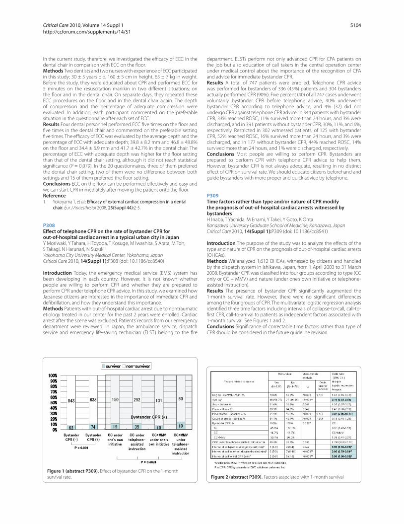
In the current study, therefore, we investigated the effi cacy of ECC in the
dental chair in comparison with ECC on the fl oor.
Methods Two dentists and two nurses with experience of ECC participated
in this study; 30 ± 5 years old, 160 ± 5 cm in height, 65 ± 7 kg in weight.
Before the study, they were educated about CPR and performed ECC for
5 minutes on the resuscitation manikin in two diff erent situations; on
the fl oor and in the dental chair. On separate days, they repeated these
ECC procedures on the fl oor and in the dental chair again. The depth
of compression and the percentage of adequate compression were
evaluated. In addition, each participant commented on the preferable
situation in the questionnaire after each set of ECC.
Results Four dental personnel performed ECC fi ve times on the fl oor and
fi ve times in the dental chair and commented on the preferable setting
fi ve times. The effi cacy of ECC was evaluated by the average depth and the
percentage of ECC with adequate depth; 39.8 ± 8.2 mm and 46.8 ± 48.8%
on the fl oor and 34.4 ± 6.9 mm and 41.7 ± 42.7% in the dental chair. The
percentage of ECC with adequate depth was higher for the fl oor setting
than that of the dental chair setting, although it did not reach statistical
signifi cance (P = 0.079). In the 20 questionnaires, three of them preferred
the dental chair setting, two of them were no diff erence between both
settings and 15 of them preferred the fl oor setting.
Conclusions ECC on the fl oor can be performed eff ectively and easy and
we can start CPR immediately after moving the patient onto the fl oor.
Reference1. Yokoyama T, et al.: Effi cacy of external cardiac compression in a dental
chair. Eur J Anaesthesiol 2008, 25(Suppl 44):2-5.
P308
Eff ect of telephone CPR on the rate of bystander CPR for out-of-hospital cardiac arrest in a typical urban city in JapanY Moriwaki, Y Tahara, H Toyoda, T Kosuge, M Iwashita, S Arata, M Toh,
S Takagi, N Harunari, N Suzuki
Yokohama City University Medical Center, Yokohama, Japan
Critical Care 2010, 14(Suppl 1):P308 (doi: 10.1186/cc8540)
Introduction Today, the emergency medical service (EMS) system has
been developing in each country. However, it is not known whether
people are willing to perform CPR and whether they are prepared to
perform CPR under telephone CPR advice. In this study, we examined how
Japanese citizens are interested in the importance of immediate CPR and
defi brillation, and how they understand this importance.
Methods Patients with out-of-hospital cardiac arrest due to nontraumatic
etiology treated in our center for the past 2 years were enrolled. Cardiac
arrest after the scene was excluded. Patients’ records from our emergency
department were reviewed. In Japan, the ambulance service, dispatch
service and emergency life-saving technician (ELST) belong to the fi re
department. ELSTs perform not only advanced CPR for CPA patients on
the job but also education of call takers in the central operation center
under medical control about the importance of the recognition of CPA
and advice for immediate bystander CPR.
Results A total of 747 patients were enrolled. Telephone CPR advice
was performed for bystanders of 336 (45%) patients and 304 bystanders
actually performed CPR (90%). Five percent (40) of all 747 cases underwent
voluntarily bystander CPR before telephone advice, 40% underwent
bystander CPR according to telephone advice, and 4% (32) did not
undergo CPR against telephone CPR advice. In 344 patients with bystander
CPR, 33% reached ROSC, 11% survived more than 24 hours, and 3% were
discharged, and in 391 patients without bystander CPR, 30%, 11%, and 6%,
respectively. Restricted in 302 witnessed patients, of 125 with bystander
CPR, 52% reached ROSC, 16% survived more than 24 hours, and 3% were
discharged, and in 177 without bystander CPR, 44% reached ROSC, 14%
survived more than 24 hours, and 1% were discharged, respectively.
Conclusions Most people are willing to perform CPR. Bystanders are
prepared to perform CPR with telephone CPR advice to help them.
However, bystander CPR is not always adequate, resulting in no distinct
eff ect of CPR on survival rate. We should educate citizens beforehand and
guide bystanders with more proper and quick advice by telephone.
P309
Time factors rather than type and/or nature of CPR modify the prognosis of out-of-hospital cardiac arrests witnessed by bystandersH Inaba, T Yachida, M Enami, Y Takei, Y Goto, K Ohta
Kanazawa University Graduate School of Medicine, Kanazawa, Japan
Critical Care 2010, 14(Suppl 1):P309 (doi: 10.1186/cc8541)
Introduction The purpose of the study was to analyze the eff ects of the
type and nature of CPR on the prognosis of out-of-hospital cardiac arrests
(OHCAs).
Methods We analyzed 1,612 OHCAs, witnessed by citizens and handled
by the dispatch system in Ishikawa, Japan, from 1 April 2003 to 31 March
2008. Bystander CPR was classifi ed into four groups according to type (CC
only or CC + MMV) and nature (under one’s own initiative or telephone-
assisted instruction).
Results The presence of bystander CPR signifi cantly augmented the
1-month survival rate. However, there were no signifi cant diff erences
among the four groups of CPR. The multivariate logistic regression analysis
identifi ed three time factors including intervals of collapse-to-call, call-to-
fi rst CPR, call-to-arrival to patients as independent factors associated with
1-month survival. See Figures 1 and 2.
Conclusions Signifi cance of correctable time factors rather than type of
CPR should be considered in the future guideline revision.
Figure 1 (abstract P309). Eff ect of bystander CPR on the 1-month
survival rate. Figure 2 (abstract P309). Factors associated with 1-month survival
Critical Care 2010, Volume 14 Suppl 1 http://ccforum.com/supplements/14/S1
S104

P310
Use of the laryngeal tube by paramedics during prehospital resuscitation: an observational studyG Papakitsos1, T Papakitsou2, A Kapsali1
1General Hospital Arta, Greece; 2General Hospital Messologi, Greece
Critical Care 2010, 14(Suppl 1):P310 (doi: 10.1186/cc8542)
Introduction In the current guidelines of the ERC, tracheal intubation has
become less important for persons not trained and diff erent supraglottic
airway devices are recommended. The present investigation deals with
the application of the laryngeal tube (LT) during prehospital resuscitation
by paramedics.
Methods The study was observational during a period of 2 years (2007
to 2009). We registered all cardiac arrest situations in which the LT had
been applied according to the ERC guidelines 2005. All participants had
completed an obligatory course in emergency medicine, but had not
been specifi cally trained in endotracheal intubation; they were therefore
designated as unfamiliar in using the endotracheal tube to secure the
airway. Primary outcome measures were placement time and successful
placement. Study endpoint was the total ‘no fl ow time’, which is defi ned
as the time without chest compression in the fi rst period of cardiac arrest.
Results During the defi ned period, 53 resuscitation attempts recorded
on standardised data sheets were included. The LT was used in 87% of all
cardiac arrest situations. Overall, the LT was successfully and signifi cantly
faster inserted (about 9.4 seconds) in more than 96% of all cases, on the
fi rst attempt. In 99% of all cases, no problems concerning ventilation of the
patient were described. During the cardiac arrest simulation, establishing
and performing fi rst ventilation took an average of 12.5 seconds with the
LT. Use of the LT during cardiac arrest signifi cantly reduced the ‘no fl ow
time’ about 87.6 seconds.
Conclusions As an alternative airway device recommended by the ERC,
the LT may enable airway control rapidly and eff ectively. Additionally,
by using the LT, a reduced ‘no-fl ow-time’ and a better outcome may be
possible. LT may be a good alternative airway device for providing and
maintaining a patent airway during resuscitation.
Reference1. Wiese CH, et al.: The use of the laryngeal tube disposable (LT-D) by
paramedics during out-of-hospital resuscitation-an observational study concerning ERC guidelines 2005. Resuscitation 2009, 80:194-198.
P311
The i-gel supraglottic airway as an alternative in the preclinical emergency medical aid? A comparative study on a manikinJ Tiesmeier, M Emmerich
Krankenhaus Bad Oeynhausen, Germany
Critical Care 2010, 14(Suppl 1):P311 (doi: 10.1186/cc8543)
Introduction Endotracheal intubation (ET) is the recognised gold
standard for securing an airway in the realm of preclinical emergency
medical aid. Supraglottic airway devices present a valid alternative in the
case of an elevated incidence of diffi cult airway securing and intubation
preconditions. Due to positive experiences with the i-gel supraglottic
airway, we – through a joint paramedic (PM) and doctor (DR) team – have
compared its use under standardised conditions with the classic laryngeal
mask (cLMA) and the laryngeal tube (LT) using an airway simulator.
Methods Seventy-one probands (40 PM and 31 DR) took part in this study.
The airway simulator used was the Ambu® Airway Man. Timekeeping
started with the laying aside of the anaesthesia bag after successful
ventilation of the airway simulator and ended after having ensured
suffi cient ventilation. Participants used three diff erent devices (i-gel size 4,
cLMA size 3 and LT size 4) each fi ve times. Additionally, an interval time was
measured after successful ventilation with the i-gel. Without interruption,
participants continued the experiment with two interval ventilations and
conducted a secondary ET through the i-gel. Timekeeping ended in this
case after the fi rst ventilation.
Results Prior to the study 95% of PM had previous experience with the
cLMA, 77.5% with the LT and 0% with the i-gel. Respective percentages
for DR were 61.2% for the cLMA, 38.7% for the LT and 32.2% for the i-gel
(P <0.001 respectively). Across the entire group of 71 study participants,
users required an average of 16.6 seconds for the successful insertion
of the cLMA, 15 seconds for the LT and 8.48 seconds for the i-gel. When
comparing the PM with the DR, the PM required 16.5 seconds for the cLMA
(vs 16.9 seconds, P = 0.693), 14.9 seconds for the LM (vs 16.8 seconds, P =
0.08) and 7.8 seconds for the i-gel (vs 9.4 seconds, P = 0.001). The total time
needed for secondary ET was on average 25.8 seconds (PM 24.2 seconds,
DR 27.9 seconds, P <0.001). Error-free insertion at all fi ve incidents was
achieved by 73.3% of DR and by 46.1% of PM (P <0.001).
Conclusions Suffi cient ventilation was achieved 50% faster through the
i-gel when compared with the cLMA and the LT. The correct usage of the
i-gel can be learnt quickly. The i-gel allows the options of secondary ET.
Reference1. Emmerich M: Use of the i-gel laryngeal mask for management of a diffi cult
airway. Anaesthetist 2008, 57:779-781.
P312
Retention of skills during emergency airway management training: a 12-month follow-upH Fischer1, A Fast1, E Kajcsa1, M Fleck1, R Greif2
1General Hospital Vienna, Austria; 2University Hospital Bern, Switzerland
Critical Care 2010, 14(Suppl 1):P312 (doi: 10.1186/cc8544)
Introduction Besides the gold standard endotracheal tube, supraglottic
airway devices are alternatives for emergency airway management [1]. The
goal of the study was to identify airway devices that provide successful
ventilation, even 12 months after training in manikins.
Methods In 2008, 288 medical students were trained to use laryngeal mask
airways (LMAs) Unique, ProSeal, Supreme, I-Gel and bag–mask ventilation
(BM) on manikins (Ambu). Students successful on the fi rst attempt (n =
190) for all devices in random order were included and tested 12 months
later without further training. The insertion time to fi rst successful
ventilation, tidal volume (TV) and gastric infl ation were assessed.
Results The time (in seconds) needed for the I-Gel and the Unique was
signifi cant longer in 2009 (BM 2008 8.2 ± 4.7 vs 2009 8.4 ± 5.4, P = 0.882;
I-Gel 2008 10.2 ± 2.7 vs 2009 11.7 ± 4.9; P = 0.008; Supreme 2008 15 ± 3.7
vs 2009 15.5 ± 4.6, P = 0.147; Unique 2008 16.5 ± 3.6 vs 2009 18.2 ± 5.5; P =
<0.001; ProSeal 2008 17.7 ± 4.9 vs 2009 17.7 ± 5.2, P = 0.856). A higher rate
of gastric infl ation for BM and the I-Gel was seen in comparison of 2008
with 2009 (BM 10.5% vs 18.6%, P = 0.012; I-Gel 0.5% vs 4.2%, P = 0.039;
Supreme 0.5% vs 2.2%, P = 0.375; Unique 3.7% vs 5.8%, P = 0.607; ProSeal
4.7% vs 1.2%, P = 0,18). About 60% of the TVs were insuffi cient with little
improvement for the Supreme and Proseal (Figure 1).
Conclusions One year after training, time for successful ventilation for all
devices was lower than 25 seconds. This is acceptable compared with the
gold standard endotracheal tube, but due to the rising gastric infl ation
rate and high amount of insuffi cient tidal volume, shorter intervals of
training maybe necessary.
References1. Nolan JP, et al.: Resuscitation 2005, 67(Suppl 1):S54-S56.
Figure 1 (abstract P312). Tidal volume (correct range 0.4 to 0.6 l).
Critical Care 2010, Volume 14 Suppl 1 http://ccforum.com/supplements/14/S1
S105

P313
Factors correlated with the advanced life support simulation performance of senior medical studentsKH Park, YJ Kang
Jeju National University Hospital, Jeju-si, Republic of Korea
Critical Care 2010, 14(Suppl 1):P313 (doi: 10.1186/cc8545)
Introduction Even though medical education using simulation for
medical students is increasing and suggests positive outcomes, little is
known about factors associated with the performance of advanced life
support simulation. We investigated the correlation between various
factors and simulation performance using senior medical students.
Methods Fourth-year medical students were enrolled and divided into six
groups. Each group underwent a 2-week emergency medicine training.
This study was conducted consecutively with a 2-week break between
each group from March to July, 2009. Every student had an advanced
cardiac life support (ACLS) simulation performance test as a moderator,
scored by two ACLS instructors certifi ed by the American Heart
Association. The following data were examined: basic life support (BLS)
practice, personality characteristics survey, case presentation, one post-
training written test, and two plan tests for the Korean Medical Licensing
Examination (KMLE). The statistical signifi cance was defi ned as P <0.05.
Results Forty-six students participated. No correlation was found between
ACLS performance scores and personality type or medical knowledge as
measured by the post-training written test and the two plan tests for the
KMLE. However, scores of the BLS examination and the case presentation
were moderately correlated with the ACLS performance scores: r = 0.390,
0.402, respectively (P <0.01).
Conclusions The ACLS simulation performance was correlated with the
BLS skills and the case presentation. This supports the idea that the ability
to manage resuscitation is associated with BLS skills and the capability
to analyze and coordinate patient management. This result provides
guidance for education of medical students.
P314
Aging augments unwllingness to the initial three links in chains of survivalH Inaba, M Enami, T Yachida, Y Takei, O Oyama, K Ohta
Kanazawa University Graduate School of Medicine, Kanazawa, Japan
Critical Care 2010, 14(Suppl 1):P314 (doi: 10.1186/cc8546)
Introduction The purpose of the study was to clarify the infl uence of
aging on attitudes toward the initial three links in chains of survival.
Methods We gave questionnaires to attendants of compulsory programs
for basic life support (BLS) or driving technique at the beginning in
authorized driving schools. The questionnaires included their backgrounds.
We studied their willingness in four hypothetical scenarios related to the
initial three links: early emergency call, cardiopulmonary resuscitation
(CPR) under one’s own initiative, telephone-assisted chest compression
and use of AED. The respondents were divided into young (17 to 29 years,
n = 6,122), middle-aged (29 to 59 years, n = 827), older person (>59 years,
n = 15,743) groups.
Results There were signifi cant diff erences in gender, occupation,
residential area and experience of BLS training, and knowledge of AED
use between the three groups. The proportions of respondents who
are willing to perform the desirable BLS actions were lowest in the older
person group (Table 1). Multiple logistic regression analysis confi rmed
that aging is one of the independent factors relating to negative attitude
in all the scenarios. Gender, occupation, resident area, experience of BLS
training, and knowledge for AED use were other independent factors
relating to negative attitude to some of the scenarios.
Conclusions The aged population is more negative to the chain of
survival. More are willing to follow the telephone-assisted direction for
chest compression. The BLS training should be modifi ed for them to gain
confi dence and to be aware of the signifi cance and benefi t of early call.
P315
Prediction of the success of cardiac resuscitation: a pattern classifi cation approach based on ECG spectral and temporal featuresM Baronio1, F Baronio2, M Campi2, G Amicucci1, A Caré2, A Sorgato3,
G Perone4, A Candiani4
1Ospedale S. Orsola Brescia, Italy; 2Department of Information Engineering,
University of Brescia, Italy; 3Division of Cardiology, Ospedale S. Orsola, Brescia,
Italy; 4Spedali Civili, University of Brescia, Italy
Critical Care 2010, 14(Suppl 1):P315 (doi: 10.1186/cc8547)
Introduction The ECG tracings recorded during a ventricular fi brillation
(VF) using an automated external defi brillator (AED) contain useful
information predictive of shock outcome. The focus is on the VF
waveforms’ morphology. The amplitude and the spectral properties of
VF may predict the likelihood of successful defi brillation [1-4]. In almost
all previous studies, the amplitude or the spectral properties of the ECG
tracings have been singularly used. However, these approaches have led
to methods lacking suffi cient predictive power.
Methods Five hundred patients with out-of-hospital cardiac arrest on
arrival in an emergency room were examined. The rhythm was identifi ed
as VF and confi rmed by two trained investigators. ECG data were stored in
modules in digitized form over a period of 20 minutes and were analyzed
retrospectively. ECG traces containing CPR artefacts were removed by
digital fi ltering. Times of collapse, dispatch, scene arrival, CPR, and initial
defi brillation were determined from dispatch records, recordings of arrest
events, interviews with bystanders, and hospital records. The preshock VF
waveform morphology was studied and diff erent parameters of VF ECG
signals were extracted. We then introduced a pattern classifi cation machine
that combines the amplitude and spectral features simultaneously.
Results The use of the pattern classifi cation machine which combines
amplitude and spectral features of VF ECG signals shows an improved
predictive power as compared with other methods.
Conclusions This technique could help to determine which patients
should receive shock fi rst and which should receive a period of CPR prior to
shock, thereby increasing the probability of survival. The potential impact
of this research is high in the direction of generating a new methodology
able to increase the probability of survival after a cardiac crisis.
References1. Strohmenger H: Chest 1997, 111:584-589.
2. Sherman LD: Resuscitation 2006, 69:479-486.
3. Hamprecht F: Resuscitation 2001, 50:297-299.
4. Eftestol T: Resuscitation 2005, 67:55-61.
Table 1 (abstract P314). Diff erence among the three age groups in the proportion of respondents who are willing to perform the desirable BLS actions
Age group
Scenario 17 to 29 years 29 to 59 years >59 years Chi-square test P value
CPR under their own initiative 78.9% (4,810/6,094) 71.9% (589/819) 57.1% (8,186/14,346) <0.001
Telephone-assisted chest compression 94.6% (5,766/6,898) 92.5% (757/818) 84.6% (12,112/14,316) <0.001
Early 119 call 91.2% (5,556/6,092) 91.2% (747/819) 79.4% (12,130/15,270) <0.001
Use of AED 56.8% (3,386/5,963) 48.4% (388/801) 21.4% (2,832/13,285) <0.001
Critical Care 2010, Volume 14 Suppl 1 http://ccforum.com/supplements/14/S1
S106
P316
Dispatcher-assisted telephone cardiopulmonary resuscitation using a French compression-only protocol: performance of volunteers with or without prior life support trainingA Ghuysen1, S Stipulante2, D Colas1, V D’Orio1
1CHU Liège – ULG, Liège, Belgium; 2CS 100 Liège, Belgium
Critical Care 2010, 14(Suppl 1):P316 (doi: 10.1186/cc8548)
Introduction Although early bystander-initiated cardiopulmonary
resuscitation (CPR) is a key factor in survival improvement from out-of-
hospital cardiac arrest, only one-third of victims actually receive CPR before
arrival of emergency medical services. Dispatcher-assisted telephone CPR
might improve the frequency and performance of bystanders’ resuscitation
eff orts. Due to the recent interest in hands-only CPR protocol [1] and the
lack of any French validated algorithm, we designed the present study in
order to validate a new French protocol called ALERT (Algorithme Liègeois
d’Encadrement à la Réanimation par Téléphone).
Methods We tested the effi cacy of the ALERT protocol on CPR performances
in a manikin model of cardiac arrest. Participants (n = 110) were divided into
four groups. Previously untrained subjects, distributed into two sub-groups
depending on whether they were benefi ciating from telephone assistance
(PUG group, n = 30) or not (PUNG group, n = 30), were compared with
previously trained nurses students distributed in two other sub-groups: with
(PTG group, n = 25) or without (PTNG group, n = 25) telephone assistance.
Results Global evaluation of CPR performances in untrained volunteers
benefi ting from telephone assistance revealed a signifi cant improvement
as compared with unguided untrained volunteers. Indeed, they
approached CPR performance levels of previously trained nurse students
with no dispatcher’s guidance. Previously trained nurses with telephone
assistance gained the best CPR scores, notably because of an improvement
in the quality of airway management.
Conclusions Our results indicate that use of the ALERT algorithm by
dispatchers may improve the CPR performance in both previously untrained
volunteers and in volunteers with prior basic life support training.
Reference1. Hallstrom AH, et al.: N Engl J Med 2000, 342:1546-1553.
P317
CPR team members’ attitudes towards family presence during cardiopulmonary resuscitationN Kianmehr, M Mofi di, H Rahmani, A Forouzan
Iran University of Medical Sciences, Tehran, Iran
Critical Care 2010, 14(Suppl 1):P317 (doi: 10.1186/cc8549)
Introduction The concerns about allowing family members to be
present during resuscitation are a matter of debate in many countries. The
purpose of this study was to determine the opinions of Iranian health-care
providers about family-witnessed resuscitations.
Methods The study population consisted of CPR team members in
four teaching hospitals. We developed a questionnaire, assessing their
opinions, using 20 questions and also considering their comments.
Results Of the total 200 participants, 77% oppose family-witnessed
resuscitation. There was no signifi cant diff erence among opponents about
FWR based on sex, age, number of CPR exposures and the experience of
respondents (P >0.05). Emergency physicians were more likely advocates
for FWR than other specialties (P = 0.004). All of the anesthesiologists
opposed FWR. Most reasons for opposition were fear of psychological
trauma to family members, interference with patient care, increased staff
stress and violation with patient confi dentiality.
Conclusions CPR team members in Iran do not believe in the presence
of relatives during resuscitation. Informing healthcare providers on the
benefi t of witnessed resuscitation and public education of CPR would be
a fundamental element for implementing a formal program.
References1. Bae H, Lee S, Jang HY: The ethical attitude of emergency physicians toward
resuscitation in Korea. J Emerg Med 2008, 34:485-490.
2. Maclean SL, Guzzetta CE, White C et al.: Family presence during cardiopulmonary resuscitation and invasive procedures: practices of critical care and emergency nurses. J Emerg Nurs 2003, 29:208-221.
3. Tucker TL: Family presence during resuscitation. Crit Care Nurs Clin North Am
2002, 14:177-185.
P318
Prehospital therapeutic hypothermia induced with cold infusions improves haemodynamic stability in nonshockable cardiac arrest patientsA Truhlar1, R Skulec2, J Seblova2, L Zabka3, V Cerny1
1University Hospital, Hradec Kralove, Czech Republic; 2Central Bohemian
Region EMS, Kladno, Czech Republic; 3Hradec Kralove Region EMS, Hradec
Kralove, Czech Republic
Critical Care 2010, 14(Suppl 1):P318 (doi: 10.1186/cc8550)
Introduction Recent studies report an increase of asystole and pulseless
electrical activity (PEA) as the fi rst monitored cardiac arrest rhythms
after arrival of the Emergency Medical Services [1]. The asystolic patients
presumably undergo ischaemia for a longer time and may benefi t from
treatment reducing hypoxic brain injury. Therapeutic hypothermia (TH)
has expanded into prehospital care to be initiated as soon as possible.
Rapid cold crystalloid infusion is the most frequent method; however,
severe haemodynamic instability is its contraindication. The aim of the
study was to assess the adverse eff ects of prehospital volume expansion
in patients with initial nonshockable rhythms when used in a setting with
only restricted cardiovascular monitoring.
Methods The nonshockable patients enrolled in a prospective nonrandomized
PRE-COOL trial (PRE-hospital COOLing in cardiac arrest patients) were rapidly
administered cold normal saline (5 to 30 ml/kg) intravenously and analysed
for body temperature, haemodynamic variables (blood pressure, heart rate,
shock index), incidence of circulatory complications (recurrence of cardiac
arrest, pulmonary oedema), and outcome. Hypothermic patients (group A, n
= 19) were compared with matched historic controls (group B, n = 22).
Results In treatment group A, 1,021 ± 526 ml (13.2 ± 6.3 ml/kg) of 4°C
cold normal saline was administered before hospital admission. Body
temperature decreased by 1.32 ± 0.71°C until arrival at hospital (calculated
rate 1.08 ± 0.44°C/30 minutes). The incidence of hypotension requiring
vasopressors (noradrenaline or dopamine) signifi cantly decreased in
patients given cold intravenous fl uids compared with control group
B: 31.6% vs 63.6% (P = 0.04). No patient developed pulmonary oedema
within 24 hours. Recurrence of cardiac arrest was comparable in both
groups (3/19 vs 3/22), shock index at hospital arrival was 0.92 vs 0.97 (P =
0.78), and favorable outcome (cerebral performance category 1 or 2)
26.3% vs 13.6% (P = 0.31).
Conclusions The early prehospital administration of cold infusions
is safe and contributes to haemodynamic optimization in patients
resuscitated from nonshockable rhythms. Incidence of hypotension
requiring vasopressors was signifi cantly reduced in the treated group. No
pulmonary oedema was observed.
Acknowledgements Supported by grant IGA MH CZ NS10383-2/2009
and research project MZO 00179906.
Reference1. Hallstrom A, et al.: Resuscitation 2009, 80:975-976.
P319
Trans-nasal cooling during CPR: a single-center experienceFS Taccone, JL Vincent
Erasme Hospital, Brussels, Belgium
Critical Care 2010, 14(Suppl 1):P319 (doi: 10.1186/cc8551)
Introduction Trans-nasal cooling started during cardiopulmonary
resuscitation (CPR) has shown to improve the return to spontaneous
circulation (ROSC) and survival rate in an experimental prolonged cardiac
arrest model. A multicenter randomized trial (PRINCE) has also suggested
an improved neurological outcome in patients receiving trans-nasal
cooling during CPR in the prehospital setting when compared with those
treated by conventional hypothermia on hospital arrival, provided a delay
between collapse and CPR of less than 10 minutes.
Methods Patients with witnessed cardiac arrest and a downtime less than
20 minutes were randomized to prehospital intra-arrest cooling versus
standard ACLS care. Trans-nasal cooling (RhinoChill, BeneChill Inc., CA,
USA) was initiated using a mixture of volatile coolant fl uid with oxygen
delivered into the nasopharynx for rapid evaporative heat transfer. Cooling
was continued during CPR and, for patients who achieved ROSC, until
initiation of systemic cooling at hospital. Resuscitation was continued for
at least 30 minutes. All patients were then cooled at the hospital.
Critical Care 2010, Volume 14 Suppl 1 http://ccforum.com/supplements/14/S1
S107

Results Twenty-four patients were included, but one in the treatment
group was excluded from the per-protocol analysis because of DNR
orders. Patients randomized to treatment group (n = 9) or standard care
(n = 14) had similar demographics, initial rhythm, time from collapse to
CPR and ALS arrival. Median time from collapse to cooling initiation was
19 minutes. In total, 6/9 (66%) treated and 6/14 (42%) control patients
achieved ROSC. Three patients (33%) in the treatment group survived
to hospital discharge with CPC 1 to 2, while only one (7%) of the control
group patients had good neurological outcome. No serious adverse
events occurred in treated patients.
Conclusions Trans-nasal cooling seems to be safe and feasible in a
prehospital setting. These single-center data confi rm that trans-nasal
cooling may improve the ROSC rate as well as good neurological outcome
if started in patients with a short delay between collapse and CPR.
P320
Intra-arrest cooling using a novel intra-nasal cooling method for immediate induction of therapeutic hypothermia in GermanyH Busch1, H Fritz2, F Eichwede3, B Inderbitzen4, D Barbut4, T Schwab1
1Intensive Care Unit Heilmeyer, Freiburg, Germany; 2Krankenhaus Martha-
Maria Halle-Dölau, Halle/Saale, Germany; 3Medizinisches Zentrum Aachen,
Würselen, Germany; 4Benechill, San Diego, CA, USA
Critical Care 2010, 14(Suppl 1):P320 (doi: 10.1186/cc8552)
Introduction Recent investigations have demonstrated improved
neurological outcome after therapeutic hypothermia in patients after
successful resuscitation. The time course and duration to achieve target
temperature may be an important factor to infl uence patient’s outcome.
To determine the safety and effi cacy of intranasal cooling during ongoing
resuscitation, for immediate induction of therapeutic hypothermia,
the Pre-Resuscitation Intra-Nasal Cooling Eff ectiveness (PRINCE) study
involved 200 patients in Europe, using a non-invasive nasal catheter
that sprays evaporating coolant liquid into the nasal cavity. Here we
demonstrate data from all German participating sites.
Methods All patients who were deemed eligible for advance cardiac life
support (ACLS) were included as long as the arrest was witnessed and
cardiopulmonary resuscitation (CPR) was initiated within 20 minutes
of collapse. Patients were randomized to treatment or control groups.
The trial was designed to determine the safety and eff ectiveness of
early cooling initiated at the site of arrest. Survival and time to target
temperature were documented.
Results Data are presented as the mean ± SD or median (interquartile
range (25, 75%)). Mean age was 67.8 ± 14 years in the intervention group
and 65.4 ± 13.9 years in the control group. On average, cooling therapy
was started in 33 ± 12 minutes in the RhinoChill™ group and 170 ± 97
minutes in the control group. Temperatures at hospital admission were
signifi cant lower in the RhinoChill™ group. Time to target tympanic
temperature, refl ecting brain temperature, were signifi cant faster in the
RhinoChill™ group (211 ± 124 minutes vs 424 ± 217 minutes; P <0.05).
Adverse events occurred in 12 patients. None was related to the cooling
therapy. In the intervention group fi ve patients (20%) survived and three
patients (12%) had a CPC of 1 to 2. In the control group only four patients
(12.5%) survived and one patient (3.1%) had a CPC of 1 to 2.
Conclusions Using the intranasal cooling method, cooling was much
faster and earlier in treated patients. Neurologically intact survival and
discharge rates were higher in treated patients. Transnasal cooling for the
induction of therapeutic hypothermia during prehospital resuscitation is
feasible and highly eff ective in lowering brain temperature rapidly. The
method off ers the possibility for immediate introduction and realization
of mild hypothermia in the fi eld.
P321
Therapeutic hypothermia after cardiac arrest: implementation in UK intensive care unitsA Binks1, R Murphy1, A Padkin2, J Nolan2
1Bristol Royal Infi rmary, Bristol, UK; 2Royal United Hospital, Bath, UK
Critical Care 2010, 14(Suppl 1):P321 (doi: 10.1186/cc8553)
Introduction In 2003 the International Liaison Committee on Resusci-
tation (ILCOR) published guidelines recommending all patients admitted
comatose to hospital following an out-of-hospital cardiac arrest (OHCA)
with an initial rhythm of ventricular fi brillation should be treated with
therapeutic hypothermia for a period of 12 to 24 hours [1]. We aimed to
determine how many units are using hypothermia as part of their post-
cardiac arrest management and when each of the ICUs implemented the
therapy. Other objectives were to determine what cooling techniques are
being used, the target temperature and the duration of cooling used.
Methods The 248 UK ICUs listed in the Directory of Critical Care Services
were contacted by telephone by one of the authors. The duty ICU
consultant or nurse in charge was asked a standardised set of questions.
Results We obtained responses from 244 of 248 (98.3%) of ICUs. Currently,
209 (85.6%) ICUs are using hypothermia as part of post-cardiac arrest
management. Since 2003, there has been an increase annually in the
number of units using hypothermia, with the majority of units starting in
2007 or 2008 (Figure 1). Of the units who use hypothermia as part of their
post-cardiac arrest management, 99% are using it to treat patients after
OHCA from shockable rhythms, 60.6% are using it to treat patients after
OHCA from nonshockable rhythms, and 63.5% are using it after in-hospital
cardiac arrest. Cooling is initiated with intravenous cold fl uid in 71%. The
commonest technique to maintain hypothermia is to use ice; used by 55%
of units. Most ICUs (89.9%) aim for a temperature of 32 to 34°C and the
duration of hypothermia is 24 hours in 141 ICUs (67.5%).
Conclusions Generalised implementation of this clinical guideline in
the UK has taken about 5 years but now the majority of ICUs are using
therapeutic hypothermia as part of post-cardiac arrest management.
Reference1. Nolan J: Circulation 2003, 108:118-121.
P322
Hospital-wide therapeutic hypothermia protocol signifi cantly reduces the time to induce hypothermia but does not improve outcomesP Hayden, AA James
Medway NHS Foundation Trust, Gillingham, UK
Critical Care 2010, 14(Suppl 1):P322 (doi: 10.1186/cc8554)
Introduction Therapeutic hypothermia (TH) is a reliable, evidence-based
modality to improve neurological outcome following out-of-hospital
cardiac arrest. Often TH is induced in the ICU and as a result the time
from cardiac arrest (CA) to induction of TH (THst) and achieving target
temperature (CAta
) is delayed. Preliminary data have shown that delaying
TH may worsen outcome [1]. We sought to investigate whether a hospital-
wide approach to TH after CA would reduce delays and result in improved
outcomes.
Methods We conducted a retrospective analysis of all TH interventions
from 2007 to 2008. Following this we implemented a hospital-wide
approach to TH and re-evaluated in 2008 to 2009. The hospital-wide
approach included an educational programme, TH guidelines in the
resuscitation room in the ED, and a cooling pack stored in the ICU which
could be taken immediately to the post-CA patient.
Results CA-ROSC and CA-Hosp delays were similar between groups. The
hospital-wide approach signifi cantly reduced the time to initiate TH and
time to achieve target temperature (see Table 1). The small sample size
may have prohibited demonstration of outcome diff erences.
Figure 1 (abstract P321). Implementation of therapeutic hypothermia in
UK ICUs by year.
Critical Care 2010, Volume 14 Suppl 1 http://ccforum.com/supplements/14/S1
S108

Table 1 (abstract P322). Diff erences between groups pre and post the
hospital-wide approach
2007 to 2008 2008 to 2009 P value
n 16 26
CA-ROSC 28.5 minutes 24.1 minutes
CA-Hosp 59.1 minutes 50.1 minutes
CA-THst 306.2 minutes 133.2 minutes <0.0001
CA-THta
527.3 minutes 252.7 minutes 0.005
HospSurv 7 13 0.69
Conclusions A hospital-wide approach to TH following cardiac arrest
signifi cantly reduces delays to achieve the target temperature, although
this did not improve outcomes at our institution. Initiation of prehospital
TH may be an ideal solution to further reduce this delay, and ongoing trials
may demonstrate improved outcomes.
Reference1. Nozari A, et al.: Critical time window for intra-arrest cooling in a dog model.
Circulation 2006, 113:2690-2696.
P323
Induced hypothermia after cardiac arrest for 12 hours: single-centre experienceR Sykora1, R Janda2
1Charles University, Medical School and Teaching Hospital, Plzen, Czech
Republic; 2Hospital of Karlovy Vary, Czech Republic
Critical Care 2010, 14(Suppl 1):P323 (doi: 10.1186/cc8555)
Introduction Induced hypothermia (IH) for 12 to 24 hours is a standard
part of post-resuscitation care. We present a cohort of patients with out-
of-hospital or in-hospital cardiac arrest (OHCA, IHCA) from our ICU who
underwent 12 to 14 hours of IH. Moreover we aimed at neurological
outcome in a subgroup of patients with initial ventricular fi brillation (VF)
and ST-elevation acute myocardial infarction (STEMI) undergoing coronary
intervention (PCI).
Methods We retrospectively evaluated patients with nontraumatic
cardiac arrest admitted to our ICU from 2006 to 2009. A standardised
therapeutic protocol including IH was implemented. IH (32 to 34°C) was
started within 30 minutes after restoration of spontaneous circulation
(ROSC), before PCI, if performed. IH was maintained for 12 to 14 hours,
than passive rewarming followed. Endovascular cooling device was not
used. Cerebral performance category (CPC) at hospital discharge was
evaluated as a main outcome.
Results Total number of patients was 88 (OHCA n = 63; IHCA n = 25), mean
age 57 years, 65% men. ICU mortality was 32% and hospital mortality
53%. Initial ECG rhythm was VF in 59%, asystole in 32% and pulseless
electrical activity in 9%. Coronary angiography was performed in 59%
and intervention in 41% of all cases. The required target temperature was
achieved in 76%. Complications were ventilator-associated pneumonia
in 6%, tracheobronchitis in 15% and gastrointestinal bleeding in 2%,
no severe bradyarhythmias or iontogram dysbalance was recorded.
In a group of 23 patients with VF/STEMI, PCI after initiation of IH was
performed. The required target temperature was achieved in 91%. The
ICU survival rate was 87% and survival to discharge from hospital was
78%. Good neurological outcome (CPC 1 or 2) in this group at hospital
discharge was in 83% (n = 15).
Conclusions Induced hypothermia has to be started as soon as possible
after ROSC. Cooling of patients after cardiac arrest should continue during
all therapeutic interventions. Twelve hours of induced hypothermia after
cardiac arrest could off er comparable neurological outcomes to 24 hours,
especially in the subgroup of patients with VF/STEMI.
Acknowledgements Supported by a research project MSM 0021620819.
P324
Survival following adult cardiac arrest in intensive care units: a 5-year retrospective analysisS Bagshaw, P Brindley, D Kutsogiannis
University of Alberta Hospital, Edmonton, Canada
Critical Care 2010, 14(Suppl 1):P324 (doi: 10.1186/cc8556)
Introduction This study aims to provide survival data for adult patients
following attempted cardiopulmonary resuscitation (CPR) in critical care
units. We hope objective data can facilitate dialogue and decision-making.
Methods We retrospectively reviewed CPR records from 1 January 1 2000
to 30 April 2005 from all critical care units of the tertiary care hospitals
of Edmonton, Canada. Full inpatient medical records were reviewed for
further details.
Results Of 529 arrests, 59.4% had return of spontaneous circulation,
29.5% survived to critical care unit discharge, 26.3% survived to hospital
discharge, 20.5% survived to 3 months, and 19.9% to 6 months. Pulseless
electrical activity (PEA) represented 29.7%, ventricular tachycardia (VT)
20.8%, asystole (ASY) 15.7%, bradycardia 15.5%, ventricular fi brillation
(VF) 13.4%, supraventricular tachycardia 2.8%, and respiratory arrest 2.1%.
A total 36.7% of arrests occurred between 08:01 and 16:00 hours, 33.0%
occurred between 00:01 and 08:00 hours and 30.3% between 16:01 and
24:00 hours. There was no signifi cant association between arrest time and
survival. There was a signifi cant association between longer CPR duration
and increased mortality (P <0.001). GSICU cardiac arrest patients had
statistically higher critical care unit mortality than CCU or CVICU patients
(75.5% vs 70.0% vs 45.7%; P = 0.002). ASY/PEA were signifi cantly more
common in GSICUs, whereas VF and VT were more common in CVICU (P =
0.001). Mortality was signifi cantly higher following PEA/ASY regardless of
location. Multivariate analysis showed no signifi cant association between
survival and age, gender or arrest time. In contrast, survival was signifi cantly
lower with increased APACHE II score (O.R 1.07, P <0.001), prolonged CPR
(O.R.1.03, P <0.01), and ASY/PEA (O.R. 5.44, P <0.001). No patient with an
APACHE II ≥30 survived to hospital discharge following PEA.ASY, and no
patient, regardless of arrest type, survived to hospital discharge with an
APACHE II >45.
Conclusions Initial survival following in-hospital cardiac arrest is higher
than from noncritical care units, and therefore should be considered
diff erently. Survival was higher still from cardiac ICUs compared with other
critical care units. Admission APACHE II, type of arrest, and resuscitation
duration provide useful prognosticators, especially in combination. In
contrast, age, gender, and arrest time do not. Despite encouraging early
survival, less than one-quarter survived to hospital discharge, and less
than one-in-six were alive at 1 year.
P325
Eff ects of bed height and time on the quality of chest compressions during cardiopulmonary resuscitationA Lewinsohn, PB Sherren, DS Wijayatilake
Queens Hospital, London, UK
Critical Care 2010, 14(Suppl 1):P325 (doi: 10.1186/cc8557)
Introduction The 2005 International Liaison Committee on Resuscitation
gave a detailed update on best practice for CPR with a discussion around
appropriate patient and rescuer position, based largely on expert opinion
[1]. The objectives of this study were: to demonstrate a diff erence in chest
compression eff ectiveness with bed height, and provide a suggestion for
an optimal and achievable bed height for eff ective chest compressions;
and to demonstrate fatigue during chest compressions, and provide a
suggestion for an upper time limit for eff ective chest compressions.
Methods Exclusion criterion for this trial; no previous basic life support
training in the past 4 years, or refused consent to participate. A modifi ed
Laerdal manikin was connected to a Dragor ventilator (to measure
intrathoracic pressures generated). The manikin was placed on a hospital
trolley, and CPR was performed by candidates at three diff erent bed
heights: mid thigh; anterior superior iliac spine; and xiphisternal area.
Chest compressions were continuous and asynchronous with ventilation,
and allowed to continue for 30 seconds before recordings were taken.
Results One hundred and one subjects took part. The diff erences in
intrathoracic pressures generated at diff erent bed heights were compared
using ANOVA variance testing for multiple groups, and were statistically
Critical Care 2010, Volume 14 Suppl 1 http://ccforum.com/supplements/14/S1
S109

signifi cant with P <0.01 (Figure 1). We also found that the eff ectiveness of
CPR decreased 17% over a 2-minute period (Figure 2).
Conclusions The most eff ective bed height position, allowing rescuers
to achieve the highest intrathoracic pressures during CPR, was with the
patient chest in line with the rescuer’s mid-thigh. The rescuer performing
CPR should change every 2 minutes.
Reference1. Resuscitation 2005, 67:187-201.
P326
New results about early therapeutic hypothermia in sudden cardiac death and following return of spontaneous circulationFR Righetti, AL Luzzani, EP Polati1Ospedale Policlinico Universitario, Verona, Italy
Critical Care 2010, 14(Suppl 1):P326 (doi: 10.1186/cc8558)
Introduction Therapeutic hypothermia is defi ned as controlled cooling of
body temperature for therapeutic purposes. At present, the use of mild–
moderate hypothermia (32 to 34°C) in patients with neurological damage
is increasing in the ICU. In this regard, recent studies suggest that it is able
to improve the neurological outcome in patients with anoxic cerebral
damage following sudden cardiac death (SCA) [1]. The aim of this study
is to assess the role of therapeutic hypothermia on neurological outcome
in patients who experienced SCA with ensuing return of spontaneous
circulation (ROSC).
Methods Ninety adult patients, aged between 18 and 85, referred to our
ICU after SCA due to cardiac disease with following ROSC were randomly
allocated to the following treatment groups: patients in group 1 were
treated immediately after admission with therapeutic hypothermia plus
standard treatment, patients in group 2 received only standard treatment.
All patients at entry presented with GCS 3. Neurological outcome was
assessed on discharge and after 6 months, by means of GOS scale (0 =
dead, 1 = vegetative, 2 = severely disabled, 3 = moderately disabled, 4 =
good recovery). We consider scores 0 to 1 as unfavourable outcome,
scores from 2 to 4 as favourable outcome. To compare the two groups we
used the Mann–Whitney U test of for continuous variables, the chi-square
test for qualitative variables.
Results Patients in group 1 (45 patients) and in group 2 (45 patients)
resulted statistically comparable for sex (P = 0.14) and age (P = 0.68) and
presentation ECG rhythm (P = 0.42) and APACHE II score (P = 0.34). At
discharge from the ICU, 66.6% of patients in group 1 presented favourable
neurological outcome (GOS 2 to 4) versus 35% of patients in group 2. At
6 months, 70% of patients in group 1 presented favourable neurological
outcome (GOS 2 to 4) versus 38.3% of patients in group 2. The mortality
was 16.6% in group 1 versus 36.6% in group 2 (P = 0.003). The patients
who improved their GOS values were 64% in group 1 and 18% in group
2 (P <0.005).
Conclusions Our study demonstrated that early treatment with therapeutic
hypothermia in the patient who had SCA improves neurological outcome. It
is independent of the rhythm of presentation. Besides, we reached statistical
signifi cance.
Reference1. Polderman KH, et al.: Therapeutic hypothermia and controlled
normotermia in the intensive care unit: practical considerations, side eff ects, and cooling methods. Crit Care Med 2009, 37:1101-1120.
P327
Infection rates among cooled patients post cardiac arrestA Binks, MJ Thomas
Bristol Royal Infi rmary, Bristol, UK
Critical Care 2010, 14(Suppl 1):P327 (doi: 10.1186/cc8559)
Introduction We conducted a retrospective study looking at infection
rates in patients treated with therapeutic hypothermia (TH) following
cardiac arrest who were admitted to the ICU of the Bristol Royal infi rmary
since May 2007. TH is recommended in all patients presenting with
persistent coma following cardiac arrest. One complication of TH is the risk
of infection. Hypothermia suppresses the immune system by inhibiting the
release of proinfl ammatory cytokines and by suppressing the chemotactic
migration of leukocytes and phagocytosis [1]. In patients with traumatic
brain injury, TH for more than 48 hours is associated with a higher risk of
infection, but not if the period of hypothermia is less than 24 hours [2]. In
patients following cardiac arrest, infection is common, reported in up to
73% of patients [3].
Methods Data were collected retrospectively, and information about
ICU length of stay, whether they had a protected catheter (PC) specimen
taken, whether they had any positive cultures and whether they were
treated with antibiotics was extracted. Data for ICU length of stay was
also obtained. Infections were defi ned as >105 colony-forming units (cfu)
grown from either PC specimens or from peripheral blood cultures.
Results We identifi ed 82 patients treated with TH post cardiac arrest.
Nineteen (29%) had proven infection either on PC or on peripheral blood
cultures. PC specimens were taken in 21 (25%) patients. Of the 21 patients
who had PC specimens taken, 16 (76%) had proven infection. We also
found that an increase in ICU length of stay was associated with increased
infection rates, 44% in patients with a length of stay of greater than 3 days
and 55% in patients with a length of stay of greater than 4 days.
Conclusions We have shown, in our ICU, that of patients who were
treated with TH following cardiac arrest, 29% had a proven infection.
On PC sampling, there was a much higher rate of infection with 76% of
patients with positive cultures. This suggests that the risk of infection in
patients treated with therapeutic hypothermia post cardiac arrest is higher
than that for patients who are cooled post traumatic brain injury.
References1. Polderman KH: Intensive Care Med 2004, 30:556-575.
2. Polderman KH: Crit Care Med 2009, 37:S186-S202.
3. Tsai MS, et al.: Intensive Care Med 2005, 31:621-626.
P328
Strategies to implement a therapeutic hypothermia programS Blosser, N Cavarocchi, S Ettinger, IC Gilchrist, R Reichwein, S Bressler,
J Burkholder, E Sarsfi eld, K Zagari
Penn State Hershey Medical Center MCH075, Hershey, PA, USA
Critical Care 2010, 14(Suppl 1):P328 (doi: 10.1186/cc8560)
Introduction In this presentation we will review the process used for
developing a therapeutic hypothermia program and our outcomes
during the fi rst year. Despite the fact that therapeutic hypothermia has
been shown to improve survival and neurologic outcome after cardiac
arrest [1], it is underused [2] and has been only slowly introduced in many
institutions. One of the reasons that it is not more widely used may be the
diffi culty of implementation of a program.
Methods A multidisciplinary team was assembled in 2007 to explore
the opportunity for therapeutic hypothermia in patients after cardiac
arrest. Pertinent literature was reviewed and a guideline and electronic
orders were drafted. The team investigated all available options for
cooling and chose a method that was non-invasive and did not require
a large fi nancial investment by the institution. Nurses were sent to an
international program to learn about hypothermia and become advocates
Figure 1 (abstract P325). Eff ects of bed height position on intrathoracic
pressure generated after 30 seconds of CPR. n = 101. *Statistically
signifi cant (P <0.05).
Figure 2 (abstract P325). Mean intrathoracic pressures generated during
CPR at 30 seconds as compared with 2 minutes. *Statistically signifi cant
(P <0.05).
Critical Care 2010, Volume 14 Suppl 1 http://ccforum.com/supplements/14/S1
S110

and educators. Education was done for caregivers in the prehospital
setting, emergency department, cardiac catheterization laboratory, and
adult ICUs. Advertising to community and other hospitals was done by
newspaper, radio, and television. The team continued to meet regularly to
assess and modify the program based on experience.
Results Prior to the start of this program, therapeutic hypothermia was
essentially not utilized at the institution. Twenty-one patients were cooled
after cardiac arrest and resuscitation at our institution in the fi rst year
(2008), with few missed opportunities. Fifteen (71%) survived, and 12
(57%) had good neurologic outcome. (Post-cardiac arrest survival rates
without hypothermia in the literature range from less than 5% to 35% [1].)
Challenges included altering historic practice patterns, integrating sectors
of the healthcare delivery system that had historically drifted apart, and
interacting with extra-institutional regulations governing prehospital care.
Conclusions The therapeutic hypothermia program at our institution
is successful in improving patient outcome after cardiac arrest. The
elements that contributed to a successful program included: teamwork;
multidisciplinary nature of the team; promoting awareness of the
program; buy-in by nurses who championed the program in the ICU;
positive patient outcomes; and low fi nancial investment for the institution.
References1. Bernard SA, et al.: N Engl J Med 2002, 346:557-563.
2. Brigham BL, et al.: Resuscitation 2009. [Epub ahead of print]
P329
A comparison of core and tympanic temperature measurement in the critically illB Sanderson, L Lim, K Lei, J Smith, L Camporota, R Beale
Guys and St Thomas’ NHS Foundation Trust, London, UK
Critical Care 2010, 14(Suppl 1):P329 (doi: 10.1186/cc8561)
Introduction Measurement of body temperature using the tympanic
method (TTM) represents a standard non-invasive method which best
approximates core temperature (CTM). However, published evidence
increasingly casts doubt on the agreement between TTM and CTM. The
aim of this study was to assess the agreement of these two methods of
temperature measurement in critically ill patients.
Methods We recruited 20 consecutive critically ill patients, who required
indwelling thermistor-tipped arterial catheters for haemodynamic monitor-
ing. For each patient we simultaneously collected the core temperature,
obtained from the arterial catheter, and tympanic temperatures in the left
and right ears, in the supine position. Tympanic temperature was recorded
using the ear setting (TTMe) and the core setting tympanic temperature
(TTMc) in accordance with the manufacturer’s instructions (Genius™ 2;
Kendall, Tyco Healthcare, MA, USA). Local ethics committee approval was
waived and the study was conducted as a service improvement audit.
Bland–Altman analysis was used to measure agreement between the two
set of measurements.
Results From the 20 patients we made 102 paired measurements.
Comparison of the TTM in the right and left ears resulted in high variability
in the recorded temperature with a percentage error of 34.4%. In view of
this observed large variability, the mean TTM between the two ears was
used for subsequent comparison with core temperature. Comparison of
core measurements against tympanic measurements using ear settings
(TTMe) showed a small positive mean bias (limits of agreement (LOA);
percentage error) of 0.3°C (–0.4 to 1.1°C, 2.4%). However, when the CTM
and TTM were compared using the core temperature setting of the
thermometer, the bias (LOA) increased to –0.97°C (–1.6 to –0.4°C), creating
a larger bias than the unadjusted TTM.
Conclusions Tympanic temperature measurements showed a large
random variability compared with core temperatures and suggest bilateral
averaged tympanic measurements may be necessary. In our series, using
the core setting increased error and increased the bias between the two
methods. Our fi ndings raise the possibility that the diagnosis of fever
and the need for further investigation may be aff ected by the choice of
temperature measurement site and device setting used.
P330
Can we predict neurological prognoses with computed tomography just after resuscitation?T Mochizuki1, N Otani1, O Takahashi2, S Ishimatsu1
1St Luke’s International Hospital, Tokyo, Japan; 2St Luke’s Life Science Institute,
Tokyo, Japan
Critical Care 2010, 14(Suppl 1):P330 (doi: 10.1186/cc8562)
Introduction Noncontrast computed tomography (CT) is often used to
evaluate a primary catastrophic brain injury after cardiac arrest. On day 3 after
resuscitation, cerebral damage presents as swelling on CT; however, there is no
strong evidence associated with how prognosis is predicted on CT performed
just after resuscitation. We assessed whether the ratio (in Hounsfi eld units
(HU)) of grey matter (GM) to white matter (WM) on CT performed just after
resuscitation can be used as a predictor of patient outcome.
Methods We utilized the electronic medical record to examine admission
to the ICU after resuscitation from out-of-hospital cardiac arrest. Brain CTs
performed just after resuscitation were re-evaluated. Patients were excluded
if they had head trauma, cerebral vessel disease and were 16 years of age or
younger. Clinical outcome was assessed at the time of discharge using the
Pittsburgh cerebral performance category (CPC). On noncontrast CTs, HU
were measured as GM in the regions of bilateral caudate head, putamen,
globus pallidaus and cortical layer of the precentral sulcus, and as WM in the
regions of the periventricular WM. We defi ned good neurological prognosis
as CPC 1 to 2 and an unfavorable prognosis as CPC 3 to 5. Each ratio of
average GM to WM was calculated, and they were compared with a good
outcome and an unfavorable outcome.
Results In 1,057 episodes of out-of-hospital cardiac arrest, 296 were
resuscitated and admitted to our ICU; 64 were excluded, and the fi nal study
sample consisted of 232 patients. CPC 1 to 5 at hospital discharge was
16%, 6%, 6%, 12% and 60%. The mean time from arrival to perform brain
CT was 66 (25 to 330) minutes. The HU of caudate head, putamen and
globus pallidaus were much higher in good outcome than in unfavorable
outcome (P <0.001, t test). The HU of cortical layer of the precentral sulcus
and periventricular WM were not signifi cantly diff erent. The caudate
head/WM ratio and globus pallidaus/WM ratio were much higher in
good outcome (P <0.01, t test). Using ROC analysis, we determined that
a caudate head/WM ratio >1.21 or a globus pallidaus/WM ratio >1.17 was
90% predictive of a good outcome in this study.
Conclusions The caudate head/WM ratio and globus pallidaus/WM
ratio by brain CT just after resuscitation are useful to predict the post-
resuscitation neurological prognosis.
Reference1. Wijdicks EFM, et al.: Neurology 2006, 67:203-210.
P331
Unintended consequences of technology improvementW McGee, PM Mailloux
Baystate Medical Center, Springfi eld, MA, USA
Critical Care 2010, 14(Suppl 1):P331 (doi: 10.1186/cc8563)
Introduction Technological improvements in ventilator design may
confound the diagnosis of brain death. We describe a series of patients
clinically perceived to be breathing spontaneously even though they
satisfi ed the criteria for brain death. The misdiagnosis of cerebral function
in these patients delayed the diagnosis of brain death and subsequent
organ donation. Resource utilization, patient/family suff ering, and staff
morale are all negatively impacted when this occurs.
Methods The ventilator triggering mode was recorded in all patients.
Patients were identifi ed by the authors with a high likelihood of brain
death and receiving pressure support ventilation. Patients without cranial
nerve function underwent formal apnea testing.
Results Seven patients were identifi ed with cessation of cranial nerve
function, but thought to have spontaneous breathing activity. All patients
were on pressure support ventilation with fl ow triggering. Formal apnea
testing was performed (Table 1).
Conclusions Improvement in triggering ventilation may confound the
diagnosis of brain death. Considering the unmet need for transplantation
and the negative impact of not recognizing death on families, staff ,
resource utilization and both patients and potential recipients of organs
mandates a simple solution to eliminate these problems. We propose that
Critical Care 2010, Volume 14 Suppl 1 http://ccforum.com/supplements/14/S1
S111
in all patients being evaluated for brain death once cranial function has
ceased an apnea test should be carried out promptly with the patient
disconnected from the ventilator.
P332
Bispectral Index Scale alterations in brain death patientsV Grosomanidis1, B Fyntanidou1, K Kotzampassi2, E Thomaidou1,
V Ourailoglou1, G Thoma1, C Skourtis1
1Department of Anaesthesiology and 2Department of Surgery, University of
Thessaloniki Medical School, Thessaloniki, Greece
Critical Care 2010, 14(Suppl 1):P332 (doi: 10.1186/cc8564)
Introduction Brain death (BD) is defi ned as the total and irreversible loss
of brain function, including the brain stem. According to current Greek
guidelines, low BD diagnosis is determined only by clinical criteria, thus
electroencephalogram (EEG), cerebral angiography or transcranial Doppler
are not considered mandatory for BD confi rmation. The Bispectral Index
Scale (BIS) is a multifactorial parameter derived from the EEG, which allows
monitoring of the hypnotic component of anaesthesia. Furthermore,
it has been reported that there is a good correlation between BIS and
neurological status in unsedated coma patients. In addition, it has been
recently suggested that BIS is associated with the Glasgow Coma Score
(GCS) and could serve as a good predictor tool for the outcome after
head trauma. Moreover it has been reported that BIS can be used in the
early detection of BD. The aim of the present study was to record the BIS
alterations in BD patients.
Methods Thirty brain injury patients (24 males and six females, with
a mean age of 48.4 ± 12.3 years) who were considered as being BD
according to the standard clinical criteria were included in the study.
Eleven of them became organ donors. Continuous BIS monitoring (BIS;
Aspect Medical Systems Inc., Natick, MA, USA) was performed for 24 to
32 hours (mean 26.7 hours).
Results All patients were haemodynamically stable, normothermic
and without any electrolytical disturbance. Ten patients showed BIS 0
continuously. In the remaining 20 patients (66.6%) BIS fl uctuations over
30 continuing for more than 30 minutes were recorded. These values then
dropped to the BIS 0 level after diff erent time periods. These alterations
were compatible with EEG activity and not associated with external
stimulus such as surgical or other manipulations.
Conclusions BIS monitoring is a continuous, simple method, easily
interpreted. Although it has been suggested to be a good predictor tool
of early detection of BD, there is a possibility of increased BIS values in BD
patients. The anaesthesiologist should be aware of these situations and in
no case should the BIS fi ndings decrease the validity of standard clinical
criteria of BD determination.
P333
Prognosis in patients presenting with nontraumatic comaS Forsberg, J Höjer, U Ludwigs
Karolinska Institute, Stockholm, Sweden
Critical Care 2010, 14(Suppl 1):P333 (doi: 10.1186/cc8565)
Introduction Studies of patients presenting with coma are limited and
little is known about the prognosis of these cases [1-3]. The aim of this
study was to investigate the acute and long-term prognosis after an
episode of nontraumatic coma.
Methods Adults admitted consecutively to an emergency department
in Stockholm, Sweden between February 2003 and May 2005 with
a Glasgow Coma Scale (GCS) score of 10 or below were enrolled
prospectively. All available data were used to explore the cause of the
impaired consciousness on admission. Patients surviving hospitalization
were followed up for 2 years regarding survival.
Results The fi nal study population of 865 patients had the following eight
diff erent coma etiologies: poisoning (n = 329), stroke (n = 213), epilepsy
(n = 113), circulatory failure (n = 60), infection (n = 56), metabolic disorder
(n = 44), respiratory insuffi ciency (n = 33), and intracranial malignancy (n =
17). The hospital mortality rate among the 865 patients was 26.5%, varying
from 0.9% for epilepsy to 71.7% for circulatory failure. The accumulated
total 2-year mortality rate was 43.0%, varying from 13.7% for poisoning
to 88.2% for malignancy. The level of consciousness on admission also
infl uenced the prognosis: a GCS score of 3 to 6.
Conclusions The prognosis in patients presenting with nontraumatic
coma is serious and depends largely both on the level of consciousness on
admission and on the etiology of the coma. Adding the suspected coma
etiology to the routine coma grading of these emergencies may more
accurately predict their prognosis.
References1. Martikainen K, Seppä K, Viita P, Rajala S, Laippala P, Keränen T: Transient loss
of consciousness as reason for admission to primary health care emergency room. Scand J Prim Health Care 2003, 21:61-64.
2. Kanich W, Brady WJ, Huff JS, Perron AD, Holstege C, Lindbeck G, Carter CT:
Altered mental status: evaluation and etiology in the ED. Am J Emerg Med
2002, 20:613-617.
3. Levy DE, Bates D, Caronna JJ, et al.: Prognosis in nontraumatic coma. Ann
Intern Med 1981, 94:293-301.
P334
Dynamic changes in cerebral oxygenation by two methods during cardiac surgery and postoperative cognitive declineY Kakihana, N Kiyonaga, T Yasuda, T Imabayashi, T Ohryoji, M Nakahara,
N Okayama, Y Kanmura, T Kikuchi, T Yonemitsu
Division of Intensive Care Medicine, Kagoshima University Hospital,
Kagoshima, Japan
Critical Care 2010, 14(Suppl 1):P334 (doi: 10.1186/cc8566)
Introduction Recently, near-infrared time-resolved spectroscopy (TRS),
which is quite eff ective in quantitative monitoring tissue oxygenation,
because it off ers the actual measurement of photon migration in the
tissues, and the photon mean path length is easily obtained from the
center of gravity of the temporal profi le, has been developed. In this study,
we investigated whether the changes in the cerebral oxygen saturation
(T-SO2) obtained with the TRS and the jugular venous oxygen saturation
(SjvO2) predicted cognitive decline after cardiac surgery.
Methods With institutional approval and informed consent, we
studied 10 patients (68.7 ± 6.1 years) undergoing cardiac surgery under
cardiopulmonary bypass (CPB). T-SO2 was continuously monitored using a
TRS-10 (Hamamatsu Photonics KK, Hamamatsu, Japan). For measurement
of SjvO2, a 5.5 Fr oximetry catheter was inserted by retrograde cannulation
of the right internal jugular vein. The values of T-SO2 and SjvO
2 were
compared with each point: before CPB, 5 minutes after the onset of CPB,
before aorta clamp, after aorta clamp, rewarming, aorta declamp, end
of rewarming, and end of CPB. The cognitive decline was evaluated by
Mini Mental State Examination before and 7 days after the operation. The
statistical analysis was performed by repeated-measures ANOVA followed
by Fisher’s PLSD. P <0.05 was considered statistically signifi cant.
Results Four of 10 cases showed postoperative cognitive decline. The
mean values of T-SO2 and SjvO
2 during operation in patients without
postoperative cognitive decline (n = 6) were 63.9 ± 4.6% and 60.5 ± 9.7%,
respectively, and there were no statistical signifi cances between these
values. However, the mean values of T-SO2 and SjvO
2 during operation
in patients with postoperative cognitive decline (n = 4) were 62.8 ± 5.6%
(T-SO2) and 70.4 ± 14.9% (SjvO
2), and showed signifi cant diff erences
between two values (P = 0.0024), and at the rewarming period, the values
of SjvO2 was signifi cantly higher than those of T-SO2 (87.9 ± 6.3% vs 65.0 ±
5.3%, P = 0.0014).
Table 1 (abstract P331)
Patient Injury Apnea Outcome
42 years old, male Gunshot Yes Donor
27 years old, male TBI Yes DCD
73 years old, male TBI Yes Donor
52 years old, female ICH Yes No
66 years old, male Anoxia Yes No
56 years old, female Anoxia Yes Donor
43 years old, male Gunshot Yes Donor
DCD, donor after cardiac death; TBI, trauma brain injury; ICH, intracranial hemorrhage.
Critical Care 2010, Volume 14 Suppl 1 http://ccforum.com/supplements/14/S1
S112
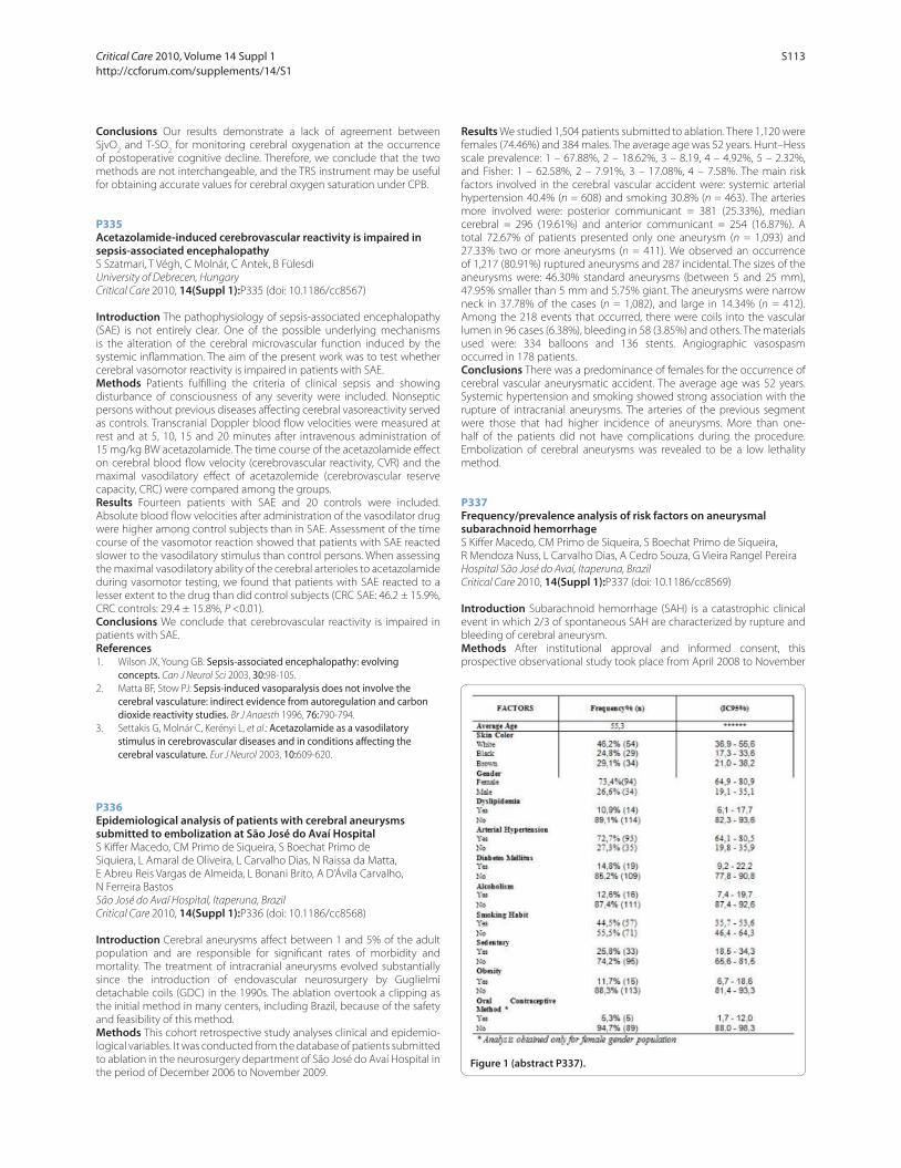
Conclusions Our results demonstrate a lack of agreement between
SjvO2 and T-SO
2 for monitoring cerebral oxygenation at the occurrence
of postoperative cognitive decline. Therefore, we conclude that the two
methods are not interchangeable, and the TRS instrument may be useful
for obtaining accurate values for cerebral oxygen saturation under CPB.
P335
Acetazolamide-induced cerebrovascular reactivity is impaired in sepsis-associated encephalopathyS Szatmari, T Végh, C Molnár, C Antek, B Fülesdi
University of Debrecen, Hungary
Critical Care 2010, 14(Suppl 1):P335 (doi: 10.1186/cc8567)
Introduction The pathophysiology of sepsis-associated encephalopathy
(SAE) is not entirely clear. One of the possible underlying mechanisms
is the alteration of the cerebral microvascular function induced by the
systemic infl ammation. The aim of the present work was to test whether
cerebral vasomotor reactivity is impaired in patients with SAE.
Methods Patients fulfi lling the criteria of clinical sepsis and showing
disturbance of consciousness of any severity were included. Nonseptic
persons without previous diseases aff ecting cerebral vasoreactivity served
as controls. Transcranial Doppler blood fl ow velocities were measured at
rest and at 5, 10, 15 and 20 minutes after intravenous administration of
15 mg/kg BW acetazolamide. The time course of the acetazolamide eff ect
on cerebral blood fl ow velocity (cerebrovascular reactivity, CVR) and the
maximal vasodilatory eff ect of acetazolemide (cerebrovascular reserve
capacity, CRC) were compared among the groups.
Results Fourteen patients with SAE and 20 controls were included.
Absolute blood fl ow velocities after administration of the vasodilator drug
were higher among control subjects than in SAE. Assessment of the time
course of the vasomotor reaction showed that patients with SAE reacted
slower to the vasodilatory stimulus than control persons. When assessing
the maximal vasodilatory ability of the cerebral arterioles to acetazolamide
during vasomotor testing, we found that patients with SAE reacted to a
lesser extent to the drug than did control subjects (CRC SAE: 46.2 ± 15.9%,
CRC controls: 29.4 ± 15.8%, P <0.01).
Conclusions We conclude that cerebrovascular reactivity is impaired in
patients with SAE.
References1. Wilson JX, Young GB: Sepsis-associated encephalopathy: evolving
concepts. Can J Neurol Sci 2003, 30:98-105.
2. Matta BF, Stow PJ: Sepsis-induced vasoparalysis does not involve the cerebral vasculature: indirect evidence from autoregulation and carbon dioxide reactivity studies. Br J Anaesth 1996, 76:790-794.
3. Settakis G, Molnár C, Kerényi L, et al.: Acetazolamide as a vasodilatory stimulus in cerebrovascular diseases and in conditions aff ecting the cerebral vasculature. Eur J Neurol 2003, 10:609-620.
P336
Epidemiological analysis of patients with cerebral aneurysms submitted to embolization at São José do Avaí HospitalS Kiff er Macedo, CM Primo de Siqueira, S Boechat Primo de
Siquiera, L Amaral de Oliveira, L Carvalho Dias, N Raissa da Matta,
E Abreu Reis Vargas de Almeida, L Bonani Brito, A D’Ávila Carvalho,
N Ferreira Bastos
São José do Avaí Hospital, Itaperuna, Brazil
Critical Care 2010, 14(Suppl 1):P336 (doi: 10.1186/cc8568)
Introduction Cerebral aneurysms aff ect between 1 and 5% of the adult
population and are responsible for signifi cant rates of morbidity and
mortality. The treatment of intracranial aneurysms evolved substantially
since the introduction of endovascular neurosurgery by Guglielmi
detachable coils (GDC) in the 1990s. The ablation overtook a clipping as
the initial method in many centers, including Brazil, because of the safety
and feasibility of this method.
Methods This cohort retrospective study analyses clinical and epidemio-
logical variables. It was conducted from the database of patients submitted
to ablation in the neurosurgery department of São José do Avaí Hospital in
the period of December 2006 to November 2009.
Results We studied 1,504 patients submitted to ablation. There 1,120 were
females (74.46%) and 384 males. The average age was 52 years. Hunt–Hess
scale prevalence: 1 – 67.88%, 2 – 18.62%, 3 – 8.19, 4 – 4.92%, 5 – 2.32%,
and Fisher: 1 – 62.58%, 2 – 7.91%, 3 – 17.08%, 4 – 7.58%. The main risk
factors involved in the cerebral vascular accident were: systemic arterial
hypertension 40.4% (n = 608) and smoking 30.8% (n = 463). The arteries
more involved were: posterior communicant = 381 (25.33%), median
cerebral = 296 (19.61%) and anterior communicant = 254 (16.87%). A
total 72.67% of patients presented only one aneurysm (n = 1,093) and
27.33% two or more aneurysms (n = 411). We observed an occurrence
of 1,217 (80.91%) ruptured aneurysms and 287 incidental. The sizes of the
aneurysms were: 46.30% standard aneurysms (between 5 and 25 mm),
47.95% smaller than 5 mm and 5.75% giant. The aneurysms were narrow
neck in 37.78% of the cases (n = 1,082), and large in 14.34% (n = 412).
Among the 218 events that occurred, there were coils into the vascular
lumen in 96 cases (6.38%), bleeding in 58 (3.85%) and others. The materials
used were: 334 balloons and 136 stents. Angiographic vasospasm
occurred in 178 patients.
Conclusions There was a predominance of females for the occurrence of
cerebral vascular aneurysmatic accident. The average age was 52 years.
Systemic hypertension and smoking showed strong association with the
rupture of intracranial aneurysms. The arteries of the previous segment
were those that had higher incidence of aneurysms. More than one-
half of the patients did not have complications during the procedure.
Embolization of cerebral aneurysms was revealed to be a low lethality
method.
P337
Frequency/prevalence analysis of risk factors on aneurysmal subarachnoid hemorrhageS Kiff er Macedo, CM Primo de Siqueira, S Boechat Primo de Siqueira,
R Mendoza Nuss, L Carvalho Dias, A Cedro Souza, G Vieira Rangel Pereira
Hospital São José do Avaí, Itaperuna, Brazil
Critical Care 2010, 14(Suppl 1):P337 (doi: 10.1186/cc8569)
Introduction Subarachnoid hemorrhage (SAH) is a catastrophic clinical
event in which 2/3 of spontaneous SAH are characterized by rupture and
bleeding of cerebral aneurysm.
Methods After institutional approval and informed consent, this
prospective observational study took place from April 2008 to November
Figure 1 (abstract P337).
Critical Care 2010, Volume 14 Suppl 1 http://ccforum.com/supplements/14/S1
S113
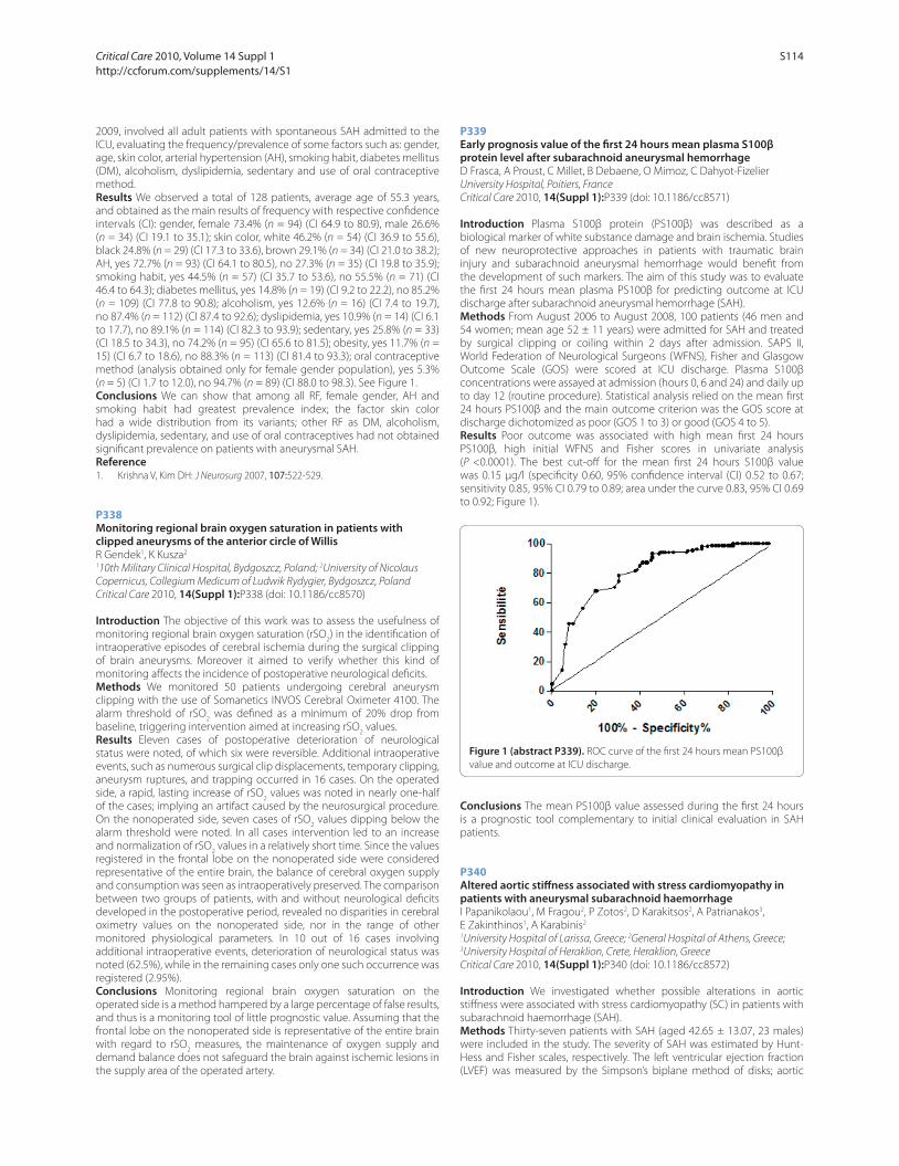
2009, involved all adult patients with spontaneous SAH admitted to the
ICU, evaluating the frequency/prevalence of some factors such as: gender,
age, skin color, arterial hypertension (AH), smoking habit, diabetes mellitus
(DM), alcoholism, dyslipidemia, sedentary and use of oral contraceptive
method.
Results We observed a total of 128 patients, average age of 55.3 years,
and obtained as the main results of frequency with respective confi dence
intervals (CI): gender, female 73.4% (n = 94) (CI 64.9 to 80.9), male 26.6%
(n = 34) (CI 19.1 to 35.1); skin color, white 46.2% (n = 54) (CI 36.9 to 55.6),
black 24.8% (n = 29) (CI 17.3 to 33.6), brown 29.1% (n = 34) (CI 21.0 to 38.2);
AH, yes 72.7% (n = 93) (CI 64.1 to 80.5), no 27.3% (n = 35) (CI 19.8 to 35.9);
smoking habit, yes 44.5% (n = 57) (CI 35.7 to 53.6), no 55.5% (n = 71) (CI
46.4 to 64.3); diabetes mellitus, yes 14.8% (n = 19) (CI 9.2 to 22.2), no 85.2%
(n = 109) (CI 77.8 to 90.8); alcoholism, yes 12.6% (n = 16) (CI 7.4 to 19.7),
no 87.4% (n = 112) (CI 87.4 to 92.6); dyslipidemia, yes 10.9% (n = 14) (CI 6.1
to 17.7), no 89.1% (n = 114) (CI 82.3 to 93.9); sedentary, yes 25.8% (n = 33)
(CI 18.5 to 34.3), no 74.2% (n = 95) (CI 65.6 to 81.5); obesity, yes 11.7% (n =
15) (CI 6.7 to 18.6), no 88.3% (n = 113) (CI 81.4 to 93.3); oral contraceptive
method (analysis obtained only for female gender population), yes 5.3%
(n = 5) (CI 1.7 to 12.0), no 94.7% (n = 89) (CI 88.0 to 98.3). See Figure 1.
Conclusions We can show that among all RF, female gender, AH and
smoking habit had greatest prevalence index; the factor skin color
had a wide distribution from its variants; other RF as DM, alcoholism,
dyslipidemia, sedentary, and use of oral contraceptives had not obtained
signifi cant prevalence on patients with aneurysmal SAH.
Reference1. Krishna V, Kim DH: J Neurosurg 2007, 107:522-529.
P338
Monitoring regional brain oxygen saturation in patients with clipped aneurysms of the anterior circle of WillisR Gendek1, K Kusza2
110th Military Clinical Hospital, Bydgoszcz, Poland; 2University of Nicolaus
Copernicus, Collegium Medicum of Ludwik Rydygier, Bydgoszcz, Poland
Critical Care 2010, 14(Suppl 1):P338 (doi: 10.1186/cc8570)
Introduction The objective of this work was to assess the usefulness of
monitoring regional brain oxygen saturation (rSO2) in the identifi cation of
intraoperative episodes of cerebral ischemia during the surgical clipping
of brain aneurysms. Moreover it aimed to verify whether this kind of
monitoring aff ects the incidence of postoperative neurological defi cits.
Methods We monitored 50 patients undergoing cerebral aneurysm
clipping with the use of Somanetics INVOS Cerebral Oximeter 4100. The
alarm threshold of rSO2 was defi ned as a minimum of 20% drop from
baseline, triggering intervention aimed at increasing rSO2 values.
Results Eleven cases of postoperative deterioration of neurological
status were noted, of which six were reversible. Additional intraoperative
events, such as numerous surgical clip displacements, temporary clipping,
aneurysm ruptures, and trapping occurred in 16 cases. On the operated
side, a rapid, lasting increase of rSO2 values was noted in nearly one-half
of the cases; implying an artifact caused by the neurosurgical procedure.
On the nonoperated side, seven cases of rSO2 values dipping below the
alarm threshold were noted. In all cases intervention led to an increase
and normalization of rSO2 values in a relatively short time. Since the values
registered in the frontal lobe on the nonoperated side were considered
representative of the entire brain, the balance of cerebral oxygen supply
and consumption was seen as intraoperatively preserved. The comparison
between two groups of patients, with and without neurological defi cits
developed in the postoperative period, revealed no disparities in cerebral
oximetry values on the nonoperated side, nor in the range of other
monitored physiological parameters. In 10 out of 16 cases involving
additional intraoperative events, deterioration of neurological status was
noted (62.5%), while in the remaining cases only one such occurrence was
registered (2.95%).
Conclusions Monitoring regional brain oxygen saturation on the
operated side is a method hampered by a large percentage of false results,
and thus is a monitoring tool of little prognostic value. Assuming that the
frontal lobe on the nonoperated side is representative of the entire brain
with regard to rSO2 measures, the maintenance of oxygen supply and
demand balance does not safeguard the brain against ischemic lesions in
the supply area of the operated artery.
P339
Early prognosis value of the fi rst 24 hours mean plasma S100β protein level after subarachnoid aneurysmal hemorrhageD Frasca, A Proust, C Millet, B Debaene, O Mimoz, C Dahyot-Fizelier
University Hospital, Poitiers, France
Critical Care 2010, 14(Suppl 1):P339 (doi: 10.1186/cc8571)
Introduction Plasma S100β protein (PS100β) was described as a
biological marker of white substance damage and brain ischemia. Studies
of new neuroprotective approaches in patients with traumatic brain
injury and subarachnoid aneurysmal hemorrhage would benefi t from
the development of such markers. The aim of this study was to evaluate
the fi rst 24 hours mean plasma PS100β for predicting outcome at ICU
discharge after subarachnoid aneurysmal hemorrhage (SAH).
Methods From August 2006 to August 2008, 100 patients (46 men and
54 women; mean age 52 ± 11 years) were admitted for SAH and treated
by surgical clipping or coiling within 2 days after admission. SAPS II,
World Federation of Neurological Surgeons (WFNS), Fisher and Glasgow
Outcome Scale (GOS) were scored at ICU discharge. Plasma S100β
concentrations were assayed at admission (hours 0, 6 and 24) and daily up
to day 12 (routine procedure). Statistical analysis relied on the mean fi rst
24 hours PS100β and the main outcome criterion was the GOS score at
discharge dichotomized as poor (GOS 1 to 3) or good (GOS 4 to 5).
Results Poor outcome was associated with high mean fi rst 24 hours
PS100β, high initial WFNS and Fisher scores in univariate analysis
(P <0.0001). The best cut-off for the mean fi rst 24 hours S100β value
was 0.15 μg/l (specifi city 0.60, 95% confi dence interval (CI) 0.52 to 0.67;
sensitivity 0.85, 95% CI 0.79 to 0.89; area under the curve 0.83, 95% CI 0.69
to 0.92; Figure 1).
Conclusions The mean PS100β value assessed during the fi rst 24 hours
is a prognostic tool complementary to initial clinical evaluation in SAH
patients.
P340
Altered aortic stiff ness associated with stress cardiomyopathy in patients with aneurysmal subarachnoid haemorrhageI Papanikolaou1, M Fragou2, P Zotos2, D Karakitsos2, A Patrianakos3,
E Zakinthinos1, A Karabinis2
1University Hospital of Larissa, Greece; 2General Hospital of Athens, Greece; 3University Hospital of Heraklion, Crete, Heraklion, Greece
Critical Care 2010, 14(Suppl 1):P340 (doi: 10.1186/cc8572)
Introduction We investigated whether possible alterations in aortic
stiff ness were associated with stress cardiomyopathy (SC) in patients with
subarachnoid haemorrhage (SAH).
Methods Thirty-seven patients with SAH (aged 42.65 ± 13.07, 23 males)
were included in the study. The severity of SAH was estimated by Hunt-
Hess and Fisher scales, respectively. The left ventricular ejection fraction
(LVEF) was measured by the Simpson’s biplane method of disks; aortic
Figure 1 (abstract P339). ROC curve of the fi rst 24 hours mean PS100β
value and outcome at ICU discharge.
Critical Care 2010, Volume 14 Suppl 1 http://ccforum.com/supplements/14/S1
S114
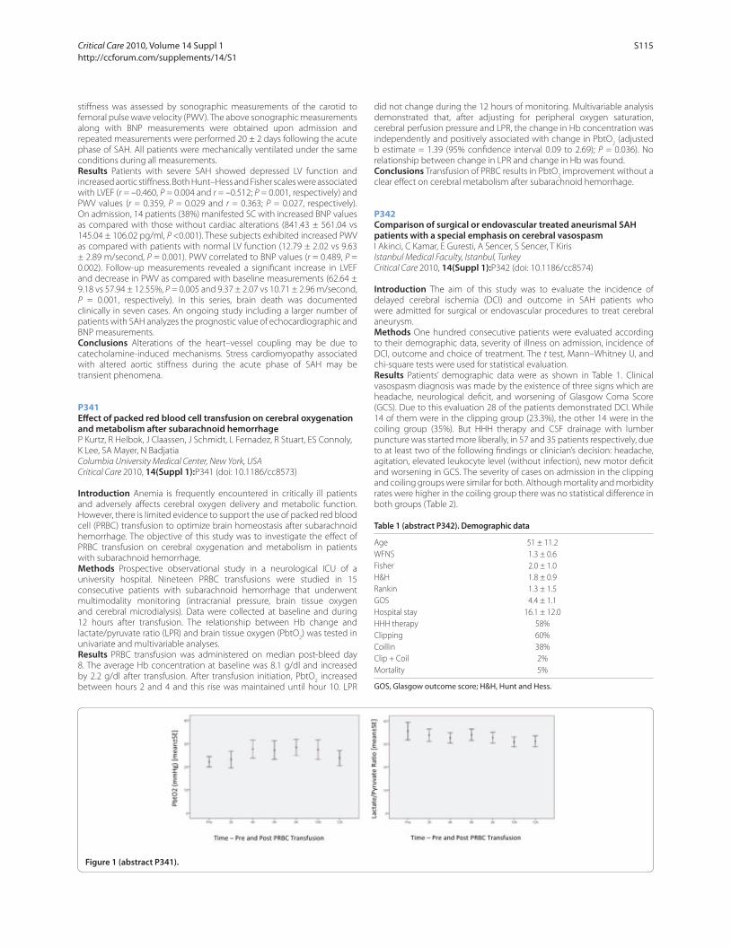
stiff ness was assessed by sonographic measurements of the carotid to
femoral pulse wave velocity (PWV). The above sonographic measurements
along with BNP measurements were obtained upon admission and
repeated measurements were performed 20 ± 2 days following the acute
phase of SAH. All patients were mechanically ventilated under the same
conditions during all measurements.
Results Patients with severe SAH showed depressed LV function and
increased aortic stiff ness. Both Hunt–Hess and Fisher scales were associated
with LVEF (r = –0.460, P = 0.004 and r = –0.512; P = 0.001, respectively) and
PWV values (r = 0.359, P = 0.029 and r = 0.363; P = 0.027, respectively).
On admission, 14 patients (38%) manifested SC with increased BNP values
as compared with those without cardiac alterations (841.43 ± 561.04 vs
145.04 ± 106.02 pg/ml, P <0.001). These subjects exhibited increased PWV
as compared with patients with normal LV function (12.79 ± 2.02 vs 9.63
± 2.89 m/second, P = 0.001). PWV correlated to BNP values (r = 0.489, P =
0.002). Follow-up measurements revealed a signifi cant increase in LVEF
and decrease in PWV as compared with baseline measurements (62.64 ±
9.18 vs 57.94 ± 12.55%, P = 0.005 and 9.37 ± 2.07 vs 10.71 ± 2.96 m/second,
P = 0.001, respectively). In this series, brain death was documented
clinically in seven cases. An ongoing study including a larger number of
patients with SAH analyzes the prognostic value of echocardiographic and
BNP measurements.
Conclusions Alterations of the heart–vessel coupling may be due to
catecholamine-induced mechanisms. Stress cardiomyopathy associated
with altered aortic stiff ness during the acute phase of SAH may be
transient phenomena.
P341
Eff ect of packed red blood cell transfusion on cerebral oxygenation and metabolism after subarachnoid hemorrhageP Kurtz, R Helbok, J Claassen, J Schmidt, L Fernadez, R Stuart, ES Connoly,
K Lee, SA Mayer, N Badjatia
Columbia University Medical Center, New York, USA
Critical Care 2010, 14(Suppl 1):P341 (doi: 10.1186/cc8573)
Introduction Anemia is frequently encountered in critically ill patients
and adversely aff ects cerebral oxygen delivery and metabolic function.
However, there is limited evidence to support the use of packed red blood
cell (PRBC) transfusion to optimize brain homeostasis after subarachnoid
hemorrhage. The objective of this study was to investigate the eff ect of
PRBC transfusion on cerebral oxygenation and metabolism in patients
with subarachnoid hemorrhage.
Methods Prospective observational study in a neurological ICU of a
university hospital. Nineteen PRBC transfusions were studied in 15
consecutive patients with subarachnoid hemorrhage that underwent
multimodality monitoring (intracranial pressure, brain tissue oxygen
and cerebral microdialysis). Data were collected at baseline and during
12 hours after transfusion. The relationship between Hb change and
lactate/pyruvate ratio (LPR) and brain tissue oxygen (PbtO2) was tested in
univariate and multivariable analyses.
Results PRBC transfusion was administered on median post-bleed day
8. The average Hb concentration at baseline was 8.1 g/dl and increased
by 2.2 g/dl after transfusion. After transfusion initiation, PbtO2 increased
between hours 2 and 4 and this rise was maintained until hour 10. LPR
did not change during the 12 hours of monitoring. Multivariable analysis
demonstrated that, after adjusting for peripheral oxygen saturation,
cerebral perfusion pressure and LPR, the change in Hb concentration was
independently and positively associated with change in PbtO2 (adjusted
b estimate = 1.39 (95% confi dence interval 0.09 to 2.69); P = 0.036). No
relationship between change in LPR and change in Hb was found.
Conclusions Transfusion of PRBC results in PbtO2 improvement without a
clear eff ect on cerebral metabolism after subarachnoid hemorrhage.
P342
Comparison of surgical or endovascular treated aneurismal SAH patients with a special emphasis on cerebral vasospasmI Akinci, C Kamar, E Guresti, A Sencer, S Sencer, T Kiris
Istanbul Medical Faculty, Istanbul, Turkey
Critical Care 2010, 14(Suppl 1):P342 (doi: 10.1186/cc8574)
Introduction The aim of this study was to evaluate the incidence of
delayed cerebral ischemia (DCI) and outcome in SAH patients who
were admitted for surgical or endovascular procedures to treat cerebral
aneurysm.
Methods One hundred consecutive patients were evaluated according
to their demographic data, severity of illness on admission, incidence of
DCI, outcome and choice of treatment. The t test, Mann–Whitney U, and
chi-square tests were used for statistical evaluation.
Results Patients’ demographic data were as shown in Table 1. Clinical
vasospasm diagnosis was made by the existence of three signs which are
headache, neurological defi cit, and worsening of Glasgow Coma Score
(GCS). Due to this evaluation 28 of the patients demonstrated DCI. While
14 of them were in the clipping group (23.3%), the other 14 were in the
coiling group (35%). But HHH therapy and CSF drainage with lumber
puncture was started more liberally, in 57 and 35 patients respectively, due
to at least two of the following fi ndings or clinician’s decision: headache,
agitation, elevated leukocyte level (without infection), new motor defi cit
and worsening in GCS. The severity of cases on admission in the clipping
and coiling groups were similar for both. Although mortality and morbidity
rates were higher in the coiling group there was no statistical diff erence in
both groups (Table 2).
Table 1 (abstract P342). Demographic data
Age 51 ± 11.2
WFNS 1.3 ± 0.6
Fisher 2.0 ± 1.0
H&H 1.8 ± 0.9
Rankin 1.3 ± 1.5
GOS 4.4 ± 1.1
Hospital stay 16.1 ± 12.0
HHH therapy 58%
Clipping 60%
Coillin 38%
Clip + Coil 2%
Mortality 5%
GOS, Glasgow outcome score; H&H, Hunt and Hess.
Figure 1 (abstract P341).
Critical Care 2010, Volume 14 Suppl 1 http://ccforum.com/supplements/14/S1
S115
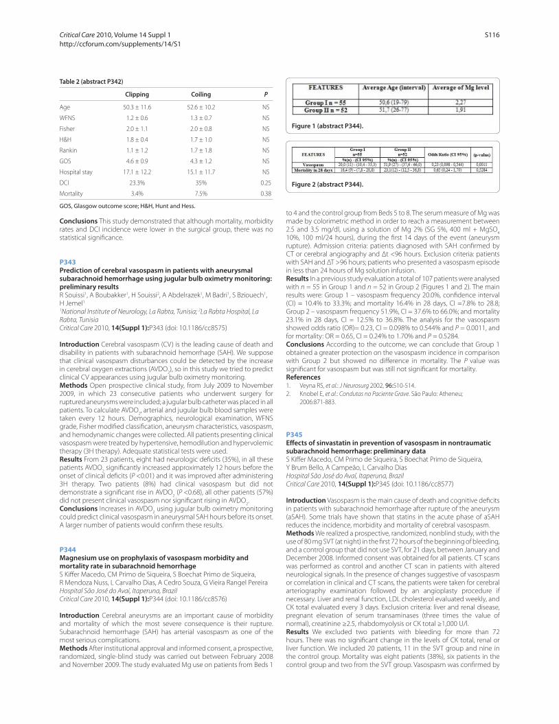
Table 2 (abstract P342)
Clipping Coiling P
Age 50.3 ± 11.6 52.6 ± 10.2 NS
WFNS 1.2 ± 0.6 1.3 ± 0.7 NS
Fisher 2.0 ± 1.1 2.0 ± 0.8 NS
H&H 1.8 ± 0.4 1.7 ± 1.0 NS
Rankin 1.1 ± 1.2 1.7 ± 1.8 NS
GOS 4.6 ± 0.9 4.3 ± 1.2 NS
Hospital stay 17.1 ± 12.2 15.1 ± 11.7 NS
DCI 23.3% 35% 0.25
Mortality 3.4% 7.5% 0.38
GOS, Glasgow outcome score; H&H, Hunt and Hess.
Conclusions This study demonstrated that although mortality, morbidity
rates and DCI incidence were lower in the surgical group, there was no
statistical signifi cance.
P343
Prediction of cerebral vasospasm in patients with aneurysmal subarachnoid hemorrhage using jugular bulb oximetry monitoring: preliminary resultsR Souissi1, A Boubakker1, H Souissi2, A Abdelrazek1, M Badri1, S Bziouech1,
H Jemel1
1National Institute of Neurology, La Rabta, Tunisia; 2La Rabta Hospital, La
Rabta, Tunisia
Critical Care 2010, 14(Suppl 1):P343 (doi: 10.1186/cc8575)
Introduction Cerebral vasospasm (CV) is the leading cause of death and
disability in patients with subarachnoid hemorrhage (SAH). We suppose
that clinical vasospasm disturbances could be detected by the increase
in cerebral oxygen extractions (AVDO2), so in this study we tried to predict
clinical CV appearances using jugular bulb oximetry monitoring.
Methods Open prospective clinical study, from July 2009 to November
2009, in which 23 consecutive patients who underwent surgery for
ruptured aneurysms were included; a jugular bulb catheter was placed in all
patients. To calculate AVDO2, arterial and jugular bulb blood samples were
taken every 12 hours. Demographics, neurological examination, WFNS
grade, Fisher modifi ed classifi cation, aneurysm characteristics, vasospasm,
and hemodynamic changes were collected. All patients presenting clinical
vasospasm were treated by hypertensive, hemodilution and hypervolemic
therapy (3H therapy). Adequate statistical tests were used.
Results From 23 patients, eight had neurologic defi cits (35%), in all these
patients AVDO2 signifi cantly increased approximately 12 hours before the
onset of clinical defi cits (P <0.01) and it was improved after administering
3H therapy. Two patients (8%) had clinical vasospasm but did not
demonstrate a signifi cant rise in AVDO2 (P <0.68), all other patients (57%)
did not present clinical vasospasm nor signifi cant rising in AVDO2.
Conclusions Increases in AVDO2 using jugular bulb oximetry monitoring
could predict clinical vasospasm in aneurysmal SAH hours before its onset.
A larger number of patients would confi rm these results.
P344
Magnesium use on prophylaxis of vasospasm morbidity and mortality rate in subarachnoid hemorrhageS Kiff er Macedo, CM Primo de Siqueira, S Boechat Primo de Siqueira,
R Mendoza Nuss, L Carvalho Dias, A Cedro Souza, G Vieira Rangel Pereira
Hospital São José do Avaí, Itaperuna, Brazil
Critical Care 2010, 14(Suppl 1):P344 (doi: 10.1186/cc8576)
Introduction Cerebral aneurysms are an important cause of morbidity
and mortality of which the most severe consequence is their rupture.
Subarachnoid hemorrhage (SAH) has arterial vasospasm as one of the
most serious complications.
Methods After institutional approval and informed consent, a prospective,
randomized, single-blind study was carried out between February 2008
and November 2009. The study evaluated Mg use on patients from Beds 1
to 4 and the control group from Beds 5 to 8. The serum measure of Mg was
made by colorimetric method in order to reach a measurement between
2.5 and 3.5 mg/dl, using a solution of Mg 2% (SG 5%, 400 ml + MgSO4
10%, 100 ml/24 hours), during the fi rst 14 days of the event (aneurysm
rupture). Admission criteria: patients diagnosed with SAH confi rmed by
CT or cerebral angiography and Δt <96 hours. Exclusion criteria: patients
with SAH and ΔT >96 hours; patients who presented a vasospasm episode
in less than 24 hours of Mg solution infusion.
Results In a previous study evaluation a total of 107 patients were analysed
with n = 55 in Group 1 and n = 52 in Group 2 (Figures 1 and 2). The main
results were: Group 1 – vasospasm frequency 20.0%, confi dence interval
(CI) = 10.4% to 33.3%; and mortality 16.4% in 28 days, CI =7.8% to 28.8;
Group 2 – vasospasm frequency 51.9%, CI = 37.6% to 66.0%; and mortality
23.1% in 28 days, CI = 12.5% to 36.8%. The analysis for the vasospasm
showed odds ratio (OR)= 0.23, CI = 0.098% to 0.544% and P = 0.0011, and
for mortality: OR = 0.65, CI = 0.24% to 1.70% and P = 0.5284.
Conclusions According to the outcome, we can conclude that Group 1
obtained a greater protection on the vasospasm incidence in comparison
with Group 2 but showed no diff erence in mortality. The P value was
signifi cant for vasospasm but was still not signifi cant for mortality.
References1. Veyna RS, et al.: J Neurosurg 2002, 96:510-514.
2. Knobel E, et al.: Condutas no Paciente Grave. São Paulo: Atheneu;
2006:871-883.
P345
Eff ects of sinvastatin in prevention of vasospasm in nontraumatic subarachnoid hemorrhage: preliminary dataS Kiff er Macedo, CM Primo de Siqueira, S Boechat Primo de Siqueira,
Y Brum Bello, A Campeão, L Carvalho Dias
Hospital São José do Avaí, Itaperuna, Brazil
Critical Care 2010, 14(Suppl 1):P345 (doi: 10.1186/cc8577)
Introduction Vasospasm is the main cause of death and cognitive defi cits
in patients with subarachnoid hemorrhage after rupture of the aneurysm
(aSAH). Some trials have shown that statins in the acute phase of aSAH
reduces the incidence, morbidity and mortality of cerebral vasospasm.
Methods We realized a prospective, randomized, nonblind study, with the
use of 80 mg SVT (at night) in the fi rst 72 hours of the beginning of bleeding,
and a control group that did not use SVT, for 21 days, between January and
December 2008. Informed consent was obtained for all patients. CT scans
was performed as control and another CT scan in patients with altered
neurological signals. In the presence of changes suggestive of vasospasm
or correlation in clinical and CT scans, the patients were taken for cerebral
arteriography examination followed by an angioplasty procedure if
necessary. Liver and renal function, LDL cholesterol evaluated weekly, and
CK total evaluated every 3 days. Exclusion criteria: liver and renal disease,
pregnant elevation of serum transaminases (three times the value of
normal), creatinine ≥2.5, rhabdomyolysis or CK total ≥1,000 U/l.
Results We excluded two patients with bleeding for more than 72
hours. There was no signifi cant change in the levels of CK total, renal or
liver function. We included 20 patients, 11 in the SVT group and nine in
the control group. Mortality was eight patients (38%), six patients in the
control group and two from the SVT group. Vasospasm was confi rmed by
Figure 1 (abstract P344).
Figure 2 (abstract P344).
Critical Care 2010, Volume 14 Suppl 1 http://ccforum.com/supplements/14/S1
S116

cerebral arteriography examination in four patients in the control group
and one patient in the SVT group. All patients who died had Fisher scale IV.
Conclusions SVT at a dose of 80 mg was eff ective in reducing the
mortality (18.1% against 66%) compared with the group that did not use
SVT, and also decreased the incidence of cerebral vasospasm despite
higher APACHE II score in the group that used SVT (14.3 vs 10.7). There
was less morbidity in the SVT group with an average of scale of Glasgow
3.25 vs 2.1.
Reference1. Lynch JR, Wang H, et al.: Simvastatin reduces vasospasm after aneurysmal
subarachnoid hemorrhage: results of a pilot randomized clinical trial. Stroke 2005, 36:2024-2026.
P346
Addition of new criteria to Sequential Organ Failure Assessment for patients with subarachnoid hemorrhageS Kiff er Macedo, N Ferreira Bastos, J Flores Paz, T De Carvalho Pessamilio,
R Barbosa Castro, M Rodrigues Lacerda
Hospital São José do Avaí, Itaperuna, Brazil
Critical Care 2010, 14(Suppl 1):P346 (doi: 10.1186/cc8578)
Introduction Sequential Organ Failure Assessment (SOFA) was originally
created for sepsis, but their quality is now used in other medical
conditions. Therefore, we add some criteria to the index to assess patients
with subarachnoid hemorrhage.
Methods Informed consent for each patient/family; APACHE II (criteria for
admission) and SOFA weekly, serum glucose, lactate, calcium, sodium and
magnesium; and measurement of axillary temperature and diuresis hourly
as the additional prognostic index SOFA. The study enrolled 103 patients
diagnosed with SAH, confi rmed by computed tomography. These patients
were divided into two groups according to their development in the ICU:
Group I – patients who had good evolution (out of ICU), and Group II –
patients who progressed to death in the ICU.
Results Among 103 patients, 74 (71.84%) were female and 29 (28.16%)
were male. The APACHE II for admission varied from 2 to 34, with an
average of 15.5. The spent time maximum in the ICU was 49 days (two
patients). Group I had 64 patients (62.14%) and the 39 remaining patients
(37.86%) were classifi ed as Group II. See Figures 1 and 2.
Conclusions We can conclude that the group with patients with SAH is
predominantly female (74:29). The APACHE II in Group I was 10.9, while
Group II was 17.9. Regarding the criteria used to assess patients with SAH
was observed that the only criterion which showed statistical signifi cance
in the prediction of death was the serum sodium (P = 0.002). The other
criteria evaluated did not have statistical signifi cance in predicting the
prognosis of patients.
Reference1. Knobel E: Condutas no Paciente Grave. Volume 1. 3rd edition. São Paulo:
Editora Atheneu; 2006:871-883.
P347
Cardiac output augmentation with fl uid resuscitation improves brain tissue oxygenation after severe brain injuryR Helbok, P Kurtz, J Claassen, J Schmidt, L Fernadez, R Stuart, ES Connoly,
N Badjatia, SA Mayer, K Lee
Columbia University Medical Center, New York, USA
Critical Care 2010, 14(Suppl 1):P347 (doi: 10.1186/cc8579)
Introduction The objective of this study was to investigate the relationship
between cardiac output response to a fl uid challenge and changes in
brain tissue oxygen pressure (PbtO2) in patients with severe brain injury.
Methods Prospective observational study conducted in a neurological ICU
in a university hospital. Seventy-eight fl uid challenges were administered
to 17 consecutive comatose patients that underwent multimodality
monitoring of cardiac output, intracranial pressure (ICP), and PbtO2. The
relationship between cardiac output and PbtO2 was analyzed with logistic
regression utilizing GEE with an exchangeable correlation structure.
Results Of the 78 fl uid boluses analyzed, 34 (44%) resulted in a ≥10%
increase in cardiac output. Median absolute (+5.4 vs +0.7 mmHg) and
percentage (20% vs 3%) changes in PbtO2 were greater in cardiac output
responders than in nonresponders within 30 minutes after the end of the
fl uid bolus infusion. In a multivariable model, a cardiac output response
was independently associated with PbtO2 response (adjusted odds ratio
15.4, 95% CI 1.9 to 122.0, P = 0.01) after adjusting for mean arterial pressure,
Figure 1 (abstract P346).
Figure 2 (abstract P346).
Figure 1 (abstract P347).
Figure 2 (abstract P347).
Critical Care 2010, Volume 14 Suppl 1 http://ccforum.com/supplements/14/S1
S117

intracranial pressure and end-tidal CO2. Stroke volume variation showed
a good ability to predict cardiac output response with an area under the
ROC curve of 0.85 with a best cutoff value of 8%. See Figures 1 and 2.
Conclusions Bolus fl uid resuscitation resulting in augmentation of cardiac
output can improve cerebral oxygenation after severe brain injury.
P348
Multicentre experience of using ICM+ for investigations of cerebrovascular dynamics with near-infrared spectroscopyP Smielewski1, M Czosnyka1, C Zweifel1, K Brady2, C Hogue2, L Steiner3,
P Hutchinson1, D Mennon1, J Pickard1
1University of Cambridge, UK; 2Johns Hopkins Hospital, Baltimore, MD, USA; 3University Hospital, Basel, Switzerland
Critical Care 2010, 14(Suppl 1):P348 (doi: 10.1186/cc8580)
Introduction ICM+ software encapsulates our 20 years’ experience
in brain monitoring. It collects data from a variety of bedside monitors
and produces time trends of parameters defi ned using confi gurable
mathematical formulae. To date it is being used in nearly 40 clinical
research centres worldwide. We present its application for continuous
monitoring of cerebral autoregulation using near-infrared spectroscopy
(NIRS).
Methods Data from multiple bedside monitors are processed by ICM+
in real time using a large selection of signal processing methods. These
include various time and frequency domain analysis functions as well as
fully customisable digital fi lters. The fi nal results are displayed in a variety
of ways including simple time trends, as well as time window based
histograms, cross histograms, correlations, and so forth. All this allows
complex information from bedside monitors to be summarized in a
concise fashion and presented to medical and nursing staff in a simple way
that alerts them to the development of various pathological processes.
Results One hundred and fi fty patients monitored continuously with
NIRS, arterial blood pressure (ABP) and intracranial pressure (ICP), where
available, were included in this study. There were 40 severely head-
injured adult patients, 27 SAH patients (NCCU, Cambridge); 60 patients
undergoing cardiopulmonary bypass (John Hopkins Hospital, Baltimore)
and 23 patients with sepsis (University Hospital, Basel). In addition,
MCA fl ow velocity (FV) was monitored intermittently using transcranial
Doppler. FV-derived and ICP-derived pressure reactivity indices (PRx, Mx),
as well as NIRS-derived reactivity indices (Cox, Tox, Thx) were calculated
and showed signifi cant correlation with each other in all cohorts. Error-
bar charts showing reactivity index PRx versus CPP (optimal CPP chart)
as well as similar curves for NIRS indices versus CPP and ABP were also
demonstrated.
Conclusions ICM+ software is proving to be a very useful tool for enhancing
the battery of available means for monitoring cerebral vasoreactivity and
potentially facilitating autoregulation guided therapy. Complexity of data
analysis is also hidden inside loadable profi les, thus allowing investigators
to take full advantage of validated protocols including advanced
processing formulas.
P349
Cerebral oxygen saturation and cerebral blood fl ow are relatively stable during single-lung ventilation, if normocapnia is maintainedT Végh, S Szatmári, M Juhász, I László, I Takács, B Fülesdi
University of Debrecen, Hungary
Critical Care 2010, 14(Suppl 1):P349 (doi: 10.1186/cc8581)
Introduction Previously a report has suggested that administration of a
lung-protective strategy for one-lung ventilation (OLV) results in oxygen
desaturation of the brain parenchyma. The aim of the present work was
to test whether the maintenance of normocapnia during a protective OLV
strategy results in alteration of cerebral blood fl ow and cerebral oxygen
saturation.
Methods Data were obtained from 15 patients undergoing thoracic
surgery and necessitating OLV of more than 1 hour. Cerebral oxygen
saturation (Satcereb) was continuously monitored by the INVOS 5100C
Cerebral Oxymeter System along with measurement of cerebral blood
fl ow velocity (MCAV) by transcranial Doppler sonography. Arterial blood
samples were taken for blood gas analysis in the awake state, in the supine
position after induction during DLV, in the lateral decubitus position
during DLV and every 15 minutes during OLV.
Results Satcereb increased signifi cantly when DLV was started with FiO2
1.0 and remained stable during the course of the study. When ventilation
was changed from DLV to OLV, no signifi cant change was observed. A
signifi cant decrease of cerebral oxygen saturation was found compared
with the value observed during DLV in the lateral decubitus at the time
point of 60 minutes after the start of OLV. No signifi cant changes in the
MCAV were observed throughout the course of the thoracic surgical
procedure.
Conclusions OLV does not result in clinically relevant decreases in
cerebral blood fl ow and cerebral oxygen saturation during application of
lung-protective ventilation if normocapnia is maintained.
P350
Factors aff ecting the transient hyperaemic response test of the cerebral autoregulationMS Sitina, V Cerny
University Hospital, Hradec Kralove, Czech Republic
Critical Care 2010, 14(Suppl 1):P350 (doi: 10.1186/cc8582)
Introduction The transient compression of the internal carotid artery
(ICA) results in dilatation of cerebral arterioles by eff ect of autoregulation.
In the THR test, the increase in middle cerebral artery (MCA) blood fl ow
immediately after release of transient compression of the ICA is measured,
which is proportional to cerebral autoregulation. The aim of the study was
to determine the signifi cance of several factors of the test – blood fl ow
inertia (acceleration), dimensions (length and radius) of ICA, patency of the
Circle of Willis (WC), and asynchrony between release of ICA compression
and arterial pressure pulsations (phase shift).
Methods We constructed a mathematical model of the elements included
in the THR test and simulated, using various parameters, blood fl ow in
cerebral vessels and the increase in fl ow velocity in the MCA after release of
ICA compression. We simulated two extreme situations – a perfectly patent
(all aff erent and eff erent arteries meet in one point) and a completely
isolated Circle of Willis (ICA bifurcates into only the anterior and middle
cerebral arteries). The series of ordinary diff erential equations describing
the THR test was solved by MATLAB R2008a. The solution assumed an
absence of autoregulation (eff ect of studied factors is independent of
autoregulation) and the rigidity of all vessels.
Results The eff ects of acceleration (inertia), dimensions of ICA and patency
of WC are negligible. The eff ect of phase shift (Figure 1) can signifi cantly
decrease the immediate post-release MCA fl ow, which can entirely nullify
the increase in post-release MCA fl ow caused by autoregulation.
Conclusions The eff ect of phase shift can signifi cantly alter the results of
the THR test. For practical purposes we recommend calculation of THR test
parameters from the second peak of post-release MCA fl ow, or using the
highest post-release MCA fl ow from several THR tests.
Figure 1 (abstract P350). Dependence of THRR on phase shift.
Critical Care 2010, Volume 14 Suppl 1 http://ccforum.com/supplements/14/S1
S118

P351
Infection associated with the external ventricular derivation catheter: preliminary dataS Kiff er Macedo, CM Primo de Siqueira, S Boechat Primo de Siqueira,
B Almeida Simão Da Mata, S Ferrari Salles, L Reggiani de Almeida,
F Oliva Battisti
Hospital São José do Avaí, Itaperuna, Brazil
Critical Care 2010, 14(Suppl 1):P351 (doi: 10.1186/cc8583)
Introduction The purpose of this study was to evaluate the total drainage
time, the risk of infection associated with external ventricular derivation
(EVD) and the most prevalent infective agent during the use of EVD
drainage in patients on the ICU.
Methods During 15 months (8 August 2008 to 28 October 2009), all
patients on the neurovascular ICU who had an EVD inserted were studied
prospectively. Each 3 days we analysed cerebral spinal fl uid (CSF) samples
for cell count, biochemistry and sputum. CSF cultures were evaluated
three times a week combined with an antibiogram. Not included were
the cultures that persisted with the same result (agent), considering only
a new agent until negativity of the culture and clinical resolution. We
excluded patients admitted with EVD from another hospital.
Results Were included 46 patients, in which were obtained a total number
of 66 EVDs. Thirty-fi ve EVDs (53,03%) had associated ICP monitoring.
A total 65.21% of patients were female. The mortality of patients was
36.95% (17 deaths). Among infected catheters, infection was confi rmed
in approximately 7.1 days after the EVD was installed. However, the
catheters discharged with negative culture or without growth of a new
agent remained approximately 6.15 days on the brain ventricle of patients.
Registered were 20 positive bacteriological cultures of CSF (30.30%)
among the total EVDs. The most prevalent organisms were Acinetobacter
baumanii on nine cultures (45%), Pseudomonas aeruginosa on six cultures
(30%), Serratia rubidaea on one culture (5%), Streptococcus oralis on one
culture (5%), Klebsiella ornithinolytica on one culture (5%), Estafi lococcus
coagulase negative on one culture(5%), Stenotrophonas maltophylia on
one culture (5%).
Conclusions Some previous studies show a substitution from positive
Gram agents to negative Gram agents on infections associated with EVD.
The study has shown a prevalence of P. aeruginosa and A. baumanii on
infection associated with the EVD catheter, which are also more prevalent
in the ICU environment. This study reinforces that, in case of suspected CNS
infection associated with catheter EVD (meningitis, ventriculitis, or abscess),
we can start empirical antimicrobial coverage for Gram-negative infections,
including intraventricular administration (intrathecal) as Polymixin B.
P352
fMRI default mode connectivity in vegetative state and locked-in syndrome patientsA Soddu1, A Vanhaudenhuyse1, M Boly1, MA Bruno1, L Tshibanda2,
C Phillips1, M Stanziano3, S Ovadia Caro4, Y Nir5, P Maquet1, M Papa3,
R Malach4, Q Noirhomme1, S Laureys1
1Coma Science Group, Liege, Belgium; 2Neurology Department, CHU Sart
Tilman Hospital, University of Liège, Belgium; 3Pubblica Clinica e Preventiva,
Second University of Naples, Italy; 4Department of Neurobiology, Weizmann
Institute of Science, Rehovot, Israel; 5Department of Psychiatry, University of
Wisconsin, Madison, WI, USA
Critical Care 2010, 14(Suppl 1):P352 (doi: 10.1186/cc8584)
Introduction The aim of this study is to assess fMRI resting-state cerebral
connectivity in vegetative state and locked-in syndrome patients by means
of a user-independent method. Resting baseline (or default mode) activity
is thought to be related to awareness of the internal world (that is, mind
wandering, and so forth) and encompasses posterior cingulate/precuneal,
anterior cingulate/mesiofrontal and posterior lateral parietal cortices.
Methods We here present a novel clinical application for a user-
independent default mode network analysis. Resting state data were
acquired for seven vegetative state patients (age range 16 to 87 years), one
locked-in syndrome patient (aged 24 years) and seven healthy subjects
(25 to 65 years). Patients’ diagnosis was based on Coma Recovery Scale
assessment prior and following scanning. Data were pre-processed and
analyzed using independent component analysis as implemented in
Brain Voyager. Connectivity studies employed 13 target regions of interest
defi ned on an average default mode map calculated from an independent
dataset of 20 controls (21 to 60 years). Resting state connectivity was
assessed by calculating the number of functional connections within
the default mode map for each subject. Next, Student t tests compared
patients with controls at the group level (P <0.05).
Results Compared with controls, vegetative patients showed a lower
total number of edges (that is, connections; 24 ± 4, range 19 to 31 and
13 ± 3, range 8 to 15, P <0.001) and less functional connections with the
precuneus (6 ± 1 and 2 ± 2; P <0.001). The default mode network shows
a reduced connectivity in vegetative patients as compared with controls
mainly between the precuneus and medial prefrontal cortex ventral.
Finally, for the locked-in syndrome patient the connectivity graph counted
21 total number of eff ective connections and fi ve eff ective connections
with the precuneus, which was not signifi cantly diff erent from healthy
controls.
Conclusions The connectivity study showed no overlap between the
number of eff ective connections or edges in vegetative state patients
and in controls. Connectivity graph analysis identifi ed anterior–posterior
midline disconnections in the vegetative state, in line with previous studies
emphasizing the critical role of the precuneus/PCC and mesiofrontal
cortices in the emergence of conscious awareness.
P353
Incidence of cerebral salt wasting syndrome: comparison in subarachnoid haemorrhagic compared with head traumaFD Di Sacco1, GB Bernardeschi1, AD Demi1, CC Conversano2, EL Lensi2,
FF Forfori1, FG Giunta1
1Pisa University, Pisa, Italy; 2Siena University, Pisa, Italy
Critical Care 2010, 14(Suppl 1):P353 (doi: 10.1186/cc8585)
Introduction Cerebral salt wasting syndrome (CSWS) is characterized by
severe hyponatriemia, caused by the reduction of sodium reabsorption at
the level of proximal tubule, with associated polyuria and hypovolemia. For
decades, this syndrome was confused with the syndrome of inappropriate
secretion of antidiuretic hormone (SIADH), but in CSWS the antidiuretic
hormone is physiologically secreted. The only way to make a diff erence
in the diagnosis is the assessment of the extracellular volume, not easily
measurable in critical care patients. We designed this study to assess
the frequency, the onset and the duration of the symptoms of CSWS in
patients hospitalized in post-neurosurgery critical care.
Methods In the 2-year period 2008 to 2009 in the post-neurosurgery ICU,
131 patients were studied retrospectively; 63 patients had subarachnoid
haemorrhage (SAH) caused by the rupture of a cerebral aneurysm and 68
patients had severe head trauma (HI). CSWS diagnosis was done evaluating
sodiemia, serum osmolality, sodiuria 24 hours and central venous pressure.
The time from the onset of hyponatriemia during ICU stay and duration of
symptoms was also evaluated.
Results Patients developed CSWS in 12.7%, of whom 7.9% within 1
week from admission and 4.8% after. Among the patients with HI, 5.8%
developed CSWS, of whom 2.9% within the fi rst week while the other 2.9%
after. CSWS symptoms lasted for more than 1 week in 11.1% of patients
with SAH, and less only in 1.6%, while in HI the symptoms lasted less than
1 week in 2.9% and more than 1 week in 2.9% of patients.
Conclusions Diff erential diagnosis of ICU hyponatriemia is important in
determining patient outcome. CSWS treatment required an appropriate
fl uid and salt replacement to normalize hydroelectrolytic balance. CSWS
has a diff erent distribution in patients with subarachnoid haemorrhage
compared with traumatic head injury. Furthermore, the syndrome starts
earlier and lasts longer in SAH compared with HI.
P354
Sodium dysbalances in neurointensive care: results of a 5-year prospective studyV Spatenkova1, A Kazda2, P Suchomel1
1Regional Hospital, Liberec, Czech Republic; 21st Faculty of Medicine, Charles
University, Prague, Czech Republic
Critical Care 2010, 14(Suppl 1):P354 (doi: 10.1186/cc8586)
Introduction Sodium dysbalances represent important complications
in neurointensive care. The aim of this study was to analyse sodium
Critical Care 2010, Volume 14 Suppl 1 http://ccforum.com/supplements/14/S1
S119
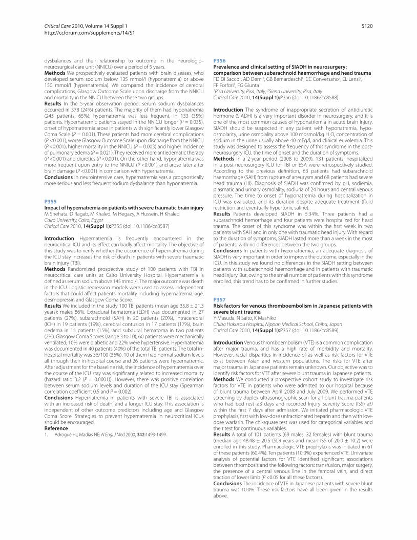
dysbalances and their relationship to outcome in the neurologic–
neurosurgical care unit (NNICU) over a period of 5 years.
Methods We prospectively evaluated patients with brain diseases, who
developed serum sodium below 135 mmol/l (hyponatremia) or above
150 mmol/l (hypernatremia). We compared the incidence of cerebral
complications, Glasgow Outcome Scale upon discharge from the NNICU
and mortality in the NNICU between these two groups.
Results In the 5-year observation period, serum sodium dysbalances
occurred in 378 (24%) patients. The majority of them had hyponatremia
(245 patients, 65%); hypernatremia was less frequent, in 133 (35%)
patients. Hypernatremic patients stayed in the NNICU longer (P = 0.035),
onset of hypernatremia arose in patients with signifi cantly lower Glasgow
Coma Scale (P = 0.001). These patients had more cerebral complications
(P <0.001), worse Glasgow Outcome Scale upon discharge from the NNICU
(P <0.001), higher mortality in the NNICU (P = 0.003) and higher incidence
of pulmonary edema (P = 0.021). They received more antiedematic therapy
(P <0.001) and diuretics (P <0.001). On the other hand, hyponatremia was
more frequent upon entry to the NNICU (P <0.001) and arose later after
brain damage (P <0.001) in comparison with hypernatremia.
Conclusions In neurointensive care, hypernatremia was a prognostically
more serious and less frequent sodium dysbalance than hyponatremia.
P355
Impact of hypernatremia on patients with severe traumatic brain injuryM Shehata, D Ragab, M Khaled, M Hegazy, A Hussein, H Khaled
Cairo University, Cairo, Egypt
Critical Care 2010, 14(Suppl 1):P355 (doi: 10.1186/cc8587)
Introduction Hypernatremia is frequently encountered in the
neurocritical ICU and its eff ect can badly aff ect mortality. The objective of
this study was to verify whether the occurrence of hypernatremia during
the ICU stay increases the risk of death in patients with severe traumatic
brain injury (TBI).
Methods Randomized prospective study of 100 patients with TBI in
neurocritical care units at Cairo University Hospital. Hypernatremia is
defi ned as serum sodium above 145 mmol/l. The major outcome was death
in the ICU. Logistic regression models were used to assess independent
factors that could aff ect patients’ mortality including hypernatremia, age,
desmopressin and Glasgow Coma Score.
Results We included in the study 100 TBI patients (mean age 35.8 ± 21.3
years); males 86%. Extradural hematoma (EDH) was documented in 27
patients (27%), subarachnoid (SAH) in 20 patients (20%), intracerebral
(ICH) in 19 patients (19%), cerebral contusion in 17 patients (17%), brain
oedema in 15 patients (15%), and subdural hematoma in two patients
(2%). Glasgow Coma Scores (range 3 to 10); 60 patients were mechanically
ventilated; 10% were diabetic and 22% were hypertensive. Hypernatremia
was documented in 40 patients (40%) of the total TBI patients. The total in-
hospital mortality was 36/100 (36%), 10 of them had normal sodium levels
all through their in-hospital course and 26 patients were hypernatremic.
After adjustment for the baseline risk, the incidence of hypernatremia over
the course of the ICU stay was signifi cantly related to increased mortality
(hazard ratio 3.2 (P = 0.0001)). However, there was positive correlation
between serum sodium levels and duration of the ICU stay (Spearman
correlation coeffi cient 0.5 and P = 0.002).
Conclusions Hypernatremia in patients with severe TBI is associated
with an increased risk of death, and a longer ICU stay. This association is
independent of other outcome predictors including age and Glasgow
Coma Score. Strategies to prevent hypernatremia in neurocritical ICUs
should be encouraged.
Reference1. Adrogué HJ, Madias NE: N Engl J Med 2000, 342:1493-1499.
P356
Prevalence and clinical setting of SIADH in neurosurgery: comparison between subarachnoid haemorrhage and head traumaFD Di Sacco1, AD Demi1, GB Bernardeschi1, CC Conversano2, EL Lensi2,
FF Forfori1, FG Giunta1
1Pisa University, Pisa, Italy; 2Siena University, Pisa, Italy
Critical Care 2010, 14(Suppl 1):P356 (doi: 10.1186/cc8588)
Introduction The syndrome of inappropriate secretion of antidiuretic
hormone (SIADH) is a very important disorder in neurosurgery, and it is
one of the most common causes of hyponatriemia in acute brain injury.
SIADH should be suspected in any patient with hyponatremia, hypo-
osmolarity, urine osmolality above 100 mosmol/kg H2O, concentration of
sodium in the urine usually above 40 mEq/l, and clinical euvolemia. This
study was designed to assess the frequency of this syndrome in the post-
neurosurgery ICU, the time of onset and the duration of symptoms.
Methods In a 2-year period (2008 to 2009), 131 patients, hospitalized
in a post-neurosurgery ICU for TBI or ESA were retrospectively studied.
According to the previous defi nition, 63 patients had subarachnoid
haemorrhage (SAH) from rupture of aneurysm and 68 patients had severe
head trauma (HI). Diagnosis of SIADH was confi rmed by pH, sodiemia,
plasmatic and urinary osmolality, sodiuria of 24 hours and central venous
pressure. The time to onset of hyponatremia during hospitalization in
ICU was evaluated, and its duration despite adequate treatment (fl uid
restriction and eventually hypertonic saline).
Results Patients developed SIADH in 5.34%. Three patients had a
subarachnoid hemorrhage and four patients were hospitalized for head
trauma. The onset of this syndrome was within the fi rst week in two
patients with SAH and in only one with traumatic head injury. With regard
to the duration of symptoms, SIADH lasted more than a week in the most
of patients, with no diff erences between the two groups.
Conclusions In patients with hyponatriemia, an adequate diagnosis of
SIADH is very important in order to improve the outcome, especially in the
ICU. In this study we found no diff erences in the SIADH setting between
patients with subarachnoid haemorrhage and in patients with traumatic
head injury. But, owing to the small number of patients with this syndrome
enrolled, this trend has to be confi rmed in further studies.
P357
Risk factors for venous thromboembolism in Japanese patients with severe blunt traumaY Masuda, N Saito, K Mashiko
Chiba Hokusou Hospital, Nippon Medical School, Chiba, Japan
Critical Care 2010, 14(Suppl 1):P357 (doi: 10.1186/cc8589)
Introduction Venous thromboembolism (VTE) is a common complication
after major trauma, and has a high rate of morbidity and mortality.
However, racial disparities in incidence of as well as risk factors for VTE
exist between Asian and western populations. The risks for VTE after
major trauma in Japanese patients remain unknown. Our objective was to
identify risk factors for VTE after severe blunt trauma in Japanese patients.
Methods We conducted a prospective cohort study to investigate risk
factors for VTE in patients who were admitted to our hospital because
of blunt trauma between April 2008 and July 2009. We performed VTE
screening by duplex ultrasonographic scan for all blunt trauma patients
who had bed rest ≥3 days and recorded Injury Severity Score (ISS) ≥9
within the fi rst 7 days after admission. We initiated pharmacologic VTE
prophylaxis, fi rst with low-dose unfractionated heparin and then with low-
dose warfarin. The chi-square test was used for categorical variables and
the t test for continuous variables.
Results A total of 101 patients (69 males, 32 females) with blunt trauma
(median age 48.48 ± 20.5 (SD) years and mean ISS of 20.0 ± 10.2) were
enrolled in this study. Pharmacologic VTE prophylaxis was initiated in 61
of these patients (60.4%). Ten patients (10.0%) experienced VTE. Univariate
analysis of potential factors for VTE identifi ed signifi cant associations
between thrombosis and the following factors: transfusion, major surgery,
the presence of a central venous line in the femoral vein, and direct
traction of lower limb (P <0.05 for all these factors).
Conclusions The incidence of VTE in Japanese patients with severe blunt
trauma was 10.0%. These risk factors have all been given in the results
above.
Critical Care 2010, Volume 14 Suppl 1 http://ccforum.com/supplements/14/S1
S120

P358
Safety of heparin prophylaxis for venous thromboembolism in patients with neurotraumaG Puri, P Joseph
Postgraduate Institute of Medical Education and Research, Chandigarh, India
Critical Care 2010, 14(Suppl 1):P358 (doi: 10.1186/cc8590)
Introduction Patients with neurotrauma are at high risk for VTE. The safety
of early anticoagulation for neurotrauma has not been established. Our
study was based on the hypothesis that unfractionated heparin can be
safely administered to most patients with neurotrauma for VTE prophylaxis.
Methods Prospective, single-cohort, observational study. All adult
patients with neurotrauma admitted to a tertiary care teaching hospital
in India from March to December 2007 received unfractionated heparin
beginning approximately 24 hours after hospital admission until discharge.
Brain CT scans were performed at admission, 24 hours after admission,
and at variable intervals thereafter based on clinical course. Patients were
excluded for coagulopathy, expected brain death or discharge within 48
hours. Complications of heparin prophylaxis were defi ned as expansion of
existing intracranial haemorrhage or development of a new haemorrhagic
lesion on follow-up CT after beginning heparin use.
Results Twenty-one of 57 (36.8%) adult neurotrauma patients were
eligible for heparin therapy. Their distribution of CT fi ndings is presented
in Table 1. Serial CT scan had progression of lesion in four of the 26 patients
not on heparin therapy and in two of the 21 patients on heparin (P >0.05).
No diff erence in complications between patients with ICH on heparin
(2/14) and not on heparin (1/13) were identifi ed (P >0.05). The mortality
in the heparin group was 3/21(14.3%). Both the patients who developed
worsening of CT fi ndings after initiation of heparin therapy survived
hospitalization. None of the patients suff ered from DVT.
Table 1 (abstract 358). Distribution of CT fi ndings and heparin therapy
CT No heparin Heparin
Normal 8 0
DBE 8 3
Contusion 13 3
Hge 27 14
# Skull 1 1
Conclusions Heparin can be safely used for VTE prophylaxis in patients
with head injury when started 24 hours after hospital admission.
P359
Prevalence and incidence of proximal deep vein thrombosis in critically ill patientsA Panitchote, W Chaiyasoot, C Permpikul
Mahidol University, Bangkok, Thailand
Critical Care 2010, 14(Suppl 1):P359 (doi: 10.1186/cc8591)
Introduction The epidemiology of DVT in critically ill medical patients
has not been studied in Thailand. The aim of this study was to assess the
prevalence and incidence rate of proximal DVT in these patients and to
determine the factors associated with the development of this condition.
Methods A single-center, prospective observational study was performed
in critically ill medical patients who developed sepsis or new organ failure
requiring organ support. DVT was detected by ultrasonography with
color Doppler imaging performed within 48 hours after the patients met
the inclusion criteria, and serial studies at days 7, 14 and 28 (or earlier if
clinically indicated) were carried out.
Results Of the 170 patients, 87 were male (51.2%). The mean age was 59.95
years (range 18 to 94), and the mean APACHE II score was 21.34 (range
4 to 42). Acute respiratory failure and sepsis were the leading diagnoses.
Prevalence and incidence of proximal DVT were 14.1% (95% CI 9.6 to 20.15)
and 8.82% (95% CI 5.4 to 14.0), respectively. Univariate analysis of factors
associated with DVT showed female sex (P = 0.017), age ≥50 years (P =
0.017), renal replacement therapy (P = 0.005) and femoral venous catheter
(P = 0.002) while multivariate analysis showed female sex (P = 0.007; OR =
4.43, 95% CI 1.5 to 13.08), age ≥50 years (P = 0.027; OR = 6.12, 95% CI 1.23
to 30.49), femoral venous catheter (P <0.001; OR = 11.77, 95% CI 3.11 to
44.53) and the absence of platelet transfusion (P = 0.015; OR = 0.056, 95%
CI 0.005 to 0.58). Clinical probability at the fi rst 48 hours but not at days 7,
14 and 28 was strongly correlated with the development of proximal DVT.
The duration of mechanical ventilation, the hospital length of stay and the
ICU mortality were not aff ected by the presence of DVT.
Conclusions Proximal DVT of lower extremities in critically ill medical
Thai patients are not uncommon. Thromboprophylaxis study is needed in
order to investigate the risk and benefi t.
References1. Geerts WH, et al.: Prevention of venous thromboembolism: the Eighth
ACCP Conference on Antithrombotic and Thrombolytic therapy. Chest
2008, 133(Suppl):381S-453S.
2. Cook DJ, et al.: Deep venous thrombosis in medical–surgical ICU patients: prevalence, incidence and risk factors. Crit Care Med 2005, 33:1565-1571.
P360
Trauma patients with prophylactic IVC fi lter and chemical prophylaxis: a descriptive studyMM Perreault, J Yan, A Karamaoun, E Villeneuve, T Razek
The Montreal General Hospital, Montreal, Canada
Critical Care 2010, 14(Suppl 1):P360 (doi: 10.1186/cc8592)
Introduction Trauma patients at high risk of thrombosis and bleeding often
have a retrievable inferior vena cava fi lter (IVCF) inserted prophyl actically
shortly after admission for the prevention of pulmonary embolism. Despite
their increasing use in trauma, controversy exists regarding patient selection,
benefi ts and safety. In addition, chemical prophylaxis should be initiated as
soon as determined to be safe to do so. Limited data exist describing the use
of chemical prophylaxis and the timing of it in relation to IVCF placement in
this patient population. We aim to describe such use and timing in relation
to IVCF placement.
Methods All trauma patients with a retrievable prophylactic IVCF inserted
over 3.5 years with an age ≥18 and an ISS ≥15 were enrolled into the
descriptive study. Patients were identifi ed using the local trauma registry
and data collected from the registry and patient chart.
Results One hundred and three patients with a prophylactic IVCF are
described. Mean age was 42.8 ± 17.9 years, 68% were male and they had a
mean ISS of 38.6 ± 12. A total of 36.9% had ≥3 injuries. A total of 60.2% had
an injury with maximum AIS score ≥5 and the type of injury associated
with maximal AIS was head (53.4%), pelvic (19.4%), thoracic (15.5%) and
spinal cord injury (11.7%). Shock was present on admission in 8.7% patients,
98.1% required ICU admission, 88.4% required intubation and 27.2% patients
required ≥4 PRBC over the fi rst 24 hours. The retrievable IVCF was inserted
prophylactically after a mean of 1.7 ± 1.9 days (range 0 to 9 days) of hospital
admission. Chemical prophylaxis was initiated 5.7 ± 5.1 days after IVCF in
77.7% patients, and dalteparin or enoxaparin were the initial agents of choice
in 93.8% while heparin was chosen in 6.2% of cases. The time from hospital
admission to the start of chemical prophylaxis ranged from 6.3 to 8.5 days.
Conclusions Chemical prophylaxis was initiated in the majority of
trauma patients with an IVCF, albeit 22% still did not receive any. When
administered, a low-molecular-weight heparin was selected. Although
rapidly inserted after admission, there was considerable delay between
insertion of IVCF and initiation of chemical prophylaxis. The reasons for this
delay require further investigation.
Reference1. Kaufman JA, et al.: Guidelines for the use of retrievable and convertible
vena cava fi lters. Surg Obes Relat Dis 2006, 2:200-212.
P361
Clinical signs and risk factors of acute deep vein thrombosis of lower extremities in patients after abdominal surgery for cancer: effi ciency and safety of diff erent types of anticoagulant therapyO Tarabrin1, V Suslov2, S Kalinchuk3, S Tarasenko1, V Grubnik1,
S Shcherbakov1, D Gavrichenko3
1Odessa Medical University, Odessa, Ukraine; 2The Institute of Urology, Kiev,
Ukraine; 3Regional Clinical Hospital, Odessa, Ukraine
Critical Care 2010, 14(Suppl 1):P361 (doi: 10.1186/cc8593)
Introduction The background of our research was to conduct a
retrospective analysis of clinical signs and risk factors for acute deep vein
Critical Care 2010, Volume 14 Suppl 1 http://ccforum.com/supplements/14/S1
S121

thrombosis (ADVT) of lower extremities, and to compare the eff ectiveness
and safety of therapy of LMWH bemiparin and UFH in patients after
abdominal surgery for cancer.
Methods For the period from 2008 to 2009 the diagnosis of ADVT was set
for 362 patients, among them 179 (49.4%) men and 183 (50.6%) women.
Among patients with ADTV with primary localization in ileofemoral and
popliteal segments treated in the department of vascular surgery, senior
persons more than 60 years old prevailed (63.9%). All patients with ADVT
received therapy with anticoagulants. Bemiparin sodium was prescribed
in 257 (70.9%) cases, UFH in 71 (19.6%) and in 34 (9.5%) others LMWH. We
analysed effi ciency and complications after antithrombotic therapy in a
hospital period. A complete coagulation screen, activated clotting time,
thromboelastography and low-frequency haemoviscoelastography (HVG)
were performed to reveal coagulation disturbances.
Results In 83.7% of cases the most frequent symptoms of disease are
edema of an extremity (in 78.1%) and pain syndrome 74.7%, which at a
monosymptomal variant (39.4% patients) are marked in 66.3% and 44.4%
accordingly. The most frequent risk factors (RF) were prolonged (more
than 8 days) immobilization and malignant tumors. Therapy of UFH and
LMWH was eff ective enough (on average, a good result obtained in more
than in 77% cases). Bemiparin treatment was related to considerably
less hemorrhagic complications than treatment of UFH (P <0.05), which
concerned all of types of such complications.
Conclusions Treatment of with bemiparin as compared with UFH
accompanied reduced frequency of all hemorrhagic complications by 2.1
times, serious 2.7 times and moderate 3.1 times. HVG is an eff ective point-
of-care monitor for routine clinical practice and clinical research.
P362
Multidrug sedation as a risk factor for deep vein thrombosis in the intensive care unitF Barbani1, L Tutino1, S Batacchi2, A Cecchi2, M Bonizzoli2, M Boddi3,
G Zagli2, A Peris2
1Postgraduate School of Anesthesia and Intensive Care, University of Florence,
Italy; 2Careggi Teaching Hospital, Florence, Italy; 3Department of Critical Care
Medicine and Surgery, University of Florence, Italy
Critical Care 2010, 14(Suppl 1):P362 (doi: 10.1186/cc8594)
Introduction Critically ill patients are at high risk for deep vein thrombosis
(DVT). Among ICU DVT risk factors, the role of muscle relaxant (MR) has
been clearly shown [1] but the possible infl uence of sedative medications
(SM) is still under debate. This study was aimed at investigating whether
the use of diff erent SM may aff ect DVT incidence in ICU patients.
Methods We retrospectively enrolled all patients admitted to a mixed
medical–surgical trauma ICU from January 2007 to August 2008 who
showed a >72 hours length of stay and were >18 years old; exclusion
criteria were DVT at admission and/or within the previous 6 months,
thrombophilic profi le, high haemorrhagic risk, admission from another ICU.
For studying ICU lower limb DVT incidence, compressive ultrasonography
(CUS) was performed by three trained ultrasonographers twice a week
until discharge. SM and MR administration was registered daily: the drugs
considered are presented in Figure 1. MR-treated patients were excluded
from statistical analysis; the diff erence in SM administration in the No DVT
group vs the DVT-positive group was assayed by chi-squared test.
Results Over the period of study, 380 patients were enrolled. Lower limb
DVT was diagnosed in 23 of 380 patients, with a DVT incidence of 6.05%.
MRs were administered in 121 patients and they were associated with
DVT incidence (P <0.001). Statistical analysis results performed on 259
patients are shown in Figure 1: none of the considered SM resulted to be
associated with DVT incidence, nor sedation as a whole or the number of
SM administered.
Conclusions Our study confi rms prior fi ndings [1] on the MR role as a DVT
risk factor. Nevertheless, SM administration in patients not treated with MR
is not associated with increased DVT risk.
References1. J Thromb Haemost 2009. [Epub ahead of print. DOI:
10.1111/j.1538-7836.2009.03664.x]
P363
New method diagnostics for coagulation disorders after surgeryO Tarabrin1, V Suslov2, V Grubnik1, S Kalinchuk3, P Pustovoyt3,
S Shcherbakov1, D Gavrichenko3
1Odessa Medical University, Odessa, Ukraine; 2The Institute of Urology, Odessa,
Ukraine; 3Regional Clinical Hospital, Odessa, Ukraine
Critical Care 2010, 14(Suppl 1):P363 (doi: 10.1186/cc8595)
Introduction Despite the evidence of perioperative hypercoagulability in
cancer patients, there are no consistent data evaluating the extent, duration,
and specifi c contribution of platelets and procoagulatory proteins by in
vitro testing. This study compared effi cacy of haemoviscoelastography
versus thromboelastography for monitoring of coagulation imbalance.
Methods Two hundred and forty-one patients undergoing surgery for
abdominal cancer were examined for the effi cacy of a variety of coagulation
tests. A complete coagulation screening, thromboelastography (TEG) and
haemoviscoelastography (HVG) were performed before and at the end of
surgery.
Results We calculated the elastic shear modulus of standard MA (Gt) and
HVG MA (Gh), which refl ect the total clot strength and procoagulatory
protein component, respectively. The diff erence was an estimate of the
platelet component (Gp). There was a 16% perioperative increase of
standard MA, corresponding to a 51% increase of Gt (P <0.05) and a 79%
to 87% contribution of the calculated Gp to Gt. We conclude that serial
standard thromboelastography and the HVG viscoelastic test may reveal
the independent contribution of platelets and procoagulatory proteins
to clot strength. Using multiple linear regression, all coagulation, TEG and
HVG variabilities were used to model postoperative hypercoagulation.
Results showed that some components of the TEG failed to identify
hypercoagulation (r <0.2, P >0.75). All components of the HVG test refl ect
postoperative coagulopathies.
Conclusions Hypercoagulability is not refl ected completely by standard
coagulation monitoring and TEG, and seems to be predominantly
caused by increased platelet reactivity. HVG provides a fast and easy-to-
perform bedside test to quantify in vitro coagulation, and may be useful
in determining the coagulation status of cancer patients perioperatively.
P364
Thrombocytopenia in surgical ICU patientsO Yhiaoui, M Metref
Military Hospital, Algiers, Algeria
Critical Care 2010, 14(Suppl 1):P364 (doi: 10.1186/cc8596)
Introduction The aim was to study the incidence, the etiology and to
assess the severity of thrombocytopenia in surgical ICU (SICU) patients
admitted to the central army hospital of Algiers (Algeria).
Methods We conducted a retrospective study; all admitted patients
during the year 2008 were enrolled in the study (351 patients). All charts
were reviewed and diff erent hemograms recorded from admission until
discharge. One hundred and fi fty-three thrombocytopenia cases defi ned
as a platelet count (plt) <150 x 109 were reported (43.6%).
Results Among 351 patients (227 survivors, 124 died), 153 developed
thrombocytopenia, 96 males and 57 females. The onset of thrombo-
cytopenia was for the majority of patients (98%) at 24 to 72 hours after
ICU admission. Only 26% were severe (plt <50 x 109) and 3% <20 x 109).
The etiopathology is multifactorial and diffi cult to obtain precisely: 77% Figure 1 (abstract P362). Statistical analysis.
Critical Care 2010, Volume 14 Suppl 1 http://ccforum.com/supplements/14/S1
S122
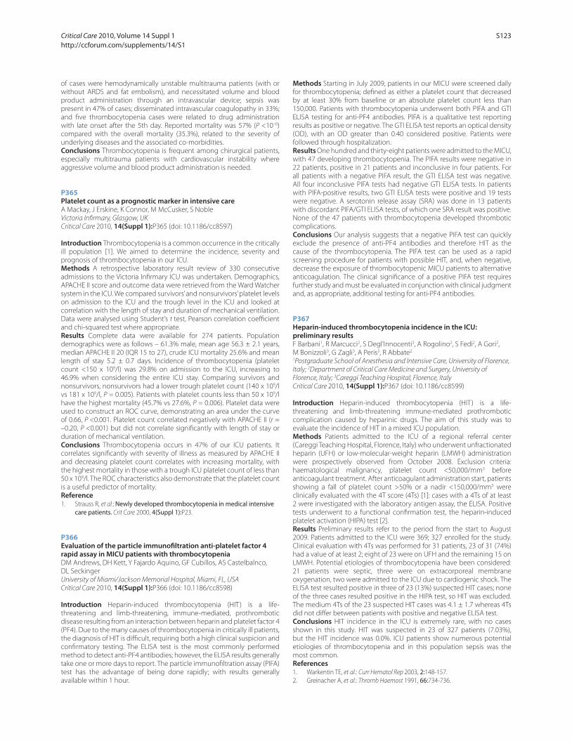
of cases were hemodynamically unstable multitrauma patients (with or
without ARDS and fat embolism), and necessitated volume and blood
product administration through an intravascular device; sepsis was
present in 47% of cases; disseminated intravascular coagulopathy in 33%;
and fi ve thrombocytopenia cases were related to drug administration
with late onset after the 5th day. Reported mortality was 57% (P <10–6)
compared with the overall mortality (35.3%), related to the severity of
underlying diseases and the associated co-morbidities.
Conclusions Thrombocytopenia is frequent among chirurgical patients,
especially multitrauma patients with cardiovascular instability where
aggressive volume and blood product administration is needed.
P365
Platelet count as a prognostic marker in intensive careA Mackay, J Erskine, K Connor, M McCusker, S Noble
Victoria Infi rmary, Glasgow, UK
Critical Care 2010, 14(Suppl 1):P365 (doi: 10.1186/cc8597)
Introduction Thrombocytopenia is a common occurrence in the critically
ill population [1]. We aimed to determine the incidence, severity and
prognosis of thrombocytopenia in our ICU.
Methods A retrospective laboratory result review of 330 consecutive
admissions to the Victoria Infi rmary ICU was undertaken. Demographics,
APACHE II score and outcome data were retrieved from the Ward Watcher
system in the ICU. We compared survivors’ and nonsurvivors’ platelet levels
on admission to the ICU and the trough level in the ICU and looked at
correlation with the length of stay and duration of mechanical ventilation.
Data were analysed using Student’s t test, Pearson correlation coeffi cient
and chi-squared test where appropriate.
Results Complete data were available for 274 patients. Population
demographics were as follows – 61.3% male, mean age 56.3 ± 2.1 years,
median APACHE II 20 (IQR 15 to 27), crude ICU mortality 25.6% and mean
length of stay 5.2 ± 0.7 days. Incidence of thrombocytopenia (platelet
count <150 x 109/l) was 29.8% on admission to the ICU, increasing to
46.9% when considering the entire ICU stay. Comparing survivors and
nonsurvivors, nonsurvivors had a lower trough platelet count (140 x 109/l
vs 181 x 109/l, P = 0.005). Patients with platelet counts less than 50 x 109/l
have the highest mortality (45.7% vs 27.6%, P = 0.006). Platelet data were
used to construct an ROC curve, demonstrating an area under the curve
of 0.66, P <0.001. Platelet count correlated negatively with APACHE II (r =
–0.20, P <0.001) but did not correlate signifi cantly with length of stay or
duration of mechanical ventilation.
Conclusions Thrombocytopenia occurs in 47% of our ICU patients. It
correlates signifi cantly with severity of illness as measured by APACHE II
and decreasing platelet count correlates with increasing mortality, with
the highest mortality in those with a trough ICU platelet count of less than
50 x 109/l. The ROC characteristics also demonstrate that the platelet count
is a useful predictor of mortality.
Reference1. Strauss R, et al.: Newly developed thrombocytopenia in medical intensive
care patients. Crit Care 2000, 4(Suppl 1):P23.
P366
Evaluation of the particle immunofi ltration anti-platelet factor 4 rapid assay in MICU patients with thrombocytopeniaDM Andrews, DH Kett, Y Fajardo Aquino, GF Cubillos, AS Castelbalnco,
DL Seckinger
University of Miami/Jackson Memorial Hospital, Miami, FL, USA
Critical Care 2010, 14(Suppl 1):P366 (doi: 10.1186/cc8598)
Introduction Heparin-induced thrombocytopenia (HIT) is a life-
threatening and limb-threatening, immune-mediated, prothrombotic
disease resulting from an interaction between heparin and platelet factor 4
(PF4). Due to the many causes of thrombocytopenia in critically ill patients,
the diagnosis of HIT is diffi cult, requiring both a high clinical suspicion and
confi rmatory testing. The ELISA test is the most commonly performed
method to detect anti-PF4 antibodies; however, the ELISA results generally
take one or more days to report. The particle immunofi ltration assay (PIFA)
test has the advantage of being done rapidly; with results generally
available within 1 hour.
Methods Starting in July 2009, patients in our MICU were screened daily
for thrombocytopenia; defi ned as either a platelet count that decreased
by at least 30% from baseline or an absolute platelet count less than
150,000. Patients with thrombocytopenia underwent both PIFA and GTI
ELISA testing for anti-PF4 antibodies. PIFA is a qualitative test reporting
results as positive or negative. The GTI ELISA test reports an optical density
(OD), with an OD greater than 0.40 considered positive. Patients were
followed through hospitalization.
Results One hundred and thirty-eight patients were admitted to the MICU,
with 47 developing thrombocytopenia. The PIFA results were negative in
22 patients, positive in 21 patients and inconclusive in four patients. For
all patients with a negative PIFA result, the GTI ELISA test was negative.
All four inconclusive PIFA tests had negative GTI ELISA tests. In patients
with PIFA-positive results, two GTI ELISA tests were positive and 19 tests
were negative. A serotonin release assay (SRA) was done in 13 patients
with discordant PIFA/GTI ELISA tests, of which one SRA result was positive.
None of the 47 patients with thrombocytopenia developed thrombotic
complications.
Conclusions Our analysis suggests that a negative PIFA test can quickly
exclude the presence of anti-PF4 antibodies and therefore HIT as the
cause of the thrombocytopenia. The PIFA test can be used as a rapid
screening procedure for patients with possible HIT, and, when negative,
decrease the exposure of thrombocytopenic MICU patients to alternative
anticoagulation. The clinical signifi cance of a positive PIFA test requires
further study and must be evaluated in conjunction with clinical judgment
and, as appropriate, additional testing for anti-PF4 antibodies.
P367
Heparin-induced thrombocytopenia incidence in the ICU: preliminary resultsF Barbani1, R Marcucci2, S Degl’Innocenti3, A Rogolino2, S Fedi2, A Gori2,
M Bonizzoli3, G Zagli3, A Peris3, R Abbate2
1Postgraduate School of Anesthesia and Intensive Care, University of Florence,
Italy; 2Department of Critical Care Medicine and Surgery, University of
Florence, Italy; 3Careggi Teaching Hospital, Florence, Italy
Critical Care 2010, 14(Suppl 1):P367 (doi: 10.1186/cc8599)
Introduction Heparin-induced thrombocytopenia (HIT) is a life-
threatening and limb-threatening immune-mediated prothrombotic
complication caused by heparinic drugs. The aim of this study was to
evaluate the incidence of HIT in a mixed ICU population.
Methods Patients admitted to the ICU of a regional referral center
(Careggi Teaching Hospital, Florence, Italy) who underwent unfractionated
heparin (UFH) or low-molecular-weight heparin (LMWH) administration
were prospectively observed from October 2008. Exclusion criteria:
haematological malignancy, platelet count <50,000/mm3 before
anticoagulant treatment. After anticoagulant administration start, patients
showing a fall of platelet count >50% or a nadir <150,000/mm3 were
clinically evaluated with the 4T score (4Ts) [1]: cases with a 4Ts of at least
2 were investigated with the laboratory antigen assay, the ELISA. Positive
tests underwent to a functional confi rmation test, the heparin-induced
platelet activation (HIPA) test [2].
Results Preliminary results refer to the period from the start to August
2009. Patients admitted to the ICU were 369; 327 enrolled for the study.
Clinical evaluation with 4Ts was performed for 31 patients, 23 of 31 (74%)
had a value of at least 2; eight of 23 were on UFH and the remaining 15 on
LMWH. Potential etiologies of thrombocytopenia have been considered:
21 patients were septic, three were on extracorporeal membrane
oxygenation, two were admitted to the ICU due to cardiogenic shock. The
ELISA test resulted positive in three of 23 (13%) suspected HIT cases; none
of the three cases resulted positive in the HIPA test, so HIT was excluded.
The medium 4Ts of the 23 suspected HIT cases was 4.1 ± 1.7 whereas 4Ts
did not diff er between patients with positive and negative ELISA test.
Conclusions HIT incidence in the ICU is extremely rare, with no cases
shown in this study. HIT was suspected in 23 of 327 patients (7.03%),
but the HIT incidence was 0.0%. ICU patients show numerous potential
etiologies of thrombocytopenia and in this population sepsis was the
most common.
References1. Warkentin TE, et al.: Curr Hematol Rep 2003, 2:148-157.
2. Greinacher A, et al.: Thromb Haemost 1991, 66:734-736.
Critical Care 2010, Volume 14 Suppl 1 http://ccforum.com/supplements/14/S1
S123

P368
Treatment of the coagulation defi cit during extracorporeal circulation in a porcine model by a prothrombin complex concentrateF Kaspereit, S Hoff man, I Pragst, G Dickneite
CSL Behring, Marburg, Germany
Critical Care 2010, 14(Suppl 1):P368 (doi: 10.1186/cc8600)
Introduction Many procedures in cardiac surgery require extracorporeal
circulation (EC). During EC, blood is pumped from a venous line to an
oxygenator, cooled and infused back via an arterial line. This procedure
is known to compromise the coagulation system, especially under
prolonged hypothermia. The aim of the present study was to investigate
the eff ects of EC and hypothermia on the coagulation system in a porcine
model. The effi cacy of a substitution therapy with a prothrombin complex
concentrate (PCC) regarding normalization of coagulation and decrease in
hemorrhage was evaluated.
Methods A total of 17 male anesthetized pigs were included in the
study. EC was performed in 12 animals by a hollowfi ber oxygenator
with a priming solution containing saline, HES and heparin. Five animals
without EC served as a control. The coagulation system was characterized
by thromboelastography (TEG), thrombin generation, coagulation factor
levels, platelet numbers and function (aggregation). A GoreTex patch
was inserted into the carotid artery, bleeding occurred from the stitch
channels. The eff ects of intravenous substitution therapy by PCC (Beriplex
P/N, 30 U/kg, n = 6) compared with placebo (n = 6) on bleeding from the
stitch channels was investigated.
Results EC and hypothermia compromised the coagulation system.
Coagulation factor levels were decreased, TEG and thrombin generation
became pathologic. Platelet count and aggregation were decreased.
Blood loss from the stitch channels of the GoreTex patch was doubled in
the EC group when compared with animals without EC. After substitution
therapy with PCC, blood loss decreased signifi cantly from 83.0 ± 48.4 ml
in the EC + placebo to 27.2 ± 35 ml in the EC + PCC group (P <0.025,
Wilcoxon test; Figure 1). PCC normalized the impaired coagulation.
Conclusions Hypothermia and EC led to a compromised coagulation
system, resulting in increased hemorrhage. The defi cit in coagulation
could be overcome by substitution therapy with PCC. Elevated blood loss
from stitch channels was decreased signifi cantly after treatment with PCC.
It was concluded that PCC should be benefi cial in patients undergoing
cardiovascular surgery with an EC-induced coagulopathy.
P369
Fibrinogen in dilutional coagulopathy: a dose study in pigsJ Martini1, S Maisch1, W Martini2, W Streif1, D Fries1
1Medical University Innsbruck, Austria; 2US Army Institute for Surgical
Research, San Antonio, TX, USA
Critical Care 2010, 14(Suppl 1):P369 (doi: 10.1186/cc8601)
Introduction Dilutional coagulopathy following massive bleeding is
the result of clotting factor dilution and impaired fi brin polymerization
after infusion of colloidal plasma expanders. Although fi brinogen has
been clinically used to treat dilutional coagulopathy [1,2], the eff ects of
fi brinogen dosages on normalizing coagulation function is unclear. This
study investigated the eff ect of six diff erent fi brinogen dosages (range:
37.5 to 600 mg/kg) on ROTEM® parameters and overall blood loss in a pig
model for dilutional coagulopathy.
Methods Forty-two pigs underwent a 60% hemodilution with Voluven®
(HES130/0.4). A standardized bone injury was performed after the
completion of hemodilution. Animals were then randomized to receive
37.5, 75, 150, 300, 450 or 600 mg/kg fi brinogen (FGTW, LFB, France) or
500 ml saline. Four hours after fi brinogen administration a standardized
liver injury was performed. Animals were then observed for 2 hours or
until death. Blood loss was measured and tissue samples were collected
at the end of the study. Hemodynamic and coagulation parameters were
measured at baseline (BL), after hemodilution, 15 minutes, 1, 2 and 4 hours
after drug infusion and 2 hours after liver injury or right before the animal’s
death. Statistical signifi cance was set at P <0.05.
Results Fibrinogen dosages of 150 mg/kg and higher completely reversed
dilutional coagulopathy: the maximum clot fi rmness (MCF) which was
decreased after hemodilution (36 ± 3 vs 65 ± 4 mm at BL, P <0.05), returned
to BL levels after fi brinogen administration (69 ± 5 mm). Blood loss from
bone and liver injury signifi cantly decreased with increased fi brinogen
dosages: 42 ± 19 (sham), 34 ± 14 (75 mg/kg), 29 ± 13 (150 mg/kg) and
28 ± 10 ml/kg BW (600 mg/kg). No thrombotic events occurred.
Conclusions In a swine model of 60% hemodilution with bone and liver
injury, fi brinogen administration (150 mg/kg and above) normalized MCF
and decreased blood loss. Infusions of 12 times the dosage recommended
in humans did not induce hypercoagulability.
References1. Fenger-Eriksen C, et al.: J Thromb Haemost 2009, 75:795-802.
2. Mittermayr M, et al.: Anesth Analg 2007, 105:905-917.
P370
Tranexamic acid reduces blood loss in postpartum haemorrhageA Ducloy-Bouthors1, F Broisin2, H Keita3, S Fontaine4, S Depret1, F Legoeff 5,
L Mandelbrot3, R Rudigoz2, B Jude1
1Centre Hospitalier Regional Universitaire, Lille, France; 2Hôpital Croix Rousse
HCL, Lyon, France; 3Hôpital Louis Mourrier APHP, Colombes, France; 4Maternité
Monaco CH, Valenciennes, France; 5Maternité Paul Gellée, Roubaix, France
Critical Care 2010, 14(Suppl 1):P370 (doi: 10.1186/cc8602)
Introduction Postpartum haemorrhage (PPH) is a leading cause of
maternal death. Present guidelines for PPH management focus on
uterotonic treatments, while the potential effi cacy of antifi brinolytic drugs
has been poorly explored in this indication.
Methods After approval by the ethics committee, we randomly assigned
women with PPH >800 ml after vaginal delivery to receive intravenous
tranexamic acid (TXA) (4 g within 1 hour, followed by 1 g/hour during
6 hours) (TXA group) or no antifi brinolytic treatment (Control group).
Monitoring and management were performed according to the
guidelines. Additional procoagulant treatments (coagulant concentrates,
fresh frozen plasma, platelets) were used only when PPH became
intractable (>2,500 ml or >500 ml/30 minutes). Embolization or surgery
was performed when considered clinically necessary. Blood loss, maternal
morbidity and safety were collected at four time points: inclusion (T1),
T1 + 30 minutes (T2), T1 + 2 hours (T3), and T1 + 6 hours (T4). The study
plan, based on the hypothesis that TXA would induce a 20% blood loss
reduction with an α risk of 5% and a β risk of 10%, required the inclusion of
72 patients in each group. Statistical analysis used the SAS system.
Results One hundred and forty-four patients were included, 72 in the
TXA group and 72 in the control group. Management data were similar
between the two groups. In the TXA group, as compared with the control
Figure 1 (abstract P368). Blood loss from a GoreTex Patch after EC.
Critical Care 2010, Volume 14 Suppl 1 http://ccforum.com/supplements/14/S1
S124

group: blood loss was 46% lower between T2 and T3 (P = 0.026), and 49%
lower between T2 and T4 (P = 0.0012); bleeding duration was shorter (31 ±
28 vs 65 ± 95 minutes, P = 0.004); and maternal morbidity was reduced
(hemoglobin drop >4 g/dl: n = 15 vs 34, P <0.001; blood transfusion
requirement: n = 14 vs 20, P = NS; overall number of transfused packed
red blood cells: n = 42 vs 62, P <0.001; number of severe PPH (Charbit
criteria 1): n = 23 vs 36, P = 0.028). Invasive procedures were needed in
four women in the TXA group and in seven women in the control group
(P = NS). Transient visual and digestive side eff ects were observed more
frequently in the TXA group than in the control group (17 vs 4 P = 0.028).
Conclusions These results show, for the fi rst time, that TXA, when
administered early in the management of PPH, reduces blood loss and
maternal morbidity, with only minor and transient side eff ects. As TXA is a
readily available and cheap hemostatic agent, we strongly suggest that it
should be further investigated as an adjuvant treatment in PPH.
Reference1. J Thromb Haemost 2007, 5:266-273.
P371
Regional variation of the off -licence use of rFVIIa for patients with uncontrolled haemorrhage in EnglandMH Spivey, RL Eve, MR Duff y
Derriford Hospital, Plymouth, UK
Critical Care 2010, 14(Suppl 1):P371 (doi: 10.1186/cc8603)
Introduction In 1999 the fi rst publication appeared of the use of
recombinant factor VIIa (rFVIIa) for uncontrolled haemorrhage [1]. Since
then there has been widespread international use of rFVIIa in this setting.
Consensus guidelines have been published [2], but there is much variation
in its administration [3].
Methods Review of the blood bank databases of all six acute hospitals in
the southwest of England between January 2000 and February 2009 was
undertaken to reveal all prescriptions of rFVIIa. Case notes were analysed
and the administration of all blood products for 24 hours either side of
rFVIIa administration was extracted from the blood bank databases.
Results Eighty-two patients were identifi ed who had received off -licence
rFVIIa. Of these, full data were available on 67 patients. There was unequal
use between hospitals; Plymouth had 33% of use compared with Taunton,
with only 4%. A total 65.7% of patients were male and the mean age was
56.6 years. The mean APACHE II score was 18.4 (± 7.4) and 40.3% patients
died in ≤96 hours. The mean dosage administered was 80.9 μg/kg (± 21.3).
The number of annual prescriptions shows a bimodal pattern with peaks in
2005 and 2008. The distribution of usage was predominantly with general
and vascular surgical patients (39%), but trauma (14.9%), obstetrics (13.4%)
and cardiac surgery (13.4%) were the next most frequent specialties. rFVIIa
was given after a mean of 17.6 (± 11.2) units of packed red cells, 8.8 (± 5.9)
units of fresh frozen plasma and 2.3 (± 1.9) pooled platelets. Following
rFVIIa transfusion of all blood products was markedly less, and only three
patients received a repeat dose of rFVIIa. The prothrombin time decreased
from a mean of 18.9 seconds to 13.9 seconds following rFVIIa. Thirty-six per
cent of patients had <50 platelets at administration and 40% were acidotic
with pH <7.2. Sixty-eight per cent patients had a haematocrit <0.25 and
30% of patients were hypothermic.
Conclusions Off -label prescribing of rFVIIa varies widely across the
southwest region. Its use as an adjunctive therapy for uncontrolled
haemorrhage has often been unsuccessful. Our analysis of prescribing
rFVIIa in this setting refl ects the practice of the larger Australian series in
terms of indications and dosage; however, the mortality at 28 days of 32%
is lower than in our series [3]. There is still heterogeneity in prescribing
rFVIIa and its use does not conform to European guidelines [2].
References1. Kenet G, et al.: Lancet 1999, 354:1879.
2. Vincent JL, et al.: Crit Care 2006, 10:R120.
3. Willis CD, et al.: Int Med J 2009. [Epub ahead of print. PMID: 19712199]
P372
Early use of factor VIIa in cardiac surgery is associated with lower rates of blood transfusions without impact on outcomeF Galas, L Hajjar, F Zampieri, A Roquim, R Kalil, V Guimaraes, J Auler Jr
Heart Institute, São Paulo, Brazil
Critical Care 2010, 14(Suppl 1):P372 (doi: 10.1186/cc8604)
Introduction Bleeding after cardiac surgery is a common state, associated
with adverse eff ects. Factor VIIa is a recombinant factor able to restore
blood coagulation without signifi cant adverse eff ects. We aimed to
evaluate whether the early use of factor VIIa reduces bleeding and the
rates of transfusion.
Methods We studied 241 patients submitted to cardiac surgery who
presented bleeding after heparin reversal and a fi rst replacement of clotting
factors. From these, 81 patients were submitted to valve procedures, 51
coronary artery bypass surgery (CABG), 60 valve + CABG, 35 aortic ascending
surgery and 14 cardiac transplant. From these, 131 received factor VIIa early
(in the intraoperative room) and 110 received the drug in the ICU.
Results Both groups of patients presented a statistically signifi cant
reduction of blood transfusion after receiving factor VIIa (P <0.001).
The group of patients undergoing early use presented a lower rate of
reoperations due to bleeding (P <0.01), and received less units of red
blood cells (P <0.01), fresh frozen plasma (P <0.01), cryoprecipitate and
platelets (P <0.01). Also, we detected a low incidence of infection in the
group who received the factor in intraoperative room.
Conclusions Early use of factor VIIa in patients who bleed after cardiac
surgery is associated with lower rates of reoperations, lower rates of
transfusion and lower incidence of infection. This suggests the drug
should not be used as a last therapeutic tool and may be indicated earlier
in the management of bleeding.
Reference1. Napolitano LM, et al.: Red blood cell transfusion in adult trauma and critical
care. Crit Care Med 2009, 37:3124-3157.
P373
Low-frequency hemoviscoelastography in monitoring coagulation disorders after abdominal surgery for cancerO Tarabrin1, V Grubnik1, P Pustovoyt2, O Osipenko2, S Shcherbakov1,
D Gavrichenko2
1Odessa Medical University, Odessa, Ukraine; 2Regional Clinical Hospital,
Odessa, Ukraine
Critical Care 2010, 14(Suppl 1):P373 (doi: 10.1186/cc8605)
Introduction Venous thromboembolism is of the most common compli-
cations in cancer patients and may be due to the hypercoagulable state of
malignancy and to its surgical treatment.
Methods Patients undergoing planned curative open surgery for
abdominal cancer received MEDNORD (Ukraine Co. analyzer) analysis
(HVG), a viscoelastic test that measures clot formation and includes
information on the cellular as well as the plasmatic coagulation system.
We examined the effi cacy of a variety of coagulation tests. A complete
coagulation screen, activated clotting time, thromboelastography (TEG)
and haemoviscoelastography (HVG) were performed before surgery, at
the end of surgery, and on postoperative days 1, 2, 3, and 7; they were
analyzed for the reaction time and the maximal amplitude (MA).
Results We calculated the elastic shear modulus of standard MA (Gt) and
HVG MA (GH), which refl ect the total clot strength and procoagulatory
protein component, respectively. The diff erence was an estimate of the
platelet component (Gp). There was a 14% perioperative increase of
standard MA, corresponding to a 48% increase of Gt (P <0.05) and an 80
to 86% contribution of the calculated Gp to Gt. We conclude that serial
standard thromboelastography and the HVG viscoelastic test may reveal
the independent contribution of platelets and procoagulatory proteins
to clot strength. Using multiple linear regressions, all coagulation, TEG
and HVG variables were used to model postoperative hypercoagulation.
However, three components of the routine coagulation assay, including
bleeding time, prothrombin time and platelet count, could be modeled to
show prolonged postoperative hypercoagulability (P <0.01). We conclude
that all components of the HVG test refl ect postoperative coagulopathies;
these results suggest that it may be useful in determining the coagulation
status of cancer patients perioperatively.
Critical Care 2010, Volume 14 Suppl 1 http://ccforum.com/supplements/14/S1
S125
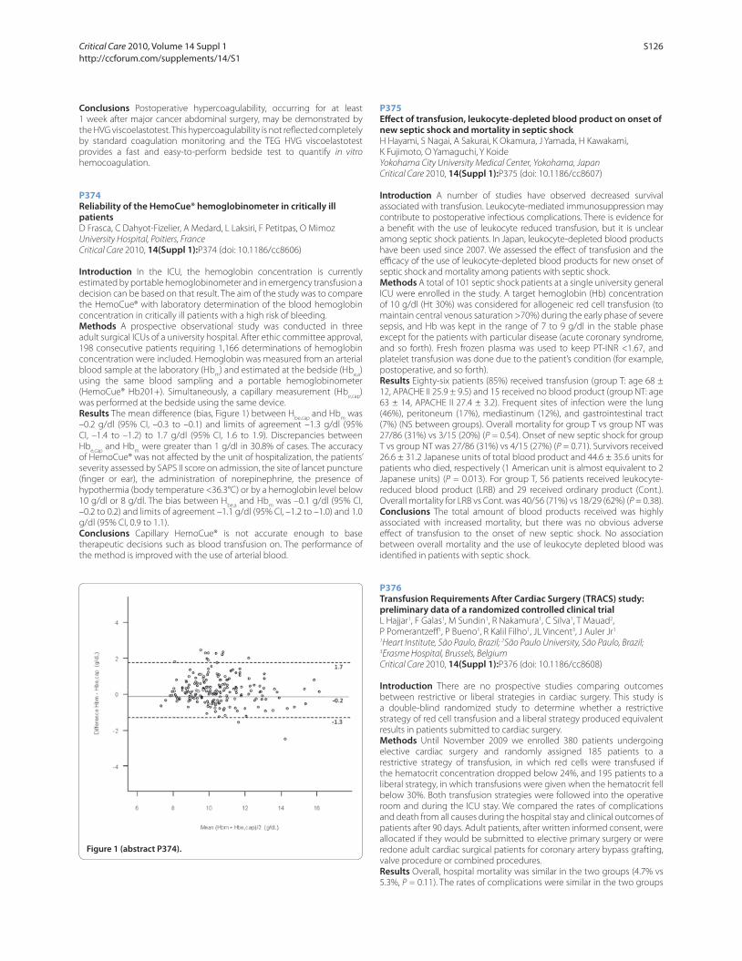
Conclusions Postoperative hypercoagulability, occurring for at least
1 week after major cancer abdominal surgery, may be demonstrated by
the HVG viscoelastotest. This hypercoagulability is not refl ected completely
by standard coagulation monitoring and the TEG HVG viscoelastotest
provides a fast and easy-to-perform bedside test to quantify in vitro
hemocoagulation.
P374
Reliability of the HemoCue® hemoglobinometer in critically ill patientsD Frasca, C Dahyot-Fizelier, A Medard, L Laksiri, F Petitpas, O Mimoz
University Hospital, Poitiers, France
Critical Care 2010, 14(Suppl 1):P374 (doi: 10.1186/cc8606)
Introduction In the ICU, the hemoglobin concentration is currently
estimated by portable hemoglobinometer and in emergency transfusion a
decision can be based on that result. The aim of the study was to compare
the HemoCue® with laboratory determination of the blood hemoglobin
concentration in critically ill patients with a high risk of bleeding.
Methods A prospective observational study was conducted in three
adult surgical ICUs of a university hospital. After ethic committee approval,
198 consecutive patients requiring 1,166 determinations of hemoglobin
concentration were included. Hemoglobin was measured from an arterial
blood sample at the laboratory (Hbm
) and estimated at the bedside (Hbe,a
)
using the same blood sampling and a portable hemoglobinometer
(HemoCue® Hb201+). Simultaneously, a capillary measurement (Hbe,cap
)
was performed at the bedside using the same device.
Results The mean diff erence (bias, Figure 1) between Hbe,cap
and Hbm
was
–0.2 g/dl (95% CI, –0.3 to –0.1) and limits of agreement –1.3 g/dl (95%
CI, –1.4 to –1.2) to 1.7 g/dl (95% CI, 1.6 to 1.9). Discrepancies between
Hbe,cap
and Hbm
were greater than 1 g/dl in 30.8% of cases. The accuracy
of HemoCue® was not aff ected by the unit of hospitalization, the patients’
severity assessed by SAPS II score on admission, the site of lancet puncture
(fi nger or ear), the administration of norepinephrine, the presence of
hypothermia (body temperature <36.3°C) or by a hemoglobin level below
10 g/dl or 8 g/dl. The bias between Hbe,a
and Hbm
was –0.1 g/dl (95% CI,
–0.2 to 0.2) and limits of agreement –1.1 g/dl (95% CI, –1.2 to –1.0) and 1.0
g/dl (95% CI, 0.9 to 1.1).
Conclusions Capillary HemoCue® is not accurate enough to base
therapeutic decisions such as blood transfusion on. The performance of
the method is improved with the use of arterial blood.
P375
Eff ect of transfusion, leukocyte-depleted blood product on onset of new septic shock and mortality in septic shockH Hayami, S Nagai, A Sakurai, K Okamura, J Yamada, H Kawakami,
K Fujimoto, O Yamaguchi, Y Koide
Yokohama City University Medical Center, Yokohama, Japan
Critical Care 2010, 14(Suppl 1):P375 (doi: 10.1186/cc8607)
Introduction A number of studies have observed decreased survival
associated with transfusion. Leukocyte-mediated immunosuppression may
contribute to postoperative infectious complications. There is evidence for
a benefi t with the use of leukocyte reduced transfusion, but it is unclear
among septic shock patients. In Japan, leukocyte-depleted blood products
have been used since 2007. We assessed the eff ect of transfusion and the
effi cacy of the use of leukocyte-depleted blood products for new onset of
septic shock and mortality among patients with septic shock.
Methods A total of 101 septic shock patients at a single university general
ICU were enrolled in the study. A target hemoglobin (Hb) concentration
of 10 g/dl (Ht 30%) was considered for allogeneic red cell transfusion (to
maintain central venous saturation >70%) during the early phase of severe
sepsis, and Hb was kept in the range of 7 to 9 g/dl in the stable phase
except for the patients with particular disease (acute coronary syndrome,
and so forth). Fresh frozen plasma was used to keep PT-INR <1.67, and
platelet transfusion was done due to the patient’s condition (for example,
postoperative, and so forth).
Results Eighty-six patients (85%) received transfusion (group T: age 68 ±
12, APACHE II 25.9 ± 9.5) and 15 received no blood product (group NT: age
63 ± 14, APACHE II 27.4 ± 3.2). Frequent sites of infection were the lung
(46%), peritoneum (17%), mediastinum (12%), and gastrointestinal tract
(7%) (NS between groups). Overall mortality for group T vs group NT was
27/86 (31%) vs 3/15 (20%) (P = 0.54). Onset of new septic shock for group
T vs group NT was 27/86 (31%) vs 4/15 (27%) (P = 0.71). Survivors received
26.6 ± 31.2 Japanese units of total blood product and 44.6 ± 35.6 units for
patients who died, respectively (1 American unit is almost equivalent to 2
Japanese units) (P = 0.013). For group T, 56 patients received leukocyte-
reduced blood product (LRB) and 29 received ordinary product (Cont.).
Overall mortality for LRB vs Cont. was 40/56 (71%) vs 18/29 (62%) (P = 0.38).
Conclusions The total amount of blood products received was highly
associated with increased mortality, but there was no obvious adverse
eff ect of transfusion to the onset of new septic shock. No association
between overall mortality and the use of leukocyte depleted blood was
identifi ed in patients with septic shock.
P376
Transfusion Requirements After Cardiac Surgery (TRACS) study: preliminary data of a randomized controlled clinical trialL Hajjar1, F Galas1, M Sundin1, R Nakamura1, C Silva1, T Mauad2,
P Pomerantzeff 1, P Bueno1, R Kalil Filho1, JL Vincent3, J Auler Jr1
1Heart Institute, São Paulo, Brazil; 2São Paulo University, São Paulo, Brazil; 3Erasme Hospital, Brussels, Belgium
Critical Care 2010, 14(Suppl 1):P376 (doi: 10.1186/cc8608)
Introduction There are no prospective studies comparing outcomes
between restrictive or liberal strategies in cardiac surgery. This study is
a double-blind randomized study to determine whether a restrictive
strategy of red cell transfusion and a liberal strategy produced equivalent
results in patients submitted to cardiac surgery.
Methods Until November 2009 we enrolled 380 patients undergoing
elective cardiac surgery and randomly assigned 185 patients to a
restrictive strategy of transfusion, in which red cells were transfused if
the hematocrit concentration dropped below 24%, and 195 patients to a
liberal strategy, in which transfusions were given when the hematocrit fell
below 30%. Both transfusion strategies were followed into the operative
room and during the ICU stay. We compared the rates of complications
and death from all causes during the hospital stay and clinical outcomes of
patients after 90 days. Adult patients, after written informed consent, were
allocated if they would be submitted to elective primary surgery or were
redone adult cardiac surgical patients for coronary artery bypass grafting,
valve procedure or combined procedures.
Results Overall, hospital mortality was similar in the two groups (4.7% vs
5.3%, P = 0.11). The rates of complications were similar in the two groups
Figure 1 (abstract P374).
Critical Care 2010, Volume 14 Suppl 1 http://ccforum.com/supplements/14/S1
S126

(28.3% in the restrictive-strategy group and 26.1% in the liberal-strategy
group, P = 0.33). The ICU stay was signifi cantly lower in the restrictive-
strategy group (2.2 days vs 4.1 days, P = 0.05). There was no diff erence in
90-day mortality between groups.
Conclusions A restrictive strategy of red cell transfusion is at least as
eff ective as and possibly superior to a liberal transfusion strategy in
patients submitted to cardiac surgery.
References1. Napolitano LM, et al.: Clinical practice guideline: red blood cell transfusion
in adult trauma and critical care. Crit Care Med 2009, 37:3124-3157.
2. Hébert PC, et al.: Controversies in RBC transfusion in the critically ill. Chest
2007, 131:1583-1590.
P377
Analysis of red cell transfusion practices in patients without active haemorrhage over a 12-month period in a UK intensive care unitRL Eve, MH Spivey, S Hart, MR Duff y
Derriford Hospital, Plymouth, UK
Critical Care 2010, 14(Suppl 1):P377 (doi: 10.1186/cc8609)
Introduction The deleterious eff ects of red blood cell (RBC) transfusion
are well known [1] and restrictive transfusion practices are safe in
patients without active haemorrhage [2]. Our objective was to determine
transfusion practices in critically ill patients without evidence of ongoing
bleeding to establish our conformity with published guidelines [3].
Methods All adult ICU patients receiving RBC transfusions between 1
September 2008 and 31 August 2009 were included in the analysis. Data
were collected on demographics, APACHE II score, ICU and hospital length
of stay (LOS), ICU and hospital mortality, presence of ischaemic heart
disease (IHD), and pre/post-transfusion haemoglobin concentrations in
g/dl ([Hb]). Subgroup analyses were performed for patients with IHD, age
<55 years or APACHE II ≤20. We analysed patients with IHD because benefi t
from liberal transfusion has not been confi rmed [1] whereas the latter two
subgroups have shown a mortality benefi t with restrictive transfusion [2].
Results A total of 1,723 patients were admitted to the ICU during the study
period. Two hundred and fi ve patients (11.9%) received RBCs, of whom
47 had active bleeding and were excluded from further analysis. The
remaining 158 patients (9.2%) received a total of 477 units RBCs (median
2.0 units/patient, IQR 1.0 to 3.8). Median pre-transfusion [Hb] was 7.7 (IQR
7.3 to 8.2) with a post-transfusion [Hb] of 9.2(IQR 8.5 to 9.8). Median ICU and
hospital LOS in days was 10.9 (IQR 4.9 to 18.8) and 26.9 (IQR 14.0 to 45.0).
ICU and hospital mortalities were 34.2% and 44.9%, respectively. Patients
with acute IHD had a pre-transfusion [Hb] of 8.3(IQR 7.8 to 8.8) with a post-
transfusion [Hb] of 9.4 (IQR 9.2 to 10.2) whilst patients with chronic IHD
had values of 7.9 (IQR 7.4 to 8.4) and 9.4 (IQR 8.5 to 9.8), respectively. In
patients aged <55 years, pre/post-transfusion values were 7.5 (IQR 7.1 to
8.0) and 9.2 (IQR 8.5 to 10.0), respectively, and patients with APACHE II ≤20
had values of 7.7 (IQR 7.3 to 8.2) and 9.2 (IQR 8.5 to 9.8), respectively.
Conclusions [Hb] transfusion triggers were >7 in all subgroups and
post-transfusion [Hb] was >9. Transfusion strategies were too liberal. It is
important to restrict transfusions to limit morbidity and mortality and to
make effi cient use of RBCs [4]. This study demonstrates the importance of
regular audit and will be used to inform local guidelines.
References1. Marik PE, et al.: Crit Care Med 2008, 36:2667-2674.
2. Hebert PC, et al.: N Engl J Med 1999, 340:409-417.
3. Thomas D, et al.: Red Cell Transfusion 2 [http://www.aagbi.org/publications/
guidelines/docs/red_cell_08.pdf ]
4. Joseph BG, et al.: Transfusion 2009, 49:2060-2069.
P378
Eff ects of red blood cell transfusion in central venous saturation and lactate levels in patients with septic shockBF Mazza, M Assunção, F Freitas, M Jackiu, FR Machado
Universidade Federal de São Paulo, Brazil
Critical Care 2010, 14(Suppl 1):P378 (doi: 10.1186/cc8610)
Introduction Transfusion of red blood cells has been used with the
objective of improving oxygen delivery to the tissues in the critically
ill patient. However, the appropriate hemoglobin (Hb) level is still
controversial in septic shock patients [1,2]. The objective of this study was
to evaluate the eff ect of red blood cell transfusion on the central venous
saturation (SvcO2) and lactate levels in patients with septic shock with two
diff erent levels of Hb.
Methods Patients with less than 48 hours of septic shock and Hb levels
under 9.0 g/dl were randomized to maintain Hb over 9.0 g/dl (Group 1)
or over 7.0g/dl (Group 2). Before and up to 1 hour after each transfusion,
Hb, lactate and SvcO2 were determined. Results were expressed as median
(25% to 75% percentile) or percentage and analyzed through chi-square
or Mann–Whitney test. P <0.05 was considered signifi cant.
Results Forty-six patients were included with a total of 74 transfusions,
being 39 in Group 1 and 35 in Group 2, without any diff erence in
demographic or hemodynamic data between the groups. Before and
after Hb levels were 8.5 (8.2 to 8.7) g/dl and 9.4 (9.1 to 9.5) g/dl in Group
1 (P = 0.000) and 6.8 (6.6 to 6.9) g/dl and 7.6 (7.4 to 7.8) g/dl in Group 2
(P = 0.000). A reduction in median lactate after transfusion was found in
Group 2 patients (22.0 (18.1 to 29.1) mg/dl and 20.0 (16.3 to 25.2) mg/dl,
P = 0.005) but not in Group 1 (17.2 (16.3 to 23.9) mg/dl and 18.1 (15.4 to
21.8) mg/dl, P = 0.23). The same occurred regarding SvcO2 with improved
values only in Group 2 (68.0 (64.0 to 72.0)% and 72.0 (69.0 to 75.0)% in
Group 2, P <0.000) and not in Group 1 (72.0 (69.0 to 74.0)% and 72.0 (71.0
to 73.0)%, P = 0.98). In patients with altered lactate levels a reduction
was found regardless of the basal levels of hemoglobin (P = 0.002 and
P = 0.001 for Groups 1 and 2, respectively). The same was demonstrated
in patients with SvcO2 <70% (P = 0.007 and P = 0.000 for Groups 1 and
2, respectively). On the other hand, patients with normal levels of both
perfusion parameters did not present a worsening of those variables,
either in Group 1 or Group 2.
Conclusions Red blood cell transfusion improved tissue perfusion in
patients with signs of hypoperfusion regardless of basal levels of Hb.
Transfusion did not seem to worsen tissue perfusion in patients without
signs of hypoperfusion, even in the group with Hb levels above 9.0 g/dl.
References1. Hebert PC: N Engl J Med 1999, 340:409-417.
2. Rivers E: N Engl J Med 2001, 345:1368-1377.
P379
Massive transfusion in paediatric trauma: a single-centre experienceR Hearn, M Tecle, S Rajan, N Edmonds
The Royal London Hospital, London, UK
Critical Care 2010, 14(Suppl 1):P379 (doi: 10.1186/cc8611)
Introduction Uncontrolled haemorrhage is a leading cause of early
mortality in trauma. Administration of blood products, packed red cells
and fresh frozen plasma (PRBC:FFP) in the ratio of 1:1 is associated with
improved mortality in major haemorrhage in adult trauma. However, there
is little evidence available for the management of major haemorrhage in
the context of paediatric trauma.
Methods We retrospectively analysed the local paediatric trauma
database of all children following trauma call activation on arrival at the
emergency department of The Royal London Hospital, a major urban
trauma centre in London. The study period was over 15 months, May 2008
to August 2009. During this period, there has been no major haemorrhage
policy within the centre for paediatric trauma. We collected data on
demographic profi le, injury severity scores, blood products transfused
in the fi rst 24 hours and the outcomes. We defi ned massive transfusion
as the requirement of packed red cells >40 ml/kg in the fi rst 4 hours or
>80 ml/kg in the fi rst 24 hours.
Results Two hundred and twenty-seven children presented to the
emergency department during this period following major trauma call
activation. The median age at presentation was 10.2 years. Thirteen (5.7%)
children had major haemorrhage. The median ISS was 35 (IQR 10 to 60). All
but one were males. Three had penetrating trauma, one of whom made
it to theatre, but all died. Four had emergency damage control surgery.
Abnormal results were seen in three patients, each having one abnormal
result (INR =1.9 and APTT = 86, low Hb = 7.6, thrombocytopaenia = 63).
Eight of the 13 patients received additional blood products such as fresh
frozen plasma, platelets and cryoprecipitate. However, no patient received
the ratio of blood products of PRBC:FFP of 1:1 as practised in adult trauma.
Two patients had no admission bloods done. Worsening coagulation
Critical Care 2010, Volume 14 Suppl 1 http://ccforum.com/supplements/14/S1
S127

parameters were seen in two patients when measured post transfusion
and the remaining 11 patients did not have routine monitoring of blood
parameters post transfusion. Eight (62%) patients died, of which seven
died in the emergency department.
Conclusions Major haemorrhage is associated with a very high mortality
in severely injured children. We recommend rigorous monitoring of
laboratory parameters to guide appropriate administration of blood
products. There is a need for instituting a major haemorrhage policy in
paediatric trauma and consideration of point-of-care testing of blood
parameters.
P380
Cytokine levels evaluation during acute isovolemic anemiaM Kahvegian1, DT Fantoni2, DA Otsuki1, CA Holms1, CO Massoco3, JO Auler Jr1
1Faculdade de Medicina da Universidade de São Paulo, Brazil; 2School
of Veterinary Medicine, Universidade de São Paulo, Brazil; 3Sales Gomes
Consulting, São Paulo, Brazil
Critical Care 2010, 14(Suppl 1):P380 (doi: 10.1186/cc8612)
Introduction To date there is poor evidence that documents the benefi t
of red blood cell transfusion on clinical outcome. Acceptance of a lower
transfusion trigger in the perioperative period requires detailed information
regarding the eff ects of volume replacement in diff erent organs and
systems. Since fl uids can have a widely divergent impact on the immune
response, this study was undertaken to determine the cytokine levels in
a model of acute isovolemic anemia (AIA) with hydroxyethyl starch (HES)
and gelatin (GEL).
Methods Twenty-one pigs were anesthetized, instrumented and
randomized into three groups: Control, HES and GEL. Animals in the HES
and GEL groups were submitted to acute isovolemic anemia to a target
hematocrit of 15% with volume replacement performed with HES 130/0.4
and GEL at a 1:1 ratio. The withdrawn blood was returned to the animals
120 minutes after the end of AIA. TNF, IL-1, IL-6 and IL-10 measurements
were performed with blood samples collected at the femoral vein at the
following time points: Baseline, after instrumentation (INST), immediately
after AIA (AIA), 60 minutes after AIA (60AIA), 120 minutes after AIA (120AIA),
60 minutes after blood infusion (60BI) and 120 minutes after blood
infusion (120BI). The cytokines were measured in serum by a commercially
available ELISA with specifi c monoclonal antibodies. Two-way analyses
of variance (ANOVA) and a Tukey test were used to assess its signifi cance
(P <0.05).
Results TNF varied signifi cantly among the groups in diff erent time points
(AIA: Control, 91 ± 16 and GEL, 221 ± 61; P <0.05; 60AIA: Control, 76 ±
15; HES, 172 ± 42; P <0.05 and GEL, 323 ± 77; P <0.001; 120AIA: Control,
98 ± 6 and GEL, 304 ± 66; P <0.01; 120BI: Control, 71 ± 7 and GEL, 211 ±
71; P <0.05). In relation to IL-1, only in the GEL group (224 ± 56) was
verifi ed a statistical signifi cant at time point 60AIA when compared with
Control (84 ± 17; P <0.05). When IL-6 levels were studied, GEL presented
a statistically signifi cant at 60AIA (GEL, 331 ± 65) when compared with
Control (146 ± 19; P <0.05). Serum IL-10 levels in GEL-treated animals were
statistically signifi cantly elevated at 60AIA (Control, 17 ± 3 and GEL, 46 ±
12; P <0.01) and 120BI (Control, 21 ± 5 and GEL, 59 ± 11; P <0.05).
Conclusions Fluid replacement infl uences the cytokine measurements
during acute isovolemic anemia expressed by serum TNF, IL-1, IL-6 and
IL-10 increases, especially in GEL group.
P381
Haemodynamic alterations in community-acquired sepsis at early stageS Hettwer, J Wilhelm, M Schürmann, H Ebelt, K Werdan
Martin-Luther-University, Halle (Saale), Germany
Critical Care 2010, 14(Suppl 1):P381 (doi: 10.1186/cc8613)
Introduction Community-acquired sepsis at an early stage is common,
but haemodynamic alterations remain unclear. The aim of the study
was to characterize cardiovascular alterations in patients of our ProFS
(monocentric observational) study, which was to characterize patients
with sepsis in the emergency department.
Methods Systemic vascular resistance (SVR) and cardiac output (CO)
were measured non-invasively using a TaskForce monitor (CNSystems,
Graz, Austria) after admission, 24 hours and 72 hours. Indexed values were
calculated (SVRI (dyn·second/cm5/m2), CI (l/minute/m2)). Procalcitonin
(PCT, ng/ml) was measured in serum.
Results A sample of 64 patients of 208 included patients received
haemodynamic examination. Mean age was 61.8 ± 18.0 years, 62.7% were
male. Patients were divided by PCT <2 and ≥2. Age, gender and previous
medical history were comparable in both groups. The heart rate was
99.8 ± 21.6 vs 104.6 ± 23.0/minute (P = NS) and the mean artery pressure
was 89.5 ± 15.6 vs 81.6 ± 21.5 mmHg (P <0.01). Mean SVRI in patients with
PCT <2 was 2,934 ± 1,045 vs 2,376 ± 842, P <0.05 at the time of admission.
No diff erence was found after 24 hours (2,959 ± 1,002 vs 2,924 ± 1,324, P =
NS) and 72 hours (3,123 ± 931 vs 3,556 ± 1,524, P = NS). On the contrary, for
patients with PCT ≥2 the increase after 72 hours was signifi cant (P <0.05).
Diff erences after admission could not be observed for CI between patients
with PCT <2 vs ≥2. Mean values after admission were 2.7 ± 1.0 vs 2.8 ± 0.8,
after 24 hours 2.5 ± 0.8 vs 2.5 ± 0.5, and after 72 hours 2.3 ± 0.7 vs 2.3 ± 0.6
(all: P = NS). See Figure 1
Conclusions Patients with community-acquired sepsis in the emergency
department had an elevated SVRI. At the time of admission patients with
high PCT had a signifi cantly lower SVRI than patients with low PCT. Cardiac
index at the time of admission was at a lower limit of normal range in
all patients. These fi ndings are in strong contrast to the classic pattern
of sepsis on the ICU, where SVRI is keenly reduced and CI elevated. They
implicate that patients with sepsis in the emergency department may
benefi t more from application of fl uid and positive inotrope substances
than from vasopressor.
Figure 1 (abstract P381). Systemic vascular resistance index and cardiac index in sepsis in the emergency department.
Critical Care 2010, Volume 14 Suppl 1 http://ccforum.com/supplements/14/S1
S128

P382
Epidemiology of severe sepsis in India: an updateS Todi1, S Chatterjee1, S Sahu2, M Bhattacharyya1
1AMRI Hospitals, Kolkata, India; 2Kalinga Hospital, Bhubaneswar, India
Critical Care 2010, 14(Suppl 1):P382 (doi: 10.1186/cc8614)
Introduction A multicentre, prospective, observational study conducted
in four intensive therapy units (ITUs) in India from June 2006 to June 2009
to determine the incidence and outcome of severe sepsis among adult
patients.
Methods All patients admitted to the ITU were screened daily for SIRS,
organ dysfunction and severe sepsis as defi ned by the ACCP and SCCM.
Patient with severe sepsis were further studied.
Results A total of 5,478 ITU admissions were studied. SIRS with organ
dysfunction was found in 1,385 (25%) patients, of which 731 (52.77%) were
due to sepsis. The incidence of severe sepsis was 16.45% of all admissions.
Mean age of the study population was 58.17 years (SD 18.66), of which
57.71% were male. The median APACHE II score was 13 (IQR 13 to 14) with
predominant (90.93%) medical admission. ITU mortality of all admissions
was 12.08% and that of severe sepsis was 59.26%. Hospital mortality and
28-day mortality of severe sepsis were 65.2% and 64.6%, respectively. The
standardized mortality ratio of severe sepsis patients was 1.45. Median
duration of stay in the ITU for the severe sepsis cohort who survived was
13 days (IQR 11 to 17). The number of episodes where infection was the
primary reason for admission to the ITU was 86.32%. Culture positivity was
found in 61.6%. The lung was the predominant source of sepsis (57.45%).
Gram-negative organisms were responsible for 72.45% of cases and Gram-
positive for 13.13%. The rest were parasitic, viral and fungal infection.
Conclusions Severe sepsis was common in Indian ITUs. ITU mortality was
higher compared with western literature. Gram-positive infections were
less common although incidences of parasitic and viral infection were
higher than in the West.
P383
Systemic infl ammatory response syndrome is reduced by inline fi ltration in intensive care patientsT Jack, M Boehne, BE Brent, A Wessel, M Sasse
Medical School Hannover, Germany
Critical Care 2010, 14(Suppl 1):P383 (doi: 10.1186/cc8615)
Introduction Sepsis, systemic infl ammatory response syndrome (SIRS) or
organ failure often complicate the clinical course on an ICU. Particulate
contamination of the infusion solution may contribute to the clinical
deterioration of these patients. Particles have been shown to induce
thrombogenesis, deterioration of microcirculation and modulation of
immunoresponse. The use of inline fi ltration with microfi lters almost
completely prevents particulate infusion. We assessed the eff ect of inline
fi ltration on the reduction of major complications in critically ill children
(Clinical Trials.gov ID NCT 00209768).
Methods In a randomised, prospective trial 807 paediatric patients
admitted to the interdisciplinary PICU of a tertiary university hospital were
assigned to either a control or an interventional group, the latter receiving
inline fi ltration (infusion fi lter Pall ELD96LLCE/ NOE96E, Braun Intrapur
Lipid/Intrapur Neonat Lipid) throughout the whole infusion therapy. Prior
to this study, the infusion regimen was optimised to prevent precipitation
and incompatibilities of solutions and drugs. Primary objectives included
a reduction in the incidence of sepsis, thrombosis, SIRS, organ failure (liver,
lung, kidney, circulation) and mortality.
Results eight hundred and seven children (343 female, 464 male) with
a heterogeneous background of underlying diagnoses and a Gaussian
distribution to either the control group (406 patients) or the inline fi ltration
group (401 patients) were included. According to the study criteria, a
signifi cant reduction in the incidence of SIRS for the interventional group
(95% CI, 145 versus 200 patients, P <0.001) was evident. No diff erences
were demonstrated for the occurrence of sepsis, thrombosis, organ failure
(liver, lung, kidney, circulation) or mortality between the control and
interventional groups.
Conclusions The occurrence of SIRS often complicates the treatment in
intensive care medicine. Inline fi ltration is most eff ective, reducing the
incidence of SIRS, and off ers a novel therapeutic option.
P384
Review of central venous catheter-related infections in neurointensive care patients in a tertiary referral centreC Tan, P Nair, A Sule, M Rothburn
The Walton Centre for Neurology and Neurosurgery, Liverpool, UK
Critical Care 2010, 14(Suppl 1):P384 (doi: 10.1186/cc8616)
Introduction Intravenous catheter-related bloodstream infections (ICR-BSI)
are a major contributing factor to in-hospital mortality and morbidity,
extending the inpatient stay by 10 days and expenditure per patient by
£2,000 to £30,000 [1]. A prospective survey was conducted in our unit on all
patients with central venous catheters to ascertain the incidence of ICR-BSI,
identify the organisms and determine the occurrence of infection from the
various sites – femoral, internal jugular and subclavian lines.
Methods The survey was carried out over a period of 13 weeks. Data
collected from patients’ case notes included site of central line insertion,
length of line in situ, reason for line removal and positive blood culture
reports.
Results During the study period, 104 patients were treated on the unit.
Fifty-two central venous lines were inserted in 36 patients (63.5% femoral
(n = 33), 32.7% internal jugular (n = 17) and 3.9% subclavian lines (n = 2)).
The lines were reviewed daily and removed if indicated clinically (pyrexia
or raised white cell count) or if not required. A total 51.5% of femoral lines
(n = 17) were removed due to clinical indications, as were 29% (n = 5) of
internal jugular and 50% (n = 1) of subclavian lines. The average duration
of a line remaining in situ was 4.5 days for femoral, 6 days for internal
jugular and 5 days for subclavian lines. Blood cultures were taken at the
time of line removal. These yielded positive results in eight femoral, seven
internal jugular and one subclavian line. Our survey indicated that the
incidence of ICR-BSI in our unit is 30.8% (of this 62.5% coagulase-negative
staphylococci (CNS), 12.5% E. coli and Pseudomonas each, and 6.25% MSSA
and MRSA each).
Conclusions The distribution of microorganisms causing bacteraemia is
broadly similar in our unit to that in other teaching hospitals in the UK [2],
in that CNS was the commonest organism isolated. However, E. coli and
Pseudomonas were the next common organisms, unlike other units where
Staphylococcus aureus was the second most prevalent organism. The
incidence of bacteraemia from femoral lines (53.7/1,000 catheter-days)
was lower than that from internal jugular lines (68.6/1,000 catheter-days)
possibly due to a higher index of suspicion in the case of femoral lines and
earlier removal (Figure 1). Our study highlights the fact that femoral lines,
which are often the safest option for unstable patients with head injury,
can be eff ectively managed with strict adherence to guidelines to reduce
ICR-BSI.
References1. Maki DG, Kluger DM, Crnich CJ: The risk of bloodstream infection in adults
with diff erent intravascular devices: a systematic review of 200 published prospective studies. Mayo Clin Proc 2006, 81:1159-1171.
2. Coello R, Charlett A, Ward V, et al.: Device-related sources of bacteraemia in English hospitals – opportunities for the prevention of hospital-acquired bacteraemia. J Hosp Infect 2003, 53:46-57.
Figure 1 (abstract P384). Incidence of ICR-BSI in The Walton Centre for
Neurology and Neurosurgery intensive therapy unit.
Critical Care 2010, Volume 14 Suppl 1 http://ccforum.com/supplements/14/S1
S129

P385
Clinical impact of diagnosis methods for catheter-related bloodstream infection in ICU patientsRO Deliberato, AR Marra, OF Dos Santos, SB Wey
Hospital Israelita Albert Einstein, São Paulo, Brazil
Critical Care 2010, 14(Suppl 1):P385 (doi: 10.1186/cc8617)
Introduction The central venous catheter (CVC) is indispensable in the
ICU. Catheter-related bloodstream infection (CR-BSI) is the leading cause
of healthcare-associated infections and one of the most important
complications from the CVC. Nowadays there are two well-known ways
to diagnose CR-BSI: the standard method (withdrawal of the catheter)
and the conservative method (without withdrawal of the catheter). Our
objective was to compare the in-hospital mortality between the two
methods in patients with CR-BSI (short-term catheter) in the ICU.
Methods This study was conducted in a 38-bed mixed ICU in a tertiary
hospital. We reviewed all episodes of CR-BSI that occurred in our ICU from
January 2000 to December 2008. The standard method was defi ned in
a patient with a CVC with at least one positive blood culture obtained
from a peripheral vein and a positive semiquantitative (>15 CFU) catheter
segment culture whereby the same organism (species and antibiogram)
was isolated from the catheter segment and peripheral blood. The
conservative method was defi ned in a patient with a CVC with at least
one positive blood culture obtained from a peripheral vein and one of the
following: diff erential time period of CVC culture versus peripheral culture
positivity of more than 2 hours, or simultaneous quantitative blood culture
with a ratio ≥5:1 (CVC versus peripheral).
Results During the study period, 247 episodes of bloodstream infection
were identifi ed; of these, 192 were catheter-associated bloodstream
infection and 55 were CR-BSI (39 standard method and 17 conservative
method). Considering the CR-BSI patients, the mean age ± standard
variation (SD) was 64 ± 19.29 (years), 75% were under mechanical
ventilation, 55% were under vasopressors, 40% were under total parental
nutrition, 40% were under hemodialysis, 72% were double lumen and the
mean time ± SD in place was 16.32 ± 8.56 (days). The in-hospital mortality
of the standard method versus the conservative method did not show any
diff erence (57% vs 75%, P = 0.208).
Conclusions This study showed that there is no diff erence in the in-
hospital mortality between the standard method versus the conservative
method in patients in the ICU with CR-BSI.
References1. O’ Grady NP, et al.: Centers for Disease Control and Prevention. MMWR
Recomm Rep 2002, 51:1-29.
2. Bouza et al.: Clin Infect Dis 2007, 44:820-826.
P386
Catheter-related bacteremia in a jugular site with tracheostomy and in a femoral siteL Lorente, S Palmero, C Naranjo, J Jiménez, J Iribarren, C García, R Galván,
J Castedo, J Martínez, M Mora
Hospital Universitario de Canarias, Santa Cruz de Tenerife, Spain
Critical Care 2010, 14(Suppl 1):P386 (doi: 10.1186/cc8618)
Introduction In previous guidelines of the Centers for Disease Control
and Prevention (CDC) [1] and in the recently published Guidelines of
the Society for Healthcare Epidemiology of America/Infectious Diseases
Society of America (SHEA/IDSA) [2] it is recommended to avoid the
femoral access to reduce the risk of catheter-related bacteremia (CRB).
However, in these guidelines there are no recommendations for the
catheter site regarding the presence of tracheostomy, and we have not
found data about the incidence of CRB comparing a central jugular site
with tracheostomy and a femoral site. The objective of this study was to
determine whether a jugular site with tracheostomy may have a higher
risk of CRB than a femoral site.
Methods A prospective, observational, 4-year study (from 1 May 2000 to
30 April 2004) was carried out in the medical–surgical ICU of the University
Hospital of the Canary Islands (Tenerife, Spain). We included all patients
undergoing a central jugular catheterization with tracheostomy or femoral
venous catheterization.
Results In the period of study, were used 208 catheters in femoral access
and 52 catheters in central internal jugular access with tracheostomy. There
were no signifi cant diff erences between patients with central jugular with
tracheostomy and femoral access in age, sex, APACHE II score, diagnosis
group, use of mechanical ventilation, use of antimicrobials, use of total
parenteral nutrition, use of pulmonary artery catheter, and duration of the
catheter. We diagnosed 16 CRB in 208 femoral catheters during 1,679 days
of catheterization and 10 CRB in 52 central internal jugular catheters with
tracheostomy during 462 days of catheterization. The incidence of CRB
was higher in the central internal jugular with tracheostomy than in the
femoral site (21.64 vs 9.52 per 1,000 catheter-days; risk ratio = 2.27; 95%
confi dence interval = 1.04 to 4.97; P = 0.04).
Conclusions The femoral site could be considered a more safe venous
access than the central internal jugular in patients with tracheostomy to
minimize the risk of CRB.
References1. MMWR 2002, 1:1-29.
2. Infect Control Hosp Epidemiol 2008, 29(Suppl 1):S22-S30.
P387
A comparative study between conventional and antiseptic impregnated central venous catheters in neurocritical patientsS Kiff er Macedo, G Castro Lima, L Bonani de Almeida Brito,
A Davila Carvalho, JL Fernandes Molina Filho
Hospital São José do Avaí, Itaperuna, Brazil
Critical Care 2010, 14(Suppl 1):P387 (doi: 10.1186/cc8619)
Introduction The central venous catheter (CVC) is very useful in the
ICU. A comparative study between antiseptic impregnated and standard
catheters is therefore of great value.
Methods Alternating the type of CVC used in each patient, we recorded
for each patient: sex, age, APACHE II score, GCS, site of the puncture, reason
for withdrawal of the catheter and the type of catheter used. The tip of the
catheter was cultured. The groups were divided: group I (41 patients, 54
punctures) used the standard CVC, and group II (38 patients, 54 punctures)
used the impregnated CVC.
Results Sixty-two patients were included (48.38% male). We studied 108
periods, of which 54 were standard CVCs and 54 were impregnated CVCs.
The average length of stay was higher for impregnated CVCs (14.11 days)
compared with standard CVCs (10.7 days). Excluding death in both groups,
the length of stay of the catheter in group I was 10.86 days, compared
with 15.43 days in group II. Adding all periods of catheterization, group I
have an amount of 578 days, and 762 days for group II. The total duration
for group II was 31.84% higher than for group I. Regarding the reason for
withdrawal of the CVC, predominant were suspected infection 77.8% of
the time for standard CVCs, and 49.1% of the time for impregnated CVCs.
The culture of the catheter’s tip was positive in 10 (18.5%) standard CVs,
against eight (15.1%) CVCs in the impregnated group. Most patients had
GCS <9.The average APACHE II score was17.97 in group I, compared with
19.63 in group II. The predominant site of puncture was the subclavian
vein (56.48%). See Figure 1.
Conclusions According to our study we observed that the length of
stay with use of the impregnated CVC was higher (15.43 days). The rate
of colonization was higher in the standard CVC. Patients who require a
CVC for long periods have benefi ted with the use of impregnated CVCs,
Figure 1 (abstract P387).
Critical Care 2010, Volume 14 Suppl 1 http://ccforum.com/supplements/14/S1
S130
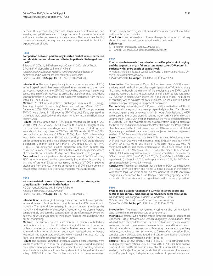
because they present long-term use, lower rates of colonization, and
avoiding complications related to the procedure of successive punctures
and related to the permanence of the catheters. The benefi t reached by
the use of antiseptic impregnated catheters compensated for the initial
expensive cost of 40%.
P388
Comparison between peripherally inserted central venous catheters and short-term central venous catheter in patients discharged from the ICUS Batacchi1, G Zagli1, S diValvasone2, M Ciapetti1, G Cianchi1, V Tucci1,
G Martini1, M Mostardini1, S Proietti1, A Peris1
1Careggi Teaching Hospital, Florence, Italy; 2Postgraduate School of
Anesthesia and Intensive Care, University of Florence, Italy
Critical Care 2010, 14(Suppl 1):P388 (doi: 10.1186/cc8620)
Introduction The use of peripherally inserted central catheters (PICCs)
in the hospital setting has been indicated as an alternative to the short-
term central venous catheter (ST-CVC) in providing prolonged intravenous
access. The aim of our study was to assess the rate of complications (deep
venous thrombosis (DVT), infections) in patients discharged from the ICU
with a PICC or a ST-CVC.
Methods A total of 239 patients discharged from our ICU (Careggi
Teaching Hospital, Florence, Italy) have been followed (March 2007 to
December 2008). PICCs were used in 114 patients (PICC group), whereas
ST-CVCs were placed in 125 patients (ST-CVC group). Data, expressed as
the mean, were analysed with the Mann–Whitney test and Fisher’s exact
test (P <0.05).
Results The PICC group and ST-CVC group resulted similar in age (54.3
vs 58.8), BMI (25.6 vs 26.5), SAPS II (46.2 vs 46.5), and ICU length of stay
(17.6 vs 16.4). Admission diagnoses in the PICC and ST-CVC groups
were also similar: major trauma (38.6% vs 44.8%), sepsis (37.7% vs 32%),
postsurgical complications (23.7% vs 23.2%). Total PICC catheter-days
were 4,024, whereas total ST-CVC catheter-days were 2,764. Despite
similar demographic and clinical characteristics, the PICC group showed
a signifi cantly higher rate of DVT than ST-CVC group (37.7% vs 14.4%;
P <0.01). This diff erence resulted signifi cant also with catheter-day
correction (number of events*1,000/catheter-days). The highest incidence
of DTV events occurred after 15 days from placement in both groups.
Conclusions In our experience, the signifi cant rate of DVT found using
PICCs induces one to consider a presumably higher thrombogenicity of
this kind of catheter. Based on our result, the use of ST-CVC in patients
discharged from the ICU, who are at higher risk of DVT development in
regard of the recent critically ill status, might be more appropriate.
P389
Vacuum-assisted closure of laparostomy, an effi cient strategy for complicated intra-abdominal infectionNG Germano, IG Gonçalves, B Moya, R Ribeiro
Hospital S Bernardo, Setúbal, Portugal
Critical Care 2010, 14(Suppl 1):P389 (doi: 10.1186/cc8621)
Introduction The chirurgical strategy for infection control in complicated
intra-abdominal infections is responsible alone for 40% reduction in
mortality. The second look strategy in tertiary peritonitis reduces the
mortality and morbidity of the patients. Vacuum-assisted closure therapy
can potentially decrease the concentration of proinfl ammatory cytokines,
bacterial count, management of third-space fl uid and improved input and
output monitoring.
Methods The authors present a revision of 60 patients with tertiary
peritonitis admitted to the ICU after chirurgical infection control. All
patients have septic shock at admission. Twelve percent of them were
admitted with an open abdomen and vacuum-assisted closure therapy
was used. The parameters evaluated were hospital mortality, ICU stay
(days) and time of mechanical ventilation (days).
Results The patients submitted to vacuum-assisted closure therapy were
similar to patients in whom the abdominal wall was closed, regarding
the risk factors for peritoneal infection (corticotherapy, oncologic disease,
renal insuffi ciency, hepatic insuffi ciency, desnutrition, hypoalbuminemia,
a high APACHE II score). The patients submitted to vacuum-assisted
closure therapy had a higher ICU stay and time of mechanical ventilation
but lower hospital mortality.
Conclusions Vacuum-assisted closure therapy is superior to primary
abdominal wall closure in patients with tertiary peritonitis.
References1. Becker HP, et al.: Scand J Surg 2007, 96:263-271.
2. Andrabi SIH, et al.: J Ayub Med Coll Abottabad 2007, 19.
P390
Comparison between left ventricular tissue Doppler strain imaging and the sequential organ failure assessment score (SOFA score) in patients with severe sepsis or septic shockM Masaki, J Pulido, T Yuasa, S Gillespie, B Afessa, D Brown, S Mankad, J Oh
Mayo Clinic, Rochester, MN, USA
Critical Care 2010, 14(Suppl 1):P390 (doi: 10.1186/cc8622)
Introduction The Sequential Organ Failure Assessment (SOFA) score is
a widely used method to describe organ dysfunction/failure in critically
ill patients. Although the majority of the studies use the SOFA score in
outcome research, little is known about its correlation to left ventricular
(LV) function in patients with severe sepsis and septic shock. The purpose
of this study was to evaluate the correlation of SOFA score and LV function
by tissue Doppler imaging in this patient population.
Methods Sixty patients (ages 66 ± 15, men n = 28) admitted to the ICU with
severe sepsis or septic shock were prospectively enrolled. Transthoracic
echocardiography was performed within 24 hours of admission to the ICU.
We measured the LV end diastolic volume index (LVEDVI), LV end systolic
volume index (LVESVI), LV ejection fraction (LVEF), mitral deceleration time
of E velocity (Dct) and myocardial tissue Doppler strain imaging profi les at
the basal, mid and apical portions of the LV septal wall in the LV apical four-
chamber view. On the ICU admission day, the SOFA scores were calculated.
Signifi cantly correlated parameters were subjected to linear regression
analysis. P <0.05 was considered signifi cant.
Results The mean heart rate was 90 ± 17 bpm, mean LV volumes, mean
LVEF and mean Dct were within normal limits (LVEDVI; 44.6 ± 15.4 ml/m2,
LVESVI; 18.7 ± 11.5 ml/m2, LVEF; 58.9 ± 15.7%, Dct; 175.8 ± 50.2 ms). The
mean peak systolic strain measurements were –16.0 ± 5.0% (basal; –18.1 ±
7.0%, mid; –15.7 ± 5.6%, apical; –14.1 ± 6.6%). The mean SOFA score was
10.8 ± 3.9. Linear regression analysis showed correlation between the
SOFA score and LVEF (r = –0.40, P = 0.02), Dct (r = –0.34, P = 0.007), mean
septal strain (r = 0.40, P = 0.002), mid-septal strain (r = 0.43, P = 0.0007) and
apical septal strain (r = 0.36, P = 0.006).
Conclusions These results suggest that the higher SOFA score had lower
LVEF, lower LV systolic strain and higher LV fi lling pressure in the patients
with severe sepsis or septic shock. An assessment of the left ventricular
longitudinal contraction by tissue Doppler strain imaging may serve as
a useful tool to evaluate multiple organ failure in this patient population.
P391
Systolic and diastolic function and survival in severe sepsis and septic shock: clinical, echocardiographic, biochemical correlationG Landesberg, M Georgieva, Y Meroz, S Goodman, P Levin
Hebrew University – Hadassah Medical Center, Jerusalem, Israel
Critical Care 2010, 14(Suppl 1):P391 (doi: 10.1186/cc8623)
Introduction The exact mechanisms causing cardiac dysfunction in
sepsis are still in major part obscure or controversial.
Methods All patients who had the criteria for severe sepsis or septic shock
underwent daily transthoracic echocardiography examinations from
which detailed data on left ventricular end-diastolic, end-systolic volumes
areas, and diastolic measurements were obtained. In addition, numerous
clinical, hemodynamic, respiratory and laboratory data were prospectively
collected, including data on survival up to 2 years after admission. Blood
samples were collected, centrifuged and serum separated for additional
biomarker tests, mainly troponin and NT-proBNP.
Results A total of 262 patients had 712 (2.5 ± 1.4) transthoracic echo-
cardiography examinations. APACHE was 20.6 ± 7.3, 41% had positive
blood cultures, >40% died within 4 months. On multivariate Cox survival
analysis, the LV ejection fraction, end-diastolic volume and Em wave on
tissue Doppler imaging independently predicted improved survival and
Critical Care 2010, Volume 14 Suppl 1 http://ccforum.com/supplements/14/S1
S131
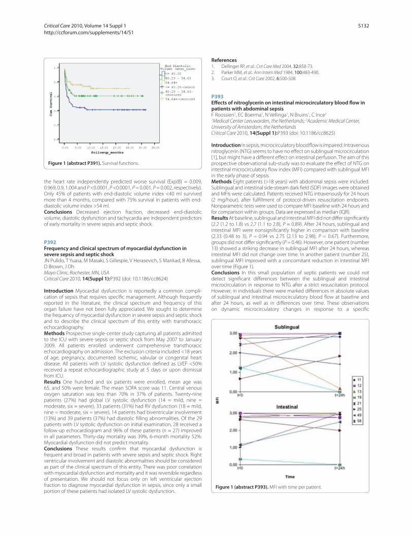
the heart rate independently predicted worse survival (Exp(B) = 0.009,
0.969, 0.9, 1.004 and P <0.0001, P <0.0001, P = 0.001, P = 0.002, respectively).
Only 45% of patients with end-diastolic volume index <40 ml survived
more than 4 months, compared with 75% survival in patients with end-
diastolic volume index >54 ml.
Conclusions Decreased ejection fraction, decreased end-diastolic
volume, diastolic dysfunction and tachycardia are independent predictors
of early mortality in severe sepsis and septic shock.
P392
Frequency and clinical spectrum of myocardial dysfunction in severe sepsis and septic shockJN Pulido, T Yuasa, M Masaki, S Gillespie, V Herasevich, S Mankad, B Afessa,
D Brown, J Oh
Mayo Clinic, Rochester, MN, USA
Critical Care 2010, 14(Suppl 1):P392 (doi: 10.1186/cc8624)
Introduction Myocardial dysfunction is reportedly a common compli-
cation of sepsis that requires specifi c management. Although frequently
reported in the literature, the clinical spectrum and frequency of this
organ failure have not been fully appreciated. We sought to determine
the frequency of myocardial dysfunction in severe sepsis and septic shock
and to describe the clinical spectrum of this entity with transthoracic
echocardiography.
Methods Prospective single-center study capturing all patients admitted
to the ICU with severe sepsis or septic shock from May 2007 to January
2009. All patients enrolled underwent comprehensive transthoracic
echocardiography on admission. The exclusion criteria included <18 years
of age, pregnancy, documented ischemic, valvular or congenital heart
disease. All patients with LV systolic dysfunction defi ned as LVEF <50%
received a repeat echocardiographic study at 5 days or upon dismissal
from ICU.
Results One hundred and six patients were enrolled, mean age was
65, and 50% were female. The mean SOFA score was 11. Central venous
oxygen saturation was less than 70% in 37% of patients. Twenty-nine
patients (27%) had global LV systolic dysfunction (14 = mild, nine =
moderate, six = severe), 33 patients (31%) had RV dysfunction (18 = mild,
nine = moderate, six = severe), 14 patients had biventricular involvement
(13%) and 39 patients (37%) had diastolic fi lling abnormalities. Of the 29
patients with LV systolic dysfunction on initial examination, 28 received a
follow-up echocardiogram and 96% of these patients (n = 27) improved
in all parameters. Thirty-day mortality was 39%, 6-month mortality 52%.
Myocardial dysfunction did not predict mortality.
Conclusions These results confi rm that myocardial dysfunction is
frequent and broad in patients with severe sepsis and septic shock. Right
ventricular involvement and diastolic abnormalities should be considered
as part of the clinical spectrum of this entity. There was poor correlation
with myocardial dysfunction and mortality and it was reversible regardless
of presentation. We should not focus only on left ventricular ejection
fraction to diagnose myocardial dysfunction in sepsis, since only a small
portion of these patients had isolated LV systolic dysfunction.
References1. Dellinger RP, et al.: Crit Care Med 2004, 32:858-73.
2. Parker MM, et al.: Ann Intern Med 1984, 100:483-490.
3. Court O, et al.: Crit Care 2002, 6:500-508.
P393
Eff ects of nitroglycerin on intestinal microcirculatory blood fl ow in patients with abdominal sepsisF Roossien1, EC Boerma1, N Vellinga1, N Bruins1, C Ince2
1Medical Center Leeuwarden, the Netherlands; 2Academic Medical Center,
University of Amsterdam, the Netherlands
Critical Care 2010, 14(Suppl 1):P393 (doi: 10.1186/cc8625)
Introduction In sepsis, microcirculatory blood fl ow is impaired. Intravenous
nitroglycerin (NTG) seems to have no eff ect on sublingual microcirculation
[1], but might have a diff erent eff ect on intestinal perfusion. The aim of this
prospective observational sub-study was to evaluate the eff ect of NTG on
intestinal microcirculatory fl ow index (MFI) compared with sublingual MFI
in the early phase of sepsis.
Methods Eight patients (>18 years) with abdominal sepsis were included.
Sublingual and intestinal side-stream dark fi eld (SDF) images were obtained
and MFIs were calculated. Patients received NTG intravenously for 24 hours
(2 mg/hour), after fulfi llment of protocol-driven resuscitation endpoints.
Nonparametric tests were used to compare MFI baseline with 24 hours and
for comparison within groups. Data are expressed as median (IQR).
Results At baseline, sublingual and intestinal MFI did not diff er signifi cantly
(2.2 (1.2 to 1.8) vs 2,7 (1.1 to 2.8), P = 0.89). After 24 hours, sublingual and
intestinal MFI were nonsignifi cantly higher in comparison with baseline
(2.33 (0.48 to 3), P = 0.94 vs 2.75 (2.13 to 2.98), P = 0.67). Furthermore,
groups did not diff er signifi cantly (P = 0.46). However, one patient (number
13) showed a striking decrease in sublingual MFI after 24 hours, whereas
intestinal MFI did not change over time. In another patient (number 25),
sublingual MFI improved with a concomitant reduction in intestinal MFI
over time (Figure 1).
Conclusions In this small population of septic patients we could not
detect signifi cant diff erences between the sublingual and intestinal
microcirculation in response to NTG after a strict resuscitation protocol.
However, in individuals there were marked diff erences in absolute values
of sublingual and intestinal microcirculatory blood fl ow at baseline and
after 24 hours, as well as in diff erences over time. These observations
on dynamic microcirculatory changes in response to a specifi c
Figure 1 (abstract P391). Survival functions.
Figure 1 (abstract P393). MFI with time per patient.
Critical Care 2010, Volume 14 Suppl 1 http://ccforum.com/supplements/14/S1
S132

thera peutic intervention add to previous reports about heterogeneity of
microcirculatory blood fl ow in sepsis.
Reference1. Boerma et al.: Crit Care Med 2009. [Epub ahead of print]
P394
Diastolic dysfunction in septic shock is an independent predictor of elevated B-type natriuretic peptide and hospital mortalityD Sturgess, T Marwick, C Joyce, C Jenkins, M Jones, P Masci, D Stewart,
B Venkatesh
University of Queensland, Brisbane, Australia
Critical Care 2010, 14(Suppl 1):P394 (doi: 10.1186/cc8626)
Introduction Diastolic dysfunction as demonstrated by tissue Doppler
imaging (TDI), particularly E/E’ (peak early diastolic transmitral/peak early
diastolic mitral annular velocity) is common in critical illness. In septic shock,
the prognostic value of TDI is undefi ned. This study sought to evaluate
and compare the prognostic signifi cance of TDI and cardiac biomarkers
(B-type natriuretic peptide (BNP); N-terminal proBNP (NTproBNP); troponin
T (TnT)) in septic shock. The contribution of fl uid management and
diastolic dysfunction to elevation of BNP was also evaluated.
Methods Twenty-one consecutive adult patients from a multidisciplinary
ICU underwent transthoracic echocardiography and blood collection
within 72 hours of developing septic shock.
Results The mean ± SD APACHE III score was 80.1 ± 23.8. Hospital mortality
was 29%. E/E’ was signifi cantly higher in hospital nonsurvivors (15.32 ± 2.74,
survivors 9.05 ± 2.75; P = 0.0002). Areas under ROC curves were E/E’ 0.94,
TnT 0.86, BNP 0.78 and NTproBNP 0.67. An E/E’ threshold of 14.5 off ered
100% sensitivity and 83% specifi city. Adjustment for APACHE III, cardiac
disease, fl uid balance and grade of diastolic function, demonstrated E/E’ as
an independent predictor of hospital mortality (P = 0.019). Multiple linear
regression incorporating APACHE III, gender, cardiac disease, fl uid balance,
noradrenaline dose, C-reactive protein, ejection fraction and diastolic
dysfunction yielded APACHE III (P = 0.033), fl uid balance (P = 0.001) and
diastolic dysfunction (P = 0.009) as independent predictors of lnBNP.
Conclusions E/E’ is an independent predictor of hospital survival in septic
shock. It off ers better discrimination between survivors and nonsurvivors
than cardiac biomarkers. Fluid balance and diastolic dysfunction were
independent predictors of BNP concentration in septic shock.
P395
Cardiac-specifi c troponin I and cardiovascular dysfunction in post-traumatic sepsisG Georgiev, SG Milanov, V Todorova, L Kozarov, M Milanov
Pirogov Emergency Institute, Sofi a, Bulgaria
Critical Care 2010, 14(Suppl 1):P395 (doi: 10.1186/cc8627)
Introduction Elevated serum levels of cardiac-specifi c troponin I (cTnI)
are claimed to be associated with the degree of sepsis-related cardiac
dysfunction and with the disease outcome in critically ill. We tested this
association in a specifi c group of ICU patients with post-traumatic sepsis.
Methods A prospective observational study was conducted in an ICU of
a level I-equivalent trauma center between September 2008 and August
2009. No exclusion criteria were adopted. Serum cTnI concentrations were
collected at peak sepsis-related cardiovascular dysfunction (defi ned by
the highest pressure adjusted heart rate (PAR) in MODS score and by the
dose of the inotrope). Data were gathered on demographics, severity of
infl ammatory response (SIRS criteria) and organ dysfunction (MODS score),
presence of superimposed acute coronary events, other potential reasons
for cTnI elevation and the disease outcome. Intergroup comparisons were
made with Student’s t test/nonparametric alternatives. The prognostic
utility of cTnI was examined by the construction of ROC curves and its
levels were categorized on the basis of the defi ned cut-off value. Probable
associations between these categories and the organ dysfunction severity,
as well as the outcome, were examined with Fisher’s exact test.
Results A total of 92 polytrauma patients were enrolled. The confi dence
intervals of PAR, MODS and SIRS were 21.26 ± 2.72, 6.99 ± 2.94 and 3.75 ±
1.10, respectively. Blunt cardiac injury had been diagnosed in 20 patients
(21.7%) on admission. Three patients (3.3%) developed acute coronary
events during their ICU stay and 12 patients (13.2%) had other potential
reasons for cTnI elevation. Signifi cantly higher cTnI concentrations were
found in the critically ill with myocardial contusion and with acute coronary
events. However, the presence of blunt cardiac injury had no measurable
outcome eff ect. Troponin I levels were signifi cantly higher in nonsurvivors
(P = 0.00) and very good prognostic performance of cTnI was found
(AUROC = 0.82; cut-off value = 0.05 ng/ml). The subsequent comparison
between its categorized values and those of PAR and MODS showed strong
positive association. Elevated cTnI concentrations (>0.05 ng/ml) signifi cantly
increased the risk of death (RR = 5.58; 95% CI = 1.67 to 20.79; P = 0.002).
Conclusions In a group of patients with post-traumatic sepsis elevated
serum cTnI levels proved to be a good marker of severe organ (including
cardiovascular) dysfunction. They also showed reasonable prognostic
value regarding the disease outcome.
P396
Both systolic and diastolic functions are impaired in critically ill patients with community-acquired sepsisS Hettwer, M Schürmann, J Wilhelm, H Ebelt, K Werdan
Martin-Luther-University, Halle (Saale), Germany
Critical Care 2010, 14(Suppl 1):P396 (doi: 10.1186/cc8628)
Introduction Alterations in haemodynamic parameters in septic
cardiomyopathy are usual. While patients with APACHE II score <15 have a
low risk of death, patients with score ≥15 have one of >25%. In a subgroup
of our monocentric observational ProFS study (depiction of patients with
community-acquired sepsis in the emergency department), systolic and
diastolic function were evaluated concerning patients risk of death.
Methods Echocardiographic examination was performed at admission,
after 24 and 72 hours. The left ventricular systolic ejection fraction (EF, %)
Figure 1 (abstract P396). Systolic and diastolic function indices in sepsis. n.s., not signifi cant.
Critical Care 2010, Volume 14 Suppl 1 http://ccforum.com/supplements/14/S1
S133

was measured using Simpson’s rule, and early peak diastolic relaxation
velocity of septal mitral annulus (E’, cm/second) was measured using
tissue Doppler imaging. APACHE II score was calculated at admission. By
APACHE II score, two groups were formed: low APACHE II score (LAS) was
defi ned as 0 to 14 patients and high APACHE II score (HAS) ≥15 patients.
Results In 65 of 208 patients an echocardiographic examination was
performed. Age (61.0 ± 18.4 vs 63.5 ± 16.9 years, P = NS), male sex (64.5% vs
58.8%, P = NS) and mean artery pressure (86.2 ± 15.7 vs 85.7 ± 21.2 mmHg,
P = NS) were comparable, whereas patients with HAS had a higher heart
rate (96.5 ± 24.5 vs 108.2 ± 17.1/minute, P <0.05). At admission, patients
with HAS had a signifi cantly lower EF than patients with LAS (55.1 ± 7.0 vs
50.5 ± 10.1, P <0.05). Diff erence could not be observed after 24 hours (53.6
± 7.2 vs 52.4 ± 7.%, P = NS) and 72 hours (53.8 ± 7.3 vs 54.3 ± 7.3, P = NS);
see Figure 1. Patients with HAS showed a lower initial E’ than patients with
LAS (6.2 ± 2.1 vs 5.1 ± 2.1, P <0.05) and after 24 hours (6.1 ± 2.2 vs 5.1 ± 1.3,
P <0.05). Three days after admission, E’ values in patients with LAS and HAS
were comparable (5.6 ± 2.0 vs 5.7 ± 2.1, P = NS).
Conclusions In patients with community-acquired sepsis and APACHE II
score ≥15 points, a signifi cantly depression of both systolic and diastolic
function could be observed. After 24 hours systolic function and after 72
hours diastolic function of patients with an APACHE II score <15 and ≥15
points were similar. This could be possibly due to alterations in afterload.
P397
Selective V1a
agonism reduces vascular leakage and cardiopulmonary dysfunction in methicillin-resistant Staphylococcus aureus sepsisS Rehberg, P Enkhbaatar, Y Yamamoto, AK Hasselbach, LD Traber,
DL Traber
The University of Texas Medical Branch, Galveston, TX, USA
Critical Care 2010, 14(Suppl 1):P397 (doi: 10.1186/cc8629)
Introduction This randomized, controlled experimental trial was
performed to compare the eff ects of the selective V1a
-receptor agonist
(Phe2Orn8) vasotocin (POV) and the mixed V1a
/V2-receptor agonist (Arg8)
vasopressin (AVP) on vascular leakage and cardiopulmonary dysfunction
associated with methicillin-resistant Staphylococcus aureus (MRSA)
pneumonia-induced septic shock using an established ovine model.
Methods After instrumentation for chronic hemodynamic monitoring
and tracheostomy, sheep were subjected to 48 breaths of cotton smoke
(<40°C) and MRSA was placed into both lungs under deep anesthesia.
Following a 10 mmHg drop in mean arterial pressure (MAP) from baseline,
sheep were randomly assigned to receive intravenous infusions of either
POV, AVP or the vehicle (0.9% NaCl; n = 6 each). POV and AVP were
titrated to keep MAP above baseline – 10 mmHg. All sheep were awake,
mechanically ventilated and fl uid resuscitated to maintain hematocrit at
baseline ±3%. Data are expressed as mean ± SEM at 24 hours. Fluids are
represented as the average over time to account for individual survival
times.
Results Both treatment strategies signifi cantly reduced fl uid input
(AVP: 7.7 ± 0.6 ml/kg/hour; POV: 5.8 ± 0.8 ml/kg/hour), positive net fl uid
balance (AVP: 4.9 ± 0.5 ml/kg/hour; POV: 0.4 ± 0.6 ml/kg/hour), thoracic
(AVP: 0.8 ± 0.2 ml/kg/hour; POV: 0.3 ± 0.1 ml/kg/hour) and abdominal
fl uid (AVP: 0.10 ± 0.04 ml/kg/hour; POV: 0.01 ± 0.01 ml/kg/hour) as well
as extravascular lung water (AVP: 22.6 ± 1.4 ml/kg; POV: 15.8 ± 1.9 ml/
kg) and increased plasma oncotic pressures (AVP: 12.5 ± 0.9 mmHg; POV:
15.9 ± 1.1 mmHg) vs control animals (9.6 ± 0.4 ml/kg/hour; 7.7 ± 0.4 ml/kg/
hour; 2.2 ± 0.4 ml/kg/hour; 0.29 ± 0.13 ml/kg/hour; 22.9 ± 2.8 ml/kg; 8.9 ±
0.1 mmHg, respectively). Notably, POV was superior to AVP in all these
variables (P <0.05 each). Myocardial contractility was higher in the POV
group vs AVP and control animals as suggested by higher left ventricular
stroke work indexes (66 ± 3 vs 48 ± 7 and 43 ± 3 g/m/m2, respectively;
P <0.05 each) at lower or similar global end-diastolic volumes. Pulmonary
dysfunction (decrease in PaO2/FiO
2 ratio) was attenuated in POV (364 ±
47 mmHg) as compared with AVP (195 ± 66 mmHg; P <0.05) and control
animals (213 ± 76 mmHg; P <0.05). Whereas one AVP and three control
animals had to be sacrifi ced prematurely, all POV-treated animals survived
the 24-hour study period.
Conclusions In MRSA pneumonia-induced ovine septic shock, the
selective V1a
-receptor agonist POV is superior to the mixed V1a
/V2-
receptor agonist AVP in reducing vascular leakage and cardiopulmonary
dysfunction when administered as a fi rst-line vasopressor.
P398
Sepsis resuscitation bundle compliance in septic shock: improves survival and yet better late than neverV Coba, HFH Sepsis Collaborative Group
Henry Ford Hospital, Detroit, MI, USA
Critical Care 2010, 14(Suppl 1):P398 (doi: 10.1186/cc8630)
Introduction Improvements in survival for acute myocardial infarction,
trauma, and stroke have been realized through continuous quality
improvement (CQI) initiatives that include early identifi cation and
implementation of time-sensitive therapies at the earliest stage of disease
presentation. In addition, challenges in the management of severe
sepsis and septic shock remain in regards to implementation of an early
sepsis resuscitation bundle (RB) in order to maximize outcome benefi t.
We examined the impact of a hospital-wide CQI initiative on septic RB
compliance and outcome and investigated the mortality benefi ts beyond
the 6-hour recommendation period.
Methods Prospective cohort study of patients who met the defi nition of
severe sepsis or septic shock over an 18-month period. Current sepsis RB
being MAP ≥65, CVP ≥8 and SvO2 ≥70% goals met within 6 hours and
18 hours upon presentation.
Results The 498 severe sepsis and septic shock patients were enrolled and
examined at what time period would the RB still be eff ective. Using a time
cut-off , the Compliers at 18 hours and NonCompliers at 18 hours were
then compared. There were 202 patients who had the RB completed in less
than or equal to 18 hours (Compliers at 18 hours). There were 296 patients
who never completed the RB within 18 hours (NonCompliers at 18 hours).
The Compliers at 18 hours had a signifi cant 10.2% lower hospital mortality
at 37.1% (22% relative reduction) compared with the NonCompliers at
18 hours hospital mortality of 47.3% (P <0.03). Adjusting for diff erences in
baseline illness severity, the Compliers at 18 hours had a greater reduction
in predicted mortality of 26.8% vs 9.4%, P <0.01. Compliance started at
30.4% and fi nished at 63%.
Conclusions A CQI initiative for severe sepsis and septic shock with particular
emphasis on the RB signifi cantly improved bundle compliance and decreased
hospital mortality. It has been previously shown that compliance to the RB
within 6 hours improves outcome; however, when the time of bundle
completion is extended to 18 hours, the mortality benefi ts are still signifi cant.
References1. Dellinger RP, et al.: Surviving Sepsis Campaign: international guidelines for
management of severe sepsis and septic shock: 2008. Crit Care Med 2008,
36:296-327.
2. Rivers E, et al.: Early goal-directed therapy in the treatment of severe sepsis and septic shock. N Engl J Med 2001, 345:1368-1377.
P399
Eff ect of myocardial preservation of early goal-directed therapy on severe sepsis/septic shock patients in the ICUGL Cai, J Yan, S Qiao
Zhejiang Hospital, Hangzhou, China
Critical Care 2010, 14(Suppl 1):P399 (doi: 10.1186/cc8631)
Introduction To investigate the eff ect of myocardial preservation of early
goal-directed therapy (EGDT) on severe sepsis/septic shock patients in the
ICU.
Methods This is a prospective and randomized controlled study, in which
the total 158 severe sepsis/septic shock patients from the ICU were randomly
assigned into two groups (EGDT group, n = 81 and control group, n = 77).
Then the concentration of serum cardiac troponin I (cTnI), high sensitivity
C-reactive protein (hs-CRP) and APACHE II score of patients were obtained
on the 0, 3rd, 7th, and 14th day after fl uid resuscitation therapy.
Results The levels of cTnI were the same between the EGDT and control
groups on day 0 (exceeded normal cTnI patients in two groups 0.41 vs
0.42, normal cTnI patients in two groups 0.05 vs 0.04, P >0.05), there was
a dramatic decrease in exceeded normal cTnI patients of the EGDT group,
in which cTnI returned to normal after EGDT (14th day: exceeded normal
cTnI patients in two groups 0.08 vs 0.16, P <0.05); however, only a little
diff erence in normal cTnI patients of the two groups (P >0.05). The level
of hs-CRP changed like cTnI (P <0.05), and there was positive correlation
between cTnI and hs-CRP on each time (P <0.05); APACHE II scores
obviously decrease in the EGDT group (P <0.05). Meanwhile, the EGDT
Critical Care 2010, Volume 14 Suppl 1 http://ccforum.com/supplements/14/S1
S134

group have an obviously higher 28-day survival rate and longer survival
time than that of the control group (74.1% vs 55.8%; 23.5 vs 19.6, P <0.05).
Conclusions EGDT has an eff ect of myocardial preservation and improves
the survival rate for severe sepsis/septic shock patients in the ICU.
References1. Richard S, Hotchkiss RS, Karl IE, et al.: The pathophysiology and treatment of
sepsis. N Engl J Med 2003, 348:138-150.
2. Rivers E, Nguyen B, Havstad S, et al.: Early goal-directed therapy in the treatment of severe sepsis and septic shock. N Engl J Med 2001, 345:1368-1377.
P400
Impact of the sepsis bundle strategy on the outcomes of the patients suff ering from severe sepsis and septic shockZ Wang
Beijing Shijitan Hospital, Beijing, China
Critical Care 2010, 14(Suppl 1):P400 (doi: 10.1186/cc8632)
Introduction Patients suff ering from severe sepsis and septic shock have a
high mortality. Studies have demonstrated that the septic bundle strategy
decreased the mortality of severe sepsis and septic shock. The aim of this
study was to compare the impact of the sepsis bundle strategy on the
outcomes of the patients suff ering from severe sepsis and septic shock in
our emergency department.
Methods A prospective study was performed. The studied population
included severe sepsis and septic shock patients entered into the
Surviving Sepsis Campaign (SSC) database, who were admitted to the
emergency department of an urban tertiary-care medical center during
June 2008 to June 2009. They were divided into two groups based on the
admission time (Group 1: June 2008 to December 2008, baseline survey of
real practice in sepsis therapy before creating a protocol. Group 2: January
2009 to June 2009, creating a protocol implementing sepsis resuscitation
bundle in sepsis therapy). Acute Physiology and Chronic Health Evaluation
(APACHE II) scores, treatments and in-hospital mortality were compared.
Results The timing giving broad-spectrum antibiotics of ED admission
was 3.67 hours in Group 1 and 2.52 hours in Group 2 (P <0.01). Blood
cultures before antibiotics administration was 39.7% in Group 1 and
56.3% in Group 2 (P <0.01). Fluid bolus (20 ml/kg) delivered followed by
vasopressors if needed was 27% in Group 1 and 76% in Group 2 (P <0.01).
In-hospital mortality was 44.8% in Group 1 and 35.6% in group, in-hospital
mortality decreased by 9.2% (P <0.01).
Conclusions Creating a protocol implementing sepsis bundles improved
compliance for the bundle strategy on the treatment of severe sepsis and
septic shock and decreased in-hospital mortality.
P401
Impact of Surviving Sepsis Campaign implementation in BrazilF Machado, A Pereira, C Silva, P Schippers, E Ferreira, N Akamine,
M Assuncao, BF Mazza, R Costa Filho, H Fernandes, C Grion, M Oliveira,
MO Maia, E Silva
Latin America Sepsis Institute, São Paulo, Brazil
Critical Care 2010, 14(Suppl 1):P401 (doi: 10.1186/cc8633)
Introduction Sepsis mortality rates in Brazil are high [1,2]. Many studies
have already shown that implementation of education programs based
on Surviving Sepsis Campaign 6-hour (6h SSC) and 24-hour bundles
successfully reduces sepsis mortality. The objective of this study was to
evaluate the impact of the SSC-based program on patient outcome in Brazil.
Methods Severe sepsis and septic shock patients were included in
voluntary participating hospitals that have sent data for at least 2 years.
The intervention was based on strategies aimed at improving compliance
with the bundles. Regular reports containing compliance and mortality
data were sent to the hospital in order to base local strategies of improving
care. Results are presented by the semester of hospital inclusion in the
campaign through 2 years, regardless of when those months occurred in
the time frame of the study. Results were considered signifi cant if P <0.05.
Results From February 2005 and August 2009, 19 hospitals included 2,135
patients in a nonconcomitant fashion. Demographic data are restricted as
in the SSC database. Septic shock patients comprised 52% of the patients
with a mean number of organ dysfunctions of 2.34 ± 1.33, and 38.6% were
admitted from the ER and 24.9% developed sepsis during their ICU stay.
Compliance to 6h SSC increased from the fi rst to the fourth semester as
follows: lactate: 65.0% to 71.5%, P = 0.01, blood culture: 42.5% to 50.3%, P =
0.08, antibiotics: 38.9% to 45.3%, P = 0.02, achieving mean arterial pressure
>65 mmHg: 43.7% to 75.4%, P <0.000, CVP achievement: 14.6% to 36.6%,
P <0.000, SvO2 achievement: 10.4 to 22.7, P = 0.000 and completion of
the whole bundle: 8.3 to 9.0, P = 0.34. A 7% absolute reduction (relative
reduction of 11.6%) in hospital mortality was found in the third semester
of the campaign (OR 1.33 (1.04 to 1.69), P = 0.01). However, in the fourth
semester mortality has increased again to 60.6%.
Conclusions Implementation of the SSC in Brazil has been associated
with increased bundle adherence. However, the impact on the mortality
rate seems not to be sustained, probably due to the diffi culty associated
with keeping an education program. It is important to develop strategies
aiming to increase motivation in Brazilian hospitals.
References1. Silva E, et al.: Crit Care 2004, 8:R251-R260.
2. Sogayar AM, et al.: Pharmacoeconomics 2008, 26:425-434.
P402
Eff ect of synthetic vasopressin in septic shock using eosphageal DopplerH Elatroush, N Abed, F Ragab, M Afi fy
Cairo University, Cairo, Egypt
Critical Care 2010, 14(Suppl 1):P402 (doi: 10.1186/cc8634)
Introduction Noradrenaline and dopamine were the standard catechol-
amines used in the treatment of septic shock. Loss of response was the
common problem that led to patient loss after large continuous doses of
noradrenaline, which was termed catecholamine refractory septic shock.
Recently vasopressin and its analog, namely terlipressin, were used in the
treatment of such a catastrophic condition.
Methods In a prospective controlled study we included 40 patients
with catecholamine-resistant septic shock (that is, noradrenaline dose
exceeded 0.6 μg/kg/minute) divided into two groups: 20 patients were
treated conventionally according to the Surviving Sepsis Campaign 2008,
who served as a control group; and the other 20 patients were treated
conventionally and when the noradrenaline dose exceeded 0.6 μg/kg/
minute, terlipressin in a dose of 1 mg intravenous bolus every 12 hours for
a study time of 48 hours was started.
Results Terlipressin therapy was associated with increased systemic
vascular resistance from 546 ± 260 dyne·sec/cm–5 to 986 ± 390 dyne·sec/
cm–5 after 48 hours, which represents normalized arterilor tone that is
expected to allow better organ bed perfusion. There was a reduction of
both stroke volume and cardiac output (from 63 ± 16 ml/beat to 51 ml/
beat and from 78 l/minute to 5.3 l/minute, respectively) yet this was not
associated with abnormal organ perfusion marked by improved urine
output from 49 ml/hour to 133 ml/hour and improved global perfusion
as marked by decrease lactic acidosis from 9.3± 3 mEq/l to 5.7 ± 3 mEq/l,
P <0.002. There was signifi cant reduction of oxygen delivery (DO2 from
848 ml/minute to 610 ± 47 ml/minute after 48 hours, P >0.02). There
was no eff ect on length of ICU stay in both groups (16 ± 6 days in the
terlipressin group and 12 ± 6 days in control group, P <0.06). Terlipressin
support showed nonsignifi cantly less mortality (60% vs 70% in control
group). Regarding organ function, terlipressin could improve the SOFA
score from 11 ± 3.2 to 8 ± 5 with P <0.02.
Conclusions Terlipressin is a rather safe, inexpensive, easy to administer
alternative in the treatment of septic shock. Further studies are needed to
decide the ideal timing for initiation of this therapy early vs late adjuvant
or as an initial treatment.
Reference1. Vincent JL: Crit Care Clin 2006, 22:187-197.
P403
Eff ects of sepsis resuscitation bundle in cirrhotic patients with septic shockL Rinaldi, E Ferrari, D Trevisan, L Donno, M Girardis
Policlinico di Modena, Italy
Critical Care 2010, 14(Suppl 1):P403 (doi: 10.1186/cc8635)
Introduction The eff ectiveness of the interventions recommended by
the Surviving Sepsis Campaign (SSC) guidelines [1] has never been tested
Critical Care 2010, Volume 14 Suppl 1 http://ccforum.com/supplements/14/S1
S135

in patients with specifi c co-morbidities such as chronic liver disease. In
this study we evaluated whether the adherence to the resuscitation sepsis
bundle improved the outcome of cirrhotic patients with septic shock
admitted to the ICU.
Methods The prospective observational cohort study included 38
patients with documented cirrhosis and septic shock admitted to a
multidisciplinary ICU at a university hospital from January 2005 to June
2009. In each patient the compliance to four resuscitation interventions
recommended by the SSC guidelines (that is, 6-hour bundle) and the
30-day mortality were measured.
Results The 6-hour bundle was completed in 50% of the patients. In these
patients the MELD and SOFA scores (39 ± 11 and 18 ± 2) were higher
(P <0.05) than those observed in patients without compliance to the
6-hour bundle (31 ± 12 and vs 15 ± 3). The 30-day mortality was 94.7% and
68.4% (P <0.05) in patients with and without 6-hour bundle compliance,
respectively. A Cox regression analysis, after adjustment for MELD and
SOFA scores, indicated that none of the single sepsis interventions as well
as the 6-hour bundle was independently associated with 30-day survival.
Conclusions The adherence to the resuscitation interventions
recommended by the SSC evidence-based guidelines did not improve
the survival rate of cirrhotic patients with septic shock admitted to our ICU.
Reference1. Dellinger RP, et al.: Surviving Sepsis Campaign: international guidelines for
management of severe sepsis and septic shock: 2008. Crit Care Med 2008,
36:296-327.
P404
Outcomes and incidence of bleeding events associated with drotrecogin alfa: a single-centre experience of 440 patientsA Boyle, C McKenzie, S Yassin, A McLuckie, D Wyncoll
Guy’s and St Thomas’ NHS Trust, London, UK
Critical Care 2010, 14(Suppl 1):P404 (doi: 10.1186/cc8636)
Introduction Although drotrecogin alfa activated (DAA) was approved in
2002 for the treatment of severe sepsis, controversy has surrounded this
therapy especially with respect to the increased risk of serious bleeding
events (SBEs). Recently, there has been further interest in the complications
of DAA following a review by Gentry and colleagues suggesting a
signifi cantly increased incidence of SBEs and death in those with baseline
bleeding precautions [1]. We are one of the largest single-centre users of
DAA worldwide and thus are keen to report our experience.
Methods In 2002 a scoring system, reporting tool, and supporting database
were developed to track the outcome of DAA-treated severe sepsis
patients in our hospital. This is maintained by the clinical pharmacists and
captures data on relative contraindications, adverse events and patient
outcome. Classifi cation of data is obtained from the electronic patient
record. Any DAA administration outside agreed criteria is recorded and
bleeding incidents are reviewed with a consultant intensivist to confi rm
categorisation into serious or minor.
Results Four hundred and forty patients, including some enrolled in
clinical trials, received DAA in our critical care unit over a 7-year period
(2003 to 2009). The mean age was 63 years (range 17 to 92). Four hundred
and twenty-eight (97.3%) patients met agreed criteria for treatment.
Twelve (2.7%) were treated despite being in single organ failure. Mean
organ dysfunction was 3.3 and over 75% of patients had at least three
organ failures. The commonest sites of infection were respiratory (56%),
abdominal (29%) and urinary tract (4%). The 28-day, critical care and
hospital mortalities were 35%, 39% and 46%, respectively. Relative
contraindications (CIs) were identifi ed for 38 (8.6%) patients treated with
DAA. Of these, 23 (5.2%) had an increased baseline bleeding risk, most
commonly thrombocytopenia (2%). Hospital mortality for patients with
any CI was 47% (n = 18) and 48% (n = 11) if existing predisposition for
bleeding. In the 440 patients, 67 (15%) bleeding events were reported, but
only 19 (4%) were considered SBEs (using the defi nitions from PROWESS
[2]). Overall mortality for all bleeding events was 57% and 74% for patients
with SBEs. Administration errors occurred in 34 (7.7%) patients, of which
three (0.7%) were considered serious. Three hundred and eighteen (72%)
patients received DAA for at least 72 hours. Of those who did not complete
the course, 81 (66%) died.
Conclusions We have observed a low incidence of SBEs in patients treated
with DAA. As expected, SBEs were associated with a poorer outcome.
We fi rmly believe an established scoring system and monitoring tool has
facilitated eff ective use of DAA whilst minimising associated risks of therapy.
References1. Gentry C, et al.: Crit Care Med 2009, 37:19-25.
2. Bernard G, et al.: N Engl J Med 2001, 344:699-709.
P405
Activated protein C: cost matters!T Dravid, B Pawar, B Bhurke, P Akole, P Rajhans, S Jog
Deenanath Mangeshkar Hospital, Pune, India
Critical Care 2010, 14(Suppl 1):P405 (doi: 10.1186/cc8637)
Introduction Activated protein C (APC) is an eff ective treatment in
multiorgan failure (MOF) due to severe sepsis. Recommended duration
of treatment of APC is 96 hours infusion. The cost of APC infusion for
96 hours in a 60 kg person is an average INR 490,000 in India (€7,000)
(€1 ~ INR 70 in November 2009, average value) whereas the average cost
of 10 days of ICU treatment for a patient with sepsis with MOF, all inclusive,
is approximately INR 140,000 in our hospital (€2,000). It is unclear whether
stopping APC before 96 hours, in patients in whom MOF resolves before
96 hours, ultimately aff ects the fi nal outcome in terms of survival. This
is more relevant in developing countries like India where the cost of 96
hours of infusion of APC is beyond the reach of the majority of patients
eligible for it. We retrospectively analysed the cost savings achieved by
early stoppage of APC ensuring MOF reversal, before completion of the
recommended 96 hours infusion.
Methods During a period of 3 years, patients with severe sepsis having
≥3 organ failures were off ered the option of APC. For those who opted
for it, infusion was initiated within 24 hours of onset of MOF. APC infusion
was stopped before completion of 96 hours only if septic shock, ARDS
and coagulopathy was completely resolved, otherwise it was continued
for 96 hours.
Results Two hundred and thirteen patients with severe sepsis having
MOF were off ered the option of APC infusion. Twenty patients opted
for it. In seven patients APC was infused for 96 hours as per the standard
recommendation. In 13 patients APC infusion could be stopped before
96 hours assuring complete reversal of MOF. They were enrolled in our
study. Eleven out of 13 patients were discharged home after complete
recovery. MOF did not reappear in any of these 13 patients. Two patients
developed a new episode of ventilatory-associated pneumonia on day 4
and day 10, respectively, after stopping the infusion of APC and could not
be salvaged. Average duration of APC infusion was 70.33 ± 20.76 hours.
Cost of APC infusion per patient for average 70 hours was INR 347,692
± 38,330 (€4,967). Duration of ICU stay (average) was 11.61 ± 5.18 days.
Average cost of ICU care for 11 days (excluding cost of APC) per patient
was INR 144,272 ± 86,754 (€2,061). The cost saving achieved due to early
stoppage of APC (per patient) was INR 132,708 ± 42,330 (€1,895).
Conclusions Stopping APC infusion before 96 hours in patients who show
rapid reversal of MOF is associated with signifi cant cost saving without
compromising the survival outcome. With this judicious early stoppage
strategy, a greater number of patients can aff ord this eff ective drug.
P406
Outcome of patients receiving two or more infusions of activated protein C: a single-centre experienceA Retter1, S Lippett2, R Beale1, D Wyncoll1
1Guy’s and St Thomas’ NHS Trust, London, UK; 2Royal Sussex County Hospital,
Brighton, UK
Critical Care 2010, 14(Suppl 1):P406 (doi: 10.1186/cc8638)
Introduction Patients frequently develop a second discrete episode of
sepsis during an ICU stay such as ventilation-acquired pneumonia (VAP),
or patients may have a second ICU admission unrelated to their initial
presentation. In the PROWESS [1] study, drotrecogin alfa (DAA) could not
be given twice to the same patient; yet this may be a potentially life-saving
intervention when a second episode of severe sepsis occurs.
Methods We conducted a retrospective study of patients who received
more than one infusion of DAA in our combined adult surgical and
medical ICU. The attending ICU physician determined who received a
second course of DAA.
Critical Care 2010, Volume 14 Suppl 1 http://ccforum.com/supplements/14/S1
S136
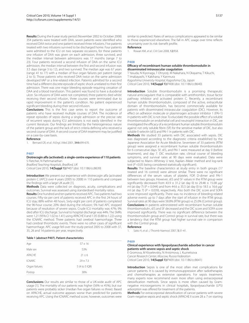
Results During the 6-year study period (November 2002 to October 2008)
354 patients were treated with DAA, seven patients were identifi ed who
received DAA twice and one patient received three infusions. Five patients
treated with two infusions survived to be discharged home. Four patients
were admitted to the ICU on two separate occasions, for these patients
one infusion of DAA was given on each admission, three survived, and
the median interval between admissions was 17 months (range 7 to
23). Four patients received a second infusion of DAA on the same ICU
admission, the median interval between the fi rst and second infusion was
3.5 days (range 3 to 12), and two survived. The median age was 67 years
(range 41 to 77) with a median of four organ failures per patient (range
2 to 5). Those patients who received DAA twice on the same admission
developed VAP or a line-related infection. Patients admitted for a second
time had a diff erent discrete episode of septic shock unrelated to their fi rst
admission. There was one major bleeding episode requiring cessation of
DAA and a blood transfusion. This patient was found to have a duodenal
ulcer. Six infusions of DAA were not completed, three patients died whilst
receiving their second infusion, three courses were terminated due to
rapid improvement in the patient’s condition. No patient experienced
signifi cant bleeding during their second infusion.
Conclusions This is the fi rst case series to report the outcome of
patients who have received multiple courses of DAA. The incidence of
repeat episodes of sepsis during a single admission or the precise rate
of recurrent sepsis during ICU admissions is not easily identifi ed in the
current literature. Our fi ndings are limited by the heterogeneous nature
of the patient group and the lack of strict criteria defi ning who received a
second course of DAA. A second course of DAA treatment may be justifi ed
on a case-by-case basis.
Reference1. Bernard GR, et al.: N Engl J Med 2001, 344:699-670.
P407
Drotrecogin alfa (activated): a single-centre experience of 110 patientsS Fletcher, N Pathmanathan
Bradford Teaching Hospitals, Bradford, UK
Critical Care 2010, 14(Suppl 1):P407 (doi: 10.1186/cc8639)
Introduction We present our experience with drotrecogin alfa (activated
protein C (APC)) over 4 years (2005 to 2008) in 110 patients and compare
the fi ndings with a large UK audit [1].
Methods Data were collected on diagnosis, acuity, complications and
outcomes. Survival was assessed using standardised mortality ratios.
Results One hundred and ten patients received APC. Two patients had two
courses. Fifty-six per cent of patients received APC in the fi rst 24 hours of
ICU stay; 88% within 48 hours. Sixty-eight per cent of patients completed
the 96-hour course; 20% died during the infusion; 5% had APC stopped
because of resolution of severe sepsis. Crude ICU mortality was 47%; 9%
died after ICU discharge. Survival to hospital discharge was 44%. The SMRs
were 1.21 (95% CI 1.02 to 1.41) using APACHE II and 1.05 (0.88 to 1.22) using
the ICNARC method. Three patients had cerebral haemorrhage. Three
had cerebral thrombotic events. There were no other cases of signifi cant
haemorrhage. APC usage fell over the study period 2005 to 2008 with 37,
35, 26 and 14 patients per year, respectively.
Table 1 (abstract P407). Patient characteristics
Age 57 ± 16
Male sex 53%
APACHE 21 ± 6
ICNARC 29 ± 7.3
Organ failures 5 (4 to 5 IQR)
Postop 38%
Conclusions Our results are similar to those of a UK-wide audit of APC
usage [1]. The mortality of our patients was higher (56% vs 45%), but our
patients were probably sicker (median fi ve organ failures vs three). Based
on APACHE, actual outcome appears worse than predicted for patients
receiving APC. Using the ICNARC method score, however, outcomes were
similar to predicted. Rates of serious complications appeared to be similar
to those experienced elsewhere. The fall in APC usage over time refl ects
uncertainty over its risk–benefi t profi le.
Reference1. Rowan KM, et al.: Crit Care 2008, 12:R58.
P408
Effi cacy of recombinant human soluble thrombomodulin in disseminated intravascular coagulationT Yasuda, N Kiyonaga, T Ohryorji, M Nakahara, N Okayama, T Kikuchi,
T Imabayashi, Y Kakihana, Y Kanmura
Kagoshima University Hospital, Kagoshima City, Japan
Critical Care 2010, 14(Suppl 1):P408 (doi: 10.1186/cc8640)
Introduction Soluble thrombomodulin is a promising therapeutic
natural anticoagulant that is comparable with antithrombin, tissue factor
pathway inhibitor and activated protein C. Recently, a recombinant
human soluble thrombomodulin, composed of the active, extracellular
domain of thrombomodulin, has become commercially available for
patients with disseminated intravascular coagulation (DIC). However, its
eff ect on adhesion molecule or plasminogen activator inhibitor 1 (PAI-1)
in patients with DIC is not clear. To elucidate the possible eff ect of a soluble
thrombomodulin on endothelial cell and neutrophil interaction in DIC, we
investigated the effi cacy of a recombinant human soluble thrombomodulin
against not only soluble fi brin (SF) for the sensitive marker of DIC, but also
soluble E-selectin (sES) and PAI-1 in patients with DIC.
Methods We studied 33 patients with DIC associated with sepsis. DIC
was diagnosed according to the diagnostic criteria established by the
Japanese Association for Acute Medicine. Seventeen of 33 patients (RTM
group) were assigned a recombinant human soluble thrombomodulin
for 6 consecutive days. SF, sES, and PAI-1 were measured at day 0 (before
treatment), and day 7. DIC resolution rate, clinical course of bleeding
symptoms, and survival rates at 90 days were evaluated. Data were
subjected to Mann–Whitney U test, Kaplan–Meier method and log-rank
test (P <0.05 being considered statistically signifi cant).
Results The baseline characteristics at study entry in both groups (17
treated and 16 control) were almost similar. There were no signifi cant
diff erences of the serum values of platelet, FDP, D-dimer and PAI-1
between two groups. However, sES and SF values in the RTM group were
signifi cantly decreased from 43.9 ± 31.2 (at day 0) to 29.46 ± 15.08 μg/
ml (at day 7) (P = 0.044) and from 44.6 ± 35.5 (at day 0) to 18.5 ± 16.6 μg/
ml (at day 7) (P = 0.039), respectively. Also both the DIC score and SOFA
score improved signifi cantly. There was no incidence of bleeding-related
adverse events up to 7 days after the start of infusion in the RTM group.
Survival rates at 90 days were 58.8% (RTM group) vs 25.0% (Control group).
Conclusions In patients administered with recombinant human soluble
thrombomodulin, sES and SF decreased and the DIC score and SOFA score
improved signifi cantly. There was no signifi cant diff erence between the
thrombomodulin group and Control group in survival rate, but there was
a tendency that the RTM group had higher survival rate in comparison
with the Control group.
Reference1. Saito H, et al.: J Thromb Haemost 2007, 5:31-41.
P409
Clinical experience with lipopolysaccharide adsorber in cancer patients with severe sepsis and septic shockE Gromova, M Kisselevskiy, N Anisimova, L Kuznetsova
Cancer Research Center, Moscow, Russia Federation
Critical Care 2010, 14(Suppl 1):P409 (doi: 10.1186/cc8641)
Introduction Sepsis is one of the most often met complications for
cancer patients. It is caused by immunosuppression after radiotherapies
and chemotherapies as extensive operations. For sepsis treatment,
many experts now recommend even more often using extracorporeal
detoxifi cation methods. Since sepsis is more often caused by Gram-
negative microorganisms in clinical hospitals, lipopolysaccharide (LPS)
adsorption was off ered for treatment of the patients.
Methods For extracorporeal detoxifi cation of cancer patients with severe
Gram-negative sepsis and septic shock (APACHE II score 28 ± 7 on starting
Critical Care 2010, Volume 14 Suppl 1 http://ccforum.com/supplements/14/S1
S137
hemoperfusion) we used LPS adsorber (Alteco® LPS Adsorber; Alteco
Medical AB, Lund, Sweden). The patient’s blood was taken before and
after adsorption, then the serum was separated and stored at –70°C. We
determined the LPS concentration by LAL test, lipopolysaccharide-binding
protein (LBP), sIL-1 RII and sCD14 by ELISA kits (Hycult Biotechnology, the
Netherlands), and proinfl ammatory and anti-infl ammatory cytokines IL-
6, TNFβ, IL-8, IL-2, INFγ, IL-12, TNFα, IL-4, IL-5, IL-1β and IL-10 by ELISA kit
(Bender MedSystems).
Results In all cases we saw reliable reduction of the LPS level in serum (1.8
to 2 times). After the investigation of some cytokine levels we obtained
similar results: IL-6, TNFβ and IL-8 levels had been reduced 30 to 40%,
87% and 62 to 76% accordingly. At the same time, IL-2, INFγ, IL-12, TNFα,
IL-4 and IL-5 levels in serum had not changed practically. However, in
approximately 1/3 cases an IL-1β and IL-10 concentration increase in serum
was observed (33 and 40% accordingly). Also our research has shown that,
in the most of the cases after application of this sorbent, LBP decreased
insignifi cantly (10 to 32%), but the sIL-1 RII level slightly increased (10 to
18%) in the majority of patients and considerably decreased (two times)
in 1/3 investigated patients. The CD14 concentration in patient serum
reliably did not change before and after adsorption. The somatic status of
patients was stabilized after LPS adsorption.
Conclusions After LPS adsorption with Alteco® the LPS level in patient
serum signifi cantly reduced. Simultaneously, concentrations of some
cytokines (IL-6, TNFβ, IL-8) were reliably decreased. But the LPS adsorber
probably does not eliminate soluble receptors like sCD14 and RIL-1 from
blood. So the LPS adsorption may represent a signifi cant improvement in
the treatment of cancer patients with severe Gram-negative sepsis and
septic shock.
P410
Apheresis of activated leukocytes with an immobilized polymyxin B fi lterN Takeyama1, T Kumagai1, M Harada1, Y Kajita1, Y Miki1, H Kanou2, Y Inoue1,
T Nakagawa1, H Noguchi2
1Aichi Medical University, Aichi, Japan; 2Fujita Health University, Toyoake,
Japan
Critical Care 2010, 14(Suppl 1):P410 (doi: 10.1186/cc8642)
Introduction It was assumed that pathologic activation of neutrophils
and monocytes is associated with sepsis, acute lung injury/ARDS, and
multiple organ failure, and that removal of these cells from the circulation
could reduce leukocyte-dependent tissue injury. Cartridges containing
polymyxin B (PMX) immobilized to fi bers (Toraymyxin; Toray Industries,
Tokyo, Japan) have been developed for selective adsorption of circulating
endotoxin in patients with Gram-negative bacterial infection, and this
treatment has proven to be highly eff ective. This study examined the
eff ect of direct hemoperfusion through fi lters with immobilized PMX
direct-hemoperfusion (DHP) on leukocyte function and plasma levels of
cytokines in patients with septic shock.
Methods We evaluated the eff ect of PMX-DHP on circulating leukocytes
in patients with septic shock by assessing the changes of neutrophil
and monocyte surface antigen expression after PMX-DHP. In another
experiment, heparinized blood from patients with sepsis was passed
through PMX fi lters in a laboratory circuit and then changes of the cell
count and surface antigen expression by neutrophils and monocytes
were assessed. After perfusion, neutrophils were isolated and the capacity
of these cells to damage cultured endothelial monolayers was also
determined.
Results We found that PMX-DHP led to an increased CXCR1 and CXCR2
expression along with a decrease of CD64 and CD11b expression by
circulating neutrophils from septic patients. Plasma levels of cytokines,
including IL-6, IL-8, IL-10, and high-mobility group box-1, were elevated in
patients with septic shock compared with healthy controls, but cytokine
levels were not altered by PMX-DHP. Ex vivo perfusion of heparinized blood
from patients with sepsis through PMX fi lters in a laboratory circuit caused
a signifi cant decrease of the neutrophil and monocyte counts. Activated
neutrophils with high CD11b/CD64 expression and low CXCR1/CXCR2
expression showed preferential adhesion to PMX fi lters. Neutrophils
isolated from the blood after ex vivo PMX perfusion caused less damage
on the endothelial cell monolayer than cells from sham-treated blood.
Conclusions PMX-DHP infl uences neutrophils via a mechanism that does
not involve cytokines. Ex vivo results indicate that PMX-DHP selectively
removes activated neutrophils and reduces the ability of circulating cells to
cause endothelial damage. Selective removal of activated neutrophils by
PMX-DHP may contribute to improvement of the systemic infl ammatory
reaction in septic patients.
P411
Therapeutic blood purifi cation using functionalized core/shell nanomagnetsIK Herrmann1, M Urner2, FM Koehler1, B Beck Schimmer2, WJ Stark1
1ETH Zurich, Switzerland; 2University Hospital Zurich, Switzerland
Critical Care 2010, 14(Suppl 1):P411 (doi: 10.1186/cc8643)
Introduction Direct and rapid removal of pathogens or noxious
metabolites from a patient is the most straightforward cure imaginable.
Dialysis and plasma fi ltration/exchange are the current broadly applicable
methods to perform a direct removal of disease-causing factors from
a patient. We describe the use of stable nanomagnets to rapidly and
selectively remove heavy metal ions, overdosed steroid drugs and proteins
from human blood. This nanomagnet-based purifi cation method avoids
fouling of fi lter membranes and benefi ts from a high external surface area,
and a correspondingly fast diff usion.
Methods Nanomagnets equipped with heavy metal complexants,
digoxin antibody fragments and entire human IL-6 antibodies were added
to a series of blood samples. The nanomagnets scan blood by Brownian
motion and capture their target. Afterwards, a small magnet was placed at
the sample tube wall accumulating the nanomagnets in the pole region of
the external magnet. The purifi ed supernatant can then easily be decanted.
The concentrations of lead, digoxin and IL-6 in blood samples were
determined by standard clinical methods. Blood integrity was observed
by rotation thromboelastography and monitoring of serum potassium,
lactate dehydrogenase, bilirubin and haptoglobin levels. To measure the
biological relevance of the IL-6 removal, the eff ect on caspase-3 activation
was assessed in camptothecin-stimulated neutrophils.
Results A signifi cant decrease of lead, digoxin and IL-6 levels was measured
after the blood purifi cation procedure. The extraction using nanomagnets
was in clear dose–eff ect dependence and could be accurately titrated.
Treatment with nanomagnets did not signifi cantly aff ect the integrity of
blood and all levels remained in the clinical norm range. Caspase-3 assays
showed a reduced anti-apoptotic eff ect after IL-6 removal, underlining the
biological relevance of the achieved removal effi ciency.
Conclusions We demonstrate the extraction of lead, digoxin and IL-6
from whole blood as an example for the rapid treatment of heavy metal
poisoning, drug overdosing and severe infl ammation. The presented
direct blood extraction could be combined with existing therapeutic
strategies and may have major implications on the treatment of severe
intoxications, sepsis (specifi c fi ltering of cytokines or toxins) [1], metabolic
disorders (thyreotoxicosis) and autoimmune diseases.
References1. Ronco C, et al.: N Engl J Med 2003, 348:1600-1602.
P412
Coupled plasma fi ltration and adsorption in septic shock: a multicentric experienceF Turani1, M Falco1, S Natoli2, F Leonardis2, M Fede2, G Berlot3,
M Pelichovskà3
1European Aurelia Hospital, Rome, Italy; 2University of Tor Vergata, Rome, Italy; 3University of Trieste, Italy
Critical Care 2010, 14(Suppl 1):P412 (doi: 10.1186/cc8644)
Introduction Recently extracorporeal removal of mediators and
endotoxins improved haemodynamics, organ dysfunction and mortality
in patients with abdominal sepsis [1]. Coupled plasma fi ltration and
adsorption (CPFA), too, may improve haemodynamics, respiratory function
and mortality through removal of proinfl ammatory mediator. The aim of
this multicentric study is to evaluate the haemodynamic response and
the respiratory function, and to evaluate the reduction of infl ammatory
markers during CPFA.
Critical Care 2010, Volume 14 Suppl 1 http://ccforum.com/supplements/14/S1
S138
Methods Fifty-fi ve septic patients were enrolled in this study. Every patient
had four CPFA treatments (LINDA; Bellco-Mirandola, Italy) for 8 hours with
Qb = 200 ml/minute, Q
ultrafi ltration = 30 ml/kg/hour and Q
plasma = 20% of Q
b. At
T0 (basal), T1 (after fi rst cycle), T2 (after second cycle), T3 (after third cycle)
and T4 (after fourth cycle) we evaluated haemodynamic parameters,
norepinephrine dosage, PaO2/FiO
2 ratio, plasma IL-6, and procalcitonin
(PCT). The ANOVA test was used to compare changes during times study.
P <0.05 was considered statistically signifi cant.
Results Patients enrolled in the study have been submitted to 256 CPFA
treatments for 2,650 hours. Table 1 presents the main results of the study.
IV quartile of IL-6 is shown in Table 1.
Table 1 (abstract P412)
T0 T4
IL-6 (pg/ml) 850 ± 127 125 ± 28*
PCT (pg/ml) 45 ± 9 6 ± 2**
Noradrenaline (μg/kg/min) 0.35 ± 0.09 0.08 ± 0.05**
PaO2/FIO
2 198 ± 28 310 ± 92**
*P <0.05, **P <0.01 vs T0.
Conclusions In this large multicentric study, CPFA may improve
haemodynamic status and respiratory function. Plasma fi ltration and
adsorption of proinfl ammatory mediators may explain this improvement.
Larger randomized controlled trials are indicated to confi rm these data.
Reference1. Cruz D, et al.: JAMA 2009, 23:2445-2452.
P413
Patients admitted to the ICU for extra-abdominal disease and operated on for emergency laparotomy have signifi cant survivalC Lentschener, J Beranger, J Charpentier, Y Ozier, J Mira
Cochin Hospital, Paris, France
Critical Care 2010, 14(Suppl 1):P413 (doi: 10.1186/cc8645)
Introduction Limited data are available concerning patients admitted to
the ICU for an extra-abdominal disease and operated on for emergency
laparotomy (EL) [1]. We investigated whether such patients had benefi t
from EL and factors likely to predict survival.
Methods EL was performed in 48 ICU patients admitted for an extra-
abdominal disease over the year 2008. The following variables were
compared between survivors (discharged alive from hospital) and non-
survivors: (a) sex ratio, BMI; (b) pre-existing co-morbidities considered
in seven additive categories: cardiac, respiratory, renal, gastrointestinal,
hepatic, diabetes, malignancies, and corticosteroid therapy; (c) pre-
operatively: IGS II score, mechanical ventilation (MV), vasopressor use,
extra-renal epuration requirement, abdominal signs (obstruction,
tender ness, contracture, compartment syndrome), records of computed
tomography (CT) and ultrasonography examinations; (d) intraoperative
fi ndings defi ned as perforation, infection, ischemia or necrosis of a visceral
organ, pancreatitis, bowel obstruction, biliary disease, no fi nding. The t,
Mann–Whitney U, and chi-square tests (P <0.05), and multiple regression
analysis (P <0.1) were used.
Results Twenty-six patients (58%) survived. EL was decided because
of abdominal signs in 35 patients, lack of improvement of one or several
organ failures in 44 patients, specifi c signs on CT scan, and/or ultrasound
examination (active bleeding, subphrenic collection, bowel obstruction,
pneumoperitoneum) in 22 patients, or nonspecifi c signs in 29 patients. EL
did not identify any surgical cause in 10 patients. Groups did not diff er in
intraoperative fi ndings (P = 0.2), preoperative creatinine level (P = 0.3), and
preoperative anuria requiring extra-renal epuration (P = 0.2). Groups diff ered
in preoperative MV (P = 0.04), vasopressor requirement (P = 0.02), lactate
levels (P = 0.03), IGS II (P = 0.0003), and pre-existing co-morbidities (P = 0.04).
No such correlation was found in multivariate analysis (all P >0.15).
Conclusions Neither clinical nor laboratory variables, or operative fi ndings
predicted death following EL. The survival rate was high enough to
warrant surgical EL for suspected intra-abdominal focus.
Reference1. Sutherland FR, et al.: J Trauma 1989, 29:1982-1986.
P414
Can ICU admission be predicted?L Orwelius, M Fredriksson, C Bäckman, J Persson, F Sjöberg
Faculty of Health Sciences, Linköping, Sweden
Critical Care 2010, 14(Suppl 1):P414 (doi: 10.1186/cc8646)
Introduction After intensive care (IC), patients report poor health-related
quality of life (HRQoL). Many factors aff ect the patients and infl uence
the HRQoL after discharge. One of these factors is the patient’s health
status before the critical care period. In a previous study we found that
the IC patients have a high frequency of pre-existing diseases. However,
it is unknown to what extent these pre-existing diseases aff ect the
consumption of hospital resources (measured as days as inpatients) in the
time period before admission to the ICU and during the years following
it. The consumption prior to the ICU event may also be claimed to herald
an increased risk for a later ICU admittance? The aim of this study was to
examine the hospital care consumption of former ICU patients 3 years
prior to and 3 years after the intensive care period. This was examined in
relation to the pre-existing health status.
Methods Two mixed ICUs from hospitals in two cities in Sweden. A
question naire including previous illnesses and HRQoL scale SF-36 were
sent 6, 12, 24 and 36 months after discharge from the ICU and hospital. All
adult patients with an ICU stay >24 hours were included. The institutional
care consumption was assessed from the regional hospital databases
Results Six hundred and thirteen patients were included in the study. Of
these 73% had pre-existing diseases. There were no signifi cant diff erences
between the previously healthy patients apart from younger age (mean
age 52 (SD 20.7) vs 60 (SD 17.3)) and lower APACHE II scores (13.4 (SD 7.2)
vs 16.4 (SD 7.7)) compared with patients with pre-existing disease. The
ICU patients with pre-existing disease consumed signifi cantly more
hospital resources 3, 2 and 1 year prior to admission to the ICU, and up to
3 years after discharge both regarding quantity of care (number of visits)
(P = 0.001), length of stay (P = 0.001), and cost (P = 0.002). There were no
signifi cant diff erences in quantity of care, length of stay, or costs during
the ICU period. Most importantly there was a signifi cant increasing trend
for the 3 years prior to the ICU event.
Conclusions Patients with co-morbidities have a signifi cant consumption
of hospital resources both prior to and after the ICU period. Interestingly,
this consumption increased prior to the ICU event and seemed to herald
the future deterioration of the patients. Further investigations should be
made to examine to what extent ICU admissions can be predicted based
on these data.
Reference1. Orwelius L, et al.: Crit Care Med 2005, 33:1557-1564.
P415
Unsuitable for ICU: what happens next?A McKeown, L Strachan, P Keeley, MG Booth, A Calder, A Panicker
Glasgow Royal Infi rmary, Glasgow, UK
Critical Care 2010, 14(Suppl 1):P415 (doi: 10.1186/cc8647)
Introduction This observational prospective study examined the outcomes
and symptoms in those patients who are unsuitable for ICU admission.
Methods All patients referred to ICU for admission but deemed unsuitable
were recorded by the team. These patients were reviewed by palliative
care researchers. Demographic information (age, sex, diagnosis) and data
on patient’s symptoms of progress and outcome were collected at four
time points (24, 48, 72 hours and 1 week).
Results Fifty patients were identifi ed between January and April 2009.
There was an age range of 24 to 86 (mean = 62). The commonest cause
for referral was respiratory failure (40%). Overwhelming sepsis requiring
inotropic and ICU support was also common (28%). Patients were most
frequently deemed unsuitable for admission as they were too well at the
time of referral (48%). The second most common reason for unsuitability
was co-morbidities (32%) with nine (18%) identifi ed as suff ering probable
fatal insults. One patient was referred for assessment for ICU support
post emergency surgery which was not required. A number of patients
remained in hospital at 1 week post referral, with ongoing illness (32%) or
rehabilitation (8%). Twenty-six per cent (13/50) of patients were discharged
home and 34% (17/50) died. Of those that died, 59% were within 24 hours
of ICU referral. All other deaths occurred within 1 week of referral. Nine
Critical Care 2010, Volume 14 Suppl 1 http://ccforum.com/supplements/14/S1
S139
of these 17 had been identifi ed as having nonsurvivable insults at ICU
referral, seven as having excess co-morbidities making ICU inappropriate,
and just one identifi ed as being too well.
Conclusions This study suggests that our ICU team is both sensitive
and specifi c at identifying those patients who are either too well for
admission or those patients for whom ICU is unlikely to be of benefi t as
their insult is presumed fatal. We suggest those patients whom the ICU
identifi es as unlikely to survive should be assessed and referred to the
palliative care team, where appropriate. This should facilitate appropriate
communication of this by clinical staff and allow the relevant social,
psychological and spiritual preparations for death that are the hallmarks
of good care of the dying.
References1. [http://www.sicsag.scot.nhs.uk]
2. Joynt G, et al.: Intensive Care Med 2001, 27:1459-1465.
3. Lyons RA, et al.: Lancet 2000, 355:595-598.
4. Thiery G, et al.: J Clin Oncol 2005, 23:4406-4412.
5. Crippen D: Crit Care Med 2009, 37:1498-1499.
6. Wood G, et al.: Crit Care Med 2009, 37:1147-1148.
P416
Admission pattern and outcome in a UK weaning centreS Goodhand, K Stewart, L McGee, N Grey, A Williams, C Davidson, N Hart
Guy’s and St Thomas’ Foundation Trust, London, UK
Critical Care 2010, 14(Suppl 1):P416 (doi: 10.1186/cc8648)
Introduction Although patients with weaning failure may be transferred
to weaning centres, there are limited data on admission practice and
clinical outcome.
Methods We collected data prospectively from March 2005 to October 2009.
Results There were 286 referrals of which 20% were not accepted for
transfer. Patients that were not accepted were categorised as weanable
locally, unweanable, or died prior to transfer. A total 172/229 accepted
patients were admitted (mean age was 60 ± 17 years; 118 male). Forty-six
per cent of these achieved ventilator independence with 21% requiring
ongoing non-invasive ventilation (NIV). The median time to wean
was 17 days with 16% of patients remaining tracheostomy ventilator
dependent. The unit mortality was 17%. Neuromuscular disease (NMD)
was the main referral group (36%) with a low mortality rate (6%). COPD
and postsurgical patients both comprised 21% of the referrals and had
respective mortalities of 37% and 25%. There were no in-hospital deaths
in the obesity-related respiratory failure (ORRF) group, but this was the
smallest referral group (3%). Of 57 patients declined transfer, 43 (83%)
were assessed as unweanable and nine (17%) were assessed as weanable
locally. Of 56 patients accepted but not transferred, 28 (50%) were weaned
awaiting transfer and 23 (41%) died prior to transfer. Mean transfer delay
was 20.3 ± 19.2 days and a major contributor to this delay was prolonged
discharge delay following weaning (12.9 ± 19.3 days) and extended
discharge delay of tracheostomy-ventilated patients (77.4 ± 51.8 days).
Conclusions In conclusion, compared with our previous data [1], there
was an absolute reduction in mortality of 10% with similar admission
diagnoses, proportions achieving ventilator independence and requiring
NIV, suggesting that this was not necessarily selection bias. NMD was the
main referral group with a low mortality rate. Although COPD patients had
the highest mortality, almost 50% were weaned from invasive ventilation.
There were no in-hospital deaths in the ORRF group, but these patients
had the longest weaning time. In addition, poor outcome was observed
in patients accepted but not transferred due to delay. This study highlights
the need for more speciality weaning facilities in the UK.
Reference1. Thorax 2005, 60:187-192.
P417
Referral to intensive care: who and when?A Mackay, J Erskine, P Doherty, E McMillan
Victoria Infi rmary, Glasgow, UK
Critical Care 2010, 14(Suppl 1):P417 (doi: 10.1186/cc8649)
Introduction Intensive care beds in the UK are a limited resource and
deciding whether or not to admit a patient to intensive care is often a
diffi cult decision. It is increasingly felt that, as intensive care consultants
are involved in the decision-making process for every admission, the
consultant from the referring team should also be involved in the referral
process. The Scottish Intensive Care Society Trainee Group found that this
occurred in less than 50% of occasions during the Referral and Review
Audit of 2007 [1]. We wished to establish what the referral process was like
in our hospital.
Methods We undertook a prospective audit of all referrals to a fi ve-bed
teaching hospital ICU between April and July 2009. Data were collected
regarding time of referral, seniority of referrer, involvement of referring
consultant, seniority of ICU trainee, involvement of ICU consultant and
whether or not the referral resulted in intensive care admission.
Results Ninety-eight referrals were received during the audit period. Of 31
medical referrals, 21 were out of hours, 16 had no consultant involvement
and ultimately 27 patients were admitted. Of nine surgical patients, three
were out of hours, three had no consultant involvement and seven were
eventually admitted. Thirty patients were referred by the emergency
department, of whom 18 were out of hours, 23 had no consultant
involvement and 26 resulted in admission. Twenty-eight patients were
referred by anaesthetics, all of whom had been seen by a consultant and
were admitted.
Conclusions General medicine is the most signifi cant contributor to out-
of-hours work of the intensive care medical staff . Over 50% of medical
referrals have no referring consultant involvement, although ultimately
many of these patients are admitted and are seen by an intensive care
consultant within a few hours of admission. Most surgical patients
are admitted postoperatively and thus are referred by a consultant
anaesthetist, resulting in a very high conversion rate for referrals to
admissions. Emergency medicine is increasingly a consultant delivered
service with increased presence in the department into the extended
working day, a fact that is sadly not refl ected in our results. It is regrettable
that despite repeated recommendations that intensive care admission
should be on the basis of consultant to consultant referral, 43% of patients
are referred to intensive care without any referring consultant involvement.
Reference1. Scottish Intensive Care Society Annual Report 2008 [http://www.
scottishintensivecare.org.uk/sics/Annual%20Report/2008%20annual%20
report.pdf ]
P418
Acute drug overdose in an ICU: 15 years experienceL Cachafeiro, JJ Manzanares, S Yus, C Soriano, M Oliveros, M Jimenez
Hospital Universitario La Paz, Madrid, Spain
Critical Care 2010, 14(Suppl 1):P418 (doi: 10.1186/cc8650)
Introduction The aim of our study is to identify the epidemiology of
patients admitted to an ICU following severe acute drug overdose.
Methods The study is a retrospective observational study of a 20-bed
medical/surgical ICU during a 15-year period. We reviewed the clinical
records of patients admitted from 1994 to 2008 with acute drug overdose.
Data on demographics, mean length of stay, days on mechanical
ventilation (MV), Glasgow Coma Scale (GCS) on admission, and mortality
were recorded. The type of drug was also stated.
Results A total of 12,881 patients were admitted to our ICU from 1994 to
2008, of them 621 patients (4.8%) had a diagnosis of acute intoxication
on admission. From these 621 patients, 159 (25.6%) were drug overdoses,
which represented 1.2% from the total admissions. The median age was
33 years, the range 15 to 87, with a standard deviation of 12.5. A total of
50.9% were male. The median GCS on admission was 6 and the APACHE
II score was 16, with a range 3 to 31 and a standard deviation of 6.7. MV
was required by 71.1% with a median duration of 2.38 days, with a range
0 to 45 and a standard deviation of 6.9. The median length of stay was
3.2 days, the range 0 to 41 and a standard deviation of 6.1. The types of
drugs found were ethanol, cocaine, opioids (methadone, heroin, opium),
LSD, ketamine, GHB, amphetamines, cannabis, and PCP. Ethanol was the
most commonly encountered in 57%, followed by cocaine in 37%, and
opioids in 27% of patients. Global mortality was 2%, and 50% of these were
due to ethanol.
Conclusions In our study, severe acute drug overdose accounts for a low
percentage of patients admitted to the ICU. The profi le is that of a middle-
aged male patient with acute ethanol, cocaine, or opioids overdose, in
Critical Care 2010, Volume 14 Suppl 1 http://ccforum.com/supplements/14/S1
S140
that order of frequency, and with rates similar to those of national and
European statistics, of short length of stay (3.2 days) and a mortality inferior
to that expected for the APACHE II score.
References1. Palazon Sanchez C, et al.: Rev Esp Salud Publica 2000, 74:55-63.
2. Caballero Valles PJ, et al.: An Med Interna 2008, 25:262-268.
P419
Prognostic factors for very short-term mortality in severe sepsisE Silva1, R Salomao2, O Rigato3, E Ferreira2, T Mohovic1, S Callas3, N Tereran2,
M Assuncao1, N Delbiagi3, F Colombari1, C Silva4, R Cal4, F Machado2
1Hospital Israelita Albert Einstein, São Paulo, Brazil; 2Federal University of São
Paulo, Brazil; 3Hospital Sirio-Libanes, São Paulo, Brazil; 4Latin American Sepsis
Institute, São Paulo, Brazil
Critical Care 2010, 14(Suppl 1):P419 (doi: 10.1186/cc8651)
Introduction Short-term mortality studies could be crucial for evaluating
new therapies as well as to better understand pathophysiological aspects.
This study aims to identify prognostic factors associated with sepsis-
related death within 4 days after the fi rst organ dysfunction development.
Methods All severe sepsis and septic shock patients over 18 years old
admitted to three Brazilian ICUs were prospectively included. Demographic,
clinical and outcome features, including compliance to the 6-hour Surviving
Sepsis Campaign bundle (SSC-6h), were collected in an electronic clinical
research form. Duration of organ dysfunction was defi ned as the time
elapsed between the installation of the dysfunction and its diagnosis by the
healthcare provider. Results were expressed as a percentage or as median
and interquartiles. Univariate analysis was performed by chi-squared or
Student t test. P <0.05 was considered signifi cant.
Results Three hundred and eighty-six patients were included, being 30
(7.8%) in Group 1 (short-term mortality group, median time from organ
dysfunction to death: 2.39 (1.75 to 3.25) days) and 356 (92.2%) in Group
2. No diff erence was found between the groups regarding gender (P =
0.14), age (P = 0.80), being in a public hospital (P = 0.94) or duration of
organ dysfunction (P = 0.79). Variables related to severity of disease were
associated with early mortality: APACHE II score (Group 1: 21.0 (15.0
to 26.0), Group 2: 18.0 (14.0 to 22.0), P = 0.01); SOFA score at admission
(SOFA0) (Group 1: 9.0 (8.0 to 11.0), Group 2: 7.0 (5.0 to 9.0), P = 0.003); Day
1 SOFA score (10.0 (8.0 to 12.0), Group 2: 7.0 (5.0 to 10.0), P = 0.0002); Day
3 SOFA score (SOFA3) (Group 1: 9.0 (8.0 to 12.0), Group 2: 7.0 (4.0 to 10.0),
P = 0.006) and the delta between SOFA0 and SOFA3 (Group 1: –6.5 (–11.0
to +1.0), Group 2: –1.0 (–3.0 to +9.0), P = 0.003). Delta SOFA with Day 1
was not diff erent between groups. Group 1 patients had a trend towards
higher number of organ dysfunction (P = 0.07) and more septic shock
episodes (P = 0.09). Regarding treatment adequacy, compliance with any
indicator or the entire SSC-6h bundle was not related to survival. However,
achievement of the CPV target was associated with a higher early mortality
(Group 1: 69.6%, Group 2: 40.6%, P = 0.013; OR – 3.3.4 (1.33 to 8.40)).
Conclusions In this observational study, factors related to disease severity
rather than to the evidence-based treatment compliance were associated
with short-term death among severe septic patients.
P420
Is sepsis mortality rate driven by diff erences in Brazilian healthcare systems?F Machado1, R Salomao1, O Rigato2, E Ferreira1, T Mohovic3, G Schettino2,
N Tereran1, M Assunçao3, R Caltabiano2, F Colombari3, C Silva4, R Cal3,
E Silva3
1Federal University of São Paulo, Brazil; 2Hospital Sirio-Libanes, São Paulo,
Brazil; 3Hospital Israelita Albert Einstein, São Paulo, Brazil; 4Latin American
Sepsis Institute, São Paulo, Brazil
Critical Care 2010, 14(Suppl 1):P420 (doi: 10.1186/cc8652)
Introduction The sepsis mortality rate is higher in Brazilian public
hospitals with no clear diff erences in risk factors. We aim to identify risk
factors that could explain why the mortality rate is diff erent in public and
private hospitals.
Methods All severe sepsis and septic shock patients over 18 years
old admitted to two private ICUs (Group 1) and one public ICU (Group
2) were prospectively included. Demographic, clinical and outcome
features, including compliance to the 6-hour Surviving Sepsis Campaign
bundle (SSC-6h), were collected in an electronic CRF. Duration of organ
dysfunction was defi ned as the time elapsed between the installation of
the dysfunction and its diagnosis by the healthcare provider. Results were
expressed as a percentage or as median and interquartiles, and P <0.05
was considered signifi cant.
Results Three hundred and eighty-six patients were included (Group
1 – 210, Group 2 – 176). Patients from Group 1 were older (69.5 (53.0 to
81.0) years vs 59.0 (43.5 to 73.5) years, P <0.000), had a higher APACHE II
score (19.0 (15.0 to 23.0) vs 17.0 (13.0 to 23.0), P = 0.01), had more organ
dysfunctions (3.0 (2.0 to 4.0) vs 2.0 (2.0 to 4.00), P = 0.08), and more septic
shock episodes (74.3% vs 60.8%, P = 0.006). No diff erence was found
regarding gender, site of infection, and day 1 or day 3 delta SOFA score.
However, the mortality rate was higher in Group 2 than in Group 1, 60.8
and 38.3, respectively. Compliance to the SSC-6h bundle did not explain
this diff erence as they were better in Group 2 for lactate (70.5% vs 49.8%),
blood culture (61.4% vs 52.4%), achieving MAP >65 mmHg (98.8% vs
90.5%), and completion of the bundle (15.9% vs 4.8%). Only antibiotics
(63.3% vs 54.5%, P = 0.10) and CVP had a better performance in Group 1
(49.4% vs 33.3%, P = 0.01). SOFA score at admission was slightly higher on
Group 2 (7.0 (4.0 to 9.0) vs 8.0 (5.5 to 10.0), P = 0.004). However, duration
of organ dysfunction was clearly longer in Group 2 (150.0 (0.0 to 650)
minutes vs 360.0 (0.0 to 1008.0) minutes, P = 0.02). In all patients, we have
identifi ed some variables associated with mortality, including being in the
public hospital (OR 2.46 (1.63 to 3.72), P = 0.00002), as well as the median
duration of organ dysfunction, SOFA at admission, age, APACHE II score
and number of organ dysfunctions.
Conclusions The mortality rate was higher in the public hospital despite
better SSC bundle adherence. A delay in initiating those interventions may
negatively impact the outcome in patients treated in public hospital.
P421
Prognoses indicators of HIV patients admitted to the ICUB Moya, N Germano, G Imelda
Hospital de S. Bernardo, Setúbal, Portugal
Critical Care 2010, 14(Suppl 1):P421 (doi: 10.1186/cc8653)
Introduction The level of immunity in HIV patients is associated with a
high mortality, particularly when CD4+ lymphocytes are inferior to 200
cells/mm3. Besides the immunity, other prognoses indicators are necessary.
Methods The author performed a revision of 1,028 admissions to the
ICU from January 2007 and November 2009. Selected were the cases of
HIV patients with low immunity (L CD4+ <200 cells/mm3). The following
parameters were collected at admission to the ICU: viral load, LDH, albumin,
hemoglobin, Hct, thrombocytopenia, presence of opportunistic infection,
nutritional status, need for mechanical ventilation, renal insuffi ciency and
history of antiretroviral therapy (ART). Demographic parameters were also
collected. The primary endpoint was mortality, secondary endpoints were
ICU length stay and days of mechanical ventilation.
Results Fifteen patients were selected; all of them were medical admissions
mainly from the emergency department. Sixty-fi ve percent had previous
history of opportunistic infection, and only two patients had ART. The main
reason to suspend ART was social and drug-dependency problems. Sepsis
was diagnosed in 73% of the patients. Pneumocystis carinii pneumonia
was diagnosed in 80% of the patients. Fungus infection was diagnosed in
60% of patients. One patient had pulmonary tuberculosis and another had
Leishmania infection. The mortality rate was 53%. The factors associated
with high mortality were: P. carinii pneumonia, malnutrition and absence
of ART.
Conclusions The patient’s nutritional status is an independent predictor
of mortality in HIV patients.
References1. Johannessen A, Naman E, et al.: Predictors of mortality in HIV-infected
patients starting antiretroviral therapy in a rural hospital in Tanzania. BMC
Infect Dis 2008, 8:52.
2. Swaminathan S, Padmapriyadarsini C, et al.: Nutritional status of persons with HIV infection, persons with HIV infection and tuberculosis, and HIV-negative individuals from southern India. Clin Infect Dis 2008, 46:946-949.
Critical Care 2010, Volume 14 Suppl 1 http://ccforum.com/supplements/14/S1
S141

P422
Outcomes of cancer patients admitted to intensive care units with severe acute kidney injuryS Lobo, M Soares, A Torelly, P Mello, U Silva, J Teles, E Silva, P Caruso,
G Friedman, P Souza, A Rea-Neto, A Vianna, M Maia, J Salluh
Brazilian Research in Intensive Care Network – BRICNet, Rio de Janeiro, Brazil
Critical Care 2010, 14(Suppl 1):P422 (doi: 10.1186/cc8654)
Introduction Critically ill cancer patients are at increased risk for acute
kidney injury (AKI), but studies on these patients are scarce and were all
single centered, conducted in specialized ICUs. The aim of this study was
to evaluate the characteristics and outcomes in a prospective cohort of
ICU cancer patients with AKI.
Methods Prospective multicenter cohort study conducted in ICUs from
28 hospitals in Brazil over a 2-month period. Univariate and multivariate
logistic regression were used to identify factors associated with hospital
mortality.
Results Out of all 717 ICU admissions, 87 (12%) had AKI and 36% of them
received dialysis. Kidney injury developed more frequently in patients with
hematological malignancies than in patients with solid tumors (26% vs
11%, P = 0.003). Ischemia/shock (76%) and sepsis (67%) were the main
contributing factors, and kidney injury was multifactorial in 79% of the
patients. The ICU mortality was 61% (53/87) and hospital mortality was 71%
(62/87). Despite the lack of statistical signifi cance, hospital mortality was
higher in patients who received RRT later on during the ICU stay (92%) in
comparison with those who received RRT on the fi rst day in the ICU (78%)
and those who were not dialyzed (64%) (P = 0.105). End-of-life decisions
(to withhold or to withstand therapies) were taken in 18 (23%) patients.
General and renal-specifi c severity-of-illness scores were inaccurate in
predicting outcomes for these patients. In a multivariate analysis, length of
hospital stay prior to ICU, acute organ dysfunctions, need for mechanical
ventilation and a poor performance status were associated with increased
mortality. Moreover, cancer-related characteristics were not associated
with outcomes.
Conclusions The present multicenter study confi rmed that AKI in
critically ill patients with cancer is frequent, usually multifactorial and
still associated with high mortality rates. On the other hand, the current
study also suggests that ICU admission and RRT should be considered in
selected patients. Mortality in these patients is mostly dependent on the
severity of acute illness and the performance status, rather than cancer-
related characteristics.
P423
Is intensive care benefi cial for patients with haematologic malignancies?M Nyunga, C Lemaire, E Ras, P Herbecq, I Verheyde
Centre Hospitalier de Roubaix, France
Critical Care 2010, 14(Suppl 1):P423 (doi: 10.1186/cc8655)
Introduction Despite a better prognosis for patients with haematologic
malignancies (PHM) over these past years due to progress in management,
intensivists are reluctant to admit those patients to the ICU because of high
mortality rates, above 50%. The aim of this study was to retrospectively
evaluate the value of intensive care therapy of PHM and predictive factors
of short-term and long-term survival with the best post-ICU quality of life.
Methods The case notes of 88 PHM patients requiring admission to a 25-
bed medical–surgical ICU of Roubaix Hospital, France were retrospectively
reviewed during a 5-year period, 2003 to 2007. We collected demographic
factors, Simplifi ed Acute Physiology Score II (SAPS II), Logistic Organ
Dysfunction System score (LODS), Charlson score, microbiology, status
of underlying HM, haematologic factor data, therapeutic support, blood
cell transfusion, and survival follow-up (at the fi rst 48 hours on the ICU, at
discharge from the ICU, at 3, 12 and 18 months after ICU).
Results Univariate logistic regression analysis of factors previously shown
to infl uence survival revealed that HM status, Charlson score, PaO2/FiO
2
<200, during mechanical ventilation, blood cell transfusion, and outcome
in the fi rst 48 hours on the ICU were signifi cantly associated with survival
(P <0.05). PHM with a favourable course (treatment response) in the fi rst
48 hours on the ICU had a better survival rate (95.7%) than an unfavourable
course (33.3%) (P <0.001). There was a correlation between mean SAPS
II, LODS and outcome at the fi rst 48 hours on the ICU (P = 0.03). Using
multivariate analysis identifi ed three variables independently associated
with ICU survival: two negative, mechanical ventilation (HR = 4.9) and
LODS score (HR = 1.12); one protective factor, red blood cell transfusion
(HR = 0.7) without additional cost. ICU, 3-month, 12-month and 18-month
survivals were respectively 69.3% (61/88), 37.5% (33/88), 27.3% (24/88)
and 23.8% (21/88) with a mean performance status at 1.03 ± 1, a mean
Karnofsky index at 77.6 ± 15.
Conclusions The ICU management provided benefi t for PHM cases
referred earlier to the ICU because of the good quality of life for long-term
survivors. Therefore, the unlimited life-sustaining treatment should be
re-evaluated on the fi rst 48 hours in the ICU based on SAPS II, LODS and
Charlson scores.
P424
Postoperative mortality in the Netherlands: identifying high-risk surgeryPG Noordzij
OLVG, Amsterdam, the Netherlands
Critical Care 2010, 14(Suppl 1):P424 (doi: 10.1186/cc8656)
Introduction Type of surgery is considered a risk factor of adverse
outcome, but mortality rates of common procedures are scarce [1,2].
The objective of the study was to further identify high-risk patients by
procedure-specifi c mortality.
Methods Data between 1991 and 2006 were obtained from Dutch
national healthcare statistics. Patients age >20, with an elective, nonday
case, open surgical procedure were enrolled. Data included main
diagnosis, secondary diagnoses, dates of admission and discharge, death
during admission, operations, age, gender, and co-morbidities according
to ICD-9-CM. Logistic regression analyses were applied to evaluate the
relationship between type of surgery and postoperative mortality.
Figure 1 (abstract P424). Absolute incidence of postoperative mortality
in relation to type of procedure.
Critical Care 2010, Volume 14 Suppl 1 http://ccforum.com/supplements/14/S1
S142
Results A total of 3,667,875 surgical procedures, from 102 hospitals,
were included. Postoperative death was observed in 67,879 patients
(1.85%). Overall, vascular surgery had highest mortality (incidence
6.0%). Abdominal surgery was associated with 2% mortality. However,
procedure-specifi c adjusted mortality identifi ed spleen (adjusted OR 6.84),
liver (adjusted OR 5.45), gastric (adjusted OR 4.86) and pancreatic surgery
(adjusted OR 4.38) as higher risk procedures than aortic surgery (adjusted
OR 2.5) (Figure 1).
Conclusions This study revealed a procedure-specifi c risk classifi cation to
accurately identify high-risk patients.
References1. Lee TH, et al.: Circulation 1999, 100:1043-1049.
2. Fleisher LA: J Am Coll Cardiol 2007, 50:1707-1732.
P425
Outcomes of 1,000 patients with cancer admitted to the intensive care unit: a Brazilian prospective studyF Galas1, L Hajjar2, J Almeida1, T Trielli1, S Vieira1, M Bazan1, H Palomba1,
C Simoes1, V Torres1, R Kalil-Filho1, P Hoff 1, J Auler Jr1
1Instituto do Cancer do Estado de São Paulo, Brazil; 2Heart Institute, São Paulo,
Brazil
Critical Care 2010, 14(Suppl 1):P425 (doi: 10.1186/cc8657)
Introduction ICU admission of critically ill cancer patients was contro-
versial until recently. In the past years, advances in the management of
malignancies and organ failures have improved outcomes of patients,
resulting in higher rates of survival in the ICU. The aim of this study is
to prospectively evaluate the characteristics, short-term and mid-term
outcomes of cancer patients requiring intensive care.
Methods During 1 year, we evaluated prospectively patients with cancer
admitted to the Instituto do Cancer do Estado de São Paulo. A total of 1,000
patients were included in the study; and collected data were: baseline
data, risk scores, clinical status, co-morbidities, admission diagnosis, ICU
interventions, ICU and hospital outcomes and 90-day outcomes.
Results From 1,000 patients, 828 (82.8%) had solid tumors and 172
(17.2%) had hematologic malignancies. The most frequent reasons for ICU
admission were: sepsis (31%), postoperative care (29%) and respiratory
failure (24%). The mean APACHE II score value at the day of admission
was 24.1 ± 8.8 (8 to 45). ICU mortality was 24%, hospital mortality was
32% and 3-month mortality was 41%. Logistic regression analysis showed
that need for mechanical ventilation (odds ratio = 6.76; 95% CI = 3.56 to
12.85), presence of metastasis (odds ratio = 3.87; 95% CI = 2.06 to 7.28),
occurrence of acute renal failure (odds ratio = 3.92; 95% CI = 1.47 to 10.46)
and higher SOFA scores 72 hours after admission (odds ratio = 5.76; 95%
CI= 4.56 to 14.85) were independently associated with increased hospital
mortality. Three-month quality of life of patients who survived was
considered unchanged in 55% patients, worse in 25% and better in 20%.
Conclusions This prospective analysis of 1,000 patients with cancer
needing intensive care shows high survival rates and good quality of life
after ICU admission. These data encourage intensive care treatment in
oncologic patients to prevent, detect and cure organ dysfunction.
References1. Azoulay E, Thiery G, Chevret S, et al.: The prognosis of acute respiratory
failure in critically ill cancer patients. Medicine 2004, 83:360-370.
2. Soares M, Salluh JI, Torres VB, et al.: Short- and long-term outcomes of critically ill patients with cancer and prolonged ICU length of stay. Chest
2008, 134:520-526.
P426
Trend in outcome of critically ill patients with co-morbidity: a 15-year studyB Afessa, O Gajic, MT Keegan
Mayo Clinic, Rochester, MN, USA
Critical Care 2010, 14(Suppl 1):P426 (doi: 10.1186/cc8658)
Introduction Critically ill patients with underlying co-morbidity have a
high hospital mortality rate. This study describes the trend in the severity-
adjusted mortality of such patients over a 15-year period.
Methods We used the Acute Physiology and Chronic Health Evaluation
(APACHE) III database of our institution to identify patients with solid
tumors, hematology malignancy, and hepatic cirrhosis. We categorized
the 15-year study period into three 5-year groups. We calculated the
standardized mortality ratio (SMR) with the 95% confi dence interval (CI)
for each co-morbidity.
Results During the study period, there were 73,626 ICU admissions;
92.6% white, 56.1% male, mean age 62.7 years, and 17.7% with co-
morbidities. The overall mortality rate of the study population was 10.2%:
7% for patients with no co-morbidity, 21.1% for solid tumors, 25.7% for
hematology malignancy, and 29.2% for hepatic cirrhosis. The predicted
mortality rates were 9.9% for patients with no co-morbidity, 25.6% for solid
tumors, 27.6% for hematology malignancy, and 35.7% for hepatic cirrhosis.
The SMR (95%) CI for patients without co-morbidity was 0.71 (0.66 to 0.75),
0.83 (0.79 to 0.87), and 0.62 (0.59 to 0.65) for the fi rst, second, and third
5-year periods, respectively. The SMR (95%) CI for the each co-morbidity
is presented in Table 1.
Table 1 (abstract P426). SMR, 95% CI for each co-morbidity
Co-morbidity First 5-year Second 5-year Third 5-year
Hematology 1.16, 1.03 to 1.30 0.95, 0.88 to 1.02 0.85, 0.79 to 0.91
Cirrhosis 1.12, 0.95 to 1.31 0.82, 0.73 to 0.92 0.70, 0.62 to 0.80
Solid tumor 0.80, 0.68 to 0.93 0.84, 0.75 to 0.93 0.82, 0.74 to 0.91
Conclusions The severity-adjusted hospital mortality of critically ill
patients with hematology malignancy and hepatic cirrhosis has improved
over the past 15 years.
P427
Clinical profi le, maternal and fetal mortality analysis of severe gestosis admitted to the ICUE Curiel Balsera, M Prieto Palomino, G Quesada Garcia, J Muñoz Bono,
J Mora Ordoñez, MJ Ruiz de Elvira
Carlos Haya Hospital, Málaga, Spain
Critical Care 2010, 14(Suppl 1):P427 (doi: 10.1186/cc8659)
Introduction Pre-eclampsia is a severe multisystemic disease that may
occur in pregnancy or in the immediate postpartum period.
Methods We included prospectively women admitted to the ICU with
the diagnosis of severe gestosis, from January 1998 to October 2007. We
defi ne pre-eclampsia, eclampsia and HELLP syndrome as used by the
American College of Obstetrics and Gynecology. Qualitative variables are
shown as a percentage and quantitative variables as mean ± standard
deviation or median and interquartile range. We used the chi-square test, t
test and multivariate testing for statistical analysis of the data.
Results We report data from 262 women. Median age was 31 years (27 to
34). The admission diagnosis was pre-eclampsia 77.48%, eclampsia 6.11%
and HELLP syndrome 16.41%. Complications registered were pulmonary
oedema 9%, coagulopathy 2% and renal failure 5%. Delivery was by cesarean
in 89.69%. The median newborn weight was 1,500 mg (1,000 to 2,140).
We did not fi nd signifi cant diff erence on systolic pressure between pre-
eclampsia, eclampsia or HELLP nor in uric acid levels. Gestational age was
signifi cantly lower in pre-eclampsia than in eclampsia or HELLP (P <0.0001)
and, additionally, the weights of the newborn were signifi cantly lower in
pre-eclampsia versus eclampsia and HELLP. Maternal mortality was 1.5%
and fetal mortality 16.3%. Maternal death is associated with complications
in the ICU (renal failure, heart failure or coagulopathy) (P <0.034), OR 6.55
(95% CI 0.89 to 48.12), but multivariate analysis shows only coagulopathy as
an independent mortality risk factor (P <0.026), OR 30.3 (95% CI 1.5 to 609.8).
Fetal mortality is associated in multivariate analysis with newborn weight
<1 kg (P = 0.026), OR 4.1 (95% CI 1.1 to 14.3), and higher gestational age at
delivery is protective (P = 0.001), OR 0.68 (95% CI 0.54 to 0.85).
Conclusions Women admitted to the ICU are mostly 31 years old, in the
32-week gestational age of their fi rst pregnancy. In our environment,
delivery mostly occurs in the following 5 to 7 days of admission. Maternal
mortality is low, but not so fetal mortality that rises up to 16,3%. Maternal
mortality is associated with complications such as coagulopathy, and fetal
mortality mostly with gestational age and low birth weight.
Reference1. Sibai BM: Diagnosis and management of gestational hypertension and
preeclampsia. Obstet Gynecol 2003, 102:181-192.
Critical Care 2010, Volume 14 Suppl 1 http://ccforum.com/supplements/14/S1
S143
P428
Obstetric critical care in developing countries: a comparison between two intensive care unitsJA Rojas-Suárez1, FP Paruk2, JE Miranda-Quintero1, AP Paternina-Caicedo 1,
OD De la Ossa-Mercado3
1Universidad de Cartagena, Cartagena de Indias, Colombia; 2University of
Witwatersrand, Johannesburg, South Africa; 3GRICIO, Cartagena de Indias,
Colombia
Critical Care 2010, 14(Suppl 1):P428 (doi: 10.1186/cc8660)
Introduction To describe both characteristics and diff erences of critically
ill obstetric patients in two ICUs from two developing countries.
Methods A comparative study between two historic cohorts (2001 to
2002 from South Africa and 2006 to 2007 from Colombia) was designed to
analyze characteristics from two ICUs in two developing countries: one in
South Africa, King Edward VII Hospital (KEH) (South Africa); and another in
Colombia, Clínica Maternidad Rafael Calvo (CMRC) (Colombia). Diff erences
between both cohorts were assessed through chi-square and ANOVA.
P <0.05 was considered statistically signifi cant.
Results There was a high rate of obstetrical admissions in both ICUs. Three
hundred and ninety-eight patients were registered during these periods
of time: 214 patients were from South Africa, 184 from Colombia. The
most frequent diagnosis was hypertensive disorders during pregnancy
(KEH = 65.4%, CMRC = 47%; P <0.001). There were no statistical diff erences
between the two ICUs regarding age, gestational age, ICU stay/days and
mechanical ventilation days. There were statistically signifi cant diff erences
in APACHE II score and mortality, being highest in South Africa. All types
of organ dysfunctions were lower in CMRC; with statistical signifi cance
in cardiovascular, haematologic and neurologic dysfunction. OR from
mechanical ventilation was 0.02 (P <0.001), being lower in CMRC (25.67%).
There were no statistical diff erences in cesarean section and haemodialysis.
Conclusions Obstetric critical care is considered of special interest in
these countries, where 90% of global maternal mortality can be found.
Although there are common characteristics regarding admissions of
these patients to ICU settings, it is also evident that diff erences are noted
when considering demographics and interventions in obstetric critically
ill patients.
References1. Soubra SH, Guntupalli KK: Critical illness in pregnancy: an overview. Crit
Care Med 2005, 33:S248-S255.
2. Martin S, Foley M: Intensive care in obstetric: an evidence-based review. Am J Obstet Gynecol 2006, 195:673-689.
3. Zeeman G: Obstetric critical care: a blueprint for improved outcomes. Crit
Care Med 2006, 34:S208-S214.
P429
Co-morbidity, demographic and social factors as predictors of outcome from intensive care: an observational cohort studyA Mackay, R Docking, J Kinsella, M Booth
Glasgow Royal Infi rmary, Glasgow, UK
Critical Care 2010, 14(Suppl 1):P429 (doi: 10.1186/cc8661)
Introduction Critically ill patients arrive in intensive care with various
degrees of impairment in chronic health. These are recognised to have
an impact upon survival and the Acute Physiology and Chronic Health
Evaluation II (APACHE II) system utilises this when predicting mortality. We
wished to look at the impact of many of these factors upon outcome from
our intensive care.
Methods A prospective case-note review of 500 consecutive admissions
to Glasgow Royal Infi rmary ICU was undertaken over an 18-month period.
Details of co-morbidity, social factors, regular medication and exercise
tolerance were sought from the patients’ case notes by hand using details
of current and previous admissions, clinic letters, results of investigations
and correspondence from the patient’s general practitioner, using agreed
criteria. Demographic, APACHE II score and outcome data were retrieved
from the Ward Watcher system in the ICU. Data were analysed using a
univariate analysis model and chi-square test.
Results Complete data were available for 500 patients. Population
demographics were as follows – 65% male, mean age 53.8 ± 4.1 years,
median APACHE II score 18 (IQR 12 to 24), crude ICU mortality 33.6%.
The following factors were associated with an increased mortality (odds
ratio, P value): age >50 years (OR 3.11, P <0.001), cigarette smoking (OR
1.57, P = 0.02), NYHA class 3/4 (OR 2.52, P <0.001), ischaemic heart disease
(OR 1.70, P = 0.01), peripheral vascular disease (OR 1.91, P = 0.04), chronic
obstructive pulmonary disease (OR 2.56, P <0.001), alcoholic liver disease
(OR 3.86, P <0.001). The following factors were not signifi cantly associated
with increased mortality: male gender, social deprivation, unemployment,
alcohol excess, intravenous drug use, polypharmacy, hypertension,
diabetes, obesity or concurrent malignancy.
Conclusions These results suggest signifi cant increased ICU mortality
in the following groups: over 50 years old, smokers, patients with poor
exercise tolerance, patients with IHD or PV, patients with COPD and
patients who drink alcohol to excess or those with ALD. While these results
should not be used as a basis upon which to permit or refuse intensive
care admission, they should be used to inform staff and patients of likely
outcome from intensive care.
P430
C-Reactive protein and albumin as prognostic markers in intensive careA Mackay, J Erskine, O Kerr, L Marshall, A Davidson
Victoria Infi rmary, Glasgow, UK
Critical Care 2010, 14(Suppl 1):P430 (doi: 10.1186/cc8662)
Introduction C-reactive protein (CRP) is an acute-phase protein, the blood
levels of which increase rapidly in response to infection, trauma, ischemia,
burns, and other infl ammatory conditions. Serum albumin decreases in
critically ill patients with similar conditions. The use of these blood tests as
risk markers or as predictors of organ failure and death has been studied
previously [1,2] and we wished to investigate their applicability to our
population.
Methods A retrospective laboratory result review of 300 consecutive
admissions to the Victoria Infi rmary ICU was undertaken. Demographic,
APACHE II score and outcome data were retrieved from the Ward Watcher
system in the ICU. We compared survivors’ and nonsurvivors’ levels of CRP
and albumin on day of admission and looked at correlation with length
of stay and duration of mechanical ventilation. Data were analysed using
Student’s t test and the Pearson correlation coeffi cient where appropriate.
Results Complete data were available for 287 patients. Population
demographics were as follows – 61% male, mean age 56.4 ± 2.0 years,
median APACHE II score 20 (IQR 15 to 27), crude ICU mortality 24.7%.
Comparing survivors and nonsurvivors, admission CRP was signifi cantly
higher in nonsurvivors (144.7 vs 113.0, P = 0.037) whereas admission
albumin was signifi cantly lower in nonsurvivors (21.3 vs 24.7, P = 0.003).
Admission CRP correlated positively with length of stay (r = 0.14, P = 0.017)
and APACHE II score (r = 0.13, P = 0.03) but did not signifi cantly correlate
with duration of mechanical ventilation (r = 0.10, P = 0.103). Admission
albumin correlated negatively with length of stay (r = –0.15, P = 0.01),
duration of mechanical ventilation (r = –0.15, P = 0.014) and APACHE II
score (r = –0.17, P = 0.004).
Conclusions CRP and serum albumin on admission are both predictors of
intensive care mortality. CRP and serum albumin also correlate signifi cantly
with other severity markers such as length of stay and duration of
mechanical ventilation and also APACHE II score.
References1. Lobo SMA, et al.: C-Reactive protein levels correlate with mortality and
organ failure in critically ill patients. Chest 2003, 123:2043-2049.
2. FHY Yap et al.: Association of serum albumin concentration and mortality risk in critically ill patients. Anaesth Intensive Care 2002, 30:202-208.
P431
Statin therapy on admission to intensive care and outcome: an observational cohort studyA Mackay, R Docking, J Kinsella, M Booth
Glasgow Royal Infi rmary, Glasgow, UK
Critical Care 2010, 14(Suppl 1):P431 (doi: 10.1186/cc8663)
Introduction Statins are powerful hypolipaemic drugs that are proven
to improve survival in patients with atherosclerosis. Their eff ects are
not limited to lowering cholesterol but also include anti-infl ammatory
and antioxidative properties. For these reasons, it has been postulated
Critical Care 2010, Volume 14 Suppl 1 http://ccforum.com/supplements/14/S1
S144
that statins may reduce mortality in critically ill patients. We aimed to
investigate the association between statin therapy on admission and
outcome from intensive care in our ICU.
Methods A prospective case-note review of 504 consecutive admissions
to Glasgow Royal Infi rmary ICU was undertaken over an 18-month period.
Details of statin prescription, cardiovascular co-morbidity and smoking
status was sought from the patients’ case notes by hand using details of
current and previous admissions, clinical letters, results of investigations
and correspondence from the patient’s general practitioner, using agreed
criteria. Demographic, Acute Physiology and Chronic Health Evaluation II
(APACHE II) score and outcome data were retrieved from the Ward Watcher
system in the ICU.
Results Complete data were available for 444 patients. One hundred and
eleven (25%) of these were on statin therapy on admission to intensive
care. All data are expressed as mean ± 95% confi dence interval or median
(interquartile range). Diff erences between groups were tested using
the chi-squared test, Mann–Whitney test and unpaired Student’s t test.
Compared with patients without concurrent statin therapy, patients on
statin therapy are more likely to be male (76.6% vs 63%, P = 0.009), older
(65.2 ± 1.93 years vs 49.7 ± 1.86 years, P <0.001), have a higher APACHE
II score (20 (15 to 25) vs 16 (11 to 23), P <0.001), have ischaemic heart
disease (69.4% vs 17.1%, P <0.001), and have hypertension (47.5% vs 0.22%,
P <0.001). There was no signifi cant diff erence in mortality in the ICU (27.9%
vs 27%, P = 0.86) or the hospital (40.5% vs 31.2%, P = 0.07).
Conclusions Statin therapy does not signifi cantly alter ICU or hospital
mortality in our population. There are signifi cant diff erences in APACHE
II scores between the groups and predicted mortality which may suggest
that patients on statins fare better than predicted, based upon their
co-morbidities.
Reference1. Mekonso-Dessap A, Brun-Buisson C: Statins: the next step in adjuvant
therapy for sepsis? Intensive Care Med 2006, 32:11-14.
P432
How did you sleep in the ICU?LF Franck, DM Le Moullec, EB Batjom, NL Libert, JT Tourtier,
SR De Rudnicki, MB Borne
HIA Val de Grace, Paris, France
Critical Care 2010, 14(Suppl 1):P432 (doi: 10.1186/cc8664)
Introduction It has been proven that patients’ sleep is extremely disturbed
in the ICU for many reasons: illness pathophysiology and severity, pain,
or the ICU environment itself. The aim of our survey was to describe the
memorization of sleep disturbances during an ICU stay and the patients’
reported sleep quality after critical care. We also assessed the possible
underlying causes of sleep disturbances including factors related to the
critical illness.
Methods A telephone interview including a random sample of 60
patients admitted to our ICU (between December 2008 and May 2009)
was performed 6 to 12 months after their discharge. Eight patients were
excluded (two for language barrier, two were dead, four were not joined).
Conventional intensive care variables were retrospectively recorded from
the ICU database and sleep disturbances were evaluated using the Basic
Nordic Sleep Questionnaire. Two nonvalidated questions were analysed,
one estimated the quality of sleep in the ICU and the other compared
the quality of sleep before and after the ICU stay. Data were presented
descriptively using logistic regression to evaluate the independent eff ects
of sex, age, BMI at admission, SAPS II, length of stay, hemodialysis, length
of mechanical ventilation, length of central venous catheterization and
prescription of vasopressor agents or transfusion.
Results Among responding patients, 47% remembered sleep disturbances
in the ICU and among them 43% still declared a decreased long-term
quality of sleep. In addition, 30% of patients report a worse quality of sleep
after the ICU than it used to be before. No relation was found when we
assessed the possible eff ects of intensive care variables on the sleeping
patterns during critical illness.
Conclusions Sleep disturbances were described in one-half of the
patients we studied during their ICU stay. As interesting (but alarming) is
that one-third of the group declared having a worse quality of sleep a long
time after their discharge from the ICU. The sleep disturbances were not
aff ected by ICU factors but their high self-reported prevalence probably
reveals the necessity of protocols evaluating the eff ects of strategies to
improve sleep quality in patients’ recovery of critical illness and long-term
sleep.
Reference1. Friese RS: Crit Care Med 2008, 36:697-705.
P433
Factors associated with longer stay on a surgical intensive care unitP Toomtong, M Raksakietisak
Faculty of Medicine Siriraj Hospital, Bangkok, Thailand
Critical Care 2010, 14(Suppl 1):P433 (doi: 10.1186/cc8665)
Introduction The advance in medical care for surgical patients results
in older patients, increasing medical problems, and complexities of
operations in surgical patients. Therefore, these patients may require a
longer stay in the surgical ICU (SICU). The aim of the present study was
to evaluate the occurrence, clinical outcome, and predictive risk factors
for the longer SICU length of stay in critically ill surgical patients in our
general SICU.
Methods This study was an observation study; after the approval of the
IRB, all patients admitted to the SICU from the operating theatres during
the study period from 1 June to 31 December 2004 were recruited to
the study. Data collection consisted of initial patient assessment and
demographics, underlying problems, SICU interventions and laboratory
investigations for TISS-28 and SOFA scores within the fi rst 24 hours of ICU
admission. Descriptive statistics and backward stepwise logistic regression
were employed to study for independent factors associated with the
longer length of SICU stay.
Results During the 6-month study period, 562 patients were admitted to
the SICU with an overall mortality rate of 6.23%, and the overall average
length of stay in the SICU was 2.72 days. There were 99 patients staying in
the SICU longer than 4 days, with a mean of 9.12 days in these patients.
The independent associated factors of SICU stay longer than 4 days were
perioperative cardiac complications, odds ratio (OR) 12.77 (95% confi dence
interval 4.52 to 36.08); P <0.01, requirement of ventilator support, OR 6.95
(3.13 to 15.42); P <0.01, bleeding in the SICU, OR 4.85 (1.33 to 17.65); P =
0.02, perioperative respiratory complications, OR 2.77 (1.09 to 7.02); P =
0.03, emergency surgical procedures, OR 2.47 (1.35 to 4.51);P <0.01, new
haemodynamic monitoring interventions in the SICU, OR 2.28 (1.28 to
4.08); P <0.01, hypotensive episodes in the SICU, OR 2.27 (1.16 to 4.45); P =
0.02, and oliguria, OR 1.82 (1.03 to 3.22); P = 0.04.
Conclusions The SICU patients who had a longer stay are associated
with emergency procedures, the development of perioperative cardiac
and respiratory complications, surgical bleeding, active monitoring
interventions upon SICU admission, they were more likely to be
hypotensive and had oliguria in the fi rst 24 hours of the SICU stay.
Reference1. Higgins T: Early indicators of prolonged intensive care unit stay: impact of
illness severity, physician staffi ng, and pre-intensive care unit length of stay. Crit Care Med 2003, 31:45-51.
P434
Social support: a signifi cant factor for health-related quality of life after critical illness?L Orwelius, M Fredriksson, C Bäckman, A Samuelsson, F Sjöberg
Faculty of Health Sciences, Linköping, Sweden
Critical Care 2010, 14(Suppl 1):P434 (doi: 10.1186/cc8666)
Introduction Decreased health-related quality of life (HRQoL) is well
known for the previous ICU population after critical care, and many
factors are of importance for this outcome. We have in a recent study
shown that pre-existing disease is most important. This fi nding was
made when comparing a control population adjusting for co-morbidities
and approximately 50% of the decrease in HRQoL was attributed to co-
morbidities. Therefore, there must be other factors that contribute to
the HRQoL decrease. In HRQoL research it has been shown that social
support also is important for the HRQoL experience. In the southeast of
Sweden, two neighbouring large cities are known to have diff erent social
support patterns depending on their diff erences in social structure. We
hypothesized that the social support is diff erent between the patients
Critical Care 2010, Volume 14 Suppl 1 http://ccforum.com/supplements/14/S1
S145
coming from city A’s ICU compared with city B. The aim of this study was
to examine to what extent social support aff ected HRQoL outcome in
the follow-up of these patients in general and to what extent it diff ered
between the patients of ICUs of the diff erent cities.
Methods Mixed ICUs from two hospitals in diff erent cities. Questionnaires,
including the social support instrument Availability of Social Integration
(AVSI), the HRQoL scale SF-36, demographic data and previous illnesses,
were sent 6 months after discharge to all adult patients.
Results Six hundred and thirty-three (57%) patients returned the question-
naire, 371 from city A. The ICU patients from city B reported a signifi cantly
lower mean score in social support compared with city A, 18.3 (SD 6.3)
compared with 19.6 (SD 6.5) (P = 0.013). The SF-36 scores in all subscales
showed no signifi cant diff erences between the two cities. The results
demonstrate that ICU patients with pre-existing disease have eight times
higher risk of a decreased physical component score, and nearly six times
higher risk of a decreased mental component score than the previously
healthy patients. ICU patients with lower mean social support scores had
0.16 higher risks of a decreased physical score and 0.46 higher risks of a
decreased mental component score.
Conclusions This study indicates that social support has a signifi cant, but
small, eff ect on HRQoL in former ICU patients. Importantly, the magnitude
of this eff ect was too small to be relevant in comparisons of HRQoL in
former ICU patients from cities with diff erent social structures within one
country.
Reference1. Orwelius L, et al.: Crit Care Med 2005, 33:1557-1564.
P435
Quality of the evaluations of clinical studies in adult critical care and emergency medicine on a specialized Internet siteT Leclerc1, D Journois2
1HIA Percy, Clamart, France; 2H. Européen G Pompidou, Paris, France
Critical Care 2010, 14(Suppl 1):P435 (doi: 10.1186/cc8667)
Introduction Given the huge publication fl ow, tools helping intensivists
to select articles worth reading and to appraise relevant information are
welcome. Evaluations of selected articles on dedicated Internet sites could
serve that purpose.
Methods As a fi rst attempt to estimate the quality of such sites, we
conducted a prospective observational study of www.f1000medicine.org
(F1000). Consecutive adult clinical studies with a fi rst F1000 evaluation
in the critical care and emergency medicine specialty were included.
We recorded predefi ned items describing the internal validity, clinical
relevance and external coherence of the studies and corresponding
information in their evaluations. The primary endpoint was conformity of
the conclusions to raw study results, compared between each study and
its fi rst F1000 evaluation.
Results Fifty studies with 56 evaluations (six articles evaluated twice)
were included. Conclusions conform to factual study results in 52% of
the studies, excessive in 30%, out of scope in 18%; conform in 36% of
all evaluations, excessive in 25%, out of scope in 39%. The conformity
level signifi cantly diff ered between the studies and their fi rst evaluation
(P = 0.026, Mantel–Haenszel chi-squared test). No association between
conformity level and journal impact factor was observed in included
studies (P = 0.84, Kruskal–Wallis test), neither in their evaluations
between conformity level and publication-to-evaluation time (P = 0.46),
evaluation length (P = 0.35) nor strictly article-focused relative length (P =
0.027, Bonferroni correction: NS). Regarding internal validity, out of 28
prospective studies (13 interventional), fi ve had been registered, three of
which before study termination with publicly traceable protocol changes;
registration was discussed in four out of 33 related evaluations. Alpha risk
infl ation was uncontrolled in 95% out of 41 nonpurely descriptive studies,
but never discussed in their 46 evaluations. Out of 20 negative studies, 16
were underpowered (an unrecognized fact in 15 studies), but power was
discussed in only fi ve out of 25 related evaluations. Clinical relevance was
discussed in only 13% of F1000 evaluations. Regarding external coherence,
52% of F1000 evaluations did not relate study results to existing literature.
Conclusions Our results suggest that F1000 evaluations often provide
information poorly suited to critical appraisal of clinical studies by
intensivists. Basing evaluations on assessment grids could improve their
overall quality.
P436
Functional status and quality of life in patients suff ering severe cranioencephalic trauma at the time of discharge from the intensive care unit and 1 year afterE Curiel Balsera1, M Prieto Palomino1, M Delange2, A Muñoz1,
JF Fernandez Ortega1, G Quesada García1
1Carlos Haya Hospital, Málaga, Spain; 2Hospital de la Axarquia, Velez-Málaga,
Spain
Critical Care 2010, 14(Suppl 1):P436 (doi: 10.1186/cc8668)
Introduction Results obtained after management of cranioencephalic
trauma (CET) are referred only in terms of morbimortality. Our objective is
to analyze physical and functional status after being discharged from the
ICU, and the quality of life 1 year later.
Methods Cohort study with adult patients admitted to the ICU from 2004
to 2008 with severe CET. Demographic, epidemiologic and clinical data
were recorded. Validated measure scales used were GCS, GOS, ISS and
the quality-of-life questionnaire PAEEC (Project for the Epidemiological
Analysis of Critical Patients). Multivariate analysis identifi ed predictor
factors of mortality.
Results We included 323 patients with a median age of 32 years old,
79.6% males, who suff ered severe CET secondary to traffi c (57.6%) or
falling (29.8%) accidents. Cranial computed tomography (CT) scan most
frequent fi ndings were diff use injury III (30%) in Marshall classifi cation; ISS
fi rst median was 33; APACHE II, 19; length of stay, 6 days (10 days if we take
away those who died very soon); days on mechanical ventilator, 8. Total
GCS at admission/discharge was 5/10 (P = 0.001). GOS 4 to 5 at discharge
from the ICU was 14.4% and 1 year later was 54.1% (P = 0.001). Mortality
in the ICU was 33.1%. Predictors of mortality were the length of stay (P =
0.0001), GCS at admission (P = 0.05), CT fi ndings type III, IV and V (P = 0.014,
0.001 and 0.028, respectively), complications (P = 0.0001), tracheotomy
(P = 0.028), days on ventilator (P = 0.0001) and APACHE II (P = 0.025). One
year after discharge, 159 from 216 patients answered the questionnaire
(73.6%): severe physiologic dysfunction, great dependence in activities of
daily living and emotional disturbances were detected when leaving the
ICU and marked as 59.3%, 88.6% and 70.1%, while 1 year after they were
18.9%, 32.1% and 35.8%, respectively (P = 0.0001).
Conclusions Severe CET was usually caused by traffi c accidents,
aff ected young adults and the mortality was over 33% in the ICU. Factors
independently associated with increased mortality were coma level, type
of fi ndings in CT, complications, prolonged mechanical ventilation, length
of stay, APACHE II, and the need for a tracheotomy. One year later, survivors
showed a better GOS and physiologic capacity.
Reference1. Temkin NR, Corrigan JD, Dikmen SS, Machamer J: Social functioning after
traumatic brain injury. J Head Trauma Rehabil 2009, 24:460-467.
P437
Long-term (>6 years) quality of life after surgical intensive care admissionT Timmers1, MJ Verhofstad2, LP Leenen1, KG Moons3, EF Van Beeck4
1Universitair Medical Centre Utrecht, the Netherlands; 2St Elizabeth Hospital,
Tilburg, the Netherlands; 3Julius Centre for Health Sciences and Primary
Care, Utrecht, the Netherlands; 4Erasmus Medical Centre, Rotterdam, the
Netherlands
Critical Care 2010, 14(Suppl 1):P437 (doi: 10.1186/cc8669)
Introduction Treatment of surgical patients in the ICU, aff ected by severe
illnesses or injuries, should be justifi ed by an acceptable survival and long-
term quality of life (QoL). The primary aim of this study was to quantify
the long-term QoL (>6 years) of a large cohort of patients admitted to a
surgical ICU. In addition, we aimed to explore the infl uence of diff erent
surgical diagnosis groups on long-term health status and to make
comparisons with general population norms.
Methods QoL was measured in all surviving surgical ICU patients
admitted to a Dutch teaching hospital between 1995 and 2000. Patient-
reported data on QoL were collected with the EuroQol-5D+ after a
mean follow up of 8 (range 6 to 11) years. Patient characteristics, surgical
diagnosis group, length of ICU stay and survival were prospectively
registered. EQ-utility scores (EQ-us), EQ Visual Analogue Scales (EQ-VAS)
Critical Care 2010, Volume 14 Suppl 1 http://ccforum.com/supplements/14/S1
S146
and prevalences of domain-specifi c health problems were calculated.
The eff ect of surgical diagnosis group on EQ-us/EQ-VAS was assessed by
multivariable generalized linear regression analysis. Logistic regression
was used to explore the infl uence of surgical diagnosis group on domain-
specifi c health problems. Long-term quality of life of surgical ICU patients
was compared with an age-matched and sex-matched general Dutch
population using the t-test analysis.
Results Eight hundred and thirty-four patients survived the ICU and were
available for follow up. In 598 (72%) patients, the health-related QoL was
measured. For all surgical groups combined, after 6 to 11 years nearly
one-half of all patients still suff ered from problems in the dimensions
of mobility (52%), usual activity (52%), pain (57%) and cognition (43%).
Compared with the age-matched and sex-matched general Dutch
population, HRQoL was worse with a diff erence of 0.11 on the EQ utilities
score (range 0 to 1). Oncological surgery patients had the best (EQ-us 0.83)
and vascular patients had the worst (EQ-us 0.72) HRQoL. Trauma (odds
ratio between 2.47 and 3.47) and vascular surgery (2.27 to 5.37) showed
signifi cantly increased prevalences of problems in mobility, self-care, usual
activities and cognition.
Conclusions More than 6 years after a surgical ICU admission, the QoL of
this patient population is largely reduced. Many patients still suff er from
a variety of health problems, including decreased cognitive functioning.
Treatment advances should be made to reduce the current health defi cit
of surgical ICU survivors compared with the general population.
P438
Children’s self-reported health-related quality of life after intensive care treatmentG Colville1, C Pierce2
1St George’s Hospital, London, UK; 2Great Ormond Street Hospital, London, UK
Critical Care 2010, 14(Suppl 1):P438 (doi: 10.1186/cc8670)
Introduction A number of studies have reported on parental/clinician
reports of children’s health-related quality of life (HRQoL) after intensive
care treatment [1]. The aim of this study was to establish children’s own
views as to their outcome in this regard.
Methods A cohort of 102 children aged over 7 years, with no pre-
existing learning diffi culties, completed the PedsQL 4.0 Pediatric Quality
of Life Inventory [2] and a post-traumatic stress screener in face-to-face
interviews, 3 months after discharge from the PICU. Of this group, 76 also
completed questionnaires, by post or telephone, at 12 months.
Results Children’s total PedsQL scores were signifi cantly lower than those
of healthy children at 3 months (PICU mean = 79.3; healthy mean = 83.9,
P <0.01), but at 12 months they were comparable. By 12 months, the
mean score for the PICU group on the physical functioning subscale had
improved signifi cantly (from 73.1 to 81.5, P <0.001) but was still below
normal population levels (healthy mean = 88.5, P <0.01). The total PedsQL
score at 12 months was not associated with PIM score or length of stay,
but was signifi cantly negatively associated with post-traumatic stress
symptoms (r = –0.40, P <0.001). Within-group analyses revealed that
electively admitted children reported higher emotional functioning than
healthy children at 3 months (PICU elective mean = 91.0 vs healthy mean =
78.5, P <0.01).
Conclusions The self-report version of the PedsQL proved to be a feasible
and responsive tool for assessing HRQoL in this group of PICU survivors.
However, in order to assess this aspect of outcome for the majority of
children admitted to the PICU, who are younger and/or have signifi cant
cognitive impairment, administration of the parent-proxy version of this
questionnaire would be necessary.
References1. Knoester H, Grootenhuis MA, Bos AP: Outcome of paediatric intensive care
survivors. Eur J Paediatr 2007, 116:1119-1128.
2. Varni JW, Seid M, Kurtin P: The PedsQLTM 4.0: reliability and validity of the Pediatric Quality of Life Inventory™ version 4.0 generic core scales in healthy and patient populations. Med Care 2001, 39:800-812.
P439
Activities of daily living (Barthel Index) at discharge from the intensive care unitCB Kancir, PK Korsgaard
Regional Hospital West Jutland, Holstebro, Denmark
Critical Care 2010, 14(Suppl 1):P439 (doi: 10.1186/cc8671)
Introduction In order to continue optimal and adequate rehabilitation
care in the nursing ward, information about the functional status of
patients at discharge from the ICU is needed [1]. The Barthel Index is a
simple index of independence to score the ability of a patient to care for
itself. It measures the patient’s daily functioning, specifi cally the activities
of daily living (ADL) and mobility [2]. The objectives of the present study
were to assess the ADL of ICU patients at discharge to the ward, and to
identify explanatory factors of functional status.
Methods A prospective, observational, cohort study was conducted
with patients who stayed in a six-bed mixed (predominantly medical)
ICU for longer than 48 hours. We used the Danish-language version of
the Barthel-20 Index, which is a validated and reliable generic instrument,
to measure ADL. The Barthel Index measures the capacity to perform
10 basic ADL and gives a quantitative estimation of the patient’s level
of dependency, with scoring from 0 (totally dependent) to 20 (totally
independent). All assessments were carried out by critical care nurses
with an experienced physiotherapist. Patient characteristics and outcome
measures were analyzed with descriptive statistics. The data are expressed
as medians and interquartile ranges.
Results At discharge from the ICU, functional status (as primary outcome)
was assessed in 73 patients. The overall functional status was poor (median
Barthel Index 2; 0 to 9). In their ability to perform basic ADL, 63% were
severely dependent, 25% were moderately dependent, and 10% were slightly
dependent on other people. The transfer from bed to wheelchair and return
with major help was the ADL component that was noted in the great majority
of the patients. Duration of mechanical ventilation (median 5 days; 1 to 12
days) was associated with poorer functional status at ICU discharge.
Conclusions This study provides information that is important for the
planning of rehabilitation of patients at ICU discharge. After transfer to the
ward, the physical resilience of critically ill patients is severely limited. This
implies that prior assessment of any care needed after discharge from the
ICU is very important.
References1. van der Schaaf M, et al.: Disabil Rehabil 2008, 30:1812-1828.
2. Mahoney FI, Barthel D: Md State Med J 1965, 14:56-61.
P440
A comparison of Short Form 36 and Hospital Anxiety and Depression Scale based on patients 1 month after discharge from the intensive care unitCB Kancir
Regional Hospital West Jutland, Holstebro, Denmark
Critical Care 2010, 14(Suppl 1):P440 (doi: 10.1186/cc8672)
Introduction Although the Hospital Anxiety and Depression Scale (HADS)
and the health-related quality of life assessed by the Short Form 36 (SF-36)
have been used concomitantly in several studies, extensive comparisons
between HADS and the diff erent components of SF-36 have previously been
reported but not in ICU survivors. The aim of this study was to compare the
scorings of anxiety and depression assessed by the HADS (HADA (Anxiety)
and HADD (Depression)) with the scorings on the various domains of SF-36.
Methods In a six-bed mixed (predominantly medical) ICU in a community
hospital, 111 adult ICU survivors with a minimum stay of 72 hours were
included. One month after ICU discharge or soon after hospital discharge
they were sent the SF-36 and the HADS using regular mail. SF-36 tests
eight domains. Four domains assess physical health, physical functioning
(PF), role limitation due to physical problems (PR), bodily pain (BP) and
general health perception (GH); while the other four assess psychological
health, social functioning (SF), role limitation due to emotional problems
(RE), energy and vitality (VT) and mental health (MH). The physical health
summary score (PCS) refl ects PF, PR, BP, and GH. The mental health
summary scale (MCS) refl ects VT, SF, RE, and MH. Pearson’s correlation
coeffi cients were calculated on item and scale levels to assess the
associations between the HADS and the SF-36 scales.
Critical Care 2010, Volume 14 Suppl 1 http://ccforum.com/supplements/14/S1
S147
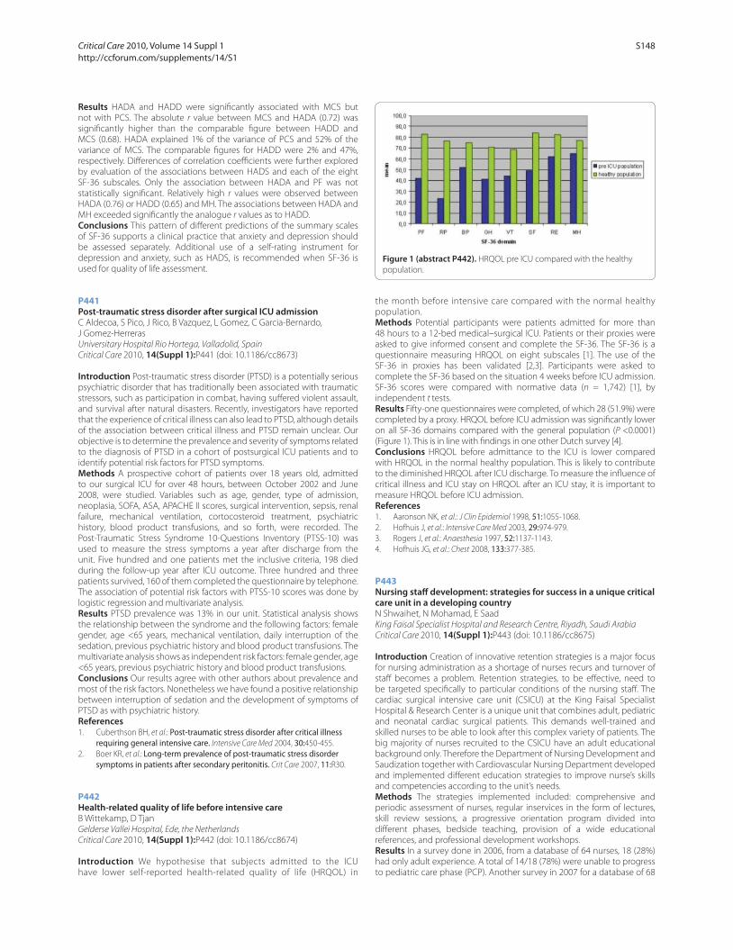
Results HADA and HADD were signifi cantly associated with MCS but
not with PCS. The absolute r value between MCS and HADA (0.72) was
signifi cantly higher than the comparable fi gure between HADD and
MCS (0.68). HADA explained 1% of the variance of PCS and 52% of the
variance of MCS. The comparable fi gures for HADD were 2% and 47%,
respectively. Diff erences of correlation coeffi cients were further explored
by evaluation of the associations between HADS and each of the eight
SF-36 subscales. Only the association between HADA and PF was not
statistically signifi cant. Relatively high r values were observed between
HADA (0.76) or HADD (0.65) and MH. The associations between HADA and
MH exceeded signifi cantly the analogue r values as to HADD.
Conclusions This pattern of diff erent predictions of the summary scales
of SF-36 supports a clinical practice that anxiety and depression should
be assessed separately. Additional use of a self-rating instrument for
depression and anxiety, such as HADS, is recommended when SF-36 is
used for quality of life assessment.
P441
Post-traumatic stress disorder after surgical ICU admissionC Aldecoa, S Pico, J Rico, B Vazquez, L Gomez, C Garcia-Bernardo,
J Gomez-Herreras
Universitary Hospital Rio Hortega, Valladolid, Spain
Critical Care 2010, 14(Suppl 1):P441 (doi: 10.1186/cc8673)
Introduction Post-traumatic stress disorder (PTSD) is a potentially serious
psychiatric disorder that has traditionally been associated with traumatic
stressors, such as participation in combat, having suff ered violent assault,
and survival after natural disasters. Recently, investigators have reported
that the experience of critical illness can also lead to PTSD, although details
of the association between critical illness and PTSD remain unclear. Our
objective is to determine the prevalence and severity of symptoms related
to the diagnosis of PTSD in a cohort of postsurgical ICU patients and to
identify potential risk factors for PTSD symptoms.
Methods A prospective cohort of patients over 18 years old, admitted
to our surgical ICU for over 48 hours, between October 2002 and June
2008, were studied. Variables such as age, gender, type of admission,
neoplasia, SOFA, ASA, APACHE II scores, surgical intervention, sepsis, renal
failure, mechanical ventilation, cortocosteroid treatment, psychiatric
history, blood product transfusions, and so forth, were recorded. The
Post-Traumatic Stress Syndrome 10-Questions Inventory (PTSS-10) was
used to measure the stress symptoms a year after discharge from the
unit. Five hundred and one patients met the inclusive criteria, 198 died
during the follow-up year after ICU outcome. Three hundred and three
patients survived, 160 of them completed the questionnaire by telephone.
The association of potential risk factors with PTSS-10 scores was done by
logistic regression and multivariate analysis.
Results PTSD prevalence was 13% in our unit. Statistical analysis shows
the relationship between the syndrome and the following factors: female
gender, age <65 years, mechanical ventilation, daily interruption of the
sedation, previous psychiatric history and blood product transfusions. The
multivariate analysis shows as independent risk factors: female gender, age
<65 years, previous psychiatric history and blood product transfusions.
Conclusions Our results agree with other authors about prevalence and
most of the risk factors. Nonetheless we have found a positive relationship
between interruption of sedation and the development of symptoms of
PTSD as with psychiatric history.
References1. Cuberthson BH, et al.: Post-traumatic stress disorder after critical illness
requiring general intensive care. Intensive Care Med 2004, 30:450-455.
2. Boer KR, et al.: Long-term prevalence of post-traumatic stress disorder symptoms in patients after secondary peritonitis. Crit Care 2007, 11:R30.
P442
Health-related quality of life before intensive careB Wittekamp, D Tjan
Gelderse Vallei Hospital, Ede, the Netherlands
Critical Care 2010, 14(Suppl 1):P442 (doi: 10.1186/cc8674)
Introduction We hypothesise that subjects admitted to the ICU
have lower self-reported health-related quality of life (HRQOL) in
the month before intensive care compared with the normal healthy
population.
Methods Potential participants were patients admitted for more than
48 hours to a 12-bed medical–surgical ICU. Patients or their proxies were
asked to give informed consent and complete the SF-36. The SF-36 is a
questionnaire measuring HRQOL on eight subscales [1]. The use of the
SF-36 in proxies has been validated [2,3]. Participants were asked to
complete the SF-36 based on the situation 4 weeks before ICU admission.
SF-36 scores were compared with normative data (n = 1,742) [1], by
independent t tests.
Results Fifty-one questionnaires were completed, of which 28 (51.9%) were
completed by a proxy. HRQOL before ICU admission was signifi cantly lower
on all SF-36 domains compared with the general population (P <0.0001)
(Figure 1). This is in line with fi ndings in one other Dutch survey [4].
Conclusions HRQOL before admittance to the ICU is lower compared
with HRQOL in the normal healthy population. This is likely to contribute
to the diminished HRQOL after ICU discharge. To measure the infl uence of
critical illness and ICU stay on HRQOL after an ICU stay, it is important to
measure HRQOL before ICU admission.
References1. Aaronson NK, et al.: J Clin Epidemiol 1998, 51:1055-1068.
2. Hofhuis J, et al.: Intensive Care Med 2003, 29:974-979.
3. Rogers J, et al.: Anaesthesia 1997, 52:1137-1143.
4. Hofhuis JG, et al.: Chest 2008, 133:377-385.
P443
Nursing staff development: strategies for success in a unique critical care unit in a developing countryN Shwaihet, N Mohamad, E Saad
King Faisal Specialist Hospital and Research Centre, Riyadh, Saudi Arabia
Critical Care 2010, 14(Suppl 1):P443 (doi: 10.1186/cc8675)
Introduction Creation of innovative retention strategies is a major focus
for nursing administration as a shortage of nurses recurs and turnover of
staff becomes a problem. Retention strategies, to be eff ective, need to
be targeted specifi cally to particular conditions of the nursing staff . The
cardiac surgical intensive care unit (CSICU) at the King Faisal Specialist
Hospital & Research Center is a unique unit that combines adult, pediatric
and neonatal cardiac surgical patients. This demands well-trained and
skilled nurses to be able to look after this complex variety of patients. The
big majority of nurses recruited to the CSICU have an adult educational
background only. Therefore the Department of Nursing Development and
Saudization together with Cardiovascular Nursing Department developed
and implemented diff erent education strategies to improve nurse’s skills
and competencies according to the unit’s needs.
Methods The strategies implemented included: comprehensive and
periodic assessment of nurses, regular inservices in the form of lectures,
skill review sessions, a progressive orientation program divided into
diff erent phases, bedside teaching, provision of a wide educational
references, and professional development workshops.
Results In a survey done in 2006, from a database of 64 nurses, 18 (28%)
had only adult experience. A total of 14/18 (78%) were unable to progress
to pediatric care phase (PCP). Another survey in 2007 for a database of 68
Figure 1 (abstract P442). HRQOL pre ICU compared with the healthy
population.
Critical Care 2010, Volume 14 Suppl 1 http://ccforum.com/supplements/14/S1
S148

nurses showed that 15 (22%) had only adult experience. Eight out of 15
(53%) could not progress to PCP. A third survey performed in 2008, for a
database of 82 nurses, showed that 25 (30%) had adult experience. Six out
of 25 (24%) were not able to progress to PCP.
Conclusions Implementing comprehensive education strategies
targeting diff erent approaches of training contributed to considerable
progressive staff development and retention in the CSICU.
References1. Aiken LH, Buchan J, Sochalski J, Nichols B, Powell M: Trends in international
nurse migration. Health Aff airs 2004, 23:69-77.
2. Atencio BL, Cohen J, Gorenberg B: Nurse retention: is it worth it? Nurs Econ
2003, 21:262-268, 299.
3. Barney S: Retaining our workforce, regaining our potential. J Healthc Manag
2002, 47:291-295.
P444
Implementing the Leapfrog standard in a developing countryJ Scribante1, S Bhagwanjee2
1University of the Witwatersrand, Johannesburg, South Africa; 2University of
Washington, Seattle, WA, USA
Critical Care 2010, 14(Suppl 1):P444 (doi: 10.1186/cc8676)
Introduction The Leapfrog standard of ICU physician staffi ng is regarded
as the gold standard. It is suggested that hospital leaders should focus
on how to implement intensivist staffi ng [1]. Internationally, especially
in developing countries, the ‘how to’ poses a tremendous challenge. The
Critical Care Society of Southern Africa identifi ed the need to objectively
quantify existing ICU resources in South Africa (SA) with a view to making
recommendations for the future. From this audit we discuss the challenges
of implementing the Leapfrog standard of ICU physician staffi ng in a
developing country.
Methods Approval to conduct a national audit was obtained from
appropriate authorities and was undertaken in 2004 and 2005 in all public
and private hospitals in SA.
Results A 100% sample was obtained. At the time there were 448 units in
SA, of which 7% of ICUs in the public sector and 1% in the private sector
were closed units. There were a total of 4,168 ICU and HC beds, with 30
intensivists and 4,584 nurses (25% ICU trained) working in the ICU [2-4].
Conclusions SA ICU services face challenges and the results show the
diffi culty of implementing the Leapfrog standard. A strategy to deal with
these challenges must be reality based and contextually appropriate. We
propose short-term, medium-term and long-term recommendations that
consider fi scal pressure and competing healthcare imperatives [5]. Short
term: Regionalisation and integration of ICU services, development of
guidelines and protocols to guide practice. Introduction of continuous
professional development programs and a structured outreach program
for medical and nursing staff . Active recruitment and training of personnel.
Medium term: Development of telemedicine and a two-tiered ICU
program. The fi rst tier to comprise nurses and doctors with intermediate
skills and experience. The second tier to comprise intensivists and fully
trained ICU nurses. Both tiers to have a clear scope of practice, training and
incentive schemes. A signifi cant percentage of ICU beds are not in use due
to various factors, these beds to be recommissioned. Long term: A strategy
to convert open units to closed units over a period of time. New ICUs to
be established as closed units. An up-to-date national ICU database to be
maintained to direct future planning.
References1. Provonost PJ, et al.: Crit Care Med 2006, 34:S18-S24.
2. Bhagwanjee S, et al.: S Afr Med J 2007, 97:1311-1314.
3. Scribante J, et al.: S Afr Med J 2007, 97:1319-1322.
4. Scribante J, et al.: S Afr Med J 2007, 97:1315-1318.
5. Bhagwanjee S, et al.: S Afr J Crit Care 2008, 24:4-6.
P445
Impact of thiopental use on cerebral perfusion pressure in severe brain trauma: an example of caregivers’ adaptationH Mehdaoui1, L Allart1, B Sarrazin1, R Valentino1, E Balestrat1, P Ravaux2
1Fort de France University Hospital, Fort de France, Martinique; 2Lille 2
University, Lille, France
Critical Care 2010, 14(Suppl 1):P445 (doi: 10.1186/cc8677)
Introduction Thiopental (TP) is used in severe brain trauma to control
high intracranial pressure episodes. Deleterious side eff ects were
described that questioned its ability to reach its objectives securely and
effi ciently. We analyzed the impact of TP use on cerebral hemodynamics
and caregivers’ behavior towards recommendations using a high-rate
recording information system.
Methods A set of 813 hours of data were recorded at a rate of 0.5 Hz.
The study was observational. Deleterious episodes were detected using
the Information System (IS) and validated by two experts. We detected
low cerebral perfusion pressure (CPP), high intracranial (HICP) and/or low
mean arterial pressure (lMAP) episodes. We used commonly admitted
threshold for the ICP (20 mmHg) and the CPP (60 mmHg). Medical orders
intended to reach the recommended objectives were analyzed.
Results Forty-eight periods stratifi ed according to TP dose were analyzed
on 16 patients. Cumulative duration with TP was 20,520 minutes and
26,294 minutes without TP. The mean dose of TP administration was 250 ±
20 mg/hour (3.4 ± 1.4 mg/kg/hour). The HICP incidence was 85 ± 16 per
100 hours with TP vs 95 ± 29 without TP (NS). HICP duration was longer
with TP (27 ± 2 vs 19 ± 2 minutes, P <0.005). The lMAP incidence was the
same with or without TP (56 ± 19 vs 34 + 8 per 100 hours of monitoring).
Duration of lMAP episodes was the same with or without TP (22 ± 2 vs
20 ± 2 minutes, NS). The incidence of orders intended to restore cerebral
hemodynamics was equivalent in both situations (TP vs no TP, 79 ± 24 vs
104 ± 29 per 100 hours, NS). One hundred and eighty-eight medical orders
were analyzed. Use of catecholamine was more frequent with TP (57% (n =
44) vs 43% (n = 33), P = 0.034).
Conclusions The use of TP complies with the recommendations
(prolonged HICP). It did not result in a signifi cant increase in cerebral
hypoperfusion episodes. We showed evidence of adaptations to its use
by physicians. The doses are lower than the doses prescribed earlier in
the literature and the use of catecholamine is more frequent during TP
infusion. This could result in a better control of deleterious side eff ects of
TP and a better compliance with recommendations. Our IS was effi cient in
physicians’ orders’ analysis.
P446
Knowledge of ICU nurses regarding selected care areas commonly guided by protocolsH Perrie, S Schmollgruber
University of the Witwatersrand, Johannesburg, South Africa
Critical Care 2010, 14(Suppl 1):P446 (doi: 10.1186/cc8678)
Introduction Pain management, glycaemic control and weaning from
mechanical ventilation are nursing care areas that can impact on patient
outcome and are commonly guided by protocols. However, in order to
ensure safe, optimal management of patients, even when care is guided by
protocols, nurses require a sound knowledge base. This study attempted
to describe the knowledge of ICU nurses regarding pain management,
glycaemic control and weaning from mechanical ventilation, to compare
the diff erence in knowledge between ICU-trained and non-ICU-trained
nurses and to describe the impact of years of ICU experience on this
knowledge.
Methods A prospective, descriptive, non-interventional study method
was used. Approval was obtained from the ethics committee and other
relevant authorities. The questionnaire used was developed and validated
by two groups of ICU nursing experts. The study population included
all consenting ICU nurses working in the ICUs of the fi ve hospitals
participating in this study.
Results There were 136 participants in the study (68 ICU trained and 68
non-ICU trained). The mean score obtained was 47.56% (SD 11.61), the ICU-
trained participants obtaining 50.11 (SD 11.96) and the non-ICU trained
obtaining 45.01 (SD 10.75). This diff erence was statistically signifi cant (P =
0.0099). The diff erence between the two groups for the section on pain
management was not statistically signifi cant (P = 0.408) with ICU-trained
participants achieving a mean score of 45.07% (SD 16.01) and non-ICU-
trained participants 42.86 (SD 14.91). The diff erence between the two
groups for the other two sections was statistically signifi cant (P = 0.025
and P = 0.006, respectively) with the ICU-trained group achieving 51.26%
(SD 11.74) and the non-ICU trained 46.16 (SD 14.34) for the glycaemic
control section and 53.99% (SD 18.18) for the ICU-trained participants
and 46.01% (SD 15.18) for non-ICU-trained participants for the section
Critical Care 2010, Volume 14 Suppl 1 http://ccforum.com/supplements/14/S1
S149
pertaining to weaning from mechanical ventilation. The correlation
between knowledge and years of ICU experience was poor (r = 0.137).
Conclusions This study found a lack of knowledge regarding the care
areas tested, a small, but statistically signifi cant diff erence between ICU-
trained and non-ICU-trained nurses and a poor correlation between
knowledge and years of ICU experience.
P447
Pharmacist clinical interventions in the ICUNG Hunfeld, PH Melief, RM Van Hest, BE Bosma
HagaTeaching Hospital, Den Haag, the Netherlands
Critical Care 2010, 14(Suppl 1):P447 (doi: 10.1186/cc8679)
Introduction We wanted to investigate the number and nature of
pharmacotherapeutical interventions of a dedicated hospital pharmacist
during rounds in the ICU.
Methods A pharmacist specially trained in ICU practice participated in the
patient rounds twice a week (from July to December 2008). The pharmacist
focused on optimalization of pharmacotherapy based on ICU guidelines
and standards as well as general pharmacotherapy. Medication overviews
were generated from the ICU Patient Data Management System and
were analyzed for each patient during the patient rounds. Interventions
were made to comply with guidelines and to avoid interactions, contra-
indications, overtreatment and undertreatment and to recognize side
eff ects. All pharmacist interventions were collected in a database and the
percentage of interventions leading to a change in pharmacotherapy was
analyzed.
Results Fifty patient rounds were attended (160 patients, mean age: 64,
male: 59%, mean APACHE II score: 25, mean length of stay: 8.7 days, number
of drug orders analyzed: 4,227). A total of 342 interventions was made in
66% of patients (mean: 2.1 interventions/patient), resulting in a change in
pharmacotherapy in 67% of the interventions. The kind of interventions
were: start drug: 18%, stop drug: 15%, reduction of dosage: 3%, increase
of dosage: 4%, toxicology: 12%, interaction: 6%, contra-indication: 9%,
adverse events: 3%, double medication: 4%, switch in accordance to
guidelines: 6%, software: 2%, dose reduction because of renal failure:
6% and other (for example, infusion incompatibility or administration by
feeding tube): 13%. Classes of drugs most involved were: antiobiotics,
gastrointestinal drugs and anticoagulants.
Conclusions Frequent presence of a dedicated pharmacist during
patient rounds in the ICU results in interventions in pharmacotherapy
in 66% of the admitted patients. The mean number of interventions
per patient is 2.1. Interventions lead to improved compliance with ICU
guidelines and pharmacotherapy standards and warrants monitoring of
pharmacotherapy. Interventions probably lead to improved effi cacy and
safety of pharmacotherapy, although future controlled research will have
to prove to what extent drug-induced harm is prevented.
P448
Adverse drug events in the critical care unitS Seynaeve, D Reyntiens, D Vandenplas, W Verbrugghe, B Claes, P Jorens
University Hospital Antwerp, Edegem, Belgium
Critical Care 2010, 14(Suppl 1):P448 (doi: 10.1186/cc8680)
Introduction Mistakes and errors may occur during the care process,
particularly in the ICU as characterized by its large number of drugs
administered to the single patient. One of the most important infl uencing
factors on intramural morbidity and mortality is indeed a harmful or
unpredicted reaction to a drug, a so-called adverse drug event (ADE) [1].
By far the largest proportion (70%) of these ADEs is dose related [1]. The
objectives of this study are to measure the frequency and severity of ADEs
in the ICU and to determine the infl uence of severity of illness and nursing
workload on their prevalence.
Methods A cross-sectional study in the ICU of a tertiary referral hospital,
based on a retrospective chart review. A tool was developed for measuring
the incidence and characteristics of ADEs based on the Global Trigger
Tool for Measuring Adverse Drug Events [2]. If an ADE was identifi ed the
severity was evaluated using the categories based on the system for
classifying medication errors by the National Coordination Council for
Medication Error Reporting and Prevention [3]. The severity of illness was
calculated using the Sepsis-related Organ Failure Assessment score [4] and
the nursing workload by the classical Therapeutic Intervention Scoring
System-28 [5].
Results The review of 1,009 nursing days in 79 patients revealed 230
ADEs, which occurred in a total of 175 nursing days. The most commonly
identifi ed ADE was a hypoglycemia of <50 mg/dl (n = 75), followed by
hypokalemia (n = 67). Ninety-six percent of the ADEs were classifi ed as
category E, whereas only 4% of ADEs were classifi ed as category F. The
mean severity of illness and nursing workload scores were signifi cant
higher on nursing days when an ADE occurred (P <0.001 and P = 0.002,
respectively).
Conclusions ADEs are common in the ICU. The lack of a golden standard
for reporting and collecting ADEs makes it diffi cult to compare with other
studies and to assess the real value of this study. However, these date
strongly and clearly indicate the infl uence of severity of illness and nursing
workload on the prevalence of ADEs.
References1. Nebeker JR, et al.: Ann Intern Med 2004, 140:795-801.
2. Rozich JD, et al.: Qual Saf Health Care 2003, 12:194-200.
3. National Coordinating Council for Medication Error Reporting and Prevention [www.nccmerp.org]
4. Vincent JL, et al.: Intensive Care Med 1996, 22:707-710.
5. Miranda DR, et al.: Crit Care Med 1996, 24:64-73.
P449
Evaluation of appropriateness of intensive care unit admissions: the GIVITI’s StART approachR Rosi1, P Malacarne2, S Livigni3, D Radrizzani4, M Postiglione5, S Piva6,
S Di Gangi7
1AOUS, Siena, Italy; 2H S Chiare, Pisa, Italy; 3H S Giovanni Bosco, Torino, Italy; 4H Legnano (MI), Milan, Italy; 5H Loreto Nuovo, Napoli, Italy; 6Spedali Civili,
Brescia, Italy; 7Istituto Mario Negri Ranica, Bolognia, Italy
Critical Care 2010, 14(Suppl 1):P449 (doi: 10.1186/cc8681)
Introduction Appropriate use of ICU resources is mandatory. When a
patient is admitted to a unit able to provide a higher (lower) level of care
than required, a waste (overuse) of resources can be advocated. StART is
an approach to identify possible mismatches between the level of care
actually delivered, assumed to correspond to what is clinically required,
and the level of care deliverable by the unit.
Methods ICU beds are classifi ed by levels of care deliverable as High
(ventilator, monitor, and 720 minutes nurse time) and Low (monitor and
360 minutes nurse time) [1]. The level of care actually delivered is classifi ed
as High (invasive or non-invasive ventilation, or two vasoactive drugs, or at
least two of the following: one vasoactive drug, dialysis, respiratory support),
Low (single vasoactive drug, or dialysis, or respiratory support) and Ordinary
(none of the above) [2]. Mismatches between the level of beds available
and the level of care delivered were evaluated both on admission and
for each ICU-day of 4,237 patients in 28 ICUs. An ICU-day was judged as
inappropriate, even without mismatch, if an Ordinary patient was present.
Results Eighteen percent of admissions resulted inappropriate (9%
overuse, 9% waste of resources). Patients receiving H level of care (58%
of the total) were appropriately admitted in 88% of cases. There were 271
patients admitted for Ordinary care (6%). They stayed in the ICU 2.5 days
on average, and 95% of them had no complications. Sixty-eight percent
of the ICU-days were inappropriate: 12% for overuse and 56% for waste
of resources. Forty-two percent of the latter days were only due to the
presence of Ordinary patients (43% of them were instead admitted as H
or L patients).
Conclusions Most of the ICU admissions (82%) were appropriate; the
inappropriateness was mostly due to Ordinary patients treated. The short
ICU stay and the absence of complications of these patients strengthen
the inappropriateness of their admission, which was probably due to the
lack of intermediate care units in Italy. The situation is completely diff erent
considering the ICU stay: the inappropriateness rose up to 68%, mainly
due to the presence of Ordinary patients. Almost one-half of them were
patients recovered from sicker condition, who were probably diffi cult to
discharge from the ICU.
References1. Iapichino G, et al.: Min Anest 2007, 73:501-506.
2. Iapichino G, et al.: Intensive Care Med 2001, 27:131-136.
Critical Care 2010, Volume 14 Suppl 1 http://ccforum.com/supplements/14/S1
S150

P450
Noise in the critical care environmentN Haskins, J Soldan
University Hospital of Wales, Cardiff , UK
Critical Care 2010, 14(Suppl 1):P450 (doi: 10.1186/cc8682)
Introduction The subject of excessive noise levels within critical care is
not new and has been identifi ed on numerous occasions in the literature.
Research illustrates the detrimental eff ect of noise on patients both
physically and psychologically. Our interest in this fi eld stemmed from
Fundamentals of Care audits which described noise as being too high and
disturbing for patients.
Methods The extent of the problem was initially explored by undertaking
an audit of noise using an on-the-spot soundmeter. This showed that noise
levels were in excess of the World Health Organisation recommendations
for a patient care environment [1]. This confi rmed the need to address
the subject of noise within our critical care unit. Noise levels were then
investigated further using a Soundlog device. This enabled us to measure
noise levels continuously over multiple 24-hour periods. The device
measures the noise level every second and then records the average
measurement every 5 minutes. Noise levels were audited over a 6-week
period in a variety of locations in an eff ort to gain an accurate assessment
of noise levels. In doing this, patterns and themes in levels of noise could
be identifi ed and analysed.
Results The audit showed that noise levels were consistently between 52
and 65 dB over a 24-hour period, which is above the WHO guidance of
35 dB and 40 dB limit.
Conclusions The current noise levels within our unit exceed the
recommendations so action needs to be taken to address this. It is a busy unit
with some environmental noise origins but also a high density of staff and
relatives, so with this in mind we are developing a project plan to enable us to
explore the current levels of noise and its sources, identify those sources that
are avoidable or open to modifi cation and enable staff to consider their role
and responsibility in relation to noise reduction. By considering the issue of
noise and developing strategies that result in sustainable reductions in noise
levels, we are aiming to improve the patients’ experience of their critical care
stay. We anticipate that there will be both practical and behavioural solutions
to issues that arise, some of which can be addressed by quick-fi x solutions
while others will require longer term work.
Reference1. World Health Organisation 1999 Guidelines for Community Noise
[whqlibdoc.who.int/hq/1999/a68672.pdf ]
P451
Allergy documentation and transfer within critical careK Hatton1, N Barrett2, J Lim2, C McKenzie2
1Chelsea and Westminster NHS Foundation Trust, London, UK; 2Guy’s and
St Thomas’ NHS Foundation Trust, London, UK
Critical Care 2010, 14(Suppl 1):P451 (doi: 10.1186/cc8683)
Introduction Allergies to medication are common and potentially life-
threatening [1]. Patients enter critical care with incomplete information
about their history. It is essential for safety that accurate allergy status
is documented early in the critical care stay. This clinical audit (CA) was
undertaken in a 43-bed, level 3 critical care unit to explore compliance
with local guidelines on allergy documentation.
Methods Critical care patient episodes were obtained retrospectively for
a 1-month period. Timing of allergy documentation and drug prescribing
was noted from the critical care electronic system (ICIP). Allergy status
prior to the critical care admission and after discharge was noted from
the ward drug chart. Additional allergy data were identifi ed from the
hospital electronic patient record (EPR). The CA was repeated 1 year after
implementation of recommendations.
Results Patient episodes were collated (initial CA n = 58, repeat CA n = 79).
A known drug/nondrug allergy was stated in 29.3% patient episodes during
the initial CA and 39.2% patient episodes in the repeat CA. Allergy status
was incomplete 24 hours after critical care admission for two patients at
the initial CA with a reduction to zero during the repeat. Allergy status was
incomplete prior to prescribing of a new drug in critical care (excludes fl uids,
drugs required for emergency intubation) for 51.7% of patient episodes in
the initial CA. This fi gure reduced to 19.0% in the repeat CA. Concordance
between EPR and ICIP allergy at the outset was 68.8%, which increased
to 76.5% in the repeat CA. Concordance with the ward drug chart pre-
admission and ICIP was 77.6%, increased to 93.9% at re-audit.
Conclusions This CA suggests that up to one in three critical care patients
have a known allergy. The potential for harm is high. More than one-
half of patients admitted to critical care did not have an allergy status
documented prior to prescribing a new drug. There was signifi cant
discordance between the paper medication chart and ICIP allergy. A
number of factors were introduced following initial fi ndings, including
making the allergy status mandatory on ICIP, not allowing the admission
summary to be saved prior to allergy documentation and ensuring current
allergy documentation on EPR.
Reference1. Bates DW, et al.: JAMA 1995, 274:29-34.
P452
Use of an anonymous medication incident reporting system on a critical care unitH Dillon, M Rosbergen, S Hutchinson
Norfolk and Norwich University Hospital, Norwich, UK
Critical Care 2010, 14(Suppl 1):P452 (doi: 10.1186/cc8684)
Introduction An anonymous error reporting system was trialled to
establish whether error reporting rates would improve and to allow the
identifi cation of medication incidents. The National Patient Safety Agency
(NPSA) feedback report for the Norfolk and Norwich Hospital shows that,
compared with similar organisations, medication error reporting is low.
A review of the medication incidents reported for the critical care facility
in 2007 and 2008 had only 19 events. Error rates in the ICU are typically
higher than in other acute settings, one study reporting 10.5 medication
errors per 100 bed-days [1]. Patients in critical care typically receive high-
risk medications. Error reporting is integral to identifying common errors
and medication risk reduction.
Methods A medication reporting form was developed to run alongside
the offi cial hospital incident reporting system for 2 weeks. Forms were
distributed throughout the critical care facility. All members of the
multidisciplinary team were asked to anonymously complete a form
every time a medication error, or near miss, occurred. After 2 weeks, the
submitted forms were analysed by the project team.
Results In total 112 reports were submitted. The largest numbers of
incidents reported were due to prescribing (67%) errors followed by
administration (15%), documentation (7%), electronic prescribing problems
(6%), storage (3%) and monitoring (2%). Within prescribing incidents there
were four recurring themes: duplication of therapy, wrong/inappropriate
drug choice, incorrect dose/regimen and omission. The numbers of
incidents reported increased as the week progressed, with the largest
number occurring on a Friday (24%). A review of the types of medicines
involved showed that insulins, proton pump inhibitors, anticoagulants,
vancomycin, and epidurals were recurring themes, refl ecting NPSA alerts.
When graded for signifi cance, two were found to be harmful, 56 signifi cant
and 54 insignifi cant.
Conclusions There is clearly considerable under-reporting of medication
errors in critical care via the offi cial incident reporting system. One of
many reasons for poor offi cial reporting of errors is the lack of tangible
local response to those errors. It is therefore important that the areas
highlighted by our project are eff ectively addressed, not only to improve
safety but also to encourage further reporting and work to develop a
culture of incident reporting within critical care.
Reference1. Valentin A, et al.: Intensive Care Med 2006, 32:1591-1598.
P453
Three years experience of a quality improvement programmeR Innes, S Burt, F Miles, S Watkins
Musgrove Park Hospital, Taunton, UK
Critical Care 2010, 14(Suppl 1):P453 (doi: 10.1186/cc8685)
Introduction As part of a safer patient initiative under the auspices of
the Institute of Health improvement (IHI), Boston, we took part in a 2-year
project to improve the safety and quality of care in adult intensive care.
Critical Care 2010, Volume 14 Suppl 1 http://ccforum.com/supplements/14/S1
S151

Methods Between October 2006 and October 2008 the IHI coached
and advised our institution on mechanisms to improve quality of care.
The aim was to develop systems of healthcare that were reliable at least
95% of the time. The changes to be introduced – central line bundle,
ventilator bundle, hand hygiene, daily patient goals – were led by a small
multidisciplinary team chaired by a hospital executive (fi nance director)
who met every 2 weeks for 2 years for short meetings (~1/2 hour). Our aim
was to reduce nosocomial infection and mortality.
Results It took ~12 months to introduce reliable process in all areas.
Hand hygiene improved from ~70% compliance to consistently >95%.
MRSA acquisition on the ICU reduced from about 10% of patients prior to
October 2006 to 0.3%. VAP was reduced (Figure 1). The average unadjusted
mortality was reduced year on year despite similar mean APACHE II and
ICNARC scores (Figure 2).
Conclusions Reliable evidence-based processes for patient care can
signifi cantly reduce nosocomial infection and mortality. The use of
executive nonmedical hospital personnel experienced in the mechanisms
of introducing change may be benefi cial.
P454
Improving team work during bedside rounds: using daily goals and best practicesM Adamson, L Kure, B Duncan, N Holmes, AA Amaral
Sunnybrook Health Sciences Centre, Toronto, Canada
Critical Care 2010, 14(Suppl 1):P454 (doi: 10.1186/cc8686)
Introduction Communication between healthcare professionals is a key step
for patient safety, its failure accounting for over 60% of root causes in sentinel
events [1]. Bedside rounds are important for teamwork communication
and can be improved by an explicit approach [2] and by process-oriented
information tools to organize and direct interprofessional rounds [3].
Methods As part of a quality improvement project, we conducted an
observation of the documentation of daily goals (DG) and best practices
(BP) in a step-down unit (both tools have been previously added to patient
fl owsheets), before and after the introduction of a structured rounds
process and team education. Our hypothesis was that these important
tools were used before rounds, without input from all team members.
Rounds were observed on two separate periods and the observer would
take notes of whether DG and BP were documented or not and whether
discussion took place before documentation. Diff erences in proportions
between the two periods were analyzed with Fisher’s exact test. P <0.05
was considered signifi cant.
Results We observed 100 bedside interactions on each period.
Documentation remained unchanged for DG (pre 55% vs post 53%,
P >0.05) and BP (pre 57% vs post 48%); however, the second period had an
improved documentation after team discussion (DG: pre 2% vs post 31%,
P <0.001; BP pre 0% vs post 33%, P <0.001).
Conclusions The intervention aided in increasing documentation
after discussion, implying an increased communication among the
interprofessional team. About 50% of patients still will not have
documentation after bedside rounds. Patient information was not
collected, therefore our study is limited in providing information on clinical
outcomes. Further research should focus on how to best implement
these tools, how to qualitatively assess the content of daily goals and to
demonstrate eff ects on patient-centered outcomes.
References1. Arora V, Johnson J, Lovinger D, Humphrey HJ, Meltzer DO: Communication
failures in patient sign-out and suggestions for improvement: a critical incident analysis. Qual Saf Health Care 2005, 14:401-407.
2. Dodek PM, Raboud J: Explicit approach to rounds in an ICU improves communication and satisfaction of providers. Intensive Care Med 2003,
29:1584-1588.
3. Gurses AP, Xiao Y: A systematic review of the literature on multidisciplinary rounds to design information technology. J Am Med Inform Assoc 2006,
13:267-276.
P455
Stress induced by simulation-based training in anesthesiologist residentsT Geeraerts, P Roulleau, G Cheisson, K Aidan, M Leguen, J Duranteau,
D Benhamou
CHU de Bicetre, Le Kremlin Bicetre, France
Critical Care 2010, 14(Suppl 1):P455 (doi: 10.1186/cc8687)
Introduction The use of a high-fi delity simulator can lead to very realistic
clinical situations sometimes diffi cult to manage psychologically. The aim of
this study was to evaluate the psychological stress induced by simulation-
based training and the associated skills in anesthesiologist residents.
Methods A cohort of 27 residents was studied. The psychological
stress just before and after the simulation session was quantifi ed by
autoevaluation scale (numeric scale 0 to 10) and by salivary amylase
sampling [1]. Nontechnical skills were quantifi ed by analysing videotapes
and scoring the Anaesthetist NonTechnical Skills [2].
Results The median stress numeric scale before the simulation session was
5 (ranging 2 to 8), and after was 7 (2 to 10) (P = 0.0004) (Figure 1). The stress
scale before the session was signifi cantly lower in residents who already
Figure 1 (abstract P453). Ventilator-associated pneumonias per 1,000
ventilator-days. C03: Musgrove Park Hospital, Taunton and Somerset NHS
Trust. CCO1 – VAP rate.
Figure 2 (abstract P453). Intensive care mortality between 2006 and
2009.
Figure 1 (abstract P455).
Critical Care 2010, Volume 14 Suppl 1 http://ccforum.com/supplements/14/S1
S152

underwent simulation-based training (P = 0.04). In 48% of residents, stress
scales after the simulation session were above 8/10. Salivary amylase after
the session was signifi cantly higher than before (P = 0.008), corresponding
to a 2.2-fold increase. They were no signifi cant relationships between
psychological stress parameters and nontechnical skills.
Conclusions Psychological stress before the simulation session, but
especially after simulation, appears to be high in anesthesiologist
residents, and particularly in those who performed a simulation session
for the fi rst time. This fact should be considered when organising such
simulation-based teaching.
References1. Dantzer R, et al.: Psychoneuroendocrinology 2009, 34:1.
2. Yee B, et al.: Anesthesiology 2005, 103:241-248.
P456
Impact of an education program in respiratory therapy quality of careWA Pinto, HB Rossetti, A Araujo, HM Do Carmo, AP Acerbi, MV Santos,
JJ Sposito, M Rabelo, SS Matos, M Assuncao, F Machado
Federal University of São Paulo, Brazil
Critical Care 2010, 14(Suppl 1):P456 (doi: 10.1186/cc8688)
Introduction The respiratory therapist has a central role in the manage-
ment of critically ill patients. The objective of this study was to evaluate
the impact of an education program aiming to improve quality of care of
respiratory therapy.
Methods A before–after study was designed to assess compliance to 15
respiratory therapy indicators, chosen due to their relevance in patient
care and availability for objective measurement, in all patients admitted
to the ICU regardless of mechanical ventilation or not. Compliance
was assessed during 1 month before implementation of the education
program. The educational process comprised of meetings, group training,
written manuals and individual feedback for noncompliance situations.
After 6 months of the implementation process, compliance assessment
was again performed. Results were expressed as percentages and
comparisons were performed through the chi-square test. Results were
considered signifi cant if P <0.05.
Results In both periods 80 patients were observed, with a total of 1,212
observations in the before period (BP) and 1,369 observations in the after
period (AP). Compliance rates improved for almost all indicators as follows:
chart registration (CR) of weight/height (BP = 97.7%; AP = 99.4%; P =
0.0026), CR of weight-predicted tidal volume (BP = 73.3%; AP = 94.7%; P =
0.000), CR of orotracheal tube fi xation level (BP = 69.5%; AP = 93.6%; P =
0.000), CR of humidifi cation line change date (BP = 83%; AP = 89.8%; P =
0.000), CR of chest X-ray pattern (BP = 86.8%; AP = 96.9%; P = 0.000); CR of
clinical decisions during round (BP = 91.3%; AP = 98.7%; P = 0.000), actual
compliance to clinical decisions during rounds (BP = 79.77%; AP = 98.8%;
P = 0.000), correct utilization of mechanical ventilation humidifi cation (BP =
75.9%; AP = 96.4%; P = 0.000), actual tidal volume between 6 and 8 ml/kg
predicted body weight (BP = 82.0%; AP = 96.3%; P = 0.000), adequacy of
ventilators alarms (BP = 69.5%; AP = 95.4%; P = 0.000), actual plato pressure
<30 mmH2O (BP = 93.2%; AP = 95.7%; P = 0.0241). Some indicators did
not show a statistically signifi cant improvement: CR of secretion aspect
(BP = 94.4%; AP = 94.2%; P = 0.861), CR of circuit change date (before =
90%; after = 92.2% P = 0.0848) and CR of round changes (BP = 92.0%. AP =
92.6%; P = 0.551). Only CR of cuff pressure had a lower compliance on the
AP (BP = 87.5%; AP = 79.4%; P = 0.0005).
Conclusions An education program based on protocol-driven actions
and training is able to improve compliance in respiratory therapy quality
indicators. The clinical relevance of this compliance improvement still
needs to be established.
P457
Use of the FAST HUG checklist in Mexican critically ill patientsJC Cerda, V Sanchez, F Perez, E Fernandez
HSJ Tec de Monterrey, Mexico
Critical Care 2010, 14(Suppl 1):P457 (doi: 10.1186/cc8689)
Introduction There have been several eff orts to improve the quality of
care of ICU patients. The use of checklists has been linked to improvements
in ICU care. In 2005, Vincent and colleagues published the FAST HUG
checklist strategy [1]. We decided to study the level of adherence of
Mexican physicians to the measures proposed in this checklist.
Methods We conducted a 3-month prospective observational study in
the adult ICU of an academic hospital in northeastern Mexico. Patients
were sorted into one of the two study groups: group 1 had a critical care
physician on charge of the ICU management, while group 2 had a non-
critical care physician on charge. We measured the adherence of the
physicians to the FAST HUG checklist items. We arbitrarily defi ned good
compliance as having fulfi lled >4 items.
Results One hundred and forty-seven patients were admitted to the
ICU during the study period, but only 129 had complete data and were
evaluated. There were 86 patients in group 1, while group 2 had 43
patients. Group 1 patients had higher age, SOFA and APACHE score on
admission. We analyzed the subgroup of patients who had an initial
APACHE score between 11 and 25, as they are usually the ones most likely
to benefi t from FAST HUG measures. These results are described in Table
1. We noticed that most of the group 1 patients had severe sepsis, while
most of the group 2 patients had ischemic heart disease, which clearly
explains the diff erences in mechanical ventilation and length of ICU stay.
Table 1 (abstract P457). Variables of the subgroup APACHE II 11 to 25
Variable Group 1 Group 2 P value
Age 64 ± 10 69 ± 17 0.25
APACHE II 18 ± 5 15 ± 4 0.08
SOFA on admission 5 ± 3 3.3 ± 2 0.12
SOFA at discharge 2.4 ± 4 1.7 ± 2 0.82
ICU length of stay 10.5 ± 9 4.6 ± 3 0.04*
Mechanical ventilation days 6.7 ± 9.6 1.8 ± 3 0.11
FAST HUG 6.4 ± 0.9 3.8 ± 1.4 0.001*
Group 1 had a critical care physician in charge of ICU management, while group 2 had a noncritical care physician in charge. We observed the same severity of illness in this group with APACHE II between 11 and 25 points and better adherence to the FAST HUG items among intensive care physicians.
Conclusions We found a better adherence to the FAST HUG items among
intensive care physicians.
Reference1. Vicent JL, et al.: Give your patient a fast hug (at least) once a day. Crit Care
Med 2005, 33:1225-1229.
P458
Bay rooms vs single-bed rooms in intensive care unit nosocomial infections: a case–control studyE Bigazzi, L Turrisi, G Zagli, P Pecile, M Bonizzoli, A Peris
Careggi Teaching Hospital, Florence, Italy
Critical Care 2010, 14(Suppl 1):P458 (doi: 10.1186/cc8690)
Introduction Nosocomial infections remain a clinical challenge in the ICU.
Based on defi nitions by the Centers for Disease Control and Prevention of
Atlanta, nosocomial infections are defi ned as infection occurring within
48 to 72 hours after hospital admission. In ICU settings, a single-bed
room location has been suggested as a structural organization helpful to
prevent nosocomial infection diff usion. Here we show preliminary results
of a study conducted in our ICU before and after a change from open bay
to single-bed room locations.
Methods Microbiological data were collected from ICU patients hosted in
bay rooms in the ICU (April 2006 to April 2007) and compared with data
collected from patients hosted in the subsequent period (May 2007 to
May 2008), after the single-bed ICU room re-allocation. For analysis, we
considered the prevalence of microorganisms isolated and the diff erence
in blood culture/tracheal aspirate positivity.
Results The cohorts examined resulted in similar demographic and
clinical data. As shown in Figure 1, microorganisms isolated did not change
signifi cantly in the two periods. Figure 2 summarizes the diff erences in
tracheal aspirate/blood culture isolation among the two periods.
Conclusions We cannot confi rm that a single-bed room location can
prevent nosocomial infection diff usion. A limit of this study, as in previous
Critical Care 2010, Volume 14 Suppl 1 http://ccforum.com/supplements/14/S1
S153

published works, remains the bias related to patients admitted from other
wards or after a prolonged emergency room stay.
P459
Impact of quality improvement process on healthcare-associated infection in the ICU in a tertiary care hospital in IndiaM Bhattacharyya, A Bhakta, S Todi
AMRI Hospitals Kolkata, India
Critical Care 2010, 14(Suppl 1):P459 (doi: 10.1186/cc8691)
Introduction Healthcare-associated infection (HCAI) has a signifi cant
contribution to mortality, morbidity and cost of treatment in critically ill
patients. The objective of this study was to conduct a process (quality
improvement) and outcome (pre and post implementation) surveillance,
HCAI being taken as the major variable.
Methods This was a prospective observational study carried out from June
2005 to September 2009. Process implementation was started from May
2006 and included compliance with hand hygiene, urinary catheter care,
central line care and VAP bundle. HCAI rates were recorded prospectively
every month from June 2005. We recorded the rates of central line-related
bloodstream infection (CLI), catheter-associated urinary tract infection
(UTI), hospital-acquired pneumonia (HAP) and ventilator-associated
pneumonia (VAP) per 1,000 device-days as per CDC–NNIS defi nition [1,2].
Results Various infection rates showed numerical improvement after the
implementation of the quality improvement (QI) process (Figure 1). The
diff erences were statistically signifi cant for two of these four endpoints (P
for HAP = 0.029 and for UTI = 0.013) and in others there was a trend towards
improvement. Device utilization rates associated with these endpoints
before and after the implementation remained unchanged, confi rming that
the drop of infection rates was not infl uenced by any reduction/increase of
respective device utilizations (Figure 1). The average compliance rate during
the study period for hand hygiene was 77.92, for urinary catheter care 98.24,
for central line care 96.62 and for VAP bundle 91.54.
Conclusions Implementation and continuous surveillance of the QI
process improved nosocomial HCAI in our hospital.
References1. Garner JS, Jarvis WR, Emori TG, Horan TC, Hughes JM: CDC defi nitions for
nosocomial infections, 1988. Am J Infect Control 1988, 16:128-140.
2. Emori TG, Culver DH, Horan TC, et al.: National nosocomial infections surveillance system (NNIS): description of surveillance methods. Am J
Infect Control 1991, 19:19-35.
P460
Teaching airway resuscitation to novices: a randomized controlled simulator mannequin studyP Brindley, C Needham
University of Alberta Hospital, Edmonton, Canada
Critical Care 2010, 14(Suppl 1):P460 (doi: 10.1186/cc8692)
Introduction The sniffi ng position is widely promoted for teaching
airway positioning prior to intubation attempts, but whether this analogy
results in novices placing the head and neck appropriately has not been
evaluated. An alternate analogy – win with the chin – or simple anatomic
instructions may perform better.
Methods Randomized controlled study comparing diff erent instructions
for enabling novices to adequately position a simulator mannequin’s head
and neck. Study volunteers included medical students and PGY1 residents
in surgery and internal medicine. Subjects independently positioned a
mannequin based upon their understanding of four randomly-assigned
written instructions for: the sniffi ng position; the win with the chin
analogy; anatomic instructions; and no instructions (control). Digital
photographs following each instruction were analyzed by two airway
experts for adequacy of overall positioning, and for the three components
of airway positioning.
Results There were 81 volunteers. The positioning was adequate most
often (43.2%) following the win with the chin analogy as compared with
the other instructions (37.0% anatomic instructions; 19.8% control; 14.8%
sniffi ng position analogy). Positioning following the sniffi ng position
instructions was not diff erent from no instruction (P = 0.53). The win with
the chin and anatomic instructions were better than no instructions (P =
0.002 and P = 0.023, respectively).
Conclusions The win with the chin analogy resulted in adequate airway
positioning signifi cantly more often than the sniffi ng position or control,
and maintained atlanto-occipital extension when compared with
providing anatomic instructions. Overall, win with the chin was a superior
teaching analogy and could replace the sniffi ng position analogy.
P461
Impact of ultrasound guidance in central venous catheterization by inexperienced trainees: a prospective observational studyS Yamao, R Suda, Y Tomishima, N Uchiyama, T Jinta, N Nishimura, S Ohde,
N Chohnabayashi
St Luke’s International Hospital, Tokyo, Japan
Critical Care 2010, 14(Suppl 1):P461 (doi: 10.1186/cc8693)
Introduction Central venous catheterization is a common procedure,
but can potentially result in serious complications. Previous studies
have shown that inexperienced trainees have lower success rates than
experienced operators. Little is known about the effi cacy of ultrasound
(US) guidance in central venous catheter (CVC) insertion by inexperienced
trainees.
Figure 1 (abstract P458).
Figure 2 (abstract P458).
Figure 1 (abstract P459). Comparing mean infection rates before and
after the quality improvement process.
Critical Care 2010, Volume 14 Suppl 1 http://ccforum.com/supplements/14/S1
S154
Methods A prospective, observational study, performed in a Japanese
training hospital. All patients who were inserted CVCs by inexperienced
trainees (postgraduate year 1 to 6) between 1 June 2008 and 31 October
2009 were included. The following parameters were evaluated: the rate
of successful insertions, the incidence of complications, the site of CVC
insertion, the method of US use, and the postgraduate year of the trainees.
We defi ned success of CVC insertion if a trainee completes catheterization
without complication and personnel exchange. We compared the
success rate and the complication rate with both insertion sites and US
techniques; Group SA (surface anatomy) underwent CVC insertion using
the landmark technique, Group indirect-ultrasound (iUS) using ultrasound
image-based skin surface marking, and Group direct-US (dUS) using real-
time ultrasound guidance. The chi-test was used for analysis and P <0.05
was considered statistically signifi cant.
Results In total 593 CVCs were inserted, 527 via the internal jugular vein
(IJV), and 66 via the subclavian vein (SV). The IJV approach includes 52
patients (9.9%) in Group SA, 145 patients (27.5%) in Group iUS, and 330
patients (62.6%) in Group dUS. The SV approach, on the other hand,
consisted of 26 patients (39.4%) in Group SA, and 40 patients (60.6%) in
Group dUS. The success rate of CVC insertion of the IJV approach linearly
rose in Group SA, Group iUS and Group dUS (76.9%, 81.4%, 88.2%, P =
0.033), but in the SV approach there is no signifi cant diff erence between
Group SA and Group dUS (76.9%, 75.0%, P = 0.83). Five patients who
underwent real-time US via the SV developed pneumothorax while no
one in Group SA had the complication (P = 0.57).
Conclusions The use of ultrasound guidance improves the success rate
of CVC insertion via the IJV by inexperienced trainees. However, in the
SV approach the effi cacy of ultrasound guidance is not proven, and the
incidence of pneumothorax increased in our study.
P462
Performance analysis of an ICU according to GiViTI’s StART approachR Rosi1, V Peccianti1, E Fatighenti1, M Giardino2, D Crespi2, A Anghileri2
1AOUS, Siena, Italy; 2Istituto Mario Negri, Ranica (BG), Italy
Critical Care 2010, 14(Suppl 1):P462 (doi: 10.1186/cc8694)
Introduction ICU beds are a limited resource; their appropriate use is
mandatory: only patients sick enough should be admitted to the ICU, and
all admitted patients should receive the adequate level of care.
Methods From 1 January 2008 to 9 March 2009, 716 admissions to
a seven-bed general ICU were analyzed according to GiViTI’s StART
approach, to evaluate appropriateness of the ICU admission and stay.
Available resources (ICU beds) were classifi ed as High Level (HL) or Low
Level (LL), according to equipment and staffi ng. Patients may require High
(H), Low (L), or Ordinary (O) levels of care (HP, LP, OP, respectively) [1]. OPs
should not be admitted to the ICU and can stay in the ICU only briefl y,
following HL or LL periods. One nurse can attend two HPs, or one HP
and two LPs, or four LPs, provided that each bed can be controlled easily
(not in diff erent rooms, for example) [2]. For each day shift, matching of
required and available care was analyzed: mismatch is expressed as nurse
defi cit: positive values indicate excess of care (too many nurses for not too
sick patients), zero suggests that assistance requirements are met, with
negative values available care is less than patients’ needs.
Results In this ICU, all seven beds are fully equipped. Of the 716 ICU
admissions, 20 (2.8%) were inappropriate OPs. One hundred and ninety-
seven further ICU-days of the 2,711 available (7.3%) were used for OPs
(mostly due to delayed discharges); altogether 217/3,427 (6.3%) ICU-
days were inappropriately used for OPs. In the analysis of resources
and requirements matching, if the seven beds were in the same room,
available assistance would meet requirements in most nurse shifts, even if
the staff would have worked at maximum almost always; in fact, in this ICU,
four beds are in a room and there are three single-bed rooms – therefore,
according to the suggested standards, a mean 1.9 nurse defi cit per shift is
observed. The relevance of this observation on patient outcome can be
speculated only.
Conclusions The relevance of the StART approach is the defi nition of
inappropriateness both from excess and insuffi ciency of care points
of view: both admissions and ICU stays of OPs and/or too many too
sick patients for the amount of care available are equally inappropriate.
Moreover, the available care is determined both by the number of ICU
nurses and by logistic constraints: seven beds in an open space are
diff erent from four beds in a room and three beds in single rooms, and in
fact this ICU pays a two nurse defi cit per shift to its design.
References1. Iapichino G, et al.: Minerva Anestesiol 2005, 71:273-280.
2. Iapichino G, et al.: Minerva Anestesiol 2007, 73:501-506.
P463
Simulation training at the point of care to decrease the incidence of airway complications on the intensive care unitSC Cook, JJ Gatward, FE Kelly, J Bell, FM O’Higgins, MJ Thomas
Bristol Royal Infi rmary, Bristol, UK
Critical Care 2010, 14(Suppl 1):P463 (doi: 10.1186/cc8695)
Introduction A recent analysis of critical incidents reported to the National
Patient Safety Agency (NPSA) by ICUs in the UK revealed a signifi cant
number of airway incidents in critical care [1]. We recently performed
an online survey with responses from 305 trainees from across the UK
regarding airway management and complications on the ICU. The results
revealed a high number of adverse airway events including 27 deaths. A
large number of junior trainees (many of whom cover the ICU alone at
night) expressed low confi dence levels with airway management in critical
care, and the majority of all trainees felt inexperienced with tracheostomy
care and emergencies. Simulation has been recommended as a suitable
way of providing critical incident training [2], and simulation at the point
of care has been shown to be especially powerful and eff ective [3].
Methods In response to our survey, we set up a multidisciplinary
simulation-based airway teaching programme for all staff on our unit. A
Resusci-Annie™ manikin with an Airway Trainer™ head (Laerdel, Norway) is
set up in a bed space on our ICU. Realistic patient monitoring is achieved
with the Laerdel SimMan™ software (free from the Laerdel website). We
have written a number of scenarios in response to incidents and training
defi ciencies highlighted by our survey. Scenarios are run with ICU staff
from all disciplines acting within their normal roles. Realism is enhanced
by staff practicing in their usual working environment, using the unit’s
own equipment.
Results So far, we have trained 43 ICU staff . Feedback has been excellent,
with scores ranging from 8.5 to 10. Participants were especially positive
about the multidisciplinary aspect of the training and on practising
uncommonly encountered scenarios in their normal workplace.
Conclusions We have created a multidisciplinary airway training
programme to practice critical incident drills at the point of care. This
teaching method is low cost, simple and can be set up quickly and
spontaneously during quiet periods on the ICU.
References1. Thomas AN, et al.: Anaesthesia 2009, 64:358-365.
2. Wheeler DW, et al.: Br J Anaesth 2009, 103:145-147.
3. Weinstock P, et al.: Pediatr Crit Care Med 2009, 10:176-181.
P464
Management of needle stick injuries from incapacitated patientsL Burrows, A Padkin
Royal United Hospital, Bath, UK
Critical Care 2010, 14(Suppl 1):P464 (doi: 10.1186/cc8696)
Introduction This survey investigated current UK intensive care practice
in testing patients for blood-borne viruses following needle stick injury
when the patient lacks capacity for consent. Needle stick injuries are a
common occurrence in the National Health Service, with an estimated
incidence of 113 to 623/10,000 healthcare workers/per year [1]. Current
UK law [2,3] does not permit the testing of a patient’s infective status
for the benefi t of a healthcare worker involved without the patient’s
consent. Incapacitated patients cannot give consent. The General Medical
Council (GMC) guidance for the management of needle stick injuries from
incapacitated patients refl ects this [4].
Methods A questionnaire was sent to the clinical lead of the 225 general
ICUs within England, Wales and Northern Ireland. Responses were
anonymous.
Critical Care 2010, Volume 14 Suppl 1 http://ccforum.com/supplements/14/S1
S155
Results Ninety-nine consultants replied out of 225 (44% response rate).
Sixty-two of the 99 consultants (63%) reported incidences of needle stick
injuries to their staff from incapacitated patients in the past year. In six
incidents the patient was not tested because their full blood-borne virus
status was already known. Thirty-six of the remaining 56 incidents (64.5%)
stated that patients were tested without consent. Sixty-one per cent
of consultants (22 of 36) reported that the patient was informed once
conscious that the test had been performed. Two consultants discussed
the decision to perform the test with the family prior to proceeding. One
patient, not tested at the time of injury, refused consent on regaining
capacity. Sixteen patients out of the 62 incidents were blood-borne virus-
positive (26%), six being new diagnoses following the needle stick injury
(37.5%).
Conclusions Despite the recent change in legislation, the majority of
incapacitated patients are being tested for blood-borne viruses when
unable to give consent. This has signifi cant benefi cial eff ects for the
healthcare worker involved; a negative test allowing prophylactic antiviral
treatment to be discontinued and minimises anxiety. This survey highlights
the need for further discussion involving healthcare professionals, the
legal profession and legislators regarding needle stick injuries from
incapacitated patients. Is there a duty of care to the healthcare worker that
is not acknowledged in current legislation?
References1. Trim JC, et al.: J Hosp Infect 2003, 53:237-242.
2. Human Tissue Act 2004 [http://www.opsi.gov.uk/acts/acts2004/
ukpga_20040030_en_1.htm]
3. Mental Capacity Act 2005 [http://www.opsi.gov.uk/acts/acts2005/
ukpga_20050009_en_1]
4. Update to Serious Communicable Disease guidance [http://www.gmc-uk.
org/guidance/update_serious_communicable_diseases.asp]
P465
Stress and fatigue among anaesthesia and intensive care doctors in LithuaniaA Macas, A Kolciniene, I Vetaite, D Bilskiene, A Mikalauskas
Kaunas University of Medicine, Kaunas, Lithuania
Critical Care 2010, 14(Suppl 1):P465 (doi: 10.1186/cc8697)
Introduction Evidence shows that anaesthesia and intensive care is
a stressful occupation and it may have an impact on anaesthetists’ and
intensive care doctors’ well-being. The goal of the study was to evaluate
stress and fatigue and their correlation with working conditions among
anaesthesiologists and intensive care specialists employed in Kaunas city
(Lithuania) hospitals.
Methods One hundred and forty-two doctors of anaesthesia and intensive
care departments were interviewed using a standard questionnaire in 2008.
It consisted of three parts: the fi rst part covered the demographic and social
situation, second – stress experienced during work, third – risk of fatigue.
Results The response rate was 71%. Average age of respondents was
41 years, 66.2% were female. A total of 25.4% had working experience
<5 years and 23.2% working experience of 11 to 15 years. The workload
of 49.3% of respondents was 37 to 54 hours per week (mean number
of on-call nights per month – 6.5; 15.3% of respondents had >9 on-call
night shifts). Often and very often stress was experienced by 24.6% of
doctors; symptoms of fatigue were demonstrated by 57% of respondents.
Respondents older than 55 years emphasized increased blood pressure,
while younger respondents – up to 34 years – reported inability to
concentrate, decreased objectiveness, unstable mood, diffi culty in making
decisions. Respondents living without partners experienced chest pain,
anger and nuisance. Doctors experience less stress if they are satisfi ed with
their work and if their workload is adequate, while increasing pressure by
hospital administration to decrease costs of care increased risk of fatigue.
If a doctor is employed for more 40 hours per week he faces diffi culties
concentrating, while more than 60 hours per week causes interference
with home life.
Conclusions Every fourth anaesthesiologist experienced stress during
work, while more than one-half had symptoms of moderate fatigue.
Growing workload increased doctor’s stress. Pressure by hospital
administration and limited freedom in decisions increased incidence of
fatigue. Work satisfaction reduced risk of stress and fatigue. Stress and
fatigue signifi cantly correlated with the state of physical health.
P466
Implementation of daily goals in the ICU reduces length of ICU stay and errors of omission in patient careJM Binnekade1, A Brunsveld2, S Arbous2, MG Dijkgraaf1, J Horn1, J Sloot1,
A Balzereit2, MB Vroom1
1Academic Medical Center, Amsterdam, the Netherlands; 2LUMC, Leiden, the
Netherlands
Critical Care 2010, 14(Suppl 1):P466 (doi: 10.1186/cc8698)
Introduction In the ICU, daily goals have improved the eff ectiveness
of communication within care teams [1]. The study objective was to
investigate the eff ect of explicit daily goals in ICU practice on the length
of intensive care stay, length of hospital stay, duration of mechanical
ventilation and errors of omission in patient care.
Methods A multicenter, sequential cohort, pretest–post-test study was
performed. During the pretest phase, daily goals were formulated by staff -
physicians, evaluated by a research physician and kept blinded for the rest
of the ICU team. Subsequently, daily goals were broadly implemented.
During the post-test phase, daily goals were routine practice. Patient data
were collected one full year before (as baseline reference) and during
the pretest and post-test phases. During pretest and post-test, medical
charts (random subsets of 10 patients a week) were reviewed for errors of
omission, defi ned as deviations from our care protocols. Patient data were
compared with data from nonparticipating academic ICUs, extracted from
the National Intensive Care Evaluation database.
Results Two academic ICUs participated in the study. Patients included:
baseline reference n = 3,641; pretest n = 3,401, and post-test 3,426. ICU
mean (SD) length of stay (days) diff ered signifi cantly between the pretest
phase 5.7 (2) and the post-test phase 4.3 (2.3), diff erence 1.4 days (95% CI
1.3/1.5). The post-ICU length of hospital stay decreased in the post-test
phase, mean diff erence 2.3 days (95% CI 1.2/3.4). Median (IQR) duration of
mechanical ventilation during pretest was 2 (1 to 4) days and during post-
test was 1 (1 to 4) days, P <0.001. The probability of an error of omission
was signifi cant larger in the pretest phase then in the post-test phase, RR
6.5 (5.7/7.4).
Conclusions The results showed a signifi cant decrease in ICU LOS,
duration of mechanical ventilation and length of post-ICU stay following
DGF implementation. Also the chance of errors of omission to occur
was six times lower, which indicates a signifi cant increase in protocol
adherence. The drawback of the study design (historic control group and a
pretest and post-test for comparison) was limited by comparing the study
results with other academic centers during the same time frame.
Reference1. Pronovost PJ, et al.: J Crit Care 2003, 18:71-75.
P467
Use of single patient rooms to decrease acquisition of antibiotic-resistant bacteria in the ICUPD Levin, M Golovanevski, AE Moses, CL Sprung, S Benenson
Hadassah Hebrew University Hospital, Jerusalem, Israel
Critical Care 2010, 14(Suppl 1):P467 (doi: 10.1186/cc8699)
Introduction The role of isolation rooms in decreasing bacterial
transmission between ICU patients has been debated. A recent change in
ICU location allowed for further investigation.
Methods Old ICU A and Old ICU B were open-plan seven-bed and four-bed
units. In March 2007, ICU A moved to single patient rooms (New ICU A –
eight beds). ICU B remained unchanged (New ICU B). The same physicians
cover both ICUs. Patient distribution is based on available space only.
Demographic and microbiological data were collected on consecutive
patients staying >48 hours. Culture of resistant organisms (MRSA, VRE,
ESBL, resistant Acinetobacter, or carbapenem-resistant Gram-negatives) in
surveillance or clinical cultures was measured. New ICU A was compared
with open-plan New ICU B to assess the eff ect of single rooms. New ICU
A was compared with Old ICU A to assess change vs historical controls
and New and Old ICU B were compared with show control group stability.
Results Data were collected on 62, 62, 44 and 39 patients from Old ICU A,
New ICU A, Old ICU B and New ICU B. Patient populations in New ICU A
and B were similar. ICU acquisition of resistant organisms was lower in New
ICU A (3/62, 5% vs 7/39, 18% New ICU B patients, P = 0.043). This fi nding
was confi rmed using survival analysis (P = 0.011). Comparing New with
Critical Care 2010, Volume 14 Suppl 1 http://ccforum.com/supplements/14/S1
S156

Old ICU A showed lower APACHE II scores (18 ± 9 vs 21 ± 9, P = 0.028), pre-
ICU antibiotic use (20/62, 32% vs 37/62, 60%, P = 0.001), pre-ICU hospital
admission (21/62, 34% vs 37/62, 60%, P = 0.004) and ICU admission
prevalence of resistant organisms (11/62, 18% vs 25/62, 40%, P = 0.006).
On Cox regression only the admitting ICU was signifi cant for acquisition
of resistant organisms (Old vs New ICU A: HR 4.07, 95% CI 1.03 to 16.06,
P = 0.025). Comparing New with Old ICU B showed no changes. Adequate
hand hygiene amongst the same physicians was found on 140/242 (58%)
occasions in New ICU A vs 23/66 (35%) in New ICU B (P <0.001).
Conclusions The change in ICU design to single patient rooms
signifi cantly decreased acquisition of multiresistant organisms. Whether
the ICU design or improved infection control awareness led to change
could not be determined. This is the fi rst study to measure the eff ect of
ICU design on acquisition of a range of resistant organisms while including
a contemporaneous control ICU environment.
P468
Time to fi rst review of new admissions to critical care by the consultant intensivistA Dhrampal
Norfolk and Norwich University Hospital, Norwich, UK
Critical Care 2010, 14(Suppl 1):P468 (doi: 10.1186/cc8700)
Introduction The National Confi dential Enquiry into Patient Outcome
and Death (NCEPOD) recommends that all new admissions to the ICU
should be reviewed by a consultant intensivist within 12 hours [1]. This
early review may reduce the ICU length of stay (LOS), but does not appear
to aff ect mortality [2]. Patient severity of illness (APACHE II scores) may also
infl uence patient outcome.
Methods The database of our clinical information system (MetaVision,
iMDsoft) was interrogated and 116 new consecutive critical care
admissions were retrospectively reviewed. We ascertained the time to fi rst
review (in hours) by a consultant intensivist from MetaVision entry logs.
Patient outcomes including mortality and LOS were also captured for
patients reviewed early (<12 hours) and late (>12 hours). The admission
APACHE II score was also calculated. Continuous and ordered categorical
variables are expressed as the median and analysed by the Mann–Whitney
U test. Outcome binary variables were analysed by the chi-squared test.
P <0.05 was considered statistically signifi cant.
Results The early reviewed group (n = 53) had a median fi rst review time
of 4.35 hours (IQR 1.70 to 6.90), median LOS of 46 hours (IQR 22 to 86) and
mortality of 17% (n = 9). The late review group (n = 63) had a median fi rst
review time of 16.55 hours (IQR 14.35 to 18.88), median LOS of 25 hours
(IQR 20 to 46) and mortality of 1.6% (n = 1). The median admission APACHE
II score for the early group and late group was 14 (IQR 10 to 24) and 10 (IQR
8 to 16), respectively. There was no signifi cant diff erence in LOS between
the two groups (P = 0.137, two-sample Mann–Whitney U test). The early
group had a signifi cantly higher mortality (P = 0.03, chi-squared test) and
admission APACHE II score (P = 0.04, two-sample Mann–Whitney U test).
Conclusions We were unable to demonstrate that early consultant
intensivist review of new critical care admissions reduces the LOS or
mortality. Despite early consultant intensivist review, this group had a
signifi cantly higher mortality. A signifi cantly higher severity of illness in the
early group may account for this higher mortality.
References1. An Acute Problem? A Report of the National Confi dential Enquiry into
Patient Outcome and Death; 2005 [www.ncepod.org.uk]
2. Mullen P, et al.: Timing of fi rst review of ICU admissions by consultant intensivists in a UK district general hospital. Crit Care 2009, 13(Suppl 1):P476.
P469
Analysis of critical care referralsM Dallison, H Jones, P Matthews
Morriston Hospital, Swansea, UK
Critical Care 2010, 14(Suppl 1):P469 (doi: 10.1186/cc8701)
Introduction Anecdotal evidence at our hospital suggested an increase
in the number of patients referred for admission to the ITU by junior
doctors without consultation with senior doctors from their own specialty.
Recent UK guidance has recommended consultant-to-consultant referral
for critical care patients [1]. Our ITU welcomes the timely referral of at-risk
patients. However, excessive numbers of poor referrals have contributed
to the prolonged absence of critical care team members from the unit. At
a time of increasing diffi culty in maintaining adequate levels of medical
cover because of regulations concerning hours of work and training
requirements, there is concern that this may adversely aff ect the effi cient
and safe management of the ITU.
Methods All referrals were prospectively recorded for a 6-week period.
The following data were recorded: date/time of referral, grade/specialty
of the referring doctor, involvement of the referring consultant, indication
for the referral, referral to assessment time, duration of the assessment and
whether the referral led to admission.
Results Fifty-seven per cent of referrals were out of working hours.
The sources of the referrals were as follows: emergency codes 29% and
specialty referrals 71%. The admission rates to the ITU for the referrals
were cardiac arrests 33%, trauma calls 75%, specialty referrals 42%. Referral
rates according to grade (excluding emergency codes): consultant 14%,
senior trainee 49%, junior trainee 37%. Admission rates according to grade
of referral: consultant 54.6%, senior trainee 42.1%, junior trainee 34.4%.
The mean time to review of referral was 20.2 minutes. The mean time
for assessment for all referrals was 38.8 minutes. This was 52.3 and 28.9
minutes for patients admitted to the ITU and not admitted, respectively.
The mean assessment and management times for the code calls were:
cardiac arrest 42 minutes and trauma calls 81.2 minutes.
Conclusions Our data suggest that consultant-to-consultant referral
would reduce ITU referrals that do not require admission, and decrease
the time that ITU trainees are off the unit.
Reference1. An Acute Problem? A Report of the National Confi dential Enquiry into
Patient Outcome and Death; 2005 [www.ncepod.org.uk]
P470
Communication with intensive care patientsR Likar1, V Langhold2, A Kovinjalo1, H Trabe1
1LKH Klagenfurt, Austria; 2FH Kaernten, Feldkirchen, Austria
Critical Care 2010, 14(Suppl 1):P470 (doi: 10.1186/cc8702)
Introduction Communication with conscious patients who are on oxygen
and cannot verbally communicate due to temporary loss of speaking
ability has proven to be anywhere from problematic to extremely diffi cult
for the nursing staff trying to understand the needs of the patients.
Methods In order to improve the situation of conscious patients on
oxygen, it is fi rst necessary to conduct a status quo analysis to determine
which communication aids can be implemented in the care of such
patients. For this reason, a questionnaire was designed and carried out
with the staff of the ICU.
Results The questionnaire of the intensive care staff showed that
from 31.7% (20 staff members), the use of diverse electronic aids was
indicated 10 times as a means for improvement in the communication
situation of conscious patients on oxygen. A further question regarding
communication aids that the intensive care staff would like – but are not
available in the ward – resulted in the computer being named as the
most wanted communication aid. In cooperation with the engineering
secondary school (HTL Mössingerstraße) a special keyboard was
developed especially to suit the needs of conscious patients on oxygen
and an operating system was installed on computers the general hospital
made available.
Conclusions The results indicated that the communication between
the patients and the nursing staff using the computer showed a 50%
improvement in understanding the needs of the patient from the point of
view of the nursing staff . Thirty percent of the patients had tracheotomies
and 20% were aphasia patients.
References1. Bazlen U: Pfl ege bei Lungenerkrankungen. In Pfl ege heute. Lehrbuch und
Atlas für Pfl egeberufe. 2nd edition. Edited by Menche N, Bazlen U, Kommerell
T. München: Urban & Fischer Verlag; 2001:703-742.
2. Besendorfer A: Das Erleben von Patienten auf Intensivstationen. In Fokus
Intensivpfl ege. Pfl egewissenschaftliche Erkenntnisse zu Critical Care Nursing.
Edited by Abt-Zegelin A. Hannover: Schlütersche Verlagsgesellschaft;
2004:95-158.
Critical Care 2010, Volume 14 Suppl 1 http://ccforum.com/supplements/14/S1
S157
P471
Quality of transfers of critically ill patients within the hospitalC Tjen, D Rowland, A Dhrampal, S Hutchinson
Norfolk and Norwich University Hospital, Norwich, UK
Critical Care 2010, 14(Suppl 1):P471 (doi: 10.1186/cc8703)
Introduction Transferring critically ill patients between clinical areas
is recognised to be potentially hazardous [1] and associated with poor
outcome [2]. This study addresses the standard of intrahospital transfer of
sick patients within our Trust including monitoring, equipment availability,
personnel and training.
Methods We surveyed senior ward staff on how they would conduct
the theoretical transfer of a deteriorating, hypoxic, shocked patient to
the ITU. Monitoring and equipment deemed necessary and its perceived
availability were recorded, as was transfer personnel. We then prospectively
reviewed actual transfers of patients with Early Warning Scores (EWS) of 3
or greater.
Results Theoretical transfer data were collected from 19 wards. Most
(74%) requested oxygen saturation (SpO2) monitoring, while less than
one-third wanted either non-invasive blood pressure or ECG monitoring.
Some wards expressed a need to borrow equipment, while others
felt this would lead to delay. Three wards considered any monitoring
unnecessary. Of 13 wards declining a defi brillator/cardiac monitor, two
did so due to lack of familiarity. Prospective data were gathered from 32
transfers between September and November 2009. EWS ranged from 3 to
9. The actual transfer monitoring mirrored the initial survey, but for SpO2
monitoring (44% vs 74%, respectively). A doctor was included by 10% for
the theoretical transfer and in 16% of actual transfers. Less than one-half of
actual transfers had a trained member of the transfer team. These patients
were better monitored but the standard of transfers did not correspond
to EWS. Patients with higher EWS were neither better monitored nor
accompanied. Out of hours activity comprised 21% of actual transfers. The
bulk of patients were from the admission units and the majority went to
critical care or radiology.
Conclusions There is considerable movement of sick patients around the
hospital. Many transfers are performed by untrained staff , without adequate
monitoring, and many are out of hours. There is poor understanding of
risks of transfer and of appropriate monitoring. The data suggest that
defi cits are due partly to equipment unavailability – we are conducting
a further audit to determine this. We propose additional investment and
training and are compiling intrahospital transfer guidelines according to
EWS.
References1. Waydhas C: Crit Care 1999, 3:R83-R89.
2. Voight L, et al.: J Intensive Care Med 2009, 24:108-115.
P472
Are bariatric patients putting a strain on intensive care beds?W Rutherford, H Wydell, S McAuliff e, C Peters
Homerton University Hospital, London, UK
Critical Care 2010, 14(Suppl 1):P472 (doi: 10.1186/cc8704)
Introduction This study looks at the characteristics and outcomes of
patients admitted to an ICU after bariatric surgery and considers the
staffi ng and fi nancial implications for the National Health Service (NHS).
There is currently increased government incentive to treat obese patients
with bariatric surgery with the aim of reducing co-morbidities in the future.
Methods A retrospective analysis of bariatric surgery patients requiring
an ICU admission between 1 February 2007 and 31 August 2009. The
study was carried out in a nine-bed mixed medical and surgical adult
ICU providing level 2 and level 3 critical care to a 430-bed hospital in East
London with a regional laparoscopic bariatric surgery service. Medical
records were reviewed and information including gender, body mass
index (BMI), age, reason for admission, treatment required and length of
stay in the unit was collected. APACHE II and SOFA scores were calculated.
Results A total of 502 bariatric operations were performed during this
period. Twenty-four (4.8%) patients required ICU admission at a cost of
£2,500 (€2,750) per day for an average of 8.7 days (range 1 to 48). The
average length of stay for all elective admissions was 2 days and for
emergency admissions was on average 10 days. Cumulatively, bariatric
patients spent a total of 209 days on the unit costing £522,500 (€575,300).
The average weight was 146 kg (range 71 to 320 kg). The average BMI
was 53.3 (range 32.1 to 102.1). The age range was 24 to 69 (mean 48)
with an average APACHE II score of 13 and average SOFA score of 5. Four
admissions (17%) were elective, due to severe co-morbidities, extreme
weight or anticipated airway diffi culties. Seven patients needed to go
back to theatre one or more times for re-exploratory surgery due to
postoperative complications. Three patients remained intubated for
19 days or more, two of whom required a tracheostomy. Three patients
died; two from intra-abdominal haemorrhage and one from sepsis.
Conclusions This is a high-risk cohort of patients, with 4.8% requiring an
ICU admission. The need for patients to return to theatre for re-exploratory
surgery was associated with longer ICU stays and multiple complications.
This demand on critical care services is likely to increase as bariatric
surgery continues to grow as a specialty and the NHS will need to increase
investment in ICUs accordingly.
P473
Five-year evolution of drug prescribing in a university adult intensive care unitP Voirol, P Eggimann, C Carron, A Pannatier, R Chiolero, JB Wasserfallen
University Hospital (CHUV), Lausanne, Switzerland
Critical Care 2010, 14(Suppl 1):P473 (doi: 10.1186/cc8705)
Introduction Drug prescription is diffi cult in ICUs as prescribers are many,
drugs expensive and decisions complex. In our ICU, specialist clinicians
(SC) are entitled to prescribe a list of specifi c drugs, negotiated with
intensive care physicians (ICP). The objective of this investigation was to
assess the 5-year evolution of quantity and costs of drug prescription in
our adult ICU and identify the relative costs generated by ICP or SC.
Methods Quantities and costs of drugs delivered on a quarterly basis
to the adult ICU of our hospital between 2004 and 2008 were extracted
from the pharmacy database by ATC code, an international fi ve-level
classifi cation system. Within each ATC fi rst level, drugs with either high
level of consumption, high costs or large variations in quantities and costs
were singled out and split by type of prescriber, ICP or SC. Cost fi gures
used were drug purchase prices by the hospital pharmacy.
Results Over the 5-year period, both quantities and costs of drugs
increased, following a nonsteady, nonparallel pattern. Four ATC codes
accounted for 80% of both quantities and costs, with ATC code B (blood
and haematopoietic organs) amounting to 63% in quantities and 41%
in costs, followed by ATC code J (systemic anti-infective, 20% of the
costs), ATC code N (nervous system, 11% of the costs) and ATC code C
(cardiovascular system, 8% of the costs). Prescription by SC amounted
to 1% in drug quantities, but 19% in drug costs. The rate of increase in
quantities and costs was seven times larger for ICP than for SC (Figure 1
overleaf ). Some peak values in costs and quantities were related to a very
limited number of patients.
Conclusions A 5-year increase in quantities and costs of drug prescription
in an ICU is a matter of concern. Rather unexpectedly, total costs and cost
increases were generated mainly by ICP. A careful follow-up is necessary to
try infl uencing this evolution through an institutional policy co-opted by
all professional categories involved in the process.
P474
A semiquantitative assessment of the eff ects of delayed discharge from the intensive care unit of survivors of critical illnessP Hopkins, J Shrosbree, M Mekki, C Bell, A Feehan, K Peters
Kings Health Partners, London, UK
Critical Care 2010, 14(Suppl 1):P474 (doi: 10.1186/cc8706)
Introduction One consequence of pressure on inpatient hospital bed
numbers is a delay in the discharge of critically ill patients to the ward.
Although this delay in discharge has been reported in a number of
countries, the clinical and economic consequences of this delay have not
been studied [1,2].
Methods We examined data from a mixed London ICU (32 beds) using
the medtrack software tool (MASH Ltd) in addition to real-time mapping
of ICU bed utilisation and patient fl ows. We defi ned delayed discharge as
beginning 3 hours after the actual medical/nursing decision to discharge.
We described a set of criteria a priori, to assess the eff ect of delayed
Critical Care 2010, Volume 14 Suppl 1 http://ccforum.com/supplements/14/S1
S158
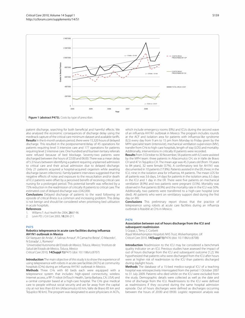
patient discharge, searching for both benefi cial and harmful eff ects. We
also analysed the economic consequences of discharge delay using the
medtrack capture of the critical care minimum dataset and available tariff s.
Results In the 6-month analysis period, there were 15,320 hours of delayed
discharge. This resulted in the postponement/delay of 45 operations for
patients requiring level 3 intensive care and 177 operations for patients
requiring level 2 intensive care. One hundred and fourteen tertiary referrals
were refused because of bed blockage. Seventy-two patients were
discharged between the hours of 22:00 and 06:00. There was a mean delay
of 5.3 hours between identifying a patient requiring unplanned admission
to critical care and their actual admission due to delayed discharge.
Only 21 patients acquired a hospital-acquired organism while awaiting
discharge (seven infections). Family/patient interviews suggested that the
negative eff ects of noise and exposure to the resuscitation and/or death
of ICU patients were off set by a perceived benefi t of receiving critical care
nursing for a prolonged period. This potential benefi t was refl ected by a
72% reduction in the readmission of critically ill patients to critical care. The
estimated cost of delayed discharge was £342,000.
Conclusions Delayed discharge of patients to the ward following an
episode of critical illness is a common and increasing problem. This delay
is not benign and should be considered when prioritising bed utilisation
in acute hospitals.
References1. Williams T: Aust Health Rev 2004, 28:87-96.
2. Levin PD: J Crit Care 2003, 18:206-211.
P475
Robotics telepresence in acute care facilities during infl uenza AH1N1 outbreak in MexicoGV Vazquez de Anda1, A Salinas Arnaut2, R Camacho Beiza2, O Macedo2,
N Estrada2, L Romero2
1Universidad Autonoma del Estado de Mexico, Toluca, Mexico; 2Instituto de
Salud del Estado de Mexico, Toluca, Mexico
Critical Care 2010, 14(Suppl 1):P475 (doi: 10.1186/cc8707)
Introduction The main objective of this study is to show the experience of
using telepresence with robots in acute care facilities (ACFs) at community
hospitals (CHs) during an infl uenza AH1N1 outbreak in Mexico.
Methods Three CHs with 60 beds each were equipped with a
telepresence system that includes: high-speed connectivity, wireless
Internet access, a RP-7i robot (InTouch Health, Santa Barbara, CA, USA) and
a central computer based at a high-care hospital. The CHs give medical
care to people without social security and are far away from the capital
city at not less than 63 km (Atlacomulco 63 km, Valle de Bravo 85 km and
Tejupilco 90 km). The program was designated to assist physicians in ACFs,
which include emergency rooms (ERs) and ICUs during the second wave
of an infl uenza AH1N1 outbreak in Mexico. The program includes: rounds
at the ACF and isolation area for patients with infl uenza-like syndrome
(ILS) every day from 9 am to 15 pm from Monday to Friday given by the
MPH specialist team (intensivist), mechanical ventilation supervision (MV),
transfer from CHs to high-care hospitals, length of stay (LOS) and mortality.
Additionally, interventions in critically ill patients were recorded.
Results From 3 October to 30 November, 56 patients with ILS were assisted
by the MPH team: three patients in Atlacomulco CH, six in Valle de Bravo
CH and 47 in Tejupilco CH. The mean age was 45.3 years old (from 19 years
to 84 years), 32 were female (57%). A confi rmatory test for AH1N1 was
documented in 10 patients (17.8%). Patients assisted in the ER, three; in the
ICU, nine; in the isolation area for infl uenza, 44 patients. The mean LOS for
all patients was 3.6 days, 3.4 days for patients in the isolation area, 6.5 days
in the ICU and 1 day in the ER. There were fi ve patients on mechanical
ventilation (8.9%) and two patients were pregnant (3.5%). Mortality was
observed in fi ve patients (8.9%) and the mortality rate in the ICU was 50%.
Additionally, two patients were transferred to a high-care hospital (one
died). All patients who went on ventilatory support died during the fi rst
day on MV.
Conclusions This preliminary report shows that the practice of
telepresence using robots at acute care facilities during an infl uenza
outbreak is feasible at CHs in Mexico.
P476
Association between out of hours discharge from the ICU and subsequent readmissionS Gopal, L Terry, C Corbett
Royal Wolverhampton Hospitals NHS Trust, Wolverhampton, UK
Critical Care 2010, 14(Suppl 1):P476 (doi: 10.1186/cc8708)
Introduction Readmission to the ICU may be considered a benchmark
quality indicator on an ICU. Previous studies have assessed the impact of
out of hours discharge from the ICU and subsequent outcome [1,2]. We
hypothesized that patients who were discharged from the ICU after hours
were at higher risk of readmission to the ICU than patients discharged
during daylight hours.
Methods The database of a 15-bed medico-surgical ICU of a teaching
hospital was retrospectively interrogated from the period 1 October 2007
to 31 July 2009. Patients who died whilst on the ICU were excluded from
the study. Demographic details were collected as well as the date and
time of discharge from the ICU. Readmissions to the ICU were defi ned
as readmissions if they occurred during the same hospital admission
episode. Out of hours discharges were defi ned as discharges occurring
between the hours of 20:00 and 09:00. Logistic regression analysis was
Figure 1 (abstract P473). Costs by type of prescriber.
y = 67838x + 1E+08
y = 77068x + 2E+08
1'400'000
1'200'000
1'000'000
800'000
600'000
in S
wis
s Fr
ancs
COSTS DRUG PRESCRIBED BY ICP
COSTS DRUG PRESCRIBED BY SC
y = 9230.3x + 2E+07
600'000
400'000
200'000
0
2004 2005 2006 2007 2008
Cos
ts
Year
TOTAL DRUG COST
Critical Care 2010, Volume 14 Suppl 1 http://ccforum.com/supplements/14/S1
S159
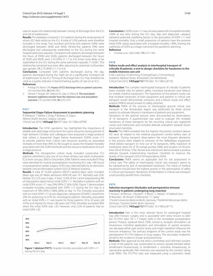
used to assess the relationship between timing of discharge from the ICU
and risk of readmission.
Results The database contained 1,257 patients during the study period, of
whom 207 died whilst on the ICU. A total of 1,050 patients were therefore
included in the study. Two hundred and forty-nine patients (24%) were
discharged between 20:00 and 09:00. Ninety-fi ve patients (9%) were
discharged and subsequently readmitted to the ICU during the same
hospital admission episode. Compared with patients discharged between
the hours of 09:00 and 20:00, patients discharged between the hours
of 20:00 and 09:00 were 2.74 (95% CI 1.7 to 4.3) times more likely to be
readmitted to the ICU during the same admission episode, P <0.001. This
relationship remained true even after adjusting for potential confounders
age, gender. OR 2.75 (95% CI 1.7 to 4.3).
Conclusions Compared with patients discharged during the day,
patients discharged during the night are at a signifi cantly increased risk
of readmission to the ICU. Timing of discharge from ICU may therefore be
used as a quality indicator in benchmarking quality of care on an ICU.
References1. Priestap FA, Martin CM: Impact of ICU discharge time on patient outcome.
Crit Care Med 2006, 34:2946-2951.
2. Hahane T, Keegan MT, Seferian EG, Gajic O, Afessa B: The association between nighttime transfer from the intensive care unit and patient outcome. Crit Care Med 2008, 36:2232-2237.
P477
Sequential Organ Failure Assessment in pandemic planningR Shahpori, T Stelfox, C Doig, P Boiteau, D Zygun
Alberta Health Services, Calgary, Canada
Critical Care 2010, 14(Suppl 1):P477 (doi: 10.1186/cc8709)
Introduction The H1N1 pandemic has highlighted the importance of
reliable and valid triage instruments for scarce resources during periods of
high demand. Christian and colleagues have proposed a triage protocol
that utilizes a Sequential Organ Failure Assessment (SOFA) score >11
to exclude patients from critical care resources quoting an associated
mortality of more than 90% [1]. We sought to assess the hospital mortality
associated with this SOFA threshold and the resource implications of such
a triage protocol.
Methods This retrospective cohort study included consecutive ICU
patients admitted to any one of our three tertiary-care adult multisystem
ICUs from January 2003 to December 2008. Patients were excluded if they
were admitted for routine postoperative monitoring (ICU stay <48 hours)
or postoperative cardiac surgery. SOFA was collected daily by an electronic
bedside clinical information system (QS; GE Medical Systems).
Results A total of 10,204 patients (69,913 patient-days) were included.
Mean age was 59. Mean admission APACHE was 19.1. Mortality was 25%.
Median ICU LOS was 4 days. A total 13.4% of the cohort (representing 9%
of total patient-days) had an initial SOFA >11. Mortality in patients with an
initial SOFA score >11 was 59% (95% CI 56%, 62%). Figure 1 demonstrates
increased mortality associated with SOFA >11 during the ICU stay to a
maximum of 78% (95% CI 68%, 86%) on day 14. The mortality associated
with an initial SOFA >11 across diagnostic categories (ICNARC) varied from
29% for poisoning to 67% for neurological patients. Mortality associated
with an initial SOFA >11 was lowest for those patients 18 to 20 years old
(37%) and highest for those >80 years old (75%). Mortality exceeded 90%
when the initial SOFA was >20. However, only 0.2% of patients had an
initial SOFA >20.
Conclusions A SOFA score >11 was not associated with a hospital mortality
>90% at any time during the ICU stay. Age and diagnostic category
represent potential modifying factors in the association of SOFA >11 and
hospital mortality. Only a small proportion of patients have the extreme
initial SOFA values associated with a hospital mortality >90%, limiting the
usefulness of SOFA as a triage instrument for pandemic planning.
Reference1. Christian et al.: CMAJ 2006, 175:1377-1381.
P478
Failure mode and eff ect analysis in interhospital transport of critically ill patients: a tool to design checklists for handovers in the mobile intensive care unitE Van Lieshout, A Hamming, D Dongelmans, S Smorenburg
Academic Medical Center, Amsterdam, the Netherlands
Critical Care 2010, 14(Suppl 1):P478 (doi: 10.1186/cc8710)
Introduction The complex interhospital transport of critically ill patients
bears multiple risks for patient safety, including handovers and reliance
on mobile medical equipment. The hypothesis of the present study was
that multiple handovers of both equipment and patients in critical care
transport would demonstrate omissions and a failure mode and eff ect
analysis (FMEA) would unravel its safety priorities.
Methods FMEA of the process of interhospital ground critical care
transport in the Amsterdam region was performed by three clinical
experts to estimate failures in handovers of patients and equipment.
Deviations of the optimal process were documented by observations
of 16 transports. A questionnaire was used to evaluate the multiple
handovers of these transports by the escorting critical care registered
nurses and intensivists and included attitude evaluation towards future
structured checklists.
Results The FMEA revealed that the highest risk priority numbers (above
45) were all related to the medical equipment control before start of
transport. During transport observations the most frequent deviations
in the process were related to omission of the defi brillator equipment
check before transport (in nine out of 16 transports, 56%), handover of
medication dose (25 of 39 syringe pumps, 64%) and location of infusion
lines (39 of 54 lines, 72%). Nurses and intensivists demonstrated a positive
attitude towards implementing handover checklists to improve the
quality of the transport (29 of 34 respondents, 85%).
Conclusions FMEA seems an applicable tool for risk assessment in
critical care. The safety of interhospital critical care transport seems to
be suboptimal by lack of standardized handovers. The check of medical
equipment should have the highest priority in the optimization of safety
in critical care transport. Handovers of information in critical care transport
could possibly benefi t from checklists.
P479
Selective neurogenic blockades and perioperative immune reactivity in patients undergoing lung resectionE Viviano1, M Renius1, J Rückert1, A Bloch1, C Meisel1, A Harbeck-Seu1,
W Boemke1, M Hensel2, K Wernecke1,3, C Spies1
1Charité-University Medicine Berlin, Germany; 2Parkklinik Weissensee, Berlin,
Germany; 3Sostana GmbH, Berlin, Germany
Critical Care 2010, 14(Suppl 1):P479 (doi: 10.1186/cc8711)
Introduction Pain is the most relevant factor for prolonged hospital
stay after thoracic surgery and is associated with stress known to alter
the Th1/Th2 ratio (Th = T helper cells) in the immediate postoperative
period. Thoracic epidural block (TEB), central α2-receptor stimulation via
intravenous clonidine application and stimulation of opioid receptors
can decrease either pain and/or stress and might therefore infl uence this
immune imbalance. The primary endpoint of the current study was the
perioperative Th1/Th2 balance in lung surgery. The secondary endpoints
aimed to the incidence of pain and pneumonia.
Methods After approval by the ethics committee and informed consent
a total of 60 patients was randomized to receive double-blinded either
remifentanil intravenously, or remifentanil + clonidine intravenously, or
ropivacaine epidurally. Pain intensity was assessed by the numeric rating
scale (NRS). The Th1/Th2 ratio was measured using a cytometric bead
Figure 1 (abstract P477). Hospital mortality associated with SOFA >11
during the ICU stay.
Critical Care 2010, Volume 14 Suppl 1 http://ccforum.com/supplements/14/S1
S160
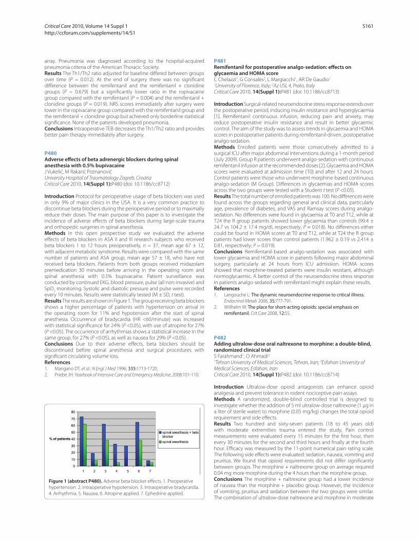
array. Pneumonia was diagnosed according to the hospital-acquired
pneumonia criteria of the American Thoracic Society.
Results The Th1/Th2 ratio adjusted for baseline diff ered between groups
over time (P = 0.012). At the end of surgery there was no signifi cant
diff erence between the remifentanil and the remifentanil + clonidine
groups (P = 0.679) but a signifi cantly lower ratio in the ropivacaine
group compared with the remifentanil (P = 0.004) and the remifentanil +
clonidine groups (P = 0.019). NRS scores immediately after surgery were
lower in the ropivacaine group compared with the remifentanil group and
the remifentanil + clonidine group but achieved only borderline statistical
signifi cance. None of the patients developed pneumonia.
Conclusions Intraoperative TEB decreases the Th1/Th2 ratio and provides
better pain therapy immediately after surgery.
P480
Adverse eff ects of beta adrenergic blockers during spinal anesthesia with 0.5% bupivacaineJ Vukelić, M Rakarić Poznanović
University Hospital of Traumatology Zagreb, Croatia
Critical Care 2010, 14(Suppl 1):P480 (doi: 10.1186/cc8712)
Introduction Protocol for perioperative usage of beta blockers was used
in only 9% of major clinics in the USA. It is a very common practice to
discontinue beta blockers during the perioperative period or to maximally
reduce their doses. The main purpose of this paper is to investigate the
incidence of adverse eff ects of beta blockers during large-scale trauma
and orthopedic surgeries in spinal anesthesia.
Methods In this open prospective study we evaluated the adverse
eff ects of beta blockers in ASA II and III research subjects who received
beta blockers 1 to 12 hours preoperatively, n = 37, mean age 67 ± 12,
with adjacent metabolic syndrome. Results were compared with the same
number of patients and ASA group, mean age 57 ± 18, who have not
received beta blockers. Patients from both groups received midazolam
premedication 30 minutes before arriving in the operating room and
spinal anesthesia with 0.5% bupivacaine. Patient surveillance was
conducted by continued EKG, blood pressure, pulse (all non-invasive) and
SpO2 monitoring. Systolic and diastolic pressure and pulse were recorded
every 10 minutes. Results were statistically tested (M ± SD, t test).
Results The results are shown in Figure 1. The group receiving beta blockers
shows a higher percentage of patients with hypertension on arrival in
the operating room for 11% and hypotension after the start of spinal
anesthesia. Occurrence of bradycardia (HR <60/minute) was increased
with statistical signifi cance for 24% (P <0.05), with use of atropine for 27%
(P <0.05). The occurrence of arrhythmias shows a statistical increase in the
same group, for 27% (P <0.05), as well as nausea for 29% (P <0.05).
Conclusions Due to their adverse eff ects, beta blockers should be
discontinued before spinal anesthesia and surgical procedures with
signifi cant circulating volume loss.
References1. Mangano DT, et al.: N Engl J Med 1996, 335:1713-1720.
2. Priebe JH: Yearbook of Intensive Care and Emergency Medicine; 2008:101-110.
P481
Remifentanil for postoperative analgo-sedation: eff ects on glycaemia and HOMA scoreC Chelazzi1, G Consales2, L Margiacchi1, AR De Gaudio1
1University of Florence, Italy; 2Az USL 4, Prato, Italy
Critical Care 2010, 14(Suppl 1):P481 (doi: 10.1186/cc8713)
Introduction Surgical-related neuroendocrine stress response extends over
the postoperative period, inducing insulin resistance and hyperglycaemia
[1]. Remifentanil continuous infusion, reducing pain and anxiety, may
reduce postoperative insulin resistance and result in better glycaemic
control. The aim of the study was to assess trends in glycaemia and HOMA
scores in postoperative patients during remifentanil-driven, postoperative
analgo-sedation.
Methods Enrolled patients were those consecutively admitted to a
surgical ICU after major abdominal interventions during a 1-month period
(July 2009). Group R patients underwent analgo-sedation with continuous
remifentanil infusion at the recommended doses [2]. Glycaemia and HOMA
scores were evaluated at admission time (T0) and after 12 and 24 hours.
Control patients were those who underwent morphine-based continuous
analgo-sedation (M Group). Diff erences in glycaemias and HOMA scores
across the two groups were tested with a Student t test (P <0.05).
Results The total number of enrolled patients was 100. No diff erences were
found across the groups regarding general and clinical data, particularly
age, prevalence of diabetes, and VAS and Ramsay scores during analgo-
sedation. No diff erences were found in glycaemia at T0 and T12, while at
T24 the R group patients showed lower glycaemia than controls (99.4 ±
24.7 vs 104.2 ± 17.4 mg/dl, respectively; P = 0.018). No diff erences either
could be found in HOMA scores at T0 and T12, while at T24 the R group
patients had lower scores than control patients (1.962 ± 0.19 vs 2.414 ±
0.81, respectively; P = 0.019).
Conclusions Remifentanil-based analgo-sedation was associated with
lower glycaemia and HOMA score in patients following major abdominal
surgery, particularly at 24 hours from ICU admission. HOMA scores
showed that morphine-treated patients were insulin resistant, although
normoglycaemic. A better control of the neuroendocrine stress response
in patients analgo-sedated with remifentanil might explain these results.
References1. Langouche L: The dynamic neuroendocrine response to critical illness.
Endocrinol Metab 2006, 35:777-791.
2. Willhelm W: The place for short-acting opioids: special emphasis on remifentanil. Crit Care 2008, 12:S5.
P482
Adding ultralow-dose oral naltrexone to morphine: a double-blind, randomized clinical trialS Farahmand1, O Ahmadi2
1Tehran University of Medical Sciences, Tehran, Iran; 2Esfahan University of
Medical Sciences, Esfahan, Iran
Critical Care 2010, 14(Suppl 1):P482 (doi: 10.1186/cc8714)
Introduction Ultralow-dose opioid antagonists can enhance opioid
analgesia and prevent tolerance in rodent nociceptive pain assays.
Methods A randomized, double-blind controlled trial is designed to
investigate whether the addition of 5 ml ultralow-dose naltrexone (1 μg in
a liter of sterile water) to morphine (0.05 mg/kg) changes the total opioid
requirement and side eff ects.
Results Two hundred and sixty-seven patients (18 to 45 years old)
with moderate extremities trauma entered the study, Pain control
measurements were evaluated every 15 minutes for the fi rst hour, then
every 30 minutes for the second and third hours and fi nally at the fourth
hour. Effi cacy was measured by the 11-point numerical pain rating scale.
The following side eff ects were evaluated: sedation, nausea, vomiting and
pruritus. We found that opioid requirements did not diff er signifi cantly
between groups. The morphine + naltrexone group on average required
0.04 mg more morphine during the 4 hours than the morphine group.
Conclusions The morphine + naltrexone group had a lower incidence
of nausea than the morphine + placebo group. However, the incidence
of vomiting, pruritus and sedation between the two groups were similar.
The combination of ultralow-dose naltrexone and morphine in moderate
Figure 1 (abstract P480). Adverse beta blocker eff ects. 1. Preoperative
hypertension. 2. Intraoperative hypotension. 3. Intraoperative bradycardia.
4. Arrhythmia. 5. Nausea. 6. Atropine applied. 7. Ephedrine applied.
Critical Care 2010, Volume 14 Suppl 1 http://ccforum.com/supplements/14/S1
S161

extremities trauma did not aff ect opioid requirements, but it decreased
the incidence of nausea.
References1. Crain SM, Shen KF: Antagonists of excitatory opioid receptor functions
enhance morphine’s analgesic potency and attenuate opioid tolerance/dependence liability. Pain 2000, 84:121-131.
2. Cepeda MS, Africano JM, Manrique AM, Fragoso W, Carr DB: The combination of low dose of naloxone and morphine in PCA does not decrease opioid requirements in the postoperative period. Pain 2002,
96:73-79.
P483
Four-day sedation using sevofl urane: fl uoride and renal functionK Wagner, A Becker, T Meier, H Kehnscherper, HM Benad
Klinikum Suedstadt Rostock, Germany
Critical Care 2010, 14(Suppl 1):P483 (doi: 10.1186/cc8715)
Introduction Volatile anesthetics for sedation in the ICU are currently
undergoing a revival, mainly due to the recent availability of a novel
delivery system, the anesthetic conserving device (ACD). Cytochrome
P450 metabolic degradation of isofl urane and sevofl urane leads to the
generation of inorganic fl uoride and hexafl uoro isopropanol. Historically,
data derived from methoxyfl uorane defi ned a fl uoride nephrotoxicity
threshold of 50 μmol/l. Therefore, we investigated the feasibility and safety,
focusing on kidney function, of 4-day sevofl urane-ACD sedation.
Methods Approval of the State Ethics Committee was obtained. Visceral
surgical patients either received sedation via sevofl urane-ACD (n = 20) or
were intravenously sedated (sufentanil, propofol, n = 20). Serum fl uoride,
renal, liver, pulmonary, cardiovascular function and depth of sedation
were measured.
Results Serum fl uoride concentrations increased signifi cantly during
sevofl urane-ACD sedation (Figure 1). Renal function parameters and
urinary output did not indicate deterioration of kidney function. Catechol-
amine support and pulmonary function were not diff erent between the
groups at equivalent depths of sedation.
Conclusions Inhalational sevofl urane sedation subsequently leads
to a transient, marked increase of serum fl uoride concentrations. The
methoxyfl urane fl uoride nephrotoxicity threshold was largely surpassed
without apparent deleterious eff ect on renal volume regulation and
detoxifi cation. The sedational use of the volatile anesthetic sevofl urane up
to 4 days is safe and feasible.
Reference1. Roehm et al.: Anesth Analg 2009, 108:1848-5184.
P484
Benefi cial eff ect of sevofl urane on endotoxin-induced pulmonary hypertensionB Fyntanidou1, V Grosomanidis1, K Kotzampassi2, E Thomaidou1, G Mitos1,
Z Aidoni1, C Skourtis1
1Department of Anaesthesiology – ICU, and 2Department of Surgery,
University of Thessaloniki Medical School, Thessaloniki, Greece
Critical Care 2010, 14(Suppl 1):P484 (doi: 10.1186/cc8716)
Introduction Pulmonary hypertension (PH) is a life-threatening disease
commonly seen in ICU septic patients and associated with poor outcome.
New and better therapies are required, since the response to various
agents such as NO, prostaglandins and phosphodiesterase inhibitors is
usually partial and the mortality rate remains high. Inhaled drugs seem
to be an attractive treatment option, since they are delivered directly
to pulmonary resistance vessels. A new device (anesthetic conserving
device –AnaConDa (ACD)) permitting direct administration of volatile
anaesthetics – such as sevofl urane – to the breathing circuit of a
conventional ICU ventilator in a safe and eff ective way has recently been
introduced. The aim of the present study was to evaluate the effi cacy and
safety of sevofl urane administration via the ACD on a porcine model of
acute PH during sepsis.
Methods PH was induced in 16 anaesthetized, mechanically ventilated
swine (25 kg) by intravenous infusion of 0.5 mg/kg LPS (Escherichia coli,
111:B4) in a period of 30 minutes. After LPS, animals were randomized into
two groups. In group A sevofl urane was infused via the ACD, according
to the manufacturer’s recommendations in order to obtain an end-tidal
concentration of 0.5%, whereas group B did not receive sevofl urane and
served as control. Haemodynamic parameters (systemic and pulmonary)
were recorded before (phase 0) and after the LPS (phase 1) and every
20 minutes for the next 2 hours (phases 2 to 7).
Results LPS was found to produce PH (phase 1) and to reduce arterial
blood pressure in both groups. After sevofl urane infusion, both systolic
(PAPs) and diastolic (PAPd) pulmonary pressures exhibited step-by-step
reduction, which became statistically signifi cant in phase 3 and thereafter,
in relation to phase 1 values. On the contrary, in group B animals, not
sevofl urane-treated, pulmonary pressures remained at high levels
throughout the study period. Systemic arterial pressure exhibited an
endotoxin-related reduction in both groups, which was not found to be
aff ected by sevofl urane treatment.
Conclusions Sevofl urane administration via the ACD was found to reduce
the pulmonary pressure in a porcine model of endotoxin-induced acute
PH without any detrimental eff ects on the systemic circulation. This may
represent a signifi cant advance in the treatment of acute PH. However, the
potential clinical implications of the method merit further study.
P485
Does etomidate aff ect prognosis in septic shock patients treated with hydrocortisone?B Jung, N Clavieras, G Chanques, M Cisse, S Jaber
Montpellier University Hospital, Montpellier, France
Critical Care 2010, 14(Suppl 1):P485 (doi: 10.1186/cc8717)
Introduction Etomidate is well tolerated and not inferior to ketamine but
because it is implicated in critical illness-related corticosteroid insuffi ciency
(CIRCI) [1], its utilization in septic shock patients is controversial. In our ICU,
the standard of care is to treat every septic shock patient with 200 mg
hydrocortisone for at least 5 days. The aim of our study was to compare
prognosis of septic shock patients intubated with etomidate with patients
intubated without etomidate.
Methods Observational study during a 2-year period including every
septic shock patient. We compared the patients’ prognosis when intubated
with etomidate (E group) to patients intubated without (NE group). We
recorded demographic characteristics, results of a cosyntropin test, dose
and length of vasopressors and hydrocortisone and outcome. The main
endpoint was mortality in the ICU. The secondary endpoints were the
length of shock, the dose of vasopressors and hydrocortisone in the ICU
and the plasma cortisol level.
Results We evaluated 62 patients in the E group and 31 in the NE group.
Although SAPS II on admission was higher in the E group (54 (42 to 64) vs Figure 1 (abstract P483).
Critical Care 2010, Volume 14 Suppl 1 http://ccforum.com/supplements/14/S1
S162

45 (32 to 54)*), the total dose (95 mg (37 to 195) vs 66 mg (31 to 112)) and
total duration (54 hours (36 to 91) vs 52 hours (31 to 81)) of vasopressors
from day 0 to day 5 was not diff erent between groups. However, the
cosyntropin responder rate was higher in the non-E group (40% vs 18%*),
hydrocortisone treatment duration (168 hours (78 to 216) vs 96 hours (72
to 144)*) and total dose of hydrocortisone (1,200 mg (500 to 1,625) vs
750 mg (350 to 1,125)*) was higher in the E group than in the non-E group.
Finally, mortality in the ICU was not diff erent between groups (32% vs 27%
in the E and the non-E groups, respectively). *P <0.05.
Conclusions In this study on septic shock treated with a short course
of hydrocortisone, patients intubated with etomidate presented a CIRCI
more frequently than patients intubated with another drug and needed
a longer and higher dose of hydrocortisone treatment. However, length
of shock, dose and duration of vasopressors and mortality were not
diff erent between groups. Etomidate-induced CIRCI when treated with
hydrocortisone was not associated with a poorer outcome in our study.
A comparison between etomidate and ketamine should be performed in
this population.
Reference1. Jabre et al.: Lancet 2009, 374:293-300.
P486
Use of methyl-naltrexone for the treatment of opioid-induced constipation in critical care patientsSB Sawh1, A Danga1, IP Selveraj2, A Cotton1, PB Patel1
1Imperial Healthcare NHS Trust, London, UK; 2Hillingdon Hospital NHS Trust,
Hillingdon, UK
Critical Care 2010, 14(Suppl 1):P486 (doi: 10.1186/cc8718)
Introduction Methyl-naltrexone (Relistor®), a peripheral μ-opioid receptor
antagonist shown to induce laxation in patients receiving opioids, is
licenced for use in palliative care; a similar benefi t is proposed in critical
care patients. Impaired gut motility and constipation are common issues
in the critical care setting with contributing factors including trauma,
surgery and use of opiate analgesics. The eff ects of methyl-naltrexone
were studied on seven patients in whom conventional treatment for
constipation with stimulant and osmotic laxatives, faecal softeners and
suppositories failed to produce eff ects.
Methods Over a 6-week period, 15 critically ill patients with opiate-
induced constipation (OIC) of more than 4 days duration were pros pec-
tively studied. All patients were treated with conventional methods from
admission to critical care, which included regular senna and sodium
docusate, supplemented with glycerin suppositories and picolax for
resistant constipation. Methyl-naltrexone was administered to seven
patients at 0.15 μg/kg subcutaneously on alternate days until laxation
occurred. The remainder of patients in the study continued with
conventional therapy.
Results Six out of seven patients responded to methyl-naltrexone with
laxation occurring between 12 and 24 hours. One patient did not respond
due to faecal impaction but subsequently experienced laxation after
manual disimpaction. Side eff ects were few (nausea 14%, vomiting 14%)
and there was no increase in opiate requirement. Those patients treated
by conventional means eventually produced laxation after a further 3 to
5 days.
Conclusions Previous studies have shown the earlier return of gut motility
improves patient progress, facilitates earlier respiratory weaning, prevents
debilitation and results in reduced length of stay. Methyl-naltrexone is a
safe and effi cacious drug for the treatment of OIC in critical care patients
in whom conventional treatments prove unsuccessful. Side eff ects are
few and opiate requirements remain unchanged. Further investigation
to elucidate the cost-eff ectiveness of methyl-naltrexone use in OIC of the
critically ill patient is warranted.
References1. Artinian V, et al.: Chest 2006, 129:960-967.
2. Mostafa SM, et al.: Br J Anaesth 2003, 91:815-819.
3. Thomas J, et al.: N Engl J Med 2008, 358:2332-2343.
P487
Safety and effi cacy of a nurse-driven sedation protocol with daily sedation interruption in the ICUKP Chan1, A Devanand1, CJ Chong2, JX Foo2, DP Ong1, PP Yap3, CM Loo1
1Singapore General Hospital, Singapore; 2National University of Singapore,
Singapore; 3Mount Elizabeth Hospital, Singapore
Critical Care 2010, 14(Suppl 1):P487 (doi: 10.1186/cc8719)
Introduction Sedation algorithms have been shown to reduce the
duration of mechanical ventilation in the ICU. We evaluated the eff ect
of the implementation of a nurse-driven sedation protocol with daily
sedation interruption in our ICU.
Methods We performed a before-and-after prospective study in our eight-
bed medical ICU over a 6-month period. All patients requiring mechanical
ventilation and who were not receiving neuromuscular blocking agents
were included. Prior to implementation, sedation was adjusted according
to physician orders. During the intervention period, sedatives and
analgesics were adjusted by nurses according to an algorithm utilizing the
Sedation-Agitation Scale.
Results We included a total of 188 patients, 89 patients in the observational
period and 99 patients during the intervention period. The duration of
mechanical ventilation was similar (2 days in both groups). However, the
ICU length of stay (LOS) was reduced by a day after implementation (3 days
vs 2 days). Amongst patients who survived and who were mechanically
ventilated for more than 4 days, the duration of mechanical ventilation and
ICU LOS were reduced (7 days vs 6 days and 8 days vs 6 days, respectively).
Fewer CT brains were performed for persistent coma after discontinuation
of sedation in the intervention period (4.5% vs 1.2%). Complications such
as extubation failure, tracheostomy and inadvertent removal of devices
were similar in both groups.
Conclusions A nurse-driven sedation protocol is safe and may reduce
duration of mechanical ventilation and ICU LOS, especially amongst
patients who have been ventilated for more than 4 days.
P488
Eff ects of propofol and midazolam on indocyanine green elimination assessed with LiMON for early sepsis patientsM Kargı, D Memis, N Sut
Trakya University, Edirne, Turkey
Critical Care 2010, 14(Suppl 1):P488 (doi: 10.1186/cc8720)
Introduction We aimed to select the sedative drug with the least
impact on hepatic blood fl ow in sedation-administered patients. In our
study, we aimed to establish whether or not midazolam and propofol
aff ect liver function during sepsis. The hepatic blood fl ow is evaluated
by the transcutaneous assessment of the indocyanine green plasma
disappearance rate (ICG-PDR) in critically ill patients.
Methods Forty sepsis patients were included in the study with either the
loading dose infusion of propofol 1 mg/kg over 15 minutes followed by
a maintenance 1 to 3 mg/kg/hour (n = 20, Group P) or a loading dose of
midazolam 0.2 mg/kg over 10 minutes followed by a maintenance 0.04
to 0.06 mg/kg/hour (n = 20, Group M), 24 hours infusion. ICG elimination
tests were conducted concurrently using the non-invasive liver function
monitoring system (LiMON). A dose of 0.3 mg/kg ICG was given through
a cubital fossa vein as a bolus and immediately fl ushed with 10 ml normal
saline. We calculated ICG-PDR. The ICG-PDR measurements were obtained
at baseline (before start of the propofol or midazolam infusion) and were
repeated at 24 hours. Biochemical and hemodynamic parameters, ICG-
PDR were recorded before start of the study and at 24 hours.
Results Biochemical and hemodynamic parameters did not diff er
signifi cantly between the groups (P <0.05). Group P compared with Group
M; baseline ICG-PDR levels (23.67 ± 12.02 vs 23.86 ± 11.4, respectively), and
after the study ICG-PDR levels (26.04 ± 13.60 vs 24.07 ± 9.07) did not diff er
in groups (P >0.05). When we examined before and after ICG-PRD changes
between groups, there was no signifi cant diff erence (P >0.05).
Conclusions In our study, we found that neither propofol nor midazolam
infusion aff ected hepatic blood fl ow.
Critical Care 2010, Volume 14 Suppl 1 http://ccforum.com/supplements/14/S1
S163

P489
Assessment of correlation between Bispectral Index monitorization and sedation scales used in the intensive care unitF Yaman, N Ozcan, A Ozcan, C Kaymak, H Basar
Ankara Training and Research Hospital, Ankara, Turkey
Critical Care 2010, 14(Suppl 1):P489 (doi: 10.1186/cc8721)
Introduction The level of sedation should be monitored as the depth of
sedation can aff ect the rate of related complications, length of hospital
stay and costs [1]. The recent study aims to monitor the depth of sedation
of patients in the ICU using the Ramsay Sedation Scale (RSS), Richmond
Agitation Sedation Scale (RASS), Sedation Agitation Scale (SAS) and
Adaptation To Intensive Care Environment scale (ATICE) and to assess the
correlation between these scales and Bispectral Index (BIS) scores and to
compare the correlation coeffi cients of these scales with each other.
Methods After approval from the hospital ethics committee, 30
mechanically ventilated patients who required sedation in the ICU were
enrolled into the study. Patients who are quadriplegic or need muscle
paralysis are excluded. The choice of sedative agent and the dose
regimen that will be used are left to consultant ICU staff diff erent from
the investigators. Demographic data and APACHE II scores of the patients
were registered. BIS, RSS, RASS, SAS, ATICE scores, mean arterial pressure,
heart rate, and peripheral oxygen saturation values (SpO2) of the patients
were registered before the initiation of the sedation and in 2-hour intervals
after initiation of the sedation.
Results Male/female ratio was 22/8, mean of ages and body weights of the
patients were 57.3 ± 15.8 years and 69.6 ± 14.3 kg, respectively. The median
of APACHE II scores was 14 (4 to 23). Mean arterial pressure, heart rate and
SpO2 changes were insignifi cant after initiation of sedation compared
with the beginning (P >0.05). BIS, RASS, SAS scores were signifi cantly
lower, RSS scores were signifi cantly higher in all intervals compared
with the beginning (P <0.05). Signifi cant change was not observed for
ATICE scores compared with the beginning value (P >0.05). Statistically
signifi cant correlation was observed between all scoring systems and
BIS values (P <0.05). Signifi cantly highest correlation was found between
BIS–RSS and BIS–RASS (r = 0.758 and r = 0.750, respectively), comparing
correlation between BIS–SAS (r = 0.656) and BIS–ATICE (r = 0.565) (P <0.05).
Also, correlation between BIS–SAS was signifi cantly higher than BIS–ATICE
correlation (P <0.05).
Conclusions All scales assessed in this study showed signifi cant correlation
with BIS. The signifi cantly highest correlation was found between BIS–RSS
and BIS–RASS and lowest correlation between BIS–ATICE.
References1. Jacobi J, et al.: Crit Care Med 2002, 30:119-141.
P490
Sedation and analgesia: early results from a web-based survey of UK practiceC Mckenzie, S Yassin, M Terblanche
Guy’s and St Thomas’ NHS Foundation Trust, London, UK
Critical Care 2010, 14(Suppl 1):P490 (doi: 10.1186/cc8722)
Introduction Evidence suggests prolonged sedation is associated with
prolonged ventilation and mortality. Further evidence suggests that some
sedatives and analgesics are more prone to cause oversedation than
others, possibly due to disturbances in pharmacokinetic properties in the
context of acute organ failure. Our aim was to describe current sedation
and analgesic practice in UK ICUs, and to determine whether recent
evidence has changed practice in recent years.
Methods We performed a web-based survey using a tool developed
via a systematic, step-wise process following recognised practice for
questionnaire design. These steps included question development and
questionnaire formatting and testing before being published on the
Zoomerang™ website. The sampling frame was pharmacists who are
members of the United Kingdom Clinical Pharmacy Association Critical
Care Group (UKCPA CCG). Participants were invited to complete the
15-minute questionnaire. Email reminders were sent to all members
every 2 weeks. After 6 months a reminder email was sent to pharmacists
in institutions that had not responded. Questions were split into four
categories: pharmacist and ICU-specifi c data; current sedation practice;
sedative choice; and analgesic choice.
Results The response rate was 68%. Eighty-four per cent of respondents
were senior pharmacists and 54% provided a service within a team. The
median number of annual ICU admissions was 750 (IQR 635 to 865)
and included a wide-ranging case mix. Forty-nine per cent of ICUs had
a sedation policy, of which 76% included sedation holds and 87% used
a sedation scoring system. Forty-three per cent assessed for delirium,
but only 29% used a known delirium scoring system. Ninety-nine of the
respondents used propofol (PPF) in the majority of patients, 56 midazolam
(MDZ). Twenty-seven still used MDZ routinely in renal or hepatic failure,
while 75 used MDZ where sedation was challenging.
Fifty-fi ve used morphine, 37 fentanyl and 33 alfentanil as fi rst-line opiate.
Conclusions These data suggest that many ICUs do not routinely use
sedation policies or assess patients for delirium. While PPF remains the
fi rst-line sedative for most ventilated patients, MDZ and morphine are
still widely used despite evidence of prolonged eff ects in the presence of
hepatic and renal failure.
References1. McKenzie CA, et al.: Crit Care 2005, 9:32-36.
P491
Severe alcohol withdrawal syndrome in ICU: is propofol a safe option?JI Chico1, C Rivero2, R Casado3, L Sayagues2, E Saborido4, E Sanmartin2,
D Sander2, J Garcia-Allut2
1Hospital Montecelo, Pontevedra, Spain; 2Hospital Clinico, Santiago
Compostela, Spain; 3Hospital Lozano Blesa, Zaragoza, Spain; 4Hospital
Montecelo, Pontevedra, Spain
Critical Care 2010, 14(Suppl 1):P491 (doi: 10.1186/cc8723)
Introduction This study aims to compare the effi cacy of midazolam or
propofol in severe refractory AWS, to detail characteristics of patients and
to test the usefulness of diff erent scores (MELD, Child, APACHE II) in severe
AWS.
Methods Patients admitted to the ICU during a 3-year period for
refractory AWS after a failed treatment with benzodiazepines according
to a symptom-triggered protocol in the general ward or emergency room were treated with neuroleptics and either midazolam or propofol infusion.
Processes that could lead to a confounding delirium were excluded. We
retrospectively analysed this cohort of patients.
Results Fifty patients were reviewed, 85.7% men. Mean age was 49 years.
Thirty percent had been previously admitted to the ICU for the same
reason. Main reason for alcohol withdrawal was abdominal pain (12.2%).
Mean alcohol ingestion was 150 g/day. Mean CHILD/MELD/APACHE score
was 6/8.5/13 (24.5% patients had a Child score ≥7). Seventy-three percent
of patients developed complications (especially respiratory infections in
19 patients). The most frequent agent found was MSSA and Gram-negative
bacilli. Seizures accounted for 38% of patients and hepatic encephalopathy
in two. We found no relationship between CHILD or MELD score and rate
of intubation, length of stay or complications. Patients with APACHE II
>12 at admission had a signifi cant increase in risk of intubation (OR 31.2,
P <0.001). Patients with more than 12 days of stay had a signifi cant risk
of intubation (OR 49.7, P <0.0001). Thirty-fi ve patients received propofol
as the fi rst intravenous sedation and 14 midazolam. Only two patients
with midazolam required a second intravenous sedation. Conversely, nine
(26.47%) patients initially with propofol required a second sedation with
midazolam. The propofol group showed a signifi cant increase in incidence
of seizures (41.17% vs 21% in midazolam group) and infections (47.1% vs
28%) independent of APACHE score. These fi ndings were more remarkable
in the subgroup that needed a second intravenous sedation (66% seizures
and 77% infection rate). This group had higher APACHE score and was
associated with a greater likelihood of intubation and a signifi cant 2-day
increase in length of stay.
Conclusions Propofol has been proposed as an alternative to
benzodiazepines in severe AWS, but it could increase the rate of
complications (especially seizures and respiratory infections) that could
lead to an increase in the intubation rate and length of stay. The APACHE
score could be a predictor for the risk of complications and intubation.
Critical Care 2010, Volume 14 Suppl 1 http://ccforum.com/supplements/14/S1
S164

P492
Agitation and pain during physiotherapy in intensive careK Everingham1, L Salisbury2, P Lapinlampi3, I Cornwall1, F Frame4, T Walsh1
1NHS Lothian, Edinburgh, UK; 2University of Edinburgh, UK; 3GE Healthcare,
Helsinki, Finland; 4University of Leicester, UK
Critical Care 2010, 14(Suppl 1):P492 (doi: 10.1186/cc8724)
Introduction The objective of this study was to identify the proportion
of patients who experienced agitation and/or pain during physiotherapy
sessions in intensive care (ICU). The prevalence and causes of distress
in critically ill patients are poorly described. Identifi ed stressors include
physiotherapy [1,2], but no standards for sedation/analgesia management
during physiotherapy exist. Recent evidence supporting routine ICU
management at lighter levels of sedation potentially increases the
importance of interventions to avoid distress during interventions such
as physiotherapy.
Methods A prospective observational study was undertaken. Forty-
nine patients admitted to the ICU requiring mechanical ventilation
and physiotherapy were recruited into the study. A single session of
physiotherapy was observed by an independent assessor. Agitation
was measured using the Richmond Agitation Sedation Scale (RASS)
and pain using the Behavioural Pain Scale (BPS). The RASS and BPS were
collected immediately pre physiotherapy, after each intervention during
physiotherapy and at 5 and 30 minutes post physiotherapy.
Results Sixteen participants (33%) experienced agitation (RASS score
≥1) while 48 participants (98%) experienced pain (BPS score ≥4). Figure
1 shows the number of participants who experienced agitation and
pain in relation to their RASS level prior to physiotherapy. Of interest,
the independent assessor observed little communication, in regards to
the management of sedation or pain at any stage during physiotherapy,
between nursing and physiotherapy staff .
Conclusions Regardless of the RASS level pre physiotherapy, the
majority of participants experienced pain. In contrast, only one-third of
participants experienced agitation; this was more prevalent for those at a
lighter sedation level. More routine use of RASS, BPS and communication
with nursing staff should be undertaken during physiotherapy to ensure
optimal levels of sedation and adequate levels of analgesia are achieved.
References1. Novaes et al.: Intensive Care Med 1999, 25:1421-1426.
2. van de Leur et al.: Crit Care 2004, 8:R467-R473.
P493
Pain evaluation in the intensive care unit: listen to the patient, a new approach!G Chanques1, B Jung1, M Cissé1, S De Lattre1, E Viel2, S Jaber1
1Saint Eloi – Montpellier University Hospital, Montpellier, France; 2Carémeau –
Nimes University Hospital, Nimes, France
Critical Care 2010, 14(Suppl 1):P493 (doi: 10.1186/cc8725)
Introduction Contrary to wards that manage usually chronic or acute
pain (algology, gerontology, surgery, recovery room, and so forth), a
comparison of the fi ve most popular self-report pain tools (vertical and
horizontal Visual Analog Scale (VAS-V, VAS-H), 0 to 10 oral Numeric Rating
Scale (NRS-O), 0 to 10 visual enlarged NRS (NRS-V), Verbal Descriptor Scale
(VDS)) has never been evaluated in an ICU setting.
Methods Consecutive patients admitted to a medical–surgical ICU
during 1 year were included when alert (RASS >–2) and able to follow
simple commands. Exclusion criteria: previous self-report pain assessment
without the presence of an investigator. Pain assessment using the fi ve
scales in random order either at baseline (T1) and after (T2) administration
of an analgesic, or during a nociceptive procedure, in absence of pain
at baseline. Evaluated parameters: psychometric properties of scales
(feasibility, validity, responsiveness and preference). Nonparametric tests
were used for statistical analysis (Statview 5.0). Data are expressed as
median (25th to 75th).
Results One hundred patients were included 24 (12 to 96) hours after
their admission to the ICU: age 58 (48 to 69) years, SAPS II 39 (32 to 48),
SOFA 7 (3 to 9), medical admission (49%), intubation before inclusion
(76%), at time to inclusion (50%). At T1, 91% of patients were able to use
at least one scale and 57% all fi ve scales. The NRS-V had the lowest failure
rate (9%) compared with NRS-O (17%), VDS (22%), VAS-H (32%) and VAS-V
(34%). Validity was measured in the 56 patients who were able to use
the fi ve scales both at T1 and T2. The pain intensity measured with the
fi ve scales changed signifi cantly between T1 and T2 (P <0.01), showing a
good discriminative validity. The correlation between the fi ve scales was
important (Spearman’s coeffi cients ranged from 0.75 to 0.96, P <0.001).
The responsiveness was very good for each of the fi ve scales, either after
an analgesia and a nociceptive procedure (eff ect size >0.8). Finally, the
NRS-V was considered respectively by 45, 37 and 44% of patients as the
easiest, most accurate and preferred scale. The preference for the other
scales was <12%.
Conclusions The self-report of pain is feasible in ICU patients as soon as
they are suffi ciently alert (RASS >–2) and able to follow simple commands.
The enlarged 0 to 10 Numeric Rating Scale is the easiest scale to use
with the lowest failure rate and it is the preferred scale for the majority of
critically ill patients who were able to communicate.
P494
Pharmacokinetics of orally administered melatonin in critically ill patientsB Moroni1, G Mistraletti1, RJ Reiter2, R Esposti1, E D’Angelo1, P Formenti1,
D Palmisano1, E Borotto1, P Di Mauro1, G Iapichino1
1Università degli Studi di Milano, Milan, Italy; 2University of Texas Health
Science Center, San Antonio, TX, USA
Critical Care 2010, 14(Suppl 1):P494 (doi: 10.1186/cc8726)
Introduction Critically ill patients exhibit reduced melatonin secretion,
both in nocturnal peaks and basal daytime levels. Oral melatonin supple-
men tation may be useful for known sedative and antioxidant properties.
Methods Melatonin early enteral absorption and daily pharmacokinetics
were determined in two cohorts of six high-risk patients in this prospective
trial. During their third and fourth ICU day, they underwent two diff erent
sets of repeated blood samples to detect serum melatonin levels through
radio-immunoassay. Cohort 1: samples taken at 20:00, 20:45, 21:30, 24:00,
03:00, 06:00, 14:00, 20:00 to describe the daily pharmacokinetics. Cohort
2: 20:00, 20:05, 20:10, 20:20, 20:30, 20:45 to study the early absorption. On
ICU day 3, endogenous levels were measured, while the absorption of
exogenous melatonin was determined on ICU day 4 after administration,
at 20:00, of 3 mg melatonin.
Results All basal levels were below the expected values. Following enteral
administration, pharmacological levels were already reached in 5 minutes,
with a serum peak after 16 minutes (half-absorption time: 3 minutes
17 seconds). The maximum serum level observed was 11,040 pg/ml
and the disappearance rate indicated a half-elimination time of 1 hour
34 minutes. Serum melatonin levels decreased signifi cantly after midnight;
pharmacological levels were maintained up to 10 hours following
administration. No excessive sleepiness was reported in this patient group.
Conclusions Critically ill patients exhibited reduced melatonin secretion, as
reported in the literature. Despite the critical illness, the oral bioavailability
was satisfactory: serum levels after oral administration showed basically
unchanged intestinal absorption, while the disappearance rate was slower
than reported elsewhere in healthy volunteers.
Figure 1 (abstract P492). Agitation and pain in relation to pre-
physiotherapy RASS scores.
Critical Care 2010, Volume 14 Suppl 1 http://ccforum.com/supplements/14/S1
S165
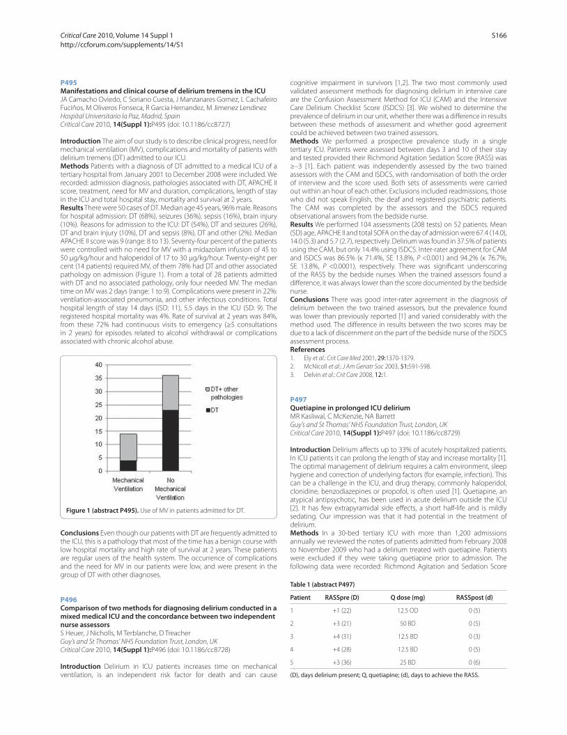
P495
Manifestations and clinical course of delirium tremens in the ICUJA Camacho Oviedo, C Soriano Cuesta, J Manzanares Gomez, L Cachafeiro
Fuciños, M Oliveros Fonseca, R Garcia Hernandez, M Jimenez Lendinez
Hospital Universitario la Paz, Madrid, Spain
Critical Care 2010, 14(Suppl 1):P495 (doi: 10.1186/cc8727)
Introduction The aim of our study is to describe clinical progress, need for
mechanical ventilation (MV), complications and mortality of patients with
delirium tremens (DT) admitted to our ICU.
Methods Patients with a diagnosis of DT admitted to a medical ICU of a
tertiary hospital from January 2001 to December 2008 were included. We
recorded: admission diagnosis, pathologies associated with DT, APACHE II
score, treatment, need for MV and duration, complications, length of stay
in the ICU and total hospital stay, mortality and survival at 2 years.
Results There were 50 cases of DT. Median age 45 years, 96% male. Reasons
for hospital admission: DT (68%), seizures (36%), sepsis (16%), brain injury
(10%). Reasons for admission to the ICU: DT (54%), DT and seizures (26%),
DT and brain injury (10%), DT and sepsis (8%), DT and other (2%). Median
APACHE II score was 9 (range: 8 to 13). Seventy-four percent of the patients
were controlled with no need for MV with a midazolam infusion of 45 to
50 μg/kg/hour and haloperidol of 17 to 30 μg/kg/hour. Twenty-eight per
cent (14 patients) required MV, of them 78% had DT and other associated
pathology on admission (Figure 1). From a total of 28 patients admitted
with DT and no associated pathology, only four needed MV. The median
time on MV was 2 days (range: 1 to 9). Complications were present in 22%:
ventilation-associated pneumonia, and other infectious conditions. Total
hospital length of stay 14 days ((SD: 11), 5.5 days in the ICU (SD: 9). The
registered hospital mortality was 4%. Rate of survival at 2 years was 84%,
from these 72% had continuous visits to emergency (≥5 consultations
in 2 years) for episodes related to alcohol withdrawal or complications
associated with chronic alcohol abuse.
Conclusions Even though our patients with DT are frequently admitted to
the ICU, this is a pathology that most of the time has a benign course with
low hospital mortality and high rate of survival at 2 years. These patients
are regular users of the health system. The occurrence of complications
and the need for MV in our patients were low, and were present in the
group of DT with other diagnoses.
P496
Comparison of two methods for diagnosing delirium conducted in a mixed medical ICU and the concordance between two independent nurse assessorsS Heuer, J Nicholls, M Terblanche, D Treacher
Guy’s and St Thomas’ NHS Foundation Trust, London, UK
Critical Care 2010, 14(Suppl 1):P496 (doi: 10.1186/cc8728)
Introduction Delirium in ICU patients increases time on mechanical
venti lation, is an independent risk factor for death and can cause
cognitive impairment in survivors [1,2]. The two most commonly used
validated assessment methods for diagnosing delirium in intensive care
are the Confusion Assessment Method for ICU (CAM) and the Intensive
Care Delirium Checklist Score (ISDCS) [3]. We wished to determine the
prevalence of delirium in our unit, whether there was a diff erence in results
between these methods of assessment and whether good agreement
could be achieved between two trained assessors.
Methods We performed a prospective prevalence study in a single
tertiary ICU. Patients were assessed between days 3 and 10 of their stay
and tested provided their Richmond Agitation Sedation Score (RASS) was
≥–3 [1]. Each patient was independently assessed by the two trained
assessors with the CAM and ISDCS, with randomisation of both the order
of interview and the score used. Both sets of assessments were carried
out within an hour of each other. Exclusions included readmissions, those
who did not speak English, the deaf and registered psychiatric patients.
The CAM was completed by the assessors and the ISDCS required
observational answers from the bedside nurse.
Results We performed 104 assessments (208 tests) on 52 patients. Mean
(SD) age, APACHE II and total SOFA on the day of admission were 67.4 (14.0),
14.0 (5.3) and 5.7 (2.7), respectively. Delirium was found in 37.5% of patients
using the CAM, but only 14.4% using ISDCS. Inter-rater agreement for CAM
and ISDCS was 86.5% (κ 71.4%, SE 13.8%, P <0.001) and 94.2% (κ 76.7%,
SE 13.8%, P <0.0001), respectively. There was signifi cant underscoring
of the RASS by the bedside nurses. When the trained assessors found a
diff erence, it was always lower than the score documented by the bedside
nurse.
Conclusions There was good inter-rater agreement in the diagnosis of
delirium between the two trained assessors, but the prevalence found
was lower than previously reported [1] and varied considerably with the
method used. The diff erence in results between the two scores may be
due to a lack of discernment on the part of the bedside nurse of the ISDCS
assessment process.
References1. Ely et al.: Crit Care Med 2001, 29:1370-1379.
2. McNicoll et al.: J Am Geriatr Soc 2003, 51:591-598.
3. Delvin et al.: Crit Care 2008, 12:1.
P497
Quetiapine in prolonged ICU deliriumMR Kasliwal, C McKenzie, NA Barrett
Guy’s and St Thomas’ NHS Foundation Trust, London, UK
Critical Care 2010, 14(Suppl 1):P497 (doi: 10.1186/cc8729)
Introduction Delirium aff ects up to 33% of acutely hospitalized patients.
In ICU patients it can prolong the length of stay and increase mortality [1].
The optimal management of delirium requires a calm environment, sleep
hygiene and correction of underlying factors (for example, infection). This
can be a challenge in the ICU, and drug therapy, commonly haloperidol,
clonidine, benzodiazepines or propofol, is often used [1]. Quetiapine, an
atypical antipsychotic, has been used in acute delirium outside the ICU
[2]. It has few extrapyramidal side eff ects, a short half-life and is mildly
sedating. Our impression was that it had potential in the treatment of
delirium.
Methods In a 30-bed tertiary ICU with more than 1,200 admissions
annually we reviewed the notes of patients admitted from February 2008
to November 2009 who had a delirium treated with quetiapine. Patients
were excluded if they were taking quetiapine prior to admission. The
following data were recorded: Richmond Agitation and Sedation Score
Figure 1 (abstract P495). Use of MV in patients admitted for DT.
Table 1 (abstract P497)
Patient RASSpre (D) Q dose (mg) RASSpost (d)
1 +1 (22) 12.5 OD 0 (5)
2 +3 (21) 50 BD 0 (5)
3 +4 (31) 12.5 BD 0 (3)
4 +4 (28) 12.5 BD 0 (5)
5 +3 (36) 25 BD 0 (6)
(D), days delirium present; Q, quetiapine; (d), days to achieve the RASS.
Critical Care 2010, Volume 14 Suppl 1 http://ccforum.com/supplements/14/S1
S166

(RASS), duration of delirium, agents used, patient demographics and
adverse events.
Results Five patients met the inclusion criteria (Table 1). All were
males, aged 37 to 85 years. All had a prolonged delirium prior to the
commencement of quetiapine and all were on a combination of four
drugs (clonidine, haloperidol, lorazepam and propofol) that were not
controlling their delirium. At 3 to 7 days following commencement of
quetiapine the RASS scores were 0 and other drugs were ceased. No
adverse eff ects were noted.
Conclusions Quetiapine was successful in controlling prolonged ICU
delirium and allowed weaning of other medications in these patients.
It may be a useful delirium therapy. Further studies are required to
demonstrate effi cacy and safety.
References1. Ely, et al.: JAMA 2004, 291:1753-1762.
2. Schwartz, et al.: Prim Care Companion J Clin Psychiatry 2000, 2:10-12.
P498
PREDICT, Prediction of Delirium in ICU Patients: development and validation of a prediction modelM Van den Boogaard, P Pickkers, H Van der Hoeven, R Donders,
T Van Achterberg, L Schoonhoven
Radboud University Medical Centre Nijmegen, the Netherlands
Critical Care 2010, 14(Suppl 1):P498 (doi: 10.1186/cc8730)
Introduction Delirium is a serious and frequent disorder in the ICU, but a
prediction model for critically ill patients is lacking.
Methods We performed two prospective cohort studies in a 33-bed ICU
for adult patients with use of the validated CAM-ICU, performed three
times daily by well-trained ICU nurses. Twenty-three identifi ed delirium
risk factors were collected within 24 hours after admission and patients
were followed during their ICU stay until death, discharge or development
of delirium. Variables with P >0.1 in univariate logistic regression, or with
a prevalence rate <10% were excluded. Multivariate logistic regression
analysis was followed by bootstrapping. Reduced coeffi cients were used
on the second cohort for external validation. Also, nurses and physicians
were asked to predict whether the patient would develop delirium during
the ICU stay.
Results In the fi rst study (February 2008 to February 2009) 2,116
consecutive patients and in the second study (May to September 2009)
748 patients were screened, of which 503 and 199 patients, respectively,
were excluded for valid reasons. Ninety per cent of the total number of
assessments that should have been performed was actually obtained and
the Cohen’s kappa for inter-rater reliability was 0.90. Out of 1,613 patients,
411 developed delirium and in the second study 171 out of 549 patients.
Eight risk factors were excluded because of a prevalence rate <10% and
one because of P >0.1. After multivariate logistic regression analysis, 10 risk
factors were included in the prediction model (age, APACHE II score, coma,
diagnose group, increased urea, infection, metabolic acidosis, morphine
use, sedation use, emergency admission) of which the area under the
receiver-operation characteristic curve (AUC) was 0.87 (95% CI 0.85 to
0.89) and after bootstrapping 0.86. The cut-off point with the optimal
predicted probability lies at 0.196 points. At this cut-off the sensitivity of
the prediction rule is 80% and the specifi city is 79%. Validation of PREDICT
on the second cohort resulted in an AUC of 0.89 (95% CI 0.86 to 0.92).
The AUC of the prediction of nurses and physicians was signifi cantly lower,
both 0.59 (95% CI 0.49 to 0.70).
Conclusions We have developed and validated a delirium prediction
model (PREDICT) with a high AUC that can be used in all adult ICU patients
within 24 hours after ICU admission to predict the chance of delirium
during their ICU stay and to guide preventive interventions.
P499
Hospital anxiety depression scale in our surgical ICUIF Filipovic-Grcic, DT Tonkovic, JG Grubisin, MP Peric, VM Majeric Kogler
University Clinical Hospital Center Zagreb, Croatia
Critical Care 2010, 14(Suppl 1):P499 (doi: 10.1186/cc8731)
Introduction All patients in the ICU go through mental, physical and
emotional adjustment. They can experience exaggerated symptoms
of anxiety and depression and it can delay their recovery [1]. Stress has
a major role and there are so many triggers for it: the psychological and
physical condition, surgery, pain, ICU environment and so forth. The aim of
this study was to implement the Hospital Anxiety Depression Scale (HADS)
in our everyday ICU practice and then to analyze the impact of the clinical
picture (APACHE II score) and pain control (VAPS – visual analog pain scale)
on the anxiety and depression symptoms (HADS) in surgical ICU patients.
Methods With ethics approval and written consent, in this prospective
study, to date are reported 70 patients. They all underwent major
abdominal surgery and were discharged from the ICU after 24 to 30 hours.
Patients with any mental disorder or neurological disease were excluded.
On admission and discharge the VAPS, APACHE II and HADS were evaluated.
HADS was evaluated again 1 month later (62 of 70 patients). Anxiety and
depression are assessed as separate components, each with seven items
that are rated from 0 to 3 [2]. A score <7 in a component is a normal result,
8 to 10 indicates mild symptoms, 11 to 14 moderate symptoms and 15 or
more indicates severe symptoms [2]. The Mann–Whitney U test, t test and
correlations were used in statistical analysis.
Results Average age of the patients was 58.61 ± 13.56, range 24 to 78, 37
male and 33 female. Average values on admission were: VAPS 3.02 ± 0.50,
APACHE II 10.31 ± 2.79, Anxiety 4.82 ± 4.01, Depression 3.91 ± 3.23, and
on discharge were: VAPS 1.34 ± 0.88, APACHE II 5.25 ± 2.78, Anxiety 3.41 ±
2.97, Depression 3.71 ± 2.97. One month later the values were: Anxiety
3.14 ± 2.79, Depression 1.27 ± 0.57.
Patients with lower pain control had more exaggerated symptoms of
anxiety and depression. After 1 month, seven patients had mild symptoms
of anxiety, nine patients had mild symptoms of depression and four
patients had moderate symptoms of depression.
Conclusions HADS has its role in everyday ICU practice. All patients with
mild to severe symptoms of anxiety or depression should be further
psychologically evaluated. There is a need to evaluate in our ICU the
patients who are staying there a few days or more and compare it with
the results of this study.
References1. Sukantarat et al.: Anaesthesia 2007, 62:239-243.
2. Zigmond et al.: Acta Psych Scand 1983, 67:361-370.
P500
Fluid therapy change from synthetic colloids to only crystalloids in patients with severe sepsisO Bayer, Y Sakr, B Kabisch, CS Hartog, N Riedemann, U Settmacher,
K Reinhart
Friedrich-Schiller-University Jena, Germany
Critical Care 2010, 14(Suppl 1):P500 (doi: 10.1186/cc8732)
Introduction After changing the standard resuscitation colloid on our
ICU from 6% HES 130/0.4 to 4% gelatin, we found that both HES 130/0.4
and gelatin resulted in a high incidence of renal failure (43.2% and 40.2%)
in patients with severe sepsis [1]. We therefore changed from synthetic
colloids to only crystalloids and herewith report on the eff ects.
Methods Before–after retrospective study in patients with severe sepsis
on a surgical ICU. The synthetic colloids group (SynColl-Group) comprised
patients who received either predominantly 6% HES 130 or predominantly
gelatin between January 2005 and June 2006. The crystalloid group (Crys-
Group) comprised patients from September 2008 to June 2009 who
received only crystalloids. Acute renal failure (ARF) was defi ned as new
need for renal replacement therapy (RRT) or at least a twofold increase in
baseline creatinine.
Results Two hundred and fi ve patients received synthetic colloids, and
141 received only crystalloids. There were minor baseline diff erences but
SAPS II and SOFA scores, age and creatinine were similar at admission.
Fluid therapy (median cumulative doses): patients in the SynColl-
Group received 4 l (IQR 2 to 8.3 l) colloids and 27.9 l (IQR 15.7 to 48.3 l)
crystalloids. Patients in the Crys-Group received only 25.0 l (IQR 13.1 to
52.2 l) crystalloids. Crystalloid volumes did not diff er signifi cantly between
groups (P = 0.605). Median cumulative dose of HES was 50 ml/kg and of
gelatin was 43.3 ml/kg. Outcomes: patients in the SynColl-group had a
higher incidence of ARF (41.0% vs 23.4%, P = 0.001) and increased need
for RRT (35.1% vs 22.0%, P = 0.009). ICU mortality and hospital mortality
did not diff er between SynColl and Crys groups (31.2% vs 30.5% and 38%
Critical Care 2010, Volume 14 Suppl 1 http://ccforum.com/supplements/14/S1
S167
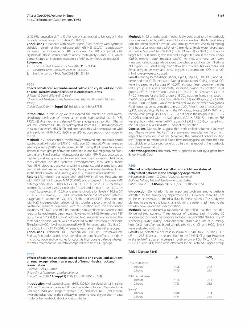
vs 36.9%, respectively). The ICU length of stay tended to be longer in the
SynColl-Group (14 versus 10 days, P = 0.055).
Conclusions In patients with severe sepsis, fl uid therapy with synthetic
colloids – gelatin or the third-generation 6% HES 130/0.4 –considerably
increases the incidence of ARF and need for RRT compared with
crystalloids. These results confi rm recent meta-analysis and RCTs, which
demonstrated an increased incidence of ARF by synthetic colloids [2,3].
References1. Schabinski et al.: Intensive Care Med 2009, 35:1539-1547.
2. Zarychanski et al.: Open Med 2009, 3:E196-E209.
3. Brunkhorst et al.: N Engl J Med 2008, 358:125-139.
P501
Eff ects of balanced and unbalanced colloid and crystalloid solutions on renal microvascular perfusion in endotoxemic ratsU Aksu1, C Demirci-Tansel2, C Ince1
1University of Amsterdam, the Netherlands; 2Istanbul University, Istanbul,
Turkey
Critical Care 2010, 14(Suppl 1):P501 (doi: 10.1186/cc8733)
Introduction In this study we investigated the eff ects on renal micro-
circulatory perfusion of resuscitation with hydroxyethyl starch (HES
130/0.42) dissolved in a balanced Ringer’s acetate salt solution (Plasma
Volume Redibag®, HES-RA) or hydroxyethyl starch (HES 130/0.4) dissolved
in saline (Voluven®, HES-NaCl) and compared this with resuscitation with
saline solution (0.9% NaCl, NaCl) in an LPS-induced septic shock model in
the rat.
Methods In 20 anesthetized, mechanically ventilated rats, endotoxic shock
was induced by infusion of LPS (10 mg/kg over 30 minutes). When the mean
arterial pressure (MAP) was decreased to 40 mmHg, fl uid resuscitation was
started in three groups of fi ve rats each, and in one fi ve-rat group no fl uids
were given. Renal cortical microvascular perfusion (RMP) was assessed at
high temporal and spatial resolution using laser speckle imaging. Additional
measurements included systemic hemodynamics, renal artery blood
fl ow (RBF), blood gas analysis, creatinine clearance, and plasma ions and
calculated renal oxygen delivery (DO2). Time points were: baseline, during
septic shock at a MAP of 40 mmHg, and at 30 minutes of resuscitation.
Results LPS infusion decreased MAP and RMP in all rats. Resuscitation
with NaCl did not improve MAP (P >0.05) and appeared to increase RMP
heterogeneity (0.95 ± 0.08 AU vs 1.83 ± 0.13 AU; P <0.001), metabolic
acidosis (7.1 ± 0.06 vs 6.93 ± 0.05 pH; P <0.05 and 11.36 ± 1.1 vs 15.9 ± 1.4
mmol/l base excess; P <0.05), and plasma chloride ion levels (110.3 ± 0.7
vs 116.2 ± 1.7 mmol/l; P <0.05). Fluid resuscitation did not aff ect systemic
oxygenation parameters (sO2, pO
2, O
2Hb) and renal DO
2. Resuscitation
with NaCl increased plasma levels of Na+, tubular reabsorption of Na+, and
creatinine clearance compared with resuscitation with the two colloid
solutions. HES-NaCl and HES-RA provided similar eff ects on systemic and
regional hemodynamic parameters. However, while HES-RA improved RBF
(2.3 ± 0.4 vs 5.7 ± 0.2), HES-NaCl did not. NaCl resuscitation worsened the
metabolic acidosis, which was not aff ected by the two colloid solutions.
The plasma HCO3
– level was increased by HES-RA resuscitation (13.18 ± 2.1
vs 19.56 ± 1 mmol/l; P <0.01), whereas it was stable in the other groups.
Conclusions Balanced HES preparation (HES-RA, PlasmaVolume
Redibag®) in endotoxemic rats showed acute benefi cial eff ects on kidney
microcirculation and on kidney function via bicarbonate balance whereas
the NaCl treatment was harmful compared with both HES groups.
P502
Eff ects of balanced and unbalanced colloid and crystalloid solutions on renal oxygenation in a rat model of hemorrhagic shock and resuscitationE Almac, U Aksu, C Ince
University of Amsterdam, the Netherlands
Critical Care 2010, 14(Suppl 1):P502 (doi: 10.1186/cc8734)
Introduction Hydroxyethyl starch (HES, 130 kD) dissolved either in saline
(Voluven®) or in a balanced Ringer’s acetate solution (PlasmaVolume
Redibag®, PVR) and Ringer’s acetate (RA) and 0.9% NaCl (NaCl) were
investigated as regards their effi cacy in restoring renal oxygenation in a rat
model of hemorrhagic shock and resuscitation.
Methods In 22 anesthetized, mechanically ventilated rats, hemorrhagic
shock was induced by withdrawing blood volume from the femoral artery
until the mean arterial pressure (MAP mmHg) was reduced to 40 mmHg.
One hour after reaching a MAP of 40 mmHg, animals were resuscitated
with either Voluven® (n = 5), PVR (n = 6), RA (n = 5), or NaCl (n = 6) until a
target MAP of 80 mmHg was reached. Oxygen tension in the renal cortex
(CμPO2 mmHg), outer medulla (MμPO
2 mmHg), and renal vein were
measured using oxygen-dependent quenched phosphorescent lifetimes
of Oxyphor G2. Renal artery blood fl ow (RBF ml/minute) was measured.
Renal oxygen delivery and renal oxygen consumption (DO2 and VO
2
ml·minute/g) were calculated.
Results During hemorrhagic shock, CμPO2, MμPO
2, RBF, DO
2, and VO
2
decreased and O2ER increased. During resuscitation, CμPO
2 and MμPO
2
were increased in all groups (P <0.001) although least prominent in the
NaCl group. RBF was signifi cantly increased during resuscitation in all
groups (PVR; 5.1 ± 0.2 P <0.001, RA; 3.5 ± 0.4 P <0.001, Voluven®; 3.4 ± 0.4
P <0.01), except for the NaCl group, and DO2 was signifi cantly increased in
the PVR group (0.20 ± 0.02 vs 0.39 ± 0.06; P <0.01) and RA group (0.22 ± 0.03
vs 0.41 ± 0.06; P <0.01), while this remained low in the other two groups.
Fluid resuscitation was not able to restore VO2. After 1 hour of resuscitation
CμPO2 was signifi cantly higher in the PVR group (49.9 ± 2.4 P <0.01) and
the Voluven® group (45.3 ± 5.1 P <0.05) but not in the RA group (40.6 ± 2.2
P >0.05) compared with the NaCl group (32.1 ± 2.55). Furthermore, RBF
was signifi cantly higher in the PVR group (5.1 ± 0.2 P <0.01) compared with
the NaCl group (2.4 ± 0.5) after 1 hour of resuscitation.
Conclusions Our results suggest that both colloid solutions (Voluven®
and PlasmaVolume Redibag®) are preferred resuscitation fl uids with
respect to crystalloid solutions. Moreover, the balanced colloid solution
(PVR) appears to restore renal blood fl ow and oxygenation more than the
crystalloids or unbalanced colloids do in this rat model of hemorrhagic
shock and resuscitation.
Acknowledgements This study was supported in part by a grant from
Baxter Health Care.
P503
Eff ect of rapidly infused crystalloids on acid–base status of dehydrated patients in the emergency departmentH Hasman, B Comert, O Cinar, A Uzun, L Yamenel
Gulhane Military Medical Academy, Ankara, Turkey
Critical Care 2010, 14(Suppl 1):P503 (doi: 10.1186/cc8735)
Introduction Dehydration is an important problem among patients
admitted to the emergency department (ED). However, there has not
yet been a consensus on the ideal fl uid for these patients. This study was
planned to evaluate the ideal crystalloid for the patients admitted to the
ED who have symptoms of dehydration.
Methods We conducted a randomized controlled trial that included
90 dehydrated patients. Three groups of patients each included 30
randomized for one of the solutions Lactated Ringer’s, 0.9% NaCl or Isolyte®
(Eczacıbaşı-Baxter, Turkey). Solutions were infused at a rate of 20 ml/kg/
hour for 2 hours. Venous blood sample pH, Na+, K+, Cl–, and HCO3
– levels
were evaluated at 0, 1, and 2 hours.
Results We detected a decrease in serum pH (7.406 to 7.365) and HCO3
–
(23.1 to 21.5) levels at the second hour in the 0.9% NaCl group. However,
in the Isolyte® group an increase in both serum pH (7.410 to 7.434) and
HCO3
– (23.4 to 24.4) levels were observed. In the Lactated Ringer’s group,
Table 1 (abstract P503)
pH HCO3
Lactated Ringer’s
0 hours 7.396 22.8
2 hours 7.408 22.4
0.9% normal saline
0 hours 7.406 23.1
2 hours 7.365 21.5
Isolyte®
0 hours 7.410 23.4
2 hours 7.434 24.4
Critical Care 2010, Volume 14 Suppl 1 http://ccforum.com/supplements/14/S1
S168
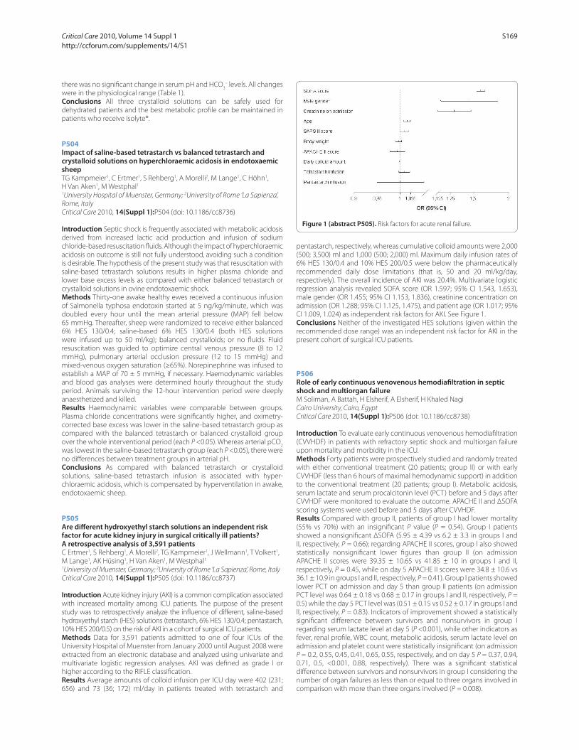
there was no signifi cant change in serum pH and HCO3
– levels. All changes
were in the physiological range (Table 1).
Conclusions All three crystalloid solutions can be safely used for
dehydrated patients and the best metabolic profi le can be maintained in
patients who receive Isolyte®.
P504
Impact of saline-based tetrastarch vs balanced tetrastarch and crystalloid solutions on hyperchloraemic acidosis in endotoxaemic sheepTG Kampmeier1, C Ertmer1, S Rehberg1, A Morelli2, M Lange1, C Höhn1,
H Van Aken1, M Westphal1
1University Hospital of Muenster, Germany; 2University of Rome ‘La Sapienza’,
Rome, Italy
Critical Care 2010, 14(Suppl 1):P504 (doi: 10.1186/cc8736)
Introduction Septic shock is frequently associated with metabolic acidosis
derived from increased lactic acid production and infusion of sodium
chloride-based resuscitation fl uids. Although the impact of hyperchloraemic
acidosis on outcome is still not fully understood, avoiding such a condition
is desirable. The hypothesis of the present study was that resuscitation with
saline-based tetrastarch solutions results in higher plasma chloride and
lower base excess levels as compared with either balanced tetrastarch or
crystalloid solutions in ovine endotoxaemic shock.
Methods Thirty-one awake healthy ewes received a continuous infusion
of Salmonella typhosa endotoxin started at 5 ng/kg/minute, which was
doubled every hour until the mean arterial pressure (MAP) fell below
65 mmHg. Thereafter, sheep were randomized to receive either balanced
6% HES 130/0.4; saline-based 6% HES 130/0.4 (both HES solutions
were infused up to 50 ml/kg); balanced crystalloids; or no fl uids. Fluid
resuscitation was guided to optimize central venous pressure (8 to 12
mmHg), pulmonary arterial occlusion pressure (12 to 15 mmHg) and
mixed-venous oxygen saturation (≥65%). Norepinephrine was infused to
establish a MAP of 70 ± 5 mmHg, if necessary. Haemodynamic variables
and blood gas analyses were determined hourly throughout the study
period. Animals surviving the 12-hour intervention period were deeply
anaesthetized and killed.
Results Haemodynamic variables were comparable between groups.
Plasma chloride concentrations were signifi cantly higher, and oximetry-
corrected base excess was lower in the saline-based tetrastarch group as
compared with the balanced tetrastarch or balanced crystalloid group
over the whole interventional period (each P <0.05). Whereas arterial pCO2
was lowest in the saline-based tetrastarch group (each P <0.05), there were
no diff erences between treatment groups in arterial pH.
Conclusions As compared with balanced tetrastarch or crystalloid
solutions, saline-based tetrastarch infusion is associated with hyper-
chloraemic acidosis, which is compensated by hyperventilation in awake,
endotoxaemic sheep.
P505
Are diff erent hydroxyethyl starch solutions an independent risk factor for acute kidney injury in surgical critically ill patients? A retrospective analysis of 3,591 patientsC Ertmer1, S Rehberg1, A Morelli2, TG Kampmeier1, J Wellmann1, T Volkert1,
M Lange1, AK Hüsing1, H Van Aken1, M Westphal1
1University of Muenster, Germany; 2University of Rome ‘La Sapienza’, Rome, Italy
Critical Care 2010, 14(Suppl 1):P505 (doi: 10.1186/cc8737)
Introduction Acute kidney injury (AKI) is a common complication associated
with increased mortality among ICU patients. The purpose of the present
study was to retrospectively analyze the infl uence of diff erent, saline-based
hydroxyethyl starch (HES) solutions (tetrastarch, 6% HES 130/0.4; pentastarch,
10% HES 200/0.5) on the risk of AKI in a cohort of surgical ICU patients.
Methods Data for 3,591 patients admitted to one of four ICUs of the
University Hospital of Muenster from January 2000 until August 2008 were
extracted from an electronic database and analyzed using univariate and
multivariate logistic regression analyses. AKI was defi ned as grade I or
higher according to the RIFLE classifi cation.
Results Average amounts of colloid infusion per ICU day were 402 (231;
656) and 73 (36; 172) ml/day in patients treated with tetrastarch and
pentastarch, respectively, whereas cumulative colloid amounts were 2,000
(500; 3,500) ml and 1,000 (500; 2,000) ml. Maximum daily infusion rates of
6% HES 130/0.4 and 10% HES 200/0.5 were below the pharmaceutically
recommended daily dose limitations (that is, 50 and 20 ml/kg/day,
respec tively). The overall incidence of AKI was 20.4%. Multivariate logistic
regression analysis revealed SOFA score (OR 1.597; 95% CI 1.543, 1.653),
male gender (OR 1.455; 95% CI 1.153, 1.836), creatinine concentration on
admission (OR 1.288; 95% CI 1.125, 1.475), and patient age (OR 1.017; 95%
CI 1.009, 1.024) as independent risk factors for AKI. See Figure 1.
Conclusions Neither of the investigated HES solutions (given within the
recommended dose range) was an independent risk factor for AKI in the
present cohort of surgical ICU patients.
P506
Role of early continuous venovenous hemodiafi ltration in septic shock and multiorgan failureM Soliman, A Battah, H Elsherif, A Elsherif, H Khaled Nagi
Cairo University, Cairo, Egypt
Critical Care 2010, 14(Suppl 1):P506 (doi: 10.1186/cc8738)
Introduction To evaluate early continuous venovenous hemodiafi ltration
(CVVHDF) in patients with refractory septic shock and multiorgan failure
upon mortality and morbidity in the ICU.
Methods Forty patients were prospectively studied and randomly treated
with either conventional treatment (20 patients; group II) or with early
CVVHDF (less than 6 hours of maximal hemodynamic support) in addition
to the conventional treatment (20 patients; group I). Metabolic acidosis,
serum lactate and serum procalcitonin level (PCT) before and 5 days after
CVVHDF were monitored to evaluate the outcome. APACHE II and ΔSOFA
scoring systems were used before and 5 days after CVVHDF.
Results Compared with group II, patients of group I had lower mortality
(55% vs 70%) with an insignifi cant P value (P = 0.54). Group I patients
showed a nonsignifi cant ΔSOFA (5.95 ± 4.39 vs 6.2 ± 3.3 in groups I and
II, respectively, P = 0.66); regarding APACHE II scores, group I also showed
statistically nonsignifi cant lower fi gures than group II (on admission
APACHE II scores were 39.35 ± 10.65 vs 41.85 ± 10 in groups I and II,
respectively, P = 0.45, while on day 5 APACHE II scores were 34.8 ± 10.6 vs
36.1 ± 10.9 in groups I and II, respectively, P = 0.41). Group I patients showed
lower PCT on admission and day 5 than group II patients (on admission
PCT level was 0.64 ± 0.18 vs 0.68 ± 0.17 in groups I and II, respectively, P =
0.5) while the day 5 PCT level was (0.51 ± 0.15 vs 0.52 ± 0.17 in groups I and
II, respectively, P = 0.83). Indicators of improvement showed a statistically
signifi cant diff erence between survivors and nonsurvivors in group I
regarding serum lactate level at day 5 (P <0.001), while other indicators as
fever, renal profi le, WBC count, metabolic acidosis, serum lactate level on
admission and platelet count were statistically insignifi cant (on admission
P = 0.2, 0.55, 0.45, 0.41, 0.65, 0.55, respectively, and on day 5 P = 0.37, 0.94,
0.71, 0.5, <0.001, 0.88, respectively). There was a signifi cant statistical
diff erence between survivors and nonsurvivors in group I considering the
number of organ failures as less than or equal to three organs involved in
comparison with more than three organs involved (P = 0.008).
Figure 1 (abstract P505). Risk factors for acute renal failure.
Critical Care 2010, Volume 14 Suppl 1 http://ccforum.com/supplements/14/S1
S169
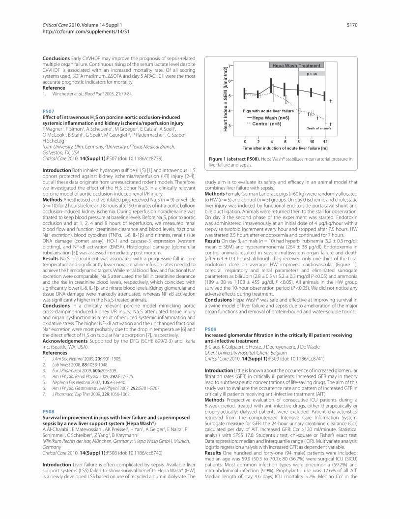
Conclusions Early CVVHDF may improve the prognosis of sepsis-related
multiple organ failure. Continuous rising of the serum lactate level despite
CVVHDF is associated with an increased mortality rate. Of all scoring
systems used, SOFA maximum, ΔSOFA and day 5 APACHE II were the most
accurate prognostic indicators for mortality.
Reference1. Winchester et al.: Blood Purif 2003, 21:79-84.
P507
Eff ect of intravenous H2S on porcine aortic occlusion-induced
systemic infl ammation and kidney ischemia/reperfusion injuryF Wagner1, F Simon1, A Scheuerle1, M Groeger1, E Calzia1, A Soell1,
O McCook1, B Stahl1, G Speit1, M Georgieff 1, P Radermacher1, C Szabo2,
H Schelzig1
1Ulm University, Ulm, Germany; 2University of Texas Medical Branch,
Galveston, TX, USA
Critical Care 2010, 14(Suppl 1):P507 (doi: 10.1186/cc8739)
Introduction Both inhaled hydrogen sulfi de (H2S) [1] and intravenous H
2S
donors protected against kidney ischemia/reperfusion (I/R) injury [2-4],
but all these data originate from unresuscitated rodent models. Therefore,
we investigated the eff ect of the H2S donor Na
2S in a clinically relevant
porcine model of aortic occlusion-induced renal I/R injury.
Methods Anesthetised and ventilated pigs received Na2S (n = 9) or vehicle
(n = 10) for 2 hours before and 8 hours after 90 minutes of intra-aortic balloon
occlusion-induced kidney ischemia. During reperfusion noradrenaline was
titrated to keep blood pressure at baseline levels. Before Na2S, prior to aortic
occlusion and at 1, 2, 4 and 8 hours of reperfusion, we measured renal
blood fl ow and function (creatinine clearance and blood levels, fractional
Na+ excretion), blood cytokines (TNFα, IL-6, IL-1β) and nitrates, renal tissue
DNA damage (comet assay), HO-1 and caspase-3 expression (western
blotting), and NF-κB activation (EMSA). Histological damage (glomerular
tubularisation [5]) was assessed immediately post mortem.
Results Na2S pretreatment was associated with a progressive fall in core
temperature and signifi cantly lower noradrenaline infusion rates needed to
achieve the hemodynamic targets. While renal blood fl ow and fractional Na+
excretion were comparable, Na2S attenuated the fall in creatinine clearance
and the rise in creatinine blood levels, respectively, which coincided with
signifi cantly lower IL-6, IL-1β, and nitrate blood levels. Kidney glomerular and
tissue DNA damage were markedly attenuated, whereas NF-κB activation
was signifi cantly higher in the Na2S-treated animals.
Conclusions In a clinically relevant porcine model mimicking aortic
cross-clamping-induced kidney I/R injury, Na2S attenuated tissue injury
and organ dysfunction as a result of reduced systemic infl ammation and
oxidative stress. The higher NF-κB activation and the unchanged fractional
Na+ excretion were most probably due to the drop in temperature [6] and
the direct eff ect of H2S on tubular Na+ absorption [7], respectively.
Acknowledgements Supported by the DFG (SCHE 899/2-3) and Ikaria
Inc. (Seattle, WA, USA).
References1. J Am Soc Nephrol 2009, 20:1901-1905.
2. Lab Invest 2008, 88:1038-1048.
3. Eur J Pharmacol 2009, 606:205-209.
4. Am J Physiol Renal Physiol 2009, 297:F27-F25.
5. Nephron Exp Nephrol 2007, 105:e33-e40.
6. Am J Physiol Gastrointest Liver Physiol 2007, 292:G201-G207.
7. J Pharmacol Exp Ther 2009, 329:1056-1062.
P508
Survival improvement in pigs with liver failure and superimposed sepsis by a new liver support system (Hepa Wash®)A Al-Chalabi1, E Matevossian1, AK Preissel1, H Yan1, A Geiger1, E Nairz1, P
Schimmel1, C Schreiber1, Z Yang1, B Kreymann2
1Klinikum Rechts der Isar, München, Germany; 2Hepa Wash GmbH, Munich,
Germany
Critical Care 2010, 14(Suppl 1):P508 (doi: 10.1186/cc8740)
Introduction Liver failure is often complicated by sepsis. Available liver
support systems (LSS) failed to show survival benefi ts. Hepa Wash® (HW)
is a newly developed LSS based on use of recycled albumin dialysate. The
study aim is to evaluate its safety and effi cacy in an animal model that
combines liver failure with sepsis.
Methods Female German Landrace pigs (~60 kg) were randomly allocated
to HW (n = 5) and control (n = 5) groups. On day 0 ischemic and cholestatic
liver injury was induced by functional end-to-side portacaval shunt and
bile duct ligation. Animals were returned then to the stall for observation.
On day 3 the second phase of the experiment was started. Endotoxin
was administered intravenously at an initial dose of 4 μg/kg/hour with a
stepwise twofold increment every hour and stopped after 7.5 hours. HW
was started 2.5 hours after endotoxemia and continued for 7 hours.
Results On day 3, animals (n = 10) had hyperbilirubinemia (5.2 ± 0.3 mg/dl;
mean ± SEM) and hyperammonemia (264 ± 38 μg/dl). Endotoxemia in
control animals resulted in severe multisystem organ failure and death
(after 6.4 ± 0.3 hours) although they received only one-third of the total
endotoxin dose on average. HW improved cardiovascular (Figure 1),
cerebral, respiratory and renal parameters and eliminated surrogate
parameters as bilirubin (2.8 ± 0.5 vs 5.2 ± 0.3 mg/dl P <0.05) and ammonia
(189 ± 38 vs 1,108 ± 455 μg/dl, P <0.05). All animals in the HW group
survived the 10-hour observation period (P <0.05). We did not notice any
adverse eff ects during treatment.
Conclusions Hepa Wash® was safe and eff ective at improving survival in
a swine model of liver failure and sepsis due to amelioration of the major
organ functions and removal of protein-bound and water-soluble toxins.
P509
Increased glomerular fi ltration in the critically ill patient receiving anti-infective treatmentB Claus, K Colpaert, E Hoste, J Decruyenaere, J De Waele
Ghent University Hospital, Ghent, Belgium
Critical Care 2010, 14(Suppl 1):P509 (doi: 10.1186/cc8741)
Introduction Little is known about the occurrence of increased glomerular
fi ltration rates (GFR) in critically ill patients. Increased GFR may in theory
lead to subtherapeutic concentrations of life-saving drugs. The aim of this
study was to evaluate the occurrence rate and pattern of increased GFR in
critically ill patients receiving anti-infective treatment (AIT).
Methods Prospective evaluation of consecutive ICU patients during a
6-week period, treated with anti-infective drugs, either therapeutically or
prophylactically; dialysed patients were excluded. Patient characteristics:
retrieved from the computerized Intensive Care Information System.
Surrogate measure for GFR: the 24-hour urinary creatinine clearance (Ccr)
calculated per day of AIT. Increased GFR: Ccr >120 ml/minute. Statistical
analysis with SPSS 17.0: Student’s t test, chi-square or Fisher’s exact test.
Data expression: median and interquartile range (IQR). Multivariate analysis:
logistic regression analysis with increased GFR as dependent variable.
Results One hundred and forty-one (94 male) patients were included;
median age was 59.9 (50.3 to 70.1); 80 (56.7%) were surgical ICU (SICU)
patients. Most common infection types were pneumonia (59.2%) and
intra-abdominal infection (9.9%). Prophylactic use was 17.6% of all AIT.
Median length of stay 4.6 days; ICU mortality 5.7%. Median Ccr in the
Figure 1 (abstract P508). Hepa Wash® stabilizes mean arterial pressure in
liver failure and sepsis.
Critical Care 2010, Volume 14 Suppl 1 http://ccforum.com/supplements/14/S1
S170

whole patient group was 97.0 ml/minute (57.5 to 164.5). In total, 753 AIT
days were available for analysis in which 302 days of increased GFR were
present (40.1%). Seventy-six patients (53.9%) had an episode of increased
GFR at some time during AIT. In 31 patients (22.0%) GFR was increased
throughout the whole episode of AIT. Patients with increased GFR were
signifi cantly younger (54.2 vs 65.6, P <0.001) and there was a trend towards
a higher incidence of increased GFR in patients treated for infections, in
male and in SICU patients. Multivariate analysis showed that age (OR 0.93
per year), therapeutic administration of AIT (OR 5.49), and SICU admission
(OR 2.27) were independently associated with increased GFR.
Conclusions Increased GFR were a frequent fi nding in critically ill patients
receiving AIT, and was associated with younger age and SICU admission.
P510
Therapeutic plasma exchange in children with acute liver failure: assessment of laboratory parametersD Demirkol, O Yanar, N Gerenli, A Citak, I Ozden, S Cantez, O Durmaz,
M Karabocuoglu
Istanbul University, Istanbul, Turkey
Critical Care 2010, 14(Suppl 1):P510 (doi: 10.1186/cc8742)
Introduction The aim of this study is to analyze the eff ect of therapeutic
plasma exchange (TPE) on biochemical and coagulation parameters.
Methods We enrolled all children who were admitted to the pediatric ICU
with diagnosis of acute liver failure and underwent TPE between December
2005 and December 2009. Serum prothrombin time, International
Normalized Ratio, total and direct bilirubin, aminotransferases and serum
ammonia values were recorded before and after TPE sessions.
Results Fourteen patients (male/female, 4/10, median age 8.5 years)
underwent a total of 37 TPE sessions. Acute liver failure was induced by viral
hepatitis in fi ve cases, Wilson disease in four cases, toxins in four cases and
autoimmune hepatitis in one case. When compared with baseline, there
were signifi cant improvements in laboratory parameters (Table 1). Five of
the 14 patients (35.7%) died in the pediatric ICU, fi ve patients underwent
liver transplantation. Four patients (28.5%) survived with extracorporeal
liver support. No serious adverse eff ect of TPE was observed in the patients
during or after completion of TPE.
Conclusions TPE is eff ective in improving liver biochemistry tests in
children with liver failure. Expected changes in laboratory parameters after
TPE must be considered especially in deciding for liver transplantation.
Reference1. Singer AL, Olthoff KM, Haewen K, Rand E, Zamir G, Shaked A: Role of
plasmapheresis in the management of acute hepatic failure in children. Ann Surg 2001, 234:418-424.
P511
New markers of infl ammation-induced renal injury subside when endotoxin tolerance develops in humans as measured by urine proteomicsS Heemskerk, A Draisma, C Laarakkers, J Van der Hoeven, R Masereeuw,
P Pickkers
Radboud University Nijmegen Medical Centre, Nijmegen, the Netherlands
Critical Care 2010, 14(Suppl 1):P511 (doi: 10.1186/cc8743)
Introduction Sepsis has been identifi ed as the most common cause of
renal injury in ICUs although the pathophysiology is not well understood.
No large clinical studies are available that show an improvement of renal
function in patients with sepsis and this may be related to the lack of
early diagnostic tests that indicate the onset of renal injury. The aim of the
current study was to search for potential new early markers of renal injury
during acute endotoxemia and to investigate whether renal injury can be
ameliorated by the induction of lipopolysaccharide (LPS) tolerance.
Methods Five healthy males received intravenous bolus injections of
2 ng/kg/day Escherichia coli LPS for 5 consecutive days. We used surface-
enhanced laser desorption/ionization time-of-fl ight mass spectrometry
(Seldi-TOF MS). This approach allows for rapid high-throughput profi ling
of multiple urine samples and detects low molecular weight biomarkers.
Results Repeated LPS administrations induced a diminished glomerular
fi ltration rate of 33 ± 7% (P = 0.02) on day 2 and an increase in serum
creatinine of 11 ± 3% (P = 0.002) on day 3, which was associated with the
appearance of 15 peak intensities in the urinary protein profi le including
an increase in β-microglobulin levels (P = 0.04) 6 hours after the fi rst LPS
administration. Four of the 15 peak intensities on day 1 correlated with
serum creatinine levels on day 3; 3,950, 4,445, 6,723 and 7,735 m/z (P = 0.03;
0.01; 0.02 and 0.05, respectively). With the development of LPS tolerance,
renal function was restored, refl ected by a decrease in serum creatinine
and β-microglobulin levels to baseline (P = 0.2 and 0.4, respectively,
between days 1 and 5), and by attenuated peak intensities in the urinary
protein profi le (P <0.0001 for all 15 peak intensities).
Conclusions In conclusion, renal injury occurs during repeated
endotoxemia and can be predicted by new urinary markers using
proteome research. The four markers that correlated with the extent of
renal injury may represent potential new biomarkers for renal injury
and need further identifi cation. The infl ammation-induced renal injury
subsided when LPS tolerance developed after 5 consecutive days of LPS
administrations.
P512
Extraction and purifi cation of 1-hydroxymidazolam glucuronide from ultrafi ltrateC Mckenzie1, D Naughton2, G Davies3, D Treacher1
1Guy’s and St Thomas’ NHS Foundation Trust, London, UK; 2Kingston
University, London, UK; 3King’s College, London, UK
Critical Care 2010, 14(Suppl 1):P512 (doi: 10.1186/cc8744)
Introduction We developed a technique for detection of MDZ and
1-hydroxymidazolam glucuronide (1-OHMG). No commercial source of
1-OHMG prevented assay development. 1-OHMG had pharmacological
activity and is renally excreted. We postulated that ultrafi ltrate (UFR) would
be rich in 1-OHMG. We describe a method to purify 1-OHMG from UFR.
Methods Ethics approval was granted. To purify, UFR was extracted
on a C-18 column, it was washed and eluted. HPLC-MS separation was
performed. The 1-OHMG (mol. wt 517/ 518 in MS) was desiccated and
re-dissolved four times to maximise purity. Electrospray (ESI) mass
spectrometry (MS) characterised 1-OHMG. The purity was calculated using
NMR. A calibration plot of 1-OHMG, MDZ and DZP (internal standard) for
HPLC-MS was performed.
Results 1-OHMG identity was confi rmed using MS/MS (Figure 1). Two
milligrams of 1-OHMG was purifi ed from 5,000 ml UFR. The 1-OHMG was
98% pure (NMR). The extinction coeffi cient was identical to MDZ. The
calibration plot resulted in correlation of 0.912. The assay was applied into
clinical practice, to report sera and UFR levels.
Table 1 (abstract P510). Biochemical and coagulation parameters before and after TPE
Variable Before TPE After TPE Delta variable P value
PT (sec) 40.9 (32.5 to 47.5) 21.1 (18.1 to 25.0) 17.8 (10.2 to 26.1) 0.0003
INR 3.6 (3.1 to 4.6) 1.7 (1.5 to 2.3) 1.7 (1.3 to 27.8) 0.0002
TB (mg/dl) 17.7 (11.7 to 21.5) 14.2 (9.6 to 18.0) 3.2 (1.1 to 6.0) 0.0006
DB (mg/dl) 10.3 (7.3 to 17.2) 7.9 (4.8 to 14.2) 1.9 (0.8 to 4.8) 0.0001
ALT (U/l) 122.5 (61.1 to 370) 87.5 (54 to 197.5) 38.1 (7 to 109.5) 0.0001
AST (U/l) 247.5 (122 to 399) 178 (85.5 to 272) 66.5 (14.1 to 223) 0.0002
Ammonia (mmol/l) 143.2 (81.5 to 195) 97.0 (71 to 150.5) 11.5 (3.5 to 59) 0.0003
Critical Care 2010, Volume 14 Suppl 1 http://ccforum.com/supplements/14/S1
S171

Conclusions We were able to extract and purify an active drug metabolite
from UFR. Five litres of UFR resulted in 2 mg 1-OHMG. This is a potentially
rich source of drugs or drug metabolites, allowing pharmacokinetic
studies greatly required in critical illness.
Reference1. McKenzie et al.: Diff erentiating MDZ oversedation from neurological
damage in the intensive care unit. Crit Care 2005, 9:32-33.
P513
Continuous renal replacement therapy: are we getting the dose right?S Martin, M Al-Haddad
Western Infi rmary, Glasgow, UK
Critical Care 2010, 14(Suppl 1):P513 (doi: 10.1186/cc8745)
Introduction The Intensive Care Society recommends achieving a dose of
35 ml/kg/hour in 85% of patients receiving continuous renal replacement
therapy (CRRT). However, a recent multicentre trial concluded there was
no benefi t of using 40 ml/kg/hour versus 25 ml/kg/hour. The extra cost of
high-dose RRT has not been quantifi ed. At our institution the target dose
is 35 ml/kg/hour.
Methods Patients receiving renal replacement therapy from April to
September 2009 were identifi ed using Careview, an electronic patient-
care record system. The effl uent rate (ml/hour) was used to calculate the
dose and adjusted for weight. A telephone survey of ICUs examined the
dose and mode of CRRT used in Scotland.
Results Thirty-fi ve patients, mean age 62, received CRRT with a mean
duration of RRT of 78 hours. Twenty-three patients (66%) were admitted
with sepsis. Continuous venovenous haemodiafi ltration was the initial
mode in 31 patients (89%). Ten patients were excluded from the dose
analysis because no weight was documented. The mean dose actually
delivered per patient was 50 ml/kg/hour. If 25 ml/kg/hour had been given
then 40% less RRT fl uid could have been used. The Scottish telephone
survey revealed that one-third of Scottish ICUs routinely use 35 ml/kg/
hour or more. If 25 ml/kg/hour was used routinely in these units alone,
then over €24,000 could be saved per annum.
Conclusions The dose of CRRT given for each patient at our institution
currently exceeds the target dose by 43%. Using 25 ml/kg/hour as the
standard dose would provide a signifi cant cost saving to the NHS in
Scotland and to ICUs throughout Europe.
Reference1. Bellomo R, Cass A, Cole L, et al.: Intensity of continuous renal-replacement
therapy in critically ill patients. N Engl J Med 2009, 361:1627-1638.
P514
Use of CRRT-CVVH in a hemato-oncological ICU in patients treated with defi brotide for veno-occlusive disease in patients after allogeneic hematopoetic stem cell transplantation: single-center experienceM Navratil, Z Koristek, F Folber
Masaryk University Hospital, Brno, Czech Republic
Critical Care 2010, 14(Suppl 1):P514 (doi: 10.1186/cc8746)
Introduction Complex care in the hemato-oncological ICU sometimes
requires the use of CRRT. As the hemato-oncological patients have some
specifi cations we retrospectively followed up the results of our eff ort in
patients with acute renal failure due to veno-occlusive disease (VOD) after
allogeneic hematopoietic stem cell transplantation. VOD is one of the most
severe early complications in this type of patients and its mortality is high.
Methods From 1 January 2007 to 30 October 2009 we performed 94
procedures (one procedure = 24 hours) in 15 patients with VOD and
acute renal oligo-anuric failure. As a standard in our ICU we perform CVVH
with post dilution on a Fresenius Multifi ltrate machine. The key drug
for the treatment of VOD is defi brotide and the patients were severely
thrombocytopenic, we have not used any anticoagulation in the CVVH
set. The dose of defi brotide ranged from 10 to 20 mg/kg/day according to
the severity of thrombocytopenia and eventually hemorrhagic symptoms.
Blood count, acid–base balance and biochemistry were monitored at least
twice daily. The thrombocytopenia was corrected with the donor platelets
and the platelet level was sustained at 20 x 109l.
Results The introduction of CVVH in all 15 patients dramatically helped to
overcome the severe part of VOD until the defi brotide helped to restore
Figure 1 (abstract P512). Centroided MS of purifi ed 1-OHMG.
Figure 1 (abstract P513). Mean dose of RRT for each patient at Western
Infi rmary.
Critical Care 2010, Volume 14 Suppl 1 http://ccforum.com/supplements/14/S1
S172
the right function of vascular endothelium. None of the 15 patients died
from VOD as a cause of death. Surprisingly, the median patency of the
CVVH tubing set was 46 hours despite no anticoagulation in the tubing
set. Defi brotide is effi cient in preventing coagulation in the tubing set.
No serious hemorrhagic events were observed. The fl uent maintenance
of fl uid balance, the removal of waste products of metabolism and the
maintenance of acid–base balance were very important for the patient in
the critical point of advanced VOD.
Conclusions CVVH is an integral part of intensive care in hemato-oncology
and the use of this therapeutic modality in patients with VOD with acute
renal failure can signifi cantly improve the results of our therapeutic eff orts.
P515
Comparison of the effi cacy and safety of two regional citrate anticoagulation protocols using acid citrate dextrose A or Prismocitrate 10/2, in patients with acute renal failure undergoing continuous venovenous haemodiafi ltrationEL Ooi, TW Lim, N Lim
Changi General Hospital, Singapore
Critical Care 2010, 14(Suppl 1):P515 (doi: 10.1186/cc8747)
Introduction Continuous renal replacement therapy (CRRT) is the
treatment of choice for acute renal failure in critically ill patients. Our study
compared the effi cacy and safety of regional citrate anticoagulation (RCA)
with acid citrate dextrose A (ACD-A) against Prismocitrate 10/2 (Gambro).
Use of Prismocitrate 10/2 eliminates the need for a separate infusion
pump for ACD-A as it doubles as replacement fl uid and anticoagulant.
This removes a source of error in the calculation of fl uid balance. The
combination of tri-sodium citrate and citric acid in Prismocitrate 10/2
avoids the metabolic alkalosis associated with ACD-A [1,2].
Methods This was a prospective sequential cohort study. All patients
admitted to the surgical ICU who required CRRT for ARF were recruited.
Group A, using ACD-A, was recruited from October 2007 to September
2008 (n = 23). Group B, using Prismocitrate 10/2, was recruited from
October 2008 to September 2009 (n = 20). We evaluated the incidence
of metabolic alkalosis and other biochemical changes, azotemia control,
fi lter lifespan and complications.
Results The incidence of metabolic alkalosis in Group B from treatment
day 2 onwards was signifi cantly lower than Group A. The Group A median
pH was 7.42 (6.81 to 7.62) compared with Group B pH 7.35 (7.2 to 7.49),
P = 0.001. The control of electrolytes and azotemia was not signifi cantly
diff erent. The mean fi lter duration was 58.8 hours (95% CI 38.0 to 79.6)
for Group A and 61.8 hours (95% CI 45.8 to 77.8) for Group B (P = 0.678).
Longitudinal analysis revealed a statistically signifi cant result for reduced
metabolic alkalosis for Group B. (standard bicarbonate P <0.001, base
excess P <0.001). Repeat treatment sessions also showed a statistically
signifi cant reduction of metabolic alkalosis using Prismocitrate 10/2 rather
than ACD-A (P = 0.029).
Conclusions RCA with Prismocitrate 10/2 reduces the incidence of
metabolic alkalosis associated with ACD-A. This regime is safe, feasible and
improved patient safety, with no increase in complication rates. Our unit
has now converted to Prismocitrate 10/2 for RCA.
References1. Silverstein FJ, et al.: Metabolic alkalosis induced by regional citrate
hemodialysis. ASAIO Trans 1989, 35:22-25.
2. Morgera S, et al.: Metabolic complications during regional citrate anticoagulation in continuous venovenous hemodialysis: a single center experience. Nephron Clin Pract 2004, 97:c31-c136.
P516
Addition of prostacyclin to heparin does not prolong circuit life during continuous haemofi ltrationM Ostermann, L Tovey, H Dickie, R Beale
Guy’s and St Thomas’ Foundation Hospital, London, UK
Critical Care 2010, 14(Suppl 1):P516 (doi: 10.1186/cc8748)
Introduction Premature circuit clotting is a major problem during
continuous venovenous haemofi ltration (CVVH). Some studies have
suggested that circuit life can be improved by adding epoprostenol to
heparin.
Methods Retrospective review of mean circuit patency in patients on
noncitrate-based anticoagulation regimens during CVVH in a 30-bed
multidisciplinary ICU between October 2008 and May 2009. Circuits which
were discontinued electively were excluded from the analysis.
Results Patency data were available for 486 noncitrate circuits. There was
no signifi cant diff erence in mean circuit life between heparin given via
circuit (n = 169 circuits; mean circuit life 19.8 hours) and epoprostenol
alone (n = 119 circuits; mean circuit life 20.9 hours). In a small group of
patients on heparin via the circuit, epoprostenol was added for patency
reasons (n = 19 circuits). In this group, the mean circuit life was only
15 hours. Circuits of patients on systemic heparin (n = 100) lasted for a
mean of 23.5 hours. In a small group of patients, epoprostenol was added
via the circuit (n = 32 circuits). The mean circuit life was 28.9 hours (P >0.05).
Other types of noncitrate anticoagulation were activated protein C and
lepuridin, alone or in combination with heparin (n = 47 circuits).
Conclusions In our practice, anticoagulation with epoprostenol alone led
to similar circuit survival as heparin administered via the circuit. Adding
epoprostenol to heparin did not signifi cantly prolong the mean circuit
life. Further analysis of circuit patency in individual patients is needed to
determine the characteristics of patients who may benefi t and for whom
the extra cost could be justifi ed.
P517
Recurrent early fi lter clotting in regional citrate anticoagulated continuous venovenous hemodialysis due to undetected antibodies to heparin–platelet factor 4 complexesT Slowinski, I Lieker, M Sander, D Khadzhynov, S Morgera, H Neumayer,
H Peters
Charité Campus Mitte, Berlin, Germany
Critical Care 2010, 14(Suppl 1):P517 (doi: 10.1186/cc8749)
Introduction We recently published a citrate anticoagulated continuous
venovenous hemodialysis (citrate-CVVHD) protocol with variable
treatment dose. The protocol was safe and easy to handle and the mean
fi lter lifetime was 61.5 hours [1]. We here report fi ve cases with unexplained
recurrent early fi lter clotting beside eff ective citrate anticoagulation due to
undetected antibodies to heparin–platelet factor 4 complexes (hep-PF4
ab).
Methods Regionale citrate anticoagulation was performed with variable
treatment dose using the MultiFiltrate™ CRRT device (Fresenius, Germany)
in CVVHD mode. Between May 2008 and August 2009 we detected fi ve
cases on the ICU. All had acute kidney injury with need for RRT. Underlying
diseases were: three sepsis, one cardiac surgery, one abdominal surgery.
All patients were anticoagulated with heparin or fractionated heparin
for more than 10 days before initiation of citrate-CVVHD. At the time of
initiation of citrate-CVVHD, none of the patients had clinically evident
signs of thomboembolism.
Results The mean fi lter lifetime before diagnosis of hep-PF4 ab was 8
(range 1.3 to 14) hours. Postfi lter ionized calcium concentrations were
always in the demanded range (mean 0.28, range 0.24 to 0.35 mmol/l). The
mean thrombocyte count was 160/nl (range 48 to 215) at time of initiation
of citrate-CVVHD. Thrombocyte counts fell to a mean 114/nl (range 36 to
177) within 5 days. Mean time to testing for and diagnosis of hep-PF4 ab
was 6 (range 3 to 12) days. After positive testing (HIPA) for hep-PF4 ab,
heparin was stopped and all patients received argatroban with a mean
PTT of 83 (range 48 to 119) seconds. Under systemic anticoagulation with
argatroban, the fi lter lifetime of citrate-CVVHD increased to a mean 58
(range 18 to 96) hours. Within 14 days after initiation of citrate-CVVHD,
three patients had a diagnosis of a clinically signifi cant thrombembolic
event.
Conclusions In patients on the ICU with moderate–low thrombocyte
count and without clinically evident signs of thomboembolism, often
sepsis or the state of critical illness is assumed as the cause for decreasing
platelet counts and tests for hep-PF4 ab are not performed. In cases of
undetected and untreated hep-PF4 ab, fi lter clotting can occur beside
eff ective citrate anticoagulation in CVVHD. In cases of unexplained
recurrent early fi lter clotting in citrate-CVVHD, patients should be
carefully examined for underlying hep-PF4 ab and heparin-induced
thrombocytopenia.
Reference1. Morgera et al.: Crit Care Med 2009, 37.
Critical Care 2010, Volume 14 Suppl 1 http://ccforum.com/supplements/14/S1
S173
P518
Pilot study on a regional citrate anticoagulated continuous venovenous hemodiafi ltration protocol with variable treatment doseT Slowinski, I Lieker, D Khadzhynov, M Sander, S Morgera, H Neumayer,
H Peters
Charité Campus Mitte, Berlin, Germany
Critical Care 2010, 14(Suppl 1):P518 (doi: 10.1186/cc8750)
Introduction In order to add convective solute transport to a recently
described citrate-CVVHD protocol, we established a citrate anticoagulated
continuous venovenous hemodiafi ltration (citrate-CVVHDF) protocol
with variable treatment dose. Citrate-CVVHD has been shown to be safe
and easy to handle [1]. Being based on dialysis, however, citrate-CVVHD
primarily relies on diff usive solute transport. To also allow convective
transport, we designed a new citrate-CVVHDF protocol with a postdilution
hemofi ltration dose.
Methods Prospective observational study. A CVVHDF-based citrate
anticoagulation protocol on the MultiFiltrate™ CRRT device (Fresenius
Medical Care (FMC), Germany) using a 4% trisodium citrate solution,
the dialysate fl uid Ci-Ca™ Dialysate K2 (FMC), and a continuous calcium
chloride (91 mmol/l) infusion. For the fi ltration dose we used a standard
bicarbonate-buff ered substitution fl uid (MultiBic™; FMC) in postdilution.
Ten patients on the ICU with acute kidney injury and need for RRT were
included. For variable doses, patients were divided into three groups
according to their body weight (1: <60 kg, 2: 60 to 90 kg, 3: >90 kg). The
initial fl ows for dialysate/substitution fl uid in the groups were 1,400/800,
1,800/1,000, and 2,200/1,200 ml/hour, blood fl ow 80, 100, and 120 ml/
minute, citrate fl ow 145, 175, and 220 ml/hour, and calcium fl ow 32, 45,
and 57 ml/minute. Citrate fl ow was adjusted to postfi lter ionized calcium
(iCa) measurements, target range 0.25 to 0.35 mmol/l. Calcium fl ow was
adjusted to patients’ systemic iCa. The treatment time was limited to
72 hours.
Results Eight of 10 patients reached the maximum treatment time of
72 hours without clotting. In one patient the treatment had to be stopped
after 50 hours because of central venous catheter fl ow problems and
one patient died after 54 hours. Acid–base control: mean (95% CI) pH;
st-bicarbonate at 48 hours: 7.42 (7.38 to 7.44); 24.6 (21.3 to 27.8) mmol/l,
and at 72 hours: 7.39 (7.35 to 7.43); 25.0 (21.3 to 27.6) mmol/l. Electrolyte
control: mean (95% CI) s-sodium; s-potassium at 48 hours: 139 (137 to
141); 4.8 (4.4 to 5.2) mmol/l, and at 72 hours: 139 (137 to 142); 4.9 (4.4 to
5.4) mmol/l. Mean (95% CI) treatment dose was 41 (37 to 46) ml/kg/hour,
mean (95% CI) s-urea at 48 hours: 51 (37 to 66) mg/dl, and at 72 hours: 50
(35 to 65) mg/dl.
Conclusions The citrate-CVVHDF protocol allows combined hemodialysis
and hemofi ltration in CRRT with variable treatment dose and with the
advantages of regional citrate anticoagulation. In this pilot study, in
10 patients the fi lter run-time in citrate-CVVHDF was remarkable and
metabolic control excellent.
Reference1. Morgera et al.: Crit Care Med 2009, 37.
P519
Characteristics and outcome of patients treated with renal replacement therapy: single-center, 2-year experienceB Kovacevic, S Gavrilovic, D Obradovic, M Kojicic, U Batranovic
Institute for Pulmonary Diseases of Vojvodina, Sremska Kamenica, Serbia
Critical Care 2010, 14(Suppl 1):P519 (doi: 10.1186/cc8751)
Introduction Continuous renal replacement therapy (CRRT) is frequently
used among hemodynamically unstable critically ill patients with acute
renal failure (ARF). However, data regarding management of these patients
in Serbia are scarce. CRRT has been available at our institution since 2007.
The objective of this study was to describe characteristics and outcome of
patients treated with renal replacement therapy in a single tertiary-care
center in Serbia.
Methods In this retrospective observational study we reviewed medical
records of adult (≥18 years) patients treated in the fi ve-bed ICU at the
Institute for Pulmonary Diseases in Sremska Kamenica, Serbia who
underwent CRRT from April 2007 to November 2009.
Results From the total 314 patients treated in the ICU, 29 (9.2%), 17 men,
median age 54 years (interquartile range (IQR) 48 to 68), underwent
CRRT. Patients were mostly nonsurgical (n = 28, 96%). Main admission
diagnoses were septic shock (n = 11) and pneumonia (n = 9). The median
APACHE II score was 27 (IQR 19 to 32), and SOFA day 1 was 10 (IQR 5 to
13). Seventeen patients (58.6%) had chronic co-morbidities; Charlson
score median 2 (IQR 0.75 to 3). Twenty-four patients required mechanical
ventilation, and 13 were diagnosed with acute respiratory distress
syndrome. Slow continuous ultrafi ltration was used in four patients
(13.7%), hemodiafi ltration (CVVHDF) modality in 13 (44.8%), hemodialysis
(CVVHD) in 11 (37.9%). Median time on CRRT was 3 days (IQR 2 to 4). Five
patients (17%) had a complete recovery of renal function at the time of
ICU discharge. Median hospital length of stay was 12 days (IQR 5 to 20) and
hospital mortality was 69%.
Conclusions The need for CRRT in the observed population is substantial.
Future studies are warranted to determine the impact of introduction of
CRRT on hospital mortality in Eastern European countries and to provide
future strategies for improving patient care in these settings.
P520
Renal function in living related kidney donors above the age of 70V Milic, N Vukovic, A Pavlovic, S Pavlovic
Clinical Center Nis, Serbia
Critical Care 2010, 14(Suppl 1):P520 (doi: 10.1186/cc8752)
Introduction Living related kidney donors are highly motivated for
donation. Older donors may have increased risks in the perioperative
and postoperative period. Age limits for donors was insisted on in the
past. The glomerular fi ltration rate in older people is only slightly lower
than in young individuals in the absence of primary renal disease or
confounding cardiovascular disease. The aim of the study was to assess
renal function of living related donors above the age of 70 in the early
postoperative period.
Methods From March 2007 to November 2009, in Clinical Centre Nis,
25 living related donors donated their kidneys. The serum creatinine
and calculated creatinine clearance (MDRD formula) were evaluated
preoperatively, fi rst, third and fi fth postoperative days in the group under
(U) and in the group above (A) the age of 70.
Results There were 16 donors in group U (mean age 59, SD 5.38) and nine
donors in group A (mean age 72.4, SD 1.88). The mean serum creatinine
level in group U was 64.66 μmol/l (SD 12.22), 120.2 μmol/l (SD 26.04),
120.91 μmol/l (SD 21.68) and 116.07 μmol/l (SD 21.35) preoperatively, on
the fi rst, third and fi fth postoperative days, respectively. In group A the
mean serum creatinine level was 84.58 μmol/l (SD 23.63), 150.64 μmol/l
(SD 38.67), 149.49 (SD 43.68) and 148.36 μmol/l (SD 39.08) preoperatively,
fi rst, third and fi fth postoperative days, respectively. Calculated creatinine
clearance in group U was 102.62 ml/minute/1.73 m2 (SD 20.61), 50.44
ml/minute/1.73 m2 (SD 10.28), 48.69 ml/minute/1.73 m2 (SD 7.88) and
51.62 ml/minute/1.73 m2 (SD 8.96) preoperatively, fi rst, third and fi fth
postoperative days, respectively, and in group A 78.11 ml/minute/1.73 m2
(SD 23.31), 39.11 ml/minute/1.73 m2 (SD 10.35), 40.56 ml/minute/1.73 m2
(SD 12.31) and 40.44 ml/minute/1.73 m2 (SD 11.36) preoperatively, fi rst,
third and fi fth postoperative days, respectively. There is a statistically
signifi cant diff erence in serum creatinine level between groups A and U
preoperatively (P = 0.039) and on the fi fth postoperative day (P = 0.043),
and there is a statistically signifi cant diff erence in glomerular fi ltration rate
between groups A and U preoperatively (P = 0.019), on the fi rst (P = 0.018)
and fi fth postoperative days (P = 0.024).
Conclusions Deterioration of residual kidney function in the early
postoperative period is higher in living donors above the age of 70. Patients
over the age of 70 must be accepted with care for kidney donation.
P521
Augmented renal clearance in traumatic brain injuryA Udy, R Boots, S Senthuran, J Stuart, R Deans, M Lassig-Smith, J Lipman
Royal Brisbane and Women’s Hospital, Brisbane, Australia
Critical Care 2010, 14(Suppl 1):P521 (doi: 10.1186/cc8753)
Introduction Use of hypertonic saline and/or vasopressor infusion to
achieve a desired cerebral perfusion pressure (CPP) in traumatic brain
Critical Care 2010, Volume 14 Suppl 1 http://ccforum.com/supplements/14/S1
S174

injury (TBI) is common. We hypothesised that the use of such therapies
would signifi cantly augment creatinine clearances (CrCl) in this population.
Methods Head-injured patients requiring hyperosmolar therapy
using 3% or 20% saline solutions and/or norepinephrine infusion for
the maintenance of a CPP >60 mmHg were recruited into the study.
Additional management was consistent with local practice and in
line with the Brain Trauma Foundation guidelines [1]. An 8-hour CrCl,
physiological variables, fl uid balance, and medications were recorded
daily during active management of CPP. A further CrCl was collected just
prior to discharge (off CPP therapy), and if this was elevated, was repeated
on the ward. Augmented renal clearance (ARC) was defi ned as a CrCl >160
ml/minute/1.73m2 for males and >150 ml/minute/1.73m2 for females [2].
Results Twenty consecutive patients were enrolled. The average ICU length
of stay was 15 days (CI 95% 11 to 18), and time to study entry averaged 2.3
days (CI 95% 1.7 to 2.8). All patients received norepinephrine (n = 20), 85%
(n = 17) received hypertonic saline, and therapy lasted on average 7.6 days
(CI 95% 5.6 to 9.5). ARC was demonstrated in 17 (85%) patients at any point
during active management of CPP. The mean maximum CrCl was 179 ml/
minute/1.73 m2 while on CPP therapy (CI 95% 159 to 198) returning to a
mean CrCl of 111 ml/minute/1.73 m2 (CI 95% 91 to 131, P <0.001) when
measured in the ward. The mean CrCl in the ICU while not receiving CPP
therapy was 150 ml/minute/1.73 m2 (CI 95% 134 to 167, P = 0.03). The
mean time to reach peak CrCl while on active treatment was 4.7 days (CI
95% 3.0 to 6.4). Norepinephrine use, saline loading, mean arterial pressure,
and central venous pressure, predicted CrCl on the day of measurement.
Conclusions ARC is common in head-injured patients receiving active
management of CPP and persists even after ceasing such therapy. This has
signifi cant implications for appropriate dosing of renally excreted drugs
in this setting.
References1. Bratton SL, et al.: Guidelines for the management of severe traumatic brain
injury. IX. Cerebral perfusion thresholds. J Neurotrauma 2007, 24(Suppl 1):S59-S64.
2. Stevens LA, et al.: Assessing kidney function – measured and estimated glomerular fi ltration rate. N Engl J Med 2006, 354:2473-2483.
P522
Pressure-controlled hemorrhagic shock in mice: a new model of acute kidney injuryN Mayeur1, V Minville1, A Jaafar2, I Tack2
1CHU Purpan, Toulouse, France; 2CHU Rangueil, Toulouse, France
Critical Care 2010, 14(Suppl 1):P522 (doi: 10.1186/cc8754)
Introduction At the present time, mice models have failed to reproduce
human acute kidney injury (AKI) and new relevant models of AKI are
required [1]. In this study we developed a model of AKI in mice based
on pressure-controlled hemorrhagic shock that closely reproduces
hypotension involved in most of human ischemic AKI.
Methods We fi rst determined shock and resuscitation modalities in a
C57/Bl6 mice population. A femoral arterial catheter was used for pressure
control and bleeding and a central jugular catheter for anesthesia and
resuscitation. We secondly explored renal repercussions of a 2-hour shock
duration at 35 mmHg mean arterial pressure. We assessed successively
the glomerular fi ltration rate (GFR), histological kidney injury score and
performed real-time PCR after the procedure. The shock group was
compared with sham mice and with a control group that underwent no
procedure (n = 5 in each group).
Results The shock group GFR was decreased 2 (D2) and 6 (D6) days after
shock when compared with control and sham (Figure 1). This AKI was
intrinsic as showed by the Na/K urinary ratio increase and the decrease in
urine concentration ability. Tissular damage prevailed in the outer medulla
with a maximal expression at D6 (paucicellular tubular epithelium,
intratubular casts) (Figure 2). These lesions are associated with an increase
of tissular KIM-1 and HIFs mRNA. Despite GFR normalization, the shock
group showed discreet defect in urine concentration ability and a slight
peritubular fi brosis 3 weeks after shock.
Conclusions This new model of AKI based on hypotension opens new
perspectives in the fi eld of short-term and long-term kidney function
following AKI.
Reference1. Rosen S, et al.: J Am Soc Nephrol 2008, 19.
P523
Continuous urinary electrolyte measurement in a swine model of mechanical ventilationD Febres1, T Langer1, M Cressoni1, A Protti1, S Coppola1, C Mietto1,
A Santini1, L Lombardi2, M Lazzerini2, P Cadringher1, P Bertoli3, L Gattinoni1
1Università degli studi di Milano, Milan, Italy; 2Fondazione IRCCS Ospedale
Maggiore Policlinico Mangiagalli e Regina Elena, Milan, Italy; 3Università degli
studi di Milano, Centro di Ricerche Chirurgiche Precliniche, Milan, Italy
Critical Care 2010, 14(Suppl 1):P523 (doi: 10.1186/cc8755)
Introduction The kidney is the organ of the human body designed
to sense and regulate intravascular volume. The KING (Kidney Instant
Monitoring; Orvim, Paderno Dugnano, Italy) is a new device that allows
continuous measurement of urinary electrolytes and real-time body
response to changes in intrathoracic and intra-abdominal pressures and
to lung injury.
Methods Sixteen pigs (weight 20 ± 3 kg) were anaesthetized, tracheo-
tomized, catheterized and mechanically ventilated. Pigs were ventilated
for approximately 10 hours with a TV of 10 ml/kg, RR 15 breaths/minute,
FIO
2 0.5 and no PEEP. After TV was increased, pigs were divided into two
groups: eight pigs with a lower TV and eight pigs with higher TV. A variable
dead space was added to maintain normocapnia. Pigs were mechanically
ventilated with the new TV up to 48 hours. The KING measured urinary
output and urinary concentration of sodium and potassium every
10 minutes. NaCl 0.9% was exclusively infused. The average urinary
concentration of each electrolyte was expressed as the total concentration
Figure 1 (abstract P522). Glomerular fi ltration rate 2 (D2), 6 (D6) and 21
(D21) days after the procedure.
Figure 2 (abstract P522). Tubular injury score at H3, D2, D6 and D21.
*P <0.05 and ** P <0.01.
Critical Care 2010, Volume 14 Suppl 1 http://ccforum.com/supplements/14/S1
S175
for each electrolyte divided by the length of mechanical ventilation. Data
were compared using the Wilcoxon test.
Results The higher TV group was ventilated with a TV of 890 ± 251 ml
and a plateau pressure of 31 ± 8 cmH2O while the lower TV group had
a TV of 472 ± 262 ml (P = 0.006) and a plateau pressure of 17 ± 9 cmH2O
(P = 0.02). The length of mechanical ventilation for the high TV group was
1,850 ± 633 minutes and for the low TV group was 3,341 ± 749 minutes
(P = 0.0006). The urinary output for the high TV was 37 ± 16 ml/hour and
for the low TV was 52 ± 19 ml/hour (P = 0.10). The high TV group showed
lower urinary sodium (40 ± 35 mEq/l vs 93 ± 26 mEq/l, P = 0.007), higher
urinary potassium (52 ± 12 mEq/l vs 35 ± 23 mEq/l, P = 0.04) and a lower
urinary sodium/potassium ratio (0.77 ± 0.7 vs 3.86 ± 3.3, P = 0.001). Before
TV was increased, urinary output, urinary concentrations of sodium,
potassium and sodium/potassium ratio were not diff erent.
Conclusions Data show that injurious ventilation is sensed by the kidney
as a relative hypovolemia with reduction in sodium urinary output,
increased excretion of potassium and inverted sodium/potassium ratio.
P524
Impact of positive fl uid balance on survival in critically ill cancer patientsJ Almeida, H Palomba, L Hajjar, V Torres, F Galas, M Ferreira, F Duarte,
T Trielli, L Yu, J Otavio
Heart Institute, São Paulo, Brazil
Critical Care 2010, 14(Suppl 1):P524 (doi: 10.1186/cc8756)
Introduction Fluid overload has recently been linked to adverse
outcomes in critically ill patients, but its impact on the outcomes of cancer
patients admitted to ICUs has not been previously described. The aim of
this study is analyze the impact of fl uid overload on outcomes of patients
with cancer needing intensive care.
Methods A total of 122 cancer patients admitted to the medical ICU in
a 6-month period were prospectively evaluated for survival. Univariate
and multivariate analysis were used to study ICU admission parameters
associated with ICU mortality. Exclusion criteria were ICU stay <24 hours
and chronic renal failure on dialysis.
Results Overall mortality was 15.5% (n = 19). The mean age of all patients
was 62.7 ± 11.6 years and 55% (n = 68) were male. One hundred and
six patients (86%) had solid tumors and 16 (14%) had hematologic
malignancies. Postoperative care (49%) and sepsis (38%) were the major
reasons for admission to the ICU. The mean APACHE II score value at
ICU admission was 17.2 ± 6.4 and the mean Karnofsky score before ICU
admission was 75.2 ± 17.2. At multivariate analysis, the following variables
at ICU admission were signifi cantly associated with ICU mortality in cancer
patients: Lung Injury Score (LIS) >2 (OR 3.3; 95% CI 1.32 to 8.23) and positive
fl uid balance (for each 100 ml/24 hours) (OR 1.03; 95% CI 1.01 to 1.06).
Conclusions Fluid overload is independently associated with increased
mortality in critically ill cancer patients. Further studies are necessary to
determine the impact of positive fl uid balance on acute organ dysfunction
and overall prognosis of cancer patients.
References1. Bouchard J, et al.: Fluid accumulation and acute kidney injury:
consequence or cause. Curr Opin Crit Care 2009, 15:509-513.
2. Bouchard J, et al.: Fluid accumulation, survival and recovery of kidney function in critically ill patients with acute kidney injury. Kidney Int 2009,
76:422-427.
P525
Fluid accumulation has an adverse eff ect on outcome of ICU patients with acute kidney injuryM Ostermann, H Cole, R Beale
Guy’s and St Thomas’ Foundation Hospital, London, UK
Critical Care 2010, 14(Suppl 1):P525 (doi: 10.1186/cc8757)
Introduction Management of acute kidney injury (AKI) is heterogeneous
with little consensus about fl uid therapy, vasopressors and diuretics. The
aim was to analyse whether fl uid management infl uences outcome in
critically ill patients with AKI.
Methods Retrospective analysis of the data for 4,645 patients admitted
to the multidisciplinary ICU at Guy’s and St Thomas’ Foundation Hospital
between April 2004 and June 2009. AKI was defi ned according to the AKI
network criteria, which distinguish between three diff erent grades of AKI.
Maximum degree of AKI and total cumulative fl uid balance between ICU
admission and the day of AKI were recorded.
Results A total of 1,225 patients (26.4%) had AKI I, 29 patients (0.6%) had
AKI II and 1,183 patients (25.5%) had AKI III of whom 89% were treated with
renal replacement therapy. Two hundred and thirty-seven (5.1%) patients
had end-stage dialysis-dependent renal failure and 1,971 patients (42.4%)
had no AKI during their stay in the ICU. The ICU mortality was 15.6% in AKI
I, 17.2% in AKI II and 34.9% in patients with AKI III (AKI I vs AKI III: P <0.0001).
Cumulative fl uid balance at diagnosis of AKI was signifi cantly higher in
patients who later died (Table 1).
Conclusions There is a correlation between fl uid accumulation on the day
of AKI and subsequent ICU outcome. Among patients with any degree
of AKI, nonsurvivors had a higher cumulative fl uid balance compared
with ICU survivors. Future analysis needs to determine whether fl uid
accumulation was the cause of worse outcome, the result of more
aggressive resuscitation or a marker of more severe AKI.
P526
Signifi cant microalbuminuria, on admission to the ICU, may indicate AKI in patients with sepsisS Basu1, M Bhattacharya1, T Chatterjee2, S Todi1, A Majumdar1
1AMRI Hospitals, Kolkata, India; 2Jadavpur University, Kolkata, India
Critical Care 2010, 14(Suppl 1):P526 (doi: 10.1186/cc8758)
Introduction In sepsis, a generalised infl ammatory onslaught causes
systemic endothelial dysfunction; the glomerular manifestation of which
is microalbuminuria. Recent histopathological evidence of extensive
glomerular capillary infi ltration by leucocytes in septic AKI suggests that
endothelial dysfunction may play a particularly important role in this
condition compared with patients with other causes of AKI. Markers of
endothelial dysfunction such as microalbuminuria may therefore have a
potential diagnostic role in septic AKI.
Methods Prospective observational study in a 20-bed ICU in a tertiary-
care hospital. Microalbuminuria estimated as the spot urine albumin–
creatinine ratio (ACR, mg/g) was measured on ICU admission. Two
hundred and sixty-six patients were recruited between January 2007 and
December 2008. Patients with an ICU stay of less than 24 hours, pregnancy,
menstruation, anuria, hematuria, proteinuria due to renal and post-renal
structural diseases were excluded. Sepsis was diagnosed by the ACCP/
SCCM criteria; AKI was identifi ed on admission using the RIFLE criteria.
Results Of the 266 patients studied, 130 patients had no AKI while 136
patients had AKI as classifi ed by the RIFLE criteria. Of these 136 patients,
59 patients were in the Risk category, 62 in the Injury and 15 patients in
the Failure, of which 26 patients (9.8%) needed RRT. For the entire cohort,
median age was 61.5 years, 61% were male, median APACHE II score was
15. Of these, 56 (21%) of patients died on the ICU. ACR showed signifi cant
correlation with serum creatinine values (P = 0.0049, Spearman rank
correlation (r) = 0.173) and was negatively associated with estimated GFR
(P = 0.0002, r = –0.225). The median level of ACR (204.1 mg/g, (IQR 119.7
to 402.0)) of patients with septic AKI (n = 66) on admission to the ICU was
signifi cantly greater (P <0.0001) than the median ACR level of 67.1 mg/g
(IQR 32.99 to 129.9) of patients who had AKI from other causes (n = 70).
Patients of sepsis who had developed AKI also had higher ACR than those
who did not (n = 51) (ACR 119.3 mg/g (IQR 37.02 to 354.3)) (P = 0.0137).
Conclusions Presence of signifi cant microalbuminuria on admission to
the ITU may be an early indicator of AKI in sepsis patients.
Table 1 (abstract P525). Mean cumulative fl uid balance (SD) in ICU survivors
and nonsurvivors
Degree of AKI Survivors Nonsurvivors P value
AKI I +2,527 ml (118) +3,694 ml (314) <0.0001
AKI II +4,348 ml (929) +6,896 ml (2,492) 0.0007
AKi III +3,228 ml (173) +4,780 ml (295) <0.0001
Critical Care 2010, Volume 14 Suppl 1 http://ccforum.com/supplements/14/S1
S176

P527
Neutrophil gelatinase-associated lipocalin as an early indicator for postoperative renal failureCD Van der Marel, H De Geus, J Bakker
ErasmusMC, Rotterdam, the Netherlands
Critical Care 2010, 14(Suppl 1):P527 (doi: 10.1186/cc8759)
Introduction Neutrophil gelatinase-associated lipocalin plasma (pNGAL)
and urine (uNGAL) concentrations can be used as early indicators for the
development of acute kidney injury (AKI) [1]. As AKI is associated with higher
risk of morbidity and mortality in ICU patients [2], higher pNGAL and uNGAL
on admission might be relevant to the clinical course. No data on pNGAL
and uNGAL as early indicators of postoperative AKI have been published.
Methods We collected plasma and urine samples from patients admitted
postoperatively following noncardiopulmonary bypass-related surgery.
NGAL concentrations were measured using an immunofl uorescence assay.
Subsequently we compared the time points of maximum pNGAL, uNGAL
and SCr concentrations in the group of patients developing postoperative
AKI (Mann–Whitney U test). Data are presented as mean ± SD.
Results One hundred and ninety-fi ve consecutive patients (age: 58 ± 16
years; APACHE II 15 ± 6) were included. Twenty-two patients (age 63 ± 17;
APACHE II 20 ± 7) developed AKI (AKI 1 n = 14; AKI 2 n = 3; AKI 3 n = 5).
Time to reach peak (Tmax
) concentrations for pNGAL, uNGAL and SCr are
respectively 21 (21), 20 (22), 31 (32) for AKI 1, 39 (32), 24 (31), 54 (12) for
AKI 2, and 17 (18), 26 (24), 34 (27) for AKI 3. Maximum concentrations of
pNGAL (ng/ml), uNGAL (ng/ml) and SCr (mmol/l) are respectively 1,348
(1,569), 364 (220), 147 (38) for AKI 1, 424 (309), 949 (389), 175 (28) for AKI
2, and 2,375 (1,731), 726 (398), 265 (81). Figure 1 shows time course of
pNGAL and uNGAL in patients developing AKI. Tmax
of pNGAL and uNGAL
was signifi cantly earlier than Tmax
of SCr (both α <0.001).
Conclusions In postoperative patients developing AKI, pNGAL and
uNGAL reach peak concentrations 1 day earlier than SCr. This might be
of clinical importance as it provides us with an opportunity to prevent
further deterioration of renal function at an earlier stage.
References1. Mishra J: Lancet 2005, 365:1231-1238.
2. Metnitz PG: Crit Care Med 2002, 30:2051-2058.
P528
Neutrophil gelatinase-associated lipocalin as an early indicator for mortality in adult critically ill patients with acute kidney injuryWP Ridder, H De Geus, J Bakker
Erasmus MC, Rotterdam, the Netherlands
Critical Care 2010, 14(Suppl 1):P528 (doi: 10.1186/cc8760)
Introduction Neutrophil gelatinase-associated lipocalin (NGAL)
measured in plasma or urine has shown to be an excellent early predictor
for the development of acute kidney injury (AKI) [1]. Furthermore, AKI
is an independent risk factor for mortality in critically ill patients [2]. We
therefore hypothesized that NGAL concentrations measured at ICU
admission in plasma and urine are associated with mortality.
Methods We collected plasma and urine samples at ICU admission from
all consecutive admitted patients (n = 700), of whom 171 during their
fi rst week of admittance, meeting criteria for any stage of AKI according
to RIFLE classifi cation for serum creatinine (sCr) increase only. NGAL
concentrations were measured using a rapid immunofl uorescence assay
(Triage® meter plus; Biosite Inc., San Diego, CA, USA). The Mann–Whitney U
test was used for comparison of mean NGAL concentrations in plasma and
urine between survivors and nonsurvivors. Exclusion criteria were patients
with a history of nephrectomy, chronic or end-stage renal disease, kidney
transplant surgery and informed consent refusal (n = 36).
Results One hundred and seventy-one patients met criteria for AKI
and were included in the statistical analysis. Median (25th to 75th
percentile) age (years), APACHE II score after 24 hours and on admission
sCr concentrations for survivors (n = 105) and nonsurvivors (n = 66) were
respectively 59 (46 to 68) and 64 (53 to 73), 21 (15 to 26) and 26 (22 to
31), and 65 (60 to 80) and 80 (65 to 100) μmol/l. Median (25th to 75th
percentile) NGAL plasma and urine concentrations were respectively 300
(160 to 504) ng/ml and 426 (127 to 1,543) ng/ml for the survivors and 457
(281 to 991) ng/ml and 1,189 (381 to 4,124) ng/ml for the nonsurvivors.
These diff erences for both plasma and urine NGAL values were statistically
signifi cant (both P <0.005).
Conclusions Plasma and urine NGAL concentrations at ICU admission
are signifi cantly higher in nonsurviving patients compared with
concentrations in surviving patients. Therefore NGAL measured at
admission might be an early indicator for mortality.
References1. Mishra J: Lancet 2005, 365:1231-1238.
2. Metnitz PG: Crit Care Med 2002, 30:2051-2058.
P529
Urinary and plasma NGAL levels refl ect the severity of acute kidney injuryK Bangert, LO Uttenthal
BioPorto Diagnostics A/S, Gentofte, Denmark
Critical Care 2010, 14(Suppl 1):P529 (doi: 10.1186/cc8761)
Introduction NGAL levels are dramatically increased in urine and plasma
from patients with acute kidney injury (AKI). In this study we tested the
clinical utility of NGAL in critically ill patients with many confounding
conditions including sepsis. In addition, a new fully automated NGAL assay
was validated with a selection of clinical samples from these patients.
Methods Plasma and urinary NGAL was monitored (daily to alternate
days) in 135 consecutive patients admitted to intensive care. AKI was
classifi ed according to the RIFLE criteria. Three patients were excluded
because of incomplete data. NGAL was determined with an ELISA kit
(BioPorto Diagnostics). Diff erences between maximal NGAL levels in the
patient were analyzed nonparametrically by the Kruskal–Wallis test. Data
are reported as median (interquartile range) in ng/ml. A fully automated
immunoturbidimetric NGAL test (The NGAL Test; BioPorto Diagnostics)
was tested. The NGAL Test was validated by means of linearity studies
with dilutions of calibrator material, determination of antigen excess zone
and a method comparison with the ELISA kit on 40 urine and 40 plasma
samples with NGAL levels from 25 to 2,995 ng/ml as measured by ELISA.
Results Sixty-two patients did not have AKI; 15 were classifi ed as risk (R),
11 as injury (I) and 44 as failure (F). The urinary NGAL was signifi cantly
(P <0.0001) higher in patients with AKI (R 377 (102 to 830), I 519 (321 to 1,845)
and F 2,747 (559 to 7,621)) than in patients without AKI (51 (29 to 213)).
Plasma NGAL levels were signifi cantly (P <0.0001) higher in patients with
AKI (R 354 (259 to 511), I 546 (331 to 1,106) and F 1,062 (584 to 1,817)) than
in patients without AKI (199 (128 to 308)). Linearity studies of The NGAL Test
demonstrated a measuring range from 25 to 5,000 ng/ml. No deleterious
eff ect of antigen excess was seen up to a level of 40,000 ng/ml. There was
good agreement between the ELISA and The NGAL Test results, the Pearson
coeffi cient of correlation being 0.98 for both urine and plasma samples.
Conclusions It is concluded that NGAL is dramatically increased in
the urine and plasma of unselected critically ill patients with AKI and
the degree of injury is refl ected by the observed levels despite several
confounding conditions. NGAL determination may therefore be useful for
the diagnosis of renal injury and monitoring the management of patients
admitted to intensive care. Validation of The NGAL Test demonstrates a
performance that complies with the expectations for a fully automated
clinical laboratory assay, which can thus be a valuable tool for the
management of critically ill patients.
Figure 1 (abstract P527).
Critical Care 2010, Volume 14 Suppl 1 http://ccforum.com/supplements/14/S1
S177
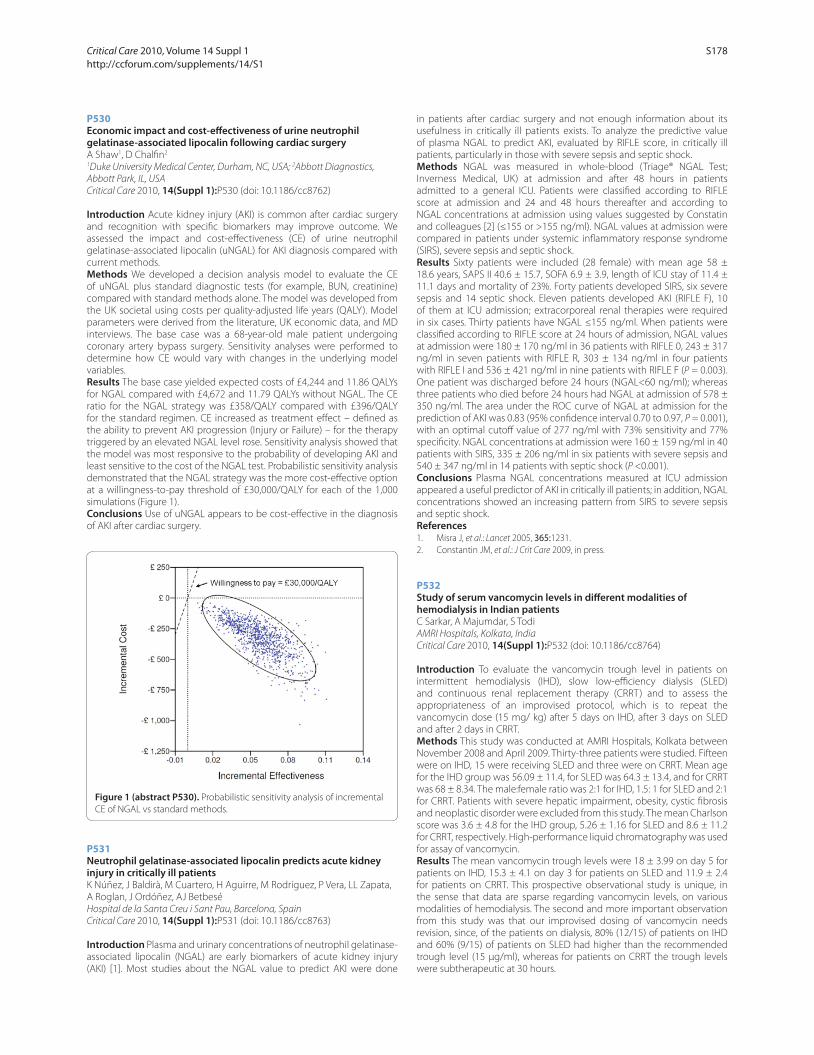
P530
Economic impact and cost-eff ectiveness of urine neutrophil gelatinase-associated lipocalin following cardiac surgeryA Shaw1, D Chalfi n2
1Duke University Medical Center, Durham, NC, USA; 2Abbott Diagnostics,
Abbott Park, IL, USA
Critical Care 2010, 14(Suppl 1):P530 (doi: 10.1186/cc8762)
Introduction Acute kidney injury (AKI) is common after cardiac surgery
and recognition with specifi c biomarkers may improve outcome. We
assessed the impact and cost-eff ectiveness (CE) of urine neutrophil
gelatinase-associated lipocalin (uNGAL) for AKI diagnosis compared with
current methods.
Methods We developed a decision analysis model to evaluate the CE
of uNGAL plus standard diagnostic tests (for example, BUN, creatinine)
compared with standard methods alone. The model was developed from
the UK societal using costs per quality-adjusted life years (QALY). Model
parameters were derived from the literature, UK economic data, and MD
interviews. The base case was a 68-year-old male patient undergoing
coronary artery bypass surgery. Sensitivity analyses were performed to
determine how CE would vary with changes in the underlying model
variables.
Results The base case yielded expected costs of £4,244 and 11.86 QALYs
for NGAL compared with £4,672 and 11.79 QALYs without NGAL. The CE
ratio for the NGAL strategy was £358/QALY compared with £396/QALY
for the standard regimen. CE increased as treatment eff ect – defi ned as
the ability to prevent AKI progression (Injury or Failure) – for the therapy
triggered by an elevated NGAL level rose. Sensitivity analysis showed that
the model was most responsive to the probability of developing AKI and
least sensitive to the cost of the NGAL test. Probabilistic sensitivity analysis
demonstrated that the NGAL strategy was the more cost-eff ective option
at a willingness-to-pay threshold of £30,000/QALY for each of the 1,000
simulations (Figure 1).
Conclusions Use of uNGAL appears to be cost-eff ective in the diagnosis
of AKI after cardiac surgery.
P531
Neutrophil gelatinase-associated lipocalin predicts acute kidney injury in critically ill patientsK Núñez, J Baldirà, M Cuartero, H Aguirre, M Rodríguez, P Vera, LL Zapata,
A Roglan, J Ordóñez, AJ Betbesé
Hospital de la Santa Creu i Sant Pau, Barcelona, Spain
Critical Care 2010, 14(Suppl 1):P531 (doi: 10.1186/cc8763)
Introduction Plasma and urinary concentrations of neutrophil gelatinase-
associated lipocalin (NGAL) are early biomarkers of acute kidney injury
(AKI) [1]. Most studies about the NGAL value to predict AKI were done
in patients after cardiac surgery and not enough information about its
usefulness in critically ill patients exists. To analyze the predictive value
of plasma NGAL to predict AKI, evaluated by RIFLE score, in critically ill
patients, particularly in those with severe sepsis and septic shock.
Methods NGAL was measured in whole-blood (Triage® NGAL Test;
Inverness Medical, UK) at admission and after 48 hours in patients
admitted to a general ICU. Patients were classifi ed according to RIFLE
score at admission and 24 and 48 hours thereafter and according to
NGAL concentrations at admission using values suggested by Constatin
and colleagues [2] (≤155 or >155 ng/ml). NGAL values at admission were
compared in patients under systemic infl ammatory response syndrome
(SIRS), severe sepsis and septic shock.
Results Sixty patients were included (28 female) with mean age 58 ±
18.6 years, SAPS II 40.6 ± 15.7, SOFA 6.9 ± 3.9, length of ICU stay of 11.4 ±
11.1 days and mortality of 23%. Forty patients developed SIRS, six severe
sepsis and 14 septic shock. Eleven patients developed AKI (RIFLE F), 10
of them at ICU admission; extracorporeal renal therapies were required
in six cases. Thirty patients have NGAL ≤155 ng/ml. When patients were
classifi ed according to RIFLE score at 24 hours of admission, NGAL values
at admission were 180 ± 170 ng/ml in 36 patients with RIFLE 0, 243 ± 317
ng/ml in seven patients with RIFLE R, 303 ± 134 ng/ml in four patients
with RIFLE I and 536 ± 421 ng/ml in nine patients with RIFLE F (P = 0.003).
One patient was discharged before 24 hours (NGAL<60 ng/ml); whereas
three patients who died before 24 hours had NGAL at admission of 578 ±
350 ng/ml. The area under the ROC curve of NGAL at admission for the
prediction of AKI was 0.83 (95% confi dence interval 0.70 to 0.97, P = 0.001),
with an optimal cutoff value of 277 ng/ml with 73% sensitivity and 77%
specifi city. NGAL concentrations at admission were 160 ± 159 ng/ml in 40
patients with SIRS, 335 ± 206 ng/ml in six patients with severe sepsis and
540 ± 347 ng/ml in 14 patients with septic shock (P <0.001).
Conclusions Plasma NGAL concentrations measured at ICU admission
appeared a useful predictor of AKI in critically ill patients; in addition, NGAL
concentrations showed an increasing pattern from SIRS to severe sepsis
and septic shock.
References1. Misra J, et al.: Lancet 2005, 365:1231.
2. Constantin JM, et al.: J Crit Care 2009, in press.
P532
Study of serum vancomycin levels in diff erent modalities of hemodialysis in Indian patientsC Sarkar, A Majumdar, S Todi
AMRI Hospitals, Kolkata, India
Critical Care 2010, 14(Suppl 1):P532 (doi: 10.1186/cc8764)
Introduction To evaluate the vancomycin trough level in patients on
inter mittent hemodialysis (IHD), slow low-effi ciency dialysis (SLED)
and continuous renal replacement therapy (CRRT) and to assess the
appropriateness of an improvised protocol, which is to repeat the
vancomycin dose (15 mg/ kg) after 5 days on IHD, after 3 days on SLED
and after 2 days in CRRT.
Methods This study was conducted at AMRI Hospitals, Kolkata between
November 2008 and April 2009. Thirty-three patients were studied. Fifteen
were on IHD, 15 were receiving SLED and three were on CRRT. Mean age
for the IHD group was 56.09 ± 11.4, for SLED was 64.3 ± 13.4, and for CRRT
was 68 ± 8.34. The male:female ratio was 2:1 for IHD, 1.5: 1 for SLED and 2:1
for CRRT. Patients with severe hepatic impairment, obesity, cystic fi brosis
and neoplastic disorder were excluded from this study. The mean Charlson
score was 3.6 ± 4.8 for the IHD group, 5.26 ± 1.16 for SLED and 8.6 ± 11.2
for CRRT, respectively. High-performance liquid chromatography was used
for assay of vancomycin.
Results The mean vancomycin trough levels were 18 ± 3.99 on day 5 for
patients on IHD, 15.3 ± 4.1 on day 3 for patients on SLED and 11.9 ± 2.4
for patients on CRRT. This prospective observational study is unique, in
the sense that data are sparse regarding vancomycin levels, on various
modalities of hemodialysis. The second and more important observation
from this study was that our improvised dosing of vancomycin needs
revision, since, of the patients on dialysis, 80% (12/15) of patients on IHD
and 60% (9/15) of patients on SLED had higher than the recommended
trough level (15 μg/ml), whereas for patients on CRRT the trough levels
were subtherapeutic at 30 hours.
Figure 1 (abstract P530). Probabilistic sensitivity analysis of incremental
CE of NGAL vs standard methods.
Critical Care 2010, Volume 14 Suppl 1 http://ccforum.com/supplements/14/S1
S178

Conclusions This pilot study underscores the need for more awareness,
mandatory checking of vancomycin trough levels and a larger study in
Indian patients to defi nitively determine dosage guidelines in this ethnic
population.
P533
RIFLE criteria for acute kidney dysfunction following liver transplantation: incidence and risk factorsA Kundakci, A Pirat, O Komurcu, A Torgay, H Karakayalı, G Arslan, M Haberal
Baskent University Hospital, Ankara, Turkey
Critical Care 2010, 14(Suppl 1):P533 (doi: 10.1186/cc8765)
Introduction Acute renal dysfunction (ARD) is one of the most common
complications of orthotopic liver transplantation (OLT), with an incidence
ranging from 12 to 64% [1,2]. These patients are often critically ill in the
perioperative period with a number of other co-morbidities, resulting in
an increased susceptibility to ARD. The RIFLE criteria were developed to
provide a consensus defi nition for acute renal dysfunction in critically ill
patients. Using the RIFLE criteria, we aimed to determine the incidence
and risk factors for acute renal dysfunction in the early postoperative
period.
Methods The records of 112 patients who underwent OLT, performed
by the same team in the Baskent University Hospital, from January 2000
to February 2009, were retrospectively analyzed. Three levels of renal
dysfunction, outlined in the RIFLE criteria, were investigated. Collected
data included demographic features; co-morbidities; etiology of the liver
failure; perioperative laboratory values; intraoperative hemodynamic
parameters; use and volume of crystalloids, colloids, blood products,
cell saver system, and albumin; portal vein clamping time; requirement
for inotropes, vasopressors, and antihypertensive drugs; duration of
anesthesia; and urine output.
Results ARD occurred in 64 of OLTs (57%). Risk, injury, and failure
frequencies were 19%, 11%, and 28%, respectively. Compared with those
who did not have ARD postoperatively, those who did had signifi cantly
higher MELD scores (19 ± 7 vs 16 ± 8; P = 0.018), more frequently required
inotropic agents intraoperatively (54% vs 35%; P = 0.070), received more
colloids (300 ± 433 ml vs 105 ± 203 ml; P = 0.007), had a longer portal vein
clamping time (88.0 ± 42.0 minutes vs 73.0 ± 20.0 minutes; P = 0.037),
and had a higher incidence of intraoperative acidosis (64% vs 44%; P =
0.047). Logistic regression analysis was performed using these data to fi nd
the predictors for development of ARD. ARD was found to be associated
with MELD score (odds ratio 1.107, 95% CI 1.022 to 1.200, P = 0.013), portal
vein clamping time (odds ratio 1.020 95% CI 1.000 to 1.040, P = 0.053) and
intraoperative acidosis (odds ratio 3.610, 95% CI 1.212 to 10.753 P = 0.021).
Conclusions Based on RIFLE criteria, more than one-half of the patients
who underwent liver transplantation developed ARD postoperatively. A
higher MELD score, longer portal vein clamping time, and occurrence of
intraoperative acidosis were identifi ed as the independent risk factors for
development of ARD in this group of patients.
P534
Prophylactic use of laxative for constipation in critically ill patientsY Masri, J Abu Baker, R Ahmed
Rashid Hospital Trauma Center, Dubai, United Arab Emirates
Critical Care 2010, 14(Suppl 1):P534 (doi: 10.1186/cc8766)
Introduction The aim of this study was to evaluate the use of laxative
prophylaxis for constipation in the ICU and the impact of early versus late
bowel movement on patient outcome.
Methods After informed consent, all adults in the ICU who were expected
to stay on a ventilator for >72 hours were randomized to Control and
Interventional groups. Patients who had major bowel surgery were
excluded. The Control group did not receive any intervention for bowel
movement for the fi rst 72 hours, while the Interventional group received
a prophylactic dose of Lactulose 20 cc orally every 12 hours for the fi rst
72 hours. After 72 hours the management of constipation in the both
groups was left to the physician’s discretion. Data were collected for
age, sex, diagnosis, Simplifi ed Acute Physiology Score (SAPS) II, doses of
narcotics and sedatives, enteral nutrition, ventilator days, ICU days, bowel
movement <72 hours and days of fi rst bowel movement.
Results Over a 6-month period a total of 100 patients were enrolled, 50
patients in each Control and Interventional group. Mean age was 38.8
years in Control and 37.5 years in Interventional group. Both groups had
a predominance of male patients. The most common diagnosis in both
groups was polytrauma. Mean SAPS II for both was 35. One hundred
percent of patients were ventilated, with a mean dose of fentanyl (323.8 ±
108.89 μg/hour in Control and 345.83 ± 94.43 μg/hour in Interventional
group) and a mean dose of midazolam (11.1 ± 4.04 mg/hour in Control
and 12.4 ± 3.19 mg/hour in Interventional group). Enteral nutrition was
maintained and tolerated by 100% of patients. There were only two (4%)
patients in the Control group and nine (18%) patients in the Interventional
group who had bowel movement in <72 hours (P <0.05). Twenty-three
(46%) patients in the Control and 29 (58%) patients in the Interventional
group had bowel movement after 5 days. Mean ventilator days were
16.19 and 17.36 days in the Control and Interventional groups. Subgroup
analysis showed that the patients who moved their bowel in <5 days in
both groups had mean ventilator days of 18.5, while it was 15.88 days for
the patients who moved their bowel after 5 days in both groups (P <0.05).
Forty-eight (96%) patients in each group were discharged from the ICU.
Mean ICU days for the Control group was 21.15 ± 10.44 and was 20.77 ±
8.33 days for the Interventional group. A total of two (4%) patients died in
each group.
Conclusions Laxative prophylaxis can be used successfully to prevent
constipation in ICU patients. Late bowel movement >5 days is associated
with less ventilator days, compared with early <5 days bowel movement.
P535
Study of intra-abdominal hypertension in the medical intensive care unitP Theerawit
Ramathibodi Hospital, Bangkok, Thailand
Critical Care 2010, 14(Suppl 1):P535 (doi: 10.1186/cc8767)
Introduction Intra-abdominal hypertension (IAH) is associated with
signifi cant mortality in surgical and trauma patients. However, IAH can
occur in medical patients as well. Thus, the main objective of this study
was to assess whether the IAH during the fi rst 24-hour period of admission
was an independent predictor for 28-day mortality.
Methods We conducted a prospective observational study. All patients
admitted to the medical ICU were enrolled in this study and underwent
intra-abdominal pressure (IAP) measurement via the bladder. The primary
outcome was the association between IAH and 28-day mortality rate.
Results Seventy-seven eligible patients were enrolled to this study. The
incidence of IAH was 44%. The mean age was 60.52 ± 18.34 years. In terms
of 28-day mortality, there was no association between the presence of IAH
and mortality outcome. However, regarding ICU mortality, nonsurvivors
had signifi cantly higher mean IAP within 24 hours of admission than
survivors: 11.55 (8.0 to 20.3) vs 9.95 (3.0 to 27.0) (P = 0.041).
Conclusions Neither the mean IAP nor the presence of IAH within the fi rst
24 hours of admission was an independent predictor for 28-day mortality.
Nonetheless, this study demonstrated the diff erence of IAP between
survivors and nonsurvivors in terms of ICU mortality in the medical ICU.
References1. Malbrain ML, Cheatham ML, Kirkpatrick A, et al.: Results from the
International Conference of Experts on Intra-abdominal Hypertension and Abdominal Compartment Syndrome. I. Defi nitions. Intensive Care Med 2006,
32:1722-1732.
2. Malbrain ML, Chiumello D, Pelosi P, et al.: Incidence and prognosis of intraabdominal hypertension in a mixed population of critically ill patients: a multiple-center epidemiological study. Crit Care Med 2005,
33:315-322.
3. Malbrain ML, Chiumello D, Pelosi P, et al.: Prevalence of intra-abdominal hypertension in critically ill patients: a multicentre epidemiological study. Intensive Care Med 2004, 30: 822-829.
4. Malbrain ML: Abdominal pressure in the critically ill: measurement and clinical relevance. Intensive Care Med 1999, 25:1453-1458.
5. Gudmundsson FF, Viste A, Gislason H, Svanes K: Comparison of diff erent methods for measuring intra-abdominal pressure. Intensive Care Med 2002,
28: 509-514.
Critical Care 2010, Volume 14 Suppl 1 http://ccforum.com/supplements/14/S1
S179

P536
Intra-abdominal pressure values of emergency department intensive care unit patients and clinical outcomesO Demirci, S Girisgin, B Cander, E Dogan, D Dündar
Selcuk University Meram Faculty of Medicine, Konya, Turkey
Critical Care 2010, 14(Suppl 1):P536 (doi: 10.1186/cc8768)
Introduction Intra-abdominal hypertension (IAH) and abdominal
compartment syndrome (ACS) have become serious causes of morbidity
and mortality in critical surgical and medical patients, especially in the
past 10 years. Increased IAP levels became one of the routine physiologic
parameter measurement elements in critical patients and indicate
prognosis. In this study, our aim was to observe IAP increase, APB decrease
and their clinical manifestations in ICU patients.
Methods Eighty-nine ICU patients were included in this study. IAP levels
were measured by infusing 25 ml saline into the urinary bladder. The
symphisis pubis plane affi liated as the zero point. Patients were divided
into groups according to IAP and APB levels. Clinical follow-up, required
medical care and survival were investigated.
Results Within the 89 patients, 36 of them diagnosed as IAH and 34 of
them diagnosed as low APB. There were 25 patients that had both IAH and
low APB. In these patients, during the fi rst 2 days of the study, statistically
signifi cant SGOT, SGPT, PO2, urea and creatinine levels were found. We also
determined increased positive inotropic support and ventilatory support
need with increased sepsis and multiple organ failure incidence in these
patients. Mortality rates were strongly related to IAH and low APB levels.
Conclusions In critically ill patients, IAP measurement, a very simple and
valuable method, must be performed. IAH and low APB levels are indicators
of high morbidity and mortality rates. Therefore, IAP measurement may
become a routine element in follow-ups and survival rate determinations,
in critically ill patients.
References1. Cheatham M, Malbrain M: Cardiovascular implications of abdominal
compartment syndrome. Acta Clin Belg Suppl 2007, 1:98-112.
2. Cheatham M, Malbrain M: Intra-abdominal hypetension and the cardiovascular system. In Abdominal Compartment Syndrome. Edited by
Ivatury R, Cheatham M, Malbrain M, Sugrue M. Georgetown, TX: Landes
Biosciences; 2006:89-104.
3. Malbrain ML, De laet I: Functional haemodynamics during intraabdominal hypertension: what to use and what not use. Acta Anaesthesiol Scand 2008,
52:576-577.
P537
Advanced haemodynamic assessment of patients with liver disease and abdominal compartment syndromeD Joshi, S Saha, W Bernal, J Wendon, G Auzinger
King’s College Hospital, London, UK
Critical Care 2010, 14(Suppl 1):P537 (doi: 10.1186/cc8769)
Introduction Intra-abdominal hypertension (IAH, >12 mmHg) and
abdominal compartment syndrome (ACS, >20 mmHg) frequently compli-
cate the disease course in critically ill patients with liver disease. There is a
paucity of data describing the circulatory changes observed during ACS
outside experimental studies. The aim of this study was to investigate
baseline haemodynamic variables in patients with ACS and their response
following abdominal decompression.
Methods Patients admitted to the liver ICU of King’s College Hospital with ACS
were studied. Transpulmonary thermodilution cardiac output monitoring and
calculation of volumetric indices of preload was performed with the PiCCO
system. MAP, CVP, ITBVI, cardiac index (CI), stroke volume index (SVI) and intra-
abdominal pressure (IAP) were analysed pre and post intervention.
Results Eighteen patients (nine female), median age 43 (range (R) 20
to 65) were studied. Ten patients had decompensated Budd–Chiari
syndrome (BCS), eight underwent orthotopic liver transplantation and
two had surgical shunt procedures. Eight patients had cirrhosis (PC) with
organ failure and severe ascites necessitating therapeutic paracentesis.
Pre-intervention IAP was raised (median 24, R 17 to 40). As a whole,
volumetric markers of pre-load were low (median ITBVI 674, R 453 to 1,619)
but improved (median ITBVI 763, R 512 to 1,110) post intervention (P =
0.02). Reduction in CVP post intervention was not statistically signifi cant
(16, R 0 to 25 vs 14, R 5 to 29). Compared with the PC group, the BCS
group had persistently low ITBVI (median 633, R 453 to 924) despite
aggressive volume resuscitation prior to decompression (median positive
fl uid balance 10 l, R 0.5 to 39). Abdominal decompression resulted in a
reduction in IAP (median 15, R 6 to 31, P <0.0001), increase in preload
(ITBVI) and improvement in CI (3.9, R 1.9 to 7.4 vs 4.6, R 2.7 to 7.7, P <0.0001)
and SVI (40, R 22 to 111 vs 44, 30 to 61, P = 0.001). There was an inverse
relationship between IAP, CI (P = 0.003) and SVI (P = 0.004). ITBVI correlated
with parameters of fl ow (CI P <0.0001 and SVI P <0.0001), CVP did not.
Conclusions ACS leads to central hypovolaemia. Abdominal decom-
pression restores preload in association with improvement in cardiac output.
References1. Cheatham et al.: Intensive Care Med 2007, 33:951-962.
2. Malbrain et al.: Intensive Care Med 2006, 32:1722-1732.
P538
Molecular adsorbent recirculating system in hepatic failure patients with a ventricular assist deviceM Cristinar, J Thiranos, H Kremer, C Chalumeau, J Mazzucotelli
Nouvel Hopital Civil, Strasbourg, France
Critical Care 2010, 14(Suppl 1):P538 (doi: 10.1186/cc8770)
Introduction The study we performed focuses on the removal of bilirubin
in hepatic failure patients with a ventricular assist device (VAD). Bilirubin is
a risk factor revealing the severe hepatic insuffi ciency and the alteration
of the clinical status. The mortality rate of patients developing liver failure
in the cardiac ICU is very high. The molecular adsorbent recirculating
system (MARS) is the hepatic failure treatment in patients with no other
therapeutic options. It is a liver support system that uses an albumin-
enriched dialysate to facilitate the removal of albumin-bound toxins.
Conventional hemodialysis techniques have little or no eff ect on liver
detoxication and cannot improve these patients’ prognosis. The study
aims to prove the decrease of the mortality rate in hepatic failure patients
with VAD as a result of using MARS.
Methods We conducted a retrospective study of 55 patients with VAD.
They were hospitalized in our cardiac ICU between January 2000 and
November 2009. All patients had a hepatic check-up when admitted to
the ICU. The patients with altered liver function had at least one check-
up per day. We had seven patients with hyperbilirubinaemia >350 μmol/l
(the threshold for starting hepatic dialysis). Five patients received MARS
therapy. Hepatic check-ups were performed at the beginning and at the
end of the session.
Results Five patients were dialysed. Patient 1 – with a maximum serum
bilirubin level of 350 μmol/l, needed four cycles of MARS therapy; the
patient survived. Patient 2 – max bilirubin 390 μmol/l, 6 MARS; the patient
survived. Patient 3 – max bilirubin 700 μmol/l, 3 MARS; the patient did not
survive. Patient 4 – max bilirubin 680 μmol/l, 3 MARS; the patient survived.
Patient 5 – max bilirubin 500 μmol/l, 15 MARS; the patient did not survive.
The two patients that did not receive MARS therapy did not survive.
Conclusions MARS is a viable option for the treatment of hepatic failure
patients and, in particular, for those with VAD. We consider that without
MARS a VAD patient cannot survive if his serum bilirubin level exceeds
350 μmol/l. Our hypothesis needs to be confi rmed by multicenter
randomized trials.
References1. Saliba F, et al.: The Molecular Adsorbent Recirculating System (MARS) in the
intensive care unit: a rescue therapy for patients with hepatic failure. Crit
Care 2006, 10:118.
2. El-Banayosy A, et al.: Evaluation of MARS technique on patients suff ering from hypoxic liver failure following cardiogenic shock. ASAIO J 2002, 48:139.
P539
Molecular adsorbent recirculating system in patients with acute liver failure: prospective follow-upD Tran-Zweifel, B Goubaux, L Bornard, J Dellamonica, M Carles, O Perus,
M Raucoules
Hopital de l’Archet II – CHU de Nice, France
Critical Care 2010, 14(Suppl 1):P539 (doi: 10.1186/cc8771)
Introduction A prospective observational study on molecular adsorbent
recirculating system (MARS) therapy in a liver-disease-dedicated ICU.
Critical Care 2010, Volume 14 Suppl 1 http://ccforum.com/supplements/14/S1
S180
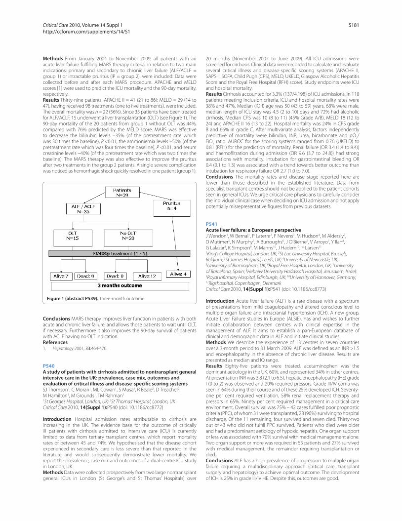
Methods From January 2004 to November 2009, all patients with an
acute liver failure fulfi lling MARS therapy criteria, in relation to two main
indications: primary and secondary to chronic liver failure (ALF/ACLF =
group 1) or intractable pruritus (IP = group 2), were included: Data were
collected before and after each MARS procedure. APACHE and MELD
scores [1] were used to predict the ICU mortality and the 90-day mortality,
respectively.
Results Thirty-nine patients, APACHE II = 41 (21 to 86); MELD = 29 (14 to
47), having received 98 treatments (one to fi ve treatments), were included.
The overall mortality was n = 22 (56%). Since 35 patients have been treated
for ALF/ACLF, 15 underwent a liver transplantation (OLT) (see Figure 1). The
90-day mortality of the 20 patients from group 1 without OLT was 44%,
compared with 76% predicted by the MELD score. MARS was eff ective
to decrease the bilirubin levels –35% (of the pretreatment rate which
was 30 times the baseline), P <0.01, the ammoniemia levels –50% (of the
pretreatment rate which was four times the baseline), P <0.01, and serum
creatinine levels –40% (of the pretreatment rate which was two times the
baseline). The MARS therapy was also eff ective to improve the pruritus
after two treatments in the group 2 patients. A single severe complication
was noticed as hemorrhagic shock quickly resolved in one patient (group 1).
Conclusions MARS therapy improves liver function in patients with both
acute and chronic liver failure, and allows those patients to wait until OLT,
if necessary. Furthermore it also improves the 90-day survival of patients
with ACLF having no OLT indication.
References1. Hepatology 2001, 33:464-470.
P540
A study of patients with cirrhosis admitted to nontransplant general intensive care in the UK: prevalence, case mix, outcomes and evaluation of critical illness and disease-specifi c scoring systemsSJ Thomson1, C Moran1, ML Cowan1, S Musa1, R Beale2, D Treacher2,
M Hamilton1, M Grounds1, TM Rahman1
1St George’s Hospital, London, UK; 2St Thomas’ Hospital, London, UK
Critical Care 2010, 14(Suppl 1):P540 (doi: 10.1186/cc8772)
Introduction Hospital admission rates attributable to cirrhosis are
increasing in the UK. The evidence base for the outcome of critically
ill patients with cirrhosis admitted to intensive care (ICU) is currently
limited to data from tertiary transplant centres, which report mortality
rates of between 45 and 74%. We hypothesised that the disease cohort
experienced in secondary care is less severe than that reported in the
literature and would subsequently demonstrate lower mortality. We
report the prevalence, case mix and outcomes of a dual-centre ICU study
in London, UK.
Methods Data were collected prospectively from two large nontransplant
general ICUs in London (St George’s and St Thomas’ Hospitals) over
20 months (November 2007 to June 2009). All ICU admissions were
screened for cirrhosis. Clinical data were recorded to calculate and evaluate
several critical illness and disease-specifi c scoring systems (APACHE II,
SAPS II, SOFA, Child Pugh (CPS), MELD, UKELD, Glasgow Alcoholic Hepatitis
Score and the Royal Free Hospital (RFH) score). Study endpoints were ICU
and hospital mortality.
Results Cirrhosis accounted for 3.3% (137/4,198) of ICU admissions. In 118
patients meeting inclusion criteria, ICU and hospital mortality rates were
38% and 47%. Median (IQR) age was 50 (43 to 59) years, 68% were male,
median length of ICU stay was 4.5 (2 to 10) days and 72% had alcoholic
cirrhosis. Median CPS was 10 (8 to 11) (45% Grade A/B), MELD 18 (12 to
24) and APACHE II 16 (13 to 22). Hospital mortality was 24% in CPS grade
B and 66% in grade C. After multivariate analysis, factors independently
predictive of mortality were bilirubin, INR, urea, bicarbonate and pO2/
FiO2 ratio. AUROC for the scoring systems ranged from 0.76 (UKELD) to
0.81 (RFH) for the prediction of mortality. Renal failure (OR 3.4 (1.4 to 8.4))
and haemofi ltration during admission (OR 9.6 (3.7 to 24.8)) had strong
associations with mortality. Intubation for gastrointestinal bleeding OR
0.4 (0.1 to 1.3) was associated with a trend towards better outcome than
intubation for respiratory failure OR 2.7 (1.0 to 7.0).
Conclusions The mortality rates and disease stage reported here are
lower than those described in the established literature. Data from
specialist transplant centres should not be applied to the patient cohorts
seen in general ICUs. We urge critical care physicians to carefully consider
the individual clinical case when deciding on ICU admission and not apply
potentially misrepresentative fi gures from previous datasets.
P541
Acute liver failure: a European perspectiveJ Wendon1, W Bernal1, P Laterre2, F Nevens3, M Hudson4, M Aldersly3,
D Mutimer5, N Murphy5, A Burroughs6, J O’Bierne6, V Arroyo7, Y Ilan8,
G Lalazar8, K Simpson9, M Manns10, J Hadem10, F Larsen11
1King’s College Hospital, London, UK; 2St Luc University Hospital, Brussels,
Belgium; 3St James Hospital, Leeds, UK; 4University of Newcastle, UK; 5University of Birmingham, UK; 6Royal Free Hospital, London, UK; 7University
of Barcelona, Spain; 8Hebrew University Hadassah Hospital, Jerusalem, Israel; 9Royal Infi rmary Hospital, Edinburgh, UK; 10University of Hannover, Germany; 11Rigshospital, Copenhagen, Denmark
Critical Care 2010, 14(Suppl 1):P541 (doi: 10.1186/cc8773)
Introduction Acute liver failure (ALF) is a rare disease with a spectrum
of presentations from mild coagulopathy and altered conscious level to
multiple organ failure and intracranial hypertension (ICH). A new group,
Acute Liver Failure studies in Europe (ALSiE), has and wishes to further
initiate collaboration between centres with clinical expertise in the
management of ALF. It aims to establish a pan-European database of
clinical and demographic data in ALF and initiate clinical studies.
Methods We describe the experience of 13 centres in seven countries
over a 3-month period to 31 March 2009. ALF was defi ned as an INR >1.5
and encephalopathy in the absence of chronic liver disease. Results are
presented as median and IQ range.
Results Eighty-fi ve patients were treated, acetaminophen was the
dominant aetiology in the UK, 60%, and represented 34% in other centres.
At presentation INR was 3.8 (2.1 to 6.5), hepatic encephalopathy (HE) grade
I (0 to 2) was observed and 20% required pressors. Grade III/IV coma was
seen in 64% during their course and of these 25% developed ICH. Seventy-
one per cent required ventilation, 58% renal replacement therapy and
pressors in 65%. Ninety per cent required management in a critical care
environment. Overall survival was 75% – 42 cases fulfi lled poor prognostic
criteria (PPC), of whom 31 were transplanted, 28 (90%) surviving to hospital
discharge. Of the 11 remaining, four survived and seven died. Thirty-two
out of 43 who did not fulfi ll PPC survived. Patients who died were older
and had a predominant aetiology of hypoxic hepatitis. One organ support
or less was associated with 70% survival with medical management alone.
Two organ support or more was required in 55 patients and 27% survived
with medical management, the remainder requiring transplantation or
died.
Conclusions ALF has a high prevalence of progression to multiple organ
failure requiring a multidisciplinary approach (critical care, transplant
surgery and hepatology) to achieve optimal outcome. The development
of ICH is 25% in grade III/IV HE. Despite this, outcomes are good.
Figure 1 (abstract P539). Three-month outcome.
Critical Care 2010, Volume 14 Suppl 1 http://ccforum.com/supplements/14/S1
S181

P542
Intracranial pressure monitoring in acute liver failure: a review of 10 years experienceM Peck, J Wendon, E Sizer, G Auzinger, W Bernal
King’s College Hospital, London, UK
Critical Care 2010, 14(Suppl 1):P542 (doi: 10.1186/cc8774)
Introduction Intracranial hypertension (ICH) complicates around 25% of
grade III/IV encephalopathy in acute liver failure. Intracranial pressure (ICP)
monitoring is controversial in this coagulopathic population.
Methods We examined the ITU charts and prospectively maintained
an ITU database of all patients admitted with acute liver failure who
underwent ICP monitoring from December 1998 to December 2008. Data
are presented as median and range.
Results One hundred and fi fteen patients were identifi ed. Aetiology was
acetaminophen (78), seronegative (14), drug induced (7), viral (14) and
autoimmune (two). All patients had a Camino ICP bolt (subdural) inserted
by the intensive care specialist. Coagulation support fresh frozen plasma
(10 ml/kg) and platelets if the count was <100 were given. Recombinant
factors were not given. Overall 66 survived and 49 died. Seventy patients
underwent transplantation with 77% survival. Age was 31 (24 to 42),
International Normalised Ratio (INR) 5.6 (3.9 to 9.2), creatinine 150 μmol/l
(105 to 143), Na 139 (135 to 143). INR pre insertion of the bolt was 3.5 (2.2
to 7.3), platelets 109 (51 to 137). At bolt insertion, pulse was 100 (89 to
111), MAP 80 (72 to 89), ICP 19.5 (12 to 25), CO2 4.8 (4.3 to 5.5), lactate 3.5
(2.2 to 5.3), jugular venous saturation 76 (67 to 84), temperature 35.5 (34.8
to 36.4). Norepinephrine dose was 0.12 μg/kg/minute (0.02 to 0.4). ICP fell
to 14 (9 to 19) at 6 hours and 13 (8 to 20) at 24 hours. Peak ICP was 30 (20
to 39), number of surges >20 per patient was 2 (0 to 7). Minimum cerebral
perfusion pressure (CPP) was 45 (38 to 52). The pre-liver-transplant peak
ICP was 27 (19 to 35), post was 23 (20 to 33). There were no correlates
between clinical parameters, ICP or CPP at 0, 6, 12 or 24 hours. No diff erence
in insertion ICP and CPP was found between groups. Sixteen patients had
postmortem examination. Cerebral oedema was noted in seven, and in
two there was evidence of an intracerebral bleed, fronto-parietal region
(bolt site). Fifteen underwent CT scanning – of whom three had evidence
of diff use ischemia, oedema and tentorial herniation. One case had a small
fronto-parietal bleed (survived). Mode of death was cerebral in 14 cases.
In those who had a CPP <45 46% survived, whilst in those with a CPP >45
66% survived.
Conclusions ICP monitoring is a relatively safe procedure in this high-risk
cohort of patients. Three ICP bolt-related complications were observed,
two died and one survived. In those who survived there were no clinically
apparent neurological sequelae. Mortality is increased in those with CPP
<45 but is not inevitable and should not preclude transplantation and/or
aggressive medical management.
P543
Microdialysis catheters in liver transplants detect vascular complications and rejectionHH Haugaa, TI Tønnessen
Oslo University Hospital, Oslo, Norway
Critical Care 2010, 14(Suppl 1):P543 (doi: 10.1186/cc8775)
Introduction Following liver transplantation, hepatic artery stenosis
and portal vein obstruction occur in 3 to 12% of the patients, and more
frequently in children than adults. Today, the standard of care is Doppler
ultrasound and liver enzyme assessment daily. Accordingly, detection of
severe hypoperfusion may be delayed. The aim of the present study was
to explore whether microdialysis catheters implanted in the left and right
liver lobe, by measurement of glucose, lactate, pyruvate and glycerol every
2 hours, detected vascular complications and rejection.
Methods Seventy-three patients undergoing 82 liver transplantations were
included. Nine of the patients were children. Two microdialysis catheters
were inserted in the liver and one in subcutaneous tissue by a split needle
technique. They were kept for as long as the catheters functioned, and
maximally 4 weeks. Metabolic parameters (glucose, pyruvate, glycerol and
lactate) were collected every 2 hours and measured bedside.
Results Median age of the patients was 52 years (6 months to 70 years).
The median time for catheters inserted in the liver was 9.5 days, with a
range from 0.5 to 26 days. Six patients had hepatic artery stenosis/
occlusion, and in fi ve of them lactate increased to values >10 mM with
a lactate/pyruvate ratio of several hundred and a concomitant decrease
in glucose and increase in glycerol. In one patient (a 6-month-old child)
lactate only increased to 2 mM and the LP ratio to 20. On the background
of pathological metabolic values, all patients underwent immediate
reoperation and blood clots were removed and the artery reanastomosed.
In the child with low lactate values, the fl ow of the artery was less than 10%
of total hepatic blood fl ow, despite reanastomosis. Thus, the exceptionally
high portal fl ow delivered enough oxygen to prevent major ischemia
despite very low fl ow in the artery.
Thirteen patients had rejection verifi ed by biopsy, and in six patients
anti-rejection therapy was given based on liver function tests and clinical
judgement. All patients had some increase in lactate during the period of
rejection, but the increase was in all but four patients only 1 mM. In the
remaining four the increase was 3 to 4 mM.
Conclusions By using microdialysis catheters measuring metabolic
parameters, hepatic artery occlusions can be detected very rapidly.
Patients with rejection show a small, but signifi cant, increase in lactate.
P544
Drug-induced hepatic cholestasis in the ICU: a case–control studyD Williamson1, M Bédard Dufresne2, V Bonhomme3, M Albert1, C Verdant1,
AJ Frenette1
1Hôpital du Sacré-Coeur de Montréal, Canada; 2Centre Hospitalier Honore-
Mercier, St-Hyacinthe, Canada; 3Hopital Maisonneuve-Rosemont, Montreal,
Canada
Critical Care 2010, 14(Suppl 1):P544 (doi: 10.1186/cc8776)
Introduction Liver dysfunction in critically ill patients represents a
major concern. Many drugs used in the ICU have been associated
with hepatotoxicity. Hepatotoxicity presents in three distinct patterns:
cholestatic (alkaline phosphatase (AP) ≥2 x ULN and ratio (ALT/ULN)/(AP/
ULN) ≤2), hepatocellular (ALT ≥3 x ULN and ratio ≥5) and mixed (AP ≥2 x
ULN, ALT ≥3 x ULN and ratio between 2 and 5). No published studies have
assessed the drug-induced cholestastic pattern of hepatotoxicity in the
ICU. The aim of this study was to assess whether use of pharmacological
classes previously associated with cholestasis are associated with an
increased risk of pure or mixed cholestasis in the ICU.
Methods A nested case–control study assessed the potential association
between use of specifi ed pharmacological classes and cholestasis. Cases
were identifi ed from a cohort of patients admitted ≥24 hours in whom at
least one value of AP <240 IU/l had been obtained in the 72 hours following
admission. We excluded patients with an identifi ed cause of cholestasis as
well as patients with bone metastasis and pregnant women. Each case the
subject was matched to a control subject based on age, gender, and length
of ICU stay and admission year. Exposure to antiepileptics, penicillins,
cephalosporins, carbapenems, macrolid antibiotics and parenteral
nutrition was collected and included in a multivariate conditional logistic
regression analysis with known risk factors.
Results A total of 113 patients developed cholestasis between May 2001
and March 2009, of which 95 had no identifi ed cause. We matched 95
cases with 95 controls and controlled for APACHE II score, sepsis, obesity,
diabetes, length of stay and prior history of cholestasis. In multivariate
logistic regression, parenteral nutrition (OR 6.26), sepsis (OR 4.05), and
penicillins (OR 3.97) were independently associated with cholestasis.
Conclusions Cholestasis in the ICU often remains of unknown origin. In
our study, sepsis, parenteral nutrition and penicillins were independently
associated with the development of cholestasis in patients admitted to
the ICU.
P545
Diagnosis of acute cholecystitis in septic patientsA Ioakimidou, M Moykas, A Theodotou, A Amygdalou, C Catsenos, K
Mandragos
Korgialenio-Benakio Red Cross Hospital, Athens, Greece
Critical Care 2010, 14(Suppl 1):P545 (doi: 10.1186/cc8777)
Introduction The aim of this retrospective observational study was to
evaluate the usefulness of clinical, laboratory and imaging fi ndings in the
diagnosis of acute cholecystitis (AC) in septic patients.
Critical Care 2010, Volume 14 Suppl 1 http://ccforum.com/supplements/14/S1
S182

Methods Twelve out of 1,248 patients were included in the study. The
patients had no intra-abdominal pathology on admission. Mean age
was 59.08 (16.14) years. The most common ICU admission diagnosis
was trauma (7/12). On admission, mean APACHIE II score was 17.916
(5.74) and SOFA score was 8.083 (3.369). On the day of diagnosis, SOFA
score was 13 (4.03). Mean length of stay in the ICU before the diagnosis
was 32.35 (13.76) days. Diagnosis was based on clinical, laboratory, U/S
and C/T criteria. Confi rmation of the diagnosis was performed with an
operative procedure: open cholecystectomy (n = 8), laparoscopy (n = 2),
percutaneous cholecystostomy (n = 2).
Results Twelve of 1,248 patients (0.96%) developed clinical, laboratory
and imaging fi ndings of AC. The AC was confi rmed by an operative
procedure in eight out of 12 patients (0.64%), one calculus and seven
acalculus. The mortality was 62.5%. Necrotic areas of the gallbladder wall
existed in three patients. Clinical diagnostic fi ndings were: tenderness in
the right upper quadrant of the abdomen and positive Murphy in 16.66%;
temperatures over 38.5°C in 50%; leucocytosis in 58.33%; metabolic
acidosis in 50%; elevation of ALT in 50% and AST in 66.66%; bilirubin >1.17
mg/dl in 91.66%; and an increase of alkaline phosphatase in 100% of
patients with confi rmed AC. There was one false negative 8% and three
false positive 75% and one true negative 25% ultrasonography. The most
common U/S fi nding was gallbladder distention (n = 8) 66.66% and
sludge (n = 8) 66.66%. Respectively, there were two false negative 25%
and two false positive 50% C/T scans. Two patients underwent diagnostics
laparoscopies that were true negative.
Conclusions There is not a specifi c diagnostic test for AC in trauma septic
patients. Laparoscopy at the bedside in the ICU is an accurate and safe
diagnostic tool.
Reference1. Laurila et al.: Acta Anaesthesiol Scand 2006, 50:173-179.
P546
Bleeding and pneumonia in intensive care unit patients given PPIs and H2RAs for prevention of stress ulcer: a meta analysisJ Zhou, X Wan, L Han
First Affi liated Hospital of Dalian Medical University, Dalian, China
Critical Care 2010, 14(Suppl 1):P546 (doi: 10.1186/cc8778)
Introduction To assess the effi cacy and safety of proton pump inhibitor
and histamine-2 receptor antagonist in ICU patients for stress ulcer
prophylaxis.
Methods Trials were identifi ed by performing a systematic search
of MEDLINE, Ebsco and CNKI. Two reviewers assessed the quality of
studies and extracted data independently. Disagreement was resolved
by discussion. Revman 4.2.2 software developed by the Cochrane
collaboration was used for meta-analysis.
Results Four trials involving 771 patients were included. Meta-analyses
showed that the incidence of clinically important bleeding was
signifi cantly lower in the PPI group as compared with H2RA (OR 0.45, 95%
CI (0.21, 0.9), P = 0.04). The incidence of nosocomial pneumonia (OR 1.03,
95% CI (0.63, 1.70), P = 0.89) and mortality (OR 1.17, 95% CI (0.76, 1.80), P =
0.47) had no signifi cant diff erence.
Conclusions In comparison with H2RA, PPI is more eff ective in the
prevention of stress ulcer bleeding (SUB) in patients in intensive care. There
is no signifi cant diff erence in the incidence of nosocomial pneumonia and
mortality between the two groups. Because the randomized controlled
trials are few and these fi ndings are based on a small number of patients,
fi rm conclusions cannot presently be proposed. More randomized,
multicenter studies with suffi cient sample size should be performed.
Reference1. Stollman N, Metz DC: Pathophysiology and prophylaxis of stress ulcer in
intensive care unit patients. J Crit Care 2005, 20:35-45.
P547
Infl uence of septic shock on energy expenditure in critically ill ICU patients, estimated with indirect calorimetryR Włodarski1, K Kusza2
1Military Clinical Hospital, Bydgoszcz, Poland; 2University of Nicolaus
Copernicus, Collegium Medicum of Ludwik Rydygier, Bydgoszcz, Poland
Critical Care 2010, 14(Suppl 1):P547 (doi: 10.1186/cc8779)
Introduction Energy requirements of critically ill septic shock patients
treated in the ICU are particularly diffi cult to determine. Recent research
shows that energy expenditure (EE) in those patients may be smaller
than it was previously believed. EE values in this group are aff ected by
the severity of the disease process as well as by administered treatment
(sedation, inactivity of skeletal and respiratory muscles, fi ghting fever).
The purpose of this study was to establish the infl uence of septic shock
on EE in sedated and mechanically ventilated patients treated in the ICU.
Using indirect calorimetry, we assessed EE in those patients and compared
results with basal energy expenditure calculated according to the Harris–
Benedict equation (BEE), and with EE values in patients subjected to
general anesthesia.
Methods Two groups of patients were studied with regard to EE
measured by means of indirect calorimetry. Group I consisted of 50
critically ill patients treated in the ICU for septic shock. They were
evaluated using the APACHE II and SOFA scores, taking into account the
28-day mortality. EE was measured continuously over the fi rst 24 hours of
treatment by means of the Datex-Ohmeda M-COVX indirect calorimeter.
Group II comprised 50 patients (ASA I and II), whose energy expenditure
was measured under general anesthesia in surgical treatment of disc
herniation. Energy expenditure measurements were taken with the use of
the Datex-Ohmeda E-CAiOVX indirect calorimeter, adapted to work in an
atmosphere containing anesthetic gases.
Results The mean EE value measured with a calorimeter (CEE) in septic
shock patients (CEE group I) was 1,467 ± 304 kcal/day or 20 ± 4 kcal/kg/
day. Their mean APACHE II score was 30.2, SOFA was 10.8. CEE in group
I amounted to 67 to 137% of BEE (mean 101% of BEE) and to 121% of
average CEE in anesthetized patients (CEE group II). Statistical dependence
between mortality and CEE was demonstrated. CEE in surviving patients
(APACHE: 26.8, SOFA: 8.1) amounted to 91 to 137% of BEE (mean 114%
of BEE) and to 139% of average CEE in group II. CEE in deceased patients
(APACHE: 32.9, SOFA: 13) amounted to 67 to 121% of BEE (mean 94% of
BEE) and to 111% of average CEE in group II.
Conclusions A decrease in the total metabolic rate of patients with
severe sepsis and septic shock can constitute a prognostic indicator of
heightened death risk in this group of ICU-treated patients.Figure 1 (abstract P546). PPIs vs H2RAs: bleeding rate.
Figure 2 (abstract P546). PPIs vs H2RAs: rate of nosocomial pneumonia.
Critical Care 2010, Volume 14 Suppl 1 http://ccforum.com/supplements/14/S1
S183
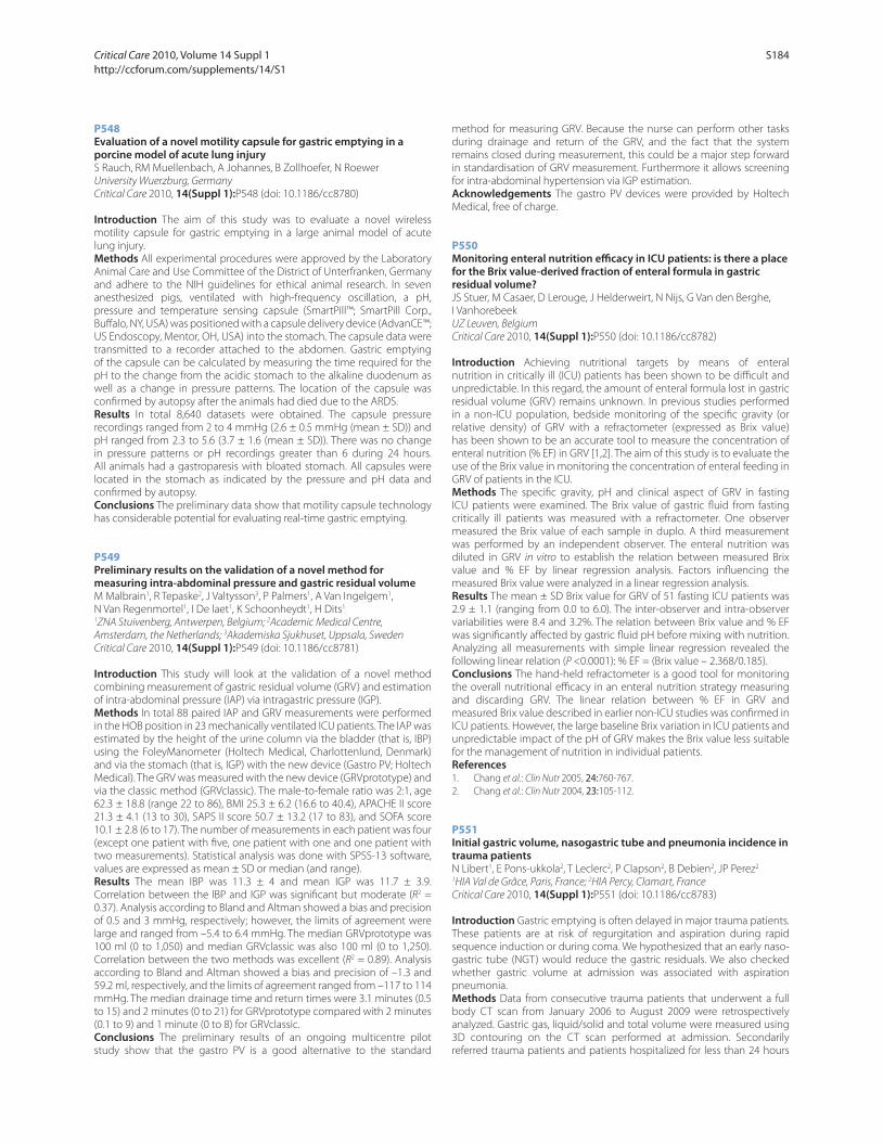
P548
Evaluation of a novel motility capsule for gastric emptying in a porcine model of acute lung injuryS Rauch, RM Muellenbach, A Johannes, B Zollhoefer, N Roewer
University Wuerzburg, Germany
Critical Care 2010, 14(Suppl 1):P548 (doi: 10.1186/cc8780)
Introduction The aim of this study was to evaluate a novel wireless
motility capsule for gastric emptying in a large animal model of acute
lung injury.
Methods All experimental procedures were approved by the Laboratory
Animal Care and Use Committee of the District of Unterfranken, Germany
and adhere to the NIH guidelines for ethical animal research. In seven
anesthesized pigs, ventilated with high-frequency oscillation, a pH,
pressure and temperature sensing capsule (SmartPill™; SmartPill Corp.,
Buff alo, NY, USA) was positioned with a capsule delivery device (AdvanCE™;
US Endoscopy, Mentor, OH, USA) into the stomach. The capsule data were
transmitted to a recorder attached to the abdomen. Gastric emptying
of the capsule can be calculated by measuring the time required for the
pH to the change from the acidic stomach to the alkaline duodenum as
well as a change in pressure patterns. The location of the capsule was
confi rmed by autopsy after the animals had died due to the ARDS.
Results In total 8,640 datasets were obtained. The capsule pressure
recordings ranged from 2 to 4 mmHg (2.6 ± 0.5 mmHg (mean ± SD)) and
pH ranged from 2.3 to 5.6 (3.7 ± 1.6 (mean ± SD)). There was no change
in pressure patterns or pH recordings greater than 6 during 24 hours.
All animals had a gastroparesis with bloated stomach. All capsules were
located in the stomach as indicated by the pressure and pH data and
confi rmed by autopsy.
Conclusions The preliminary data show that motility capsule technology
has considerable potential for evaluating real-time gastric emptying.
P549
Preliminary results on the validation of a novel method for measuring intra-abdominal pressure and gastric residual volumeM Malbrain1, R Tepaske2, J Valtysson3, P Palmers1, A Van Ingelgem1,
N Van Regenmortel1, I De laet1, K Schoonheydt1, H Dits1
1ZNA Stuivenberg, Antwerpen, Belgium; 2Academic Medical Centre,
Amsterdam, the Netherlands; 3Akademiska Sjukhuset, Uppsala, Sweden
Critical Care 2010, 14(Suppl 1):P549 (doi: 10.1186/cc8781)
Introduction This study will look at the validation of a novel method
combining measurement of gastric residual volume (GRV) and estimation
of intra-abdominal pressure (IAP) via intragastric pressure (IGP).
Methods In total 88 paired IAP and GRV measurements were performed
in the HOB position in 23 mechanically ventilated ICU patients. The IAP was
estimated by the height of the urine column via the bladder (that is, IBP)
using the FoleyManometer (Holtech Medical, Charlottenlund, Denmark)
and via the stomach (that is, IGP) with the new device (Gastro PV; Holtech
Medical). The GRV was measured with the new device (GRVprototype) and
via the classic method (GRVclassic). The male-to-female ratio was 2:1, age
62.3 ± 18.8 (range 22 to 86), BMI 25.3 ± 6.2 (16.6 to 40.4), APACHE II score
21.3 ± 4.1 (13 to 30), SAPS II score 50.7 ± 13.2 (17 to 83), and SOFA score
10.1 ± 2.8 (6 to 17). The number of measurements in each patient was four
(except one patient with fi ve, one patient with one and one patient with
two measurements). Statistical analysis was done with SPSS-13 software,
values are expressed as mean ± SD or median (and range).
Results The mean IBP was 11.3 ± 4 and mean IGP was 11.7 ± 3.9.
Correlation between the IBP and IGP was signifi cant but moderate (R2 =
0.37). Analysis according to Bland and Altman showed a bias and precision
of 0.5 and 3 mmHg, respectively; however, the limits of agreement were
large and ranged from –5.4 to 6.4 mmHg. The median GRVprototype was
100 ml (0 to 1,050) and median GRVclassic was also 100 ml (0 to 1,250).
Correlation between the two methods was excellent (R2 = 0.89). Analysis
according to Bland and Altman showed a bias and precision of –1.3 and
59.2 ml, respectively, and the limits of agreement ranged from –117 to 114
mmHg. The median drainage time and return times were 3.1 minutes (0.5
to 15) and 2 minutes (0 to 21) for GRVprototype compared with 2 minutes
(0.1 to 9) and 1 minute (0 to 8) for GRVclassic.
Conclusions The preliminary results of an ongoing multicentre pilot
study show that the gastro PV is a good alternative to the standard
method for measuring GRV. Because the nurse can perform other tasks
during drainage and return of the GRV, and the fact that the system
remains closed during measurement, this could be a major step forward
in standardisation of GRV measurement. Furthermore it allows screening
for intra-abdominal hypertension via IGP estimation.
Acknowledgements The gastro PV devices were provided by Holtech
Medical, free of charge.
P550
Monitoring enteral nutrition effi cacy in ICU patients: is there a place for the Brix value-derived fraction of enteral formula in gastric residual volume?JS Stuer, M Casaer, D Lerouge, J Helderweirt, N Nijs, G Van den Berghe,
I Vanhorebeek
UZ Leuven, Belgium
Critical Care 2010, 14(Suppl 1):P550 (doi: 10.1186/cc8782)
Introduction Achieving nutritional targets by means of enteral
nutrition in critically ill (ICU) patients has been shown to be diffi cult and
unpredictable. In this regard, the amount of enteral formula lost in gastric
residual volume (GRV) remains unknown. In previous studies performed
in a non-ICU population, bedside monitoring of the specifi c gravity (or
relative density) of GRV with a refractometer (expressed as Brix value)
has been shown to be an accurate tool to measure the concentration of
enteral nutrition (% EF) in GRV [1,2]. The aim of this study is to evaluate the
use of the Brix value in monitoring the concentration of enteral feeding in
GRV of patients in the ICU.
Methods The specifi c gravity, pH and clinical aspect of GRV in fasting
ICU patients were examined. The Brix value of gastric fl uid from fasting
critically ill patients was measured with a refractometer. One observer
measured the Brix value of each sample in duplo. A third measurement
was performed by an independent observer. The enteral nutrition was
diluted in GRV in vitro to establish the relation between measured Brix
value and % EF by linear regression analysis. Factors infl uencing the
measured Brix value were analyzed in a linear regression analysis.
Results The mean ± SD Brix value for GRV of 51 fasting ICU patients was
2.9 ± 1.1 (ranging from 0.0 to 6.0). The inter-observer and intra-observer
variabilities were 8.4 and 3.2%. The relation between Brix value and % EF
was signifi cantly aff ected by gastric fl uid pH before mixing with nutrition.
Analyzing all measurements with simple linear regression revealed the
following linear relation (P <0.0001): % EF = (Brix value – 2.368/0.185).
Conclusions The hand-held refractometer is a good tool for monitoring
the overall nutritional effi cacy in an enteral nutrition strategy measuring
and discarding GRV. The linear relation between % EF in GRV and
measured Brix value described in earlier non-ICU studies was confi rmed in
ICU patients. However, the large baseline Brix variation in ICU patients and
unpredictable impact of the pH of GRV makes the Brix value less suitable
for the management of nutrition in individual patients.
References1. Chang et al.: Clin Nutr 2005, 24:760-767.
2. Chang et al.: Clin Nutr 2004, 23:105-112.
P551
Initial gastric volume, nasogastric tube and pneumonia incidence in trauma patientsN Libert1, E Pons-ukkola2, T Leclerc2, P Clapson2, B Debien2, JP Perez2
1HIA Val de Grâce, Paris, France; 2HIA Percy, Clamart, France
Critical Care 2010, 14(Suppl 1):P551 (doi: 10.1186/cc8783)
Introduction Gastric emptying is often delayed in major trauma patients.
These patients are at risk of regurgitation and aspiration during rapid
sequence induction or during coma. We hypothesized that an early naso-
gastric tube (NGT) would reduce the gastric residuals. We also checked
whether gastric volume at admission was associated with aspiration
pneumonia.
Methods Data from consecutive trauma patients that underwent a full
body CT scan from January 2006 to August 2009 were retrospectively
analyzed. Gastric gas, liquid/solid and total volume were measured using
3D contouring on the CT scan performed at admission. Secondarily
referred trauma patients and patients hospitalized for less than 24 hours
Critical Care 2010, Volume 14 Suppl 1 http://ccforum.com/supplements/14/S1
S184
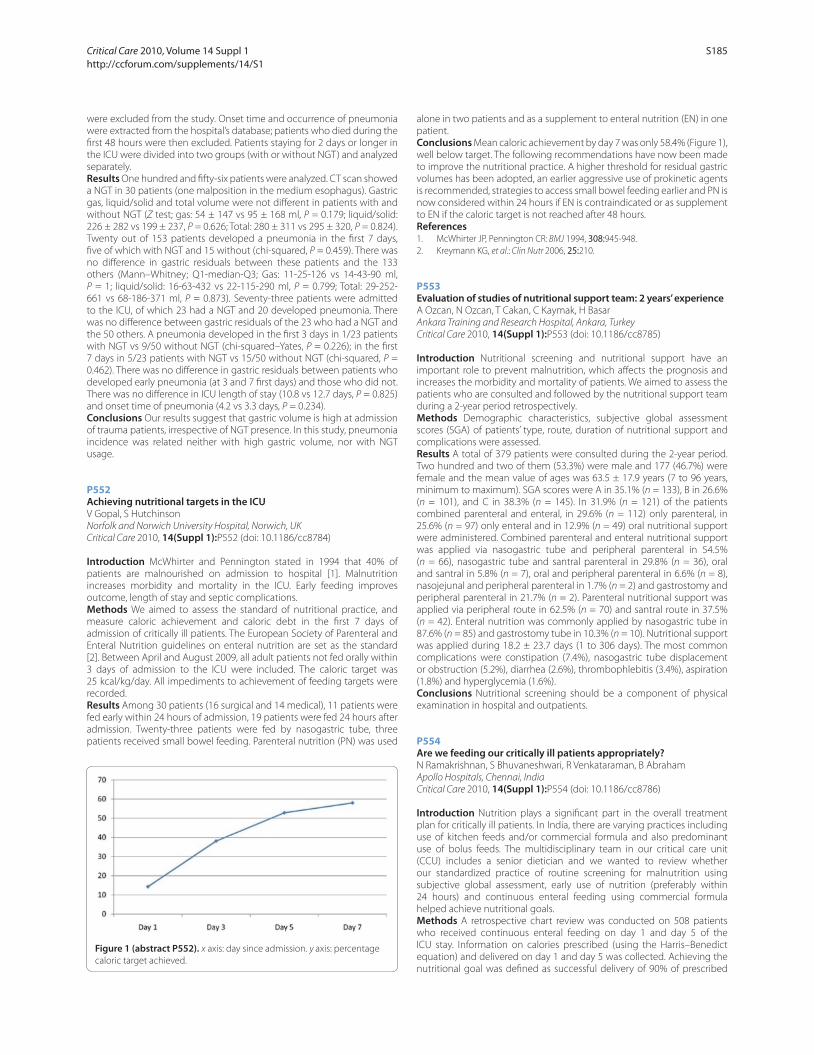
were excluded from the study. Onset time and occurrence of pneumonia
were extracted from the hospital’s database; patients who died during the
fi rst 48 hours were then excluded. Patients staying for 2 days or longer in
the ICU were divided into two groups (with or without NGT) and analyzed
separately.
Results One hundred and fi fty-six patients were analyzed. CT scan showed
a NGT in 30 patients (one malposition in the medium esophagus). Gastric
gas, liquid/solid and total volume were not diff erent in patients with and
without NGT (Z test; gas: 54 ± 147 vs 95 ± 168 ml, P = 0.179; liquid/solid:
226 ± 282 vs 199 ± 237, P = 0.626; Total: 280 ± 311 vs 295 ± 320, P = 0.824).
Twenty out of 153 patients developed a pneumonia in the fi rst 7 days,
fi ve of which with NGT and 15 without (chi-squared, P = 0.459). There was
no diff erence in gastric residuals between these patients and the 133
others (Mann–Whitney; Q1-median-Q3; Gas: 11-25-126 vs 14-43-90 ml,
P = 1; liquid/solid: 16-63-432 vs 22-115-290 ml, P = 0.799; Total: 29-252-
661 vs 68-186-371 ml, P = 0.873). Seventy-three patients were admitted
to the ICU, of which 23 had a NGT and 20 developed pneumonia. There
was no diff erence between gastric residuals of the 23 who had a NGT and
the 50 others. A pneumonia developed in the fi rst 3 days in 1/23 patients
with NGT vs 9/50 without NGT (chi-squared–Yates, P = 0.226); in the fi rst
7 days in 5/23 patients with NGT vs 15/50 without NGT (chi-squared, P =
0.462). There was no diff erence in gastric residuals between patients who
developed early pneumonia (at 3 and 7 fi rst days) and those who did not.
There was no diff erence in ICU length of stay (10.8 vs 12.7 days, P = 0.825)
and onset time of pneumonia (4.2 vs 3.3 days, P = 0.234).
Conclusions Our results suggest that gastric volume is high at admission
of trauma patients, irrespective of NGT presence. In this study, pneumonia
incidence was related neither with high gastric volume, nor with NGT
usage.
P552
Achieving nutritional targets in the ICUV Gopal, S Hutchinson
Norfolk and Norwich University Hospital, Norwich, UK
Critical Care 2010, 14(Suppl 1):P552 (doi: 10.1186/cc8784)
Introduction McWhirter and Pennington stated in 1994 that 40% of
patients are malnourished on admission to hospital [1]. Malnutrition
increases morbidity and mortality in the ICU. Early feeding improves
outcome, length of stay and septic complications.
Methods We aimed to assess the standard of nutritional practice, and
measure caloric achievement and caloric debt in the fi rst 7 days of
admission of critically ill patients. The European Society of Parenteral and
Enteral Nutrition guidelines on enteral nutrition are set as the standard
[2]. Between April and August 2009, all adult patients not fed orally within
3 days of admission to the ICU were included. The caloric target was
25 kcal/kg/day. All impediments to achievement of feeding targets were
recorded.
Results Among 30 patients (16 surgical and 14 medical), 11 patients were
fed early within 24 hours of admission, 19 patients were fed 24 hours after
admission. Twenty-three patients were fed by nasogastric tube, three
patients received small bowel feeding. Parenteral nutrition (PN) was used
alone in two patients and as a supplement to enteral nutrition (EN) in one
patient.
Conclusions Mean caloric achievement by day 7 was only 58.4% (Figure 1),
well below target. The following recommendations have now been made
to improve the nutritional practice. A higher threshold for residual gastric
volumes has been adopted, an earlier aggressive use of prokinetic agents
is recommended, strategies to access small bowel feeding earlier and PN is
now considered within 24 hours if EN is contraindicated or as supplement
to EN if the caloric target is not reached after 48 hours.
References1. McWhirter JP, Pennington CR: BMJ 1994, 308:945-948.
2. Kreymann KG, et al.: Clin Nutr 2006, 25:210.
P553
Evaluation of studies of nutritional support team: 2 years’ experienceA Ozcan, N Ozcan, T Cakan, C Kaymak, H Basar
Ankara Training and Research Hospital, Ankara, Turkey
Critical Care 2010, 14(Suppl 1):P553 (doi: 10.1186/cc8785)
Introduction Nutritional screening and nutritional support have an
important role to prevent malnutrition, which aff ects the prognosis and
increases the morbidity and mortality of patients. We aimed to assess the
patients who are consulted and followed by the nutritional support team
during a 2-year period retrospectively.
Methods Demographic characteristics, subjective global assessment
scores (SGA) of patients’ type, route, duration of nutritional support and
complications were assessed.
Results A total of 379 patients were consulted during the 2-year period.
Two hundred and two of them (53.3%) were male and 177 (46.7%) were
female and the mean value of ages was 63.5 ± 17.9 years (7 to 96 years,
minimum to maximum). SGA scores were A in 35.1% (n = 133), B in 26.6%
(n = 101), and C in 38.3% (n = 145). In 31.9% (n = 121) of the patients
combined parenteral and enteral, in 29.6% (n = 112) only parenteral, in
25.6% (n = 97) only enteral and in 12.9% (n = 49) oral nutritional support
were administered. Combined parenteral and enteral nutritional support
was applied via nasogastric tube and peripheral parenteral in 54.5%
(n = 66), nasogastric tube and santral parenteral in 29.8% (n = 36), oral
and santral in 5.8% (n = 7), oral and peripheral parenteral in 6.6% (n = 8),
nasojejunal and peripheral parenteral in 1.7% (n = 2) and gastrostomy and
peripheral parenteral in 21.7% (n = 2). Parenteral nutritional support was
applied via peripheral route in 62.5% (n = 70) and santral route in 37.5%
(n = 42). Enteral nutrition was commonly applied by nasogastric tube in
87.6% (n = 85) and gastrostomy tube in 10.3% (n = 10). Nutritional support
was applied during 18.2 ± 23.7 days (1 to 306 days). The most common
complications were constipation (7.4%), nasogastric tube displacement
or obstruction (5.2%), diarrhea (2.6%), thrombophlebitis (3.4%), aspiration
(1.8%) and hyperglycemia (1.6%).
Conclusions Nutritional screening should be a component of physical
examination in hospital and outpatients.
P554
Are we feeding our critically ill patients appropriately?N Ramakrishnan, S Bhuvaneshwari, R Venkataraman, B Abraham
Apollo Hospitals, Chennai, India
Critical Care 2010, 14(Suppl 1):P554 (doi: 10.1186/cc8786)
Introduction Nutrition plays a signifi cant part in the overall treatment
plan for critically ill patients. In India, there are varying practices including
use of kitchen feeds and/or commercial formula and also predominant
use of bolus feeds. The multidisciplinary team in our critical care unit
(CCU) includes a senior dietician and we wanted to review whether
our standardized practice of routine screening for malnutrition using
subjective global assessment, early use of nutrition (preferably within
24 hours) and continuous enteral feeding using commercial formula
helped achieve nutritional goals.
Methods A retrospective chart review was conducted on 508 patients
who received continuous enteral feeding on day 1 and day 5 of the
ICU stay. Information on calories prescribed (using the Harris–Benedict
equation) and delivered on day 1 and day 5 was collected. Achieving the
nutritional goal was defi ned as successful delivery of 90% of prescribed
Figure 1 (abstract P552). x axis: day since admission. y axis: percentage
caloric target achieved.
Critical Care 2010, Volume 14 Suppl 1 http://ccforum.com/supplements/14/S1
S185

calories. Data were analyzed using SPSS software and standard statistical
methods.
Results On day 1, 37.6% of patients achieved delivery of caloric goals, and
on day 5, this increased to 67.3%. Fluid restriction was the single most
common reason for not meeting caloric goals both on days 1 and 5.
Conclusions Despite the availability of a clinical dietician and standardized
nutritional practice, compliance of achieving caloric goals is low.
Commercial formulas with a maximum of 2 kcal/ml are available in India,
unlike the West where more calorie-dense formulas (up to 4.5 to 5 kcal/
ml) are available. Availability of these higher calorie dense commercial
formulas may help increase our rates of achieving nutritional goals as fl uid
restriction appears to be the single most common limiting factor.
P555
Eff ect of increased enteral nutrition on ICU-acquired infections: a multicenter observational studyDK Heyland1, KE Stephens2, AG Day1, SA McClave3
1Clinical Evaluation Research Unit, Kingston General Hospital, Kingston,
Canada; 2Discovery Biometrics GlaxoSmithKline, Stevenage, UK; 3Department
of Medicine, Louisville School of Medicine, Louisville, KY, USA
Critical Care 2010, 14(Suppl 1):P555 (doi: 10.1186/cc8787)
Introduction The objective of this study was to evaluate the eff ect of
increasing enteral nutrition (EN) adequacy on acquired infection in the ICU.
Methods Prospective, multicenter, observational study in three medical/
surgical ICUs. We enrolled consecutive patients within 24 hours of
admission to the ICU who were expected to stay for more than 24 hours.
For this analysis, we excluded patients who were not mechanically
ventilated in the ICU for more than 72 hours and who received parenteral
nutrition. No interventions. Charts were reviewed to determine nutritional
adequacy and clinical outcomes. Suspected infections were adjudicated
by two or more clinicians to determine the presence or absence of
infection (rated as either probable or possible infection).
Results A total of 598 patients were enrolled in the overall study; 206 were
included in this analysis. The average patient age was 62.2 years; APACHE II
score was 23.3; BMI 28.6; and 73% were medical. Overall, patients received
48.8% (range 0 to 120%) of their energy and 45.0% (range 0 to 120%) of their
protein requirements from EN. On average, patients remained ventilated
for 9.1 days (interquartile range (IQR) 6 to 11), in the ICU for 13.5 days (IQR, 7
to 14) and 25.2% and 21.8% developed an infection after 72 and 96 hours,
respectively. Overall 28-day mortality was 29.1%. In the overall analysis,
greater amounts of energy and protein were consistently associated with a
reduction in infection but only achieved levels near statistical signifi cance
when considering the incremental eff ect of 1,000 kcal/day and 30 g/day
on patients with at least one probable infection after >96 hours (odds
ratio (OR) 0.32, 95% confi dence interval (CI), 0.10 to 1.03, P = 0.055 and OR
0.41, 95% CI 0.19 to 0.89, P = 0.024 for energy and protein, respectively). In
all cases, the OR was lower when considering infections that developed
after 96 hours compared with infections that developed after 72 hours
and when considering probable infections compared with all infections.
There was a trend towards a reduction in pneumonia that developed after
96 hours with increasing amounts of energy (OR 0.40, 95% CI 0.10 to 1.53,
P = 0.18) and protein (OR 0.42, 95% CI 0.16 to 1.09, P = 0.076).
Conclusions Increased EN adequacy was associated with a reduction in
infectious complication, particularly after 96 hours of ICU admission.
P556
Impact of early enteral nutrition up to goal on the prognosis of patients with diff erent severity of illness in the ICUL Fang, W Fang, L Liu
Second Affi liated Hospital of Soochow University, Suzhou, Jiangsu, China
Critical Care 2010, 14(Suppl 1):P556 (doi: 10.1186/cc8788)
Introduction This study aimed to determine the value of early enteral
nutrition up to goal on the prognosis of the patients with diff erent severity
of illness in the ICU.
Methods A total of 192 critically ill patients who met the criteria for
enrollment were included in this study during the past 18 months in our
ICU. They were divided by grade as the severity of illness according to
APACHE II scores. Retrospective analysis on early enteral nutrition checked
whether the third day of enteral nutrition supply has reached the 60%
nutrition goal (25 kcal/kg/day). So the patients were divided into two
groups: up to goal and not up to goal. According to APACHE II scores,
the patients were divided into three sub-groups with score inferior to 15,
between 15 and 25 and superior to 25.
Results When the scores of APACHE II were between 15 and 25, the LOS
in the ICU (t = 3.966, P = 0.000), in hospital (t = 8.165, P = 0.000), the cost of
medical care (t = 4.812, P = 0.000) and the mortality (t = 5.421, P = 0.038)
were all signifi cantly lower in patients with EEN up to goal. See Table 1.
Conclusions The EEN up to goal can signifi cantly increase the survival rate
of critically ill patients, shorten the LOS in hospital and reduce the cost of
medical care, when the patients have APACHE II scores between 15 and
25. The clinical value of EEN up to goal varies depending on the severity
of illness.
Reference1. Nartindale RG, et al.: Guidelines for the provision and assessment of
nutrition support therapy in the adult critically ill patient: Society of Critical Care Medicine and American Society for Parenteral and Enteral Nutrition: Executive Summary. Crit Care Med 2009, 37:1757-1761.
P557
Benefi t of supplemental parenteral nutrition in the critically ill patient? Results of a multicenter observational studyDK Heyland1, NE Cahill1, M Wang1, J Kutsogiannis2, C Alberda3, L Gramlich2
1Clinical Evaluation Research Unit, Kingston General Hospital, Kingston,
Canada; 2University of Alberta, Edmonton, Alberta, Canada; 3Alberta Health
Services, Edmonton, Alberta, Canada
Critical Care 2010, 14(Suppl 1):P557 (doi: 10.1186/cc8789)
Introduction Early enteral nutrition (EN) is the preferred strategy for
feeding the critically ill; however, it is not always possible to achieve
suffi cient calories and protein with EN alone. The use of supplemental
parenteral nutrition (PN) has been advocated as a strategy to avoid
complications associated with protein/calorie defi cit from inadequate
enteral feeding. The purpose of this study was to evaluate the eff ect of this
practice on nutritional and clinical outcomes.
Methods An international, observational study conducted in 2007 and
again in 2008 examined nutrition practices in ICUs. Eligible patients were
the mechanically ventilated who remained in the ICU for >72 hours and
received early EN within 48 hours from admission. Data were collected
on patient characteristics and daily nutrition practices for up to 12
days. Patient outcomes were recorded after 60 days. We compared the
outcomes of patients who received early EN alone, early EN + early PN, and
early EN + late PN (after 48 hours of admission). Regression analyses were
conducted to determine the eff ect of increasing age, APACHE score, days
in hospital prior to ICU admission, gastrointestinal dysfunction, nutritional
strategy, and other baseline variables and their relationship to being alive
and discharged within 60 days.
Table 1 (abstract P556). Comparison of patients with APACHE II scores 15 to 25 in the two groups
Group n APACHE II scores LOS ICU (days) LOS hospital (days) Treatment cost (¥10,000) Mortality (%)
Up to nutrition goal 28 17.58 ± 3.02 7.50 ± 3.94 8.42 ± 3.45 4.20 ± 3.49 0
Not up to nutrition goal 54 19.96 ± 2.97 13.11 ± 9.80* 28.63 ± 16.44* 9.42 ± 5.67* 14.8
*P <0.05.
Critical Care 2010, Volume 14 Suppl 1 http://ccforum.com/supplements/14/S1
S186
Results A total of 2,920 patients from 260 ICUs were eligible for this study;
2,562 (87.7%) in the Early EN group, 188 (6.4%) in the Early PN group, and
170 (5.8%) in the Late PN group. Adequacy of calories and protein was
highest in the Early PN group (81.2% and 80.1%, respectively) and lowest
in the Early EN group (63.4% and 59.3%) (P <0.0001). The 60-day mortality
rate was 27.8% in the Early EN group, 34.6% in the Early PN group, and
35.3% in the Late PN group (P = 0.02). In a single predictor model, the
proportion of patients discharged alive from hospital at 60 days was lower
in the group that received Early PN (hazard ratio (HR) 0.75, 95% confi dence
interval (CI) 0.59 to 0.96) and Late PN (HR = 0.64, 95% CI 0.51 to 0.81) (P =
0.0003) compared with Early EN. These fi ndings persisted after adjusting
for known confounders. In subgroup analyses, we could not demonstrate
any benefi t to supplemental PN in patients with BMI <20.
Conclusions The supplemental use of PN may improve provision of
calories and protein but is not associated with any clinical benefi t.
P558
Role of fi ber diet formula in surgical critically ill septic patientsK Chittawatanarat, P Pokawinpudisnun
Chaing Mai University, Chiang Mai, Thailand
Critical Care 2010, 14(Suppl 1):P558 (doi: 10.1186/cc8790)
Introduction Diarrhea commonly occurs in surgical critically ill septic
patients who received broad-spectrum antibiotics. Some reports
demonstrated that a fi ber diet with prebiotic properties could decrease
the risk of diarrhea in nonseptic patients. However, most reports excluded
septic patients who received broad-spectrum antibiotics. The objective of
this study was to compare risk of diarrhea and diarrhea score between
fi ber and nonfi ber diets in surgical septic patients who received broad-
spectrum antibiotics.
Methods We conducted a prospective, randomized control, double-blind
study on a general surgical ICU in a university hospital. The patients who
received broad-spectrum antibiotics and no contraindication to enteral
feeding were enrolled into two arms after informed consent. Allocation to
intervention was randomized individually to receive fi ber diet or nonfi ber
diet up to 14 days. Demographic data, disease severity, nutritional delivery
and diarrhea score were recorded each day. Intention-to-treat analysis was
performed after completed study.
Results Thirty-four patients were enrolled in the study (17 patients each
in the fi ber group and the nonfi ber group). These two patient groups
were similar in demographics, disease severity, nutritional status, cause
of sepsis and total feeding per day. Although the proportion of patients
who confronted with diarrhea score >15 was higher in the nonfi ber
group than the fi ber group, these did not show statistical diff erence
signifi cantly (8/17 patients (47.06%) vs 4/17 patients (23.53%); P = 0.15).
However; the group of the fi ber diet had a lower mean diarrhea score
(fi ber vs nonfi ber = 3.6 ± 2.3 vs 6.3 ± 3.6; P = 0.005) and the generalized
estimation equation model for repeated measurement (GEE) revealed a
lower global diarrhea score in the fi ber group than the nonfi ber group
about 3.03 (coeffi cient –3.03 (95% CI = –5.03 to –0.92); P = 0.005). Cox
proportional hazard ratio of fi ber group was 0.45 (95% CI = 0.13 to 1.49;
P = 0.19) but no statistical diff erence.
Conclusions Enteral nutrition with a fi ber diet formula could reduce the
diarrhea score in surgical critically ill septic patients who received broad-
spectrum antibiotics.
P559
Eff ect of oral supplementation with immunonutrients on radiation-induced hematologic toxicity in cancer patientsMC Calaguas
St Luke’s Medical Center, Manila, Philippines
Critical Care 2010, 14(Suppl 1):P559 (doi: 10.1186/cc8791)
Introduction Radiation therapy in cancer patients causes hematologic
abnormalities mainly attributed to an increased infl ammatory state
induced by radiation toxicity that are observed to be modulated by
immunonutrition [1-3]. The objective of this study was to determine the
eff ect of oral supplementation with immunonutrients on cancer patients
undergoing radiation therapy on the following hematologic indices:
hemoglobin, total WBC, neutrophil, lymphocyte, and platelet counts.
Methods Cancer patients on radiotherapy from 2007 to 2009 were
randomly assigned to a study group (n = 24), which received standard
diet plus oral supplementation with an immunonutrition regimen
(= 200 ml drink given twice per day providing 600 kcal, 40 g protein,
eicosapentanoic acid = 2 g, antioxidants (Vit A = 600 μg, Vit C = 75.2 mg,
Vit E = 15 mg, B-carotene = 1,500 μg)), and a control group (n = 32), which
only had standard diet. The hematologic data – hemoglobin, total WBC
count, neutrophil count, lymphocyte count, and platelet count – were
taken before and after completion of the prescribed radiation dose. The
results were analyzed using either paired or independent-samples t test.
Results In the study group the blood indices were higher compared
with the control group at the end of radiation therapy: hemoglobin in
gm% (12.8 >11.8, P = 0.002), total WBC (7,276 >4,877, P = 0.0005), and
neutrophil count (5,363 >3,315, P = 0.0007). There were no diff erences in
the lymphocyte and platelet counts. The WBC change in the study group
followed a decreasing pattern (–162.5) while in the control group it was
an increasing pattern (+1,657). This indicated lesser infl ammation in the
study group while there was higher infl ammation in the control group
(P = 0.004).
Conclusions Oral supplementation with immunonutrients in cancer
patients undergoing radiation therapy resulted in lesser hematologic toxic
changes in hemoglobin, total WBC, and neutrophil count.
References1. Mell LK, et al.: Int J Radiat Oncol Biol Phys 2008, 70:1431-1437.
2. Suchner U, et al.: Proc Nutr Soc 2000, 59:553-563.
3. Jho DH, et al.: Integr Cancer Ther 2004, 3:98-111.
P560
Impact of multichamber bag parenteral nutrition (OliClinomel) upon infl ammatory markers (EPICOS Study)A Pontes-Arruda1, L Martins1, S Lima1, M Pinheiro2
1Hospital Fernandes Tavora, Fortaleza, Brazil; 2Laboratorio Evandro Pessoa,
Fortaleza, Brazil
Critical Care 2010, 14(Suppl 1):P560 (doi: 10.1186/cc8792)
Introduction The use of a multichamber bag (MCB) as a way to deliver
parenteral nutrition to critically ill patients has been associated by
retrospective analysis with several benefi ts including lower bloodstream
infection rates, and lower ICU stay [1]. This study aims to evaluate the
eff ects of a MCT TPN system (considered a closed system) versus a
conventional compounding system (COM, considered an open system)
regardless of the type of lipid emulsion used.
Methods Serum samples were collected in a prospective multicenter
open label study (NCT#00798681) from 100 critical care patients receiving
TPN with an olive oil-based lipid emulsion using either a premixed MCB
and ready-to-use system (n = 50, OliClinomel; Baxter) or COM made
either with an olive oil-based lipid emulsion (ClinOleic; Baxter) or with
a LCT/MCT-based lipid emulsion (COM ClinOleic n = 25, COM LCT/MCT
n = 25, total COM group n = 50). All samples were collected at baseline,
day 4 and day 7 (three samples per time point). Changes in the levels
of infl ammatory markers from baseline to day 7 were compared using
analysis of covariance and P <0.05 was considered statistically signifi cant.
Results At baseline, demographic characteristics of the patients were
balanced between the groups. Regardless of the type of lipid used,
critically ill patients receiving TPN using a MCB system presented at day
4 lower levels of: TNFα (3.4 vs 8.2 pg/ml), IL-6 (50.4 vs 62.0 pg/ml), IL-8
(56.4 vs 93.7 pg/ml), procalcitonin (PCT) (2.7 vs 7.6 ng/ml), lymphocyte
externalization of phosphatidylserine (5.2 vs 9.6%), and resistin (9.1 vs 12.3
ng/ml) (all MCB vs COM and P <0.05). Statistically signifi cant diff erences
were maintained throughout day 7. No diff erences were observed for the
levels of C-reactive protein and IL-10.
Conclusions The administration of TPN using a MCB system led to a
signifi cant reduction in the infl ammatory response when compared with
COM, regardless of the type of lipid used. Particularly relevant are the
diff erences in terms of PCT as an early marker of sepsis since COM TPN was
associated in previous studies with higher bloodstream infection rates [1].
Reference1. Wischmeyer et al.: Crit Care Med 2009, 37(Suppl).
Critical Care 2010, Volume 14 Suppl 1 http://ccforum.com/supplements/14/S1
S187
P561
Eff ect of n-3 fatty acids on cerebral markers and the infl ammatory response in sepsisCS Burkhart1, M Siegemund1, SP Strebel1, LA Steiner2
1University Hospital Basel, Switzerland; 2CHUV et Université de Lausanne,
Switzerland
Critical Care 2010, 14(Suppl 1):P561 (doi: 10.1186/cc8793)
Introduction Patients with sepsis are at high risk of developing sepsis-
associated delirium (SAD), which is associated with increased mortality
and may be an important risk factor for the development of long-term
cognitive dysfunction. Mechanisms leading to SAD are unclear, therapy
and prophylaxis unsatisfactory. Infl ammation and its eff ects on the brain
may play an important role. We hypothesized that n-3 fatty acids will have
a benefi cial eff ect on systemic infl ammation and hence cerebral markers
in sepsis.
Methods Randomized single-blind design. Patients >18 years admitted
for treatment of sepsis. Exclusion criteria were thrombocytopenia, brain
injury or tumors, intracranial infection and history of stroke. Patients
received treatment according to the Surviving Sepsis Campaign and
were randomly allocated to a group with or without daily supplemental
intravenous n-3 fatty acids (Omegaven™; Fresenius Kabi, 2 ml/kg/day).
Daily screening for SAD was performed. CRP, IL-6, IL-8, S-100β, and neuron-
specifi c enolase (NSE) were monitored on days 1, 2, 3, and 7. The primary
endpoint was the area under the curve of the S-100β time course.
Results Eighteen patients were included. SAPS II scores and baseline
values for CRP, IL-6, IL-8, NSE and S-100β for patients with and without
supplemental n-3 fatty acids were 51 ± 14 vs 57 ± 22, 241 ± 65 vs 218 ±
106 mg/l, 2,054 ± 2,822 vs 734 ± 1,467 pg/ml, 312 ± 503 vs 90 ± 96 pg/ml,
25 ± 7 vs 21 ± 8 μg/ml, and 0.57 ± 0.90 vs 0.24 ± 0.24 μg/ml, respectively.
In agreement with earlier data, patients with SAD had higher values for
S-100β (P = 0.019) [1]. AUC for S-100β was 2.73 ± 2.89 vs 0.95 ± 0.74 (P =
0.14) for patients receiving supplemental n-3 fatty acids and controls,
respectively. While none of the diff erences reached signifi cance, IL-6 and
IL-8 declined more rapidly in the n-3 fatty acid-treated group whereas
cerebral markers showed an opposite trend. The number of patients with
SAD and the duration of SAD were not diff erent in both groups.
Conclusions Interpretation of the results is hampered by unevenly
distributed baseline characteristics and more patients need to be
investigated. In this small sample no benefi cial eff ect of n-3 fatty acids on
cerebral markers was found.
Reference1. Pfi ster D, et al.: Cerebral perfusion in sepsis-associated delirium. Crit Care
2008, 12:R63.
P562
Eff ects of an olive oil-based lipid emulsion (ClinOleic) versus LCT/MCT-based lipid emulsions upon the infl ammatory markers of critically ill patients (EPICOS Study)S Lima1, A Pontes-Arruda1, L Martins1, M Pinheiro2
1Hospital Fernandes Tavora, Fortaleza, Brazil; 2Laboratório Evandro Pessoa,
Fortaleza, Brazil
Critical Care 2010, 14(Suppl 1):P562 (doi: 10.1186/cc8794)
Introduction Olive oil-based lipid emulsions have been used for more
than a decade to reduce the polyunsaturated fatty acid (PUFA) content in
TPN, thus preventing the deleterious eff ects associated with PUFA excess.
MCT/LCT-based lipid emulsions were recently associated with deleterious
eff ects such as lipotoxicity, activation of infl ammatory cascade and
increased risk of cardiovascular disease [1]. This study aims to compare the
eff ects of these two lipid emulsions upon several infl ammatory markers in
critically ill patients using TPN.
Methods Serum samples were collected in a prospective multicenter and
open-label study (NCT#00798681) from 100 critical care patients receiving
TPN with an olive oil-based lipid emulsion using either a premixed ready-
to-use system (G1, n = 50, OliClinomel; Baxter) or a compounded system
(G2, n = 25, ClinOleic; Baxter), and the results were compared with those
from patients receiving a compounded TPN made with an LCT/MCT
lipid emulsion (G3, n = 25). All samples were collected at baseline, day
4 and day 7 (three samples in each time point). Changes in the levels
of infl ammatory markers from baseline to day 7 were compared using
analysis of covariance and P <0.05 was considered statistically signifi cant.
Results At baseline, demographic characteristics of the patients were
balanced between the groups. Regardless of the delivery system used,
patients receiving TPN with an olive oil-based lipid emulsion presented
at day 4 lower levels of: IL-8 (54.4 vs 67.8 vs 120.7 pg/ml), TNFα (3.4 vs 3.5
vs 13.0 pg/ml), lymphocyte phosphatidylserine externalization (PS) (5.2 vs
4.8 vs 14.7%), and resistin (R) (9.1 vs 9.2 vs 15.4 ng/ml) (all G1 vs G2 vs G3
and P <0.05). No diff erences were observed in the levels of IL-10 and CRP.
Statistically signifi cant diff erences were maintained throughout day 7 for
IL-8, TNFα, PS and R.
Conclusions The administration of an olive oil-based lipid emulsion
within the TPN regimen led to a signifi cant reduction in the infl ammatory
response when compared with a LCT/MCT-based lipid emulsion.
Particularly relevant are the diff erences for resistin (an indicator of insulin
resistance) and PS (an indicator of apoptosis).
Reference1. Pontes-Arruda: Clin Nutr Suppl 2009, 4:19.
P563
Abstract withdrawn
Critical Care 2010, Volume 14 Suppl 1 http://ccforum.com/supplements/14/S1
S188
P564
Abstract withdrawn
P565
Does a fi sh-oil-containing lipid emulsion improve liver function in comparison with a soybean oil lipid emulsion?J Linke1, H Topp1, I Mueller1, S Bark2, N Mertes3, H Grimm4, P Singer5,
J Shaff er6
1Fresenius Kabi Deutschland GmbH, Oberursel, Germany; 2Visby Hospital,
Visby, Sweden; 3Raphaelsklinik, Münster, Germany; 4Clinical Centre of Passau,
Germany; 5General Intensive Care Department and Institute for Nutrition
Research, Rabin MC, Petah Tikva, Israel; 6Salford Royal NHS Foundation Trust,
Salford, UK
Critical Care 2010, 14(Suppl 1):P565 (doi: 10.1186/cc8797)
Introduction Liver function disturbances have been of concern in
parenteral nutrition. The aim of the post hoc analyses of two pooled
studies was to compare liver function parameters using fi sh-oil-containing
vs soybean oil lipid emulsions.
Methods Two prospective, controlled, randomized, parallel-group,
double-blind, multicenter studies compared SMOFlipid 20%, a fi sh-
oil-containing lipid emulsion (SMOF: soybean oil 60 g, medium-chain
triglycerides 60 g, olive oil 50 g, fi sh oil 30 g per l), vs standard soybean
oil emulsion (SO, 200 g soybean oil/l). The studies were as follows:
A: postsurgical adult patients, 5 days total parenteral nutrition, 100 SMOF
vs 103 SO patients; B: adult patients receiving parenteral nutrition for 28
days, 22 SMOF vs 32 SO patients. Patients with data at baseline and study
end were selected. The data were pooled and diff erences of laboratory
data at 1 week minus baseline and study end minus baseline were
calculated. Analyses of variance were applied using diff erences between
1 week/study end values and baseline values of bilirubin (BIL), AST and
ALT as dependent variables and treatment group as independent variable.
Covariates used: baseline values for BIL, AST, ALT and mean daily dose of
fat per kg body weight.
Results Baseline values were not signifi cantly diff erent between the two
treatment groups. The mean daily intake of fat/kg bw after 1 week and
study end was the same between both treatment groups. One-week –
baseline values of liver parameters (mean; SMOF group: BIL –5.1* μmol/l,
AST –5.4** U/l, ALT 0.6*** U/l; SO group: BIL –2.0* μmol/l, AST 0.6** U/l,
ALT 11.1*** U/l) were signifi cantly diff erent between treatment groups (P:
0.035*, 0.001**, 0.001***). Extending the analysis until 28 days did not alter
the results.
Conclusions Infusion of SMOFlipid 20% compared with a soybean oil
standard lipid emulsion exerts a signifi cant decrease in the values of
BIL and AST. It also signifi cantly attenuates the rise seen in ALT. These
results indicate that SMOFlipid 20% is promising in preventing parenteral
nutrition-induced liver disturbances.
P566
Therapy of compartmentalization of the proinfl ammatory and anti-infl ammatory cytokines in sepsisEG Grigoryev
Kemerovo State Medical Academy, Kemerovo, Russia Federation
Critical Care 2010, 14(Suppl 1):P566 (doi: 10.1186/cc8798)
Introduction We supposed the diagnostic signifi cance of the ratio
between proinfl ammatory and anti-infl ammatory cytokines in the
peritoneal cavity and system blood fl ow in patients with abdominal sepsis.
So we tried to correct such disbalance with glutamine intravenous and
enteral supplementation.
Methods Prospective controlled randomized investigation of the
patients with abdominal sepsis (excluding pancreatitis). Group 1 (n = 16):
standard therapy. Group 2 (n = 11): with intravenous infusion of glutamine
(Dipeptiven; Fresenius Kabi, Germany). Group 3 (n = 11): with intravenous
infusion of glutamine and enteral supplementation of glutamine
(Intestamine; Fresenius Kabi). The patients in groups were identical
according to the severity of disease (APACHE II), the standard of intensive
care, the volume of surgery care. We investigated the proinfl ammatory
cytokines: TNFα, IL-1, IL-6, IL-8; and anti-infl ammatory cytokines: soluble
sTNF-RI and sTNF-RII, antagonist receptor IL-1, IL-10 in blood serum and in
the peritoneal cavity during the initial 3 days of intensive care (ELISA; BD
Biosciences Pharmingen, San Diego, CA, USA; EASIAs; Biosource, Nivelles,
Belgium).
Results The probability of survival on day 28 was 73% in the standard
therapy group, in the group with glutamine intravenous – 78%, in the
group with glutamine intravenous and enteral – 84%. We did not fi x the
decrease of the duration of respiratory support in all of the groups. The
duration of acute intestinal injury was signifi cantly diff erent (standard
group 49 hours vs 38 hours in group with glutamine intravenous
supplementation – 35 hours in group with intravenous and enteral
glutamine supplementation). We investigated the prevalence of the
concentration of proinfl ammatory cytokines in the peritoneal cavity and
in blood serum according to the molar coeffi cient in the control group.
The molar coeffi cient had a positive correlation with the SOFA scale. In
group 2 (glutamine intravenous) the molar coeffi cients were decreased
to the prevalence of anti-infl ammatory cytokines (in serum on day 3,
in peritoneal on day 2). In group 3 (glutamine intravenous and enteral)
we investigated the signifi cance diff erence and decrease of all cytokine
levels in blood serum and in the peritoneal cavity.
Conclusions Intravenous and enteral supplementation of glutamine
improved the cytokine balance in blood and the peritoneal
compartment.
Critical Care 2010, Volume 14 Suppl 1 http://ccforum.com/supplements/14/S1
S189
P567
Intensive insulin therapy and brief hypoglycemia do not increase neurological injury markers in critically ill childrenM Gielen, I Vanhorebeek, M Boussemaere, PJ Wouters, D Mesotten,
G Van den Berghe
Katholieke Universiteit Leuven, Belgium
Critical Care 2010, 14(Suppl 1):P567 (doi: 10.1186/cc8799)
Introduction Targeting glycemia to age-adjusted normal fasting blood
glucose levels with intensive insulin therapy (IIT) improved short-term
outcome of critically ill children as compared with conventional insulin
therapy (CIT), but concomitantly increased the incidence of hypoglycemia
[1]. Both hyperglycemia and hypoglycemia may adversely aff ect the
developing brain of young children. We therefore studied the impact of
targeting normoglycemia with IIT on circulating markers of brain injury in
these patients.
Methods This was a pre-planned analysis of all 700 pediatric critically
ill patients included in a prospective, randomized, controlled study on
IIT [1]. Patients were randomly assigned to the target of normal for age
fasting blood glucose levels (2.8 to 4.4 mmol/l for age <1 year and 3.9 to
5.6 mmol/l for age ≥1 year) with insulin infusion throughout the ICU stay
(IIT), or to insulin infusion only to prevent excessive hyperglycemia (CIT).
Serum S100B and neuron-specifi c enolase (NSE), markers of astrocytic and
neuronal damage, were measured by ELISA on fi xed days (n = 700) and
before and after hypoglycemia in a nested case–control design (n = 126).
Results Patients admitted to the ICU after cardiac surgery had higher
admission S100B and NSE levels than patients in the other diagnostic
categories (P <0.0001), with the highest levels observed for patients
with the highest Risk Adjustment for Congenital Heart Surgery (RACHS)
score (both P = 0.002). Admission levels of S100B and NSE were higher in
patients needing intensive care for at least 3 days vs short-stay patients
(both P <0.0001) and in ICU nonsurvivors vs survivors (P = 0.002 and P
= 0.0002). IIT did not aff ect the levels of S100B or NSE as compared with
CIT. Patients experiencing hypoglycemia at any time during the ICU stay
revealed higher S100B and NSE levels on admission (P <0.0001 and P =
0.0007) than those without hypoglycemia. In the nested case–control
study, S100B and NSE decreased after hypoglycemia (P = 0.001 and P =
0.009) in the cases, unlike in the controls on matched days.
Conclusions IIT in the pediatric ICU did not evoke neurological damage,
as evaluated by S100B and NSE, despite an increased incidence of brief
hypoglycemia. Elevated markers in patients with hypoglycemia were
not caused by hypoglycemia but explained by an increased risk of
hypoglycemia in the most severely ill.
References1. Vlasselaers D, et al.: Lancet 2009, 373:547-556.
P568
A cluster-controlled implementation project of intensive insulin therapy: eff ects on blood glucose control and incidence of severe hypoglycemiaR Harmsen1, F Van Braam Houckgeest2, J Van der Sluijs3, H Van den Oever4,
L Hofstra5, M Kuiper6, A Abu-Hanna1, P Spronk7, M Schultz1
1Academic Medical Center, Amsterdam, the Netherlands; 2Tergooi Hospitals,
Hilversum, the Netherlands; 3MCH, The Hague, the Netherlands; 4Deventer
Hospital, Deventer, the Netherlands; 5Scheper Hospital, Emmen, the
Netherlands; 6MCL, Leeuwarden, the Netherlands; 7Gelre Hospitals, Apeldoorn,
the Netherlands
Critical Care 2010, 14(Suppl 1):P568 (doi: 10.1186/cc8800)
Introduction Intensive insulin therapy (IIT) is a complex intervention
involving several steps that may all contain potential sources of variability.
Changing practice in complex multidisciplinary environments is diffi cult.
In addition, it is undecided whether IIT should be applied by nurses and
physicians, or nurses alone. We implemented IIT using a conceptual
implementation framework and compared overall blood glucose control
and incidence of severe hypoglycemia (SH) (blood glucose level (BGL)
<40 mg/dl) before and after implementation.
Methods Three ICUs developed and implemented an evidence-based
guideline for IIT; three ICUs with no change in blood glucose control
served as control ICUs. In the guideline ICUs, the IIT practice change
comprised two periods: in the fi rst year after implementation of the
guideline, physicians and nurses used the guideline together; in the
second year the guideline was applied solely by nurses. BGL collection
and analysis included the 1 year before implementation of the guideline.
Analysis was performed using statistical process control.
Results IIT guideline: the new guideline emphasized fast correction of
high BGLs, acceptance of low BGLs and frequent BGL measurements (up
to one/hour). Blood glucose control: BGLs of approximately 8,000 patients
were collected and analyzed. The case mix was similar in guideline and
control ICUs, and did not change over time. No change in overall blood
glucose control or incidence of SH was observed in the control ICUs during
the study duration. In the guideline ICUs, the median number of daily
blood glucose measurements increased from 4 (3 to 7) per patient to 9 (5
to 12) per patient (P <0.001). Median morning BGL declined in guideline
ICUs, from 114 (96 to 139) mg/dl to 100 (85 to 123) mg/dl (P <0.001).
The change in blood glucose control was accompanied by a rise in the
incidence of SH, from 7% to 13% (P <0.001). SH, however, did never cause
short-term harm (no death or coma). Nurses alone performed equally well
with regard to overall blood glucose control, and even showed a decline
of incidence of SH.
Conclusions Implementation of an evidence-based IIT guideline
improves blood glucose control, with results comparable with the original
studies on IIT in Leuven. The increase of incidence of SH is temporary, and
improves when IIT is applied by nurses alone.
P569
Incidence of hypoglycemia in NICE sugar not so nice after consideration of the treatment periodJK Mader, G Bock, J Plank, G Köhler, TR Pieber, M Ellmerer
Medical University Graz, Austria
Critical Care 2010, 14(Suppl 1):P569 (doi: 10.1186/cc8801)
Introduction Hypoglycaemia is one of the major side eff ects when
establishing glycaemic control in critically ill patients and may contribute
to increased mortality rates. Until today, no comparable measure of
hypoglycaemia frequency has been suggested. Current assessments
depend either on the treatment period or the number of glucose
measurements performed per patient. Therefore, we suggest an
alternative method to present the hypoglycaemia incidence by number
of episodes per patient normalized for a 24-hour treatment period.
Methods Two major studies performing glycaemic control in critically
ill patients were chosen for comparison: the Leuven study with 1,200
patients at a medical ICU (605 standard care (STD), 595 tight glycaemic
control (TGC); [1]) and the NICE study with 6,030 patients in a mixed
ICU population (3,014 STD, 3,016 TGC; [2]). For both studies, rates of
hypoglycaemia per patient per 24 hours were calculated based on
published data.
Results Mean duration of treatment was 12.5 days for the Leuven study
and 4.2 days for the NICE study. APACHE scores were 23 for Leuven and
21 for NICE, respectively. Number of hypoglycaemic events per study
population was reported as follows: 19 (3.1%) for STD and 111 (18.7%) for
TGC for the Leuven study and 15 (0.5%) for STD and 206 (6.8%) for TGC
in the NICE study. When applying the new evaluation method, rates of
hypoglycaemia per patient per 24 hours were 0.25% (STD) vs 1.49% (TGC)
for Leuven and 0.12% (STD) vs 1.63% (TGC) for NICE, respectively.
Conclusions The results reveal that the treatment period may have a
signifi cant impact on the reported rate of hypoglycemia. We conclude
that reporting hypoglycaemic episodes in critically ill patients on a per-
patient and per-24-hour basis may be a more adequate tool to accurately
describe hypoglycaemia incidence which facilitates the comparison
between diff erent studies and study protocols.
References1. Van den Berghe et al.: N Engl J Med 2006, 354:449-461.
2. NICE-SUGAR Study Investigators: N Engl J Med 2009, 360:1283-1297.
Critical Care 2010, Volume 14 Suppl 1 http://ccforum.com/supplements/14/S1
S190
P570
Comparison of a planned versus emergent approach implementation of tight glycemic control therapy on an ICU: an experimental designM Luiking1, L Bras1, R Van Linge2, M Grypdonck2
1St Antonius Ziekenhuis, Nieuwegein, the Netherlands; 2Universiteit Utrecht,
the Netherlands
Critical Care 2010, 14(Suppl 1):P570 (doi: 10.1186/cc8802)
Introduction In this study the eff ectiveness of a hierarchic planned
approach to implementation was compared with an emergent approach
with a high degree of nurse participation. The innovation was a new
tight glycemic control (TGC) therapy in the ICU. The implementation
method to introduce a new evidence-based treatment to the ICU can
make a diff erence to its eff ectiveness. New treatment procedures are
very often strictly defi ned and introduced on the ICU in a top-down
manner. On theoretical grounds, a bottom-up implementation method,
with more participation and involvement of the ICU nurses, should be
more eff ective.
Methods In an experiment a TGC protocol was introduced on a 24-bed
ICU with 120 ICU nurses. In one nursing team the implementation was
by means of the usual planned approach (PA). In the other team an
emergent implementation approach (EA) was applied. During 7 months,
1,182 patients were admitted and randomly assigned to either team. The
eff ectiveness of the implementation methods was measured at patient-
outcome (glucose) level and on a process-execution (nurse compliance)
level. The patient-outcome measurements were: mean glucose value,
percentage of normal glucose values (defi ned as between 4.0 and
6.5 mmol/l) and the time from admission to reach a normal glucose
level (6.5 mmol/l). For the mean glucose value a diff erence was made
between short-stay (<3 days) and long-stay patients (≥3 days). The
compliance was expressed as compliance for safe, insulin treatment (the
patient received the right amount of insulin) and compliance for effi cient
treatment (glucose measurements as well as insulin were according to
the protocol).
Results The mean glucose for the short-stay patients was signifi cantly
lower in the EA than the PA team (6.42 vs 6.52 mmol/l), no signifi cant
diff erence was found for the long-stay patients. The percentage of normal
glucose values was 53.9% in the EA and 52.8% in the PA team (signifi cant
P <0.001). The time to reach normal glucose levels was 6 to 7 hours in the
EA and 10 to 11 hours in the PA team (signifi cant, P <0.05). The compliance
for safe insulin treatment was 91.3% in the EA and 79.0% in the PA team.
The compliance for effi cient treatment was 83.5% in the EA and 66.8% in
the PA team.
Conclusions An emergent approach to implementation (with an
increased participation of the ICU nurses) of a TGC resulted in better
patient results and a higher compliance to the protocol.
P571
Coagulation profi le in hyperglycemic septic shock patientsLC Sanches, D Lourenco, R Salomao, MA Noguti, M Brunialti, C Steula,
M Assuncao, F Machado
Federal University of São Paulo, Brazil
Critical Care 2010, 14(Suppl 1):P571 (doi: 10.1186/cc8803)
Introduction A hypercoagulable state is frequently found in sepsis
patients as well as hyperglycemia [1]. The objective of this study was to
determine the coagulation profi le in hyperglycemic septic shock patients
and the impact of glycemia control.
Methods Septic shock patients with less than 48 hours of organ
dysfunction with normoglycemia (<150 mg/dl, Group 1) or hyperglycemia
(>200 mg/dl, Group 2) were included. Exclusion criteria were previous
diabetes, coagulation diseases, use of heparin or fresh frozen plasma/
platelets, sepsis in the past 30 days or insulin use in the past 24 hours. The
coagulation and infl ammation profi le was determined at inclusion and
after 24 hours, only if the patients remained normoglycemic or achieved
glycemic control with insulin infusion (Group 2). Results were expressed in
median and interquartiles, P <0.05 was considered signifi cant.
Results Forty-one patients were included, 59.7% male; mean APACHE II
score: 16.9 ± 8.6, P = 0.21; mean SOFA score: 8.1 ± 2.0; mean number of
organ dysfunctions: 3.6 ± 0.9, with a signifi cant diff erence between the
groups only regarding age (Group 1 (n = 21): 53.8 ± 16.7, Group 2 (n =
20): 68.8 ± 16.4, P = 0.006). In both groups, a hypercoagulation profi le was
detected, with high levels of fragment 1+2 (F1+2), thrombin/antithrombin
complex (TAT), D-dimer and tissue factor and with low levels of factor VII.
Consumption of coagulation inhibitors (protein C (PC), antithrombin (AT),
tissue factor pathway inhibitor) and increased fi brinolysis (plasminogen
activator inhibitor 1 (PAI-1), tissue plasminogen activator (TPA) and
plasminogen) were also found. However, at baseline no signifi cant
diff erence between groups was found except for a trend towards high
levels of F1+2 (P = 0.08) and TAT (P = 0.058) in Group 2. After 24 hours
higher levels of AT (P = 0.02), PC (P = 0.02), plasminogen (P = 0.04) and
a reduction in PAI-1 (P = 0.01) and TPA (P = 0.03) were detected in Group
2. Infl ammatory parameters (IL-6, IL-8, IL-10, TNF) were similar at baseline,
with decreased levels in both groups after 24 hours but the IL-6 (P = 0.02)
and IL-8 (P = 0.0004) reduction was more pronounced in Group 2.
Conclusions Septic shock patients have a hypercoagulation pattern
regardless of their glycemic status. However, glycemia control seems to
be associated with reduction in infl ammation and hypercoagulation
parameters, possibly related to insulin infusion as glycemia levels did not
diff er between groups after 24 hours.
Acknowledgements Financial support from FAPESP.
Reference1. Machado FR, et al.: Endocr Metab Immune Disord 2006, 26:175-182.
P572
Impact of glycemic control on survival in critical care patients with sepsisR Tiruvoipati, D Lewis, K Ong, M Gupta, K Haji, J Botha
Frankston Hospital, Frankston, Victoria, Australia
Critical Care 2010, 14(Suppl 1):P572 (doi: 10.1186/cc8804)
Introduction Several studies have shown an increased mortality and
morbidity in various groups of patients with hyperglycemia requiring
admission to the ICU. Contrary to these studies, a recent large multicentric
international trial in ICU patients has shown an increased mortality in
patients where normoglycemia was maintained. However, very few
studies specifi cally evaluated the association between hyperglycemia
and mortality in critical care patients with sepsis. Since hyperglycemia is
known to be associated with immunosuppression and increased risk of
life-threatening infections, we aimed to assess the impact of glycemic
control (normoglycemia) in ICU patients with sepsis.
Methods We studied all patients admitted to our ICU with sepsis
between July 2004 and May 2009. Patients were excluded if their
blood sugar levels (BSL) were not measured at least once every 6 hours
during their ICU stay. Patients were divided into two groups based on
their glycemic control in the ICU: normoglycemic group (BSL 3.9 to 6.2
mmol/l) and conventional glucose control group (BSL 8 to 10 mmol/l).
BSLs were controlled using an infusion of actrapid insulin. Both groups
were compared with regard to demographics, co-morbidities, severity of
illness at ICU admission (APACHE III and SAPS II scores), ICU and hospital
mortality and length of stay.
Results Over the 5-year study period a total of 314 patients were admitted
with sepsis and 297 patients were included. On average every patient
included in the study had BSL once every 4.2 hours throughout their ICU
stay. Of these patients, 68 had normoglycemia (mean BSL 5.6) and 229
had conventional glucose control (mean BSL 8.5) during their ICU stay.
There was no statistically signifi cant diff erence between the groups in
age, sex, APACHE III and SAPS II scores. The ICU mortality was signifi cantly
higher in the normoglycemic group (30.8% vs 14.8% P <0.01). There was
no statistically signifi cant diff erence in hospital mortality between both
groups (33.8% vs 25.3% P = 0.16). The ICU (median and IQR days: 1 (1 to 3)
vs 4 (2 to 7) P = <0.01) and hospital duration of stay (6.0 (2.00 to 13.75) 6.0
(2.00 to 13.75) vs 12.0 (7.00 to 27.00); P <0.01) were signifi cantly higher in
the conventional group.
Conclusions The ICU mortality in patients admitted to the ICU with sepsis
who had normoglycemia was higher than in those patients who had
conventional glucose control during their ICU stay. Aiming for a normal
blood glucose levels is not indicated in patients with sepsis while they
are in the ICU.
Critical Care 2010, Volume 14 Suppl 1 http://ccforum.com/supplements/14/S1
S191

P573
Therapeutic impact on intravenous insulin dosing with three new point-of-care glucose meters in an adult ICUL Schmitz, M Suball, C Hanicq, T El-Mahi, V Piersoel, E Stevens, A Roman
CHU Saint-Pierre, Brussels, Belgium
Critical Care 2010, 14(Suppl 1):P573 (doi: 10.1186/cc8805)
Introduction Glycemic control is mandatory in the critically ill, because
hypoglycemia and hyperglycemia are associated with increased mortality
[1]. We compared in a prospective observational study three new point-
of-care devices with the hexokinase reference method and we evaluated
whether their results would modify insulin titration.
Methods Arterial blood glucose was simultaneously measured by the
blood gas analyser RapidLab 1265, by three glucose meters (Accu-Chek
performa, Precision XceedPro, Nova StatStrip), and with the hexokinase
reference method. All values were duplicated and the average value of
each was computed. Bland–Altman, Passing Bablok, Kanji [2] and modifi ed
Kanji approaches were performed. Biases were expressed as the glucose
result of the point-of-care method minus the reference method. We
evaluated the theoretical impact on insulin titration by comparing glucose
meter results with the hexokinase reference method on a dynamic sliding
scale targeting a glycemia of 80 to 130 mg/dl.
Results A total of 156 matched analyses were done in 80 patients. The
mean fl ash SOFA score was 4.5. The range of the reference glucose was
25 to 327 mg/dl. The numbers of discrepancies of dosing insulin were
respectively 8-5-6-14 at 0.1 U/hour, 0-6-3-6 at 0.2 U/hour and 2-3-0-0 at
0.3 U/hour. None was greater than 0.3 U/hour. Regarding the point-of-care
results, total theoretical insulin dose changes were respectively: –11.7 U,
–19.2 U, –22.2 U, +4.9 U for all these measurements (devices order as in
Methods). Table 1 presents the standard comparisons.
Table 1 (abstract P573)
Bias and 2SD Passing reg CI of slope
RapidLab –4.7 ± 14.6 Y = –4.8 + 1.1X 1.04 to 1.10
AccuChek P –9.5 ± 20.5 Y = 3.6 + 1.05X 1.00 to 1.11
P Xceed Pro –0.9 ± 15.4 Y = –4 + 1.03X 1.00 to 1.08
Statstrip No 1.2 ± 18.1 Y = –0.4 + 0.98X 0.93 to 1.03
Conclusions Glucose monitoring with new glucose meters results has
minor impact on intravenous insulin dose titration compared with the
reference method.
References1. Van den Berghe G: J Clin Endocrinol Metab 2009, 94:3163.
2. Kanji S: Crit Care Med 2005, 33:2778.
P574
Signifi cance of blood glucose variability in the early phase for predicting survival in acutely ill patients with glucose intoleranceM Hoshino1, Y Haraguchi2, I Mizushima3, M Sakai4, S Kajiwara1, M Takagi1
1Shisei Hospital, Saitama, Japan; 2National Hospital Organization Disaster
Medical Center, Tokyo, Japan; 3Nippon Engineering College, Tokyo, Japan; 4Tokyo Women’s Medical University Hospital, Tokyo, Japan
Critical Care 2010, 14(Suppl 1):P574 (doi: 10.1186/cc8806)
Introduction Although high blood glucose (BG) causes high mortality in
ICU patients, signifi cance of BG variability is not elucidated. The purpose
of the study was to clarify the signifi cance of BG variability in the early
phase for predicting survival and to suggest the importance of stabilizing
BG variability for mortality reduction.
Methods Strict BG control was performed by bedside-type artifi cial
pancreas (AP), STG22. The number of the selected patients was 67, or
patients with daily mean BG (BGm) below 200 mg/dl in early phase (3.2 ±
2.5 days after admission). Studied items: (1) mortality, (2) BG parameters
(mg/dl): BGm, daily standard deviation of BG (BGsd), daily maximal and
minimal BG (BGmax, BGmin), and daily BG diff erence (BGd:BGmax –
BGmin), (3) accuracy (%) of the BG parameters for predicting survival (AS),
(4) relationships among BG parameters, and (5) probable factors aff ecting
BG variability (BGsd) including demographic data, SOFA score and MOF
score (calculated using the criteria proposed by the Japanese Association
for Critical Care Medicine, 1990).
Results (1) Mortality of the patients with BGsd below 14 mg/dl (group
BGsd14b, n = 41) was signifi cantly lower than that with BGsd above
14 mg/dl (group BGsd14a, n = 26) (17% vs 46%, P <0.025). (2) AS (%) was
as follows: BGm <196 mg/dl (75%), BGsd <17 mg/dl (73%), BGmax <225
mg/dl (72%), BGmin <172 mg/dl (72%), and BGd <8 0 mg/dl (70%). (3)
Signifi cant (P <0.002) positive correlation was found between BGsd and
BGd (r = 0.87), BGsd and BGmax (r = 0.79), and BGd and BGmax (r = 0.77).
(4) There was a tendency that the ratio of the patients with MOF score
above 3 was lower in group BGsd14b than that in group BGsd14a (7% vs
23%, P <0.10), although there was no signifi cant diff erence between the
groups in BGm, demographic data, and SOFA score.
Conclusions First, high BG variability in the early phase, which appeared
to accompany severe illness and could be evaluated by BGsd, BGd, and
BGmax, was considered to be one of the signifi cant risk factors as well
as high BG or elevated BGm. Stabilizing BG variability from the early
phase was suggested to be important for mortality reduction. Second,
AP was reliable for elucidating the signifi cance of strict BG control, and
was considered to be a valuable therapeutic artifi cial organ for the better
outcome of the ICU patients through the strict BG control.
P575
Blood glucose amplitude variation: eff ects of intensive insulin therapy and relative association with mortalityE Bekaert, P Wouters, A Wilmer, G Van den Berghe, G Meyfroidt
UZ Leuven, Belgium
Critical Care 2010, 14(Suppl 1):P575 (doi: 10.1186/cc8807)
Introduction There is growing evidence that not only blood glucose (BG)
level but also BG amplitude variation (BGAV) is associated with mortality
in critically ill patients [1].
Methods Retrospective analysis of the data of the Leuven intensive insulin
therapy (IIT) trial in 1,200 MICU patients, randomized to receive either IIT
or conventional insulin therapy [2]. The hyperglycemic index (HGI) and
hypoglycemic index (HoGI) were used as measures of BG level, the standard
deviation of all BG readings per patient (SD BG) as a measure of BGAV. The
univariable eff ect of IIT on these indices was analyzed, the independent
association with hospital mortality was assessed by multivariable logistic
regression (MVR), corrected for baseline risks.
Results IIT reduced the median HGI from 3.2 to 0.8 mmol/l (P <0.0001),
increased the median HoGI from 0.005 to 0.048 mmol/l (P <0.0001), and
did not aff ect median SD BG (conventional: 2.12; IIT: 1.99 mmol/l (P =
0.161)). The results of the MVR are summarized in Table 1. HGI, HoGI and
SD BG were independently associated with mortality.
Conclusions BGAV was associated with mortality in MICU patients,
independent of baseline risks and BG level. IIT reduced HGI, increased
HoGI, and did not aff ect BGAV. Reducing BGAV, in addition to IIT, may
theoretically increase its potential for clinical benefi t.
References1. Krinsley et al.: Crit Care Med 2008, 36:3008-3013.
2. Van den Berghe et al.: N Engl J Med 2006, 354:449-461.
Table 1 (abstract P575)
Risk P value
Malign <0.001
Female 0.309
BMI >25 0.044
Age 0.001
Adm BG 0.002
HGI <0.001
HoGI 0.003
SD BG 0.024
Critical Care 2010, Volume 14 Suppl 1 http://ccforum.com/supplements/14/S1
S192
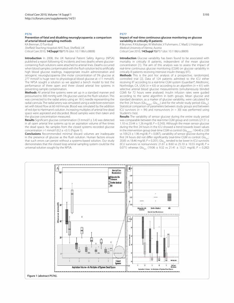
P576
Prevention of fatal and disabling neuroglycopaenia: a comparison of arterial blood sampling methodsKA Brennan, D Turnbull
Sheffi eld Teaching Hospitals NHS Trust, Sheffi eld, UK
Critical Care 2010, 14(Suppl 1):P576 (doi: 10.1186/cc8808)
Introduction In 2008, the UK National Patient Safety Agency (NPSA)
published a report following 42 incidents and two deaths where glucose-
containing fl ush solutions were attached to arterial lines. Deaths occurred
when blood samples contaminated with the fl ush solution led to artifi cially
high blood glucose readings, inappropriate insulin administration and
iatrogenic neuroglycopaenia (the molar concentration of 5% glucose at
277 mmol/l is huge next to physiological blood glucose at <11 mmol/l).
The NPSA sought a solution so we applied a bench model to test the
performance of three open and three closed arterial line systems in
preventing sample contamination.
Methods All arterial line systems were set up in a standard manner and
pressurised to 300 mmHg with 5% glucose used as the fl ush solution. This
was connected to the radial artery using an 18 G needle representing the
radial cannula. The radial artery was simulated using a wide bore extension
set with blood fl ow at 60 ml/minute. Blood was simulated by the addition
of red dye to Hartmann’s solution. Increasing multiples of arterial line dead
space were aspirated and discarded. Blood samples were then taken and
the glucose concentration measured.
Results Signifi cant glucose contamination (3 mmol/l ± 3.4) was detected
in all open arterial line systems up to an aspiration volume of fi ve times
the dead space. No samples from the closed systems recorded glucose
concentration >1 mmol/l (0.2 ± <0.1) (Figure 1).
Conclusions Recommended minimal discard volumes are inadequate
in the presence of glucose as the fl ush solution. Human factors ensure
that such errors can persist without a systems-based solution. Our study
demonstrates that the closed loop arterial sampling system could be the
universal solution sought by the NPSA.
P577
Impact of real-time continuous glucose monitoring on glucose variability in critically ill patientsR Brunner, R Kitzberger, W Miehsler, V Fuhrmann, C Madl, U Holzinger
Medical University of Vienna, Austria
Critical Care 2010, 14(Suppl 1):P57 (doi: 10.1186/cc8809)
Introduction Glucose variability has been found to be associated with
mortality in critically ill patients, independent of the mean glucose
concentration [1]. The aim of this analysis was to assess the impact of
real-time continuous glucose monitoring (CGM) on glucose variability in
critically ill patients receiving intensive insulin therapy (IIT).
Methods This is the post hoc analysis of a prospective, randomized,
controlled trial [2]. Data of 124 patients admitted to the ICU either
receiving IIT according to a real-time CGM system (Guardian®; Medtronic,
Northridge, CA, USA) (n = 63) or according to an algorithm (n = 61) with
selective arterial blood glucose measurements (simultaneously blinded
CGM) for 72 hours were analysed. Insulin infusion rates were guided
according to the same algorithm in both groups. Mean glucose and
standard deviation, as a marker of glucose variability, were calculated for
the fi rst 24 hours (GluMEAN1
, GluSD1
) and for the whole study period (GluSD
).
Statistical comparison of parameters between study groups and between
ICU survivors (n = 94) and nonsurvivors (n = 30) was performed using
Student’s t test.
Results The variability of sensor glucose during the entire study period
was comparable between the real-time CGM group and controls (21.51 ±
1.10 vs 23.44 ± 1.26 mg/dl; P = 0.243). Although the mean sensor glucose
during the fi rst 24 hours in the ICU showed a trend towards lower values
in the intervention group (real-time CGM vs control: GluMEAN1
: 104.40 ± 2.00
vs 109.23 ± 1.96 mg/dl; P = 0.087), variability of sensor glucose during the
fi rst 24 hours did not diff er signifi cantly (real-time CGM vs control: GluSD1
:
20.85 vs 18.46 mg/dl; P = 0.201). GluSD
tended to be lower in ICU survivors
(ICU survivors vs nonsurvivors: 21.61 ± 8.60 vs 25.10 ± 10.55 mg/dl; P =
0.071) whereas GluSD1
(19.06 ± 9.32 vs 21.41 ± 13.21 mg/dl; P = 0.282)
Figure 1 (abstract P576).
Critical Care 2010, Volume 14 Suppl 1 http://ccforum.com/supplements/14/S1
S193

and GluMEAN1
(106.39 ± 14.83 vs 108.00 ± 18.44 mg/dl; P = 0.627) were not
diff erent between ICU survivors and nonsurvivors.
Conclusions IIT guided by real-time CGM did not result in reduced
glucose variability compared with IIT according to an algorithm with
selective arterial blood glucose measurements. Although mean sensor
glucose during the fi rst 24 hours tended to be lower in the real-time CGM
group, glucose variability was not diff erent between the two groups. GluSD
tended to be lower in ICU survivors irrespective of glucose monitoring.
References1. Egi M, et al.: Crit Care 2009, 13:302.
2. Holzinger U, et al.: Diab Care 2009, in press.
P578
Automated blood glucose monitor is insensitive to hematocrit variation and common saccharide interferentsS Hendee, S Vanslyke, J Faught, A Truong, W Patterson, R Thompson
Luminous Medical, Carlsbad, CA, USA
Critical Care 2010, 14(Suppl 1):P578 (doi: 10.1186/cc8810)
Introduction Luminous Medical is developing an automated, patient-
attached system (Argus Glucose Monitoring System) that uses a fl ow-
through glucose oxidase sensor to measure plasma glucose levels in
whole blood. The Argus GMS will aid caregivers in achieving glycemic
control in critical care patients. The FDA has recently indicated that ICU
glucose monitors need to satisfy tighter accuracy requirements than
those specifi ed by ISO 15197 (±15 mg/dl or ±20%). Luminous conducted
a study to demonstrate Argus GMS accuracy under conditions of variable
hematocrit, and in the presence of common saccharide interferents.
Methods Hematocrit Study Group: one unit of whole blood was collected
under an IRB-approved protocol from two healthy volunteers. Blood
samples were prepared at three hematocrit levels (~24, 36, and 48%).
Three glucose levels were tested at each hematocrit: native glucose,
native plus 100, and native plus 200 mg/dl. Repeats were measured for
each hematocrit/glucose combination (n = 99 total samples in this study
group). Interferent Study Group: blood samples were prepared at native
hematocrit (~37%) and native glucose level (~100 mg/dl). Testing followed
established interference testing guidelines [1]. Blood samples were spiked
with concentrated interferent solution or with equal volume of normal
saline. Testing alternated between interferent-containing and interferent-
free samples (n = 5 test and fi ve control samples for each interferent).
Saccharide interferents included maltose (55 mg/dl), galactose (15 mg/dl),
fructose (18 mg/dl), and xylose (40 mg/dl). In both study groups, blood
samples were measured in vitro, in real time, using the Argus GMS in
multisample mode. Plasma measurements from two YSI 2700 instruments
were averaged to provide reference glucose values.
Results Hematocrit Study Group: the Argus GMS measured plasma
glucose across a range of 50 to 350 mg/dl with RMS error of 5.5 mg/dl
(mean error 2.6, 95% CI = –7.0 to 12.2, R = 0.998). The Argus GMS showed
no sensitivity to hematocrit variation. Interferent Study Group: the Argus
GMS showed minimal glucose measurement bias for each of the four
saccharide interferents (maltose 2.21 mg/dl, galactose 4.09 mg/dl, fructose
2.63 mg/dl, and xylose 2.03 mg/dl).
Conclusions The Argus GMS provides highly accurate glucose readings
in whole blood samples with no observable sensitivity to hematocrit
variation and with minimal infl uence from saccharide interferences. Argus
demonstrated measurement accuracy within ±10 mg/dl or ±10% at 95%
confi dence limits.
Reference1. Clinical and Laboratory Standards Institute: Interference Testing in Clinical
Chemistry: Approved Guideline – Second Edition. Wayne, PA: Clinical and
Laboratory Standards Institute; 2005.
P579
Performance of an automated blood glucose monitor in the OR/ICUM Higgins1, G Bochicchio2, P Simpson3, J Leach3, A Kamath3, D Deyette1
1Edwards Lifesciences, LLC, Irvine, CA, USA; 2University of Maryland School of
Medicine, Baltimore, MD, USA; 3Dexcom, Inc., San Diego, CA, USA
Critical Care 2010, 14(Suppl 1):P579 (doi: 10.1186/cc8811)
Introduction DexCom and Edwards Lifesciences collaborated to
develop an intravenous blood glucose monitor for use in a critical care
environment. The system provides automatic, real-time monitoring and
trending of blood glucose (BG) values for critical care patients. The system
is designed to address the needs of clinicians to monitor BG in critical care
units. The sensor was also designed to accurately measure BG even in the
presence of many drugs commonly used in the hospital.
Methods This was a blinded, multicenter study evaluating 17 adult
patients presenting to the OR and ICU. They were monitored for up to 72
hours with reference samples taken at 4-hour intervals and measured with
a YSI-2300 laboratory analyzer. Data are from initial roll-in patients used for
training and assessing protocol complications. Reference blood samples
were taken from either a peripheral vein, a central venous catheter or
an arterial line. Glucose values were calculated prospectively but not
displayed during the study.
Results Of 211 paired BG measurements, 94.3% were within Clarke Error
Grid A, 5.7% in B and 0% in C, D and E (Figure 1a). The mean absolute
relative diff erence was 6.5%. No signifi cant errors due to drugs were
measured. These results compare favorably with a previous study of 50
subjects who were monitored continuously for up to 72 hours in a diabetic
Figure 1 (abstract P579). Clarke Error Grid for (a) OR/ICU and (b) in-clinic.
Critical Care 2010, Volume 14 Suppl 1 http://ccforum.com/supplements/14/S1
S194
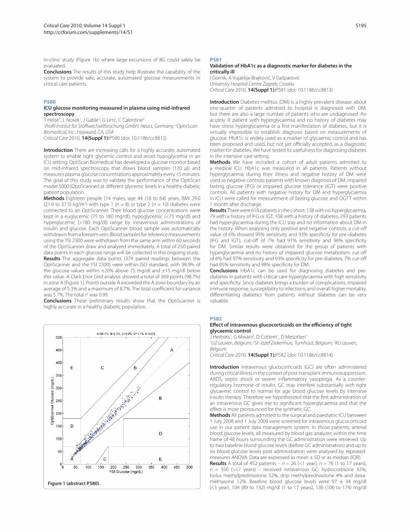
in-clinic study (Figure 1b) where large excursions of BG could safely be
evaluated.
Conclusions The results of this study help illustrate the capability of the
system to provide safe, accurate, automated glucose measurements in
critical care patients.
P580
ICU glucose monitoring measured in plasma using mid-infrared spectroscopyT Heise1, L Nosek1, J Gable2, G Lim2, C Calentine2
1Profi l Institut für Stoff wechselforschung GmbH, Neuss, Germany; 2OptisScan
Biomedical, Inc., Hayward, CA, USA
Critical Care 2010, 14(Suppl 1):P580 (doi: 10.1186/cc8812)
Introduction There are increasing calls for a highly accurate, automated
system to enable tight glycemic control and avoid hypoglycemia in an
ICU setting. OptiScan Biomedical has developed a glucose monitor based
on mid-infrared spectroscopy that draws blood samples (120 μl) and
measures plasma glucose concentrations approximately every 15 minutes.
The goal of this study was to validate the performance of the OptiScan
model 5000 (OptiScanner) at diff erent glycemic levels in a healthy diabetic
patient population.
Methods Eighteen people (14 males, age 44 (18 to 64) years, BMI 29.0
(21.9 to 37.5) kg/m2) with type 1 (n = 8) or type 2 (n = 10) diabetes were
connected to an OptiScanner. Their blood glucose concentrations were
kept in a euglycemic (75 to 180 mg/dl), hypoglycemic (<75 mg/dl) and
hyperglycemic (>180 mg/dl) range by intravenous administrations of
insulin and glucose. Each OptiScanner blood sample was automatically
withdrawn from a forearm vein. Blood samples for reference measurements
using the YSI 2300 were withdrawn from the same arm within 60 seconds
of the OptiScanner draw and analyzed immediately. A total of 250 paired
data points in each glucose range will be collected in this ongoing study.
Results The aggregate data points (374 paired readings between the
OptiScanner and the YSI 2300) were within ISO standard, with 98.9% of
the glucose values within ±20% above 75 mg/dl and ±15 mg/dl below
this value. A Clark Error Grid analysis showed a total of 369 points (98.7%)
in zone A (Figure 1). Points outside A exceeded the A zone boundary by an
average of 5.3% and a maximum of 8.7%. The total coeffi cient for variance
was 5.7%. The total r2 was 0.99.
Conclusions These preliminary results show that the OptiScanner is
highly accurate in a healthy diabetic population.
P581
Validation of HbA1c as a diagnostic marker for diabetes in the critically illI Gornik, A Vujaklija-Brajković, V Gašparović
University Hospital Centre Zagreb, Croatia
Critical Care 2010, 14(Suppl 1):P581 (doi: 10.1186/cc8813)
Introduction Diabetes mellitus (DM) is a highly prevalent disease: about
one-quarter of patients admitted to hospital is diagnosed with DM,
but there are also a large number of patients who are undiagnosed. An
acutely ill patient with hyperglycaemia and no history of diabetes may
have stress hyperglycaemia or a fi rst manifestation of diabetes, but it is
virtually impossible to establish diagnosis based on measurements of
glucose. HbA1c is widely used as a marker of glycaemic control and has
been proposed and used, but not yet offi cially accepted, as a diagnostic
marker for diabetes. We have tested its usefulness for diagnosing diabetes
in the intensive care setting.
Methods We have included a cohort of adult patients admitted to
a medical ICU. HbA1c was measured in all patients. Patients without
hyperglycaemia during their illness and negative history of DM were
used as negative controls; patients with known diagnosis of DM, impaired
fasting glucose (IFG) or impaired glucose tolerance (IGT) were positive
controls. All patients with negative history for DM and hyperglycaemia
in ICU were called for measurement of fasting glucose and OGTT within
1 month after discharge.
Results There were 618 patients in the cohort: 138 with no hyperglycaemia,
79 with a history of IFG or IGT, 108 with a history of diabetes; 293 patients
had hyperglycaemia during the ICU stay and no information about DM in
the history. When analysing only positive and negative controls, a cut-off
value of 6% showed 95% sensitivity and 93% specifi city for pre-diabetes
(IFG and IGT), cut-off of 7% had 91% sensitivity and 96% specifi city
for DM. Similar results were obtained for the group of patients with
hyperglycaemia and no history of impaired glucose metabolism: cut-off
of 6% had 97% sensitivity and 93% specifi city for pre-diabetes, 7% cut-off
had 95% sensitivity and 98% specifi city for DM.
Conclusions HbA1c can be used for diagnosing diabetes and pre-
diabetes in patients with critical care hyperglycaemia with high sensitivity
and specifi city. Since diabetes brings a burden of complications, impaired
immune response, susceptibility to infections, and overall higher mortality,
diff erentiating diabetics from patients without diabetes can be very
valuable.
P582
Eff ect of intravenous glucocorticoids on the effi ciency of tight glycaemic controlJ Herbots1, G Mwani2, D Cottem1, D Mesotten3
1UZ Leuven, Belgium; 2St-Jozef Ziekenhuis, Turnhout, Belgium; 3KU Leuven,
Belgium
Critical Care 2010, 14(Suppl 1):P582 (doi: 10.1186/cc8814)
Introduction Intravenous glucocorticoids (GC) are often administered
during critical illness in the context of post-transplant immunosuppression,
ARDS, septic shock or severe infl ammatory vasoplegia. As a counter-
regulatory hormone of insulin, GC may interfere substantially with tight
glycaemic control to normal for age blood glucose levels by intensive
insulin therapy. Therefore we hypothesized that the fi rst administration of
an intravenous GC gives rise to signifi cant hyperglycaemia and that the
eff ect is more pronounced for the synthetic GC.
Methods All patients admitted to the surgical and paediatric ICU between
1 July 2008 and 1 July 2009 were screened for intravenous glucocorticoid
use in our patient data management system. In those patients, arterial
blood glucose levels, all measured by blood gas analyzer, within the time
frame of 48 hours surrounding the GC administration were retrieved. Up
to two baseline blood glucose levels (before GC administration) and up to
six blood glucose levels post administration were analysed by repeated-
measures ANOVA. Data are expressed as mean ± SD or as median (IQR).
Results A total of 452 patients – n = 26 (<1 year), n = 76 (1 to 17 years),
n = 350 (>17 years) – received intravenous GC: hydrocortisone 32%,
bolus methylprednisolone 52%, drip methylprednisolone 4% and dexa-
methasone 12%. Baseline blood glucose levels were 97 ± 34 mg/dl
(<1 year), 104 (89 to 132) mg/dl (1 to 17 years), 136 (106 to 174) mg/dl Figure 1 (abstract P580).
Critical Care 2010, Volume 14 Suppl 1 http://ccforum.com/supplements/14/S1
S195
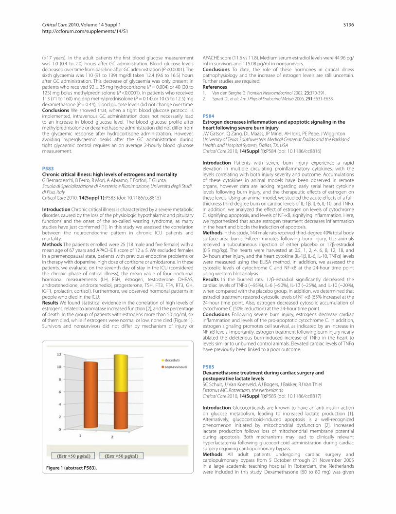
(>17 years). In the adult patients the fi rst blood glucose measurement
was 1.0 (0.4 to 2.0) hours after GC administration. Blood glucose levels
decreased over time from baseline after GC administration (P <0.0001). The
sixth glycaemia was 110 (91 to 139) mg/dl taken 12.4 (9.6 to 16.5) hours
after GC administration. This decrease of glycaemia was only present in
patients who received 92 ± 35 mg hydrocortisone (P = 0.004) or 40 (20 to
125) mg bolus methylprednisolone (P <0.0001). In patients who received
113 (71 to 160) mg drip methylprednisolone (P = 0.14) or 10 (5 to 12.5) mg
dexamethasone (P = 0.44), blood glucose levels did not change over time.
Conclusions We showed that, when a tight blood glucose protocol is
implemented, intravenous GC administration does not necessarily lead
to an increase in blood glucose level. The blood glucose profi le after
methylprednisolone or dexamethasone administration did not diff er from
the glycaemic response after hydrocortisone administration. However,
avoiding hyperglycaemic peaks after the GC administration during
tight glycaemic control requires an on average 2-hourly blood glucose
measurement.
P583
Chronic critical illness: high levels of estrogens and mortalityG Bernardeschi, B Ferro, R Mori, A Abramo, F Forfori, F Giunta
Scuola di Specializzazione di Anestesia e Rianimazione, Università degli Studi
di Pisa, Italy
Critical Care 2010, 14(Suppl 1):P583 (doi: 10.1186/cc8815)
Introduction Chronic critical illness is characterized by a severe metabolic
disorder, caused by the loss of the physiologic hypothalamic and pituitary
functions and the onset of the so-called wasting syndrome, as many
studies have just confi rmed [1]. In this study we assessed the correlation
between the neuroendocrine pattern in chronic ICU patients and
mortality.
Methods The patients enrolled were 25 (18 male and fi ve female) with a
mean age of 67 years and APACHE II score of 12 ± 5. We excluded females
in a premenopausal state, patients with previous endocrine problems or
in therapy with dopamine, high dose of cortisone or amiodarone. In these
patients, we evaluate, on the seventh day of stay in the ICU (considered
the chronic phase of critical illness), the mean value of four nocturnal
hormonal measurements (LH, FSH, estrogen, testosterone, DHEAS,
androstenedione, androstenediol, progesterone, TSH, FT3, FT4, RT3, GH,
IGF1, prolactin, cortisol). Furthermore, we observed hormonal patterns in
people who died in the ICU.
Results We found statistical evidence in the correlation of high levels of
estrogens, related to aromatase increased function [2], and the percentage
of death. In the group of patients with estrogens more than 50 pg/ml, six
of them died, while if estrogens were normal or low, none died (Figure 1).
Survivors and nonsurvivors did not diff er by mechanism of injury or
APACHE score (11.6 vs 11.8). Medium serum estradiol levels were 44.96 pg/
ml in survivors and 115.08 pg/ml in nonsurvivors.
Conclusions To date, the role of these hormones in critical illness
pathophysiology and the increase of estrogen levels are still uncertain.
Further studies are required.
References1. Van den Berghe G: Frontiers Neuroendocrinol 2002, 23:370-391.
2. Spratt DI, et al.: Am J Physiol Endocrinol Metab 2006, 291:E631-E638.
P584
Estrogen decreases infl ammation and apoptotic signaling in the heart following severe burn injuryJW Gatson, Q Zang, DL Maass, JP Minei, AH Idris, PE Pepe, J Wigginton
University of Texas Southwestern Medical Center at Dallas and the Parkland
Health and Hospital System, Dallas, TX, USA
Critical Care 2010, 14(Suppl 1):P584 (doi: 10.1186/cc8816)
Introduction Patients with severe burn injury experience a rapid
elevation in multiple circulating proinfl ammatory cytokines, with the
levels correlating with both injury severity and outcome. Accumulations
of these cytokines in animal models have been observed in remote
organs, however data are lacking regarding early serial heart cytokine
levels following burn injury, and the therapeutic eff ects of estrogen on
these levels. Using an animal model, we studied the acute eff ects of a full-
thickness third-degree burn on cardiac levels of IL-1β, IL-6, IL-10, and TNFα.
In addition, we analyzed the eff ect of estrogen on levels of cytochrome
C, signifying apoptosis, and levels of NF-κB, signifying infl ammation. Here,
we hypothesized that acute estrogen treatment decreases infl ammation
in the heart and blocks the induction of apoptosis.
Methods In this study, 144 male rats received third-degree 40% total body
surface area burns. Fifteen minutes following burn injury, the animals
received a subcutaneous injection of either placebo or 17β-estradiol
(0.5 mg/kg). The hearts were harvested at 0.5, 1, 2, 4, 6, 8, 12, 18, and
24 hours after injury, and the heart cytokine (IL-1β, IL-6, IL-10, TNFα) levels
were measured using the ELISA method. In addition, we assessed the
cytosolic levels of cytochrome C and NF-κB at the 24-hour time point
using western blot analysis.
Results In the burned rats, 17β-estradiol signifi cantly decreased the
cardiac levels of TNFα (~95%), IL-6 (~50%), IL-1β (~25%), and IL-10 (~20%),
when compared with the placebo group. In addition, we determined that
estradiol treatment restored cytosolic levels of NF-κB (65% increase) at the
24-hour time point. Also, estrogen decreased cytosolic accumulation of
cytochrome C (50% reduction) at the 24-hour time point.
Conclusions Following severe burn injury, estrogens decrease cardiac
infl ammation and levels of the pro-apoptotic cytochrome C. In addition,
estrogen signaling promotes cell survival, as indicated by an increase in
NF-κB levels. Importantly, estrogen treatment following burn injury nearly
ablated the deleterious burn-induced increase of TNFα in the heart to
levels similar to unburned control animals. Elevated cardiac levels of TNFα
have previously been linked to a poor outcome.
P585
Dexamethasone treatment during cardiac surgery and postoperative lactate levelsSC Schuit, JJ Van Koesveld, AJ Bogers, J Bakker, RJ Van Thiel
Erasmus MC, Rotterdam, the Netherlands
Critical Care 2010, 14(Suppl 1):P585 (doi: 10.1186/cc8817)
Introduction Glucocorticoids are known to have an anti-insulin action
on glucose metabolism, leading to increased lactate production [1].
Alternatively, glucocorticoid-induced apoptosis is a well-recognized
phenomenon initiated by mitochondrial dysfunction [2]. Increased
lactate production follows loss of mitochondrial membrane potential
during apoptosis. Both mechanisms may lead to clinically relevant
hyperlactatemia following glucocorticoid administration during cardiac
surgery requiring cardiopulmonary bypass.
Methods All adult patients undergoing cardiac surgery and
cardiopulmonary bypass from 5 October through 21 November 2005
in a large academic teaching hospital in Rotterdam, the Netherlands
were included in this study. Dexamethasone (60 to 80 mg) was given Figure 1 (abstract P583).
Critical Care 2010, Volume 14 Suppl 1 http://ccforum.com/supplements/14/S1
S196
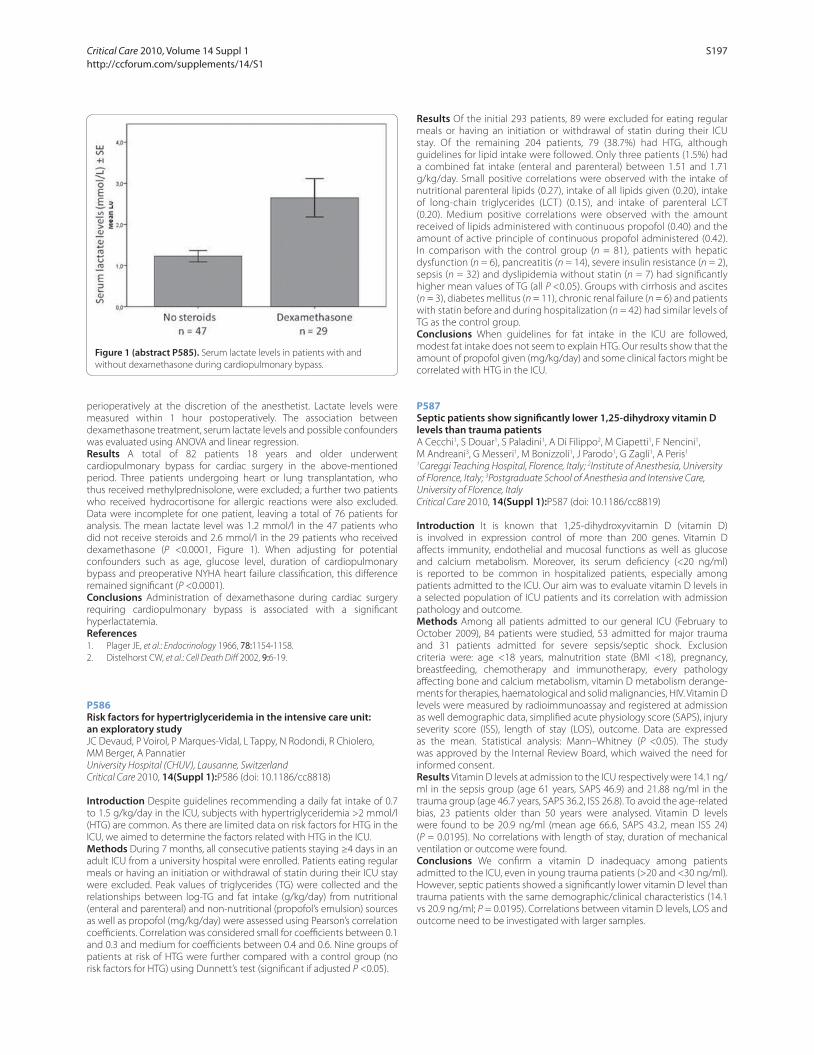
perioperatively at the discretion of the anesthetist. Lactate levels were
measured within 1 hour postoperatively. The association between
dexamethasone treatment, serum lactate levels and possible confounders
was evaluated using ANOVA and linear regression.
Results A total of 82 patients 18 years and older underwent
cardiopulmonary bypass for cardiac surgery in the above-mentioned
period. Three patients undergoing heart or lung transplantation, who
thus received methylprednisolone, were excluded; a further two patients
who received hydrocortisone for allergic reactions were also excluded.
Data were incomplete for one patient, leaving a total of 76 patients for
analysis. The mean lactate level was 1.2 mmol/l in the 47 patients who
did not receive steroids and 2.6 mmol/l in the 29 patients who received
dexamethasone (P <0.0001, Figure 1). When adjusting for potential
confounders such as age, glucose level, duration of cardiopulmonary
bypass and preoperative NYHA heart failure classifi cation, this diff erence
remained signifi cant (P <0.0001).
Conclusions Administration of dexamethasone during cardiac surgery
requiring cardiopulmonary bypass is associated with a signifi cant
hyperlactatemia.
References1. Plager JE, et al.: Endocrinology 1966, 78:1154-1158.
2. Distelhorst CW, et al.: Cell Death Diff 2002, 9:6-19.
P586
Risk factors for hypertriglyceridemia in the intensive care unit: an exploratory studyJC Devaud, P Voirol, P Marques-Vidal, L Tappy, N Rodondi, R Chiolero,
MM Berger, A Pannatier
University Hospital (CHUV), Lausanne, Switzerland
Critical Care 2010, 14(Suppl 1):P586 (doi: 10.1186/cc8818)
Introduction Despite guidelines recommending a daily fat intake of 0.7
to 1.5 g/kg/day in the ICU, subjects with hypertriglyceridemia >2 mmol/l
(HTG) are common. As there are limited data on risk factors for HTG in the
ICU, we aimed to determine the factors related with HTG in the ICU.
Methods During 7 months, all consecutive patients staying ≥4 days in an
adult ICU from a university hospital were enrolled. Patients eating regular
meals or having an initiation or withdrawal of statin during their ICU stay
were excluded. Peak values of triglycerides (TG) were collected and the
relationships between log-TG and fat intake (g/kg/day) from nutritional
(enteral and parenteral) and non-nutritional (propofol’s emulsion) sources
as well as propofol (mg/kg/day) were assessed using Pearson’s correlation
coeffi cients. Correlation was considered small for coeffi cients between 0.1
and 0.3 and medium for coeffi cients between 0.4 and 0.6. Nine groups of
patients at risk of HTG were further compared with a control group (no
risk factors for HTG) using Dunnett’s test (signifi cant if adjusted P <0.05).
Results Of the initial 293 patients, 89 were excluded for eating regular
meals or having an initiation or withdrawal of statin during their ICU
stay. Of the remaining 204 patients, 79 (38.7%) had HTG, although
guidelines for lipid intake were followed. Only three patients (1.5%) had
a combined fat intake (enteral and parenteral) between 1.51 and 1.71
g/kg/day. Small positive correlations were observed with the intake of
nutritional parenteral lipids (0.27), intake of all lipids given (0.20), intake
of long-chain triglycerides (LCT) (0.15), and intake of parenteral LCT
(0.20). Medium positive correlations were observed with the amount
received of lipids administered with continuous propofol (0.40) and the
amount of active principle of continuous propofol administered (0.42).
In comparison with the control group (n = 81), patients with hepatic
dysfunction (n = 6), pancreatitis (n = 14), severe insulin resistance (n = 2),
sepsis (n = 32) and dyslipidemia without statin (n = 7) had signifi cantly
higher mean values of TG (all P <0.05). Groups with cirrhosis and ascites
(n = 3), diabetes mellitus (n = 11), chronic renal failure (n = 6) and patients
with statin before and during hospitalization (n = 42) had similar levels of
TG as the control group.
Conclusions When guidelines for fat intake in the ICU are followed,
modest fat intake does not seem to explain HTG. Our results show that the
amount of propofol given (mg/kg/day) and some clinical factors might be
correlated with HTG in the ICU.
P587
Septic patients show signifi cantly lower 1,25-dihydroxy vitamin D levels than trauma patientsA Cecchi1, S Douar1, S Paladini1, A Di Filippo2, M Ciapetti1, F Nencini1,
M Andreani3, G Messeri1, M Bonizzoli1, J Parodo1, G Zagli1, A Peris1
1Careggi Teaching Hospital, Florence, Italy; 2Institute of Anesthesia, University
of Florence, Italy; 3Postgraduate School of Anesthesia and Intensive Care,
University of Florence, Italy
Critical Care 2010, 14(Suppl 1):P587 (doi: 10.1186/cc8819)
Introduction It is known that 1,25-dihydroxyvitamin D (vitamin D)
is involved in expression control of more than 200 genes. Vitamin D
aff ects immunity, endothelial and mucosal functions as well as glucose
and calcium metabolism. Moreover, its serum defi ciency (<20 ng/ml)
is reported to be common in hospitalized patients, especially among
patients admitted to the ICU. Our aim was to evaluate vitamin D levels in
a selected population of ICU patients and its correlation with admission
pathology and outcome.
Methods Among all patients admitted to our general ICU (February to
October 2009), 84 patients were studied, 53 admitted for major trauma
and 31 patients admitted for severe sepsis/septic shock. Exclusion
criteria were: age <18 years, malnutrition state (BMI <18), pregnancy,
breastfeeding, chemotherapy and immunotherapy, every pathology
aff ecting bone and calcium metabolism, vitamin D metabolism derange-
ments for therapies, haematological and solid malignancies, HIV. Vitamin D
levels were measured by radioimmunoassay and registered at admission
as well demographic data, simplifi ed acute physiology score (SAPS), injury
severity score (ISS), length of stay (LOS), outcome. Data are expressed
as the mean. Statistical analysis: Mann–Whitney (P <0.05). The study
was approved by the Internal Review Board, which waived the need for
informed consent.
Results Vitamin D levels at admission to the ICU respectively were 14.1 ng/
ml in the sepsis group (age 61 years, SAPS 46.9) and 21.88 ng/ml in the
trauma group (age 46.7 years, SAPS 36.2, ISS 26.8). To avoid the age-related
bias, 23 patients older than 50 years were analysed. Vitamin D levels
were found to be 20.9 ng/ml (mean age 66.6, SAPS 43.2, mean ISS 24)
(P = 0.0195). No correlations with length of stay, duration of mechanical
ventilation or outcome were found.
Conclusions We confi rm a vitamin D inadequacy among patients
admitted to the ICU, even in young trauma patients (>20 and <30 ng/ml).
However, septic patients showed a signifi cantly lower vitamin D level than
trauma patients with the same demographic/clinical characteristics (14.1
vs 20.9 ng/ml; P = 0.0195). Correlations between vitamin D levels, LOS and
outcome need to be investigated with larger samples.
Figure 1 (abstract P585). Serum lactate levels in patients with and
without dexamethasone during cardiopulmonary bypass.
Critical Care 2010, Volume 14 Suppl 1 http://ccforum.com/supplements/14/S1
S197
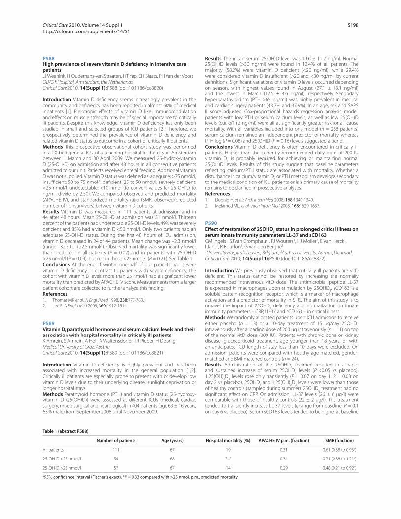
P588
High prevalence of severe vitamin D defi ciency in intensive care patientsJJ Weenink, H Oudemans-van Straaten, HT Yap, EH Slaats, PH Van der Voort
OLVG hHospital, Amsterdam, the Netherlands
Critical Care 2010, 14(Suppl 1):P588 (doi: 10.1186/cc8820)
Introduction Vitamin D defi ciency seems increasingly prevalent in the
community, and defi ciency has been reported in almost 60% of medical
inpatients [1]. Pleiotropic eff ects of vitamin D like immunomodulation
and eff ects on muscle strength may be of special importance to critically
ill patients. Despite this knowledge, vitamin D defi ciency has only been
studied in small and selected groups of ICU patients [2]. Therefore, we
prospectively determined the prevalence of vitamin D defi ciency and
related vitamin D status to outcome in a cohort of critically ill patients.
Methods This prospective observational cohort study was performed
in a 20-bed general ICU of a teaching hospital in the city of Amsterdam
between 1 March and 30 April 2009. We measured 25-hydroxyvitamin
D (25-OH-D) on admission and after 48 hours in all consecutive patients
admitted to our unit. Patients received enteral feeding. Additional vitamin
D was not supplied. Vitamin D status was defi ned as: adequate: >75 nmol/l,
insuffi cient: 50 to 75 nmol/l, defi cient: 25 to 50 nmol/l, severely defi cient:
<25 nmol/l, undetectable: <10 nmol (to convert values for 25-OH-D to
ng/ml, divide by 2.50). We compared observed and predicted mortality
(APACHE IV), and standardized mortality ratio (SMR, observed/predicted
number of nonsurvivors) between vitamin D cohorts.
Results Vitamin D was measured in 111 patients at admission and in
44 after 48 hours. Mean 25-OH-D at admission was 31 nmol/l. Thirteen
percent of the patients had undetectable 25-OH-D levels, 49% was severely
defi cient and 85% had a vitamin D <50 nmol/l. Only two patients had an
adequate 25-OH-D status. During the fi rst 48 hours of ICU admission,
vitamin D decreased in 24 of 44 patients. Mean change was –2.3 nmol/l
(range –32.5 to +22.5 nmol/l). Observed mortality was signifi cantly lower
than predicted in all patients (P = 0.02) and in patients with 25-OH-D
>25 nmol/l (P = 0.04), but not in those <25 nmol/l (P = 0.21). See Table 1.
Conclusions At the end of winter, one-half of our patients had severe
vitamin D defi ciency. In contrast to patients with severe defi ciency, the
cohort with vitamin D levels more than 25 nmol/l had a signifi cant lower
mortality than predicted by APACHE IV score. Measurements from a larger
patient cohort are collected to further analyze this fi nding.
References1. Thomas MK et al.: N Engl J Med 1998, 338:777-783.
2. Lee P: N Engl J Med 2009, 360:1912-1914.
P589
Vitamin D, parathyroid hormone and serum calcium levels and their association with hospital mortality in critically ill patientsK Amrein, S Amrein, A Holl, A Waltensdorfer, TR Pieber, H Dobnig
Medical University of Graz, Austria
Critical Care 2010, 14(Suppl 1):P589 (doi: 10.1186/cc8821)
Introduction Vitamin D defi ciency is highly prevalent and has been
associated with increased mortality in the general population [1,2].
Critically ill patients are especially prone to present with or develop low
vitamin D levels due to their underlying disease, sunlight deprivation or
longer hospital stays.
Methods Parathyroid hormone (PTH) and vitamin D status (25-hydroxy-
vitamin D (25(OH)D)) were assessed at diff erent ICUs (medical, cardiac
surgery, mixed surgical and neurological) in 404 patients (age 63 ± 16 years,
65% male) from September 2008 until November 2009.
Results The mean serum 25(OH)D level was 19.6 ± 11.2 ng/ml. Normal
25(OH)D levels (>30 ng/ml) were found in 12.4% of all patients. The
majority (58.2%) were vitamin D defi cient (<20 ng/ml), while 29.4%
were considered vitamin D insuffi cient (>20 and <30 ng/ml) by current
defi nitions. Signifi cant variations of vitamin D levels occurred depending
on season, with highest values found in August (27.1 ± 13.1 ng/ml)
and the lowest in March (12.5 ± 4.6 ng/ml), respectively. Secondary
hyperparathyroidism (PTH >65 pg/ml) was highly prevalent in medical
and cardiac surgery patients (43.7% and 37.9%). In an age, sex and SAPS
II score adjusted Cox-proportional hazards regression analysis model,
patients with low PTH or serum calcium levels, as well as low 25(OH)D
levels (cut-off 12 ng/ml) were all at signifi cantly greater risk for all-cause
mortality. With all variables included into one model (n = 268 patients)
serum calcium remained an independent predictor of mortality, whereas
PTH log (P = 0.08) and 25(OH)D (P = 0.16) levels suggested a trend.
Conclusions Vitamin D defi ciency is often encountered in critically ill
patients. Higher than the currently recommended daily dose of 200 IU
vitamin D3 is probably required for achieving or maintaining normal
25(OH)D levels. Results of this study suggest that baseline parameters
refl ecting calcium/PTH status are associated with mortality. Whether a
disturbance in calcium/vitamin D3 or PTH metabolism develops secondary
to the medical condition of ICU patients or is a primary cause of mortality
remains to be clarifi ed in prospective analyses.
References1. Dobnig H, et al.: Arch Intern Med 2008, 168:1340-1349.
2. Melamed ML, et al.: Arch Intern Med 2008, 168:1629-1637.
P590
Eff ect of restoration of 25OHD3 status in prolonged critical illness on
serum innate immunity parameters LL-37 and sCD163CM Ingels1, SJ Van Cromphaut1, PJ Wouters1, HJ Moller2, E Van Herck1,
I Jans1, R Bouillon1, G Van den Berghe1
1University Hospitals Leuven, Belgium; 2Aarhus University, Aarhus, Denmark
Critical Care 2010, 14(Suppl 1):P590 (doi: 10.1186/cc8822)
Introduction We previously observed that critically ill patients are vitD
defi cient. This status cannot be restored by increasing the normally
recommended intravenous vitD dose. The antimicrobial peptide LL-37
is expressed in macrophages upon stimulation by 25OHD3. sCD163 is a
soluble pattern-recognition receptor, which is a marker of macrophage
activation and a predictor of mortality in SIRS. The aim of this study is to
unravel the impact of 25OHD3 defi ciency and normalization on innate
immunity parameters – CRP, LL-37 and sCD163 – in critical illness.
Methods We randomly allocated patients upon ICU admission to receive
either placebo (n = 13) or a 10-day treatment of 15 μg/day 25OHD3
intravenously after a loading dose of 200 μg intravenously (n = 11) on top
of the normal vitD dose (200 IU). Patients with chronic bone or kidney
disease, glucocorticoid treatment, age younger than 18 years, or with
an anticipated ICU length of stay less than 10 days were excluded. On
admission, patients were compared with healthy age-matched, gender-
matched and BMI-matched controls (n = 24).
Results Administration of the 25OHD3 regimen resulted in a rapid
and sustained increase of serum 25OHD3 levels (P <0.05 vs placebo).
1,25(OH)2D
3 levels rose only transiently (P = 0.07 on day 1, P = 0.08 on
day 2 vs placebo). 25OHD3 and 1,25(OH)
2D
3 levels were lower than those
of healthy controls (sampled during summer). 25OHD3 treatment had no
signifi cant eff ect on CRP. On admission, LL-37 levels (26 ± 6 μg/l) were
comparable with those of healthy controls (22 ± 2 μg/l). The treatment
tended to transiently increase LL-37 levels (change from baseline: P = 0.1
on day 6 vs placebo). Serum sCD163 levels tended to be higher at baseline
Table 1 (abstract P588)
Number of patients Age (years) Hospital mortality (%) APACHE IV p.m. (fraction) SMR (fraction)
All patients 111 67 19 0.31 0.61 (0.38 to 0.93a)
25-OH-D <25 nmol/l 54 68 24* 0.34 0.71 (0.38 to 1.21a)
25-OH-D >25 nmol/l 57 67 14 0.29 0.48 (0.21 to 0.92a)
a95% confi dence interval (Fischer’s exact). *P = 0.33 compared with >25 nmol. p.m., predicted mortality.
Critical Care 2010, Volume 14 Suppl 1 http://ccforum.com/supplements/14/S1
S198

(P = 0.06 vs healthy controls) and rose signifi cantly over time (P <0.05 on
day 5 and on day 10 vs day 0) but were not infl uenced by the treatment.
Conclusions The preliminary results of this randomized placebo-
controlled pilot study indicate that, in contrast to a high intravenous 500 IU
vitD supplement, intravenous 25OHD3 treatment resulted in an immediate
and persistent increase in serum 25OHD3, but did not aff ect 1,25(OH)
2D
3
levels. Plasma LL-37 alterations in the studied prolonged critically ill
patients hints at ameliorated LL-37 production provided that the 25OHD3
status is restored. Serum sCD163 and CRP levels evolved during the ICU
stay, but were not signifi cantly altered by 25OHD3 supplementation.
The results of this pilot study warrant further research into the potential
modulation of innate immune processes by 25OHD3 in the critically ill.
P591
Alterations in adipose tissue during critical illness: an adaptive and protective response?L Langouche1, S Vander Perre1, S Thiessen1, G Hermans1, A D’Hoore1,
B Kola2, M Korbonits2, G Van den Berghe1
1KU Leuven, Belgium; 2Barts and the London School of Medicine and Dentistry –
Queen Mary University of London, UK
Critical Care 2010, 14(Suppl 1):P591 (doi: 10.1186/cc8823)
Introduction Critical illness is characterized by lean tissue wasting
whereas adipose tissue is preserved. Obese critically ill patients may have
a lower risk of death than nonobese patients, a recent observation that
may suggest a protective role for adipose tissue during illness, a role which
has not been previously investigated. We hypothesized that adipose tissue
could function as a waste bin for potentially toxic metabolites, such as
glucose, during critical illness.
Methods We studied adipose tissue biopsies from 61 of critically ill
patients, more specifi cally morphology and the potential to take up and
metabolize glucose, and compared this with biopsies from 20 matched
controls.
Results Adipose tissue biopsies from critically ill patients revealed a higher
number and a smaller size of adipocytes as compared with matched
controls, coinciding with increased preadipocyte marker levels. Also,
>95% of adipose biopsies from critically ill patients displayed positive
macrophage staining. Gene and protein expression of insulin-independent
GLUTs, and tissue glucose content was increased. Glucokinase expression
was upregulated whereas glycogen and glucose-6-phosphate levels
were low. Acetyl-CoA-carboxylase protein level and activity of fatty-acid
synthase were increased. A substantially increased activity of AMPK may
play a crucial role.
Conclusions The larger number of small adipocytes in response to critical
illness appears to have an increased ability to take up and metabolize
glucose into fatty acids. Such changes may render adipose tissue
biologically active as a functional waste bin for toxic metabolites during
critical illness, which could contribute to survival.
P592
Impact of collection method and sample handling on measured levels of circulating ACTHC Borin, D Mesotten, G Van den Berghe, H Vervenne
KU Leuven, Belgium
Critical Care 2010, 14(Suppl 1):P592 (doi: 10.1186/cc8824)
Introduction Cortisol, which is produced and released by the adrenal
gland under the regulation of the adrenocorticotropic hormone (ACTH),
plays a crucial role in the survival mechanisms involved in critical illness.
A dissociation between high plasma cortisol and low ACTH in patients
has been observed in the more chronic phase of critical illness induced
by severe sepsis and multiple trauma [1]. In order to assess the potential
confounding impact of errors in the measurement of ACTH, we evaluated
the stability of ACTH during sample processing.
Methods Two tests were performed. For the fi rst test, two blood samples
per patient were taken from 10 randomly selected patients in the surgical
ICU. One sample was collected warm (that is, samples were collected
at room temperature (RT), left for 60 to 90 minutes at RT followed by
24 hours at 4°C, centrifuged (10 minutes, 1,000 rpm), and serum was stored
at –80°C), the other sample was collected cold (that is, recommended
collection procedure for ACTH detection: samples were collected on ice,
centrifuged (10 minutes, 1,000 rpm), and plasma was stored at –80°C).
For a second test, again, two blood samples were taken per patient (n =
10) in the surgical ICU. One sample was kept frozen until ACTH testing
(original), whereas the other sample was defrosted several times at RT
before analysis (defrosted). ACTH levels were measured using an ACTH RIA
kit (BRAHMS) and the diff erent sampling methods and sample treatments
were compared using Bland–Altman statistics.
Results No clinically relevant diff erences in ACTH levels were observed
between cold (28.2 ± 22.3 pg/ml) and warm (24.3 ± 20.6 pg/ml) sampling
(bias 3.8 ± 7.3 pg/ml) or between original (22.1 ± 10.8 pg/ml) and
defrosted (22.3 ± 10.5 pg/ml) samples (bias 0.2 ± 1.9 pg/ml). Moreover, the
diff erences in ACTH levels measured between the two collection methods
or sample treatments were independent of the amount of ACTH present
in the samples.
Conclusions We showed that warm collection or frequent defrosting
of samples does not induce a clinically signifi cant error in ACTH levels
measured, implying that nonideally collected or treated samples can still
be used for rough ACTH analysis.
Reference1. Vermes et al.: J Clin Endocrinol Metab 1995, 80:1238-1242.
P593
Prognostic value of serum zinc levels in critically ill patientsB Cander, D Dundar, M Gul, S Girisgin
Selcuk University Meram Faculty of Medicine, Konya, Turkey
Critical Care 2010, 14(Suppl 1):P593 (doi: 10.1186/cc8825)
Introduction Zinc is a trace element that is required for normal function
of many enzymes. Intact zinc homeostasis must be present for normal
function of the immune system, oxidative stress responses. We explored
the hypothesis that a decline in serum zinc concentrations among
critically ill patients is related to mortality, length of stay in the ICU, APACHE
II and SOFA scores.
Methods This prospective study was conducted in the ICU of the
emergency department. All patients (>18 years) admitted to the ICU
from September through November 2009 were enrolled into the study.
Burn victims, alcoholics, patients with gastrointestinal fi stulas and
severe diarrhea were excluded from the study. Patients’ demographic
characteristics, APACHE and SOFA scores were recorded; blood samples
were collected within 24 hours after admission and analyzed for serum
zinc, copper, C-reactive protein and albumin levels. Patients were followed
up for mortality and length of stay in the ICU for 28 days.
Results Thirty-six patients were enrolled during the study period, with
median age 70.5 years (18 to 87). The male to female ratio was 1:1.1.
Serum zinc levels correlated inversely with SOFA scores (r = –0.41, P =
0.01). Patients with ≥8 SOFA scores had signifi cantly lower serum zinc
levels compared with patients with <8 SOFA scores (6.74 ± 1.63 μmol/l
(n = 11) and 9.17 ± 2.76 μmol/l (n = 25), respectively; P <0.05). There was
no signifi cant diff erence between serum zinc levels of patients with <25
and ≥25 APACHE II scores and also there was no signifi cant diff erence
between survivors and nonsurvivors. Serum zinc and copper levels had no
signifi cant relationship with length of stay in the ICU.
Conclusions Serum zinc concentrations correlated inversely with SOFA
scores in this study. This result supports that organ failure and critical
illness leads to a decline in serum zinc concentrations and that zinc
administration may be benefi cial for critically ill patients.
References1. Cvijanovich NZ, et al.: Zinc homeostasis in pediatric critical illness. Pediatr
Crit Care Med 2009, 10:29-34.
2. Berger MM, et al.: Trace element requirements in critically ill burned patients. J Trace Elem Med Biol 2007, 21(Suppl 1):44-48.
3. Heyland DK, et al.: Zinc supplementation in critically ill patients: a key pharmaconutrient? J Parenter Enteral Nutr 2008, 32:509-519.
Critical Care 2010, Volume 14 Suppl 1 http://ccforum.com/supplements/14/S1
S199
P594
High stress during admission predicts the impact of a PICU follow-up clinic on parents’ psychological outcomeG Colville, P Cream, S Kerry
St George’s Hospital, London, UK
Critical Care 2010, 14(Suppl 1):P594 (doi: 10.1186/cc8826)
Introduction There are no formal reports of follow-up services in
pediatric intensive care settings despite mounting evidence that parents
report negative psychological symptoms for many months after a child’s
admission to the pediatric intensive care unit (PICU). The aim of this study
was to establish the impact of a PICU follow-up clinic on parents’ distress.
Methods In this prospective, randomised controlled trial, parents of 133
children admitted to an eight-bed PICU for >12 hours were randomly
allocated to either the intervention (follow-up clinic appointment off ered
2 months after discharge) or control (no appointment) condition. Parents’
baseline stress was assessed at discharge using the Parental Stressor Scale:
PICU [1]. Five months later, post-traumatic stress, anxiety and depression
were assessed by postal questionnaires (Impact of Event Scale [2] and
Hospital Anxiety and Depression Scale [3]).
Results Outcome data were obtained for 105 parents. Whole group
analyses revealed no signifi cant diff erences in psychological outcome
between the control and intervention groups. However, post hoc analyses
revealed eff ects in favour of the intervention, for a sub-group of parents
(n = 55) who had reported baseline stress scores above median during
their child’s hospitalisation. Within this sub-group, parents who received
the intervention were less likely to report clinically signifi cant levels of
post-traumatic stress symptoms (25% vs 56%, P = 0.018) or depression
(19% vs 52%, P = 0.009) than controls.
Conclusions Whilst these results do not justify routine provision of PICU
follow-up clinic appointments for all parents, they do suggest that rates of
long-term distress in those who fi nd the admission particularly traumatic
could be reduced by a simple one-off intervention, which was feasible in
a clinical setting.
References1. Carter MC, Miles MS: The Parental Stressor Scale: Pediatric Intensive Care
Unit. Matern Child Nurs J 1989, 18:187-198.
2. Horowitz M, Wilner N, Alvarez W: Impact of Event Scale: a measure of subjective stress. Psychosom Med 1979, 41:209-218.
3. Zigmond AS, Snaith RP: The hospital anxiety and depression scale. Acta
Psychiatr Scand 1983, 67:361-370.
P595
Patient’s contact person in the intensive care unit: is it really important?AH Nielsen, CB Kancir
Regional Hospital West Jutland, Holstebro, Denmark
Critical Care 2010, 14(Suppl 1):P595 (doi: 10.1186/cc8827)
Introduction The concept of a patient’s contact person was originally
introduced in Denmark as a political goal. It was elevated into law in 2009.
As an integrated part of the Danish Healthcare Quality Program, Danish
hospitals must have programs allowing each patient a contact person,
who should take an active part in treatment and care. However, the
concept is still not well implemented on a broader scale. Implementing a
patient’s contact person concept in an ICU produces new obstacles. The
ICU diff ers from other clinical settings in various ways. It is heavier staff ed
at all hours. The number of doctors is limited and nurses perform primary
care. Each staff member has good insight in all patient cases, which are
discussed at the daily conferences. So, is the patient’s contact person really
important in the ICU?
Methods Questionnaires were handed out to the close relatives of
every patient who stayed more than 72 hours in the ICU. Ninety-four
questionnaires have been returned with usable data. No attempt to
retrieve questionnaires from nonrespondents has been made. All were
returned in marked envelopes, the questionnaire itself being unmarked.
The questionnaire contained two questions on the patient’s contact
person: 1. In your experience, did you have a contact person? 2. Do you
think there is a need for a contact person? Both questions were simple
yes/no answers, with the possibility of adding comments. The analysis
focused on quantity and quality, as displayed in the remarks made.
Results The median age of the respondents was 56 years (range 27 to
81 years). On the fi rst question, whether you had a contact person, 40
answered Yes and 54 No. On the second question, whether there is a need
for a contact person, 55 answered Yes, 31 No and eight did not answer.
Twenty-six and 23 answered respectively Yes and No in both questions.
Analyzing the comments made on whether you had a contact person,
a diff erent picture emerges. Of 39 comments, four are indiff erent and 35
are positive – feeling that they have been met in a competent and caring
manner. The comments made on whether there is a need for a contact
person refl ect an important discussion: some wish for a contact person,
arguing that it will make them feel safer when in distress. Others point
out that it would be diffi cult and that the contact person might not
be available when needed due to the rotation shift work of nurses and
doctors.
Conclusions This study shows that there is still a place and need for
improvement in the concept of a patient’s contact person.
P596
Evaluating the quality of communication with patients’ relatives in critical careS Siddiqui, C Scott, D Bryden
Northern General Hospital, Sheffi eld, UK
Critical Care 2010, 14(Suppl 1):P596 (doi: 10.1186/cc8828)
Introduction Good communication with patients’ relatives has humani-
tarian, professional and medico-legal benefi ts. The NHS Litigation
Authority requires clear information documentation and monitoring that
this is satisfactory [1]. This review was performed to ascertain relatives’
opinions regarding the current standard of communication received from
medical staff .
Methods A paper questionnaire was made available to relatives after
discussion with a member of the medical staff . Data for the fi rst month
are discussed below, based on an average 50 admissions/month. The
questionnaire was at the main reception desk, and staff also off ered them
to relatives after each professional contact. Closed questions with single
answers chosen from fi ve options (including one neutral option) were
asked along with space for free comment.
Results Eighteen questionnaires were completed: to our knowledge no
relatives refused completion. Thirteen out of 18 discussions took place
between 8 am and 6 pm, Monday to Friday, three between 8 pm and 6 am
weekdays, and two at the weekend. All 18/18 respondents felt the doctor
had explained their role in care. Thirteen out of 18 discussions took place in
the dedicated interview room, four at the bedside and one by telephone.
Seventeen out of 18 rated the service as very good for information given,
clarity and the eff ect of the exchange on overall impressions of care; one
questionnaire rated the service as good.
Conclusions Relative communication is highly subjective but is known
to infl uence impressions formed of overall quality of care. National audit
frameworks emphasise objective quantifi able standards, which do not
capture the quality of interaction [2]. We have monitored service quality
based on subjective relative impressions, however the response rate
(<40%) has potential for a positive bias in our favour. Many discussions
are complex and there is a possibility that questionnaires were not off ered
in certain situations. Additionally, despite full anonymity of the form,
relatives may have been concerned about what was written impacting
on their family member’s care. It is known that benefi ts in the processes
of clinical care leading to improvements in quality can arise purely from
measurement; that is, performing service reviews such as this one [3]. If
targets are to be used to benchmark quality of care in this area, there is
a need for more consideration of the appropriate targets or alternatively
an acceptance that there is the potential to have lower data capture in
these areas.
References1. NHSLA Risk Management Standards [http://www.nhsla.com/NR/rdonlyres/
EC2E2A20-C905-4A4A-9D71-AF989F736C7B/0/Introductiontomanual.pdf ]
2. Humble SR et al.: Intensive Care Med 2007, 33:S209.
3. Wright J, Shojania KG: Br Med J 2009, 38:783-784.
Critical Care 2010, Volume 14 Suppl 1 http://ccforum.com/supplements/14/S1
S200

P597
Family satisfaction on the intensive care unitC Meadows, A Bastin, S Kaul, S Finney
Royal Brompton Hospital, London, UK
Critical Care 2010, 14(Suppl 1):P597 (doi: 10.1186/cc8829)
Introduction Patient satisfaction data collation is a priority for the UK’s
Care Quality Commission. In the adult intensive care unit (AICU), patients
are often sedated for prolonged periods. Family satisfaction data may be
useful to gauge quality of service delivery. The Family Satisfaction-ICU
(FS-ICU 34) questionnaire was developed and validated in the USA [1]. To
our knowledge, we are the fi rst group to use it in the UK.
Methods The FS-ICU 34 is an anonymised questionnaire. We adapted
the American terminology for use in the UK. Inclusion criteria were: AICU
admission of 5 days or more; presence of next-of-kin. Patients who died
were excluded. It was handed personally to AICU patients’ next-of-kin
following discharge. Nursing staff from both the AICU and general wards
assisted in returning completed questionnaires. Key areas of questioning
using the FS-ICU were: perception of treatment of patient discomfort;
coordination of AICU services; skill and competencies of AICU staff ;
consistency and frequency of communication; standard of family facilities;
and emotional support. Relatives were asked to grade their answers on a
fi ve-point scale (excellent to poor).
Results Data from the fi rst 4 months of this survey have now been analysed.
One hundred per cent of patients’ relatives who fulfi lled the inclusion
criteria received a questionnaire. The response rate was 68%. The majority
of respondents were satisfi ed with overall care and decision-making.
Similarly to published data [2], families were most satisfi ed with nursing skill
and competence (94.7% satisfi ed), and least satisfi ed with waiting room
atmosphere (42.4%) and frequency of communication with doctors (71.2%).
Conclusions The FS-ICU has enabled us to identify and target resources
at key aspects of our service delivery. It has presented an opportunity for
us to address misunderstandings and misconceptions regarding the ICU
within our client base. This survey is unique in examining the links the ICU
has with the community and informs the process of understanding that
relationship.
References1. Wall et al.: Crit Care Med 2007, 35:271-279.
2. Heyland et al.: Crit Care Med 2002, 30:1413-1418.
P598
Survey into bereavement of family members of patients who died in the intensive care unitL Heijboer1, J Van der Klink1, J Hofhuis1, A Hovingh1, J Rommes1,
M Westerman2, P Spronk1
1Gelre Hospitals, Apeldoorn, the Netherlands; 2VU University, Amsterdam, the
Netherlands
Critical Care 2010, 14(Suppl 1):P598 (doi: 10.1186/cc8830)
Introduction The death of a family member in the ICU is often sudden
and unexpected and may have a strong impact. The care provided in
the ICU diff ers from other hospital settings, resulting in special needs
for family members of the dying patient. We studied the characteristics
of bereavement, the need for follow-up bereavement service and to
determine whether the information and care in the ICU is suffi cient for
relatives of deceased ICU patients.
Methods A structured telephone interview was conducted in a 10-bed
mixed medical–surgical ICU. Patients and respondents were selected
according to four criteria: (1) the patient had died between June 2008 and
June 2009 in the ICU, (2) there had to be at least one relative, who (3)
was involved during the ICU stay preceding death and (4) had suffi cient
knowledge of the Dutch language. For each deceased patient, one family
member was identifi ed as eligible for study participation. All eligible family
members were fi rst contacted by an ICU nurse to ask for agreement with
the study. Furthermore, the structured interview was then emailed or sent
to the family members so they would be able to read the questionnaire
prior to the actual telephone interview. Three weeks after the initial
telephone call by the nurses, the respondents were contacted for the
structured telephone interview with specifi c attention to the use of new
medication, work-related problems, resuming normal activities, money
problems and sleeping problems.
Results A substantial portion of the respondents (37%) complained about
sleeping problems. A majority (77%) was satisfi ed with the provided ICU
care and the information provided. Almost all of the respondents (90%)
understood the fatal sequence of events during the dying process. Most
common complaints concerned communication and the information
provided. The need for a follow-up bereavement service was reported by
35% of the respondents. Reasons for such a service were fi rstly to deal
with the death of the family member and secondly to answer remaining
questions.
Conclusions This study demonstrates that most relatives had resumed
their normal activities. In addition, there was a high level of satisfaction
with the care provided in the ICU, although many considered a follow-
up bereavement service potentially useful. The most frequent persisting
bereavement characteristic was related to sleeping problems.
P599
Exploration of the needs and experiences of family members of a patient being admitted to a neurosurgery intensive care unit in Shiraz, IranS Gholamzadeh1, S Gholamzadeh2, H Sorbi Akbari3
1Fatemeh Nursing College, Shiraz, Iran; 2Fars Medical Legal Organization,
Darab, Iran; 3Private Medical Offi ce, Darab, Iran
Critical Care 2010, 14(Suppl 1):P599 (doi: 10.1186/cc8831)
Introduction Sudden hospitalization of a relative to a critical care setting
is a catastrophic event for families. Nurses may not always assess the needs
of relatives the way they themselves experience their needs to provide
support. This qualitative study was conducted to explore the experiences
of relatives of patients in adult neurosurgery ICUs in Shiraz, Iran.
Methods In this qualitative study, grounded theory was used to explore
the experiences of 10 relatives of patients in neurosurgery ICUs. The data
consisted of in-depth interviews and participant and nonparticipant
observation. All data were recorded by writing in a notebook, and
subsequent listening to the audio recordings and reading through the
transcription of each interview was done.
Results Five major categories emerged from the data: information
seeking, vigilant attendance, intense emotion, need for respect, and need
to care about patient privacy. The most important needs of relatives were
to have information about the condition of their loved one, talk to a doctor
and their questions are answered honestly. The other theme was vigilant
attendance. Many informants pointed out that they were willing to stay
continuously inside the ICU to see what is going on and they wanted
fl exible open visiting. Also relatives showed the suff ering they experience
and their needs for support and empathy. The study also defi ned two new
concepts in this area: need for respect and care about patient privacy.
Conclusions It is essential that intensive care nurses are able to identify
the specifi c needs of family members to provide appropriate supportive
interventions.
References1. Engli M, Kirsivali-Farmer K: Needs of family of critically ill patients with and
without acute brain injury. J Neurosci Nurs 1993, 25:78-85.
2. Curry S: Identifying family needs and stresses in the intensive care unit. Br J
Nurs 1995, 4:15-19.
P600
Deciding the end of life for patients with disorders of consciousness: a European surveyA Demertzi1, D Ledoux1, MA Bruno1, A Vanhaudenhuyse1, O Gosseries1,
A Soddu1, M Boly1, C Schnakers1, G Moonen2, S Laureys1
1Cyclotron Research Center, University of Liege, Belgium; 2CHU, Liege, Belgium
Critical Care 2010, 14(Suppl 1):P600 (doi: 10.1186/cc8832)
Introduction Withdrawal of artifi cial nutrition and hydration (ANH) is
ethically justifi ed for patients with irreversible disorders of consciousness
(coma, vegetative (VS), minimally conscious state (MCS)) [1]. However, the
absence of defi nite ethical steps within the medical community makes
objective end-of-life decisions diffi cult to take. The present survey aims to
debrief attitudes towards end-of-life decisions in these patients in order to
facilitate future guidelines.
Critical Care 2010, Volume 14 Suppl 1 http://ccforum.com/supplements/14/S1
S201

Methods A 16-item questionnaire around various issues on consciousness
was presented to attendees at conferences in Europe. Data were obtained
from 3,672 respondents (M age 36 ± 16 years, range 14 to 88; 55% women;
34 EU countries) and were analyzed with SPSS v. 16.0.
Results Sixty-seven percent (n = 2,454) agreed with ANH withdrawal in
chronic VS (31%, n = 1,138 disagreed; 2%, n = 80 no response). A signifi cant
agreement was expressed by nonreligious respondents (vs religious; B =
0.70, P <0.001) and nonmedical professionals (vs doctors; B = 0.34, P =
0.001). Signifi cant disagreement was expressed by women (vs men; B =
–0.25, P = 0.004), central and south Europeans (vs northern; B = –0.85,
P <0.001 and B = –1.23, P <0.001, respectively) and those of higher age
(B = –0.008, P = 0.01). Eighty percent (n = 2,956) did not wish to stay alive
if themselves were in a permanent VS (18%, n = 625 wished to stay alive;
2%, n = 64 no response). Seventy-eight percent considered that being in
permanent VS is worse than death for the patient’s family (55% considered
it worse than death for patients themselves). Sixty-nine percent (n = 2,523)
disagreed with ANH withdrawal in chronic MCS (29%, n = 1,073 agreed;
2%, n = 76 no response). A signifi cant disagreement was expressed by
central and south Europeans (vs northern; B = –0.58, P <0.001 and B =
–1.3, P <0.001, respectively) and respondents of higher age (B = –0.007,
P = 0.019); a signifi cant agreement was expressed by non-religious
respondents (vs religious; B = 0.65, P <0.001). Sixty-four percent (n = 2,355)
did not wish to be kept alive if themselves were in a permanent MCS (34%,
n = 1,248 wished to stay alive; 2%, n = 69 no response). Forty percent
considered that being in a MCS is worse than VS for the patient’s family
(50% considered it worse than VS for patients themselves).
Conclusions These fi ndings raise important ethical issues concerning our
care for patients with chronic disorders of consciousness. In light of high
rates of diagnostic error in these patients [2], the necessity for adapted
standards of care is warranted.
References1. Royal College of Physicians: Clin Med 2003, 3:249-254.
2. Schnakers C, et al.: BMC Neurol 2009, 9:35.
P601
Evaluation of a new limitations and withdrawal of care framework order form in the intensive care unitPB Sherren, L Turnbull, S Yoganathan
Queens Hospital, Romford, UK
Critical Care 2010, 14(Suppl 1):P601 (doi: 10.1186/cc8833)
Introduction The aim of this study was to demonstrate improvements
in both staff and patient experiences with end-of-life care. The ICU is a
setting where death is common; it has been suggested that 20% of
patients in the US die on the ICU [1]. Given that the majority of ICU deaths
involve the withholding or withdrawing of treatment [2], the importance
of end-of-life care is clear. Despite this frequency, studies suggest that the
current quality of end-of-life care is suboptimal on the ICU [3,4]. As a result,
we developed a new framework to address this issue.
Methods We introduced our new framework over a 1-year period; we
circulated questionnaires to the staff pre and post study to demonstrate
any improvements in end-of-life care.
Results Our framework was found to be helpful by 97% of respondents
and was associated with an improvement in communication and
knowledge of end-of-life care. We discovered an increase in the number
of staff who felt that patients, along with having their analgesia/sedation
needs met, were now experiencing care that was more conducive to a
good quality of dying. The number of staff who now felt confi dent in
managing withdrawal of care trended towards a signifi cant P value. See
Figure 1 for details.
Conclusions Quality of end-of-life care was improved with our new
framework; however, further research is vital to ensure our patients receive
the same kind of evidence-based medicine in their fi nal hours as they did
during their acute illness.
References1. Angus DC, et al.: Crit Care Med 2004, 32:638-643.
2. Wood GG, et al.: Can J Anaesth 1995, 42:186-191.
3. Desbiens NA, et al.: J Am Geriatr Soc 2000, 48:S183-S186.
4. Treece PD, et al.: Crit Care Med 2004, 32:1141-1147.
P602
End-of-life decisions in Hungarian intensive care unitsL Zubek, G Élő, L Szabó
Semmelweis University, Budapest, Hungary
Critical Care 2010, 14(Suppl 1):P602 (doi: 10.1186/cc8834)
Introduction The acknowledgement of local practice with respect to end-
of-life decisions in accordance with laws and ethical principles is inevitable
for intensive physicians in all countries. The fi rst step for the required social
dialogue is to search for local customs for harmonisation with ethical and
legal regulations, as well as the interests of physicians and patients.
Methods In 2007 we performed the fi rst Hungarian survey with the
purpose to learn more about the local practice of end-of-life decisions.
Questionnaires were sent out electronically to 743 registered members of
the Hungarian Society of Anaesthesiology and Intensive Care. Respecting
anonymity we have statistically evaluated 103 replies (response rate was
13.8%) and compared with data from other European countries.
Results As expected, it turned out from the replies that the practice of
domestic intensive care physicians is very paternal and this is promoted
by legal regulations that share a similar character. Intensive care physicians
generally make their decisions alone (3.75/5 points) without respecting
the opinion of the patient (2.57/5 points), the relatives (2.14/5 points) or
other medical personnel (2.37/5 points). Furthermore they prefer not to
start a therapy rather than withdraw an ongoing treatment. Nevertheless,
the frequency of end-of-life decisions (3 to 9% of ICU patients) is similar to
other European countries.
Conclusions Hungarian intensive care physicians make end-of-life
actions routinely. They usually decide based on their own opinion, slightly
considering the opinion of nursing personnel, the patients or their relatives.
They are not supported by the Hungarian legal regulation in making these
decisions. Although the living will and the advanced directive are both
acknowledged, they are not as widespread as required. Our study is the
fi rst step to commence a social dialogue, which is taken in the evolution
of end-of-life decision-making procedures.
References1. Van der Heide A, et al.: End of life decision making in six European
countries: descriptive study. Lancet 2003, 362:345-350.
2. Sprung CL, et al.: End-of-life practices in European intensive care units; the Ethicus Study. JAMA 2003, 290:790-797.
3. Vincent JL: Forgoing life support in western European intensive care units: the results of an ethical questionnaire. Crit Care Med 1999, 27:1626-1633.
Figure 1 (abstract P601).
Critical Care 2010, Volume 14 Suppl 1 http://ccforum.com/supplements/14/S1
Cite abstracts in this supplement using the relevant abstract number, e.g.:
Zubek L, Élő G, Szabó L: End-of-life decisions in Hungarian intensive care units [abstract]. Critical Care 2010, 14(Suppl 1):P602.
S202
Copyright © 2022 FDOKUMEN