Behaviour and cross-contamination of pathogenic bacteria in ...
Upload
independentCategory
view
1download
0
(This is a sample cover image for this issue. The actual cover is not yet available at this time.)
This article appeared in a journal published by Elsevier. The attachedcopy is furnished to the author for internal non-commercial researchand education use, including for instruction at the authors institution
and sharing with colleagues.
Other uses, including reproduction and distribution, or selling orlicensing copies, or posting to personal, institutional or third party
websites are prohibited.
In most cases authors are permitted to post their version of thearticle (e.g. in Word or Tex form) to their personal website orinstitutional repository. Authors requiring further information
regarding Elsevier’s archiving and manuscript policies areencouraged to visit:
http://www.elsevier.com/copyright

Author's personal copy
Comparison of fecal indicators with pathogenic bacteria and rotavirusin groundwater
Andrew S. Ferguson a,⁎, Alice C. Layton b, Brian J. Mailloux c, Patricia J. Culligan a, Daniel E. Williams b,Abby E. Smartt b, Gary S. Sayler b, John Feighery d, Larry D. McKay e, Peter S.K. Knappett e,Ekaterina Alexandrova c, Talia Arbit c, Michael Emch f, Veronica Escamilla f, Kazi Matin Ahmed g,Md. Jahangir Alam g, P. Kim Streatfield h, Mohammad Yunus h, Alexander van Geen i
a Department of Civil Engineering and Engineering Mechanics, Columbia University, NY, USAb Department of Microbiology and Center for Environmental Biotechnology, University of Tennessee, Knoxville, TN, USAc Department of Environmental Sciences, Barnard College, NY, USAd Department of Earth and Environmental Engineering, Columbia University, NY, USAe Department of Earth and Planetary Sciences, University of Tennessee, Knoxville, TN, USAf Department of Geography, University of North Carolina at Chapel Hill, NC, USAg Department of Geology, University of Dhaka, Dhaka, Bangladeshh International Center for Diarrhoeal Research, Bangladesh, Dhaka, Bangladeshi Lamont-Doherty Earth Observatory of Columbia University, Palisades, NY 10964, USA
a b s t r a c ta r t i c l e i n f o
Article history:Received 2 February 2012Received in revised form 16 April 2012Accepted 17 May 2012Available online xxxx
Keywords:GroundwaterPathogensDiarrheal diseaseFecal indicatorsColiphageBangladesh
Groundwater is routinely analyzed for fecal indicators but direct comparisons of fecal indicators to the pres-ence of bacterial and viral pathogens are rare. This study was conducted in rural Bangladesh where thehuman population density is high, sanitation is poor, and groundwater pumped from shallow tubewells isoften contaminated with fecal bacteria. Five indicator microorganisms (E. coli, total coliform, F+RNA coli-phage, Bacteroides and human-associated Bacteroides) and various environmental parameters were com-pared to the direct detection of waterborne pathogens by quantitative PCR in groundwater pumped from50 tubewells. Rotavirus was detected in groundwater filtrate from the largest proportion of tubewells(40%), followed by Shigella (10%), Vibrio (10%), and pathogenic E. coli (8%). Spearman rank correlations andsensitivity–specificity calculations indicate that some, but not all, combinations of indicators and environ-mental parameters can predict the presence of pathogens. Culture-dependent fecal indicator bacteria mea-sured on a single date did not predict total bacterial pathogens, but annually averaged monthlymeasurements of culturable E. coli did improve prediction for total bacterial pathogens. A qPCR-based E.coli assay was the best indicator for the bacterial pathogens. F+RNA coliphage were neither correlated norsufficiently sensitive towards rotavirus, but were predictive of bacterial pathogens. Since groundwater can-not be excluded as a significant source of diarrheal disease in Bangladesh and neighboring countries withsimilar characteristics, the need to develop more effective methods for screening tubewells with respect tomicrobial contamination is necessary.
© 2012 Elsevier B.V. All rights reserved.
1. Introduction
Human pathogenic microorganisms, including rotavirus (Lodderet al., 2010; Verheyen et al., 2009), Shigella (Faruque et al., 2002), Vib-rio (Alam et al., 2006) and pathogenic Escherichia coli (E. coli) (Begumet al., 2007; Momba et al., 2006) are readily detected worldwide insurface water and more recently in low concentrations in groundwa-ter (Borchardt et al., 2003a; Gibson and Schwab, 2011; Hunt et al.,2010; Wampler and Sisson, 2011). The increasing identification of
pathogens in groundwater is troubling because there is a general be-lief that groundwater does not require treatment before drinking.This is of particular concern in densely populated areas residing onshallow sandy aquifers without adequate sanitation as found in Ban-gladesh (Leber et al., 2011; Luby et al., 2008; van Geen et al., 2011).The installation of tens of millions of shallow tubewells throughoutrural South and Southeast Asia in order to provide groundwater as aprimary source of drinking water was expected to greatly reducethe exposure to waterborne pathogens and the incidence of diarrhealdisease. However, across the developing world diarrheal disease stillremains a leading cause of illness and mortality in children underfive (Kosek et al., 2003) and little information is available with
Science of the Total Environment 431 (2012) 314–322
⁎ Corresponding author. Tel.: +1 646 510 0591.E-mail address: [email protected] (A.S. Ferguson).
0048-9697/$ – see front matter © 2012 Elsevier B.V. All rights reserved.doi:10.1016/j.scitotenv.2012.05.060
Contents lists available at SciVerse ScienceDirect
Science of the Total Environment
j ourna l homepage: www.e lsev ie r .com/ locate /sc i totenv

Author's personal copy
respect to the extent that tubewell water may still serve as a sourcefor waterborne pathogens.
The detection of waterborne pathogens is expensive, time con-suming, and complex due to pathogen variability. Consequently, indi-cators of fecal contamination are routinely used by environmentalagencies and health organizations to monitor water quality with anemphasis on culture-dependent assays for total coliforms and E. colias fecal indicator bacteria (FIB). In relation to groundwater, the occur-rence of total coliforms is interpreted as an indication of contaminat-ed surface water infiltration and the potential occurrence of fecalpathogens (Payment and Locas, 2010). However, the recently docu-mented survival and proliferation of FIB within soils and sediments(Brennan et al., 2010; Pote et al., 2009) and tubewell hand-pumps(Ferguson et al., 2011), make it potentially more difficult to correlatepathogens with FIB that were originally present in recharge water.Also, there is little reason to believe that viral presence will bereflected by FIB, given the different survival and transport character-istics of these two classes of microorganisms. For this reason, coli-phage is the proposed surrogate for human enteric viruses (Bushon,2003). More sophisticated culture-independent (Schriewer et al.,2010), isotope (Boehm et al., 2004), and chemical (Glassmeyer etal., 2005) assays for determining fecal contamination have been pro-posed to address the limitations associated with traditional culture-dependent assays. However, such methods are impractical in devel-oping countries where the need is greatest for ‘ASSURED’ (Affordable,Sensitive (few false-negatives), Specific, (few false-positives), User-friendly, Rapid, Equipment-free, and Delivered to those who needit) diagnostic testing of drinking water quality (Urdea et al., 2006).
It is with this need in mind that groundwater samples were col-lected for parallel analysis of FIB and pathogens from a set of 50tubewells within a single village of Bangladesh. The original motiva-tion for this work was that shallow low-Arsenic (As) wells might bemore prone to contamination with FIB than shallow high-As wellsand, therefore, inducing households to switch from a high- to a low-As well might increase their exposure to microbial pathogens(Ahmed et al., 2006; van Geen et al., 2011). The location and timingof sample collection were based on the high levels of FIB previouslymeasured in tubewell water, particularly during the monsoon(Leber et al., 2011; van Geen et al., 2011). These tubewells werealso previously characterized for depth, platform quality, the needto prime (or not prime) the well and the human population residingwithin a 25 m and 50 m radius of each well (van Geen et al., 2011).Groundwater from each well was tested for total coliform, E. coli,and F+RNA coliphage using established culture-dependent methodsand water quality parameters. Groundwater was also filtered andthe filters preserved for analysis in the laboratory by culture-independent methods for pathogenic (Shigella, Vibrio, E. coli, and rota-virus) and non-pathogenic markers (E. coli, Bacteroides and human-associated Bacteroides (HuBacteroides)).
2. Materials and methods
2.1. Study site
The 50 tubewells selected for sampling between late July and earlyAugust, 2009, are located in the village of Bara Haldia (23.370°N;90.646°E) in Matlab upazilla, approximately 45 km southeast ofDhaka (Fig. A1). The location of each monitored well was measuredby differential GPS. Consent was obtained from a senior member ofthe household before all samples were taken, which was approvedby the Institutional Review Boards of ICDDRB and Columbia Universi-ty. The village is located within an embankment built for flood protec-tion and sits on a 3–6 m clay layer underlain by a shallow sandyaquifer. Throughout the Bengal Basin, tubewells are typically con-structed out of 2.5 inch diameter PVC pipe with a 5 foot screen andare installed by the traditional hand-flapper (mud circulation)
method. The bottom portion of well annulus where the slotted-PVC filter is located is first filled with coarse sand. The remainingportion of the annulus is packed with sediment removed duringdrilling and sometimes capped using clay. A grout seal is rarelyused for privately installed household wells. Some households in-stall a concrete and brick platform around their tubewell and othersdo not. Groundwater is typically extracted with a cast-iron hand-pump. The tubewells range between 8 and 37 m (25–120 ft) indepth and were selected from a set of 81 wells within Matlabupazilla that had been monitored for total coliforms and E. coliquasi-monthly (van Geen et al., 2011). Additional information avail-able from these tubewells, including the average and range of waterquality, fecal indicator and pathogen concentration, is summarizedin Table 1. The complete dataset is available as supplementary data.
2.2. Field measurements and sampling
Prior to groundwater sample collection approximately twice thevolume of water within the entire well casing was purged with theexisting tubewell hand-pump at a rate of 30–40 L/min, irrespectiveof well depth. Wells which required priming were done so usingstored water from the same pump. At each site, tubewell water(2–8 L) was filtered for DNA and RNA based molecular methods andculture-dependent F+RNA coliphage. Groundwater was collected,stored and transported at room temperature in 20 mL scintillationvials with PolySeal caps for As, Fe, Mn, and S analysis by high-
Table 1Summary of tubewell and groundwater characteristics. Platform quality was assignedas either good (0) or cracked and deteriorating (1). Microbial indicator and pathogendata were log transformed gene copies/100 mL. Units are: depth (ft), temperature(°C), EC (mS/cm), ORP (mV), Salinity (ppt), As (ppb), NH3 (ppm/N), NO2 (ppm/N),NO3 (ppm/N), PO4 (ppm), SO4 (ppm), Br (ppm), Cl (ppm), Cl/Br (ppm), total coliformand culturable E. coli (MPN/100 mL) and coliphage (PFU/100 mL). Averaged frequencyof culturable E. coli detection at >1 MPN/100 mL was calculated annually and for the2008–2009 monsoon.
Variable Mean SD Min Max
Tubewell feature Platform quality 0.36 0.48 0.00 1.00Mo. priming 0.07 0.09 0.00 0.38Depth 54.76 19.82 25.00 120.00Population (25 m) 21.54 11.78 5.00 51.00Population (50 m) 53.44 30.40 6.00 130.00
Water chemistry Temp 26.43 0.34 25.83 27.33pH 6.43 0.23 6.02 6.90EC 1.43 0.87 0.34 3.93ORP 101.82 82.83 −207.70 199.60Sal 0.72 0.45 0.16 2.06As 87.65 111.55 0.62 435.87NH3 2.98 2.68 0.00 10.00NO2 2.34 3.57 0.00 18.41NO3 2.02 4.65 0.00 25.75PO4 0.33 0.47 0.00 2.86SO4 4.83 20.30 0.00 126.67Cl 238.74 260.84 4.44 933.85Br 1.25 1.26 0.00 4.72ClBr 338.44 170.66 54.98 769.09
Culture dependentindicators
Total coliform 243.48 656.95 0.25 >2419E. coli 85.48 434.78 0.25 >2419Freq. E. coli annual 0.41 0.15 0.14 0.68Freq. E. colimonsoon
0.43 0.20 0.13 0.88
F+RNA coliphage 15.41 32.10 0.025 145.95mE. coli 2.06 1.05 0.60 5.18
Cultureindependent
Bacteroides 3.28 1.53 0.60 7.45HuBacteroides 1.19 1.36 0.60 6.82
Pathogen gene log ipaH 0.74 0.45 0.60 2.68log elt 0.61 0.05 0.60 0.99log eae 0.67 0.33 0.60 2.77log stx 0.62 0.13 0.60 1.52log ompW 0.66 0.20 0.60 1.74log rota 1.83 1.38 0.85 5.14
315A.S. Ferguson et al. / Science of the Total Environment 431 (2012) 314–322

Author's personal copy
resolution inductively-coupled plasma mass spectrometry (Chenget al., 2004), and at −20 °C in 15 mL centrifuge tubes (Fisher Scien-tific, Pittsburgh, PA) for fluoride, chloride, bromide, nitrite, nitrate,sulfate and phosphate analysis using a Dionex DX2000 ion chro-matograph in gradient mode equipped with an AS-11HC column(Dionex, Sunnyvale, CA). Samples were analyzed at the tubewellfor pH, ORP, specific conductance, and temperature (YSI, Yellow-springs, Ohio), and ammonia (Chemetrics, Calverton, VA).
2.3. Fecal bacteria sampling and MPN analysis
Groundwater samples were collected directly from the hand-pumpdischarge in duplicate sterile 100 mL bottles containing sodium thiosul-fate and stored at 4 °C in the dark until incubation for measuring totalcoliform and E. coli. Samples were analyzed for total coliforms and cul-ture based E. coli using theMPN based Colilert™ assay (IDEXX Laborato-ries, Inc) within 8 h of collection according to the manufacturer'sinstructions. The MPN solution of Hurley and Roscoe (1983) wasused to determine an MPN/100 mL and respective 95% confidence in-tervals. The detection limit based on duplicate measurements was0.5 MPN/100 mL. For undiluted samples, the range of the method(>2419 MPN/100 mL) was exceeded by one sample for E. coli andtwo for total coliforms. Two sterile water blanks were analyzed duringeach sampling period and were always negative for total coliforms orE. coli. As part of a larger monitoring study of 113 wells (van Geen etal., 2011), the annually-averaged frequency of culturable E. coli detec-tion at >1 MPN/100 mL was calculated for the subset of 50 wells.
2.4. Groundwater filtration
Duplicate groundwater (2–8 L) samples were filtered throughtwo nitrocellulose 0.2 μm filters (Corning, Corning, NY) using avacuum pump (Gast, Benton Harbor, MI). One filter was utilizedfor bacterial molecular analyses and the second filter was usedfor F+RNA coliphage and virus molecular analyses. Groundwaterwas filtered directly for bacterial analyses. The method of Sintonet al. (1996) was used for virus and F+RNA coliphage samples.Before filtration, the water sample was acidified to pH 2.5–3 withHCl (1:1 v/v) and amended with MgCl2 to a final concentration of1 mM. Following filtration, half of the acidified filter sample wascut from its housing, placed into a 50 mL tube containing 5 mLbeef extract solution and stored at 4 °C in the dark for up to 8 h.The beef extract solution was 0.75 M NaCl, 1% beef extract, and3% Tween 80. The remaining filters were cut from their housing,placed into separate sterile petri dishes, sealed with parafilm,immediately placed on dry-ice and stored at −20 °C for subse-quent molecular analysis. Water samples (50 mL) were also collect-ed from each sample point for use in F+RNA coliphage elution.
2.5. Enumeration of coliphage
E. coli (ATCC#700891) (Debartolomeis and Cabelli, 1991) was usedas the specific F+RNA coliphage host in all phage assays. Host cellswere grown in tryptic soy broth (Fisher Scientific) with 50 μg/mLampicillin and streptomycin, and incubated at 37 °C for 4 h. The acidi-fied phage filter, eluted in beef extract, was amended with itscorresponding water sample (25 mL) and 150 μL MgCl2 (1.33 M).Water samples were then screened for the presence of F+RNAcoliphage using the Easyphage test kit (Scientific Methods, Granger,Indiana) according to manufacturer's instructions. The detection limitfor F+RNA coliphage was ½ the lowest count we could achieve or2.5×10−2 PFU/100 mL. F+RNA strain MS2 (ATCC#15597-B1) wasused as a positive control.
2.6. Extraction, quantification and cloning of DNA and RNA from filters
DNA and RNA were extracted from all tubewell filter samples. Inthe laboratory, ¼ to ½ of each filter was sliced into ~1 mm stripsand placed into bead beat tubes (Fast DNA Spin Kit for Soil or FastRNA Spin Kit for Soil, MP Biomedicals, LLC, Solon, OH). SubsequentDNA and RNA extraction were performed according to the manu-facturer's procedures and all extractions were stored at −20 °C.DNA and RNA were quantified and diluted to 1–2 ng/μL for quanti-tative PCR assays.
Fecal indicator (E. coli 23S rRNA, Bacteroides, and HuBacteroides)genes were assayed in triplicate using previously described methods(Knappett et al., 2011). In addition, spike controls to survey for PCRinhibition were run for all samples as well as negative controls to sur-vey for cross contamination. Pathogen genes were assayed in dupli-cate using the primers, probes and master mix types listed in TableA1 and following standard quantitative PCR protocols (Layton et al.,2006). Bacterial standards were made from a relevant gene fragmentcloned into PCR4-TOPO cloning vector (Layton et al., 2006). Apartfrom an initial incubation step of 55 °C for 30 min for the reversetranscriptase step and variations in the annealing temperature as in-dicated in Table A1, the QRT-PCR and QPCR amplification protocolswere identical. The rotavirus standard consisted of a synthesized200 nucleotide oligomer based on rotavirus VP6 from X94617 con-tained in a plasmid (GenScript, Piscataway, NJ). Data were calculatedonly for samples in which both PCR reactions had >1 gene copy andwere quantified as copies/μL nucleic acid extract. Gene copies wereadjusted to copies/100 mL for tubewell water based on the fractionof the filter extracted, multiplied by the volume of DNA extract anddivided by the filtered sample volume. The detection limits for thePCR assays varied due to differences in the amount of tube wellwater filtered and nucleic acid extracted per tubewell. However, themean detection limit was ~4 copies/100 mL and ~7 copies/100 mLfor DNA and RNA based assays, respectively. Molecular data was logtransformed.
Following QPCR or QRT-PCR, PCR products in selected positivewells were purified using the QIAquick PCR Purification Kit (QiagenSciences, Germantown, MD). Two μLs of the purified PCR productwas reamplified using FastStart PCR master mix (Roche AppliedScience, Indianapolis, IN) and the primers used for the qPCR reaction(Table A1) using previously described PCR conditions (Layton et al.,2006). Two μLs of this second PCR product was ligated into thePCR4-TOPO vector, transformed into E. coli top10 cells (TOPO TACloning Kit, Invitrogen) and plated on to LB media containing50 μg/mL Kanamycin in accordance to the manufacturer instructions.Plasmids in individual E. coli colonies were screened for inserts, se-quenced using M13f and M13r primers at the University of Tennes-see Molecular Biology Resource Facility and identified using theBLASTN program at National Center for Biotechnology Information(NCBI) (Altschul et al., 1990).
2.7. Statistical analysis
Data was analyzed using PASW software version 18. The corre-lations between indicator and pathogen concentrations were deter-mined by Spearman rank correlations. Binary outcomes were alsocalculated for various combinations of pathogens to compensatefor the limited number of detections. Binary and multinomial logis-tic regression (0–1 for pathogens) was used to determine whetherfield based indicators or environmental parameters used indivi-dually or in combination were predictive of the presence or absenceof a pathogen or groups of pathogens. Sensitivity and specificitywere calculated for each indicator following their comparison withthe individual or combined viral and/or bacterial pathogens at avariety of cutoff values. Sensitivity is the fraction of time that an in-dicator correctly corresponds with the presence of a pathogen in
316 A.S. Ferguson et al. / Science of the Total Environment 431 (2012) 314–322
Author's personal copy
tubewell water. High sensitivity is required to ensure that atubewell contaminated with a pathogen is identified. Specificity isthe second desirable characteristic of an indicator and representsan indicator's ability to correctly identify tubewells without patho-gens. If the specificity is too low, then the number of false positiveswill be high and the usefulness of the indicator is lost. Water qual-ity parameters may be statistically correlated but not necessarilygood indicators of each other because of either low sensitivity orspecificity (Pepe et al., 2004).
For combining two indicators into one new parameter, logisticregression was utilized to determine sensitivity and specificitytowards different combinations of pathogen groups according tofunction (Eq. (1)):
z ¼ β0þ β1x1þ β2x2 ð1Þ
where β0 is the intercept, and β1 and β2 are the regression coefficientsfor parameters x1 and x2. The logistic function (z) was calculated forevery possible paired combination of indicators using a programwritten in Matlab (version 7.10.0.499, The MathWorks, Inc.).
3. Results
3.1. Fecal indicators and pathogens in tubewell water
Culturable E. coli, total coliforms, and coliphage (F+RNA)were de-tectable in groundwater from 40%, 92%, and 64% of the 50 tubewellssampled over 8 days during the monsoon, with average concentra-tions shown in Table 1. Although concrete or brick platforms were ei-ther cracked or non-existent for 18 out of the 50 sampled wells,culturable E. coli was not detected in most (n=14) of these wells.Using WHO risk categories based on culturable E. coli (WHO, 2008),water pumped from 30 (60%) tubewells met the WHO guideline fordrinking water (b1 MPN/100 mL), 14 (28%) were within the lowrisk category (1–10 MPN/100 mL), and the remaining 6 (12%) in theintermediate to very high risk category (10 to >1000 MPN/100 mL)(Table 2). Within each of the three risk categories defined by cul-turable E. coli, the mean of total coliform concentrations increasesfrom 9 to 220 and 1470 MPN/100 mL, respectively. The mean concen-trations of coliphage (F+RNA) increased from 4.9 to 25.6 and38.9 PFU/100 mL across the same risk categories.
Molecular E. coli 23S rRNA genes (mE coli) were detected in 43(86%) tubewell samples. Mean concentrations of mE coli increasedfrom 1.4×10² to 4.1×10² and 4.1×10⁴ genes/100 mL across thesame three risk categories (Table 2). Bacteroides and HuBacteroideswere detected in 43 (86%) and 13 (23%) tubewell samples, respec-tively. Across the same three risk categories defined by culturableE. coli, mean concentrations increased from 7.3×10³ to 1.3×10⁴
and 5.7×10⁶ genes/100 mL for Bacteroides, and from 19.1 to9.6×102 and 1.3×10⁶ genes/100 mL for HuBacteroides (Table 2).
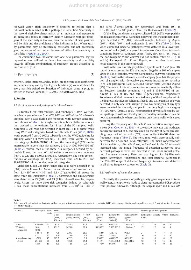
Of the 50 groundwater samples collected, 23 (46%) were positivefor at least one microbial pathogen. Rotavirus was the dominant path-ogen detected in 20 (40%) tubewell samples, followed by Shigella(10%), Vibrio (10%), and pathogenic E. coli (8%) (Table 2). Evenwhen combined, bacterial pathogens were detected in a lower pro-portion of wells (24%) compared to rotavirus. Only three tubewellscontaining bacterial pathogen genes (ipaH, eltA, stx1, vtx1, eae, orthe non-toxigenic Vibrio ompW) did not contain rotavirus (Fig. 1aand b). Pathogenic E. coli and Shigella, on the other hand, werenever detected in the same tubewell.
Within the low risk category defined by culturable E. coli (n=30),rotavirus was present in 40% of samples, Shigella in 10% of samples,Vibrio in 13% of samples, whereas pathogenic E. coliwere not detected(Table 2). Within the intermediate risk category (n=14), the propor-tion of samples with detectable pathogens increases for rotavirus(57%) and pathogenic E. coli (21%) but not for Vibrio (7%) and Shigella(7%). The mean of rotavirus concentrations was not markedly differ-ent between samples containing b1 and 1–10 MPN/100 mL cul-turable E. coli at 8.5 and 9.8×10³ genes/100 mL, respectively.Rotavirus and Vibriowere not detected in any of the 6 samples withinthe highest risk category whereas Shigella and pathogenic E. coliweredetected in only one well sample (17%). No pathogens of any typewere detected in the only sample in the very high risk category(>1000 MPN/100 mL E. coli). The proportion of wells affected by var-ious pathogens within risk categories defined by culturable E. coli didnot change markedly when considering only those wells with a goodplatform.
Using the frequency of culturable E. coli detection averaged overa year (van Geen et al., 2011) to categorize indicator and pathogenoccurrence instead of E. coli measured on the day of pathogen sam-pling only, half of the wells (52%) were in the 25%–50% detectionfrequency range (Table 2). The remaining wells were equally splitbetween the >50% and b25% categories. The mean concentrationsof total coliform, culturable E. coli, and mE. coli in the 50 tubewellsincreased with the annual frequency of detection categories. Totalbacterial pathogens were not detected in the b25% annual detec-tion frequency category. Detection was highest for F+RNA coli-phage, Bacteroides, HuBacteroides, and total bacterial pathogen inthe 25%–50% range of detection frequency. Rotavirus was detectedin all three frequency categories (Table 2).
3.2. Verification of molecular assays
To verify the presence of pathogenicity gene sequences in tube-well water, attempts were made to clone representative PCR productsfrom positive tubewells. Although the Shigella ipaH and E. coli eltA
Table 2Detection of fecal indicators, bacterial pathogens and rotavirus categorized against no criteria, WHO risk category limits, and annually-averaged E. coli detection frequency(>1 MPN/100 mL).
Criteria Descriptor Num.wells
Mean Percentage of wells positive
E. coli(MPN/100 mL)
Total coliforms(MPN/100 mL)
F+RNA(PFU/100 mL)
mE. coli(genes/100 mL)
Bacteroides(genes/100 mL)
HuBacteroides(genes/100 mL)
PathogenicE. coli
Shigella Vibrio Rotavirus
None All wells 50 85.5 243.5 15.4 5147.8 692,788.1 154,412.7 8 10 10 40WHO riskcategories
10 to1000
6 701.0 1470.0 38.9 41,207.7 5,707,285.0 1,284,441.5 17 17 0 0
1 to 10 14 4.3 220.3 25.6 414.9 12,574.9 958.2 21 7 7 57b1 30 0.3 9.0 4.9 144.5 7321.5 19.1 0 10 13 40
Annually-averagedE. coli
>50% 13 315.5 672.5 9.0 13,500.7 468,037.7 80,926.7 8 0 8 2325–50% 26 6.4 125.8 25.0 3117.1 1,094,590.4 256,481.5 12 19 15 46b25% 11 0.6 14.5 2.9 76.0 8687.6 6.6 0 0 0 45
317A.S. Ferguson et al. / Science of the Total Environment 431 (2012) 314–322
Author's personal copy
were cloned from surface water samples (data not shown), cloning ofthese genes from groundwater samples was unsuccessful. This mayreflect their very low concentration in the groundwater samples. Incontrast, rotavirus gene sequences could be cloned from a subset ofpositive well samples (9 total). The gene products from rotaviruswere then sequenced to verify their presence in the well water sam-ples. The majority of these sequences (37 out of 39) were 99% similarto G12 rotavirus (EU839971). The other 2 sequences were only 90%similar to the G12-like sequences. The second QRT-PCR assay usedin this study was designed based on 37 G12-like sequences fromtubewell samples to more specifically target the G12 VP6. This assayconfirmed the presence of rotavirus in 7 of the 15 tubewell samplestesting positive using the SYBR green assay. Thus, the presence of ro-tavirus was verified by two separate quantitative methods and by theidentification of sequences from 9 different tubewell samples.
3.3. Correlation of pathogens with fecal indicators andenvironmental parameters
The data indicate some statistically significant Spearman rank corre-lations of pathogens with fecal indicators or environmentalparameters but none are very strong correlations. Spearman rank corre-lations indicate that the presence of rotavirus was not correlated to the
log-transformed concentration of any fecal indicator (Table 3). Howev-er, the presence of rotavirus was correlated with the combined pres-ence of three bacterial pathogens, pathogenic E. coli, Shigella, andVibrio and the presence of only pathogenic E. coli and Shigella combined.With respect to environmental parameters, only the negative correla-tion between rotavirus detection and the level of nitrate in well waterwas significant. Within this data set, population density and platformqualitywere not correlatedwith the presence of rotavirus inwell water.
Pathogenic E. colidetectionwas positively correlatedwith the detec-tion of culturable E. coli, total coliform, and F+RNA coliphage (Table 3).However, pathogenic E. coli detection was not significantly correlatedwith the concentration of molecular E. coli. The likelihood of detectingpathogenic E. coli, and only this pathogen, declines significantlywith in-creasingwell depth. The detection of pathogenic E. coliwasnot correlat-ed with any other environmental parameters, including platformquality and population density.
The presence of Shigella was significantly correlated with theconcentration of molecular E. coli, but not with the concentrationof culturable E. coli, total coliforms, or F+RNA coliphage (Table 3).There was a significant positive correlation between the detectionof Shigella and ORP and negative correlation between Shigelladetection and ammonia concentrations. Vibrio was the onlypathogen for which the likelihood of detection increases for wells
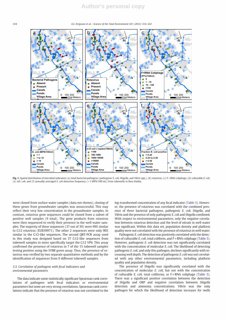
a b c
d e f
Fig. 1. Spatial distribution of microbial indicators (a) total bacterial pathogens (pathogenic E. coli, Shigella, and Vibrio spp.), (b) rotavirus, (c) F+RNA coliphage, (d) culturable E. coli,(e) mE. coli, and (f) annually-averaged E. coli detection frequency (>1 MPN/100 mL) from tubewells in Bara Haldia.
318 A.S. Ferguson et al. / Science of the Total Environment 431 (2012) 314–322

Author's personal copy
with a broken or missing platform. The presence of Vibrio was notsignificantly correlated with any other indicator or environmentalparameter.
The combined presence of pathogenic E. coli and Shigella improvedthe significance for five out of the previous seven individualpathogen-indicator correlations (Table 3). There was an additionalsignificant negative correlation between the combined detection ofpathogenic E. coli or Shigella and As concentration. The presence ofVibrio combined with pathogenic E. coli and Shigella reduced thenumber of significant correlations from eight to four. The presenceof bacterial pathogens and rotavirus combined was not significantlycorrelated with any fecal indicator or environmental parameter.
4. Discussion
4.1. Temporal and spatial context for the pathogen data
Culturable levels of E. coli in shallow tubewells vary both geograph-ically (Fig. 1) and seasonally within Matlab and in another region ofBangladesh (Leber et al., 2011; van Geen et al., 2011). The annually-averaged frequency of culturable E. coli detection was 38±15% for the30 wells with b1 MPN/100 mL E. coli when sampling for pathogens,43±13% in the 14 wells within the low risk category, and 57±8% inthe six remaining wells in intermediate to very high risk category. Dur-ing themonsoon of 2008 and 2009 the averaged frequency of culturableE. coli detection was 34±18% for the 30 wells with b1 MPN/100 mL E.coli when sampling for pathogens, 53±13% in the 14 wells within thelow risk category, and 65±20% in the six remainingwells in intermedi-ate to very high risk category. Levels of culturable E. coli measured intubewell water on the 8 days when samples were collected for patho-gens were therefore broadly consistent with the monitoring of thesame wells over a more extended period.
Spatial patterns of culturable E. coli in shallow groundwater havebeen related to variations in local population density (van Geen et al.,2011). The population residingwithin 25 mof each of the 50wells sam-pled for pathogens averages 22 and ranges from 5 to 51. Linear regres-sion for these 50 wells indicates a statistically significant increase from,on average, 30 to 60% in the frequency of culturable E. coli detection dur-ing the monsoon season across the full range of population densitywithin Bara Haldia (pb0.05). The relationship between shallowgroundwater contamination and presumed variations in sourcestrength related to population density at the surface helps explainwhy all but 3 of the wells with detectable levels of culturable E. coliwere located in the northern, more densely populated, portion of thevillage (Fig. 1d). Five out of 6 wells in the intermediate to very highrisk categories were located in northern Bara Haldia. Wells positive forF+RNA coliphage (Fig. 1c) and mE. coli (Fig. 1e) were also more preva-lent in the northern half. The context provided by the culturable E. coli,
mE.coli and F+RNA coliphage data suggests that pathogen concentra-tions measured on a single occasion in 50 wells of Bara Haldia likely re-flect recurring patterns of monsoon contamination that are linked tolocal but relatively stable environmental conditions.
4.2. Evidence of downward transport for some pathogens
Spatial patterns of culturable E. coli in shallow groundwater havebeen linked to the rate of local groundwater recharge, using groundwa-ter As as a proxy for the age of shallow groundwater (Leber et al., 2011;van Geen et al., 2011). Groundwater dating using the 3H–3He methodindicates a broad increase in As concentrations with the age of ground-water (i.e. the elapsed time since the infiltrating water was isolatedfrom the atmosphere) andwith progressivelymore reducing conditionsin the aquifer (Stute et al., 2007).Within the subset of 50 wells sam-pled for pathogens within Bara Haldia, however, there was no sta-tistically significant relationship between the As content of shallowgroundwater and either the levels of culturable E. coli measured inparallel with pathogens or the frequency of culturable E. coli detec-tion during the monsoon of 2008 and 2009. On the other hand,both the frequency of culturable E. coli over the entire year (Pear-son correlation r=−0.39, pb0.01), and detection of pathogenic E.coli and Shigella combined (rho: −0.30, pb0.05) (Table 2) are neg-atively correlated with As concentrations in the same well watersamples and therefore the local hydrogeology. Ammonia levelsmeasured in the 50 wells provide some support for the modulationof a downward pathway of contamination by the local hydrogeol-ogy. In Bara Haldia and in other areas of Bangladesh where Ascontamination is an issue, concentrations of ammonia and As areoften positively correlated (Harvey et al., 2002). This is becausethe dominant source of ammonia to groundwater is the naturaldegradation of organic matter, rather than sewage (Dowling etal., 2002) and the impact of this source is diluted in areas wheregroundwater is relatively young. For the 50 wells sampled inBara Haldia, concentrations of ammonia are negatively correlatedwith the detection of Shigella individually and combined withpathogenic E. coli and Vibrio (Table 3). The positive correlation be-tween ORP and the presence of Shigella alone, as well as pathogen-ic E. coli and Shigella combined, is also consistent with the notionthat less reduced, more rapidly recharged aquifers are more likelyto be contaminated with pathogenic bacteria. The relationships be-tween the presence of bacterial pathogens and several environ-mental parameters therefore support the notion that areas ofrapid recharge, e.g. sandy unconfined aquifers that are not cappedwith an impermeable clay layer (Aziz et al., 2008), are particularlylikely to be contaminated with fecal matter.
It is presently unclear why the presence of rotavirus appears tobe unrelated to any of the environmental parameters suggesting
Table 3Spearman rank correlations between indicators and environmental parameters towards fecal pathogens. Only significant correlations at the ⁎ b0.05 and ⁎⁎ b0.01 levels are shown.
Variable Pathogenic E. coli Shigella Vibrio Pathogenic E. coli+Shigella Total bacterial pathogens Rotavirus
Tubewell feature Platform 0.30⁎
Depth −0.32⁎ −0.29⁎
Water chemistry ORP 0.37⁎ 0.40⁎⁎
As −0.30⁎
NH3 −0.46⁎⁎ −0.47⁎⁎ −0.41⁎⁎
NO3 −0.36⁎
Indicator mE. coli 0.32⁎ 0.44⁎⁎ 0.40⁎⁎
Total coliform 0.35⁎ 0.37⁎⁎ 0.32⁎
E. coli 0.30⁎ 0.28⁎
F+RNA 0.34⁎ 0.47⁎⁎ 0.36⁎
Pathogen Pathogenic E. coli+Shigella 0.36**Total bacterial 0.40**
319A.S. Ferguson et al. / Science of the Total Environment 431 (2012) 314–322

Author's personal copy
downward transport of bacterial pathogens. The negative correla-tion between the presence of rotavirus and nitrate is hard to ex-plain in terms of local recharge because nitrate levels areelevated in only the most recently recharged groundwater in re-ducing aquifers (Andersen et al., 2007). Although there is a statis-tically significant correlation between the presence of rotavirusand that of all bacterial pathogens combined, the relation to envi-ronmental parameters may be obscured by differences in resis-tance to environmental factors and groundwater transport amongspecies and strains (Borchardt et al., 2003a, 2003b; Taylor et al.,2004). Rotavirus may also contaminate shallow aquifers along apathway that differs from that followed by bacterial pathogens.
4.3. Sensitivity and selectivity of fecal indicators
Although there are some significant correlations between thedetection of certain pathogens and water quality, these might befairly site-specific and difficult to generalize. This section thereforefocuses on the effectiveness of using six indicators of fecal contam-ination, individually or in pairs, to predict the presence of microbi-al pathogens. Indicator and pathogen data were converted to abinary outcome and classified to determine the maximum sensitiv-ity and specificity by modifying a cutoff value. The cutoff valuewas chosen to first maximize sensitivity (minimize false negatives)and then maximize specificity (minimize false positives). Althoughthe trade-offs between sensitivity and selectivity can be balanceddifferently (Harwood et al., 2005), only those indicators that cap-ture all wells containing a pathogen, or combination of pathogens,and correctly identify ≥50% of wells without pathogens are con-sidered here.
The analysis shows that the effective fecal indicators for predictingthe presence of pathogenic E. coli, detected in 4 out of 50 wells, aremolecular E. coli, total coliform, culturable E. coli, and F+RNA coli-phage (Table A2a). The number is reduced from four to three indica-tors if molecular E. coli, which currently requires PCR analysis, is notincluded. For Shigella (5 wells positive), molecular E. coli, total coli-form, and F+RNA coliphage are again effective indicators but cul-turable E. coli is not. Only molecular E. coli is an effective predictorof the presence of pathogenic E. coli or Shigella combined (9 wells)as well as the presence of any of the bacterial pathogens (12 wells)(Table A2b). F+RNA coliphage is also an effective predictor of thepresence of pathogenic E. coli or Shigella combined. Using the samecriteria, none of the individual fecal indicators are effective forpredicting the presence of Vibrio (5 wells) or rotavirus (20 wells).Similarly, no indicator is individually effective for predicting the pres-ence of any of the bacterial pathogens as well as rotavirus combined(23 wells).
Paired combinations of fecal indicators can improve sensitivityand specificity above that achieved using an indicator exclusively(Table A3). For pathogenic E. coli, three different combinations ofpaired fecal indicators increased specificity by up to 14% (total coli-form and F+RNA coliphage) above that achieved using total coliformexclusively. This number is reduced to two if you discount PCR basedmethodologies. For Vibrio spp. (18% increase), combined pathogenicE. coli and Shigella (6% increase), and total bacterial pathogens (10%increase), one combination of paired indicators improved specificityin each case. Other than for pathogenic E. coli, all combinations ofpaired indicators require PCR based analysis, which in resource-limited areas is not a viable option in the context of ASSURED diag-nostic testing (Urdea et al., 2006). Combinations of paired fecal indi-cators were not effective for predicting the presence of Shigella,rotavirus, or total pathogens at 100% sensitivity and ≥50% specificity.
4.4. Practical and health implications
The frequency (40%) and concentrations (up to 1.4×10⁵ genes/100 mL) of human rotavirus measured in Bara Haldia were more than10 fold higher than those reported for shallow groundwater inWiscon-sin (0.5% (Borchardt et al., 2003a)), unspecified drinkingwater samplesin Benin (~2% (Verheyen et al., 2009)), and drinkingwater samples dur-ing an epidemic in France (~2% (Gratacap-Cavallier et al., 2000)). Thefrequency of occurrence of rotavirus in shallow wells of Bara Haldiawas similar to that for a surface water source in the Netherlands 48%(Lodder et al., 2010). Rotavirus is widely believed to be transmitted byperson-to-person contact (Anderson and Weber, 2004), but our datasuggest that shallow groundwater might be a source even if the risk ofinfection cannot be directly determined from molecular assays(Gassilloud et al., 2003). The presence ofmore than one rotavirus geno-type in the well water samples is not surprising because rotavirus hasconsiderable genomic diversity (Dennehy, 2008). In addition, the dom-inant circulating strains can change over time (Rahman et al., 2007).The dominant type found in this study was G12, first isolated on theAsian subcontinent from stool samples between 2004 and 2006 andmight be increasing in frequency (Paul et al., 2008; Ramani et al.,2007). A more systematic study of potential rotavirus infection fromdrinking groundwater seems warranted, especially given the predomi-nance of rotavirus-caused cases of diarrhea reported in the Matlab hos-pital (Tanaka et al., 2007; Zaman et al., 2009).
Bacterial pathogenicity genes were correlated with the presenceof rotavirus with 75% of wells positive for one of these genes also pos-itive for rotavirus. The mechanism for co-contamination of wells isunclear, but could result from the observations that multiple patho-gens can frequently be isolated from patients with diarrhea (Albertet al., 1999) or could be indicative of shallow wells subject to fecalcontamination. This suggests that surveillance for rotavirus alonemay identify drinking water wells impacted by pathogen contami-nation. Thus rotavirus may serve as a conservative indicator of fecalcontamination because rotavirus can remain for months after intro-duction into groundwater in the lab (Espinosa et al., 2008) and alsomay be preferentially transported in groundwater relative to bacteria(Taylor et al., 2004).
The main freshwater FIB (culturable E. coli) used by regulatoryagencies and international health organizations to determinegroundwater quality was significantly correlated and exhibited nofalse negatives (100% sensitivity) towards only one bacterial group(pathogenic E. coli). Contingency analysis also showed a statisticallysignificant relationship between the annually-averaged E. coli detec-tion frequencies for the 50 wells and the presence of total bacterialpathogens, (pb0.01) (Fig. A2a and c). The available data indicatesthat wells with a low (b25%) annually-averaged frequency of E. colidetection are less likely to have bacterial pathogens than wells withmoderate (25–50%) and high (>50%) frequency of E. coli detection.This still provides some justification for repeated E. coli measure-ments as a way of screening wells for microbial contamination.However, annually-averaged E. coli detection frequency was notpredictive for the presence or absence of rotavirus (Fig. A2b and d).
Total coliforms exhibited the highest sensitivity (100%) towards ro-tavirus and agreed with previous work on the viral contamination ofgroundwater (Payment and Locas, 2010) and the screening of sixwastewater reclamation facilities in the US (Harwood et al., 2005).However, as total coliforms were present in 92% of tubewells the lowcutoff value required meant their specificity was low, exhibited by89% false positive rate (Table A2a). No bacterial indicator, exclusive orin paired combination, effectively distinguished tubewells positive forrotavirus, possibly because of different survival and transport character-istics between bacteria and viruses (Borchardt et al., 2003a; Taylor et al.,2004). Interestingly, the common fecal indicator virus (F+RNA coli-phage) used by regulatory agencies did not significantly correlate orprovide high sensitivity or specificity towards rotavirus (74% and 36%,
320 A.S. Ferguson et al. / Science of the Total Environment 431 (2012) 314–322
Author's personal copy
respectively). Lodder et al. (2010) also discounted the use of somaticand F-specific coliphage as indicators of sourcewater quality, highlight-ing that currently themost effective approach of identifying pathogenicviral contamination is through direct detection. The need for assured di-agnostic testing (Urdea et al., 2006) indicates that alternate phage, coli-phage host, and indicators for enteric viruses should be investigated(Harwood et al., 2005).
Based on our study, groundwater cannot be excluded as a signifi-cant source of diarrheal disease in Bangladesh and neighboring coun-tries with similar characteristics. The most promising individualindicators for detecting bacterial pathogens in groundwater areF+RNA coliphage and molecular E. coli, but both require more effortand expertise than E. coli detection by culturing. F+RNA coliphage isnot an effective indicator of rotavirus, which is present in groundwaterpumped from a particularly high proportion of wells.
Acknowledgments
This study was supported by NIH/Forgarty International Centergrant 5R01TW8066-2. We thank M. Watson of Barnard College,and Md. Ariful Hasan Majumder, Md. Mahfuzur Rahman Khan,Ferhana Yesmin, Saleh Ahmed, Imtiaz Chowdhury, Md. Rezaul Huqof Dhaka University as well as local field staff in Matlab for helpduring field sampling.
Appendix A. Supplementary data
The complete dataset can be found online at http://dx.doi.org/10.1594/PANGAEA.774444.
References
Ahmed MF, Ahuja S, Alauddin M, Hug SJ, Lloyd JR, Pfaff A, et al. Ensuring safe drinkingwater in Bangladesh. Science 2006;314:1687–8.
Alam M, Sultana M, Nair GB, Sack RB, Sack DA, Siddique AK, et al. Toxigenic Vibriocholerae in the aquatic environment of Mathbaria, Bangladesh. Appl EnvironMicrobiol 2006;72:2849–55.
Albert MJ, Faruque ASG, Faruque SM, Sack RB, Mahalanabis D. Case–control study ofenteropathogens associated with childhood diarrhea in Dhaka, Bangladesh. J ClinMicrobiol 1999;37:3458–64.
Altschul SF, Gish W, Miller W, Myers EW, Lipman DJ. Basic local alignment search tool.J Mol Biol 1990;215:403–10.
Andersen MS, Baron L, Gudbjerg J, Gregersen J, Chapellier D, Jakobsen R, et al. Dischargeof nitrate-containing groundwater into a coastal marine environment. J Hydrol2007;336:98-114.
Anderson EJ, Weber SG. Rotavirus infection in adults. Lancet Infect Dis 2004;4:91–9.Aziz Z, van Geen A, Stute M, Versteeg R, Horneman A, Zheng Y, et al. Impact of local
recharge on arsenic concentrations in shallow aquifers inferred from the electro-magnetic conductivity of soils in Araihazar, Bangladesh. Water Resour Res2008;44.
Begum Y, Talukder K, Nair G, Khan S, Svennerholm A, Sack R, et al. Comparison of en-terotoxigenic Escherichia coli isolated from surface water and diarrhoeal stool sam-ples in Bangladesh. Can J Microbiol 2007;53:19–26.
Boehm AB, Shellenbarger GG, Paytan A. Groundwater discharge: potential associationwith fecal indicator bacteria in the surf zone. Environ Sci Technol 2004;38:3558–66.
Borchardt MA, Bertz PD, Spencer SK, Battigelli DA. Incidence of enteric viruses ingroundwater from household wells in Wisconsin. Appl Environ Microbiol2003a;69:1172–80.
Borchardt MA, Bertz PD, Spencer SK, Battigelli DA. Incidence of Enteric Viruses inGroundwater from Household Wells in Wisconsin, 69. ; 2003b. p. 1172–80.
Brennan FP, O'Flaherty V, Kramers G, Grant J, Richards KG. Long-term persistence andleaching of Escherichia coli in temperate maritime soils. Appl Environ Microbiol2010;76:1449–55.
Bushon RN. Fecal indicator viruses: U.S. geological survey techniques of water-resources investigations; 2003. book 9, chap. A7, section 7.2, November.
Cheng Z, Zheng Y, Mortlock R, van Geen A. Rapid multi-element analysis of groundwa-ter by high-resolution inductively coupled plasma mass spectrometry. AnalBioanal Chem 2004;379:513–8.
Debartolomeis J, Cabelli VJ. Evaluation of an Escherichia coli host strain for enumerationof F male-specific bacteriophages. Appl Environ Microbiol 1991;57:1301–5.
Dennehy P. Rotavirus vaccines: an overview. Clin Microbiol Rev 2008;21:198–208.Dowling CB, Poreda RJ, Basu AR, Peters SL, Aggarwal PK. Geochemical study of arsenic
release mechanisms in the Bengal Basin groundwater. Water Resour Res 2002;38.
Espinosa AC, Mazari-Hiriart M, Espinosa R, Maruri-Avidal L, Mendez E, Arias CF. In-fectivity and genome persistence of rotavirus and astrovirus in groundwaterand surface water. Water Res 2008;42:2618–28.
Faruque SM, Khan R, Kamruzzaman M, Yamasaki S, Ahmad QS, Azim T, et al. Isolationof Shigella dysenteriae type 1 and S. flexneri strains from surface waters in Bangla-desh: comparative molecular analysis of environmental Shigella isolates versusclinical strains. Appl Environ Microbiol 2002;68:3908–13.
Ferguson AS, Mailloux BJ, Ahmed KM, van Geen A, McKay LD, Culligan PJ. Hand-pumpsas reservoirs for microbial contamination of well water. J Water Health 2011;9(4):708–17.
Gassilloud B, Schwartzbrod L, Gantzer C. Presence of viral genomes in mineral water: asufficient condition to assume infectious risk? Appl Environ Microbiol 2003;69:3965–9.
Gibson KE, Schwab KJ. Detection of bacterial indicators and human and bovine entericviruses in surface water and groundwater sources potentially impacted by animaland human wastes in Lower Yakima Valley, Washington. Appl Environ Microbiol2011;77:355–62.
Glassmeyer ST, Furlong ET, Kolpin DW, Cahill JD, Zaugg SD, Werner SL, et al. Transportof chemical and microbial compounds from known wastewater discharges: poten-tial for use as indicators of human fecal contamination. Environ Sci Technol2005;39:5157–69.
Gratacap-Cavallier B, Genoulaz O, Brengel-Pesce K, Soule H, Innocenti-Francillard P,Bost M, et al. Detection of human and animal rotavirus sequences in drinkingwater. Appl Environ Microbiol 2000;66:2690–2.
Harvey CF, Swartz CH, Badruzzaman AB, Keon-Blute N, Yu W, Ali MA, et al.Arsenic mobility and groundwater extraction in Bangladesh. Science 2002;298:1602–6.
Harwood VJ, Levine AD, Scott TM, Chivukula V, Lukasik J, Farrah SR, et al. Validity of theindicator organism paradigm for pathogen reduction in reclaimed water and pub-lic health protection. Appl Environ Microbiol 2005;71:3163–70.
Hunt RJ, Borchardt MA, Richards KD, Spencer SK. Assessment of sewer source con-tamination of drinking water wells using tracers and human enteric viruses. Envi-ron Sci Technol 2010;44:7956–63.
Hurley MA, Roscoe ME. Automated statistical-analysis of microbial enumeration by di-lution series. J Appl Bacteriol 1983;55:159–64.
Knappett PSK, Layton A, McKay LD, Williams D, Mailloux BJ, Huq MR, et al. Efficacy ofhollow-fiber ultrafiltration for microbial sampling in groundwater. Ground Water2011;49:53–65.
Kosek M, Bern C, Guerrant RL. The global burden of diarrhoeal disease, as estimatedfrom studies published between 1992 and 2000. Bull World Health Organ2003;81:197–204.
Layton A, McKay L, Williams D, Garrett V, Gentry R, Sayler G. Development of Bacte-roides 16S rRNA gene TaqMan-based real-time PCR assays for estimation of total,human, and bovine fecal pollution in water. Appl Environ Microbiol 2006;72:4214–24.
Leber J, Rahman MM, Ahmed KM, Mailloux B, van Geen A. Contrasting Influenceof geology on E-coli and arsenic in aquifers of Bangladesh. Ground Water2011;49:111–23.
Lodder WJ, van den Berg H, Rutjes SA, Husman AMD. Presence of enteric viruses insource waters for drinking water production in the Netherlands. Appl EnvironMicrobiol 2010;76:5965–71.
Luby SP, Gupta SK, Sheikh MA, Johnston RB, Ram PK, Islam MS. Tubewell water qualityand predictors of contamination in three flood-prone areas in Bangladesh. J ApplMicrobiol 2008;105:1002–8.
Momba MN, Malakate VK, Theron J. Abundance of pathogenic Escherichiacoli, Salmonella typhimurium and Vibrio cholerae in Nkonkobe drinking watersources. J Water Health 2006;4:289–96.
Paul S, Kobayashi N, Nagashima S, Ishino M, Watanabe S, Alam M, et al. Phylogeneticanalysis of rotaviruses with genotypes G1, G2, G9 and G12 in Bangladesh: evidencefor a close relationship between rotaviruses from children and adults. Arch Virol2008;153:1999–2012.
Payment P, Locas A. Pathogens in water: value and limits of correlation with microbialindicators. Ground Water 2010;49:4-11.
Pepe MS, Janes H, Longton G, LeisenringW, Newcomb P. Limitations of the odds ratio ingauging the performance of a diagnostic, prognostic, or screening marker. Am JEpidemiol 2004;159:882–90.
Pote J, Haller L, Kottelat R, Sastre V, Arpagaus P, Wildi W. Persistence and growthof faecal culturable bacterial indicators in water column and sediments of VidyBay, Lake Geneva, Switzerland. J Environ Sci (China) 2009;21:62–9.
Rahman M, Sultana R, Ahmed G, Nahar S, Hassan Z, Saiada F, et al. Prevalence of G2P[4]and G12P[6] rotavirus, Bangladesh. Emerg Infect Dis 2007;13:18–24.
Ramani S, Banerjee I, Primrose Gladstone B, Sarkar R, Selvapandian D, Le Fevre AM,et al. Geographic information systems and genotyping in identification of rotavirusG12 infections in residents of an urban slum with subsequent detection in hospi-talized children: emergence of G12 genotype in South India. J Clin Microbiol2007;45:432–7.
Schriewer A, Miller WA, Byrne BA, Miller MA, Oates S, Conrad PA, et al. Presence ofBacteroidales as a predictor of pathogens in surface waters of the central Californiacoast. Appl Environ Microbiol 2010;76:5802–14.
Sinton LW, Finlay RK, Reid AJ. A simple membrane filtration-elution method for theenumeration of F-RNA, F-DNA and somatic coliphages in 100-ml water samples.J Microbiol Methods 1996;25:257–69.
Stute M, Zheng Y, Schlosser P, Horneman A, Dhar RK, Datta S, et al. Hydrological controlof As concentrations in Bangladesh groundwater. Water Resour Res 2007;43. (-).
321A.S. Ferguson et al. / Science of the Total Environment 431 (2012) 314–322

Author's personal copy
Tanaka G, Faruque ASG, Luby SP, Malek MA, Glass RI, Parashar UD. Deaths from rotavi-rus disease in Bangladeshi children — estimates from hospital-based surveillance.Pediatr Infect Dis J 2007;26:1014–8.
Taylor R, Cronin A, Pedley S, Barker J, Atkinson T. The implications of groundwater ve-locity variations on microbial transport and wellhead protection — review of fieldevidence. FEMS Microbiol Ecol 2004;49:17–26.
Urdea M, Penny LA, Olmsted SS, Giovanni MY, Kaspar P, Shepherd A, et al. Require-ments for high impact diagnostics in the developing world. Nature2006;444(Suppl. 1):73–9.
van Geen A, Ahmed KM, Akita Y, Alam MJ, Culligan PJ, Emch M, et al. Fecal contamina-tion of shallow tubewells in Bangladesh inversely related to arsenic. Environ SciTechnol 2011;45:1199–205.
Verheyen J, Timmen-Wego M, Laudien R, Boussaad I, Sen S, Koc A, et al. Detection ofadenoviruses and rotaviruses in drinking water sources used in rural areas ofBenin, West Africa. Appl Environ Microbiol 2009;75:2798–801.
Wampler PJ, Sisson AJ. Spring flow, bacterial contamination, and water resources inRural Haiti. Environ Earth Sci 2011;62(8):1619–28.
WHO. Guidelines for drinking-water quality, incorporating first and second addenda tothird edition. Recommendations, vol. 1; 2008. Geneva.
Zaman K, Yunus M, Faruque ASG, El Arifeen S, Hossain I, Azim T, et al. Surveillance ofrotavirus in a rural diarrhoea treatment centre in Bangladesh, 2000–2006. Vaccine2009;27:F31–4.
322 A.S. Ferguson et al. / Science of the Total Environment 431 (2012) 314–322
Copyright © 2022 FDOKUMEN