C 727 - Control valves Altitude valve electrically operated and ...
Upload
independentCategory
view
2download
0
ORIGINAL RESEARCH PAPER
Comparison of electrically induced flare response patternsin human and pig skin
M. Dusch Æ M. Schley Æ O. Obreja Æ E. Forsch ÆM. Schmelz Æ Roman Rukwied
Received: 22 January 2009 / Revised: 4 March 2009 / Accepted: 4 March 2009 / Published online: 25 March 2009
� Birkhauser Verlag, Basel/Switzerland 2009
Abstract
Objective We compared the characteristics of neurogenic
flare responses in human and pig skin to establish a
translational research animal model.
Material and subjects Eight domestic pigs and six male
subjects were investigated.
Treatment Electrical pulses were delivered transcutane-
ously with increasing current intensities, pulse frequencies
and pulse widths.
Methods Inflammatory skin responses were recorded by
laser Doppler imaging and analyzed by ANOVA and
Fisher’s (LSD) post hoc test.
Results Transcutaneous stimuli of 5 mA onward induced
a significant flare development in humans. In the pig, sig-
nificantly lower currents of 2.5 mA already induced a flare
response. Smaller flare sizes of about 3.5 cm2 were ana-
lyzed. The flare continuously declined despite ongoing
stimulation.
Conclusions Lower excitation thresholds and smaller
receptive fields of nociceptors can be suggested in pigs.
Impaired neuropeptide release, altered vesicle replenish-
ment, different neuropeptide sensitivity, or insufficient
peripheral decoding of action potentials may contribute to
steadily decreasing flare responses. These attributes may be
objectives of pre-clinical anti-hyperalgesic studies and
their accurate analysis in pigs reveals a particularly sensi-
tive translational animal model for nociceptor researches.
Keywords Axon reflex flare �Mechanically insensitive nociceptors � Heat nociceptors �Electrical thresholds
Introduction
Pig skin is closely related to human skin considering
structure, vascularization epidermal enzyme patterns [1],
peptide nerve fiber immunoreactivity (e.g., CGRP, VIP,
substance P) [2], and thus is highly suitable for inflam-
matory research. In addition, pig skin heat sensitive
nociceptors, of which some are also mechanically insen-
sitive, are involved in the progress of the flare development
[3]. It was shown previously in humans that the activation
of heat sensitive nociceptors [4] and mechano-insensitive
C-fibers [5] contribute considerably to the flare reaction.
One characteristic of mechano-insensitive nerve fiber
classes is that they can increase their responsiveness to
external stimuli and thereupon contribute to the develop-
ment and maintenance of hyperalgesia [6]. Thus, focused
investigation and characterization of these nociceptors
helps to understand the mechanisms behind hyperalgesia.
Here, we compare the flare development upon noci-
ceptor activation at matching anatomical sites of human
and pig skin in response to transcutaneously delivered
electrical stimuli. Supra-threshold excitation of skin noci-
ceptors induces the generation of action potentials, which
are conducted retrograde at branching points of the nerve
toward the terminal endings to evoke a depletion of
M. Dusch and M. Schley contributed equally to the manuscript.
Responsible Editor: G. Geisslinger.
M. Dusch � M. Schley � O. Obreja � E. Forsch � M. Schmelz �R. Rukwied (&)
Department of Anaesthesiology and Intensive Care Medicine,
University Medicine Mannheim, Ruprecht-Karls-University
Heidelberg, Theodor-Kutzer-Ufer 1-3,
68167 Mannheim, Germany
e-mail: [email protected]
Inflamm. Res. (2009) 58:639–648
DOI 10.1007/s00011-009-0029-3 Inflammation Research

neuropeptides, particularly calcitonin gene-related peptide
(CGRP), causing widespread vasodilatation (axon reflex
flare) [7, 8]. Spatial extension of this flare response has
been explored with accuracy as objective output measures
of nociceptor activation [9, 10]. In the present study, the
parameters of the administered current intensity, pulse
width and pulse frequency were kept identical in human
and pigs. Electrical thresholds for flare development,
maximum flare sizes, and flare progress over time were
identified to characterize and compare in both species the
excitation of skin nociceptors, i.e., mechano-insensitive
and heat sensitive C-fibers. Their comparison between the
species serves the establishment of the pig as translational
experimental animal model for anti-hyperalgesic research,
even though elevated resources may be needed for their
keeping compared to commonly used laboratory animals.
Materials and methods
The animal protection authorities of the regional council of
Baden-Wurttemberg, Germany, and the central animal
research unit at the University of Heidelberg approved the
study protocol and experimental procedure performed on
pig skin according the European Communities Council
Directive and the Guide for the Care and Use of Laboratory
Animals. The local Ethic Committee of the Medical Fac-
ulty Mannheim, University of Heidelberg, approved the
experimental procedure on human volunteers according to
the Code of Ethics of the World Medical Association,
Declaration of Helsinki and its amendment in Tokyo and
Venice.
Animal preparation
Investigations were performed on eight domestic pigs (Sus
scrofa) supplied by a local breeder (Sommerhof, Dielheim-
Unterhof, Germany). The age of the animals was on
average 12 ± 4 weeks and body weight 20–27 kg.
According to a previous investigation, pig skin at this age
is most similar to human skin [11]. All pigs were used
twice with a time interval of at least 7 days between the
experiments. Prior to the experiments animals were
allowed to familiarize with the new environmental condi-
tion for at least 7 days and fed daily with Muskator EM DE
BW 200060 (MuskatoWerke, Mannheim, Germany) and
water, the latter ad libidum. Six hours prior to the experi-
ment the food was withdrawn.
As described previously [12], pigs were pre-medicated
by injections of 2 mg/kg Stresnil� (Azaperon, Janssen
Pharmaceutica, Beerse, Belgium), 0.05 mg/kg Atropin
(Atropinsulfat-1-Hydrat, Eifelfango, Bad Neuenahr,
Germany) and 0.3 mg/kg Dormicum� (Midazolam, Roche,
Basle, Switzerland) into the trapezius muscle. Following a
sedation period of approximately 20 min animals were
anesthetized with an intravenous infusion of 1.5 mg/kg
Propofol� (Fresenius, Bad Homburg, Germany) into an ear
vein. Subsequently, pigs were intubated by means of
a 4.0 mm ID microlaryngeal tubus (Rusch, Gauting,
Germany) and artificially ventilated at a tidal volume of
2.5–3 l/min with O2 (25–50%), which was adjusted indi-
vidually according to the continuously measured O2
consumption and exhaled CO2 concentration, the latter
kept at 40 mmHg (Narkomat�, Heyer Anesthesia, Bad
Ems, Germany). Following intubation, i.e., approximately
5 min after Propofol� infusion, anesthesia was achieved by
an intravenous infusion of 20 mg/kg Narcoren� (Pento-
barbital-Natrium, Rhone Merieux, Laupheim, Germany)
administered as bolus using a 20G 11/400 infusion cannula
(Vasofix�, B. Braun, Melsungen, Germany) placed into the
saphenous vein of the lower leg. The cannula was con-
nected via a three-way valve to a drip and continuously but
slowly perfused with saline (NaCl 0.9%, Delta Select,
Pfullingen, Germany) using an administration set R 87
RLS Luer-Lock IG-P (Becton Dickinson, Heidelberg,
Germany).
Electrocardiogram, body core temperature, pulse rate
and O2 blood concentration were monitored throughout the
experiment (Lohmeier M211-371, Munchen, Germany).
Body core temperature was maintained constant at
37.2 ± 1.1�C throughout the experiments. This tempera-
ture is lower as compared to unrestrained pig temperature
(38–39�C) but has no effect on peripheral blood flow [13].
Sufficient depth of anesthesia was estimated by heart fre-
quency, physiological reflexes evoked by touching the eye
lid, or spontaneous muscular movements. Additional doses
of pentobarbital were administered on demand through a
three-way valve system.
Volunteers
Six healthy male volunteers (mean age 39 ± 8 years)
participated in the study, after having been informed about
the experimental procedure and given their written
informed consent. Subjects receiving any medication
influencing vascular responses (e.g., anti-histamines, cor-
ticosteroids, etc.) were excluded from the study. All
experiments were performed in a temperature-controlled
environment (23�C).
Electrical stimulation
For transcutaneous electrical stimulation, a pair of self-
adhesive rectangular (3 9 10 mm) surface electrodes
(Pierenkemper, Wetzlar, Germany) was attached to the
dorso-lateral back skin 5 cm lateral to the spine of the
640 M. Dusch et al.

volunteer and the animal, respectively. The dorsum area
was investigated in both pig and human to allow direct
comparisons of flare sizes between the species. A distance
of 3 mm between each electrode was kept constant.
Only in pig skin intradermal electrical currents were
delivered and compared to transcutaneous nerve stimula-
tion in order to characterize the extension of the axon reflex
flare in relation to the depth of the neuronal activation.
Intradermal stimulation was performed by means of a
pair of 27G injection cannulae (outer diameter 0.41 mm,
Neoject�, Dispomed, Gelnhausen, Germany) inserted
intradermally at a distance of 3 mm and a length of 0.5–
1.5 mm into the dorso-lateral back skin of the animal.
Insertion depth of the intradermal electrodes was approxi-
mately 1 mm.
Both, self-adhesive electrodes and injection cannulae
were connected to a constant current stimulator (DS7,
Digitimer, Hertfordshire, UK) delivering rectangular elec-
trical stimuli triggered by an external pulse generator
(Rimkus Pulsgenerator PG1, Parsdorf, Germany). The
current stimulator was equipped with an overload indicator
light to show if the load exceeded its capacity.
Study design
Current intensity, pulse width, and frequency of the
administered current pulses were systematically studied in
randomized order in both species. For each investigated
parameter the other remaining variables of the current were
kept constant throughout the experiments (Table 1). If cur-
rent intensity was explored, the electrical pulse duration and
frequency was kept at 0.5 ms and 1 Hz. If pulse width was
studied, the current intensity and the pulse frequency were
kept at 5 mA and 1 Hz. If pulse frequency was investigated,
the current intensity and pulse width was kept at 5 mA and
1 ms. The influence of the frequency order with which the
current pulses were delivered, i.e., increasing or decreas-
ing frequency, was assessed by administering pulses at
0.1–100 Hz (increasing) or 100–0.1 Hz (decreasing).
In the pig, continuous stimuli were applied for 30 min at
current pulses delivered at 0.5 and 50 Hz, respectively. The
concept of an increasing number of stimuli was investi-
gated also by delivering 75, 150, and 225 pulses at
frequencies of 0.3–100 Hz. Finally, the development of
flare responses was studied upon transcutaneously and
intradermally delivered stimuli at varying current intensi-
ties and pulse widths, respectively (Table 1).
Duration of stimulus administration was 4 min and
increased successively for each parameter with increasing
current intensity, pulse width, or discharge frequency. This
stimulation paradigm was applied to both human and pig,
thus any effect of the previously delivered stimulus on the
analyzed response would affect both species equally.
Current intensity
Intensities of constant current pulses were increased in 4-
min intervals in steps of 2.5 mA and up to a maximum of
15 mA. Current was delivered transcutaneously to both
species, human and pig, at a pulse frequency of 1 Hz and a
pulse duration of 0.5 ms. In pig skin, intradermal current
pulses were delivered in steps of 2.5 mA to a maximum of
15 mA and flare development was compared to those
evoked after transcutaneous current stimuli. For the com-
parisons of intradermal versus transcutaneous stimuli,
pulse widths were set at 0.2 and 0.5 ms, respectively, at a
pulse frequency of 1 Hz.
Pulse width
Duration of electrical pulses was increased at 4-min
intervals from 0.05 to 0.1 to 0.2 to 0.5 to 1 to 2 ms at a
constant current intensity of 5 mA and a pulse frequency of
1 Hz. In pig skin, transcutaneous and intradermal current
delivery was investigated.
Pulse frequency
Transcutaneous electrical stimuli were administered at
pulse frequencies of 0.1–0.2–0.3–0.5–1–5–10–50–100 Hz
at 5 mA current intensity and 1 ms pulse width. Duration
of stimulation was constant at 4 min for each frequency.
Pulses up to 1 Hz were delivered biphasic, whereas
electrical pulses at higher frequencies (5–100 Hz) were
delivered via monophasic bursts. Each burst consisted of
five single current stimuli. The time interval between each
stimulus within the burst determined the pulse frequency
(5–100 Hz). Bursts of electrical pulses were administered
in 2-s intervals and both from low to high frequencies
(0.1–100 Hz) and in reversed order (100–0.1 Hz). The
area under the curve (AUC) of the flare response was
calculated as sum total of all recorded flare areas for each
individual and depicted as mean AUC for the investigated
species.
Continuous stimulation and number of current pulses
assessed in pigs
In three animals continuous constant current pulses were
delivered transcutaneously for 30 min at frequencies of 0.5
and 50 Hz, respectively. The current intensity was set at
5 mA and pulse duration at 1 ms. In addition, the influence
of the number of stimuli on the flare development was
assessed further in three pigs by delivering 75, 150 and 225
pulses at frequencies of 0.3–100 Hz. Similarly, in these
experiments the current intensity was 5 mA at a pulse
width of 1 ms.
Electrically induced flare response pattern 641
Flare assessment
Superficial skin blood flow was recorded online by a series
of color-coded laser Doppler images (MoorLDI2-IR Laser
Doppler Imager, Axminster, UK) covering an area of
32 cm2. Scan resolution was set at 132 9 155 pixels;
image capture required 115 s and recording interval was
set at 120 s. The distance between laser scanner and skin
surface was kept constant at 40 cm. For off-line analysis of
vasodilatation, mean flux values and standard deviation
were determined in the baseline image recorded prior to the
onset of the electrical stimulation. A rectangular area of
approximately 12 cm2 was defined in the vicinity of the
stimulation site and mean flux values were calculated
within this area using the manufacturers’ software (Moor-
LDI 3.08, Axminster, UK). The threshold for a significant
increase of blood flow was defined as mean flux
value ? 2 times standard deviation. Flux values exceeding
the threshold were identified as significant flare reaction
and included into the calculation. Total flare area was
assessed in each image of the laser Doppler sequence by
means of the calculated number of pixels and their size,
respectively.
Statistical analysis
Data were analyzed using Statistica� 6.0 software package
(Statsoft, Tulsa, US). Values were calculated by ANOVA
multi-variant analysis and repeated measures. Signifi-
cant differences were identified between the factors
‘‘species’’ - ‘‘current intensity’’ - ‘‘pulse width’’ - ‘‘fre-
quency’’ by Fisher’s least-significant difference (LSD) post
hoc test. P values \0.05 were considered significant. Data
are depicted as mean ± standard error of the mean (SEM)
throughout the manuscript.
Results
Transcutaneous nerve stimulation
Current intensity
In human skin, transcutaneously delivered currents from
2.5 to 15 mA, which had been administered at a constant
pulse width of 0.5 ms and a frequency of 1 Hz, evoked a
dose-dependent and almost linear increase of flare area
without reaching a plateau. Statistically significant flare
sizes (P \ 0.0001, ANOVA) were identified from 5 mA
onwards. Flare size was analyzed on average at
2.5 ± 0.7 cm2 following 5 mA stimuli with maximum
flare sizes of about 13.4 ± 1.4 cm2 following 15 mA
stimulation. Half-maximum flare size developed during the
7.5 mA administration period. Vasodilatation declined
when current supply had been terminated, but was still
significantly elevated (P \ 0.0001, ANOVA) 6 min post-
stimulation when compared to baseline value. Similar to
human skin, transcutaneous electrical nerve stimuli evoked
in pig skin a linear increase of flare development from
0.99 ± 0.27 cm2 at 2.5 mA to a maximum flare size of
about 3.23 ± 0.42 cm2 after 7.5 mA. Further increase of
the current intensity to 10, 12.5, and 15 mA, however, did
not additionally enhance the flare size. Thus, maximum
flare development was significantly smaller as compared to
humans (P \ 0.0001, ANOVA). Half-maximum flare
development was analyzed after the 2.5 mA stimulation
period, which induced already significantly elevated
vasodilatation in comparison to baseline value (P \ 0.001,
Fisher’s LSD post hoc). Thus, when compared to human
skin, flare area was significantly larger in pig skin
(P \ 0.02, Fisher’s LSD post hoc) in response to 2.5 and
5 mA stimulation (Fig. 1a). No significant difference
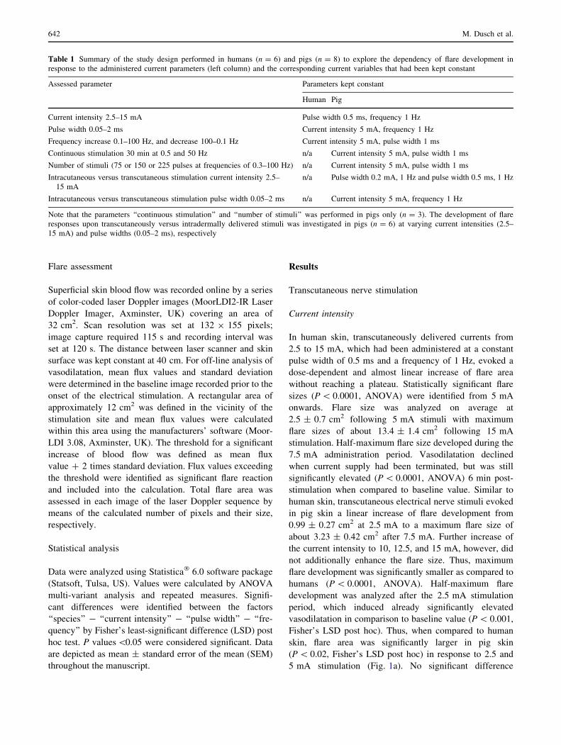
Table 1 Summary of the study design performed in humans (n = 6) and pigs (n = 8) to explore the dependency of flare development in
response to the administered current parameters (left column) and the corresponding current variables that had been kept constant
Assessed parameter Parameters kept constant
Human Pig
Current intensity 2.5–15 mA Pulse width 0.5 ms, frequency 1 Hz
Pulse width 0.05–2 ms Current intensity 5 mA, frequency 1 Hz
Frequency increase 0.1–100 Hz, and decrease 100–0.1 Hz Current intensity 5 mA, pulse width 1 ms
Continuous stimulation 30 min at 0.5 and 50 Hz n/a Current intensity 5 mA, pulse width 1 ms
Number of stimuli (75 or 150 or 225 pulses at frequencies of 0.3–100 Hz) n/a Current intensity 5 mA, pulse width 1 ms
Intracutaneous versus transcutaneous stimulation current intensity 2.5–
15 mA
n/a Pulse width 0.2 mA, 1 Hz and pulse width 0.5 ms, 1 Hz
Intracutaneous versus transcutaneous stimulation pulse width 0.05–2 ms n/a Current intensity 5 mA, frequency 1 Hz
Note that the parameters ‘‘continuous stimulation’’ and ‘‘number of stimuli’’ was performed in pigs only (n = 3). The development of flare
responses upon transcutaneously versus intradermally delivered stimuli was investigated in pigs (n = 6) at varying current intensities (2.5–
15 mA) and pulse widths (0.05–2 ms), respectively
642 M. Dusch et al.

was obtained between male and female pigs (P [ 0.5,
ANOVA), which may be attributed to the pre-menstrual
cycle of the female animals given at an average age of
12 weeks.
Pulse width
Increase of the pulse duration (0.2–2 ms) significantly
enhanced flare development in a dose dependent manner in
both human and pig skin (P \ 0.0001, ANOVA). In
humans, flare size increased linearly in response to elec-
trical stimuli delivered at a constant current of 5 mA and a
frequency of 1 Hz from 0.6 ± 0.2 cm2 (0.2 ms pulse
width) to a maximum level of 9.5 ± 0.7 cm2 (2 ms pulse
width). Compared to baseline value, a significantly larger
flare area (P \ 0.0001, ANOVA) was recorded at the end
of the experimental protocol. Half-maximum flare size of
4.9 ± 0.5 cm2 was obtained following a pulse width of
1 ms, even though the flare response does not reach a
plateau in this experimental setting. In pig skin a linear
increase of flare size was analyzed in response to the pulse
duration. Flare development, however, levelled following
0.5 ms pulse width with a peak maximum flare of about
3.5 ± 0.5 cm2, which did not further increase even though
pulse duration was extended up to 2 ms. After termination
of the electrical stimulation flare development returned to
baseline values within 6 min. In comparison to the baseline
value, half-maximum and significantly elevated flare size
of about 1.2 ± 0.3 cm2 (P \ 0.001, Fisher’s LSD post
hoc) was analyzed after the 0.2 ms stimulation period.
When compared to human skin, flare sizes analyzed in pigs
were significantly smaller (p \ 0.02, Fisher’s LSD post
hoc) at pulses exceeding 1-ms duration and after current
was switched off (Fig. 1b).
Pulse frequency
Flare development was dependent on the pulse frequency
of the administered current stimuli (P \ 0.0001, ANOVA),
which were delivered at a constant intensity of 5 mA and a
pulse width of 1 ms. In humans, when compared to base-
line value, a significant increase of flare development was
induced from 0.3 Hz onward (P \ 0.035, Fisher’s LSD
post hoc). Maximum flare size was determined at
9.2 ± 0.08 cm2 in response to 50 and 100 Hz stimulation
frequency, with half-maximum flare sizes developing
during 1 Hz stimuli. Current offset caused a slight but
continuous reduction of the flare size to 7.6 ± 0.6 cm2 at
Fig. 1 Flare responses in
human (n = 6) and pig skin
(n = 8) upon transcutaneous
electrical nerve stimulation.
a The parameter ‘‘current
intensity’’ (2.5–15 mA) was
investigated at a pulse width of
0.5 ms and 1 Hz frequency.
b ‘‘Pulse widths’’ (0.05–2 ms)
were studied at a current
intensity of 5 mA and 1 Hz
frequency. c The dependency of
‘‘pulse frequency’’
(0.1–100 Hz) was assessed at a
current of 5 mA and 1 ms pulse
width at increasing
(0.1–100 Hz, left) or decreasing
(100–0.1 Hz, right) frequencies.
d Area under the curve (AUC)
of the frequency dependent flare
recording in human (left) and
pig (right). Significant
differences were identified
between the species in response
to the analyzed parameters
(P \ 0.05, ANOVA, marked by
asterisks) and in comparison to
the baseline recordings
(P \ 0.001, Fisher’s LSD post
hoc, marked by crosses)
Electrically induced flare response pattern 643

the end of the observation period. In pig skin, sustained
stimulation at 0.1 and 0.2 Hz induced already a significant
flare development in comparison to baseline values
(P \ 0.05, Fisher’s LSD post hoc) and human skin
(P \ 0.01, Fisher’s LSD post hoc), respectively. Half-
maximum flare developed at a pulse frequency of 0.3 Hz
and further increase of current frequency to 5 Hz evoked
already a maximum flare response of 3.2 ? 0.07 cm2 on
average. High frequent stimuli of 10–100 Hz caused a
continuous and in comparison to human skin significant
(P \ 0.001, Fisher’s LSD post hoc) diminution of vaso-
dilatation that reached baseline levels after current
termination (Fig. 1c, left).
Administration of electrical stimuli starting at 100 Hz
evoked an immediate and significant steep incline of flare
development, causing already half-maximum flare sizes in
both human and pig skin (P \ 0.05, Fisher’s LSD post
hoc). In humans, a maximum flare of 10.7 ± 1.1 cm2 was
recorded when 1 Hz stimuli were delivered, and less fre-
quent stimulation caused a linear decrease of the flare of
finally 4.1 ± 0.9 cm2 after current offset. In pig skin,
maximum flare sizes of 4.5 ± 1.3 cm2 were obtained
during the 50 Hz stimulation period, which was followed
by a linear reduction of vasodilatation in response to fur-
ther decreased pulse frequencies, reaching baseline levels
following 0.5–0.1 Hz stimuli. In comparison to human
skin, frequency-dependent flare development was sig-
nificantly smaller in pig skin after 50 Hz stimulation
(P \ 0.05, Fisher’s LSD post hoc) and until the end of the
observation period (P \ 0.001, Fisher’s LSD post hoc)
(Fig. 1c, right).
Comparing the order of the administered pulses, i.e.,
increasing versus decreasing frequencies, analyses of the
AUC of the recorded flare revealed no significant differ-
ence in human (P = 0.08, ANOVA) and pig skin
(P = 0.32, ANOVA) (Fig. 1d).
Transcutaneous versus intradermal nerve stimulation
in pig skin
Current intensity
A comparison of the flare development upon transcutane-
ous and intradermal electrical nerve stimulation was
investigated in pig skin only. Stimuli were administered
with increasing current intensities from 2.5 to 15 mA at a
frequency of 1 Hz and a pulse width of 0.2 and 0.5 ms,
respectively. In general, a significantly larger flare was
determined at a pulse width of 0.5 ms as compared to
pulses of 0.2 ms duration (P \ 0.001, ANOVA). In addi-
tion, transcutaneous current application induced
significantly larger flare in comparison to intradermally
delivered stimuli at 0.2 ms (P \ 0.002, ANOVA) and
0.5 ms (P \ 0.05, ANOVA). Significant differences
between transcutaneous and intradermal stimulation were
determined at 0.2 ms pulse width at current intensities of
7.5 mA onward, and in response to 0.5 ms pulse duration
at currents exceeding 2.5 mA (P \ 0.05, Fisher’s LSD post
hoc). Half-maximum flare size developed in both transcu-
taneous and intradermal stimulation at current intensities of
5 mA (0.2 ms pulse width) and 2.5 mA (0.5 ms pulse
width), respectively (Fig. 2a).
Pulse width
No significant differences were obtained between transcu-
taneous and intradermal stimulation (P = 0.27, ANOVA)
when investigating the pulse widths of the administered
current (5 mA, 1 Hz). Compared to baseline condition, a
significant increase of flare development was analyzed
following transcutaneous (P \ 0.001, Fisher’s LSD post
hoc) and intradermal (P \ 0.05, Fisher’s LSD post hoc)
stimuli delivered at 0.2 ms pulse width and reaching almost
half-maximum flare size. Ongoing increase of pulse dura-
tion to 1 ms evoked maximum flare responses of
3.5 ± 0.5 cm2 and 2.4 ± 0.5 cm2 to transcutaneous and
intradermal stimuli. Pulses of 2 ms duration, however, did
not further enhance flare sizes and current offset caused an
immediate and linear drop of vasodilatation to baseline
conditions (Fig. 2b).
Continuous transcutaneous stimulation in pig skin
Transcutaneous electrical stimuli of 5 mA and a pulse
width of 1 ms were administered continuously over a time
period of 30 min at a frequency of 0.5 and 50 Hz.
Regardless the frequency and compared to baseline level a
significant increase of flare development was determined
immediately after current onset and throughout 14 min of
continuous electrical stimulation (P \ 0.05, Fisher’s LSD
post hoc). Maximum flare sizes of 3.1 ± 0.97 cm2 (0.5 Hz)
and 3.1 ± 0.88 cm2 (50 Hz) were found about 5 min fol-
lowing stimulation onset. Thereafter, an almost linear
reduction of the flare was observed even though electrical
stimuli continued, and after approximately 20 min of
continuous stimulation flare sizes had returned to baseline
levels of 0.08 ± 0.05 cm2 (0.5 Hz) and 0.22 ± 0.19 cm2
(50 Hz) (Fig. 2c). The AUC of the recorded flare was
analyzed on average at 18.9 ± 1.16 cm2 for 0.5 Hz and
17.2 ? 1.02 cm2 for 50 Hz (NS; P = 0.21, ANOVA)
(Fig. 2c, inlet).
Number of electrical pulses
Increasing the number of stimuli from 75 to 150 and further
to 225 pulses caused a significant increase of the flare area
644 M. Dusch et al.

(P \ 0.05, ANOVA). No significant difference of the AUC
was identified between 150 and 225 pulses (P = 0.37,
ANOVA). Regardless the administered frequency of cur-
rent pulses (0.3–100 Hz) the AUC of flare development
was analyzed on average at 8.9 ± 0.5 cm2 (75 pulses),
21.0 ± 1.12 cm2 (150 pulses) and 29.9 ± 1.43 cm2 (225
pulses) (data not shown), respectively.
Discussion
In the present study we compared the flare response pat-
terns in pig and human skin upon transcutaneous electrical
stimulation. In the pig, significantly lower excitation
thresholds, smaller flare sizes and a ‘‘fatigue’’ of the flare
response was observed. Thus, ongoing electrical stimula-
tion tends to result in a gradually declining flare area
irrespective the frequency, pulse width or current intensity.
This characteristic attribute in pig skin was most prominent
when electrical stimuli were delivered continuously for
half an hour at a constant pulse duration and current
intensity. By contrast, in human skin, we determined an
almost linear increase of the flare area with enhanced
current intensity, pulse duration, or current frequency.
Noteworthy, the flare response does not reach a plateau
under the given experimental settings. Similarly, previous
Fig. 2 Comparison between
transcutaneous and intradermal
electrical nerve stimulation in
pig skin. a The impact of the
current intensity on the flare
development was assessed at a
pulse frequency of 1 Hz and
pulse durations of 0.2 ms (left)or 0.5 ms (right), respectively.
Flare development upon
transcutaneous stimulation was
significantly larger as compared
to intradermal stimulation
(P \ 0.05, ANOVA, marked by
asterisks) and dependent on the
administered current intensity
(P \ 0.001, Fisher’s LSD post
hoc, marked by asterisks).
b Analysis of the pulse duration
on the flare development
assessed at 5 mA and 1 Hz. No
significant difference was
identified between
transcutaneous and intradermal
stimuli (P = 0.27, ANOVA).
c Ongoing transcutaneousstimuli (30 min) administered at
0.5 and 50 Hz at 5 mA and
1 ms pulse width evoked a
maximum flare response within
5 min, which declined to
baseline level within 20 min of
stimulation [inset in c, area
under the curve (AUC)].
Significant differences
compared to baseline recordings
are marked by crosses
Electrically induced flare response pattern 645

human studies also demonstrated a linear increase of flare
development, which does not saturate at current intensities
of 30 mA [14, 15]. If current intensities are further
increased, the flare area gains a maximum after 50 min of
continuous stimulation at about 50–80 mA [16]. Only
when the stimulation period exceeded 100 min at this
current intensity the flare area significantly decreases [16],
which suggests that an evacuation of the neuropeptide
stores in both human and pigs may have caused the
observed declining flare responses.
Similar to humans [17, 18], the neuropeptide CGRP is
one of the major neurotransmitter in the pig and contributes
to the vasodilatation evoked by, e.g., histamine or brady-
kinin [19]. CGRP-containing nerves were identified in
close association around blood vessels [20] and electrically
induced vasodilatation could be inhibited in pigs by
reducing the neurotransmitter release from capsaicin-sen-
sitive nerves after opioid receptor stimulation [21]. As
shown previously, the proportion of CGRP-positive nerve
fibers has been demonstrated similar in pig and human [2].
Further, in comparison to sparsely haired domestic mam-
mals, only the domestic pig revealed morphological and
functional skin characteristics comparable to human skin in
terms of, e.g., structure, vascularisation, or epidermal
enzyme patterns [1]. The innervation density of nerve
fibers between the species, unfortunately, was not analyzed
in these studies [1, 2]. The recorded differences between
the species presented here, particularly plateau of flare and
declined vasodilatation, therefore may be due to varying
dynamics of neuropeptide release.
Apart from emptied vesicle stores, the decrease of the
flare area observed in pigs might be attributed to alterations
of synaptic vesicle docking, or to changes in receptors and
their coupling to blood vessels, or to altered levels of
extracellular enzymes involved in mediator degradation. In
addition, impaired action potential propagation at the ter-
minal endings of the stimulated nerve fibers may be
enhanced in pig skin and contribute to the apparent ‘‘fati-
gue’’ of flare development in the animal. As described
previously in man in response to high-frequent trains of
electrical pulses [22], impaired stimulus decoding also
might contribute in pigs to an affected action potential
generation and thus reduced neuropeptide release, leading
to a plateau of the flare response.
In addition, decreased flare response upon ongoing
stimulation might be explained by counteracting influences
of the autonomic nervous system. Electrical stimuli
delivered at high frequencies caused a short-lasting vaso-
constriction in humans, probably due to the activation of
sympathetic neurons [23, 24]. In the pig, cutaneous glands
responded to injections of adrenaline and noradrenaline
[25], and in guinea pig ear skin sympathetically induced
vasoconstriction revealed maximum responses at electrical
stimulation frequencies of 10–20 Hz [26]. The frequency-
dependent vasoconstriction is in accordance to the weak-
ened flare responses observed herein in pig skin upon
electrical stimulation exceeding a frequency of 10 Hz.
Comparing the flare development in human and pig skin
in dependence on the administered current intensity, we
obtained in pigs a significant increase of vasodilatation
already when electrical stimuli were administered at low
current intensities. Apparently, electrical thresholds to
induce a flare reaction are lower in pigs than in humans. In
addition, maximum flare responses were analyzed to be
significantly smaller in pig skin. These observations might
have been expected considering the smaller size of the
animals. Receptive field sizes of heat-sensitive and
mechanically insensitive units of the saphenous nerve in
pigs were relatively small indeed, on average 0.25 cm2
[27]. In contrast, mechano-insensitive units from the
peroneal nerve innervating the lower leg and the dorsum of
the foot of humans showed considerably larger receptive
fields of on average 5.34 cm2 [28]. Thus, the variations of
the receptive field sizes between the investigated species
might be responsible for the different flare sizes analyzed
in pig and human skin.
Previously, Lynn and colleagues [3] described the flare
development in pigs upon antidromic nerve stimulation.
The authors demonstrated that polymodal nociceptors
apparently are not involved in the mediation of vasodila-
tation, whereas all units responsive to heat did evoke
vasodilatation upon their stimulation. Of these, the majority
were unresponsive to mechanical stimuli [3]. In agreement
with this study, persistent vasodilatation in human skin was
dependent on the excitation of mechano-insensitive [5] and
heat-sensitive [4] nociceptors. Therefore, we assume that in
both human and pig skin mechano-insensitive and heat-
sensitive nociceptors mediate widespread flare responses,
but additional electrophysiological studies still are required
to fully characterize the properties of axon reflex mediating
nerve fibers in pig skin.
An impact of the sedation of the pig on these observa-
tions appears unlikely, as recent reports demonstrated that
sedation had no adverse effect on the neurogenic-mediated
flare development [29, 30]. In anesthetized pigs the
peripheral administration of a local anesthetic completely
attenuates the flare development [12]. This observation
indicates that in the present experimental setting the flare
development depends on the conduction of action poten-
tials at axonal branches in the periphery.
In experimental human studies, current stimuli of higher
intensity were needed for mapping the receptive fields of
mechano-insensitive units [28] and for evoking a wide-
spread flare reaction [5]. Apparently, high electrical
thresholds are functional attributes of those nerve fibers
that contribute to the flare reaction. By contrast, current
646 M. Dusch et al.

intensities required to induce a flare reaction in pigs were
significantly lower [3]. Confirming the study of Lynn and
colleagues [3], we demonstrate in the present investigation
an instant flare development upon low currents, indicating
its independence from the activation of electrical high-
threshold nociceptors. We further obtained that intradermal
electrical stimuli elicited in pig skin a significantly smaller
flare reaction as compared to transcutaneous nerve stimu-
lation. It may be suggested that the administration of
electrical currents in different skin layers evoke varying
flare responses. Given that the type of stimulation probes,
i.e., intradermal needles versus transcutaneous surface
electrodes, however also may influence the dynamic of
flare development, histologic studies of pig skin would be
needed to evaluate whether structural profiles of vascular-
ization or nerve fiber distribution contribute to the smaller
flare size.
In conclusion, we compared in human and pig skin
specific nociceptors sharing the similarity to induce a
neurogenic-mediated flare response. Particular nerve fiber
attributes were identified between the species. In the ani-
mal, excitation thresholds of nociceptors to evoke a flare
response were significantly lower, the flare area was
smaller, and a continuously declining vasodilatation
(‘‘fatigue’’) was identified upon ongoing electrical stimu-
lation. These parameters may be specific targets for pre-
clinical anti-hyperalgesic studies. Thus, a particularly
sensitive assessment is required for its exploration. Having
demonstrated that these nociceptor attributes can be
examined in pigs with high sensitivity, this laboratory
species may be a promising translational animal model for
anti-hyperalgesic researches and modulation processes of
specific skin nociceptor sup-classes.
Acknowledgments This work was supported by grants from the
Deutsche Forschungsgemeinschaft, KFG 107, and the Kompetenz-
zentrum Schmerz, State Baden-Wurttemberg.
References
1. Meyer W, Schwarz R, Neurand K. The skin of domestic mam-
mals as a model for the human skin, with special reference to the
domestic pig. Curr Probl Dermatol. 1978;7:39–52.
2. Karanth SS, Springall DR, Kuhn DM, Levene MM, Polak JM. An
immunocytochemical study of cutaneous innervation and the
distribution of neuropeptides and protein gene product 9.5 in man
and commonly employed laboratory animals. Am J Anat.
1991;191:369–83.
3. Lynn B, Schutterle S, Pierau FK. The vasodilator component of
neurogenic inflammation is caused by a special subclass of heat-
sensitive nociceptors in the skin of the pig. J Physiol. 1996;
494:587–93.
4. Magerl W, Treede RD. Heat-evoked vasodilatation in human
hairy skin: axon reflexes due to low-level activity of nociceptive
afferents. J Physiol. 1996;497(Pt 3):837–48.
5. Schmelz M, Michael K, Weidner C, Schmidt R, Torebjork HE,
Handwerker HO. Which nerve fibers mediate the axon reflex flare
in human skin? NeuroReport. 2000;11:645–8.
6. Schmidt R, Schmelz M, Forster C, Ringkamp M, Torebjork HE,
Handwerker HO. Novel classes of responsive and unresponsive C
nociceptors in human skin. J Neurosci. 1995;15(1):333–41.
7. Brain SD, Cox HM. Neuropeptides and their receptors: innova-
tive science providing novel therapeutic targets. Br J Pharmacol.
2006;147(Suppl 1):S202–11.
8. Brain SD, Grant AD. Vascular actions of calcitonin gene-related
peptide and adrenomedullin. Physiol Rev. 2004;84:903–34.
9. Boutsiouki P, Clough GF. Modulation of microvascular function
following low-dose exposure to the organophosphorous com-
pound malathion in human skin in vivo. J Appl Physiol.
2004;97:1091–7.
10. Katugampola R, Church MK, Clough GF. The neurogenic
vasodilator response to endothelin-1: a study in human skin in
vivo. Exp.Physiol. 2000;85:839–46.
11. Veiro JA, Cummins PG. Imaging of skin epidermis from various
origins using confocal laser scanning microscopy. Dermatology.
1994;189:16–22.
12. Rukwied R, Dusch M, Schley M, Forsch E, Schmelz M.
Nociceptor sensitization to mechanical and thermal stimuli in pig
skin in vivo. Eur J Pain. 2008;12:242–50.
13. Ingram DL, Legge KF. The influence of deep body temperatures
and skin temperatures on peripheral blood flow in the pig. J
Physiol. 1971;215:693–707.
14. Namer B, Bickel A, Kramer H, Birklein F, Schmelz M. Chemi-
cally and electrically induced sweating and flare reaction. Auton
Neurosci. 2004;114:72–82.
15. Kramer HH, Schmelz M, Birklein F, Bickel A. Electrically
stimulated axon reflexes are diminished in diabetic small fiber
neuropathies. Diabetes. 2004;53:769–74.
16. Koppert W, Dern SK, Sittl R, Albrecht S, Schuttler J, Schmelz M.
A new model of electrically evoked pain and hyperalgesia in
human skin: the effects of intravenous alfentanil, S(?)-ketamine,
and lidocaine. Anesthesiology. 2001;95:395–402.
17. Brain SD, Tippins JR, Morris HR, MacIntyre I, Williams TJ.
Potent vasodilator activity of calcitonin gene-related peptide in
human skin. J Invest Dermatol. 1986;87:533–6.
18. Weidner C, Klede M, Rukwied R, Lischetzki G, Neisius U, Skov
PS, et al. Acute effects of substance P and calcitonin gene-related
peptide in human skin—a microdialysis study. J Invest Dermatol.
2000;115:1015–20.
19. Malis DD, Rist B, Nicoucar K, Beck-Sickinger AG, Morel DR,
Lacroix JS. Modulatory effect of two novel CGRP receptor
antagonists on nasal vasodilatatory responses to exogenous
CGRP, capsaicin, bradykinin and histamine in anaesthetised pigs.
Regul Pept. 2001;101:101–8.
20. Alving K, Sundstrom C, Matran R, Panula P, Hokfelt T,
Lundberg JM. Association between histamine-containing mast
cells and sensory nerves in the skin and airways of control and
capsaicin-treated pigs. Cell Tissue Res. 1991;264:529–38.
21. Bartho L, Ernst R, Pierau FK, Sann H, Faulstroh K, Petho G. An
opioid peptide inhibits capsaicin-sensitive vasodilatation in the
pig’s skin. Neuropeptides. 1992;23:227–37.
22. Weidner C, Schmidt R, Schmelz M, Torebjork HE, Handwerker
HO. Action potential conduction in the terminal arborisation of
nociceptive C-fibre afferents. J Physiol. 2003;547:931–40.
23. Westerman RA, Low A, Pratt A, Hutchinson JS, Szolcsanyi J,
Magerl W, et al. Electrically evoked skin vasodilatation: a
quantitative test of nociceptor function in man. Clin Exp Neurol.
1987;23:81–9.
24. Dusch M, Schley M, Rukwied R, Schmelz M. Rapid flare
development evoked by current frequency-dependent stimulation
Electrically induced flare response pattern 647

analyzed by full-field laser perfusion imaging. NeuroReport.
2007;18:1101–5.
25. Ingram DL. Stimulation of cutaneous glands in the pig. J Comp
Pathol. 1967;77:93–8.
26. Morris JL. Cotransmission from sympathetic vasoconstrictor
neurons to small cutaneous arteries in vivo. Am J Physiol.
1999;277:H58–64.
27. Lynn B, Faulstroh K, Pierau FK. The classification and properties
of nociceptive afferent units from the skin of the anaesthetized
pig. Eur J Neurosci. 1995;7:431–7.
28. Schmidt R, Schmelz M, Weidner C, Handwerker HO, Torebjork
HE. Innervation territories of mechano-insensitive C nociceptors
in human skin. J Neurophysiol. 2002;88:1859–66.
29. Ando K, Wallace MS, Braun J, Schulteis G. Effect of oral
mexiletine on capsaicin-induced allodynia and hyperalgesia: a
double-blind, placebo-controlled, crossover study. Reg Anesth
Pain Med. 2000;25:468–74.
30. Wallace MS, Schulteis G. Effect of chronic oral gabapentin on
capsaicin-induced pain and hyperalgesia: a double-blind,
placebo-controlled, crossover study. Clin J Pain. 2008;24:544–9.
648 M. Dusch et al.
Copyright © 2022 FDOKUMEN